Embed Size (px)
Citation preview
8/8/2019 carcinoma inflamatório
http://slidepdf.com/reader/full/carcinoma-inflamatorio 1/8
Breast Cancer Research and Treatment 78: 141–148, 2003.© 2003 Kluwer Academic Publishers. Printed in the Netherlands.
Report
Canine inammatory mammary carcinoma: histopathology,immunohistochemistry and clinical implications of 21 cases
Laura Peña, M. Dolores Perez-Alenza, Antonio Rodriguez-Bertos, and Ana Nieto Department of Animal Pathology II, Veterinary Teaching Hospital, School of Veterinary Medicine, ComplutenseUniversity, Madrid, Spain
Key words: canine, histopathology, immunohistochemistry, inammatory mammary carcinoma
Summary
Human inammatory breast carcinoma (IBC) is the most malignant type of breast cancer with an extremely poorprognosis. The dog is the unique animal species in which spontaneous inammatory mammary carcinoma (IC)has been reported, although it is not well documented. The purpose of this study was to characterize histopa-thologically and immunohistochemically the canine IC, considering associated clinical features. Twenty-one dogsdiagnosed with IC and with known clinical and necropsy data were included in the study. Tissue samples fromnecropsies underwent a histopathological review and an immunohistochemical study (Ki-67, estrogen receptor(ER), progesterone receptor (PR), and P53 tumor suppressor protein). The histological study revealed severaltypes of carcinomas (solid, tubular, papillary, and adenosquamous) and three lipid-rich carcinomas. All tumorswere ER negative. Two histological patterns of neoplastic dermal inltration were observed: tubular/papillaryand sarcomatous-like. Dermal sarcomatous-like inltration was signicantly related to previous treatments withprogestagens (p = 0.006 ) , primary type of IC (p = 0.03 ) , extreme local pain (p = 0.02 ) , reduced observation of emboli in dermal lymphatic vessels (p = 0.01 ) , and increased expression of p53 (p = 0.001 ) . PR expression wassignicantly higher in secondary post-surgical IC (p = 0.04 ) . The absence of PR was related to the existence of pulmonary metastases at necropsy (p = 0.04 ) . Canine primary IC is the most aggressive form of this disease withdistinct histopathological and immunohistochemical characteristics. Progestins and endocrine-related mechanismsseem to be involved in canine IC development. Canine IC could serve as a spontaneous model for human IBC,particularly in studies concerned with new therapeutics approaches.
Introduction
Inammatory breast carcinoma (IBC) is a special typeof a very aggressive human mammary carcinoma inwhich the clinical presentation resembles an inam-matory process (dermatitis or mastitis) [1]. It is con-sidered the most malignant type of breast carcinoma
with a fulminant clinical course and extremely poorsurvival rate [2–5]. The etiology of IBC is unknown.Lately, alterations on Rho C and LIBC genes have beenidentied in IBC as specically correlated with theaggressive and invasive inammatory phenotype [6–8]and increased angiogenesis [9]. Enhanced E -cadherinexpression, a transmembrane glycoprotein involvedin homotypic epithelial cells adhesion, has been alsorelated to IBC, suggesting a different pathogenetic
mechanism in the invasiveness of IBC compared withother non-IBC invasive mammary carcinomas [10].
Research on this special type of mammary cancerremains problematic. In addition to the diagnostic dif-culties, there are few published studies on IBC usingbiomodels. Recently, a new human transplantable in-ammatory carcinoma xenograft has been established
in scid/nude mice [11]. Also, models of human IBChave been developed using a primary IBC cell line(SUM149) [6] and human mammary epithelial cellsoverexpressing the Rho C gene [7].
The dog is the unique animal species in whichspontaneous inammatory mammary carcinoma (IC)has been reported. It was initially described in 10bitches which developed similar clinical and patho-logical characteristics to those observed in humans

8/8/2019 carcinoma inflamatório
http://slidepdf.com/reader/full/carcinoma-inflamatorio 2/8
142 L Peña et al.
[12]. No other specic studies on canine IC havebeen published since then, probably due to the rarityof this neoplasm. Recently, in a 5-year retrospectivestudy including 33 cases, IC has been described as anuncommon but distinct entity in dogs [13]. Two clin-ical types of IC (primary and secondary) have been
observed in the dog [13] and in the woman [14–17].Taking into account the potential use of dogs as anatural model for studying IBC, the aim of the presentstudy was to characterize the canine IC histopatho-logically and inmunohistochemically (Ki-67 prolifer-ation index, estrogen and progesterone receptor (ERand PR) status, expression of P53 tumor suppressorprotein). The relationship between clinical, histopath-ological, and immunohistochemical features of canineIC were studied in order to give light to this unique,aggressive, and sparsely studied type of mammarytumor.
Materials and methods
Animals
Thirty-three female dogs clinically diagnosed of ICwere retrospectively selected from a total of 436 dogspresenting dysplasias and/or tumors of the mammarygland at the Veterinary Teaching Hospital of Mad-rid (VTHM) over a period of 5 years (1995–99) andwhich were used for a previous study on canine IC[13]. Due to the aggressive behavior of the disease,
dogs that were not euthanatized at diagnosis surviveda mean of 25 days ( ± 1.7). Necropsy was conductedin 21 cases with the consent of the owners. Onlythese 21 animals out of the 33 diagnosed dogs havebeen used in the present study. The diagnostic cri-teria for IC were based on clinical features describedin dogs [12] and women [1]: rapidly growing diseaseof the mammary gland and overlaying skin character-ized by diffuse involvement of multiple glands (withor without mammary nodules), rmness, warmth, ed-ema, erythema, thickening, and pain. Descriptive,reproductive, and clinical data, including radiological
examination of the thorax and special features of themammary region and extremities, were available fromclinical records. The mean age of the bitches was11.4 ± 0.3 years. Five dogs were mixed-breed and 16were pure breed. Most of them (20/21, 95.2%) weresexually intact. All dogs had multiple mammary glandinvolvement. Both mammary chains were involvedin 11 cases (52.8%). Edema of the proximal portion
of the limb causing lameness was seen in 14 dogs(66.6%). Physical examination revealed signs of painwhich was categorized as moderate ( n = 11, 52.3%)and severe ( n = 10, 47.6%). The type of inam-matory carcinoma was known in each case (primary,n = 9; secondary, n = 12). All necropsies were
performed at the Veterinary Pathology Service of theVTHM.
Histopathology
Parafn blocks and H&E slides (xed in 10% buff-ered formalin) from necropsies were recovered forthis study and all available samples (mammary tu-mors, affected skin, macroscopically altered subcu-taneous and muscular tissues, regional lymph nodes,and internal organs) underwent a histopathologic re-view. Mammary tumors were diagnosed following theWHO’s classication system of canine mammary tu-mors [18]. The histologic criteria for the diagnosis of lipid-rich carcinoma were adopted from human breastcancer pathology [1]. In each tumor, the histologicalmalignant grade was established by scoring tubuleformation, nuclear pleomorphism, and mitotic ratefrom 1 to 3 points, according to a human grading sys-tem [19]. Other histologic features of the parenchymaand stroma were separately evaluated in the mammarytumors. Microscopic examination of the skin was per-formed to study the possible neoplastic inltration andother histological alterations. Specic lipids staining(Sudan III) from all formalin-xedspecimens and gly-
cogen staining (Best’s Carmine method) (2 cases inwhich liquid nitrogen − 90 ◦ C conserved frozen speci-mens were available) were done in order to conrmthe H&E diagnosis.
Immunohistochemistry
Ki-67, ER, PR and P53 tumor suppressor proteinimmunostaining were done on deparafned represen-tative sections of skin and mammary tumor, using thestreptavidin–biotin-complex peroxidase method aftera high temperature antigen unmasking protocol. The
primary antibodies used were: mouse monoclonal an-tibody anti-human Ki-67 (clone MIB 1, Immunotech,dilution 1:25, incubation 1 h at room temperature),mouse monoclonal antibody anti-human ER (cloneCC4-5, Novocastra NCL-ER-LH2, dilution 1:40,incubation overnight at 4 ◦ C), mouse monoclonalantibody anti-human PR (clone 1A6, NovocastraNCL-PR-123, dilution 1:25, incubated overnight at

8/8/2019 carcinoma inflamatório
http://slidepdf.com/reader/full/carcinoma-inflamatorio 3/8
Canine inammatory mammary carcinoma 143
4◦ C) and rabbit polyclonal antibody anti-human p53(Novocastra CM1, dilution 1:200, incubation over-night at 4 ◦ C). After the incubation with the mousemonoclonal primary antibodies (Ki-67, ER, and PR),the slides were incubated with anti-mouse biotinylatedsecondary antibody (Dako E04233, dilution 1:200,
30 min at room temperature). P53 slides were sub-sequently incubated with anti-rabbit biotinylated sec-ondary antibody (Vector Laboratories BA1000, 1:400,30 min at room temperature). Next, all the slideswere incubated with streptavidin conjugated with per-oxidase (Zymed P50242, 1:400, 30 min, at roomtemperature). All washes and dilutions were madein Tris–Buffered-Saline (TBS) (pH 7.4). The slideswere developed with a chromogen solution containing3-3 diaminobenzidine tetrachloride (Sigma ChemicalCo. D5059) and H 2O2 in TBS and counterstained inhematoxylin (Sigma GH5-2-16). Positive and negativecontrol slides were used.
In each case, Ki-67 index was calculated as themean of the proportion of positive nuclei in 8–10representative elds. ER and PR were consideredpositive when more than 10% of positive cells wereobserved in 10 selected elds. Positive PR immun-ostaining intensity was also evaluated as low (+ ) ,moderate (++ ) , and intense ( +++ ). P53 immuno-staining was evaluated as 0 (negative, when less than5% of cells were positive), + (low), ++ (moderate),and +++ (intense). Evaluation of the intensity wasmade by two observers simultaneously. Counting wasdone with a computer-assisted image analyzer (Olym-
pus MicroimageTM
image analysis, software version4.0. for Windows).
Statistical analysis
The associations of epidemiological, clinical, patho-logical, and immunohistochemical data were studiedusing computer software (BMDP) [20]. Categoricalvariables were analyzed by Pearson and Yates χ 2
tests. Pearson correlation coefcients were used tostudy continuous variables. Levene F -tests were usedto analyze the homogeneity of variances. If vari-
ances were equal, F -tests or pooled t -tests werechosen to evaluate the variables. If variances were notequal, then Welch tests or separate variance t -testswere selected. Values of p < 0.05 were consideredsignicant.
Selected descriptive, clinical, and histopatho-logical variables were analyzed. The histological typeof dermal invasion and the presence of lipid-
containing cells in the mammary tumors were addedas variables after the histological examination. Im-munohistochemical variables included: Ki-67 index,PR (positive or negative), PR intensity (negative, low,moderate/intense), and P53 intensity (negative/low,moderate/intense).
Results
Histopathology
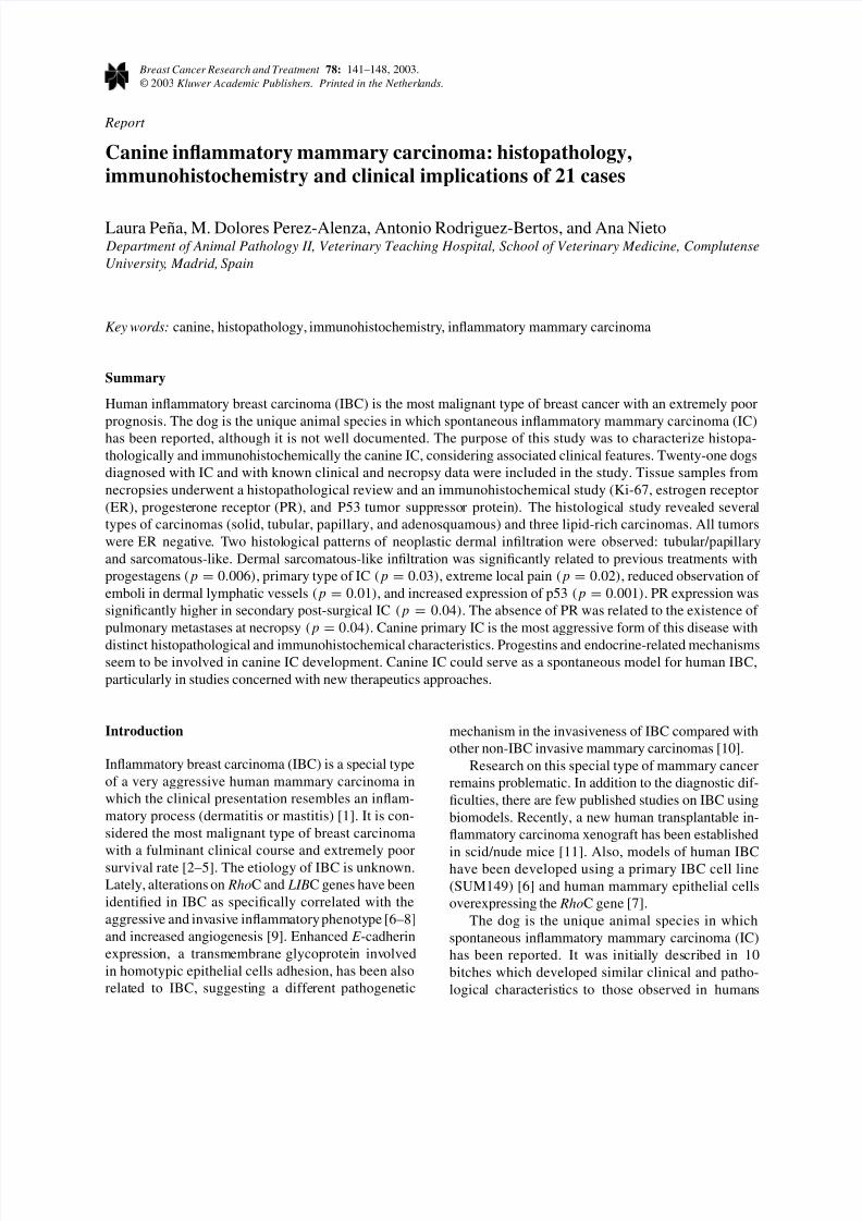
Histological diagnosis of the mammary gland tumorcausing IC was made in 20 cases after the reexami-nation of the necropsy samples. In one animal, therewere three different malignant mammary tumors (car-cinosarcoma, complex tubular carcinoma, and simpletubular carcinoma) containing malignant lymphaticemboli, and the tumor originating the IC could not beestablished. In this case, the interpretation of immuno-histochemistry was based on the immunoreactivityfound in neoplastic cells of emboli. The 20 mam-mary neoplasms were classied as simple carcinomas(solid, tubular, papillary, n = 15), adenosquamouscarcinomas (n = 2) , and lipid-rich carcinomas (n =
3) (Figure 1). The diagnosis of lipid-rich carcinoma(n = 3) was established when more than 80% of the tumor cells were lipid-producing (lipid dropletslled the cytoplasms). All the lipid-rich carcino-mas presented tubular disposition. The positive lipid
Figure 1. Canine inammatory carcinoma. Four views of alipid-rich carcinoma in different locations: (A) Mammary tumor.H&E. (B) Inltration of the dermis. H&E. (C) Lymph node metas-tasis. Small aggregate of lymphocytes remain on the left. H&E. (D)Detail of the dermal inltration. The neoplastic cells show multiplelipid droplets and cytological evidence of malignancy. H&E.
8/8/2019 carcinoma inflamatório
http://slidepdf.com/reader/full/carcinoma-inflamatorio 4/8
144 L Peña et al.
staining (Sudan III method) and negative glycogenstaining (Best’s Carmine method, frozen samplesavailable in 2 cases) conrmed the lipid nature of thesecretion. Multifocal groups of cells containing cyto-plasmatic lipid-droplets were found in many non-lipidrich tumors (14/20, 70.0%) with less than 80% of
cells with lipid droplets. Focal areas of cellular squa-mous transformation were additionally seen in threetumors. All mammary tumors were of a high histo-logical malignant grade (HMG III). Tubular formationwas minimal in 5 cases (solid tumors, < 10% of tu-bule formation), moderate in 6 cases (10–75% of tubule formation) and elevated in 9 cases ( > 75% of tubule formation). Nuclear pleomorphism was moder-ate (2 points) in 10 cases (50%) and marked (3 points)in another 10 cases (50%). Multinucleated cells wereseen in 8 tumors (40.0%). Mitotic rate was always el-evated (3 points). Atypic mitoses were observed in 14tumors (70.0%). Necrotic areas were absent (10/20,50.0%), moderate (5/20, 25.0%), or abundant (5/20,25.0%). Comedocarcinoma pattern (central necrosis)was found in 7 cases (35.0%). Marked perivascularbrosis surrounding embolized lymphatic vessels wasseen in some mammary tumors (10/20, 50.0%) andgeneralized desmoplastic stroma was only observed in1 case. Hemorrhages were seen in 6 cases (30.0%).Considering non-ulcerated areas, the presence of in-ammatory cells (lymphocytes, plasma cells, andmacrophages) was not relevant in any case; they weremoderate in the majority of cases (17/20, 85.0%) andvery low in the others (3/20, 15.0%).
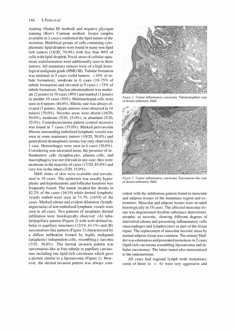
H&E slides of skin were available and reevalu-ated in 19 cases. The epidermis was usually hyper-plastic and hyperkeratotic and follicular keratosis wasfrequently found. The tumor invaded the dermis in82.2% of the cases (16/19) while dermal lymphaticvessels emboli were seen in 73.7% (14/19) of thecases. Marked edema and evident dilatation (lymph-angiectasia) of non-embolized lymphatic vessels wereseen in all cases. Two patterns of neoplastic dermalinltration were histologically observed: (A) tubu-lar/papillary pattern (Figure 2) with well-dened tu-bular or papillary structures (12/19, 63.1%) and (B)sarcomatous-like pattern (Figure 3) characterized bya diffuse inltration formed by highly malignant(anaplastic) independent cells, resembling a sarcoma(7/19, 36.8%). The dermal invasion pattern wassarcomatous-like in four tubular or papillary carcino-mas including one lipid-rich carcinoma which gavea picture similar to a liposarcoma (Figure 1). How-ever, the dermal invasion pattern was always coin-
Figure 2. Canine inammatory carcinoma. Tubular/papillary typeof dermal inltration. H&E.
Figure 3. Canine inammatory carcinoma. Sarcomatous-like typeof dermal inltration. H&E.
cident with the inltration pattern found in muscularand adipose tissues of the mammary region and ex-tremities. Muscular and adipose tissues were invadedhistologically in 19 cases. The affected muscular tis-sue was degenerated (hyaline substance deposition),atrophic or necrotic, showing different degrees of interstitial edema and presenting inammatory cells(macrophages and lymphocytes) as part of the tissue
repair. The replacement of muscular necrotic areas bynormal adipose tissue was common. The urinary blad-der was edematousand presented metastases in 2 cases(lipid-rich carcinoma resembling liposarcoma and tu-bular carcinoma). The latter tumor also metastatizedto the endometrium.
All cases had regional lymph node metastases;some of them (n = 6) were very aggressive and

8/8/2019 carcinoma inflamatório
http://slidepdf.com/reader/full/carcinoma-inflamatorio 5/8
Canine inammatory mammary carcinoma 145
Table 1. Immunohistochemistry of 21 canine inammatorycarcinomas a
Negative, Positivetotal (%)
+ ++ +++ Total (%)
ER 21( ∗) (100) 0 0 0 0 (0)
PR 6 (28.6) 3 6 6 15 (71.4)P53 2 (9.5) 7 6 6 19 (90.5)
a Ki-67 index: Mean = 34.86%; SEM = 2.28; Min =
17.36%; Max = 51%.
substituted completely the parenchyma. In 2 of the 3lipid-rich carcinomas, the lipid-producing cells wereobserved, as well, in metastatic lymph nodes but notin pulmonary metastases (Figure 1).
Several different types of tumors in internal organswere present in one dog: (1) an adenosquamous mam-mary carcinoma (IC) inltrating the skin with metas-tases in axillary and inguinal lymph nodes, spleen, andlungs; (2) a primary spleenic hemangiosarcoma withmetastases in the adrenal glands, peritoneum, lungs,and myocardium; (3) an unilateral adrenocortical ad-enoma and (4) a sebaceous gland adenoma in skin.Another animal presented unilateral cortical adrenalhyperplasia.
Immunohistochemistry results and statistical asso-ciations are summarized in Tables 1 and 2 respectively.
Discussion
The term inammatory breast carcinoma (IBC) wasinitially used in a clinical sense to describe a specialtype of locally advanced human mammary carcinomawhich simulated an inammatory condition of theskin [1, 21]. In the seventies, this cutaneous lesionwas found to be associated with an undifferentiatedbreast carcinoma with massive involvement of dermallymphatic vessels [3]. There are few reports concern-ing histological aspects of IBC. It is well known thatIBC is not a specic histologic subtype: inltratingductal carcinomas, other carcinomas and unspeciedmalignant tumors have been described as involvedwith IBC [7, 22, 23]. In the previous study whichincluded 10 cases of canine IC [12], ve differenttypes of adenocarcinomas (squamous, scirrhous andsolid) and one malignant mixed tumor were found.Several types of carcinomas were also found in ourstudy although, singularly, 3 cases of lipid-rich car-cinoma were diagnosed. Lipid-rich carcinoma has
been included in the most recent WHO’s classica-tion for mammary tumors of the dog [18], althoughit is not referenced in the veterinary literature. Lipid-rich carcinoma is considered a rare type of mammarycarcinoma in humans [1] and in dogs [18]. The signif-icance of the high proportion of lipid-rich carcinomas
found in this study is unknown, although some hor-monal inuences have been described in this type of carcinoma in the women [24].
Histological study of the skin demonstrated embo-lization of lymphatic dermal vessels in 14 of 19animals (73.7%). The histological exam showedtwo histological patterns of neoplastic dermal inva-sion: one of a tubular/papillary pattern with well-differentiated structures and one very anaplastic withindependent highly malignant cells resembling a sar-coma (sarcomatous-like type). There are no previousstudies on dermal characteristics of IC in humans ordogs. This lack of information can be attributed tothat human IBC specimens available for research areminute in size (from ne needle aspiration and Tru-cutskin biopsy) [25] or, in other cases, biopsies are takenafter chemotherapy has been instaured and the corre-sponding cutaneous lesion disappeared. In the presentstudy, samples were taken at necropsy and no previ-ous anti-neoplastic treatment was used (only palliativetreatments with anti-inammatories and antibiotics);thus, our results correspond to the untreated diseasein the dog. Furthermore, the possibility to performautopsies on humans is limited.
The tumor proliferation index measured by Ki-
67 immunostaining is considered a good prognosticfactor in canine malignant mammary tumors [26] butin the present study, the Ki-67 index was very elevatedin all cases and had no association with clinical orhistopathological variables.
Several studies on ER and PR expression in humanIBC have shown variable results. ER-positive casesvaried from 22 to 52% and PR varied from 16 to 34%[27–30]. In metastatic IBC, the proportion of steroidreceptors was low, with the PR being more expressedthan the ER (8.7% were ER positive and 17.4% werePR positive) [31]. In our study, in which all dogshad regional lymph node metastases, all cases wereER negative while 71.4% were PR positive. ReducedER expression has been previously described in caninemalignant mammary tumors [32]. In our cases, PR ex-pression was signicantly associated with secondaryIC while the absence of PR was related to the existenceof pulmonarymetastases at necropsy. Thisnding is inaccordance with previous reports of human IBC where

8/8/2019 carcinoma inflamatório
http://slidepdf.com/reader/full/carcinoma-inflamatorio 6/8
146 L Peña et al.
Table 2. Signicant variables associated to the type of clinical presentation and histological inltrationin canine inammatory carcinoma
Type of clinical presentation
Primary IC Secondary IC
Type of histological inltration (n = 19)
Tubular/papillary (n = 12) 3 (25%) 9 (75%) p < 0.05Sarcomatous-like (n = 7) 5 (71.4%) 2 (28.6%)
PR (n = 21)
Negative (n = 6) 4 (66.7%) 2 (33.3%) p < 0.05Positive (n = 15) 5 (33.3%) 10 (66.7%)
P53 (n = 21)
Negative/low (n = 9) 1 (11.1%) 8 (88.9%) p < 0.05Moderate/intense (n = 12) 8 (66.7%) 4 (33.3%)
Type of histological inltration
Tubular/papillary Sarcomatous-like
Previous treatments with progestins (n = 16)No (n = 11 ) 9 (81.8%) 2 (18.2%) p < 0.01Yes (n = 5) 0 (0%) 5 (100%)Unknown (n = 3)
Local pain (n = 17)
Moderate (n = 8) 7 (87.5%) 1 (12.5%) p < 0.05Extreme (n = 9) 3 (33.3%) 6 (66.6%)Unknown (n = 2)
Emboli in dermal lymphatics (n = 19)
No (n = 5) 1 (20%) 4 (80%) p < 0.05Yes (n = 14) 11 (78.6%) 3 (21.4%)
Pleomorphism (n = 19)Low/moderate (n = 9) 8 (88.9%) 1 (11.1%) p < 0.05
High (n = 10) 4 (40%) 6 (60%)
Multinucleated cells (n = 19)No (n = 11 ) 9 (81.8%) 2 (18.2%) p < 0.05Yes (n = 8) 3 (37.5%) 5 (62.5%)
P53 (n = 19)Negative/low (n = 9) 9 (100%) 0 (0%) p < 0.01Moderate/intense (n = 10) 3 (30%) 7 (70%)
combined ER/PR status are suggested as prognosticfactors [27, 30].
The immunohistochemical expression of the p53protein is due to mutations of the p53 tumor sup-pressor gene which has been associated with poorprognosis in human [33] and canine [34] malig-nant mammary tumors. P53 overexpression intensitywas related with a worse clinical and pathologicalaggressive behavior of the tumor in the present study(primary IC).
Our statistical analysis conrm that the two clinicalpresentations of IC found in the dog [13] (primary andsecondary) are different subtypes of IC with distincthistopathological and immunohistochemical charac-teristics. Despite the low number of cases includedin this study, justied by the low prevalence of thedisease, the signicance of the statistical associationsfound was very high. Further studies, including agreater number of cases, would be desirable in orderto conrm these results.

8/8/2019 carcinoma inflamatório
http://slidepdf.com/reader/full/carcinoma-inflamatorio 7/8
Canine inammatory mammary carcinoma 147
Research on human IBC has increased in the lastdecades although many problems still remain unre-solved. Although an animal model of IBC has beenrecently established in scid/nude mice [11], the dogis the unique animal species in which spontaneousIC has been reported. Several similarities have been
found between human and canine inammatory mam-mary carcinoma concerning histopathology, clinicalcharacteristics, and prevalence. Histological invasionof dermal lymphatic vessels is similar in the two spe-cies [13, 27]. In both species, the clinical features arecomparable, the two clinical presentations (primaryand secondary post-surgical) exist, and the clinicaloutcome in both species is very aggressive. CanineIC is a rare disease although its prevalence seems tohave increased in the last decades (from 4.4 to 7.6% of all mammary tumors [12, 13]) as has occurred in thenineties in the woman [35]. Both species are probablyunder the inuences of the same environmental andnutritional carcinogens, since the dog, as a companionanimal, share many aspects of the owner’s life. Thereported prevalence of IC in the dog among all ma-lignant mammary tumors (17.7%) [13] is higher thanthat reported in women [2, 22, 27, 35, 36]. In ouropinion, canine IC can serve as a spontaneous modelfor human IBC, being especially useful in studies onnew therapeutics approaches.
Acknowledgements
This work was supported in part by the researchproject of the Complutense University No PR-269/98-8178. We thank Dr Pedro Cuesta for assistance withstatistical analysis and Pedro Aranda for his technicalhelp.
References
1. Tavassoli FA: Pathology of the Breast. 2nd edn, McGraw-Hill,New York, 1999, pp 519–522, 538–541
2. Levine PH, Steinhorn SC, Ries LG, Levine AJ: Inammatorybreast cancer. The experience of the Surveillance, Epidemi-ology, and End Results (SEER) Program. J Natl Cancer Inst74: 291–297, 1985
3. Ellis DL, Teitelbaum SL: Inammatory carcinoma of thebreast: a pathologic denition. Cancer 33: 1045–1047, 1974
4. Somlo G, Doroshow JH, Forman SJ, Odom-Maryon T, LeeJ, Chow W, Hamasaki V, Leong L, Morgan Jr R, MargolinK, Raschko J, Shibata S, Tetef M, Yen Y, Simpson J, MolinaA: High-dose chemotherapy and stem cell rescue in thetreatment of high risk breast cancer: prognostic indicatorsof progression-free and overall survival. J Clin Oncol 15:2882–2893, 1997
5. Victor SJ, Horwitz EM, Kini VR, Martinez AA, PettingaJE, Dmuchowski CF, Decker DA, Wilner FM, Vicini FA:Impact of clinical, pathologic, and treatment-related factorsof patients with locally advanced breast cancer treated withmultimodality therapy. Am J Clin Oncol 22: 119–125, 1999
6. van Golen KL, Davies S, Wu ZF, Wang Y, Bucana CD, Root H,Chandrasekharappa S, Strawderman M, Ethier SP, MerajverSD: A novel putative low-afnity insulin-like growth factor-binding protein, LIBC (lost in inammatory breast cancer),and Rho C GTPase correlate with the inammatory breastcancer phenotype. Clin Cancer Res 5(9): 2511–2519, 1999
7. van Golen KL, Wu ZF, Qiao XT, Bao LW, Merajver SD: Rho CGTPase, a novel transforming oncogene for human mammaryepithelial cells that partially recapitulates the inammatorybreast cancer phenotype. Cancer Res 60(20): 5832–5838,2000 Oct 15
8. Kleer CG, van Golen KL, Zhang Y, Wu ZF, Rubin MA,Merajver SD: Characterization of Rho C expression in benignand malignant breast disease: a potential new marker for smallbreast carcinomas with metastatic ability. Am J Pathol 160(2):579–584, 2002
9. van Golen KL, Wu ZF, Qiao XT, Bao L, Merajver SD: Rho CGTPase overexpression modulates induction of angiogenicfactors in breast cells. Neoplasia 2(5): 418–425, 2000
10. Kleer CG, van Golen KL, Braun T, Merajver SD: PersistentE -cadherin expression in inammatory breast cancer. ModPathol 14(5): 458–464, 2001
11. Alpaugh ML, Tomlinson JS, Shao ZM, Barsky SH: A novelhuman xenograft model of inammatory breast cancer. CancerRes 59: 5079–5084, 1999
12. Susaneck SJ, Allen TA, Hoopes J, Withrow SJ, Macy DW:Inammatory mammary carcinoma in the dog. J Am An HospAssoc 19: 971–976, 1983
13. Perez-Alenza MD, Tabanera E, Peña L: Inammatory mam-mary carcinoma in dogs: 33 cases (1995–1999). J Am Vet MedAssoc 219(8): 1110–1114, 2001
14. Taylor GW, Meltzer A: Inammatory carcinoma of the breast.Ann Surg 33: 33–49, 1938
15. Richards FJ, Lewison EF: Inammatory carcinoma of the
breast. Surg Gynecol Obstet 113: 729–732, 196116. Attia-Sobol J, Ferriere JP, Cure H, Kwiatkowski F, AchardJL, Verrelle P, Feillel V, De Latour M, Lafaye C, Deloche Cet al.: Treatment results, survival and prognostic factors in109 inammatory breast cancers: univariate and multivariateanalysis. Eur J Cancer 29A: 1081–1088, 1993
17. Nishimura R, Koyama H, Kasumi F, Takashima S, KobayashiS, Komaki K, Ohkawa T, Shin E, Kodama H, FukutomiT, Nishi T, Sonoo H, Sano S, Kimishima I, Nakaue K,Nakamura S, Kusama M, Okumura K: A case control study onrisk factors involved in inammatory breast recurrence afterbreast-conserving surgery. Oncology 55: 391–399, 1998
18. Misdorp W, Else RW, Helmén E, Lipscomb TP: Histolog-ical Classication of Mammary Tumors of the Dog and Cat.Second Series. Vol 7, Armed Forces Institute of Pathology andWorld Health Organization, Washington, 1999
19. Elston CW, Ellis IO: Method for grading breast cancer. J ClinPathol 46: 189–190, 1993
20. Dixon W: BMDP Statistical Software. Release 7.0, Universityof California Press, Los Angeles, 1993
21. Chambler AF, Drew PJ, Hill AD, Darzi A, Monson JR:Inammatory breast carcinoma. Surg Oncol 4: 245–254,1995
22. Tardivon AA, Viala J, Corvellec Rudelli A, GuinebretiereJM, Vanel D: Mammographic patterns of inammatory breast

8/8/2019 carcinoma inflamatório
http://slidepdf.com/reader/full/carcinoma-inflamatorio 8/8
148 L Peña et al.
carcinoma: a retrospective study of 92 cases. Eur J Radiol24(2): 124–130, 1997
23. Dershaw DD, Moore MP, Liberman L, Deutch BM: Inam-matory breast carcinoma: mammographic ndings. Radiology190: 831–834, 1994
24. Tsubura A, Hatano T, Murata A, Shoji T, Shikata N, MoriiS: Breast carcinoma in patients receiving neuroleptic therapy.Morphologic and clinico-pathologic features of thirteen cases.Acta Pathol Japon 7: 494–499, 1991
25. IBC Research Foundation. Research of inammatorybreast cancer and related matters. Available from URL:http://www.ibcresearch.org/research/
26. Peña L, Nieto AI, Perez-Alenza MD, Cuesta P, Castaño M:Immunohistochemical detection of Ki-67 and PCNA in ca-nine mammary tumors: relationship to clinical and pathologicvariables. J Vet Diagn Invest 10: 237–246, 1998
27. Brooks HL, Mandava N, Pizzi WF, Shah S: Inammatorybreast carcinoma: a community hospital experience. J Am CollSurg 186: 622–629, 1998
28. Ueno NT, Buzdar AU, Singletary SE, Ames FC, McNeeseMD, Holmes FA Theriault RL, Strom EA, Wasaff BJ, AsmarL, Frye D, Hortobagyi GN: Combined-modality treatment of inammatory breast carcinoma: twenty years of experience atM.D. Anderson Cancer Center. Cancer Chemoth Pharm 40:321–329, 1997
29. Fleming RYD, Asmar L, Buzdar AU, McNeese MD, AmesFC, Ross MI: Effectiveness of mastectomy by response toinduction chemotherapy for control in inammatory breastcarcinoma. Ann Surg Oncol 4: 452–461, 1997
30. Wilke D, Colwell B, Dewar R: Inammatory breast car-cinoma: comparison of survival of those diagnosed clinically,
pathologically, or with both features. Am Surgeon 64:428–431, 1998
31. Atlan D, Chevallier B, Sheng RG: Tamoxifen for the treatmentof metastatic inammatory breast carcinoma. Am J Clin Oncol18: 74–77, 1995
32. Nieto A, Peña L, Perez Alenza MD, Sánchez MA, Flores JM,Castaño M: Immunohistologic detection of estrogen receptoralpha in canine mammary tumors: clinical and patholog-ical associations and prognostic signicance. Vet Pathol 37:239–247, 2000
33. Falette N, Paperin MP, Treilleux I, Gratadour AC, Peloux N,Mignotte H, Tooke N, Lofman E, Inganas M, Bremond A,Ozturk M, Puisieux A: Prognostic value of p53 gene muta-tion in a large series of node-negative breast cancer patients.Cancer Res 58: 1451–1455, 1998
34. Wakui S, Muto T, Yokoo K, Yokoo R, Takahashi H, MasaokaT, Hano H, Furusato M: Prognostic status of p53 gene muta-tion in canine mammary carcinoma. Anticancer Res 21:611–616, 2001
35. Chang S, Parker SL, Pham T, Buzdar A, Hursting SD: Inam-matory breast carcinoma incidence and survival: the surveil-lance, epidemiology, and end results program of the NationalCancer Institute, 1975–1992. Cancer 82: 2366–2372, 1998
36. Berg JW, Hutter RV: Breast cancer. Cancer 75(suppl 1):275–282, 1994
Address for offprints and correspondence: Dr Laura Peña, Depart-ment of Animal Pathology II, Veterinary Medicine School, Com-plutense University, 28040, Madrid, Spain; Tel.: + 34 91 3943740;Fax: + 34 91 3943808; E-mail: [email protected]