Embed Size (px)
Citation preview
UFSM
Dissertação de Mestrado
INFLUÊNCIA DO EXERCÍCIO FÍSICO SOBRE PARÂMETROS DE
COMPORTAMENTO E ESTRESSE OXIDATIVO EM MODELO
ANIMAL DE DISCINESIA TARDIA
Angélica Martelli Teixeira
PPGFarm
Santa Maria, RS, Brasil
2008
Generated by Foxit PDF Creator © Foxit Softwarehttp://www.foxitsoftware.com For evaluation only.

Livros Grátis
http://www.livrosgratis.com.br
Milhares de livros grátis para download.

INFLUÊNCIA DO EXERCÍCIO FÍSICO SOBRE PARÂMETROS DE
COMPORTAMENTO E ESTRESSE OXIDATIVO EM MODELO
ANIMAL DE DISCINESIA TARDIA
______________________________
por
Angélica Martelli Teixeira
Dissertação apresentada ao Programa de Pós-Graduação em Farmacologia da Universidade Federal de Santa Maria (UFSM, RS),
como requisito parcial para obtenção do grau de Mestre em Farmacologia.
Santa Maria, RS, Brasil
2008
Generated by Foxit PDF Creator © Foxit Softwarehttp://www.foxitsoftware.com For evaluation only.

Universidade Federal de Santa Maria Centro de Ciências da Saúde
Programa de Pós-Graduação em Farmacologia
A Comissão Examinadora, abaixo assinada, aprova a Dissertação de Mestrado
INFLUÊNCIA DO EXERCÍCIO FÍSICO SOBRE PARÂMETROS DE COMPORTAMENTO E ESTRESSE OXIDATIVO EM MODELO ANIMAL DE
DISCINESIA TARDIA
elaborada por
Angélica Martelli Teixeira
como requisito parcial para obtenção do grau de Mestre em Farmacologia
COMISSÃO EXAMINADORA:
_______________________
Marilise Escobar Bürger (Presidente/Orientadora)
_______________________ Paulo César Ghedini
(UNIMES)
_______________________ Luiz Fernando Freire Royes
(UFSM)
Santa Maria, 07 de março de 2008.
Generated by Foxit PDF Creator © Foxit Softwarehttp://www.foxitsoftware.com For evaluation only.

1
“Ainda que eu falasse línguas, as dos homens e dos anjos, se eu não tivesse o amor, seria
como sino ruidoso ou como címbalo estridente. Ainda que eu tivesse o dom da profecia, o
conhecimento de todos os mistérios e de toda a ciência; ainda que eu tivesse toda a fé, a
ponto de transportar montanhas, se não tivesse o amor, eu não seria nada.”
(1Cor 13,1-2)
Generated by Foxit PDF Creator © Foxit Softwarehttp://www.foxitsoftware.com For evaluation only.

v
AGRADECIMENTOS
À Deus-Trindade e a Mãezinha Maria, por terem me conduzido com todo cuidado e
carinho, por Seus ensinamentos sobre o Amor e por todas as pessoas colocadas em meu
caminho durante este trabalho.
Aos meus pais, Paulo e Aderoci, e toda minha família, pois sempre estiveram ao meu
lado torcendo e incentivando. Vocês são um presente de Deus, são meu porto-seguro.
À minha orientadora, Profª. Marilise Escobar Bürger, pela oportunidade, amizade e
entusiasmo, qualidades que a fazem brilhar mais. Obrigada por seus conselhos e por ter
compartilhado comigo seus conhecimentos. É um espelho profissional e pessoal nesta minha
caminhada.
Ao Prof. João Batista Teixeira da Rocha, que abriu as portas do seu laboratório e foi
mestre e amigo com seu humor extravagante e simplicidade no transmitir, característica dos
sábios.
Aos bolsistas do laboratório, por sua dedicação e cuidado com meus queridos ratinhos.
Sua amizade, comprometimento e alegria me ajudaram a chegar até aqui.
A Rose e aos bolsistas da Bioquímica que estiveram sempre prontos a ajudar.
Obrigada pelo trabalho e saber partilhados.
Ao professor Carlos Fernando de Mello e ao pessoal do Laboratório de Toxicologia e
Psicofarmacologia, pelo apoio técnico e por sua disposição e amizade.
Aos funcionários Rejane, Sandra, Florindo, Cleci e Berna, pelos momentos de
descontração de nossos almoços, pelos conselhos e carinho.
À Profª. Liliane Bauermann por seu apoio de laboratório, amizade e companheirismo.
Generated by Foxit PDF Creator © Foxit Softwarehttp://www.foxitsoftware.com For evaluation only.

vi
Ao Prof. Bernardo Baldisserotto pela acessibilidade e disponibilidade em ajudar e aos
demais professores do Programa de Pós-Graduação em Farmacologia, que contribuíram de
alguma forma para minha formação.
Ao CNPq e a CAPES pela bolsa de estudos e pelos recursos financeiros concedidos.
Aos animais utilizados, que foram os meios para realização deste trabalho, todo o meu
respeito e gratidão.
Enfim, agradeço à Universidade Federal de Santa Maria e ao Programa de Pós-
Graduação em Farmacologia pela possibilidade de realização deste curso.
Generated by Foxit PDF Creator © Foxit Softwarehttp://www.foxitsoftware.com For evaluation only.

vii
SUMÁRIO
LISTA DE ABREVIATURAS............................................................................................... viii
LISTA DE FIGURAS E TABELAS........................................................................................ ix
APRESENTAÇÃO.................................................................................................................... x
RESUMO.................................................................................................................................. xi
ABSTRACT........................................................................................................................... xiii
1.INTRODUÇÃO..................................................................................................................... 1
1.1. Estresse Oxidativo e Vulnerabilidade Cerebral.................................................................. 1
1.2. Defesas Antioxidantes......................................................................................................... 2
1.3. Doenças Neurodegenerativas e Estresse Oxidativo............................................................ 2
1.3.1. Doença de Alzheimer........................................................................................... 3
1.3.2. Doença de Parkinson............................................................................................ 3
1.3.3. Doença de Huntington.......................................................................................... 3
1.3.4. Esclerose lateral amiotrófica................................................................................ 3
1.3.5. Esquizofrenia e Discinesia Tardia........................................................................ 4
1.4. Atividade Física.................................................................................................................. 5
1.4.1. Efeitos benéficos do exercício.............................................................................. 5
1.4.2. Efeitos prejudiciais do exercício.......................................................................... 6
1.5. Modelo Animal de Estresse Oxidativo Induzido por Reserpina......................................... 7
2. OBJETIVOS......................................................................................................................... 9
3. ARTIGOS CIENTÍFICOS................................................................................................ 10
3.1. Artigo 1............................................................................................................................. 11
3.2. Artigo 2............................................................................................................................. 20
4. DISCUSSÃO E CONCLUSÃO FINAL........................................................................... 47
5. PERSPECTIVAS............................................................................................................... 50
6. REFERÊNCIAS BIBLIOGRÁFICAS............................................................................. 51
Generated by Foxit PDF Creator © Foxit Softwarehttp://www.foxitsoftware.com For evaluation only.
viii
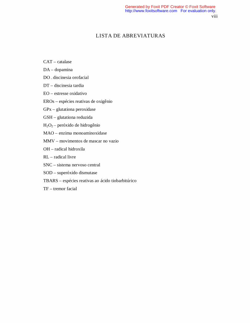
LISTA DE ABREVIATURAS
CAT – catalase
DA – dopamina
DO – discinesia orofacial
DT – discinesia tardia
EO – estresse oxidativo
EROs – espécies reativas de oxigênio
GPx – glutationa peroxidase
GSH – glutationa reduzida
H2O2 – peróxido de hidrogênio
MAO – enzima monoaminoxidase
MMV – movimentos de mascar no vazio
OH – radical hidroxila
RL – radical livre
SNC – sistema nervoso central
SOD – superóxido dismutase
TBARS – espécies reativas ao ácido tiobarbitúrico
TF – tremor facial
Generated by Foxit PDF Creator © Foxit Softwarehttp://www.foxitsoftware.com For evaluation only.
ix
LISTA DE FIGURAS E TABELAS
Introdução
Figura 1. Proposta do mecanismo de ação da reserpina e auto-oxidação da dopamina........... 8
Artigo 1
Figura 1. Effects of reserpine/vehicle administration on sedentary and exercised rats for A)
vacuous chewing frequency; B) duration of facial twitching…………….………………… 14
Figura 2. Linear regression between vacuous chewing frequency, facial twitching and
catalase
activity…………………………………...……….................................................................. 15
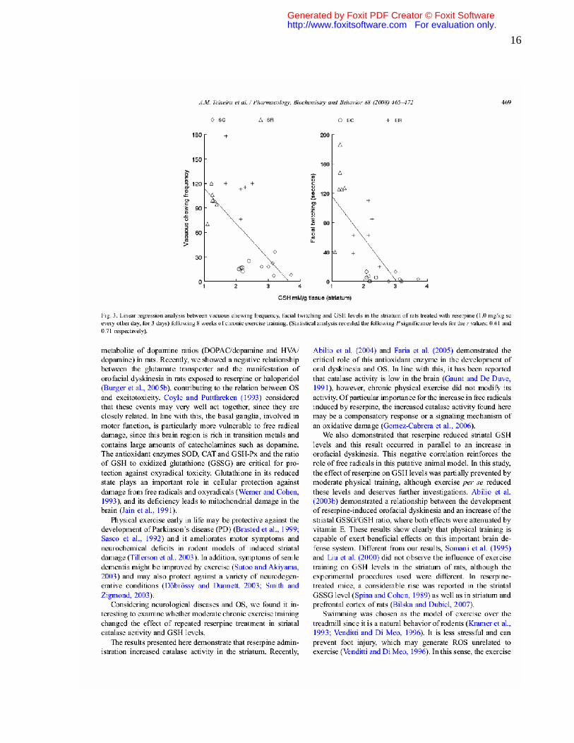
Figura 3. Linear regression between vacuous chewing frequency, facial twitching and GSH
levels……………..……………………...…………………………………………………... 16
Tabela 1. Catalase activity and GSH levels………………………………………………... 15
Artigo 2
Figura 1: Effects of reserpine/vehicle administration on sedentary and heavy exercised rats
for 1a) rearing frequency; 1b) locomotion frequency…………………………………….. 32
Figura 2: Effects of reserpine/vehicle administration on sedentary and heavy exercised rats
for 2a) vacuous chewing frequency 2b) facial twitching..………………………………... 33
Figura 3. Linear regression between 3a) TBARS and vacuous chewing frequency; 3b)
TBARS and catalase activity………………..……………………………………………… 34
Tabela 1: Verification of training: Mean values of heart weight, final body weight, heart
weight/body weigh ratios and blood lactate levels……......………………………………… 35
Tabela 2. Catalase activity and TBARS................................................................................. 36
Generated by Foxit PDF Creator © Foxit Softwarehttp://www.foxitsoftware.com For evaluation only.

x
APRESENTAÇÃO No item INTRODUÇÃO, está descrita uma revisão bibliográfica sobre os temas
trabalhados nesta dissertação. Os RESULTADOS estão apresentados sob a forma de artigos,
os quais se encontram no item ARTIGOS CIENTÍFICOS. As seções MATERIAIS E
MÉTODOS, RESULTADOS, DISCUSSÃO DOS RESULTADOS E REFERÊNCIAS
BIBLIOGRÁFICAS, encontram-se nos próprios artigos e representam a íntegra deste
estudo.
O item, DISCUSSÃO E CONCLUSÃO FINAL, encontrado no final desta
dissertação, apresenta interpretações e comentários gerais sobre os artigos científicos contidos
neste trabalho.
As REFERÊNCIAS BIBLIOGRÁFICAS referem-se somente às citações que
aparecem nos itens INTRODUÇÃO, DISCUSSÃO e CONCLUSÃO FINAL desta
dissertação.
Generated by Foxit PDF Creator © Foxit Softwarehttp://www.foxitsoftware.com For evaluation only.

xi
RESUMO
Dissertação de Mestrado Programa de Pós-Graduação em Farmacologia
Universidade Federal de Santa Maria, RS, Brasil
IFLUÊNCIA DO EXERCÍCIO FÍSICO SOBRE PARÂMETROS DE COMPORTAMENTO E ESTRESSE OXIDATIVO EM MODELO ANIMAL DE
DISCINESIA TARDIA
AUTORA: Angélica Martelli Teixeira ORIENTADORA: Drª Marilise Escobar Burger
LOCAL E DATA DA DEFESA: Santa Maria, março de 2008.
A atividade física praticada de maneira regular promove adaptações benéficas ao organismo, enquanto a inadequação do tempo e intensidade pode exceder a tolerância individual ao exercício gerando estresse oxidativo (EO). Estudos mostram esses efeitos em diversos órgãos como, por exemplo, coração e músculos, mas pouco se conhece sobre sua ação e mecanismos em nível cerebral. Diversas doenças neurológicas e neurodegenerativas estão associadas ao EO e neurotoxicidade. Considerando esses aspectos, o primeiro objetivo desse estudo foi determinar a influência do exercício crônico moderado em modelo de EO induzido por reserpina em ratos. Os animais foram submetidos a sessões diárias de natação com aumento gradual no tempo de treinamento e, após oito semanas, receberam duas doses de solução de reserpina ou controle (1 mg/kg-sc) em dias alternados. Fez-se avaliação comportamental, eutanásia dos animais e retirada da região estriatal do cérebro para determinação enzimática e bioquímica. A reserpina aumentou a freqüência dos movimentos de mascar vazio (MMV) e o tempo de tremor facial (TF); aumentou a atividade da catalase e diminui os níveis de glutationa reduzida (GSH). O exercício preveniu parcialmente o TF e houve recuperação parcial nos níveis de GSH, mas não modificou os efeitos sobre a catalase e MMV. Foi observada uma correlação positiva entre a atividade da catalase e o desenvolvimento de discinesia orofacial (DO), e uma correlação negativa entre GSH e DO. O segundo objetivo desse trabalho foi avaliar os efeitos de uma atividade física intensa sobre este mesmo modelo de EO. Os animais foram submetidos a onze semanas de natação (1 h/dia) com aumento gradual na carga de treinamento até que essa atingisse 7% de seu peso corporal. Realizaram-se as avaliações de comportamento, eutanásia e retirada do estriado para análises. A efetividade do treinamento foi confirmada através dos níveis diminuídos de lactato sérico e do desenvolvimento de hipertrofia cardíaca, observados nos animais exercitados. O exercício intenso reduziu a atividade locomotora e exploratória dos animais, demonstrando desenvolvimento de estresse emocional. Na presença de reserpina, o exercício elevou a peroxidação lipídica (TBARS) e provocou aumento na atividade da catalase, cujos parâmetros apresentaram correlação positiva. Com estes estudos se concluiu que a atividade física crônica de intensidade moderada foi capaz de melhorar as defesas antioxidantes nos distúrbios motores associados ao EO cerebral. Por outro lado, o exercício excessivo provocou alterações emocionais negativas e, quando na presença de um agressor adicional, modificou a
Generated by Foxit PDF Creator © Foxit Softwarehttp://www.foxitsoftware.com For evaluation only.
xii
capacidade antioxidante do cérebro, o que poderia agravar casos de doenças neurológicas e/ou neurodegenerativas associadas a processos oxidativos.
Palavras-chave: exercício, discinesia orofacial, reserpina, estresse oxidativo, neurodegeneração.
Generated by Foxit PDF Creator © Foxit Softwarehttp://www.foxitsoftware.com For evaluation only.

xiii
ABSTRACT
Dissertation of Master’s Degree Post-Graduate Course in Pharmacology
Federal University of Santa Maria, RS, Brazil
INFLUENCE OF PHYSICAL EXERCISE ON BEHAVIORAL PARAMETERS AND OXIDATIVE STRESS IN AN ANIMAL MODEL OF TARDIVE DYSKINESIA
AUTHOR: Angélica Martelli Teixeira ADVISOR: Marilise Escobar Bürger
PLACE AND DATE OF THE DEFENSE: Santa Maria, 2008
Regular practice of physical activity promotes beneficial effects to the body. However, excessive duration and intensity of exercise may surpass individual tolerance to exercise, generating oxidative stress (OS). Studies have shown these effects in various organs, such as the heart and muscles, but little is known about their action and mechanisms in the brain. Various neurological and neurodegenerative diseases are associated with OS and neurotoxicity. Considering these aspects, the first objective of this study was to determine the influence of chronic moderate exercise in an OS model induced by reserpine in rats. The animals were submitted to daily sessions of swimming, with a gradual increase in the length of training. After eight weeks, the animals received two injections of reserpine or control solutions (1 mg/kg-sc), alternately. A behavioral evaluation was performed, after which the rats were euthanized and the striatum was dissected for enzymatic and biochemical assays. Reserpine increased the vacuous chewing movements frequency (VCM) and facial twitching (FT), as well as catalase activity, but decreased reduced glutathione levels (GSH). Exercise partially prevented FT, and partially recovered GSH levels, but did not modify the effects on catalase and VCM. There was a positive correlation between catalase activity and orofacial dyskinesia (OD) and a negative correlation between GSH and OD. The second objective of this study was to evaluate the effects of an intense physical activity in the same model of OS. Rats were submitted to eleven weeks of swimming (1 h/day), where each rat’s load was increased according to its body weight until reaching 7% of its weight. Behavioral evaluations were performed before euthanasia and the striatum was then dissected for assays. The effectiveness of the training was confirmed through reduced levels of serum lactate and cardiac hypertrophy, observed in exercised animals. Intense exercise reduced the locomotor index and exploratory activity of the animals, demonstrating the development of emotional stress. In the presence of reserpine, exercise increased lipid peroxidation (TBARS) and caused an increase in catalase activity, which were positively correlated with each other. Based on the results, it was concluded that chronic physical activity of moderate intensity improved the antioxidant defenses in movement disorders associated with cerebral OS. On the other hand, excessive exercise caused negative emotional disorders and, in the presence of another aggressor agent, modified brain antioxidant capacity, which possibly could aggravate cases of neurological and/or neurodegenerative diseases associated with oxidative processes.
Keywords: exercise, orofacial dyskinesia, reserpine, oxidative stress, neurodegeneration.
Generated by Foxit PDF Creator © Foxit Softwarehttp://www.foxitsoftware.com For evaluation only.
1
1. INTRODUÇÃO
1.1 Estresse Oxidativo e Vulnerabilidade Cerebral Todos os organismos aeróbicos são suscetíveis ao estresse oxidativo (EO) devido ao fato
de espécies semi-reduzidas de oxigênio, radical superóxido (O2-●) e peróxido de hidrogênio
(H2O2) ser produzido na mitocôndria durante a respiração (Chance e cols., 1979). Essas espécies
são denominadas radicais livres (RL) e também podem ser geradas no citoplasma ou na
membrana celular sendo seu alvo relacionado ao sítio de formação (Anderson, 1996; Yu &
Anderson, 1997). O oxigênio é utilizado pela enzima mitocondrial citocromo oxidase em um
processo de redução tetravalente que resulta na formação de água (Halliwell & Gutteridge, 1999)
e estima-se que em torno de 98% do oxigênio consumido em organismos aeróbios seja reduzido
desta forma, sem a geração paralela de RL (Chance e cols., 1979). O cérebro é extremamente
sensível ao EO devido sua grande quantidade de ácidos graxos poliinsaturados e baixas defesas
antioxidantes, provendo ampla área para atuação da cascata de peroxidação lipídica (Lohr e cols.,
2003).
O Homem é constantemente exposto aos RL provenientes do meio ambiente (radiação
eletromagnética) e do metabolismo interno celular (aminoácidos excitatórios, neurotransmissores,
consumo de O2 mitocondrial, transporte de elétrons pela citocromo P450, atividade da
monoamino oxidase - MAO). Os aminoácidos excitatórios (glutamato) e neurotransmissores
(dopamina) cujo metabolismo produz RL ou espécies reativas são as principais fontes de EO no
cérebro (Gilgun-Sherki e cols., 2001). Em concentrações baixas e moderadas ocorrem os efeitos
benéficos dos RL como, por exemplo, na defesa contra agentes infecciosos e na resposta
mitogênica sendo utilizados como sinalizadores para a estimulação de antioxidantes e processos
de reparo celular (Pani e cols., 2000; Valko e cols., 2007). Por outro lado, quando a produção
excede a capacidade natural antioxidante é que ocorre o EO que, dependendo da extensão, pode
levar à morte celular por apoptose, pois as espécies reativas de oxigênio formadas (EROs)
oxidam componentes celulares vitais como lipídios, proteínas e DNA (Simonian & Coyle, 1996),
envolvidos em inúmeras doenças como câncer, inflamação, artrite reumatóide, doenças
neurodegenerativas, assim como nos processos de envelhecimento (Goodwin e cols., 1983; Beal,
1995; Perry e cols., 1997; Emerit e cols., 2004; Radak e cols., 2005; Valko e cols., 2007).
Generated by Foxit PDF Creator © Foxit Softwarehttp://www.foxitsoftware.com For evaluation only.

2
1.2 Defesas Antioxidantes
O delicado balanço entre benefícios e malefícios dos RL é de extrema importância para os
seres vivos, sendo alcançado através do mecanismo de “regulação redox”, mantendo a
homeostase celular. Este estado de equilíbrio é determinado pela razão entre produção e remoção
de RL através de antioxidantes endógenos e/ou exógenos (Dröge, 2002). Os mecanismos de
defesa antioxidante incluem a remoção de espécies reativas de oxigênio/nitrogênio e seus
precursores, inibição da formação de EROs, ligação aos íons metálicos necessários à catálise da
geração das EROs e regulação das defesas antioxidantes endógenas. O efeito eficaz dos
antioxidantes depende do tipo de radical gerado, do local de formação e da severidade do dano
causado (Halliwell, 1994; 1997).
O sistema de defesa antioxidante está dividido em enzimático e não enzimático. O
primeiro inclui as enzimas superóxido dismutase (SOD), catalase (CAT) e glutationa peroxidase
(GPx). A SOD catalisa a formação de H2O2 a partir do radical superóxido, enquanto a catalase
age na eliminação do H2O2 promovendo sua catálise até água. A GPx converte a glutationa
reduzida (GSH) à glutationa oxidada (GSSG), removendo H2O2 e formando água (revisado por
Lohr e cols., 2003). O sistema não enzimático inclui compostos sintetizados pelos seres vivos
como bilirrubina, ceruloplasmina, hormônios sexuais, melatonina, coenzima Q, ácido úrico, e
outros compostos presentes na dieta como ácido ascórbico (vitamina C), α-tocoferol (vitamina
E), β-caroteno (precursor da vitamina A) e grupos fenóis de plantas (flavonóides) (Schneider &
Oliveira, 2004).
1.3 Doenças Neurodegenerativas e Estresse Oxidativo
O cérebro é exposto durante toda a vida ao EO e vários distúrbios do sistema nervoso
central (SNC) estão associados à geração de RL e danos oxidativos, como causa primária ou
como conseqüência da própria doença. (Gilgun-Sherki e cols., 2001). Descrevemos abaixo
algumas destas doenças, com especial enfoque sobre a discinesia tardia:
Generated by Foxit PDF Creator © Foxit Softwarehttp://www.foxitsoftware.com For evaluation only.

3
1.3.1 Doença de Alzheimer
Caracterizada por degeneração neuronal e deterioração cognitiva especialmente em
pacientes idosos (Flynn & Runho, 1999), cuja fisiopatologia tem sido relacionada ao EO através
de diversas constatações como: aumento da peroxidação lipídica em áreas específicas de cérebro
(Lovell e cols., 1995); aumento na atividade da catalase, SOD, GPx e glutationa redutase na
região do hipocampo e amígdala (Zemlan e cols., 1989; Pappella e cols., 1992). Além disso,
Pappolla et al. (1998) evidenciaram que a proteína β-amilóide (principal constituinte da placa
senil) é neurotóxica e que tal toxicidade é mediada por RL.
1.3.2 Doença de Parkinson
Doença progressiva caracterizada por tremor, rigidez muscular, anormalidades posturais e
bradicinesia. Dados de estudos cerebrais postmortem indicam que o estresse oxidativo tem
fundamental importância nos neurônios dopaminérgicos da região nigro-estriatal (Fahn & Cohen,
1992). Espécies reativas de oxigênio podem ser produzidas durante o metabolismo normal de
dopamina, cujos produtos de oxidação se polimerizam para formar neuromelanina, que pode
também ser tóxica (Offen e cols., 1999). Além disso, existem várias evidências sobre a toxicidade
de dopamina em culturas de células, provocando uma morte celular programada (Ziv e cols.,
1994; Offen e cols., 1995).
1.3.3 Doença de Huntington
É uma doença progressiva, com perda maciça de neurônios estriatais (Bartzokis e cols.,
1999), com elevados níveis de ferro (Chen e cols., 1993). Estudos dão suporte à teoria de uma
disfunção metabólica associada ao EO na doença de Huntington (Gu e cols., 1996; Browne e
cols., 1997), assim como o envolvimento de uma ativação glutamatérgica excessiva, conduzindo
a produção de EROs (Olney & Gubareff, 1978).
1.3.4 Esclerose lateral amiotrófica
Caracterizada por uma degeneração seletiva e progressiva dos neurônios motores
inferiores da medula espinhal e dos neurônios motores superiores do córtex cerebral (Gilgun-
Sherki e cols., 2001). Detectou-se o conteúdo de proteína carbonilada (indicador de oxidação de
proteínas) elevado em 85% dos pacientes com a doença, indicando o envolvimento de EO em
Generated by Foxit PDF Creator © Foxit Softwarehttp://www.foxitsoftware.com For evaluation only.

4
todos os tipos de esclerose lateral amiotrófica (Coyle & Puttfarcken, 1993). Em pacientes com
doença esporádica foi observado um aumento acentuado de substâncias reativas ao ácido
tiobarbitúrico (TBARS) no plasma, as quais são produtos de peroxidação lipídica (Oteiza e cols.,
1997).
1.3.5 Esquizofrenia e Discinesia Tardia (DT)
A presença de níveis elevados de EROs é descrita tanto para a esquizofrenia como para a
DT induzida por antipsicóticos (Lohr e cols., 1990). A esquizofrenia é uma desordem psiquiátrica
que acomete aproximadamente 1% da população, e o envolvimento do EO na fisiopatologia da
doença é indicado pela presença de produtos de peroxidação lipídica no plasma e fluido cérebro-
espinhal de pacientes, e em níveis alterados de antioxidantes enzimáticos e não-enzimáticos
(Reynolds, 1992; Mahadik & Scheffer, 1996). A discinesia tardia (DT) é uma síndrome
extrapiramidal que pode desenvolver-se após uso prolongado de fármacos antipsicóticos
clássicos, manifestando-se através de movimentos involuntários anormais e repetidos da região
orofacial e pescoço, principalmente (American Psychiatric Association, 1994). Atualmente, a
síndrome é considerada um dos problemas psiquiátricos mais graves, desenvolvendo-se em cerca
de 20 a 25% dos pacientes que fazem uso de neurolépticos (Andreassen & Jorgensen, 2000).
Antipsicóticos clássicos utilizados no tratamento da esquizofrenia podem aumentar a
neurotransmissão glutamatérgica estriatal através do bloqueio de receptores pré-sinápticos de
dopamina (DA), promovendo EO e degeneração neuronal. Essa degeneração pode resultar em
lesões irreversíveis dos neurônios nigro-estriatais com conseqüente morte celular, o que
explicaria a irreversibilidade da DT (Elkashef & Wyatt, 1999; Sachdev e cols., 1999). Além
disso, regiões do cérebro como substância negra e gânglios basais contêm diferentes metais de
transição e altas concentrações de DA, aumentando a vulnerabilidade ao EO e o envolvimento
dessas regiões na patogênese de desordens degenerativas e do movimento (Hanig & Aprison,
1967; Larson e cols., 1979).
Generated by Foxit PDF Creator © Foxit Softwarehttp://www.foxitsoftware.com For evaluation only.

5
Como se pode evidenciar existe uma variedade de doenças relacionadas ao EO e nossas
defesas podem não ser completamente efetivas, considerando também o aumento da exposição
ambiental aos agentes pró-oxidantes. Deste modo, torna-se importante o estudo de novas
substâncias e/ou alternativas terapêuticas, já que o EO tem sido relacionado com a patogênese de
diferentes doenças neurológicas e neurodegenerativas, além do envolvimento em outras doenças
como as cardiovasculares, câncer, diabetes, isquemia/reperfusão e envelhecimento (Dhalla e
cols., 2000; Sayre e cols., 2001; Jenner 2003; Dalle-Donne e cols., 2006).
1.4 Atividade Física
1.4.1 Efeitos benéficos do exercício
A prática de atividade física tem sido cada vez mais reconhecida e recomendada por
médicos e estudiosos da área da Saúde, com evidente importância para a manutenção de uma boa
qualidade de vida. O mecanismo bioquímico pelo qual o exercício beneficia a saúde e bem estar,
incluindo a incidência de certas doenças e depressão ainda não é bem conhecido (Holloszy, 1993;
Sarna e cols., 1993; Blair e cols., 1995). Alguns estudos demonstram que a atividade física
crônica pode aumentar a produção de EROs especialmente no fígado e músculos (Ji & Fu, 1992;
Atalay e cols., 1996). Por outro lado, um treinamento regular é capaz de reduzir esta produção
frente a um exercício intenso, pois vários agentes de defesa contra o estresse oxidativo (enzimas
antioxidantes, vitamina C e E, GSH, etc.) estão presentes nas células e podem ser reforçados
durante o exercício crônico (Niess e cols., 1996; Leeuwenburgh e cols., 1997).
Os efeitos da relação exercício físico/estresse oxidativo sobre músculos e órgãos como
coração principalmente, são bem descritos. Foi demonstrada uma estreita conexão entre as
funções cardiovascular e cognitiva, sugerindo efeitos do exercício sobre a função cerebral (Hicks
& Birren, 1970). Ao manter a integridade cerebrovascular (McFarland, 1963) o exercício
aumenta os capilares (Black e cols., 1987) e as conexões dendríticas (Pysh & Weiss, 1979),
melhorando as funções cerebrais como na prevenção e/ou melhora do déficit cognitivo da velhice
(Chodzko-Zajko & Moore, 1994). Os efeitos benéficos do exercício são observados em modelos
animais através do aumento da plasticidade e neurogênese, resultando na proteção das funções
cerebrais, melhora do aprendizado e redução do EO cerebral (Fordyce & Wehner, 1993; Van
Generated by Foxit PDF Creator © Foxit Softwarehttp://www.foxitsoftware.com For evaluation only.

6
Praag e cols., 1999; Radak e cols., 2001a; Cotman & Berchtold, 2002; Molteni e cols., 2004). Na
doença de Parkinson até um nível moderado, o exercício melhora de forma significativa a
atividade motora, a cognição, o estado de humor e as atividades diárias dos pacientes (Hurwitz,
1989; Sunvisson e cols., 1997; Baatile e cols., 2000; Miyai e cols., 2000). Independente do grau
da referida doença, Kuroda e cols. (1990,1992) constataram uma redução na taxa de mortalidade
em pacientes submetidos ao exercício físico regular. Além disso, a atividade física está associada
a um menor risco para o desenvolvimento da doença de Alzheimer e demência de qualquer grau
(Broe e cols., 1990; Shimamura e cols., 1998; Laurin e cols., 2001), benefícios também
comprovados em portadores de esquizofrenia (Marder e cols., 2004; Connolly e Kelly, 2005).
Tem sido demonstrado que os efeitos benéficos da atividade física regular (crônica)
dependem de processos adaptativos iniciais, pois o exercício agudo estimula o estado redox
cerebral (Radal e cols., 2001a,b; 2002, 2003, 2004). De acordo com o exposto, os agentes
oxidantes produzidos pelo exercício podem agir como sinalizadores para a ativação de defesas
antioxidantes, enquadrando-se no conceito de “hormesis”, onde se discute que uma dose baixa de
determinada substância causaria uma adaptação celular benéfica enquanto uma dose elevada seria
prejudicial (Radak e cols, 2005; Ji e cols., 2006).
1.4.2 Efeitos prejudiciais do exercício
De acordo com a teoria da “hormesis”, Gomez-Cabrera e cols. (2006) demonstraram que
o exercício provoca uma importante regulação nas enzimas antioxidantes, e que uma produção
elevada de oxidantes pode causar dano às células. Peijie e cols. (2003) observaram em estudo
com animais, que o exercício extremo (overtraining) pode comprometer o sistema imunológico
celular e humoral, provocando alterações sobre a secreção hormonal e neurotransmissores de
estresse. Neste estudo, foi observado um aumento no conteúdo de beta-endorfina, dinorfina A,
arginina, vasopressina e ocitocina no hipotálamo e uma diminuição na hipófise, sugerindo um
aumento na produção de neuropeptídeos cerebrais relacionados ao estresse.
Tem sido descrito que o estresse emocional pode ativar tanto o sistema nervoso simpático
quanto o eixo hipotálamo-pituitário-adrenal (Ganong, 2001), aumentando a liberação de
noradrenalina, dopamina, epinefrina, fator liberador de corticotrofina e glicocorticóides em certas
áreas do cérebro e na circulação periférica (Kandel e cols., 2000). Além disso, o estresse
fisiológico e patológico pode aumentar a concentração de glicocorticóides no plasma e diminuir
Generated by Foxit PDF Creator © Foxit Softwarehttp://www.foxitsoftware.com For evaluation only.

7
sua capacidade de ligação aos receptores (Xu & Tan, 1990; Chen e cols., 1991). Howells e cols.
(2005) sugeriram através de observações comportamentais que o exercício voluntário oferece
neuroproteção, mas que agentes estressores são capazes de cancelar este benefício. Demonstrou-
se que o exercício exaustivo está relacionado a um aumento de RL em diferentes tecidos
biológicos (Reide e cols., 1992; Sastre e cols., 1992; Bejma e cols., 2000), podendo gerar
diferentes sintomas centrais como depressão, dificuldade de concentração, dores de cabeça e
perda de peso (Fry e cols., 1991).
1.5 Modelo Animal de Estresse Oxidativo Induzido por Reserpina
A reserpina vem sendo utilizada por diferentes laboratórios para induzir discinesia
orofacial (um modelo animal de discinesia tardia), relacionada à formação de RL e EO
(Raghavendra e cols., 2001; Dutra e cols., 2002; Abílio e cols., 2003; Carvalho e cols., 2003;
Burger e cols., 2003, 2004, 2005), e também como modelo experimental para a doença de
Parkinson (Colpaert, 1987; Menzaghi e cols., 1997; Dawson e cols., 2000). Está estabelecido que
a reserpina seja capaz de prevenir a estocagem de DA nas vesículas neuronais interferindo com o
transporte vesicular de monoaminas (bloqueio do transportador). Como resultado, ocorre
aceleração do catabolismo de DA citosólica através da enzima MAO, seguido da formação
simultânea de metabólitos acídicos e peróxido de hidrogênio, associados ao EO (Abílio e cols.,
2002; Burger e cols., 2003; Naidu e cols., 2004; Bilzka & Dubiel, 2007). Independente da ação
da MAO e do transportador pode ocorrer uma auto-oxidação da dopamina formando
aminocromo, um importante precursor de RL. Este metabólito poderá ter diferentes destinos
como à formação de subprodutos (neuromelanina, leucoaminocromo), descritos como fontes
geradoras de espécies reativas endógenas envolvidas em processos degenerativos (Figura 1).
Generated by Foxit PDF Creator © Foxit Softwarehttp://www.foxitsoftware.com For evaluation only.

8
Figura 1. Proposta do mecanismo de ação da reserpina e auto-oxidação da dopamina
(Fuentes e cols., 2007).
Generated by Foxit PDF Creator © Foxit Softwarehttp://www.foxitsoftware.com For evaluation only.

9
2. OBJETIVOS
2.1 Objetivo Geral
Considerando o envolvimento do estresse oxidativo no desenvolvimento de patologias
neurodegenerativas, este estudo propõe avaliar a influência do exercício físico em diferentes
intensidades sobre um modelo animal de estresse oxidativo, relacionado ao desenvolvimento de
discinesia orofacial.
2.2 Objetivos Específicos
- Avaliar o desenvolvimento de estresse oxidativo estriatal através da observação de
discinesia orofacial (tempo de tremor facial- TF e freqüência de movimentos de mascar vazio-
MMV) induzida por reserpina em animais submetidos ou não a sessões de exercício físico
moderado e intenso;
- Avaliação comportamental de animais submetidos ao exercício físico intenso através da
observação do índice locomotor (movimentação espontânea e freqüência de erguer as patas
dianteiras-“rearing”) em campo aberto (open-field);
- Avaliações bioquímica e enzimática de estresse oxidativo (TBARS, GSH, catalase) em
estriado (região cerebral envolvida em distúrbios de movimento) de animais submetidos ao
exercício físico moderado e intenso.
Generated by Foxit PDF Creator © Foxit Softwarehttp://www.foxitsoftware.com For evaluation only.
10
3. ARTIGOS CIENTÍFICOS
O artigo 1 está disposto na forma que foi publicado na edição do periódico
Pharmacology, Biochemistry and Behavior. O artigo 2 está disposto na forma submetida para
publicação.
Generated by Foxit PDF Creator © Foxit Softwarehttp://www.foxitsoftware.com For evaluation only.

11
3.1 – EFEITO DO EXERCÍCIO CRÔNICO MODERADO SOBRE O MODELO DE
ESTRESSE OXIDATIVO INDUZIDO POR RESERPINA EM RATOS
Artigo 1
INFLUENCE OF CHRONIC EXERCISE ON RESERPINE-INDUCED
OXIDATIVE STRESS IN RATS: BEHAVIORAL AND ANTIOXIDANT
EVALUATIONS
TEIXEIRA, A.M., TREVIZOL, F., COLPO, G., GARCIA, S.C., CHARÃO, M., PEREIRA,
R.P., FACHINETTO, R., ROCHA, J.B.T., BURGER, M.E.
Pharmacology, Biochemistry and Behavior 88 (2008) 465-472.
Generated by Foxit PDF Creator © Foxit Softwarehttp://www.foxitsoftware.com For evaluation only.
12
Generated by Foxit PDF Creator © Foxit Softwarehttp://www.foxitsoftware.com For evaluation only.

13
Generated by Foxit PDF Creator © Foxit Softwarehttp://www.foxitsoftware.com For evaluation only.

14
Generated by Foxit PDF Creator © Foxit Softwarehttp://www.foxitsoftware.com For evaluation only.

15
Generated by Foxit PDF Creator © Foxit Softwarehttp://www.foxitsoftware.com For evaluation only.
16
Generated by Foxit PDF Creator © Foxit Softwarehttp://www.foxitsoftware.com For evaluation only.
17
Generated by Foxit PDF Creator © Foxit Softwarehttp://www.foxitsoftware.com For evaluation only.
18
Generated by Foxit PDF Creator © Foxit Softwarehttp://www.foxitsoftware.com For evaluation only.

19
Generated by Foxit PDF Creator © Foxit Softwarehttp://www.foxitsoftware.com For evaluation only.
20
3.2 – EFEITO DO EXERCÍCIO INTENSO SOBRE UM MODELO ANIMAL DE
ESTRESSE OXIDATIVO
Artigo 2
BRAIN RESPONSE TO HEAVY EXERCISE IN AN ANIMAL MODEL OF
OXIDATIVE STRESS
TEIXEIRA, A., SANTOS, A., BONIATTI, T., REICKZIEGEL, P., MÜLLER, L., PEREIRA, R.,
ROOS, D., ROCHA, J.B.T., BURGER, M.E.
(Submetido à Brain Research Bulletin em 8/02/2008)
Generated by Foxit PDF Creator © Foxit Softwarehttp://www.foxitsoftware.com For evaluation only.

21
ABSTRACT
Regular physical activity exerts beneficial effects for mental and physical health,
but little is known about intense chronic exercise, which may cause an increase in brain
monoamines and oxidative stress, mainly in the striatal region. Both intense exercise
and reserpine can cause oxidative stress, mainly in regions such as the striatum. The
neurotoxicity developed by reserpina may be accessed through behavioral and
biochemical evaluations. Considering that moderate physical exercise is beneficial for
antioxidant defenses, the objective of this study was to evaluate the influence of an
intense exercise program in an animal model of oxidative stress. Male rats were
submitted to swimming training (1 hour/day, during eleven weeks), where each rat’s load
was increased according to its body weight until reaching 7% of its weight in the last
week of exercise. After the last training session, all the animals were treated with vehicle
or repeated reserpine solution (1mg/kg-sc). After behavioral evaluations, all rats were
sacrificed and the striatum was dissected for enzymatic and biochemical assays. The
effectiveness of the physical training was assessed by reduced lactate levels and
cardiac hypertrophy. The intense exercise per se reduced locomotor index,
demonstrating emotional stress development. Exercise and reserpine together
increased lipid peroxidation and striatal catalase activity (positively correlated with each
other). These results suggest that heavy exercise can promote oxidative stress and
emotional changes and when acting together with an oxidant agent (reserpine) its
harmful effects are not overcome by the brain.
Keywords: intense exercise, heavy exercise, orofacial dyskinesia, reserpine, oxidative
stress, neurotoxicity.
Generated by Foxit PDF Creator © Foxit Softwarehttp://www.foxitsoftware.com For evaluation only.
22
1. Introduction
Regular physical exercise exerts many benefits for mental and physical health,
reducing disease incidence, improving cognitive processes and increasing resistance to
brain injury [28,51,58]. Accumulated evidence has shown that regular physical activity
may increase antioxidant defenses against brain oxidative damage [60,75] promoting
antiapoptotic effects [81]. In this sense, physical activity can offer protection against
seizures [68] and neurodegenerative diseases [32,73,77], as well as alleviating
symptoms of anxiety and depression [45].
Of particular importance, the brain is more susceptible to oxidative stress (OS)
when compared to other organs or systems, because it contains high levels of
membrane lipids, excitotoxic amino acids, low levels of antioxidant defenses and mainly
autoxidizable neurotransmitters [31]. Free radicals are formed in the central nervous
system (CNS) as part of normal metabolic processes [30], but when the production of
reactive oxygen species (ROS) exceeds the ability of the antioxidant system to eliminate
them, oxidative damage occurs [34]. This subject is extremely important since the brain
is exposed throughout life to OS and there is evidence of the involvement of free
radicals, which arise from oxidative damage, in the pathophysiology of several diseases
of the brain and nervous system [26]. Among these diseases are included Alzheimer’s
[53], Parkinson’s [50,84], Huntington’s [11], schizophrenia [43] and tardive dyskinesia
[82].
There is growing evidence that the continuous presence of low ROS
concentrations may induce the expression of antioxidant enzymes, DNA repair
molecules and protein degrading enzymes, resulting in decreased OS [59]. This
phenomenon has been related to the hormesis concept (a low dose of a substance is
stimulatory and a high dose is inhibitory) and has been extended to the ROS generating
effects of exercise [36,59,83]. In this sense, free radicals may be beneficial since they
act as signals to enhance antioxidant defenses. Antioxidant enzymes, which constitute a
defense mechanism against free radicals, may also be affected by exercise [17].
Gomez-Cabrera et al. [27] indicated that when oxidants are produced at moderate
Generated by Foxit PDF Creator © Foxit Softwarehttp://www.foxitsoftware.com For evaluation only.
23
levels, they act as signals to adapt cells to exercise, but when overproduced they may
cause cellular damage.
Susuki et al. [78] observed an increase in brain lipid peroxidation caused by
voluntary exercise, while Radak et al. [58] showed an attenuation of the age-associated
decline in memory by regular swimming, and unmodified levels of lipid peroxidation.
These conflicting data have shown that exercise can alter the brain response in the
generation and inhibition of oxidative damage. Considering different forms and intensity
of physical activity, some research groups have demonstrated a relationship between
exhaustive exercise and the increased generation of ROS in different biological tissues
[8,62,66]. Excessive physical activity may lead to overtraining and generate
psychological symptoms that resemble clinical depression [5,12]. A concept of
overtraining is that it generally occurs after a long term period of strenuous exercise or
when the duration of exercise sessions increases abruptly [57]. Recently it was reported
that a stress-related condition may alter physiological and immunological functions
associated with biochemical abnormalities [6]. Besides depression, this effect can
generate alterations in emotionality leading to other symptoms such as impaired
concentration, anorexia, headache, low appetite and loss of body mass and/or motor
coordination [24].
Considering OS, reserpina is a monoamine depletor which acts preventing the
storage of DA in neuronal vesicles by interfering with the vesicular monoamine
transporter (VMAT), resulting in oxidative catabolism of cytosolic DA (by MAO). This
accelerated metabolism leads to the formation of metabolites and hydrogen peroxide,
associated with the OS process in dopaminergic neurons [1,9,13,48]. Besides the MAO
action, the autoxidation of DA is also precursor of O-quinone aminochrome, with
different pathways in DA neurons: a polymerization to neuromelanin and a one-electron
reduction to the leukoaminochrome O-semiquinone radical, which is thought to be one of
the major sources of endogenous reactive species involved in the degenerative
processes [25,54,55,67]. Particularly, the brain basal ganglia is rich in monoamines and
therefore more vulnerable to free radical damage leading to OS [42]. Experimentally,
Generated by Foxit PDF Creator © Foxit Softwarehttp://www.foxitsoftware.com For evaluation only.
24
reserpine has been used by various laboratories to study movement disorder related to
OS and neurodegenerative disease [2,9,15,21,48,56].
Recently, the beneficial effects of moderate chronic exercise were showed in an
animal model of reserpine-induced OS [79], however the effects of an intense exercise
program on oxidative damage and brain function continue to be unknown. The aim of
the present study was to evaluate the influence of intense (heavy) physical activity on
brain susceptibility to OS, either alone or in association with reserpina, an OS-inductor
(behavior, enzymatic and biochemical evaluation).
2. Method
2.1. Drugs
Reserpine (methyl reserpate 3,4,5-trimethoxybenzoic acid ester- Sigma
Chemical) was dissolved in glacial acetic acid and then diluted to a final concentration of
0.5% acetic acid with distilled water. The vehicle consisted of a 0.5% acetic acid
solution. These solutions were injected subcutaneously (sc), at a volume of 1.0 mL/kg
body weight.
2.2. Animals
Male Wistar rats weighing 270-360g (about 3-month of age) were used. Groups of
five animals were kept in Plexiglas cages with free access to food and water in a room
with controlled temperature (23ºC ± 1°C) and on a 12h-light/dark cycle with lights on at
7:00 a.m. The animals were maintained and used in accordance to the guidelines of the
Committee on Care and Use of Experimental Animal Resources, School of Veterinary
Medicine and Animal Science of the University of São Paulo, Brazil. The rats were
randomly assigned to four groups: sedentary-control (SC), sedentary-reserpine (SR),
exercise-control (EC), exercise-reserpine (ER).
2.3. Training protocol and experimental procedure
All exercised rats were subjected to swimming exercise in a plastic container
(depth 45 cm) under continuous supervision, with the water temperature set to 34ºC ±
Generated by Foxit PDF Creator © Foxit Softwarehttp://www.foxitsoftware.com For evaluation only.

25
1ºC, 1 h/day, 5 times per week during 11 weeks. The animals had a 1 week adaptation
period in which swimming time was increased every day so that, by the second week,
they could swim 1 h/day. Exercise intensity was then increased every week, until the
animals’ load reached 7% of their body weight (swimming with a weight fixed around
their abdomens) [16].
One day after the last training session, all the animals received an injection of
vehicle (0.5% acetic solution) or reserpine solution (1mg/kg body weight in 0.5% of acid
acetic solution), subcutaneously, for 3 days every other day, totaling two injections, in
accordance with the following protocol:
SC: sedentary rats - Vehicle
SR: sedentary rats -Reserpine solution;
EC: exercised rats - Vehicle
ER: exercised rats - Reserpine solution.
The animals’ weight was recorded weekly during the experiment.
2.4. Behavioral testing
On the fourth day (24h after the second reserpine or vehicle injection), all the rats
were observed for the quantification of orofacial dyskinesia (OD): The animals were
placed individually in cages (20x20x19cm) containing mirrors under the floor to allow
behavioral quantification when the animal was faced away from the observer. To
quantify the occurrence of oral dyskinesia, the incidence of vacuous chewing
movements (VCM) frequency and the duration of facial twitching (FT) was recorded
during 5 min after a period of 2 min adaptation (hand operated counters were
employed). Observers were blind to the drug treatments.
Open field test: The rats were placed individually in an open-field arena, for
measurement of locomotion (number of floor units entered) and rearing (number of times
that the animal stood on hind legs) frequencies for 5 min.
All behavioral experiments were conducted between 09:00 and 11:00 a.m.
Generated by Foxit PDF Creator © Foxit Softwarehttp://www.foxitsoftware.com For evaluation only.

26
2.5. Tissue preparation and oxidative stress parameters
After behavioral evaluation the rats were decapitated, the brains were removed
and put on ice, cut coronally at the caudal border of the olfactory tubercle. The striatum
was dissected from the anterior part, weighed and homogenized for analysis.
For catalase activity determination, brain homogenates were centrifuged at 3000
x g for 10 min and the supernatants were used for enzyme quantification. Catalase was
quantified by measuring the disappearance of H2O2 at 240 nm. One unit of catalase was
defined as the number of mol of H2O2 degraded per minute [4].
Lipid peroxidation was determined by measuring accumulation of thiobarbituric
acid reactive substances (TBARS) as described by Ohkawa et al. [52].
2.6. Statistical analysis
Data of orofacial movements (VCM and FT), TBARS and catalase activity were
analyzed by a two-way ANOVA (2 (sedentary/exercise) X 2 (control/reserpine)) followed
by Duncan’s multiple range test, when appropriated. Behavioral data obtained in open-
field (locomotion and rearing) were analyzed by Kruskal-Wallis analysis of variance
followed by the two-tailed Mann-Whitney U-test [69].
3- Results
Verification of training program: heart weight (mg), final body weight (g), heart
weight/body weight ratios (heart hypertrophy index) and lactate levels are shown in
Table 1. The exercised group showed heart hypertrophy [F(1,16)=9.75, P<0.05] and
reduced blood lactate levels [F(1,16)=17.51, P<0.01], indicating the effectiveness of the
exercise. Body weight was not modified by physical training.
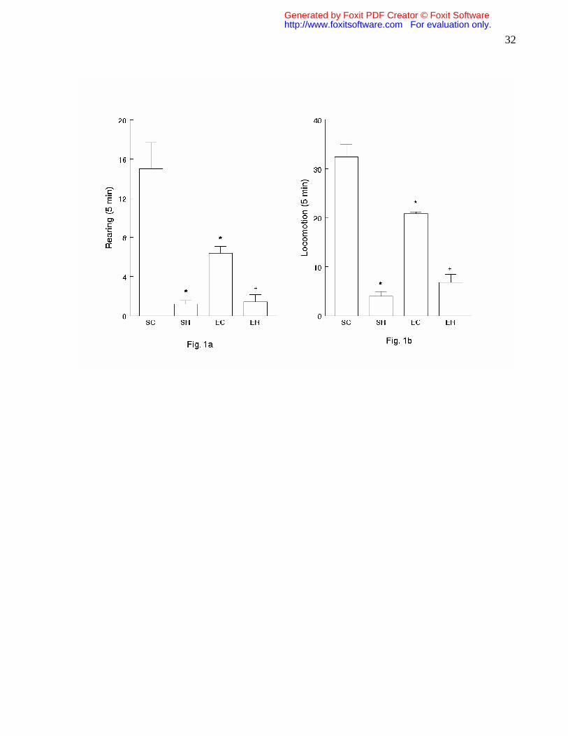
Open-field test: The effects of the intense exercise program on locomotor index
are represented by the number of crossing and rearing in the open-field test (Fig.1).
Kruskal-Wallis analyses of variance revealed significant differences in the two behavioral
parameters observed (H=16.5, P<0.001 and H=16.0, P<0.001, locomotion and rearing
respectively). Mann-Whitney U-test revealed that exercised rats (EC) displayed a
reduced number of crossings and rearings, when compared to the sedentary group
Generated by Foxit PDF Creator © Foxit Softwarehttp://www.foxitsoftware.com For evaluation only.

27
(SC). Reserpine treatment caused a marked reduction in crossing and rearing in both
sedentary (SR) and exercised (ER) rats (Figure 1a and 1b).
Orofacial movements: The effects of reserpine administration on orofacial
movements are shown in Fig.2. For VCM and FT, two way ANOVA showed only a
significant main effect of reserpine [F(1,16)=116.7; P<0.001] and [F(1,16)=147.77;
P<0.001], respectively. Univariate analysis, followed by Duncan’s multiple range test,
revealed that sedentary and exercised rats treated with reserpine (SR and ER)
displayed an increase in VCM and FT when compared to control groups (SC and EC). In
fact, intense exercise did not modify the effect of reserpine administration (Figure 2a and
2b).
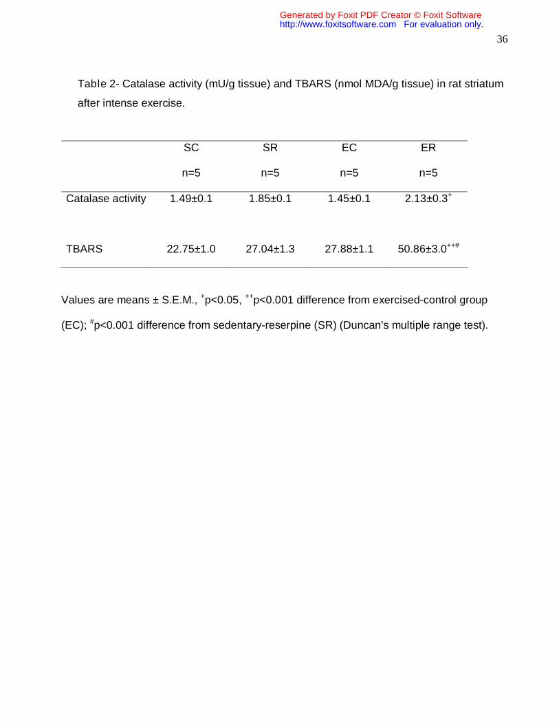
Biochemical analysis:
Two way ANOVA revealed a significant main effect of heavy exercise on catalase
activity [F(1,16)=8.21, P<0.05]. Univariate ANOVA followed by Duncan’s multiple range
test revealed that co-treatment of reserpine and exercise (ER) significantly increased
catalase activity when compared to the rat exercise-control group (EC) (Table 2)
Two way ANOVA of striatal TBARS levels revealed a significant main effect of
heavy exercise [F (1,16) =45.98; P<0.001], reserpine treatment [F (1,16)=40.76;
P<0.001] and interaction of exercise and reserpine [F (2,16)=19.16; P<0.001). Post hoc
comparisons by Duncan’s multiple range test indicated a significant difference in TBARS
levels in the exercise-reserpine treated rats (ER) when compared to the other groups
(SR and EC) (Table 2).
Regression analysis revealed a significant positive correlation between vacuous
chewing frequency (r=0.56, P<0.05, Fig. 3a), catalase activity (r=0.52, P<0.05, Fig. 3b)
and TBARS in striatum of rats.
Generated by Foxit PDF Creator © Foxit Softwarehttp://www.foxitsoftware.com For evaluation only.

28
4- Discussion
Physical exercise, as a stimulus, is dependent on the time of day, frequency (how
many times a day, or a week) and content (aerobic, weight bearing and so on), and
these variations have an influence on brain functions [40] leading to various
disturbances, including emotional stress, which can be measured by behavioral,
biochemical and genetic modifications [64]. Disturbances of emotionality were
considered in our study through behavioral evaluation in an open-field arena, which has
been used by other laboratories to study emotional and anxiolytic effects [19,37,80]. The
results presented here showed that animals submitted to intense exercise program were
more emotional (a less exploratory behavior) than the other groups, indicating that the
exercise intensity employed was strong enough to provoke the development of
emotional stress. Interestingly, we did not observe variation in the body weight of the
exercised animals at the end of the experiment, which may indicate a physiological
adaptation to this kind of exercise.
According to Kiraly & Kiraly [39], exercise is a major protective factor against
neurodegeneration of various etiologies. Regular physical training of moderate intensity
can have beneficial effects against senile dementia, Parkinson’s and Alzheimer’s
disease [10,65,73,77] as well as in status epilepticus [20,68] and can induce resistance
to OS [7,35]. In accordance with these findings, recently the beneficial effects of chronic
physical exercise (moderate intensity) were shown in an OS animal model, suggesting
beneficial effects in neurological diseases associated with movements [79]. After
behavioral observations, Howells et al. [32] confirmed that voluntary exercise can exert
neuroprotective effects, but also that mild stressors cancel this afforded neuroprotection.
According to Kandel et al. [38], any kind of stress can increase the release of
norepinephrine, DA, glutamate, epinephrine and corticotrophin releasing factor in certain
areas of the brain and peripheral circulation. More specifically, DA and glutamate have
the capacity to be neurotoxic during prolonged stress as they act synergistically to
promote neuronal loss [71,72].
Generated by Foxit PDF Creator © Foxit Softwarehttp://www.foxitsoftware.com For evaluation only.
29
Freed and Yamamoto [23] considered dopaminergic neurons to play important
roles during motor activation. In line with this, an increase in dopamine metabolism was
observed in several brain regions of rats during physical activity [46]. Theoretically,
exhaustive exercise may increase the brain’s synthesis of DA, whose metabolites,
formed by autoxidation or MAO action, could form ROS [77]. Fortifying this hypothesis,
some studies have indicated an increase in lipid peroxidation in different brain regions
after acute exercise [33,74].
Recently, Nybo and Secher [49] discussed the effects of strenuous exercise and
their relation with homeostatic disturbances, showing important consequences on the
cardiorespiratory, locomotive and mainly central nervous systems. Therefore, in view of
the sometimes conflicting data found in the literature, our group was motivated to study
the influence of intense exercise and its relationship to OS in the CNS.
Of particular interest for the animal model chosen here, different laboratories have
associated OS with neurodegeneration and movement disorders [14,47,63], while
searching for antioxidant therapeutic agents [2,13,22,61,70]. In fact, Sussman et al. [76]
showed that reserpine administration causes long-term spontaneous orofacial
dyskinesia (OD) in rats by decreasing striatal DA levels and increasing DA metabolite
ratios (DOPAC/DA and HVA/DA).
In a current study, moderate chronic exercise displayed a beneficial effect by
partially preventing the increase in FT [79]. In this experiment, we observed that the
reserpine treatment (used as an OS inductor) increased VCM and FT and that intense
physical activity did not alter these parameters. The exercise model employed here was
not able to change either catalase activity or TBARS striatal levels, but when associated
with the OS inductor (reserpine), increased these parameters. The harmful effect of the
association was confirmed through the positive correlation between OD development
(VCM) and lipid peroxidation (TBARS determination). Similarly, the positive correlation
between lipid peroxidation (TBARS) and catalase activity observed in our study may be
a compensatory response or a signaling mechanism of oxidative damage, since this
enzyme exerts a critical role in the development of oral dyskinesia and OS [3,22]. In fact,
different researchers have submitted rats to moderate chronic exercise and observed an
increase of enzymatic activity, including catalase activity, in cerebral and other tissues,
Generated by Foxit PDF Creator © Foxit Softwarehttp://www.foxitsoftware.com For evaluation only.

30
relating this effect to an increase of antioxidant defenses [18,29,41,75]. In our study, the
rats that were submitted chronically to an intense exercise program and co-treated with
reserpine, presented an increase of striatal catalase activity, suggesting OS
development. In line with our findings, Margonis et al. [44] explained a similar increase
of catalase activity by a compensatory mechanism to scavenge hydrogen peroxide in
times of greater demand (catalase increased only when overtraining and peak oxidative
stress levels occurred).
In conclusion, the results found here reinforce the relationship between the
generation of oxidative stress and a compensatory response toward aggressor agents,
which was more evident for the association of chronic intense exercise and reserpine
treatment. Further studies are necessary considering the activity of other enzymes and
endogenous antioxidants, as well as the relationship between physical training, the
release of dopamine and its effects on emotional stress.
Generated by Foxit PDF Creator © Foxit Softwarehttp://www.foxitsoftware.com For evaluation only.
31
Legends
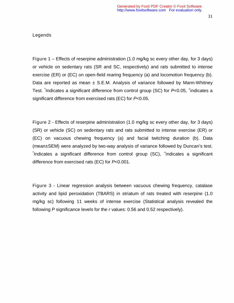
Figure 1 – Effects of reserpine administration (1.0 mg/kg sc every other day, for 3 days)
or vehicle on sedentary rats (SR and SC, respectively) and rats submitted to intense
exercise (ER) or (EC) on open-field rearing frequency (a) and locomotion frequency (b).
Data are reported as mean ± S.E.M. Analysis of variance followed by Mann-Whitney
Test. *Indicates a significant difference from control group (SC) for P<0.05, +indicates a
significant difference from exercised rats (EC) for P<0.05.
Figure 2 - Effects of reserpine administration (1.0 mg/kg sc every other day, for 3 days)
(SR) or vehicle (SC) on sedentary rats and rats submitted to intense exercise (ER) or
(EC) on vacuous chewing frequency (a) and facial twitching duration (b). Data
(mean±SEM) were analyzed by two-way analysis of variance followed by Duncan’s test.
*Indicates a significant difference from control group (SC), +indicates a significant
difference from exercised rats (EC) for P<0.001.
Figure 3 - Linear regression analysis between vacuous chewing frequency, catalase
activity and lipid peroxidation (TBARS) in striatum of rats treated with reserpine (1.0
mg/kg sc) following 11 weeks of intense exercise (Statistical analysis revealed the
following P significance levels for the r values: 0.56 and 0.52 respectively).
Generated by Foxit PDF Creator © Foxit Softwarehttp://www.foxitsoftware.com For evaluation only.
32
Generated by Foxit PDF Creator © Foxit Softwarehttp://www.foxitsoftware.com For evaluation only.

33
Generated by Foxit PDF Creator © Foxit Softwarehttp://www.foxitsoftware.com For evaluation only.

34
Generated by Foxit PDF Creator © Foxit Softwarehttp://www.foxitsoftware.com For evaluation only.

35
Table 1: Verification of training: Mean values of heart weight, final body weight, heart
weight/body weight ratios and blood lactate levels.
Group
Heart Wt. (mg) Body Wt. (g) Ratio (mg/g) Lactate
(mmol/L)
S
997.80±60.8 353.80±25.6 2.83±0.11 5.05±0.17
E
1317.60±55.4* 385.80±12.1 3.42±0.15 ** 3.94±0.2*
Data (mean ± S.E.M.) were analyzed by one-way analysis of variance followed by
Duncan’s test.
* (P<0.01) and **(P<0.05) difference from sedentary-control group (SC)
Generated by Foxit PDF Creator © Foxit Softwarehttp://www.foxitsoftware.com For evaluation only.
36
Table 2- Catalase activity (mU/g tissue) and TBARS (nmol MDA/g tissue) in rat striatum
after intense exercise.
SC
n=5
SR
n=5
EC
n=5
ER
n=5
Catalase activity 1.49±0.1 1.85±0.1 1.45±0.1 2.13±0.3+
TBARS 22.75±1.0 27.04±1.3 27.88±1.1 50.86±3.0++#
Values are means ± S.E.M., +p<0.05, ++p<0.001 difference from exercised-control group
(EC); #p<0.001 difference from sedentary-reserpine (SR) (Duncan’s multiple range test).
Generated by Foxit PDF Creator © Foxit Softwarehttp://www.foxitsoftware.com For evaluation only.
37
References
[1] V.C. Abílio, J.A.R. Vera, L.S.M. Ferreira, C.R.M. Duarte, C.R. Martins, D. Torres-
Leite, R.D.A. Ribeiro, R. Frussa-Filho. Effects of melatonin on behavioral dopaminergic
supersensitivity. Life Sci 72 (2003) 3003-3015.
[2] V.C. Abílio, C.C. Araujo, M. Bergamo, P.R. Calvente, V. D’Almeida, R. Ribeiro, R.
Frussa-Filho. Vitamin E attenuates reserpine-induced oral dyskinesia and striatal
oxidized glutathione/reduced glutathione ratio (GSSG/GSH) enhancement in rats. Prog
Neuro-Psychopharmacol Biol Psychiat 27 (2003) 109-114.
[3] V.C. Abílio, R.H. Silva, R.C. Carvalho, C. Grassl, M.B. Calzavara, S. Registro, V.
D’almeida, R.A. Ribeiro, R. Frussa-Filho. Important role of striatal catalase in aging and
reserpine-induced oral dyskinesia. Neuropharmacology 47 (2004) 263-272.
[4] U. Aebi, W. Chiu, R. Milligan. Role of catalase on antioxidative defenses. J Struct Biol
2 (1995) 117-118.
[5] L. Armstrong. J. Van Heest. The unknown mechanism of the overtraining syndrome:
clues from depression and psychoneuroimmunology. Sports Med. 32 (2002) 185-209.
[6] A. Angeli, M. Minetto, A. Dovio, P. Paccotti. The overtraining syndrome in athletes: a
stress-related disorder. J Endocrinol Invest 27 (2004) 603-612.
[7] A.K. Banerjee, A. Mandal, D. Chanda, S. Chakraborti. Oxidant, antioxidant and
physical exercise. Mol Cell Biochem 253 (2003) 307-312.
[8] J. Bejma, P. Ramires, L.L. Ji. Free radical generation and oxidative stress with aging
and exercise: differential effects in the myocardium and liver. Acta Physiol Scand 169
(2000) 343-351.
Generated by Foxit PDF Creator © Foxit Softwarehttp://www.foxitsoftware.com For evaluation only.

38
[9] A. Bilska, M. Dubiel. Alpha-lipoic acid differently affects the reserpine-induced
oxidative stress in the striatum and prefrontal cortex of rat brain. Neuroscience 146
(2007) 1758-1771.
[10] P.J. Brasted, C. Watts, E.M. Torres, T.W. Robbins, S.B. Dunnett. Behavioural
recovery following striatal transplantation: effects of postoperative training and P-zone
volume. Exp Brain Res 128 (1999) 535-538.
[11] S.E. Browne, A.C. Bowling, U. MacGarvey, M.J. Baik, S.C. Berger, M.M. Muqit, E.D.
Bird, M.F. Beal. Oxidative damage and metabolic dysfunction in Huntington’s disease:
selective vulnerability of the basal ganglia. Ann Neurol 41 (1997) 646-653.
[12] R. Budgett. Fadigue and underperformance in athletes: the overtraining syndrome.
Br J Sports Med 32 (1998) 107-110.
[13] M. Burger, A. Alves, L. Callegari, F.R. Athayde, C.W. Nogueira, G. Zeni, J.B.T.
Rocha. Ebselen attenuates reserpine-induced orofacial dyskinesia and oxidative stress
in rat striatum. Prog Neuro-Psychopharmacol Biol Psychiat 27 (2003) 135-140.
[14] J.L. Cadet, L.A. Kahler. Free radical mechanisms in schizophrenia and tardive
dyskinesia. Neurosci Biobehav 18 (1994) 457-467.
[15] J.P.M.V. Castro, R. Frussa-Filho, D.F. Fukushiro, R.H. Silva, W.A. Medrano, R.
Ribeiro, V.C. Abílio. Effects of baclofen on reserpine-induced vacuous chewing
movements in mice. Brain Res Bull 68 (2006) 436-441.
[16] S.B. Chaumont, V. Maupoil, J.J. Lahet, A. Berthelot. Effect of exercise training on
metallothionein levels of hypertensive rats. Med. Sci. Sports Exerc. 33 (2001) 724-728.
[17] P.M. Clarkson. Antioxidant and physical performance. Crit Res Food Sci Nutr 35
(1995) 131-141.
Generated by Foxit PDF Creator © Foxit Softwarehttp://www.foxitsoftware.com For evaluation only.

39
[18] S.A. Devi, T.R. Kiran. Regional responses in antioxidant system to exercise training
and dietary vitamin E in aging rat brain. Neurobiol Aging 25 (2004) 501-508.
[19] RK Dishman, AL Dunn, SD Youngstedt, JM Davis, ML Burgess, SP Wilson, MA
Wilson. Increased open-field locomotion and decreased striatal GABAA binding after
activity whell running. Physiol Behav 60 (1996) 699-705.
[20] J.S. Dubow, J.P. Kelly. Epilepsy in sports and recreation. Sports Med 33 (2003)
499–516.
[21] R.C. Dutra, A.P. Andreazza, R. Andreatini, S. Tufik, M.A.B.F. Vital. Behavioral
effects of MK-801 on reserpine-treated mice. Prog Neuro-Psychopharmacol Biol
Psychiat 26 (2002) 487-495.
[22] R.R. Faria, V.C. Abilio, C. Grassl, C.C. Chinen, L.T.R. Negrão, J.P.M.V. Castro,
D.F. Fukushiro, M.S.D. Rodrigues, P.H.Z. Gomes, S. Registro, R.C. Carvalho, V.
D’Almeida, R.H. Silva, R.A. Ribeiro, R. Frussa-Filho. Beneficial effects of vitamin C and
Vitamin E on reserpine-induced oral dyskinesia in rats: critical role of striatal catalase
activity. Neuropharmacology 48 (2005) 993-1001.
[23] C. Freed, B. Yamamoto. Regional brain dopamine metabolism: a marker for the
speed, direction and posture of moving animals. Science 229 (1985) 62-65.
[24] R.W. Fry, A.R. Morton, D. Keast. Overtraining in athletes: an update. Sports Med 12
(1991) 32-65.
[25] P. Fuentes, I. Paris, M. Nassif, P. Caviedes, S. Segura-Aguilar. Inhibition of VMAT-2
and DT-diaphorase induced cell death in a substantia nigra-derived cell line-na
experimental cell model for dopamine toxicity studies. Chem Res Toxicol 20 (2007) 776-
783.
Generated by Foxit PDF Creator © Foxit Softwarehttp://www.foxitsoftware.com For evaluation only.

40
[26] Y. Gilgun-Sherki, E. Melamed, D. Offen. Oxidative stress induced-
neurodegenerative diseases: the need for antioxidants that penetrate the blood barrier.
Neuropharmacology 40 (2001) 959-975.
[27] M.C. Gomez-Cabrera, L.L. Domenech, L.L. Ji, J. Viña. Exercise as an antioxidant:
up-regulates important enzymes for cell adaptations to exercise. Sci Sports 21 (2006)
85-89.
[28] G.S. Griesbach, D.A. Hovda, R. Molteni, A. Wu, F. Gomes-Pinilha. Voluntary
exercise following traumatic brain injury: brain-derived neurotrophic factor upregulation
and recovery of function. Neuroscience 125 (2004) 129-139.
[29] F. Gündüz, U.K. Sentürk, O. Kuru, B. Aktekin, M.R. Aktekin. The effect of one year’s
swimming exercise on oxidant stress and antioxidant capacity in aged rats. Physiol Res
53 (2004) 171-176.
[30] B. Halliwell. Reactive oxygen species and the central nervous system. J Neurochem
59 (1992) 1609-1623.
[31] B. Halliwell, J.M.C. Gutteridge. Free radicals in biology and medicine. Third ed,
Oxford University Press, New York, 1999, pp 645-660.
[32] F.M. Howells, V.A. Russell, M.V. Mabandla, L.A. Kellaway. Stress reduces
neuroprotective effect of exercise in a rat model of Parkinson’s disease. Behav Brain
Res 165 (2005) 210-220.
[33] K. Husain, S.M. Somani. Influence of exercise and ethanol on cholinesterase
activity and lipid peroxidation in blood and brain regions of rat. Prog Neuro-
Psychopharmacol Biol Psychiatry 21 (1997) 659-670.
Generated by Foxit PDF Creator © Foxit Softwarehttp://www.foxitsoftware.com For evaluation only.
41
[34] R. Jenkins, A. Goldfarb. Introduction: Oxidant stress, aging, and exercise. Med Sci
Sports Exer 25 (1993) 210-212.
[35] L.L. Ji. Antioxidants and oxidative stress in exercise. Proc Soc Exp Biol Med 222
(1999) 283-292.
[36] L.L. Ji, M.C. Gomez-Cabrera, J. Viña. Exercise and hormesis: activation of cellular
antioxidant signaling pathway. Ann NY Acad Sci 1067 (2006) 425-435.
[37] D.L. Jones, G.J. Mogenson, M. Wu. Injections of dopaminergic, cholinergic,
serotoninergic and GABAergic drugs into the nucleus accumbens: Effects on locomotor
activity in the rat. Neuropharmacology 20 (1981) 29-37.
[38] E.R. Kandel, J.H. Schwartz, T.M. Jessell. Principles of neural science. United
States: McGraw-Hill, 2000.
[39] M.A. Kiraly, S.J. Kiraly. The effect of exercise on hippocampal integrity: review of
recent research. Int J of Psychiatry Med 35 (2005) 75-89.
[40] J.A. Kleim, T.A. Jones, T. Schallert. Motor enrichment and the induction of plasticity
before or after brain injury. Neurochem Res 28 (2003) 1757-1769.
[41] J. Liu, H.C. Yeo, E. Övervik-Douki, T. Hagen, S.J. Doniger, D.W. Chu, G.A. Brooks,
B.N. Ames. Chronically and acutely exercised rats: biomarkers of oxidative stress and
endogenous antioxidants. J Appl Physiol 89 (2000) 21-28.
[42] J.B. Lohr, R. Kuczenski, A.B. Niculescu. Oxidative mechanisms and tardive
dyskinesia. CNS Drugs 17 (2003) 47-62.
[43] S.P. Mahadik, R.E. Scheffer. Oxidative injury and potential use of antioxidants in
schizophrenia. Prostagl Leuk Ess Fatty Acids 55 (1996) 45-54.
Generated by Foxit PDF Creator © Foxit Softwarehttp://www.foxitsoftware.com For evaluation only.

42
[44] K Margonis, IG Fatouros, AZ Jamurtas, MG Nikolaidis, I Douroudos, A
Chatzinikolaou, A Mitrakou, G Mastorakos, I Papassotiriou, K Taxildaris, D Kouretas.
Oxidative stress biomarkers responses to physical overtraining: Implications for
diagnosis 43 (2007) 901-910.
[45] E.W. Martinsen, A. Medhus, L. Sandvik. Effects of aerobic exercise on depression,
a controlled study. Br Med J (Clin res ed) 291 (1985) 109.
[46] R. Meeusen, K. De Meirleir. Exercise and brain neurotransmission. Sports Med 20
(1995) 160-188.
[47] P.S. Naidu, A. Singh, P. Kaur, R. Sandhir, S.K. Kulkarni. Possible mechanism of
action in melatonin attenuation of haloperidol-induced orofacial dyskinesia. Pharmacol
Biochem Behav 74 (2003) 641-648.
[48] P.S. Naidu, A. Singh, S.K. Kulkarni. Reversal of reserpine-induced orofacial
dyskinesia and cognitive dysfunction by quercetin. Pharmacology 70 (2004) 59-67.
[49] L. Nybo, N.H. Secher. Cerebral perturbations provoked by prolonged exercise. Prog
Neurobiol 72 (2004) 223-261.
[50] D. Offen, S. Gorodin, E. Melamed, J. Hanania, Z. Malik. Dopamine melanin is
actively phagocytized by PC12 cells and cerebellar granular cells: possible implications
for the etiology of Parkinson’s disease. Neurosci Lett 260 (1999) 101-104.
[51] H. Ogonovszky, I. Berkes, S. Kumagai, T. Kaneko, S. Tahara, S. Goto, Z. Radak.
The effects of moderate, strenuous and over-training on oxidative stress markers, DNA
repair, and memory, in rat brain. Neurochem Int 46 (2005) 635-640.
[52] H. Ohkawa, H. Ohishi, K. Yagi. Assay for lipid peroxide in animal tissue by
thiobarbituric acid reaction. Anal Biochem 95 (1979) 351-357.
Generated by Foxit PDF Creator © Foxit Softwarehttp://www.foxitsoftware.com For evaluation only.
43
[53] M.A. Pappolla, Y.J. Chyan, R.A. Omar, K. Hsiao, G. Perry, M.A. Smith, P. Bozner.
Evidence of oxidative stress and in vivo neurotoxicity of beta-amyloid in a transgenic
Mouse model of Alzheimer’s disease: a chronic oxidative paradigm for testing
antioxidant therapies in vivo. Am J Pathol 152 (1998) 871-877.
[54] I. Paris, A. Dagnino-Subiabre, K. Marcelain, L.B. Bennett, P. Caviedes, R.
Caviedes, C.O. Azar, J. Segura-Aguilar. Copper neurotoxicity is dependent on
dopamine-mediated copper uptake and one-electron reduction of aminochrome in a rat
substantia nigra neuronal cell line. J Neurochem 77 (2001) 519-529.
[55] I. Paris, P. Martinez-Alvarado, S. Cardenas, C. Perez-Pastene, R. Graumann, P.
Fuentes, C. Olea-Azar, P. Caviedes, J. Segura-Aguilar. Dopamine-dependent iron
toxicity in cells derived from rat hypothalamus. Chem Res Toxicol 18 (2005) 415-419.
[56] M.F. Peixoto, N.P. Araujo, R.H. Silva, J.P.M.V. Castro, D.F. Fukushiro, R.R. Faria,
P.H. Zanier-Gomes, W.A. Medrano, R. Frussa-Filho, V.C. Abílio. Effects of gabaergic
drugs on reserpine-induced oral dyskinesia. Behav Brain Res 160 (2005) 51-59.
[57] C. Petibois, G. Cazorla, J.R. Poortmans, G. Deleris. Biochemical aspects of
overtraining in endurance sports: the metabolism alteration process syndrome. Sports
Med 33 (2003) 83-94.
[58] Z. Radak, T. Kaneko, S. Tahara, H. Nakamoto, J. Pucsok, M. Sasvari, C. Nyakas,
S. Goto. Regular exercise improves cognitive function and decreases oxidative damage
in rat brain. Neurochem Int 38 (2001) 17-23.
[59] Z. Radak, H.Y. Chung, S. Goto. Exercise and hormesis: oxidative stress-related
adaptation for successful aging. An Hypothesis. Biogerontology 6 (2005) 71-75.
Generated by Foxit PDF Creator © Foxit Softwarehttp://www.foxitsoftware.com For evaluation only.

44
[60] Z. Radak, A. Toldy, Z. Szabo, S. Siamilis, C. Nyakas, G. Silye, J. Jakus, S. Goto.
The effects of training and detraining on memory, neurotrophins and oxidative stress
markers in rat brain. Neurochem Int 49 (2006) 387-392.
[61] V. Raghavendra, P.S. Naidu, S.K. Kulkarni. Reversal of reserpine-induced vacuous
chewing movements in rats by melatonin: involvement of peripheral benzodiazepine
receptors. Brain Res 904 (2001) 149-152.
[62] M.B. Reid, K.E. Haak, K.M. Franchek, P.A. Valberg, L. Kobzik, M.S. West. Reactive
oxygen in skeletal muscle. I. Intracellular oxidant kinetics and fadigue in vitro. J Appl
Physiol 73 (1992) 1797-1804.
[63] Y. Sagara. Induction of reactive oxygen species in neurons by haloperidol. J
Neurochem 71 (1998) 1002-1012.
[64] S.N. Sarbadhikari. A neural network confirms that physical exercise reverses EEG
changes in depressed rats. Med Eng Physics 17 (1995) 579-582.
[65] A.J. Sasco, R.S. Paffenbarger Jr, I. Gendre, A.L. Wing. The role of physical
exercise in the occurrence of Parkinson’s disease. Arch Neurol 49 (1992) 360-365.
[66] J. Sastre, M. Asensi, E. Gasco, F.V. Pallardo, J.A. Ferrero, T. Furukawa, J. Vina.
Exhaustive physical exercise causes oxidation of glutathione status in blood: Prevention
by antioxidant administration. Am J Physiol 263 (1992) 992-995.
[67] J. Segura-Aguilar, G. Diaz-Veliz, S. Mora, M. Herrera-Marschitz. Inhibition of DT-
diaphorase is a requirement for Mn3+ to produce a 6-OH-dopamine like rotational
behaviour. Neurotoxicol Res 4 (2002) 127-131.
[68] Z. Setkowicz, A. Mazur. Physical training decreases susceptibility to subsequent
pilocarpine-induced seizures in the rat. Epil Res 71 (2006) 142-148.
Generated by Foxit PDF Creator © Foxit Softwarehttp://www.foxitsoftware.com For evaluation only.

45
[69] S. Siegel. Nonparametric statistic for the behavioral sciences. McGraw-Hill, New
York, 1956; pp. 117-127.
[70] A. Singh, P.S. Naidu, S.K. Kulkarni. Possible antioxidant and neuroprotective
mechanisms of FK506 in attenuating haloperidol-induced orofacial dyskinesia. Eur J
Pharmacol 477 (2003) 87-94.
[71] A.D. Smith, S.L. Castro, M.J. Zigmond. Stress-induced Parkinson’s disease: a
working hyphotesis. Physiol Behav 77 (2002) 527-531.
[72] A.D. Smith, M. Amalric, G.F. Koob, M.J. Zigmond. Effect of bilateral 6-
hydroxydopamine lesions of the medial forebrain bundle on reaction time.
Neuropsychopharmacol 26 (2002) 756-764.
[73] A.D. Smith, M.J. Zigmond. Can the brain be protected through exercise? Lessons
from an animal model of parkinsonism. Exp Neurol 184 (2003) 31-39.
[74] S.M. Somani, K. Husain, L. Diaz-Phillips, D.J. Lanzotti, K.R. Kareti, G.L. Trammell.
Interaction of exercise and ethanol on antioxidant enzymes in brain regions of the rat.
Alcohol 6 (1996) 603-610.
[75] S.M. Somani, R. Ravi, L.P. Rybak . Effect of exercise training on antioxidant system
in brain regions of rat. Pharmacol Biochem Behav 50 (1995) 635-639.
[76] A.N. Sussman, L.T.L. Tran-Nguyen, J.L. Neisewander. Acute reserpine
administration elicits long-term spontaneous oral dyskinesia. Eur J Pharmacol 337
(1997) 157-160.
[77] D. Sutoo, K. Akiyama. Regulation of brain function by exercise. Neurobiol Dis 13
(2003) 1-14.
Generated by Foxit PDF Creator © Foxit Softwarehttp://www.foxitsoftware.com For evaluation only.
46
[78] M. Suzuki, S. Katamine, S. Tatsumi. Exercise-induced enhancement of lipid
peroxide metabolism in tissues and their transference into the brain in rat. J Nutr Sci
Vitaminol 29 (1983) 141-151.
[79] A.M. Teixeira, F. Trevizol, G. Colpo, S.C. Garcia, M. Charão, R.P. Pereira, R.
Fachinetto, J.B.T. Rocha, M.E. Bürger. Influence of chronic exercise on reserpine-
induced oxidative stress in rats: Behavioral and antioxidant evaluations. Pharmacol
Biochem Behav 88 (2008) 465-472.
[80] G.D. Tharp, W.H. Carson. Emotionality changes in rats following chronic exercise.
Med Sci Sports Exer 7 (1975) 123-126.
[81] A. Toldy, K. Stadler, M. Sasvári, J. Jakus, K.J. Jung, H.Y. Chung, I. Berkes, C.
Nyakas, Z. Radak. The effect of exercise and nettle supplementation on oxidative stress
markers in the rat brain. Brain Res Bull 65 (2005) 487-493.
[82] G. Tsai, D.C. Goff, R.W. Chang, J. Flood, L. Baer, J.T. Coyle. Markers of
glutamatergic neurotransmission and oxidative stress associated with tardive dyskinesia.
Am J Psychiatry 155 (1998) 1207-1213.
[83] J. Vina, C. Borras, M.C. Gomez-Cabrera, W.C. Orr. Importance of reactive oxygen
species to modulate molecular and cellular response to stress. Free Rad Res 40 (2006)
111-119.
[84] I. Ziv, E. Melamed, N. Nardi, D. Luria, A. Achiron, D. Offen, A. Barzilai . Dopamine
induces apoptosis like cell death in cultured sympathetic neurons: a possible novel
pathogenic mechanism in Parkinson’s disease. Neurosci Lett 170 (1994) 136-140.
Generated by Foxit PDF Creator © Foxit Softwarehttp://www.foxitsoftware.com For evaluation only.

47
4. DISCUSSÃO E CONCLUSÃO FINAL
Modelos animais de discinesia orofacial (DO) têm sido empregados no estudo de novos
compostos que possam aliviar sintomas e/ou prevenir o desenvolvimento da discinesia tardia
(DT) humana, e também no conhecimento da fisiopatologia da síndrome e de outras doenças
neurodegenerativas associadas ao EO (Abílio e cols., 2002, 2003; Burger e cols. 2003, 2005;
Faria e cols., 2005). A reserpina, como um indutor de DO em animais, é capaz de desenvolver
EO através de múltiplos mecanismos, o que resulta na elevação da formação de metabólitos
associados à geração de radicais livres e conseqüentemente ao processo oxidativo (Sussman e
cols., 1997; Paris e cols., 2001; Dutra e cols., 2002; Bilska & Dubiel, 2007).
No Artigo 1, demonstramos os efeitos benéficos do exercício físico regular de intensidade
moderada sobre este modelo animal de estresse oxidativo induzido por reserpina, observado
através do desenvolvimento de DO.
No Artigo 2, associamos duas fontes deletérias: o exercício físico intenso, desenvolvendo
um estresse emocional e a administração de reserpina, desenvolvendo o estresse oxidativo. A
atividade física intensa modificou parâmetros de comportamento locomotor (atividade
exploratória), sugerindo o desenvolvimento de estresse emocional. Tem sido descrito que
alterações emocionais tornam os animais mais tímidos quando expostos a um ambiente diferente,
fazendo com que o medo se sobreponha ao seu comportamento exploratório natural (Tharp &
Carson, 1975). Recentemente, Howells e cols. (2005) demonstraram através de um modelo
animal para a doença de Parkinson, que o estresse emocional reduz o efeito neuroprotetor do
exercício, e é capaz de alterar a secreção de diferentes neurotransmissores e hormônios em
determinadas regiões do cérebro e na circulação periférica (Kandel e cols., 2000). De acordo com
Smith e cols. (2002) estas alterações, principalmente as provocadas por DA e glutamato, podem
exercer efeitos benéficos sobre regiões cerebrais como o estriado. No entanto, quando este
estresse se torna crônico, pode atuar sinergicamente na promoção da perda neuronal através da
neurotoxicidade.
Generated by Foxit PDF Creator © Foxit Softwarehttp://www.foxitsoftware.com For evaluation only.
48
A geração de RL através dos processos fisiológicos ou exógenos (incluindo a reserpina
empregada nos experimentos, Artigos 1 e 2), pode desencadear cascatas de peroxidação lipídica
e processos oxidativos que nem sempre o organismo consegue controlar. O sistema enzimático
faz parte do mecanismo de defesa dos seres vivos contra os danos causados por RL, agindo
através da eliminação dessas espécies reativas ou convertendo-as em substâncias menos tóxicas.
Os resultados obtidos no Artigo 1 demonstram que a administração de reserpina foi capaz
de aumentar a atividade da catalase estriatal, relacionada diretamente ao desenvolvimento de DO,
e o exercício moderado não modificou a atividade desta enzima. Com relação ao exercício
intenso (Artigo 2), foi observado um aumento na atividade da catalase somente com o grupo de
animais que recebeu administração de reserpina, mostrando uma resposta sinérgica entre os dois
agentes agressores: o estresse emocional do exercício intenso e o estresse oxidativo da reserpina,
demonstrado através dos níveis elevados de peroxidação lipídica. A importância crítica da
catalase no desenvolvimento de EO já foi anteriormente descrita por diferentes pesquisadores
(Abílio e cols. 2004; Faria e cols. 2005), assim como sua resposta compensatória ou mecanismo
de sinalização de dano oxidativo (Gómez-Cabrera e cols., 2006).
Neste trabalho, observamos que a administração de reserpina reduziu os níveis de GSH no
estriado, demonstrando uma correlação negativa com o desenvolvimento de movimentos
orofaciais. O exercício físico moderado per se também reduziu os níveis estriatais de GSH,
entretanto, quando associado à administração de reserpina foi capaz de reverter parcialmente este
parâmetro, acompanhado da reversão parcial da discinesia orofacial (Artigo 1). Existem
evidências de que um descontrole na produção de RL pode contribuir para o desenvolvimento de
DT humana e outras desordens do movimento como distonias e parkinsonismo (Cadet & Kahler,
1994). Este efeito deletério pode estar relacionado, em parte, a uma diminuição dos mecanismos
antioxidantes endógenos, entre eles, a diminuição dos níveis de GSH (Shivakumar &
Ravindranath, 1993).
Tem sido evidenciado que o exercício regular está associado à redução do risco de uma
diversidade de doenças incluindo cardiovasculares, câncer de mama e cólon, obesidade, diabetes
tipo II e depressão (Polidori e cols., 2000; Dishman e cols, 2006). Estudos prospectivos têm
Generated by Foxit PDF Creator © Foxit Softwarehttp://www.foxitsoftware.com For evaluation only.

49
encontrado uma menor probabilidade para o desenvolvimento de Alzheimer e outras formas de
demência em indivíduos mais ativos fisicamente (Laurin e cols., 2001; Podewils e cols., 2005;
Larson e cols. 2006). Além disto, a atividade física mostrou melhora da memória recente
(Hurwitz, 1989) e da coordenação motora e tremores relacionados à doença de Parkinson (Palmer
e cols., 1986).
Mesmo com o reconhecimento dos efeitos vantajosos do exercício físico sobre funções
fisiológicas e neurológicas, esta atividade também está associada ao aumento de consumo de
oxigênio molecular, elevando a geração de RL (Heunks e cols., 1999; Viña e cols., 2000). Este
evento se reflete na elevação dos níveis de substâncias reativas ao ácido tiobarbitúrico (TBARS)
e glutationa oxidada (GSSG) (Laaksonen e cols., 1999) e pode ser responsável por danos
oxidativos (Alessio & Goldfarb, 1988; Sen, 1995). Alessio e cols. (2000) demonstraram que o
exercício exaustivo provocou EO observado através da elevação de hidroperóxidos lipídicos,
carbonilação de proteínas e da capacidade de absorbância do radical oxigênio na circulação
sangüínea. Neste sentido, os resultados apresentados no Artigo 2 mostram uma elevada produção
de TBARS estriatal, como conseqüência do exercício intenso e do tratamento com reserpina,
assim como uma correlação positiva entre os níveis de TBARS e movimentos orofaciais
(movimentos de mascar vazios-MMV), confirmando os efeitos deletérios do exercício excessivo
sobre este modelo animal de estresse oxidativo aqui proposto.
Analisados em conjunto, os resultados encontrados nos Artigos 1 e 2 fortalecem a relação
entre EO e neurodegeneração e fortalecem o emprego de reserpina como um modelo experimental
de EO cerebral. Além disto, o avanço dos estudos e o conseqüente conhecimento da fisiopatologia
das doenças neurodegenerativas conduzem à busca de mecanismos que possam tornar estes
processos lentos, ou mesmo, evitá-los. A atividade física quando bem empregada, pode contribuir
com o tratamento farmacológico clássico e servir como uma terapêutica alternativa, acompanhada
por profissionais qualificados e integrados, formando uma equipe de saúde completa, preocupada
não só com a saúde dos indivíduos, mas com a qualidade de vida dos mesmos.
Generated by Foxit PDF Creator © Foxit Softwarehttp://www.foxitsoftware.com For evaluation only.

50
5. PERSPECTIVAS
Com base nos resultados obtidos nesta dissertação, tem-se por objetivos posteriores:
1. Investigar diferentes modalidades e intensidades de exercício físico, em busca da minimização
de estresse emocional dos animais, observando também as defesas antioxidantes sobre diferentes
órgãos e tecidos;
2. Estudar e comparar as respostas de outros indutores de estresse oxidativo no SNC, em
comparação ao modelo induzido com reserpina;
3. Avaliar o papel da dopamina estriatal, através de modelos animais de distúrbios do movimento
provocados por estresse oxidativo, e seus possíveis benefícios com a atividade física estudada no
item 1;
4. Associar ao modelo de exercício (1) outras fontes de proteção contra os danos oxidativos
como, por exemplo, plantas medicinais com potencial antioxidante;
5. Avaliar os efeitos desta atividade física (1) e /ou plantas (4) sobre parâmetros de estresse
oxidativo em diferentes regiões do cérebro e outros tecidos, em diferentes fases do
desenvolvimento de animais.
Generated by Foxit PDF Creator © Foxit Softwarehttp://www.foxitsoftware.com For evaluation only.
51
6. REFERÊNCIAS BIBLIOGRÁFICAS ABÍLIO, V.C.; et al. Effects of melatonin on orofacial movements in rats.
Psychopharmacology, 161, 340-347, 2002.
ABÍLIO, V.C.; et al. Vitamin E attenuates reserpine-induced oral dyskinesia and striatal oxidized glutathione/reduced glutathione ratio (GSSG/GSH) enhancement in rats. Prog Neuro-Psychopharmacol Biol Psychiat, 27, 109-114, 2003.
ABÍLIO, V.C.; et al. Important role of striatal catalase in aging and reserpine-induced oral
dyskinesia. Neuropharmacology, 47, 263-72, 2004.
ALESSIO, H.M.; GOLDFARB, A.H. Lipid peroxidation and scavenger enzymes during exercise: adaptative response to training. J Appl Physiol, 64, 1333-1336, 1988.
ALESSIO, H.M.; et al. Generation of reactive oxygen species after exhaustive aerobic and isometric exercise. Med Sci Sports Exer, 32, 1576-1581, 2000.
American Psychiatric Association, Diagnostic and Statistical Manual of Mental Disorders (4th ed), American Psychiatric Association, Washington, D.C, 1994.
ANDERSON D. Antioxidant defenses against reactive species causing genetic and other damage. Mutat Res, Amsterdan, 350(1), 103-108, 1996.
ANDREASSEN A.O.; JORGENSEN H.A. Neurotoxicity associated with neuroleptic-induced oral dyskinesias in rats. Implications for tardive dyskinesia? Prog Neurobiol, 61, 525-541, 2000.
ATALAY, M.; et al. Skeletal muscle and heart antioxidant defenses in response to sprint training. Acta Physiol Scand, 158, 129-134, 1996.
BAATILE, J.; et al. Effect of exercise on perceived quality of life of individuals with Parkinson’s disease. J Rehabil Res Dev, 37, 529-534, 2000.
Generated by Foxit PDF Creator © Foxit Softwarehttp://www.foxitsoftware.com For evaluation only.

52
BARTZOKIS, G.; et al. Increased basal ganglia iron levels in Huntington’s disease. Arch Neurol, 56, 569-574, 1999.
BEAL, M.F. Aging, energy and oxidative stress in neurodegenerative diseases. Ann Neurol, 38, 357-366, 1995.
BEJMA, J.; RAMIRES, P.; JI, L.L. Free radical generation and oxidative stress with aging and exercise: differential effects in the myocardium and liver. Acta Physiol Scand, 169, 343-351, 2000.
BILSKA, A.; DUBIEL, M. Alpha-lipoic acid differently affects the reserpine-induced oxidative stress in the striatum and prefrontal cortex of rat brain. Neuroscience, 146, 1758-1771, 2007.
BLACK, J.E.; et al. Environment and the aging brain. Can J Psychol, 41, 111-130, 1987.
BLAIR, S.N.; et al. Changes in physical fitness and all-cause mortality. A prospective study of healthy and unhealthy men. J Am Med Assoc, 273, 1093-1098, 1995.
BROE , G.A.; et al. A case-control study of Alzheimer’s disease in Australia. Neurology, 40, 1698-1707, 1990.
BROWNE, S.E.; et al. Oxidative damage and metabolic dysfunction in Huntington’s disease: selective vulnerability of the basal ganglia. Ann Neurol, 41, 646-653, 1997.
BURGER, M.; et al. Ebselen attenuates reserpine-induced orofacial dyskinesia and oxidative stress in rat striatum. Prog Neuro-Psychopharmacol Biol Psychiat, 27, 135-140, 2003.
BURGER, M.; et al. Effects of age on reserpine-induced orofacial dyskinesia and possible protection of diphenyl diselenide. Brain Res Bull, 64, 339-345, 2004.
BURGER, M.; et al. Acute reserpine and subchronic haloperidol treatments change synaptosomal brain glutamate uptake and elicit orofacial dyskinesia in rats. Brain Res Bull, 1031, 202-210, 2005.
Generated by Foxit PDF Creator © Foxit Softwarehttp://www.foxitsoftware.com For evaluation only.
53
CADET, J.L.; KAHLER, L.A. Free radical mechanisms in schizophrenia and Tardive dyskinesia. Neurosci Biobehav Rev, 18, 457-467, 1994.
CARVALHO, R.C.; et al. Antidyskinetic effects of risperidone on animal models of tardive dyskinesia in mice. Brain Res Bull, 60, 115-124, 2003.
CHANCE, B.; SIES, H.; BOVERIS, A. Hydroperoxide metabolism in mammalian organs. Physiol Rev, 59, 527-605,1979.
CHEN, J.C.; HURDY, D.A.; HUCHARCZYK, W. MRI of human postmortem brain tissues correlative study between T2 and assays of iron and ferritin in Parkinson and Huntington’s disease. Am J Neuroradiol, 14, 275-281, 1993.
CHEN, P.J.; TAO, X.M.; XU, R.B. The effect of chronic stress on glucocorticoid receptor. Chin J Physiol Sci, 7(2), 163-169, 1991.
CHODZKO-ZAJKO, W.J.; MOORE, K.A. Physical fitness and cognitive functioning in aging. Exerc Sport Sci Rev, 22, 195-220, 1994.
COLPAERT, F.C. Pharmacological characteristics of tremor, rigidity and hypokinesia induced by reserpine in rats. Neuropharmacology, 26, 1431-1440, 1987.
CONNOLLY, M.; KELLY, C. Lifestyle and physical health in schizophrenia. Adv Psychiatr Treat, 11, 125-132, 2005.
COTMAN, C.W.; BERCHTOLD, N.C. Exercise: a behavioral intervention to enhance brain health and plasticity. Trends Neurosci, 25, 295-301, 2002.
COYLE, J.T.; PUTTFARCKEN, P. Oxidative stress, glutamate, and neurodegenerative disorders. Science, 262, 689-695, 1993.
DALLE-DONNE, I.; et al. Biomarkers of oxidative damage in human disease. Clin Chem, 52, 601-623, 2006.
Generated by Foxit PDF Creator © Foxit Softwarehttp://www.foxitsoftware.com For evaluation only.

54
DAWSON, L.; et al. The group II metabotropic glutamate receptor agonist, DCG-IV, alleviates akinesia following intranigral or intraventricular administration in the reserpine-treated rat. Br J Pharmacol, 129, 541-546, 2000.
DHALLA, N.S.; TEMSAH, R.; NETTICADAN, T. Role of oxidative stress in cardiovascular diseases. J Hypertens, 18, 655-673, 2000.
DISHMAN, R.K.; et al. The neurobiology of exercise. Obes Res, 14, 345-356, 2006.
DRÖGE, W. Free radicals in the physiological control of cell function. Physiol Rev, 82, 47-95, 2002.
DUTRA, R.C.; et al. Behavioral effects of MK-801 on reserpine-treated mice. Prog Neuro-Psychopharmacol Biol Psychiat, 26, 487-495, 2002.
ELKASHEF, A.M.; WYATT, R.J. Tardive dyskinesia: possible involvement of free radicals and treatment with vitamin E. Schizophr Bull, 25, 731-740, 1999.
EMERIT, J.; EDEAS, M.; BRICAIRE, F. Neurodegenerative diseases and oxidative stress. Biomed Pharmacother, 58, 39-46, 2004.
FAHN, S.; COHEN, G. The oxidant stress hypothesis in Parkinson’s disease. Evidence supporting it. Ann Neurol, 32, 804-812, 1992.
FARIA, R.R.; et al. Beneficial effects of vitamin C and Vitamin E on reserpine-induced oral dyskinesis in rats: critical role of striatal catalase activity. Neuropharmacology, 48, 993-1001, 2005.
FLYNN, B.L.; RUNHO, A. Pharmacological management of Alzheimer’s disease part II: antioxidants, antihypertensives and Ergoloid derivatives. Ann Pharmacoter, 33, 188-197, 1999.
FORDYCE, D.E.; WEHNER, J.M. Physical activity enhances spatial learning performance with an associated alteration in hippocampal protein kinase C activity in C57BL/6 and DBA/2 mice. Brain Res, 619, 111-119, 1993.
Generated by Foxit PDF Creator © Foxit Softwarehttp://www.foxitsoftware.com For evaluation only.

55
FRY, R.W.; MORTON, A.R.; KEAST, D. Overtraining in athletes: an update. Sports Med, 12, 32-65, 1991.
FUENTES, P.; et al. Inhibition of VMAT-2 and DT-diaphorase induced cell death in a substantia nigra-derived cell line-na experimental cell model for dopamine toxicity studies. Chem Res Toxicol, 20, 776-783, 2007.
GANONG, W.F. Review of medical physiology. United States: McGraw-Hill, 2001.
GILGUN-SHERKI, Y.; MELAMED, E.; OFFEN, D. Oxidative stress induced-neurodegenerative diseases: the need for antioxidants that penetrate the blood barrier. Neuropharmacology, 40, 959-975, 2001.
GOMEZ-CABRERA, M.C.; et al. Exercise as an antioxidant: up-regulates important enzymes for cell adaptations to exercise. Sci Sports, 21, 85-89, 2006.
GOODWIN, J.S.; GOODWIN, J.M.; GARRY, P.J. Association between nutritional status and cognitive functioning in a healthy elderly population. JAMA, 249, 2917-2921, 1983.
GU, M.; et al. Mitochondrial defect in Huntington’s disease caudate nucleus. Ann Neurol, 39, 385-389, 1996.
HALLIWELL, B. Free radicals, antioxidants and human disease: curiosity, cause or consequence? The Lancet, 344, 721-724, 1994.
HALLIWELL, B. Antioxidants: the basics – what they are and how to evaluate them. Adv Pharmacol, 38, 3-20, 1997.
HALLIWELL, B.; GUTTERIDGE, J.M.C. Free radicals in biology and medicine, third ed., Oxford University Press, New York, 1999, pp. 645-60.
HANIG, R.C.; APRISON, M.H. Determination of calcium, cooper, iron, magnesium, manganese, potassium, sodium, zinc and chloride concentrations in several brain areas. Ann. Biochem., 21, 169-177, 1967.
Generated by Foxit PDF Creator © Foxit Softwarehttp://www.foxitsoftware.com For evaluation only.
56
HEUNKS, L.M.A.; et al. Xanthine oxidase is involved in exercise-induced oxidative stress in chronic obstructive pulmonary disease. Am J Physiol, 277, R1697-R1704, 1999.
HICKS, L.; BIRREN, J.E. Aging, brain damage and psychomotor slowing. Psychol Bull, 74, 377-396, 1970.
HOLLOSZY, J.O. Exercise increases longevity of female rats despite increased food intake and no growth retardation. J Gerontol, 48, B97-B100, 1993.
HOWELLS, F.M.; et al. Stress reduces neuroprotective effect of exercise in a rat model of Parkinson’s disease. Behav Brain Res, 165, 210-220, 2005.
HURWITZ, A. The benefit of a home exercise regimen for ambulatory Parkinson’s disease patients. J Neurosci Nurs, 21, 180-184, 1989.
JENNER, P. Oxidative stress in Parkinson’s disease. Ann Neurol, 53, S26-S36, 2003.
JI, L.L.; FU, R. Response of glutathione system and antioxidant enzymes to exhaustive exercise and hydroperoxide. J Appl Physiol, 72, 549-554, 1992.
JI, L.L.; GOMEZ-CABRERA, M.C.; VIÑA, J. Exercise and hormesis: activation of cellular antioxidant signaling pathway. Ann NY Acad Sci, 1067, 425-435, 2006.
KANDEL, E.R.; SCHWARTZ, J.H.; JESSELL, T.M. Principles of neural science. United States: McGraw-Hill, 2000.
KURODA, K.; et al. A study of attitudes toward illness and its effect on mortality in patients with Parkinson’s disease [Japanese]. Nippon Koshu Eisei Zasshi, 37, 333-339, 1990.
KURODA, K.; et al. Effect of physical exercise on mortality in patients with Parkinson’s disease. Acta Neurol Scand, 86, 55-59, 1992.
LAAKSONEN, D.E.; et al. Blood glutathione homeostasis as a determinant of resting and exercise-induced oxidative stress in young men. Redox Rep, 4, 53-59, 1999.
Generated by Foxit PDF Creator © Foxit Softwarehttp://www.foxitsoftware.com For evaluation only.

57
LARSON, N.A.; et al. Topographical distribution of arsenic, manganese, and selenium in the human brain. J Neurol Sci, 42, 407-416, 1979.
LARSON, E.B.; et al. Exercise is associated with reduced risk for incident dementia among persons 65 years of age or older. Ann Intern Med, 144, 73-81, 2006.
LAURIN, D.; et al. Physical activity and risk of cognitive impairment and dementia in elderly persons. Arch Neurol, 58, 498-504, 2001.
LEEUWENBURGH, C.; et al. Adapatations of glutathione antioxidant system to endurance training are tissue and muscle fiber specific. Am J Physiol, 272, 363-369, 1997.
LOHR, J.B.; et al. Increased indices of free radical activity in the cerebrospinal fluid of patients with tardive dyskinesia. Biol Psychiatry, 28, 535-539, 1990.
LOHR, J.B.; KUCZENSKI, R.; NICULESCU, A.B. Oxidative mechanisms and tardive dyskinesia. CNS Drugs, 17(1), 47-62, 2003.
LOVELL, M.A.; et al. Elevated thiobarbituric acid reactive substances and antioxidant enzyme activity in the brain in Alzheimer’s disease. Neurology, 45, 1594-1601, 1995.
MCFARLAND, D.J.J. Experimental evidence of the relationship between aging and oxygen want: in search of a theory of aging. Ergonomics, 6, 339-366, 1963.
MAHADIK, S.P.; SCHEFFER, R.E. Oxidative injury and potential use of antioxidants in schizophrenia. Prostagl Leuk Ess Fatty Acids, 55, 45-54, 1996.
MARDER, R.S.; et al. Physical health monitoring of patients with schizophrenia. Am J Psychiatry, 161, 1334-1349, 2004.
MENZAGHI, F.; et al. Interactions between a novel cholinergic ion channel agonist, SIB-1765F and L-Dopa in the reserpine model of Parkinson’s disease in rats. J Pharmacol Exp Ther, 280, 393-401, 1997.
MIYAI, I.; et al. Treadmill training with weight support: its effect on Parkinson’s disease. Arch Phys Med Rehabil, 81, 849-852, 2000.
Generated by Foxit PDF Creator © Foxit Softwarehttp://www.foxitsoftware.com For evaluation only.
58
MOLTENI, R.; et al. Voluntary exercise increases axonal regeneration from sensory neurons. Proc Natl Acad Sci USA, 101, 8473-8478, 2004.
NAIDU, P.S.; SINGH, A.; KULKARNI, S.K. Reversal of reserpine-induced orofacial dyskinesia and cognitive dysfunction by quercetin. Pharmacology, 70, 59-67, 2004.
NIESS, A.M.; et al. DNA damage after exhaustive treadmill running in trained and untrained men. In J Sports Med, 17, 397-403, 1996.
OFFEN, D.; et al. Dopamine induced programmed cell death in mouse thymocytes. Biochim Biophys Acta, 1268, 171-177, 1995.
OFFEN, D.; et al. Dopamine melanin is actively phagocytized by PC12 cells and cerebellar granular cells: possible implications for the etiology of Parkinson’s disease. Neurosci Lett, 260, 101-104, 1999.
OLNEY, J.W.; GUBAREFF, T. Glutamate neurotoxicity and Huntington’s chorea. Nature, 271, 557-559, 1978.
OTEIZA, P.I.; et al. Evaluation of antioxidants, protein, and lipid oxidation products in blood from sporadic amyotrophic lateral sclerosis patients. Neurochem Res, 22 (4), 535-539, 1997.
PALMER, S.S.; et al. Exercise therapy for Parkinson’s disease. Arch Phys Med Rehabil, 67, 741-745, 1986.
PANI, G.; et al. A redox signaling mechanism for density-dependent inhibition of cell growth. J Biol Chem, 275, 38891-38899, 2000.
PAPPELLA, M.A.; et al. Immunohistochemical evidence of antioxidant stress in Alzheimer’s disease. Am J Pathol, 140, 621-628, 1992.
PAPPOLLA, M.A.; et al. Evidence of oxidative stress and in vivo neurotoxicity of beta-amyloid in a transgenic Mouse model of Alzheimer’s disease: a chronic oxidative paradigm for testing antioxidant therapies in vivo. Am J Pathol, 152 (4), 871-877, 1998.
Generated by Foxit PDF Creator © Foxit Softwarehttp://www.foxitsoftware.com For evaluation only.
59
PARIS, I.; et al. Copper neurotoxicity is dependent on dopamine-mediated copper uptake and one-electron reduction of aminochrome in a rat substantia nigra neuronal cell line. J Neurochem, 77, 519-529, 2001.
PEIJIE, C.; et al. Heavy load exercise induced dysfunction of immunity and neuroendocrine responses in rats. Life Sci, 72, 2255-2262, 2003.
PERRY, W.J.; PERRY, P.; STAHELIN, H.B. The relation between antioxidants and memory performance in the old and very old. J Am Geriatr Soc, 45, 718-724, 1997.
PODEWILS, L.J.; et al. Physical activity, apoe genotype and dementia risk: findings from the cardiovascular health cognition study. Am J Epidemiol, 161, 639-651, 2005.
POLIDORI, M.C.; et al. Physical activity and oxidative stress during aging. Int J Sports Med, 21, 154-157, 2000.
PYSH, J.J.; WEISS, G.M. Exercise during development induces and increases in Purkinje cell dendritic tree size. Science, 206, 230-231, 1979.
RADAK, Z.; et al. Regular exercise improves cognitive function and decreases oxidative damage in rat brain. Neurochem Int, 38, 17-23, 2001a.
RADAK, Z.; et al. Adaptation to exercise induced oxidative stress: from muscle to brain. Exerc Immunol Rev, 7, 90-107, 2001b.
RADAK, Z.; et al. Exercise training decreases DNA damage and increases DNA repair and resistance against oxidative stress of proteins in aged rat skeletal muscle. Pflugers Arch, 445, 273-278, 2002.
RADAK, Z.; et al. Maratón running alters the DNA base escisión repair in human skeletal muscle. Life Sci, 72, 1627-1633, 2003.
RADAK, Z.; et al. Age-associated increase in oxidative stress and nuclear factor kB activation are attenuated in rat liver by regular exercise. FASEB J, 18, 749-750, 2004.
Generated by Foxit PDF Creator © Foxit Softwarehttp://www.foxitsoftware.com For evaluation only.

60
RADAK, Z.; CHUNG, H.Y.; GOTO, S. Exercise and hormesis: oxidative stress-related adaptation for successful aging. Biogerontology, 6, 71-75, 2005.
RAGHAVENDRA, V.; NAIDU, P.S.; KULKARNI, S.K. Reversal of reserpine-induced vacuous chewing movements in rats by melatonin: involvement of peripheral benzodiazepine receptors. Brain Res, 904, 149-152, 2001.
REID, M.B.; et al. Reactive oxygen in skeletal muscle. I. Intracellular oxidant kinetics and fadigue in vitro. J Appl Physiol, 73, 1797-1804, 1992.
REYNOLDS, G.P. Developments in the drug treatment of schizophrenia. TIPS, 13, 116-121, 1992.
SACHDEV, P.; SAHAROV, T.; CTHCART, S. The preventative role of antioxidants (selegiline and vitamin E) in a rat model of tardive dyskinesia. Biol Psychiatry, 46, 1672-1681, 1999.
SARNA, S.; et al. Increased life expectancy of world class male athletes. Med Sci Sports Exer, 25, 237-244, 1993.
SASTRE, J.; et al. Exhaustive physical exercise causes oxidation of glutathione status in blood: Prevention by antioxidant administration. Am J Physiol, 263, 992-995, 1992.
SAYRE, L.; SMITH, M.A.; PERRY, G. Chemistry and biochemistry of oxidative stress in neurodegenerative disease. Curr Med Chem, 8, 721-738, 2001.
SCHNEIDER, C.D.; OLIVEIRA, A.R. Radicais livres de oxigênio e exercício: mecanismos de formação e adaptação ao treinamento físico. Rev Bras Med Esporte, 10(4), 308-313, 2004.
SEN, C.K. Oxidants and antioxidants in exercise. J Appl Physiol, 79, 675-686, 1995.
SHIMAMURA, K.; et al. Environmental factors possibly associated with onset of senile dementia [Japanese]. Nippon Koshu Eisei Zasshi, 45, 203-212, 1998.
SHIVAKUMAR, B.R.; RAVINDRANATH, V. Oxidative stress and thiol modification induced by chronic administration of haloperidol. J Pharm Exp Ther 265, 1137–1141, 1993.
Generated by Foxit PDF Creator © Foxit Softwarehttp://www.foxitsoftware.com For evaluation only.

61
SIMONIAN, N.A.; COYLE, J.T. Oxidative stress in neurodegenerative diseases. Annu Rev Pharmacol Toxicol, 36, 83-106, 1996.
SMITH, A.D.; CASTRO, S.L.; ZIGMOND, M.J. Stress-induced Parkinson’s disease: a working hyphotesis. Physiol Behav, 77, 527-531, 2002.
SUNVISSON, H.; et al. Changes in motor performance in persons with Parkinson’s disease after exercise in a mountain area. J Neurosci Nurs, 29, 255-260, 1997.
SUSSMAN, A.N.; TRAN-NGUYEN, L.T.L.; NEISEWANDER, J.L. Acute reserpine administration elicits long-term spontaneous oral dyskinesia. Eur J Pharmacol, 337, 157-160, 1997.
THARP, G.D.; CARSON, W.H. Emotionality changes in rats following chronic exercise. Med Sci Sports Exer, 7(2), 123-126, 1975.
TSAI, G.; et al. Markers of glutamatergic neurotransmission and oxidative stress associated with tardive dyskinesia. Am J Psychiatry, 155, 1207-1213, 1998.
VALKO, M.; et al. Free radicals and antioxidant physiological functions and human disease. Int J Biochem Cell Biol, 39, 44-84, 2007.
VAN PRAAG, H.; et al. Running enhances neurogenesis, learning and long-term potentiation in mice. Proc Natl Acad Sci USA, 96, 13427-13431, 1999.
VIÑA, J.; et al. Mechanism of free radical production in exhaustive exercise in humans and rats; Role of xanthine oxidase and protection by allopurinol. Biochem Mol Biol Int, 49, 539-544, 2000.
XU, Y.L.; TAN, J.X. Regulation of glucocorticoid receptor by glucocorticoids(II) – The studies on rat liver, brain and spleen. Sci China (Series B), 33(3), 288-293, 1990.
YU, T.W.; ANDERSON, D. Reactive oxygen species induced DNA damage and its modification: a chemical investigation. Mutat Res (Amst), 379(2), 201-210, 1997.
Generated by Foxit PDF Creator © Foxit Softwarehttp://www.foxitsoftware.com For evaluation only.

62
ZEMLAN, F.P.; THEINHAUS, O.J.; BOSMANN, H.B. Superoxide dismutase activity in Alzheimer’s disease: possible mechanism for paired helical formation. Brain Res, 476, 160-162, 1989.
ZIV, I.; et al. Dopamine induces apoptosis like cell death in cultured sympathetic neurons: a possible novel pathogenic mechanism in Parkinson’s disease. Neurosci Lett, 170, 136-140, 1994.
Generated by Foxit PDF Creator © Foxit Softwarehttp://www.foxitsoftware.com For evaluation only.

Livros Grátis( http://www.livrosgratis.com.br )
Milhares de Livros para Download: Baixar livros de AdministraçãoBaixar livros de AgronomiaBaixar livros de ArquiteturaBaixar livros de ArtesBaixar livros de AstronomiaBaixar livros de Biologia GeralBaixar livros de Ciência da ComputaçãoBaixar livros de Ciência da InformaçãoBaixar livros de Ciência PolíticaBaixar livros de Ciências da SaúdeBaixar livros de ComunicaçãoBaixar livros do Conselho Nacional de Educação - CNEBaixar livros de Defesa civilBaixar livros de DireitoBaixar livros de Direitos humanosBaixar livros de EconomiaBaixar livros de Economia DomésticaBaixar livros de EducaçãoBaixar livros de Educação - TrânsitoBaixar livros de Educação FísicaBaixar livros de Engenharia AeroespacialBaixar livros de FarmáciaBaixar livros de FilosofiaBaixar livros de FísicaBaixar livros de GeociênciasBaixar livros de GeografiaBaixar livros de HistóriaBaixar livros de Línguas

Baixar livros de LiteraturaBaixar livros de Literatura de CordelBaixar livros de Literatura InfantilBaixar livros de MatemáticaBaixar livros de MedicinaBaixar livros de Medicina VeterináriaBaixar livros de Meio AmbienteBaixar livros de MeteorologiaBaixar Monografias e TCCBaixar livros MultidisciplinarBaixar livros de MúsicaBaixar livros de PsicologiaBaixar livros de QuímicaBaixar livros de Saúde ColetivaBaixar livros de Serviço SocialBaixar livros de SociologiaBaixar livros de TeologiaBaixar livros de TrabalhoBaixar livros de Turismo