Embed Size (px)
Citation preview

Genetics and Molecular Research 16 (2): gmr16029585
DNA methylation in sugarcane somaclonal variants assessed through methylation-sensitive amplified polymorphism
J.H.M.B. Francischini1, E.L. Kemper1, J.B. Costa2, J.R.V. Manechini2,3 and L.R. Pinto2
1Monsanto do Brasil Ltda., São Paulo, SP, Brasil2Centro de Cana do Instituto Agronômico de Campinas, Ribeirão Preto, SP, Brasil3Faculdade de Ciências Agrárias e Veterinárias da Universidade Estadual Paulista, Campus Jaboticabal, Jaboticabal, SP, Brasil
Corresponding author: L.R. PintoE-mail: [email protected]
Genet. Mol. Res. 16 (2): gmr16029585Received December 15, 2016Accepted March 3, 2017Published May 4, 2017DOI http://dx.doi.org/10.4238/gmr16029585
Copyright © 2017 The Authors. This is an open-access article distributed under the terms of the Creative Commons Attribution ShareAlike (CC BY-SA) 4.0 License.
ABSTRACT. Micropropagation is an important tool for large-scale multiplication of plant superior genotypes. However, somaclonal variation is one of the drawbacks of this process. Changes in DNA methylation have been widely reported as one of the main causes of somaclonal variations in plants. In order to investigate the occurrence of changes in the methylation pattern of sugarcane somaclonal variants, the MSAP (methylation-sensitive amplified polymorphism) technique was applied to micro-propagated plantlets sampled at the third subculture phase. The mother plant, in vitro normal plantlets, and in vitro abnormal plantlets (somaclonal variants) of four sugarcane clones were screened against 16 MSAP selective primers for EcoRI/MspI and EcoRI/HpaII restriction enzymes. A total of 1005 and 1200 MSAP-derived markers with polymorphism percentages of 28.36 and 40.67

2J.H.M.B. Francischini et al.
Genetics and Molecular Research 16 (2): gmr16029585
were obtained for EcoRI/HpaII and EcoRI/MspI restriction enzyme combinations, respectively. The genetic similarity between the mother plant and the somaclonal variants ranged from 0.877 to 0.911 (EcoRI/MspI) and from 0.928 to 0.955 (EcoRI/HpaII). Most of the MASPs among mother plant and micro-propagated plantlets were derived from EcoRI/MspI restriction enzymes suggesting alteration due to gain or loss of internal cytosine methylation. A higher rate of loss of methylation (hypomethylation) than gain of methylation (hypermethylation) was observed in the abnormal in vitro sugarcane plantlets. Although changes in the methylation pattern were also observed in the in vitro normal plantlets, they were lower than those observed for the in vitro abnormal plantlets. The MASP technique proved to be a promising tool to early assessment of genetic fidelity of micro-propagated sugarcane plants.
Key words: Epigenetic stability; DNA methylation; Somaclonal variation; Sugarcane
INTRODUCTION
Sugarcane is among the most important economical crops, mainly in the tropics, due to its high potential for sugar, alcohol, and biomass production. The sugarcane- breeding programs are becoming dependent on tissue culture for its support, such as, growing callus for genetic transformation, large-scale multiplication of promising cultivars, among others (Joyce et al., 2014; Lal et al., 2015; Wekesa et al., 2015; Rastogi et al., 2015). The use of tissue culture to produce clonally propagated plants is very advantageous as it allows the production of pathogen-free plants in a large scale multiplication at a low cost and in a reduced working space (Lal et al., 2015). However, the major drawback of this technique is the frequent occurrence of individuals with phenotypic variation differing from the original plant in one or more traits termed somaclonal variation (Larkin and Scowcroft, 1981; Bairu et al., 2011). This phenotypic variation can be either genetic or epigenetic in origin (Shawn et al., 2000; Miguel and Marum, 2011).
The DNA methylation is an epigenetic event, in which, a methyl group is added to the DNA cytosine bases to form 5-methylcytosine (He et al., 2011) without changing the DNA sequence. This modification changes the compression levels of chromatin and therefore can affect gene expression (D’Alessio and Szyf, 2006) leading to morphological changes of an organism. The changes in the DNA methylation have been reported in different crops as the main cause of somaclonal variation (Gonzalez et al., 2013; Landey et al., 2015). According to López and Wilkinson (2015), the efficiency of the micropropagation and genetic transformation systems depends on the production of plants genetically and epigenetically faithful to the original plant material.
Several approach such as phenotypic characterization, cytological and molecular marker analyses have been used to detect somaclonal variants derived from micro-propagated plants (Bobadilla Landey et al., 2013; Landey et al., 2015; Sudarshana et al., 2015; Hsie et al., 2015; Rathore et al., 2015). The methylation-sensitive amplification polymorphism (MSAP) technique allows the evaluation of the global DNA methylation status of an organism (Yaish et al., 2014). Basically, the MSAP is a modification of the AFLP (amplified fragment length

3Sugarcane methylation assessed through MSAP technique
Genetics and Molecular Research 16 (2): gmr16029585
polymorphism) protocol (Vos et al., 1995) in which one of the standard methylation insensitive enzymes such as EcoRI and MseI, usually used in the AFLP assays are replaced by methylation-sensitive restriction endonucleases such as the isoschizomers HpaII and MspI in the restriction digestion step. As the methylation-sensitive isoschizomers MspI and HpaII differ in their sensitivity to cytosine methylation (Lei et al., 2006; Schulz et al., 2013; Yaish et al., 2014), the comparison of the differential digestion pattern generated by each enzyme allows us to identify the methylation status of the DNA. The MSAP technique has been considered efficient in the detection and characterization of somaclonal variants in several crops (Lei et al., 2006; Gao et al., 2010; Bobadilla Landey et al., 2013). Despite the economic importance of sugarcane and also the impact of tissue culture technique to this crop, reports on the methylation status of sugarcane plants derived from tissue culture have not been published yet. In the present study, the methylation status of in vitro sugarcane plantlets was investigated through the MSAP technique. In addition, the efficiency of the MSAP technique to detected alterations in the epigenome of in vitro sugarcane plantlets is discussed.
MATERIAL AND METHODS
Plant material
Four sugarcane clones identified as SVA, SVB, SVC, and SVD were supplied by a sugarcane commercial biofactory at the third subculture phase. These clones have different discard percentages and were categorized according to the in vitro visual frequency of somaclonal variation symptoms (abnormal in vitro plantlets) in two groups: high and low somaclonal variation clones (Table 1). The mother plant (used as the source tissue for regeneration) of each clone, and their micro-propagated treatments (in vitro normal plantlets and abnormal in vitro plantlets) were evaluated with the MSAP technique. The in vitro normal plantlets showed 100% of normal phenotype expected for in vitro plantlets while the abnormal in vitro plantlets (somaclonal variants) as having 100% of abnormal in vitro phenotypic appearance.
Table 1. Sugarcane clones and respective treatments (mother plant, in vitro normal plantlets, abnormal in vitro plantlets), percentage of discard and categorization according to in vitro visual frequency of somaclonal variation symptoms.
Clones Treatments Discard (%) Category SVA SVA1 Mother plant
SVA2 In vitro normal plantlets SVA3 Abnormal in vitro plantlets 9.92 High
SVB SVB1 Mother plant SVB2 Normal in vitro plantlets SVB3 Abnormal in vitro plantlets 1.01 Low
SVC SVC1 Mother plant SVC2 In vitro normal plantlets SVC3 Abnormal in vitro plantlets 28.5 High
SVD SVD1 Mother plant SVD2 In vitro normal plantlets SVD3 Abnormal in vitro plantlets 1.19 Low
Methylation patterns (hypermethylation, hypomethylation) and respective numbers observed among the mother plant and the abnormal in vitro plantlets.

4J.H.M.B. Francischini et al.
Genetics and Molecular Research 16 (2): gmr16029585
DNA extraction
The genomic DNA was extracted from mother plant and in vitro tissue culture plantlets according to the CTAB method (Aljanabi et al., 1999). For each treatment (normal and abnormal in vitro plantlets), a bulk of five plantlets was sampled for DNA extraction.
MSAP technique
The MSAP technique was performed in 200 ng DNA based on the AFLP protocol (Vos et al. 1995) adapted for the MSAP technique as described by Lei et al. (2006) in which MseI was replaced by the methylation-sensitive restriction enzymes MspI and HpaII in combination with EcoRI (EcoRI/MspI and EcoRI/HpaII). The adapter ligation and pre-selective amplification reactions were conducted according to Vos et al. (1995) adopting the MSAP adapter sequences described in Lei et al. (2006). The list of the 16 selective primer combinations (EcoRI/MspI and EcoRI/HpaII) is shown in Table 2. The selective amplification reaction was performed in a total final volume of 10 µL containing 2 µL of the pre-amplification reaction diluted 10X, 0.1 µM of each EcoRI and MspI/HpaII selective primer, 100 µM of each dNTP, 2.0 mM MgCl2, 10 mM Tris-HCl, 50 mM KCl, and 0.5 U Taq DNA polymerase. The EcoRI selective primers were labeled with infra-red (IR700 or IR800). Reactions were amplified on a thermocycler using a touchdown program as follows: 94°C for 30 s; annealing temperature at 65°C for 30 s in the first cycle, and an extension step at 72°C for 1 min. In the next cycle, the annealing temperature of 65°C was reduced (-0.7°C) in each cycle during 12 cycles until reach 56°C for the remaining 23 cycles. A final elongation step was performed at 72°C for 2 min. Amplification products were separated by electrophoresis on 6% denatured polyacrylamide gels in a 4300 DNA Analyzer (LiCor). The selective amplification products of two reactions (IR700/IR800) were mixed at equal volumes, diluted 5X in water and added to 2 µL of the sample buffer (LiCor, Bioscience) before denaturation at 95°C for 5 min.
Table 2. MSAP (EcoRI/HpaII-MspI) selective primer combinations.
In bold: three base selective nucleotides.
MSAP selective primer combination Sequence (5'-3'): (EcoRI / HpaII-MspI) Sequence (5'-3'): (EcoRI / HpaII-MspI) CTGCGTACCAATTCaac / GATGAGTCCTGATCGGaca CTGCGTACCAATTCagc / GATGAGTCCTGATCGGaca CTGCGTACCAATTCaac / GATGAGTCCTGATCGGact CTGCGTACCAATTCagc / GATGAGTCCTGATCGGgaa CTGCGTACCAATTCaac / GATGAGTCCTGATCGGgaa CTGCGTACCAATTCagg / GATGAGTCCTGATCGGacc CTGCGTACCAATTCaac / GATGAGTCCTGATCGGtcg CTGCGTACCAATTCagg / GATGAGTCCTGATCGGaca CTGCGTACCAATTCagc / GATGAGTCCTGATCGGacc CTGCGTACCAATTCagg / GATGAGTCCTGATCGGgaa CTGCGTACCAATTCagc / GATGAGTCCTGATCGGact CTGCGTACCAATTCagg / GATGAGTCCTGATCGGgag CTGCGTACCAATTCagc / GATGAGTCCTGATCGGgaa CTGCGTACCAATTCagg / GATGAGTCCTGATCGGtcg CTGCGTACCAATTCagc / GATGAGTCCTGATCGGttg CTGCGTACCAATTCagg / GATGAGTCCTGATCGGttg
Data analysis
The mother plant and respective treatments were genotyped by the presence (1) and absence (0) of markers. The pairwise genetic similarity was based on the Jaccard coefficient using the NTSYS-pc (Numerical Taxonomy and Multivariate Analysis System) software (version 2.02) (Rohlf, 1998). To estimate the extent of the in vitro micro-propagated clones, deviance from the true-to-type mother plant, both monomorphic and polymorphic markers

5Sugarcane methylation assessed through MSAP technique
Genetics and Molecular Research 16 (2): gmr16029585
were used to calculate the similarity coefficient, once clonal propagation is expected to produce individuals identical to the mother plant, i.e., similarity coefficient of 100%. A dendrogram was constructed based on UPGMA (unweighted pair-group method using arithmetic average), and the coefficient of variation (CV%) associated with the genetic similarity estimate was calculated with 10,000 bootstraps using the dBOOD (Coelho, 2001) software. The reliability of the dendrogram nodes was evaluated by the BOOD program (Coelho, 2000) with 10,000 bootstraps.
Change in methylation patterns
Although the isoschizomers MspI and HpaII recognize the same sequence 5'-CCGG-3' they display differential sensitivity to DNA methylation (Lei et al., 2006). According to Schulz et al. (2013), “MspI only recognizes sites being hemi- or fully methylated at the internal cytosine (mCG or mCG) while HpaII only recognizes sites that are hemi-methylated at the external cytosine (mCCG) when any of the two cytosines is fully methylated”. The evaluation of changes in the cytosine methylation, i.e., loss of methylation (hypomethylation), gain of methylation (hypermethylation) was done by comparing side by side the MSAP profile displayed by both EcoRI/HpaII and EcoRI/MspI among the mother plant and the abnormal in vitro plantlets and estimated in terms of percentages.
RESULTS
Methylation profile stability of the mother plant and reproducibility of the MSAP profile
To discard previous instability of the mother plant MSAP profile, two DNA samples of each mother plant from each clone were individually extracted and submitted separately to the MSAP technique. The mother plant MSAP profiles used as the source tissue for regeneration were stable (Figure 1A) allowing the use of the mother plant MSAP profile as reference pattern (control) expected for the micro-propagated treatments (in vitro normal plantlets and abnormal in vitro plantlets). In addition, the MSAP profile reproducibility was also tested by using two independent DNA samples extracted from the SVD mother plant clone. The MSAP profile obtained with the 16 selective combinations was exactly the same, discarding the possibility of artifacts (Figure 1B).
MSAP polymorphism
The 16 MSAP primer combinations produced a total of 1005 and 1200 markers, respectively, for EcoRI/HpaII and EcoRI/MspI among mother plant and respective treatments (in vitro normal plantlets and abnormal in vitro plantlets). The total number of polymorphic markers ranged from 285 to 488 with polymorphism percentage of 28.36 and 40.67 for EcoRI/HpaII and EcoRI/MspI, respectively (Table 3).
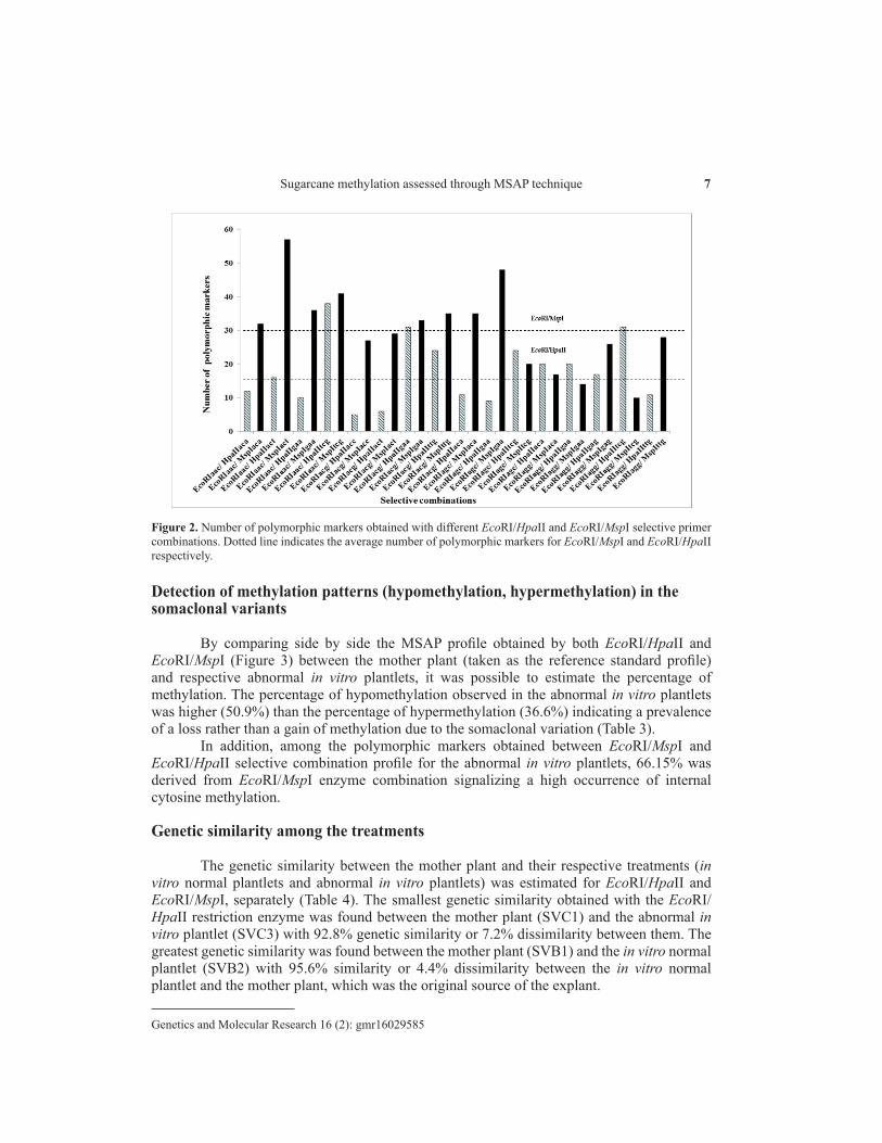
The EcoRI-AAC/MspI-ACT selective combination showed the highest number 57 (11.65%) of polymorphic markers while for the EcoRI/HpaII restriction enzyme the highest number 31 (10.87%) of polymorphic markers was obtained for the selective combination EcoRI-AAC/HpaII-TCG (Figure 2). An average of 31 and 17 polymorphic markers was obtained with the EcoRI/MspI and EcoRI/HpaII selective primers, respectively.

6J.H.M.B. Francischini et al.
Genetics and Molecular Research 16 (2): gmr16029585
Figure 1. MSAP profile revealed in 6% denaturing polyacrylamide gels at 4300 DNA Analyser (Licor). A. MSAP profile stability of two sampled mother plants (SVA, SVB, SVC, SVD) for the selective combinations: EcoRI.AGC/HpaII.ACA and EcoRI.AGC/HpaII.ACA, respectively. B. SVD clone MSAP profile reproducibility test of two sampled DNAs (SVD1 and SVD2) for four selective combinations (left to right: EcoRI.AAC/HpaII.GAA; EcoRI.AGC/HpaII.ACA; EcoRI.AGC/HpaII.GAA; EcoRI.ACG/ HpaII.ACA).
Table 3. MSAP polymorphism for EcoRI/HpaII and EcoRI/MspI enzyme combination.
Number of selective combination (Nsc), total number of markers (N), total number of polymorphic markers (Np), and percentage of polymorphism (%P).
Enzyme combination Nsc N Np %P EcoRI/HpaII 16 1005 285 28.36 EcoRI/MspI 16 1200 488 40.67 Methylation pattern Hypermethylation Hypomethylation 41 (36.6%) 57 (50.9)
7Sugarcane methylation assessed through MSAP technique
Genetics and Molecular Research 16 (2): gmr16029585
Detection of methylation patterns (hypomethylation, hypermethylation) in the somaclonal variants
By comparing side by side the MSAP profile obtained by both EcoRI/HpaII and EcoRI/MspI (Figure 3) between the mother plant (taken as the reference standard profile) and respective abnormal in vitro plantlets, it was possible to estimate the percentage of methylation. The percentage of hypomethylation observed in the abnormal in vitro plantlets was higher (50.9%) than the percentage of hypermethylation (36.6%) indicating a prevalence of a loss rather than a gain of methylation due to the somaclonal variation (Table 3).
In addition, among the polymorphic markers obtained between EcoRI/MspI and EcoRI/HpaII selective combination profile for the abnormal in vitro plantlets, 66.15% was derived from EcoRI/MspI enzyme combination signalizing a high occurrence of internal cytosine methylation.
Genetic similarity among the treatments
The genetic similarity between the mother plant and their respective treatments (in vitro normal plantlets and abnormal in vitro plantlets) was estimated for EcoRI/HpaII and EcoRI/MspI, separately (Table 4). The smallest genetic similarity obtained with the EcoRI/HpaII restriction enzyme was found between the mother plant (SVC1) and the abnormal in vitro plantlet (SVC3) with 92.8% genetic similarity or 7.2% dissimilarity between them. The greatest genetic similarity was found between the mother plant (SVB1) and the in vitro normal plantlet (SVB2) with 95.6% similarity or 4.4% dissimilarity between the in vitro normal plantlet and the mother plant, which was the original source of the explant.
Figure 2. Number of polymorphic markers obtained with different EcoRI/HpaII and EcoRI/MspI selective primer combinations. Dotted line indicates the average number of polymorphic markers for EcoRI/MspI and EcoRI/HpaII respectively.

8J.H.M.B. Francischini et al.
Genetics and Molecular Research 16 (2): gmr16029585
Figure 3. MSAP profile comparison (EcoRI.ACG/HpaII.TTG and EcoRI.ACG/MspI.TTG) among mother plant and regenerants. Arrow shows marker absence in the regenerants (SCV2 and SCV3) and present in the mother plant (SCV1) when digested by EcoRI/MspI.
Table 4. Pairwise genetic similarity obtained for the clones evaluated.
Clones Clones Clones SVA1 SVA2 SVA3 Clones SVC1 SVC2 SVC3 SVA1 1 0.907 0.877 SVC1 1 0.908 0.885 SVA2 0.954 1 0.921 SVC2 0.943 1 0.944 SVA3 0.955 0.946 1 SVC3 0.928 0.966 1 Clones SVB1 SVB2 SVB3 Clones SVD1 SVD2 SVD3 SVB1 1 0.935 0.911 SVD1 1 0.893 0.884 SVB2 0.956 1 0.941 SVD2 0.940 1 0.948 SVB3 0.949 0.967 1 SVD3 0.950 0.940 1
Mother plant (SVA1, SVB1, SVC1, SVD1); in vitro normal plantlets (SVA2, SVB2, SVC2, SVD2); abnormal in vitro plantlets (SVA3, SVB3, SVC3, SVD3). EcoRI/MspI (above the diagonal) and EcoRI/HpaII (below the diagonal).
The smallest genetic similarity observed with the EcoRI/MspI restriction enzyme was observed between the mother plant (SVA1) and the abnormal in vitro plantlet (SVA3) with 87.7% genetic similarity and 12.3% dissimilarity. The greatest genetic similarity was found between the mother plant (SVB1) and its respective treatment in in vitro normal plantlet (SVB2) with 93.5% genetic similarity and only 6.5% dissimilarity of the in vitro normal plantlet in relation to the mother plant. The CV%, which measures the reliability of the genetic similarity, was 1.27 and 1.19%, for the EcoRI/MspI and EcoRI/HpaII combination, respectively.
According to the dendrogram (Figure 4A), it was clear that the EcoRI/MspI enzyme combination was more efficient to distinguish the mother plant from their tissue culture

9Sugarcane methylation assessed through MSAP technique
Genetics and Molecular Research 16 (2): gmr16029585
treatments than the EcoRI/HpaII enzyme combination (Figure 4B). The in vitro normal plantlets and the in vitro somaclonal variant were more similar to each other than to the respective mother plants. The reliability of the dendrogram nodes observed in the clustering of the mother plant and respective treatments was high ranging from 76.8% (SVA1, SVA2, and SVA3 node) to 100% (SVB1, SVB2, and SVB3 node) for the EcoRI/MspI and EcoRI/HpaII enzyme combination, respectively.
Figure 4. UPGMA dendrogram based on genetic similarity matrix (Jaccard coefficient) among mother plant (SVA1, SVB1, SVC1 and SVD1) and respective micropropagated treatments (in vitro normal plantlets: SVA2, SVB2, SVC2 and SVD2; abnormal in vitro plantlets: SVA3, SVB3, SVC3 and SVD3). A. EcoRI/MspI enzyme combinations. B. EcoRI/HpaII enzyme combination.
DISCUSSION
In the present study, the molecular marker technology was used to characterize normal and abnormal in vitro plantlets sampled at the third subculture stage. These abnormal sugarcane plantlets taken as somaclonal variants are discarded in the early stages of in vitro clonal propagation during the multiplication process. Therefore, the molecular profile comparative analysis obtained by the MSAP technique among the mother plant (used as the source tissue for regeneration), abnormal in vitro plantlets (somaclonal variant), and normal in vitro plantlets allowed us to access changes occurred at the sugarcane epigenome during the micro-propagated process. These changes were quantified by estimating the genetic similarity

10J.H.M.B. Francischini et al.
Genetics and Molecular Research 16 (2): gmr16029585
between the in vitro treatments and the mother plant.The stability analysis of the mother plants used as the source tissue for regeneration
reinforced the possibility that the polymorphisms obtained between mother plants and treatments can be attributed to changes occurred during the micro-propagation process. This point is important since somaclonal variation can arise from pre-existing somatic mutations in donor plants, namely genetic instability (Leva and Petruccelli, 2012). In addition, the reproducibility analysis of the MSAP technique also contributed to eliminate the possibility that the observed polymorphisms were assigned to possible technical artifacts.
In our study with sugarcane, the MSAP technique was efficient to detect polymorphism among the mother plant and respective tissue culture treatments and hence distinguish the mother plant from the micro-propagation treatments. In fact, it was noted that the genetic similarity values obtained with the EcoRI/MspI enzyme combination were more efficient to distinguish between in vitro abnormal plantlets (somaclonal variants) and the respective mother plant than the EcoRI/HpaII enzyme combination.
Our results suggest that the tissue culture process probably may lead to modifications at the epigenome of micro-propagated derived materials. According to Kaeppler et al. (2000), DNA methylation variation frequently occurs in the tissue culture process. Indeed, we observed that even in the in vitro normal plantlets (plantlets without symptoms of somaclonal variation) it was possible to detect differences in the MSAP molecular profile in comparison to the mother plant. Thus, not all changes associated with DNA methylation due to the tissue culture process may produce abnormal plants. These results were similar to that described by Matthes et al. (2001) in oil palm where changes in methylation were not necessarily associated with the abnormal phenotype derived from tissue culture.
In our study, it was noted a higher rate of loss methylation (hypomethylation) in the abnormal in vitro sugarcane plantlets than methylation gain (hypermethylation). Significant losses of methylation were also reported in rice plants regenerated by tissue culture in comparison with non-regenerated plants (Stroud et al., 2013) as also in other crops (Jaligot et al., 2000; Matthes et al., 2001). Stelpflug et al. (2014) characterized DNA methylation patterns in callus, primary regenerants, and maize regenerant-derived progenies by immunoprecipitation of methylated DNA and found that hypomethylation events were more frequently than hypermethylation after tissue culture.
In general, the detection of alterations in the MSAP molecular profile reflects changes in the states of methylation between mother plants (adult plants) and micro-propagated plantlets during the third stage of subculture. This result also reflects the power of the MSAP technique to detected changes in the epigenome of tissue culture-derived plants even subjected to a number of subcultures below the upper limit of subcultures recommended for sugarcane, which is pointed as up to 5 subcultures. In fact, no evidence of polymorphism was reported by Hsie et al. (2015) in micro-propagated sugarcane shoot tips after fifteen consecutive subcultures assessed by inter-simple sequence repeat DNA markers. Thus, based in our results, the MASP technique seems to be a promising tool to the early assessment of genetic fidelity of micro-propagated sugarcane plants with a great potential to be used as a tool in the quality control of large scale micro-propagation plants.
ACKNOWLEDGMENTS
Research supported by FUNDAG (Fundação de Pesquisa Agrícola) and IAC Postgraduate program (PROAP funding).
11Sugarcane methylation assessed through MSAP technique
Genetics and Molecular Research 16 (2): gmr16029585
REFERENCES
Aljanabi SM, Forget L and Dookun A (1999). An improved and rapid protocol for the isolation of polysaccharide and polyphenol-free sugarcane DNA. Plant Mol. Biol. Report. 17: 1-8. http://dx.doi.org/10.1023/A:1007692929505
Bairu M, Aremu A and Staden JV (2011). Somaclonal variation in plants: causes and detection methods. Plant Growth Regul. 63: 147-173. http://dx.doi.org/10.1007/s10725-010-9554-x
Bobadilla Landey R, Cenci A, Georget F, Bertrand B, et al. (2013). High genetic and epigenetic stability in Coffea arabica plants derived from embryogenic suspensions and secondary embryogenesis as revealed by AFLP, MSAP and the phenotypic variation rate. PLoS One 8: e56372 http://dx.doi.org/10.1371/journal.pone.0056372.
Coelho ASG (2000). BOOD: avaliação de dendrogramas baseados em estimativas de distâncias/similaridades genéticas através do procedimento de bootstrap. Universidade Federal de Goiás, Goiânia.
Coelho ASG (2001). Dboot - Avaliação dos erros associados a estimativas de distâncias/similaridades genéticas através do procedimento de bootstrap com número variável de marcadores. Laboratório de Genética Vegetal, Instituto de Ciências Biológicas, UFG, Goiânia.
D’Alessio AC and Szyf M (2006). Epigenetic tête-à-tête: the bilateral relationship between chromatin modifications and DNA methylation. Biochem. Cell Biol. 84: 463-476.
Gao X, Yang D, Cao D, Ao M, et al. (2010). Affiliated withKey Laboratory of Molecular Epigenetics of MOE, Institute of Genetics and Cytology, Northeast Normal University In vitro micropropagation of Freesia hybrida and the assessment of genetic and epigenetic stability in regenerated plantlets. J. Plant Growth Regul. 29: 257-267. http://dx.doi.org/10.1007/s00344-009-9133-4
Gonzalez AI, Saiz A, Acedo A, Ruiz ML, et al. (2013). Analysis of genomic DNA methylation patterns in regenerated and control plants of rye (Secale cereale L.). Plant Growth Regul. 70: 227-236. http://dx.doi.org/10.1007/s10725-013-9794-7
He XJ, Chen T and Zhu JK (2011). Regulation and function of DNA methylation in plants and animals. Cell Res. 21: 442-465. http://dx.doi.org/10.1038/cr.2011.23
Hsie BS, Brito JZ, Vila Nova MX, Borges-Paluch LR, et al. (2015). Determining the genetic stability of micropropagated sugarcane using inter-simple sequence repeat markers. Genet. Mol. Res. 14: 17651-17659. http://dx.doi.org/10.4238/2015.December.21.38
Jaligot E, Rival A, Beulé T, Dussert S, et al. (2000). Somaclonal variation in oil palm (Elaeis guineensis Jacq.): the DNA methylation hypothesis. Plant Cell Rep. 19: 684-690. http://dx.doi.org/10.1007/s002999900177
Joyce P, Hermann S, O’Connell A, Dinh Q, et al. (2014). Field performance of transgenic sugarcane produced using Agrobacterium and biolistics methods. Plant Biotechnol. J. 12: 411-424. http://dx.doi.org/10.1111/pbi.12148
Kaeppler SM, Kaeppler HF and Rhee Y (2000). Epigenetic aspects of somaclonal variation in plants. Plant Mol. Biol. 43: 179-188. http://dx.doi.org/10.1023/A:1006423110134
Lal M, Tiwari AK, Gupta GN and Kavita (2015). Commercial scale micropropagation of sugarcane: constraints and remedies. Sugar Tech. 17: 339-347. http://dx.doi.org/10.1007/s12355-014-0345-y
LandeyAffiliated withUMR IPME, CIRAD RB, Cenci A, GuyotAffiliated withUMR IPME, IRD R, Bertrand B, et al. (2015). Email author Assessment of genetic and epigenetic changes during cell culture ageing and relations with somaclonal variation in Coffea arabica. Plant Cell Tissue Organ Cult.122: 517-531.
Larkin PJ and Scowcroft WR (1981). Somaclonal variation - a novel source of variability from cell cultures for plant improvement. Theor. Appl. Genet. 60: 197-214. http://dx.doi.org/10.1007/BF02342540
Lei CP, Jiun KS, Choo CS and Singh R (2006). Analysis of tissue culture-derived regenerants using methylation sensitive AFLP. As. Pac. J. Mol. Biol. Biotechnol. 14: 47-55.
Leva AR and Petruccelli R (2012). Monitoring of cultivar identity in micropropagated olive plants using RAPD and ISSR markers. Biol. Plant. 56: 373-376. http://dx.doi.org/10.1007/s10535-012-0102-6
López CMR and Wilkinson MJ (2015). Epi-fingerprinting and epi-interventions for improved crop production and food quality. Front. Plant Sci. 6: 1-14.
Matthes M, SinghAffiliated withPalm Oil Research Institute of Malaysia, 6, Persiaran Institusi, Bandar Baru Bangi, 43000 Kajang, Selangor, Malaysia R and Karp A (2001). Variation in oil palm (Elaeis guineensis Jacq.) tissue culture-derived regenerants revealed by AFLPs with methylation-sensitive enzymes (2001). Theor. Appl. Genet. 102: 971-979. http://dx.doi.org/10.1007/s001220000491
Miguel C and Marum L (2011). An epigenetic view of plant cells cultured in vitro: somaclonal variation and beyond. J. Exp. Bot. 62: 3713-3725. http://dx.doi.org/10.1093/jxb/err155
Rathore MS, Mastan SG and Agarwal PK (2015). Evaluation of DNA methylation using methylation-sensitive amplification polymorphism in plant tissues grown in vivo and in vitro. Plant Growth Regul. 75: 11-19. http://dx.doi.org/10.1007/

12J.H.M.B. Francischini et al.
Genetics and Molecular Research 16 (2): gmr16029585
s10725-014-9926-8Rastogi J, Siddhant, Bubber P and Sharma BL (2015). Somaclonal Variation: A new dimension for sugarcane improvement.
GERF Bull. Biosciences 6: 5-10.Rohlf FJ (1998). NTSYSpc: Numerical Taxonomy and Multivariate Analysis System version 2.02 Exeter Software,
Setauket, New York.Shawn M, Kaeppler HF and Rhee Y (2000). Epigenetic aspects of somaclonal variation in plants. Plant Mol. Biol. 43:179-188.Schulz B, Eckstein RL and Durka W (2013). Scoring and analysis of methylation-sensitive amplification polymorphisms
for epigenetic population studies. Mol. Ecol. Resour. 13: 642-653. http://dx.doi.org/10.1111/1755-0998.12100Stelpflug SC, Eichten SR, Hermanson PJ, Springer NM, et al. (2014). Consistent and heritable alterations of DNA methylation
are induced by tissue culture in maize. Genetics 198: 209-218. http://dx.doi.org/10.1534/genetics.114.165480Stroud H, Ding B, Simon SA, Feng S, et al. (2013). Plants regenerated from tissue culture contain stable epigenome
changes in rice. Available at [http://elife.elifesciences.org]. Accessed June 12, 2013.Sudarshana MS, Mahendra C, Sampathkumara KK and Manasa G (2015). Cytological variations of in vitro stem cultures
of Xanthophyllum flavascens roxb. an endangered tree species of western ghats. Ind. J. Pl. Sc. 4: 78-83.Vos P, Hogers R, Bleeker M, Reijans M, et al. (1995). AFLP: a new technique for DNA fingerprinting. Nucleic Acids Res.
23: 4407-4414. http://dx.doi.org/10.1093/nar/23.21.4407Wekesa R, Onguso JM, Nyende BA and Wamocho LS (2015). Sugarcane in vitro AFLP: a new technique for DNA
fingerprinting culture technology: Opportunities for Kenya’s sugar industry. Afr. J. Biotechnol. 14: 3170-3178. http://dx.doi.org/10.5897/AJB2015.14440
Yaish MW, Peng M and Rothstein SJ (2014). Global DNA methylation analysis using methyl-sensitive amplification polymorphism (MSAP). Methods Mol. Biol. 1062: 285-298. http://dx.doi.org/10.1007/978-1-62703-580-4_16