Embed Size (px)
Citation preview
Published: June 01, 2011
r 2011 American Chemical Society 5215 dx.doi.org/10.1021/cr200060g |Chem. Rev. 2011, 111, 5215–5246
REVIEW
pubs.acs.org/CR
The Methylation Effect in Medicinal ChemistryEliezer J. Barreiro,*,†,‡,§ Arthur E. K€ummerle,||,†,§ and Carlos A. M. Fraga†,‡,§
†Laborat�orio de Avaliac-~ao e Síntese de Substancias Bioativas (LASSBio), Faculdade de Farm�acia, Universidade Federal doRio de Janeiro, CCS, Cidade Universit�aria, CP 68.006, 21941-902 Rio de Janeiro, RJ, Brazil‡Programa de P�os-Graduac-~ao em Farmacologia e Química Medicinal, Instituto de Ciencias Biom�edicas, Universidade Federal doRio de Janeiro, Cidade Universit�aria, Ilha do Fund~ao, Rio de Janeiro, RJ, Brazil§Programa de P�os-Graduac-~ao em Química, Instituto de Química, Universidade Federal do Rio de Janeiro,Cidade Universit�aria, Ilha do Fund~ao, Rio de Janeiro, RJ, Brazil
CONTENTS
1. Introduction: The Methyl Group and Its BiologicalInteractions 5215
2. Effects of Methyl Groups on the Solubility of Bio-active Compounds: Increase Lipophilicity andPromote the Hydrophobic Interactions 5216
3. Methylated Natural Products 52173.1. Morphine and Methylated Analogues 5217
3.2. Methyl Effects in Natural Amino Acid 52194. Specialized Cofactors in the Biomethylation Process 5220
4.1. S-Adenosylmethionine (SAM) 52204.2. Folic Acid Pathway 5222
5. From Methylated Natural Products to Drugs: TheDiscovery of Statins 5223
6. Stereoelectronic Effects of Methyl Groups in DrugDesign and Discovery 52246.1. The Inductive Effect of Methyl in the Discovery
of Anti-Ulcer Drugs: Cimetidine and Hþ-Kþ-AT-Pase Inhibitors 5224
6.2. The Conformational Effect Induced by Methyl:The Discovery of Imatinib 5228
6.3. Conformational Changes Induced by N-Methyl-ation of Bioactive N-Acylhydrazones (NAH) 5228
6.4. The Steric Effect of the Methyl in the Selectivityof Lumiracoxib for PGHS-1/PGHS-2 5229
6.5. Methyl-Induced Effects in ChloroquineAnalogues 5230
6.6. Methyl-Induced Atropoisomerism in Psychoac-tive Drugs and Male Contraceptive Drugs 5231
7. Stereoelectronic Effects of Methyl Groups and DrugMetabolism 52327.1. The ortho-Effect Due to Methyl Groups: The
Metabolism of Lidocaine 5232
7.2. TheMethyl Group As a SoftMetabolic Point: TheDiscovery of Celecoxib 5233
7.3. Methyl Improves the Metabolic Stability ofThiazole- And Isoxazole-Containing Drugs 5233
7.4. The Methyl Effect in the Design of Orally ActiveSynthetic Prostaglandins 5235
7.5. Methyl in Metabolic Activation of Prodrugs 52378. Methylation in the SOSA Strategy of Drug Design 52379. The Methyl in Generating Me-Too Drugs 523910. Conclusions 5239Author Information 5239Biographies 5239Acknowledgment 5240List of Abbreviations 5240References 5241
1. INTRODUCTION: THE METHYL GROUP AND ITSBIOLOGICAL INTERACTIONS
The monovalent methyl group is derived from methanethrough the removal of a hydrogen atom,1 and its etymology isdirectly related to the discovery of methanol.2 The first reports ofthe use of methanol were from ancient Egyptians. In theirembalming process, they used a mixture of substances obtainedfrom the pyrolysis of wood shards that contained a significantamount of methyl alcohol.2 Pure methanol was first isolated anddescribed in 1661 by Robert Boyle, who called it “spirit of box,” asit is the product of the distillation of “Boxwood,” the generic namegiven to ca. 70 types of trees from the Buxaceae family. In 1834,the French chemists Jean-Baptiste Dumas and Eugene Peligotdetermined its elementary composition as CH4O through com-bustion analysis. They introduced the term methyl to organicchemistry through a mistranslation from the Greek, methy =“wine” þ hyle = wood (wood bark), when the intention was todefine “wood alcohol” or methanol.2
Themethyl group is very important in themolecular recognitionof endogenous and exogenous organic compounds by bioreceptors.Although it only participates in London dispersion interactions,which are the weakest of all intermolecular interactions,3 methylgroups have stereoelectronic effects4 on micromolecules andbiomacromolecules, thereby leading to diverse biological effects,including selectivity among bioreceptors, increased potency, andprotection against enzyme metabolism.5 Cognizant of the methylgroup’s importance in molecular recognition, Wermuth wrote:5
“The methyl group, so often considered as chemically inert,is able to alter deeply the pharmacological properties of amolecule.”
Received: February 18, 2011
5216 dx.doi.org/10.1021/cr200060g |Chem. Rev. 2011, 111, 5215–5246
Chemical Reviews REVIEW
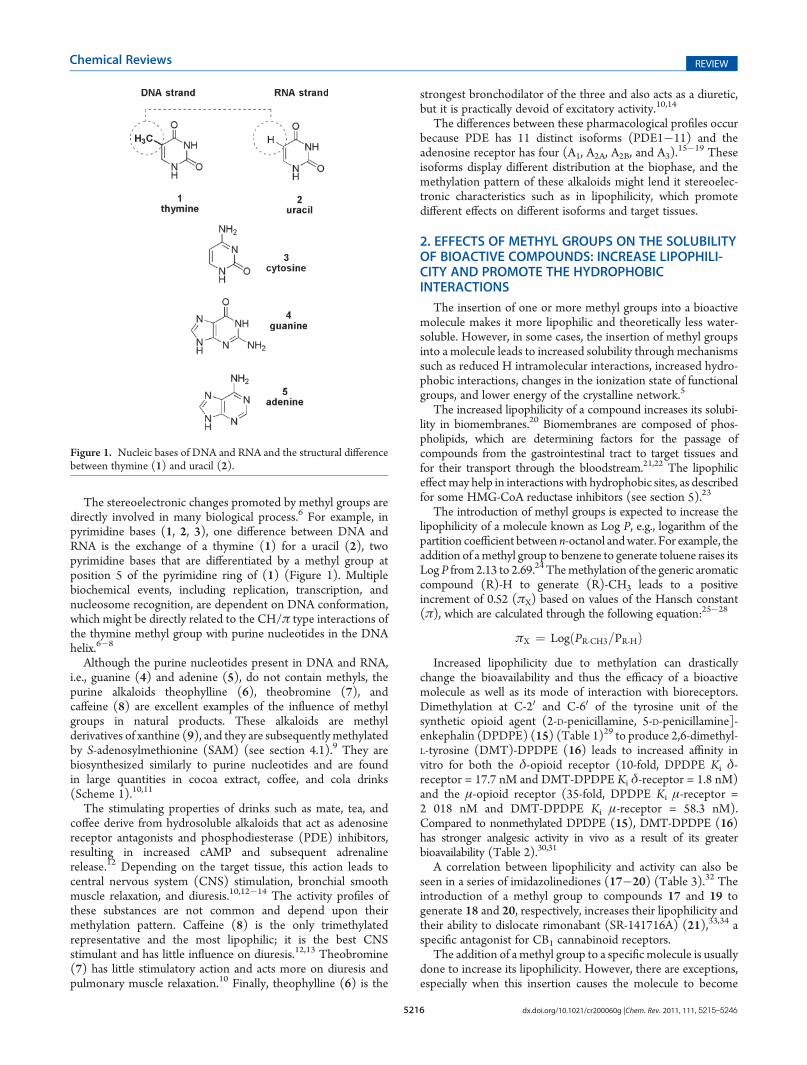
The stereoelectronic changes promoted by methyl groups aredirectly involved in many biological process.6 For example, inpyrimidine bases (1, 2, 3), one difference between DNA andRNA is the exchange of a thymine (1) for a uracil (2), twopyrimidine bases that are differentiated by a methyl group atposition 5 of the pyrimidine ring of (1) (Figure 1). Multiplebiochemical events, including replication, transcription, andnucleosome recognition, are dependent on DNA conformation,which might be directly related to the CH/π type interactions ofthe thymine methyl group with purine nucleotides in the DNAhelix.6�8
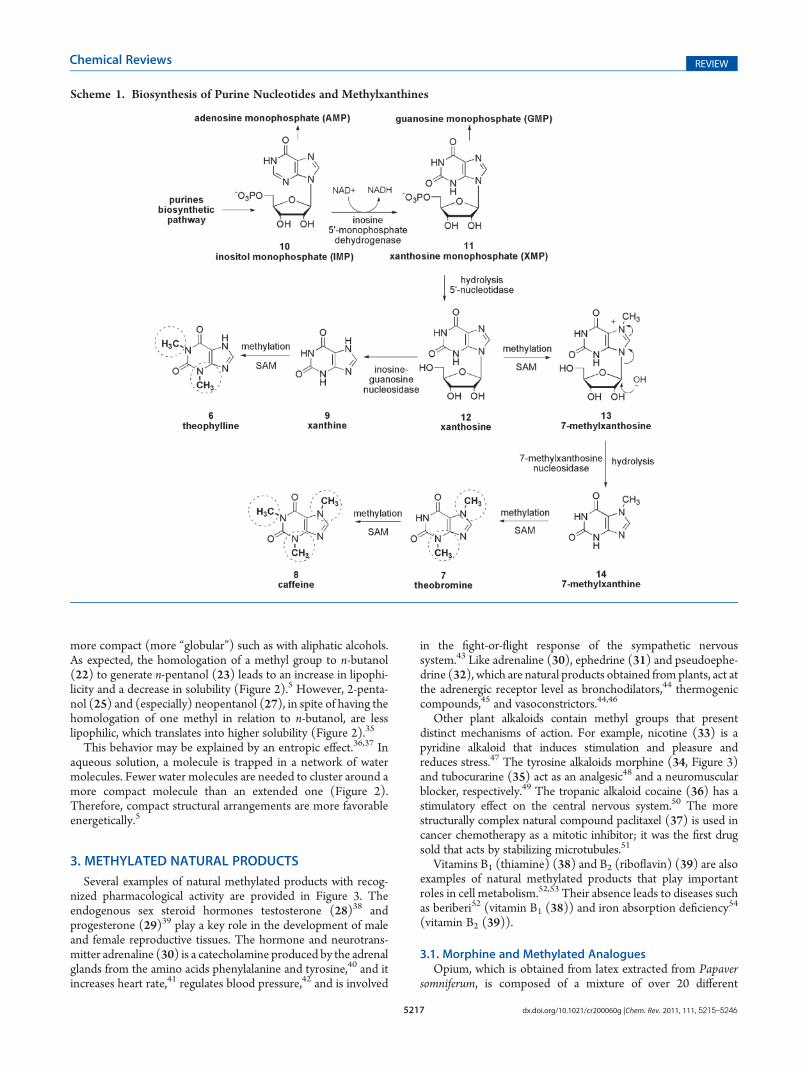
Although the purine nucleotides present in DNA and RNA,i.e., guanine (4) and adenine (5), do not contain methyls, thepurine alkaloids theophylline (6), theobromine (7), andcaffeine (8) are excellent examples of the influence of methylgroups in natural products. These alkaloids are methylderivatives of xanthine (9), and they are subsequently methylatedby S-adenosylmethionine (SAM) (see section 4.1).9 They arebiosynthesized similarly to purine nucleotides and are foundin large quantities in cocoa extract, coffee, and cola drinks(Scheme 1).10,11
The stimulating properties of drinks such as mate, tea, andcoffee derive from hydrosoluble alkaloids that act as adenosinereceptor antagonists and phosphodiesterase (PDE) inhibitors,resulting in increased cAMP and subsequent adrenalinerelease.12 Depending on the target tissue, this action leads tocentral nervous system (CNS) stimulation, bronchial smoothmuscle relaxation, and diuresis.10,12�14 The activity profiles ofthese substances are not common and depend upon theirmethylation pattern. Caffeine (8) is the only trimethylatedrepresentative and the most lipophilic; it is the best CNSstimulant and has little influence on diuresis.12,13 Theobromine(7) has little stimulatory action and acts more on diuresis andpulmonary muscle relaxation.10 Finally, theophylline (6) is the
strongest bronchodilator of the three and also acts as a diuretic,but it is practically devoid of excitatory activity.10,14
The differences between these pharmacological profiles occurbecause PDE has 11 distinct isoforms (PDE1�11) and theadenosine receptor has four (A1, A2A, A2B, and A3).
15�19 Theseisoforms display different distribution at the biophase, and themethylation pattern of these alkaloids might lend it stereoelec-tronic characteristics such as in lipophilicity, which promotedifferent effects on different isoforms and target tissues.
2. EFFECTS OF METHYL GROUPS ON THE SOLUBILITYOF BIOACTIVE COMPOUNDS: INCREASE LIPOPHILI-CITY AND PROMOTE THE HYDROPHOBICINTERACTIONS
The insertion of one or more methyl groups into a bioactivemolecule makes it more lipophilic and theoretically less water-soluble. However, in some cases, the insertion of methyl groupsinto a molecule leads to increased solubility through mechanismssuch as reduced H intramolecular interactions, increased hydro-phobic interactions, changes in the ionization state of functionalgroups, and lower energy of the crystalline network.5
The increased lipophilicity of a compound increases its solubi-lity in biomembranes.20 Biomembranes are composed of phos-pholipids, which are determining factors for the passage ofcompounds from the gastrointestinal tract to target tissues andfor their transport through the bloodstream.21,22 The lipophiliceffect may help in interactions with hydrophobic sites, as describedfor some HMG-CoA reductase inhibitors (see section 5).23
The introduction of methyl groups is expected to increase thelipophilicity of a molecule known as Log P, e.g., logarithm of thepartition coefficient between n-octanol andwater. For example, theaddition of amethyl group to benzene to generate toluene raises itsLog P from2.13 to 2.69.24Themethylation of the generic aromaticcompound (R)-H to generate (R)-CH3 leads to a positiveincrement of 0.52 (πX) based on values of the Hansch constant(π), which are calculated through the following equation:25�28
πX ¼ LogðPR-CH3=PR-HÞIncreased lipophilicity due to methylation can drastically
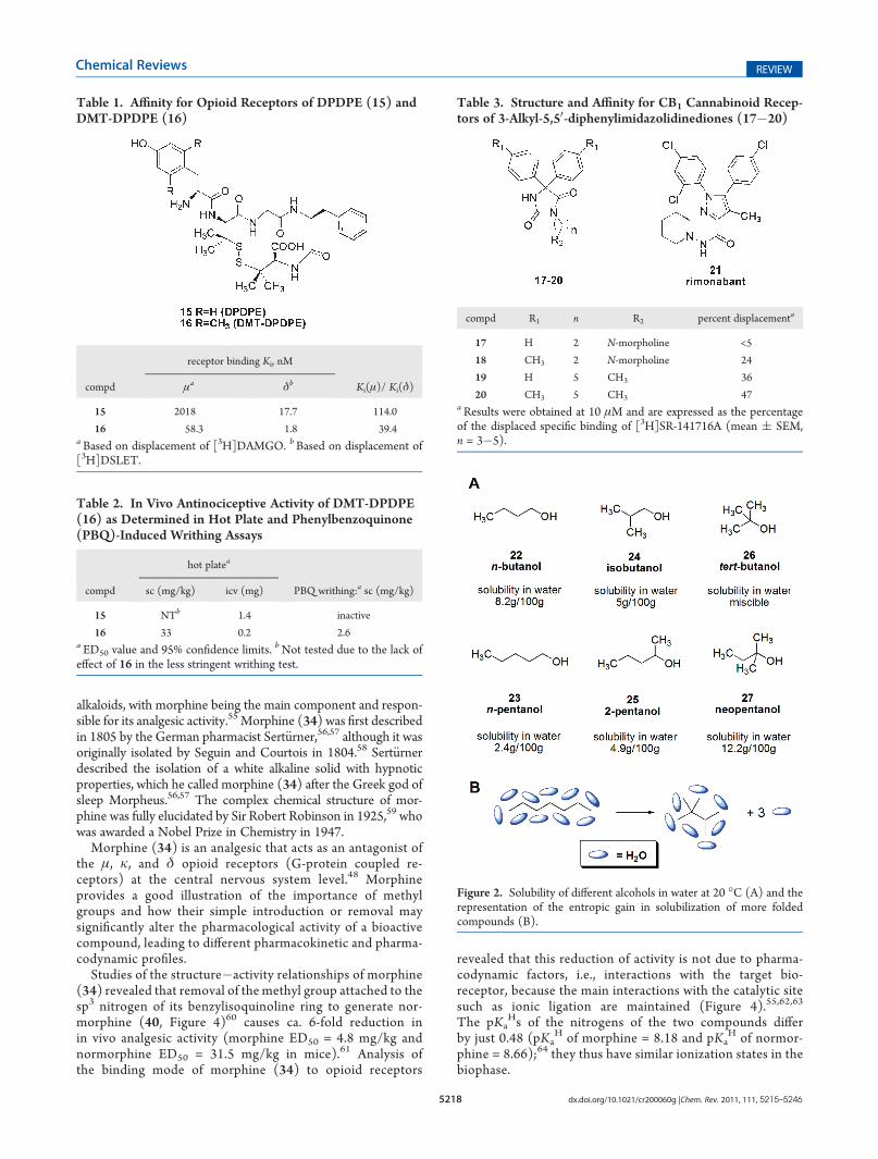
change the bioavailability and thus the efficacy of a bioactivemolecule as well as its mode of interaction with bioreceptors.Dimethylation at C-20 and C-60 of the tyrosine unit of thesynthetic opioid agent (2-D-penicillamine, 5-D-penicillamine]-enkephalin (DPDPE) (15) (Table 1)29 to produce 2,6-dimethyl-L-tyrosine (DMT)-DPDPE (16) leads to increased affinity invitro for both the δ-opioid receptor (10-fold, DPDPE Ki δ-receptor = 17.7 nM and DMT-DPDPE Ki δ-receptor = 1.8 nM)and the μ-opioid receptor (35-fold, DPDPE Ki μ-receptor =2 018 nM and DMT-DPDPE Ki μ-receptor = 58.3 nM).Compared to nonmethylated DPDPE (15), DMT-DPDPE (16)has stronger analgesic activity in vivo as a result of its greaterbioavailability (Table 2).30,31
A correlation between lipophilicity and activity can also beseen in a series of imidazolinediones (17�20) (Table 3).32 Theintroduction of a methyl group to compounds 17 and 19 togenerate 18 and 20, respectively, increases their lipophilicity andtheir ability to dislocate rimonabant (SR-141716A) (21),33,34 aspecific antagonist for CB1 cannabinoid receptors.
The addition of amethyl group to a specificmolecule is usuallydone to increase its lipophilicity. However, there are exceptions,especially when this insertion causes the molecule to become
Figure 1. Nucleic bases of DNA and RNA and the structural differencebetween thymine (1) and uracil (2).
5217 dx.doi.org/10.1021/cr200060g |Chem. Rev. 2011, 111, 5215–5246
Chemical Reviews REVIEW
more compact (more “globular”) such as with aliphatic alcohols.As expected, the homologation of a methyl group to n-butanol(22) to generate n-pentanol (23) leads to an increase in lipophi-licity and a decrease in solubility (Figure 2).5 However, 2-penta-nol (25) and (especially) neopentanol (27), in spite of having thehomologation of one methyl in relation to n-butanol, are lesslipophilic, which translates into higher solubility (Figure 2).35
This behavior may be explained by an entropic effect.36,37 Inaqueous solution, a molecule is trapped in a network of watermolecules. Fewer water molecules are needed to cluster around amore compact molecule than an extended one (Figure 2).Therefore, compact structural arrangements are more favorableenergetically.5
3. METHYLATED NATURAL PRODUCTS
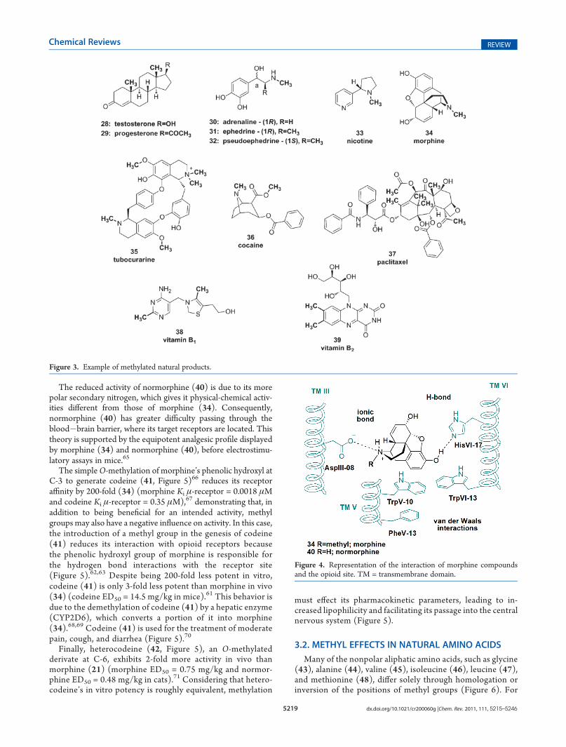
Several examples of natural methylated products with recog-nized pharmacological activity are provided in Figure 3. Theendogenous sex steroid hormones testosterone (28)38 andprogesterone (29)39 play a key role in the development of maleand female reproductive tissues. The hormone and neurotrans-mitter adrenaline (30) is a catecholamine produced by the adrenalglands from the amino acids phenylalanine and tyrosine,40 and itincreases heart rate,41 regulates blood pressure,42 and is involved
in the fight-or-flight response of the sympathetic nervoussystem.43 Like adrenaline (30), ephedrine (31) and pseudoephe-drine (32), which are natural products obtained from plants, act atthe adrenergic receptor level as bronchodilators,44 thermogeniccompounds,45 and vasoconstrictors.44,46
Other plant alkaloids contain methyl groups that presentdistinct mechanisms of action. For example, nicotine (33) is apyridine alkaloid that induces stimulation and pleasure andreduces stress.47 The tyrosine alkaloids morphine (34, Figure 3)and tubocurarine (35) act as an analgesic48 and a neuromuscularblocker, respectively.49 The tropanic alkaloid cocaine (36) has astimulatory effect on the central nervous system.50 The morestructurally complex natural compound paclitaxel (37) is used incancer chemotherapy as a mitotic inhibitor; it was the first drugsold that acts by stabilizing microtubules.51
Vitamins B1 (thiamine) (38) and B2 (riboflavin) (39) are alsoexamples of natural methylated products that play importantroles in cell metabolism.52,53 Their absence leads to diseases suchas beriberi52 (vitamin B1 (38)) and iron absorption deficiency
54
(vitamin B2 (39)).
3.1. Morphine and Methylated AnaloguesOpium, which is obtained from latex extracted from Papaver
somniferum, is composed of a mixture of over 20 different
Scheme 1. Biosynthesis of Purine Nucleotides and Methylxanthines
5218 dx.doi.org/10.1021/cr200060g |Chem. Rev. 2011, 111, 5215–5246
Chemical Reviews REVIEW
alkaloids, with morphine being the main component and respon-sible for its analgesic activity.55 Morphine (34) was first describedin 1805 by the German pharmacist Sert€urner,56,57 although it wasoriginally isolated by Seguin and Courtois in 1804.58 Sert€urnerdescribed the isolation of a white alkaline solid with hypnoticproperties, which he called morphine (34) after the Greek god ofsleep Morpheus.56,57 The complex chemical structure of mor-phine was fully elucidated by Sir Robert Robinson in 1925,59 whowas awarded a Nobel Prize in Chemistry in 1947.
Morphine (34) is an analgesic that acts as an antagonist ofthe μ, k, and δ opioid receptors (G-protein coupled re-ceptors) at the central nervous system level.48 Morphineprovides a good illustration of the importance of methylgroups and how their simple introduction or removal maysignificantly alter the pharmacological activity of a bioactivecompound, leading to different pharmacokinetic and pharma-codynamic profiles.
Studies of the structure�activity relationships of morphine(34) revealed that removal of the methyl group attached to thesp3 nitrogen of its benzylisoquinoline ring to generate nor-morphine (40, Figure 4)60 causes ca. 6-fold reduction inin vivo analgesic activity (morphine ED50 = 4.8 mg/kg andnormorphine ED50 = 31.5 mg/kg in mice).61 Analysis ofthe binding mode of morphine (34) to opioid receptors
revealed that this reduction of activity is not due to pharma-codynamic factors, i.e., interactions with the target bio-receptor, because the main interactions with the catalytic sitesuch as ionic ligation are maintained (Figure 4).55,62,63
The pKaHs of the nitrogens of the two compounds differ
by just 0.48 (pKaH of morphine = 8.18 and pKa
H of normor-phine = 8.66);64 they thus have similar ionization states in thebiophase.
Table 1. Affinity for Opioid Receptors of DPDPE (15) andDMT-DPDPE (16)
receptor binding Ki, nM
compd μa δb Ki(μ)/ Ki(δ)
15 2018 17.7 114.0
16 58.3 1.8 39.4aBased on displacement of [3H]DAMGO. bBased on displacement of[3H]DSLET.
Table 2. In Vivo Antinociceptive Activity of DMT-DPDPE(16) as Determined in Hot Plate and Phenylbenzoquinone(PBQ)-Induced Writhing Assays
hot platea
compd sc (mg/kg) icv (mg) PBQ writhing:a sc (mg/kg)
15 NTb 1.4 inactive
16 33 0.2 2.6a ED50 value and 95% confidence limits. bNot tested due to the lack ofeffect of 16 in the less stringent writhing test.
Table 3. Structure and Affinity for CB1 Cannabinoid Recep-tors of 3-Alkyl-5,50-diphenylimidazolidinediones (17�20)
compd R1 n R2 percent displacementa
17 H 2 N-morpholine <5
18 CH3 2 N-morpholine 24
19 H 5 CH3 36
20 CH3 5 CH3 47aResults were obtained at 10 μM and are expressed as the percentageof the displaced specific binding of [3H]SR-141716A (mean ( SEM,n = 3�5).
Figure 2. Solubility of different alcohols in water at 20 �C (A) and therepresentation of the entropic gain in solubilization of more foldedcompounds (B).
5219 dx.doi.org/10.1021/cr200060g |Chem. Rev. 2011, 111, 5215–5246
Chemical Reviews REVIEW
The reduced activity of normorphine (40) is due to its morepolar secondary nitrogen, which gives it physical-chemical activ-ities different from those of morphine (34). Consequently,normorphine (40) has greater difficulty passing through theblood�brain barrier, where its target receptors are located. Thistheory is supported by the equipotent analgesic profile displayedby morphine (34) and normorphine (40), before electrostimu-latory assays in mice.65
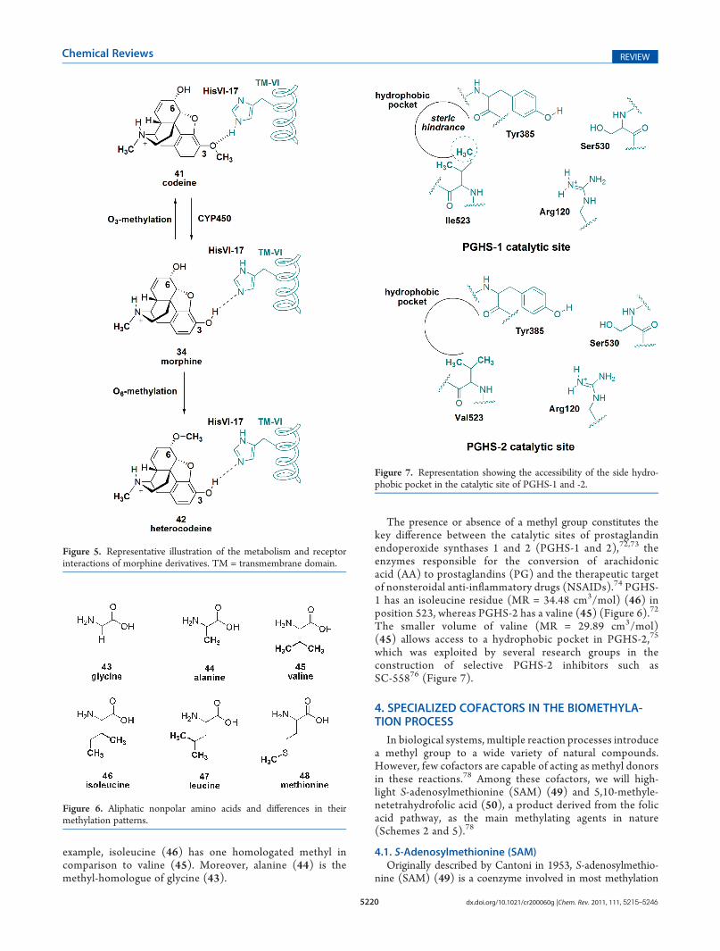
The simpleO-methylation of morphine’s phenolic hydroxyl atC-3 to generate codeine (41, Figure 5)66 reduces its receptoraffinity by 200-fold (34) (morphine Ki μ-receptor = 0.0018 μMand codeine Ki μ-receptor = 0.35 μM),67 demonstrating that, inaddition to being beneficial for an intended activity, methylgroups may also have a negative influence on activity. In this case,the introduction of a methyl group in the genesis of codeine(41) reduces its interaction with opioid receptors becausethe phenolic hydroxyl group of morphine is responsible forthe hydrogen bond interactions with the receptor site(Figure 5).62,63 Despite being 200-fold less potent in vitro,codeine (41) is only 3-fold less potent than morphine in vivo(34) (codeine ED50 = 14.5 mg/kg in mice).61 This behavior isdue to the demethylation of codeine (41) by a hepatic enzyme(CYP2D6), which converts a portion of it into morphine(34).68,69 Codeine (41) is used for the treatment of moderatepain, cough, and diarrhea (Figure 5).70
Finally, heterocodeine (42, Figure 5), an O-methylatedderivate at C-6, exhibits 2-fold more activity in vivo thanmorphine (21) (morphine ED50 = 0.75 mg/kg and normor-phine ED50 = 0.48 mg/kg in cats).71 Considering that hetero-codeine’s in vitro potency is roughly equivalent, methylation
must effect its pharmacokinetic parameters, leading to in-creased lipophilicity and facilitating its passage into the centralnervous system (Figure 5).
3.2. METHYL EFFECTS IN NATURAL AMINO ACIDS
Many of the nonpolar aliphatic amino acids, such as glycine(43), alanine (44), valine (45), isoleucine (46), leucine (47),and methionine (48), differ solely through homologation orinversion of the positions of methyl groups (Figure 6). For
Figure 3. Example of methylated natural products.
Figure 4. Representation of the interaction of morphine compoundsand the opioid site. TM = transmembrane domain.
5220 dx.doi.org/10.1021/cr200060g |Chem. Rev. 2011, 111, 5215–5246
Chemical Reviews REVIEW
example, isoleucine (46) has one homologated methyl incomparison to valine (45). Moreover, alanine (44) is themethyl-homologue of glycine (43).
The presence or absence of a methyl group constitutes thekey difference between the catalytic sites of prostaglandinendoperoxide synthases 1 and 2 (PGHS-1 and 2),72,73 theenzymes responsible for the conversion of arachidonicacid (AA) to prostaglandins (PG) and the therapeutic targetof nonsteroidal anti-inflammatory drugs (NSAIDs).74 PGHS-1 has an isoleucine residue (MR = 34.48 cm3/mol) (46) inposition 523, whereas PGHS-2 has a valine (45) (Figure 6).72
The smaller volume of valine (MR = 29.89 cm3/mol)(45) allows access to a hydrophobic pocket in PGHS-2,75
which was exploited by several research groups in theconstruction of selective PGHS-2 inhibitors such asSC-55876 (Figure 7).
4. SPECIALIZED COFACTORS IN THE BIOMETHYLA-TION PROCESS
In biological systems, multiple reaction processes introducea methyl group to a wide variety of natural compounds.However, few cofactors are capable of acting as methyl donorsin these reactions.78 Among these cofactors, we will high-light S-adenosylmethionine (SAM) (49) and 5,10-methyle-netetrahydrofolic acid (50), a product derived from the folicacid pathway, as the main methylating agents in nature(Schemes 2 and 5).78
4.1. S-Adenosylmethionine (SAM)Originally described by Cantoni in 1953, S-adenosylmethio-
nine (SAM) (49) is a coenzyme involved in most methylation
Figure 5. Representative illustration of the metabolism and receptorinteractions of morphine derivatives. TM = transmembrane domain.
Figure 6. Aliphatic nonpolar amino acids and differences in theirmethylation patterns.
Figure 7. Representation showing the accessibility of the side hydro-phobic pocket in the catalytic site of PGHS-1 and -2.
5221 dx.doi.org/10.1021/cr200060g |Chem. Rev. 2011, 111, 5215–5246
Chemical Reviews REVIEW
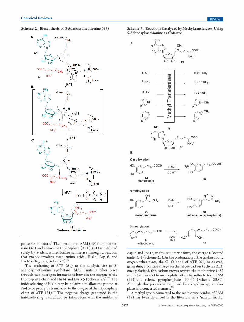
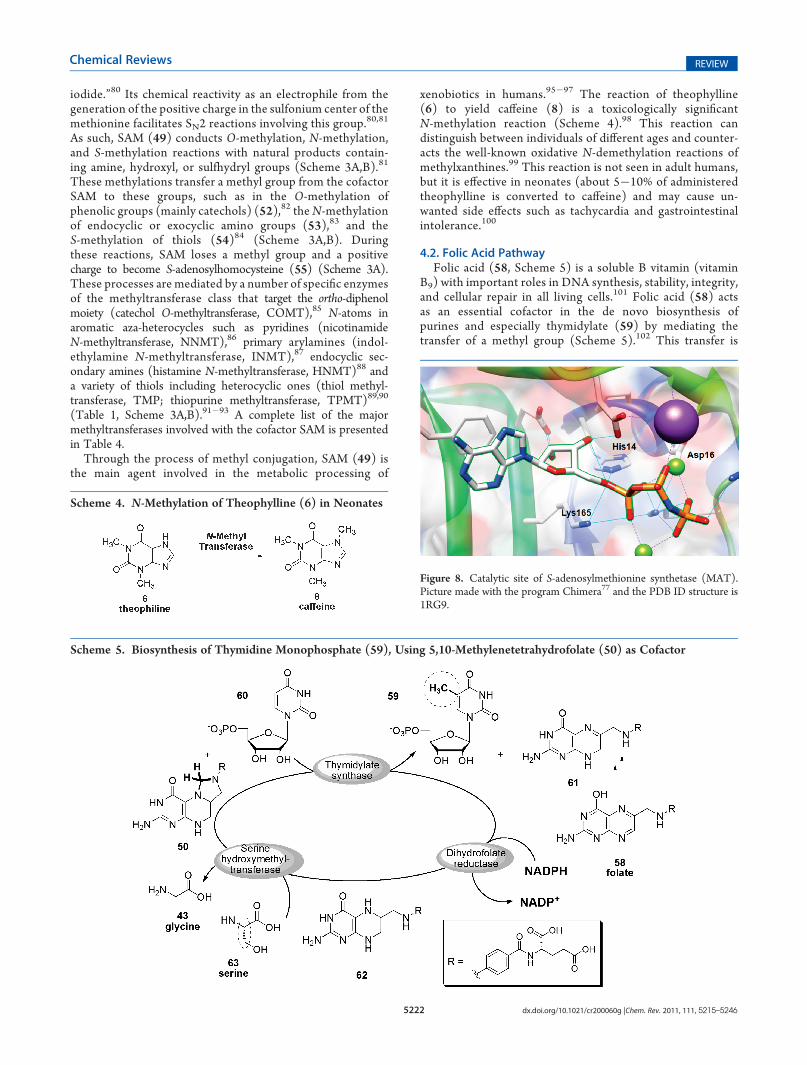
processes in nature.9 The formation of SAM (49) from methio-nine (48) and adenosine triphosphate (ATP) (51) is catalyzedsolely by S-adenosylmethionine synthetase through a reactionthat mainly involves three amino acids: His14, Asp16, andLys165 (Figure 8, Scheme 2).79
The anchoring of ATP (51) to the catalytic site of S-adenosylmethionine synthetase (MAT) initially takes placethrough two hydrogen interactions between the oxygen of thetriphosphate chain and His14 and Lys165 (Scheme 2A).79 Theimidazole ring of His14 may be polarized to allow the proton atN-4 to be promptly transferred to the oxygen of the triphosphatechain of ATP (51).79 The negative charge generated in theimidazole ring is stabilized by interactions with the amides of
Asp16 and Lys17; in this tautomeric form, the charge is locatedunderN-1 (Scheme 2B). As the protonation of the triphosphoricoxygen takes place, the C�O bond of ATP (51) is cleaved,generating a positive charge on the ribose carbon (Scheme 2B);once polarized, this carbon moves toward the methionine (48)and is then subject to nucleophilic attack by sulfur to form SAM(49) and release pyrophosphate (PPPi) (Scheme 2B,C).Although this process is described here step-by-step, it takesplace in a concerted manner.79
A methyl group connected to the methionine residue of SAM(49) has been described in the literature as a “natural methyl
Scheme 2. Biosynthesis of S-Adenosylmethionine (49) Scheme 3. Reactions Catalyzed byMethyltransferases, UsingS-Adenosylmethionine as Cofactor
5222 dx.doi.org/10.1021/cr200060g |Chem. Rev. 2011, 111, 5215–5246
Chemical Reviews REVIEW
iodide.”80 Its chemical reactivity as an electrophile from thegeneration of the positive charge in the sulfonium center of themethionine facilitates SN2 reactions involving this group.
80,81
As such, SAM (49) conducts O-methylation, N-methylation,and S-methylation reactions with natural products contain-ing amine, hydroxyl, or sulfhydryl groups (Scheme 3A,B).81
These methylations transfer a methyl group from the cofactorSAM to these groups, such as in the O-methylation ofphenolic groups (mainly catechols) (52),82 theN-methylationof endocyclic or exocyclic amino groups (53),83 and theS-methylation of thiols (54)84 (Scheme 3A,B). Duringthese reactions, SAM loses a methyl group and a positivecharge to become S-adenosylhomocysteine (55) (Scheme 3A).These processes are mediated by a number of specific enzymesof the methyltransferase class that target the ortho-diphenolmoiety (catechol O-methyltransferase, COMT),85 N-atoms inaromatic aza-heterocycles such as pyridines (nicotinamideN-methyltransferase, NNMT),86 primary arylamines (indol-ethylamine N-methyltransferase, INMT),87 endocyclic sec-ondary amines (histamine N-methyltransferase, HNMT)88 anda variety of thiols including heterocyclic ones (thiol methyl-transferase, TMP; thiopurine methyltransferase, TPMT)89,90
(Table 1, Scheme 3A,B).91�93 A complete list of the majormethyltransferases involved with the cofactor SAM is presentedin Table 4.
Through the process of methyl conjugation, SAM (49) isthe main agent involved in the metabolic processing of
xenobiotics in humans.95�97 The reaction of theophylline(6) to yield caffeine (8) is a toxicologically significantN-methylation reaction (Scheme 4).98 This reaction candistinguish between individuals of different ages and counter-acts the well-known oxidative N-demethylation reactions ofmethylxanthines.99 This reaction is not seen in adult humans,but it is effective in neonates (about 5�10% of administeredtheophylline is converted to caffeine) and may cause un-wanted side effects such as tachycardia and gastrointestinalintolerance.100
4.2. Folic Acid PathwayFolic acid (58, Scheme 5) is a soluble B vitamin (vitamin
B9) with important roles in DNA synthesis, stability, integrity,and cellular repair in all living cells.101 Folic acid (58) actsas an essential cofactor in the de novo biosynthesis ofpurines and especially thymidylate (59) by mediating thetransfer of a methyl group (Scheme 5).102 This transfer is
Scheme 5. Biosynthesis of Thymidine Monophosphate (59), Using 5,10-Methylenetetrahydrofolate (50) as Cofactor
Scheme 4. N-Methylation of Theophylline (6) in Neonates
Figure 8. Catalytic site of S-adenosylmethionine synthetase (MAT).Picture made with the program Chimera77 and the PDB ID structure is1RG9.
5223 dx.doi.org/10.1021/cr200060g |Chem. Rev. 2011, 111, 5215–5246
Chemical Reviews REVIEW
made specifically by 5,10-methylenetetrahydrofolate (50), areduced form of folate generated by the consecutive actions ofdihydrofolate reductase and serine hydroxymethyltransferase.A reductive methylation reaction catalyzed by thymidylatesynthase (TS) yields thymidine monophosphate (dTMP)(59) from deoxyuridine monophosphate (dUMP) (60)(Scheme 5).103�105 The presence of TS in most living organ-isms coupled with its important biochemical function inthe synthesis of thymine (1) by the methylation of uridine(2) has made this enzyme an attractive target for multipleresearch groups interested in anticancer106�108 and antiviralactivities.109,110
5. FROM METHYLATED NATURAL PRODUCTS TODRUGS: THE DISCOVERY OF STATINS
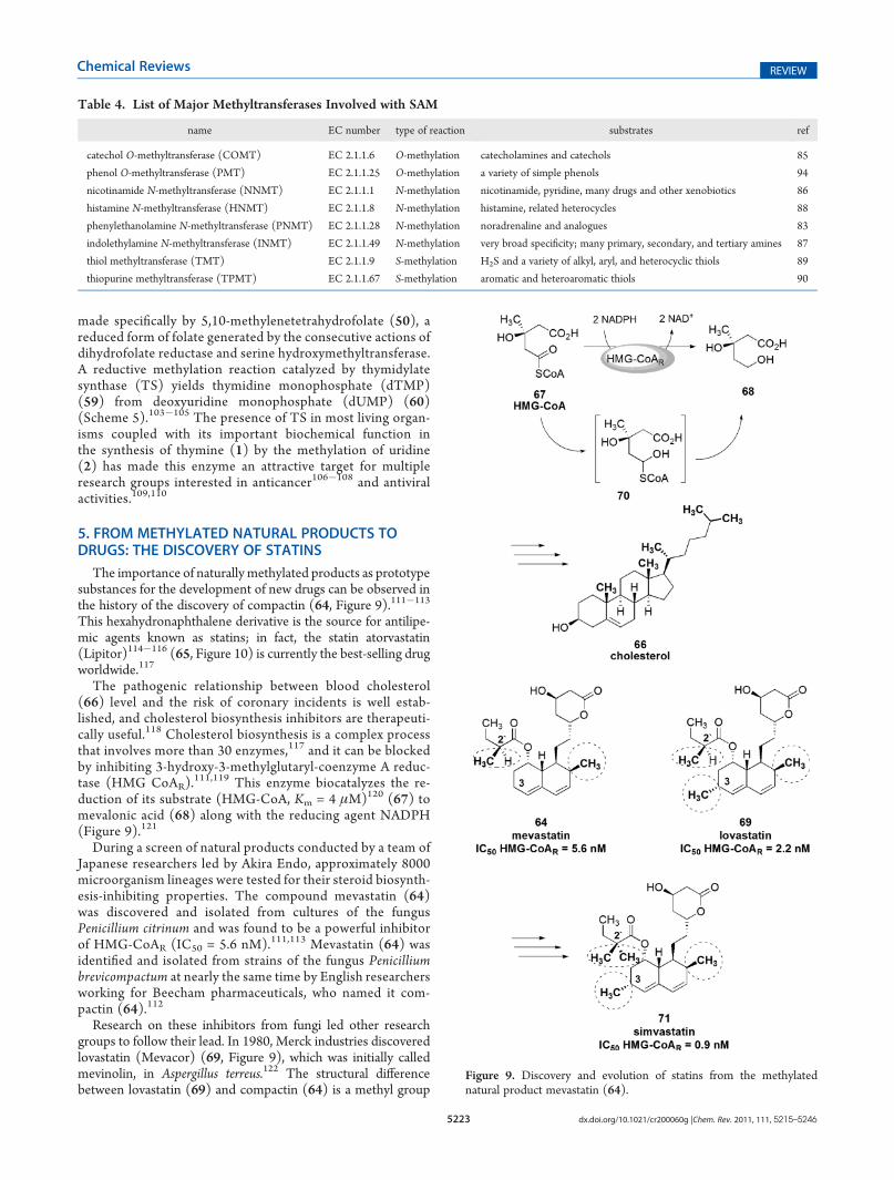
The importance of naturally methylated products as prototypesubstances for the development of new drugs can be observed inthe history of the discovery of compactin (64, Figure 9).111�113
This hexahydronaphthalene derivative is the source for antilipe-mic agents known as statins; in fact, the statin atorvastatin(Lipitor)114�116 (65, Figure 10) is currently the best-selling drugworldwide.117
The pathogenic relationship between blood cholesterol(66) level and the risk of coronary incidents is well estab-lished, and cholesterol biosynthesis inhibitors are therapeuti-cally useful.118 Cholesterol biosynthesis is a complex processthat involves more than 30 enzymes,117 and it can be blockedby inhibiting 3-hydroxy-3-methylglutaryl-coenzyme A reduc-tase (HMG CoAR).
111,119 This enzyme biocatalyzes the re-duction of its substrate (HMG-CoA, Km = 4 μM)120 (67) tomevalonic acid (68) along with the reducing agent NADPH(Figure 9).121
During a screen of natural products conducted by a team ofJapanese researchers led by Akira Endo, approximately 8000microorganism lineages were tested for their steroid biosynth-esis-inhibiting properties. The compound mevastatin (64)was discovered and isolated from cultures of the fungusPenicillium citrinum and was found to be a powerful inhibitorof HMG-CoAR (IC50 = 5.6 nM).111,113 Mevastatin (64) wasidentified and isolated from strains of the fungus Penicilliumbrevicompactum at nearly the same time by English researchersworking for Beecham pharmaceuticals, who named it com-pactin (64).112
Research on these inhibitors from fungi led other researchgroups to follow their lead. In 1980, Merck industries discoveredlovastatin (Mevacor) (69, Figure 9), which was initially calledmevinolin, in Aspergillus terreus.122 The structural differencebetween lovastatin (69) and compactin (64) is a methyl group
Table 4. List of Major Methyltransferases Involved with SAM
name EC number type of reaction substrates ref
catechol O-methyltransferase (COMT) EC 2.1.1.6 O-methylation catecholamines and catechols 85
phenol O-methyltransferase (PMT) EC 2.1.1.25 O-methylation a variety of simple phenols 94
nicotinamide N-methyltransferase (NNMT) EC 2.1.1.1 N-methylation nicotinamide, pyridine, many drugs and other xenobiotics 86
histamine N-methyltransferase (HNMT) EC 2.1.1.8 N-methylation histamine, related heterocycles 88
phenylethanolamine N-methyltransferase (PNMT) EC 2.1.1.28 N-methylation noradrenaline and analogues 83
indolethylamine N-methyltransferase (INMT) EC 2.1.1.49 N-methylation very broad specificity; many primary, secondary, and tertiary amines 87
thiol methyltransferase (TMT) EC 2.1.1.9 S-methylation H2S and a variety of alkyl, aryl, and heterocyclic thiols 89
thiopurine methyltransferase (TPMT) EC 2.1.1.67 S-methylation aromatic and heteroaromatic thiols 90
Figure 9. Discovery and evolution of statins from the methylatednatural product mevastatin (64).

5224 dx.doi.org/10.1021/cr200060g |Chem. Rev. 2011, 111, 5215–5246
Chemical Reviews REVIEW
at the C-3 position of the hexahydronaphthalene ring in (69),which increases its inhibition of HMG-CoAR ∼2.5-fold (IC50 =2.2 nM).123 This compound was launched in 1987 by MerckPharmaceuticals as the first safe and effective pro-drug of thestatin class.124�126
Countless structural changes in lovastatin (69) and compactin(64) have been introduced with the goal of understandingthe pharmacophoric contributions of each functional groupto the overall pharmacological activity (structure�activityrelationships).123,127�130 From these studies, it became clear thatthe stereochemistry of the δ-lactonic subunit (after being hydro-lyzed) is extremely important for the inhibitory activity (reversiblecompetitive inhibition), as it mimics the intermediate (70)(Figure 9) in the reduction of HMG-CoA (67) by HMG-CoAR.
127 In addition, the methyl groups at position 3 of thehexahydronaphthalene ring and position 20 of the butanoate sidechain are responsible for other important interactions with thetarget enzyme.123 Removal of the methyl from the hexahydro-naphthalene ring, as seen in compactin (64),112 leads to a 2.5-foldreduction in inhibitory potency; however, removal of the methylgroup R to the ester carbonyl unit (C-20) reduces its potencytoward HMG-CoAR by 4-fold (Figure 9).
123
Hoffman123 realized that the stereochemistry of the methylgroup R to the carbonyl group does not influence the activity ofthese compounds. Led by A. Patchett, Merck developed simvas-tatin (Zocor) (71) (IC50 HMG-CoAR = 0.9 nM),123 a 20-methylderivative of lovastatin (69). Simvastatin (71) is 2.5 times morepotent than lovastatin (69); in addition, it has one less chiralcenter and a longer half-life because the additional methyl groupblocks ester hydrolysis (Figure 9).123 Simvastatin (71) waslaunched in the North American pharmaceutical market in 1992.
Bruce Roth and associates at Parke-Davis laboratories designeda new class of HMG-CoAR inhibitors (second-generation in-hibitors) that have a 1,2-diphenyl pyrrole system (Figure 10).131
The molecular design of this new class was based on thehexahydronaphthalene system of lovastatin (69). This hydro-phobic pharmacophoric unit was replaced with a 1,2-diphenylpyrrole scaffold to achieve the desired hydrophobicity, therebycreating atorvastatin (65). This drug was launched in the phar-maceutical market in 1998 (Figure 10).114�116,131�133
Although the hexahydronaphthalene ring and side ester groupare replaced by other groups in these second-generation inhibitors,the methyl groups are still present. Some examples of second-generation inhibitors are fluvastatin (72),134�136 rosuvastatin(73),137,138 cerivastatin (74)139,140 (withdrawn from U.S. market
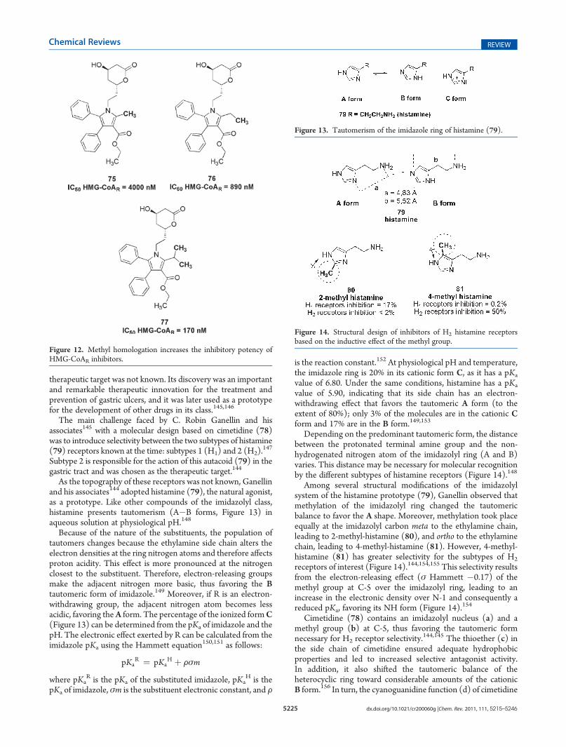
in 2001), and atorvastatin (65). All of these structures contain twomethyl groups in an isopropyl group connected to the centralheterocyclic system (Figure 11). These methyl groups are im-portant for activity due to their involvement in hydrophobicinteractions with Leu562, Cys561, and His752, which create anentropic gain in the interaction with the target receptor. Theremoval of the methyl groups leads to an 18-fold reduction inpotency, as shown in initial structure�activity relationship studieswith certain pyrrole derivatives (75�77) (Figure 12).23,132
6. STEREOELECTRONIC EFFECTS OF METHYL GROUPSIN DRUG DESIGN AND DISCOVERY
As seen with the discovery and evolution of statins fromnaturalmethylated products, the methyl group has been widely used inmolecular modification strategies employed by numerous re-search groups dedicated to the invention of new drugs. Theintroduction of methyl groups can radically alter the potency,duration, and nature of the pharmacological effect.5 As such, themedicinal chemist uses this group to modify aspects related tosolubility, conformation, electronic factors, bioavailability, andpharmacokinetics. We must bear in mind that methyl effects arenot limited to the simple transformation of functional groups, asin the alkylation of alcohols and carboxylic acid to form ethers andesters, respectively. Similarly, the process of extending alkyl chainsby adding a linearmethyl group, which is known as homologation,should not be considered a methyl effect. Conversely, if thisaddition is made through a branch to achieve steric effects or newhydrophobic interactions with a bioreceptor, an influence of thestereoelectronic effects of this group is clearly present.
6.1. The Inductive Effect of Methyl in the Discovery of Anti-Ulcer Drugs: Cimetidine and Hþ-Kþ-ATPase Inhibitors
Cimetidine (78)141�143 is the first selective antagonist ofsubtype 2 histamine receptors,144 and it is used in the treatmentand prevention of gastric ulcers. This drug was discovered almost40 years ago at a time when the structure of the chosen
Figure 10. The genesis of atorvastatin (65) from first-generationHMG-CoAR inhibitors.
Figure 11. Second-generation HMG-CoAR inhibitors.
5225 dx.doi.org/10.1021/cr200060g |Chem. Rev. 2011, 111, 5215–5246
Chemical Reviews REVIEW
therapeutic target was not known. Its discovery was an importantand remarkable therapeutic innovation for the treatment andprevention of gastric ulcers, and it was later used as a prototypefor the development of other drugs in its class.145,146
The main challenge faced by C. Robin Ganellin and hisassociates145 with a molecular design based on cimetidine (78)was to introduce selectivity between the two subtypes of histamine(79) receptors known at the time: subtypes 1 (H1) and 2 (H2).
147
Subtype 2 is responsible for the action of this autacoid (79) in thegastric tract and was chosen as the therapeutic target.144
As the topography of these receptors was not known, Ganellinand his associates144 adopted histamine (79), the natural agonist,as a prototype. Like other compounds of the imidazolyl class,histamine presents tautomerism (A�B forms, Figure 13) inaqueous solution at physiological pH.148
Because of the nature of the substituents, the population oftautomers changes because the ethylamine side chain alters theelectron densities at the ring nitrogen atoms and therefore affectsproton acidity. This effect is more pronounced at the nitrogenclosest to the substituent. Therefore, electron-releasing groupsmake the adjacent nitrogen more basic, thus favoring the Btautomeric form of imidazole.149 Moreover, if R is an electron-withdrawing group, the adjacent nitrogen atom becomes lessacidic, favoring theA form. The percentage of the ionized formC(Figure 13) can be determined from the pKa of imidazole and thepH. The electronic effect exerted by R can be calculated from theimidazole pKa using the Hammett equation150,151 as follows:
pKaR ¼ pKa
H þ Fσm
where pKaR is the pKa of the substituted imidazole, pKa
H is thepKa of imidazole, σm is the substituent electronic constant, and F
is the reaction constant.152 At physiological pH and temperature,the imidazole ring is 20% in its cationic form C, as it has a pKa
value of 6.80. Under the same conditions, histamine has a pKa
value of 5.90, indicating that its side chain has an electron-withdrawing effect that favors the tautomeric A form (to theextent of 80%); only 3% of the molecules are in the cationic Cform and 17% are in the B form.149,153
Depending on the predominant tautomeric form, the distancebetween the protonated terminal amine group and the non-hydrogenated nitrogen atom of the imidazolyl ring (A and B)varies. This distance may be necessary for molecular recognitionby the different subtypes of histamine receptors (Figure 14).148
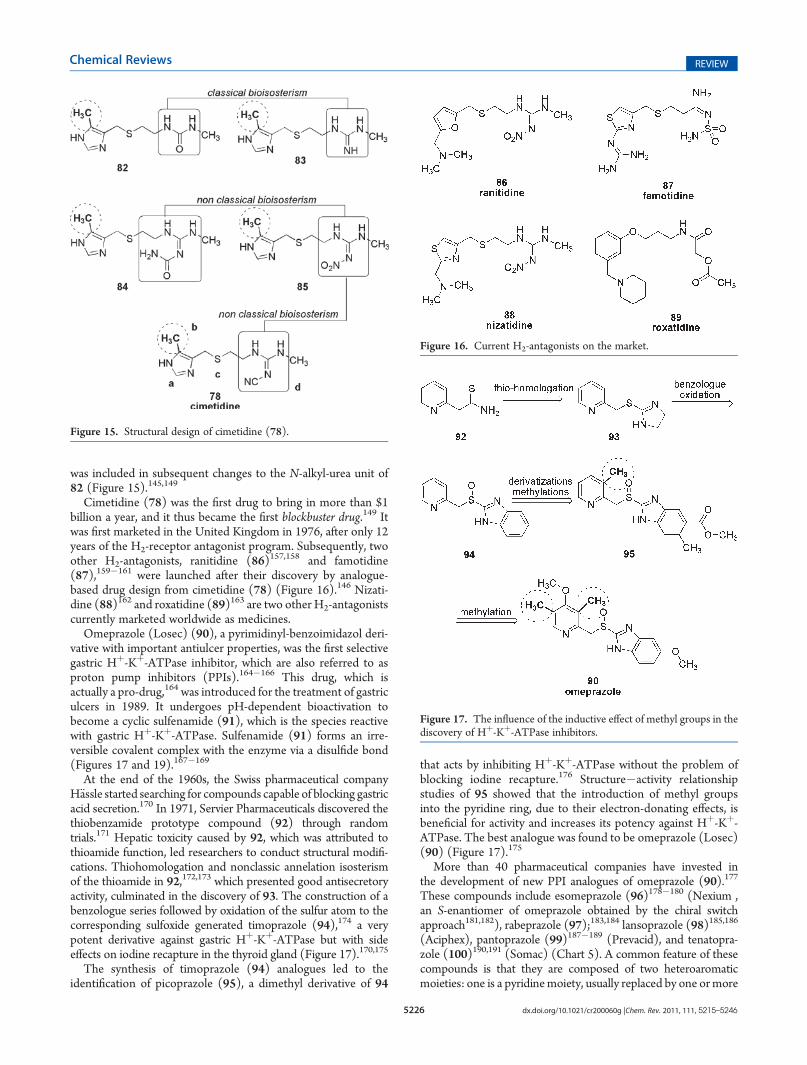
Among several structural modifications of the imidazolylsystem of the histamine prototype (79), Ganellin observed thatmethylation of the imidazolyl ring changed the tautomericbalance to favor the A shape. Moreover, methylation took placeequally at the imidazolyl carbon meta to the ethylamine chain,leading to 2-methyl-histamine (80), and ortho to the ethylaminechain, leading to 4-methyl-histamine (81). However, 4-methyl-histamine (81) has greater selectivity for the subtypes of H2
receptors of interest (Figure 14).144,154,155 This selectivity resultsfrom the electron-releasing effect (σ Hammett �0.17) of themethyl group at C-5 over the imidazolyl ring, leading to anincrease in the electronic density over N-1 and consequently areduced pKa, favoring its NH form (Figure 14).154
Cimetidine (78) contains an imidazolyl nucleus (a) and amethyl group (b) at C-5, thus favoring the tautomeric formnecessary for H2 receptor selectivity.
144,145 The thioether (c) inthe side chain of cimetidine ensured adequate hydrophobicproperties and led to increased selective antagonist activity.In addition, it also shifted the tautomeric balance of theheterocyclic ring toward considerable amounts of the cationicB form.156 In turn, the cyanoguanidine function (d) of cimetidine
Figure 12. Methyl homologation increases the inhibitory potency ofHMG-CoAR inhibitors.
Figure 13. Tautomerism of the imidazole ring of histamine (79).
Figure 14. Structural design of inhibitors of H2 histamine receptorsbased on the inductive effect of the methyl group.
5226 dx.doi.org/10.1021/cr200060g |Chem. Rev. 2011, 111, 5215–5246
Chemical Reviews REVIEW
was included in subsequent changes to the N-alkyl-urea unit of82 (Figure 15).145,149
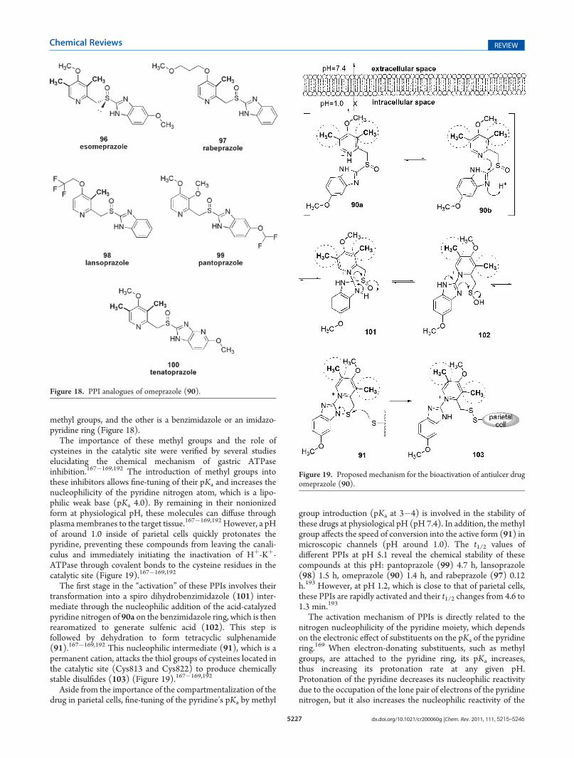
Cimetidine (78) was the first drug to bring in more than $1billion a year, and it thus became the first blockbuster drug.149 Itwas first marketed in the United Kingdom in 1976, after only 12years of the H2-receptor antagonist program. Subsequently, twoother H2-antagonists, ranitidine (86)157,158 and famotidine(87),159�161 were launched after their discovery by analogue-based drug design from cimetidine (78) (Figure 16).146 Nizati-dine (88)162 and roxatidine (89)163 are two other H2-antagonistscurrently marketed worldwide as medicines.
Omeprazole (Losec) (90), a pyrimidinyl-benzoimidazol deri-vative with important antiulcer properties, was the first selectivegastric Hþ-Kþ-ATPase inhibitor, which are also referred to asproton pump inhibitors (PPIs).164�166 This drug, which isactually a pro-drug,164 was introduced for the treatment of gastriculcers in 1989. It undergoes pH-dependent bioactivation tobecome a cyclic sulfenamide (91), which is the species reactivewith gastric Hþ-Kþ-ATPase. Sulfenamide (91) forms an irre-versible covalent complex with the enzyme via a disulfide bond(Figures 17 and 19).167�169
At the end of the 1960s, the Swiss pharmaceutical companyH€assle started searching for compounds capable of blocking gastricacid secretion.170 In 1971, Servier Pharmaceuticals discovered thethiobenzamide prototype compound (92) through randomtrials.171 Hepatic toxicity caused by 92, which was attributed tothioamide function, led researchers to conduct structural modifi-cations. Thiohomologation and nonclassic annelation isosterismof the thioamide in 92,172,173 which presented good antisecretoryactivity, culminated in the discovery of 93. The construction of abenzologue series followed by oxidation of the sulfur atom to thecorresponding sulfoxide generated timoprazole (94),174 a verypotent derivative against gastric Hþ-Kþ-ATPase but with sideeffects on iodine recapture in the thyroid gland (Figure 17).170,175
The synthesis of timoprazole (94) analogues led to theidentification of picoprazole (95), a dimethyl derivative of 94
that acts by inhibiting Hþ-Kþ-ATPase without the problem ofblocking iodine recapture.176 Structure�activity relationshipstudies of 95 showed that the introduction of methyl groupsinto the pyridine ring, due to their electron-donating effects, isbeneficial for activity and increases its potency against Hþ-Kþ-ATPase. The best analogue was found to be omeprazole (Losec)(90) (Figure 17).175
More than 40 pharmaceutical companies have invested inthe development of new PPI analogues of omeprazole (90).177
These compounds include esomeprazole (96)178�180 (Nexium ,an S-enantiomer of omeprazole obtained by the chiral switchapproach181,182), rabeprazole (97);183,184 lansoprazole (98)185,186
(Aciphex), pantoprazole (99)187�189 (Prevacid), and tenatopra-zole (100)190,191 (Somac) (Chart 5). A common feature of thesecompounds is that they are composed of two heteroaromaticmoieties: one is a pyridinemoiety, usually replaced by one ormore
Figure 15. Structural design of cimetidine (78).
Figure 16. Current H2-antagonists on the market.
Figure 17. The influence of the inductive effect of methyl groups in thediscovery of Hþ-Kþ-ATPase inhibitors.
5227 dx.doi.org/10.1021/cr200060g |Chem. Rev. 2011, 111, 5215–5246
Chemical Reviews REVIEW
methyl groups, and the other is a benzimidazole or an imidazo-pyridine ring (Figure 18).
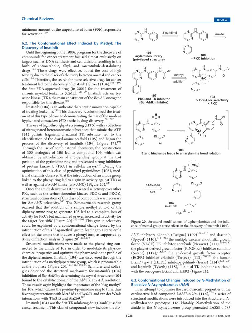
The importance of these methyl groups and the role ofcysteines in the catalytic site were verified by several studieselucidating the chemical mechanism of gastric ATPaseinhibition.167�169,192 The introduction of methyl groups intothese inhibitors allows fine-tuning of their pKa and increases thenucleophilicity of the pyridine nitrogen atom, which is a lipo-philic weak base (pKa 4.0). By remaining in their nonionizedform at physiological pH, these molecules can diffuse throughplasmamembranes to the target tissue.167�169,192 However, a pHof around 1.0 inside of parietal cells quickly protonates thepyridine, preventing these compounds from leaving the canali-culus and immediately initiating the inactivation of Hþ-Kþ-ATPase through covalent bonds to the cysteine residues in thecatalytic site (Figure 19).167�169,192
The first stage in the “activation” of these PPIs involves theirtransformation into a spiro dihydrobenzimidazole (101) inter-mediate through the nucleophilic addition of the acid-catalyzedpyridine nitrogen of 90a on the benzimidazole ring, which is thenrearomatized to generate sulfenic acid (102). This step isfollowed by dehydration to form tetracyclic sulphenamide(91).167�169,192 This nucleophilic intermediate (91), which is apermanent cation, attacks the thiol groups of cysteines located inthe catalytic site (Cys813 and Cys822) to produce chemicallystable disulfides (103) (Figure 19).167�169,192
Aside from the importance of the compartmentalization of thedrug in parietal cells, fine-tuning of the pyridine’s pKa by methyl
group introduction (pKa at 3�4) is involved in the stability ofthese drugs at physiological pH (pH 7.4). In addition, the methylgroup affects the speed of conversion into the active form (91) inmicroscopic channels (pH around 1.0). The t1/2 values ofdifferent PPIs at pH 5.1 reveal the chemical stability of thesecompounds at this pH: pantoprazole (99) 4.7 h, lansoprazole(98) 1.5 h, omeprazole (90) 1.4 h, and rabeprazole (97) 0.12h.193 However, at pH 1.2, which is close to that of parietal cells,these PPIs are rapidly activated and their t1/2 changes from 4.6 to1.3 min.193
The activation mechanism of PPIs is directly related to thenitrogen nucleophilicity of the pyridine moiety, which dependson the electronic effect of substituents on the pKa of the pyridinering.169 When electron-donating substituents, such as methylgroups, are attached to the pyridine ring, its pKa increases,thus increasing its protonation rate at any given pH.Protonation of the pyridine decreases its nucleophilic reactivitydue to the occupation of the lone pair of electrons of the pyridinenitrogen, but it also increases the nucleophilic reactivity of the
Figure 19. Proposed mechanism for the bioactivation of antiulcer drugomeprazole (90).
Figure 18. PPI analogues of omeprazole (90).
5228 dx.doi.org/10.1021/cr200060g |Chem. Rev. 2011, 111, 5215–5246
Chemical Reviews REVIEW
minimum amount of the unprotonated form (90b) responsiblefor activation.169
6.2. The Conformational Effect Induced by Methyl: TheDiscovery of Imatinib
Until the beginning of the 1980s, programs for the discovery ofcompounds for cancer treatment focused almost exclusively ontargets such as DNA synthesis and cell division, resulting in thebirth of antimetabolic, alkyl, and microtubule-destabilizingdrugs.194 These drugs were effective, but at the cost of hightoxicity due to their lack of selectivity between normal and cancercells.194 Therefore, the search for more selective drugs for cancertreatment led to the discovery of imatinib (Glivec) (104),195�197
the first FDA-approved drug (in 2001) for the treatment ofchronic myeloid leukemia (CML).198,199 Imatinib acts on tyr-osine kinase (TK), the main constituent of the Bcr-Abl oncogeneresponsible for this disease.200
Imatinib (104) is an authentic therapeutic innovation capableof treating leukemia.198 This discovery revolutionized the treat-ment of this type of cancer, demonstrating the use of the modernhyphenated combichem-HTS tactic in drug discovery.201,202
The use of high-throughput screening (HTS) with a collectionof nitrogenated heteroaromatic substances that mimic the ATP(51) purinic fragment, a natural TK substrate, led to theidentification of the diaryl-amine scaffold (105) and began theprocess of the discovery of imatinib (104) (Figure 17).194
Through the use of combinatorial chemistry, the constructionof 300 analogues of 105 led to compound 106, which wasobtained by introduction of a 3-pyridinyl group at the C-4position of the pyrimidine ring and presented strong inhibitionof protein kinase C (PKC) in cellular assays.194 During theoptimization of this class of pyridinyl-pyrimidines (106), med-icinal chemists observed that the introduction of an amide grouplinked to the phenyl ring led to a gain in activity against TKs aswell as against Bcr-Abl kinase (Bcr-AblK) (Figure 20).203
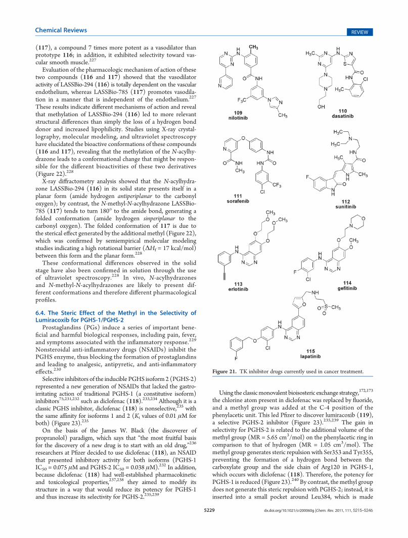
Once the amide derivative 107 presented selectivity over otherPKs, such as the serine/threonine kinases PKC-R and PKC-δ,structural optimization of this class of compounds was necessaryfor Bcr-AblK selectivity.203 The Zimmermann research grouprealized that the addition of a simple methyl at C-6 of thediphenylamine ring to generate 108 led to a complete loss ofactivity for PKCs but maintained or even increased its activity forthe target Bcr-AblK (Figure 20).203�205 This gain in selectivitycould be explained by a conformational change forced by theintroduction of this “flag-methyl” group, leading to a steric orthoeffect on the amine that induces a phenyl turn, as supported byX-ray diffraction analysis (Figure 20).203,206
Structural modifications were made to the phenyl ring con-nected to the amide of 108 in order to modulate its physico-chemical properties and optimize the pharmacokinetic profile ofthe diphenylamines. Imatinib (104) was discovered through theintroduction of a methylpiperazine group, which is protonatableat the biophase (Figure 20).194,196,197,203 Schindler and collea-gues described the structural mechanism for imatinib’s (104)inhibition of Bcr-AblK by determining the crystal structure of 104bound to the catalytic domain of the Abl TK at 2.4 Å resolution.These results again highlight the importance of the “flag-methyl”for 108, which causes the pyridinyl-pyrimidine ring to turn, thusfavoring interactions withMet318 and Lys271 and van derWaalsinteractions with Thr315 and Ala269.207
Imatinib (104) was the first TK inhibiting drug (“tinib”) used incancer treatment. This class of compounds now includes the Bcr-
AblK inhibitors nilotinib (Tasigna) (109)208�210 and dasatinib(Sprycel) (110),211�213 the multiple vascular endothelial growthfactor (VEGF) TK inhibitor sorafenib (Nexavar) (111),214�217
the platelet-derived growth factor (PDGF-Rs) inhibitor sunitinib(Sutent) (112),218,219 the epidermal growth factor receptor(EGFR) inhibitor erlotinib (Tarceva) (113),220,221 the humanEGFR type 1 (HER1) inhibitor gefitinib (Iressa) (114),222�224
and lapatinib (Tykerb) (115),225 a dual TK inhibitor associatedwith the oncogenes EGFR and HER2 (Figure 21).
6.3. Conformational Changes Induced by N-Methylation ofBioactive N-Acylhydrazones (NAH)
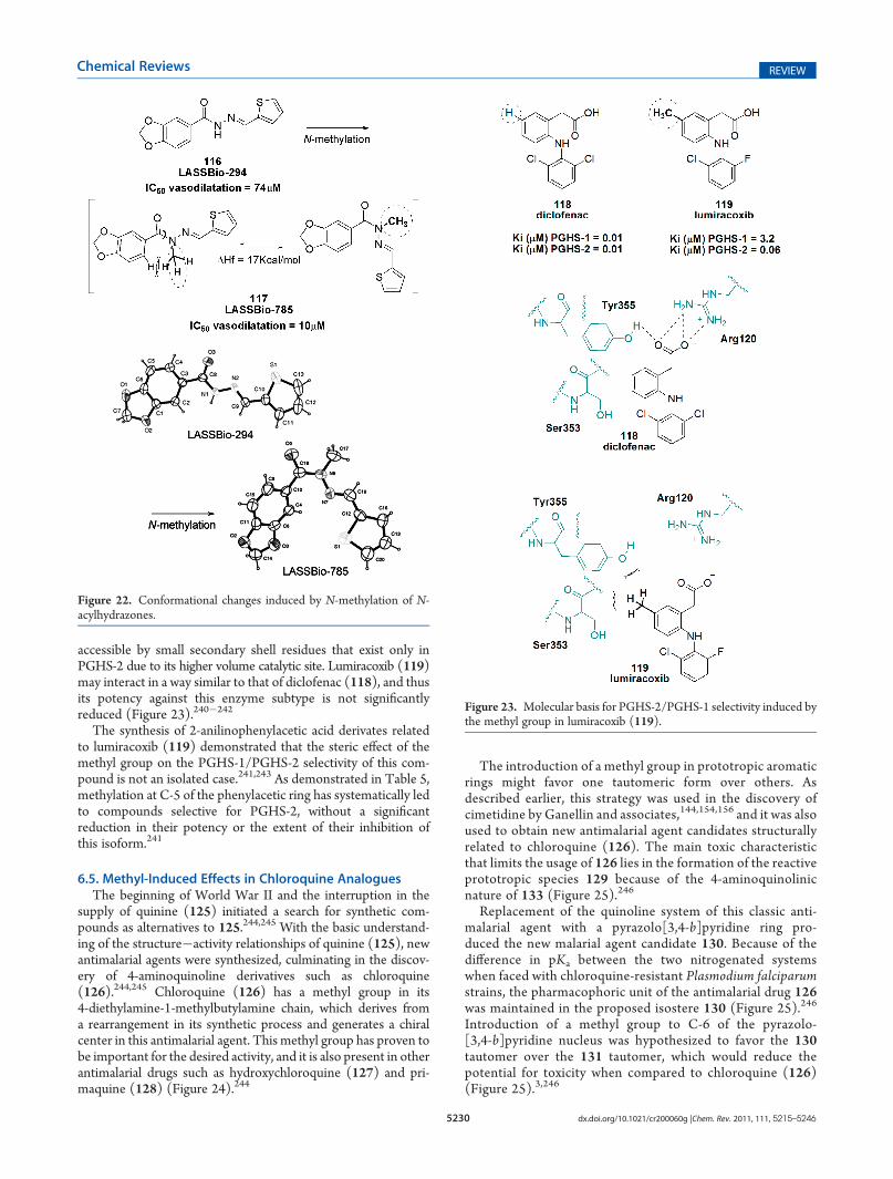
In an attempt to optimize the cardiovascular properties of thecardioactive lead compound LASSBio-294 (116),226 a series ofstructural modifications were introduced into the structure of N-acylhydrazone prototype 116. Notably, N-methylation of theamide in the N-acylhydrazone group generated LASSBio-785
Figure 20. Structural modifications of diphenylamines and the influ-ence of methyl group steric effects in the discovery of imatinib (104).
5229 dx.doi.org/10.1021/cr200060g |Chem. Rev. 2011, 111, 5215–5246
Chemical Reviews REVIEW
(117), a compound 7 times more potent as a vasodilator thanprototype 116; in addition, it exhibited selectivity toward vas-cular smooth muscle.227
Evaluation of the pharmacologic mechanism of action of thesetwo compounds (116 and 117) showed that the vasodilatoractivity of LASSBio-294 (116) is totally dependent on the vascularendothelium, whereas LASSBio-785 (117) promotes vasodila-tion in a manner that is independent of the endothelium.227
These results indicate different mechanisms of action and revealthat methylation of LASSBio-294 (116) led to more relevantstructural differences than simply the loss of a hydrogen bonddonor and increased lipophilicity. Studies using X-ray crystal-lography, molecular modeling, and ultraviolet spectroscopyhave elucidated the bioactive conformations of these compounds(116 and 117), revealing that the methylation of the N-acylhy-drazone leads to a conformational change that might be respon-sible for the different bioactivities of these two derivatives(Figure 22).228
X-ray diffractometry analysis showed that the N-acylhydra-zone LASSBio-294 (116) in its solid state presents itself in aplanar form (amide hydrogen antiperiplanar to the carbonyloxygen); by contrast, the N-methyl-N-acylhydrazone LASSBio-785 (117) tends to turn 180� to the amide bond, generating afolded conformation (amide hydrogen sinperiplanar to thecarbonyl oxygen). The folded conformation of 117 is due tothe sterical effect generated by the additional methyl (Figure 22),which was confirmed by semiempirical molecular modelingstudies indicating a high rotational barrier (ΔHf = 17 kcal/mol)between this form and the planar form.228
These conformational differences observed in the solidstage have also been confirmed in solution through the useof ultraviolet spectroscopy.228 In vivo, N-acylhydrazonesand N-methyl-N-acylhydrazones are likely to present dif-ferent conformations and therefore different pharmacologicalprofiles.
6.4. The Steric Effect of the Methyl in the Selectivity ofLumiracoxib for PGHS-1/PGHS-2
Prostaglandins (PGs) induce a series of important bene-ficial and harmful biological responses, including pain, fever,and symptoms associated with the inflammatory response.229
Nonsteroidal anti-inflammatory drugs (NSAIDs) inhibit thePGHS enzyme, thus blocking the formation of prostaglandinsand leading to analgesic, antipyretic, and anti-inflammatoryeffects.230
Selective inhibitors of the inducible PGHS isoform2 (PGHS-2)represented a new generation of NSAIDs that lacked the gastro-irritating action of traditional PGHS-1 (a constitutive isoform)inhibitors75,231,232 such as diclofenac (118).233,234 Although it is aclassic PGHS inhibitor, diclofenac (118) is nonselective,235 withthe same affinity for isoforms 1 and 2 (Ki values of 0.01 μM forboth) (Figure 23).235
On the basis of the James W. Black (the discoverer ofpropranolol) paradigm, which says that “the most fruitful basisfor the discovery of a new drug is to start with an old drug,”236
researchers at Pfizer decided to use diclofenac (118), an NSAIDthat presented inhibitory activity for both isoforms (PGHS-1IC50 = 0.075 μM and PGHS-2 IC50 = 0.038 μM).232 In addition,because diclofenac (118) had well-established pharmacokineticand toxicological properties,237,238 they aimed to modify itsstructure in a way that would reduce its potency for PGHS-1and thus increase its selectivity for PGHS-2.235,239
Using the classicmonovalent bioisosteric exchange strategy,172,173
the chlorine atom present in diclofenac was replaced by fluoride,and a methyl group was added at the C-4 position of thephenylacetic unit. This led Pfizer to discover lumiracoxib (119),a selective PGHS-2 inhibitor (Figure 23).235,239 The gain inselectivity for PGHS-2 is related to the additional volume of themethyl group (MR = 5.65 cm3/mol) on the phenylacetic ring incomparison to that of hydrogen (MR = 1.05 cm3/mol). Themethyl group generates steric repulsion with Ser353 and Tyr355,preventing the formation of a hydrogen bond between thecarboxylate group and the side chain of Arg120 in PGHS-1,which occurs with diclofenac (118). Therefore, the potency forPGHS-1 is reduced (Figure 23).240 By contrast, themethyl groupdoes not generate this steric repulsion with PGHS-2; instead, it isinserted into a small pocket around Leu384, which is made
Figure 21. TK inhibitor drugs currently used in cancer treatment.
5230 dx.doi.org/10.1021/cr200060g |Chem. Rev. 2011, 111, 5215–5246
Chemical Reviews REVIEW
accessible by small secondary shell residues that exist only inPGHS-2 due to its higher volume catalytic site. Lumiracoxib (119)may interact in a way similar to that of diclofenac (118), and thusits potency against this enzyme subtype is not significantlyreduced (Figure 23).240�242
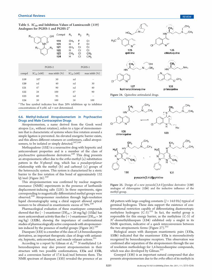
The synthesis of 2-anilinophenylacetic acid derivates relatedto lumiracoxib (119) demonstrated that the steric effect of themethyl group on the PGHS-1/PGHS-2 selectivity of this com-pound is not an isolated case.241,243 As demonstrated in Table 5,methylation at C-5 of the phenylacetic ring has systematically ledto compounds selective for PGHS-2, without a significantreduction in their potency or the extent of their inhibition ofthis isoform.241
6.5. Methyl-Induced Effects in Chloroquine AnaloguesThe beginning of World War II and the interruption in the
supply of quinine (125) initiated a search for synthetic com-pounds as alternatives to 125.244,245 With the basic understand-ing of the structure�activity relationships of quinine (125), newantimalarial agents were synthesized, culminating in the discov-ery of 4-aminoquinoline derivatives such as chloroquine(126).244,245 Chloroquine (126) has a methyl group in its4-diethylamine-1-methylbutylamine chain, which derives froma rearrangement in its synthetic process and generates a chiralcenter in this antimalarial agent. This methyl group has proven tobe important for the desired activity, and it is also present in otherantimalarial drugs such as hydroxychloroquine (127) and pri-maquine (128) (Figure 24).244
The introduction of a methyl group in prototropic aromaticrings might favor one tautomeric form over others. Asdescribed earlier, this strategy was used in the discovery ofcimetidine by Ganellin and associates,144,154,156 and it was alsoused to obtain new antimalarial agent candidates structurallyrelated to chloroquine (126). The main toxic characteristicthat limits the usage of 126 lies in the formation of the reactiveprototropic species 129 because of the 4-aminoquinolinicnature of 133 (Figure 25).246
Replacement of the quinoline system of this classic anti-malarial agent with a pyrazolo[3,4-b]pyridine ring pro-duced the new malarial agent candidate 130. Because of thedifference in pKa between the two nitrogenated systemswhen faced with chloroquine-resistant Plasmodium falciparumstrains, the pharmacophoric unit of the antimalarial drug 126was maintained in the proposed isostere 130 (Figure 25).246
Introduction of a methyl group to C-6 of the pyrazolo-[3,4-b]pyridine nucleus was hypothesized to favor the 130tautomer over the 131 tautomer, which would reduce thepotential for toxicity when compared to chloroquine (126)(Figure 25).3,246
Figure 22. Conformational changes induced by N-methylation of N-acylhydrazones.
Figure 23. Molecular basis for PGHS-2/PGHS-1 selectivity induced bythe methyl group in lumiracoxib (119).
5231 dx.doi.org/10.1021/cr200060g |Chem. Rev. 2011, 111, 5215–5246
Chemical Reviews REVIEW
6.6. Methyl-Induced Atropoisomerism in PsychoactiveDrugs and Male Contraceptive Drugs
Atropoisomerism, a name derived from the Greek wordatropos (i.e., without rotation), refers to a type of stereoisomer-ism that is characteristic of systems where free rotation around asimple ligation is prevented. An elevated energetic barrier exists,and this allows different rotamers or conformers, called atropoi-somers, to be isolated or simply detected.247,248
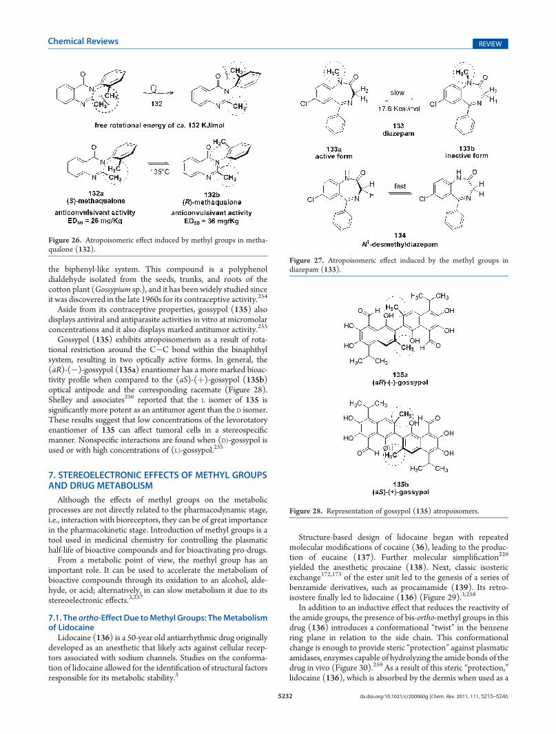
Methaqualone (132) is a neuroactive drug with hypnotic andanticonvulsant properties and is a member of the class ofpsychoactive quinazolinone derivatives.249 This drug presentsan atropoisomeric effect due to the ortho-methyl (a) substitutionpattern in the N-phenyl ring, which has a pseudoperiplanarrelationship with the methyl (b) and carbonyl (c) groups ofthe heterocyclic system. This system is characterized by a stericbarrier to the free rotation of this bond of approximately 132kJ/mol (Figure 26).250
This atropoisomerism was confirmed by nuclear magneticresonance (NMR) experiments in the presence of lanthanidedisplacement-inducing salts (LIS). In these experiments, signscorresponding to magnetically differentiated methyl groups wereobserved.250 Atropoisomer resolution through high-resolutionliquid chromatography using a chiral support allowed opticalisomers to be obtained in enantiomeric excess of 70%.250
Pharmacological evaluation of these enantiopure compoundsshowed that the (�)-enantiomer (DE50 = 26 mg/kg) (132a) hasmore anticonvulsant activity than the (þ)-enantiomer (DE50 = 36mg/kg) (132b), showing the importance of conformationalstudies of pharmacologically active compoundswith atropoisomer-ism induced by the presence of methyl groups (Figure 26).250
Diazepam (133) is a member of the class of 1,4-benzodiazepinederivatives, an important therapeutic class of drugs with antianxi-ety, hypnotic, anticonvulsant, and muscle relaxant actions.251
According to a report by Gilman et al.,252 N-methylated 1,4-benzodiazepines may also present atropoisomerism in theirstructure with two possible conformations, 133a and 133b,and a conversion barrier of 17.6 kcal/mol between them. TheNMR spectrum of diazepam (133) revealed the presence of an
AB pattern with large coupling constants (J = 14.0 Hz) typical ofgerminal hydrogens. These data support the existence of con-formational restriction capable of differentiating diastereotopicmethylene hydrogens (C-3).252 In fact, the methyl group isresponsible for this energy barrier, as the methylene (C-3) ofN1-desmethyldiazepam (134) exhibited only a singlet in itsNMR spectrum, indicative of a quick interconversion betweenthe two atropisomeric forms (Figure 27).252
Biological assays with diazepam enantiomeric pairs (133a,133b) indicated that the enantiomer 133a is stereoselectivelyrecognized by benzodiazepine receptors. This observation wasconfirmed after separation of the atropoisomers through the useof resolution methodology for 1,4-benzodiazepine compounds,which was also developed by Gilman.253
Gossypol (135) is an important natural compound that alsopresents atropoisomerism due to the ortho effect of its methyls in
Table 5. IC50 and Inhibition Values of Lumiracoxib (119)Analogues for PGHS-1 and PGHS-2a
PGHS-2 PGHS-1
compd IC50 (nM) max inhib (%) IC50 (nM) max inhib (%)
119 107 50 nd �120 nd 40 nd 30
121 47 90 nd 40
122 28 100 67 90
123 60 95 nd �124 20 100 58 100
aThe line symbol indicates less than 20% inhibition up to inhibitorconcentrations of 4 μM. nd = not determined.
Figure 24. Quinoline antimalarial drugs.
Figure 25. Design of a new pyrazole[3,4-b]pyridine derivative (130)analogue of chloroquine (126) and the inductive influence of themethyl group.
5232 dx.doi.org/10.1021/cr200060g |Chem. Rev. 2011, 111, 5215–5246
Chemical Reviews REVIEW
the biphenyl-like system. This compound is a polyphenoldialdehyde isolated from the seeds, trunks, and roots of thecotton plant (Gossypium sp.), and it has been widely studied sinceit was discovered in the late 1960s for its contraceptive activity.254
Aside from its contraceptive properties, gossypol (135) alsodisplays antiviral and antiparasite activities in vitro at micromolarconcentrations and it also displays marked antitumor activity.255
Gossypol (135) exhibits atropoisomerism as a result of rota-tional restriction around the C�C bond within the binaphthylsystem, resulting in two optically active forms. In general, the(aR)-(�)-gossypol (135a) enantiomer has a more marked bioac-tivity profile when compared to the (aS)-(þ)-gossypol (135b)optical antipode and the corresponding racemate (Figure 28).Shelley and associates256 reported that the L isomer of 135 issignificantly more potent as an antitumor agent than the D isomer.These results suggest that low concentrations of the levorotatoryenantiomer of 135 can affect tumoral cells in a stereospecificmanner. Nonspecific interactions are found when (D)-gossypol isused or with high concentrations of (L)-gossypol.255
7. STEREOELECTRONIC EFFECTS OF METHYL GROUPSAND DRUG METABOLISM
Although the effects of methyl groups on the metabolicprocesses are not directly related to the pharmacodynamic stage,i.e., interaction with bioreceptors, they can be of great importancein the pharmacokinetic stage. Introduction of methyl groups is atool used in medicinal chemistry for controlling the plasmatichalf-life of bioactive compounds and for bioactivating pro-drugs.
From a metabolic point of view, the methyl group has animportant role. It can be used to accelerate the metabolism ofbioactive compounds through its oxidation to an alcohol, alde-hyde, or acid; alternatively, in can slow metabolism it due to itsstereoelectronic effects.3,257
7.1. The ortho-Effect Due toMethyl Groups: TheMetabolismof Lidocaine
Lidocaine (136) is a 50-year old antiarrhythmic drug originallydeveloped as an anesthetic that likely acts against cellular recep-tors associated with sodium channels. Studies on the conforma-tion of lidocaine allowed for the identification of structural factorsresponsible for its metabolic stability.3
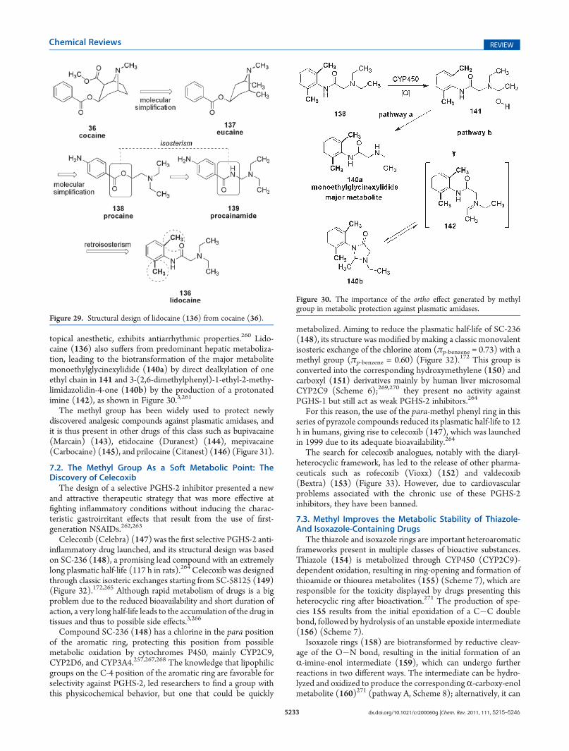
Structure-based design of lidocaine began with repeatedmolecular modifications of cocaine (36), leading to the produc-tion of eucaine (137). Further molecular simplification226
yielded the anesthetic procaine (138). Next, classic isostericexchange172,173 of the ester unit led to the genesis of a series ofbenzamide derivatives, such as procainamide (139). Its retro-isostere finally led to lidocaine (136) (Figure 29).3,258
In addition to an inductive effect that reduces the reactivity ofthe amide groups, the presence of bis-ortho-methyl groups in thisdrug (136) introduces a conformational “twist” in the benzenering plane in relation to the side chain. This conformationalchange is enough to provide steric “protection” against plasmaticamidases, enzymes capable of hydrolyzing the amide bonds of thedrug in vivo (Figure 30).259 As a result of this steric “protection,”lidocaine (136), which is absorbed by the dermis when used as a
Figure 26. Atropoisomeric effect induced by methyl groups in metha-qualone (132).
Figure 27. Atropoisomeric effect induced by the methyl groups indiazepam (133).
Figure 28. Representation of gossypol (135) atropoisomers.
5233 dx.doi.org/10.1021/cr200060g |Chem. Rev. 2011, 111, 5215–5246
Chemical Reviews REVIEW
topical anesthetic, exhibits antiarrhythmic properties.260 Lido-caine (136) also suffers from predominant hepatic metaboliza-tion, leading to the biotransformation of the major metabolitemonoethylglycinexylidide (140a) by direct dealkylation of oneethyl chain in 141 and 3-(2,6-dimethylphenyl)-1-ethyl-2-methy-limidazolidin-4-one (140b) by the production of a protonatedimine (142), as shown in Figure 30.3,261
The methyl group has been widely used to protect newlydiscovered analgesic compounds against plasmatic amidases, andit is thus present in other drugs of this class such as bupivacaine(Marcain) (143), etidocaine (Duranest) (144), mepivacaine(Carbocaine) (145), and prilocaine (Citanest) (146) (Figure 31).
7.2. The Methyl Group As a Soft Metabolic Point: TheDiscovery of Celecoxib
The design of a selective PGHS-2 inhibitor presented a newand attractive therapeutic strategy that was more effective atfighting inflammatory conditions without inducing the charac-teristic gastroirritant effects that result from the use of first-generation NSAIDs.262,263
Celecoxib (Celebra) (147) was the first selective PGHS-2 anti-inflammatory drug launched, and its structural design was basedon SC-236 (148), a promising lead compound with an extremelylong plasmatic half-life (117 h in rats).264 Celecoxib was designedthrough classic isosteric exchanges starting from SC-58125 (149)(Figure 32).172,265 Although rapid metabolism of drugs is a bigproblem due to the reduced bioavailability and short duration ofaction, a very long half-life leads to the accumulation of the drug intissues and thus to possible side effects.3,266
Compound SC-236 (148) has a chlorine in the para positionof the aromatic ring, protecting this position from possiblemetabolic oxidation by cytochromes P450, mainly CYP2C9,CYP2D6, and CYP3A4.257,267,268 The knowledge that lipophilicgroups on the C-4 position of the aromatic ring are favorable forselectivity against PGHS-2, led researchers to find a group withthis physicochemical behavior, but one that could be quickly
metabolized. Aiming to reduce the plasmatic half-life of SC-236(148), its structure was modified by making a classic monovalentisosteric exchange of the chlorine atom (πp-benzene = 0.73) with amethyl group (πp-benzene = 0.60) (Figure 32).172 This group isconverted into the corresponding hydroxymethylene (150) andcarboxyl (151) derivatives mainly by human liver microsomalCYP2C9 (Scheme 6);269,270 they present no activity againstPGHS-1 but still act as weak PGHS-2 inhibitors.264
For this reason, the use of the para-methyl phenyl ring in thisseries of pyrazole compounds reduced its plasmatic half-life to 12h in humans, giving rise to celecoxib (147), which was launchedin 1999 due to its adequate bioavailability.264
The search for celecoxib analogues, notably with the diaryl-heterocyclic framework, has led to the release of other pharma-ceuticals such as rofecoxib (Vioxx) (152) and valdecoxib(Bextra) (153) (Figure 33). However, due to cardiovascularproblems associated with the chronic use of these PGHS-2inhibitors, they have been banned.
7.3. Methyl Improves the Metabolic Stability of Thiazole-And Isoxazole-Containing Drugs
The thiazole and isoxazole rings are important heteroaromaticframeworks present in multiple classes of bioactive substances.Thiazole (154) is metabolized through CYP450 (CYP2C9)-dependent oxidation, resulting in ring-opening and formation ofthioamide or thiourea metabolites (155) (Scheme 7), which areresponsible for the toxicity displayed by drugs presenting thisheterocyclic ring after bioactivation.271 The production of spe-cies 155 results from the initial epoxidation of a C�C doublebond, followed by hydrolysis of an unstable epoxide intermediate(156) (Scheme 7).
Isoxazole rings (158) are biotransformed by reductive cleav-age of the O�N bond, resulting in the initial formation of anR-imine-enol intermediate (159), which can undergo furtherreactions in two different ways. The intermediate can be hydro-lyzed and oxidized to produce the correspondingR-carboxy-enolmetabolite (160)271 (pathway A, Scheme 8); alternatively, it can
Figure 29. Structural design of lidocaine (136) from cocaine (36).
Figure 30. The importance of the ortho effect generated by methylgroup in metabolic protection against plasmatic amidases.

5234 dx.doi.org/10.1021/cr200060g |Chem. Rev. 2011, 111, 5215–5246
Chemical Reviews REVIEW
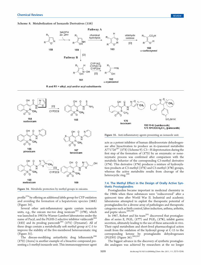
be N-oxidized to form an oxime intermediate (161), which isconverted into the respective R-cyano-enol metabolite (162)throughCYP450-dependent dehydration272 (pathwayB, Scheme8).Moreover, 162 can also be produced after enzymatic or none-nzymatic removal of the hydrogen vicinal to the nitrogen atom ofthe isoxazole ring (158).273
Enolic anti-inflammatory drugs, which belong to the oxicamclass, were described in the 1970s by the pioneering work ofLombardino and co-workers as a therapeutic alternative withimproved efficacy and plasmatic half-life in comparison to drugswith carboxylate units as the pharmacophoric group.274 Sudo-xicam (164) was the first compound of this class to demonstrateefficacy in various animal models of inflammation.275 However,despite the potential therapeutic application of the 1,2-ben-zothiazine derivative sudoxicam (164), its clinical developmentwas discontinued due to its induction of severe hepatotoxicity. Inaddition, depletion of glutathione was promoted by its acylthioureametabolite (165), which forms after CYP2C9 oxidation of thethiazole ring attached to carboxamide unit.276 For these reasons,
2-pyridine isostere piroxicam277 (166) was developed andlaunched as the first marketed enol-carboxamide drug, and thiswas followed by thieno[2,3-e]-1,2-thiazine derivative tenoxicam278
(167) a few years later (Figure 34). All of these oxicam derivativesprovide their anti-inflammatory action by inhibiting PGHS andthereby blocking the production of prostaglandins.279 The dis-covery of a second and inducible isoform of PGHS (namedPGHS-2) responsible for the biosynthesis of prostaglandinsduring inflammatory processes changed the paradigm for thediscovery of novel anti-inflammatory drugs.280,281 The methy-lated analogue of sudoxicam (164) meloxicam (168) wascharacterized as a weakly selective PGHS-2 inhibitor.282 Theselectivity displayed by this compound results at least in part fromthe increased molecular volume due to the introduction of a methylgroup. The methylation dramatically affects the metabolic
Figure 31. Analgesic compounds presenting the methyl ortho effect.
Figure 32. The discovery of celecoxib (147).
Scheme 6. Major Metabolic Pathway of Celecoxib (147)
Figure 33. Selective PGHS-2 inhibitors.
Scheme 7. Metabolization of Thiazole Derivatives (154)
5235 dx.doi.org/10.1021/cr200060g |Chem. Rev. 2011, 111, 5215–5246
Chemical Reviews REVIEW
profile276 by offering an additional labile group for CYP oxidationand avoiding the formation of a hepatotoxic species (165)(Figure 34).
Several other anti-inflammatory agents contain isoxazoleunits, e.g., the oxicam me-too drug isoxicam283 (170), whichwas launched in 1983 byWarner-Lambert laboratories under thename of Pacyl, and the PGHS-2 selective inhibitor valdecoxib284
(153) and its prodrug parecoxib285 (171) (Dynastat). All ofthese drugs contain a metabolically soft methyl group at C-5 toimprove the stability of the five-membered heteroaromatic ring(Figure 35).
The disease-modifying antiarthritic drug leflunomide286
(172) (Arava) is another example of a bioactive compound pre-senting a 5-methyl-isoxazole unit. This immunosuppressor agent
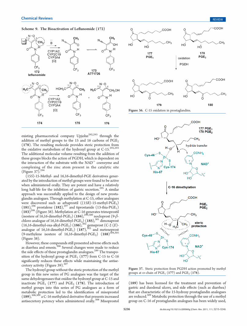
acts as a potent inhibitor of human dihydroorotate dehydrogen-ase after bioactivation to produce an R-cyanoenol metaboliteA771726287 (173) (Scheme 9). C3�Hdeprotonation during thefirst step of the formation of (173) by an enzymatic or none-nzymatic process was confirmed after comparison with themetabolic behavior of the corresponding C3-methyl derivative(174). This derivative (174) produces a mixture of hydroxyla-tion products at C3-methyl (175) and C5-methyl (176) groups,whereas the active metabolite results from cleavage of theheterocyclic ring.288
7.4. The Methyl Effect in the Design of Orally Active Syn-thetic Prostaglandins
Prostaglandins became important in medicinal chemistry inthe 1960s when these substances were “rediscovered” after aquiescent time after World War II. Industrial and academiclaboratories attempted to exploit the therapeutic potential ofprostaglandins for a diverse array of pathologies and therapeuticcategories such as birth control, labor induction, asthma, arthritis,and peptic ulcers.289,290
In 1967, Robert and his team291 discovered that prostaglan-dins of series E, PGE1 (177) and PGE2 (178), inhibit gastricsecretion, ultimately leading to the use of these autacoids in vivo.Their rapid metabolism and short-lived pharmacological actionresult from the oxidation of the hydroxyl group at C-15 to thecorresponding ketone by prostaglandin dehydrogenases(PGDH) (Figure 36).289,291
The biggest advance in the discovery of synthetic prostaglan-din analogues was achieved by researchers at the no longer
Scheme 8. Metabolization of Isoxazole Derivatives (158)
Figure 34. Metabolic protection by methyl groups in oxicams.
Figure 35. Anti-inflammatory agents presenting an isoxazole unit.
5236 dx.doi.org/10.1021/cr200060g |Chem. Rev. 2011, 111, 5215–5246
Chemical Reviews REVIEW
existing pharmaceutical company Upjohn292,293 through theaddition of methyl groups to the 15 and 16 carbons of PGE2(178). The resulting molecule provides steric protection fromthe oxidative metabolism of the hydroxyl group at C-15.292,293
The additional molecular volume resulting from the addition ofthese groups blocks the action of PGDH, which is dependent onthe interaction of the substrate with the NADþ coenzyme andcomplexing of the zinc atom present in the catalytic site(Figure 37).294
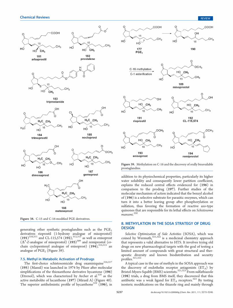
(15S)-15-Methyl- and 16,16-dimethyl-PGE derivatives gener-ated by the introduction of methyl groups were found to be activewhen administered orally. They are potent and have a relativelylong half-life for the inhibition of gastric secretion.295 A similarapproach was successfully applied to the design of new prosta-glandin analogues. Through methylation at C-15, other analogueswere discovered such as arbaprostil ((15R)-15-methyl-PGE2)(181),296 prostalene (182),297 and tiprostanide (13-thia-PGE1)(183)298 (Figure 38). Methylation at C-16 generates trimoprostil(isostere of 16,16-dimethyl-PGE2) (184),
299,300 nocloprost (9-β-chloro analogue of 16,16-dimethyl-PGE2) (185),
301 dimoxaprost(16,16-dimethyl-oxa-alkyl-PGE2) (186),
302 gemeprost (C-2 (E)-analogue of 16,16-dimethyl-PGE1) (187),303 and meteneprost(9-methylene isostere of 16,16-dimethyl-PGE2) (188)304,305
(Figure 38).However, these compounds still presented adverse effects such
as diarrhea and emesis.306 Several changes were made to reducethe side effects of these prostaglandin analogues.289 The transpo-sition of the hydroxyl group at PGE1 (177) from C-15 to C-16significantly reduces these effects while maintaining the antise-cretory activity (Figure 39).307
The hydroxyl group without the steric protection of themethylgroup in this new series of PG analogues was the target of thesame dehydrogenases that oxidize the hydroxyl group at C-15 andinactivate PGE1 (177) and PGE2 (178). The introduction ofmethyl groups into this series of PG analogues as a form ofmetabolic protection led to the identification of misoprostol(189),308,309 a C-16methylated derivative that presents increasedantisecretory potency when administered orally.308 Misoprostol
(189) has been licensed for the treatment and prevention ofgastric and duodenal ulcers, and side effects (such as diarrhea)that are characteristic of the 15-hydroxy prostaglandin analoguesare reduced.309 Metabolic protection through the use of a methylgroup on C-16 of prostaglandin analogues has been widely used,
Scheme 9. The Bioactivation of Leflunomide (172)
Figure 36. C-15 oxidation in prostaglandins.
Figure 37. Steric protection from PGDH action promoted by methylgroups at ω-chain of PGE1 (177) and PGE2 (178).
5237 dx.doi.org/10.1021/cr200060g |Chem. Rev. 2011, 111, 5215–5246
Chemical Reviews REVIEW
generating other synthetic prostaglandins such as the PGE1derivatives rioprostil (1-hydroxy analogue of misoprostol)(191)310,311 and CL-115,574 (192),312,313 as well as enisoprost(Δ4-Z-analogue of misoprostol) (193)309 and remiprostol (ω-chain cyclopentenyl analogue of enisoprost) (194),314,315 ananalogue of PGE2 (Figure 39).
7.5. Methyl in Metabolic Activation of ProdrugsThe first-choice schistosomicide drug oxaminiquine316,317
(195) (Mansil) was launched in 1974 by Pfizer after molecularsimplifications of the thioxanthone derivative hycantone (196)(Etrenol), which was characterized by Archer et al.318 as theactive metabolite of lucanthone (197) (Mirasil A) (Figure 40).The superior antihelmintic profile of hycanthone319 (196), in
addition to its physiochemical properties, particularly its higherwater solubility and consequently lower partition coefficient,explains the reduced central effects evidenced for (196) incomparison to the prodrug (197). Further studies of themolecular mechanism of action indicated that the benzyl alcoholof (196) is a selective substrate for parasitic enzymes, which canturn it into a better leaving group after phosphorylation orsulfation, thus favoring the formation of reactive azo-typequinones that are responsible for its lethal effects on Schistosomamansoni.320
8. METHYLATION IN THE SOSA STRATEGY OF DRUGDESIGN
Selective Optimization of Side Activities (SOSA), which wascoined by Wermuth,321,322 is a medicinal chemistry approachthat represents a valid alternative to HTS. It involves trying olddrugs on new pharmacological targets with the goal of testing alimited amount of compounds with great structural and ther-apeutic diversity and known biodistribution and securityprofiles.321,322
A typical case in the use of methyls in the SOSA approach wasthe discovery of endothelin receptor antagonists (ETA) byBristol-Myers-Squibb (BMS) scientists.321,323 From sulfathiazole(198) trials, a drug from BMS itself, they discovered that thisantibiotic was a weak ligand for ETA receptors.323 By testingisosteric modifications on the thiazole ring and mainly through
Figure 38. C-15 and C-16-modified PGE derivatives.
Figure 39. Methylation on C-16 and the discovery of orally bioavailableprostaglandins.
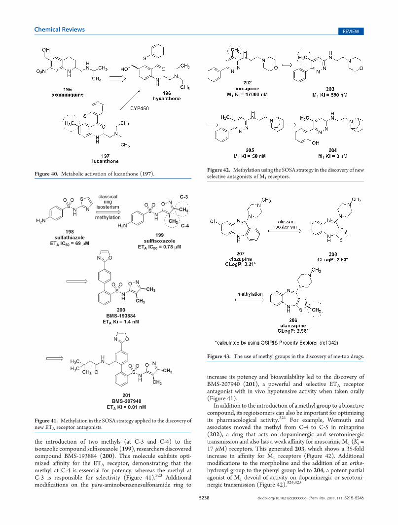
5238 dx.doi.org/10.1021/cr200060g |Chem. Rev. 2011, 111, 5215–5246
Chemical Reviews REVIEW
the introduction of two methyls (at C-3 and C-4) to theisoxazolic compound sulfisoxazole (199), researchers discoveredcompound BMS-193884 (200). This molecule exhibits opti-mized affinity for the ETA receptor, demonstrating that themethyl at C-4 is essential for potency, whereas the methyl atC-3 is responsible for selectivity (Figure 41).323 Additionalmodifications on the para-aminobenzenesulfonamide ring to
increase its potency and bioavailability led to the discovery ofBMS-207940 (201), a powerful and selective ETA receptorantagonist with in vivo hypotensive activity when taken orally(Figure 41).
In addition to the introduction of amethyl group to a bioactivecompound, its regioisomers can also be important for optimizingits pharmacological activity.321 For example, Wermuth andassociates moved the methyl from C-4 to C-5 in minaprine(202), a drug that acts on dopaminergic and serotoninergictransmission and also has a weak affinity for muscarinic M1 (Ki =17 μM) receptors. This generated 203, which shows a 35-foldincrease in affinity for M1 receptors (Figure 42). Additionalmodifications to the morpholine and the addition of an ortho-hydroxyl group to the phenyl group led to 204, a potent partialagonist of M1 devoid of activity on dopaminergic or serotoni-nergic transmission (Figure 42).324,325
Figure 40. Metabolic activation of lucanthone (197).
Figure 41. Methylation in the SOSA strategy applied to the discovery ofnew ETA receptor antagonists.
Figure 42. Methylation using the SOSA strategy in the discovery of newselective antagonists of M1 receptors.
Figure 43. The use of methyl groups in the discovery of me-too drugs.
5239 dx.doi.org/10.1021/cr200060g |Chem. Rev. 2011, 111, 5215–5246
Chemical Reviews REVIEW
9. THE METHYL IN GENERATING ME-TOO DRUGS
Some examples of the use of methyls in the isosteric exchangeof a six-membered ring for a heteroaromatic five-memberedring can be found in the literature, such as in the design ofme-too drugs. This strategy aims to adjust the lipophilicity ofmolecules because the contraction of a six-membered ring into afive-membered one decreases its lipophilicity. In addition, thismodification provides protection from oxidative metabolism,which might generate toxic species from these five-memberedheterocycles.5
The drug olanzapine (Zyprexa) (206), an atypical antipsy-chotic approved by the FDA in 1996 for the treatment ofschizophrenia,326 was designed based on the benzodiazepinestructure of clozapine (207) through the removal of thechlorine atom at C-8 and a classic isosteric exchange ofphenyl in (207) by thiophene in (208).327 However, thesemodifications led to a decrease of over one logarithmic unitin the calculated ClogP by OSIRIS Property Explorer.328
Moreover, because this class of compounds acts on the centralnervous system (CNS), this reduction could also reflect phar-macological activity. The methyl introduced into C-2 of(208) aimed to increase lipophilicity and to provide metabolicprotection for the thiophene ring of olanzapine (206)(Figure 43).327,329,330
10. CONCLUSIONS
In this review, we aimed to highlight the importance of thesimple methyl group as a very useful structural modification inthe rational design of bioactive compounds and drugs. Weselected different examples from distinct classes of organiccompounds with several pharmacological properties in order toillustrate that an apparent subtle molecular modification canstrongly alter the biological profile of a drug. The methyl effectalter both biological phases of a drug, represented by itspharmacodynamic and pharmacokinetc profile, due to the mod-ifications introduced in the stereoeletronic properties. In fact, itwas illustrated that distinct metabolic behavior or receptoraffinity could be modified by the methyl effect. Among theexamples included many other important ones could be used,such as the importance of the methyl inmodified the bioprofile ofphenyl ethanolamines with differentiation in the molecularrecognition pattern among different adrenergic receptor sub-types. The introduction of an simple extra methyl connected tothe N-alkylpiperidine moiety present in PDE5 inhibitorraisng the discovery of me-too drug vardenafil for erectiledysfunction. In fact, the correct use of this strategy for molecularmodification and design of drugs or drug-candidate compoundsmay be useful from both a pharmacokinetic and a pharmacody-namic point of view, representing an important tool for themedicinal chemist.
AUTHOR INFORMATION
Corresponding Author*Phone: 55-21-25626644. E-mail: [email protected]: http://www.farmacia.ufrj.br/lassbio.
Present Addresses
)Departamento de Química, Instituto de Ciencias Exatas,Universidade Federal Rural do Rio de Janeiro, 23.890-000,Serop�edica, RJ, Brazil.
BIOGRAPHIES
Eliezer J. Barreiro concluded his scientific education (Docteur-E�s-Sciences d0�Etat) in Medicinal Chemistry at the University ofGrenoble, France, in 1978, under the direction of ProfessorPierre Crabb�e and Dr Andrew E. Greene. He spent four years asAssociate Professor of Organic Chemistry at Federal Universityof S~aoCarlos, S.P. from 1979 to 1983, when he joined the FederalUniversity of Rio de Janeiro where he got a permanent position.He works in the medicinal chemistry field and created theLaborat�orio de Avaliac-~ao e Síntese de Substancias Bioativas(LASSBio) at the School of Pharmacy of Federal University ofRio de Janeiro. Professor Barreiro has published over 200 journalscientific articles and book chapters and a book concerning thefield of medicinal chemistry and he is the inventor of 14 patentsof bioactive compounds, drug candidates acting as cardioactive,CNS-active, anti-inflammatory, and antileishmania diseases.
Arthur Eugen K€ummerle was born in Rio de Janeiro, Brazil, in1979. He received his degree in Pharmacy (2003), his M.Sc. inOrganic Chemistry (2005), and his Ph.D. in Chemistry in 2009from Federal University of Rio de Janeiro (UFRJ), Brazil, wherehe studied the effects of the methyl group on the analgesic andantiinflamatory properties of a chemical library of N-acylhyadra-zones under the guidance of Prof. Eliezer J. Barreiro and Prof.Carlos A. M. Fraga. In 2009, he was assistant professor ofChemical Pharmaceutical Technology at the same university,and since 2010 he is professor of Organic and MedicinalChemistry at Rural Federal University of Rio de Janeiro(UFRRJ), Brazil. His research interests focus on heterocyclicand microwave chemistry in the scope of medicinal chemistry,aiming at bioactive compounds for the treatment of anti-inflam-matory, cardiovascular, tropical, and degenerative diseases.
5240 dx.doi.org/10.1021/cr200060g |Chem. Rev. 2011, 111, 5215–5246
Chemical Reviews REVIEW
Carlos Alberto Manssour Fraga was born in Rio de Janeiro(Brazil) in 1964. He obtained a B.Sc. degree in Pharmacy in 1988and his M.Sc. degree in Sciences (Medicinal Chemistry) fromFederal University of Rio de Janeiro (UFRJ). After obtaining hisPh.D. degree from Chemistry Institute of UFRJ in 1994, workingwith the synthesis of novel stable prostacyclinmimetics under thesupervision of Professor Eliezer J. Barreiro, Carlos AlbertoManssour Fraga joined the Faculty of Pharmacy of UFRJ (Riode Janeiro) as Associate Professor in 1996 and was promoted toProfessor in May 2006. He was Head of the Drug Department ofthe Faculty of Pharmacy (UFRJ) from 2008 to 2010, andcurrently he is Coordinator of the Post Graduate Program inPharmacology and Medicinal Chemistry of the Institute ofBiomedical Sciences of UFRJ. Dr. Fraga is an effective memberof Brazilian Chemical Society since 1991, where he was Directorof the Medicinal Chemistry Division from 2002 to 2004. Apartfrom teaching, Professor Fraga develops his research activities inLASSBio (Laborat�orio de Avaliac-~ao e Síntese de SubstanciasBioativas, UFRJ), focusing the design, synthesis, and pharmaco-logical evaluation of novel drug candidates able to act in multi-factorial diseases, with particular emphasis in the use of N-acylhydrazone framework as a privileged structure to discovernovel therapeutically valuable compounds.
ACKNOWLEDGMENT
We thank the CNPq (BR) and FAPERJ (BR) for fellowshipsand grants (CNPq nos. 302.918/07-8, 303.977/09-4, 142613/05-3; FAPERJ nos. E-26/102.695/08, E-26/102.350/09, E-26/100.329/08) and to Professor Angelo C. Pinto (UFRJ, BR) forhelpful discussions and suggestions.
LIST OF ABBREVIATIONSA1 adenosine A1 receptorA2A adenosine A2A receptorA2B adenosine A2B receptorA3 adenosine A3 receptorAla alanineAMP adenosine monophosphateAMPc cyclic adenosine monophosphateArg arginineAsp aspartic acidATP adenosine triphosphateATP adenosine triphosphateATPase adenosine triphosphataseBcr-Abl Bcr (breakpoint cluster region)-abl (gene) fusion
protein
Bcr-AblK Bcr-Abl kinaseCB1 CB1 cannabinoid receptorClogP Ccalculated Log PCML chronic myeloid leukemiaCNS central nervous systemcompd compoundCOMT catechol O-methyl transferaseCYP1A2 cytochrome P450 1A2CYP2C19 cytochrome P450 2C19CYP2C9 cytochrome P450 2C9CYP2D6 cytochrome P450 2D6CYP3A4 cytochrome P450 3A4CYP450 cytochrome P450Cys cysteineDNA deoxyribonucleic aciddTMP thymidine monophosphatedUMP deoxyuridine monophosphateED50 effective doseEGFR epidermal growth factor receptorETA endothelin receptor antagonistsFDA Food and Drug AdministrationGMP guanosine monophosphateHþ-Kþ-ATPase hydrogen potassium adenosine triphosphataseH1 histamine receptors of subtype 1H2 histamine receptors of subtype 2H-bond hydrogen bondHER1 human EGFR type 1HER2 human EGFR type 2His histidineHMG CoAR 3-hydroxy-3-methylglutaryl-coenzyme A
reductaseHMG-CoA 3-hydroxy-3-methylglutaryl-coenzyme AHNMT histamine N-methyl transferaseHTS high-throughput screeningIC50 half maximal inhibitory concentrationicv intracerebroventricular administrationIle isoleucineIMP inositol monophosphateINMT indolethylamine N-methyl transferaseKi affinity constantKM Michaelis Menten constantLASSBio Laborat�orio de Avaliac-~ao e Síntese de Sub-
stancias BioativasLeu leucineLIS lanthanide dislocation inducing saltsLog P logarithm of partition coefficientLys lysineM1 muscarinic receptor M1
MAT S-adenosylmethionine synthetasemax inhib maximal inhibitionMet methionineMR molar refractivityNADþ nicotinamide adenine dinucleotideNADH nicotinamide adenine dinucleotideNADPþ nicotinamide adenine dinucleotide phosphateNADPH nicotinamide adenine dinucleotide phosphatenM nanomolarNMR nuclear magnetic resonance experimentsNNMT nicotinamide N-methyl transferaseNSAIDs nonsteroidal anti-inflammatory drugsPBQ writhing phenylbenzoquinone writhing testPDB ID Protein Data Bank identification
5241 dx.doi.org/10.1021/cr200060g |Chem. Rev. 2011, 111, 5215–5246
Chemical Reviews REVIEW
PDE phosphodiesterasePDGF-Rs platelet-derived growth factorPG prostaglandinPGDH 15-hydroxyprostaglandin dehydrogenasePGE1 prostaglandina E1PGE2 prostaglandina E2PGHS-1 prostaglandin endoperoxide synthase 1PGHS-2 prostaglandin endoperoxide synthase 2Phe phenylalaninePKC protein kinase CPKs protein kinasesPMT phenol O-methyl-transferasePNMT phenylethanolamine N-methyl-transferasePPi pyrophosphatePPIs proton pump inhibitorsRNA ribonucleic acidSAM S-adenosylmethioninesc subcutaneous injectionSEM standard error of the meanSer serineSN2 bimolecular nucleophilic substitutionSOSA selective optimization of side activitiest1/2 half-lifeThr threonineTK tyrosine kinaseTM trans membrane helixTMP thiol methyltransferase
REFERENCES
(1) McNaught, A. D.; Wilkinson, A. Compendium of ChemicalTerminology, 2nd ed. (The “Gold Book”); Blackwell Scientific Publica-tions: Oxford, UK, 1997.(2) Farber, E. Evolution of Chemistry; 2nd ed.; Ronald Press: New
York, 1969.(3) Barreiro, E. J.; Fraga, C. A. M. Química Medicinal—As Bases
Moleculares da Ac-~ao dos F�armacos, 2nd ed.; Artmed: Porto Alegre, Brazil,2008.(4) Chu, K. C. InThe Basis of Medicinal Chemistry/Burger’s Medicinal
Chemistry; John Wiley: New York, 1980; pp 393�418.(5) Bazzini, P.; Wermuth, C. G. In The Practice of Medicinal
Chemistry; Academic Press: San Diego, 2008; pp 431�463.(6) Umezawa, Y.; Nishio, O. Nucleic Acids Res. 2002, 30, 2183.(7) Saenger, W. Principles of Nucleic Acid Structure; Springer: New
York, 1984.(8) Nishio, M.; Umezawa, Y.; Hirota, M.; Takeuchi, Y. Tetrahedron
1995, 51, 8665.(9) Cantoni, G. L. J. Biol. Chem. 1953, 204, 403.(10) Dewick, P. M. In Medicinal Natural Products; John Wiley &
Sons: Chichester, 2002; pp 291�403.(11) Ashihara, H.; Crozier, A. Trends Plant Sci. 2001, 6, 407.(12) Nehlig, A.; Daval, J.-L.; Debry, G. Brain. Res. Rev. 1992, 17, 139.(13) Fredholm, B. B.; B€attig, K.; Holm�en, J.; Nehlig, A.; Zvartau,
E. E. Pharmacol. Rev. 1999, 51, 83.(14) Persson, C. G. A. J. Allergy Clin. Immunol. 1986, 78, 780.(15) Snyder, S. H.; Katims, J. J.; Annau, Z.; Bruns, R. F.; Daly, J. W.
Proc. Natl. Acad. Sci. U.S.A. 1981, 78, 3260.(16) Lugnier, C. Pharmacol. Ther. 2006, 109, 366.(17) Conti, M.; Beavo, J. Annu. Rev. Biochem. 2007, 76, 481.(18) Burnstock, G. Cell. Mol. Life Sci. 2007, 64, 1471.(19) Fredholm, B. B.; Abbracchio, M. P.; Burnstock, G.; Daly, J. W.;
Harden, T. K.; Jacobson, K. A.; Leff, P.; Williams, M. Pharmacol. Rev.1994, 46, 143.(20) Testa, B.; Carrupt, P.; Gaillard, P.; Billois, F.; Weber, P. Pharm.
Res. 1996, 13, 335.
(21) M€alki€a, A.; Murtom€aki, L.; Urtti, A.; Kontturi, K. Eur. J. Pharm.Sci. 2004, 23, 13.
(22) Fagerholm, U. Pharm. Res. 2008, 25, 625.(23) Istvan, E. S.; Deisenhofer, J. Science 2001, 292, 1160.(24) Rekker, R. F.; Mannhold, R. Calculation of Drug Lipophilicity:
The Hydrophobic Fragmental Constant Approach; Wiley-VCH: Wein-heim, 1992.
(25) Fujita, T.; Iwasa, J.; Hansch, C. J. Am. Chem. Soc. 1964, 86, 5175.(26) Hansch, C. Acc. Chem. Res. 1969, 2, 232.(27) Leo, A.; Hansch, C.; Elkins, D. Chem. Rev. 1971, 71, 525.(28) Hansch, C.; Leo, A.; Unger, S. H.; Kim, K. H.; Nikaitani, D.;
Lien, E. J. J. Med. Chem. 1973, 16, 1207.(29) Mosberg, H. I.; Hurst, R.; Hruby, V. J.; Gee, K.; Yamamura,
H. I.; Galligan, J. J.; Burks, T. F. Proc. Natl. Acad. Sci. U.S.A. 1983, 80,5871.
(30) Witt, K. A.; Davis, T. AAPS J. 2006, 8, E76.(31) Hansen, D. W.; Stapelfeld, A.; Savage, M. A.; Reichman, M.;
Hammond, D. L.; Haaseth, R. C.; Mosberg, H. I. J. Med. Chem. 1992,35, 684.
(32) Ooms, F.; Wouters, J.; Oscari, O.; Happaerts, T.; Bouchard, G.;Carrupt, P.; Testa, B.; Lambert, D. M. J. Med. Chem. 2002, 45, 1748.
(33) Rinaldi-Carmona, M. FEBS Lett. 1994, 350, 240.(34) Rinaldi-Carmona, M.; Barth, F.; H�eaulme, M.; Alonso, R.;
Shire, D.; Congy, C.; Soubri�e, P.; Breli�ere, J. -C.; Le Fur, G. Life Sci.1995, 56, 1941.
(35) Ginnings, P. M.; Baum, R. J. Am. Chem. Soc. 1937, 59, 1111.(36) N�emethy, G. Angew. Chem., Int. Ed. Engl. 1967, 6, 195.(37) Southall, N. T.; Dill, K. A.; Haymet, A. D. J. J. Phys. Chem. B
2002, 106, 521.(38) Matsumoto, A.M.Endocrinol.Metab. Clin. North Am. 1994, 23, 857.(39) Chabbert-Buffet, N.; Meduri, G.; Bouchard, P.; Spitz, I. M.
Hum. Reprod. Update 2005, 11, 293.(40) Udenfriend, S.; Wyngaarden, J. Biochim. Biophys. Acta 1956,
20, 48.(41) Suga, H.; Sagawa, K.; Shoukas, A. A. Circ. Res. 1973, 32, 314.(42) Saavedra, J.; Grobecker, H.; Axelrod, J. Science 1976, 191, 483.(43) Wortsman, J. Endocrinol. Metab. Clin. North Am. 2002, 31, 79.(44) Drew, C. D.; Knight, G. T.; Hughes, D. T.; Bush, M. Br. J. Clin.
Pharmacol. 1978, 6, 221.(45) Liu, Y. -L.; Toubro, S.; Astrup, A.; Stock, M. J. Int. J. Obes. 1995,
19, 678/.(46) Patil, P. N.; Tye, A.; LaPidus, J. B. J. Pharmacol. Exp. Ther. 1965,
148, 158.(47) Benowitz, N. L. Annu. Rev. Pharmacol. Toxicol. 2009, 49, 57.(48) Raynor, K.; Kong, H.; Chen, Y.; Yasuda, K.; Yu, L.; Bell, G. I.;
Reisine, T. Mol. Pharmacol. 1994, 45, 330.(49) Wenningmann, I.; Dilger, J. P. Mol. Pharmacol. 2001, 60, 790.(50) Kalivas, P. W.; Duffy, P.; DuMars, L. A.; Skinner, C. J.
Pharmacol. Exp. Ther. 1988, 245, 485.(51) Schiff, P. B.; Fant, J.; Horwitz, S. B. Nature 1979, 277, 665.(52) Singleton, C. K.; Martin, P. R. Curr. Mol. Med. 2001, 1, 197.(53) Massey, V. Biochem. Soc. Trans. 2000, 28, 283.(54) Fairweather-Tait, S. J.; Powers, H. J.; Minski, M. J.; Whitehead,
J.; Downes, R. Ann. Nutr. Metab. 1992, 36, 34.(55) Patrick, G. L. In An Introduction to Medicinal Chemistry; Oxford
University Press: New York, 2009; pp 632�652.(56) Sert€uner, F. J. Pharm. Aerzte Apotheker 1805, 13, 229.(57) Sert€uner, F. J. Pharm. Aerzte Apotheker 1806, 14, 47.(58) Seguim, M. A. Ann. Chim. 1814, 92, 225.(59) Gulland, J. M.; Robinson, R. Mem. Proc. Manchester Lit. Philos.
Soc. 1925, 69, 79.(60) Braun, J. V. Ber. Deutsch. Bot. Ges. 1914, 47, 2312.(61) Miller, J. W.; Anderson, H. H. J. Pharm. Pharmacol. 1954,
112, 191.(62) Beckett, A. H.; Casy, A. F. J. Pharm. Pharmacol. 1954, 6, 986.(63) Kane, B. E.; Svensson, B.; Ferguson, D. M. AAPS J. 2006,
8, E126.(64) Liao, C.; Nicklaus, M. C. J. Chem. Inf. Model. 2009, 49, 2801.
5242 dx.doi.org/10.1021/cr200060g |Chem. Rev. 2011, 111, 5215–5246
Chemical Reviews REVIEW
(65) Lockeet, M. F.; Davis, M. M. J. Pharm. Pharmacol. 1958,10, 80.(66) Robiquet, P. J. Ann. Chim. Phys. 1832, 51, 225.(67) Mignat, C.; Wille, U.; Ziegler, A. Life Sci. 1995, 56, 793.(68) Modi, S.; Paine, M. J.; Sutcliffe, M. J.; Lian, L.; Primrose, W. U.;
Wolf, C. R.; Roberts, G. C. K. Biochemistry 1996, 35, 4540.(69) Gasche, Y.; Daali, Y.; Fathi, M.; Chiappe, A.; Cottini, S.; Dayer,
P.; Desmeules, J. N. Engl. J. Med. 2004, 351, 2827.(70) Eddy, N. B.; Friebel, H.; Hahn, K. J.; Halbach, H. Bull. WHO
1969, 40, 425.(71) Eddy, N. B. J. Pharmacol. Exp. Ther. 1935, 55, 127.(72) Gierse, J. K.; McDonald, J. J.; Hauser, S. D.; Rangwala, S. H.;
Koboldt, C. M.; Seibert, K. J. Biol. Chem. 1996, 271, 15810.(73) Wong, E.; Bayly, C.; Waterman, H. L.; Riendeau, D.; Mancini,
J. A. J. Biol. Chem. 1997, 272, 9280.(74) Needleman, P.; Turk, J.; Jakschik, B. A. Annu. Rev. Biochem.
1986, 55, 69.(75) FitzGerald, G. A. Nature Rev. Drug Discovery 2003, 2, 879.(76) Kurumbail, R. G.; Stevens, A. M.; Gierse, J. K.; McDonald, J. J.;
Stegeman, R. A.; Pak, J. Y.; Gildehaus, D.; Iyashiro, J. M.; Penning, T. D.;Seibert, K.; Isakson, P. C.; Stallings, W. C. Nature 1996, 384, 644.(77) Pettersen, E. F.; Goddard, T. D.; Huang, C. C.; Couch, G. S.;
Greenblatt, D. M.; Meng, E. C.; Ferrin, T. E. J. Comput. Chem. 2004,25, 1605.(78) Takusagawa, F.; Kamitori, S.; Misaki, S.; Markham, G. D. J. Biol.
Chem. 1996, 271, 136.(79) Komoto, J.; Yamada, T.; Takata, Y.;Markham,G.D.; Takusagawa,
F. Biochemistry 2004, 43, 1821.(80) Silverman, R. B. In The Organic Chemistry of Drug Design and
Drug Action; Academic Press: Burlington, 2004; pp 405�495.(81) Dewick, P. M. In Medicinal Natural Products: A Biosynthetic
Approach; Wiley: West Sussex, UK, 2002; pp 6�34.(82) Lautala, P.; Ulmanen, I.; Taskinen, J. Mol. Pharmacol. 2001,
59, 393.(83) Axelrod, J. J. Biol. Chem. 1962, 237, 1657.(84) Schupke, H.; Hempel, R.; Peter, G.; Hermann, R.; Wessel, K.;
Engel, J.; Kronbach, T. Drug Metab. Dispos. 2001, 29, 855.(85) Axelrod, J.; Tomchick, R. J. Biol. Chem. 1958, 233, 702.(86) Cantoni, G. L. J. Biol. Chem. 1951, 189, 203.(87) Ansher, S. S.; Jakoby, W. B. J. Biol. Chem. 1986, 261, 3996.(88) Brown, D. D.; Tomchick, R.; Axelrod, J. J. Biol. Chem. 1959,
234, 2948.(89) Bremer, J.; Greenberg, D. M. Biochim. Biophys. Acta 1961,
46, 217.(90) Remy, C. N. J. Biol. Chem. 1963, 238, 1078.(91) Usdin, E.; Borchardt, R. T.; Creveling, C. R. The Biochemistry of
S-Adenosylmethionine and Related Compounds; Macmillan Press: Lon-don, 1982.(92) Clarke, S.; Banfield, K. In Homocysteine in Health and Disease;
Cambridge University Press: Cambridge, UK, 2001; pp 63�78.(93) Martin, J. L.; McMillan, F. M. Curr. Opin. Struct. Biol. 2002,
12, 783.(94) Axelrod, J.; Daly, J. Biochim. Biophys. Acta 1968, 159, 472.(95) Ansher, S. S.; Jacoby, W. D. In Conjugation Reactions in Drug
Metabolism; Taylor & Francis: London, 1990; p 233.(96) Stevens, J. L.; Bakke, J. E. In Conjugation Reactions in Drug
Metabolism; Taylor & Francis: London, 1990; p 251.(97) Thakker, D. R.; Creveling, C. R. In Conjugation Reactions in
Drug Metabolism; Taylor & Francis: London, 1990; p 193.(98) Ginsberg, G.; Hattis, D.; Russ, A.; Sonawane, B. J. Toxicol.
Environ. Health, Part A 2004, 67, 297.(99) Testa, B.; Kr€amer, S. D. Chem. Biodivers. 2007, 4, 257.(100) Romagnoli, C.; De Carolis, M. P.; Muzii, U.; Zecca, E.;
Tortorolo, G.; Chiarotti, M.; De Giovanni, N.; Carnevale, A. Ther. DrugMonit. 1992, 14, 14.(101) Kim, Y. -I. Cancer Epidemiol., Biomarkers Prev. 2004, 13, 511.(102) Wagner, C. In Biochemical Role of Folate in Cellular Metabo-
lism; Marcel Dekker Inc: New York, 1995; pp 23�42.
(103) Stroud, R. M.; Finer-Moore, J. S. FASEB J. 1993, 7, 671.(104) Carreras, C.W.; Santi, D. V.Annu. Rev. Biochem. 1995, 64, 721.(105) Kompis, I. M.; Islam, K.; Then, R. L. Chem. Rev. 2005,
105, 593.(106) Chu, E.; Callender, M. A.; Farrell, M. P.; Schmitz, J. C. Cancer
Chemother. Pharmacol. 2003, 52, S80.(107) Danenberg, P. V.; Malli, H.; Swenson, S. Semin. Oncol. 1999,
26, 621.(108) Rustum, Y. M.; Harstrick, A.; Cao, S.; Vanhoefer, U.; Yin, M. -B.;
Wilke, H.; Seeber, S. J. Clin. Oncol. 1997, 15, 389.(109) De Clercq, E. Clin. Microbiol. Rev. 2001, 14, 382.(110) Verri, A.; Focher, F.; Duncombe, R. J.; Basnak, I.; Walker,
R. T.; Coe, P. L.; De Clercq, E.; Andrei, G.; Snoeck, R.; Balzarini, J.;Spadari, S. Biochem. J. 2000, 351, 319.
(111) Endo, A.; Kuroda, M.; Tanzawa, K. FEBS Lett. 1976, 72, 323.(112) Brown, A. G.; Smale, T. C.; King, T. J.; Hasenkamp, R.;
Thompson, R. H. J. Chem. Soc., Perkin Trans. 1 1976, 1165.(113) Endo, A.; Tsujita, Y.; Kuroda, M.; Tanzawa, K. Eur. J. Biochem.
1977, 77, 31.(114) Nawrocki, J. W.; Weiss, S. R.; Davidson, M. H.; Sprecher,
D. L.; Schwartz, S. L.; Lupien, P. -J.; Jones, P. H.; Haber, H. E.; Black,D. M. Arterioscler. Thromb. Vasc. Biol. 1995, 15, 678.
(115) Bocan, T. M.; Ferguson, E.; McNally, W.; Uhlendorf, P. D.;Mueller, S. B.; Dehart, P.; Sliskovic, D. R.; Roth, B. D.; Krause, B. R.;Newton, R. S. Biochim. Biophys. Acta 1992, 1123, 133.
(116) Baumann, K. L.; Butler, D. E.; Deering, C. F.; Mennen, K. E.;Millar, A.; Nanninga, T. N.; Palmer, C. W.; Roth, B. D. Tetrahedron Lett.1992, 33, 2283.
(117) Tobert, J. A. Nature Rev. Drug Discovery 2003, 2, 517.(118) Steinberg, D. J. Lipid Res. 2006, 47, 1339.(119) Rodwell, V. W.; Nordstrom, J. L.; Mitschelen, J. J. Adv. Lipid
Res. 1976, 14, 1.(120) Bischoff, K. M.; Rodwell, V. W. Biochem. Med. Metab. Biol.
1992, 48, 149.(121) Goldstein, J. L.; Brown, M. S. Nature 1990, 343, 425.(122) Alberts, A. W.; Chen, J.; Kuron, G.; Hunt, V.; Huff, J.;
Hoffman, C.; Rothrock, J.; Lopez, M.; Joshua, H.; Harris, E.; Patchett,A.; Monaghan, R.; Currie, S.; Stapley, E.; Albers-Schonberg, G.; Hensens,O.;Hirshfield, J.; Hoogsteen, K.; Liesch, J.; Springer, J.Proc. Natl. Acad. Sci.U.S.A. 1980, 77, 3957.
(123) Hoffman, W. F.; Alberts, A. W.; Anderson, P. S.; Chen, J. S.;Smith, R. L.; Willard, A. K. J. Med. Chem. 1986, 29, 849.
(124) Tobert, J. A.; Bell, G. D.; Birtwell, J. J. Clin. Invest 1982, 69, 913.(125) Hunninghake, D. B.; Miller, V. T.; Palmer, R. H. JAMA, J. Am.
Med. Assoc. 1986, 256, 2829.(126) Henwood, J. M.; Heel, R. Drugs 1988, 36, 429.(127) Stokker, G. E.; Hoffman, W. F.; Alberts, A. W.; Cragoe, E. J.;
Deana, A. A.; Gilfillan, J. L.; Huff, J. W.; Novello, F. C.; Prugh, J. D.J. Med. Chem. 1985, 28, 347.
(128) Hoffman, W. F.; Alberts, A. W.; Cragoe, E. J., Jr.; Deana, A. A.;Evans, B. E.; Gilfillan, J. L.; Gould, N. P.; Huff, J. W.; Novello, F. C.;Prugh, J. D.; Rittle, K. E.; Smith, R. L.; Stokker, G. E.; Willard, A. K.J. Med. Chem. 1986, 29, 159.
(129) Stokker, G. E.; Alberts, A. W.; Anderson, P. S.; Cragoe, E. J.,Jr.; Deana, A. A.; Gilfillan, J. L.; Hirshfield, J.; Holtz, W. J.; Hoffman,W. F.; Huff, J. W.; Lee, T. J.; Novello, F. C.; Prugh, J. D.; Rooney, C. S.;Smith, R. L.; Willard, A. K. J. Med. Chem. 1986, 29, 170.
(130) Stokker, G. E.; Alberts, A.W.; Gilfillan, J. L.; Huff, J. W.; Smith,R. L. J. Med. Chem. 1986, 29, 852.
(131) Roth, B. D.; Ortwine, D. F.; Hoefle, M. L.; Stratton, C. D.;Sliskovic, D. R.;Wilson,M.W.; Newton, R. S. J. Med. Chem. 1990, 33, 21.
(132) Roth, B. D.; Blankley, C. J.; Chucholowski, A. W.; Ferguson,E.; Hoefle,M. L.; Ortwine, D. F.; Newton, R. S.; Sekerke, C. S.; Sliskovic,D. R.; Wilson, M. J. Med. Chem. 1991, 34, 357.
(133) Shaw,M. K.; Newton, R.; Sliskovic, D. R.; Roth, B. D.; Ferguson,E.; Krause, B. R. Biochem. Biophys. Res. Commun. 1990, 170, 726.
(134) Tse, F. L. S.; Smith, H. T.; Ballard, F. H.; Nicoletti, J.Biopharm. Drug Dispos. 1990, 11, 519.
5243 dx.doi.org/10.1021/cr200060g |Chem. Rev. 2011, 111, 5215–5246
Chemical Reviews REVIEW
(135) Prous, J.; Castaner, J. Drugs Future 1991, 16, 804.(136) Tse, F. L. S.; Jaffe, J. M.; Troendle, A. J. Clin. Pharmacol. 1992,
32, 630.(137) McTaggart, F.; Buckett, L.; Davidson, R.; Holdgate, G.;
McCormick, A.; Schneck, D.; Smith, G.; Warwick, M. Am. J. Cardiol.2001, 87, 28B.(138) Watanabe, M.; Koike, H.; Ishiba, T.; Okada, T.; Seo, S.; Hirai,
K. Bioorg. Med. Chem. 1997, 5, 437.(139) Angerbauer, R.; Bischoff, H.; Steinke, W.; Ritter, W. Drugs
Future 1994, 19, 537.(140) Corsini, A.; Arnaboldi, L.; Raiteri, M.; Quarato, P.; Faggiotto,
A.; Paoletti, R.; Fumagalli, R. Pharmacol. Res. 1996, 33, 55.(141) Burland, W. L.; Duncan, W. A. M.; Hesselbo, T. Br. J. Clin.
Pharmacol. 1975, 2, 481.(142) Brimblecome, R.; Duncan, W.; Durant, G. J. Int. Med. Res.
1975, 3, 86.(143) Brimblecombe, R.; Duncan, W.; Durant, G. Br. J. Pharmacol.
1975, 53, 435P.(144) Black, J. W.; Duncan, W. A. M.; Durant, C. J.; Ganellin, C. R.;
Parsons, E. M. Nature 1972, 236, 385.(145) Ganellin, R. J. Med. Chem. 1981, 24, 913.(146) Ganellin, C. R. InAnalogue-BasedDrugDiscovery;Wiley-VCH:
Weinheim, 2006; pp 71�80.(147) Hill, S. J.; Ganellin, C. R.; Timmerman, H.; Schwartz, J. C.;
Shankley, N. P.; Young, J. M.; Schunack, W.; Levi, R.; Haas, H. L.Pharmacol. Rev. 1997, 49, 253.(148) Weinstein, H.; Chou, D.; Johnson, C. L.Mol. Pharmacol. 1976,
12, 738.(149) Silverman, R. B. In The Organic Chemistry of Drug Design and
Drug Action; Academic Press: Burlington, 2004; pp 121�172.(150) Hammett, L. P. Chem. Rev. 1935, 17, 125.(151) Hammett, L. P. J. Am. Chem. Soc. 1937, 59, 96.(152) Silverman, R. B. In The Organic Chemistry of Drug Design and
Drug Action; Academic Press: Burlington, 2004; pp 7�120.(153) Ganellin, C. R. J. Pharm. Pharmacol. 1973, 25, 787.(154) Durant, G. J.; Ganellin, C. R.; Parsons, M. E. J. Med. Chem.
1975, 18, 905.(155) Ash, A. S.; Schild, H. O. Br. J. Pharmacol. 1966, 27, 427.(156) Black, J. W.; Durant, G. J.; Emmett, J. C.; Ganellin, C. R.
Nature 1974, 248, 65.(157) Woodings, E. P.; Dixon, G. T.; Harrison, C.Gut 1980, 21, 187.(158) Bradshaw, J.; Brittain, R.; Clitherow, J. Br. J. Pharmacol. 1979,
66, 464P.(159) Takag, T.; Takeda, M.; Maeno, H.Agents Actions 1982, 256, 49.(160) Takeda, M.; Takagi, T.; Yashima, Y.; Maeno, H. Arzneim.
Forsch. 1982, 32, 734.(161) Takeda, M.; Takaghi, T.; Maeno, H. Jpn. J. Pharmacol. 1981,
31, 222P.(162) Lin, T. M.; Evans, D. C.; Warrick, M. W.; Pioch, R. P.; Ruffalo,
R. R. Gastroenterology 1983, 84, 1231.(163) Tarutani, M.; Sakuma, H.; Shiratsuchi, K.; Mieda, M. Arzneim.
Forschung 1985, 35, 703.(164) Wallmark, B.; Brandstrom, A.; Larsson, H. Biochim. Biophys.
Acta 1984, 778, 549.(165) Larsson, H.; Carlsson, E.; Junggren, U. Gastroenterology 1983,
85, 900.(166) Walt, R. P.; Gomes De., M. F. A.; Wood, E. C. Br. Med. J. 1983,
287, 12.(167) Lindberg, P.; Nordberg, P.; Alminger, T.; Braendstroem, A.;
Wallmark, B. J. Med. Chem. 1986, 29, 1327.(168) Besancon, M.; Simon, A.; Sachs, G.; Shin, J. M. J. Biol. Chem.
1997, 272, 22438.(169) Shin, J. M.; Cho, Y. M.; Sachs, G. J. Am. Chem. Soc. 2004,
126, 7800.(170) Olbe, L.; Carlsson, E.; Lindberg, P.Nature Rev. Drug Discovery
2003, 2, 132.(171) Pascaud, X. B. L.; Malen, C.; Danree, B. J. Med. Chem. 1971,
14, 244.
(172) Lima, L. M.; Barreiro, E. J. Curr. Med. Chem. 2005, 12, 23.(173) Patani, G. A.; LaVoie, E. J. Chem. Rev. 1996, 96, 3147.(174) Sundell, G.; Sjostrand, S. E.; Olbe, L. Acta Pharmacol. Toxicol.
1977, 41, 77.(175) Lindberg, P.; Br€andstr€om, A.; Wallmark, B.; Mattsson, H.;
Rikner, L.; Hoffmann, K. Med. Res. Rev. 1990, 10, 1.(176) Fellenius, E.; Berglindh, T.; Sachs, G. Nature 1981, 290, 159.(177) Lindberg, P.; Carlsson, E. In Analogue-Based Drug Discovery;
Wiley-VCH: Weinheim, 2006; pp 81�113.(178) Erlandsson, P.; Isaksson, R.; Lorentzon, P.; Lindberg, P. J.
Chromatogr., B: Biomed. Sci. Appl. 1990, 532, 305.(179) Spencer, C. M.; Faulds, D. Drugs 2000, 60, 321.(180) Olbe, L.; Carlsson, E.; Lindberg, P.Nature Rev. Drug. Discovery
2003, 2, 132.(181) Cotton, H.; Elebring, T.; Larsson,M.; Li, L.; S€orensen, H.; von
Unge, S. Tetrahedron Asymmetry 2000, 11, 3819.(182) Agranat, I.; Caner, H.; Caldwell, J.Nature Rev. Drug. Discovery
2002, 1, 753.(183) Morii, M.; Takata, H.; Fujisaki, H.; Takegucht, N. Biochem.
Pharmacol. 1990, 39, 661.(184) Fujisaki, H.; Shibata, H.; Oketani, K.; Murakami, M.; Fujimoto,
M.;Wakabayashi, T.; Yamatsu, I.; Yamaguchi,M.; Sakai,H.; Takeguchi, N.Biochem. Pharmacol. 1991, 42, 321.
(185) Satoh, H.; Inatomi, N.; Nagaya, H.; Inada, I.; Nohara, A.;Nakamura, N.; Maki, Y. J. Pharmacol. Exp. Ther. 1989, 248, 806.
(186) Nagaya, H.; Satoh, H.; Kubo, K.; Maki, Y. J. Pharmacol. Exp.Ther. 1989, 248, 799.
(187) Kromer, W.; Postius, S.; Riedel, R.; Simon, W.; Hanauer, G.;Brand, U.; Gonne, S.; Parsons, M. J. Pharmacol. Exp. Ther. 1990,254, 129.
(188) SimonMuller, B. P.; Hartmann, M.; Bliesath, H.; Luhmann, R.;Huber, R.; Bohnenkamp, W.; Wurst, W. Z. Gastroenterol. 1990, 28, 443.
(189) Simon, B.; Muller, P.; Marinis, E.; Luhmann, R.; Huber, R.;Hartmann, R.; Wurst, W. Aliment. Pharmacol. Ther. 1990, 4, 373.
(190) Uchiyama, K.; Wakatsuki, D.; Kakinoki, B.; Takeuchi, Y.;Araki, T.; Morinaka, Y. J. Pharm. Pharmacol. 1999, 51, 457.
(191) Galmiche, J. P.; Varannes, S. B. D.; Ducrott�e, P.; Sacher-Huvelin, S.; Vavasseur, F.; Taccoen, A.; Fiorentini, P.; Homerin, M.Aliment. Pharmacol. Ther. 2004, 19, 655.
(192) Shin, J. M.; Besancon, M.; Prinz, C.; Simon, A.; Sachs, G.Aliment. Pharmacol. Ther. 1994, 8, 11.
(193) Kromer, W.; Kr€uger, U.; Huber, R.; Hartmann, M.; Steinijans,V. Pharmacology 1998, 56, 57.
(194) Capdeville, R.; Buchdunger, E.; Zimmermann, J.; Matter, A.Nature Rev. Drug Discovery 2002, 1, 493.
(195) Druker, B. J.; Tamura, S.; Buchdunger, E.; Ohno, S.; Segal,G. M.; Fanning, S.; Zimmermann, J.; Lydon, N. B. Nature Med. 1996,2, 561.
(196) Buchdunger, E.; Zimmermann, J.;Mett, H.;Meyer, T.;M€uller,M.; Druker, B. J.; Lydon, N. B. Cancer Res. 1996, 56, 100.
(197) Carroll, M.; Ohno-Jones, S.; Tamura, S.; Buchdunger, E.;Zimmermann, J.; Lydon, N. B.; Gilliland, D. G.; Druker, B. J. Blood 1997,90, 4947.
(198) Druker, B. J.; Talpaz, M.; Resta, D. J.; Peng, B.; Buchdunger,E.; Ford, J. M.; Lydon, N. B.; Kantarjian, H.; Capdeville, R.; Ohno-Jones,S.; Sawyers, C. L. N. Engl. J. Med. 2001, 344, 1031.
(199) Demetri, G. D.; von Mehren, M.; Blanke, C. D.; Van denAbbeele, A. D.; Eisenberg, B.; Roberts, P. J.; Heinrich, M. C.; Tuveson,D. A.; Singer, S.; Janicek, M.; Fletcher, J. A.; Silverman, S. G.; Silberman,S. L.; Capdeville, R.; Kiese, B.; Peng, B.; Dimitrijevic, S.; Druker, B. J.;Corless, C.; Fletcher, C. D.; Joensuu, H. N. Engl. J. Med. 2002, 347, 472.
(200) Lugo, T. G.; Pendergast, A.; Muller, A. J.; Witte, O. N. Science1990, 247, 1079.
(201) Silverman, L.; Campbell, R.; Broach, J. R. Curr. Opin. Chem.Biol. 1998, 2, 397.
(202) Macarron, R. Drug Discovery Today 2006, 11, 277.(203) Zimmermann, J.; Buchdunger, E.;Mett, H.;Meyer, T.; Lydon,
N. B.; Traxler, P. Bioorg. Med. Chem. Lett. 1996, 6, 1221.
5244 dx.doi.org/10.1021/cr200060g |Chem. Rev. 2011, 111, 5215–5246
Chemical Reviews REVIEW
(204) Zimmermann, J.; Buchdunger, E.; Mett, H.;Meyer, T.; Lydon,N. B. Bioorg. Med. Chem. Lett. 1997, 7, 187.(205) Buchdunger, E.; Zimmermann, J.;Mett, H.;Meyer, T.;M€uller,
M.; Regenass, U.; Lydon, N. B. Proc. Natl. Acad. Sci. U.S.A. 1995,92, 2558.(206) Zimmermann, J.; Furet, P.; Buchdunger, E. ACS Symp. Ser.
2001, 796, 245.(207) Schindler, T.; Bornmann, W.; Pellicena, P.; Miller, W. T.;
Clarkson, B.; Kuriyan, J. Science 2000, 289, 1938.(208) O’Hare, T.; Walters, D. K.; Deininger, M. W.; Druker, B. J.
Cancer Cell 2005, 7, 117.(209) Weisberg, E.; Manley, P. W.; Breitenstein, W.; Br€uggen, J.;
Cowan-Jacob, S. W.; Ray, A.; Huntly, B.; Fabbro, D.; Fendrich, G.; Hall-Meyers, E.; Kung, A. L.; Mestan, J.; Daley, G. Q.; Callahan, L.; Catley, L.;Cavazza, C.; Mohammed, A.; Neuberg, D.; Wright, R. D.; Gilliland,D. G.; Griffin, J. D. Cancer Cell 2005, 7, 129.(210) O’Hare, T.; Walters, D. K.; Stoffregen, E. P.; Jia, T.; Manley,
P. W.; Mestan, J.; Cowan-Jacob, S. W.; Lee, F. Y.; Heinrich, M. C.;Deininger, M. W.; Druker, B. J. Cancer Res. 2005, 65, 4500.(211) Lombardo, L. J.; Lee, F. Y.; Chen, P.; Norris, D.; Barrish, J. C.;
Behnia, K.; Castaneda, S.; Cornelius, L. A. M.; Das, J.; Doweyko, A. M.;Fairchild, C.; Hunt, J. T.; Inigo, I.; Johnston, K.; Kamath, A.; Kan, D.;Klei, H.; Marathe, P.; Pang, S.; Peterson, R.; Pitt, S.; Schieven, G. L.;Schmidt, R. J.; Tokarski, J.; Wen, M.; Wityak, J.; Borzilleri, R. M. J. Med.Chem. 2004, 47, 6658.(212) Shah, N. P.; Tran, C.; Lee, F. Y.; Chen, P.; Norris, D.; Sawyers,
C. L. Nature 2004, 305, 399.(213) Burgess,M.; Skaggs, B. J.; Shah, N. P.; Lee, F. Y.; Sawyers, C. L.
Proc. Natl. Acad. Sci. U.S.A. 2005, 102, 3395.(214) Lyons, J. F.; Wilhelm, S.; Hibner, B.; Bollag, G. Endocr. Relat.
Cancer 2001, 8, 219.(215) Lowinger, T. B.; Riedl, B.; Dumas, J.; Smith, R. A.Curr. Pharm.
Des. 2002, 8, 2269.(216) Strumberg, D.; Voliotis, D.; Moeller, J.; Hilger, R.; Richly, H.;
Kredtke, S.; Beling, C.; Scheulen, M.; Seeber, S. Int. J. Clin. Pharmacol.Ther. 2002, 40, 580.(217) Khire, U. R.; Bankston, D.; Barbosa, J.; Brittelli, D. R.;
Caringal, Y.; Carlson, R.; Dumas, J.; Gane, T.; Heald, S. L.; Hibner,B.; Johnson, J. S.; Katz, M. E.; Kennure, N.; Kingery-Wood, J.; Lee, W.;Liu, X.; Lowinger, T. B.; McAlexander, I.; Monahan, M.; Natero, R.;Renick, J.; Riedl, B.; Rong, H.; Sibley, R. N.; Smith, R. A.; Wolanin, D.Bioorg. Med. Chem. Lett. 2004, 14, 783.(218) Mendel, D.; Douglas Laird, A.; Xin, X.; Louie, S.; Christensen,
J.; Li, G.; Schreck, R.; Abrams, T.; Ngai, T.; Lee, L.; Murray, L.; Carver,J.; Chan, E.; Moss, K. G.; Haznedar, J. €O.; Sukbuntherng, J.; Blake, R. A.;Sun, L.; Tang, C.; Miller, T.; Shirazian, S.; McMahon, G.; Cherrington,J. M. Clin. Cancer Res. 2003, 9, 327.(219) Sun, L.; Liang, C.; Shirazian, S.; Zhou, Y.; Miller, T.;
Cui, J.; Fukuda, J. Y.; Chu, J.; Nematalla, A.; Wang, X.; Chen, H.; Sistla,A.; Luu, T. C.; Tang, F.; Wei, J.; Tang, C. J. Med. Chem. 2003, 46,1116.(220) Moyer, J. D.; Barbacci, E. G.; Iwata, K. K.; Arnold, L.; Boman,
B.; Cunningham, A.; DiOrio, C.; Doty, J.; Morin, M. J.; Moyer, M. P.;Neveu, M.; Pollack, V. A.; Pustilnik, L. R.; Reynolds, M. M.; Sloan, D.;Theleman, A.; Miller, P. Cancer Res. 1997, 57, 4838.(221) Pollack, V. A.; Savage, D. M.; Baker, D. A.; Tsaparikos, K. E.;
Sloan, D. E.; Moyer, J. D.; Barbacci, E. G.; Pustilnik, L. R.; Smolarek,T. A.; Davis, J. A.; Vaidya, M. P.; Arnold, L. D.; Doty, J. L.; Iwata, K. K.;Morin, M. J. J. Pharmacol. Exp. Ther. 1999, 291, 739.(222) Ciardiello, F.; Caputo, R.; Bianco, R.; Damiano, V.; Pomatico,
G.; De Placido, S.; Bianco, A. R.; Tortora, G. Clin. Cancer Res. 2000,6, 2053.(223) Baselga, J.; Averbuch, S. Drugs 2000, 60, 33.(224) Barker, A. J.; Gibson,K.H.;Grundy,W.;Godfrey, A. A.; Barlow,
J. J.; Healy, M. P.; Woodburn, J. R.; Ashton, S. E.; Curry, B. J.; Scarlett, L.;Henthorn, L.; Richards, L. Bioorg. Med. Chem. Lett. 2001, 11, 1911.(225) Xia, W.; Mullin, R. J.; Keith, B. R.; Liu, L.; Ma, H.; Rusnak,
D.W.; Owens, G.; Alligood, K. J.; Spector, N. L.Oncogene 2002, 21, 6255.
(226) Barreiro, E. J. Quim. Nova 2002, 25, 1172.(227) Silva, A. G.; Zapata-Sudo, G.; Kummerle, A. E.; Fraga,
C. A. M.; Barreiro, E. J.; Sudo, R. T. Bioorg. Med. Chem. 2005, 13, 3431.(228) Kummerle, A. E.; Raimundo, J. M.; Leal, C.M.; da Silva, G.M.;
Balliano, T. L.; Pereira, M. A.; de Simone, C. A.; Sudo, R. T.; Fraga,C. A. M.; Barreiro, E. J. Eur. J. Med. Chem. 2009, 44, 4004.
(229) Smith, W. L. Am. J. Physiol. 1992, 263, F181.(230) Vane, J. R.; Botting, R. M. Inflamm. Res. 1995, 44, 1.(231) Cromlish, W. A.; Kennedy, B. P. Biochem. Pharmacol. 1996,
52, 1777.(232) Warner, T. D.; Giuliano, F.; Vojnovic, I.; Bukasa, A.; Mitchell,
J. A.; Vane, J. R. Proc. Natl. Acad. Sci. U.S.A. 1999, 96, 7563.(233) Krupp, P.; Menass�e-Gdynia, R.; Sallmann, A.; Wilhelmi, G.;
Ziel, R.; Jaques, R. Cell. Mol. Life Sci. 1973, 29, 450.(234) Ku, E. C.; Wsvary, J. M.; Cash, W. D. Biochem. Pharmacol.
1975, 24, 641.(235) Esser, R.; Berry, C.; Du, Z.; Dawson, J.; Fox, A.; Fujimoto,
R. A.; Haston, W.; Kimble, E. F.; Koehler, J.; Peppard, J.; Quadros, E.;Quintavalla, J.; Toscano, K.; Urban, L.; Duzer, J.; Zhang, X.; Zhou, S.;Marshall, P. J. Br. J. Pharmacol. 2005, 144, 538.
(236) Raju, T. N. K. Lancet 2000, 355, 1022.(237) Willis, J. V.; Kendall, M. J.; Flinn, R. M.; Thornhill, D. P.;
Welling, P. G. Eur. J. Clin. Pharmacol. 1979, 16, 405.(238) Wade, L. T.; Kenna, J. G.; Caldwell, J. Chem. Res. Toxicol.
1997, 10, 546.(239) Sorbera, L. A.; Casta~ner, J.; Bay�es, M.; Silvestre, J. S. Drugs
Future 2002, 27, 740.(240) Correa, C. M.; de Paula, A. F.; da Silva, G. M.; SantAnna,
C. M. R.; Fraga, C. A. M.; Barreiro, E. J. Lett. Drug Des. Discovery 2007,4, 422.
(241) Blobaum, A. L.; Marnett, L. J. J. Biol. Chem. 2007, 282, 16379.(242) Clark, K.; Kulathila, R.; Koehn, J.; Rieffel, S.; Strauss, A.; Hu,
S.; Kalfoglou, M.; Szeto, D.; Lasala, D.; Sabio, M.; Wang, X.; Marshall, P.Book of Abstracts; American Chemical Society: Washington, DC, 2004;pp 22�26.
(243) Moser, P.; Sallmann, A.; Wiesenberg, I. J. Med. Chem. 1990,33, 2358.
(244) Thompson, P. E.; Werbel, L. M. Antimalarial Agents: Chem-istry and Pharmacology; Academic Press: New York, 1972.
(245) O’Neill, P. M.; Bray, P. G.; Hawley, S. R.; Ward, S. A.; Park,B. K. Pharmacol. Ther. 1998, 77, 29.
(246) Dias, R. S.; Freitas, A. C. C.; Barreiro, E. J.; Goins, D. K.;Nanayakkara, D.; McChesney, J. D. Boll. Chim. Farm. 2000, 139, 14.
(247) Eliel, E. L.; Wilen, S. H. Stereochemistry of Organic Compounds;1st ed.; Wiley-Interscience: New York, 1994.
(248) dos Santos, A. R.; Pinheiro, A. C.; Sodero, A. C. R.; da Cunha,A. S.; Padilha, M. C.; Souza, P. M.; Fontes, S. P.; Veloso, M. P.; Fraga,C. A. M. Quim. Nova 2007, 30, 125.
(249) Parsons, T. W.; Thomson, T. J. Br. Med. J. 1961, 1, 171.(250) Mannschreck, A.; Koller, H.; Stuhler, G. Eur. J. Med. Chem.
1984, 19, 381.(251) Sternbach, L. H. J. Med. Chem. 1979, 22, 1.(252) Gilman, N. W.; Rosen, P.; Earley, J. V.; Cook, C.; Todaro, L. J.
J. Am. Chem. Soc. 1990, 112, 3969.(253) Gilman, N. W.; Rosen, P.; Earley, J. V.; Cook, C.; Blount, J. F.;
Todaro, L. J. J. Org. Chem. 1993, 58, 3285.(254) Coutinho, E. M. Contraception 2002, 65, 259.(255) Dodou, K.; Anderson, R. J.; Lough,W. J.; Small, D. A.; Shelley,
M. D.; Groundwater, P. W. Bioorg. Med. Chem. 2005, 13, 4228.(256) Shelley, M. D.; Hartley, L.; Fish, R. G.; Groundwater, P.;
Morgan, J. J. G.; Mort, D.; Mason, M.; Evans, A. Cancer Lett. 1999,135, 171.
(257) Uetrecht, J. P.; Trager, W. In Drug Metabolism: Chemical andEnzymatic Aspects: Textbook Edition; Informa Healthcare: New York,2007; pp 33�108.
(258) Ruetsch, Y. A.; B€oni, T.; Borgeat, A. Curr. Top. Med. Chem.2001, 1, 175.
(259) Williams, F. M. Pharmacol. Ther. 1987, 34, 99.
5245 dx.doi.org/10.1021/cr200060g |Chem. Rev. 2011, 111, 5215–5246
Chemical Reviews REVIEW
(260) Hondeghem, L. M.; Katzung, B. G. Annu. Rev. Pharmacol.Toxicol. 1984, 24, 387.(261) Imaoka, S.; Enomoto, K.; Oda, Y.; Asada, A.; Fujimori, M.;
Shimada, T.; Fujita, S.; Guengerich, F.; Funae, Y. J. Pharmacol. Exp. Ther.1990, 255, 1385.(262) Allison, M. C.; Howatson, A. G.; Torrance, C. J.; Lee, F. D.;
Russel, R. I. N. Engl. J. Med. 1992, 327, 749.(263) Masferrer, J. L.; Zweifel, B. S.; Manning, P. T.; Hauser, S. D.;
Leahy, K. M.; Smith, W. G.; Isakson, P. C.; Seibert, K. Proc. Natl. Acad.Sci. U.S.A. 1994, 91, 3228.(264) Penning, T. D.; Talley, J. J.; Bertenshaw, S. R.; Carter, J. S.;
Collins, P. W.; Docter, S.; Graneto, M. J.; Lee, L. F.; Malecha, J. W.;Miyashiro, J. M.; Rogers, R. S.; Rogier, D. J.; Yu, S. S.; Anderson, G. D.;Burton, E. G.; Cogburn, J. N.; Gregory, S. A.; Koboldt, C. M.; Perkins,W. E.; Seibert, K.; Veenhuizen, A. W.; Zhang, Y. Y.; Isakson, P. C. J. Med.Chem. 1997, 40, 1347.(265) Seibert, K.; Zhang, Y.; Leahy, K.; Hauser, S.; Masferrer, J.;
Perkins, W.; Lee, L.; Isakson, P. Proc. Natl. Acad. Sci. U.S.A. 1994,91, 12013.(266) Testa, B. In The Practice of Medicinal Chemistry; Academic
Press: San Diego, 2008; pp 655�673.(267) Meunier, B.; de Visser, S. P.; Shaik, S. Chem. Rev. 2004,
104, 3947.(268) Danielson, P. B. Curr. Drug Metab. 2002, 3, 561.(269) Tang, C.; Shou, M.; Mei, Q.; Rushmore, T. H.; Rodrigues,
A. D. J. Pharmacol. Exp. Ther. 2000, 293, 453.(270) Davies, N. M.; McLachlan, A. J.; Day, R. O.; Williams, K. M.
Clin. Pharmacokinet. 2000, 38, 225.(271) Dalvie, D. K.; Kalgutkar, A. S.; Khojasteh-Bakht, S. C.; Obach,
R. S.; O’Donnell, J. P. Chem. Res. Toxicol. 2002, 15, 269.(272) Boucher, J. -L.; Delaforge, M.; Mansuy, D. Biochemistry 1994,
33, 7811.(273) Wakefield, B. J.;Wright,D. J.Adv.Heterocycl. Chem.1963,25, 147.(274) Wiseman, E. H.; Lombardino, J. G. In Chronicles of Drug
Discovery; John Wiley & Sons: New York, 1982; p 173.(275) Lombardino, J. G.; Wiseman, E. H. J. Med. Chem. 1972,
15, 848.(276) Obach, R. S.; Kalgutkar, A. S.; Ryder, T. F.; Walker, G. S.
Chem. Res. Toxicol. 2008, 21, 1890.(277) Lombardino, J. G.; Wiseman, E. H.; Chiaini, J. J. Med. Chem.
1973, 16, 493.(278) Hromatka, O.; Binder, D.; Pfister, R.; Zeller, P. . U.S. Patent
4,076,709, 1978.(279) Lombardino, J. G. Nonsteroidal Antiinflammatory Drugs; John
Wiley & Sons: New York, 1985.(280) Rodrigues, C. R.; Veloso, M. P.; Verli, H.; Fraga, C. A. M.;
Miranda, A. L. P.; Barreiro, E. J. Curr. Med. Chem. 2002, 9, 849.(281) Praveen-Rao, P. N.; Knaus, E. E. J. Pharm. Pharm. Sci. 2008,
11, 81s.(282) Lazer, E. S.;Miao, C. K.; Cywin, C. L.; Sorcek, R.;Wong,H. -C.;
Meng, Z.; Potocki, I.; Hoermann, M.; Snow, R. J.; Tschantz, M. A.; Kelly,T. A.;McNeil, D.W.; Coutts, S. J.; Churchill, L.; Graham, A. G.; David, E.;Grob, P. M.; Engel, W.; Meier, H.; Trummlitz, G. J. Med. Chem. 1997,40, 980.(283) DiPasquale, G.; Rassaert, C. L.; Richter, R. S.; Tripp, L. V.
Agents Actions 1973, 203, 92.(284) Talley, J. J.; Brown, D. L.; Carter, J. S.; Graneto,M. J.; Koboldt,
C.M.;Masferrer, J. L.; Perkins,W. E.; Rogers, R. S.; Shaffer, A. F.; Zhang,Y. Y.; Zweifel, B. S.; Seibert, K. J. Med. Chem. 2000, 43, 775.(285) Talley, J. J.; Bertenshaw, S. R.; Brown, D. L.; Carter, J. S.;
Graneto, M. J.; Kellogg, M. S.; Koboldt, C. M.; Yuan, J.; Zhang, Y. Y.;Seibert, K. J. Med. Chem. 2000, 43, 1661.(286) Bartlett, R. R.; Dimitrijevic, M.; Mattar, T.; Zielinski, T.;
Germann, T.; Rude, E.; Thoenes, G. H.; Kuchle, C. C. A.; Schorlemmer,H. -U.; Bremer, E.; Finnegan, A.; Schleyerbach, R. Agents Actions 1991,32, 10.(287) Davis, J. P.; Cain, G. A.; Pitts, W. J.; Magolda, R. L.; Copeland,
R. A. Biochemistry 1996, 35, 1270.
(288) Kalgutkar, A. S.; Nguyen, H. T.; Vaz, A. D. N.; Doan, A.;Dalvie, D. K.; McLeod, D. G.; Murray, J. C. Drug Metab. Dispos. 2003,31, 1240.
(289) Collins, P. W. J. Med. Chem. 1986, 29, 437.(290) Collins, P. W.; Djuric, S. W. Chem. Rev. 1993, 93, 1533.(291) Robert, A.; Nezamis, J.; Phillips, J.Dig. Dis. Sci. 1967, 12, 1073.(292) Bundy, G.; Lincoln, F.; Nelson, N.; Pike, J.; Schneider,W.Ann.
N.Y. Acad. Sci. 1971, 180, 76.(293) Robert, A.; Magerlein, B. J. In Advances in the Biosciences;
Pergamon/Vieweg: Braunschweig, 1973; Vol. 9, pp 247�253.(294) Hammes-Schiffer, S.; Benkovic, S. J. Annu. Rev. Biochem. 2006,
75, 519.(295) Robert, A.; Schultz, J. R.; Nezamis, J. E.; Lancaster, C.
Gastroenterology 1976, 70, 359.(296) Johansson, C.; Kollberg, B. Eur. J. Clin. Invest 1979, 9, 229.(297) Castaner, J.; Hillier, K. Drugs Future 1977, 2, 755.(298) Morozowich, W.; Oesterling, T. O.; Miller, W. L.; Lawson,
C. F.; Weeks, J. R.; Stehle, R. G.; Douglas, S. L. J. Pharm. Sci. 1979,68, 833.
(299) Karim, S. M. M.; Carter, D. C.; Bhana, D.; Ganesan, P. A.Prostaglandins 1973, 4, 71.
(300) Wilson, D. E.; Winter, S. L. Prostaglandins 1978, 16, 127.(301) Dammann, H. G.; Dreyer, M.; Walter, T. A.; M€uller, P.;
Simon, B.; Wabnitz, R. W. Prog. Clin. Biol. Res. 1987, 242, 295.(302) Beck, G.; Bartmann, W.; Lerch, U. Prostaglandins 1980,
20, 153.(303) Karim, S. M. M.; Ratnam, S. S.; Ilancheran, A. Prostaglandins
1977, 14, 615.(304) Kimball, F. A.; Bundy, G. L.; Robert, A.; Weeks, J. R.
Prostaglandins 1979, 17, 657.(305) Bygdeman, M.; Green, K.; Bergstrom, S. Lancet 1979, 1, 1136.(306) Karim, S. M.; Fung, W. P. Adv. Prostaglandins Thromboxane
Res. 1976, 2, 529.(307) Floyd, M. B.; Schaub, R. E.; Weiss, M. J. Prostaglandins 1975,
10, 289.(308) Dajani, E. Z.; Driskill, D. R.; Bianchi, R. G.; Collins, P. W.;
Pappo, R. Dig. Dis. Sci. 1976, 21, 1049.(309) Collins, P. W.; Dajani, E. Z.; Driskill, D. R.; Bruhn, M. S.; Jung,
C. J.; Pappo, R. J. Med. Chem. 1977, 20, 1152.(310) Demol, P.; Wingender, W.; Weihrauch, T. R.; Graefe, K. H.
Arzneim. Forsch. 1985, 35, 861.(311) Shriver, D. A.; Rosenthale, M. E.; Kluender, H. C. Arzneim.
Forsch. 1985, 35, 839.(312) Wilson, D.; Scruggs, W.; Birnbaum, J. Prostaglandins 1981,
22, 971.(313) Kaymakcalan, H.; Wilson, D. E.; Khader, M.; Ramsamooj, E.;
Adams, A. Clin. Res. 1981, 29, 758A.(314) Perkins, W. E.; Collins, P. W.; Bianchi, R. G.; Gasiecki, A. F.;
Bauer, R. F.; Jones, P. H.; Gaginella, T. S. Drug Dev. Res. 1991, 23, 349.(315) Perkins,W. E.; Burton, E. G.; Tsai, B. S.; Collins, P.W.; Casler,
J. J.; Gasiecki, A. F.; Bauer, R. F.; Jones, P. H.; Gaginella, T. S. J.Pharmacol. Exp. Ther. 1991, 259, 1004.
(316) Richards, H. C.; Foster, R. Nature 1969, 222, 581.(317) Baxter, C. A. R.; Richards, H. C. J. Med. Chem. 1971, 14, 1033.(318) Rosi, D.; Peruzzotti, G.; Dennis, E. W.; Berberian, D. A.;
Freele, H.; Archer, S. Nature 1965, 208, 1005.(319) Rosi, D.; Peruzzotti, G.; Dennis, E. W.; Berberian, D. A.;
Freele, H.; Tullar, B. F.; Archer, S. J. Med. Chem. 1967, 10, 867.(320) Archer, S.; Pica-Mattoccia, L.; Cioli, D.; Seyed-Mozaffari, A.;
Zayed, A. -H. J. Med. Chem. 1988, 31, 254.(321) Wermuth, C. G. J. Med. Chem. 2004, 47, 1303.(322) Wermuth, C. G. Drug Discovery Today 2006, 11, 160.(323) Stein, P. D.; Hunt, J. T.; Floyd, D. M.; Moreland, S.; Dickinson,
K. E. J.; Mitchell, C.; Liu, E. C.; Webb, M. L.; Murugesan, N. J. Med. Chem.1994, 37, 329.
(324) Wermuth, C. G.; Bourguignon, J. -J.; Hoffmann, R.; Boigegrain,R.; Brodin, R.; Kan, J. -P.; Soubrie, P. Bioorg. Med. Chem. Lett. 1992,2, 833.

5246 dx.doi.org/10.1021/cr200060g |Chem. Rev. 2011, 111, 5215–5246
Chemical Reviews REVIEW
(325) Kan, J. -P.; Steinberg, R.; Oury-Donat, F.; Michaud, J. -C.;Thurneyssen, O.; Terranova, J. -P.; Gueudet, C.; Souilhac, J.; Brodin, R.;Boigegrain, R.; Wermuth, C. G.; Worms, P.; Soubrie, P.; Le Fur, G.Psychopharmacology 1993, 112, 219.(326) Moore, N. A.; Tye, N. C.; Axton, M. S.; Risius, F. C. J.
Pharmacol. Exp. Ther. 1992, 262, 545.(327) Chakrabarti, J. K.; Horsman, L.; Hotten, T. M.; Pullar, I. A.;
Tupper, D. E.; Wright, F. C. J. Med. Chem. 1980, 23, 878.(328) Sander, T. ; Organic Chemistry Portal, 2001.(329) Moore, N. A.; Tye, N. C.; Axton, M. S.; Risius, F. C. J.
Pharmacol. Exp. Ther. 1992, 262, 545.(330) Fuller, R. W.; Snoddy, H. D. Res. Commun. Chem. Pathol.
Pharmacol. 1992, 77, 87.