Embed Size (px)
Citation preview

UNIVERSIDADE FEDERAL DE SANTA MARIA CENTRO DE CIÊNCIAS RURAIS
PROGRAMA DE PÓS-GRADUAÇÃO EM AGRONOMIA
EFEITOS FISIOLÓGICOS DO MERCÚRIO EM PLANTAS DE Pfaffia glomerata (Spreng.) Pedersen
DISSERTAÇÃO DE MESTRADO
Nicéia Spanholi Calgaroto
Santa Maria, RS, Brasil 2009
1
EFEITOS FISIOLÓGICOS DO MERCÚRIO EM PLANTAS DE Pfaffia glomerata (Spreng.) Pedersen
por
Nicéia Spanholi Calgaroto
Dissertação apresentada ao Programa de Pós-Graduação em Agronomia, da Universidade Federal de Santa Maria (UFSM, RS),
como requisito parcial para obtenção do grau de Mestre em Agronomia.
Orientador: Prof. Fernando Teixeira Nicoloso
Santa Maria, RS, Brasil
2009

2
Universidade Federal de Santa Maria Centro de Ciências Rurais
Programa de Pós-Graduação em Agronomia
A Comissão Examinadora, abaixo assinada, aprova a Dissertação de Mestrado
EFEITOS FISIOLÓGICOS DO MERCÚRIO EM PLANTAS DE Pfaffia glomerata (Spreng.) Pedersen
elaborada por Nicéia Spanholi Calgaroto
como requisito parcial para a obtenção do grau de Mestre em Agronomia
COMISSÃO EXAMINADORA:
Fernando Teixeira Nicoloso, Dr. (Presidente/Orientador)
Maria Rosa Chitolina Schetinger, Dr (UFSM) (co-orientadora)
Etiane Caldeira Skrebsky, Dr (UERGS)
Santa Maria, 26 de fevereiro de 2009.

3
DEDICATÓRIA
Aos meus pais, Nice e Deonildo,Aos meus pais, Nice e Deonildo,Aos meus pais, Nice e Deonildo,Aos meus pais, Nice e Deonildo, e ao meu irmão, e ao meu irmão, e ao meu irmão, e ao meu irmão,
Julian, pela compreensão e Julian, pela compreensão e Julian, pela compreensão e Julian, pela compreensão e pelo pelo pelo pelo apoio durante mais apoio durante mais apoio durante mais apoio durante mais
esta etapaesta etapaesta etapaesta etapa de minha vida de minha vida de minha vida de minha vida....
Ao Marcelo, meu namorado, Ao Marcelo, meu namorado, Ao Marcelo, meu namorado, Ao Marcelo, meu namorado,
pelo pelo pelo pelo carinhocarinhocarinhocarinho e incentivo e incentivo e incentivo e incentivo que me desteque me desteque me desteque me deste
em todos os momentos em todos os momentos em todos os momentos em todos os momentos....

4
AGRADECIMENTOS Ao professor Fernando Nicoloso, pelas orientações e auxílios.
À professora Maria Rosa, pela colaboração na realização das análises bioquímicas.
Aos colegas de laboratório, pelo incentivo, amizade, respeito e carinho que permitiram-nos
conduzir tais experimentos.
Aos amigos que não puderam acompanhar esta etapa, mas que estiveram presentes em
momentos importantes.
À universidade, ao Programa de Pós-Graduação em Agronomia e aos seus professores, que
contribuíram de várias formas para a conclusão deste trabalho.
A todos, muito obrigada!

5
RESUMO
Dissertação de Mestrado Programa de Pós-Graduação em Agronomia
Universidade Federal de Santa Maria
EFEITOS FISIOLÓGICOS DO MERCÚRIO EM PLANTAS DE Pfaffia glomerata
(Spreng.) Pedersen
AUTORA: NICÉIA SPANHOLI CALGAROTO ORIENTADOR: FERNANDO TEIXEIRA NICOLOSO
Data e local da Defesa: Santa Maria, 26 de fevereiro de 2009.
O mercúrio (Hg) é um metal pesado não-essencial altamente tóxico aos organismos. A contaminação dos solos, ar e água por este metal leva a sua acumulação em peixes, aves e mamíferos e a intoxicação humana, através da alimentação. Devido aos efeitos danosos do Hg ao ecossistema, é necessário que estas áreas sejam reabilitadas. A fitorremediação é uma técnica de baixo custo que pode ser utilizada com tal objetivo. Para que a planta seja utilizada na fitorremediação, no entanto, é necessário haver conhecimento sobre o comportamento vegetal quando exposto ao contaminante. Portanto, o objetivo deste trabalho foi caracterizar aspectos fisiológicos e bioquímicos da toxidez do Hg e o papel do Zn em aliviar estes efeitos em plantas de P. glomerata. Foram realizados dois experimentos, cujas plantas foram cultivadas in vitro e aclimatizadas ex vitro, quando foram aplicados os tratamentos. Após nove dias de exposição aos tratamentos, foram avaliados parâmetros de crescimento e parâmetros bioquímicos ligados ao estresse oxidativo. No primeiro experimento, as plantas foram expostas a quatro concentrações de Hg (0,0, 1,0, 25 e 50 µM). Já no segundo experimento, o Hg e o Zn foram adicionados à solução nutritiva de modo a formar quatro tratamentos (sem Zn e Hg (controle); 50 µM Zn; 50 µM Hg; 50 µM Zn + 50 µM Hg). A concentração de Hg foi maior nas raízes do que na parte aérea das plantas expostas ao Hg e sua presença induziu o estresse oxidativo, bem como causou danos aos tecidos vegetais nas concentrações de 25 e 50 µM Hg. A concentração de Hg foi maior na parte aérea do que nas raízes apenas nos tratamentos em que o Hg não foi adicionado à solução nutritiva (controle e 50 µM Zn). A massa fresca e seca das raízes e da parte aérea diminuiu e foi observado aumento da concentração de malondialdeído (MDA) nos tecidos em relação ao controle, quando expostos a 50 µM Hg, indicando que houve dano aos tecidos. A atividade das enzimas antioxidantes e a concentração de antioxidantes não enzimáticos aumentaram com a presença de Hg e com a adição de Zn. O tratamento de Zn diminuiu a massa fresca e seca das plantas, por outro lado não houve alteração na peroxidação lipídica. Houve interação entre os elementos Zn e Hg quando fornecidos simultaneamente na solução nutritiva. O sistema antioxidante de P. glomerata foi importante na regulação do dano oxidativo. No entanto, em 50 µM Hg, estes mecanismos não foram suficientes para reverter o dano causado por este elemento. O Zn parcialmente evitou os danos causados pelo Hg, observados pela significante diminuição da peroxidação lipídica e pela maior porcentagem de sobrevivência de plantas neste tratamento.
Palavras-chave: Pfaffia glomerata; estresse oxidativo; interação Zn-Hg; metal pesado; antagonismo iônico

6
ABSTRACT
Master Dissertation Graduate Program in Agronomy
Universidade Federal de Santa Maria
PHYSIOLOGICAL EFFECTS OF MERCURY IN PLANTS OF Pfaffia glomerata (Spreng.) Pedersen
AUTHOR: NICÉIA SPANHOLI CALGAROTO
ADVISER: FERNANDO TEIXEIRA NICOLOSO
Mercury (Hg) is a highly toxic non-essential heavy metal. Soil, water and air contamination by Hg leads to its accumulation in fish, birds and mammals, thus entering human food. Due to the hazardous effects to the ecosystem, it is necessary that contaminated soils be rehabilitated. Phytoremediation is a cheap technique that can be utilizated with this objective, however, it is necessary to be knowledgeable about plant behavior during metal exposure. Therefore, the aim of this study was to characterize biological and biochemical aspects of Hg toxicity and the role of Zn to alleviate these effects in P. glomerata plants. For both experiments, plants were grown in vitro and acclimated ex vitro, for the application of treatments. Nine days later, growth and biochemical parameters of oxidative stress were evaluated. In the first experiment, plants were exposed to four concentrations (0.0, 1.0, 25 and 50 µM Hg). In the second experiment, four treatments of Hg and Zn were added to the nutrient solution (without Zn or Hg (control); 50 µM Zn; 50 µM Hg and; 50 µM Zn + 50 µM Hg). The Hg concentration was higher in shoot than in roots only in treatments without Hg (control and 50 µM Zn). Fresh and dry weight of roots and shoot decreased and a high malondialdeide (MDA) concentration was observed at 50 µM Hg, indicating that tissue damage occurred. The antioxidant enzyme activity and non-enzymatic concentration increased with the presence of Zn+Hg. Plants exposed to Zn showed a decrease in fresh and dry weight, but there was no significant increase in MDA production. There was an interaction between Zn and Hg. The antioxidant system of P. glomerata plants was important in oxidative damage regulation, however, at 50 µM Hg these mechanisms were not able to revert the damage caused by Hg. Zinc addition parcially prevented Hg damage, observed by the significant decrease in lipid peroxidation and higher survival percentage.
Keywords: Pfaffia glomerata; oxidative stress; mercury, Zn-Hg interaction, heavy metal,
ionic antagonism

7
LISTA DE FIGURAS REVISÃO BIBLIOGRÁFICA
FIGURA 01 – Principais vias de exposição pelas quais contaminantes do solo atingem
plantas, animais e homem.........................................................................................................24
FIGURA 02 – Processos da fitorremediação do solo...............................................................25
FIGURA 03 – Relação entre a produção de espécies reativas de oxigênio (ROS = Reactive
Oxigen Species) e a ação do sistema antioxidante ...................................................................30
FIGURA 04 – Formação de espécies reativas de oxigênio (EROs) ........................................32
FIGURA 05 – Mecanismos de remoção de EROs em plantas.................................................34
FIGURA 06 - Estruturas primárias das fitoquelatinas..............................................................35
MANUSCRITO 1
FIGURE 1 – Effect of increasing Hg concentration on the pigment concentration (A) and δ-
aminolevulinic acid dehydratase (δ-ALA-D) activity (B) in P. glomerata plantlets. Data
represent the mean ± SD of three replicates. Identical letters indicate no significant
differences among the Hg concentrations (p<0.05) according to Tukey´s multiple range
test………………………………………………………………………...………...…….......47

8
FIGURE 2 – Effect of Hg on H2O2 (A) and lipid peroxidation (B) in P. glomerata plantlets.
Data represent the mean ± SD of three replicates. Identical letters indicate no significant
differences among the Hg concentrations (p<0.05) according to Tukey´s multiple range
test.………………………………………………………..………………………………..…48
FIGURE 3 – Effect of Hg on superoxide dismutase (SOD) (A), catalase (CAT) (B) and
ascorbate peroxidase (APX) (C) activities of P. glomerata plantlets. Data represent the mean
± SD of three replicates. Identical letters indicate no significant differences among the Hg
concentrations (p<0.05) according to Tukey´s multiple range test..…..……………………...49
FIGURE 4 – Effect of Hg on (A) ascorbic acid (AsA), (B) non-protein thiol compounds and
(C) proline concentration of Pfaffia glomerata plantlets. Data represent the mean ± SD of
three replicates. Identical letters indicate no significant differences among the Hg
concentrations (p<0.05) according to Tukey´s multiple range test.………..……………..…..51
MANUSCRITO 2
FIGURE 1 – Survival percentage (A), fresh weight (B) and dry weight (C) of P. glomerata
plantlets exposed to different treatments. Control: without Zn and Hg; Zn: 50 µM Zn as
ZnCl2; Hg: 50 µM Hg as HgCl2; Zn + Hg: 50 µM Zn + 50µM Hg. Data represent the mean ±
SD of three replicates. Identical letters indicate no significant differences among the
treatments according to Tukey´s multiple range test (p<0.05). (p<0.05)…..….......................71
FIGURE 2 – Pigment concentration (A) and δ-aminolevulinic acid (δ-ALA-D) activity (B) of
P. glomerata plantlets exposed to different treatments. Control: without Zn and Hg; Zn: 50
µM Zn as ZnCl2; Hg: 50 µM Hg as HgCl2; Zn + Hg: 50 µM Zn + 50µM Hg. Data represent
the mean ± SD of three replicates. Identical letters indicate no significant differences among
the treatments according to Tukey´s multiple range test (p<0.05).………..…..…………......72
FIGURE 3 – Shoot hydrogen peroxide concentration (A) and lipid peroxidation (B) of P.
glomerata plantlets exposed to different treatments. Control: without Zn and Hg; Zn: 50 µM
Zn as ZnCl2; Hg: 50 µM Hg as HgCl2; Zn + Hg: 50 µM Zn + 50µM Hg. Data represent the

9
mean ± SD of three replicates. Identical letters indicate no significant differences among the
treatments (p<0.05) according to Tukey´s multiple range test.…………………..………..…73
FIGURE 4 – Superoxide dismutase (SOD) activity (A), catalase (CAT) activity (B) and
ascorbate peroxidase (APX) activity (C) of P. glomerata plantlets exposed to different
treatments. Control: without Zn and Hg; Zn: 50 µM Zn as ZnCl2; Hg: 50 µM Hg as HgCl2;
Zn+Hg: 50 µM Zn + 50µM Hg. Data represent the mean ± SD of three replicates. Identical
letters indicate no significant differences among the treatments according to Tukey´s multiple
range test (p<0.05)….....………………………………………..………….................………74
FIGURE 5 - Non-protein thiol (NPSH) concentration of P. glomerata plantlets exposed to
different treatments. Control: without Zn and Hg; Zn: 50 µM Zn as ZnCl2; Hg: 50 µM Hg as
HgCl2 and; Zn+Hg: 50 µM Zn + 50µM Hg. Data represent the mean ± SD of three replicates.
Identical letters indicate no significant differences among the treatments according to Tukey´s
multiple range test (p<0.05).………………………………………………….........…………75

10
LISTA DE TABELAS REVISÃO BIBLIOGRÁFICA
TABELA 1 – Valores orientadores para solo e água subterrânea para a detecção de
contaminação por mercúrio.......................................................................................................20
MANUSCRITO 1
TABLE 1 – Hg concentration and fresh weight of shoot and roots of Pfaffia glomerata
plantlets exposed to the treatments for nine days……………………………….……………46
MANUSCRITO 2
TABLE 1 - Hg and Zn concentrations of shoot and roots of Pfaffia glomerata plantlets
exposed to the treatments for nine days...………..………………………………………..….70

11
LISTA DE ABREVIATURAS
ALA - ácido 5-aminolevulínico
APX - ascorbato peroxidase
AsA - ácido ascórbico
ATP - adenosina trifosfato
CAT - catalase
Cd - cádmio
CETESB - Centro Tecnológico de Saneamento Básico
CH3Hg+ - metilmercúrio
cDNA - ácido desoxirribonucléico complementar
Cu - cobre
CuSO4 - sulfato de cobre
CuZnSOD - superóxido dismutase dependente de cobre e zinco
DMSO - dimetilsulfóxido
DNA - ácido desoxirribonucléico
DNPH - dinitrofenilidrazina
DTNB - ácido 5-5´-ditio-bis-(nitrobenzóico)
EDTA - ácido etilenodiaminotetracético
EROs - espécies reativas de oxigênio
Fe - ferro
FeSOD - superóxido dismutase dependente de ferro
GPX - glutationa peroxidase
GSH - glutationa reduzida
GSSG - glutationa oxidada
HCl - ácido clorídrico
Hg - mercúrio

12
Hg2+ - íon mercúrio
HgCl2 - cloreto de mercúrio
HO• - radical hidroxila
H2O2 - peróxido de hidrogênio
HNO3 - ácido nítrico
H2SO4 - ácido sulfúrico
K - potássio
KI - iodeto de potássio
K2HPO4 - fosfato de potássio
MDA - malondialdeído
Mn - manganês
MnSOD - superóxido dismutase dependente de manganês
NADPH - nicotinamida adenina dinucleotídeo fosfato
NPSH - tióis não-protéicos
O2 - oxigênio molecular
O2- - radical superóxido
1O2 - radical oxigênio singleto
PBG - porfobilinogênio
pH - potencial de hidrogênio
PVP - polivinilpirrolidona
SOD - superóxido dismutase
TBA - ácido tiobarbitúrico
TCA - ácido tricloroacético
Zn - zinco
Zn2+ - íon zinco
ZnCl2 - cloreto de zinco
δ-ALA-D - delta-aminolevulinato desidratase
-SH - grupo sulfidril

13
SUMÁRIO
RESUMO..................................................................................................................................05
ABSTRACT..............................................................................................................................06
LISTA DE FIGURAS...............................................................................................................07
LISTA DE TABELAS..............................................................................................................10
LISTA DE ABREVIATURAS ................................................................................................11
SUMÁRIO................................................................................................................................13
1 INTRODUÇÃO GERAL...................................................................................................15
1.1 Objetivos ............................................................................................................................17
1.1.1 Objetivo Geral..................................................................................................................17
1.1.2 Objetivos Específicos.......................................................................................................17
1 REVISÃO DE LITERATURA ........................................................................................18
1.1 Mercúrio – escala global ...................................................................................................18
1.2 Mercúrio no Brasil .............................................................................................................19
1.3 Efeitos fisiológicos do mercúrio e do zinco em plantas ....................................................21
1.4 Fitorremediação .................................................................................................................23
1.5 Pfaffia ................................................................................................................................27
1.6 Espécies reativas de oxigênio e estresse oxidativo ............................................................29
2 RESULTADOS ..................................................................................................................37
2.1 MANUSCRITO 1 - Antioxidant system activation by mercury in Pfaffia glomerata
plantlets. Nicéia Spanholi Calgaroto, Maria Rosa Chitolina Schetinger, Gabriel Y Castro,
Denise Cargnelutti, Luciane Belmonte Pereira, Jamile Fabrin Gonçalves, Liana Veronica

14
Rossato, Fabiane Goldschmidt Antes, Valderi Luiz Dressler, Fernando Teixeira Nicoloso.
...................................................................................................................................................37
3.2 MANUSCRITO 2 - Protective effect of zinc during oxidative stress induced by mercury
in Pfaffia glomerata plantlets. Nicéia Spanholi Calgaroto, Maria Rosa Chitolina Schetinger,
Gabriel Y Castro, Denise Cargnelutti, Luciane Belmonte Pereira, Jamile Fabrin Gonçalves,
Liana Veronica Rossato, Fabiane Goldschmidt Antes, Valderi Luiz Dressler, Fernando
Teixeira Nicoloso......................................................................................................................60
3 DISCUSSÃO ......................................................................................................................87
4 CONCLUSÕES .................................................................................................................90
5 REFERÊNCIAS BIBLIOGRÁFICAS ............................................................................91
15
1 INTRODUÇÃO
Devido às atividades antropogênicas tais como a mineração, a aplicação de
fertilizantes, de lodo de esgoto e de fungicidas contendo mercúrio (Hg) no solo, tem
aumentando significativamente a disponibilidade desse metal pesado nas áreas agricultáveis e
outros ecossistemas (PATRA & SHARMA, 2000). No ano 2000, o nível médio de Hg
colocado em terras aráveis foi de 39 kg km-2 (HAN et al., 2002). Esta grande quantidade de
Hg adicionada no solo resultou na contaminação da cadeia alimentar (ZHOU et al., 2008).
Estudos demonstraram que o Hg pode acumular-se nos tecidos de plantas superiores (WANG
& GREGER, 2004; ISRAR et al., 2006), invertebrados aquáticos e peixes (BOENING, 2000).
Diferente de outros metais, tais como Cu, Zn ou Mn, o Hg não é um elemento
essencial às plantas e aos organismos em geral, e a exposição a concentrações relativamente
baixas de Hg no solo resulta em séria toxidez (SALT et al., 1995).
O aumento dos níveis de Hg no solo pode induzir a uma série de efeitos adversos no
crescimento e no metabismo das plantas (VERMA & DUBEY, 2003; PATRA et al., 2004),
tais como no processo fotossintético e na transpiração (KRUPA & BASZYNSKI, 1995),
redução da absorção de água e nutrientes minerais (CHO & PARK, 2000; PATRA &
SHARMA, 2000), inibição da síntese de clorofila (GODBOLD & HUTTERMANN, 1986;
CARGNELUTTI et al., 2006) e aumento da peroxidação lipídica (CHO & PARK, 2000).
Diversos estudos mostraram que a toxidez induzida por Hg nas plantas resulta da
ligação de sua forma iônica Hg2+ aos grupos sulfidril (-SH) das proteínas, com consequente
quebra de sua estrutura e substituição de elementos essenciais (VAN ASSCHE &
CLIJSTERS, 1990; HALL, 2002; SCHÜTZENDÜBEL & POLLE, 2002). A interferência do
Hg na atividade mitocondrial também foi observada (MESSER et al., 2005), bem como a
formação de ligações covalentes dos íons Hg2+ com o DNA (CHAOUI et al., 1997) e a
indução da troca de cromátides irmãs no núcleo (BEAUFORD et al., 1977). Deste modo, o
Hg altera o desenvolvimento normal da plantas, levando à inibição do crescimento da raiz e
da parte aérea (SUSZCYNSKY & SHANN, 1995).
A redução da taxa fotossintética na presença de Hg pode ser uma consequência da
inibição da síntese de clorofilas, devido à inibição da atividade da enzima delta-
aminolevulinato desidratase (δ-ALA-D). A δ-ALA-D é sensível aos metais pesados porque
contém grupos -SH em sua estrutura (MORSCH et al., 2002). Esta enzima catalisa a

16
condensação assimétrica de duas moléculas de ácido δ-aminolevulínico (ALA) originando o
porfobilinogênio (PBG) (GIBSON et al., 1955). A síntese de PBG promove a formação de
porfirinas, hemes e clorofila, que são essenciais para o metabolismo da clorofila e da
fotossíntese (JAFFE et al., 2000).
Outro efeito do Hg é induzir o estresse oxidativo (ALI et al., 2000; CHO & PARK,
2000; ORTEGA-VILLASANTE et al., 2005; CARGNELUTTI et al., 2006; ZHOU et al.,
2008) através da produção acelerada de espécies reativas de oxigênio (EROs), tais como o
ânion superóxido (O2-), o peróxido de hidrogênio (H2O2) e o radical hidroxila (OH˙) (PATRA
& SHARMA, 2000; ISRAR & SAHI, 2006).
As plantas desenvolveram uma variedade de estratégias para prevenir a acumulação
excessiva de metais não-essenciais nas células e/ou transformar estes metais em formas
menos tóxicas (COBBETT, 2000). Algumas plantas produzem metabólitos que se ligam aos
metais pesados no citosol, tais como glutationa (GSH), peptídeos (metalotioneínas) e prolina
(HALL, 2002). No entanto, quando estes mecanismos de defesa não são suficientes e a
superprodução de EROs ocorre, causando o estresse oxidativo, outros mecanismos
antioxidantes são acionados (PATRA et al., 2004). Enzimas antioxidantes, tais como a
superóxido dismutase (SOD, E.C. 1.15.1.1), a ascorbato peroxidase (APX, E.C. 1.11.1.11) e a
catalase (CAT, E.C. 1.11.1.6), bem como o sistema de defesa não-enzimático, que inclui os
tióis não-protéicos (NPSH), o ácido ascórbico (AsA) e os carotenóides, têm suas atividades ou
produção aumentadas. Estas alterações metabólicas podem ser utilizadas como um indicativo
do grau de estresse oxidativo (ZHOU et al., 2006).
Em vista da crescente contaminação dos solos por Hg e da expansão da contaminação
a outros ecossistemas, é importante estabelecer metodologias para a reabilitação destas áreas
contaminadas. A idéia de se utilizar plantas para remover e reciclar seletivamente metais em
excesso no solo surgiu com a descoberta de plantas, geralmente endêmicas de solos
naturalmente mineralizados, que acumulam altas concentrações de metais em sua folhagem
(GARBISU & ALKORTA, 2001). A utilização de plantas hiperacumuladoras de metais
pesados na remediação de ambientes contaminados tem sido atribuída como uma tecnologia
prática e com maior custo benefício do que as técnicas convencionais (CHANEY et al., 1997).
Uma espécie não identificada pertencente ao gênero Pfaffia foi encontrada em uma
área contaminada por minério de calamina, da Companhia Mineira de Metais, no estado de
Minas Gerais. O conteúdo de metais pesados presente nos tecidos desta espécie foi estudado
por Carneiro et al. (2002). Esses autores constataram que as plantas acumularam quantidades

17
bastante significativas de Cd e Zn na parte aérea, o que pode indicar um potencial genético à
tolerância a metais pesados.
Para que uma espécie vegetal seja utilizada na reabilitação de áreas contaminadas por
metais pesados é necessário estudar os aspectos fisiológicos e bioquímicos da planta quando
exposta ao contaminante. No entanto, não há disponibilidade de informações acerca da
toxicologia do Hg e sobre os mecanismos pelo qual esse elemento produz estresse oxidativo
nas plantas de Pfaffia glomerata. Deste modo, os objetivos do presente trabalho foram:
1.1 Objetivos:
1.1.2. Objetivos Gerais
Caracterizar os efeitos do Hg em aspectos morfológicos, fisiológicos e bioquímicos de
plantas de Pfaffia glomerata.
Caracterizar os efeitos fisiológicos da suplementação de zinco em plantas de P.
glomerata submetidas ao estresse por Hg.
1.1.3. Objetivos Específicos
Avaliar a atividade de enzimas antioxidantes (catalase, ascorbato peroxidase e
superóxido dismutase) e os níveis de antioxidantes não-enzimáticos (carotenóides, ácido
ascórbico e tióis não-protéicos) em plântulas de P. glomerata após exposição ao Hg e ao Zn.
Determinar os níveis de peroxidação lipídica e a concentração de peróxido de
hidrogênio após exposição ao Hg e ao Zn.
Avaliar a atividade da enzima δ-ALA-D e as concentrações de clorofila e de prolina
em plântulas de P. glomerata após exposição ao Hg e ao Zn.
Determinar o conteúdo de Hg absorvido pelas plântulas de P. glomerata após
exposição ao Hg e ao Zn.

18
2 REVISÃO DE LITERATURA
2.1 Mercúrio - escala global
O Hg ocorre naturalmente no ambiente associado a outros elementos, principalmente
com o enxofre, com quem forma o minério cinábrio (HgS), composto de cor vermelha ou
preta, atóxico, cujas maiores reservas encontram-se na Espanha (Almadén) e na Itália
(CANELA, 1995). O Hg metálico é obtido por aquecimento do cinábrio seguido de
condensação. Outras fontes naturais de Hg são a degradação da crosta terrestre através das
erupções vulcânicas, a evaporação natural e as minas de Hg, as quais são responsáveis por
emissões deste elemento na ordem de 2700-6000 toneladas/ano (CLARKSON, 1992).
O aporte antrópico do Hg ocorre através de indústrias que queimam combustíveis
fósseis, da produção eletrolítica de cloro-soda, produção de acetaldeído, incineradores de lixo,
polpa de papel, tintas, pesticidas, fungicidas, lâmpadas de vapor de Hg, baterias, produtos
odontológicos, amalgamação de Hg em extração de ouro, entre outros. Estimativas indicam
que 200.000 toneladas de Hg foram emitidas para a atmosfera desde 1890, sendo que
aproximadamente 95% permanecem no solo terrestre, 3% nas águas oceânicas superficiais e
2% na atmosfera (CANELA, 1995; OSA, 1994).
A concentração de Hg normalmente encontrada no solo varia de 20–625 ppb (ATSDR,
1999). O Hg está presente no solo em várias formas e estados de oxidação. Assim como o
efeito de um metal é determinado sinergisticamente ou antagonisticamente por outros cátions
metálicos e seus ânions associados presentes no solo (MUNZUROGLU & GECKIL, 2002),
alguns elementos podem formar complexos estáveis com o Hg (CATHUM et al., 2005)
reduzindo tanto a quantidade de Hg absorvida pelas plantas quanto a sua disponibilidade na
solução do solo, além de reduzir a toxicidade dos solos contaminados. Além disso, há uma
forte afinidade do Hg2+
e seus compostos inorgânicos às substâncias que contêm enxofre
(grupos –SH e cisteína). O Hg se liga a esses compostos formando um complexo que limita
grandemente a sua mobilidade nos solos (USEPA, 1997).
Muitos compostos inorgânicos de Hg, quando adicionados à matéria orgânica e a
outros fatores redutores do solo, decompõem-se à forma não-iônica (Hg0), bastante volátil e

19
facilmente absorvida pela planta através dos estômatos (FREAR & DILLS, 1967). As formas
iônicas Hg+2 e CH3Hg+ (metilmercúrio) são fortemente complexadas por ácidos húmicos,
fúlvicos e outras moléculas orgânicas presentes nos ecossistemas naturais (MIRETSCKI et
al., 2005). No solo, esses complexos organo-mercuriais são adsorvidos na superfície das
argilas e na matriz sólida, que consiste principalmente de óxidos de ferro, alumínio, manganês
e substâncias húmicas (ROULET et al., 1998). Os solos argilosos, portanto, têm alta
capacidade de retenção de Hg. Nos solos bem oxigenados, as moléculas de Hg predominantes
são os compostos solúveis HgCl2, Hg(OH)Cl e Hg(OH)2. O Hg orgânico pode ser convertido
a CH3Hg+ ou (CH3)2Hg (dimetilmercúrio), suas formas mais tóxicas, pela ação de
microrganismos (bactérias metanogênicas), processo conhecido como biotransformação
(FARREL et al., 1990; DAUGHNEY et al., 2002). A biotransformação do Hg inorgânico em
CH3Hg+ representa um sério risco ambiental, visto que se acumula na cadeia alimentar
aquática. Por ter capacidade de permanecer por longos períodos nos tecidos dos organismos, o
Hg pode ser encontrado nos peixes predadores da extremidade da cadeia em concentrações
elevadas, podendo culminar no regime alimentar da população humana (BOENING, 2000).
2.2 Mercúrio no Brasil
No Brasil, a maior parte dos estudos sobre a contaminação dos solos com Hg está
relacionada à região da Amazônia, onde ocorreu grande parte da extração de ouro em décadas
passadas (DOREA et al., 2003; BRABO et al., 2000; CASTILHOS & BIDONE, 2000) e cuja
contaminação têm levado à acumulação de Hg em peixes de água doce, bem como à
contaminação das populações humanas ribeirinhas por utilizarem o peixe como fonte de
alimentação.
Existem estudos relacionados à contaminação por Hg na região sul do Brasil, tal
como nos sedimentos do rio Camaquã, devido à mineração do ouro e prata durante o período
colonial, até a década de 1990 (PESTANA & FORMOSO, 2003; PESTANA et al., 2000).
Também há a descrição da contaminação por Hg de solos do município de Lavras do Sul -
RS, e sedimentos do rio Camaquã que passam pela cidade, devido à exploração de ouro no
século XX. Nestes locais foram encontrados hot spots de concentração de Hg, demonstrando a
persistência da contaminação, que iniciou em 1900 (PESTANA & FORMOSO, 2003).

20
O CETESB (Centro Tecnológico de Saneamento Básico) publicou em 2001 o
“Relatório de Estabelecimento de Valores Orientadores para Solos e Águas Subterrâneas no
Estado de São Paulo”, no qual apresentou uma lista preliminar de valores orientadores para
proteção da qualidade de solos e das águas subterrâneas. Estes valores foram reformulados em
2005 e apresentados pela DECISÃO DE DIRETORIA Nº 195-2005-E (CETESB, 2005)
(Tabela 1). Os valores orientadores são divididos em:
Tabela 1 - Valores orientadores para solo e água subterrânea para detecção de contaminação por mercúrio.
Solo (mg.kg-1 de peso seco) Água subterrânea (µg.L-1) Referência
de Qualidade
Prevenção Intervenção Intervenção
Agrícola (APMax)
Residencial Industrial
0,05 0,5 12 36 70 1
Fonte: CETESB (2005).
- Valor de Referência de Qualidade (VRQ): é a concentração de determinada substância no
solo ou na água subterrânea, que define um solo como limpo ou a qualidade natural da água
subterrânea, devendo ser utilizado em ações de prevenção da poluição do solo e das águas
subterrâneas e no controle de áreas contaminadas.
- Valor de Prevenção (VP): é a concentração de determinada substância, acima da qual podem
ocorrer alterações prejudiciais à qualidade do solo e da água subterrânea. Este valor indica a
qualidade de um solo capaz de sustentar as suas funções primárias, protegendo-se os
receptores ecológicos e a qualidade das águas subterrâneas. Deve ser utilizada para disciplinar
a introdução de substâncias no solo e, quando ultrapassado, a continuidade da atividade será
submetida à nova avaliação, devendo os responsáveis legais pela introdução das cargas
poluentes procederem ao monitoramento dos impactos decorrentes.
- Valor de Intervenção (VI): é a concentração de determinada substância no solo ou na água
subterrânea acima da qual existem riscos potenciais, diretos ou indiretos, à saúde humana.
Para o solo, foi calculado utilizando-se procedimento de avaliação de risco à saúde humana
21
para cenários de exposição Agrícola - Área de Proteção Máxima (APMax), Residencial e
Industrial.
Grazia & Pestana (2006) constataram que as amostras de solo analisadas de vários
locais no município de Lavras do Sul estão contaminadas em níveis superiores aos de
intervenção para Hg, conforme dados obtidos da CETESB (2001; 2005) para o estado de São
Paulo.
2.3 Efeitos fisiológicos do mercúrio e do zinco em plantas
Stefanov et al. (1995) relataram que as espécies de plantas diferem na sua
sensibilidade aos metais. As plantas que crescem em habitats com altas concentrações de
metais provavelmente têm a habilidade para inativar estes elementos. Este processo acontece
devido a formação de complexos entre o íon metálico e os grupos –SH produzidos pelas
plantas. Também, as plantas que crescem em habitats metalíferos mudam a composição
química e a organização física das suas membranas celulares, impedindo que os íons sejam
absorvidos pelas células. Portanto, a absorção do Hg orgânico e inorgânico do solo pelas
plantas é pequena (LODENIUS, 1990), e há provavelmente uma barreira para a translocação
desse elemento das raízes para a parte aérea das plantas (PATRA et al., 2004). Os fatores que
afetam a disponibilidade do Hg presente na solução do solo às plantas incluem a sua
concentração externa e o tempo de exposição, o conteúdo de matéria orgânica presente no
solo, a capacidade de troca de cátions, o conteúdo de óxidos e de carbonatos e o potencial
redox (CHO & PARK, 1999).
As diferenças no conteúdo de Hg variam entre os diferentes tipos e idade dos tecidos e
entre as árvores de mesma espécie (RASMUSSEN et al., 1991). O Hg inorgânico na forma
Hg2+ tem alta afinidade pelos grupos -SH e, consequentemente, podem interromper funções
onde proteínas estão envolvidas (CLARKSON, 1972). Um íon de Hg pode se ligar a dois
locais em uma molécula de proteína sem deformar a cadeia, mas uma concentração
suficientemente alta deste elemento pode levar a precipitação protéica (PATRA & SHARMA,
2000). A ação tóxica do Hg pode estar relacionada a uma inibição não-específica de uma
variedade de enzimas e a várias enzimas respiratórias contendo tióis in vitro (PATRA et al.,
1994).

22
O Hg afeta tanto as reações fotoquímicas como as de carboxilação da fotossíntese.
Ele inibe fortemente a cadeia de transporte de elétrons fotossintética, sendo que o
fotossistema II é o alvo mais sensível. Íons Hg2+ interagem com Zn2+ situados nas proteínas
D1 e D2 e com o grupo de íons Mn do complexo evoluidor de oxigênio, ambos localizados no
lado doador do fotossistema II, e também com o dímero de clorofila a no centro do
fotossistema I. Os íons Hg2+ formam complexos organometálicos com aminoácidos das
proteínas do cloroplasto e também causam a depleção de um polipeptídeo da submembrana do
fotossistema II (BERNIER & CARPENTIER, 1995; BERNIER et al., 1993; SERSEN et al.,
1998; YOU et al., 1999). O Hg, quando adicionado na solução nutritiva na forma de HgCl2,
apresentou efeito oxidante em plântulas de pepino (Cucumis sativus), caracterizado por
significante decrescimento no conteúdo de clorofila e danos à membrana, consequentes dos
altos níveis de peroxidação de lipídios e oxidação de proteínas (CARGNELUTTI et al.,
2006).
Nas raízes, o Hg interage fortemente com grupos sulfidril de enzimas e proteínas vitais
(VAN ASSCHE & CLIJSTERS, 1990). O íon Hg+2 liga-se com as proteínas dos canais de
água, causando uma obstrução física do fluxo de água (MAGGIO & JOLY, 1995) e,
consequentemente, afetando a transpiração das plantas. Zhang e Tyerman (1999) relataram
que o Hg inibe a captação de água via aquaporinas da membrana plasmática de vegetais.
Outro sintoma da toxidez de Hg em plantas é a redução do crescimento (DU et al.
2005), indicando que a acumulação de Hg nas raízes bloqueia a captação e o transporte de
nutrientes (BOENING, 2000), e induz ao excesso de produção de etileno (GOREN &
SIEGEL, 1976).
Sendo um metal de transição, o Hg pode induzir ao estresse oxidativo em plantas,
resultando na peroxidação de lipídios, no vazamento de K+, na alteração da atividade de
enzimas antioxidantes e na indução de compostos contendo tióis (ALI et al, 2000). Os danos
oxidativos induzidos por Hg nas células vegetais têm sido vinculados ao excesso de produção
de espécies reativas de oxigênio (EROs) (ALI, 2000).
Estudos recentes têm demonstrado que o Hg pode induzir à expressão de genes
codificadores das enzimas superóxido dismutase, peroxidase e catalase (SÄVENSTRAND &
STRID, 2004), enzimas antioxidantes responsáveis pela defesa da planta contra as EROs.
Zhou et al. (2006, 2008) constataram que, em concentrações de 10, 20 e 40 µM Hg, as plantas
de alfafa (Medicago sativa) apresentaram aumento na peroxidação de lipídios, alteração na
atividade de várias enzimas antioxidantes, tais como as enzimas ascorbato peroxidase,

23
glutationa, superóxido dismutase (SOD) e peroxidase (POD), o que evidencia a indução do
estresse oxidativo pelo Hg.
O Zn, diferente do Hg, é um elemento essencial para o crescimento e o
desenvolvimento vegetal (CAKMAK & MARSCHNER, 1993). As funções do Zn estão
ligadas à atividade da enzima delta-aminolevulinato desidratase (δ-ALA-D, enzima
dependente de Mg ou de Zn para que sua atividade seja máxima) (JAFFE, 1995); à atividade
da enzima superóxido dismutase dependente de Cu e Zn (CuZnSOD) (GRENE, 2002); à
indução da síntese de fitoquelatinas (HIRATA et al., 2001), à proteção dos grupos –SH de
enzimas (CAKMAK, 2000) e à proteção das membranas celulares da oxidação pelas EROs
(ARAVIND & PRASAD, 2005). Além disso, o Zn tem um papel importante na regulação da
absorção de vários íons (CAKMAK & MARSCHNER 1988). Há relatos da interação entre Zn
e Cd e do efeito protetor do Zn em plantas, efeito decorrente da ativação do sistema de defesa
antioxidante pelo Zn, bem como por evitar a absorção e/ou translocação do Cd para a parte
aérea (CATALDO et al., 1983; THYS et al., 1991; TSUJI et al., 2002; ARAVIND &
PRASAD, 2003).
2.4 Fitorremediação
Solo contaminado é aquele que apresenta concentrações de determinado elemento
químico acima do esperado em condições naturais. A contaminação por metais pesados tem
origem da atividade antrópica ou da acumulação resultante de processos biogeoquímicos
ocorridos na natureza (McBRIDE, 1994). As principais rotas antrópicas de entrada de metais
pesados no solo são a deposição de rejeitos industriais, fertilizantes e pesticidas e resíduos
urbanos como compostos de lixo e lodo de esgoto. No solo, estes rejeitos sofrem
transformações químicas que podem liberar metais pesados para a solução do solo e causar
toxidez às plantas e organismos, ou ainda serem adsorvidos às argilas ou complexados à
matéria orgânica, representando uma fonte poluidora potencial e importante via de exposição
dos metais poluentes.
A destruição da cobertura vegetal em áreas contaminadas agrava a degradação do solo,
promovendo erosão hídrica e eólica e a lixiviação dos contaminantes para o lençol freático,

24
desencadeando progressivo grau de contaminação de outras áreas, animais e plantas (Fig. 1).
Essas áreas precisam ser reabilitadas, e para tal exigem estudos diversificados sobre o solo, a
vegetação e a água (CUNNINGHAM et al., 1996). A presença de metais pesados no solo é
perigosa porque, além dos desequilíbrios ambientais causados, os contaminantes podem entrar
na cadeia alimentar e colocar em risco à saúde humana (LANTSY & MACKENZIE, 1979;
ANGELONE & BINI, 1992).
Figura 1 - Principais vias de exposição pelas quais contaminantes do solo atingem plantas, animais e homem.
Fonte: Accioly & Siqueira (2000).
A recuperação de áreas contaminadas por metais pesados, tal como o Hg, pode ser
feita através de vários métodos, tais como escavação, incineração, extração com solvente,
oxidorredução e outros que são bastante dispendiosos. Alguns processos deslocam a matéria
contaminada para locais distantes, causando riscos de contaminação secundária e aumentando
ainda mais os custos com tratamento (CUNNINGHAM et al, 1996). Por isso, em anos
recentes passou-se a dar preferência por métodos in situ que perturbem menos o ambiente e
sejam mais econômicos. Dentro deste contexto, a biotecnologia oferece a fitorremediação
como alternativa capaz de empregar sistemas vegetais fotossintetizantes e sua microbiota com
o fim de desintoxicar ambientes degradados ou poluídos (CUNNINGHAM et al, 1996).
A fitorremediação envolve técnicas biológicas e químicas, como o uso de plantas e sua
microbiota associada, de amenizantes de solo e de práticas agronômicas que, aplicadas em
conjunto, removem, imobilizam ou tornam os contaminantes menos disponíveis ao
ecossistema (ACCIOLY & SIQUEIRA 2000). Na Alemanha, a remediação de locais
contaminados com o auxílio de plantas é utilizada para o tratamento de esgoto municipal a

25
pelo menos 300 anos (CUNNINGHAM et al., 1996). O tipo de poluente, a concentração e a
presença de toxinas no solo ou na água devem estar dentro dos limites de tolerância da planta.
Portanto, é fundamental conhecer o sistema do metabolismo vegetal a ser empregado e os
fatores determinantes da técnica para posterior avaliação de sua eficiência.
A fitorremediação oferece várias vantagens que devem ser levadas em conta: (a)
permite a reciclagem de metais e produção de madeira; (b) pode ser uma solução permanente;
(c) aplicação in situ, evitando escavação; (d) usa energia solar para realizar os processos; (e)
aplicável a grande variedade de contaminantes, e (f) tem ótima resposta social, melhor
estética, razão pelo qual é mais aceitável publicamente do que outras tecnologias. Grandes
áreas podem ser tratadas de diversas maneiras, a baixo custo, com possibilidades de remediar
águas contaminadas, o solo e subsolo e ao mesmo tempo embelezar o ambiente. Entretanto, o
tempo para se obter resultados satisfatórios pode ser longo (CUNNINGHAM et al., 1996).
Riscos tais como a possibilidade dos vegetais entrarem na cadeia alimentar, devem ser
considerados quando empregar esta tecnologia (CUNNINGHAM et al., 1996). Em contraste
com outras tecnologias que são apropriadas para pequenas áreas com altos níveis de
contaminação, a fitorremediação é ideal para grandes áreas cuja contaminação do solo seja
média ou baixa ou quando se empregam amenizantes (WATANABE, 1997). Além disso, a
vegetação reduz a erosão eólica ou hídrica, contribuindo para minimizar a disseminação dos
contaminantes para outras áreas, enquanto o processo de remediação está em curso
(ACCIOLY & SIQUEIRA, 2000).
A fitorremediação pode ser classificada, dependendo da técnica a ser empregada, da
natureza química ou da propriedade do poluente e baseada nos processos fisiológicos da
planta em fitoestabilização e fitodescontaminação (Fig. 2).
Figura 2 - Processos da fitorremediação do solo. Fonte: Accioly & Siqueira (2000).

26
A fitodescontaminação visa reduzir a concentração dos contaminantes do solo e da
água a um nível aceitável, através da ação direta das plantas, da degradação do contaminante
pela microflora e/ou da associação destes. Os processos que estão incluídos na
descontaminação do solo incluem a fitodegradação (os contaminantes orgânicos são
mineralizados dentro das células vegetais por enzimas específicas); a fitoextração (absorção
dos contaminantes pelas raízes, os quais são nelas armazenados ou são transportados e
acumulados nas partes aéreas) a fitovolatilização (os íons são absorvidos pelas raízes,
convertidos em formas não tóxicas e depois liberados na atmosfera) (BROOKS, 1998) e a
fitoestimulação, na qual, em razão da liberação de exsudatos radiculares, há o estímulo à
atividade microbiana, que atua degradando o composto contaminante no solo (SANTOS et al.,
2007).
A fitoestabilização visa reduzir o potencial de dano ao meio ambiente, pela redução da
mobilidade e disponibilidade do contaminante no solo. Consiste no uso de plantas para
imobilizar contaminantes no sistema solo-planta, visando reduzir a biodisponibilidade destes e
prevenir sua entrada nas águas subterrâneas ou na cadeia alimentar. Portanto, não há a
remoção do contaminante do solo, mas sim sua imobilização, humificação e lignificação nos
tecidos vegetais (ACCIOLY & SIQUEIRA, 2000).
A tolerância, definida como a capacidade da planta em conviver com o excesso de
contaminantes acumulados em seus tecidos, é um aspecto fundamental na fitorremediação,
envolvendo inúmeros mecanismos. Esses resultam do impedimento na absorção (SHAW,
1989), ou de mecanismos bioquímicos de tolerância ao contaminante (BARCELÓ &
POSCHENRIEDER, 1992). As plantas que apresentem impedimento à absorção são
interessantes para os processos de fitoestimulação e fitoestabilização, porque, na fitoextração,
é imprescindível que haja absorção do contaminante (ACCIOLY & SIQUEIRA, 2000).
Segundo Schat & Kalff (1992), os mecanismos bioquímicos envolvidos na tolerância podem
ser através de: (a) produção intercelular de compostos ligantes, tais como aminoácidos, ácidos
orgânicos e fitoquelatinas; (b) alterações nas formas de compartimentalização; (c) alterações
no metabolismo celular, e (d) alterações na estrutura da membrana.
De acordo com Antosiewics (1992), espécies herbáceas coletadas em áreas
contaminadas são fontes potenciais para programas de fitorremediação, uma vez que as
mesmas apresentam adaptações a ambientes estressantes. A maioria das plantas
fitorremediadoras conhecidas são de clima temperado e pertence a família Brassicaceae
(ACCIOLY & SIQUEIRA, 2000).

27
2.5 Pfaffia
O gênero Pfaffia é composto por cerca de 90 espécies distribuídas na América Central
e do Sul. No Brasil, 27 espécies foram descritas (TANIGUCHI et al., 1997), sendo que a
Pfaffia glomerata é a espécie do gênero de maior importância medicinal e comercial (VIGO
et al., 2003; FIGUEIREDO et al., 2004; ZIMMER et al., 2006). Em condições naturais, a
Pfaffia glomerata ocorre principalmente à beira de rios e nas orlas das matas de galeria, onde
pode receber bastante luz, e por isso é tida como uma espécie higrófita e heliófita (SMITH &
DOWNS, 1972). Entretanto, esta espécie se desenvolve bem em solos drenados, tanto
argilosos quanto arenosos (RIBEIRO & PEREIRA, 1994; MONTANARI et al., 1999;
BENTES et al., 2000), em altitudes de até 1000 m e em regiões com precipitação
pluviométrica entre 1200 – 1500 mm anuais (CORREA JÚNIOR et al., 2002). Caracteriza-se
por apresentar plantas perenes, subarbustivas ou arbustivas, com caules eretos ou semi-eretos,
geralmente ocos, glabros ou levemente pilosos, com altura de 0,5 a 2,5 metros. A raiz é
tuberosa e geralmente bifurcada. Floresce nos meses de novembro a junho, e suas
inflorescências são do tipo espiga subglobosa. As flores são hermafroditas, bracteadas, com
cinco sépalas livres iguais, cinco estames, anteras monotecas e ovário contendo apenas um
óvulo; estilete curto ou ausente, estigma capitado ou bilobado. Apresenta folhas de pecíolo
curto, ovado-lanceoladas, com 5-12 cm de comprimento e 1-2cm de largura (SMITH &
DOWNS, 1972).
A P. glomerata é conhecida como Ginseng Brasileiro, devido ao formato de suas
raízes, muito semelhante às do Ginseng Coreano (Panax ginseng C.A. Meyer) e às suas
propriedades tônicas e estimulantes, as quais são amplamente empregadas na medicina
popular. Portanto, o interesse comercial da espécie está nas raízes tuberosas, que são
utilizadas como anti-reumáticas, anti-inflamatórias, analgésicas (NETO et al., 2005), anti-
tumorais, antidiabetes e tônico afrodisíaco (MAGALHÃES, 2000; CORREA JÚNIOR et al.,
2002), anticancerígeno (LAZZARINI, 2001), para tratar de distúrbios gástricos (FREITAS et
al., 2004) e em doenças relacionadas a memória, estresse e envelhecimento (DIAS et al.,
1996; MARQUES, 1998; GALVÃO, 1996; TASCHETTO & PAGLIARINI, 2001).
Vários compostos já foram identificados em extratos de raízes destas plantas.
Takemoto et al. (1983), em pesquisa realizada com Pfaffia paniculata (Martius) Kuntze,
descrevem a presença de saponinas com atividade sobre certos tipos de tumores malignos.

28
Shiobara et al. (1993) isolaram ácidos fáficos até então desconhecidos de Pfaffia glomerata: o
ácido glomérico (triterpenóide) e o ácido famérico (nortriterpenóide).
O extrato de P. glomerata, quando administrado na dose de 1000 mg kg-1 induziu
maior taxa de natalidade e espermatogênese vigorosa, histologicamente comprovada, em
hamsters machos (MICHIHIRO et al., 1998). Efeito semelhante foi observado em ratos, que
apresentaram aumento de síntese de DNA nas espermatogônias. Estes resultados indicam uma
possível atividade estênica de P. glomerata.
Além destas propriedades, a maioria das espécies de Pfaffia já citadas forneceu
quantidades variáveis de ecdisteróides. A ecdisterona é um hormônio precursor da ecdisona,
indutor das mudas nos insetos (ecdises). Rações compostas a base de P. glomerata foram
administradas em criações de bichos-da-seda, no Japão, prolongando seu estágio larval, a fim
de obterem maior rendimento na sericultura (NINAGI & MARUYAMA, 1996). À ecdisona
(20-hidroxiecdierona) é também atribuído um efeito antioxidante, o qual confere mais este
uso medicinal para espécies de Ginseng Brasileiro (DANIEL et. al, 2006).
O amplo emprego dos metabólitos secundários das espécies de Pfaffia na medicina
popular e como matéria-prima na indústria de fitoterápicos e de cosméticos tem afetado as
reservas naturais devido à exploração predatória, levando à sua colocação como espécies em
risco de extinção. Segundo Montanari (2001) e Ferreira (1998), a exportação de biomassa de
Pfaffia para, principalmente, o Japão e os Estados Unidos, põem em risco a variabilidade
genética das populações naturais.
Recentemente, uma nova utilidade tem sido associada ao gênero Pfaffia. Carneiro et
al. (2002), ao avaliar um total de 31 espécies herbáceas visando a escolha destas para emprego
em programas de fitorremediação, encontrou em área de mineração de hemimorfita
(Zn4S2O7(OH)2.2H2O) uma planta do gênero Pfaffia sp., conhecida vulgarmente como
calaminacia, que se mostrou bastante tolerante ao excesso de metais pesados no solo em
comparação a outras espécies estudadas. Neste estudo, Pfaffia apresentou grande potencial de
acúmulo de metais na parte aérea, uma vez que acumulou teores de até 133 e 272 mg kg-1 de
Cd e Zn, respectivamente. Os teores de Cd encontrados na Pfaffia sp. foram superiores ao
valor mínimo de 100 mg kg-1 para definir espécies hiperacumuladoras (BAKER, 1981), sendo
este o primeiro relato de espécie tropical considerada hiperacumuladora.
29
2.6 Espécies Reativas de Oxigênio (EROs) e estresse oxidativo
As EROs são formas parcialmente reduzidas do oxigênio atmosférico, resultantes da
excitação do O2 para formar oxigênio singleto (O21) ou da transferência de um, dois ou três
elétrons para o O2 para formar, respectivamente, o radical superóxido (O2-), peróxido de
hidrogênio (H2O2) ou o radical hidroxila (OH-) (MITTLER, 2002). Elas são geradas
endogenamente durante transições do desenvolvimento, tais como na maturação de sementes,
e como um resultado normal do metabolismo respiratório e do processo fotossintético.
No entanto, uma ampla variedade de estresses ambientais (tais como temperaturas
extremas, seca, salinidade, UV, metais pesados e infecção por patógenos) é potencialmente
danosa às plantas (VAN BREUSEGEM et al., 2001), uma vez que sob tais condições
ambientais adversas a homeostase redox da célula é interrompida (FOYER et al., 1994),
porque cessam o transporte de elétrons na cadeia transportadora de elétrons (CTE) (VAN
BREUSEGEM et al., 2001). Com o rompimento da homeostase celular ocorre a aceleração da
produção de EROs (LAMB & DIXON, 1997), o que pode causar estresse oxidativo (ASADA,
1994). A produção aumentada de EROs durante o estresse pode ser uma ameaça às células,
mas sua produção pode ter função de sinalização para a ativação dos processos de defesa e
resposta ao estresse (DESIKIN et al., 2001; KNIGHT & KNIGHT, 2001). A ação das EROs
como danosas ou como sinalizadoras e protetoras depende do equilíbrio delicado entre a
produção de EROs e sua limpeza em local e tempo apropriado, através da ação do sistema de
defesa antioxidante (Fig. 3). A toxicidade do oxigênio pode resultar em dano tecidual e até
morte celular se houver produção descontrolada ou limpeza ineficiente das EROs (EDREVA,
2005).

30
Figura 3 - Relação entre a produção de espécies reativas de oxigênio (ROS = Reactive Oxigen Species) e a ação
do sistema antioxidante. Disponível em <http://www.asiaandro.com/1008-682X/6/59.htm>.
As células vegetais respondem defensivamente ao estresse oxidativo pela remoção das
EROs e através da manutenção dos compostos de defesa antioxidante em níveis que reflitam
as condições ambientais (SCANDALIOS, 1997). Quando as EROs são incompletamente
reduzidas, elas podem ser extremamente reativas e oxidar as moléculas biológicas, tais como
o DNA, as proteínas e os lipídios (RICHTER & SCHWEIZER, 1997) (Fig. 4). Logo, os
papéis funcionais da resposta antioxidante incluem a proteção de processos enzimáticos
sensíveis ao estado redox, a preservação da integridade das membranas e a proteção do DNA
e proteínas (SCANDALIOS, 1997).
O O2 molecular é reduzido através de quatro etapas, gerando várias outras EROs (Eq.
(1); (HIPPELI et al., 1999)):
O2 �(H)O2- � H2O2 �OH˙ + H2O � 2H2O (1)
Um produto oxidante inicial, o radical superóxido (O2-), sob posteriores reações dentro
da célula, pode formar outras EROs, tais como o oxigênio singleto (O21), o peróxido de
hidrogênio (H2O2) e o radical hidroxila (OH˙).
O radical O2- é altamente reativo, formando hidroperóxidos com enes e dienes
(SALIN, 1987). Os aminoácidos específicos, tais como histidina, metionina e triptofano,
podem ser oxidados pelo O2- (KNOX & DODGE, 1985), causando peroxidação lipídica e
enfraquecendo as membranas celulares (HALLIWELL & GUTTERIDGE, 1989). O O21 não
está sujeito ao processamento enzimático. Os vegetais desenvolveram mecanismos para evitar
a sua formação, minimizando a produção de clorofila tripleto, a fonte que gera o O21. Se a

31
formação desta espécie excitada não pode ser completamente abolida, a destruição da
clorofila tripleto e do O21, uma vez produzidos, deve ser necessariamente acelerada
(EDREVA, 2005). Os carotenóides são os principais compostos envolvidos em ambas as
estratégias, prevenindo assim o dano do sistema fotossintético. Eles absorvem a energia em
excesso das moléculas de clorofila excitadas e dissipam-na como calor (EDGE et al., 1997;
MITTLER, 2002).
Uma posterior redução do O2- gera o H2O2. Diferente de outras EROs, o H2O2 é capaz
de se difundir através das membranas devido a sua alta estabilidade e permeabilidade. A
toxicidade do H2O2 é pequena quando comparada com outros radicais de oxigênio, mas na
presença de metais de transição, H2O2 produz o radical hidroxila (OH˙), o mais reativo
oxidante (VAN BREUSEGEM et al., 2001). Logo, a toxicidade do H2O2 através da oxidação
dos grupos -SH é promovida pela presença de metais catalíticos através das reações Haber-
Weiss ou Fenton:
O2 .− + Fe3+ � Fe2+ + O2
H2O2+Fe2+ � Fe3+ + OH−+OH˙
Total: H2O2 + O2˙ � OH−+ OH˙ + O2
Portanto, limpar o H2O2 é essencial para evitar o dano oxidativo nas células vegetais
(SAKIHAMA et al., 2002). Alguns metais que se auto-oxidam através das reações de Fenton
são o Fe2+ e o Cu+. No entanto, este tipo de reação não tem sido descrito ocorrer para os
metais de transição Cd2+ e Hg2+ em plantas (SCHÜTZENDÜBEL & POLLE, 2002).
A última espécie a ser reduzida é o radical hidroxila (OH˙), que tem uma alta afinidade
pelas moléculas biológicas, e pode modificar proteínas de maneira a torná-las mais suscetíveis
ao ataque proteolítico (Fig. 4). Devido ao fato de os radicais OH˙ serem muito reativos para
serem controlados diretamente, organismos aeróbicos tentam eliminar as formas precursoras
menos reativas, O2- e H2O2 (VAN BREUSEGEM et al., 2001).

32
Figura 4 - Formação de espécies reativas de oxigênio (EROs). SOD: superóxido dismutase; O2
-: radical
superóxido; GSSG: glutationa oxidada; GSH: glutationa reduzida; OH•: radical hidroxila. Fonte: disponível em
<http://www.biozentrum.uni-frankfurt.de/Pharmakologie/EU-Web/Bilder/OxidativestressLPO.jpg>
Os principais mecanismos enzimáticos de limpeza de EROs nas plantas incluem a
superóxido dismutase (SOD), ascorbato peroxidase (APX) e catalase (CAT) (ASADA &
TAKAHASHI, 1987; WILLEKENS et al., 1997; BOWLER et al, 1992).
Dentro da célula, as SODs constituem a primeira linha de defesa contra as EROs (Fig.
5A). O O2- é produzido em qualquer local onde uma cadeia transportadora de elétrons esteja
presente, e a ativação do O2 pode ocorrer em diferentes compartimentos celulares
(ELSTNER, 1991). Sendo assim, não é surpreendente encontrar que as SODs estejam
presentes em todos estes locais subcelulares, principalmente nos cloroplastos e nas
mitocôndrias (FRIDOVICH, 1986).
Diferentes isoformas da enzima SOD são distinguidas com base no seu metal como
co-fator. Em geral, as plantas contêm uma MnSOD mitocondrial, tão bem como uma
citosólica e cloroplástica Cu/ZnSOD (BOWLER et al., 1994), e uma isoforma FeSOD
localizada nos cloroplastos (BOWLER et al., 1994; VAN CAMP et al., 1990). Portanto, as
superóxido dismutases são enzimas que contém metais em sua estrutura e que eliminam os
radicais superóxidos conforme a reação a seguir:
2O2˙ + 2H+ � H2O2 + O2
A eliminação do H2O2 acontece pela ação das enzimas CAT e APX (Fig. 5). As
catalases, presentes apenas nos peroxissomos, removem o H2O2 quando este está presente em
altas concentrações na célula (mM), enquanto que a APX remove o H2O2 que é inacessível à

33
CAT, devido à sua alta afinidade ao H2O2 (µM) e à sua presença em diferentes
compartimentos celulares (SCANDALIOS, 1994). Assim, a APX é responsável pela
modulação fina das EROs por sinalização, enquanto que a CAT é responsável pela remoção
de EROs durante estresse (WILLEKENS et al., 1997).
As APXs são as mais importantes destruidoras de H2O2, operando tanto no citosol
quanto nos cloroplastos. Elas usam o ácido ascórbico (AsA) como substrato redutor e formam
parte de um ciclo, conhecido como ciclo ascorbato-glutationa ou ciclo Halliwell-Asada
(FOYER et al., 1994) (Fig. 5b). Neste ciclo, o H2O2 é destruído em H2O e O2, sem a produção
de novas formas de espécies reativas. Outras enzimas envolvidas neste ciclo são a
monodehidroascorbato redutase (MDAR), dehidroascorbato redutase (DHAR) e a glutationa
redutase (GR), além da presença do ascorbato e da GSH como oxirredutores, do H2O2 como
aceptor de elétrons, e do NADPH como doador de H, que são estreitamente
compartimentalizados e agem de maneira altamente coordenada (EDREVA, 2005).
A manutenção do ascorbato na sua forma reduzida é devido à ação do MDAR e do
NAD(P)H ou ferredoxina como redutor. O último processo de redução do dehidroascorbato
está acoplado a oxidação da glutationa (GSH), que é reduzida pela GSHR através da oxidação
do NADPH (FOYER & HALLIWELL, 1976).
O fato de o ciclo ascorbato-glutationa ser encontrado em quase todos os
compartimentos celulares, tão bem como a alta afinidade da APX pelo seu substrato, o H2O2,
sugere que este ciclo tem papel crucial no controle dos níveis de EROs nestes compartimentos
(MITTLER, 2002).

34
Figura 5 - Mecanismos de remoção de espécies reativas de oxigênio em plantas: (a) Ciclo água-água. (b) Via
Halliwell-Asada ou ciclo ascorbato-glutationa. (c) Ciclo glutationa peroxidase. (d) CAT: catalase. APX:
ascorbato peroxidase; MDHAR, monodehidroascorbato redutase; DHAR, dehidroascorbato redutase; GR,
glutationa redutase (Adaptado de Mittler, 2002).
Outra resposta celular observada após a exposição ao estresse são mudanças no
metabolismo dos peptídeos tióis (RAUSER, 1991). Existem várias formas de tióis não-
protéicos (i) o tripeptídeo GSH (γ-glutamilcisteinil glicina), (ii) homoglutationa hGSH (γ-L-
glutamil-cisteinil-β-alanina) e (iii) fitoquelatinas ou homofitoquelatinas, sintetizadas pela
polimerização enzimática da GSH ou da hGSH (GEKELER et al., 1989). Estes compostos
têm um papel chave na regulação do balanço redox e podem ser usados como um indicador de
estresse oxidativo (RIJSTENBIL & WIJNHOLDS, 1996), na destoxificação de xenobióticos
(MARRS, 1996) e metais pesados (COBBETT, 2000).

35
A influência dos metais pesados sobre os tióis não-protéicos é devido a extrema alta
afinidade dos metais aos resíduos -SH (VIARENGO & NOTT, 1993). Os grupos -SH podem
formar complexos com formas de Hg em condições in vitro e in vivo (RABENSTEIN et al.,
1985). Sob estresse oxidativo, o pool de GSH é convertido a GSH oxidada (GSSG) e a
biossíntese de GSH é estimulada (MAY & LEAVER, 1993; MADAMANCHI et al., 1994).
As fitoquelatinas são rapidamente induzidas por uma ampla faixa de espécies vegetais
através da metilação com metais pesados, tais como Cd2+ (Fig. 6B), As5+, Cu2+, Ag+, Hg2+ e
Pb2+ (RAUSER, 1999; COBBETT, 2000; GOLDSBROUGH, 2000; HALL, 2002; RAAB et
al., 2004) e têm a função de imobilizar os íons metais e os direcionar aos vacúolos,
prevenindo assim a sua interferência com o metabolismo celular (VÖGELI-LANGE &
WAGNER, 1990; ORTIZ et al., 1995) (Fig. 6A).
Figura 6 - Estruturas primárias das fitoquelatinas (PCs) (A) e complexos PC-Cd (B). A estrutura das PCs é
geralmente indicada como (γ-Glu-Cys)n-Gly, onde n=2 a 11. Os íons de metais pesados, tais como o cádmio
(Cd), ligam um, dois, três ou, no máximo, quatro átomos de enxofre de uma única ou de múltiplas moléculas de
PCs, resultando em complexos amorfos. Fonte: Hirata et al. (2005).
Os radicais OH˙ podem ser destruídos pela ação do ascorbato, dos tocoferóis e da
GSH, se tais antioxidantes estiverem presentes em quantidades suficientes nos locais de
liberação deste radical (NOCTOR & FOYER, 1998; NIYOGI, 1999).
Os metais pesados, tal como o Hg, interferem na fotossíntese por afetar a síntese de
pigmentos fotossintéticos, as clorofilas e os carotenóides, e têm portanto, um importante papel
no desenvolvimento dos cloroplastos em folhas jovens (SANITÀ DI TROPPI &
GABRIELLI, 1999). A enzima responsável pela síntese de porfirinas, hemes e clorofilas é a
δ-aminolevulínico ácido desidratase (δ-ALA-D), que é sensível ao Hg devido à sua natureza
A B

36
sulfidrílica (MORSCH et al., 2002). A δ-ALA-D catalisa a condensação assimétrica de duas
moléculas de δ-aminolevulínico ácido (ALA) a porfobilinogênio (PBG) (GIBSON et al.,
1955). A atividade diminuída da δ-ALA-D simultaneamente com a redução do conteúdo de
clorofila tem sido observada em plantas de Cucumis sativus expostas ao Hg
(CARGNELUTTI et al., 2006).
A acumulação de prolina livre também foi observada em resposta a uma ampla
variedade de estresses abióticos e bióticos em plantas (BACKOR et al., 2004). Possivelmente
um osmótico, a prolina é considerada ser uma das primeiras respostas metabólicas ao estresse,
e é provável que seja um mensageiro secundário (DELAUNEY et al., 1993; HARE &
CRESS, 1997). A acumulação de prolina é mediada pela síntese aumentada de glutamato ou
ornitina e menos pela degradação oxidativa diminuída de seus aminoácidos (BACKOR et al.,
2004).
Os mecanismos da ação da prolina não são bem conhecidos, mas foi sugerido que sua
acumulação permite ajustamento osmótico e a proteção das enzimas (SHARMA et al., 1998;
BASAK et al., 2001), membranas biológicas e polirribossomos. A prolina é capaz de
destoxificar os radicais livres e EROs (HASEGAWA et al., 2000; HONG et al., 2000;
OKUMA et al., 2004; CHEN & DICKMAN, 2005) através da formação de um complexo
estável com eles, mantendo assim a taxa NAD(P)+/NAD(P)H durante estresse em valores
similares aos da condição normal (DALLAVALLE et al., 1998; HOYAU & OHANESSIAN,
1998; SOUGANDI et al., 2002). A prolina também inibe a perda de íons potássio durante o
estresse por metais pesados (WU et al., 1995a; MEHTA & GAUR, 1999).
A acumulação de prolina em plantas tem sido observada em resposta ao excesso de Cu
(LIAO et al., 2000; CHEN et al., 2001). Os tratamentos com Cu e Cd promoveram a sua
acumulação em algas verdes (Chlorella sp., Pediastrum duplex), em diatomáceas (Nitzschia
palea) e na cianobactéria Anacystis nidulans (WU et al, 1995a e 1995b; WU et al., 1998).
37
3 RESULTADOS 3.1 Manuscrito 1
Antioxidant system activation by mercury in Pfaffia glomerata plantlets
Nicéia Spanholi Calgaroto, Gabriel Y Castro, Denise Cargnelutti, Luciane Belmonte Pereira,
Jamile Fabrin Gonçalves, Liana Veronica Rossato, Fabiane Goldschmidt Antes, Valderi Luiz
Dressler, Érico Marlom de Moares Flores, Maria Rosa Chitolina Schetinger, Fernando
Teixeira Nicoloso

38
Antioxidant system activation by mercury in Pfaffia glomerata plantlets
Nicéia Spanholi Calgaroto1,3, Gabriel Y Castro1,3, Denise Cargnelutti2,4, Luciane Belmonte
Pereira2,4, Jamile Fabrin Gonçalves2,4, Liana Veronica Rossato2,4, Fabiane Goldschmidt
Antes2, Valderi Luiz Dressler2, Érico Marlom de Moares Flores2, Maria Rosa Chitolina
Schetinger2,4*, Fernando Teixeira Nicoloso1,3**
Departamento de Biologia1, Química2, Programa de Pós-Graduação em Agronomia3 e
Bioquímica Toxicológia4, Centro de Ciências naturais e exatas, Universidade Federal de Santa
Maria, 97105-900, Santa Maria, RS, Brasil.
*,**Corresponding authors: respectively [email protected] and
Abstract
The oxidative stress caused by mercury (Hg) was investigated in Pfaffia glomerata
plantlets grown in nutrient solution using sand as substrate. Thirty-day-old acclimated plants
were treated with four Hg levels (0, 1, 25 and 50 µM). Plants were harvested 9 days after Hg
exposure and divided into roots and shoot to analyze growth and several biochemical
parameters. Hg concentration increased with Hg levels. Roots and shoot fresh weight and δ-
ALA-D activity were significantly decreased at 50 µM Hg, and chlorophyll and carotenoid
concentration were not affected. Shoot H2O2 concentration increased curvilinearly with Hg
levels, whereas lipid peroxidation increased at 25 and 1 µM Hg, respectively. SOD activity
showed a straight correlation with H2O2 concentration, whereas CAT activity increased in
shoots at 1 and 50 µM Hg. APX activity decreased at 1 µM Hg or increased at 50 µM Hg in
shoot, whereas it increased at 1 µM in roots. In general, AsA, NPSH and proline increased
upon addition of Hg, with the exception of proline in roots, which decreased. These results
39
contribute to a deeper understanding of Hg effects on oxidative stress in order to assess the
role of antioxidants in protecting plants from Hg stress.
Keywords: Pfaffia glomerata; Mercury; Oxidative stress; Lipid peroxidation
Introduction
Amongst the metal pollutants, mercury (Hg) is both the best known and the most
hazardous metal in the environment. Considerable amounts of Hg are added to agricultural
land with the application of sludge, fertilizers, pesticides, lime and manures (Boening 2000).
Being a transition metal, Hg2+ is able to induce oxidative stress in plants, resulting in
lipid peroxidation, alteration of antioxidant enzyme activities and induction of thiol-
containing compounds (Ali et al. 2000). Hg-induced oxidative damage in plant cells has been
linked to the excess production of reactive oxygen species (ROS) (Cargnelutti et al. 2006;
Zhou et al. 2008). Mercury is able to bind with water channel proteins of root cells causing a
physical obstruction to the water flow (Maggio and Joly 1995), which affects photosynthesis,
mineral nutrient uptake and transpiration (Patra and Sharma 2000). Another toxic symptom of
Hg accumulation in plants is the decreased levels of chlorophyll and proteins (Cargnelutti et
al. 2006).
Meanwhile, generation of ROS, particularly H2O2 has been proposed as part of the
signaling cascade leading to protection from stresses (Neill et al. 2002) that can be caused by
abiotic factors, such as drought, chilling, heavy metals, or biotic factors, such as pathogen
invasions. In order to protect tissues from oxidative stress, plant cells contain both oxygen
radical detoxifying (antioxidant) enzymes such as catalase (CAT, E.C. 1.11.1.6), ascorbate
peroxidase (APX, E.C. 1.11.1.11) and superoxide dismutase (SOD, E.C. 1.15.1.1), and non-
enzymatic antioxidants such as ascorbate and glutathione (Asada 1996; Rio et al. 1998).
Altered antioxidant enzyme activities are frequently used as indicators of stress (Koricheva et
al. 1997). However, changes in ROS metabolism and enzyme activities involved in
scavenging ROS in plants exposed to Hg have not been investigated in detail.
The genus of Pfaffia belongs to the Amaranthaceae family and has about 90 species
distributed throughout Central and South America. In Brazil, 27 species have been described
(Taniguchi et al. 1997). Carneiro et al. (2002) showed that an undetermined species of the

40
genus Pfaffia exhibited high tolerance to soil contamination, growing quite abundantly in soil
mixtures with 90 and 1,450 mg kg-1 of Cd and Zn, respectively. In a recent study, Skrebsky et
al. (2008) showed that Pfaffia glomerata (Spreng.) Pedersen plantlets grown hydroponically
seemed to have some degree of Cd tolerance. In line with this and taking into account the high
commercial value of P. glomerata to the pharmaceutical industries (Montanari et al. 1999), it
is important to verify whether this species accumulates other metals, shows Hg tolerance and,
if so, which mechanisms are involved in Hg tolerance.
Under this context, the present study was designed to analyze the importance of
enzymatic and non-enzymatic antioxidants in both roots and shoots of P. glomerata plantlets
during a 9-day period of exposure to different mercury concentrations, ranging from those
observed in moderately contaminated (1 µM) to highly contaminated soils (25 and 50 µM).
Materials and methods
Plant material and growth conditions
Pfaffia glomerata (Spreng.) Pedersen plantlets for tissue culture were obtained from
the Brazilian Ginseng Germplasm Program, Universidade Federal de Santa Maria, RS, Brasil.
Nodal segments (1.0 cm long) without leaves were micropropagated in MS medium
(Murashigue and Skoog 1962), supplemented with 30 g L-1 of sucrose, 0.1 g L-1 of myo-
inositol and 6 g L-1 of agar according to Nicoloso et al. (2001). Thirty-day-old plantlets grown
in vitro were transferred into pots containing washed sand. These plantlets were supplemented
daily with nutrient solution containing the following composition: 65.1 mg L-1 NH4Cl, 76.2
mg L-1 MgSO4.7H2O, 135.2 mg L-1 MgCl2.6H2O, 33.1 mg L-1 KH2PO4, 181.5 mg L-1 KCl,
575.3 mg L-1 Ca(NO3)2.4H2O, 0.11 mg L-1 CuSO4.5H2O, 0.39 mg L-1 MnCl2.4H2O, 0.57 mg
L-1 ZnSO4.7H2O, 0.04 mg L-1 NiSO4, 1.54 mg L-1 H3BO3, 0.09 mg L-1 H2MoO4.H2O and
13.34 mg L-1 FeSO4.7H2O. After one month of plantlet acclimation, Hg was added to the
nutrient solution as HgCl2 at concentrations of 0 (control), 1, 25 and 50 µM. After nine days
of Hg exposure, 3 plantlets per replicate (each treatment consisted of three replicates) were
harvested randomly. The plantlets were divided into roots and shoot for evaluation of fresh
biomass. Three independent and representative tissue samples were used for Hg

41
determination. For measurements of H2O2, MDA, chlorophyll concentrations, antioxidant
enzyme activities and non-enzymatic antioxidant concentrations, fresh samples were used.
Both in vitro and ex vitro cultured plantlets were grown in a growth chamber at 25±1°C on a
16/8 h light/dark cycle with 35 µmol m-2 s-1 of irradiance by cold fluorescent lamps.
Tissue Hg concentration
To metal determination plantlets were oven-dried at 65°C to constant mass. Dried shoot
and roots (0.07–0.1 g) were ground and digested with 5 mL HNO3 and 0.2 mL H2O in closed
Teflon vessels, which were heated at 100 °C for 3 h in a digester block (Tecnal TE 007D).
The samples were then diluted to 50 mL with high-purity water. Hg concentrations were
determined using a Varian Atomic Absorption Spectrophotometer (Spectr AA 600, Australia)
equipped with a vapor generative accessory (Varian VGA-76). The content absorbed was
expressed as µg g-1 dry weight.
Chlorophyll and carotenoids determination
Fresh biomass (leaves plus stem) were homogenized in 80% ice-cold acetone in dark
and then centrifuged at 10,000 rpm for 10 min at 4°C, the supernatant was used for the
immediate determination of pigments. Absorbance of the solution was measured at 663, 645,
510 and 480 nm in a Spectrophotometer (Celm E-205D) in order to determine the
concentration of carotenoids, chlorophyll a and chlorophyll b, respectively, with the help of
Arnon’s formulae (Arnon 1949). Chlorophyll and carotenoids content was expressed as µg g-1
fresh weight.
Delta-aminolevulinic acid dehydratase (δ-ALA-D; E.C. 4.2.1.24) activity
Shoots were homogenized in 10 mM Tris-HCl buffer, pH 9.0, at the proportion of 1:1
(w/v). The homogenate was centrifuged at 12,000 g at 4°C for 10 min to yield a supernatant
(S1) that was used for the enzyme assay. The supernatant was pre-treated with 0.1% Triton X-

42
100 and 0.5 mM dithiotreithol (DTT). δ-ALA-D activity was assayed as described by Barbosa
et al. (1998) by measuring the rate of porphobilinogen (PBG) formation. The incubation
medium for the assays contained 100 mM Tris-HCl buffer, pH 9.0 and 3.6 mM ALA.
Incubation was started by adding 100 µL of the tissue preparation to a final volume of 400 µL
and stopped by adding 350 µL of the mixture containing 10% trichloroacetic acid (TCA) and
10 mM HgCl2. The product of the reaction was determined with the Ehrlich reagent at 555 nm
using a molar absorption coefficient of 6.1×10⁴ L-1 mol-1 cm–1 (Sassa 1982) for the Ehrlich-
porphobilinogen salt. δ-ALA-D activity was expressed as nmol PBG mg-1 protein h-1.
Determination of hydrogen peroxide
The H2O2 concentration was determined according to Loreto and Velikova (2001).
Approximately 0.1 g of both roots and shoot was homogenized at 4°C in 2 mL of 0.1%
trichloroacetic acid (TCA) (w/v). The homogenate was centrifuged at 12,000 g for 15 min.
Then, 0.5 mL of the supernatant was added to 0.5 mL of 10 mM potassium phosphate buffer
(pH 7.0) and 1 mL of 1M KI. The H2O2 concentration of the supernatant was evaluated by
comparing its absorbance at 390 nm with a standart calibration curve. Hydrogen peroxide
concentration was expressed as µmol g-1 fresh weight.
Estimation of lipid peroxidation
The level of lipid peroxidation products was estimated following the method of El-
Moshaty et al. (1993) by measuring the concentration of malondialdehyde (MDA) as an end
product of lipid peroxidation by reaction with thiobarbituric acid (TBA). Fresh tissue samples
(0.1 g fresh weight) were ground in 2 mL of 0.2 M citrate-phosphate buffer (pH 6.5)
containing 0.5% Triton X-100, using mortar and pestle. The homogenate was filtered through
two layers of paper and centrifuged for 15 min at 20,000 g. One milliliter of the supernatant
fraction was added to an equal volume of 20% (w/v) TCA containing 0.5% (w/v) TBA. The
mixture was heated at 95°C for 40 min and then quickly cooled in an ice bath for 15 min.
After centrifugation at 10,000 g for 15 min, the absorbance of the supernatant was measured
at 532 nm. A correction for non-specific turbidity was made by subtracting the absorbance

43
value taken at 600 nm. The lipid peroxides were expressed as nmol MDA (mg protein)-1, by
using an extinction coefficient of 155 mM-1 cm–1.
Catalase (CAT, E.C. 1.11.1.6) assay
Catalase activity was assayed according to the method of Aebi (1984) with some
modifications. Fresh samples (1g) were homogenized in 5 mL of 50 mM KH2PO4/K2HPO4
(pH 7.0), 10 g L-1 PVP, 0.2 mM EDTA and 10 mL L-1 Triton X-100. The homogenate was
centrifuged at 12,000 g for 20 min at 4°C and then, the supernatant was used for the enzyme
assay. Catalase activity was determined by monitoring the disappearance of H2O2 by
measuring the decrease in absorbance at 240 nm from a reaction mixture containing 2 mL of
15 mM H2O2 in KPO4 buffer (pH 7.0) and 30 µL extract. Activity was expressed as ∆E min-1
mg-1 protein.
Ascorbate peroxidase (APX, E.C. 1.11.1.11) assay
Ascorbate peroxidase (APX) was measured according to Zhu et al. (2004). The
reaction mixture, at a total volume of 2 mL, contained 25 mM (pH 7.0) sodium phosphate
buffer, 0.1 mM EDTA, 0.25 mM ascorbate, 1.0 mM H2O2 and 100 µL enzyme extract. H2O2-
dependent oxidation of ascorbate was followed by a decrease in the absorbance at 290 nm (E
= 2.8 L-1 mol-1 cm-1) and activity was expressed as µmol ascorbate oxidated min-1 mg-1
protein.
Superoxide dismutase (SOD, E.C. 1.15.1.1) assay
The activity of SOD was assayed according to Misra and Fridovich (1972). About 200
mg of roots and shoots were homogenized in 5 mL of 100 mmol L-1 K-phosphate buffer (pH
7.8) containing 0.1 mmol L-1 EDTA, 0.1% (v/v) Triton X-100 and 2% PVP (w/v). The extract
was filtered and centrifuged at 22,000 g for 10 min at 4ºC, and the supernatant was used for
assays. The assay mixture consisted of a total volume of 1 mL, containing glycine buffer (pH

44
10.5), 1 mmol L-1 epinephrine and enzyme material. Epinephrine was the last added
component. Adenochrome formation over the next 4 min was spectrophotometrically
recorded at 480 nm. One unit of SOD activity is expressed as the amount of enzyme required
to cause 50% inhibition of epinephrine oxidation under the experimental conditions used. This
method is based on the ability of SOD to inhibit the autoxidation of epinephrine at an alkaline
pH. Since the oxidation of epinephrine leads to the production of a pink adrenochrome, the
rate of increase of absorbance at 480 nm, which represents the rate of autoxidation of
epinephrine, can be conveniently followed. The enzyme has been found to inhibit this radical-
mediated process.
Ascorbic acid (AsA) concentration
Ascorbic acid determination was performed as described by Jacques-Silva et al.
(2001). Both roots and shoot were homogenized in a solution containing 50 mmol L-1 Tris-
HCl and 10 mL L-1 Triton X-100 (pH 7.5), centrifuged at 6,800 g for 10 min. To the
supernatant 10% TCA was added at a proportion of 1:1 (v/v) followed by centrifugation
(6,800 g for 10 min) to remove protein. An aliquot of the sample (300 µL) was incubated at
37°C in a medium containing 100 µL TCA 13.3%, 100 µL deionized water and 75 µL 2,4-
Dinitrophenylhydrazine (DNPH). The DNPH solution contained 2% DNPH, 0.23% thiourea,
0.27% CuSO4 diluted in 49% H2SO4. After 3 h, 500 µL of 65% H2SO4 was added and
samples were read at 520 nm. A standard curve was constructed using L(+) ascorbic acid.
Non-protein thiols groups (NPSH) concentration
Non-protein thiols concentration was measured spectrophotometrically with Ellman’s
reagent (Ellman 1959). Roots and shoots samples were homogeneized in a solution containing
50 mmol L-1 Tris-HCl and 10 mL L-1 Triton X-100 (pH 7.5), centrifuged at 6,800 g for 10
min, and NPSH were determined in a fraction obtained after mixing 1 volume of supernatant
with 1 volume of 10% TCA followed by centrifugation (6,800 g for 10 min) and
neutralization (to pH 7.4) with 1 M Tris-HCl as described by Jacques-Silva et al. (2001).
Reaction was read at 412 nm after the addition of 0.05 mL of 10 mM 5-5-dithio-bis (2-

45
nitrobenzoic acid) (DTNB). A standard curve using cysteine was used to calculate the content
of thiol group in samples, and was expressed as µmol SH g-1 fresh weight.
Proline concentration
Determination of proline concentration was according to the procedures of Bates et al.
(1973). Approximately 0.25g of fresh tissues was homogenized in 5 mL of 10% aqueous
sulfosalicylic acid. The homogenate filtered was centrifuged at 5,000 g for 20 min at 4ºC, and
the supernatant was used for assays. Two milliliter of filtrate was reacted with 2 mL acid-
ninhydrin and 2 mL of glacial acetic acid in a test tube for 1 h at 100°C, and the reaction
terminated in an ice bath. The reaction mixture was extracted with 4 mL toluene, mixed
vigorously with a test tube stirrer for 20 s. The chromophore containing toluene was aspirated
from the aqueous phase, warmed to room temperature and the absorbance read at 520 nm
using toluene for a blank. Proline concentration was determined from a standard curve and
calculated on a fresh weight basis as follows: [(µg proline/mL × mL toluene) / 115.5
µg/µmole]/[(g sample)/5] = µmol proline/g of fresh weight material.
Protein extraction
In all the enzyme preparations, protein was determined by the method of Bradford
(1976) using bovine serum albumin as standard and was expressed in mg L-1.
Statistical analysis
The analyses of variance were computed on statistically significant differences
determined based on the appropriate F-tests. The results are the means ± SD of at least three
independent replicates. The mean differences were compared utilizing Tukey test with p<
0.05.
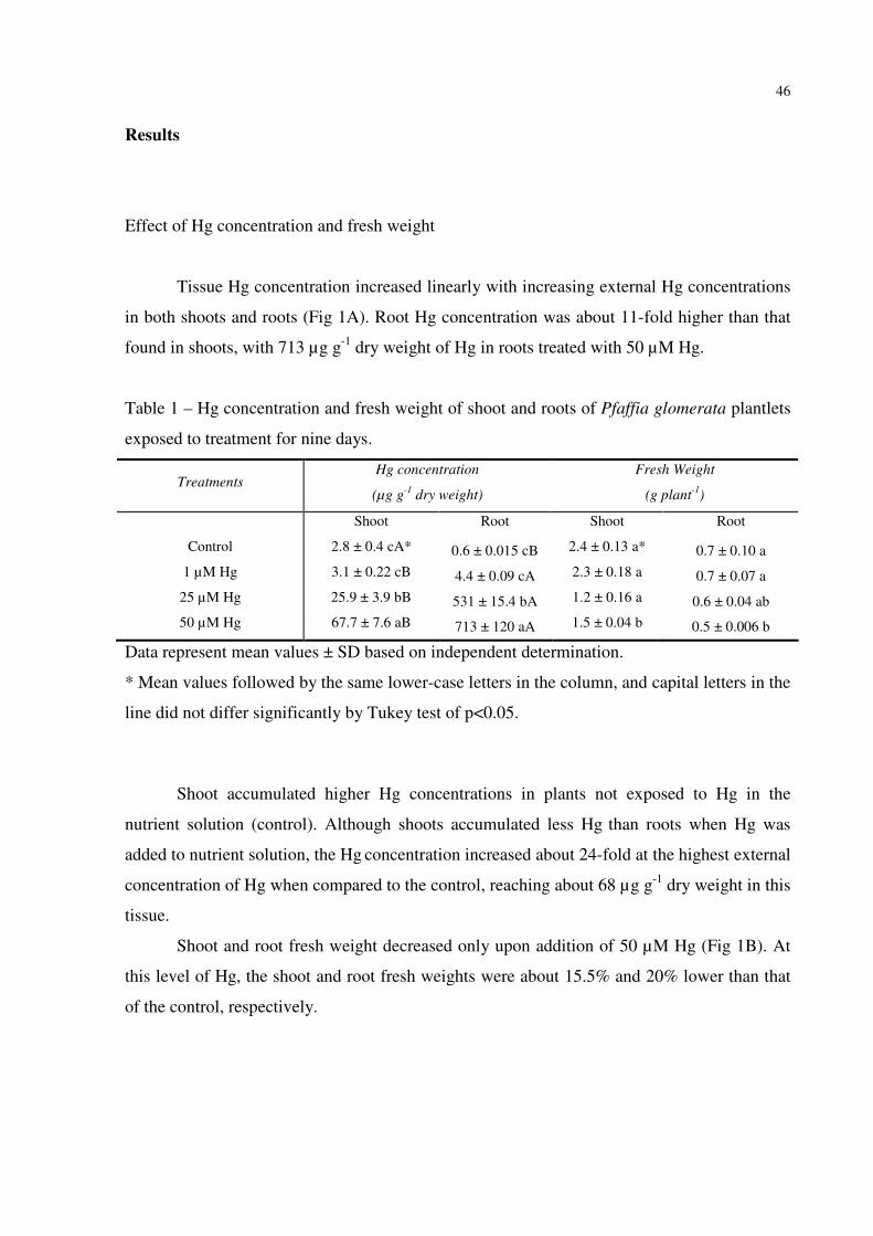
46
Results
Effect of Hg concentration and fresh weight
Tissue Hg concentration increased linearly with increasing external Hg concentrations
in both shoots and roots (Fig 1A). Root Hg concentration was about 11-fold higher than that
found in shoots, with 713 µg g-1 dry weight of Hg in roots treated with 50 µM Hg.
Table 1 – Hg concentration and fresh weight of shoot and roots of Pfaffia glomerata plantlets
exposed to treatment for nine days.
Treatments Hg concentration
(µg g-1
dry weight)
Fresh Weight
(g plant-1
)
Shoot Root Shoot Root
Control 2.8 ± 0.4 cA* 0.6 ± 0.015 cB 2.4 ± 0.13 a* 0.7 ± 0.10 a
1 µM Hg 3.1 ± 0.22 cB 4.4 ± 0.09 cA 2.3 ± 0.18 a 0.7 ± 0.07 a
25 µM Hg 25.9 ± 3.9 bB 531 ± 15.4 bA 1.2 ± 0.16 a 0.6 ± 0.04 ab
50 µM Hg 67.7 ± 7.6 aB 713 ± 120 aA 1.5 ± 0.04 b 0.5 ± 0.006 b
Data represent mean values ± SD based on independent determination.
* Mean values followed by the same lower-case letters in the column, and capital letters in the
line did not differ significantly by Tukey test of p<0.05.
Shoot accumulated higher Hg concentrations in plants not exposed to Hg in the
nutrient solution (control). Although shoots accumulated less Hg than roots when Hg was
added to nutrient solution, the Hg concentration increased about 24-fold at the highest external
concentration of Hg when compared to the control, reaching about 68 µg g-1 dry weight in this
tissue.
Shoot and root fresh weight decreased only upon addition of 50 µM Hg (Fig 1B). At
this level of Hg, the shoot and root fresh weights were about 15.5% and 20% lower than that
of the control, respectively.
47
Effect of Hg on pigments concentration and δ-ALA-D activity of P. glomerata plantlets
0 1 25 50
Hg (µM)
0
1
2
3
4
5
Pig
ments
concentr
ation (
mg g
-1 F
W)
Chlorophyll a+b Carotenoids
a
a
a
a
aa
a
a
0 1 25 50
Hg (µM)
0.00
0.80
1.60
2.40
3.20
4.00
ALA
-D (
PB
G h
-1 m
g-1 p
rote
in)
Shoot
b
c
b
a
Figure 1 Effect of increasing Hg concentration on the pigment concentration (A) and δ-
aminolevulinic acid dehydratase (δ-ALA-D) activity (B) in P. glomerata plantlets. Data
represent the mean ± SD of three replicates. Identical letters indicate no significant differences
among the Hg concentrations (p<0.05) according to Tukey´s multiple range test.
There was no effect of Hg on the chlorophyll and carotenoid concentration. Shoot δ-
ALA-D activity increased at 1 µM of Hg, whereas it decreased approximately 1.2-fold, when
compared to the control, at the highest Hg concentration (50 µM) used.
H2O2 concentration and lipid peroxidation (MDA concentration)
The effects of Hg on H2O2 formation and lipid peroxidation are shown in Fig 2.
Increasing Hg levels in the substrate caused an increase of shoot H2O2 concentration, but it
peaked at 25µM (Fig 2A). In roots, H2O2 concentration either increased at 1µM Hg or
decreased at 50 µM Hg, when compared to the control.
A significant increase in malondialdeide (MDA) levels in shoots was only observed at
50 µM Hg, which was about 3-fold higher when compared to the control (Fig 2B). In contrast,
the root MDA concentration only increased at 25 µM Hg and significantly decreased at 50
µM Hg.
(A) (B)

48
0 1 25 50
Hg (µM)
0
1
2
3
H2O
2 c
oncentr
ation (
µm
ol g
-1 F
W)
Shoot Root
ab
ba
b
c
dc
0 1 25 50
Hg (µM)
0.00
0.80
1.60
2.40
3.20
4.00
TB
AR
S (
nm
ol M
DA
mg
-1 p
rote
in)
Shoot Root
b
a a
b
b
b
b
b
Figure 2 Effect of Hg on H2O2 (A) and lipid peroxidation (B) of P. glomerata plantlets. Data
represent the mean ± SD of three replicates. Identical letters indicate no significant
differences among the Hg concentrations (p<0.05) according to Tukey´s multiple range test.
(A)
(B)

49
CAT, APX and SOD activities
0 1 25 50
Hg (æM)
0
1000
2000
3000
4000
5000
SO
D a
ctivity (
U S
OD
mg
-1 p
rote
in)
Shoot Root
bb
bccaabc
a
0 1 25 50
Hg (µM)
0.00
0.60
1.20
1.80
2.40
3.00
CA
T a
ctivity (
E m
in-1 m
g-1
)
Shoot Root
bc
ab
aa
a
a
c
a
0 1 25 50
Hg (æM)
0.00
1.20
2.40
3.60
4.80
6.00
AP
X a
ctivity (
æm
ol oxid
ate
d a
scorb
ate
min
-1 m
g-1
pro
tein
)
Shoot Root
a
bbc
a
cbb
b
Figure 3 Effect of Hg on superoxide dismutase (SOD) (A), catalase (CAT) (B) and ascorbate
peroxidase (APX) (C) activities of P. glomerata plantlets. Data represent the mean ± SD of
three replicates. Identical letters indicate no significant differences among the Hg
concentrations (p<0.05) according to Tukey´s multiple range test.
In general, the antioxidative enzyme activities were higher in shoot than in roots. SOD
activity increased in shoot with increasing Hg levels in nutrient solution, whereas it decreased
in roots (Fig 3A).
CAT activity increased at 1 and 50 µM Hg in shoot, whereas it was not affected in
roots (Fig 3B).
APX activity either decreased at 1 µM Hg or increased at 50 µM Hg in shoot, whereas
it was altered only at 1 µM Hg in roots, where it increased when compared to the control (Fig
3C).
(B) (C)
(A)

50
AsA, NPSH and proline concentration
As shown in Fig 4A, shoot AsA concentration increased only at 1 and 50 µM Hg. On the
other hand, AsA concentration in roots either decreased at 1 µM Hg or increased at 25 and 50
µM Hg, when compared to the control.
The non-protein thiol (NPSH) concentration in both shoot and root increased
concomitantly with increasing Hg concentration in substrate (Fig 4B). Treatment with 50 µM
Hg resulted in the maximal accumulation of NPSH of 1.5-fold higher than that of the shoot
control plants.
The shoot proline concentration was slightly, but not significantly, increased by Hg levels
up to 25 µM, and at 50 µM Hg level it increased 1.8 fold compared to the control (Fig 4C). In
addition, the root proline concentration was significantly altered at 25 µM Hg, where it
decreased when compared to the control.

51
0 1 25 50
Hg (µM)
0
160
320
480
640
800
AsA
co
nce
ntr
atio
n (
µm
ol g
-1 F
W)
Shoot Root
d
ba
a
b
a
b
c
0 1 25 50
Hg (µM)
0
1
2
3
NP
SH
co
nce
ntr
atio
n (
µm
ol g
-1 F
W)
Shoot Root
b
a
bab a a
c
b
0 1 25 50
Hg (µM)
0
10
20
30
Pro
line c
oncentr
ation (
µm
ol g
-1 F
W)
Shoot Root
a
b
ab
ab
ab
ab
ab
Figure 4 Effect of Hg on (A) ascorbic acid (AsA), (B) non-protein thiol compounds and (C)
proline concentration of Pfaffia glomerata plantlets. Data represent the mean ± SD of three
replicates. Identical letters indicate no significant differences among the Hg concentrations
(p<0.05) according to Tukey´s multiple range test.
Discussion
Plants may survive in heavy metal-contaminated environments by preventing metals
from entering into the cytoplasm and/or detoxifying them inside the cytoplasm. When metals
(B)
(C)
(A)

52
are initially absorbed by roots, some of them are trapped in the cell wall, which reduces the
amount of metals entering the cytoplasm. In the present study, Hg accumulation in the root
system (Table 1) indicates that roots serve as a partial barrier to the transport of Hg to shoots
(Cavallini et al. 1999). Inversely, shoots accumulate more Hg than roots in plants grown
without Hg in the nutrient solution. Uptake of Hg from air via stomata of Hg-untreated plants
is a possible reason for the relatively high shoot Hg concentration in these plants. Patra et al.
(2004) reported that Hg salts in soil may be reduced by biological and chemical reactions to
metallic or methylated compounds, which may volatilize and be taken up through the leaves
in plants grown in enclosed spaces. Uptake of gaseous Hg via stomata was also observed in
laboratory studies (Cavallini et al. 1999; Iglesia-Turiño et al. 2006).
We also checked the effect of Hg on the growth of nodal segments of P. glomerata
grown in vitro, and observed (data not shown) that as the plant organ used initially did not
contain roots, Hg treatment as low as 1 µM had a significant supresive effect on tissue
growth. This demonstrates that roots are very important to avoid Hg uptake into the
protoplasm.
Hg is known to inhibit water uptake via aquaporins on plasma membranes in higher
plants (Zang and Tyerman 1999), which could explain, in the present study, the detrimental
effect of high concentrations of Hg on the fresh weight of P. glomerata plantlets (Table 1).
Destruction of lipid components of the membrane by lipid peroxidation causes
membrane impairment and leakage (Cho and Park 2000). In addition, it has been suggested
that the reduction in chlorophyll content in the presence of heavy metals is caused by an
inhibition of chlorophyll biosynthesis (Pereira et al. 2006) that, in part, may have been caused
by reduction of δ-ALA-D activity. δ-ALA-D is sensitive to metals due to its sulfhydrylic
nature (Rocha et al. 1995). This enzyme catalyzes the condensation of two molecules of δ-
aminolevulinic acid (ALA) to porphobilinogen (Gibson et al. 1955). The synthesis of
porphobilinogen promotes the formation of porphyrins, hemes and chlorophylls, which are
essential for adequate aerobic metabolism and for photosynthesis (Jaffe et al. 2000). Altered
δ-ALA-D activity concomitant with reduced chlorophyll contents has been reported in many
terrestrial plants exposed to various metals (Stobart et al. 1985). In the present study, plantlets
of P. glomerata exposed to 1µM of Hg had increased δ-ALA-D activity, but did not show any
change in the chlorophyll concentration. The stimulating effects of low heavy metal
concentrations on several plant growth parameters is normally related to the hormetic effect

53
that probably represents an overcompensation response to a disruption in the homeostasis of
the organism (Calabrese 1999; Lin et al. 2007).
Interestingly, P. glomerata plants exposed to 1 µM Hg showed an increase in the H2O2
concentration in both shoot and roots (Fig 2A), whereas lipid peroxidation (Fig 2B) did not
change, when compared to the control. At the same level of Hg, a significant increase in the
activity of SOD and CAT was observed in shoot. Therefore, it seems that a low concentration
of Hg in the substrate was enough to activate the antioxidant system which aims to protect
important metabolic enzymes, such as δ-ALA-D. However, ALA-D activity decreased at the
highest concentration of Hg used, where it was 16% lower than that of the control.
At 25 µM Hg, shoot lipid peroxidation was significantly increased, when compared
with the control, indicating that P. glomerata could not tolerate the highest Hg concentration.
Lipid peroxidation was also higher at 25 µM Hg in roots, even though H2O2 concentration did
not differ from that of the control. In contrast, SOD activity was inhibited while CAT and
APX activities were not affected. Therefore, the significant increase in AsA and NPSH
concentrations (Fig 4A and 4B) was not enough to protect roots from lipid peroxidation,
hence this caused a relatively depressive effect on fresh biomass (Table 1).
Compared to the control, SOD activity markedly increased in shoots (Fig 3A) exposed
to increasing Hg levels, and paralleled the levels of H2O2 formed in these tissues. However, at
50µM of Hg the shoot H2O2 concentration was lower than that of 25 µM Hg (Fig 2A). This
reduction in H2O2 levels at 50 µM Hg, surprising in light of the increased SOD activity, might
have been due to increased CAT (Fig 3B) and APX (Fig 3C) activities in shoots. In roots, the
highest H2O2 concentration occurred at 1µM Hg. It is noteworthy that the highest level of
APX activity was observed at this Hg concentration, whereas CAT activity did not differ
among Hg treatments. In general, the concentration of 1 µM Hg was sufficient to induce
alterations in the enzyme activities, and APX showed higher sensitivity, reaching maximal
activity at this concentration.
In the present study, concentrations of both NPSH and AsA in shoots and roots
increased at 25 and 50 µM Hg (Fig 4A and B). Thiol-based complexing substances are
comprised of several acid-soluble sulphydryl-components, such as cysteine, glutathione or
GSH and phytochelatins (De Vos et al. 1992). GSH is a key component of the antioxidant
network that scavenges ROS either directly or indirectly by participating in the ascorbate-
glutathione cycle (Smirnoff 2005). It is also effective in chelating Hg2+ (toxic form) to form
Hg (GSH)2, a non-toxic form of mercury, reducing the bioavailability of Hg (Gupta et al.

54
1998). The increase in accumulation of NPSH caused by Hg exposure may indicate that
phytochelatins are being constantly produced (Rellán-Alvarez et al. 2005). Ascorbate is
another antioxidant that regulates stress-induced peroxidation (Pignocchi and Foyer 2003).
According to Smirnoff (2000), L-ascorbic acid (AsA) is involved in the regulation of
photosynthesis, cell expansion, root elongation and transmembrane electron transport. AsA is
an important component of the plant antioxidant defense system and serves as a reductant for
the removal of H2O2 among other peroxides (Noctor and Foyer 1998). It has also been shown
to detoxify Hg in Chlorella vulgaris by donating electrons to free radicals, thus protecting the
integrity of –SH groups (Rai 1979).
Proline has several functions during stress: osmotic adjustment (Voetberg and Sharp,
1991), osmoprotection (Kishor et al. 1995, 2005), free radical scavenging and as an
antioxidant (Sharma and Dietz 2006), protection of macromolecules from denaturation
(Vanrensburg et al. 1993), regulation of cytosolic acidity (Sivakumar et al. 2000) and as a
carbon and nitrogen reserve after stress relief (Dıaz et al. 1999). Backor et al. (2004) found
that proline treatment produced a significant increase in chlorophyll a of the tolerant strain
exposed to Cu and a similar response was found for chlorophyll b, chlorophyll a + b and total
carotenoids. In wild-type Trebouxia erici grown in the presence of supplemental Cu, proline
significantly increased the yield of photosystem II. Also, proline improved salt tolerance by
up-regulating stress-protective proteins (Khedr et al. 2003; Zhu et al. 2005) and reduced
oxidation of lipid membranes (Demiral and Turkan 2004). In the present study, the shoot
proline concentration also increased upon addition of Hg (Fig 4C). Interestingly, increasing
Hg levels did not alter chlorophyll and carotenoid concentrations. Therefore, our results
suggest that the increase in proline content might account for the stability of photosynthetic
pigments in P. glomerata.
These results present circumstantial evidence for the occurrence of oxidative stress in
P. glomerata plantlets exposed to Hg, as well as for importance of the antioxidant system in
regulating Hg-induced oxidative damage in P. glomerata. On the other hand, at 50 µM of Hg,
the antioxidant system of the plantlets was not able to reverse Hg-mediated damage.

55
References
Aebi H (1984) Catalase in vitro. Methods Enzymol 105:121–126
Ali MB, Vajpayee P, Tripathi, RD, Rai UN, Kumar A, Singh N, Behl HM, Singh SP (2000)
Bull Environ Contam Toxicol 65:573–582
Arnon DI (1949) Copper enzymes in isolated chloroplasts: polyphenoloxidase in Beta
vulgaris. Plant Physiol 24:1–15
Asada K (1996) Radical producion and scavenging in chloroplasts. In: Baker N (ed)
Photosynthesis and the environment. Kluwer Academy Press, Dordrecht, pp 123–150
Backor M, Fahselt D, Wu CT, (2004) Free proline content is positively correlated with copper
tolerance of the lichen photobiont Trebouxia erici (Chlorophyta). Plant Sci 167:151-157
Barbosa NVB, Rocha JBT, Zeni G, Emanuelli T, Beque MC, Braga AL (1998) Effect of
organic forms of selenium on £- aminolevulinate dehydratase from liver, kidney and
brain of adult rats. Toxicol Appl Pharmacol 149:243–253
Bates LS, Waldren RP, Tear ID (1973) Rapid determination of free proline for water-stress
studies. Plant Soil 39:205-207
Boening DW (2000) Ecological effects, transport and fate of mercury: a general review.
Chemosphere 40:1335-1351
Bradford M (1976) A rapid and sensitive method for quantification of microgram quantities of
protein utilizing the principle of protein-dye binding. Anal Biochem 72:248–254
Calabrese EJ (1999) Evidence that Hormesis Represents an ‘‘Overcompensation’’ Response
to a Disruption in Homeostasis. Ecotoxicol Environ Saf 42:135–137
Cargnelutti D, Tabaldi LA, Spanevello RM, Jucoski GO, Battisti V, Redin M, Linares CEB,
Dressler VL, Flores EMM, Nicoloso FT, Morsch VM, Schetinger MRC (2006)
Mercury toxicity induces oxidative stress in growing cucumber seedlings. Chemosphere
65:999-1006
Carneiro MAC, Siqueira JO, Moreira FMS (2002) Comportamento de espécies herbáceas em
misturas de solo com diferentes graus de contaminação com metais pesados. Pesquisa
Agropecu Bras 37:1629-1638
Cavallini A, Natali L, Durante M, Maserti B (1999) Mercury uptake, distribution and DNA
affinity in durum wheat (Triticum durum Desf.) plants. Sci Total Environ 243:119–127
Cho U, Park J (2000) Mercury-induced oxidative stress in tomato seedlings. Plant Sci 156:1-9
56
Demiral T, Turkan I (2004) Does exogenous glycinebetaine affect antioxidative system of rice
seedlings under NaCl treatment? J Plant Physiol 161:1089–100
De Vos CHR, Vonk MJ, Vooijs RV, Schat H (1992) Plant Physiol 98:853–858
Díaz P, Borsani A, Monza J (1999) Acumulación de prolina em plantas en respuesta al estrés
osmotico. Agrociencia 3:1–10
Ellman GL (1959) Tissue sulphydryl groups. Arch Biochem Biophys 82:70-77
El-Moshaty FIB, Pike SM, Novacky AJ, Sehgal OP (1993) Lipid peroxidation and superoxide
production in cowpea (Vigna unguiculata) leaves infected with tobacco ringspot virus
or southern bean mosaic virus. Physiol Mol Plant Pathol 43:109–119
Gibson KD, Neuberger A, Scott JJ (1955) The purification and properties of delta-
aminolevulinic acid dehydratase. Biochem J 61:618–629
Gupta M, Tripathi RD, Rai UN, Chandra P (1998) Role of glutathione and phytochelatin in
Hydrilla verticillata (I.f.) royle and Valusneria spiraus L. under mercury stress.
Chemosphere 37:785–800
Iglesia-Turiño S, Febrero A, Jauregui O, Caldelas C, Araus JL, Bort J (2006) Detection and
quantification of Unbound Phytochelatin 2 In: Plant Extracts of Brassica napus Grown
with Different Levels of Mercury. Plant Physiol 142:742-749
Jaffe EK, Kervinen J, Dunbrack J, Litwin S, Martins J, Scarrow RC, Volin M, Yeung AT,
Yonn E (2000) Porphobilinogen synthase from pea: expression from an artificial gene,
kinetic characterization, and novel implications for subunit interactions. Biochemistry
39:9018–9029
Jacques-Silva MC, Nogueira CW, Broch LC, Flores EMM, Rocha JBT (2001) Diphenyl
diselenide and ascorbic acid changes deposition of selenium and ascorbic acid in liver
and brain of mice. Pharmacol Toxicol 88:119-125
Khedr AHA, Abbas MA, Wahid AAA, Quick WP, Abogadallah GM (2003) Proline induces
the expression of salt-stressresponsive proteins and may improve the adaptation of
Pancratium maritimum L. to salt-stress. J Exp Bot 54:2553–62
Kishor PBK, Hong Z, Miao G, Hu CAA, Verma DPS (1995) Overexpression of D1-
pyrroline-5-carboxylate synthetase increases proline overproduction and confers
osmotolerance in transgenic plants. Plant Physiol 108:1387–94
Kishor PBK, Sangam S, Amrutha RN, Laxmi PS, Naidu KR, Rao KS, et al (2005) Regulation
of proline biosynthesis, degradation, uptake and transport in higher plants: its
implications in plant growth and abiotic stress tolerance. Curr Sci 88:424–38

57
Koricheva J, Roy A, Vranjic JA, Haukioja E, Hughes PR, Hanninen O (1997) Antioxidant
responses to simulated acid rain and heavy metal deposition in birch seedlings. Environ
Pollut 95:249–258
Lin PH, Lin CH, Huang CC, Chuang MC, Lin P (2007) 2,3,7,8-Tetrachlorodibenzo-p-dioxin
(TCDD) induces oxidative stress, DNA strand breaks, and poly(ADP-ribose)
polymerase-1 activation in human breast carcinoma cell lines. Toxicol Lett 172:146–
158
Loreto F, Velikova V (2001) Isoprene produced by leaves protects the photosynthetic
apparatus against ozone damage, quenches ozone products, and reduces lipid
peroxidation of cellular membranes. Plant Physiol 127:1781-1787
Maggio A, Joly RJ (1995) Effects of Mercuric Chloride on the Hydraulic Conductivity of
Tomato Root Systems (Evidence for a Channel-Mediated Water Pathway). Plant
Physiol 109:331–335
Misra HP, Fridovich I (1972) The role of superoxide anion in the autoxidation of epinephrine
and simple assay for superoxide dismutase. J Biol Chem 244:6049-6055
Montanari JI, Magalhães PM, Queiroga CL (1999) Influence of plantation density and
cultivation cycle in root productivity and tenors of beta ecdisone in Pfaffia glomerata
(Spreng.) Pedersen. Acta Hortic 3:125-129
Murashige T, Skoog F (1962) A revised medium for rapid growth and bioassays with tobacco
tissue cultures. Physiol Plant 15:473–97
Neill S, Desikan R, Hancock J (2002) Hydrogen peroxide signaling. Curr Opin Plant Biol 5:
388–395
Nicoloso FT, Erig AC, Martins CF, Russowski D (2001) Micropropagação de ginseng
brasileiro (Pfaffia glomerata (Spreng.) Pedersen). Braz J Med Plants 3:11-18
Noctor G, Foyer CH (1998) Ascorbate and gluthatione: keeping active oxygen under control.
Annu Rev Plant Physiol Plant Mol Biol Med 31:1287-1312
Patra M, Bhowmik N, Bandopadhyay B, Sharma A (2004) Comparison of mercury systems
and the development of genetic tolerance. Environ Exp Bot Rev 52:199–223
Patra M, Sharma A (2000) Mercury toxicity in plants. Bot Rev 66:379–422
Pereira LB, Tabaldi LA, Gonçalves JF, Jucoski JO, Pauletto MM, Weis SN, Nicoloso FT,
Borher D, Rocha JBT, Schetinger MRC (2006) Effect of aluminum on d-aminolevulinic
acid dehydratase (ALA-D) and the development of cucumber (Cucumis sativus).
Environ Exp Bot 57:106–115

58
Pignocchi CH, Foyer CH (2003) Apoplastic ascorbate metabolism and its role in the
regulation of cell signalling. Curr Opin Plant Biol 6:379–389
Rai LC (1979) Mercury toxicity to Chlorella vulgaris. �. Reduction of toxicity by ascorbic
acid cycle in pumpkin plants grown under polluted air in open-top chambers. J Plant
Physiol 142:286-290
Rellán-Álvarez R, Hernández LE, Abadía J, Álvarez-Fernández A (2005) Direct and
simultaneous determination of reduced and oxidised glutathione and homoglutathione
by liquid chromatographyelectrospray/mass spectrometry in plant tissue extracts. Anal
Biochem 356:254-264
Rio LA del, Pastori GM, Palma JM, Sandalio LM, Sevilla F, Corpas FJ, Jimenez A, Lopez-
Huertas E, Hernandez JA (1998) The activated oxygen role of peroxisomes in
senescence. Plant Physiol 116:1195-1200
Rocha JBT, Pereira ME, Emanuelli T, Christofari RS, Souza D (1995) Effects of
methylmercury exposure during the second stage of rapid postnatal brain growth on
delta-aminolevulinic acid dehydratase (ALA-D) activity in brain, liver, kidney and
blood of suckling rats. Toxicology 100:27–37
Sassa S (1982) Delta-aminolevulinic acid dehydratase assay. Enzyme 28:133-145
Sharma SS, Dietz KJ (2006) The significance of amino acids and amino acid-derived
molecules in plant responses and adaptation to heavy metal stress. J Exp Bot 57:711–26
Sivakumar P, Sharmila P, Saradhi PP (2000) Proline alleviates salt-stress induced
enhancement in Rubisco oxygenase activity. Biochem Biophys Res Commun 279:512–
5
Skrebsky EC, Tabaldi LA, Pereira LB, Rauber R, Maldaner J, Cargnelutti D, Gonçalves JF,
Castro GY, Schetinger MRC, Nicoloso FT (2008) Effect of cadmium on growth,
micronutrient concentration, and δ-aminolevulinic acid dehydratase and acid
phosphatase activities in plants of Pfaffia glomerata. Braz J Plant Physiol 20: in press
Smirnoff N (2005) Antioxidants and reactive oxygen species in plants. Oxford: Blackwell
Publishing.
Smirnoff, N (2000) Ascorbic acid: metabolism and finction of a multifaceted molecule. Curr
Opin Plant Biol 3:229-235
Stobart AK, Grifiths WT, Bukhari IA, Sherwood RP (1985) The effect of aluminum on the
biosynthesis of chlorophyll on leaves of barley. Plant Physiol 63:223–228

59
Taniguchi SF, Bersani-Amado CA, Sudo LS, Assef SMC, Oga S (1997) Effect of Pfaffia
iresinoides on the experimental inflammatory process in rats. Phytother Res 11:568-571
Voetberg GS, Sharp RE (1991) Growth of the maize primary root in low water potentials. III.
Roles of increased proline depositions in osmotic adjustment. Plant Physiol 96:125–30
Vanrensburg L, Kruger GHJ, Kruger RH (1993) Proline accumulation the drought tolerance
selection: its relantionship to membrane integrity and chloroplast ultra structure in
Nicotiana tabacum L. J Plant Physiol 141:188–94
Zang WH, Tyerman SD (1999) Inhibition of water channels by HgCl2 in intact wheat root
cells. Plant Physiol 120:849–857
Zhou ZS, Guo K, Elbaz AA, Yang ZM (2008) Salicylic acid alleviates mercury toxicity by
preventing oxidative stress in roots of Medicago sativa. Environ Exp Bot 65:27-34
Zhu X, Gong H, Chen G, Wang S, Zhang C (2005) Different solute levels in two spring wheat
cultivars induced by progressive field water stress at different developmental stages. J
Arid Environ 62:1–14
Zhu Z, Wei G, Li J, Qian Q, Yu J (2004) Silicon alleviates salt stress and increases
antioxidant enzymes activity in leaves of salt-stressed cucumber (Cucumis sativus L.).
Plant Sci 167:527–533

60
3.2 Manuscrito 2
Protective effect of zinc during oxidative stress induced by mercury in Pfaffia glomerata
plantlets
Nicéia Spanholi Calgaroto, Denise Cargnelutti, Liana Veronica Rossato, Sibila Trojahn
Nunes, Júlia Gomes Farias, Luciane Almeri Tabaldi, Fabiane Goldschmidt Antes, Valderi
Luiz Dressler, Érico Marlom de Moares Flores, Maria Rosa Chitolina Schetinger, Fernando
Teixeira Nicoloso

61
Protective effect of zinc during oxidative stress induced by mercury in Pfaffia glomerata
plantlets
Nicéia Spanholi Calgaroto1,3, Denise Cargnelutti2,4, Liana Veronica Rossato2,4, Júlia Gomes
Farias, Sibila Trojahn Nunes, Luciane Almeri Tabaldi, Fabiane Goldschmidt Antes2, Valderi
Luiz Dressler2, Érico Marlom de Moares Flores2, Maria Rosa Chitolina Schetinger2,4*,
Fernando Teixeira Nicoloso1,3**
Departamento de Biologia1, Química2, Programa de Pós-Graduação em Agronomia3 e
Bioquímica Toxicológica4, Centro de Ciências naturais e exatas, Universidade Federal de
Santa Maria, 97105-900, Santa Maria, RS, Brasil
*,**Corresponding authors: respectively [email protected] and
Abstract
The possible role of zinc (Zn) to reverse oxidative stress caused by mercury (Hg) was
investigated in Pfaffia glomerata plantlets. Thirty-day-old acclimated plantlets were exposed
to four treatments: control, 50 µM Zn, 50 µM Hg and 50 µM Zn + 50 µM Hg for 9 days. Hg
concentration was higher in shoot than in roots without Hg in the substrate (control and Zn-
alone), whereas it was higher in roots than in shoot when treated with Hg. Fresh and dry
weight, as well as δ-aminolevulinic acid dehydratase activity were reduced in Hg-treated
plants. Survival percentage, fresh and dry weight and δ-aminolevulinic acid dehydratase
(ALA-D) activity of plants treated with 50 µM Zn + 50 µM Hg were greater than in those
treated with Hg alone. Moreover, Zn treatment reduced lipid peroxidation caused by Hg, and
this effect was related to increased root SOD activity, and shoot CAT and APX activities. In
conclusion, the presence of Zn in the substrate caused a significant reduction in Hg-induced
oxidative stress.

62
Keywords: Pfaffia glomerata; Mercury; Zn protection; Oxidative stress
Introduction
Mercury (Hg) is extremely toxic to plants and animals; it has a long half-life and is
extremely persistent in the environment (Salt et al. 1995). To combat Hg toxicity, plant cells
activate the antioxidant system, which includes glutathione (GSH) and ascorbate and
antioxidative enzymes, such as superoxide dismutase (SOD, E.C. 1.15.1.1), ascorbate
peroxidase (APX, E.C. 1.11.1.11) and catalase (CAT, E.C. 1.11.1.6), that participate in
scavenging reactive oxygen species (ROS) such as O2-, OH and H2O2 (Edreva 2005). Within a
cell, SOD constitutes the first line of defense against ROS, and converts O2 radicals to H2O2
(Alscher et al. 2002). Unlike other reactive oxygen species, H2O2 is able to diffuse across
membranes due to its high stability and membrane-permeability. H2O2 toxicity itself is weak
compared with other ROS, but in the presence of transition metals, such as Fe2+, H2O2
produces the hydroxyl radical (OH), the most reactive active oxygen. Thus, scavenging of
H2O2 is essential to avoid oxidative damage of plant cells (Sakihama et al. 2002). H2O2 is
eliminated by CAT and peroxidase enzymes. Catalase removes the bulk of H2O2, whereas
ascorbate peroxidase can scavenge the H2O2 that is inaccessible to catalase because of its
higher affinity to H2O2 and its presence in different subcellular locations (Scandalios 1994;
Creissen et al. 1994).
Reduced glutathione (GSH) plays an important role in the defense against oxidative
stress in plant cells and is involved in the complex enzymatic machinery that controls the
intracellular levels of H2O2 (May et al. 1998). There are several reports showing changes in
the levels of GSH and GSSG upon exposure of plants to different heavy metals
(Schützendübel and Polle 2002). Therefore, there is evidence pointing towards the
implication, at least partially, of GSH in the tolerance to heavy metals (Rellán-Álvarez et al.
2006). Reduced glutathione (GSH) is used to synthesize phytochelatins (PCs). PC
accumulation is considered to be the plants’ response to addition of heavy metals and,
therefore, may play a central role in metal detoxification (Cobbett and Goldsbrough 2002).
Moreover, some authors link the drainage of GSH caused by PC synthesis to the impairment
of the cellular redox status (Noctor et al. 1998; Xiang and Olivier 1998). Taken together,
these reports suggest that maintenance of the GSH pool is important for plant tolerance to
metals (Ortega-Villasante et al. 2005).

63
Studies with animals showed that Zn is an important antioxidant, decreasing ROS
production (Fernandez et al. 2003). Some studies have reported the ability of Zn to interact
with essential elements such as Cu and Fe, decreasing their content in tissues and retarding
the oxidative processes (Santon et al. 2003). It is also involved in cell membrane stabilization,
metallothionein (Mt) synthesis (Scheuhammer et al. 1985; Waalkes and Klaassen 1985) and
superoxide dismutase (Cu/Zn SOD) structure. Numerous studies with animals have shown
that Zn supply may reduce Cd absorption and accumulation, and also prevent or reduce the
adverse actions of Cd (Ueda et al. 1987; Sato and Nagai 1989; Dorian and Klaassen 1995)
whereas Zn deficiency can intensify Cd accumulation and toxicity (Sarkar et al. 1995).
Moreover, Tsuji et al. (2002) reported that Zn can induce PC synthesis much more efficiently
than Cd in a marine alga, Dunaliella tertiolecta. It was also confirmed that Zn was conjugated
by PCs less strongly than other toxic heavy metals (Satofuka et al. 1999). Generally, Zn
decreases Cd uptake and accumulation in plants (Honma and Hirata 1978; McLaughlin et al.
1994; Oliver et al. 1997). Hart et al. (2002) attributed the competition for uptake between Cd
and Zn to the existence of a common transport system on the plasma membranes. Wu and
Zhang (2002) found that an increase of Zn alleviated Cd toxicity stress in barley plants by
improving growth and reducing membrane damage.
The genus Pfaffia belongs to the Amaranthaceae family and there are about 90 species
distributed in Central and South America (Taniguchi et al. 1997). Carneiro et al. (2002)
showed that an undetermined species of the genus Pfaffia exibited high tolerance to soil
contamination, growing quite abundantly in soil mixtures with 90 and 1,450 mg Kg-1 of Cd
and Zn, respectively. In a recent study developed in our group (Skrebsky et al. 2008) it was
shown that P. glomerata (Spreng.) Pedersen had some degree of Cd tolerance. Moreover, we
also observed (see previous section in this thesis) that the growth and the antioxidant
mecanisms of P. glomerata plants were significantly affected by high levels of Hg (25 and 50
µM) in the substrate.
Under this context, the present study was designed to analyse the effect of Zn nutrition
on the biochemical and physiological alterations caused by Hg in P. glomerata.
64
Materials and methods
Plant material and growth conditions
Plantlets of Pfaffia glomerata (Spreng.) Pedersen (JB accession) were grown in vitro
from nodal segments during thirty days in MS (Murashigue and Skoog 1962) medium,
supplemented with 30 g L-1 sucrose, 6 g L-1 of agar and 0.1 g L-1 of myo-inositol, under
climatized room conditions according to Nicoloso et al. (2001). Thirty-day-old plantlets were
acclimated during three days and transferred to pots containing sand. Both in vitro and ex
vitro plantlets were grown in a growth chamber at 25±1°C on a 16/8 h light/dark cycle with
35 µmol m-2 s-1 of irradiance by cold fluorescent lamps. Plantlets were supplemented daily
with a solution containing the following nutrients: 65.1 mg L-1 NH4Cl, 76.2 mg L -1
MgSO4.7H2O, 135.2 mg L-1 MgCl2.6H2O, 33.1 mg L-1 KH2PO4, 181.5 mg L-1 KCl, 575.3 mg
L-1 Ca(NO3)2.4H2O, 0.11 mg L-1 CuSO4.5H2O, 0.39 mg L-1 MnCl2.4H2O, 0.57 mg L-1
ZnSO4.7H2O, 0.04 mg L-1 NiSO4, 1.54 mg L-1 H3BO3, 0.09 mg L-1 H2MoO4.H2O and 13.34
mg L-1 FeSO4.7H2O. After one month of acclimation, four treatments (control; 50 µM Zn; 50
µM Hg; and 50 µM Zn + 50 µM Hg) were added to the nutrient solution. Hg and Zn were
added as HgCl2 and ZnCl2, respectively. After nine days of treatment, 3 plantlets per replicate
(each treatment consisted of five replicates) were randomly harvested. The plantlets were
divided into roots and shoot for evaluation of fresh and dry biomass. Three independent and
representative tissue samples (shoot and roots) were dried at 65°C to a constant weight, and
the material was used for Hg and Zn determination. Fresh samples were used for
measurements of H2O2, MDA, chlorophyll concentrations, antioxidant enzyme activities and
non-enzymatic antioxidant concentrations.
Tissue Hg concentration
To metal determination plantlets were oven-dried at 65°C to constant mass. Between 70
and 100 mg of shoots and roots were digested with 5 mL HNO3 and 0.2 mL H2O in closed
Teflon vessels, which were heated at 100°C for 3 h in a digester block (Tecnal TE 007D). The
samples were then diluted to 50 mL with high-purity water. Hg concentrations were
determined using a Varian Atomic Absorption Spectrophotometer (Spectr AA 600, Australia)
65
equipped with a vapor generative accessory (Varian VGA-76). The content of Hg was
expressed as µg g-1 dry weight.
Tissue Zn concentration
Zn concentration was determined by Inductively Coupled Plasma Optical Emission
Spectrometry (ICP-OES) using a PerkinElmer Optima 4300DV (Shelton, USA) equipped with
a cyclonic spray chamber and a concentric nebulizer. The emission lines used were 213.857
nm. Instrumental parameters were adjusted according manufacturer recommendations.
Nebulizer, intermediate and principal gas flow rates were set to 0.65, 0.20 and 14 L min-1,
respectively. Sample decomposition was carried out in an open digestion system, using a
heating block from Velp Scientifica (Milano, Italy) equipped with glass vessels. The content
of Zn was expressed as µg g-1 dry weight.
Chlorophyll and carotenoids determination
Fresh biomass (leaves plus stem) were determinated following the method of Hiscox
and Israelstam (1979). Briefly, 0.1g of shoot was macerated in liquid nitrogen, weighted and
placed in a vial containing 7 mL dimethylsulphoxide (DMSO). The photosynthetic pigments
were extracted from fluid at 65°C by incubing for 2 h. To liquid extract was added 3 mL of
DMSO. A 3 mL sample was transferred to a cuvette and the absorbances at 645, 663 and 470
nm were read in order to determine chlorophyll a, chlorophyll b and carotenoids, respectively,
in a Spectrophotometer (Celm E-205D). Pigment concentration was calculated following the
equation used by Lichtenthaler (1987). Chlorophyll and carotenoid concentration was
expressed as mg g-1 fresh weight.
Delta-aminolevulinic acid dehydratase (ALA-D; E.C. 4.2.1.24) activity
Shoots were homogenized in 10 mM Tris-HCl buffer, pH 9.0, at a proportion of 1:1
(w/v). The homogenate was centrifuged at 12,000 g at 4°C for 10 min to yield a supernatant

66
(S1) that was used for the enzyme assay. The supernatant was pre-treated with 0.1% Triton X-
100 and 0.5 mM dithiotreithol (DTT). δ-ALA-D activity was assayed as described by Barbosa
et al. (1998) by measuring the rate of porphobilinogen (PBG) formation. The incubation
medium for the assays contained 100 mM Tris-HCl buffer, pH 9.0 and 3.6 mM ALA.
Incubation was started by adding 100 µL of the tissue preparation to a final volume of 400 µL
and stopped by adding 350 µL of the mixture containing 10% trichloroacetic acid (TCA) and
10 mM HgCl2. The product of the reaction was determined with the Ehrlich reagent at 555 nm
using a molar absorption coefficient of 6.1×10⁴ L-1 mol-1 cm–1 (Sassa 1982) for the Ehrlich-
porphobilinogen salt. δ-ALA-D activity was expressed as nmol PBG mg-1 protein h-1.
Determination of hydrogen peroxide
The H2O2 concentration was determined according to Loreto and Velikova (2001).
Approximately 0.1 g of both roots and shoot was homogenized at 4°C in 2 mL of 0.1%
trichloroacetic acid (TCA) (w/v). The homogenate was centrifuged at 12000 g for 15 min.
Then, 0.5 mL of the supernatant was added to 0.5 mL of 10 mM potassium phosphate buffer
(pH 7.0) and 1 mL of 1M KI. The H2O2 concentration of the supernatant was evaluated by
comparing its absorbance at 390 nm with a standart calibration curve. Hydrogen peroxide
concentration was expressed as µmol g-1 fresh weight.
Estimation of lipid peroxidation
The level of lipid peroxidation products was estimated following the method of El-
Moshaty et al. (1993) by measuring the concentration of malondialdehyde (MDA) as an end
product of lipid peroxidation by reaction with thiobarbituric acid (TBA). Fresh tissue samples
(0.1 g fresh weight) were ground in 2 mL of 0.2 M citrate-phosphate buffer (pH 6.5)
containing 0.5% Triton X-100, using mortar and pestle. The homogenate was filtered through
two layers of paper and centrifuged for 15 min at 20,000 g. One milliliter of the supernatant
fraction was added to an equal volume of 20% (w/v) TCA containing 0.5% (w/v) TBA. The
mixture was heated at 95°C for 40 min and then quickly cooled in an ice bath for 15 min.
After centrifugation at 10,000 g for 15 min, the absorbance of the supernatant was measured

67
at 532 nm. A correction for non-specific turbidity was made by subtracting the absorbance
value taken at 600 nm. The lipid peroxides were expressed as nmol MDA (mg protein)-1, by
using an extinction coefficient of 155 mM-1 cm–1.
Catalase (CAT, E.C. 1.11.1.6) assay
Catalase activity was assayed according to the method of Aebi (1984) with some
modifications. Fresh samples (1g) were homogenized in 5 mL of 50 mM KH2PO4/K2HPO4
(pH 7.0), 10 g L-1 PVP, 0.2 mM EDTA and 10 mL L-1 Triton X-100. The homogenate was
centrifuged at 12,000 g for 20 min at 4°C and then, the supernatant was used for the enzyme
assay. Catalase activity was determined by monitoring the disappearance of H2O2 by
measuring the decrease in absorbance at 240 nm from a reaction mixture containing 2 mL of
15 mM H2O2 in KPO4 buffer (pH 7.0) and 30 µL extract. Activity was expressed as ∆E min-1
mg-1 protein.
Ascorbate peroxidase (APX, E.C. 1.11.1.11) assay
Ascorbate peroxidase (APX) was measured according to Zhu et al. (2004). The
reaction mixture, at a total volume of 2 mL, contained 25 mM (pH 7.0) sodium phosphate
buffer, 0.1 mM EDTA, 0.25 mM ascorbate, 1.0 mM H2O2 and 100 µL enzyme extract. H2O2-
dependent oxidation of ascorbate was followed by a decrease in the absorbance at 290 nm (E
= 2.8 L-1 mol-1 cm-1) and activity was expressed as µmol oxidated ascorbate min-1 mg-1
protein.
Superoxide dismutase (SOD, E.C. 1.15.1.1) activity
The activity of SOD was assayed according to Misra and Fridovich (1972). About 200
mg of roots and shoots were homogenized in 5 mL of 100 mmol L-1 K-phosphate buffer (pH
7.8) containing 0.1 mmol L-1 EDTA, 0.1% (v/v) Triton X-100 and 2% PVP (w/v). The extract
was filtered and centrifuged at 22,000 g for 10 min at 4ºC, and the supernatant was used for

68
assays. The assay mixture consisted of a total volume of 1 mL, containing glycine buffer (pH
10.5), 1 mmol L-1 epinephrine and enzyme material. Epinephrine was the last added
component. Adenochrome formation over the next 4 min was spectrophotometrically
recorded at 480 nm. One unit of SOD activity is expressed as the amount of enzyme required
to cause 50% inhibition of epinephrine oxidation under the experimental conditions used. This
method is based on the ability of SOD to inhibit the autoxidation of epinephrine at an alkaline
pH. Since the oxidation of epinephrine leads to the production of a pink adrenochrome, the
rate of increase of absorbance at 480 nm, which represents the rate of autoxidation of
epinephrine, can be conveniently followed. The enzyme has been found to inhibit this radical-
mediated process.
Ascorbic acid (AsA) concentration
Ascorbic acid determination was performed as described by Jacques-Silva et al.
(2001). Both roots and shoot were homogenized in a solution containing 50 mmol L-1 Tris-
HCl and 10 mL L-1 Triton X-100 (pH 7.5), centrifuged at 6,800 g for 10 min. To the
supernatant was added 10% TCA in the proportion of 1:1 (v/v) followed by centrifugation
(6,800 g for 10 min) to remove protein. An aliquot of the sample (300 µL) was incubated at
37°C in a medium containing 100 µL TCA 13.3%, 100 µL deionized water and 75 µL 2,4-
Dinitrophenylhydrazine (DNPH). The DNPH solution contained 2% DNPH, 0.23% thiourea,
0.27% CuSO4 diluted in 49% H2SO4. After 3 h, 500 µL of 65% H2SO4 was added and
samples were read at 520 nm. A standard curve was constructed using L(+) ascorbic acid.
Non-protein thiols groups (NPSH) concentration
Non-protein thiols concentration was measured spectrophotometrically with Ellman’s
reagent (Ellman 1959). Roots and shoots samples were homogeneized in a solution containing
50 mmol L-1 Tris-HCl and 10 mL L-1 Triton X-100 (pH 7.5), centrifuged at 6,800 g for 10
min, and NPSH were determined in a fraction obtained after mixing 1 volume of supernatant
with 1 volume of 10% TCA followed by centrifugation (6,800 g for 10 min) and
neutralization (to pH 7.4) with 1 M Tris-HCl as described by Jacques-Silva et al. (2001).
69
Reaction was read at 412 nm after the addition of 0.05 mL of 10 mM 5-5-dithio-bis (2-
nitrobenzoic acid) (DTNB). A standard curve using cysteine was used to calculate the content
of thiol group in samples, and was expressed as µmol SH g-1 fresh weight.
Protein extraction
In all the enzyme preparations, protein was determined by the method of Bradford
(1976) using bovine serum albumin as standard and was expressed in mg L-1.
Statistical analysis
The analyses of variance were computed on statistically significant differences
determined based on the appropriate F-tests. The results are the means ± SD of at least three
independent replicates. The mean differences were compared utilizing Tukey test with p<
0.05.
Results
Tissue metal concentration
Hg concentrations were higher in roots than in shoot upon addition of Hg in the
substrate. Conversely, Hg concentrations were, approximately, 15% and 6.8% higher in shoot
when compared with roots, respectively, in control and Zn treatments (Table 1). The addition
of Hg and Zn together significantly decreased root and shoot Hg concentrations, when
compared to Hg alone. In this treatment, shoot and root Hg levels were 59% and 24% lower
than in plants exposed to 50 µM Hg alone. In addition, there was an increase in Zn levels in
shoot of plants exposed to Zn + Hg, although in roots, Zn concentration was not altered, when
compared to the control.

70
Table 1 – Hg and Zn concentrations of shoot and roots of Pfaffia glomerata plantlets exposed
to treatments for 9 days.
Treatments Zn concentration
(µg g-1
dry weight)
Hg concentration
(µg g-1
dry weight)
Shoot Root Shoot Root
Control 79.66 ± 4.04 bB* 161.66 ± 10.01 aA 16.86 ± 1.5 cA 2.53 ± 0.25 bB
50 µM Zn 116 ± 4.58 bB 176 ± 37.9 aA 15.5 ± 0.7 cA 1.06 ± 0.52 bB
50 µM Hg 73.66 ± 4.72 bB 143 ± 34 aA 351.5 ± 39.5 aB 797 ± 149 aA
50 µM Zn + 50 µM Hg 216.66 ± 65.57 aA 150.66 ± 23.35 aB 208 ± 94.02 bB 605.33 ± 159.91
aA
Data represent mean values ± SD based on independent determination.
* Mean values followed by the same lower-case letters in the column, and capital letters in the
line did not differ significantly by Tukey test of p<0.05.
Survival percentage, fresh and dry weight of P. glomerata plantlets
In comparison to the control, fresh and dry weight, as well as survival percentage in the
Hg-exposed Pfaffia glomerata plants decreased significantly, while Zn supplementation
partially reversed the negative effect caused by Hg (Fig 1). The survival percentage was about
47% and 20% lower than the control in plants exposed to Hg-alone and Zn+Hg treatments,
respectively. In 50 µM Hg treatment, shoot fresh weight decreased about 71.5% when
compared to the control, whereas root fresh weight decreased by 59%. On the other hand, in
plants treated with Zn+Hg, shoot and root fresh weight decreased about 51.4% and 24.4%,
respectively, in relation to the control.
Shoot dry weight in Hg-exposed plants decreased by 69% in relation to the control. In
plants exposed to Zn+Hg treatment, the dry weight also decreased, but only by about 26%
when compared to the control. Interestingly, root dry weight in Hg-treated plants was slightly
but not significantly, decreased by the presence of Hg, when compared to the control (Fig
1C), whereas Zn-treated plants showed greater root dry weight than Hg-treated plants.
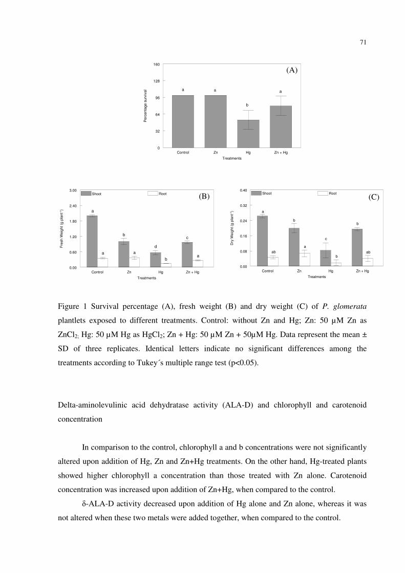
71
Control Zn Hg Zn + Hg
Treatments
0
32
64
96
128
160
Perc
enta
ge s
urv
ival
a
b
a a
Control Zn Hg Zn + Hg
Treatments
0.00
0.60
1.20
1.80
2.40
3.00
Fre
sh
We
igh
t (g
pla
nt-1
)
Shoot Root
b
aa
c
d
b
a
a
Control Zn Hg Zn + Hg
Treatments
0.00
0.08
0.16
0.24
0.32
0.40
Dry
We
igh
t (g
pla
nt-1
)
Shoot Root
c
b
ab
a
bab
a
b
Figure 1 Survival percentage (A), fresh weight (B) and dry weight (C) of P. glomerata
plantlets exposed to different treatments. Control: without Zn and Hg; Zn: 50 µM Zn as
ZnCl2; Hg: 50 µM Hg as HgCl2; Zn + Hg: 50 µM Zn + 50µM Hg. Data represent the mean ±
SD of three replicates. Identical letters indicate no significant differences among the
treatments according to Tukey´s multiple range test (p<0.05).
Delta-aminolevulinic acid dehydratase activity (ALA-D) and chlorophyll and carotenoid
concentration
In comparison to the control, chlorophyll a and b concentrations were not significantly
altered upon addition of Hg, Zn and Zn+Hg treatments. On the other hand, Hg-treated plants
showed higher chlorophyll a concentration than those treated with Zn alone. Carotenoid
concentration was increased upon addition of Zn+Hg, when compared to the control.
δ-ALA-D activity decreased upon addition of Hg alone and Zn alone, whereas it was
not altered when these two metals were added together, when compared to the control.
(A)
(C) (B)

72
Control Zn Hg Zn + Hg
Treatments
0
1
2
3
4
5P
igm
en
ts c
on
ce
ntr
atio
n (
mg
g-1
FW
)Chlorophyll a Chlorophyll b Carotenoids
a
b
a
a
ab
acabca
ab
ab
Control Zn Hg Zn + Hg
Treatments
0
2
4
6
8
10
AL
A-D
activity (
nm
ol P
BG
min
-1 m
g p
rote
in-1
) Shoot
bb
a
a
Figure 2 Pigment concentration (A) and δ-aminolevulinic acid (δ-ALA-D) activity (B) of P.
glomerata plantlets exposed to different treatments. Control: without Zn and Hg; Zn: 50 µM
Zn as ZnCl2; Hg: 50 µM Hg as HgCl2; Zn + Hg: 50 µM Zn + 50µM Hg. Data represent the
mean ± SD of three replicates. Identical letters indicate no significant differences among the
treatments according to Tukey´s multiple range test (p<0.05).
Hydrogen peroxide and lipid peroxidation levels
Only treatments containing Hg, applied either alone or supplemented with Zn, had
higher tissue H2O2 concentrations, when compared to the control.
Shoot lipid peroxidation was significantly increased upon addition of Hg, while it
decreased significantly in the treatments with Zn+Hg added together, when compared to the
control. On the other hand, lipid peroxidation in roots was not altered in any treatment.
(B) (A)
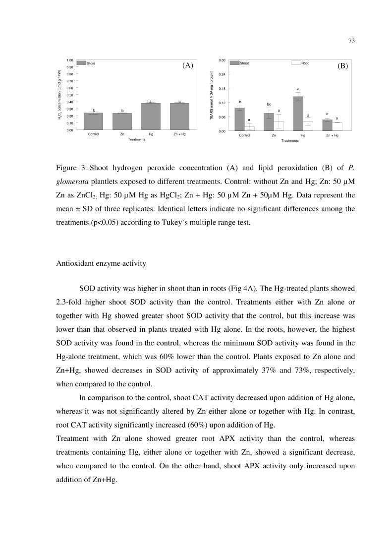
73
Control Zn Hg Zn + Hg
Treatments
0.00
0.10
0.20
0.30
0.40
0.50
0.60
0.70
0.80
0.90
1.00H
2O
2 c
oncentr
ation (
µm
ol g
-1 F
W)
Shoot
bb
aa
Control Zn Hg Zn + Hg
Treatments
0.00
0.06
0.12
0.18
0.24
0.30
TB
AR
S (
nm
ol M
DA
mg
-1 p
rote
in)
Shoot Root
a
a
a
a
c
bcb
a
Figure 3 Shoot hydrogen peroxide concentration (A) and lipid peroxidation (B) of P.
glomerata plantlets exposed to different treatments. Control: without Zn and Hg; Zn: 50 µM
Zn as ZnCl2; Hg: 50 µM Hg as HgCl2; Zn + Hg: 50 µM Zn + 50µM Hg. Data represent the
mean ± SD of three replicates. Identical letters indicate no significant differences among the
treatments (p<0.05) according to Tukey´s multiple range test.
Antioxidant enzyme activity
SOD activity was higher in shoot than in roots (Fig 4A). The Hg-treated plants showed
2.3-fold higher shoot SOD activity than the control. Treatments either with Zn alone or
together with Hg showed greater shoot SOD activity that the control, but this increase was
lower than that observed in plants treated with Hg alone. In the roots, however, the highest
SOD activity was found in the control, whereas the minimum SOD activity was found in the
Hg-alone treatment, which was 60% lower than the control. Plants exposed to Zn alone and
Zn+Hg, showed decreases in SOD activity of approximately 37% and 73%, respectively,
when compared to the control.
In comparison to the control, shoot CAT activity decreased upon addition of Hg alone,
whereas it was not significantly altered by Zn either alone or together with Hg. In contrast,
root CAT activity significantly increased (60%) upon addition of Hg.
Treatment with Zn alone showed greater root APX activity than the control, whereas
treatments containing Hg, either alone or together with Zn, showed a significant decrease,
when compared to the control. On the other hand, shoot APX activity only increased upon
addition of Zn+Hg.
(A) (B)
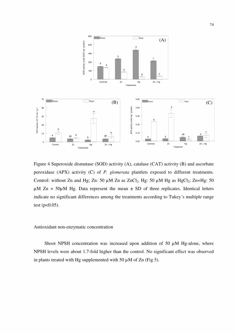
74
Controle Zn Hg Zn + Hg
Treatments
0
160
320
480
640
800
SO
D a
ctivity (
un
its S
OD
mg
-1 p
rote
in)
Shoot Root
a
b
d
cd
b
a
c
Control Zn Hg Zn + Hg
Treatments
0
14
28
42
56
70
CA
T a
ctivity (
10
-9 E
min
-1 g
-1)
Shoot Root
b
abb
a
b
b
aba
Controle Zn Hg Zn + Hg
Treatment
0.00
0.16
0.32
0.48
0.64
0.80
AP
X a
ctivity (
units m
g-1 p
rote
in)
Shoot Root
a
cab
b
b
b
a
c
Figure 4 Superoxide dismutase (SOD) activity (A), catalase (CAT) activity (B) and ascorbate
peroxidase (APX) activity (C) of P. glomerata plantlets exposed to different treatments.
Control: without Zn and Hg; Zn: 50 µM Zn as ZnCl2; Hg: 50 µM Hg as HgCl2; Zn+Hg: 50
µM Zn + 50µM Hg. Data represent the mean ± SD of three replicates. Identical letters
indicate no significant differences among the treatments according to Tukey´s multiple range
test (p<0.05).
Antioxidant non-enzymatic concentration
Shoot NPSH concentration was increased upon addition of 50 µM Hg-alone, where
NPSH levels were about 1.7-fold higher than the control. No significant effect was observed
in plants treated with Hg supplemented with 50 µM of Zn (Fig 5).
(A)
(C) (B)
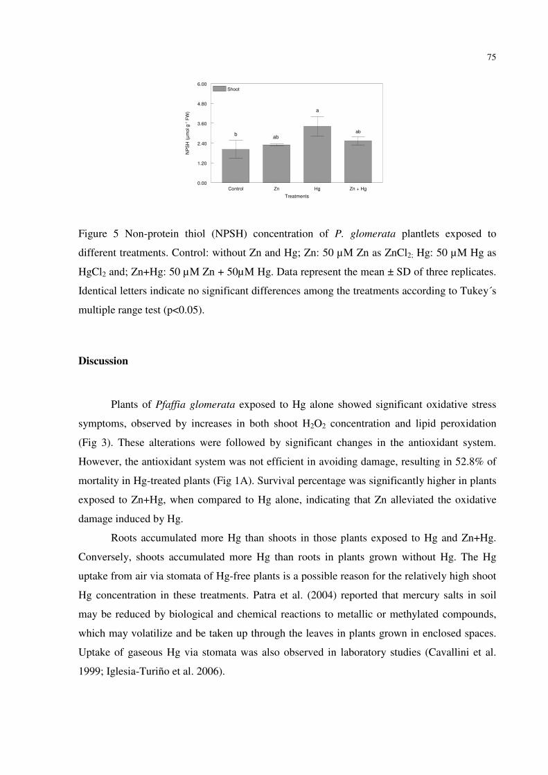
75
Control Zn Hg Zn + Hg
Treatments
0.00
1.20
2.40
3.60
4.80
6.00
NP
SH
(µ
mo
l g
-1 F
W)
Shoot
abab
b
a
Figure 5 Non-protein thiol (NPSH) concentration of P. glomerata plantlets exposed to
different treatments. Control: without Zn and Hg; Zn: 50 µM Zn as ZnCl2; Hg: 50 µM Hg as
HgCl2 and; Zn+Hg: 50 µM Zn + 50µM Hg. Data represent the mean ± SD of three replicates.
Identical letters indicate no significant differences among the treatments according to Tukey´s
multiple range test (p<0.05).
Discussion
Plants of Pfaffia glomerata exposed to Hg alone showed significant oxidative stress
symptoms, observed by increases in both shoot H2O2 concentration and lipid peroxidation
(Fig 3). These alterations were followed by significant changes in the antioxidant system.
However, the antioxidant system was not efficient in avoiding damage, resulting in 52.8% of
mortality in Hg-treated plants (Fig 1A). Survival percentage was significantly higher in plants
exposed to Zn+Hg, when compared to Hg alone, indicating that Zn alleviated the oxidative
damage induced by Hg.
Roots accumulated more Hg than shoots in those plants exposed to Hg and Zn+Hg.
Conversely, shoots accumulated more Hg than roots in plants grown without Hg. The Hg
uptake from air via stomata of Hg-free plants is a possible reason for the relatively high shoot
Hg concentration in these treatments. Patra et al. (2004) reported that mercury salts in soil
may be reduced by biological and chemical reactions to metallic or methylated compounds,
which may volatilize and be taken up through the leaves in plants grown in enclosed spaces.
Uptake of gaseous Hg via stomata was also observed in laboratory studies (Cavallini et al.
1999; Iglesia-Turiño et al. 2006).

76
Our results showed a reduction of shoot Hg concentration together with a significant
increase of Zn concentration in plants exposed to Zn+Hg (Table 1), indicating a possible
interaction between these metals. Zn, Cd and Hg are divalent metals that constitute group II B
of the periodic table of elements. Zn is an essential trace metal while the remaining two are
toxic metals. Nevertheless, they share biological responses such as affinity towards thiol
groups, antagonism to biological cations, participation in redox reaction and modulation of
essential element homeostasis (Peixoto et al. 2003). Hart et al. (2002) attributed the
competition for uptake between Cd and Zn to the existence of a common transport system on
the plasma membranes. Several other findings (Thys et al. 1991; Cataldo et al. 1983;
McKenna et al. 1993; Aravind and Prasad 2003) point to depressed Cd uptake in the presence
of Zn as well as an antagonistic effect of Zn under other essential transition metals like Cu
and Fe (Powell 2000; Zago and Oteiza 2001). Thus, in our study, Zn may have interacted with
Hg.
In the present study, the concentration of Zn in overground tissues (leaves plus stem)
of plants exposed to Zn+Hg might have been an antioxidant action that resulted in the
significant decrease of lipid peroxidation (Fig 3B) together with the significant increase of
APX activity (Fig 5C) in these tissues, when compared to the control.
Shoot and root fresh weight from plants exposed to Hg added alone showed reductions
of about 70% and 60%, respectively, when compared to the control. Hg has a high affinity to
sulphydryl (-SH) groups generally abundant in proteins, and it might bind to the crucial and
functional membrane, activating and inactivating their enzyme functions (Patra et al. 2004).
Plant cells contain aquaporins, proteins that facilitate water transport in the vacuolar
membrane (tonoplast) and the plasma membrane (Patra and Sharma 2000). Mercury induces
conformational changes in aquaporins of higher plants because it is thought to bind to –SH
groups of aquaporin proteins, physically blocking the channels and reducing their hydraulic
conductivity (Tyermam et al. 1999). Partial recovery of the water flow rate upon addition of
HgCl2 was observed in tomato and aspen root systems, implying the presence of aquaporins
as regulators of the plant water status (Maggio and Joly 1995; Wan and Zwiazek 1999). In the
present study, the symptoms of withering shown by plants treated with Hg may explain the
decrease in fresh weight and this data corroborates with others reports (Zang and Tyerman
1999; Cargnelutti et al. 2006).
Zn is an essential component of many vital enzymes and a structural stabilizer for
proteins, membrane and DNA-binding proteins (Vallee and Auld 1990). It acts either as a

77
metal component or as a functional, structural, or regulatory cofactor of a large number of
enzymes (Marshner 1986). On the other hand, an excess of Zn in the substrate causes
nutritional disorders in plants, hindering development (Soares et al. 2001). In the present
study, significant decreases of fresh and dry weight in plants exposed to Zn alone may have
been due to Fe deficiency since the shoot Fe concentration was about 53% lower than that of
control plants (data not shown). The significant decrease of the intracellular Fe concentration
upon Zn addition may have been associated with the significant reduction (50% and 24%,
respectively) in shoot fresh and dry weight when compared to the control. Iron is an essential
element for plants and plays an important role in several plant biological processes, including
the catalytic groups for many redox enzymes and is required for nitrogen fixation and
synthesis of chloroplast, DNA, and hormones (Hopkins 1997). In the present study, the lack
of bioavailable Fe may have been due to its precipitation in nutrient solution, or by
antagonistic interaction between Zn and Fe, which inhibited Fe uptake by roots.
The toxicity of non-essential metals, such as Hg and Cd, is generally accepted to occur
by inactivation of enzymes and/or functional proteins by direct binding. However, several
studies have also shown that toxicity may be due, at least in part, to oxidative damage caused
by generation of reactive oxygen species (ROS) due to the presence of heavy metals (Stohs
and Bagchi 1995).
Hg ions can also substitute metal ions in photosynthetic pigments, causing a decrease
in photosynthesis rates (Xylander et al. 1996). Van Assche and Clijsters (1990) reported that
lipid peroxidation causes membrane impairment and leakage, and suggested that the reduction
in chlorophyll content in the presence of metals is caused by an inhibition of chlorophyll
biosynthesis. Mercury has been reported to decrease the pigment content in several plants (Ali
et al. 2000; Cho and Park 2000; Cargnelutti et al. 2006). However, no effect was observed in
pigment concentration in P. glomerata plants exposed to Hg, although a significant alteration
in δ-ALA-D activity was observed (about 53% when compared to the control). Due to its
sulphydryl nature (Barnard et al. 1977), δ-ALA-D is highly sensitive to the presence of heavy
metals such as Hg (Rocha et al. 1993, Emanuelli et al. 1996). Thus, the significant decrease in
δ-ALA-D activity observed in this study may have resulted from Hg binding with δ-ALA-D
–SH groups.
δ-ALA-D is a zinc-dependent enzyme in animals, yeasts, and some bacteria (Jordan
1990). Cysteine residues participate in Zn binding and these enzymes contain a cysteine rich
domain that may be involved in metal binding. Plant dehydratases are located in plastids and
78
are needed for the synthesis of chlorophyll and other cellular tetrapyrroles. They share 35 to
50% identity with non-plant enzymes, but they require Mg to display maximum activity rather
than Zn. The peptide region in the plant enzyme corresponding to the Zn domain of animals
lacks cysteine and histidine residues and contains aspartate, alanine, or threonine instead
(Boese et al. 1991). The reduction in δ-ALA-D activity in Zn treated plants may have been
due to Mg utilization for some processes which require Fe during Fe deficiency, as suggested
by Oladiji (2003) in a study with rats. Thus, Fe deficiency could have led to Mg unavailability
for δ-ALA-D activity. Plants exposed to Zn+Hg, in contrast, showed a sharp increase in δ-
ALA-D activity, such that it was equal to the control. The present study indicated that the
decrease in δ-ALA-D activity induced by Hg was prevented by the increase of Zn
concentration in shoot. Probably, Zn prevented Hg-binding with –SH groups of this enzyme,
also preventing its inactivation.
Oxidative stress occurs due to the excess production of free radicals, the inadequate
availability of antioxidants or a combination of both. Lipid peroxidation is a basic
deteriorating change in unsaturated fatty acids of the cell membranes induced by excess free
radicals (Haliwell and Gutteridge 1999). Estimation of malonyldialdehyde continues to be a
reliable method to assess the degree of peroxidative damage to cell membranes, as it is the
most abundant aldehyde formed as a by-product during this process (Gurer et al. 1998). In the
present study, the root MDA concentration was not altered in the presence of Zn or Hg,
although other stress symptoms occurred, such as growth reduction and root darkening (data
not shown). However, in shoot MDA reached higher concentrations in plants exposed to Hg
alone, when compared to the control. This result is in agreement with Cho and Park (2000),
who showed that tomato plants exposed to 50 µM HgCl2 had a significant increase in the
MDA concentration. The increase of MDA indicates that plants experienced oxidative
damage when exposed to Hg alone at 50 µM. In addition, the higher MDA concentration in
the shoot indicates that overground tissues are more sensitive to metals than roots, and Zn
addition had an important role in ROS detoxification induced by Hg.
The H2O2 concentration is, in part, a result of SOD activity, the first antioxidant
enzyme involved with ROS detoxification. H2O2, although not as reactive as the superoxide
radical (O2-), can be changed by transition metals, such as Fe and Cu, producing the OH•
radical, the most dangerous cellular oxidant (Zago and Oteiza 2001). To avoid a hazardous
situation, APX and CAT antioxidant enzymes act to degrade these molecules to H2O (Fig 4).
H2O2 and SOD activity were higher in shoot of plants exposed to Hg alone (Fig 3 and 4),
79
indicating an over-production of superoxide radicals (O2-) that resulted in a 2.3-fold increase
in SOD activity when compared to the control.
Perhaps Zn addition had a major role in detoxification of superoxide radicals, because
its presence allowed the increase of SOD activity. The higher SOD activity observed in shoot
of plants exposed to Zn and Zn+Hg may have been due to the presence of Zn, which has a
structural role in the Cu-ZnSOD enzyme.
As a result of SOD action in plants exposed to Hg, shoot H2O2 was produced, inducing
to some extent the scavenging-activity of APX in this tissue. Conversely, in roots exposed to
Hg alone, SOD activity was lower, whereas CAT activity was higher. It seems that an excess
of ROS production in the presence of Hg in roots would have inactivated antioxidant enzymes
such as SOD and APX.
Non-protein-thiol compounds are non-enzymatic antioxidants that protect plant tissue
from cellular damage, acting together with the enzymatic antioxidant system. They exist
under various forms, such as glutathione tripeptide, homoglutathione and polymerized
peptides, phytochelatins or homophytochelatins (Ferrat et al. 2003). These molecules are
synthesized by enzymatic polymerization of glutathione or homoglutathione (Gekeler et al.
1989). Glutathione is a well-known antioxidant that plays a key role in the regulation of the
redox balance and can be used as an indicator of oxidative stress (Rijstenbil and Wijnholds
1996), in the detoxification of xenobiotics (Marrs 1996) and heavy metals (Cobbett 2000). Hg
has high affinity to -SH residues present in non-protein thiols, and can form complexes with
Hg forms both in vivo and in vitro (Rabenstein et al. 1985). In the present study, Hg-treated
plants had significant increases in shoot NPSH concentrations (Fig 5), possibly reflecting the
a defense mechanism against Hg-triggered oxidative stress. The increase of NPSH
concentrations can indicate an increase in GSH concentrations and, consequently, an increase
in phytochelatin production. There is strong evidence that PC forms complexes with metals to
which they have a high affinity (Mehra et al. 1996) such as Pb, Ag and Hg in vitro (Rauser
1999).
In conclusion, the supplementation of Zn to Hg-treated plants alleviated the Hg-toxic
effects in P. glomerata plants by decreasing Hg uptake and translocation, maintaince of δ-
ALA-D activity, decreasing lipid peroxidation and increasing the protective effect of the
antioxidant system.

80
References
Aebi H (1984) Catalase in vitro. Methods Enzymol 105:121–126
Ali MB, Vajpayee P, Tripathi, RD, Rai UN, Kumar A, Singh N, Behl HM, Singh SP (2000)
Bull Environ Contam Toxicol 65:573–582
Alscher RG, Erturk N, Heath LS (2002) Role of superoxide dismutases (SODs) in controlling
oxidative stress in plants. J Exp Bot 53(372):1331-1341
Aravind P, Prasad MNV (2003) Zinc alleviates cadmium-induced oxidative stress in
Ceratophyllum demersum L.: a free floating freshwater macrophyte. Plant Physiol
Biochem 41:391-397
Barbosa NVB, Rocha JBT, Zeni G, Emanuelli T, Beque MMC, Braga AL (1998) Effect of
organic forms of selenium on £- aminolevulinate dehydratase from liver, kidney and
brain of adult rats. Toxicol Appl Pharmacol 149:243–253
Barnard GF, Itoh R, Hohberger LH, Shemin D (1977) Mechanism of porphobilinogen
synthase – possible role of essential thiol groups. J Biol Chem 252:8965-8974
Boese QF, Spano AJ, Li JM, Timko MP (1991) Aminolevulinic Acid Dehydratase in Pea
(Pisum sativum L). J Biol Chem 266(26):17060-17066
Bradford M (1976) A rapid and sensitive method for quantification of microgram quantities of
protein utilizing the principle of protein-dye binding. Anal Biochem 72:248–254
Cargnelutti D, Tabaldi LA, Spanevello RM, Jucoski GD, Battisti V, Redin M, Linares CEB,
Dressler VL, Flores EMD, Nicoloso FT, Morsch VM, Schetinger MRC (2006) Mercury
toxicity induces oxidative stress in growing cucumber seedlings. Chemosphere 65:999-
1006
Carneiro MAC, Siqueira JO, Moreira FMS (2002) Comportamento de espécies herbáceas em
misturas de solo com diferentes graus de contaminação com metais pesados. Pesq
Agropec Bras 37(11):1629-1638
Cataldo DA, Garland TR, Wildung RE (1983) Cadmium uptake kinetics in intact soyabean
plants. Plant Physiol 73:844–848
Cavallini A, Natali L, Durante M, Maserti B (1999) Mercury uptake, distribution and DNA
affinity in durum wheat (Triticum durum Desf.) plants. Sci Total Environ 243:119–127
Cho U, Park J (2000) Mercury-induced oxidative stress in tomato seedlings. Plant Sci 156:1-9
Cobbett CS (2000) Phytochelatin biosynthesis and function in heavy-metal detoxification.
Curr Opin Plant Biol 3:211-216

81
Cobbett C, Goldsbrough P (2002) Phytochelatins and metallothioneins: roles in heavy metal
detoxification and homeostasis. Annu Rev Plant Biol 53:159–182
Creissen GP, Edwards EA, Mullineaux PM (1994) Glutathione reductase and ascorbate
peroxidase. In: Foyer CH, Mullineaux PM Causes of Photooxidative stress and
amelioration of defense system in plants. CRC Press 343-364
Dorian C, Klaassen CD (1995) Prtotection by zinc-metallothionein (ZnMT) against cadmium-
metallothionein-induced nephrotoxicity. Fundam Appl Toxicol 26:99-106
Edreva A (2005) Generation and scavenging of reactive oxygen species in chloroplasts: a
submolecular approach. Agricult Ecosyst Environ 106:119-133
Ellman GL (1959) Tissue sulphydryl groups. Arch Biochem Biophys 82:70-77
El-Moshaty FIB, Pike SM, Novacky AJ, Sehgal OP (1993) Lipid peroxidation and superoxide
production in cowpea (Vigna unguiculata) leaves infected with tobacco ringspot virus
or southern bean mosaic virus. Physiol Mol Plant Pathol 43:109–119
Emanuelli T, Rocha JBT, Pereira ME, Porciuncula LO, Morsch VM, Martins AF, Souza DOG
(1996) Effect of mercury chloride intoxication and dimercaprol treatment on δ-
aminolevulinate dehydratase from brain, liver and kidney of adult mice. Pharmacol
Toxicol 79:138-143
Fernandez E, Gustafson A, Anderson M, Hellman B, Dencker L (2003) Cadmium-induced
changes in apoptic gene expression blocked by zinc supplementation. Toxicol Sci
10:85-99
Ferrat L, Gnassia-Barelli M, Pergent-Martini C, Roméo M (2003) Mercury and non-protein
thiol compounds in the seagrass Posidonia oceanica. Comp Biochem Physiol C
134:147-155
Gekeler W, Grill E, Winnacker EL, Zenk MH (1989) Survey of the plant kingdom for the
ability to bind heavy metals through phytochelatins. J Biosci 44:361–369
Gurer H, Ozgunes H, Neal R, Spitz DR, Ercal N (1998) Antioxidant effects of N-acetyl
cysteine and succimer in red blood cells from lead exposed rats. Toxicology 128:181–
189
Haliwell B, Gutteridge JMC (1999) Free Radicals in Biology and Medicine, 3rd ed Oxford
University Press Oxford.
Hart JJ, Welch RM, Norvell WA, Kochian LV (2002) Transport interactions between
cadmium and zinc in roots of bread and durum wheat seedlings. Physiol Plant 116:73-
78

82
Honma Y, Hirata H (1978) A noticeable increase in Cd absorption by Zn deficient rice plants.
Soil Sci Plant Nutr 24:295-297
Hiscox JD, Israelstam GF (1979) A method for the extraction of chlorophyll from leaf tissue
without maceration. Can J Bot 57:1132-1334
Hopkins WG (1997) Introduction to Plant Physiology. Wiley 61–76
Iglesia-Turiño S, Febrero A, Jauregui O, Caldelas C, Araus JL, Bort J (2006) Detection and
quantification of Unbound Phytochelatin 2 In: Plant Extracts of Brassica napus Grown
with Different Levels of Mercury. Plant Physiol 142:742-749
Jacques-Silva MC, Nogueira CW, Broch LC, Flores EMM, Rocha JBT (2001) Diphenyl
diselenide and ascorbic acid changes deposition of selenium and ascorbic acid in liver
and brain of mice. Pharmacol Toxicol 88:119-125
Jordan PM (1990) Biosynthesis of hemes and chlorophylls. In: Dailey HA (ed) 55-121
McGraw-Hill Book Co., New York.
Lichtenthaler HK (1987) Chlorophylls and carotenoids – pigments of photosynthetic
biomembranes. Methods Enzymol 148:350–82
Loreto F, Velikova V (2001) Isoprene produced by leaves protects the photosynthetic
apparatus against ozone damage, quenches ozone products, and reduces lipid
peroxidation of cellular membranes. Plant Physiol 127:1781-1787
Maggio A, Joly RJ (1995) Effects of mercury chloride on the hydraulic conductivity of
tomato root systems (evidence for a channel – mediated water pathaway). Plant Physiol
109:331–335
Marrs KA (1996) The functions and regulation of glutathione S-transferases in plants. Ann
Rev Plant Physiol Plant Mol Biol 47:127–158
Marschner H (1986) Mineral nutrition of higher plants. 674p.
May MJ, Vernoux T, Leaver C, Van Montagu M, Inze D (1998) Glutathione homeostasis in
plants: implications for environmental sensing and plant development. J Exp Bot
49:649–667
McKenna IM, Chaney RL, Williams FM (1993) The effect of cadmium and zinc interactions
on the accumulation and tissue distribution of zinc and cadmium in lettuce and spinach.
Envir Pollut 79:113–120
McLaughlin MJ, Palmer LT, Tiller KG, Beech TA, Smart MK (1994) Increased soil-salinity
causes elevated cadmium concentrations in filed-grown potato tubers. J Envir Qual
23:1013–1018

83
Mehra RK, Tran K, Scott GW, Mulchandani P, Sani SS (1996) Ag(I)-binding to
phytochelatins. J Inorg Biochem 61: 125–142
Misra HP, Fridovich I (1972) The role of superoxide anion in the autoxidation of epinephrine
and simple assay for superoxide dismutase. J Biol Chem 244:6049-6055
Murashige T, Skoog F (1962) A revised medium for rapid growth and bioassays with tobacco
tissue cultures. Physiol Plant 15:473–97
Nicoloso FT, Erig AC, Martins CF, Russowski D (2001) Micropropagação de ginseng
brasileiro (Pfaffia glomerata (Spreng.) Pedersen). Braz J Med Plants 3:11-18
Noctor G, Arisi ACM, Jouanin L, Kunert KJ, Rennenberg H, Foyer CH (1998) Glutathione:
biosynthesis, metabolism and relationship to stress tolerance explored in transformed
plants. J Exp Bot 49:623-647
Oladiji TA (2003) Tissue levels of iron, copper, zinc and magnesium in iron deficient rats.
Niger Soc Exp Biol 14:75-81
Oliver DP, Wilhelm NS, McFarlane JD, Tiller KG, Cozens GD (1997) Effect of soil and
foliar applications of zinc on cadmium concentration in wheat grain. Aust J Exp
Agricult 37:677-681
Ortega-Villasante C, Rellán-Álvarez R, Del Campo FF, Carpena-Ruiz R, Hernández LE
(2005) Cellular damage induced by cadmium and mercury in Medicago sativa. J Exp
Bot 56(418):2239-2251
Patra M, Sharma A (2000) Mercury toxicity in plants. Bot Rev 66(3):379–422
Patra M, Bhowmik N, Bandopadhyay B, Sharma A (2004) Comparison of mercury systems
and the development of genetic tolerance. Environ Exp Bot 52:199–223
Peixoto NC, Roza T, Flores EMM, Pereira ME (2003) Effects of zinc and cadmium on
HgCl2-δ-ALA-D inhibition and Hg levels in tissues of suckling rats. Toxicol Lett
146:17-25
Powel SR (2000) The antioxidant properties of zinc. J Nutr 130:1447s-1454s
Rabenstein DL, Guevremont R, Evans CA (1985) Glutathione and its metal-complexes. In:
Siegel H (Ed) Metal Ions in Biological Systems. Marcel Dekker, New York, 104–141
Rauser WE (1999) Structure and function of metal chelators produced by plants. Cell
Biochem Biophys 31:19–33
Rellán-Álvarez R, Ortega-Villasante C, Álvarez-Fernández A, Del Campo FF, Hernández LE
(2006) Stress Responses of Zea mays to Cadmium and mercury. Plant Soil 279:41–50

84
Rijstenbil JW, Wijnholds JA (1996) HPLC analysis of nonprotein thiols in planktonic
diatoms: pool size, redox state and response to copper and cadmium exposure. Mar Biol
127:45–54
Rocha JBT et al (1993) Effects of methylmercury exposure during the second stage og rapid
postnatal brain growth on negative geotaxis and on delta-aminolevulinate dehydratase
of suckling rats. Braz J Med Biol Res 26:1077-1083
Sakihama Y, Cohen MF, Grace SC, Yamasaki H (2002) Plant phenolic antioxidant and
prooxidant activities: phenolics-induced oxidative damage mediated by metals in
plants. Toxicology 177:67-80
Salt DE, Blaylock M, Kumar NPBA, Dushenkov V, Ensley BD, Chet I, Raskin I (1995)
Phytoremediation: a novel strategy for the removal of toxic metals from the
environment using plants. Biotechnology 13:468–474
Santon A, Irato P, Medici V, D'Inca R, Albergoni V, Sturniolo GC (2003) Effect and possible
role of Zn treatment in LEC rats, an animal model of Wilson´s disease. Biochim
Biophys Acta 1637:91-97
Scheuhammer AM, Onosaka S, Rodgens K, Cherian MG (1985) The interaction of zinc and
cadmium chloride in the synthesis of hepatic metallothionein in rats. Toxicology
36:101–8
Sarkar S, Yadav P, Trivedi R, Bansal AK, Bhatnagar D (1995) Cadmium-induced lipid
peroxidation and the status antioxidant system in rat tissues. J Trace Element Med Biol
9:144-149
Sassa S (1982) Delta-aminolevulinic acid dehydratase assay. Enzyme 28:133-145
Sato M, Nagai Y (1989) Effect of zinc deficiency on the accumulation of metallothionein and
cadmium in the rat liver and kidney. Arch Environ Contam Toxicol 18:587–93
Satofuka H, Amano S, Atomi H, Takagi M, Hirata K, Miyamoto K, Imanaka T (1999) Rapid
method for detection and detoxification of heavy metal ions in water environments
using phytochelatin. J Biosci Bioeng 88:287-292
Scandalios JG (1994) Regulation and properties of plant catalases. In: Foyer CH, Mullineaux
PM. Causes of Photooxidative stress and amelioration of defense system in plants. CRC
Press, Boca Raton 275-315
Schützendübel A, Polle A (2002) Plant responses to abiotic stresses: heavy metal-induced
oxidative stress and protection by micorrhization. J Exp Bot 53:1351–1365

85
Skrebsky EC, Tabaldi LA, Pereira LB, Rauber R, Maldaner J, Cargnelutti D, Gonçalves JF,
Castro GY, Schetinger MRC, Nicoloso FT (2008) Effect of cadmium on growth,
micronutrient concentration, and δ-aminolevulinic acid dehydratase and acid
phosphatase activities in plants of Pfaffia glomerata. Braz J Plant Physiol 20:in press
Soares CRFS, Accioly AMA, Marques TCLLSM, Siqueira JO, Moreira FMS (2001)
Acúmulo e distribuição de metais pesados nas raízes, caules e folhas de mudas de
árvores em solo contaminado por rejeitos de indústria de zinco. Rev Bras Fisiol Veg
13:302-315
Stohs SJ, Bagchi D (1995) Oxidative mechanisms in the toxicity of metal ions. Free Radic
Biol Med 18:321-336
Taniguchi SF, Bersani-Amado CA, Sudo LS, Assef SMC, Oga S (1997) Effect of Pfaffia
iresinoides on the experimental inflammatory process in rats. Phytother Res 11:568-571
Thys C et al (1991) Interactions of Cd with Zn, Cu, Mn, and Fe for lettuce (Lactuca sativa L.)
in hydroponic culture. Plant Cell Environ 14:713–717
Tsuji N, Hirayanagi N, Okada M, Miyasaka H, Kazumasa H, Zenk MH, Miyamoto K (2002)
Enhancement of tolerance to heavy metals and oxidative stress in Dunaliella tertiolecta
by Zn-induced phytochelatin synthesis. Biochem Biophys Res Comun 293:653-659
Tyerman SD, Bohnert HJ, Maurel C, Steudle E, Smith JAC (1999) Plant aquaporins:their
molecular biology, biophysics and significance for plant water relations. J Exp Bot
50:1055-1071
Ueda F, Seki H, Fujiwara H, Ebara K, Minomiya S, Shimaki Y (1987) Interacting effects of
zinc and cadmium on the cadmium distribution in the mouse. Vet Hum Toxicol
29(5):367–72
Waalkes MP, Klaassen CD (1985) Concentration of metallothionein in major organs of rats
after admimistration of various metals. Fundam Appl Toxicol 5:473–7
Wan X, Zwiazek JJ (1999) Mercuric cloride effects on root water transport in aspen seedlings.
Plant Physiol 121:939-946
Wu FB, Zhang GP (2002) Alleviation of cadmium-toxicity by application of zinc and
ascorbic acid in barley. J Plant Nutr 25:2745-2761
Vallee BL, Auld DS (1990) Zinc coordination, function and structure of zinc enzymes and
other proteins. Biochemistry 29:5647–5659
Van Assche F, Clijsters H (1990) Effect of metals on enzyme activity in plants. Plant Cell
Environ 13:195-206
86
Xiang C, Oliver DJ (1998) Glutathione metabolic genes coordinately respond to heavy metals
and jasmonic acid in Arabidopsis. Plant Cell 10:1539–1550
Xylander M, Hagen C, Braune W (1996) Mercury increases light susceptibility in the green
alga Haematococcus lacustris. Bot Acta 109:222–228
Zago P, Oteiza PL (2001) The antioxidant properties of zinc: interactions with iron and
antioxidants. Free Radic Biol Med 31(2):266-274
Zang WH, Tyerman SD (1999) Inhibition of water channels by HgCl2 in intact wheat root
cells. Plant Physiol 120:849–857
Zhu Z et al (2004) Silicon alleviates salt stress and increases antioxidant enzymes activity in
leaves of salt-stressed cucumber (Cucumis sativus L.). Plant Sci 167:527–533

87
4 DISCUSSÃO
Em ambos os experimentos foi observado o efeito negativo do Hg no crescimento de
plantas de Pfaffia glomerata. O crescimento das raízes, a biomassa fresca e seca das raízes e
da parte aérea, as atividades enzimáticas e a concentração de antioxidantes não-enzimáticos
foram amplamente afetadas na concentração de 50 µM de Hg. Estas plantas experimentaram
estresse oxidativo e tiveram seus tecidos danificados.
O primeiro sintoma visível dos efeitos do Hg foi o murchamento das folhas e o
escurecimento das raízes. Zhang & Tyerman (1999) relataram que o Hg se liga aos grupos -
SH das aquaporinas e obstrui os canais de passagem de água, impedindo assim a sua
absorção. O déficit hídrico resultante da obstrução das aquaporinas pelo Hg pode ter
interferido em todos os processos metabólicos, incluindo a taxa fotossintética, o que pode ter
levado a perda de biomassa fresca e seca das plantas. A adição de 50 µM de Zn na solução
nutritiva pode ter evitado a ligação do Hg aos grupos -SH das aquaporinas, um vez que as
matérias fresca e seca foram significativamente aumentadas quando comparadas com os
valores obtidos de plantas expostas a 50 µM de Hg (Manuscrito 2, Fig. 1B e C).
A concentração de Hg nos tecidos da parte aérea e das raízes aumentou com a
presença deste elemento no substrato (Manuscrito 1, Tabela 1; Manuscrito 2, Tabela 1). A
concentração de Hg nas raízes foi significativamente maior do que na parte aérea naqueles
tratamentos em que o Hg foi adicionado à solução nutritiva. Este fato pode indicar que as
raízes funcionaram como uma barreira para o transporte do Hg aos tecidos da parte aérea
(CAVALLINI et al., 1999), evitando assim, danos maiores a estes tecidos.
Inversamente, a concentração de Hg foi significativamente maior na parte aérea do que
nas raízes de plantas não-tratadas com Hg (Manuscritos 1 e 2, Tabela 1). Este fato pode ter
ocorrido devido à facilidade de volatilização do Hg na forma elementar Hg0, que, conforme
relatado por Frear & Dills (1967), pode se difundir facilmente através dos estômatos para o
mesofilo foliar e aumentar a sua concentração nos tecidos verdes. Segundo estes autores, a
maioria dos compostos inorgânicos de Hg adicionados aos solos é decomposto para produzir
Hg0, quando na presença de matéria orgânica e outros fatores que conduzem para a sua
redução. Em geral, as reações do tipo Hg22+ = Hg2+ + Hg0 são comuns na maioria dos solos.
O Hg não causou alteração significativa na concentração das clorofilas e carotenóides
(Manuscrito 1, Fig. 1A; Manuscrito 2, Fig. 2A). No entanto, houve significativa diminuição
88
na atividade da δ-ALA-D de plantas expostas a 25 e 50 µM Hg (Manuscrito 1, Fig. 1B e 2,
Fig. 2B). Este resultado confirma o poder inibitório do Hg sobre a atividade da δ-ALA-D
(MORSCH et al., 2002). Por outro lado, observou-se aumento significativo da atividade da δ-
ALA-D na concentração mais baixa de Hg (1 µM) em relação ao controle. Tal efeito pode ter
ocorrido porque a concentração de Hg nos tecidos não foi suficiente para inibir a enzima ou
ainda devido ao aumento da síntese de moléculas de enzimas pelo sistema de homeostase das
plantas.
Inesperadamente, a atividade da δ-ALA-D foi significativamente diminuída em plantas
expostas ao Zn aplicado isoladamente (Manuscrito 2, Fig. 2B). Adicionalmente, não foi
observado aumento da concentração de Zn nos tecidos das plantas neste tratamento
(Manuscrito 2, Tabela 1), e a concentração de Fe nos tecidos da parte aérea diminuiu
significativamente (dados não mostrados). A presença do Zn na solução nutritiva pode ter
evitado a translocação do Fe para os tecidos da parte aérea. Em substituição ao Fe, o Mg pode
ter sido utilizado por outros processos, como foi sugerido por Oladiji (2003), em seu estudo
com ratos. Deste modo, o Mg utilizado em outros processos metabólicos resultou em sua
menor disponibilidade para a ativação da enzima δ-ALA-D. A adição de Zn+Hg na solução
nutritiva não resultou em diminuição na atividade da δ-ALA-D, quando comparada ao
controle. A presença do Zn na parte aérea destas plantas pode ter evitado a ligação do Hg aos
grupos –SH da enzima δ-ALA-D, evitando assim sua inativação.
Foi observado haver interação entre os elementos Zn e Hg, quando adicionados
simultaneamente à solução nutritiva. A interação Zn-Hg interferiu diretamente nas
concentrações destes elementos nos tecidos da planta (Manuscrito 2, Tabela 1) e,
consequentemente, na atividade do sistema de defesa antioxidante. A interação Zn-Hg
permitiu a reversão do estresse oxidativo induzido pelo Hg, provavelmente porque o Zn
competiu com o Hg pelos grupos -SH que constituem várias enzimas e proteínas, o que evitou
a inativação de suas funções biológicas. A adição do Zn+Hg na solução nutritiva resultou em
aumento significativo da porcentagem de sobrevivência das plantas (Manuscrito 2, Fig. 2A),
proporcionada pela significativa diminuição da peroxidação lipídica (Manuscrito 2, Fig. 4B).
Nossos resultados indicam que a exposição ao Hg resulta em aumento na concentração
de H2O2 nos tecidos das plantas de P. glomerata (Manuscritos 1 e 2, Fig. 2A e 3A,
respectivamente). Este aumento coincidiu com o aumento nas atividades das enzimas CAT e
APX da parte aérea (Manuscrito 1, Fig. 3B e C) e na atividade da CAT nas raízes (Manuscrito
2, Fig. 4B). A concentração aumentada do H2O2 pode estar correlacionada ao aumento da

89
atividade da SOD (Manuscritos 1 e 2, Fig. 3A e 4A, respectivamente) na parte aérea. Este
efeito estimulatório está geralmente relacionado ao efeito hormético, que representa uma
resposta para compensar o rompimento da homeostase no organismo (CALABRESE, 1999;
LIN et al., 2007).
Efeito oposto ocorreu nas raízes, onde a concentração de H2O2 foi menor que no
controle e a atividade da SOD foi inibida, enquanto as atividades da CAT e da APX não
foram afetadas (Manuscrito 1, Fig. 3 e 4). Assim, o significante aumento nas concentrações de
AsA e NPSH (Manuscrito 1, Fig. 4A e 4B) não foi suficiente para proteger as raízes da
peroxidação lipídica (Fig. 2B).
Em relação aos antioxidantes não-enzimáticos, foi observado aumento na concentração
dos grupos –SH com o aumento da concentração de Hg adicionada. O aumento dos grupos –
SH leva à inativação da reatividade do Hg por mecanismos de detoxificação citoplasmáticos e
representa um mecanismo de defesa contra o dano celular causado pelo metal (PATRA et al.,
2004). Os níveis de AsA aumentaram nas concentrações de 25 e 50 µM Hg, o que indica que
o mecanismo de remoção das EROs estava sendo ativado. Adicionalmente, a concentração de
prolina também aumentou com a concentração de Hg no substrato, mas a concentração de
clorofila e carotenóides não foi alterada. O aumento da concentração de prolina pode estar
relacionada na manutenção da estabilidade da concentração dos pigmentos fotossintéticos em
P. glomerata.
Devido à razoável capacidade das plantas de Pfaffia glomerata em acumular Hg nas
raízes, o uso desta espécie como fitoestabilizadora permitiria a retenção do Hg no solo
evitando a expansão do contaminante para áreas adjacentes. Por outro lado, a morfologia
tuberosa das raízes também permitiria a retirada do Hg do solo com relativa facilidade.
Portanto, a Pfaffia glomerata poderia ser utilizada tanto na fitoestabilização como na
fitodescontaminação dos solos.

90
5 CONCLUSÕES
Observou-se estresse oxidativo em plantas de P. glomerata em resposta ao Hg, bem
como ativação do sistema antioxidante na regulação do dano oxidativo induzido pelo Hg. Na
concentração de 50 µM Hg, as plantas não foram capazes de reverter o dano causado por este
elemento.
A interação entre Zn e Hg evitou parcialmente os danos causados pelo Hg, observados
pela significante diminuição da peroxidação lipídica e pela maior porcentagem de
sobrevivência de plantas neste tratamento.

91
6 REFERÊNCIAS BIBLIOGRÁFICAS – Introdução e Discussão geral ACCIOLY, A. M. A.; SIQUEIRA, J. O. Contaminação química e biorremediação do solo. In: NOVAIS, R.F.; ALVAREZ, V. H. V.; SCHAEFER, C. E. G. R. Tópicos em Ciência do Solo. Viçosa: Sociedade Brasileira de Ciência do Solo, vol. 1, p. 299-352, 2000.
Agency for Toxic Substances and Disease Registry (ATSDR). 1999. Toxicological profile for Mercury. Atlanta, GA: U.S. Department of Health and Human Services, Public Health Service.
ALI, M.B. et al. Mercury bioaccumulation induces oxidative stress and toxicity to submerged macrophyte Potamogeton crispus L. Bulletin of Environmental Contamination and Toxicology, New York, v. 65, n. 5, p. 573-582, 2000. ANGELONE, M.; BINI, C. Trace elements concentrations in soils and plants of western Europe. In: ADRIANO, D. C., Biogeochemistry of Trace Metals. Boca Raton, FL: Lewis Publishers, p. 19-60, 1992. ANTOSIEWICZ, D.M. Adaptation of plants to an environment polluted with heavy metals. Acta Societatis Botanicorum Poloniae, v. 61, p. 281-299, 1992. ARAVIND, P.; PRASAD, M. N. V. Cadmium-Zinc interactions in a hydroponic system using Ceratophyllum demersum L.: adaptive ecophysiology, biochemistry and molecular toxicology. Brazilian Journal of Plant Physiology, v. 17, n.1, p.3-20, 2005. ARAVIND, P.; PRASAD, M. N. V. Zinc alleviates cadmium-induced oxidative stress in Cerathophyllum demersum L.: a free floating freshwater macrophyte. Plant Physiology and Biochemistry, v. 41, p. 391-397, 2003. ASADA, K. Production and action of reactive oxygen species in photosynthetic tissue. In: FOYER, C. H., MULLINEAUX, P. M. Causes of photooxidative stress and amelioration of defense systems in plants. Boca Raton: CRC Press, p. 77-104, 1994. ASADA, K.; TAKAHASHI, M. Production and scavenging of active oxygen in photosynthesis. In: KYLE, D.J. et al. (eds), Photoinhibition, p. 227–287, 1987.

92
BACKOR, M.; FAHSELT, D.; WU, C. T. Free proline content is positively correlated with copper tolerance of the lichen photobiont Trebouxia erici (Chlorophyta). Plant Science, v. 167, p. 151-157, 2004. BAKER, A. J. M. Accumulators and excluder: strategies in the response of plants to heavy metals. Journal of Plant Nutrition, New York, v. 3, n. ¼, p. 643-654, 1981. BARCELÓ, J.; POSCHENRIEDER, Ch. Respuestas de las plantas a la contaminacion por metales pesados. Suelo y Planta, v. 2, p. 345-361, 1992. BASAK, M.; SHARMA, M.; CHAKRABORTY,U. Biochemical responses of Camellia
sinensis (L.) O. Kuntze to heavy metal stress, Journal of Environmental Biology, v. 22, p. 37-41, 2001. BEAUFORD, W.; BARBER, J.; BARRINGER, A. R. Uptake and distribution of mercury within higher plants. Physiologia Plantarum, v. 39, p. 195-206, 1977. BENTES, L.B.; HIDALGO, A.F.; SILVA, J.F. Produção e crescimento de raízes de Pfaffia
glomerata sob cultivo orgânico. In: XVI SIMPÓSIO BRASILEIRO DE PLANTAS MEDICINAIS. AG – 033. 2000. BERNIER, M.; CARPENTIER, R. The action of mercury on the binding of the extrinsic polypeptides associated with the water oxidizing complex of photosystem II. FEBS Letters, v. 360, p. 251–254, 1995. BERNIER, M.; POPOVIC, R.; CARPENTIER, R. Mercury inhibition at the donor side of photosystem II is reversed by chloride. FEBS Letters, v. 321, p. 19–23, 1993. BOENING, D.W. Ecological effects, transport, and fate of mercury: A general review. Chemosphere, Oxford, v. 40, n. 12, p. 1335–1351, 2000. BOWLER, C. et al. Superoxide dismutases and stress tolerance. Annual Review of Plant Physiology and Plant Molecular Biology, v. 43, p. 83–116, 1992. BOWLER, C. et al. Superoxide dismutase in plants. Critical Reviews in Plant Science, v. 13, p. 199-218, 1994. BRABO E. et al. Mercury contamination of fish and exposures of an indigenous community in Pará State, Brazil. Environmental Research Section, v. 84, p. 197-203, 2000.

93
BROOKS, R. R. Phytoremediation by volatilisation. In: BROOKS, R. R. [Ed], Plants that hyperaccumulate heavy metals, CAB International, Wallingford, p. 289, 1998. CAKMAK, I. Role of zinc in protecting plant cells from reactive oxygen species. New Phytology. v. 146, p. 185-205, 2000. CAKMAK, I.; MARSCHNER, H. Effect of Zn nutritional status on activities of superoxide radical and hydrogen peroxide scavenging enzymes in bean leaves. Plant Soil, v. 155, pp. 127–130, 1993. CAKMAK, I.; MARSCHNER, H. Increase in membrane permeability and exudation in roots of zinc-deficient plants. Journal of Plant Physiology, v. 132, p. 356–361, 1988. CALABRESE, E.J. Evidence that hormesis represents an ‘‘overcompensation’’ response to a disruption in homeostasis. Ecotoxicology and Environmental Safety, v. 42, p. 135–137, 1999. CANELA, M. C. Determinação de Mercúrio a nível de traço: aplicação em amostras de interesse ambiental. 1995. 98f. Dissertação (Mestrado) – Universidade Estadual de Campinas, Campinas, 1995. CARGNELUTTI, D. et al. Mercury toxicity induces oxidative stress in growing cucumber seedlings. Chemosphere, Oxford, v. 65, n. 6, p. 999-1006, 2006. CARNEIRO, M. C.; SIQUEIRA, J. O.; MOREIRA, F. M. S. Comportamento de espécies herbáceas em misturas de solo com diferentes graus de contaminação com metais pesados. Pesquisa Agropecuária Brasileira, Brasília, v. 37, n. 11, p. 1629-1638, 2002. CASTILHOS, Z. C.; BIDONE, E. D. Mercury biomagnification in the icthyofauna of the Tapajós River Region, Brazil. Bulletin of Environmental Contamination and Toxicology, v. 64, n. 5, p. 693-700, 2000. CATALDO, D. A.; GARLAND, T. R.; WILDUNG, R. E. Cadmium uptake kinetics in intact soyabean plants. Plant Physiology, v. 73, p. 844–848, 1983.
94
CATHUM, S., VELICOGNA, D., OBENAUF, A., DUMOUCHEL, A., PUNT, M., BROWN, C.E., RIDAL, J. Detoxification of mercury in the environment. Analytical and Bioanalytical Chemistry, v. 381, p. 1491 – 1498, 2005. CAVALLINI, A.; NATALI, L.; DURANTE, M.; MASERTI, B. Mercury uptake, distribution and DNA affinity in durum wheat (Triticum durum Desf.) plants. Science of Total Environment, v. 243, pp. 119–127, 1999. COMPANHIA DE TECNOLOGIA DE SANEAMENTO AMBIENTAL - CETESB. Relatório de Estabelecimento de Valores Orientadores para Solos e Águas Subterrâneas. São Paulo, 2001. COMPANHIA DE TECNOLOGIA DE SANEAMENTO AMBIENTAL - CETESB. Relatório de Estabelecimento de Valores Orientadores para Solos e Águas Subterrâneas. São Paulo, 2005. CHANEY, R. L. et al. Phytoremediation of soil metals. Current Opinion in Biotechnology, Philadelphia, v. 8, n. 3, p. 279-284, 1997. CHAOUI, A. et al. Cadmium and zinc induction of lipid peroxidation and effects on antioxidant enzyme activities in bean (Phaseolus vulgaris L.). Plant Science, v. 127, p. 139–147, 1997. CHEN, C.; DICKMAN, M. B. Proline suppresses apoptosis in the fungal pathogen Colletotrichum trifolii. Proceedings of National Academy of Sciences of the United States of America, v. 102, n. 9, p. 3459–64, 2005. CHEN, C. T. et al. Regulation of proline accumulation in detached rice leaves exposed to excess copper, Plant Science, v. 160, p. 283–290, 2001. CHO, U., PARK, J. Changes in hydrogen peroxide content and activities and antioxidant enzymes in tomato seedlings exposed to mercury. Journal of Plant Biology, v. 42, p. 41–48, 1999. CHO, U.; PARK, J. Mercury-induced oxidative stress in tomato seedlings. Plant Science, v. 156, p. 1-9, 2000. CLARKSON, T.W. The pharmacology of mercury compounds. Annual Review Pharmacology, v. 12, p. 375–406, 1972.
95
CLARKSON, T. W. Mercury: major issues in environmental health. Environmental Health Perspectives, v. 100, n. 31, 1992. COBBETT, C. S. Phytochelatin biosynthesis and function in heavy-metal detoxification. Current Opinion in Plant Biology, v. 3, p. 211-216, 2000. CÔRREA JÚNIOR, C. et al. Aspectos gerais da espécie fáfia (Pfaffia glomerata (Spreng.) Pedersen e recomendações técnicas para o seu cultivo. In: JORNADA IBEROAMERICANO DE AGROTECNOLOGIA DE PLANTAS MEDICINAIS, 1, 2002, Guatemala. Monografia da primeira jornada iberoamericana de agrotecnologia de plantas medicinais. Quatemala: CYTED, 2002, p.11. CUNNINGHAM, S. D.; ANDERSON, T. A.; SCHWAB, A. P.; HSU, F. C. Phytoremediation of soils contaminated with organic pollutants. Advance in Agronomy, New York, v. 56, p. 55-114, 1996. DALLAVALLE, F. et al. Formation equilibria of copper (II) ternary complexes with (S)-leucinehydroxamic acid and (R)- or (S)-amino acids in aqueous solution, Journal of Coordination Chemistry, v. 44, p. 225–235, 1998.
DANIEL, J. F. S. et al. Free radical scavenging activity of Pfaffia glomerata (Spreng.) Pederson (Amaranthaceae). Indian Journal of Pharmacological, Pondicherry, v. 37, n. 3, p. 74-178, 2006.
DAUGHNEY, C. J. et al. Hg (II) absorption by bacteria: a surface complexation model and its application to shallow acidic lakes and wetlands in kejimbujic National Park, Nova Escócia, Canadá. Environmental Science and Technology, v. 36, p. 1546 - 1553, 2002. DELAUNEY, A. J. et al. Cloning of ornithine-inotransferase cDNA from Vigna aconitifolia
by trans complementation in Escherichia coli and regulation of proline biosynthesis, Journal of Biology and Chemistry, v. 268, p. 18673–18678, 1993. DESIKIN, R. et al. Regulation of the Arabidopsis transcriptosome by oxidative stress. Plant Physiology, v. 127, p. 159–172, 2001. DIAS, R. F.et al. Avaliação farmacológica de plantas medicinais brasileiras com possível efeito adaptógeno – II. Determinação da atividade motora e da esquiva passiva. XIV SIMPÓSIO DE PLANTAS MEDICINAIS DO BRASIL. Florianópolis-SC. F-212. 1996.

96
DOREA, J. G. et al. Mercury in hair and in fish consumed by Riparian women of the Rio Negro, Amazon, Brazil. International Journal of Environmental Health Research, v. 13, p. 239-48, 2003. DU et al. Uptake of mercury (Hg) by seedlings of rice (Oryza sativa L.) grown in solution culture and interactions with arsenate uptake. Environmental and Experimental Botany, v. 54, p. 1-7, 2005.
EDGE, R.; MCGARVEY, D.J.; TRUSCOTT, T.G. The carotenoids as anti-oxidant—a review. Journal of Photochemistry and Photobiology B - Biology, v. 41, p. 189–200, 1997. EDREVA, A. Generation and scavenging of reactive oxygen species in chloroplasts: a submolecular approach. Agriculture, Ecosystems and Environment, v. 106, p. 119-133, 2005. ELSTNER, E. F. Mechanisms of oxygen activation in different compartments of plant cells. In: PELL, E. J.; STEFFEN, K. L. Active oxygen/oxidative stress and plant metabolism. Rockville, M. D: American Society of Plant Physiologists, p. 13-25, 1991. FARRELL, R.E., GERMIDA, J.J, HUANG, P.M. Biotoxicity of mercry as influenced by mercury (ΙΙ) speciation. Applied and Environmental Microbiology, v. 56, p. 3006 - 3016, 1990. FERREIRA, S. H. Medicamentos a partir de Plantas Medicinais no Brasil. São Paulo: Academia Brasileira de Ciências, 1998. 141 p. Disponível em <http://www.abc.org.br/arquivos/medicamentos.pdf>. Acesso em: 13 de janeiro de 2009. FIGUEIREDO, L. S. et al. Comportamento de 23 acessos de Pfaffia glomerata nas condições de campos de Goytacazez-RJ. Revista Brasileira de Plantas Medicinais, v.7, n.1, p.67-72, 2004. FOYER, C. H.; HALLIWELL, B. The presence of the glutathione and glutathione reductase in chloroplasts: a proposed role in ascorbic acid metabolism. Planta, v. 133, p. 21-25, 1976. FOYER, C.H.; LELANDAIS, M. K.; KUNERT, J. Photooxidative stress in plants, Physiologia Plantarum, v. 92, p. 696–717, 1994.

97
FREAR, D. E. H.; DILLS, L. E. Mechanism of the insecticidal action of mercury and mercury salts. Journal of Economic Entomology, Annapolis, v. 60, p. 970-974, 1967. FREITAS, C. S.et al. Involvement of nitric oxide in the gastroprotective effects of na aqueous extract of Pfaffia glomerata (Spreng.) Pedersen, Amaranthaceae, in rats. Life Sciences, v. 74, n. 9, p. 1167-1179, 2004. FRIDOVICH, I. Biological effects of the superoxide radical. Archives of Biochemistry and Biophysics, v. 24, p. 1–11, 1986. GALVÃO, S. M. P. et al. Avaliação farmacológica de plantas medicinais brasileiras com possível efeito adaptógeno – I. Estudos preliminares. XIV SIMPÓSIO DE PLANTAS MEDICINAIS DO BRASIL. Florianópolis-SC. F-209. 1996. GARBISU, C.; ALKORTA, J. Phytoextraction: A cost effective plant-based technology for the removal of metals from the environment. Bioresource technology, Netherlands, v. 77, n. 3, p. 229-236, 2001. GEKELER, W. et al. Survey of the plant kingdom for the ability to bind heavy metals through phytochelatins. Zeitschrift fur Naturforschung C-A Journal of Biosciences, v. 44, p. 361–369, 1989. GIBSON, K.D.; NEUBERGER, A.; SCOTT, J.J. The purification and properties of delta-aminolevulinic acid dehydratase. Biochemistry Journal, v. 61, p. 618–629, 1955. GODBOLD, D.L.; HUTTERMANN, A. The uptake and toxicity of mercury and lead to spruce seedlings. Water Air Soil Pollution, v. 31, p. 509–515, 1986. GOLDSBROUGH, P. Metal tolerance in plants: the role of phytochelatins and metallothioneins. In: TERRY, N.; BANUELOS, G. [eds]. Phytoremediation of Contaminated Soil and Water. CRC Press, Boca Raton, FL, pp 221–233, 2000. GOREN, R.; SIEGEL, M. S. Mercury-induced Ethylene Formation and Abscission in Citrus and Coleus Explants, Plant Physiology, Denville, v. 57 p. 628–631, 1976. GRAZIA, C. A.; PESTANA, M. H. D. Contaminação por mercúrio antrópico em solos e sedimentos de corrente de Lavras do Sul, RS, Brasil. In: SILVA, C. R. da. (Org.). Geologia médica no Brasil: efeitos dos materiais e fatores geológicos na saúde humana, animal e meio ambiente. Rio de Janeiro: CPRM - Serviço Geológico do Brasil, p. 140-147, 2006.
98
GRENE, R. Oxidative stress and acclimation mechanisms in plants. The Arabidopsis Book, v. 49, n. 1, pp. 1-20, 2002. HALL, J.L. Cellular mechanisms for heavy metal detoxification and tolerance. Journal of Experimental Botany, v. 53, p. 1–11, 2002. HALLIWEEL, B.; GUTTERIDGE, G. M. C. Free Radicals in Biology and Medicine. 2nd
ed. Oxford: Clarendon Press, 1989. HAN, F.X. et al. Industrial age anthropogenic inputs of heavy metals into the pedosphere. Naturwissenschaften v. 89, p. 497–504, 2002. HARE, P.D.; CRESS,W.A. Metabolic implications of stress-induced proline accumulation in plants, Plant Growth Regulation, v. 21, p. 79–102, 1997. HASEGAWA, P. M.; BRESSAN, R. A.; ZHU, J. K.; BOHNERT, H. J. Plant cellular and molecular responses to high salinity. Annual Review of Plant Physiology and Plant Molecular Biology, v. 51, p. 463–99, 2000. HIPPELI, S.; HEISER, I.; ELSTNER, E.F. Activated oxygen and free oxygen radicals in pathology: new insights and analogies between animals and plants. Plant Physiology and Biochemistry, v. 37, p. 167-178, 1999. HIRATA, K. et al. Strong induction of phytochelatin synthesis by zinc in marine green alga, Dunaliella tertiolecta. Journal of Bioscience and Bioengineering, v. 92, p. 24–29, 2001. HIRATA, K; TSUJI, N.; MIYAMOTO, K. Biosynthetic regulation of phytochelatins, heavy metal-binding peptides. Journal of Bioscience and Bioengineering, v. 100, n. 6, p. 593–599, 2005. HONG, Z.; LAKKINENI, K.; ZHANG, Z.; VERMA, D. P. S. Removal of feedback inhibition of D1-pyrroline-5-carboxylate synthase results in increased proline accumulation and protection of plants from osmotic stress. Plant Physiology, v. 122, p. 1129–36, 2000. HOYAU, S.; OHANESSIAN, G. Interaction of proline with Cu+ in gas phase, Comptes Rendus del Academie des Sciences IIC, v. 1, n. 12, p. 795–799, 1998.

99
ISRAR, M. et al. Bioaccumulation and physiological effects of mercury in Sesbania
drumondii. Chemosphere, v. 65, p. 591-598, 2006. ISRAR, M., SAHI, S. Antioxidative responses to mercury in the cell cultures of Sesbania
drummondii. Plant Physiology and Biochemistry, v. 44, p. 590–595, 2006. JAFFE, E. K. Porphobilinogen synthase, the first source of heme symmetry. Journal of Bioenergetics and Biomembranes, v. 27, p. 169-179, 1995. JAFFE, E. K. et al. Porphobilinogen synthase from pea: expression from an artificial gene, kinetic characterization, and novel implications for subunit interactions. Biochemistry, v. 39, p. 9018–9029, 2000. KNIGHT, H.; KNIGHT, M. R. Abiotic stress signalling pathways: specificity and cross-talk. Trends Plant Science, v. 6, p. 262–267, 2001. KNOX, J. P.; DODGE, A. D. The photodynamic action of eosin, a singlet oxygen generator. 2. The inhibition of photosynthetic electron transport. Planta, v. 164, p. 30-34, 1985. KRUPA, Z.; BASZYNSKI, T. Some aspects of heavy metals toxicity towards photosynthetic apparatus: direct and indirect effects on light and dark reactions. Acta Physiologiae Plantarum, v. 17, n. 2, p. 177-190, 1995. LAMB, C.; DIXON, R. A. The oxidative burst in plant disease resistance. Annual Review of Plant Physiology and Plant Molecular Biology, v. 48, p. 251-275, 1997. LANTZY, R. J.; MACKENZIE, F. T. Atmospheric trace metals: global cycles and assessment of man´s impact. Geochimica et Cosmochimica Acta, v. 43, p. 511, 1979. LAZZARINI ,G.; LUZ, J.M.Q; MOTA, J.S.;VASCONCELLOS, M.C. Avaliação de tipos de estacas do caule para propagação de fáfia (Pfaffia glomerata). Horticultura Brasileira, Brasília, v. 19, suplemento CD-Rom. 2001. LIAO, M.T. et al. Copper uptake and translocation in chicory (Cichorium intybus L. cv. Grasslands Puna) and tomato (Lycopersicon esculentum Mill. cv. Rondy) plants grown in NFT system. II. The role of nicotinamine and histidine in xylem sap copper transport, Plant and Soil, v. 223, p. 243–252, 2000.

100
LIN, P. H.; LIN, C. H.; HUANG, C. C.; CHUANG, M. C.; LIN, P. 2,3,7,8-Tetrachlorodibenzo-p-dioxin (TCDD) induces oxidative stress, DNA strand breaks, and poly(ADP-ribose) polymerase-1 activation in human breast carcinoma cell lines. Toxicology Letters, v. 172, p. 146–158, 2007. LODENIUS, M., 1990. Environmental mobilisation of mercury and cadmium. Publication of the Department of Environmental Conservation, University of Helsinki, No. 13. MADAMANCHI, N. et al. Acquired resistance to herbicides in pea cultivars by exposure to sulfur dioxide. Pesticide Biochemistry and Physiology, v. 48, p. 31-40, 1994. MAGALHÃES, P. M. de. Agrotecnologia para el cultivo de fáfia o ginseng brasileiro. In: MARTINEZ, J. V. et al. Fundamentos de agrotecnología de cultivo de plantas medicinales iberoamericanas. Santafé de Bogotá. Convênio Andrés Bello/CYTED, p.323-332, 2000. MAGGIO, A.; JOLY, R. J. Effects of mercury chloride on the hydraulic conductivity of tomato root systems (evidence for a channel – mediated water pathaway). Plant Physiology, v. 109, p. 331 – 335, 1995. MARQUES, L. C.; GALVÃO, S. M. P.; OLIVEIRA, M. G. M.; CARLINI, E. A. Estudo farmacológico pré-clínico de Pfaffia glomerata (Spreng.) Pedersen – In: Amaranthaceae. XV Simpósio Brasileiro de Plantas Medicinais. Águas de Lindóia-SP. 01.104. 1998. MARRS, K.A. The functions and regulation of glutathione S-transferases in plants. Annual Review of Plant Physiology and Plant Molecular Biology, v. 47, p. 127–158, 1996. MAY, M.J.; LEAVER, C.J. Oxidative stimulation of glutathione synthesis in Arabidopsis
thaliana suspension cultures. Plant Physiology, v. 103, p. 621-627, 1993. McBRIDE, M. D. Environmental Chemistry of soils. New York, Oxford University, 1994. 406p. MEHTA, S.K.; GAUR, J.P. Heavy metal induced proline accumulation and its role in ameliorating metal toxicity in Chlorella vulgaris, New Phytology, v. 143, p. 253–259, 1999. MESSER, R.L. et al. Mercury (II) alters mitochondrial activity of monocytes at sublethal doses via oxidative stress mechanisms. Journal of Biomedical Materials Research Part B -Applied Biomaterials, v. 75, p. 257–263, 2005.

101
MICHIHIRO, K. et al. Enhancing effect of Brasilian Pfaffia glomerata on reprodutive ability of male golden hamsters and male mice. Natural Medicines, London, v. 52, n. 1, p. 68-73, 1998. MIRETZKY, P.; BISINOTI, M.; JARDIM, W. F. Sorption of mercury (II) in Amazon soils from column studies. Chemosphere, Oxford, v. 60, n. 11, p. 1583-1589, september 2005. MITTLER, R. Oxidative stress, antioxidants and stress tolerance. Trend in Plant Science, v. 7, p. 405-410, 2002. MONTANARI, I. JR.; MAGALHÃES, P. M.;QUEIROGA, C.L. Influence of plantation density and tenors of β-ecdysone in Pfaffia glomerata (Spreng) Pedersen. Acta Horticulturae, v.3, n. 502, p. 125-128, 1999. MONTANARI, I. JR. Produção e Mercado de Plantas Medicinais Orgânicas e Nativas. In: Congresso Brasileiro de Horticultura Orgânica, Natural, Ecológica e Biodinâmica – Hortibio. 2001, Piracicaba – SP. Anais do evento. 2001. 21p. MORSCH, V.M. et al. Effects of cadmium, lead, mercury and zinc on δ-aminolevulinic acid dehydratase activity from radish leaves. Biologia Plantarum, v. 45, p. 85 – 89, 2002. MUNZUROGLU, O., GECKIL, H. Effects of Metals on Seed Germination, Root Elongation, and Coleoptile and Hypocotyl Growth in Triticum aestivum and Cucumis sativus. Archives of Environmental Contamination and Toxicology, v. 43, p. 203 – 213, 2002. NETO, A. G.; COSTA, J. M. L. C.; BELATI, C. Analgesic and anti-inflammatory activity of a crude root extract of Pfaffia glomerata (Spreng.) Pedersen. Journal of Ethnopharmacology, v. 96, p. 87-91, 2005. NINAGI, O.; MARUYAMA, M. Utilization of 20-hydroxy ecdysone estrated from a plant in sericulture. Japan Agricultural Research Quarterly, Ibaraki, v. 30, n. 2, p. 123-128 , 1996. NIYOGI, K.K. Photoprotection revisited: genetic and molecular approaches. Annual Review of Plant Physiology and Plant Molecular Biology, v. 50, p. 333–359, 1999.

102
NOCTOR, G.; FOYER, C. H. Ascorbate and gluthatione: keeping active oxygen under control. Annual Review of Plant Physiology and Plant Molecular Biology and Medicine, v. 31, p. 1287-1312, 1988. OLADIJI, T. A. Tissue levels of iron, copper, zinc and magnesium in iron deficient rats. Nigerian Society of Experimental Biology, v. 14, p. 75-81, 2003. ORTEGA-VILLASANTE, C. et al. Cellular damage induced by cadmium and mercury in Medicago sativa. Journal of Experimental Botany, v. 56, p. 2239–2251, 2005. OKUMA, E. et al. Effects of exogenous application of proline and betaine on the growth of tobacco cultured cells under saline conditions. Soil Science and Plant Nutrition, v. 50, p. 1301–5, 2004. ORTIZ, D.F. et al. Transport of metalbinding peptides by HMT1, a fission yeast ABC-type vacuolar membrane protein, Journal of Biology and Chemistry, v. 270, p. 4721–4728, 1995. OSA, R. H., [ed]. Mercury Atmospheric Processes: A Synthesis Report. Workshop Proceedings, Tampa, Flórida, E.U.A. 1994. PATRA J.; LENKA M.; PANDA B. B. Tolerance and co-tolerance of the grass Chloris
barbata Sw. to mercury, cadmium and zinc. New Phytology, v. 128, p. 165-171, 1994. PATRA, M. et al. Comparison of mercury systems and the development of genetic tolerance. Environmental and Experimental Botany, v. 52, p. 199–223, 2004. PATRA, M.; SHARMA, A. Mercury toxicity in plants. The Botanical Review, v. 66, n. 3, p. 379–422, 2000. PESTANA, M. H. D.; FORMOSO, M. L. L. Mercury contamination in Lavras do Sul, south Brazil: a legacy from past and recent gold mining. The Science of Total Environmental, v. 307, p. 125-140, 2003.
PESTANA, M. H. D. et al. Mercury in sediments from gold and copper exploitation areas in the Camaquã River Basin, southern Brazil. Journal of South America Earth Sciences, n. 13, p. 537-547, 2000.

103
RAAB, A.; FELDMANN, J.; MEHARG, A. A. The nature of arsenic-phytochelatin complexes in Holcus lanatus and Pteris cretica. Plant Physiology, v. 134, p. 1113–1122, 2004. RABENSTEIN, D.L.; GUEVREMONT, R.; EVANS, C.A. Glutathione and its metal-complexes. In: SIEGEL, H. (Ed.). Metal Ions in Biological Systems. Marcel Dekker, New York, pp. 104–141, 1985. RASMUSSEN, P. E.; MIERLE, G.; NRIAGU, J.O. The analysis of vegetation for total mercury. Water Air Soil Pollution, v. 56, p. 379–390, 1991. RAUSER, W. E. Cadmium-binding peptides from plants. Methods in Enzymology, v. 205, p. 319–333, 1991. RAUSER, W.E. Structure and function of metal chelators produced by plants. Cell Biochemistry and Biophysics, v. 31, p. 19 – 33, 1999. RIBEIRO, P. G. F.; PEREIRA, E. F. Influência do método de propagação e tipo de solo na produção de raízes de fáfia (Pfaffia glomerata). In: SIMPÓSIO DE PLANTAS MEDICINAIS DO BRASIL, 13, 1994, Fortaleza, Resumos de temas livres, Petrolina, FINEP/CNPQ, 1994. RICHTER, C.; SCHEWEITZER, M. Oxidative stress in mitochondria. In: Scandalios, J.G. (Ed.), Oxidative stress and the Molecular Biology of Antioxidant Defenses, Monograph Series, v. 34. Cold Spring Harbor Laboratory Press, Cold Spring Harbor, p. 169 – 200, 1997. RIJSTENBIL, J. W.; WIJNHOLDS, J. A. HPLC analysis of nonprotein thiols in planktonic diatoms: pool size, redox state and response to copper and cadmium exposure. Marine Biology, v. 127, p. 45–54, 1996. ROULET, M. et al. The geochemistry of mercury in central Amazonian soils developed on the Alter-do-Chão formation of the lower Tapajós River Valley, Pará state, Brazil. The Science of The Total Environment, Netherlands, v. 223, n. 1, p. 1-24, 1998. SAKIHAMA, Y. et al. Plant phenolic antioxidant and prooxidant activities: phenolics-induced oxidative damage mediated by metals in plants. Toxicology, v. 177, p. 67-80, 2002. SALIN, M.L. Toxic oxygen species and protective systems of the chloroplast. Physiologia Plantarum, v. 72, p. 681–689, 1987.

104
SALT, D. E. et al. Phytoremediation: a novel strategy for the removal of toxic metals from the environment using plants. Biotechnology, v. 13, p. 468 – 474, 1995. SANITÁ DI TOPPI, L.; GABBRIELLI, R. Response to cadmium in higher plants. Experimental Botany, v. 41, p. 105 – 130, 1999. SANTOS, E. A. et al. Fitoestimulação por Stizolobium aterrimum como processo e remediação de solo contaminado com trifloxysulfuron–sodium. Planta Daninha, v. 25, n. 2, p. 259-265, 2007. SÄVENSTRAND H.; STRID Å. Six genes strongly regulated by mercury in Pisum sativum roots, Plant Physiology Biochemistry, Paris, v. 42, n. 2, p. 135–142, 2004.
SCANDALIOS, J.G. Regulation and properties of plant catalases. In: Foyer, C.H.; Mullineaux, P.M. (Eds.). Causes of Photooxidative Stress and Amelioration of Defense Systems in Plants, CRC Press, Boca Raton, pp. 275–315, 1994.
SCANDALIOS, J.G. Molecular Genetics of Superoxide Dismutases in Plants. In: Oxidative Stress and the Molecular Biology of Antioxidative Defenses. (Scandalios, J. G. eds), p. 527-568. Plainview, New York, 1997.
SCHAT, H.; KALFF, M. M. A. Are phytochelatins involved in differential metal tolerance or do they merely reflect metal-imposed strain? Plant Physiology, Rockville, v. 99, n. 4, p. 1475-1480, 1992.
SHARMA, S. S.; SCHAT, H.; VOOIJS, R. In vitro alleviation of heavy metal induced enzyme inhibition by proline, Phytochemistry, v.49, p. 531–1535, 1998. SHAW, A. J. Heavy metal tolerance in plants: evolutionary aspects. New York: CRC Press, 1989. 355 p. SHIOBARA, Y. et al. Pfaffana-type nortriterpenoids from Pfaffia pulverulenta. Part.2. Phytochemistry, Oxford, v. 33, n. 4, p. 897-899, 1993. SCHÜTZENDÜBEL, A.; POLLE, A. Plant responses to abiotic stresses: heavy metal-induced oxidative stress and protection by micorrhization. Journal of Experimental Botany, v. 53, p. 1351–1365, 2002.
105
SERSEN, F.; KRALOVA, K.; BUMBALOVA, A. Action of mercury on the photosynthetic apparatus of spinach chloroplasts. Photosynthetica, v. 35, p. 551–559, 1998. SMITH, L. B.; DOWNS, R. J. Flora ilustrada catarinense: Amarantáceas. Itajaí: Herbário Barbosa Rodrigues, 1972, 110p.
SOUGANDI, I. et al. Synthesis, electrochemical and e.p.r. spectral studies of binuclear copper (II) complexes with new pentadentate L-proline based binucleating ligands, Transition Metal Chemistry, v. 27, p. 512–519, 2002. STEFANOV, K.; SEIZOVA, K.; YANISHLIEVA, N.; MARINOVA, E.; POPOV, S. Accumulation of lead, zinc and cadmium in plant seeds growing in metalliferous habitats in Bulgaria. Food Chemistry, v. 54, p. 311–313, 1995. SUSZEYNSKY, E. M.; SHANN, J. R. Phytotoxicity and accumulation of mercury in tobacco subjected to different exposure routes. Environmental Toxicology and Chemistry, v. 14, p. 61–67, 1995.
TAKEMOTO, T. et al. Pfaffic acid, a novel nortriterpene from P. paniculata Kuntze. Tetrahedron Letters, Oxford, v. 24, n. 10, p. 1057-60, 1983.
TANIGUCHI, S. F.; BERSANI-AMADO, C. A.; SUDO, L. S.; ASSEF, S. M. C.; OGA, S. Effect of Pfaffia iresinoides on the experimental inflammatory process in rats. Phytotherapy Research, v.11, p.568-571, 1997.
TASCHETTO, O. M.; PAGLIARINI, M. S. Caracterização citogenética de populações de Pfaffia glomerata (Amaranthaceae). In: III SIMPÓSIO DE RECURSOS GENÉTICOS PARA A AMÉRICA LATINA E CARIBE. Anais... p.442-443, 2001. THYS, C. et al. Interactions of Cd with Zn, Cu, Mn, and Fe for lettuce (Lactuca sativa L.) in hydroponic culture. Plant, Cell and Environment, v. 14, p. 713-717, 1991. TSUJI, N. et al. Enhancement of tolerance to heavy metals and oxidative stress in Dunaliella
tertiolecta by Zn-induced phytochelatin synthesis. Biochemical and Biophysical Research Communications, v. 293, p. 653-659, 2002. USEPA. Mercury Study, Report to Congress. EPA-452/R-97-004, 1997.

106
WANG, Y.; GREGER, M. Clonal differences in mercury tolerance, accumulation and distribution in willow. Journal of Environmental Quality, v. 33, p. 1779-1785, 2004. WATANABE, M. E. Phytoremediation on the brink of commercialization. Environmental Science & Technology, Washington, v. 31, p. 182-186, 1997. WILLEKENS, H. et al. Catalase is a sink for H2O2 and is indispensable for stress defence in C3 plants. EMBO Journal, v. 16, p. 4806-4816, 1997. WU, J. T.; CHANG, S. C.; CHEN, K. S. Enhancement of intracellular proline level in cells of Anacystis nidulans (Cyanobacteria) exposed to deleterious concentrations of copper, Journal of Phycology, v. 31, p. 376–379, 1995a. WU, J. T.; CHANG, S. J.; CHOU, T. L. Intracellular proline accumulation in some algae exposed to copper and cadmium, Botanical Bulletin of Academia Sinica, v. 36, p. 89–93, 1995b. WU, J.T.; HSIEH, M. T.; KOW, L. C. Role of proline accumulation in response to toxic copper in Chlorella sp. (Chlorophyceae) cells, Journal of Phycology, v. 34, p. 113–117, 1998. VAN ASSCHE, F.; CLIJSTERS, H. Effects of metals on enzyme activity in plants. Plant Cell and Environment, Oxford, v. 13, p. 195–206, 1990. VAN BREUSEGEM et al. The role of active oxygen species in plant signal transduction. Plant Science, v. 161, p. 405-414, 2001. VAN CAMP, W. et al. Characterization of iron superoxide dismutase cDNAs from plants obtained by genetic complement&ion in kherichia coli. Proceedings of National Academy of Sciences of the United States of America, v. 87, p. 9903-9, 1990.
VERMA, S.; DUBEY, R.S. Lead toxicity induces lipid peroxidation and alters the activities of antioxidant enzymes in growing rice plants. Plant Science, v. 164, p. 645–655, 2003.
VIARENGO, A., NOTT, J. Mechanisms of heavy metal cation homeostasis in marine invertebrates. Comparative Biochemistry and Physiology C-Pharmacology Toxicology & Endocrinology, v. 104, n. 3, p. 355–372, 1993.

107
VIGO, C. L. S.; NARITA, E.; MARQUES, L. C. Validação da metodologia de quantificação espectrofotométrica das saponinas de Pfaffia glomerata (Spreng.) Pedersen – Amaranthaceae. Revista Brasileira de Farmacognosia, v. 13, n. 2, p. 46-49, 2003. VÖGELI-LANGE, R.; WAGNER, G. J. 1990 R. Vögeli-Lange and G.J. Wagner, Subcellular localisation of cadmium and cadmium-binding peptides in tobacco leaves, Plant Physiology, v. 92, p. 1086–1093, 1990. YOU, W. et al. Injury of Hg2+ on roots and leaves of Nymphoides indica (L.) Kuntze. Journal of Plant Resources Environment, v. 8, p. 56–58, 1999. ZHANG, W. H.; TYERMAN, S. D. Inhibition of water channels by HgCl2 in intact wheat root cells. Plant Physiology, Denville, v. 120, p. 849-857, 1999.
ZHOU et al. Metabolic adaptations to mercury-induced oxidative stress in roots of Medicago
sativa L. Journal of Inorganic Biochemistry, New York, v. 101, n. 1, p. 1-9, 2006. ZHOU, Z. S.;WANG, S. J.; YANG, Z. M. Biological detection and analysis of mercury toxicity to alfalfa (Medicago sativa) plants. Chemosphere, v. 70, p. 1500–1509, 2008. ZIMMER, A. R. et al. HPLC method for the determination of ecdysterone in extractive solution from Pfaffia glomerata. Journal of Pharmaceutical and Biomedical Analysis, v.40, p.450-453, 2006.