Embed Size (px)
Citation preview

Mestrado em Medicina Molecular
Efeitos Miocárdicos da Angiotensina II e suas Implicações na Fisiopatologia da Insuficiência Cardíaca
Paulo Manuel Barreiros de Castro Chaves
Orientador da Tese de Mestrado: Prof. Doutor Adelino Leite Moreira
Serviço de Fisiologia
Faculdade de Medicina
Universidade do Porto
Outubro 2004

ÍNDICE
ÍNDICE 2
Introdução Geral J
Síntese da Ang II e Tipos de Receptores 4
Vias de sinalização e acções fisiológicas 8
Fenómenos imediatos 12
Fenómenos precoces 14
Fenómenos tardios 1'
Vias activadas pelo Receptor AT2 18
Interacção da Ang II com outros sistemas neuro-humorais 19
Papel do Sistema Renina-Angiotensin na fisiopatologia de algumas doenças
cardiovasculares 21 Perspectivas futuras sobre o sistema renina angiotensina aldosterona 23
Objectivos 25
Métodos 27
Resultados 28
1-Efeito inotrópico positivo da Angiotensina II e sua
modulação pelo Endotélio Endocárdico e pela Endotelina-1 28
2-Efeitos da estimulação selectiva dos receptores AT2 sobre a
contractilidade cardíaca 47
3-Efeitos da angiotensina II sobre as propriedades
diastólicas do miocárdio 63
4-Papel da Angiotensina II na modulação da função miocárdica
num modelo animal de insuficiência cardíaca 82
Considerações Finais 88
Conclusões dos Trabalhos Experimentais 88
Trabalhos elaborados na sequência dos Protocolos Experimentais 91
2

Introdução Geral
A angiotensina II (Ang II) é um octapeptídeo com papel central na homeostasia cardiovascular. Tal como acontece com outras hormonas, o seu nome deriva de uma suas principais acções fisiológicas - a capacidade de causar vasoconstrição.
Embora existissem evidências anteriores que ligavam o rim à hipertensão arterial e à hipertrofia ventricular esquerda, a história da descoberta do sistema renina-angiotensina (SRA) teve verdadeiramente início em 1898 com a descoberta de uma substância, extraída do rim, com efeito vasopressor prolongado a que Tigerstedt deu o nome de renina . Várias décadas mais tarde dois grupos de investigadores, um argentino e outro norte-americano, descobriram e isolaram, a partir do sangue venoso colhido do rim isquémico de um cão hipertenso Goldblatt, uma outra substância com efeito vasoconstritor formada a partir da acção enzimática da renina sobre um componente plasmático 2'3,4. Braun e Menendez baptizaram este novo mediador de hipertensina, enquanto Page e Helmer a designaram de angiotonina. Este último grupo também isolou o substrato activado pela renina, que mais tarde foi designada por angiotensinogénio. Em 1958 os investigadores envolvidos nestes trabalhos pioneiros chegaram a acordo e a partir de então esta nova substância passou a chamar-se angiotensina56.
A Ang II actua sobre diversos tecidos como a glândula suprarrenal, o rim, cérebro, coração, células musculares lisas ou sistema nervoso simpático. Classicamente, era vista como uma hormona sistémica reguladora da pressão arterial, da libertação de aldosterona e da reabsorção de sódio. Contudo, hoje sabe-se que este mediador é formado também a nível tecidular no cérebro, rim ou coração, sugerindo uma acção local autócrina ou parácrina 7. A este nível parece estar envolvida no controlo da proliferação celular e apoptose, da migração celular, da inflamação, da síntese e libertação de outros mediadores (como o factor de crescimento derivado das plaquetas ou a endotelina-1 ) e da formação da matriz extracelular8,9.
As acções da Ang II são mediadas pela activação de múltiplas vias intracelulares, altamente complexas, na sequência da sua ligação a receptores na superfície membranar 9. Para além da própria Ang II, existem outras substâncias
3

derivadas do seu metabolismo com acção biológica, como a angiotensina 2-8 (Ang III), a angiotensina 1-7 ou a angiotensina 3-8 (Ang IV) 10.
Sabe-se hoje que a Ang II, juntamente com outros elementos do chamado eixo renina-angiotensina-aldosterona, tem um papel fundamental na íisiopatologia da hipertensão arterial, da hipertrofia e remodelagem cardíacas, da progressão para a insuficiência cardíaca, das respostas inflamatórias à lesão vascular na aterosclerose e na diabetes e ainda no desenvolvimento da glomerulosclerose M'12.
Síntese da Ang II e Tipos de Receptores O octapeptídeo Ang II é o elemento com actividade biológica mais relevante do
SRA. A Ang II pode ser produzida sistemicamente através do SRA renal ou clássico, ou localmente através do SRA tecidular 9 (Figurai).
Angiotensinogénio
Renhia EPN24.11 EPN 24.15 EPN 24.26
Vias alternativas (carboxipeptidase, quimase,
catepsina G)
Angiotensina I
ECA < :
Bradicinina
Fragmentos inactivos
Angiotensina II —► Angiotensina (1-7)
* Angiotensina III i
Aminopeptidases \ 1
T Angiotensina IV
Sinalização intracelular
Sinalização intracelular
! i Expressão
Génica Expressão
Génica
Figura 1 : Cascata de formação da Angiotensina II (adaptado de Kim S, 2001) ECA: enzima conversora da angiotensina; EPN: endopeptidase neutra
4

No SRA clássico, a renina circulante produzida no rim actua sobre o seu substrato, o angiotensinogénio produzido a nível hepático, o qual sofre uma clivagem na sua extremidade N-terminal. Esta clivagem conduz à formação do decapeptídeo Ang I que, por sua vez, sofre a nível pulmonar a acção de uma carboxipeptidase, a enzima conversora da angiotensina (ECA), levando à formação do octapeptídeo Ang II 9'13. A Ang I também pode ser convertida no heptapeptídeo Ang 1-7 por três endopeptidases tecidulares, a endopeptidase neutra (EPN) 24.11, a EPN 24.15 e a EPN 24.26 14. A Ang II é degradada por aminopeptidases em Ang III e Ang IV.
O SRA foi descrito inicialmente como um sistema circulante. Contudo, muitos dos seus elementos estão também expressos a nível tecidular, sugerindo a existência de um sistema local 7. A ECA está expressa no plasma, no interstício celular e a nível intracelular. A ECA tecidular está presente em locais como o coração, cérebro, vasos sanguíneos, suprarrenais, rim, fígado e órgãos reprodutores 9. Esta enzima parece estar mais a ctiva n estes t ecidos p recisamente n a fase d e m aior d esenvolvimento d o ó rgão, diminuindo em seguida 15. A nível vascular, à excepção da renina, todos os componentes do SRA são produzidos localmente na adventícia, células musculares lisas e endoteliais vasculares 7. A nível cardíaco foram identificados todos os componentes do SRA 16. A resposta destes elementos a determinados estímulos humorais, nervosos e mecânicos sugere um sistema cardíaco local funcionante. Este sistema parece ter algumas especificidades. Assim, por exemplo, no coração humano cerca de 90% da Ang II formada a nível cardíaco resulta da acção da químase. A observação de que os níveis de ECA são superiores nas aurículas, enquanto os níveis de químase são superiores nos ventrículos, sugere que a contribuição relativa das duas enzimas para a síntese de Ang II varia de acordo com a região cardíaca 16. Contudo, não é ainda totalmente claro o papel relativo da Ang II formada sistémica ou localmente na fisiopatologia das doenças cardíacas 16.
A ECA é uma metalopeptidase de zinco transmembranar expressa em grandes quantidades nas células endoteliais vasculares, e que procede à clivagem de dipeptídeos na extremidade carboxil de vários peptídeos. Tem um papel fundamental na regulação do tónus vascular ao converter o decapeptídeo inactivo angiotensina I no octapeptídeo activo, a angiotensina II17. Para além da sua acção sobre a angiotensina I, a ECA actua sobre outros substratos, tendo um papel central na inactivação do nonapeptídeo bradicinina. A bradicinina endotelial ou exógena tem efeitos vasodilatores, através da
5

estimulação dos receptores endoteliais B2, os quais promovem a síntese e libertação de outras substâncias vasodilatadoras, como o factor hiperpolarizante derivado do endotélio (EDHF)18, a prostaciclina e o óxido nítrico (NO). Desta forma, o papel terapêutico dos inibidores da ECA pode dever-se à inibição da formação de Ang II e à acumulação de cininas produzidas pelo endotélio 19.
Para além da via dependente da acção da ECA, a Ang II pode ser formada pela acção de outras enzimas como a químase, a carboxipeptidase e a catepsina G. Estas vias podem tornar-se importantes especialmente em estados patológicos 9.
O heptapeptídeo angiotensina 1-7 (Ang 1-7) é formado a partir da Ang I ou da Ang II por várias endopeptidases . Curiosamente, duas destas endopeptidases (a EPN 24.11 e a metanol endopeptidase 24.15) estão igualmente envolvidas no metabolismo de peptídeos vasodilatadores como a bradicinina e o factor natriurético auricular . A Ang 1-7 parece ter uma acção vasodilatadora directa ou mediada pela libertação de NO ou por um aumento da resposta vasodilatadora da bradicinina l9. Alguns dos seus efeitos podem ser mediados pela sua ligação a receptores não-ATl/não-AT2 com alta afinidade para a Ang 1-7 22, pela síntese e libertação de NO 23M e pela potenciação da ligação da bradicinina ao receptor B2 24. Os níveis plasmáticos da Ang 1-7 estão aumentados em indivíduos normotensos e em doentes hipertensos após a administração de um inibidor da ECA . Recentemente, demonstrou-se num modelo de insuficiência cardíaca isquémica em ratos que a administração de Ang 1-7 preserva função cardíaca e melhora a perfusão coronária e a função do endotélio aórtico 26. Finalmente, sabe-se que a administração de Ang 1-7 num modelo animal de lesão vascular previne a proliferação da neoíntima carotídea 27. Estes estudos sugerem um papel cardio-protector da Ang(l-7) em condições de activação do SRA bem como uma possível contribuição para o efeito terapêutico dos inibidores da ECA e dos antagonistas dos receptores ATI na insuficiência cardíaca 28.
A Ang II exerce os seus efeitos biológicos através da ligação a receptores membranares nas células-alvo. Usando ligandos selectivos, foram identificados dois subtipos principais de receptores: ATI e AT2 10,29. Ambos os subtipos foram já clonados e pertencem à família dos receptores com sete domínios transmembranares ligados à proteína G.
O gene do receptor ATI codifica uma proteína com 359 aminoácidos cuja estimulação activa a fosfolípase Cbeta, resultando na elevação dos níveis citosólicos de
6

cálcio, inositol 1,4,5-trifosfato (IP3) e diacilglicerol (DAG). Nos roedores existem ainda dois subtipos de receptores ATI com grande homologia, designados por receptores AT IA e AT 1B. NO rato, o gene do receptor AT1A está localizado no cromossoma 17 e o do receptor AT1B no cromossoma 2. Nos humanos, apenas existe um gene do receptor ATI localizado no cromossoma 3 29. O receptor ATI é responsável pela maior parte dos efeitos fisiológicos conhecidos da Ang II nas células cardíacas, vasculares, renais, cerebrais ou endócrinas. Está expresso nas várias estruturas renais (glomérulo, vasos, células tubulares e intersticiais), na glândula suprarrenal (córtex e medula) e no cérebro
. A nível cardíaco e vascular a sua expressão é muito dependente da espécie animal e do local considerado. Assim, no rato os receptores ATI são o principal subtipo de receptor existente nas células musculares lisas da aorta, artérias pulmonares e artérias mesentéricas. Já a nível das artérias cerebrais médias, os receptores AT2 são os predominantes 29,30. Estas diferenças podem explicar em parte a diferente sensibilidade de vários territórios vasculares ao efeito vasoconstritor da Ang II31. No coração de rato existem igualmente marcadas diferenças regionais na expressão dos receptores ATI, sendo maior a sua densidade no sistema de condução e menor no miocárdio ventricular e nas artérias coronárias . Em humanos, a expressão destes receptores diminui em doentes com insuficiência cardíaca, enquanto a expressão dos receptores AT2 aumenta
. A expressão de receptores ATI está também diminuída em doentes com insuficiência renal crónica34. Ratinhos knockout para os dois genes do receptor ATI apresentam um desenvolvimento embrionário normal, uma diminuição da pressão arterial basal e uma ausência completa da resposta vasoconstritora da Ang II35.
O gene do receptor AT2 está localizado no cromossoma X e codifica uma proteína que tem apenas 34% de homologia com o receptor ATI 29. A expressão dos receptores AT2 é elevada nos tecidos fetais especialmente nos tecidos mesenquimatosos (como a língua, endoderme ou diafragma), diminuindo após o nascimento. Por exemplo, na pele existe uma diminuição para níveis não detectáveis, enquanto em outros tecidos como o coração ou a suprarrenal este decréscimo ocorre apenas até um certo nível 10. No adulto existem fundamentalmente na medula suprarrenal, miométrio e miocárdio . No miométrio uterino a expressão dos receptores AT2 diminui durante a gravidez, voltando aos níveis normais após o parto 36. Um estudo recente utilizando a técnica de RT-PCR demonstrou que, no coração de rato adulto, cerca de 50% dos cardiomiócitos exprimem o receptor ATI, enquanto apenas 10% exprimem o receptor AT2 37. Como já
7

foi referido, embora os receptores AT2 tenham uma expressão baixa no miocárdio adulto, os seus níveis estão aumentados em condições patológicas como a hipertrofia cardíaca ou a insuficiência cardíaca 39. Apesar destes receptores terem um papel importante na proliferação celular e no crescimento, em particular durante o desenvolvimento fetal, os ratinhos knockout para o gene deste receptor têm um desenvolvimento normal sem alterações morfológicas aparentes . Contudo, apresentam in vivo uma elevação da pressão arterial basal e uma maior sensibilidade ao efeito vasopressor na Ang II38. Pelo contrário, a sobrexpressão de receptores AT2 em ratinhos atenua significativamente a elevação da pressão arterial induzida pela Ang II, sobretudo à custa de um efeito cronotrópico negativo bastante marcado presente para concentrações fisiológicas de Ang II39.
Para além destes subtipos principais, estudos de ligandos demonstraram a existência de mais dois subtipos de receptores (AT-3 e AT-4), cujas vias subcelulares permanecem por esclarecer e que ainda não foram clonados 10. O receptor AT-3 foi identificado numa linhagem de células de neuroblastoma do ratinho, sendo um local de ligação não bloqueado por antagonistas selectivos ATI ou AT2 e não afectado por análogos do GTP. Este local de ligação tem uma fraca afinidade pela Ang III, permanecendo por identificar os seus principais efeitos fisiológicos e vias activadas 40. O ligando endógeno do receptor AT-4 é a Ang 3-8, ou Ang IV. Os mecanismos de sinalização intracelular activados ainda não são conhecidos parecendo, contudo, envolver a activação de genes de resposta imediata, como o c-fos 41. Estes receptor é expresso no sistema nervoso central em locais relacionados com funções cognitivas, motoras e sensitivas. A nível periférico está localizado no rim, bexiga, coração, baço e suprarrenal, entre outros. Parece estar envolvido nos processos de aquisição da memória, na regulação do fluxo sanguíneo, na inibição da reabsorção tubular de sódio e na hipertrofia cardíaca10.
Vias de sinalização e acções fisiológicas
A Ang II exerce as suas acções directamente, através da ligação directa a receptores na superfície membranar, e indirectamente, através da libertação de outros factores ou através de um cross-talk com vias intracelulares de outros agentes vasoactivos ou factores de crescimento 9.
8
A ligação da Ang II aos receptores ATI provoca a internalização destes receptores em vesículas intracelulares. A internalização do complexo Ang li/receptor ATI ocorre ao fim de menos de 2 minutos. Estes receptores participam num circuito interno contínuo entre as vesículas endossómicas e a membrana plasmática. Pelo contrário, os receptores AT2 não são internalizados 42. A Ang II internalizada é degradada ou exerce efeitos intracelulares, ligando-se a locais específicos no citosol ou no núcleo. O papel destes locais de ligação ainda permanece por clarificar 43.
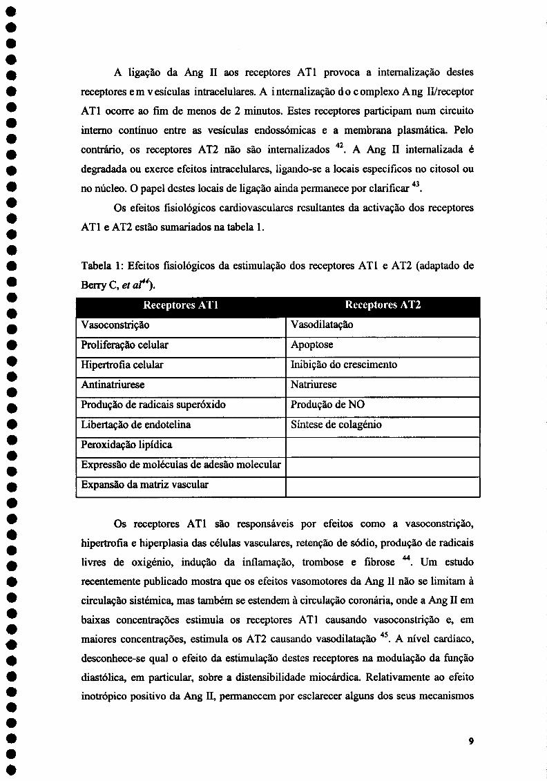
Os efeitos fisiológicos cardiovasculares resultantes da activação dos receptores ATI e AT2 estão sumariados na tabela 1.
Tabela 1: Efeitos fisiológicos da estimulação dos receptores ATI e AT2 (adaptado de Berry C, et ai44).
Receptores ATI Receptores AT2
Vasoconstrição Vasodilatação
Proliferação celular Apoptose
Hipertrofia celular Inibição do crescimento
Antinatriurese Natriurese
Produção de radicais superóxido Produção de NO
Libertação de endotelina Síntese de colagénio
Peroxidação lipídica
Expressão de moléculas de adesão molecular
Expansão da matriz vascular
Os receptores ATI são responsáveis por efeitos como a vasoconstrição, hipertrofia e hiperplasia das células vasculares, retenção de sódio, produção de radicais livres de oxigénio, indução da inflamação, trombose e fibrose 44. Um estudo recentemente publicado mostra que os efeitos vasomotores da Ang II não se limitam à circulação sistémica, mas também se estendem à circulação coronária, onde a Ang II em baixas concentrações estimula os receptores ATI causando vasoconstrição e, em maiores concentrações, estimula os AT2 causando vasodilatação 4S. A nível cardíaco, desconhece-se qual o efeito da estimulação destes receptores na modulação da função diastólica, em particular, sobre a distensibilidade miocárdica. Relativamente ao efeito inotrópico positivo da Ang II, permanecem por esclarecer alguns dos seus mecanismos
9

subjacentes, nomeadamente que diz respeito à interacção aguda com o endotélio endocárdico e com outros sistemas neurohumorais, como a endotelina-1.
A Ang II estimula igualmente a secreção do mineralocorticóide aldosterona pelo córtex suprarrenal. Esta hormona promove a reabsorção de sódio nos segmentos distais do nefrónio bem como no cólon e nas glândulas salivares e sudoríparas.46 Para além destas acções, e tal como a Ang II, a aldosterona parece estar envolvida na reparação e inflamação após a lesão tecidular. A aldosterona é produzida não só a nível suprarrenal, mas também a nível local cardíaco 47. Embora a discussão em torno do papel deste mineralocorticóide na físiopatologia das doenças cardiovasculares ultrapasse o âmbito deste trabalho, não se pode deixar de referir que a aldosterona parece estar envolvida na na proliferação celular, na promoção da fibrose cardíaca e na remodelagem coronária e renovascular 4Ó. Este papel é reforçado pelos estudos clínicos mais recentes (vide infra).
A activação do receptor ATI pela Ang II depende da interacção do resíduo Tyr4 do agonista e do Asnm localizado no terceiro domínio transmembranar do receptor 48. A Ang II recruta uma complexa cascata de mensageiros intracelulares com diferentes padrões de activação temporal, podendo observar-se efeitos imediatos (em segundos), precoces (em minutos) e tardios (em horas) 9 (Figura 2). A fosforílação da fosfolípase C (PLC) e a activação do Src ocorrem ao fim de segundos e são eventos de sinalização imediatos. A activação da fosfolípase A2 (PLA2), da fosfolípase D (PLD), das cínases de tirosina e das cínases de proteínas activadas por mitogénios (MAPK) ocorre em minutos, correspondendo a fenómenos precoces. Finalmente, a génese de radicais livres, a expressão de proto-oncogenes e a síntese proteica ocorrem ao fim de algumas horas, constituindo fenómenos tardios 9.
10

Angiotensina II
I
■#■ MAPK —► MAPK-p
ERKl/2,JNK,p38 I
Sinalização Nuclear
I Síntese proteica
H,0: 2^2
Crescimento celular Produção de factores
Formação da matriz de crescimento extracelular
Sequência temporal
Figura 2: Sequência temporal da cascara de sinalização intracelular resultante da ligação da Angiotensina II ao receptor ATI (adaptado de Touys RM, 2000)
11

Fenómenos imediatos A nível vascular, a Ang II induz contracção das células musculares lisas ao fim de
alguns segundos. Este efeito imediato pode ser mediado pela activação de várias vias de sinalização intracelular :
1. Activação da PLC : Um dos fenómenos mais rápidos resultante da estimulação dos receptores ATI nas células musculares lisas consiste na hidrólise pela PLC do fosfatilinositol-4,5-bifosfato em inositol trifosfato (IP3) e diacilglicerol (DAG). A PLC é codificada por uma família de três genes relacionados: PLC-P, PLC-y e PLC-ô49. A isoforma PLC-P é regulada pela subunidade a e Py da proteína G, enquanto a isoforma PLC-y é regulada pela fosforílação de resíduos de tirosina 49. Quando activado, o receptor ATI liga-se à PLC-P 1 através da proteína Goq/nPy e Ga^Py e à PLC-y através de uma cínase de tirosina. A PLC-P 1 parece ser importante para a génese rápida de IP3 (ao fim de 15 segundos) enquanto a PLC-y tem um papel na formação de IP3 numa fase mais tardia50. A Ang II induz uma elevação rápida e transitória do IP3 e uma elevação bifásica e sustendada do DAG 51. O IP3 estimula a libertação de cálcio a partir do retículo sarcoplasmático e o DAG activa a proteína c ínase C (PKC). Estes fenómenos relacionam-se temporalmente com o início da contracção das células musculares lisas vasculares, constituindo as vias que antecedem a fosforílação, dependente do cálcio, pela calmodulina da cadeia leve de miosina, que acaba por conduzir à contracção muscular 9.
2. Aumento do cálcio livre citosólico: A Ang II induz uma elevação bifásica d a concentração intracelular de cálcio: uma fase inicial transitória e uma fase tardia sustentada . A primeira fase depende da mobilização do cálcio intracelular provocada pelo IP3 e em menor grau da libertação de cálcio induzida pelo cálcio . A segunda fase, que contribui para a vasoconstrição sustentada induzida pela Ang II, é dependente do cálcio extracelular e resulta do influxo transmembranar de cálcio . O mecanismo exacto deste influxo não é totalmente claro mas parece envolver canais de cálcio dependentes da voltagem, canais não-dihidropirídínicos não específicos, canais de cálcio ligados a receptores, canais de cálcio activados pelo cálcio ou ainda o trocador Na+/Ca2+ 52.
12

3. Activação da proteína cínase C: O DAG activa a PKC a qual é uma cínase de serina e treonina. A Ang II estimula a ssim a t ranslocaçâo d a P KC c itosólica p ara a m embrana p lasmática onde a enzima activada fosforila proteínas específicas relacionadas com a função vascular 54. A PKC está envolvida nos mecanismos de vasoconstrição dependentes da Ang II bem como no crescimento e proliferação das células musculares lisas vasculares. Estes efeitos são mediados pela activação do trocador Na+/H+, conduzindo a alcalinização citosólica, pela fosforilação de diversas cínases de tirosina e pela activação da v ia de sinalização das M APK 55,56. Para além da sua função de sinalização, a PKC parece também estar implicada na dessensibilização rápida do receptor ATI após a ligação do seu agonista57.
4. Alcalinização citosólica: A Ang II induz uma alteração bifásica do pH intracelular, com uma acidificação transitória inicial seguida por uma alcalinização sustentada . O primeiro fenómeno deve-se a uma mobilização de cálcio regulada por uma Ca2+-ATPase, enquanto o segundo depende da activação do trocador Na+/H+ por mecanismos dependentes e independentes da PKC 55. A activação deste trocador induz alcalinização citosólica e vasoconstrição ao permitir uma elevação das concentrações intracitosólicas de Ca2+ e Na+ e aumentar a sensibilidade dos mio filamentos para o Ca2+ . Para além disso esta alcalinização é um estímulo potente para a síntese de DNA 9.
5. Alterações da concentração intracelular de Na+ e Mg2+: Para além de aumentar a concentração citosólica de Ca e o pH intracelular, a Ang II aumenta a [Na+]i e diminui a [Mg2+]j52.
6. Activação da família de cínases Src: Esta família de cínases de tirosina interage com receptores transmembranares com actividade de cínase de tirosina, mas também com receptores ligados à proteína G, como o receptor ATI. A Ang II promove uma rápida fosforilação da proteína S rc ( ao fim d e 6 Os) q ue, p or s ua v ez, i nduz fosforilação d a P LC-y e formação de IP3 9. Para além disso, a cínase Src fosforila outras proteínas como a p 13 0cas ( molécula d e sinalização e nvolvida n a a desão c elular m ediada p elas integrinas), as ERK (cínases reguladas por sinais extracelulares) e outras proteínas envolvidas na sinalização e proliferação celular .
13

Fenómenos precoces Para além dos efeitos rápidos relacionados com a contracção muscular, o receptor
ATI está acoplado a vias de transdução do sinal que regulam a função muscular a mais longo prazo, como o crescimento celular, migração, deposição da matriz extracelular e produção de factores de crescimento. Estes efeitos dependem de:
1. Activação de cínases de tirosina A Ang II é responsável pela fosforilação de um resíduo de tirosina em diversas proteínas intracelulares incluindo o próprio receptor ATI, cínases da família Src (activadas em segundos) e proteínas JAK, TYK, FAK, Pyk2, pl30Cas e PI3K, activadas em minutos 44. A família de cínases Src parece ter um papel importante na fosforilação da PLC-y, na formação de IP3 e na mobilização do cálcio intracelular, dependentes da Ang II. As proteínas Src, o cálcio intracelular e a proteína cínase C, por sua vez, regulam a fosforilação da proteína pl30Cas, uma molécula de sinalização envolvida na adesão celular dependente das integrinas 58. As proteínas Src estão ainda envolvidas na activação de PYK2 e na fosforilação de cínases reguladas por sinais extracelulares (ERK), bem como na activação de outras proteínas sinalizadoras (ppl20, pl25FAK, paxilina, JAK2 STAT1, caveolina, etc.)44. Tal como os receptores clássicos das citocinas, o receptor ATI fosforila proteínas da família das cínases Janus (JAK1, JAK2, JAK3 e Tyk2). As proteínas JAK são mediadores centrais na expressão do RNAm, fosforílando proteínas STAT, as quais são translocadas para o núcleo, onde activam a transcrição de genes de resposta precoce envolvidos na proliferação, remodelagem e reparação celular59. A activação pela Ang II de cínases de adesão focal (FAK) promove a migração celular e induz alterações na forma e volume celulares 9. A FAK é abundante em vasos sanguíneos em desenvolvimento e a sua activação pela Ang II provoca a sua translocação para locais de adesão focal com a matriz extracelular e a fosforilação das proteínas paxilina e talina, as quais estão envolvidas na regulação da morfologia e no movimento celular 60. A proteína Pyk2, um membro da família FAK, pode constituir um elemento comum às vias dependentes do cálcio e às activadas pela fosforilação de resíduos de tirosina 9.
14
A proteína pi30 é uma cínase de tirosina activada pela Ang II com um papel fundamental na adesão celular mediada pelas integrínas e na reorganização do citosqueleto (através do recrutamento de proteínas, como as FAK ou a paxilina, para adesões focais) 9. A sua acção parece ser fulcral no desenvolvimento cardíaco e vascular e na romodelagem cardiovascular em determinados estados patológicos61. Finalmente, a família das fosfatidilinositol 3-cínases (PI3K) influencia a sobrevivência celular, o metabolismo, a reorganização do citosqueleto e a proliferação 62. Embora o papel desta família de proteínas nas acções da Ang II ainda não esteja perfeitamente estabelecido, as vias complexas activadas pelas PDK podem controlar o balanço entre a mitogénese e a apoptose 9.
2. Activação de MAPK
As cínases de proteínas activadas por mitogénios (MAPK) constituem uma família de cínases de serina e treonina que medeiam a transdução de sinais extracelulares, conduzindo à activação de factores de transcrição, aumento da expressão génica (de genes de proteínas do ciclo celular, por exemplo) e respostas tróficas 44. Estão divididas em seis sub famílias: ERK1/2, JNK/SAPK, p38, ERK6, ERK3 e ERK5.
A cascata de sinalização das MAPK é iniciada nos miócitos pela activação de receptores ligados à proteína G (Ang II, endotelina-1, noradrenalina), por cínases de tirosina (IGF-1, TGF-fJ) ou pelo stress 63. Esta cascata é formada por três componentes activados sequencialmente (MAPKKK, MAPKK, MAPK). A Ang II activa três membros desta família: ERKs, JNK e p38 M. As vias de sinalização dependentes das MAPK aumentam a expressão dos genes de resposta rápida c-fos, c-myc e c-jun e estão envolvidas na proliferação celular, apoptose, diferenciação e transformação celular e ainda na contracção vascular . Estão implicados na fisiopatologia de doenças cardiovasculares, nomeadamente na hipertrofia e na insuficiência cardíaca. A via envolvendo a p38 parece estar particularmente implicada na lesão após a isquemia-reperfusão, na hipertrofia cardíaca e na progressão para a aterosclerose 65. A via que envolve a ERK1/2 parece ter um papel regulador central na sinalização do miócito cardíaco dada a sua capacidade única para responder a praticamente todos os estímulos hipertróficos e d e stress e a s ua c apacidade p ara p romover a p roliferação d os miócitos quer in vitro quer in vivo 64.
15

3. Activação da fosfolipase A2 (PLA2) e do metabolismo do ácido araquidónico (AA) A Ang II estimula a actividade da PLA2, a qual é responsável pela formação de AA a partir dos fosfolípidos da membrana. O AA formado é processado por diversas cicloxigenases, lipoxigenases e oxigenases do P450 em vários eicosanóides 9. Os eicosanóides derivados da PLA2 influenciam mecanismos renais e vasculares importantes na regulação da pressão arterial, como a contracção e proliferação vascular, envolvendo a activação de MAPK e radicais livres 44. Os tromboxanos estão envolvidos na contracção induzida pela Ang II, enquanto as prostaglandinas vasorelaxantes (PGE2 e PGI2) atenuam a vasocontricção dependente da Ang II. Nas células musculares lisas vasculares e nas células endoteliais, estes efeitos são mediados pelos receptores ATI, enquanto nos miócitos cardíacos neonatais de rato, neurónios e células epiteliais do tubo contornado proximal dependem da activação dos receptores AT2 66.
4. Activação da fosfolipase D (PLD) A P LD h idrolisa fosfolípidos dam embrana ( nomeadamente a fosfatidilcolina) levando à formação de ácido fosfatídico, um componente fundamental na sinalização celular associada à mitogénese. A activação da PLD conduz à formação sustentada de s egundos mensageiros, como DAG 9. Ao contrário da activação da PLC que ocorre ao fim de alguns segundos, a activação da PLD é apenas detectável ao fim de 2 minutos e permanece elevada por mais de 60 minutos 67. O DAG formado contribui para a activação prolongada da PKC. Mais uma vez, estas vias estão implicadas na hipertrofia cardíaca e na proliferação das células musculares lisas 9.
5. Modulação de nucleotídeos cíclicos Os nucleotídeos cíclicos GMPc e AMPc são formados ao fim de alguns minutos após a activação do receptor pela adenilcíclase e pela guanilcíclase. Os alvos destes nucleotídeos incluem cínases dependentes do AMPc, cínases dependentes do GMPc e canais iónicos 44. A Ang II causa vasodilatação directamente, aumentado a concentração intracelular de AMPc e GMPc, e indirectamente pela libertação de factores vasodiladatores. A Ang II leva à formação de AMPc e GMPc em cardiomiócitos, células musculares lisas vasculares e células mesangiais. Estes efeitos envolvem mecanismos dependentes da formação de cininas pelos receptores AT2 68. Nas artérias carotídeas de rato, a Ang II
16

aumenta a libertação de NO e a produção de GMPc pela activação de receptores
AT2 endoteliais 9'69.
Fenómenos tardios A Ang II influencia o controlo a longo prazo do crescimento, proliferação,
adesão, e migração celulares e da deposição da matriz extracelular a nível vascular e cardíaco. Desta forma é capaz de influenciar as adaptações crónicas envolvidas na remodelagem vascular, na aterosclerose e na hipertrofia cardíaca. As cascatas intracelulares envolvidas nos efeitos tardios da Ang II incluem a activação precoce de várias cínases (referidas acima) que fosforilam alvos a jusante os quais regulam as acções crónicas e sustentadas. Destes alvos fazem parte vias activadas pela génese de radicais livres de oxigénio, a expressão de proto-oncogenes, o cross-talk com receptores de cínases de tirosina, a produção de outros factores de crescimento e a estimulação de cascatas de sinalização nuclear que induzem crescimento e proliferação celular 9.
A síntese de radicais livres de oxigénio é regulada por várias citocinas e factores de crescimento, incluindo a Ang II. Esta aumenta a produção de O2" e H2O2 a nível cardíaco, muscular liso, endotelial, e mesangial 9. Curiosamente, a génese de radicais livres de oxigénio parece estar envolvida na etiopatogenia da hipertensão induzida pela Ang II, mas não pelas catecolaminas. Os mecanismos subjacentes à hipertensão incluem a degradação do NO endotelial e os potentes efeitos mitogénicos destes radicais livres70,71.
A Ang II induz a expressão de vários proto-oncogenes como o c-fos, c-jun, c-myc, erg-1, VL-30, entre outros 9. A estimulação de genes de resposta rápida pela Ang II está associada com a indução da expressão de factores de crescimento, como o PDGF, EGF, TGF-p, IGF-1, bFGF, PAF, de factores vasoconstritores, como a endotelina-1, de moléculas de adesão, como a ICAM-1, VCAM-1, E-selectina e integrinas, e de factores quimiotácticos, como o TNF-cc ou o MCP-1 9>72,73>74.
A Ang II é também capaz de influenciar a proliferação celular através da regulação da apoptose. Desta forma, inibe ou estimula a apoptose através dos receptores ATI ou AT2, respectivamente, em células musculares lisas, cardiomiócitos, células endoteliais e fibroblastos 80. Os receptores ATI podem também promover a apoptose. De facto, sabe-se que, após o enfarte agudo do miocárdio, o estiramento promove a libertação de Ang II e activação d os receptores ATI 75. Esta activação, por sua vez,
17

aumenta a expressão de bax e a activação da caspase 3 e diminui a expressão de bcl-2 . A diminuição da razão bcl-2/bax toma os cardiomiócitos mais susceptíveis aos sinais de apoptose. A formação do IP3 e DAG mediada pelos receptores ATI, ao activar a DNase tipo I e o stress oxidativo, contribui também para este fenómeno75.
Finalmente, a Ang II pela activação dos receptores ATI é também capaz de promover a deposição de colagénio do tipo I e III, contribuindo para a fibrose cardíaca que acompanha por exemplo a hipertensão arterial77.
Vias activadas pelo Receptor AT2 Embora a maioria dos efeitos fisiológicos da Ang II pareça depender dos
receptores ATI, a estimulação dos receptores AT2 pode ter um papel fisiológico na regulação da pressão arterial e da função renal, contrariando os efeitos vasoconstritor e antinatriurético dos receptores ATI. Como já foi referido anteriormente, a estimulação destes receptores é responsável por um efeito vasodilatador periférico e coronário. Contudo, o seu papel na modulação da contractilidade cardíaca aguda ainda não é conhecido. Este ponto será alvo da nossa atenção durante o presente trabalho.
Como já referido anteriormente, os estudos em ratos knockout para o gene do receptor ATI mostraram que o receptor AT2 pode ter um papel na modulação da resposta vasoconstritora da Ang II 38. Os efeitos vasodilatadores, anti-proliferativos e pró-apoptóticos mostram que muitas das acções dos receptores AT2 são opostas às do receptor ATI. Assim, em alguns modelos experimentais, a redução das dimensões do enfarte miocárdico pelos antagonistas ATI depende de uma cascata de sinalização
78
envolvendo a activação de receptores AT2, abradicininae asprostaglandinas .De forma semelhante, a estimulação dos receptores AT2 em corações insuficientes de hamsters contraria a progressão da fibrose intersticial induzida pelos receptores ATI79. Alguns autores defendem que este antagonismo pode dever-se à activação pelo receptor AT2 de fosfatases proteicas 80 ou à heterodimerização dos receptores AT1/AT281.
Embora os efeitos da estimulação dos receptores AT2 estejam muitas vezes associados a acções benéficas e cardioprotectoras, como a atenuação da hipertrofia ou fibrose cardíaca, algumas acções podem ser maléficas. De facto, o receptor AT2 parece estar envolvido na disfunção contráctil após a isquemia-reperfusão cardíaca e na hipertrofia e remodelagem vascular 82'83. Estas acções sublinham a complexidade das acções da Ang II, que dependem muitas vezes da espécie animal considerada e do contexto envolvente12. Curiosamente, algumas vias de sinalização intracelular dos
18

receptores ATI e AT2 podem ser comuns. De facto, ambos activam a via do factor NF-kB, o qual está envolvido no crescimento celular, fibrose ou apoptose, dependendo do contexto envolvente84. Os efeitos maléficos dos receptores AT2 na isquemia-reperfusão podem dever-se à formação de IP3 e à activação da fosfolípase C, fenómenos envolvidos na sinalização ATI 85. Contudo, os dados existentes não permitem afirmar inequivocamente a hipótese de uma contra-regulação dos receptores ATI pelos AT2 durante o desenvolvimento cardíaco ou a evolução da hipertrofia e insuficiência cardíacas 86. De facto, os dados de vários modelos experimentais são ainda contraditórios e em ensaios clínicos, como o ELITE II ou o Val-HeFT, o efeito benéfico dos fármacos pode ser explicado sem recorrer ao papel protector dos receptores AT2 no miocárdio insuficiente86.
As vias de sinalização activadas pelo receptor AT2 não são ainda totalmente conhecidas. Parecem envolver vias dependentes e independentes da proteína G.
Estudos recentes mostram que o receptor AT2 está acoplado a uma proteína Gi. A sua activação resulta em vários efeitos como a estimulação de correntes de potássio ou a inibição de canais de cálcio tipo T . A estimulação dos receptores AT2 está também associada à génese de bradicinina, NO e GMPc quer a nível vascular, quer a nível cardíaco. Os efeitos vasodilatadores e inibidores da fibrose perivascular dos receptores AT2 parecem depender da síntese e libertação destes mediadores 44,87.
Os efeitos inibidores da proliferação celular parecem estar dependentes da activação de fosfatases de tirosina, as quais inibem as MAPK activadas pelo receptor
O/A
ATI ou por factores de crescimento .
Interacção da Ang II com outros sistemas neuro-humorais Na hipertrofia cardíaca resultante da sobrecarga de pressão são activados
diversos sistemas humorais, autócrínos e parácrínos. As interacções entre estes sistemas criam vias, até certo ponto, redundantes, de tal forma que se um destes sistemas (Ang II, endotelina-1, citocinas) for anulado, a resposta não desaparece totalmente 88. Por exemplo, em miócitos de ratos knockout para o gene do angiotensinogénio, a activação da MAPK em resposta ao estiramento depende da activação da via envolvendo o gpl30, enquanto nos ratos wild type essa activação depende da estimulação dos receptores ATI 88
19
Os sistemas renina-angiotensina e endotelina-1 constituem duas vias vasoconstritoras com diversos locais de potencial interacção recíproca .
Muitas das acções resultantes da activação dos receptores ATI são semelhantes às observadas com a endotelina-1. Existem dois subtipos principais de receptores da endotelina-1. Os receptores ETA estão associados a um efeito inotrópico positivo, vasoconstritor e promotor da proliferação celular, enquanto os receptores ETB causam um efeito inotrópico negativo e vasodilatador dependente do endotélio 90.
Estes dois sistemas têm múltiplos pontos de contacto quer a nível funcional, quer a nível das vias subcelulares activadas. Desta forma, sabe-se que a hipertensão e a vasoconstrição renal produzidas pelas administração crónica de Ang II podem ser, em larga medida, atenuadas pela inibição dos receptores da endotelina-1 91'92. A endotelina-
I está também envolvida na resposta ííbrótica vascular normalmente atribuída à Ang II 93. A endotelina-1 endógena pode ainda contribuir para a promoção da resposta
O^
hipertrófica da Ang II . Curiosamente, embora a síntese de endotelina-1 dependa em grande medida da acção da enzima conversora da endotelina tipo 1 (ECE-1), a big-endotelina pode ser clivada igualmente pela químase, a principal responsável pela conversão da Ang I em Ang II a nível miocárdico 94.
Em termos agudos, existe também uma interacção entre os dois sistemas. De facto, a endotelina-1, através da activação de receptores ETA, contribui para o efeito vasopressor, vasoconstritor e natriurético agudo de doses baixas de angiotensina II, quer em modelos animais, quer em voluntários humanos saudáveis 95,%. Os mecanismos subjacentes a esta interacção aguda ainda não são totalmente conhecidos. Embora a Ang II aumente a transcrição do gene da pré-pro-endotelina-1, através da sua ligação aos receptores ATI e da activação da PKC, e desta forma estimule a síntese e libertação de endotelina-1 97, este efeito parece demorar várias horas. Um mecanismo mais precoce, que pode explicar o efeito observado, consiste na libertação de endotelina-1 pré-formada e armazenada em células endoteliais ou vasculares em cultura induzida pela Ang II 9S. Este mecanismo pode estar presente mesmo sem se detectar um aumento significativo dos níveis plasmáticos de endotelina-1, na medida em que reflecte um aumento da concentração tecidual pela libertação preferencial do peptídeo na membrana basolateral das células endoteliais 98. Para além disso, sabe-se que o bloqueio do receptor ETA pode amplificar as acções do receptor ETB, resultando num aumento da produção de óxido nítrico, o que diminui quer o efeito vasoconstritor quer inotrópico positivo originado pela angiotensina II 90. Finalmente, como os dois sistemas possuem
20

vias intra-celulares comuns, a endotelina-1 pode funcionar como um "primer" na cascata de sinalização da angiotensina II95.
A nível cardíaco, é ainda desconhecido qual o papel da interacção entre estes dois sistemas na modulação aguda da contractilidade miocárdica. Este ponto será analisado posteriormente no presente trabalho.
Papel do Sistema Renina-Angiotensina na fisiopatologia de algumas doenças cardiovasculares
Os ensaios clínicos com inibidores da enzima conversora da angiotensina (IECA) e antagonistas dos receptores ATI (ARA) mostram uma melhoria da morbilidade e mortalidade na hipertensão arterial, i nsuficiência c ardíaca c ongestiva e enfarte agudo do miocárdio, o que suporta a evidência do papel central da Ang II na patogénese das doenças cardiovasculares.
Relativamente à hipertensão arterial, os estudos experimentais e clínicos mostram que os IECA e os ARA não só diminuem a pressão arterial, mas também diminuem a remodelagem cardíaca e vascular, melhoram a função endotelial e normalizam os mecanismos que regulam os segundos mensageiros intracelulares 9. Entre os vários peptídeos implicados na hipersensibilidade vascular presente na hipertensão arterial (como a endotelina-1, a vasopressina ou a noradrenalina), a Ang II é aquele que está associado de uma forma mais forte e consistente com esta propriedade OQ
. Os mecanismos de sinalização da Ang II parecem estar super-activados na hipertensão arterial. Algumas alterações que parecem ser responsáveis por este fenómeno incluem a interacção entre o receptor ATI e a proteína G, aumento da fosforilação do receptor ATI ou desregulação de segundos mensageiros 9.
O papel da Ang II na patogénese da hipertensão arterial é ainda reforçado por estudos que mostram que ratos transgénicos com sobrexpressão de gene do angiotensinogcnio desenvolvem hipertensão enquanto ratos knockout para o mesmo gene desenvolvem hipotensão 10°.
Os estudos de polimorfismos em populações humanas mostram também que determinados polimorfismos do gene do angiotensinogénio (alelo AGT T235), da enzima conversora da angiotensina (genótipo DD) ou do receptor ATI (polimorfismo A1166C) estão relacionados o desenvolvimento de hipertensão arterial e maior risco cardiovascular. Em particular, o genótipo DD da ECA está relacionado com maior risco
21

de enfarte agudo do miocárdio, hipertrofia ventricular esquerda e carotídea, reestenose após angioplastia das artérias coronárias e redução da resposta do índice de massa do ventrículo esquerdo ao enalapril101.
Sabe-se que a Ang II actua localmente a nível cardíaco influenciando a síntese proteica e a proliferação celular. Em corações humanos de doentes com insuficiência cardíaca, a progressão da doença está relacionada com um aumento progressivo da formação de Ang II a nível cardíaco, independentemente da etiologia da insuficiência cardíaca 102. O receptor ATI está particularmente envolvido no desenvolvimento da hipertrofia cardíaca. De facto, os IECA e os antagonistas do receptor ATI previnem o desenvolvimento da hipertrofia cardíaca em modelos animais e promovem a sua regressão em doentes hipertensos 10. Estes fármacos previnem ainda a dilatação ventricular após o enfarte do miocárdio ao inibirem a expressão de genes associados com a remodelagem cardíaca. Ratos transgénicos com sobrexpressão miocitária do receptor ATI desenvolvem espontaneamente remodelagem e hipertrofia cardíaca, com aumento da expressão de peptídeo natriurético auricular e da deposição intersticial de colagénio. Estes animais não apresentam alterações significativas da frequência cardíaca ou da pressão arterial e morrem precocemente por insuficiência cardíaca . Apesar dos efeitos da Ang II estarem classicamente ligados à estimulação dos receptores ATI, os receptores AT2 parecem ter igualmente um papel fundamental nesta resposta.
A expressão de receptores AT2 aumenta em paralelo com a progressão da fibrose intersticial, da remodelagem cardíaca e da elevação das pressões de enchimento intracardíacas 104. Este receptor parece ter um efeito inibitório na síntese, mediada pelo receptor ATI, de colagénio, fibronectina e cínases proteicas activadas por mitogénios, inibindo a progressão da fibrose intersticial e da hipertrofia miocárdica após lesões miocárdicas 104. Estes e outros dados fazem supor um papel protector para os receptores AT2, até certo ponto oposto ao dos receptores ATI. Contudo, o contexto específico da activação destes receptores pode influenciar significativamente a resposta observada ' . De facto, num modelo animal de ratos knockout para o gene do receptor AT2, a infusão crónica de Ang II não provoca hipertrofia cardíaca, nem comprometimento do relaxamento ventricular ou fibrose significativa, ao contrário dos ratos wild type. Estes dados indicam que a ausência dos receptores AT2 suprime a reprogramação genética da Ang II no sentido da síntese da matriz extracelular e da fibrose, mostrando que a estimulação simultânea dos dois tipos de receptores é necessária para a activação da via sinalizadora da hipertrofia e fibrose induzidas pela Ang II 106. As diferenças entre este
22

estudo e outros que usam ligandos dos receptores mostram que estes ligandos exercem efeitos dependentes do contexto específico que não podem ser classificados de forma simplista como "bons ou maus". De facto, em doentes com enfarte do miocárdio o aumento da expressão dos receptores AT2 pode ser benéfico numa fase inicial, opondo-se à hipertrofia e à fibrose e libertando NO. Contudo, a estimulação crónica destes receptores miocárdicos tem efeitos cumulativos deletérios 105.
Dados clínicos mostram que doentes com diabetes mellitus apresentam frequentemente disfunção miocárdica, mesma na ausência de doença coronária. Parece, portanto, existir uma cardiomiopatia diabética primária . Estudos em modelos experimentais de diabetes mostram que a hiperglicemia aumenta a expressão de RNAm e a densidade dos receptores ATI a nível cardíaco e que os IECA previnem o aumento da expressão de RNAm de TGF-pi no ventrículo esquerdo destes animais 107.
As doenças glomerulares renais, causadas pela hipertensão arterial, diabetes ou inflamação, caracterizam-se por proliferação mesangial e acumulação excessiva de proteínas da matriz extracelular. A Ang II tem um papel central não apenas na regulação da hemodinâmica glomerular e da taxa de filtração glomerular, mas também na hipertrofia e proliferação das células mesangiais, contribuindo desta forma para a progressão da glomerulosclerose 12. A glomerulosclerose dependente da Ang II parece estar particularmente ligada à formação e libertação de TGF-01 108.
Para finalizar, embora a Ang II seja considerada o principal efector do sistema renina-angiotensina, alguns metabolitos da degradação da angiotensina, como a Ang III, Ang IV e Ang (1-7) possuem actividade biológica e podem ter um papel relevante em algumas patologias. Assim, sabe-se que a Ang III pode regular a proliferação celular e a acumulação da matriz extracelular, nomeadamente através da activação da via do NF-kB pelos receptores AT2 ". Nas placas de aterosclerose existe uma maior expressão do receptor AT-4, sugerindo um papel promotor da proliferação celular para a Ang IV n .
Perspectivas futuras sobre o sistema renina angiotensina aldosterona
O receptor ATI tem a capacidade de recrutar inúmeras cascatas de sinalização intracelular altamente complexas. As abordagens experimentais convencionais que tentam explorar esta complexidade incidem fundamentalmente sobre o estudo de genes candidatos 109. As tecnologias de DNA microarrays e proteomics constituem uma nova
23

abordagem para o estudo destas vias. Recentemente, um estudo usando a técnica de DNA microarrays identificou cerca de 90 genes regulados de forma significativa pela Ang II. Esta lista inclui genes que actualmente já estão relacionados com as cascatas dependentes da Ang II, como a ERK ou a p38. Mas muitos são genes ainda não relacionados com este mediador, como a calpactina I de cadeia leve ou a calpactina II (envolvidas na remodelagem da matriz extracelular), ou mesmo sequências cuja identidade ainda nem sequer é conhecida uo.
24

Objectivos
Tendo em conta os pontos deixados em aberto durante a introdução anterior, o presente trabalho teve como principal objectivo o estudo da modulação aguda da função miocárdica pela Angiotensina II, num modelo de preparações multicelulares de miocárdio de coelho. Neste sentido, pretendeu-se avaliar o efeito da Angiotensina II sobre as propriedades sistólicas e diastólicas de corações de animais saudáveis e com insuficiência cardíaca. No que respeita à função sistólica, foi nosso objectivo estudar, em particular, a modulação do efeito inotrópico positivo da Ang II pelo endotélio endocárdico e pela endotelina-1, bem como o efeito da estimulação selectiva dos receptores AT2 sobre a contractilidade miocárdica. Relativamente à função diastólica, pretendeu-se avaliar o papel da Ang II na modulação aguda da distensibilidade miocárdica e os seus mecanismos subjacentes.
Finalmente, foi ainda nosso objectivo estudar o papel da Ang II na modulação aguda da função miocárdica, através da avaliação dos seus efeitos sobre a contractilidade e distensibilidade miocárdica num modelo animal de insuficiência cardíaca.
Protocolos experimentais De forma a concretizar os objectivos propostos procedemos à realização dos
seguintes Protocolos experimentais: 1. Estudo da modulação do efeito inotrópico da Angiotensina II pelo endotélio endocárdico e pela interacção com a Endotelina-1: adição de concentrações crescentes de Angiotensina II:
■ em condições basais; ■ após a destruição selectiva do endotélio endocárdico, pela imersão numa
solução de Triton X-100 0,5% durante 1 s; ■ na presença de um antagonista competitivo não selectivo dos receptores da
ET-1 (PD-145065); ■ na presença de um antagonista competitivo selectivo dos receptores ETA
(BQ-123).
25

2. Efeito da estimulação selectiva dos receptores AT2 sobre a contractilidade cardíaca e seus mecanismos subjecentes: adição de concentrações crescentes de: ■ H-9395, um agonista dos receptores AT2, em condições basais e na presença
de um antagonista selectivo dos receptores ATI (ZD-7155);
■ Ang II na presença de um antagonista dos receptores ATI (ZD-7155):
■ em condições basais; ■ após a destruição selectiva do endotélio endocárdico pela imersão
numa solução de Triton X-100 0,5% durante 1 s; ■ na presença de um inibidor da síntese de NO (NG-nitro-L-
arginina); ■ na presença de um inibidor da síntese de prostaglandins
(Indometacina); ■ na presença de um inibidor da síntese de EDHF (Proadifen).
3. Estudo do papel da Angiotensina II na modulação da distensibilidade miocárdica: adição de concentrações crescentes de Angiotensina II em condições basais e na presença de um:
■ antagonista dos receptores AT 1 (ZD-7155); ■ inibidor do trocador Na+/H+ (Amilorido); ■ inibidor da Proteína Cínase C (Cheleritrine).
4. Avaliação dos efeitos agudos da Angiotensina II sobre a contractilidade e sobre a distensibilidade miocárdica num modelo experimental de insuficiência cardíaca induzida pela administração de doxorrubicina.
26

Métodos
Nesta secção faz-se uma breve descrição da metodologia experimental. Em cada um dos trabalhos que compõem o corpo da presente tese é feita uma descrição mais detalhada.
A investigação foi efectuada de acordo com as regras do "Guide for the Care and Use of Laboratory Animals" publicado pelo National Institutes of Health (NIH publicação n° 85-23, revisão de 1996).
Os estudos foram levados a cabo em coelhos brancos neo-zelandeses machos, adultos (Oryctolagus cuniculus; 2,9±0,12 Kg). Os animais foram anestesiados com pentobarbital de sódio (25 mg/Kg, iv), submetidos a uma toracotomia esquerda e o coração foi excisado. Este foi, imediatamente, imerso numa solução oxigenada de Krebs-Ringer (35°C), procedendo-se de seguida à dissecção de trabéculas e/ou músculos papilares. A solução de Krebs-Ringer continha (em mM): NaCl 98; KC1 4,7; MgS04 2,4; KH2P04 1,2; CaCl21,8; NaHC03 20; CH3COONa 5; C3H303Na 15; glicose 4,5 e atenolol 0,02. O atenolol foi adicionado para prevenir os efeitos mediados pela estimulação P-adrenérgica .
Os músculos foram montados verticalmente em banho próprio (no mesmo tipo de solução em que o coração foi imerso) e oxigenados a partir de uma mistura gasosa de 95% O2 e 5% CO2. Os músculos foram depois conectados a um transdutor isotónico/isométrico (Universidade de Antuérpia), estimulados electricamente a uma frequência de 1 Hz e estabilizados a 35°C. Após cerca de duas horas de estabilização, procedeu-se ao início do protocolo experimental.
Os parâmetros relativos à função mecânica muscular foram quantificados em condições basais e durante diferentes protocolos experimentais.
27
Resultados
1- Efeito inotrópico positivo da Angiotensina II e sua modulação pelo Endotélio Endocárdico e pela Endotelina-1

ETA Receptors and Endothelium Partially Mediate the
Positive Inotropic Effect of Angiotensin II
Castro-Chaves P, Roncon-Albuquerque R Jr, Leite-Moreira AF
Department of Physiology, Faculty of Medicine, University of Porto, Porto, Portugal
Correspondence address:
Prof. Dr. Adelino Leite-Moreira
Department of Physiology, Faculty of Medicine
Alameda Prof. Hernâni Monteiro
4200-319 Porto, PORTUGAL Tel. +351-22.550.84.52; Fax. +351-22.551.91.94; E-mail: [email protected]
Supported by grants from FCT (PRAXIS/C/SAU/11301/98; partially funded by FEDER) and
Calouste Gulbenkian Foundation, through Cardiovascular R&D Unit (51/94-FCT, Portugal).

ABSTRACT
Renin-angiotensin and endothelin systems have multiple interactions both in the subcellular
pathways activated and in their physiological actions. It has been shown that a low dose of
angiotensin II (Ang-II) has a positive inotropic effect through the autocrine/paracrine release
of endothelin-1 (ET-1). In the present study we characterized the receptor subtype of ET-1
involved in this functional interaction and investigated its modulation by the endocardial
endothelium. Effects of Ang-II (10"9-10"5M) were evaluated in rabbit right papillary muscles
(1.8 mM Ca2+; 0.6 Hz; 35°C) in absence (Protocol A; n=ll) or presence of PD-145065
(nonselective ET receptor antagonist; 10"7 M; Protocol B, n=9) or BQ-123 (selective ETA
receptor antagonist; 10"7 M; Protocol C, n=7), as well as, after removing the endocardial
endothelium (EE) with Triton X-100 (0.5%, Is; Protocol D, n=8). In Protocol A Ang II had a
dose dependent positive inotropic effect, maximal at 10"6 M increasing: 122±13% AT,
117±16% dT/dtmax, 86±9% dT/dtmin, 12±2% tHR, 121±18% PS and 101±17% dL/dtmax. In
Protocols B, C and D the inotropic effect of Ang II was blunted. In Protocol B, 10"6 M of Ang
II increased 48±11% AT, 54±14% dT/dtmax, 39±8% dT/dtmin and 40±10% PS, without
altering tHR and dL/dtmax. Similarly, ETA receptor inhibition (Protocol C) attenuated Ang II
positive inotropic effect, increasing at 10"6 M: 59±27% AT, 54±20% dT/dtmax, 48±19%
dT/dtmin 83±43% PS, 72±32% dL/dtmax, without altering tHR. After EE removal (Protocol
D), 10"6 M of Ang II increased 72±16% AT, 32±9% dT/dtmax, 59±11% dT/dtmin, 95±24%
PS and 41±22% dL/dtmax, without altering tHR. When comparing EC50 and Emax for each
of the Protocols several differences were also found. In Protocol D, the selective removal of
endocardial endothelium maintained Emax but caused a significant decrease in EC50 when
comparing to Protocol A. In Protocols B and C, the presence of the endothelin receptor
antagonist decreased both EC50 and Emax. In conclusion, Ang II has a dose dependent
positive inotropic effect that depends, to a great extent, of ETA receptor activation and of the
EE. These results further characterize the functional interaction between Ang II and the ET
system in myocardium.
Keywords: Angiotensin II; Endocardial Endothelium; Endothelin-1; Inotropism; Myocardium.
30
INTRODUCTION Classically, the renin-angiotensin system (RAS) has been considered an endocrine axis
with a central role in blood pressure regulation, aldosterone release and sodium homeostasis.
However, later evidences have demonstrated the presence of local RAS with autocrine-
paracrine actions in several organs like the adrenal gland, blood vessels, central nervous
system, heart, kidney and sympathetic system1'2'3 In the heart, local RAS regulates myocardial
function, cell growth and extracellular matrix deposition 4'5'6. Chronic activation of local RAS
promotes hypertrophy and fibrosis being upregulated in ischemic heart disease and heart
failure2'7,8. These effects depend, at least in part, on the interaction of this system with other
autocrine/paracrine mediators, like endothelin-1 9 '10 'n. In fact, Ang II induces ET-1 gene
expression and release in cardiomyocytes and this interaction is essential for the cardiac
hypertrophic effects of Ang II912
It is well established that Ang II has a positive inotropic effect in several animal
species (rabbit, cat and pig) and human auricular myocardium 13'14. More recently, it was
described that ET-1 production/release is involved in the positive inotropic effect of a small
dose of Ang II15. Thus, there are growing evidences for an acute interaction between local
RAS and the endothelin system. However, it remains to be clarified if this cross-talk is
relevant for the dose-dependent positive inotropic effect of Ang II, which endothelin receptor
subtype is activated and if endothelial are involved in this cross-talk
In this context, we evaluated, in rabbit myocardial multicellular preparations, the dose
dependent positive inotropic effect of Ang II in the presence of ET receptor antagonists and
after selective removal of the endocardial endothelium.
31

MATERIALS AND METHODS The investigation conforms to the Guide for the Care and Use of Laboratory Animals
published by US National Institutes of Health (NIH Publication No 85-23, revised 1996).
Experimental preparation The effects of Ang II were studied in isolated right papillary muscles of New Zealand
White rabbits (Oryctolagus cuniculus; 2.9±0.12 Kg). Rabbits were anaesthetized with sodium
pentobarbital (25 mg/Kg, iv) and the heart was quickly excised. The tissues were immersed in
a modified Krebs-Ringer (KR) solution1617 at 35°C, with cardioplegic 2,3-butanedione
monoxime (BDM; 3%)18 and calf serum (5%; Bio-Whittaker, St. Louis, Maryland, USA). The
modified KR solutions contained (in tnM): NaCl 98, KC1 4.7, MgS04 2.4, KH2P041.2» CaCl2
1.8, NaHC03 20, CH3COONa 5, C3H303Na 15, glucose 4.5 and Atenolol 0.02. Atenolol was
used to prevent p-adrenergic mediated effects19.The solutions were in equilibrium with 95%
0 2 and 5% C02, maintaining the pH between 7.38-7.42. Rabbit papillary muscles (n=35;
length: 3.5+1.3 mm; weight: 2.5+1.6 mg; cross-sectional area: 0.7+0.4 mm2; preload: 5.0+1.2
mN) were then carefully dissected. Afterwards, they were vertically mounted in a 10 ml plexi
glass organ bath and connected to an isotonic/isometric transducer (University of Antwerp,
Belgium). Preload was estimated according to muscle dimensions and the electrical stimulus
(0.6 Hz) was set at 10% above threshold. Twenty minutes later, bathing solutions were
replaced by corresponding KR solutions without BDM. During the next 2 hours, muscles
were stabilized. Bathing solutions were then replaced by corresponding KR solutions without
calf serum and Lmax was calculated. Protocols were initiated after obtaining two similar
isotonic and isometric control twitches separated by a 10 min interval.
Experimental protocols In all experimental protocols, Ang II was added in increasing concentrations to the
bathing solutions (10"9, 10"8, 10~7, 10"6 and 10"5 M). In Protocol A (n=ll), the effect of Ang II
was studied in control muscles with intact endocardial endothelium (EE). In Protocol B (n=9),
the increasing concentrations of Ang II were added in the presence of PD-145065 (1 uM), a
nonselective antagonist of endothelin-1 receptors. In Protocol C (n=7) the effects of the same
concentrations of Ang II were studied in the presence of a selective antagonist of ET-1
receptor type A BQ-123 (0.1 uM). Finally, in Protocol D (n=8), the effect of Ang II was
studied in muscles whose EE had been previously removed. The selective removal of EE was
32

performed according to the methodology described by Brutsaert and collaborators Briefly,
this consisted in the immersion of the papillary muscles in a 0.5% solution of Triton X-100
during 1 second, followed by an abundant washout.
Ang II, PD-145065 and BQ-123 were purchased from Sigma Chemical Co, St Louis, Mo.
Data analysis
Isotonic and isometric twitches were recorded and analyzed with dedicated software
(University of Antwerp, Belgium). Selected parameters include: Active Tension (AT,
mN-mm"2); Maximum Velocity of Tension Rise (dT/dtmax, mN-mm"2-s'1); Maximum Velocity
of Tension Decline (dT/dtmin, mN-mm"2*'1); Peak Isotonic Shortening (PS, %Lmax); Maximum
Velocity of Shortening (dL/dtmax, Lmax-s"1); Maximum Velocity of Lengthening (dL/dtrai„,
Lmax-s"1), Time to Half Relaxation (tHR, ms); negative logarithm of the concentration required
to produce 50% of the maximal response (EC50); and maximum value attained in response to
agonists (Emax).
Statistical methods Values are means±SEM. In each protocol, concentration-response effects of Ang II
were analyzed with repeated-measures one-way ANOVA. Comparison between experimental
protocols of Ang II effects, expressed as percentage of baseline, were analyzed with repeated-
measures two-way ANOVA. When significant differences were detected, the Student-
Newman-Keuls test was selected to perform pairwise multiple comparisons. Regarding EC50
and Emax, the differences between experimental protocols were analyzed with one-way
ANOVA. PO.05 was accepted as significant.
33

RESULTS Baseline performance of rabbit papillary muscles was similar in all experimental
protocols. Mean values of the contractile parameters from the 35 papillary muscles were: AT
18.9±2.8 mN-min2; dT/dW 135.6±16.8 niN-mm2*"1; dT/dWn -110.7±14.4 m N m i n V 1 ;
PS 0.070±0.008 % o f U a x ; dL/dtmax 0.52±0.05 W - s " 1 ; dL/dUn -1.69±0.22 W » 1 ; tHR
333.0±13.8 ms. The presence of PD-145065 and the selective destruction of EE caused a
significant decrease of AT in 6.41±0.93% and 45.2±3.78%, respectively. BQ-123 didn't
change significantly basal muscle performance.
Protocol A: Myocardial Effects of Angiotensin II in Rabbit Papillary Muscles with intact
endocardial endothelium
Ang II in the presence of an intact endocardial endothelium had a concentration-
dependent positive inotropic effect that stabilized after -20 min, maximal at IO"6 M (Figure
1). This concentration of Ang II significantly increased: 122±13% AT, 117±16% dT/dtmax,
86±9% dT/dtmin, 12*2% tHR, 121±18%PS, 101±17% dL/dtmax and 158±3 dL/dtmin.
Protocols B & C: Myocardial effects of Angiotensin II in the presence of PD-145065 and BQ-
123
As stated above, the basal muscular performance decreased after the addition of PD-
145065 and was not significantly altered by BQ-123. In Protocol B, in the presence of a
nonselective endothelin receptor antagonist (PD-145065), Ang II maintained its positive
inotropic effect although it was significantly attenuated when compared with Protocol A
(Figure 2). The effect was again maximal for the concentration of IO"6 M, which increased:
48±11% AT, 54±14% dT/dtmax, 39±8% dT/dtmin, 40±10% PS and 54±13% dL/dtmin,
without altering tHR and dL/dtmax. This attenuation was also evident when Ang II was added
in the presence of a selective ETA receptor antagonist (BQ-123; Protocol C), as can be seen
in Figure 2. The maximal effect was again observed at the concentration of IO"6 M which
increased 59±27% AT, 54±20% dT/dtmax, 48±19 dT/dtmin, 83±43% PS, 72±32 dL/dtmax
without altering tHR.
Protocol D: Myocardial effects of Angiotensin II in Rabbit Papillary Muscles whose
Endocardial Endothelium was selectively removed
When compared with the results obtained in Protocol A, the positive inotropic effect
of Ang II after the selective removal of endocardial endothelium (Protocol D) was
34
significantly attenuated (Figure 2). The maximal effect was present at the concentration of 10" 6 M increasing: 72±16% AT, 32±9% dT/dtmax, 59±11% dT/dtmin, 95±24% PS and 41±22%
dL/dtmax without altering tHR and dL/dtmin.
The addition of Ang II in Protocol A induced a dose-dependent positive inotropic effect, maximal for the concentration of IO"6 M. This effect was significantly attenuated by the selective removal of endocardial endothelium and by the presence of PD-145065 and BQ-123, nonselective and selective antagonists of endothelin receptors, respectively. However, when comparing EC50 and Emax for each of the Protocols other differences were also found. Analyzing the parameter Active Tension, in Protocol D, the selective removal of endocardial endothelium maintained Emax (113.9±24.9 in Protocol D and 105.6±14.5 in Protocol A) but caused a significant decrease in EC50 when comparing to Protocol A as seen in Figure 1 (-6.83±0.33 and -7.8±0.12, respectively). In Protocols B and C, the presence of the endothelin receptor antagonist decreased both EC50 (-7.15±0.34; -6.68±0.34, respectively) and Emax (44.9±13.6; 51.3±19.3, respectively).
35

DISCUSSION In protocol A, Ang II had a dose-dependent positive inotropic effect. This is in
accordance with previous reports carried out in ventricular rabbit myocardium and is
attributed to ATI receptor subtype activation13. Similarly, studies performed in other
mammalian species, like cat and pig ventricular myocardium and human atrial trabeculae,
have shown an Ang II positive inotropic effect21,22. However the positive inotropic effect was
not observed in rat, ferret and human ventricular myocardium ' .
In protocol B, we confirmed the acute interaction between the local RAS and the
endothelin system. In fact, the presence of an nonselective endothelin receptor significantly
attenuated the positive inotropic effect of Ang II. This is in agreement with a recent work
performed in feline myocardium, where a small dose of Ang II increased inotropism by
endothelin release15. This appears to depend on the ET-1 induced activation of Na+/H+
exchanger, which increases intracellular [Na+] promoting Ca2+ influx through reverse mode
Na+/Ca2+ exchanger23. In our study, we demonstrated that this interaction is present
throughout the entire range of the analyzed concentration-response curve.
In order to further characterize the mechanisms of this acute local RAS-endothelin
system interaction we performed protocols C & D. In protocol C, the addition of Ang II in the
presence of a selective ETA receptor antagonist had an effect on the concentration-dependent
positive inotropic effect similar to that of the nonselective antagonist, suggesting that it is
mediated through ETA receptor activation. This is in accordance with several reports
documenting that ETA receptor subtype is responsible for the positive inotropic effect of ET-
l24.
In protocol D, we studied Ang II positive inotropic effect after the selective removal of
the endocardial endothelium. The destruction of the endocardial endothelium with a brief
immersion of the muscles in 0.5% Triton X-100 for 1 second has been shown by several
laboratories to damage the endocardial endothelium without damaging cardiac myocytes
Treatment with Triton X-100 disrupts the endocardial endothelium and removes about 80% of
the endothelial cell population of a trabeculae 25'26. It is well known that, in the normal heart,
ET-1 is mainly expressed in endothelial cells of the endocardium and the vasculature ' I t
is therefore reasonable to hypothesize that the endocardial endothelium could play a major
role in the acute interaction between local RAS and the endothelin system. This was
confirmed in protocol D, where the selective removal of the endocardial endothelium
attenuated Ang II positive inotropic effect.
36
As can be seen in Figure 2, the analysis of EC50 and Emax complements the
information obtained in a significant way. The selective removal of endocardial endothelium
maintained Emax but decreased significantly EC50 when compared with baseline, thus
causing a parallel rightward shift of the dose-response curve. In these conditions, the positive
inotropic effect of Ang II seems to be largely maintained, but depends on the concentration of
Ang II used, thus meaning a decrease in receptor affinity. This can be explained if the
endothelium secretes a cofactor, like endothelin-1, that is partially responsible for the effect of
Ang II in contractility. In Protocols B and D, the presence of a nonselective and a selective
antagonist of endothelin-1 receptors, respectively, decreased both Emax and EC50,
representing a rightward and downward shift of the dose-response curve and a lower receptor
affinity and efficacy.
The conclusions observed in the present work are in accordance with what is known to
happen in vascular tissues. In fact, in peripheral vascular beds, local RAS also interacts with
the endothelin system9. Chronically, Ang II induced ET-1 production and release causes
vascular hypertrophy, which is prevented in the presence of ETA receptor antagonists
Acutely, bosentan, a nonselective ETA/ETB receptor antagonist, and BQ-123 inhibit Ang II
induced hypertension and reduction in renal blood flow, both in animal models and in human
healthy volunteers27'28-29. The mechanisms underlying the acute interaction between these two
systems, both in vascular tissues and in the myocardium, still remain elusive. Ang II induces
ET-1 expression and release by endothelial 10'30 and vascular smooth muscle cells "' ,
contributing to the Ang II vasoconstrictive effect. Although Ang II stimulates ET-1 synthesis
in endothelial cells, several hours are required for this effect u. However, release of ET-1
takes place within minutes from in vitro vessels 32'33, implying that preformed ET-1 is stored
in cells and that this release may be Ang II dependent 28. This mechanism may be present
even without a significant increase in plasmatic level of ET-1, since it is released in the
basolateral membrane of endothelial cells, thus implying a paracrine mechanism that
influences mostly tecidual levels of ET-1 34. Alternatively, the blockade of ETA receptors
could amplify the effects of ETB receptors, resulting in vasodilating and negative inotropic
effect32,35. However, in the present study the attenuation of the positive inotropic effect was
present even in the presence of nonselective blockade of endothelin receptors.
Ang II is also able to increase cardiac contractile function by stimulating intrinsic
cardiac adrenergic neurons to release norepinephrine into the cardiac interstitium in vitro and
in vivo 36,37'38. This potential interaction was prevented in our experimental protocol by the
presence of atenolol in the bathing solutions.
37
Finally, our results could be relevant for the understanding of differences in the
inotropic effects of Ang II between species. This could be due to differences in acute local
RAS-endothelin system interactions 39, given that endothelin receptor expression varies 40,41
among species
In conclusion, in this animal species the positive inotropic effect of Ang II is partially
mediated by ETA receptor activation and the endocardial endothelium. These results may
contribute for a more complete understanding of the role of Ang II in acute modulation of
myocardial function.
ACKNOWLEDGEMENTS Supported by Portuguese grants from FCT (PRAXIS/SAU/11301/98; partially funded by FEDER)
and Calouste Gulbenkian Foundation, through Unidade I&D Cardiovascular (51/94-FCT). There
are no financial or other relations that could lead to a conflict of interest.
38

FIGURE LEGENDS Figure 1. Effects of increasing doses of Angiotensin II on active tension. Angiotensin II had a
positive inotropic effect maximal for the concentration of lO^M. aP<0.05 vs. control; PP<0.05 vs. Ang II 10"9 M; YP<0.05 vs. Ang II10"8 M; 9P<0.05 vs. Ang II IO"7 M.
Figure 2. Effects of increasing doses of Angiotensin II on active tension (AT, upper panel),
peak rate of tension rise (dT/dtmax, middle panel) and tension decline (dT/dW lower panel)
were tested in muscles with intact endocardial endothelium (Protocol A), in the presence of
nonselective (Protocol B) and selective (Protocol C) endothelin-1 receptors antagonists and
after the selective removal of endocardial endothelium (Protocol D). In the right panels are
shown the EC50 and Emax for AT and dT/dtmin in these Protocols. *P<0.05. For the sake of
clarity the symbols of statistical significance are displayed only for EC50 and Emax. Values
are means±SEM.
Figure 3. Effects of 10~6M Angiotensin II on the contractile parameters of rabbit papillary
muscles. aP<0.05 vs. baseline; Values are means±SEM.
39

FIGURE 1
<u
140
120 O </> « JQ 100 (•■ O
■ * " "
Rn H < c a> 60 (A « o i_ o 40 c
20
«,P
*
<*,P,Y
I <*,P,Y
..Í" a,p,e
Baseline 10s
10^ 10"' 10* 105
40
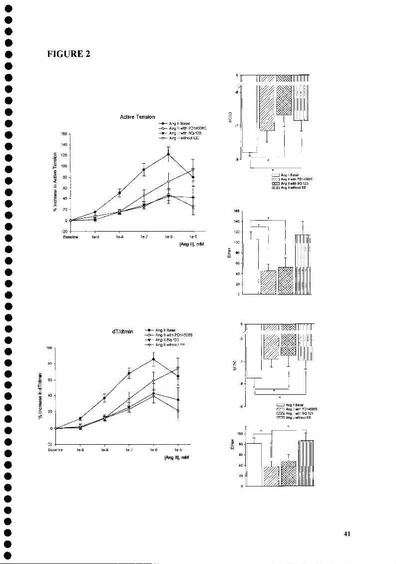
FIGURE 2
I
Active Tension Ang 11 Basai Ang II with PD145065 Ang II with BQ-123 Ang II without EE
1e-6 1e-5
[Ang II], mM
I I Ang II Basai r ^ 7 ] Ang II with PD145065 EXES AngllwithBQ-123 i m m Ang II without EE
dT/dtmin
F -a
S 20
- Ang II Basai - AngMwithPD145065 - Ang II Bq-123 - Ang II without EE
60 -
40 -
20 -
0
Ang II Basai rm Ang II with PD145065 f w a Ang II with BQ-123 [TTTTTfl Ang II without EE
41

FIGURE 3
c õ (0 CD
m 6^ <
160 -,
140 -
120 -
100 -
l ^ i
I Protocol A 1 ProtocolB 1 Protocol C H Protocol D
T r AT dT/dtmax dT/dtmin tHR
— i 1
PS dl/dtmax
42

REFERENCES
1 Allen AM, Zhuo J, Mendelsohn FA. Localization of angiotensin ATI and AT2 receptors. J Am Soc Nephrol. 1999 Jan;10 Suppl ll:S23-9.
2 Kim S, Iawo H. Molecular and Cellular Mechanisms of Angiotensin II - Mediated Cardiovascular and Renal Diseases. Pharmacol Rev, 2001; 52 (1): 11-34.
3 Dzau VJ and Gibbons GH (1987) Autocrine and paracrine mechanisms of vascular myocytes in systemic hypertension. Amer J Cardiol 60:991-1031.
4 Gasparo M, Catt KJ, Wright JW, Unger TH. International Union of Pharmacology. XXIII. The Angiotensin II Receptors. Pharmacol Rev, 2000; 52: 415-472.
5 Wollert KC, Drexler H The renin-angiotensin system and experimental heart failure. Cardiovasc Res. 1999 Sep;43(4):838-49.
6 Kim S, Ohta K, Hamaguchi A, Yukimura T, Miura K, Iwao H. Angiotensin II induces cardiac phenotypic modulation and remodeling in vivo in rats. Hypertension. 1995 Jun;25(6): 1252-9.
7 Serneri GG, Boddi M, Cecioni I, Vanni S, Coppo M, Papa ML, Bandinelli B, Bertolozzi I, Polidori G, Toscano T, Maccherini M, Modesti PA. Cardiac angiotensin II formation in the clinical course of heart failure and its relationship with left ventricular function. Circ Res. 2001 May ll;88(9):961-8.
8 Passier RC, Smits JF, Verluyten MJ, Studer R, Drexler H, Daemen MJ. Activation of angiotensin-converting enzyme expression in infarct zone following myocardial infarction. Am J Physiol. 1995 Oct;269(4 Pt 2):H 1268-76.
9 Rossi GP, Sacchetto A, Cesari M, Pessina AC. Interactions between endothelin-1 and the renin-angiotensin-aldosterone system. Cardiovasc Res. 1999 Aug l;43(2):300-7
10 Imai T, Hirata Y, Emori T, Yanagisawa M, Masaki T, Marumo F. Induction of endothelin-1 gene by angiotensin and vasopressin in endothelial cells. Hypertension. 1992 Jun;19(6 Pt 2):753-7.
11 Moreau P, d'Uscio LV, Shaw S, Takase H, Barton M, Luscher TF. Angiotensin II increases tissue endothelin and induces vascular hypertrophy: reversal by ET(A)-receptor antagonist. Circulation. 1997 Sep 2;96(5): 1593-7.
12 Ito H, Hirata Y, Adachi S, Tanaka M, Tsujino M, Koike A, Nogami A, Murumo F, Hiroe M. Endothelin-1 is an autocrine/paracrine factor in the mechanism of angiotensin II-induced hypertrophy in cultured rat cardiomyocytes.J Clin Invest. 1993 Jul;92(l):398-403.
43

13 Ishihata A, Endoh M. Species-related differences in inotropic effects of angiotensin II in mammalian ventricular muscle: receptors, subtypes and phosphoinositide hydrolysis. Br J Pharmacol. 1995 Jan;114(2):447-53.
14 Holubarsch C, Hasenfuss G, Schmidt-Schweda S, Knorr A, Pieske B, Ruf T, Fasol R, Just H. Angiotensin I and II exert inotropic effects in atrial but not in ventricular human myocardium. An in vitro study under physiological experimental conditions. Circulation. 1993 Sep;88(3): 1228-37.
15 Perez NG, Villa-Abrille MC, Aiello EA Dulce RA Cingolani HE, Camilion de Hurtado MC. A low dose of angiotensin II increases inotropism through activation of reverse Na(+)/Ca(2+) exchange by endothelin release. Cardiovasc Res. 2003 Dec l;60(3):589-97.
16 Mohan P, Brutsaert DL, Paulus WJ, Sys SU. Myocardial contractile response to nitric oxide and cGMP. Circulation 1996; 93: 1223-9.
17 Leite-Moreira AF, Bras-Silva C, Pedrosa CA Rocha-Sousa AA. ET-1 increases distensibility of acutely loaded myocardium: a novel ETA and Na+/H+ exchanger-mediated effect. Am J Physiol Heart Circ Physiol. 2003 Apr;284(4):H1332-9. Epub 2002 Dec 19.
18 Mulieri LA Hasenfuss G, Ittleman F, Blanchard EM, and Alpert NR. Protection of human left ventricular myocardium from cutting injury with 2,3-butanedione monoxime. Circ Res 65: 1441-1444, 1989.
19 Mebazaa A De Keulenaer GW, Paqueron X, Andries LJ, Ratajczak P, Lanone S, Frelin C, Longrois D, Payen D, Brutsaert DL, Sys SU. Activation of cardiac endothelium as a compensatory component in endotoxin-induced cardiomyopathy: role of endothelin, prostaglandins, and nitric oxide. Circulation 2001; 104: 3137-44.
20 Brutsaert DL, Meulemans AL, Spido KR, Sys SU. Effects of damaging endocardial surface on the mechanical performance of isolated cardiac muscle. Circ Res 1988;62:358-366.
21 Petroff MG, Aiello EA Palomeque J, Salas MA Mattiazzi A. Subcellular mechanisms of the positive inotropic effect of angiotensin II in cat myocardium. J Physiol. 2000 Nov 15;529 Pt 1:189-203.
22 Broome M, Haney M, Osterlund B, Haggmark S, Johansson G, Biber B. The cardiac effects of intracoronary angiotensin II infusion. Anesth Analg. 2002 Apr;94(4):787-93
23 Pérez NG, Camilión de Hurtado MC, Cingolani HE. Reverse mode of the Na+/Ca2+ exchange following myocardial stretch. Underlying mechanism of the slow force response. Circ Res. 2001;88:376-382
24 Talukder MA Norota I, Sakurai K, Endoh M. Inotropic response of rabbit ventricular myocytes to endothelin-1: difference from isolated papillary muscles. Am J Physiol Heart Circ Physiol. 2001 Aug;281(2):H596-605.
44
25 Ramaciotti C, Sharkey A, McClellan G, Winegrad S. Endothelial cells regulate cardiac contractility. Proc Natl Acad Sci USA 1989; 89: 365-7.
26 Brutsaert DL. Cardiac endothelial-myocardial signaling: its role in cardiac growth,
contractile performance, and rhythmicity. Physiol Rev. 2003 Jan;83(l):59-115.
27 Herizi A, Jover B, Bouriquet N, Mimran A. Prevention of the cardiovascular and renal effects of angiotensin II by endothelin blockade. Hypertension. 1998 Jan;31(1): 10-4.
28 Riggleman A, Harvey J, Baylis C. Endothelin mediates some of the renal actions of acutely administered angiotensin II. Hypertension. 2001 Jul;38(l): 105-9.
29 Montanari A, Biggi A, Carra N, Ziliotti M, Fasoli E, Musiari L, Perinotto P, Novarini A. Endothelin-A receptors mediate renal hemodynamic effects of exogenous Angiotensin II in humans. Hypertension. 2003 Oct;42(4):825-30.
30 Dohi Y, Hahn AW, Boulanger CM, Buhler FR, Luscher TF. Endothelin stimulated by angiotensin II augments contractility of spontaneously hypertensive rat resistance arteries. Hypertension. 1992 Feb;19(2):131-7.
31 Sung C-P, Arleth AJ, Storer BL, Ohlstein EH. Angiotensin type 1 receptors mediate smooth muscle proliferation and endothelin biosynthesis in rat vascular smooth muscle. J Pharmacol Exp Ther. 1994;271:429-437.
32 Riggleman A, Harvey J, Baylis C. Endothelin mediates some of the renal actions of acutely administered angiotensin II. Hypertension. 2001 Jul;38(l): 105-9.
33 Kedzierski RM, Yanasiawa M. Endothelin system: the double-edged sword in health and disease. Annu Rev Pharmacol Toxicol 2001; 52: 11-34.
34 Ferri C, Desideri G, Baldoncini R, Bellini C, Valenti M, Santucci A, De Mattia G. Angiotensin II increases the release of endothelin-1 from human cultured endothelial cells but does not regulate its circulating levels. Clin Sci (Lond). 1999 Mar;96(3):261-70.
35 Leite-Moreira AF, Bras-Silva C. Inotropic effects of ETB receptor stimulation and their modulation by endocardial endothelium, NO, and prostaglandins. Am J Physiol Heart Circ Physiol. 2004 Sep;287(3):Hl 194-9.
36 Reid AC, Mackins CJ, Seyedi N, Levi R, Silver RB. Coupling of angiotensin II ATI receptors to neuronal NHE activity and carrier-mediated norepinephrine release in myocardial ischemia. Am J Physiol Heart Circ Physiol. 2004 Apr;286(4):H1448-54.
45

37 Kumagai K, Reid I A. Angiotensin II exerts differential actions on renal nerve activity and heart rate. Hypertension. 1994 Oct;24(4):451-6. 38 Farrell DM, Wei CC, Tallaj J, Ardell JL, Armour J A, Hageman GR, Bradley WE, Dell'Italia LJ. Angiotensin II modulates catecholamine release into interstitial fluid of canine myocardium in vivo. Am J Physiol Heart Circ Physiol 2001; 281: H813-H822.
39 Holubarsch C, Schmidt-Schweda S, Knorr A, Duis J, Pieske B, Ruf T, Fasol R, Hasenfuss G, Just H. Functional significance of angiotensin receptors in human myocardium. Significant differences between atrial and ventricular myocardium.Eur Heart J. 1994 Dec; 15 Suppl D:88-91.
40 Molenaar P, O'Reilly G, Sharkey A, Kuc RE, Harding DP, Plumpton C, Gresham GA, Davenport AP. Characterization and localization of endothelin receptor subtypes in the human atrioventricular conducting system and myocardium. Circ Res. 1993 Mar;72(3):526-38
41 Kobayashi T, Miyauchi T, Sakai S, Kobayashi M, Yamaguchi I, Goto K, Sugishita Y. Expression of endothelin-1, ETA and ETB receptors, and ECE and distribution of endotheiin-1 in failing rat heart. Am J Physiol. 1999 Apr;276(4 Pt 2):H1197-206.
46

2- Efeitos da estimulação selectiva dos receptores AT2 sobre a contractilidade cardíaca

Negative Inotropic Effect of Selective AT2 Receptor Stimulation and
its Modulation by
Endocardial Endothelium, NO, Prostaglandins and EDHF
Department of Physiology, Faculty of Medicine, University of Porto, Porto, Portugal
Correspondence address:
Prof Dr. Adelino Leite-Moreira
Department of Physiology, Faculty of Medicine
Alameda Prof. Hernâni Monteiro
4200-319 Porto, PORTUGAL
Tel. +351-22.550.84.52; Fax. +351-22.551.91.94; E-mail: [email protected]

INTRODUCTION
Angiotensin II (Ang II) mediates its biological actions by binding to distinct
membrane-bound receptors and activating multiple intracellular pathways. In 1989,
different research teams independently provided evidence for the existence of two major
subtypes of Ang II receptors u . These two major subtypes have been identified, cloned
and named as ATI and AT2 3. ATI receptors are responsible for mediating all the well
known stimulatory actions of Ang II on blood pressure, water and sodium intake, renal
sodium retention, secretion of vasopressin and aldosterone and cell growth and
proliferation .
Although the exact physiological functions of AT2 receptors are not established,
it is known that these receptors may offset or oppose the ATI receptor mediated actions
of Ang II on cell growth, blood pressure and fluid intake 4. However, the expression of
AT2 receptors within cardiovascular tissues under certain physiological/pathological
conditions complicates matters and suggests that there is a far from complete
understanding of the role of AT1/AT2 receptors in cardiovascular regulation 5.
In fetal tissues, AT2 receptor is the predominant subtype expressed but this is
rapidly reversed after birth. In the adult ATI receptor becomes the dominant subtype 3.
The common misconception that AT2 receptors do not exist in appreciable amounts in
adult animal vasculature is however slowly changing. They are located in many
different vessel types, albeit at lower, but functional levels. For example, in rat aorta
these receptors constitute about 30-40% of Ang II receptors 6. In the heart, AT2
receptors are expressed at low levels in adult rat cardiomyocytes 7 but are increased in 8 9 10
both absolute and relative levels in hypertrophied rat and failing hamster hearts
AT2 receptors also gain particular prominence in adult human heart. In both normal
noninfarcted or hypertrophied human hearts, there is a predominance of AT2 receptor
binding sites in the myocardium. '
There is an increasing amount of literature that has demonstrated AT2 receptor-
mediated relaxation directly in isolated arteries, including rabbit renal arterioles
rabbit cerebral arteries 14 and rat mesenteric arteries. '
The AT2 receptor has been implicated also in suppression of myocardial
hypertrophy 17'18, fibroblast proliferation19 and vascular cell hyperplasia20. The
overexpression of AT2 receptors in cardiomyocites attenuates perivascular fibrosis by a
kinin/NO dependent mechanism 21. In a rat model of chronic heart failure, left
49

ventricular remodeling and cardiac function were improved by blockade of ATI
receptors 22. This effect may be partially explained by a higher activation of AT2
receptors. In the heart, Ang II also exerts a vasodilating effect in the coronary
microcirculation
In rabbit hearts, this receptor subtype is almost totally absent from nervous,
conductive and atrial tissue but, in the ventricle, approximately 20% of the Ang II
receptors are AT2. 24 AT2 receptor function is likely to be context-specific, as recently
suggested 25. The acute effects of AT2 stimulation in myocardial function are not yet
known. The main objectives of the present study are to describe the myocardial effects
caused by AT2 stimulation and their underlying effects.
50

METHODS The investigation conforms to the Guide for the Care and Use of Laboratory
Animals published by US National Institutes of Health (NIH Publication No 85-23,
revised 1996).
Experimental preparation
The effects of Ang II were studied in isolated right papillary muscles of New
Zealand White rabbits (Oryctolagus cuniculus; 2.9±0.12 Kg). Rabbits were
anaesthetized with sodium pentobarbital (25 mg/Kg, iv) and the heart was quickly
excised. The tissues were immersed in a modified Krebs-Ringer (KR) solution, at 35°C,
with cardioplegic 2,3 butanedione monoxime (BDM; 3%) and calf serum (5%; Bio-
Whittaker, St. Louis, Maryland, USA). The modified KR solutions contained (in mM):
NaCl 98, KC1 4.7, MgS04 2.4, KH2P04 12, CaCl2 1.8, NaHC03 20, CH3COONa 5,
C3H303Na 15, glucose 4.5 and atenolol 0.02. Atenolol was used to prevent P-adrenergic
mediated effects. The solutions were in equilibrium with 95% 0 2 and 5% CO2,
maintaining the pH between 7.38-7.42. Rabbit papillary muscles (n=68; length: 3.2±1.1
mm; weight: 2.2±1.3 mg; cross-sectional area: 0.6±0.3 mm2; preload: 5.0±1.1 mN)
were then carefully dissected. Afterwards, they were vertically mounted in a 10 ml plexi
glass organ bath and connected to an isotonic/isometric transducer (University of
Antwerp, Belgium). Preload was estimated according to muscle dimensions and the
electrical stimulus (0.6 Hz) was set at 10% above threshold. Twenty minutes later,
bathing solutions were replaced by corresponding KR solutions without BDM. During
the next 2 hours, muscles were stabilized. Bathing solutions were then replaced by
corresponding KR solutions without calf serum and Lmax was calculated. Protocols
were initiated after obtaining two similar isotonic and isometric control twitches
separated by a 10 min interval.
Experimental protocols
The effects of selective AT2 receptor activation were evaluated in rabbit right
papillary muscles (n=68).
In a first step, we studied the effect of selective stimulation of AT2 receptors
using an agonist of this subtype of receptors (H3995) in increasing concentrations (10" ,
IO"8, IO"7, IO"6, IO"5 M) in basal conditions (n=15) and in the presence of a selective
antagonist of ATI receptors (ZD-7155; IO"7 M; n=9).
51
In a second step, the underlying mechanisms to the inotropic effect of AT2
stimulation were studied. This was performed by adding to the superfusing solution
increasing doses of Ang II (IO"8, IO"7, IO"6, IO"5 M) in the presence of a selective ATI
receptor antagonist (ZD7155, IO"6 M) and in different experimental conditions.
Selective AT2 receptor activation was performed at baseline (n=12), after removal of
the endocardial endothelium (n=7) and in the presence of NG-nitro-L-Arginine (L-NA;
3*10"5 M; n=9), Indomethacin (INDO; 10"5 M; n=7) or Proadifen (PRO; IO"6 M; n=9).
These three later substances are inhibitors of the synthesis of nitric oxide,
prostaglandins and endothelium-derived hyperpolarizing factor (EDHF), respectively.
Selective removal of the endocardial endothelium was performed according to
the methodology described by Brutsaert and collaborators26. Briefly, this consisted in
the immersion of the papillary muscles in a 0.5% solution of Triton X-100 during 1
second, followed by an abundant washout.
Data analysis
Isotonic and isometric twitches were recorded and analyzed with dedicated
software (University of Antwerp, Belgium). Selected parameters include: Active
Tension (AT, mN-mm"2); Maximum Velocity of Tension Rise (dT-dtmax, mN-mm"2.s"1);
Maximum Velocity of Tension Decline (dT-dtmin, mN-mm"2-s"1); Peak Isotonic
Shortening (PS, %Lmax); Maximum Velocity of Shortening (dL/dtmax, Lmax-s"1);
Maximum Velocity of Lengthening (dL/dtmin, Lmaxs"1), Time to Half Relaxation
(tHR, ms).
Statistical Methods
Values are means ± SE. The effects of increasing doses of a given drug were
analyzed by one-way repeated-measures ANOVA. Comparison between protocols of
the effects of Ang II was performed with a repeated-measures two-way ANOVA. When
significant differences were detected with any of the ANOVA tests, the Student-
Newman-Keuls test was selected to perform pairwise multiple comparisons. P<0.05 was
accepted as significant.
52

RESULTS The selective stimulation of AT2 receptors with the agonist H3995 induced a
concentration-dependent negative inotropic effect (Figure 1). This effect was maximal
for the concentration of IO"5 M, decreasing 16.4±6.8% AT, 20.3±6.0% dT/dtmax,
19.6±7.3% dT/dtmin, 16.0±5.6% PS and 32.3±12.2% dL/dtmax (p<0.05). The addition
of the same agonist in the presence of a selective ATI receptor antagonist (ZD-7155;
IO"7 M) increased the selectivity of the stimulation and caused a slight amplification of
the negative inotropic effect. The same magnitude of the effect was now observed for a
concentration of H3995 ten times lower (Figure 2).
In the following Protocols, we studied the mechanisms underlying the negative
inotropic effect previously observed, by adding increasing concentrations of Ang II in
the presence of ZD-7155 in several experimental conditions. In a baseline protocol,
selective AT2 stimulation induced a dose-dependent negative inotropic effect,
decreasing, at 10'5 M of Ang II, 29.3±7.7% AT, 26.1±7.0% dT/dtmax, 27.9±7.5%
dT/dtmin, 30.7±9.3% PS and 22.0±5.7% dL/dtmax. This effect was similar to the one
observed with the addition of H3995 in the presence of ZD-7155.
The addition of increasing concentrations of Ang II in the presence of ZD-7155
after the selective removal of endocardial endothelium significantly blunted the negative
inotropic effect previously observed (Figure 3).
Afterwards, the mechanisms underlying the endothelium-derived negative
inotropic effect were studied by adding Ang II in the presence of ZD-7155 and
inhibitors of the synthesis nitric oxide, prostaglandins and EDHF. The inhibitors used
were L-NA (3.105 M), indomethacin (10"3 M) and proadifen (IO"6 M), respectively. In
all three Protocols, the negative inotropic effect was not significantly altered (Figure 4).
The selective stimulation of AT2 receptors in the presencen of L-NA caused a negative
inotropic effect maximal for the concentration of 10"5 M that decreased 32.5±10.2% AT,
25.7±7.8% dT/dtmax, 26.7±8.6% dT/dtmin, 22.4±6.5% PS, 16.90±7.1% dL/dtmax and
32.8±9.6% dL/dtmin. In the presence of indomethacin, the negative inotropic effect was
also maximal for the same concentration decreasing: 34.4±7.1% AT, 27.9±6.1%
dT/dtmax, 33.2±7.9% dT/dtmin, 25.3±7.1% PS, 20.2±5.0% dL/dtmax and 36.6±10.2%
dL/dtmin. Finally, the addition of increasing concentrations of Ang II in the presence of
ZD-7155 and proadifen induced a negative inotropic effect maximal for the
concentration of 10~5 M decreasing 26.0±8.3% AT, 25.5±6.6% dT/dtmax, 26.4±7.6%
dT/dtmin, 23.7±8.0% PS, 22.9±7.0% dL/dtmax and 30.4±9.7% dL/dtmin.
53

DISCUSSION In the present study, we demonstrated that the selective stimulation of rabbit
myocardial AT2 receptors caused an endocardial endothelium-dependent negative
inotropic effect that was not abolished by the inhibition of nitric oxide, prostaglandins
orEDHF.
Similar to ATI receptors, AT2 receptors have been implicated in cardiovascular
regulation. In the vasculature, AT2 receptor stimulation induces a vasodilating effect. In
the presence of an ATI receptor antagonist, Ang II caused a 30% increase in the
diameter of preconstricted, microperfused rabbit afferent and efferent arterioles in a
PD123319-sensitive manner. 12'13 An AT2-dependent vasodilator effect was confirmed
using the selective AT2 receptor agonist, CGP42112, in the absence of ATI receptor
blockade. n This acute AT2 receptor-mediated vasodilator response may be
endothelium-dependent or -independent and appears to involve a range of signalling
pathways, including NO and bradykinin production, activation of cytochrome P-450
epoxygenase pathways and modulation of K+ channel activity.
In the human heart, AT2 receptors are the main subtype whereas in rodents their
expression is low 27. In rabbit ventricles this subtype constitutes about 20% of Ang II
receptors 24. These receptors are up-regulated in rats and humans under certain
pathophysiological conditions, such as myocardial infarction, and are therefore
suggested to play an essential role in tissue remodeling27. In the failing ventricle of the
human heart, the level of ATI receptor gene expression appears to decrease, whereas
the level of AT2 receptor expression is unaffected 28. A study performed by Wharton et
al.29 demonstrated that the density of AT2 binding sites in endocardial, interstitial, and
infarcted regions in the ventricles of patients with end-stage ischemic heart disease or
dilated cardiomyopathy is significantly increased compared with the noninfarcted
myocardium.
In a rat model with chronic heart failure, left ventricular remodeling and cardiac
function were improved by blockade of ATI receptors 30. This effect was inhibited by
treatment with an AT2 receptor antagonist and also, in part, by treatment with a
bradykinin B2 receptor antagonist. In pigs, infarct size was reduced after regional
myocardial ischemia by blockade of the ATI receptor, and this reduction was abolished
by pretreatment with the AT2 receptor antagonist PD 123319 and by blockade of B2
receptors 31. The aforementioned reduction of perivascular fibrosis by overexpressed
cardiac AT2 receptors after pressure overload was abolished after blockade of the B2
54

receptors or NO synthase. Thus, as in the vasculature, the myocardial kinin/NO system
appears to be involved in AT2 receptor-mediated cardiac chronic effects.
The role of AT2 receptors in acute modulation of myocardial function was
previously unknown. Recently, it has been described that in human coronary
microarteries AT2 receptor stimulation induces vasodilation in an endothelium-
dependent manner and mediated by B2 receptors and NO 23. The present study
demonstrates, for the first time, that AT2 receptor stimulation induces a negative
inotropic effect, thus clarifying the role of these receptors in myocardial contractility.
This effect was significantly blunted after the selective removal of endocardial
endothelium. The endocardial endothelium constitutes an important interface between
the lumen of the heart and the underlying myocardium and is responsible for the
synthesis and release of different substances known to influence myocardial contractile
properties32. In order to understand the mechanisms involved in the endothelium
dependent negative inotropic effect, we performed different protocols with selective
stimulation of AT2 receptors in the presence of inhibitors of the synthesis of nitric
oxide, prostaglandins and EDHF. However, the negative inotropic effect was not
significantly changed by either of the inhibitors. Thus, the mechanisms underlying the
observed effect still remain unclear. An attractive hypothesis is that this effect is
mediated through the activation of B2 receptors. This assumption is speculative and will
be tested in future protocols.
In conclusion, selective AT2 stimulation induces a negative inotropic effect,
which is modulated by the endocardial endothelium, but is not mediated neither by NO,
prostaglandins or EDHF. Such findings might help to better understand the therapeutic
effects of selective ATI antagonists, which are increasingly used in for treating
cardiovascular diseases.
55

Figure 1
Selective stimulation of AT2 recepors with the agonist H9395 (n=15) induced a
concentration-dependent negative inotropic effect maximal for 10"5 M. a P<0.05 vs.
baseline; Values are means ± SEM.
10 -,
-10 CD c 0 CD -20 m
-30
-40
-50
a
^ ^
a a a
a — i 1 1 1
AT dT/dtmax dT/dtmin tHR PS dL / dtmax
56

Figure 2 Concentration-response curves of AT2 selective stimulation with the agonist H9395 in
the absence and in the presence of an ATI receptor antagonist, ZD-7155 (10" M; n=9)
H9395 H9395withZD7155
Baseline 1e-9 1e-8 1e-7 1e-6 1e-5
[Ang II]
57
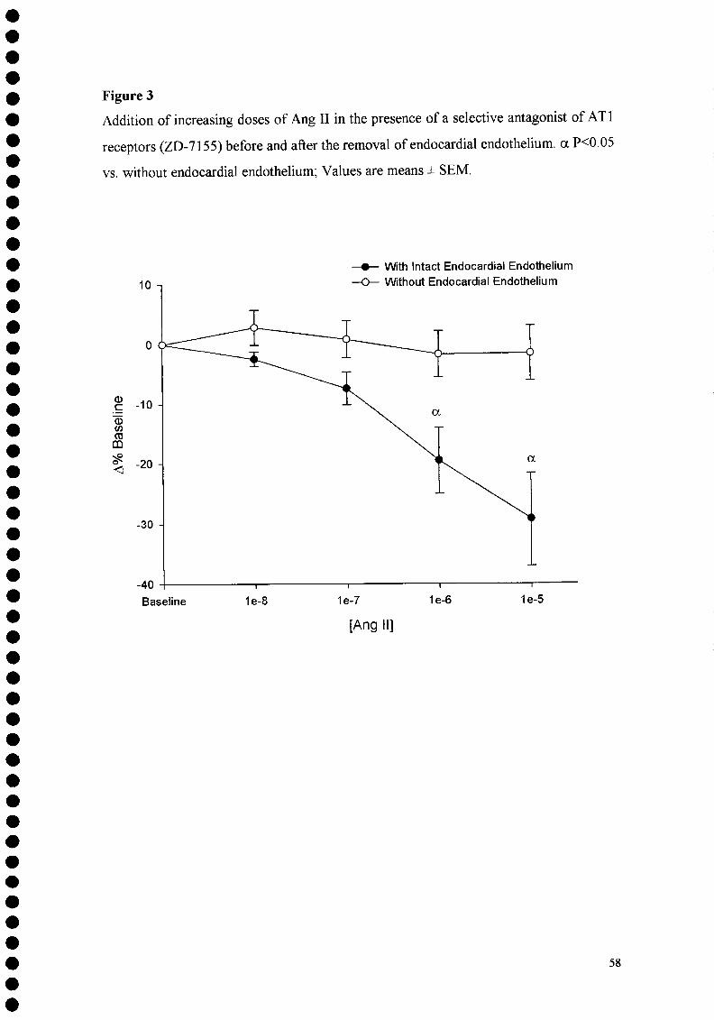
Figure 3
Addition of increasing doses of Ang II in the presence of a selective antagonist of ATI
receptors (ZD-7155) before and after the removal of endocardial endothelium, a P<0.05
vs. without endocardial endothelium; Values are means ± SEM.
CD C 03 c/) ra m
<
With Intact Endocardial Endothelium Without Endocardial Endothelium
Baseline 1e-8 1e-7 1e-6 1e-5
[Ang II]
58

Figure 4 Negative inotropic effect of induced by the addition of Ang II in the presence of
ZD7155 and L-NA, indomethacin and proadifen. Selected parameters are active tension
and peak rate of tension rise (dT/dtmax). * PO.05 vs. baseline; values are means ±
SEM.
c o
'(/> c <D H (D
> is
Baseline
AngZD Ang ZD L-NA Ang ZD Indo Ang ZD Proad
1e-8 1e-7 1e-6
[Ang II], M
1e-5
x E
Baseline 1e-8 1e-7
AngZD Ang ZD L-NA Ang ZD Indo Ann 7 n Prr>orl
1e-6 1e-5
[Ang II], M
59

REFERENCES
1 Chiu AT, Herblin WF, McCall DE, Ardecky RJ, Carini DJ, Duncia JV, Pease LJ,
Wong PC, Wexler RR, Johnson AL, et al. Identification of angiotensin II receptor
subtypes. Biochem Biophys Res Commun. 1989 Nov 30;165(1): 196-203.
2 Whitebread S, Mele M, Kamber B, de Gasparo M. Preliminary biochemical
characterization of two angiotensin II receptor subtypes. Biochem Biophys Res
Commun. 1989 Aug 30;163(1):284-91.
3 de Gasparo M, Catt KJ, Inagami T, Wright JW, Unger T. International union of
pharmacology. XXIII. The angiotensin II receptors. Pharmacol Rev. 2000
Sep;52(3):415-72.
4 Gallinat S, Busche S, Raizada MK, Sumners C. The angiotensin II type 2 receptor: an
enigma with multiple variations. Am J Physiol Endocrinol Metab. 2000
Mar;278(3):E357-74.
5 Horiuchi M, Akishita M, Dzau VJ. Recent progress in angiotensin II type 2 receptor
research in the cardiovascular system. Hypertension. 1999 Feb;33(2):613-21.
6 Chang RS, Lotti VJ. Angiotensin receptor subtypes in rat, rabbit and monkey tissues:
relative distribution and species dependency. Life Sci. 1991;49(20): 1485-90.
7 Busche S, Gallinat S, Bohle RM, Reinecke A, Seebeck J, Franke F, Fink L, Zhu M,
Sumners C, Unger T. Expression of angiotensin AT(1) and AT(2) receptors in adult rat
cardiomyocytes after myocardial infarction. A single-cell reverse transcriptase-
polymerase chain reaction study. Am J Pathol. 2000 Aug; 157(2):605-11.
8 Lopez JJ, Lorell BH, Ingelfinger JR, Weinberg EO, Schunkert H, Diamant D, Tang
SS. Distribution and function of cardiac angiotensin ATI- and AT2-receptor subtypes in
hypertrophied rat hearts. Am J Physiol. 1994 Aug;267(2 Pt 2):H844-52
9 Ohkubo N, Matsubara H, Nozawa Y, Mori Y, Murasawa S, Kijima K, Maruyama K,
Masaki H, Tsutumi Y, Shibazaki Y, Iwasaka T, Inada M. Angiotensin type 2 receptors
are reexpressed by cardiac fibroblasts from failing myopathic hamster hearts and inhibit
cell growth and fibrillar collagen metabolism. Circulation. 1997 Dec 2;96(11):3954-62
10 Bartunek J, Weinberg EO, Tajima M, Rohrbach S, Lorell BH. Angiotensin II type 2
receptor blockade amplifies the early signals of cardiac growth response to angiotensin
11 in hypertrophied hearts. Circulation. 1999 Jan 5-12;99(l):22-5.
11 Matsubara H. Pathophysiological role of angiotensin II type 2 receptor in
cardiovascular and renal diseases. Circ Res. 1998 Dec 14-28;83(12): 1182-91.
60

12 Arima S, Endo Y, Yaoita H, Omata K, Ogawa S, Tsunoda K, Abe M, Takeuchi K,
Abe K, Ito S. Possible role of P-450 metabolite of arachidonic acid in vasodilator
mechanism of angiotensin II type 2 receptor in the isolated microperfiised rabbit
afferent arteriole. J Clin Invest. 1997 Dec 1; 100(11):2816-23
13 Endo Y, Arima S, Yaoita H, Omata K, Tsunoda K, Takeuchi K, Abe K, Ito S.
Function of angiotensin II type 2 receptor in the postglomerular efferent arteriole
Kidney Int Suppl. 1997 Dec;63:S205-7
14 Haberl RL. Role of angiotensin receptor subtypes in the response of rabbit brain
arterioles to angiotensin. Stroke. 1994 Jul;25(7): 1476-9; discussion 1480
15 Widdop RE, Matrougui K, Levy BI, Henrion D. AT2 receptor-mediated relaxation is
preserved after long-term ATI receptor blockade. Hypertension. 2002 Oct;40(4):516-20
16 Widdop RE, Jones ES, Hannan RE, Gaspari TA. Angiotensin AT2 receptors:
cardiovascular hope or hype? Br J Pharmacol. 2003 Nov; 140(5):809-24.
17 Booz GW, Baker KM. Role of type 1 and type 2 angiotensin receptors in angiotensin
II-induced cardiomyocyte hypertrophy. Hypertension. 1996; 28: 635-640
18 Bartunek J, Weinberg E O, Tajima M, Rohrbach S, Lorell BH. Angiotensin II type 2
receptor blockade amplifies the early signals of cardiac growth response to angiotensin
II in hypertrophied hearts. Circulation. 1999; 99: 22-25
19 Tsutsumi Y, Matsubara H, Ohkubo N, Mori N, Nozawa Y, Murasawa S, Kijima K,
Maruyama K, Masaki H, Moriguchi Y, Shibasaki Y, Kamihata H, Inada M, Iwasaka T.
Angiotensin II type 2 receptor is upregulated in human heart with interstitial fibrosis,
and cardiac fibroblasts are the major cell type for its expression. Circ Res. 1998; 83:
1035-1046.
20 Nakajima M, Hutchinson HG, Fujinaga M, Hayashida W, Morishita R Zhang L,
Horiuchi M, Pratt R E, Dzau VJ. The angiotensin II type 2 (AT2) receptor antagonizes
the growth effects of the ATI receptor: gain-of-function study using gene transfer. Proc
Natl Acad Sci USA. 1995; 92: 10663-10667
21 Kurisu S, Ozono R, Oshima T, Kambe M, Ishida T, Sugino H, Matsuura H,
Chayama K, Teranishi Y, Iba O, Amano K, Matsubara H. Cardiac angiotensin II type 2
receptor activates the kinin/NO system and inhibits fibrosis. Hypertension. 2003
Jan;41(l):99-107.
61

22 Y.H. Liu, X.P. Yang, V.G. Sharov et al, Effects of angiotensin-converting enzyme
inhibitors and angiotensin II type 1 receptor antagonists in rats with heart failure. Role
of kinins and angiotensin II type 2 receptors. J. Clin. Invest. 99 (1997), pp. 1926-1935
23 Batenburg WW, Garrelds IM, Bernasconi CC, Juillerat-Jeanneret L, van Kats JP,
Saxena PR, Danser AH. Angiotensin II type 2 receptor-mediated vasodilation in human
coronary microarteries. Circulation. 2004 May 18;109(19):2296-301
24 Brink M, de Gasparo M, Rogg H, Whitebread S, Bullock G. Localization of
angiotensin II receptor subtypes in the rabbit heart. J Mol Cell Cardiol. 1995
Jan;27(l):459-70
25 Schneider MD, Lorell BH. AT(2), judgment day: which angiotensin receptor is the
culprit in cardiac hypertrophy? Circulation. 2001 Jul 17;104(3):247-8
26 Brutsaert DL, Meulemans AL, Spido KR, Sys SU. Effects of damaging endocardial
surface on the mechanical performance of isolated cardiac muscle. Circ Res
1988;62:358-366.
27 Johren O, Dendorfer A, Dominiak P. Cardiovascular and renal function of
angiotensin II type-2 receptors. Cardiovasc Res. 2004 Jun l;62(3):460-7
28 Haywood, G. A, L. Gullestad, T. Katsuya, H. G Hutchinson, R. E. Pratt, M.
Horiuchi, and M. B. Fowler. ATI and AT2 angiotensin receptor gene expression in
human heart failure. Circulation 95: 1201-1206, 1997
29 Wharton, J., K. Morgan, R. A. Rutherford, J. D. Catravas, A. Chester, B. F.
Whitehead, M. R. De Levai, M. H. Yacoub, and J. M. Polak. Differential distribution of
angiotensin AT2 receptors in the normal and failing human heart. J. Pharmacol. Exp.
Ther. 284: 323-336, 1998
30 Liu, Y H; Yang, X P; Sharov, V G; Nass, O; Sabbah, H N; Peterson, E; Carretero, O
A. Effects of angiotensin-converting enzyme inhibitors and angiotensin II type 1
receptor antagonists in rats with heart failure. Role of kinins and angiotensin II type 2
receptors. J. Clin. Invest. 99: 1926-1935, 1997.
31 Jalowy A, Schulz R, Dorge H, Behrends M, Heusch G. Infarct size reduction by
ATI-receptor blockade through a signal cascade of AT2-receptor activation, bradykinin
and prostaglandins in pigs. J Am Coll Cardiol. 1998 Nov 15;32(6): 1787-96.
32 Brutsaert DL. Cardiac endothelial-myocardial signaling: its role in cardiac growth,
contractile performance, and rhythmicity. Physiol Rev. 2003 Jan;83(l):59-115.
62

3- Efeitos da a ngiotensina 11 s obre a s propriedades d iastólicas do miocárdio

Angiotensin II increases myocardial distensibility: a novel ATI
mediated effect.
Department of Physiology, Faculty of Medicine, University of Porto, Porto, Portugal
Correspondence address:
Prof. Dr. Adelino Leite-Moreira
Department of Physiology, Faculty of Medicine
Alameda Prof. Hernâni Monteiro
4200-319 Porto, PORTUGAL
Tel. +351-22.550.84.52; Fax. +351-22.551.91.94; E-mail: [email protected]

INTRODUCTION
The octapeptide Angiotensin II (Ang II) has a central role in cardiovascular homeostasis exerting its effects by binding to two different subtypes of G protein-coupled receptors: ATI and AT2 . Both subtypes are expressed in the heart of several animal species. The ATI receptor is responsible for most of the classical effects of the renin-angiotensin system (RAS) , More recently, the role of AT2 receptors has become more clear. It was shown that AT2 receptors have globally opposite effects to those of ATI, inducing vasodilation, apoptosis and growth inhibition 4'5'6,7.
In the heart, the activation of ATI receptor causes acute and chronic effects. For instance, in response to both chronic pressure and volume overload8, locally and systemically produced Ang II have mitogenic effects9 and promote extracellular matrix synthesis10, contributing to ventricular remodelling11, cardiac hypertrophy12 and, ultimately, to the deterioration of diastolic function13.
Acutely, Ang II induces vasoconstriction14 and has a species-dependent positive inotropic effect 15,16,17. These actions result from ATI receptor stimulation, and subsequent phosphoinositide hydrolysis and activation of protein kinase C (PKC) dependent pathways ,8,19,15. Also an increase of calcium transients, through the L-type Ca2+ current 20<21'22 and myofilament sensitization 23 are involved, although there is some controversy, in part because of differences between species. B y opposition, the selective activation of AT-2 receptors appears to induce an endothelium-dependent negative inotropic effect24.
Unlike its effects on myocardial contractility, the influence of Ang II in relaxation and in diastolic myocardial properties is still largely unknown. Recently, it was shown that endothelin-1 increases diastolic distensibility of acutely loaded human and nonhuman myocardium. This effect is mediated by ET-A receptors and requires Na+/H+
exchanger activation 25.
In this paper, our objectives were to characterize the effects of Ang II on myocardial diastolic properties and investigate possible underlying mechanisms through the selective inhibition of Ang II activated pathways, such as the protein kinase C and the Na+ZH* exchanger.
65

METHODS
The investigation conforms to the Guide and Care and Use of Laboratory Animals published by the National Institutes of Health (NLH Publication No. 85-23, revised 1996).
Experimental preparation The study was performed in isolated right papillary muscles (n= 43) from male
New Zealand White rabbits (Oryctolagus cuniculus; 2.0-3.0 kg). Rabbits were anesthetized with intravenous pentobarbital sodium (25 mg/kg). A left thoracotomy was performed, and beating hearts were quickly excised and immersed in modified Krebs-Ringer solution (composition in mmol/1: 98 NaCl, 4.7 KC1, 2.4 MgS04-7H20, 1.2 KH2P04,4.5 glucose, 1.8 CaCl2-2H20, 17 NaHC03,15 C3H3Na03, 5 CH3COONa, 0.02 atenolol) at 35°C with cardioplegic 2,3-butanedione monoxime (BDM; 3%) and 5% newborn calf serum. Atenolol was used to prevent adrenergic-mediated effects26. Solutions were in equilibrium with 95% 02 and 5% CO2, maintaining the pH between 7.38 and 7.42. After dissection, papillary muscles (length: 3.4 ± 2 mm; weight: 2.9 ± 0.3 mg; preload: 4.8 ±0.1 mN) were vertically mounted in a 10 ml Plexiglas organ bath containing the aforementioned solutions and attached to an electromagnetic length-tension transducer (University of Antwerp, Belgium). Preload was estimated according to muscle dimensions, and the electrical stimulus (0.6 Hz) was set at 10% above threshold. Twenty minutes later, bathing solutions were replaced by corresponding ones without B DM. During t he n ext 2 h, m uscles w ere s tabilized. B athing so lutions w ere then replaced by corresponding ones without calf serum, and maximum physiological length (Lmax) was calculated. Protocols were initiated after two superimposable isotonic and isometric control twitches separated by a 10-min interval were obtained.
Experimental protocol Effects of increasing concentrations of Ang II (10 , 10"6 and 10"5 M ) on
contraction, relaxation, and diastolic properties of cardiac muscle were studied in rabbit papillary muscles with intact endocardial endothelium (n=l 1). In order to further clarify the role of Ang II in myocardial distensibility, we studied the effects of Ang II in ventricular stiffness by comparing the slope of the passive length-tension curves for a single concentration of Ang II (10"5 M; n=6). Increasing concentrations of Ang II were
66

also added to the bathing solution in the presence of: 1) ZD-71SS, a selective antagonist of AT-1 receptors (10"7 M; n=8); 2) Chelerythrine, an inhibitor of PKC (CHE, 10"5M; n=8); and 3) 5-(N-methyl-N-isobutyl)-amiloride, an inhibitor of the Na+/H+ exchanger (MIA;10"6M;n=10)
Chemicals were obtained from Sigma Chemical, St. Louis, MO.
Data acquisition and analysis Isotonic, afterloaded, and isometric twitches were recorded and analyzed.
Selected parameters include the following: active tension (AT; mN-mm"2); maximum velocity of tension rise (dT/dtmaxi mN-mm" -s" ); maximum velocity of tension decline (dT/dtmin; mN-mm^-s"1); peak isotonic shortening (PS; %Lmax); maximum velocity of shortening (dL/dtmaxi Lmax-s"1); maximum velocity of lengthening (dL/dtmin; Lmax-s" ); time to half-relaxation (tHR, ms); variation in muscle length relative to Lmax ( A%Lmax). A11 he e nd o f t he e xperimental P rotocol, m use le 1 ength w as c orrected t o initial length and the variation in passive tension relative to the initial tension was calculated (A%PT).
Statistical methods Values are means ± SE. Effects on the various parameters of a single dose of a
given drug were analyzed by a paired t-test, whereas the effects of increasing doses of a given drug were analyzed by one-way repeated-measures ANOVA. Comparison between protocols of the effects of Ang II was performed with a repeated-measures two-way ANOVA. When significant differences were detected with any of the ANOVA tests, the Student-Newman-Keuls test was selected to perform pairwise multiple comparisons. PO.05 was accepted as significant.
67

RESULTS
Baseline performance of rabbit papillary muscles was similar in all experimental protocols. Mean values of the contractile parameters from the 43 papillary muscles were as follows: AT 30.8±3.8 mN-mm"2; dT/dW 187.6±21 mNmm'V1; dT/dt,™ -130±25 mN-mrnV; PS 12±0.8% of Lmax; dL/dW, 0.77±0.4 Lmaxs"1; dL/dt™, -2.7±0.3 Lmaxs*1; tHR, 441±15 ms.
Effects of increasing concentrations of Ang II (IO7, 10"6 and IO"5 M) on contraction and relaxation of papillary muscles are summarized and illustrated in Figure I where it can be seen that these concentrations increased AT, dT/dtmax and dT/dtmin. The highest dose of Ang II (10"5 M) increased 43.3±6.25% AT, 58.6±9.6% dT/dW and 49.2±9.8% dT/dtmin. Effects on tHR were not statistically significant.
The present study aimed to evaluate the effect of Ang II in acute diastolic myocardial properties, namely distensibility. As shown in Figure 2, the addition of Ang II had a significant concentration dependent acute effect on muscle length. The increase in muscle length, expressed as variation of muscle length relative to Lmax (A% Lmax), was maximal for the concentration of 10s M and was as follows: 0.38±0.09% of Lmax at 10"7M; 1.02±0.25% of Lmax at lO^M; 1.97±0.42% of Lmax at 10"5M. At the end of this Protocol, we corrected muscle length to its initial value and observed a 46.1 ±4% decrease in passive tension. This correction did not significantly affect muscle contractile parameters.
Figure 3 represents muscle passive length-tension relation. In this Figure, it can be seen that Ang II induced a rightward shift in muscle length-tension relation and a decrease in the curve slope. This represents a higher muscle distensibility and lower ventricular stiffness.
In order to study the mechanisms underlying the increase in distensibility, the same concentrations of Ang II were added in the presence of a selective ATI receptor antagonist (ZD71S5), an inhibitor of protein kinase C (chelerythrine) and an inhibitor of Na'VuT exchanger (5-(N-methyl-N-isobutyl)-amiloride). None of these three substances significantly changed per se basal muscle length or contractile performance. In the presence of CHE and MIA, the positive inotropic effect of Ang II was nonsignificantly smaller.
As can be seen in Figure 4, the effect of Ang II in myocardial distensibility was abolished in the presence of ZD71SS. In the presence of CHE, Ang II in the
68

concentration of 10"5 M induced an increase in muscle length of 0.21 ±0.08% of Lmax corresponding to a decrease in passive tension of 10.9±4.5%. The same concentration of Ang II in the presence of MIA increased muscle length by 0.78±0.25% of Lmax corresponding to a decrease in passive tension of 23.2±5.0%. In conclusion, the inhibition of both protein kinase C (PKC) and Na+/H+ exchanger (NHE) significantly attenuated the effect of Ang II in myocardial distensibility, but did not abolish it. There were no significant differences between the effects registered in the presence of CHE and MIA.
69

DISCUSSION We now have evidence that Ang II induces a significant dose-dependent
lengthening of cardiac muscle in a very short time frame. Our data suggest that the described increase in distensibility is mediated by binding of Ang II to its ATI receptor, being dependent on the activation of PKC and Na+/H+ exchanger exchanger. This effect adds to the previously well known effects of Ang II in acute myocardial contractility. Recently, it was described that endothelin-1, another neuro-humoral substance with an important role in myocardial pathophysiology, exerts, in the same animal species, an increase in muscle distensibility mediated by ETA receptors and dependent on the activation of Na+/H+ exchanger 25. These two studies thus reinforce the role of both substances in the acute modulation of myocardial diastolic properties.
In the present study Ang II induced a concentration-dependent positive inotropic effect that is globally in accordance with the effect previously described for this animal species27.
Ang II type 1 receptors, together with oti-adrenoreceptors and ETA receptors, belong to a family of G protein (Gq)-coupled receptors linked to phospholipase C (PLC) activation and consequent production of inositol triphosphate and diacylglicerol. The later substance activates Ca2+ dependent and Ca2+ independent isoforms of PKC . In rabbit heart, the positive inotropic effect of Ang II is associated with an increase in phosphoinositide turnover 15,28. PKC is also known to mediate some of the effects of Ang II in cardiac muscle. Indeed, it has been shown that PKC mediates the increase in intracellular pH (pHj) produced by Ang II through activation of the sarcolemmal Na+/H+ exchanger (NHE)29,30,31. The NHE is an important extrusion mechanism that contributes to the integrated control of pHj in cardiac myocytes . PKC mediated activation of NHE and the consequent intracellular alkalinization is associated with an increase in calcium sensitivity of contractile proteins33 that may be involved both in the regulation of myocardial contractility29 and distensibility 25, although this seems to be strongly influenced by the animal species considered 15. In the present study we observed that the increase in myocardial distensibility induced by Ang II is significantly attenuated by the inhibition of PKC and NHE. The later also seems to be involved in the effects observed with endothelin-1 on myocardial distensibility .
In the present work, the increase in muscle length was accompanied by an increase in the development of active tension and in peak shortening. One may, therefore, think that the inotropic effect of Angiotensin II is partially dependent on the
70

increase of muscle length or preload throughout the protocol, operating therefore through t he F rank-Starling m echanism. H owever, w e o bserved t hat p reload r emained altogether constant and that restoring final muscle length to the initial length did not significantly alter tension development or peak shortening. By looking at the passive tension-length curves we can infer that the addition of Angiotensin II changes not only muscle length but also its stiffness, as the curves are not parallel. Passive tension decreased by 46% with the aforementioned correction. We have excluded external factors by comparison with numerous other protocols involving positive inotropic agents, such as calcium chloride, pyruvate and endothelin-1 (data not shown). Finally, we may speculate that the mechanisms underlying muscle lengthening do not involve only contractile myofilaments, as this would for surely affect active tension development. It may be possible that Ang II acutely influences the properties of structural proteins, like those of the cytoskeleton. For instance, it has been recently described that titin N2B domain phosphorylation by protein kinase A may reduce passive tension of cardiac muscle34. This mechanism acts in a matter of minutes and allows the modulation of diastolic function. However, at the moment any possible relation between this mechanism and Ang II remains purely speculative.
This description of the acute alteration of the diastolic properties of myocardium by Ang II may be considered to be somewhat at odds with its known role in progression to cardiac fibrosis and ventricular remodelling. It has been shown in an number of different experimental settings that ACE inhibition or AT-1 receptor blockade have an evident beneficial effect on diastolic function 36'37«38'39>40 and vascular distensibility 41. A number of studies involving the presence of Angiotensin II, immediately after or preceding myocardial infarction, or its antagonism in the same circumstances 42,43,44
portrait the RAS as a major culprit in hypertrophy, deterioration of ventricular function and progression to heart failure, following ischemia. The present work was performed using isolated rabbit papillary muscles. This model is particularly suited for the study of distensibility since it allows its evaluation independently of load or coronary perfusion changes . The particular setting of ischemia not only alters gene expression patterns in the myocardium 4S, but also elicits peripheral and coronary vasoconstriction that, in whole heart or in vivo models, can determine changes in ventricular load and myocardial distensibility 46'47,48,49
> by themselves. Because the present study was performed in isolated muscle strips, this limitation was overcome, and the observed
71

increase in muscle distensibility presumably reflects a true change in intrinsic myocardial properties.
In this study we did not explore the possibility that the endothelium might modulate the action of Angiotensin II, through an endothelium derived factor. Endothelin-1 could be regarded as a major candidate hormone for this role, as the expression of its mRNA in endothelial cells is regulated by Angiotensin II50 and as both
c i C'y
hormones can be secreted by coronary endothelium, simultaneously ' . Yet, it cannot explain, by itself, the exact effects above described, since it induces a load-dependent reduction in passive tension 25. We cannot exclude the possibility that other signals derived from the endothelium, such as nitric oxide 53 or prostaglandins 54 play a role, laying the ground for future studies on this aspect.
The value of the relative change in length as expressed in percentage (1.97% of Lmax) can be misleading. Cardiac muscle surrounds all four cardiac chambers. Consequently a 1.97% change in length reflects itself in all three dimensions. Additionally, by correcting the 2% percent change in length to the initial muscle length we observed a decrease in passive tension of 46.1%, reflecting changes in muscle stiffness. Based on these observations, we may speculate that, in the presence of cardiac overload, Ang II increases myocardial distensibility and leads to acute ventricular dilation without compromising contractile function, allowing the ventricle to reach higher volumes with smaller increases of filling pressures.
In conclusion, besides its well known effects in myocardial contractility, Ang II in this animal species induces an increase in myocardial distensibility that is dependent on the activation of ATI receptors and is abolished by the inhibition of PKC and NHE. This effect indicates that together with load, nitric oxide and endothelin-1, Ang II may acutely modulate the diastolic pressure-volume relation.
72
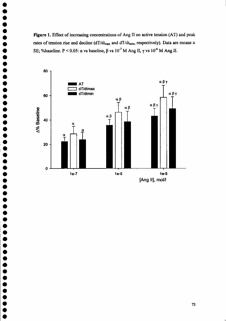
Figure 1. Effect of increasing concentrations of Ang II on active tension (AT) and peak
rates of tension rise and decline (dT/dtmax and dT/dt,™, respectively). Data are means ±
SE; %baseline. P < 0.05: a vs baseline, p vs 10'7 M Ang II, y vs 10"6 M Ang II.
80
AT dT/dtmax dT/dtmin
apy
1e-7 1e-6 1e-5 [Ang II], mol/l
73

Figure 2. Effects of increasing concentrations of Ang II on muscle length expressed as
variation in muscle length relative to Lmax (A%Lmax). Lmax represents maximum
physiological length before the addition of Ang II (baseline). Compared with baseline,
Ang II dose dependently increased muscle length reflecting an increase in myocardial
distensibility. Data are means ± SE; %baseline. P < 0.Ò5: a vs baseline, B vs 10"7 M Ang
I I .yvs lO^MAngl l .
x (0
#
3,0-
2,5-
2,0
< LO H
0,5
0,0 Baseline
ctBy
aB
a
— i —
1e-7 — i — 1e-6
— i 1e-5
[Ang II], M
74

Figure 3. Effects of Ang II (10"5M) in muscle diastolic properties given by the passive
length- tension relation. Ang II induced a rightward shift and a decrease in the curve
slope.
8-1
7 ■
5-
4-
2 -
1 J— 3,80
Basal Length-Tension Relation Ang II Length Tension Relation
— i i
3,90 3,95 Muscle length (mm)
3,85 4,00
75

Figure 4. Effect of Ang II (10"5M) in muscle length (up) and in passive tension after
correction of muscle length to its initial value (down) in different experimental
conditions. Data are means ± SE; % baseline, a vs baseline; P vs Ang II. P < 0.05.
4- |
3-
X (0 E
<
1 ■
1
a B
Angll ZD7155
Angll ZD7155 CHE MIA
-10-
| -20 c o I -
£ -30 '«5 (A S £ gS -40
-50-
-60 J
T ap
Angll Ang II with ZD 7155 Ang II with CHE Ang II with MIA
76

REFERENCES
1 De Gasparo M, Catt KJ, Wright JW, Unger TH. International Union of Pharmacology. XXIII. The Angiotensin II Receptors. Pharmacol Rev, 2000; 52:415-472. 2 Allen AM, Zhuo J, Mendelsohn FA. Localization of angiotensin ATI and AT2 receptors. J Am Soc Nephrol. 1999 Jan; 10 Suppl 1 l:S23-9. 3 Kim S, Iwao H. Molecular and Cellular Mechanisms of Angiotensin II-Mediated Cardiovascular and Renal Diseases. Pharmacol Rev. 2001 52(1): 11-34 4 L, Barsh GS, Pratt RE, Dzau VJ, Kobilka BK. Behavioral and cardiovascular effects of disrupting the angiotensin II type-2 receptor in mice. Nature 1995; 377: 744-747.
5 H, Kurihara T, Yamaki A, Inomata N, Nozawa Y, Mori Y, Murasawa S, Kizima K, Maruyama K, Horiuchi M, Dzau V, Takahashi H, Iwasaka T, Inada M, Matsubara H. Cardiac-specific overexpression of angiotensin II AT-2 receptor causes attenuated response to AT-1 receptor-mediated pressor and chronotropic effects. J Clin Invest 1998; 101 (3): 527-535.
6 H, Kurihara T, Yamaki A, Inomata N, Nozawa Y, Mori Y, Murasawa S, Kizima K, Maruyama K, Horiuchi M, Dzau V, Takahashi H, Iwasaka T, Inada M, Matsubara H. Cardiac-specific overexpression of angiotensin II AT-2 receptor causes attenuated response to AT-1 receptor-mediated pressor and chronotropic effects. J Clin Invest 1998; 101 (3): 527-535.
7 Stoll M, Unger T. Angiotensin and its AT2 receptor: new insights into an old system. Regulatory Peptides 2001; 99: 175-182 8 Modesti P, Vanni S, Bertolozzi L, Cecioni I, Polidori G, Paniccia R, Bandinelli B, Perna A, Liguori P, Boddi M, Galanti G, Serdeneri G. Early sequence of cardiac adaptations and growth factor formation in pressure- and volume- overload hypertrophy. Am J Physiol Heart Circ Physiol 2000; 279: H976-H985
9 Sugden P, Signaling Pathways Activated by Vasoactive Peptides in the Cardiac Myocyte and Their Role in Myocardial Pathologies. J Cardiac Fail 2002; 8 (6): S359-S369 10 Brooks W, Bing O, Robinson K, Slawsky M, Chaletsky D, Conrad C. Effect of Angiotensin-Converting Enzyme Inhibition on Myocardial Fibrosis and Function in Hypertrophied and Failing Myocardium From the Spontaneously Hypertensive Rat. Circulation 1997; 96:4002-4010
77

11 Weber KT, Sun Y, Campbell SE. Structural remodelling of the heart by fibrous tissue: Role of circulating hormones and locally produced peptides. Eur Heart J 1995; 16 (SupplN): 12-18. 12 Berry C, Touys R, Dominiczak AF, Webb RC, Jonhs DG. Angiotensin receptors: signaling, vascular pathophysiology and interactions with ceramide. Am J Physiol Heart Circ Physiol 2001; 281: H2337-H2365. 13 McElmurray III J, Mukherjee R, New R, Sampson A, King M, Hendrick J, Goldberg A, Peterson T, Hallack H, Zile A, Spinale F. Angiotensin-Converting Enzyme and M atrix M etalloproteinase Inhibition w ith D eveloping H eart F ailure: Comparative Effects on Left Ventricular Function and Geometry. J Pharm Exp Therap 1999; 291: 799-811 14 Basso N, Terragno NA. History about the discovery of the Renin-Angiotensin System. Hypertension 2001; 38: 1246-1249 15 Ishihata A, Endoh M. Species-related differences in inotropic effects of Angiotensin II in mammalian ventricular muscle: receptors, subtypes and phosphoinositide hydrolysis. Br J Pharmacol. 1995; 114(2): 447-453. 16 Ahmed SS, Levinson GE, Weisse AB, Regan TJ. The effect of angiotensin on myocardial contractility. J Clin Pharmacol. 1975; 15(4 Pt 1): 276-85. 17 Moravec CS, Schluchter MD, Paranandi L, Czerska B, Stewart RW, Rosenkratz E, Bond M. Inotropic effects of angiotensin 11 on human cardiac muscle in vitro. Circulation 1990; 82: 1973-1984 18 Petroff M, Aiello E, Palomeque J, Salas M, Mattiazzi A. Positive Inotropic and Negative Lusitropic Effect of Angiotensin II: Intracellular Mechanisms and Second Messengers. J Phyisiology 2000; 529(1) : 189-203. 19 Ishihata A, Endoh M. Pharmacological characteristics of the positive inotropic effect of angiotensin II in the rabbit ventricular myocardium. Br J Pharmacol. 1993; 108(4): 999-1005. 20 Petroff M, Aiello E, Palomeque J, Salas M, Mattiazzi A. Subcellular mechanisms of the positive inotropic effect in cat myocardium. J Physiol 2000; 539 (1): 189-203 21 Aiello E, Cingolani H. Angiotensin II stimulates cardiac L-type Ca2+ current by a Ca2+ and protein kinase C-dependent mechanism. Am J Physiol Heart Circ Physiol 2001; 280: H1528-H1536
78

22 Skolnick R, Litwin S, Barry W, Spitzer K. Effect of ANG II on pHi, [Ca2+]i, and contraction in rabbit ventricular myocytes from infarcted hearts. Am J P hysiol H eart Circ Physiol 1998; 275: H1788-H1797 23 Watanabe A, Relationship between the increase in Ca2+ transient and contractile force induced by angiotensin II in aequorin-loaded rabbit ventricular myocardium. Cardio Res 1998; 37: 524-531 24 Leite-Moreira AF, Soares SS, Lima-Carneiro A, Castro-Chaves P. Negative inotropic effect of selective AT2 receptor stimulation and its modulation by endocardial endothelium, NO and prostaglandins. Journal of the American College of Cardiology 2004; 43 (Suppl. A): 492A-493A.
25 Leite-Moreira A, Brás-Silva C, Pedrosa C, Rocha Sousa A. ET-1 increases distensibility of acutely loaded myocardium: a novel ETA and Na+/H+ exchanger effect. Am J Physiol Heart Circ Physiol 2002; 284: H1332-H1339
26 Mebazaa A, De Keulenaer GW, Paqueron X, Andries LJ, Ratajczak P, Lanone S, Frelin C, Longrois D, Payen D, Brutsaert DL, Sys SU. Activation of cardiac endothelium as a compensatory component in endotoxin-induced cardiomyopathy: role of endothelin, prostaglandins, and nitric oxide. Circulation 2001; 104: 3137-44. 27 Ishihata A, Endoh M. Pharmacological characteristics of the positive inotropic effect of angiotensin II in the rabbit ventricular myocardium. Br J Pharmacol 1993 Apr; 108(4): 999-1005 28 Ishihata A, Endoh M. Pharmacological characteristics of the positive inotropic effect of angiotensin II in the rabbit ventricular myocardium. Br J Pharmacol. 1993 Apr;108(4):999-1005.
29 Salas MA, Vila-Petroff MG, Palomeque J, Aiello EA, Mattiazzi A. Positive inotropic and negative lusitropic effect of angiotensin II: intracellular mechanisms and second messengers. J Mol Cell Cardiol. 2001 Nov;33(l 1):1957-71. 30 Wakabayashi S, Shigekawa M, Pouyssegur J. Molecular physiology of vertebrate Na+/H+ exchangers. Physiol Rev 1997; 77:51-74 31 Takabashi E, Abe J, Berk B. Angiotensin II stimulates p90rsk in vascular smooth muscle cells: a potential Na+/H+ exchanger kinase. Circ Res 1997; 81:268 -273 32 Snabaitis AK, Yokoyama H, Avkiran M. Roles of mitogen-activated protein kinases and protein kinase C in alpha(lA)-adrenoceptor-mediated stimulation of the sarcolemmal Na(+)-H(+) exchanger. Circ Res. 2000 Feb 4;86(2):214-20.
79
33 Mayoux E, Coutry N, Lechene P, Marotte F, Hoffmann C, Ventura-Clapier R. Effects of acidosis and alkalosis on mechanical properties of hypertrophiée rat heart fiber bundles. Am J Physiol. 1994 May;266(5 Pt 2):H2051-60. 34 Yamasaki R, Wu Y, McNabb M, Greaser M, Labeit S, Granzier H. Protein kinase A phosphorylates titin's cardiac-specific N2B domain and reduces passive tension in rat cardiac myocytes. Circ Res. 2002 Jun 14;90(11):1181-8. 35 Yamamoto K, Masuyama T, Sakata Y, Nishikawa N , Mano T, Yoshida J, Miwa T, Sugawara M, Yamaguchi Y, Ookawara T, Suzuki K, Hori M. Myocardial stiffness is determined by ventricular fibrosis, but not by compensatory or excessive hypertrophy in hypertensive heart. Cardio Res 2002 Jul; 55(1): 76-82 36HayashiT,SohmiyaK,Ukimura A,EndohS,MoriT,ShimomuraH, OkabeM, Terasaki F and Kitaura Y.Angiotensin II receptor blockade prevents microangiopathy and preserves diastolic function in the diabetic rat heart. Heart 2003; 89:1236-1242 37 Spinale F, Holzgrefe H, Mukherjee R, Hird R, Walker J, Arnim-Barker A, Powell J, Koster W. Angiotensin-Converting Enzyme Inhibition and the Progression of Congestive Cardiomyopathy: Effects on Left Ventricular and Myocyte Structure and Function. Circulation 1995; 92:562-578 38 Kyriakidis M, Triposkiadis F, Dernellis J, Androulakis A, Mellas P, Kelepeshis G, Gialafos J. Effects of Cardiac Versus Circulatory Angiotensin-Converting Enzyme Inhibition on Left Ventricular DiastolicFunction and Coronary Blood Flow in Hypertrophic Obstructive Cardiomyopathy. Circulation. 1998;97:1342-1347 39 Yamamoto K, Masuyama T, Sakata Y, Mano T, Nishikawa N, Kondo H, Akehi H, Kuzuya T, Miwa T, Hori M. Roles of renin-angiotensin and endothelin systems in development of diastolic heart failure in hypertensive hearts. Cardio Res 2000; 47: 274-283 40 Anning P, Grocott-Mason R, Lewis M, Shah A. Enhancement of left ventricular relaxation in the isolated heart by an angiotensin-converting enzyme inhibitor. Circulation 1995; 92: 2660-2665 41 Aartsena W, Hilgersa R, Schiffersa P, Daemenb M, De Meya J, Smitsa J. Changes in Vascular Distensibility during Angiotensin-Converting Enzyme Inhibition Involve Bradykinin Type 2 Receptors. J Vase Res 2004;41:18-27
80

42 Jalowy A, Schulz R, Heusch G. ATI receptor blockade in experimental myocardial ischemia/reperfusion. Basic Res Cardiol 1998; 93(2): 85 - 91 43 Thai H, Van H, Gaballa M, Goldman S, Raya T. Effects of ATI receptor blockade after myocardial infarct on myocardial fibrosis, stiffness, and contractility. Am J Physiol 1999; 276 (45): H873-H880
44 Walsh J, Batin P, Hawkins M, McEntegart D, Cowley A. Ventricular dilatation in the absence of ACE inhibitors: influence of haemodynamic and neurohormonal variables following myocardial infarction. Heart 1999; 81:33-39
45 Stanton L, Garrard L, Damm D, Garrick B, Lam A, Kapoun A, Zheng Q, Protter A,Schreiner G, White R. Altered Patterns of Gene Expression in Response to Myocardial Infarction. Circ Res. 2000; 86:939-945.
46 Barry W, Brooker J, Alderman E, Harrison D. Changes in diastolic stiffness and tone of the left ventricle during angina pectoris. Circulation 1974; 49: 255-263
47 Leite-Moreira A, Correia-Pinto J. Load as an acute determinant of end-diastolic pressura volume relation. Am J Physiol 2001; 280: H51-H58 48 Leite-Moreira A, Correia-Pinto J, Gillebert T. Afterload induced changes in relaxation. A mechanism for diastolic dysfunction. Cardio Res 1999; 43: 344-353 49 Paulus W, Grossman W, Serizawa T, Bourdillon P, Pasipoularides A, Mirsky I. Different effects of two types of ischemia on myocardial systolic and diastolic function. Am J Phyiol 1985; 248: H719-H728
50 Chua B, Chua C, Diglio C, Siu B. Regulation of endothelin-1 mRNA by angiotensin II in rat heart endothelial cells. Biochim Biophys Acta, 1993; 1178: 201-206
51 Kusaka Y, Kelly R, Williams G, Kifor I. Coronary microvascular endothelial cells cosecrete angiotensin II and endothelin-1 via a regulated pathway. Am J Physiol Heart Circ Physiol 2000; 279: H1087-H1096 52 Brutsaert D. Cardiac Endothelial-Myocardial Signaling: Its Role in Cardiac Growth, Contractile Performance, and Rhythmicity. Physiol Rev 2003; 83: 59-115 53 Paulus WJ, Shah AM. NO and cardiac diastolic function. Cardiovasc Res 43: 595-606,1999. 54 Mohan P, Brutsaert D, Sys S. Myocardial performance is modulated by interaction of cardiac endothelium derived nitric oxide and prostaglandins. Cardiovasc Res 1995; 29: 637-640
81

4- Papel da Angiotensina II na modulação da função miocárdica num modelo animal de insuficiência cardíaca

Efeitos Miocárdicos da Angiotensina II no Modelo de Insuficiência
Cardíaca Induzida pela Doxorrubicina
Departamento de Fisiologia, Faculdade de Medicina, Universidade do Porto, Portugal
Correspondência:
Prof. Dr. Adelino Leite-Moreira
Departamento de Fisiologia, Faculdade de Medicina da Universidade do Porto
Alameda Prof. Hernâni Monteiro
4200-319 Porto, PORTUGAL
Tel. +351-22.550.84.52; Fax. +351-22.551.91.94; E-mail: [email protected]

INTRODUÇÃO A doxorrubicina (ou adriamicina) é uma antraciclina usada na prática clínica
oncológica desde a década de 1960. A longo prazo, um dos efeitos laterais mais sérios desta terapêutica consiste na génese de uma cardiomiopatia tóxica, dependente da dose, a qual acaba por conduzir num número significativo de doentes a uma insuficiência cardíaca grave . Num modelo animal de coelho, a administração repetida de doses elevadas de doxorrubicina por via endovenosa causa igualmente uma insuficiência cardíaca de baixo débito, com algumas semelhanças relativamente à doença humana. Caracteriza-se por uma insuficiência biventricular associada a vacuolização dos miócitos e perda marcada de miofibrilas . Alguns dos mecanismos subjacentes a este efeito parecem estar relacionados com génese de radicais livres e peroxidação lipídica, indução de apoptose, inibição da síntese de mRNA e proteínas (a-actina, troponina, cínase da creatina, por exemplo), sobrecarga de cálcio citosólico e diminuição da densidade dos receptores P-adrenérgicos e depleção de noradrenalina .
Desta forma, foi nosso objectivo proceder à implementação do modelo animal de coelho de insuficiência cardíaca induzida pela doxorrubicina e caracterizar os efeitos miocárdicos agudos da Angiotensina II neste modelo.
MÉTODOS O Protocolo de administração da Doxorubicina consistiu na injecção do fármaco
na veia marginal da orelha numa dose de 1 mg/Kg, duas vezes por semana durante 7-9 semanas. Antes dos protocolos em músculo isolado, procedeu-se à realização de registos hemodinâmicos num modelo de coração in situ para avaliar a função sistólica ventricular esquerda: pressão sistólica (P-sist, mmHg), pico de pressão isovolumétrica (P-isov, mmHg) e dP/dtmax (mmHg/s). Após a quantificação da disfunção ventricular, procedeu-se ao isolamento e estabilização dos músculos papilares do ventrículo direito de acordo com a metodologia atrás descrita. Finalmente, os efeitos da adição de concentrações crescentes de Ang II (IO9, IO"8, IO"7, IO"6, IO"5 M) foram estudados em músculos papilares direitos de coelhos Neo-Zelandeses normais e injectados com Doxorrubicina (n=ll e n=8, respectivamente). Parâmetros calculados no músculo isolado: tensão activa (TA; mN/mm2), velocidades máximas de elevação e queda da tensão (dT/dtmax e dT/dtmin, respectivamente; mN/mm2/s), encurtamento máximo (EM; mm), velocidade máxima de encurtamento (dL/dtmax; mm/s) e tempo de abalo
84
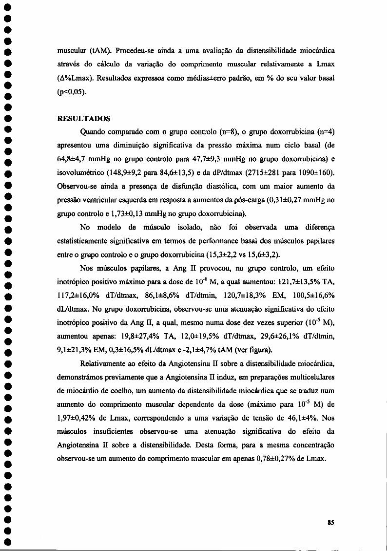
muscular (tAM). Procedeu-se ainda a uma avaliação da distensibilidade miocárdica através do cálculo da variação do comprimento muscular relativamente a Lmax (A%Lmax). Resultados expressos como mcdias±erro padrão, em % do seu valor basal (p<0,05).
RESULTADOS Quando comparado com o grupo controlo (n=8), o grupo doxorrubicina (n=4)
apresentou uma diminuição significativa da pressão máxima num ciclo basal (de 64,8±4,7 mmHg no grupo controlo para 47,7±9,3 mmHg no grupo doxorrubicina) e isovolumétrico (148,9±9,2 para 84,6±13,5) e da dP/dtmax (2715±281 para 1090±160). Observou-se ainda a presença de disfunção diastólica, com um maior aumento da pressão ventricular esquerda em resposta a aumentos da pós-carga (0,31 ±0,27 mmHg no grupo controlo e 1,73±0,13 mmHg no grupo doxorrubicina).
No modelo de músculo isolado, não foi observada uma diferença estatisticamente significativa em termos de performance basal dos músculos papilares entre o grupo controlo e o grupo doxorrubicina (15,3±2,2 vs 15,6±3,2).
Nos músculos papilares, a Ang II provocou, no grupo controlo, um efeito inotrópico positivo máximo para a dose de IO"6 M, a qual aumentou: 121,7±13,5% TA, 117,2±16,0% dT/dtmax, 86,1±8,6% dT/dtmin, 120,7±18,3% EM, 100,5±16,6% dL/dtmax. No grupo doxorrubicina, observou-se uma atenuação significativa do efeito inotrópico positivo da Ang II, a qual, mesmo numa dose dez vezes superior (IO5 M), aumentou apenas: 19,8±27,4% TA, 12,0±19,5% dT/dtmax, 29,6±26,1% dT/dtmin, 9,1±21,3% EM, 0,3±16,5% dL/dtmax e -2,1±4,7% tAM (ver figura).
Relativamente ao efeito da Angiotensina II sobre a distensibilidade miocárdica, demonstrámos previamente que a Angiotensina II induz, em preparações multicelulares de miocárdio de coelho, um aumento da distensibilidade miocárdica que se traduz num aumento do comprimento muscular dependente da dose (máximo para IO"5 M) de 1,97±0,42% de Lmax, correspondendo a uma variação de tensão de 46,1 ±4%. Nos músculos insuficientes observou-se uma atenuação significativa do efeito da Angiotensina II sobre a distensibilidade. Desta forma, para a mesma concentração observou-se um aumento do comprimento muscular em apenas 0,78±0,27% de Lmax.
85

CONCLUSÕES Os resultados aqui apresentados constituem dados preliminares de Protocolos
ainda em curso e que irão ser alvo de estudo mais aprofundado num futuro próximo. A Doxorrubicina, quando administrada segundo o protocolo acima descrito,
induz i nsuficiência p or cardiomiopatia t óxica. O e feito i no trópico p ositivo d a A ng II encontra-se significativamente atenuado neste modelo de insuficiência cardíaca (IC) induzida pela doxorrubicina. Permanece por esclarecer qual o mecanismo subjacente a esta atenuação do efeito inotrópico positivo. Sabendo que, na insuficiência cardíaca, em termos gerais, existe um aumento da expressão de receptores cardíacos AT-2 e que, como foi atras descrito, a estimulação selectiva destes receptores está associada a um efeito inotrópico negativo, parece-nos plausível afirmar que esta atenuação possa estar relacionada com uma maior activação deste subtipo de receptores nos músculos de animais insuficientes. Contudo, esta hipótese permanece ainda por esclarecer.
Relativamente ao efeito da Angiotensina II sobre as propriedades diastólicas do miocárdio, veríficou-se uma atenuação do efeito sobre a distensibilidade nos músculos de animais insuficientes.
O estudo dos efeitos da Ang II neste modelo poderá contribuir para uma melhor compreensão do papel deste mediador neuro-humoral na patofisiopatologia da insuficiência cardíaca.
86
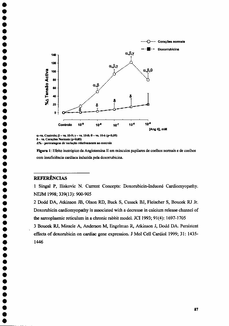
140
120
£ loo ti < 80 O «g 60 c H 40 S 20
a.P,Y
.i
a,p
0 • C : ——"~
O'* 5
Controlo 109 10-« IO7 10-»
O Corações normais
— H • - Doxorrubicina
a,p,e
••••i o
_]. 1— io-«
[Ang iq, mM
a-vs. Controlo; 0 - vs. 10-9; y - vs. 10-8; 0 - vs. 10-6 (p<0,05) 5 - vs. Corações Normais (p<0,05) A% - percentagem de variação relativamente ao controlo
Figura 1: Efeito inotrópico da Angiotensina II em músculos papilares de coelhos normais e de coelhos
com insuficiência cardíaca induzida pela doxorrubicina.
REFERENCIAS 1 Singal P, Uiskovic N. Current Concepts: Doxorubicin-Induced Cardiomyopathy. NEJM 1998; 339(13): 900-905 2 Dodd DA, Atkinson JB, Olson RD, Buck S, Cusack BJ, Fleischer S, Boucek RJ Jr. Doxorubicin cardiomyopathy is associated with a decrease in calcium release channel of the sarcoplasmic reticulum in a chronic rabbit model. JCI1993; 91(4): 1697-1705 3 Boucek RJ, Miracle A, Anderson M, Engelman R, Atkinson J, Dodd DA. Persistent effects of doxorubicin on cardiac gene expression. J Mol Cell Cardiol 1999; 31: 1435-1446
87
Considerações Finais
Conclusões dos Trabalhos Experimentais
Como referido anteriormente, o presente trabalho teve como principal objectivo o estudo da modulação aguda da função miocárdica pela Angiotensina II, num modelo de preparações multicelulares de miocárdio de coelho. Neste sentido, procedeu-se à avaliação do efeito da Angiotensina 11 sobre dois aspectos distintos, contractilidade e distensibilidade miocárdica, em corações de animais saudáveis e com insuficiência cardíaca.
No primeiro estudo apresentado, observou-se que o efeito inotrópico da Ang II depende da presença de um endotélio endocárdico intacto. De facto, a destruição selectiva do endotélio originou uma atenuação significativa do efeito inotrópico positivo da Ang II característico desta espécie animal, sugerindo a presença de um factor endotelial que poderia funcionar como co-factor. Neste sentido, verificou-se que o efeito era igualmente atenuado de forma significativa na presença de antagonistas dos receptores da endotelina, não selectivos e selectivos ETA. Os mecanismos subjacentes a esta interacção aguda ainda não são perfeitamente claros. Embora a Ang II estimule a síntese de novo de endotelina-1 pelas células endoteliais, são necessárias várias horas para que esta síntese se manifeste 97. Contudo, a Ang II é capaz de induzir, em minutos, a libertação de endotelina-1 pré-formada e armazenada nas células endoteliais 9S. Para além disso, a inibição dos receptores ETA com BQ-123 pode permitir uma maior activação dos receptores ETB os quais, a nível cardíaco, têm um efeito inotrópico negativo 90'95. Estes resultados permitem aprofundar o papel do sistema renina-angiotensina-aldosterona e endotelina-1 na modulação aguda da função miocárdica. Para além disso, podem ajudar a explicar algumas das diferenças entre espécies animais encontradas nos efeitos miocárdicos destes mediadores, na medida em que a expressão destes receptores varia com a espécie considerada ' .A descrição desta interacção aguda entre o sistema da Ang II e da endotelina-1 na modulação da função miocárdica parece-nos ainda particularmente interessante já que ambos os sistemas são elementos centrais da fisiopatologia de várias doenças cardiovasculares, como a hipertensão arterial ou a insuficiência cardíaca, constituindo alvos terapêuticos já estabelecidos ou potenciais.
88

No segundo trabalho apresentado, estudaram-se os efeitos miocárdicos da estimulação dos receptores AT2. Em termos gerais, os receptores AT2 parecem apresentar acções fisiológicas em muitos aspectos opostos aos dos receptores ATI. A nível vascular, a estimulação selectiva destes receptores é responsável por um efeito vasodilatador dependente do endotélio e envolvendo diversas vias, como a produção de óxido nítrico, bradicinina ou prostaglandins ou ainda a activação de canais de potássio. A nível cardíaco, embora exista uma expressão substancial destes receptores (na espécie considerada os receptores AT2 ventricular constituem cerca de 20% dos receptores da Ang II), o seu papel na modulação aguda da contractilidade miocárdica permanecia ainda por esclarecer. O estudo realizado permitiu descrever, pela primeira vez, um efeito inotrópico negativo resultante da activação destes receptores. Este efeito foi praticamente anulado após a destruição selectiva do endotélio endocárdico. A nível vascular o efeito vasodilatador induzido pelos receptores AT2 depende da libertação de factores endoteliais. Contudo, a inibição da síntese de óxido nítrico, prostaglandinas e factor hiperpolarizante derivado do endotélio não alterou significativamente o efeito observado. Existem ainda outras vias a testar e que poderão ser responsáveis pelos resultados obtidos, como a activação dos receptores B2 da bradicinina. Este aspecto será avaliado em estudos próximos. Os resultados deste trabalho contribuem para o esclarecimento do papel dos receptores AT2 na modulação aguda da função miocárdica e podem ainda ajudar a compreender melhor alguns dos efeitos dos antagonistas dos receptores ATI.
Nos dois trabalhos anteriores procedeu-se ao estudo dos efeitos da Ang II sobre a contractilidade cardíaca. No trabalho seguinte, a nossa atenção incidiu sobre os efeitos agudos deste mediador na distensibilidade miocárdica. Este aspecto permanece ainda largamente desconhecido. Sabe-se que a angiotensina II, ao promover a fibrose e a proliferação celular, pode alterar cronicamente as propriedades diastólicas do miocárdio114. Recentemente, demonstrou-se que a endotelina-1 aumenta agudamente a distensibilidade miocárdica em músculos humanos e animais, sendo este efeito mediado pelos receptores ETA e pelo trocador Na+/H+n5. Contudo, o efeito da Angiotensina II sobre a modulação aguda da distensibilidade permanecia por esclarecer. O modelo do músculo isolado é particularmente indicado para realizar este estudo na medida em que permite uma avaliação rigorosa das propriedades mecânicas do miocárdio independentemente de factores extrínsecos, como a perfusão coronária. Desta forma, verificámos que a Angiotensina II induz, pela sua ligação a receptores ATI, um
89
aumento significativo da distensibilidade miocárdica, o qual é atenuado pela inibição da proteína cínase C e do trocador Na'VtT'". Este efeito poderá ser particularmente importante na adaptação miocárdica a variações da carga. Assim, na sequência de uma sobrecarga cardíaca aguda, a qual está associada à libertação parácrina de angiotensina II, este mediador poderá induzir uma maior distensibilidade miocárdica, permitindo um aumento do volume ventricular sem grande variação da pressão intra-cavitária, facilitando o enchimento ventricular. Embora este mecanismo seja benéfico a curto prazo, a longo prazo os seus efeitos podem eventualmente tornar-se deletérios e contribuir para a dilatação ventricular. Finalmente, demonstrámos que, juntamente com a carga, o óxido nítrico e a endotelina-1, a angiotensina II é capaz de modular agudamente a relação passiva tensão-comprimento miocárdica.
Finalmente, procedemos à avaliação dos efeitos da Angiotensina II sobre a contractilidade e sobre a distensibilidade num modelo animal de insuficiência cardíaca induzida pela doxorrubicina. As conclusões apresentadas neste último trabalho são ainda fruto de resultados preliminares. Contudo, foi possível observar que nestes corações, o efeito inotrópico positivo da Angiotensina II se encontra significativamente atenuado. Relativamente às propriedades diastólicas do miocárdio, observou-se nos músculos dos animais insuficientes uma atenuação significativa do efeito sobre a distensibilidade, traduzindo maior rigidez miocárdica. Permanecem ainda por esclarecer os mecanismos subjacentes a estes resultados e que serão alvo de Protocolos experimentais a realizar no futuro.
Desta forma, estes resultados contribuem para esclarecer o papel da Angiotensina II na modulação aguda da função miocárdica.
90

Trabalhos elaborados na sequência dos Protocolos Experimentais
Artigos por extenso:
1. Castro-Chaves P, Leite-Moreira A. Sistema Renina-Angiotensina e seu papel na fisiopatologia e terapêutica cardiovascular. Revista Portuguesa de Cardiologia. Maio 2004; 23 Suplem 2:1161-77
2. Castro-Chaves P, Roncon-Albuquerque R Jr, Leite-Moreira. ETA Receptors and Endothelium Partially Mediate the Positive Inotropic Effect of Angiotensin II (submetido)
3. Leite-Moreira A, Castro-ChavesP, Nunes P, Lima-Carneiro A. Angiotensin II increases myocardial distensibility: a novel ATI mediated effect, (submetido)
4. Negative Inotropic Effect of Selective AT2 Receptor Stimulation and its Modulation by Endocardial Endothelium, NO, Prostaglandins and EDHF (em preparação).
Apresentações em Congressos Internacionais: 1. Castro-Chaves P, Roncon-Albuquerque R Jr, Leite-Moreira AF. Modulation of
the positive inotropic effect of angiotensin II by the endocardial endothelium and endothelin-1. European Heart Journal. 2002; 23 (Abstr. Suppl.): 648.
2. Castro-Chaves P, Roncon-Albuquerque R Jr , Leite-Moreira AF. Modulation of the Positive Inotropic Effect of Angiotensin II by the Endocardial Endothelium and Endothelin-1. Circulation 106 (19): 131 Suppl. S NOV 5 2002.
3. Leite-Moreira AF, Soares SS, Lima-Carneiro A, Castro-Chaves P. Negative inotropic effect of selective AT2 receptor stimulation and its modulation by endocardial endothelium, NO and prostaglandins. Journal of the American College of Cardiology 2004; 43 (Suppl. A): 492A-493A.
4. Leite-Moreira AF, Soares SS, Lima-Carneiro A, Castro-Chaves P. Negative inotropic effect of selective AT2 receptor stimulation and its modulation by endocardial endothelium, nitric oxide, prostaglandins and endothelium-derived hyperpolarizing factor. European Heart Journal 2004 25: 36-37 Suppl.
5. Castro-Chaves P, Lima Carneiro A, Nunes P, Soares SS, Leite-Moreira AF. Angiotensin II increases myocardial distensibility: a novel endothelial and ATI mediated effect. European Heart Journal 2004 25: 282 (Suppl).
91

6. Leite-Moreira AF, Castro-Chaves P, Lima Carneiro A, Nunes P, Soares SS. Angiotensin II increases myocardial distensibility: a novel endothelial and ATI mediated effect. American Heart Association Scientific Sessions, 7-10 November, New Orleans, Louisiana, USA.
Apresentações em Congressos Nacionais: 1. Castro-Chaves P, Roncon-Albuquerque R Jr, Lima-Carneiro A, Lopes T, Leite-
Moreira AF. O efeito inotrópico positivo da angiotensina II é modulado pelo endotélio endocárdico e pela endotelina-1. Revista Portuguesa de Cardiologia 2002; 21 (supl. I): 22.
2. Castro-Chaves P, Lima-Carneiro A, Guerra M, Leite-Moreira A. Atenuação do efeito inotrópico positivo da angiotensina II no modelo de insuficiência cardíaca induzida pela doxorrubicina. Revista Portuguesa de Cardiologia 2003, 22 (supl. IH), 20.
3. Lima-Carneiro A, C astro-Chaves P, S oares JB, Miguelote R, S oares S, L eite-Moreira A. Efeitos miocárdicos da estimulação selectiva dos receptores tipo 2 da angiotensina II. Revista Portuguesa de Cardiologia 2003,22 (supl. III), 21.
4. Guerra M, Castro-Chaves P, Brás-Silva C, Leite-Moreira A. Caracterização da disfunção diastólica no modelo de insuficiência cardíaca induzida pela doxorrubicina. Revista Portuguesa de Cardiologia 2003,22 (supl. III), 145.
5. Castro-Chaves P, Lima-Carneiro A, Nunes P, Soares S, Leite-Moreira A. Aumento da distensibilidade miocárdica pela angiotensina II e sua dependência dos receptores ATI e do endotélio. Revista Port Cardiologia 2004; 23 (supl. I): 1-35
6. Soares S, Castro-Chaves P, Leite-Moreira A. Efeito inotrópico negativo da estimulação selectiva AT2 e sua modulação pelo endotélio, NO, prostaglandinas e EDHF. Revista Port Cardiol 2004; 23 (supl I): 1-98.
Trabalhos em Curso: 1. Efeitos Miocárdicos da Angiotensina II no Modelo de Insuficiência Cardíaca
Induzida pela Doxorrubicina
92

Referências 1 Tigerstedt R, Bergman PG. Niere und Kreislauf. Skand Arch Physiol. 1898; 8:223-271. 2 Braun-Menéndez E, Fasciolo JC, Leloir LF, Muãoz JM. La sustancia hipertensora de la sangre dei rinón isquemiado. Rev Soc Arg Biol. 1939; 15:420 -425. 3 Braun-Menéndez E, Fasciolo JC, Leloir LF, Munoz JM, Taquini AC. Hipertensión Arterial Nerrógena. Buenos Aires, Argentina: El Ateneo; 1943. 4 Page IH, Helmer OM. A crystalline pressor substance: angiotonin. Proc Center Soc Clin Invest. 1939;12:17. 5 Braun-Menéndez E, Page IH. Suggested revision of nomenclature: an-giotensin. Science. 1958;127:242. 6 Basso N, Terragno NA. History about the discovery of the Renin-Angiotensin System. Hypertension 2001;38:1246-1249. 7 Dzau VJ, Gibbons GH. Autocrine and paracrine mechanisms of vascular myocytes in systemic hypertension. Am J Cardiol 1987; 60: 991-1031. 8 Weber KT, Sun Y, Campbell SE. Structural remodelling of the heart by fibrous tissue: Role of circulating hormones and locally produced peptides. Eur Heart J 1995; 16 (Suppl N): 12-18. 9 Touys RM, Schiffrin EL. Signal transduction mechanisms mediating the physiological and pathophysiological actions of angiotensin II in vascular smooth cells. Pharmacol Rev, 2000; 52: 639-672.
De G asparo M, Catt K J, W right JW, U nger T H. I nternational U nion o f P harmacology. X XIII. The Angiotensin II Receptors. Pharmacol Rev, 2000; 52: 415-472. 11 Ruiz-Ortega M, Lorenzo O, Rupérez M, Esteban V, Susuki Y, Mezzano S, Plaza JJ, Egido J. Role of the Renin-Angiotensin System in Vascular Diseases - Expanding the field. Hypertension, 2001; 38: 1382-1387. 12 Kim S, Iawo H. Molecular and Cellular Mechanisms of Angiotensin II - Mediated Cardiovascular and Renal Diseases. Pharmacol Rev, 2001; 52 (1): 11-34 13 Skeggs LT, Lentz KE, Gould AB, Hochstrasser H Kahn Jr. Biochemistry and kinetics of the renin-angiotensin system. Fed Proc; 1967; 26:42-47. 14 Ferrario CM, Chappell MC, Tallant EA, Brosnihan KB, Diz DI. Counter-regulatory actions of angiotensin (1-7). Hypertension 1997: 30:535-541. 15 Esther CR, Marino EM, Howard TE. The critical role of tissue angiotensin-converting enzmes as revealed by gene targeting in mice. J Clin Invest 1997; 99:2375-2385. 16 Dostal DE, Baker KM. The cardiac renin angiotensin system: conceptual or a regulator of cardiac function? Circ Res 1999; 85: 643-650. 17 Gohlke P, Bunning P, Unger T. Distribution and metabolism of angiotensin I and II in the blood vessel wall. Hypertension 1992; 20: 151-157. 18 Wiemer G, Scholkens BA, Becker RHA, Busse R. Ramiprilat enhances endothelial autacoid formation by inhibiting breakdown of endothelium-derived bradikinin. Hypertension 1991; 18: 558-563. 1 Linz W, Wohlfart P, Scholkens BA, Malinski T, Wiemer G. Interactions among ACE, Kinins and NO. Cardiov Res 1999; 43: 549-561. 20 Welches WR, Brosnihan KB, Ferrario CM. A comparison of the properties and enzymatic activities of three angiotensin processing enzymes: angiotensin converting enzyme, prolyl endopeptidase and neutral endopeptidase. Life Sci 1993; 52: 1461-1480. 21 Ferrario CM. Angiotensin 1-7 and antihypertensive mechanisms. J Nephrol 1998; 11: 278-283. 22 Tallant EA, Lu X, Weiss RB, Cappell MC, Ferrario CM. Bovine aortic endothelial cells contain an angiotensin 1-7 receptor. Hypertension 1997: 29 (part 2): 388-389 23 Seyedi N, Xu X, Nasjletti A, Hintze TH. Coronary kinin generation mediates nitric oxide release after angiotensin receptor stimulation. Hypertension 1995; 26: 164-170. 24 Li P, Chappell MC, Ferrario CM, Brosnihan KB. Angiotensin 1-7 augments bradykinin-induced vasodilation by competing with ACE and releasing nitric oxide. Hypertension 1997 (part 2) 9: 394-400. 25 Moriguchi A, Brosnihan KB, Kumagai H, Ganten D, Ferrario CM. Mechanism of hypertension in transgenic rats expressing the mouse ren-2-gene. Am J Physiol 1994; 266: R1273-R1279. 26 Loot AE, Roks AJ, Henning RH, Tio RA, Suurmeijer AJ, Boomsma F, van Gilst WH. Angiotensin-^ 1-7) attenuates the development of heart failure after myocardial infarction in rats. Circulation. 2002;105(13): 1548-50. 27 Strawn WB, Ferrario CM, Tallant EA. Angiotensin 1-7 reduces smooth muscle growth after vascular injury. Hypertension 1999; 33 (part 1): 207-211.
93
28 Ferrario CM. Does Angiotensin 1-7 contribute to cardiac adaptation and preservation of endothelial function in heart failure? Circulation 2002; 105: 1523-1525. 29 Allen AM, Zhuo J, Mendelsohn FA. Localization of angiotensin ATI and AT2 receptors. J Am Soc Nephrol. 1999 Jan;10 Suppl 1 l:S23-9. 30 Tsutsumi K, Saavedra JM. Characterization of AT-2 angiotensin recetors in rat anterior cerebral arteries. Am J Physiol 1991; 261: H667-H670. 31 Jackson EK, Herzer WA. Regional vascular selectivity of Ang II. J Pharmacol Exp Ther 2001; 297:736-745. 32 Lopez JJ, Lorell BH, Ingelfinger JR, Weinberg EO, Schunkert H, Diamant D, Tang SS. Distribution and function of cardiac angiotensin ATI and AT-2 receptor subtypes in hypertrophied rat hearts. Am J Physiol 1994; 267: H844-H852. 33 Wharton J, Morgan K, Rutherford RAD, Catravas JD, Chester A, Whitehead BF, de Levai MR Yacoub MH, Polack JM. Differential distribution of angiotensin AT-2 receptors in the normal and failing human heart. J Pharmacol Exp Ther 1998; 284: 323-336. 34 Wagner J, Gehlen F, Ciechanowicz A, Ritz E.Angiotensin II receptor typel gene expression in human glomerulonephritis and diabetes mellitus. J Am Soc Nephrol 1999; 10:545-551. 35 Oliverio MI, Kim H-S, Ito M, Le T, Audoly L, Best CF, Hiller S, Kluckman K, Maeda N, Smithies O, Coffman TM. Reduced growth, abnormal kidney struc-ture, and type 2 (AT 2 ) angiotensin receptor-mediated blood pressure regulation in mice lacking both AT 1A and AT IB receptors for angiotensin II. Proc Nad Acad Sci USA 1998; 95:15496-15501. 36 De Gasparo M, Wbitebread S, Kalenga MK, de Hertogh R Crevoisier P, Thomas K. Down regulation of the angiotensin II receptor subtype AT 2 in human myometrium during pregancy. Regul Pept 1994; 53:39-45. 37 Busche S, Gallinat S, Bohle RM, Reinecke A, Seebeck J, Franke F, Fink L, Zhu M, Sumners C, Unger T. E xpression o f a ngiotensin A T( 1 ) a nd A T(2) r eceptors i n a dult r at c ardiomyocytes a fter myocardial infarction. A single-cell reverse transcriptase-polymerase chain reaction study. Am J Pathol. 2000;157(2):605-11. 38 Hein L, Barsh GS, Pratt RE, Dzau VJ, Kobilka BK. Behavioral and cardiovascular effects of disrupting the angiotensin II type-2 receptor in mice. Nature 1995; 377: 744-747. 39 Masaki H, Kurihara T, Yamaki A, Inomata N, Nozawa Y, Mori Y, Murasawa S, Kizima K, Maruyama K, Horiuchi M, Dzau V, Takahashi H, Iwasaka T, Inada M, Matsubara H. Cardiac-specific overexpression of angiotensin II AT-2 receptor causes attenuated response to ATI receptor-mediated pressor and chronotropic effects. J Clin Invest 1998; 101 (3): 527-535.
Chaki S, Inagami T. Identification and characterization of a new binding site for angiotensin II in mouse neuroblastoma neuro-2A cells. Biochem Biophys Res Commun 1992; 182:388-394. 41 Roberts KA, Krebs LT, Kramer EA, Shaffer MJ, Harding JW, Wright JW. Autoradiographic identification of brain angiotensin IV binding sites and differential c-Fos expression following intracerebroventricular injection of angiotensin II and IV in rats. Brain Res 1995; 682:13-21. 42 Hein L, Meinel L, Pratt RL, Dzau VJ, Kobilka BE. Intracellular trafficking of angiotensin II an dits ATI and AT-2 receptors: evidence for selective sorting of receptor and ligand. Mol Endocrinol 1997; 11 : 1266-1277. 43 De Mello WC, Danser AHJ. Angiotensin and the heart. On the intracrine renin-angiotensin systema. Hypertension 2000; 1183-1118. 44 Berry C, Touys R, Dominiczak AF, Webb RC, Jonhs DG. Angiotensin receptors: signaling, vascular pathophysiology and interactions with ceramide. Am J Physiol Heart Circ Physiol 2001; 281: H2337-H2365. 45 Zhang C, Hein TW, Wang W, Kuo L. Divergent roles of angiotensin II ATI and AT-2 receptors in modulating coronary microvascular function. Circ Res. 2003 ;92(3):322-9. 46 Weber KT. Aldosterone in congestive heart failure. NEJM 2001; 345 (23): 1689-1697. 47 Silvestre JS, Heymes C, Oubenaissa A, Robert V, Aupetit-Faisant B, Carayon A, Swynghedauw B, Delcayre C. Activation of cardiac aldosterone production in rat myocardial infarction: effect of angiotensin II receptor blockade and role in cardiac fibrosis. Circulation. 1999;99(20):2694-701. 48 Noda K, Feng YH, Liu XP, Saad Y, Husain A, Karnik SS. The active state of the AT 1 angiotensin receptor is generated by angiotensin II induction. Biochemistry 1996;35:16435-16442. 49 Rhee SG, Choi KD. Regulation of inositol phospholipid-specific phospho-lipase C isozymes. J Biol Chem 1992; 267:12393-12396. 50 Ushio-Fukai M, Alexander RW, Akers M, Griendling KK. p38 Mitogen-activated protein kinase is a critical component of the redox-sensitive signaling pathways activated by angiotensin II. Role in vascular smooth muscle cell hypertrophy. J Biol Chem 1998; 271:15022-15029.
94
51 Griendling KK, Tsuda T, Berk BC, Alexander RW. Angiotensin II stimulation of vascular smooth muscle. J Cardiovasc Pharmacol 1989; 14 (Suppl 6):S27-S33. 52 Touyz RM, Schiffhn EL. Role of calcium influx and intracellular calcium stores in angiotensin II-mediated calcium hyperresponsiveness in smooth muscle from spontaneously hypertensive rats. J Hypertens 1997; 15:1431-1439. 53 Touyz RM, He G, Deng L Y, Schiffhn EL. Role of extracellular signal-regulated kinases in angiotensin II-stimulated contraction of smooth muscle cells from human resistance vessels. Circulation 1999; 99:392-399. 54 Damron DS, Nadim HS, Hong SJ, Darvish A, Murray PA. Intracellular translocation of PKC iso forms in canine pulmonary artery smooth muscle cells by Ang II. Am J Physiol 1998; 274:L278-L288. 55 Touyz RM, El Mabrouk M, Schiffhn EL. MEK inhibition attenuates Ang II-mediated signaling and contraction in SHR vascular smooth muscle cells. Circ Res 1999; 84:505-515. 56 Li JS, Touyz RM, Schiffhn EL. Effects of AT 1 and AT 2 receptor antagonists on blood pressure and small artery structure in angiotensin II-infused rats. Hypertension 1998; 31:487-492. 57 Balmforth AJ, Shepherd FH, Warburton P, Ball SG. Evidence of an important and direct role for protein kinase C in agonist-induced phosphorylation leading to desensitization of the angiotensin AT 1A receptor. Br J Pharmacol 1997; 122:1469-1477. 58 Sayeski PP, Ali MS, Harp JB, Marrero MB, Bernstein KE. Phosphorylation of pi 30 Cas by angiotensin II is dependent on c-Src, intracellular Ca 21, and protein kinase C. Circ Res 1998; 82: 1279-1288. 59 Hefti MA, Harder BA, Eppenberger HM, Schaub MC. Signaling pathways in cardiac myocyte hypertrophy. J Mol Cell Cardiol 1997; 29:2873-2892.
Howe A, Aplin AE, Alahari SK, Juliano RL. Integrin signaling and cell growth control. Curr Opin Cell Biol 1998; 10:220-231. 61 Honda H, Oda H, Nakamoto T, Honda Z, Sakai R, Suzuki T, Saito T, Nakamura K, Nakao K, Ishikawa T, Katsuki M, Yazaki M, Hirai H. Cardiovascular anomaly, impaired actin bundling and resistance to Src-induced transformation in mice lacking pl30Cas. Nat Genet 1998; 19:361-365. 62 Leevers SJ, Vanhaesebroek B, Waterfield MD. Signaling through phos-phoinositide 3-kinases: The lipids take centre stage. Curr Opin Cell Biol 1999; 11:219-225. 6 Sudgen PH, Clerk A. Stress responsive mitogen activated protein kinases in the myocardium. Circ Res 1998; 24; 345-352. 64 Bueno OF, Molkentin JD. Involvement of extracellular signal-regulated kinases 1/2 in cardiac hypertrophy and cell death. Circ Res 2002; 91: 776-781. 6 New L, Han J. The p38 MAP Kinase pathway and its biological function. Trends Cardiovasc Med 1998;8:220-229. 66 Dulin NO, Alexander LD, Harrwalkar S, Falck Jr, Douglas JG. Phospho-lipase A2-mediated activation of mitogen-activated protein kinase by angiotensin II. Proc Natl Acad Sci USA 1998; 95:8098-8102. 67 Lassègue B, Alexander RW, Clark M, Akers MA, Griendling KK. Phosphatidylcholine is a major source of phosphatidic acid and diacylglycerol in angio-tensin II-stimulated vascular smooth muscle cells. Biochem J 1993;292:509-517. 68 Tsutsumi Y, Matsubara H, Masaki H, Kurihara H, Murasawa S, Takai S, Miyazaki M, Nozawa Y, Ozono R, Nakagawa K, Miwa T, Kawada N, Moh Y, Shibasaki Y, Tanaka Y, Fujiyama S, Koyama Y, Fujiyama A, Takahashi H, Iwasaka T. Angiotensin II type 2 receptor overexpression activates the vascular kinin system and causes vasodilation. J Clin Invest. 1999; 104(7):925-35. 69 Caputo L, Benessiano J, Boulanger CM, Levy BI. Angiotensin II increases cGMP content via endothelial angiotensin II AT 1 subtype receptors in the rat carotid artery. Arterioscler Thromb Vase Biol 1995;15:1646-1651. 70 Mclntyre M, Bohr DF, Dominiczak AF. Endothelial functin in hypertension. The role of superoxide anion. Hypertension 1999; 34:539-545.
Landmesser U, Spiekermann S, Dikalov S, Tatge H, Wilke R, Kohler C, Harrison DG, Hornig B, Drexler H. Vascular oxidative stress and endothelial dysfunction in patients with chronic heart failure: role of xanthine-oxidase and extracellular superoxide dismutase. Circulation. 2002; 10;106(24):3073-8. 72 Schultz Jel J, Witt SA, Glascock BJ, Nieman ML, Reiser PJ, Nix SL, Kimball TR, Doetschman T. TGF-betal mediates the hypertrophic cardiomyocyte growth induced by angiotensin II. J Clin Invest. 2002;109(6):787-96. 73 Moreau P, d'Uscio LV, Shaw S, Takase H, Barton M, Luscher TF. Angiotensin II increases tissue endothelin and induces vascular hypertrophy: reversal by ET(A)-receptor antagonist Circulation. 1997;96(5): 1593-7. 74 Chen YQ, Gilliam DM, Rydzewski B, Naftilan AJ. Multiple enhancer elements mediate induction of c-fos in vascular smooth muscle cells. Hypertension 1998; 27:1224-1233.
95

73 Nadal-Ginard B, Kajstura J, Leri A, Anversa P. Myocyte death, growth, and regeneration in cardiac hypertrophy and failure. Circ Res. 2003;92(2): 139-50. 7 Diep QN, El Mabrouk M, Yue P, Schiffrin EL. Effect of AT(1) receptor blockade on cardiac apoptosis in angiotensin II-induced hypertension. Am J Physiol Heart Circ Physiol. 2002;282(5):H 1635-41. 77 Gonzalez A, Lopez B, Querejeta R, Diez J. Regulation of myocardial fibrillar collagen by angiotensin II. A role in hypertensive heart disease? J Mol Cell Cardiol. 2002;34( 12): 1585-93. 78 Van Leeuwen RTJ, Kol A, Andreotti F, Kluft C, Maseri A, Sperti G. Angiotensin II increases plasminogen activator inhibitor type 1 and tissue-type plasminogen activator messenger RNA in cultured rat aortic smooth muscle cells. Circulation 1994; 90: 362-368. 79 Rakugi H, Wang D, Dzau VJ, Pratt RE. Potential importance of tissue angiotensin converting enzyme inhibition in preventing neointima formation. Circulation 1994; 90: 449-455. 80 Horiuchi M, Akishiita M, Dzau VJ. Recent progress in angiotensin II type 2 receptor research in the cardiovascular system. Hypertension 1999; 33:613-621. 81 Abdalla S, Lother H, Abdel-Tawab AM, Quitterer U. The Angiotensin IIAT-2 receptor is an ATI receptor antagonist. J Biol Chem 2001; 276 (43): 39721-39726. 82 Ford WR, Clanachan AS, Jugdutt BI. Opposite effects of angiotensin ATI and AT-2 receptor antagonists on recovery of mechanical function after ischemia-reperfusion in isolated working rat hearts. Circulation 1996;94:3087-3089. 83 Levy BI, Benessiano J, Henrion D, Caputo L, Heymes C, Duriez M, Poitevin P, Samuel JL. Chronic blockade of AT-2-subtype receptors prevents the effect of angiotensin II on the rat vascular structure. J Clin Invest 1996; 98:418-425. 84 Ruiz-Ortega M, Lorenzo O, Ruperez M, Konig S, Wittig B, Egido J. Angiotensin II activates nuclear transcription factor-kB through ATI and AT-2 in vascular smooth muscle cells: molecular mechanisms. Circ Res 2000; 86: 1266-1272. 83 Xu Y, Clanachan A, Jugdutt B. Enhanced expression of angiotensin II type 2 receptor, IP3 and protein kinase ce during cardioprotection induced by angiotensin II type 2 receptor blockade. Hypertension 2000; 35:506-510. 86 Opie L, Sack MN. Enhanced angiotensin II activity in heart failure: réévaluation of the counterregulatory hypothesis of receptor subtypes. Cir Res 2001; 88: 654-658. 87 Kurisu S, Ozono R, Oshima T, Kambe M, Ishida T, Sugino H, Matsuura H, Chayama K, Teranishi Y, Iba O, Amano K, Matsubara H. Cardiac angiotensin II type 2 receptor activates the kinin/NO system and inhibits fibrosis. Hypertension. 2003;41(1):99-107. 88 Dostal DE, Baker KM. Angiotensin and endothelin: messengers that couple ventricular stretch to the Na+/H+ exchanger and cardiac hypertrophy. Circ Res 1998; 83: 870-873. 89 Rossi GP, Sacchetto A, Cesari M, Pessina AC. Interactions between endothelin-1 and the renin-angiotensin-aldosterone system. Cardiovasc Res. 1999;43(2):300-7 90 Leite-Moreira AF, Bras-Silva C. Inotropic effects of ETB receptor stimulation and their modulation by endocardial endothelium, NO, and prostaglandins. Am J Physiol Heart Circ Physiol. 2004 Sep;287(3):H 1194-9. 91 Herizi A, Jover B, Bouriquet N, Mimran A. Prevention of the cardiovascular and renal effects of angiotensin II by endothelin blockade. Hypertension. 1998 Jan;31(1): 10-4. 92 d'Uscio LV, Moreau P, Shaw S, Takase H, Barton M, Luscher TF. Effects of chronic ETA-receptor blockade in angiotensin II-induced hypertension. Hypertension. 1997 Jan;29(l Pt 2):435-41. 93 Chatziantoniou C, Dussaule JC. Endothelin and renal vascular fibrosis: of mice and men. Curr Opin Nephrol Hypertens. 2000 Jan;9(l):31-6. 94 Nakano A, Kishi F, Minami K, Wakabayashi H, Nakaya Y, Kido H. Selective conversion of big endothelins to tracheal smooth muscle-constricting 31-amino acid-length endothelins by chymase from human mast cells. J Immunol. 1997 ;159(4):1987-92. 95 Riggleman A, Harvey J, Baylis C. Endothelin mediates some of the renal actions of acutely administered angiotensin II. Hypertension. 2001 Jul;38(l): 105-9. 96 Montanari A, Biggi A, Carra N, Ziliotti M, Fasoli E, Musiari L, Perinotto P, Novarini A. Endothelin-A receptors mediate renal hemodynamic effects of exogenous Angiotensin II in humans. Hypertension. 2003 Oct;42(4):825-30. 97 Ito H, Hirata Y, Adachi S, Tanaka M, Tsujino M, Koike A, Nogami A, Murumo F, Hiroe M. Endothelin-1 is an autocrine/paracrine factor in the mechanism of angiotensin II-induced hypertrophy in cultured rat cardiomyocytes. J Clin Invest. 1993 ;92(l):398-403. 98 Ferri C, Desideri G, Baldoncini R, Bellini C, Valenti M, Santucci A, De Mattia G. Angiotensin II increases the release of endothelin-1 from human cultured endothelial cells but does not regulate its circulating levels. Clin Sci (Lond). 1999 Mar;96(3):261-70.
96
99 Bodin P, Travo C, Stoclet JC , Travo P. High sensitivity of hypertensive aortic myocytes to norepinephrine and angiotensin. Am J Physiol 1993; 264:C441-C445. 100 Kim HS, Krege JH, Kluckman KD, Hagaman JR, Hodgin JB, Best CF, Jennette JC, Coffman TM, Maeda N, Smithies O. Genetic control of blood pressure and the angiotensinogen locus. Proc Nad Acad Sei U S A. 1995 Mar 28;92(7):2735-9. 101 Lee WK, Padmanabhan S, Dominiczak AF. Genetics of hypertension: from experimental models to clinical applications. J Hum Hypertens. 2000 Oct-Nov; 14(10-11):631-47. 102 Serneri GG, Boddi M, Cecioni I, Vanni S, Coppo M, Papa ML, Bandinelli B, Bertolozzi I, Polidori G, Toscano T, Maccherini M, Modesti PA. Cardiac angiotensin II formation in the clinical course of heart failure and its relationship with left ventricular function. Circ Res. 2001 ;88(9):961-8. 103 Paradis P, Dali-Youcef N, Paradis FW, Thibault G, Nemer M. Overexpression of angiotensin II type 1 receptor in cardiomyocytes induces cardiac hypertrophy and remodelling. Proc Natl Acad Sci USA 2000; 97:931-936. 104 Matsubara H. Pathophysiological role of angiotensin II type 2 receptor in cardiovascular and renal diseases. Circ Res 1998; 83:1182-1191. 105 Schneider MD, Lorell BH. AT-2 judgement day. Which angiotensin receptor is the culprit in cardiac hypertrophy? Circulation 2001; 104: 247-248. 1 Ichihara S, Senbonmatsu T, Price E Jr, Ichiki T, Gaffney FA, Inagami T. Angiotensin II type 2 receptor is essential for left ventricular hypertrophy and cardiac fibrosis in chronic angiotensin II-induced hypertension. Circulation. 2001;104(3):346-51. 1 Kim S, Wanibuchi H, Hamaguchi A, Miura K, Yamanaka S, Iwao H. Angiotensin blockade improves cardiac and renal complications of type II diabetic rats. Hypertension 1997; 30:1054-1061. 108 Border WA, Noble NA. Interactions of transforming growth factor-beta and angiotensin II in renal fibrosis. Hypertension 1998;31:181-188. 109 Taubman MB. Angiotensin II: a vasoactive hormone with ever increasing biological roles. Cir Res 2003;92:9-11. 110 Campos AH, Zhao Y, Pollman MJ, Gibbons GH. DNA microarray profiling to identify angiotensin-responsive genes in vascular smooth muscle cells: potential mediators of vascular disease. Circ Res. 2003; 10;92(1):111-8 111 Mebazaa A, De Keulenaer GW, Paqueron X, Andries LJ, Ratajczak P, Lanone S, Frelin C, Longrois D, Payen D, Brutsaert DL, Sys SU. Activation of cardiac endothelium as a compensatory component in endotoxin-induced cardiomyopathy: role of endothelin, prostaglandins, and nitric oxide. Circulation 2001; 104:3137-44. 112 Molenaar P, OReilly G, Sharkey A, Kuc RE, Harding DP, Plumpton C, Gresham GA, Davenport AP. Characterization and localization of endothelin receptor subtypes in the human atrioventricular conducting system and myocardium. Circ Res. 1993 Mar;72(3):526-38 113 Kobayashi T, Miyauchi T, Sakai S, Kobayashi M, Yamaguchi I, Goto K, Sugishita Y. Expression of endothelin-1, ETA and ETB receptors, and ECE and distribution of endothelin-1 in failing rat heart. Am J Physiol. 1999 Apr;276(4 Pt 2):H1197-206. 114 McElmurray III J, Mukherjee R New R Sampson A, King M, Hendrick J, Goldberg A, Peterson T, Hallack H, Zile A, Spinale F. Angiotensin-Converting Enzyme and Matrix Metalloproteinase Inhibition with Developing Heart Failure: Comparative Effects on Left Ventricular Function and Geometry. J Pharm Exp Therap 1999; 291: 799-811
Leite-Moreira A, Bras-Silva C, Pedrosa C, Rocha Sousa A. ET-1 increases distensibility of acutely loaded myocardium: a novel ETA and Na+/H+ exchanger effect. Am J Physiol Heart Circ Physiol 2002; 284: H1332-H1339
97