Embed Size (px)
Citation preview
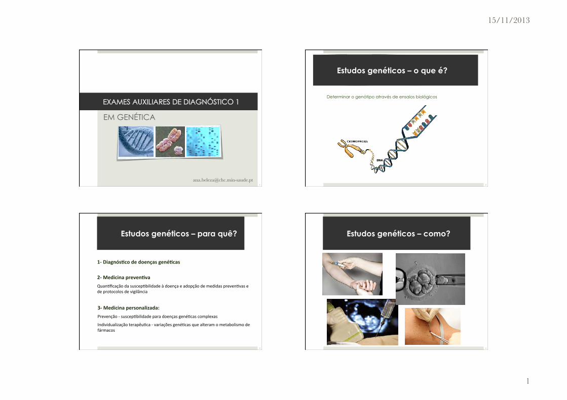
15/11/2013
1
EM GENÉTICA
Estudos genéticos – o que é?
Determinar o genótipo através de ensaios biológicos
2
2-‐ Medicina preven.va Quan%ficação da suscep%bilidade à doença e adopção de medidas preven%vas e de protocolos de vigilância
3-‐ Medicina personalizada: Prevenção -‐ suscep%bilidade para doenças gené%cas complexas
Individualização terapêu%ca -‐ variações gené%cas que alteram o metabolismo de fármacos
1-‐ Diagnós.co de doenças gené.cas
Estudos genéticos – para quê?
3
Estudos genéticos – como?
4
15/11/2013
2
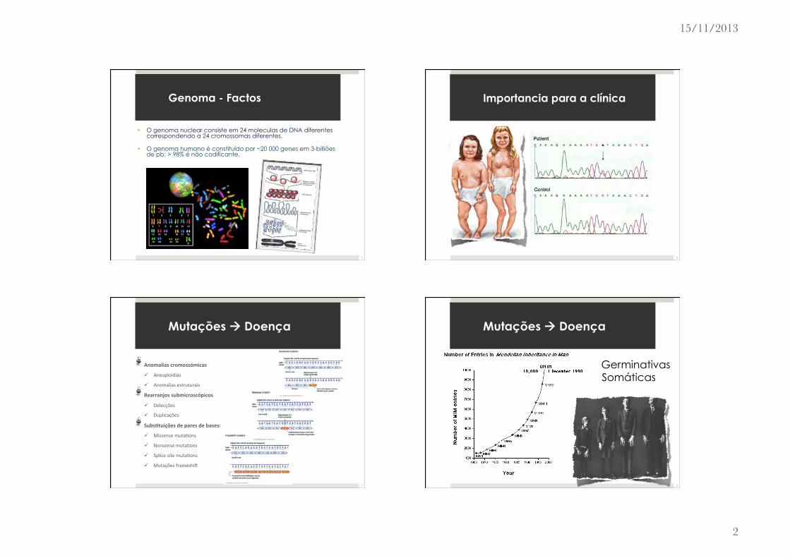
• O genoma nuclear consiste em 24 moleculas de DNA diferentes correspondendo a 24 cromossomas diferentes.
• O genoma humano é constituído por ~20 000 genes em 3-billiões de pb; > 98% é não codificante.
Genoma - Factos
5
Importancia para a clínica
6
" Anomalias cromossómicas
ü Aneuploidias
ü Anomalias estruturais " Rearranjos submicroscópicos
ü Delecções
ü Duplicações " Subs.tuições de pares de bases:
ü Missense muta%ons
ü Nonsense muta%ons
ü Splice site muta%ons
ü Mutações frameshiK
Mutações à Doença
7
" Germinativas " Somáticas
Mutações à Doença
8

15/11/2013
3
Estudos dos cromossomas
9
" Técnicas desenvolvidas nos anos 70 para produzir bandas nos cromossomas.
" Detecta anomalias estruturais e numéricas dos cromossomas
" O bandeamento por giemsa (bandas G) é o mais usual.
http://www.nature.com/scitable/topic/chromosomes-and-cytogenetics-7
Citogenética
10
Citogenética
11
Ajuda na detecção de delecções, duplicações e anomalias estruturais. Mas…
100 Mbp
10 Mbp
1 Mbp
100 Kbp
10 Kbp
1 Kbp
100 bp
10 bp
1 bp
Tamanho da anomalia
Cariótipo
Citogenética
12
15/11/2013
4
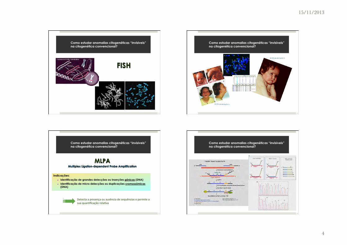
FISH
Como estudar anomalias citogenéticas “invisiveis” na citogenética convencional?
13
46,XX.ish del(4)(p16.1).
46,XX.ish del(1)(p36.3)
Como estudar anomalias citogenéticas “invisiveis” na citogenética convencional?
14
Indicações: " Identificação de grandes delecções ou inserções génicas (DNA)
" Identificação de micro delecções ou duplicações cromossómicas (DNA)
Detecta a presença ou ausência de sequências e permite a sua quan%ficação rela%va
MLPA Multiplex Ligation-dependent Probe Amplification
Como estudar anomalias citogenéticas “invisiveis” na citogenética convencional?
15
Como estudar anomalias citogenéticas “invisiveis” na citogenética convencional?
16
15/11/2013
5
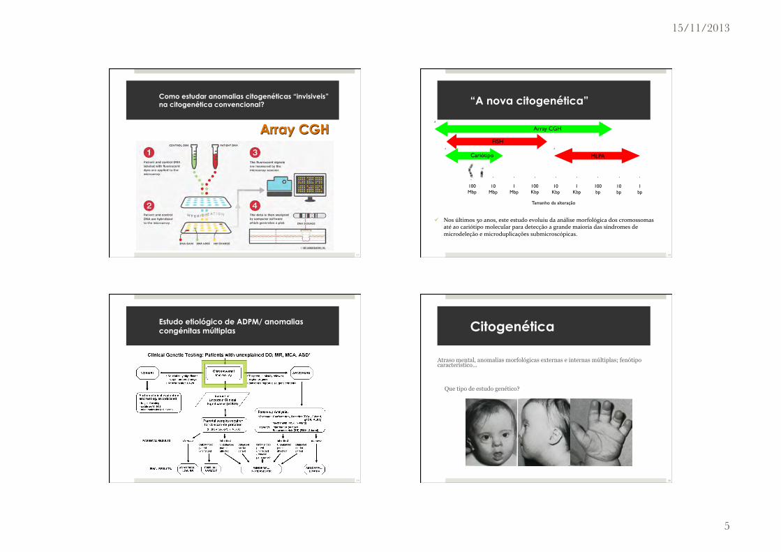
Como estudar anomalias citogenéticas “invisiveis” na citogenética convencional?
Array CGH
17
ü Nos últimos 50 anos, este estudo evoluiu da análise morfológica dos cromossomas até ao cariótipo molecular para detecção a grande maioria das síndromes de microdeleção e microduplicações submicroscópicas.
100 Mbp
10 Mbp
1 Mbp
100 Kbp
10 Kbp
1 Kbp
100 bp
10 bp
1 bp
Tamanho da alteração
Cariótipo MLPA
FISH
Array CGH
“A nova citogenética”
18
Estudo etiológico de ADPM/ anomalias congénitas múltiplas
techniques for identifying the composition of small markerchromosomes when they contain sufficient euchromaticmaterial.90 CMA is also superior to FISH for detecting sub-microscopic duplications because of its higher resolution(multiple small oligonucleotide probes can recapitulatethe coverage of a single BAC probe) and because of the tech-nical difficulty of visualizing tandem duplications by meta-phase FISH analysis.26 Clinically significant submicroscopicduplications, including the reciprocal duplication of knownmicrodeletion syndromes such as the 7q11 Williams-Beuren syndrome region91 or the 17p11.2 Potocki-Lupskisyndrome region,92 are more easily identified by CMA.
The ISCA Consortium proposes the clinical algorithm inFigure 3 to guide postnatal testing in this patient popula-tion. For clinical testing, traditional cytogenetic methods,such as FISH, might offer the best confirmation for certainabnormal findings. For example, terminal deletions orduplications are more likely than interstitial events to be
involved in a rearrangement, especially when more thanone deletion or duplication is identified in a single indi-vidual. Some labs might use other methods, such as quan-titative PCR (qPCR) and multiplex ligation and probeamplification (MLPA). The need for confirmatory testingpurely for copy-number determination is debatable incases such as those involving very large deletions or dupli-cations (typically involving dozens of consecutive probes).
In general, traditional cytogenetic methods are stillneeded for single-cell analysis. Other circumstances inwhich traditional cytogenetic methods are indicatedinstead of (or at least before) CMA include when the patienthas a recognizable chromosomal syndrome such as trisomy21, trisomy 13, Turner syndrome, or Klinefelter syndrome.For these circumstances, conventional cytogenetic analysisor interphase FISH analysis might provide a more rapidturn-around time, allow more sensitive detection oflow-level mosaicism, and provide information regarding
Figure 3. Algorithm for CMA Testing in Patients with Unexplained DD, MR, MCA, and ASDThis algorithm assumes that the patient does not present with features of a recognizable syndrome or metabolic disorder or that testshave been negative for a suspected disorder. The first-tier test is a chromosomal copy-number array or CMA. If no copy-number changesare identified, or if only known CNVs that are known to be benign are identified, this testing is considered ‘‘normal’’ (left side of figure),and further clinical evaluation is warranted to determine whether other testing should be pursued on the basis of the clinical presenta-tion. If a CNV is detected within a known, clinically relevant region or gene, or if the CNV is in the genomic backbone and meets rec-ommended size and gene content guidelines, then the result is considered a pathogenic CNV and ‘‘abnormal’’ (right side of figure). Forthese cases, follow-up analyses include confirmation studies and determination of the mechanism of imbalance in the proband andparental analysis to determine recurrence risk. All other results are considered VOUS until parental analysis is performed to aid in thefinal clinical interpretation. After the parental analyses of ‘‘abnormal’’ and ‘‘VOUS’’ results, final results may be classified into three majorcategories: familial variant, abnormal with a low recurrence risk (RR), or abnormal with an increased RR. In addition, the final interpre-tation may remain VOUS in some instances, even after parental testing.
758 The American Journal of Human Genetics 86, 749–764, May 14, 2010
19
Atraso mental, anomalias morfológicas externas e internas múltiplas; fenótipo característico…
Que tipo de estudo genético?
Citogenética
20
15/11/2013
6
" Causa de infertilidade e de abortamentos recorrentes.
" Mais de 50% dos fetos abortam espontaneamente no 1º trimestre com anomalias cromossómicas.
" Até 1/200 RN tem anomalias congénitas múltiplas por causa de anomalias cromossómicas;
✓ O DPN pode identificar essas anomalias a tempo de uma IMG.
" A maioria das crianças com anomalias cromossómicas são filhos de pais completamente normais;
ü mas cerca de 1% têm uma alt. estrutural equilibrada que os coloca em risco
" Células cancerígenas tipicamente têm extensas alterações cromossómicas, algumas com importância no prognóstico.
Citogenética
21
Estudos da sequência do DNA
22
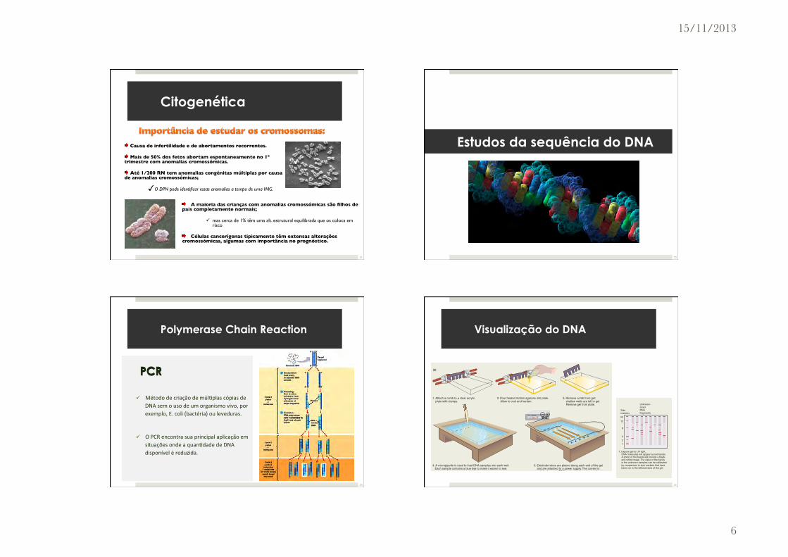
PCR
ü Método de criação de múl%plas cópias de DNA sem o uso de um organismo vivo, por exemplo, E. coli (bactéria) ou leveduras.
ü O PCR encontra sua principal aplicação em situações onde a quan%dade de DNA disponível é reduzida.
Polymerase Chain Reaction
23
296 Chapter 9 Digital Analysis of DNA
FEATURE FIGURE 9.4
Gel Electrophoresis
(a) Preparing the gel. To prepare an agarose gel containing wells for samples, you follow the steps illustrated in (a)1.–3. You then place the prepared gel on a base inside a gel tank that contains a buffered solution. With a micropipette, you load a different DNA sample into each well (step 4). A special “size marker” sample containing DNA fragments of known size is load-ed into the fi rst well. You now connect wires at either end of the box to a power supply, turn on the electric current, and allow the fragments to migrate for 1–20 hours. You then remove the gel from the electrophoresis chamber and place it into a box containing a solution of ethidium bromide, a fl uorescent dye that will bind tightly to any DNA fragments in the gel. After incubat-ing the gel for several hours, you immerse the gel in water to wash away any unbound dye mol-ecules. Then, with exposure to ultraviolet light, the bound dye absorbs photons in the UV range and gives off photons in the visible red range. The DNA molecules appear as red bands, and a digital image shows the relative positions to which they have migrated in the gel.
To determine the length of a DNA fragment, you chart the mobility of the band composed of that fragment relative to the migration of the size marker bands in the fi rst gel lane.
+
+ + + + +
1. Attach a comb to a clear acrylic plate with clamps.
2. Pour heated molten agarose into plate. Allow to cool and harden.
3. Remove comb from gel; shallow wells are left in gel. Remove gel from plate.
4. A micropipette is used to load DNA samples into each well. Each sample contains a blue dye to make it easier to see.
5. Electrode wires are placed along each end of the gel and are attached to a power supply. The current is switched on, and DNA molecules in each sample migrate toward the “+” end of the box (along the paths depicted with orange arrows). Electrophoresis continues for 1–20 hours.
6. Remove gel from gel box. Incubate with ethidium bromide, then wash to remove excess dye.
–
––– – –
7. Expose gel to UV light. DNA molecules will appear as red bands. A photo of the bands will provide a black- and-white image. The sizes of the bands in the unknown samples can be calibrated by comparison to size markers that have been run in the leftmost lane of the gel.
kb
Sizemarkers
Unknown-sizedDNAfragments
12
8
4
2
1
(a)
har2526x_ch09_290-333.indd Page 296 6/17/10 8:08:12 AM user-f499har2526x_ch09_290-333.indd Page 296 6/17/10 8:08:12 AM user-f499 /Users/user-f499/Desktop/Temp Work/JUNE2010/17:06:10/Hartwell:MHDQ122/Users/user-f499/Desktop/Temp Work/JUNE2010/17:06:10/Hartwell:MHDQ122
Visualização do DNA
24
15/11/2013
7
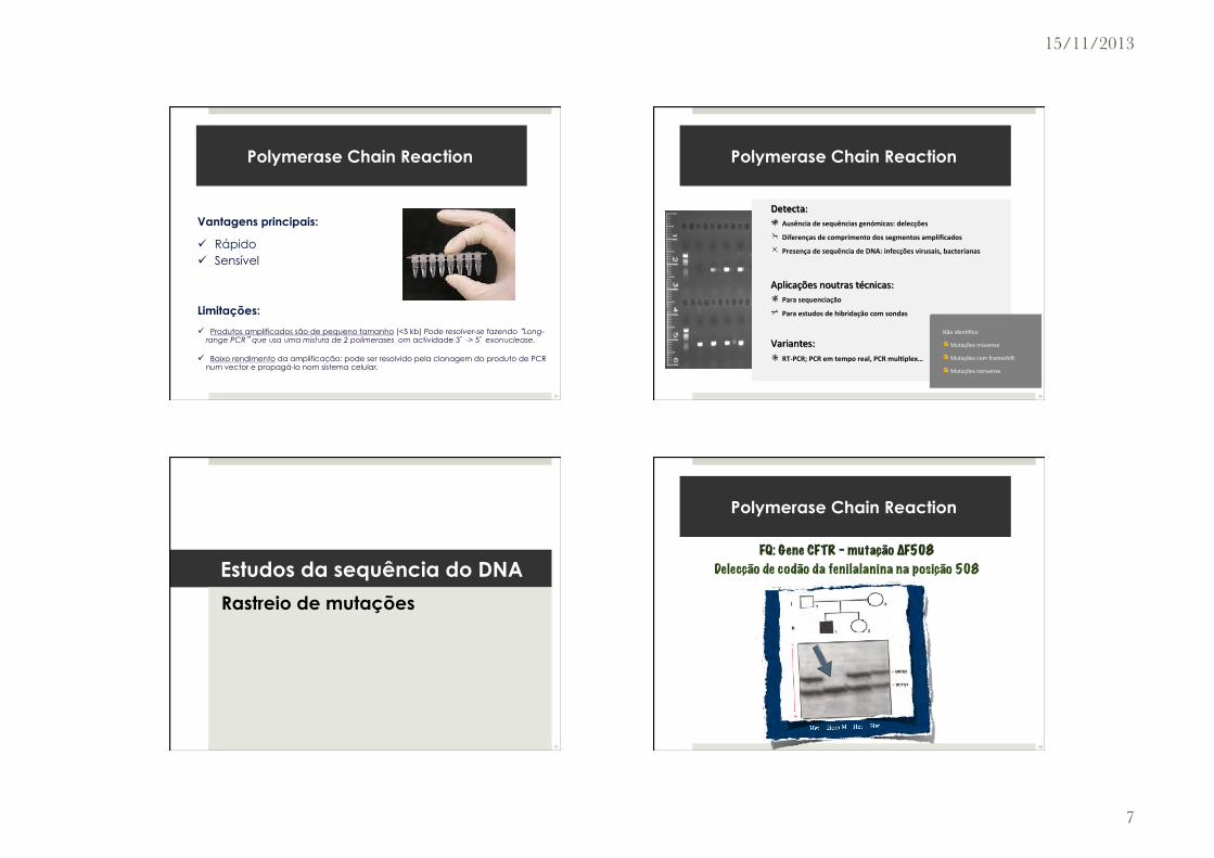
Vantagens principais:
ü Rápido ü Sensível
Limitações:
ü Produtos amplificados são de pequeno tamanho (<5 kb) Pode resolver-se fazendo “Long-range PCR” que usa uma mistura de 2 polimerases om actividade 3’ -> 5’ exonuclease.
ü Baixo rendimento da amplificação: pode ser resolvido pela clonagem do produto de PCR num vector e propagá-lo nom sistema celular.
Polymerase Chain Reaction
25
Detecta: " Ausência de sequências genómicas: delecções
" Diferenças de comprimento dos segmentos amplificados
" Presença de sequência de DNA: infecções virusais, bacterianas
Aplicações noutras técnicas: " Para sequenciação
" Para estudos de hibridação com sondas
Variantes: " RT-‐PCR; PCR em tempo real, PCR mul.plex…
Não iden%fica:
" Mutações missense
" Mutações com frameshiK
" Mutações nonsense
Polymerase Chain Reaction
26
Estudos da sequência do DNA
Rastreio de mutações
27
FQ: Gene CFTR - mutação ∆F508
Delecção de codão da fenilalanina na posição 508
Polymerase Chain Reaction
28
15/11/2013
8
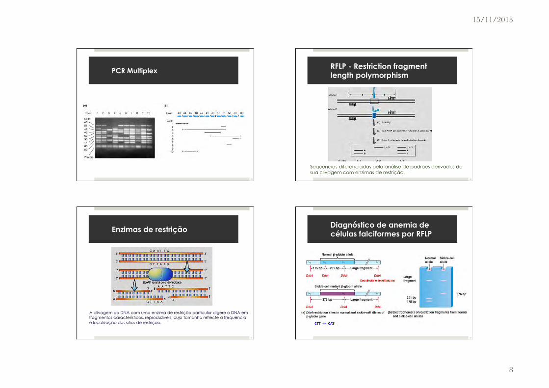
" Produto de PCR mul%plex de 9 exões, em 10 pacientes com Duchenne/Becker muscular dystrophy, para pesquisa de delecções.
PCR Multiplex
29
Sequências diferenciadas pela análise de padrões derivados da sua clivagem com enzimas de restrição.
RFLP - Restriction fragment length polymorphism
30
" A clivagem do DNA com uma enzima de restrição particular digere o DNA em fragmentos característicos, reproduziveis, cujo tamanho reflecte a frequência e localização dos sitios de restrição.
Enzimas de restrição
31
Diagnóstico de anemia de células falciformes por RFLP
32

15/11/2013
9
• Método da biologia molecular que serve para verificar se uma determinada sequência de DNA está ou não presente.
Southern Blot
33
Southern Blot
ü O Southern blot permite a detecção da mutação e a e determinação estado de metilação num só teste.
ü Fidedigno na detecção de grandes pré-mutações e mutações completas
ü A digestão dupla com enzimas de restrição, uma delas enzima sensível metilação, é realizada, seguida de hibridação com uma sonda específica, para identificar aproximadamente o número de repetições trinucleotídicas e para avaliar com precisão o estado de metilação
34
Single-strand conformation polymorphism - SSCP
ü SSCP é a separação electroforética de ácidos nucleicos de cadeia simples
ü Tem por base diferenças na sequência (muitas vezes um único par de bases)
ü As diferenças resultam numa estrutura secundária diferente e uma diferença mensurável na mobilidade através de um gel.
2
! Princípio:
! A mobilidade electroforética que essas estruturas
complexas apresentam em geis não desnaturantes
depende do comprimento das cadeias e das suas
conformações
! Uma alteração numa única base pode provocar uma
alteração conformacional da molécula de DNA,
alteração essa, que poderá ser visualizada por
diferente mobilidade electroforética (shift)
SSCP- Princípio
Ana Alarcão
! Single-Strand Conformation Polymorphism (SSCP)
SSCP- Princípio
Wirth, J., Color Atlas of Genetics (George Thieme Verlag 2001), pag 251Ana Alarcão
35
Single-strand conformation polymorphism - SSCP
ü Antes da sequenciação estar disponível, o SSCP era usado como meio de descobrir novos polimorfismos de DNA
ü Actualmente, o SSCP usado como ferramenta de diagnóstico, para detectar indivíduos homozigóticos e indivíduos heterozigóticos para determinada mutação.
ü SSCP também é amplamente utilizada em virologia para detectar variações de diferentes estirpes de um vírus.
36

15/11/2013
10
" Método de rastreio de mutações através da separação por electroforese baseado em diferenças no comportamento de desnaturação de fragmentos de DNA de cadeia dupla. Não é necessário o conhecimento prévio da localização da mutação.
Denaturing high performance liquid chromatography - DHPLC
Cromatografia líquida de alta performance desnaturante:
37
Denaturing high performance liquid chromatography - DHPLC
Baseada na diferença de afinidade dos heteroduplexes e homoduplexes de um fragmento de DNA pela fase sólida da cromatografia em condições parcialmente desnaturantes.
38