Embed Size (px)
Citation preview
Faculdade de Medicina de São José do Rio Preto Programa de Pós-graduação em Ciências da
Saúde
Adriana Antônia da Cruz Furini
Malária vivax no Estado do Pará:
influência de polimorfismos nos genes
TNFA, IFNG e IL10 associados à resposta
imune humoral e ancestralidade genômica.
São José do Rio Preto
2016
Adriana Antônia da Cruz Furini
Malária vivax no Estado do Pará:
influência de polimorfismos nos genes TNFA,
IFNG e IL10 associados à resposta imune
humoral e ancestralidade genômica.
Tese apresentada à Faculdade
de Medicina de São José do Rio
Preto para obtenção do Título de
Doutor no Programa de Pós
Graduação em Ciências da
Saúde, Eixo Temático: Medicina
e Ciências Correlatas.
Orientador: Prof. Dr. Ricardo Luiz Dantas
Machado
São José do Rio Preto
2016
Furini, Adriana Antônia da Cruz Malária vivax no Estado do Pará: influência de polimorfismos nos genes TNFA, IFNG e IL10 associados à resposta imune humoral e ancestralidade genômica./ Adriana Antônia da Cruz Furini São José do Rio Preto, 2016. 124p. Tese (Doutorado) – Faculdade de Medicina de São José do Rio Preto – FAMERP Eixo Temático: Medicina e Ciências Correlatas Orientador: Prof. Dr. Ricardo Luiz Dantas Machado 1.Ancestralidade; 2. Anticorpos; 3. Citocinas; 4. Malária; 5.Plasmodium vivax.
ADRIANA ANTÔNIA DA CRUZ FURINI
Malária vivax no Estado do Pará:
influência de polimorfismos nos genes
TNFA, IFNG e IL10 associados à resposta
imune humoral e ancestralidade genômica.
BANCA EXAMINADORA
TESE PARA OBTENÇÃO DO GRAU DE DOUTOR
Presidente/Orientador: Prof. Dr. Ricardo Luiz Dantas Machado
2º Examinador:Prof. Dr. Carlos Eugênio Cavasini
3º Examinador: Profa. Dra. Heloísa da Silveira Paro Pedro
4º Examinador: Profa. Dra. Eny Maria Goloni Bertollo
5º Examinador: Profa. Dra. Maristela Sanches Bertasso
Borges
Suplentes: Prof. Dra. Érika Cristina Pavarino
Prof. Dra. Sônia Maria Oliani
São José do Rio Preto, 05/08/2016.
.

SUMÁRIO
Dedicatória........................................................................................................... i
Agradecimentos...................................................................................................ii
Epígrafe................................................................................................................v
Lista de Figuras...................................................................................................vi
Lista de Tabelas.................................................................................................vii
Lista de Abreviaturas e Símbolos......................................................................viii
Resumo..............................................................................................................xii
Abstract.............................................................................................................xiv
Introdução ...........................................................................................................1
Objetivos..............................................................................................................9
Artigos Científicos..............................................................................................11
Artigo 1...............................................................................................................12
Artigo 2...............................................................................................................40
Conclusões........................................................................................................49
Referências........................................................................................................51
Apêndices..........................................................................................................69
Anexos.............................................................................................................109

i
DEDICATÓRIA
Às minhas filhas Marina e Fernanda, por toda felicidade, de me fazer
descobrir tesouros nunca antes sonhados. O melhor de todos os
sonhos, ser mãe. Vocês são únicas, arteiras, amorosas, geniosas e
felizes! Amo vocês até o infinito.
Ao meu eterno namorado, meu marido, meu amigo, meu grande amor:
Marcos Amorielle Furini. Amo você.
Aos meus pais: “Eu teria tanto para falar, que com palavras nem sei
dizer, como foi, é, e sempre será infinito meu amor por vocês”. Tenho
certeza que me assistiram do céu.....
À minha irmã Edna, querida companheira, incansável na busca da
felicidade de todos.
Aos meus irmãos, sobrinhos, sobrinhas e família.
ii
AGRADECIMENTOS
A Deus por me dar coragem, força, persistência e oportunidades.
Ao meu orientador, Prof. Dr. Ricardo Luiz Dantas Machado, por
toda sua dedicação ao meu trabalho nesses quatro anos, e por eu
ter compartilhado da sua sabedoria e conhecimento. Obrigada, por
tudo que me ensinou meu professor, meu mestre, meu amigo.
À aluna Michele Encinas do Curso de graduação em Farmácia e ao
aluno Diego Longo Madi do Curso de graduação em Biomedicina
(Centro Universitário de Rio Preto), pela disponibilidade, empenho,
dedicação, prontidão, enfim por tudo o que fizeram por mim para
que meu projeto se concretizasse. Meu eterno obrigada.
À pesquisadora Dra. Joseli Ferreira de Oliveira da Fundação
Oswaldo Cruz (FioCruz) – Rio de Janeiro pela disponibilidade,
prontidão e dedicação para utilização do laboratório e análises de
citocinas.
Ao Dr. Gustavo Capatti Cassiano. Meu amigo foi um prazer realizar
um projeto de Doutorado no mesmo laboratório que você, por sua
dedicação a pesquisa, por acreditar que as coisas tem que dar
certo, e que isso requer persistência. Obrigada pelos
ensinamentos, aconselhamentos. Você é um exemplo de
pesquisador a ser seguido.
Às minhas companheiras de laboratório, Maíra, Marcela, Pamella,
Simone pelos momentos que passamos juntas, pela amizade.
iii
Aos amigos Juan Camilo Sanchéz Arcilla, Virginia Pereira da
FioCruz Rio de Janeiro pela disponibilidade, empenho, dedicação,
prontidão. Pelo acolhimento impecável nos momentos que
passamos juntos.
Obrigada ao Prof. Dr. Carlos Eugênio Cavasini, e as minhas
amigas do Laboratório CIM, Valeria Fraga e Luciana Moran, pelo
comprometimento, dedicação, ajuda nas análises laboratoriais, e
fluxo das amostras. Sem vocês esse trabalho não seria possível.
Aos meus amigos e amigas: Bárbara Cruz, Nayara Cruz e Gabriela
Cruz, Alessandra Cruz, Franciele Maira, Karina Santana, Maristela
Bertasso, Greici Gomes, Regiane Rocha, Neide Blaz, Patricia
Perez, Ricardo Fochi, Gisele, Tabata, Cleide Silveira, Edna Matins,
Paula Guimarães, Daniela Goés, Daniela Hubner, Glacy Claro,
Angela Schempp, Gisele Assunção, Gisele Bueno, Cintia, Raquel
Arid, Ivone Fontes, Heloisa. Amizade verdadeira é aquela que o
tempo não apaga, a distancia não destrói e acima de tudo o
coração não esquece.” Alguns não vejo há anos, mas não poderia
deixar de lembrá-los em um momento tão especial da minha vida.
Ao meu sogro Décio, minha sogra Sandra e a Tia Darci que me
auxiliaram inúmeras vezes ao cuidar das minhas filhas para as
viagens a congressos durante o Doutorado.
À Faculdade de Medicina de São José do Rio Preto – FAMERP e
ao Programa de Pós Graduação em Ciências da Saúde pela
excelência em qualidade nos cursos Strictu sensu.
iv
À Capes – Coordenação de Aperfeiçoamento de Pessoal de Nível
Superior pela Bolsa de Estudos.
Ao Centro Universitário de Rio Preto, pelo incentivo para a
realização desse trabalho.
Aos pacientes, muito obrigada.
Às instituições de fomento e pesquisa, em especial aos auxílios
CNPq, pelos recursos financeiros que foram imprescindíveis para a
execução desse trabalho.
Aos organizadores do seminário Laveran/Deane sobre malária, Dr.
Cláudio Tadeu Daniel Ribeiro e Dra. Maria de Fátima Ferreira da
Cruz, pela oportunidade de participar deste evento.
Enfim, obrigada a todos, que diretamente ou não, contrubíram para
a realização desse trabalho.

v
EPÍGRAFE
“Tenho a impressão de ter sido uma criança brincando à beira-mar,
divertindo-me em descobrir uma pedrinha mais lisa ou uma concha mais
bonita que as outras, enquanto o imenso oceano da verdade continua
misterioso diante de meus olhos”.
(Isaac Newton)

vi
LISTA DE FIGURAS
Artigo 1:
Figura 1. Binary logistic regression model used to evaluate the frequency of
individuals carrying the mutant allele of all analyzed SNPs relative to the
individual proportions of genetic ancestry. The shading around the lines
represents the 95% confidence interval. The graph was constructed using the
ggplot2 package in the R program ....................................................................... 24

vii
LISTA DE TABELAS
Artigo 1:
Tabela1.Characteristics of the study population ................................................... 21
Tabela 2. Distribution of the genotypes between vivax malaria-infected and
non-infected individuals ........................................................................................ 22
Tabela 3. Haplotype frequencies in the TNFA and IL10 genes in vivax
malaria-infected and non-infected individuals ...................................................... 22
Tabela 4. Haplotype frequency and its association with the proportions of
African, European and Native American ancestry ................................................ 23
Artigo 2:
Tabela 1. List of polymorphisms, methods, restriction enzymes, primers used
for investigation of SNPs in genes TNFA, IFNG, IL10 .......................................... 55
Tabela 2. Levels of IgG antibodies against P. vivax blood stage proteins
according to the studied genotypes ...................................................................... 56
Tabela 3. Frequency of haplotypes and their association with levels of IgG
antibodies against the PvAMA-1, PvDBP and PvMSP-119 proteins .................... 57

viii
LISTA DE ABREVIATURAS E SÍMBOLOS
ADCI - Inibição Celular Dependente de Anticorpo
AIMs - Marcadores Informativos de Ancestralidade
Anova - Análise de variância
APC - Células apresentadoras de antígenos
B7-1 - Ligante da molécula co-estimuladora CD28 expresso na superfície de linfócitos B e
macrófagos/monócitos (CD80)
B7-2 - Ligante da molécula co-estimuladora CD28 expresso na superfície de linfócitos B e
macrófagos/monócitos (CD86)
BCMA - Antígeno de maturação de linfócito B
Blys - Estimulante de linfócitos B
BR3 - Blys receptor 3
CD28 - Receptor de Linfócitos T (Cluster de diferenciação 28)
CD28 - Gene CD28
CD40 - Receptor CD40 (Cluster de diferenciação 40)
CD40L - Ligante de CD40
CD80 - Ligante da molécula co-estimuladora CD28
CD86 - Ligante da molécula co-estimuladora CD28
CSP - Proteína circumesporozoítica
CTLA-4 - Antigénio 4 dos linfócitos T citotóxicos
ºC - Grau Celsius
DNA - Ácido desoxirribonucleico
dNTP - Desoxirribonucleótidos trifosfato
Duffy - Antígeno de superfície das hemácias
ELISA - Ensaio imunoenzimático
FcγRIIa - Receptor de fração constante de anticorpos (CD32)
G6PD - Glicose 6-fosfato desidrogenase
HLA - Antígeno leucocitário humano
HbS - Hemoglobina S
HWE - Equilíbrio de Hardy-Weinberg
ICB2-5 - Proteína N-terminal da MSP-1 do Plasmodium vivax

ix
ICOS - Proteína imunológica indutível co-estimuladora
ICOSL - Ligante da molécula ICOS
IgA - Imunoglobulina A
IgE - Imunoglobulina E
IgG - Imunoglobulina G
IgG2 - Imunoglobulina G da subclasse 2
IgG3 - Imunoglobulina G da subclasse 3
IgG4 - Imunoglobulina G da subclasse 4
IgM - Imunoglobulina M
IL-1 - Interleucina 1
IL-2 - Interleucina 2
IL-3 - Interleucina 3
IL-4 - Interleucina 4
IL-6 - Interleucina 6
IL-8 - Interleucina 8
IL-10 - Interleucina 10
IL10 - Gene da Interleucina 10
IL-13 - Interleucina 13
INDELS - Inserção/deleção
INFG - Gene do Interferon Gama
INFγ - Citocina Interferon Gama
IR - Índice de Reatividade
LTCD4 - Linfócito TCD4
LTCD8 - Linfócito TCD8
Linfócitos Tγδ - Linfócitos T gama-delta
LB - Linfócitos B
MgCl2 - Cloreto de magnésio
µg - Micrograma
MHC - Complexo Maior de Histocompatibilidade
µL - Microlitro
nM - Nano molar
MSP-1 - Proteína 1 da superfície de merozoíto
x
NFkB - Via NFK beta
OD - Densidade ótica
pB - Pares de bases
PBS - Tampão fosfato salino
PCR-ASO - PCR Alelo Específica
Pg - Picograma
pH - Potencial Hidrogeniônico
pmol - Picomol
P. vivax - Plasmodium vivax
PvAMA-1 - Antígeno de membrana apical-1 do Plasmodium vivax
PvDBP - Proteína do Plasmodium vivax, ligante do Duffy
Pv MSP-119 - Proteína da superfície de merozoíto do Plasmodium vivax
RI - Reactivity Index
RFLP - Polimorfismo do tamanho do fragmento de restrição
SNP - Polimorfismo de nucleotídeo único
TACI - Receptor do linfócito B interagente ligante ciclofilina
TCLE - Termo de consentimento livre e esclarecido
TCR - Receptor de células T
TGFBeta - Fator de Transformação de crescimento (Citocina)
Th1 - Linfócitos T auxiliares (1)
Th2 - Linfócitos T auxiliares (2)
TNF-α - Fator de necrose tumoral alfa (Citocina)
TNFA - Gene do fator de necrose tumoral alfa
TNFR1 - Receptor do Fator de Necrose Tumoral Alfa
VK247 - Variante 247 do Plasmodium vivax
xi
RESUMO
Introdução: A malária é uma das maiores causas de morbidade e mortalidade em países
tropicais e subtropicais. Objetivos: Avaliar a influência da ancestralidade genética na
distribuição de polimorfismos em genes envolvidos na resposta imune e os níveis de anticorpos
contra proteínas expressas no estágio de merozoíto do Plasmodium vivax. Material e
Métodos: Foram avaliados 90 indivíduos com malária vivax e 51 não infectados de Goianésia
do Pará, região Norte do Brasil. Nove polimorfismos de nucleotídeo único (SNPs) distribuídos
nos genes: TNFA, INFG e IL10 foram genotipados por PCR-ASO ou PCR-RFLP. A
ancestralidade genômica para os três grupos étnicos (africana, europeia e ameríndia) foi
categorizada com a utilização de 48 INDELs. As respostas de anticorpos específicos contra as
proteínas C-terminal (MSP-119) da MSP-1, da DBP e da AMA-1 do P. vivax foram determinadas
por ELISA. Resultados: Não houveram diferenças nas proporções de ancestralidade na
maioria dos SNPs investigados, apenas para o alelo TNF-308A e a ancestralidade europeia.
Nenhuma associação significativa foi observada entre as frequências alélicas e genotípicas dos
SNPs entre os grupos investigados. Não foi encontrada diferença significativa nos níveis de
anticorpos IgG em relação aos polimorfismos estudados. Conclusões: Esses resultados
ressaltam que os polimorfismos nos genes TNFA, INFG e IL10 não influenciam na resposta
imune anti-merozoítos do P. vivax. Discutimos o perfil imunogenético envolvido na resposta
imune humoral na malária vivax em região endêmica da Amazônia brasileira.
Palavras-Chave: Anticorpos. IFNG. IL10. Plasmodium vivax. TNFA

xii
ABSTRACT
Introduction: Malaria is one of the mayor cause of morbidity and mortality in tropical and
subtropical countries. Objectives: To evaluate the influence of genetic ancestry in the
distribution of polymorphisms in genes involved in the immune response and antibody levels
against proteins expressed in the merozoite stage of Plasmodium vivax. Material and
Methods: To evaluated 90 patients with vivax malaria and 51 non-infected patients from
Goianésia do Pará, northern Brazil. Nine single nucleotide polymorphisms (SNPs) in the genes:
TNFA, IL-10 INFG were genotyped by PCR-ASO or RFLP-PCR. The genetic ancestry for three
ethnic groups (African, European and American Indian) were categorized using 48 INDELs. The
responses of specific antibodies against the C-terminal proteins (MSP-119) MSP-1, BPD and
AMA-1 of P. vivax were determined by ELISA. Results: There were no differences in ancestry
proportions in most SNPs investigated only for TNF-308A allele and European ancestry. No
significant association was observed between the allele and genotype frequencies of the SNPs
between the groups investigated. There was no significant difference in the levels of IgG
antibodies to the studied polymorphisms. Conclusions: These results indicated that the
polymorphisms in the TNFA, INFG e IL10 genes can not influence the anti-merozoites immune
response of P. vivax. We discussed the immunogenetic profile involved in the humoral immune
response in malaria vivax in an endemic area of the Brazilian Amazon.
Keywords: Antibodies. IFNG. IL10. Plasmodium vivax.TNFA.

INTRODUÇÃO

1
1. INTRODUÇÃO
1.1 Considerações gerais: epidemiologia, transmissão e agentes
etiológicos.
Apesar dos progressos nas estratégias de controle da malária, a doença
ainda é uma das maiores causas de morbidade e mortalidade em muitos
países tropicais e subtropicais. (1,2,3) Cento e quatro países são endêmicos
com 207 milhões de casos clínicos por ano e aproximadamente 627.000 mil
mortes.(3) Nas Américas, três países concentram 76% dos casos de malária,
sendo o Brasil responsável por 52% dos casos. (4)
Os perfis de transmissão da doença no Brasil são diferentes e
observados em três ambientes distintos. Na Amazônia e na Mata Atlântica,
ambos com uma predominância de casos autóctones, e em outras regiões,
com casos importados de recentes viagens a áreas endêmicas de malária no
país, ou em outros da América Central e do Sul, países africanos ou asiáticos
(5,6).
A malária é uma doença protozoária na qual a infecção ocorre pela
inoculação de esporozoítos de Plasmodium por meio da picada de fêmeas do
mosquito do gênero Anopheles. (4,6) O ciclo da doença é heteroxênico, com
fase sexuada no vetor e assexuada no homem. No vertebrado, ocorre
esquizogonia hepática e eritrocitária. (7,8,9) Cinco espécies de Plasmodium são
responsáveis pela etiologia humana da malária: Plasmodium falciparum,
Plasmodium vivax, Plasmodium malariae, Plasmodium ovale e, recentemente,
2
o Plasmodium knowlesi foi detectado na Malásia.(3,10) O P. falciparum está
associado aos maiores índices de morbimortalidade, enquanto que o P. vivax é
amplamente disseminado pelo mundo.(3,11,12) No Brasil, o P. vivax tem sido
responsável por aproximadamente 85% dos casos. (3,4)
1.2 Resposta imune no paciente com malária
1.2.1 Participação de citocinas e do receptor CD28 na resposta imune a
malária.
Mecanismos inatos, humorais e celulares são envolvidos na resposta
imune da malária, com a participação de células, citocinas, receptores e
anticorpos, que podem eliminar o agente etiológico ou acarretar em
complicações imunopatológicas.(7,12,13) Os linfócitos TCD4+ (auxiliares)
participam das respostas imunes celulares e humorais por meio da ativação por
citocinas pro e anti-inflamatórias. Essas células são fundamentais para
ativação de linfócitos B (LB) por citocinas anti-inflamatórias que resultam na
diferenciação em plasmócitos e secreção de anticorpos. (12,14)
Os receptores de antígenos dos linfócitos T (LT-TCR), e os co-
receptores CD4 ou CD8 ligam-se ao complexo maior de histocompatibilidade
(MHC) de células apresentadoras de antígenos (APCs), para ativação dos
linfócitos T (LT) na resposta imune celular.(15,16,17) No entanto, essa ligação não
determina a expansão clonal dos LT, que requer um segundo sinal co-
estimulatório que é emitido pela mesma APC, por meio de glicoproteínas de
membrana denominadas de B7.1 ou CD80 e B7.2 ou CD86.(16) O receptor

3
dessas moléculas nas células T é o CD28, expresso constitutivamente na
superfície dessas células.(15) A ligação do CD28 com seus ligantes (CD80 ou
CD86) potencializa a transcrição e produção da interleucina-2 (IL-2), que
resulta em proliferação e expansão clonal das células T(15,16) e liberação de
outras citocinas. O significado da coestimulação via CD28 no desenvolvimento
da imunidade depende do agente etiológico, como reportado em infecções por
Salmonella enterica (18) e Trypanosoma cruzi (19), mas com pouca, ou nenhuma
função na imunidade contra Toxoplasma gondii (20).
Em relação à malária, Taylor-Robinson e Smith (1994) (21) reportaram
que o tratamento de camundongos infectados pelo Plasmodium chabaudi com
anticorpos monoclonais anti-CD86 impediu o clareamento da parasitemia,
sugerindo uma possível função da via CD86/CD28 no controle da malária
crônica. Por outro lado, Kemp e colaboradores (2002) (22) avaliaram a
expressão de IFN-γ e IL-4 por LTCD28+ e LTCD28- em crianças africanas com
malária falciparum, e verificaram que os níveis de IFN-γ produzidos pelas
LTCD28- foram menores. Elias e colaboradores (16), no ano de 2005, avaliando
o papel do CD28 em modelo murino, encontraram que após uma semana de
infecção a expressão de IFN-γ foi 50% menor nos LTCD28-.
Assim como as células e receptores possuem um papel fundamental na
resposta imune ao Plasmodium, o balanço entre as citocinas pró (Th1 -
celulares) e antiinflamatórias (Th2 - humorais) é crucial para o prognóstico na
malária. (7,13, 14, 23-25) A superprodução e persistência desses mediadores podem
levar a imunopatologia, com gravidade e óbito (13,14,24,26,27), mas por outro lado,
4
pequenos níveis não são suficientes para inibir o crescimento do parasito.
(9,14,26)
O fator de necrose tumoral alfa (TNF-α) é uma citocina pró-inflamatória
que participa no recrutamento e ativação de monócitos, macrófagos e
neutrófilos para o sitio da infecção,(28,29), na modulação positiva para resposta
imune humoral de IgG total (30), como fator de crescimento autócrino para os
LB. (31) Na patogênese da febre atua em conjunto com a interleucina-1 (IL-1)
para ativação de células hipotalâmicas, além de participar da negativação
parasitária tanto in vivo com in vitro.(26,32) Níveis elevados TNF-α estão
relacionados ao paroxismo malárico(33) , malária grave(31) e malária cerebral(9) .
Respostas acentudas do interferon gama (IFN-γ) são reportadas no
controle de infecções agudas por Plasmodium berghei, Plasmodium yoelii, e
P.chabaudi em modelos murinos e para o P.falciparum na malária humana. (25)
Essa citocina pró-inflamatória é produzida por LTCD4, LTCD8, linfócitos T γδ e
natural kille (34) e contribui para o controle da infecção na fases hepática e
eritrocítica (33,34) com ativação de macrófagos e outras APCs. Corrobora na
modulação negativa para a resposta imunológica do tipo anti-inflamatória (Th2),
contribui para o processo de homeostase e pode aumentar a produção de
IgG2.(35) Por outro lado, o balanço de citocinas é obtido pela modulação
negativa das anti-inflamatórias IL-10 e TGF-beta na resposta do tipo pró-
inflamatória (IL-1, IL-6, IL-8, IL-12, IFN-γ e TNF-α) do tipo Th1. (25,26,34,36-38)
O excesso de resposta TH1, na incapacidade de produção de IL-10 e
TGF-beta, pode acarretar em inflamação excessiva e dano tecidual na
malária.(14) A IL-10 também sinergiza a produção dos anticorpos IgG, IgA e

5
IgM, induzidas por IL-4.(31,38) Na malária altos níveis de IL-10 estão
relacionados ao clareamento da parasitemia.(14, 37)
1.2. 2 Polimorfismos no genes CD28, TNFA, IFNG E IL10
A susceptibilidade e resistência para malária podem estar relacionadas à
seleção natural, fatores genéticos do hospedeiro e do agente, idade, etnia, e
esses por sua vez, envolvidos na resposta imunológica, sintomas e níveis de
parasitemia.(39-41) Corroboram também as situações epidemiológicas,
ambientais, geográficas e de tempo de moradia em regiões endêmicas.(2,41)
Polimorfismos em genes de citocinas tem sido (13,27) associado com níveis
circulantes dessas proteínas e de anticorpos (39,42) na malária para evolução
clínica e prognóstico.(27) Dessa maneira polimorfismos de nucleotídeo único
(SNPs) podem influenciar no desenvolvimento de vacinas e de novas
alternativas terapêuticas para malária.(40,43)
Um SNP na posição +17T/C (rs3116496), situado no íntron 3 do
receptor CD28 localiza-se próximo a um sítio de recomposição que pode
interferir na eficiência desse receptor. Associações significativas foram
descritas entre esse SNP com diabetes de tipo 1 (44) e artrite reumatóide. (45) Na
malária o papel deste polimorfismo foi descrito por Cassiano e colaboradores
(46) quanto a presença do alelo T com níveis mais baixos de IgG1 específica
para a proteína ICB2-5. Na posição -372G/A (rs35593994) no gene CD28 as
duas variantes alélicas foram caracterizadas na população australiana, porém
sem associação com a esclerose múltipla. (47) Esta variação alélica também foi
descrita em amostragem da população brasileira, mas sem associação com
pênfigo foliáceo.(48)

6
O gene IL10 é localizado no cromossomo 1q3-q32 e apresenta pelo
menos 27 sítios polimórficos. (27,37, 49) Na região promotora do gene, SNPs tem
sido associados à produção de citocinas (37) e níveis de anticorpos(39,42,50) na
malária. Os haplótipos IL10 -1082/-819/-592 GCC, ACC e ATA são associados
respectivamente à alta, intermediária e baixa atividade de transcrição da
citocina. (27,37,51)
O IFN-γ é codificado pelo gene situado no cromossomo 12q24.1, que
consiste de 4 exons e 3 introns. (52-54) Polimorfismos no gene do IFNG tem sido
associados com tuberculose (51,55), dermatite(56) , mas não com artresia biliar.
(53) Na posição -183G/T (rs2069709), o alelo T foi associado ao aumento da
atividade de transcrição (52,53) malária cerebral na África Ocidental (52), Hepatite
B na China. (57) O SNP +874 A/T (rs2430561) está localizado no intron 1 do
gene do IFNG e influencia a expressão do RNAm e secreção da proteína. (54) O
alelo T é associado com elevada produção de citocina. (13,56)
O gene do TNFA é situado no cromossomo 6p21.3, em uma região
altamente polimórfica (51,58) entre os genes do HLA de classe I e classe II. (43) O
SNP na posição +308G/A (rs1800629) tem sido amplamente estudado na
malária. (59-62) Apesar de maior distribuição do alelo ancestral, com
aproximadamente 87% (63, 64), o alelo A é descrito para aumento nos níveis da
proteína, porém sem efeito aparente na susceptibilidade ao P. vivax (26,65) ou
com resistência ao P. falciparum . (62) Os alelos T [-1031T/C (rs1799964)] e G
(-308G/A) foram associados com episódios de malária não complicada (29) em
Burkina Faso. Para os SNPs -238G/A (rs361525) e para o -308G/A o alelo A foi
associado com redução da parasitemia na malária. (29,66)

7
1.2.3 Polimorfismos em genes de citocinas e ancestralidade
No Brasil, a heterogenicidade tri-híbrida da população é originária da
migração de nativo americanos (asiáticos) para o continente por meio do
Estreito de Bering (67), seguida da colonização Europeia no Nordeste brasileiro
a partir de 1530, e fluxo migratório de escravos africanos. (68,69) A miscigenação
populacional pode ser uma causa para resultados não totalmente esclarecidos
ou contraditórios na distribuição de alelos e genótipos envolvidos na
transcrição, expressão do gene e produção de citocinas. (69-71)
A susceptibilidade à malária ou fenótipos tem sido avaliada por estudos
de associação, do tipo caso e controle (41,43,72), com etnia auto declarada ou
indicadores de aparência física, nos quais existe o risco de se encontrar
associações espúrias. Esse fato decorre da estratificação populacional, ou em
populações miscigenadas com diferentes frações de ancestralidade.(70,71)
Dessa maneira, a análise de ancestralidade por meio de marcadores
informativos de ancestralidade (MIAs) do tipo inserção e deleção (INDEL),
pode contribuir para eliminar a possibilidade das associações espúrias.
Entretanto, poucos estudos demonstram as diferenças étnicas na distribuição
de SNPs baseado em uso de MIAs (17, 71,73), e para a ancestralidade genética
nativo americana esses dados são ainda mais escassos. (74,75)
1.2.4 Polimorfismos em genes de citocinas e produção de anticorpos na
malária

8
Epítopos imunogênicos da superfície do parasito, tanto de proteínas do
esporozoíto e merozoíto têm sido amplamente estudados como potenciais
alvos para formulação de vacinas. Em geral o principal marcador de proteção
são anticorpos anti-merozoítos (7) descritos em estudos conduzidos na
Amazônia brasileira. (76,77,78,79) As proteínas do merozoíto mais estudadas são
as que participam do processo de invasão dos eritrócitos, como a MSP119, do
inglês Merozoite Surface Protein-1 (80,81) a DBP, do inglês Duffy Binding Protein
(82,83) e AMA-1, do inglês Apical Membrane Antigen-1. (78,84,85)
A imunidade humoral para o P. vivax é descrita para ser mais rápida do
que para o P. falciparum(23,86), entretanto vários anos de exposição contínua em
áreas endêmicas é necessária para a situação de premunição e redução do
risco de malária clínica, com baixas parasitemias e altos níveis de anticorpos
anti-merozoítos. (7,23,39)
A resposta imune humoral na malária vivax é amplamente descrita para
estar associada hemoglobinopatias, traço falciforme (HbS), deficiência de
G6PD, (40,41) variabilidade genética do HLA, (78) antígeno Duffy(41,87) variantes da
proteína CSP de P.vivax (23,24,88) e mais recente por polimorfismos em genes de
citocinas e de moléculas co-estimulatórias da resposta imune. (13,27,43,46,72,79,85)
Polimorfismos em genes de citocinas tem sido (13,27) associados com
níveis e classes de anticorpos (39,42) na malária. Em estudos (Tanzânia) com
P.falciparum, para os SNPs no gene da IL-10 (-592) e (-1082), o alelo A foi
associado a baixos níveis de IgE e IgG4, (50) o genótipo AA de IL-10-1082 com
altos níveis de anticorpos para AMA-1 e MSP2-3DT em mães e recém-

9
nascidos.(39) O alelo A do TNFA nas posições -308 (50) e -238 (29) com altos
níveis de anticorpos IgG anti P. falciparum. No Brasil, não foram descritas
associação de SNPs (-590C/T, IL4 -33C/T e o VNTR) no gene da IL4 com a
parasitemia ou com níveis de anticorpos contra a PvAMA-1 (85) em indivíduos
maláricos do município de Goianésia do Pará, e também entre anticorpos
contra esporozoítos e merozoítos de P. vivax com SNPs no genes CD40 e
BLyS numa população de Macapá no Estado do Amapá. (72) Por outro lado,
indivíduos infectados naturalmente por P. vivax em Goianésia do Pará, os
SNPs nos genes do BLYS (–871C/T) foram associados com a frequência de
respostas IgG para PvAMA-1 e PvMSP-119, no gene do CD40 (-1C/T) para
IgG contra PvDBP e no gene do CD86 (+ 1057G/A) para IgG contra PvMSP-
119. (79)
2. OBJETIVOS
2.1 Objetivo Geral
Avaliar a influência de polimorfismos nos genes CD28, INFG, TNFA e IL-
10 na resposta imune humoral na malária vivax.
2.2 Objetivos Específicos

10
a) Determinar as frequências alélicas e genotípicas de variantes nos genes
INFG TNFA e IL-10 em indivíduos com malária vivax e indivíduos não
infectados.
b) Avaliar a frequência de polimorfismos nos genes TNFA, IFNG e IL10 em
uma amostra da população brasileira, relacionando suas distribuições às
frações de ancestralidade genética determinada com o auxílio de
Marcadores Informativos de Ancestralidade.
c) Estabelecer possíveis associações entre os polimorfismos e proteção na
malária vivax.
d) Identificar possíveis associações entre os polimorfismos de genes de
citocinas e haplótipos com níveis de anticorpos para PvDBP, Pv-AMA-1
e Pv-MSP-1-19.

ARTIGOS CIENTÍFICOS
11
ARTIGOS CIENTÍFICOS
Esse trabalho é composto de dois artigos originais.
Artigo 1
Título: TNF-alpha, IFN-gamma and IL10 cytokine SNPs: Comparison of
polymorphisms by genomic ancestry in an admixed population.
Autores: Adriana Antônia da Cruz Furini, Gustavo Capatti Cassiano, Marcela
Petrolini Capobianco, Sidney Emanuel dos Santos, Ricardo Luiz Dantas
Machado.
Periódico: Mediators of Inflammation (artigo aceito para publicação). Fator de
impacto: 3,418
Artigo 2
Título: Cytokine gene polymorphisms are not associated with anti-Pv-DBP, Pv-
AMA-1 or Pv-MSP-119 IgG antibody levels in a malaria-endemic area of the
Brazilian Amazon.
Autores: Adriana Antônia da Cruz Furini, Marcela Petrolini Capobianco,
Luciane Moreno Storti-Mello, Maristela Gomes da Cunha, Gustavo Capatti
Cassiano, Ricardo Luiz Dantas Machado.
Periódico: Malaria Journal (artigo publicado). Fator de impacto: 3,079

Marcos Amorielle Furini <[email protected]>
Fwd: 5168363: Revised Version Received
Adriana Antonia da Cruz Furini <[email protected]> 8 de agosto de 2016 11:16Para: Marcos Amorielle Furini <[email protected]>
Forwarded message From: Mediators of Inflammation <[email protected]>Date: 20160714 4:15 GMT03:00 Subject: 5168363: Revised Version Received To: [email protected] Cc: [email protected], [email protected], [email protected], [email protected],[email protected]
Dear Dr. Furini,
The revised version of Research Article 5168363 titled "TNFalpha, IFNgamma and IL10 cytokine SNPs:Comparison of polymorphisms by genomic ancestry in an admixed population" by Adriana Antônia da Cruz Furini,Gustavo Capatti Cassiano, Marcela Petrolini Capobianco, Sidney Emanuel Batista Santos and Ricardo Luiz DantasMachado has been received. The editor assigned to handle the review process of your manuscript will inform you assoon as a decision is reached.
Thank you for submitting your work to Mediators of Inflammation.
Best regards,
Karim HabashyEditorial Office Hindawi Publishing Corporationhttp://www.hindawi.com

13
Frequency of TNFA, INFG and IL10 gene polymorphisms and their association with
malaria vivax and genomic ancestry
*Adriana Antônia da Cruz Furini1, Gustavo Capatti Cassiano
2, Marcela Petrolini
Capobianco3, Sidney Emanuel dos Santos
4, Ricardo Luiz Dantas Machado
3, 5
1Department of Dermatologic, Infectious, and Parasitic Diseases, College of Medicine of
São José do Rio Preto, São José do Rio Preto, São Paulo (SP), Brazil.
2 Laboratory of Tropical Diseases – Prof. Luiz Jacintho da Silva, Department of Genetics,
Evolution and Bioagents, University of Campinas, Campinas, SP, Brazil
3Department of Biology, São Paulo State University (Universidade Estadual Paulista -
UNESP), São José do Rio Preto, state of São Paulo (SP), Brazil.
4 Laboratory of Human and Medical Genetics, Federal University of Pará, Belém, Pará
(PA), Brazil.
5Laboratory of Basic Research in Malaria, Section of Parasitology, Evandro Chagas
Institute, Belém, PA, Brazil.
Corresponding author: E-mail: [email protected] (AACF)

14
BACKGROUND
Polymorphisms in cytokine genes can alter the production of these proteins and
consequently affect the immune response. The tri-hybrid heterogeneity of the Brazilian
population is characterized as a condition for the use of ancestry informative markers. The
objective of this study was to evaluate the frequency of TNFA, INFG and IL10 gene
polymorphisms and their association with malaria vivax and genomic ancestry
. Samples from 90 vivax malaria-infected individuals and 51 non-infected individuals from
northern Brazil were evaluated. Six single nucleotide polymorphisms (SNPs) in TNF-
alpha, IFN-gamma and IL10 genes were genotyped using allele-specific oligonucleotide
polymerase chain reaction or PCR/RFLP. The genomic ancestry of the individuals was
classified using 48 insertion/deletion polymorphism biallelic markers. There were no
differences in the proportions of African, European and Native American ancestry between
men and women. No significant association was observed for the allele and genotype
frequencies of the 6 SNPs between malaria-infected and non-infected individuals.
However, the frequency of individuals carrying the TNF-308A allele decreased
progressively with the increasing proportion of European ancestry. No genotypic marker
appeared in only one ethnicity, and there was no allelic or genotypic association with
susceptibility or resistance to vivax malaria. Understanding the genomic mechanisms by
which ancestry influences this association is critical and requires further study.
1. Introduction
With the completion of the Human Genome Project and the ease of identifying
variations in DNA using currently available tools, several studies on genetic associations
have evaluated the genetic bases of certain traits (e.g., the susceptibility to or different
clinical manifestations of various types of diseases, including diabetes, cancer and
hypertension, as well as autoimmune, infectious parasitic and cardiac diseases) [1, 2, 3, 4,
5, 6]. These association studies are based on comparisons of the allele frequencies of
candidate genes between a group of people who have the disease or the outcome of interest
and an unaffected group [7,8].

15
Malaria is one of the most studied infectious diseases. It is the primary parasitic
disease worldwide and is responsible for approximately 214 million cases annually,
resulting in more than 438, 000 deaths [9]. Currently, it is widely accepted that genetic
factors of the human host contribute to the infection and different clinical manifestations of
the disease [10, 11, 12]. The observed genetic variants associated with malaria include
those present in erythrocytes, which play an essential role as host cells during the asexual
life cycle of the parasite [13,14,15]. Moreover, polymorphisms in cytokine genes can alter
the production of these proteins and consequently affect the inflammatory response to
malaria [16,17,18], and they may be associated with susceptibility to or progression of the
disease [17, 19].
The prognosis of Plasmodium infection depends on the balance between pro- and
anti-inflammatory cytokines [20, 21, 22, 23 ] . IFN-γ, TNF-α, IL-6, IL-12, IL-1β and IL-8
are reported at higher levels in individuals infected with Plasmodium than in controls or in
individuals with severe malaria [21, 24, 25]. However, contradictory results have also been
observed, with lower levels of these cytokines reported in infected patients [25,26].
TNF-α participates in tumorigenesis, apoptosis, immune cell activation,
hyperthermia [18,22] and parasitemia reduction [27,28]. However, it can play different,
concentration-dependent roles in malaria, ranging from protection against the destructive
activity of infection on the vascular and brain endothelium to changes in blood glucose
levels [29,30]. SNPs in this gene have the potential to alter transcription factors,
influencing the circulating levels of the cytokine [16]. The A (-308) and C (-1031) alleles
have been associated with circulating levels of the cytokine and with clinical symptoms but
not with susceptibility [27], whereas the G allele (-308) has been associated with increased
susceptibility to malaria vivax [19]. Other alleles at positions -1031T, -863C, -857T, -
308G, and -238G have been associated with an increased risk of developing cerebral
malaria in patients in Myanmar [31].
IFNG acts as a regulator of antigen presentation, proliferation and differentiation in
lymphocyte populations and plays a modulatory role in the immune response mediated by
anti-inflammatory cytokines [32], such as IL-10. This Th2-type cytokine has a negative
immunoregulatory effect [33,34] on IL-1, IL-6, IL-8, IL-12, IFN-γ and TNF-α [17,27] that
is essential for maintaining homeostasis and limiting tissue damage by infectious agents
16
[34]. The production of the IgG, IgA and IgM isotypes induced by IL-4 is synergistic [35]
However, high levels can contribute to the maintenance of the parasite in the host and can
be related to cerebral malaria and high levels of parasitemia [20,2124].
However, certain aspects of these observed associations have proven irreproducible
in subsequent studies performed in different populations [36,37,38], with contradictory
results for different SNP associations with susceptibility to different Plasmodium species
and levels of circulating cytokines and antibodies.. There are many reasons for the lack of
consistency in these results, but discrepancies are often due to population stratification,
which can occur in populations with different allele frequencies between and within
subgroups [8]. If the population subgroups are represented in different proportions
between individuals of the case and control groups, then spurious associations may be
observed; thus, ancestry informative markers (AIMs) have been employed in an attempt to
avoid the population stratification problem [39,40].
This consideration is particularly important in studies involving admixed populations, as is
the case in the Brazilian population due to crosses involving primarily Europeans, Africans
and Native Americans. Previous studies employing AIMs in Brazil demonstrated that the
allele distributions in genes involved in pharmacokinetics [41,42] or in the co-stimulation
of B and T lymphocytes [43] were affected by the proportions of genetic ancestry. The
frequencies of several cytokine gene alleles vary significantly among some ethnic groups
and geographic populations. Moreover, the lack of data on Native Americans in the
Brazilian population motivated us to investigate the frequency of polymorphisms in TNFA,
INFG and IL10 genes in people living in a malaria endemic area of the Brazilian Amazon e
their possible association with malaria vivax and genomic ancestry.
2. Materials and Methods
2.1 Sample
The sample used in this study was from the municipality of Goianésia, Pará (03° 50’
33” S; 49° 05’ 49” W), Brazil, which is a malaria-endemic area in the Brazilian Amazon.
The sample was a subset of the individuals analyzed in Cassiano et al., 2015 [43]. A total of
17
141 unrelated individuals older than 14 years were recruited at the Goianésia malaria
diagnosis center. Of these individuals, 90 were diagnosed with vivax malaria by
microscopy, and infection was subsequently confirmed using nested-PCR; no infections by
any human malaria species were observed in the remaining 51 individuals. All participants
or guardians signed the consent form, and the project was approved by the Goianésia do
Pará health authorities and by the Research Ethics Committee (CAAE
01774812.2.0000.5415) of the College of Medicine of São José do Rio Preto (Faculdade de
Medicina de São José do Rio Preto).
2.2 Genotyping
DNA was extracted using an Easy-DNATM
extraction/purification kit (Invitrogen,
CA, USA).
2.2.1 TNFA genotyping: Polymerase Chain Reaction Restriction Fragment
Length Polymorphism (PCR-RFLP).
The following oligonucleotides were used for the -308 G>A position (rs1800629):
forward 5’- GAG GCA ATA GGT TTT GAG GGC CAT -3’ and reverse 5’- GGG ACA
CAC AAG CAT CAAG -3’. A quantity of 2.1 l of DNA was used in 2.5 l of 1x buffer
(200 mM Tris-HCl [pH 8.4], 500 mM KCl), 2.5 l of glycerol, 1.5 mM MgCl2, 0.2 M of
each dNTP; 1.5 l of each primer, and 0.1 l of Taq Platinum (0.5 U) (Invitrogen, São,
Paulo, Brazil). The amplification process consisted of an initial denaturation step of 94°C
for 5 min and 35 denaturing cycles (94°C for 30 s, 59°C for 30 s, and 72°C for 1 min),
which was followed by a final extension at 72°C for 5 min. The PCR products were
visualized on a 2% agarose gel stained with 2.5% GelRedTM
(Biotium, Hayward, USA).
The PCR products at 147 bp were digested with NcoI (Fermentas, Vilnius, Lithuania)
restriction endonuclease for 15 minutes at 37°C to identify the genotypes [28]. The
digestion products were stained with 2.5% GelRedTM
(Biotium, Hayward, USA) and
viewed on a 12.5% polyacrylamide gel after ethidium bromide staining..The resulting

18
fragment for the A/A genotype was 147 bp, while the fragments for the G/G genotypes
were 126 and 121 bp, and those for the G/A genotypes were 147, 126 and 21 bp [44]
The following oligonucleotides were used for the TNFA-1031T>C position (rs
1799964): forward 5’-TAT GTG ATG GAC TCA CCA GGT -3’ and reverse 5’- CCT
CTA CAT GGC CCT GTC TT -3’. Genomic DNA (3.0 l) was amplified with 0.1 l of
Taq Platinum (0.5 U) (Invitrogen, São, Paulo, Brazil), 1.5 mM MgCl2, 0.2 M of each
dNTP and 1.5 l of each primer. Polymerase chain reactions were run for 35 cycles: 5 min
at 94°C, 30 s at 57°C, and 1 min at 72°C, followed by a final extension at 72°C for 5 min.
These oligonucleotides generated a 251-bp fragment visualized on a 2% agarose gel stained
with 2.5% GelRedTM
(Biotium, Hayward, USA). The product (10 l)
was digested with 10,5 μL of BbsI (Fermentas, Vilnius, Lituânia) at 37ºC for 12 h,
subjected to electrophoresis in a 12.5% polyacrylamide gel after ethidium bromide
staining, resulting in 251 and 13 bp fragments for the TT genotype; 251, 180, 71 and 13 bp
fragments for the T/C genotype; and 180, 71 and 13 bp fragments for the CC genotype [44].
The PCR and RFLP reactions for the TNFA-238G>A position (rs361525) were
standardized according to the protocols of Hedayati et al., 2012 [45]. The following
oligonucleotides were used: forward 5’-ATC TGG AGG AAG CGG TAG TG -3’ and
reverse 5’- AGA AGA CCC CCC TCG GAA CC -3’. Briefly, amplification was performed
in a final volume of 25 l containing 1.0 l of total extracted DNA, 0.1 l of Taq Platinum
(0.5 U) (Invitrogen, São, Paulo, Brazil), 1.5 mM MgCl2, 0.2 M of each dNTP, and 1.0 l
of each primer. The amplification reactions were performed under the following conditions:
initial denaturation for 5 min at 94°C; 35 cycles of 30 s at 94°C, 30 s at 60°C, and 1 min at
72°C; and a final extension of 5 min at 72°C, which generated a 153-bp fragment that was
visualized on a 2% agarose gel stained with 2.5% GelRedTM
(Biotium, Hayward, USA). A
total of 10 l of the PCR product was subjected to restriction enzyme digestion with MspI
(Thermo Scientific) using 10.5 μl of the required enzyme at 37°C for 15 min. The
genotypes were identified as AA for the 156-bp fragment, GG for the 133-bp fragment and
G/A for 153- and 133-bp fragments in a 2% agarose gel stained with 2.5% GelRedTM
(Biotium, Hayward, USA)

19
2.2.2 IL10 genotyping: Polymerase Chain Reaction Restriction Fragment
Length Polymorphism (PCR-RFLP).
For the IL10 SNPs at the -592C>A (rs 1800872) and -819 C>T positions (1800871),
the reactions were standardized in-house with the following oligonucleotides: forward 5’-
GGG TGA GGA AAC CAA ATT CEC -3’ and reverse 5’- GAG GGG GTG GGC TAA
ATA TC -3’. The 25 l PCR mixture contained 1.0 l of total extracted DNA, 0.1 l of
Taq Platinum (0.5 U) (Invitrogen, São, Paulo, Brazil), 1.5 mM MgCl2, 0.2 M of each
dNTP, 1.2 l of each primer, and 2.5 l glycerol. The cycling conditions were as follows:
94°C for 5 min; 35 cycles of 94°C for 30 s, 54°C for 30 s, and 72°C for 1 min; and a final
extension at 72°C for 10 min. These oligonucleotides generated a 361-bp fragment. The
PCR products were digested overnight at 37°C with 0.5 μl of RseI (Fermentas, Vilnius,
Lithuania), and in another reaction, 10 μl of the PCR product for the IL10-819C>T SNP
was digested with 0.5 μl of the enzyme RsaI (Invitrogen, CA, EUA) for the IL10-592C>A
SNP. After digestion, the fragments generated at the -592C>A position were 240, 77, 36
and 8-bp for the AA genotype; 317, 36 and 8bp for the CC genotype; and 317, 240, 77, 36
and 8-bp for the CA genotype. At the -819 position, the TT, CC and TC genotypes were
identified with 270 and 91bp; 217, 91 and 53bp; and 270, 217, 91 and 53bp bands,
respectively. A 2% agarose gel stained with 2.5% GelRedTM
(Biotium, Hayward, USA)
was used.
2.2.3 IFNG genotyping: ASO-PCR
The polymorphism at the +874A>T position in the IFNG gene (rs 2430561) was
identified using allele-specific oligonucleotide-polymerase chain reaction (ASO-PCR [21] .
The following oligonucleotides were used: IFNG (+874) CP: 5'-TCA ACA CTG ATA

20
AAG CTC AC-3 ', IFNG (+874) T: 5'-TTC TTA CAA CAC AAA ATCAAA TCT -3 ', or
IFNG (+874) A: 5'-TTC TTA CAA CAC AAA ATC AAA ATC-3'.
These oligonucleotides resulted in a 262-bp fragment after changing the annealing
conditions from 56°C for 40 s to 53°C for 1 min, modified from Medina et al., 2011 [30].
The amplified product was analyzed using electrophoresis on a 2% agarose gel stained with
2.5% GelRedTM
(Biotium, Hayward, USA). The AA genotype was identified when a 264-
bp fragment was observed in the electrophoresis of the A allele tube, and the TT genotype
was identified with the presence of a 264-bp fragment for the T allele tube. For the AT
genotype, one 264-bp fragment was observed in each of the two reaction tubes (A and T).
2. 3 Determination of ancestry
Individual ancestry estimates were based on a panel of 48 insertion-deletion (InDel)
ancestry informative markers (AIMs) as described in Santos et al. (2010). The ancestry data
for the samples from Goianésia do Pará were previously presented in a larger subset of
samples in Cassiano et al. (2015) [46]. The AIMs were genotyped in three multiplex
reactions with 16 markers in each reaction, and electrophoresis was performed on a
capillary sequencer (ABI®3130 Genetic Analyzer, Applied Biosystems) under the
conditions described by De Seixas et al. (2016) [47]. A standard ladder (ABIGS LIZ-500,
Applied Biosystems) was used in each sample as a reference for the identification of InDel
markers. All of the investigated AIMs significantly differed in frequency in populations of
different geographical origins. The individual proportions of European, African and Native
American ancestry were estimated in the program Structure v2.3.4 using the Admixture
Model with a 100,000 burn length and 100,000 interations after burning; the allele
frequencies were independently modeled [48]. For the ancestry estimates, the data obtained
in the investigated sample were plotted against the parental population data that formed the
Brazilian population, which included Amerindian (246), Western European (290) and Sub-
Saharan African (201) individuals. The analysis showed that the main contribution was
European (44.2%), but there was also a significant African (31.8%) and Amerindian
(24.0%) contribution.
21
2.4 Statistical analysis
All statistical analyses were performed using R software. The allele, genotype and
haplotype frequencies and deviations from the Hardy-Weinberg equilibrium were estimated
using the SNPassoc package [49]. Differences in the ancestry proportions between
genotypes were determined using fitted logistic regression models for age, gender and
infection status. A similar analysis was performed to evaluate differences in ancestry
proportions among the different haplotypes using the haplo.glm function [50]. Binary
logistic regression was used to graphically explore the associations between the
polymorphisms and ancestry proportions using the multinom package [51]. Differences in
the genotype and haplotype frequencies between the infected and non-infected individuals
were tested using the SNPassoc package with adjustment for the covariates age, gender and
ancestry. In all multivariate analyses, the SNPs were included following different genetic
models (co-dominant, recessive, dominant and additive). P-values <0.05 were considered
significant.
3. Results
3.1 Epidemiological characteristics of the study participants
The demographic data of the subjects included in the study are listed in Table 1. Of
the 141 participants, 90 (63.8%) had mild malaria, and 51 (36.2%) individuals were not
infected at the time of collection. The proportion of men was higher in the group with
malaria (74.4%) than in group of non-infected individuals (56.9%) (p = 0.03). Additionally,
the proportion of individuals that reported previous episodes of clinical malaria was higher
in the group of malaria-infected individuals (91.1% vs 68.6%, p < 0.01). Age, number of
previous malaria episodes and proportion of genetic ancestry (European, African and
Native American) were similar between the two groups. There were no differences in the
proportions of African, European and Native American ancestry between the men and the
women (p = 0.99, 0.65 and 0.48, respectively, Mann–Whitney U-test).

22
3.2 Genotype and haplotype distributions
The genotype and allele distributions of the studied SNPs are shown in Table 3. The
IFNG+874A>T SNP was successfully genotyped in 92.2% of the samples; the other SNP in
the IFNG gene (-183G>A) was removed from the analysis because it was monomorphic.
When the allele and genotype frequencies of the remaining six SNPs were compared
between malaria-infected and non-infected individuals, no significant association was
observed. All SNPs were at Hardy-Weinberg equilibrium in both groups (all p-values >
0.05) (Table 2). We conducted the tests following the additive, dominant, recessive and
heterozygous models, and the lowest p-values are shown in Supplementary Table 1.
Although the highest AA genotype frequency was observed for the IFNG+874A>T SNP in
the group of malaria-infected individuals, this difference did not reach the significance
level (OR = 1.87, 95% CI: 0.91-3.82, p = 0.08).
Haplotype analyses were performed for the three SNPs in the TNF gene and for the
two SNPs in the IL10 gene. Four haplotypes in the TNF gene were responsible for more
than 98% of all potential combinations. The TNF-1031T>C SNP was in moderate linkage
disequilibrium with the TNF-308G>A and -238 G>A SNPs (D’ = 0.70 and 0.67,
respectively), whereas the TNF-308G>A and -238 G>A SNPs exhibited a D’ of 0.85. For
the IL10 gene, strong linkage disequilibrium occurred between the -819C>T and -592C>A
SNPs (D’ and three haplotypes were observed. The comparison of the haplotype
frequencies between the malaria-infected and non-infected individuals is shown in Table 3;
no significant differences were observed (all p-values > 0.06).
3.3 Association between polymorphisms and genetic ancestry
The individual proportions of the African, European and Native American genetic
ancestries were analyzed as continuous variables. In the present study, no differences were
observed in the mean proportion of any ancestry among the different genotypes and
haplotypes analyzed (Table 4 and Supplementary Table 2). Figure 1 shows the graphical
representation of the binary logistic regression model used to evaluate the frequency of
individuals carrying the mutant allele of all analyzed SNPs in relation to the individual

23
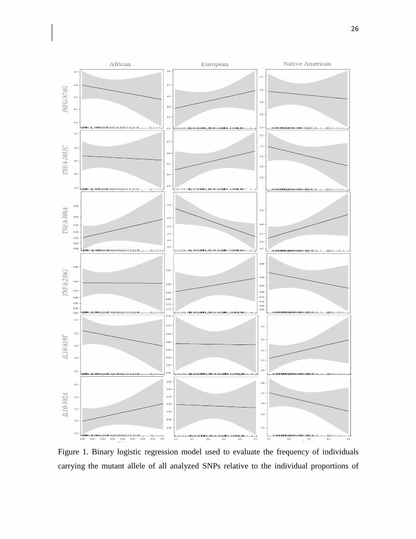
genetic ancestry proportions. The frequency of individuals carrying the TNF-308A allele
progressively decreased with the increasing proportion of European ancestry (p = 0.03).
However, when the Bonferroni correction for multiple tests was used, this association was
no longer significant (p = 0.18). No other association was observed.
Table 1. Characteristics of the study population.
Characteristic
Mild vivax malaria
(n = 90)
Non-infected
(n = 51)
p-value
Gender, malea
74.4
56.9
0.03
Age (years)b
32.5 (23.75-43.5) 37.0 (26.0-45.0) 0.62
Genetic ancestryc
European 0.442 ± 0.130 0.449 ± 0.130 0.76
African 0.318 ± 0.120 0.295 ± 0.112 0.26
Native American 0.240 ± 0.094 0.256 ± 0.111 0.35
Previous malaria episodesb
5.0 (2.0-7.0)
2.0 (0-6.0)
0.06
Previous history of malariaa
91.1 68.6 < 0.01 aPercentage
bMedian (IQR)
cMean ± SD
Table 2. Distribution of the genotypes between vivax malaria-infected and non-infected individuals
Malaria
Non-infected
Gene
SNP
Genotype
n (%)
MAF
HWE
n (%)
MAF
HWE
IFNG
+874A>T
AA
39 (48.7)
0.30
0.91
17 (34.0)
0.37
0.08
AT 34 (42.5) 29 (58.0)
TT 7 (8.8) 4 (8.0)
TNF -1031T>C TT 51 (56.7) 0.28 0.11 24 (47.1) 0.28 0.14
TC 37 (41.1) 25 (49.0)
CC 2 (2.2) 2 (3.9)
TNF -308G>A GG 69 (76.7) 0.12 0.21 35 (68.7) 0.18 0.69
GA 21 (23.3) 14 (27.4)
AA 0 2 (3.9)
TNF -238G>A GG 80 (88.9) 0.06 0.22 46 (90.2) 0.05 0.71
GA 9 (10.0) 5 (9.8)
AA 1 (1.1) 0
IL10 -819C>T CC 39 (43.3) 0.34 0.87 19 (37.2) 0.35 0.15
CT 41 (45.6) 28 (54.9)
TT 10 (11.1) 4 (7.8)
IL10 -592C>A CC 41 (45.6) 0.29 0.05 20 (39.2) 0.34 0.21
CA 45 (50.0) 27 (52.9)
AA 4 (4.4) 4 (7.8)
Abbreviations: MAF, Minor allele frequency; HWE, Hardy-Weinberg equilibrium

24
Table 3. Haplotype frequencies in the TNF and IL10 genes in vivax malaria-infected and
non-infected individuals.
Haplotype
Malaria
Non-infected
OR (95% CI)
p-value
TNF-1031/ -308/ -238
T/G/G 0.632 0.555 Reference 0.11
C/G/G 0.195 0.220 0.63 (0.30-1.31) 0.45
T/A/G 0.113 0.161 0.48 (0.20-1.13) 0.17
C/G/A 0.037 0.049 0.62 (0.15-2.35) 0.75
IL10-819/-592
C/C 0.642 0.647 Reference 0.94
T/A 0.291 0.343 0.80 (0.44-1.44) 0.33
T/C 0.054 0.009 7.19 (0.89-57.7) 0.06
Odds ratios (OR), 95% confidence interval (CI)

25
Table 4. Haplotype frequency and its association with the proportions of African, European and Native American ancestry.
African
European
Native American
Haplotype
Frequency
Proportion
Difference (95%
CI)
p-
value
Proportion
Difference (95%
CI)
p-
value
Proportion
Difference (95%
CI)
p-
value
TNF-1031/ -308/ -
238
T/G/G 0.615 0.31 Reference 0.44 Reference 0.25 Reference
C/G/G 0.191 -0.01 (-0.05 – 0.03) 0.61 -0.01 (-0.06 – 0.03) 0.57 0.01 (-0.02 – 0.05) 0.49
T/A/G 0.121 -0.05 (-0.10 – 0.00) 0.05 0.06 (0.00 – 0.11) 0.05 -0.02 (-0.06 – 0.02) 0.37
C/G/A 0.044 -0.01 (-0.08 – 0.06) 0.78 -0.02 (-0.11 – 0.06) 0.55 0.06 (-0.03 – 0.15) 0.17
IL10-819/-592
C/C 0.650 0.30 Reference 0.45 Reference 0.25 Reference
T/A 0.303 -0.01 (-0.04 – 0.03) 0.67 0.00 (-0.04 – 0.04) 0.94 0.01 (-0.02 – 0.04) 0.55
T/C 0.040 0.01 (-0.07 – 0.08) 0.86 -0.02 (-0.10 – 0.06) 0.60 0.02 (-0.05 – 0.08) 0.63
The effects of each haplotype were relative to the most frequent haplotype used as a reference. ∆% indicates relative change in the ancestry proportions compared
to the reference haplotypes with 95% confidence intervals.
26
Figure 1. Binary logistic regression model used to evaluate the frequency of individuals
carrying the mutant allele of all analyzed SNPs relative to the individual proportions of

27
genetic ancestry. The shading around the lines represents the 95% confidence interval. The
graph was constructed using the ggplot2 package in the R program.
4. Discussion
Previous studies reported different allele frequencies in cytokine genes among
different ethnicities. Due to these studies and the participation of these proteins in
numerous processes related to the pathogenesis of various diseases, we evaluated the
frequencies of polymorphisms in the TNFA, IFNG and IL10 genes in a highly admixed
Brazilian population and related their distributions to the proportions of genetic ancestry
using AIMs. We selected a population from northern Brazil where there was a higher
contribution of Native American ancestry due to the lack of data in studies of this nature
involving indigenous populations [52]. Because these cytokines play a key role in the
modulation of the immune response in malaria, we evaluated whether these polymorphisms
were related to protection against vivax malaria. However, this study did not provide
evidence of such associations.
The -308G>A SNP (rs1800629) is located in the promoter region of the gene, and
the presence of the A allele forms a binding site for the AP1 transcription factor that has
been associated with increases in TNF-α production [18]. The frequency distribution of the
A allele observed in our study (13.83%) was similar to that observed in previous studies in
the Brazilian population (12-16%) [53, 54, 55]. According to data from the 1000Genomes
project, the frequency of the A allele is similar between Europeans (13%) and Africans
(12%). This finding was in agreement with our results because no differences were
observed in the frequencies of this allele according to the proportions of genetic ancestry.
Contradictory results were observed for malaria, with the TNF-308A allele associated with
higher susceptibility/severity [56,57,58], without alterations [36] or with resistance to P.
falciparum malaria [59]. Regarding vivax malaria, which was the focus of the present
study, our results were in agreement with other studies, including those in the Brazilian
Amazon that did not observe any associations between the TNF-308G>A SNP and
susceptibility or clinical manifestations due to P. vivax infection [10, 19, 60].
28
The -238 G/A SNP (rs361525) does not have a clearly established function but
seems to affect the circulating cytokine levels because it is located on a repressor site in the
TNF-α gene [16]. The 5.38% frequency of the A allele (-238) in our results was similar to
the data for Europeans and Africans, which ranged from 4 to 6% [61]. The frequency of the
presence of the A allele at the -238 and -376 positions is low worldwide. In the Brazilian
Amazon, previous indices ranged from 5-7% [19, 62], and no associations were described
with vivax malaria in Pará [19]. In contrast, the G/A genotype was associated with psoriasis
in southeastern Brazil [63], and the A allele was associated with a decrease in falciparum
malaria parasitemia in Burkina Faso [30], cerebral malaria in Kenya [64] and malarial
anemia [56]. This SNP was associated with increased susceptibility to vivax malaria in the
Amazon region only when evaluated in the TATGG haplotype (−1031/-863/-857/-308/-
238) [19].
The 24.82% frequency of the C allele at the -1031 position (rs1799964) of the
TNFA gene is similar to data from the 1000Genomes project (15% and 21% for Africans
and Europeans, respectively) and Brazilian studies on leprosy and vivax malaria [62]. In
malaria, this SNP was associated with cytokine levels and clinical symptoms but not with
susceptibility in India [27]. The C allele is associated with a two-fold higher chance of
cerebral malaria caused by P. falciparum [65] in Thailand. In Africa, the CC genotype is
associated with repeated malaria episodes [44,59 ] and the T allele is associated with high
parasitemia [30 ]. In Brazil, the CC genotype is associated with protection against leprosy
but not malaria [62].
One hypothesis for the lack of association of the evaluated SNPs is that malaria can
occur due to possible linkage disequilibrium of the SNPs in TNFA with the human
leukocyte antigens (HLAs), which can cause non-functional mutations [59, 60 ]. The A
allele (-308) is described as having a strong linkage disequilibrium with HLA-Bw53 and
DRB1*1302-DQB1*0501, whereas the A allele at the -238 position of the TNFA gene
appears to be linked to HLA-B53 but with different immune characteristics [56 ]. The
haplotype frequencies in cytokine genes can vary extensively among different ethnic
groups most likely due to selective pressure on the human genome and thus affect the

29
susceptibility and clinical outcomes of diseases such as malaria [33]. This effect might have
affected our results due to the admixture observed in the Brazilian population.
. The gene sequence of this cytokine is highly conserved, with few polymorphisms.
The SNP at the -183 G/T position is related to increased transcription activity [26], whereas
+874 (A>T) is located in a region where the number of replicates can modulate the
expression of messenger RNA and the production of cytokines [21,66 ]. The T allele is
associated with a high number of replicate copies and activates the transcription site for the
NF-B pathway, which correlates with high cytokine expression [67, 68 ]. The A/A, T/A
and T/T genotypes are associated with low, intermediate and high production of IFN-γ,
respectively [21,69 ].
The highest frequency of the A allele (IFNG+874) is described in individuals with
European ancestry and is 46% (http://hapmap.ncbi.nlm.nih.gov). Indeed, the evaluated
population in the present study had a European contribution of almost 50% [43 ], and the
frequency of this allele was detected in 67.3% of the evaluated sample. However, no
association was detected with any ancestry or with malaria. Studies conducted in the United
States with African-American and Caucasian populations found higher frequencies of 66%
and 37% [70] and 48% and 25% [71], respectively. Our data showing the higher frequency
of the mutant A allele are in agreement with studies in the Brazilian Amazon that found
frequencies of 70.13% [21] and 73% [17], but all lacked an association with malaria caused
by P. vivax or P. falciparum. Few studies have described an association between this SNP
with malaria; however, its association with dermatitis was observed in India [72] and with
an increase in susceptibility to malaria in Brazil [21]. Importantly, higher levels of this
cytokine allow a better immune response against obligate intracellular pathogens; thus, low
levels of the A allele are associated with susceptibility to the disease.
The IL10 gene has more than 27 polymorphic sites associated with SNPs that result
in the differential production and expression of the cytokine [17, 33, 73 ], auto-immune and
inflammatory diseases [74], bacterial and viral infections [75] and human malaria [21].
Particularly, -819C and -1082G increase the protein production in peripheral blood

30
lymphocytes in vitro [76]. The (-1082, -819 and -592) GCC, ACC and ATA haplotypes are
associated with high, intermediate and low IL-10 production, respectively [17, 33].
The allele distributions for T (-819) and A (-592) in our results were 35.4% and
31.2%, respectively; these distributions were higher in Europeans than in Africans but
lacked significant associations. These data disagree with those from the 1000Genomes
project [61], which reports a higher frequency of the mutant alleles in Africans. Lokossu et
al. 2013 [77] reported higher frequencies (41.53% and 41.31% for the T and A alleles,
respectively) for falciparum malaria in Benin. The allele and genotype distributions of
SNPs in IL10 are described as variables according to ethnic group [21, 33] and the A (-
592), T (-819) and A (-1082) alleles are more frequent among African-Americans [77,78 ].
Moraes et al. 2003 [74] also found no associations of genotypes, alleles and haplotypes
with five IL10 SNPs (–3575, –2849, 2763, –1082, –819) in a study with Brazilian and
Dutch populations. However, studies with indigenous populations are scarce. In Brazil, a
study with the Terena of Mato Grosso do Sul state showed that the mutation rate was
significant for the IL10 -819 and -1082 SNPs [79]; in contrast, we obtained the lowest rates
for this ancestry and no association was observed.
The CC genotypes for the two SNPS were associated with a decrease in IL-10 levels
and low parasitemia in northern Brazil [17], which agreed with our data indicating no
significant association with susceptibility to malaria. Two studies in Pará state, Brazil, also
described no haplotype associations of the IL10 gene with malaria [19, 21 ] and falciparum
malaria in Africa [33]. In Piracicaba, southeastern Brazil, these SNPs were associated with
chronic periodontitis in Caucasians [80]. Future analyses of parasitemia and cytokine
indices may identify associations between the SNPs in the evaluated sample. One
hypothesis for the lack of association is that the patients involved in the present study did
not have malarial complications caused by P. vivax. Additionally, the transmission profile
of the malaria of the area investigated could have had an effect, and the epidemiology was
different from that observed in Africa. Another explanation may be the low frequency of
some genotypes in the present study. Thus, the sample size may have been too small to find
any possible association. This finding warrants further investigation.
31
Conclusion
The evaluation of ancestry informative markers (AIMs) allows estimations of
admixtures at the individual level and avoids possible confounding factors due to ethnicity,
such as in the tri-hybrid population sample evaluated in this study. Although most
polymorphisms in the TNFA, IFNG and IL10 genes investigated in this study did not
significantly differ according to ancestry and were not associated with risk or protection
against vivax malaria, the A allele of TNF-308 progressively decreased with the increasing
proportion of European ancestry. In Brazil, this is the first study to evaluate the distribution
of these genes according to ancestry. The results support the application of ancestry
informative markers in future studies.
Conflict of Interests
None of the authors declare a conflict of interests.
Acknowledgments
Financial support was provided by CNPq and Cappes.
References
[1] V. G. Haver, N. Verweij, J. Kjekshus et al., “The impact of coronary artery disease risk
loci on ischemic heart failure severity and prognosis: association analysis in the Controlled
Rosuvastatin multiNAtional trial in heart failure (CORONA),” BMC Med Genet, vol. 21,
nº.140, 2014.
[2] Z. Cheng, J. Zhou, K. K. To et al., “Identification of TMPRSS2 as a Susceptibility Gene
for Severe 2009 Pandemic A(H1N1) Influenza and A(H7N9) Influenza,” J Infect Dis, vol.
212, nº.8, pp.1214-1221, 2015.
[3] Y. Gong, C. W. McDonough, A. L. Beitelshees et al., “PTPRD gene associated with
blood pressure response to atenolol and resistant hypertension,” J Hypertens, vol. 33, nº.11,
pp. 2278-2285, 2015.

32
[4] S. N. Kariuki, Y. Ghodke-Puranik, J. M. Dorschner et al., “Genetic analysis of the
pathogenic molecular sub-phenotype interferon-alpha identifies multiple novel loci
involved in systemic lupus erythematosus,” Genes Immun, vol.16, nº.1, pp. 15-23, 2015.
[5] S. Onengut-Gumuscu, W. M. Chen, O. Burren et al., “Fine mapping of type 1 diabetes
susceptibility loci and evidence for colocalization of causal variants with lymphoid gene
enhancers,” Nat Genet, vol. 47, nº. 4, pp. 381-386, 2015.
[6] J. Ye, L. Jiang, C. Wu C et al., “Three ADIPOR1 Polymorphisms and Cancer Risk: A
Meta-Analysis of Case-Control Studies,” PLoS One, vol. 10, nº 6, 2015.
[7] J. N. Hirschhorn, K. Lohmueller, E. Byrne et al., “A comprehensive review of genetic
association studies,”Genet Med, vol. 4, nº. 2, pp. 45-61, 2002 .
[8] C. M. Lewis, J. Knight, “Introduction to genetic association studies,” Cold Spring Harb
Protoc, vol. 2012, nº. 3, pp. 297-306, 2012.
[9] World Health Organization (WHO). World malaria report 2015. Geneva (Switzerland):
World Health Organization; 2015.
[10] S. da Silva Santos, T. G. Clark, S. Campino et al., “Investigation of host candidate
malaria-associated risk/protective SNPs in a Brazilian Amazonian population,” PLoS One.
vol. 7, nº 5, 2012.
[11] G. Band, Q.S. Le, L. Jostins et al., “Malaria Genomic Epidemiology Network.
Imputation-based meta-analysis of severe malaria in three African populations,” PLoS
Genet, vol.9, nº.6, 2013.
[12] A. V. Grant, C. Roussilhon, R. Paul, A. Sakuntabhai, “The genetic control of
immunity to Plasmodium infection,”BMC Immunol, vol.16, nº.14, pp. 1-7, 2015.
[13] C. E. Cavasini, Mattos L.C, A. A. R. D'Almeida Couto et al., “Duffy blood group gene
polymorphisms among malaria vivax patients in four areas of the Brazilian Amazon
region,” Malaria Journal, vol. 19, nº.6, p 167, 2007.
[14]E. Tarazona-Santos, L. Castilho, D. R. Amaral et al., “Population genetics of GYPB
and association study between GYPB*S/s polymorphism and susceptibility to P. falciparum
infection in the Brazilian Amazon,” PLoS One, vol. 6, 2011.
[15] A. Rosanas-Urgell, E. Lin, L. Manning Let al., “Reduced risk of Plasmodium vivax
malaria in Papua New Guinean children with Southeast Asian ovalocytosis in two cohorts
and a case-control study, ” PLoS Med, vol. 9, nº. 9, 2012.

33
[16] A. Essadik, H. Jouhadi, T. Rhouda et al., “Polymorphisms of Tumor Necrosis Factor
Alpha in Moroccan Patients with Gastric Pathology: New Single-Nucleotide
Polymorphisms in TNF-𝛼 −193 (G/A),” Mediators of Inflammation, vol. 2015, pp.1-5,
2015.
[17] V.A Pereira, J. C. Sánchez-Arcila, A. Teva et al., “IL10 a genotypic association with
decreased IL-10circulating levels in malaria infected individuals from endemic area of the
Brazilian Amazon,”. Malar J, vol. 28, nº. 14, 2015.
[18] A. L. Van Dyke, M. L. Cotea, A. S. Wenzlaffa et al., “Cytokine SNPs: Comparison of
Allele Frequencies by Race & Implications for Future Studies,” Cytokine, vol. 46, nº.2 pp.
236-244, 2009.
[19] V. A. Sortica, M. G. Cunha, M. D. Ohnishi et al., “IL1B, IL4R, IL12RB1 and TNF
gene polymorphisms are associated with Plasmodium vivax malaria in Brazil,” Malar J,
vol.11, 2012.
[20] B. P. Ribeiro, G. C. Cassiano, R. M Souza et al., “Polymorphisms in Plasmodium
vivax Circumsporozoite Protein (CSP) Influence Parasite Burden and Cytokine Balance in a
Pre-Amazon Endemic Area from Brazil,’’Plos Neglected Tropical Diseases vol. 10, nº. 3,
2016.
[21] T. S. Medina, S. P. T. Costa, M. D. Oliveira et al., “Increased interleukin-10 and
interferon-g levels in Plasmodium vivax malaria suggest a reciprocal regulation which is
not altered by IL-10 gene promoter polymorphism,” Malar J, vol.10, pp.264, 2011.
[22] R. Gazzinelli, K. Parisa, A. Katherine et al. “Innate sensing of malaria parasites,” Nat
Rev Immunol, vol.14, nº.11, pp. 744-757, 2014.
[23] A.A. M. Fernandes, L. J. M. Carvalho, G. M. Zanini et al., “Similar Cytokine
responses and degressInfections in the Brazilian Amazon Region falciparum and
Plasmodium vivax of Anemia in Patients with Plasmodium,” Clin. Vaccine Immunol vol.
15, nº 4. pp. 650-658, 2008.
[24] B. B. Andrade, A. Reis-Filho, S. M. Souza-Neto et al., “ Severe Plasmodium vivax
malaria exhibits marked inflammatory imbalance,’’ Malaria Journal vol. 13, nº 13, 2010.
[25] E. K. Riccio, P. R. Totino, L.R. Pratt-Riccio, et al., “Cellular and humoral immune
responses against the Plasmodium vivax MSP-1(1)(9) malaria vaccine candidate in

34
individuals living in an endemic area in northeastern Amazon region of Brazil,’’ Malaria
Journal vol. 12, pp. 326, 2013.
[26] S. Cabantous, B. Poudiougou, A. Traore et al., “Evidence. That Interferon-g Plays a
Protective Role during Cerebral Malaria,” J Infect Dis, vol.192, pp.854-860, 2005.
[27] M. Sohail, A. Kaul, P. Bali et al., “Allels -308A and -1031C in the TNFα gene
promoter do not increase the risk but associated with circulating levels of TNFα and
clinical features of vivax malaria in Indian patients,” Mol Immunol, vol.45, pp.1682-1692,
2008.
[28] V.R.Mendonça, M. Barral-Netto, “Immunoregulation in human malaria: the challenge
of understanding asymptomatic infection,” Mem Inst Oswaldo Cruz,vol 110, nº 8, pp. 945-
955, 2015.
[29] M. U. Capei, E. Dametto, M. E. Fasano et al., “Genotyping for cytokine
polymorphisms: allele frequencies in the Italian population European,” Journal of
Immunogenetics, vol. 30, nº.1, pp. 5-10, 2003.
[30] L. Flori, N. F. Delahaye, F.A. Iraqi et al., “TNF as a malaria candidate gene:
polymorphism-screening and family-based association analysis of mild malaria attack and
parasitemia in Burkina Faso,” Genes Immun, vol. 6, nº. 6, pp. 472-480, 2005.
[31] R.Ubalee, F. Suzuki, M. Kikuchi M et al., “Strong association of a tumor necrosis
factor-alpha promoter allele with cerebral malaria in Myanmar.,” Tissue Antigens vol. 58,
pp.407-410, 2001.
[32] M. B. B. McCall, R. W. Sauerwein, “ Interferon-_—central mediator of protective
immune responses against the pre-erythrocytic and blood stage of malaria,” Journal of
Leukocyte Biology vol.88, nº 6, pp. 1131-1143, 2010.
[33] C. Ouma, G. C. Davenport, T. Were et al., “Haplotypes of IL-10 promoter variants are
associated with susceptibility to severe malarial anemia and functional changes in IL-10
production,”Hum Genet, vol. 124, nº. 5, pp. 515-524, 2008.
[34] W. Ouyang, S. Rutz, N. K. Crellin et al., “Regulation and Functions of the IL-10
Family of Cytokines in Inflammation and Disease,” Annual Rev Immunol vol. 29, pp.71-
109, 2011.
[35] L. E. Alvarado-Arnez, E. P. Amaral, C. Sales-Marques et al., “Association of IL10
Polymorphisms and Leprosy: A Meta-Analysis,” PLOS ONE, 2015.

35
[36] L. M. Randall, E. Kenangalem, D. A. Lampah et al., “A study of the TNF/LTA/LTB
locus and susceptibility to severe malaria in highland papuan children and adults,” Malar J,
vol. 9, pp. 302, 2010.
[37] B. Maiga, A. Dolo, O. Touré, “Human candidate polymorphisms in sympatric ethnic
groups differing in malaria susceptibility in Mali,” PLoS One, vol. 9, nº.7, 2013.
[38] F. M. Tomaz, A. A. da Cruz Furini, M. P. Capobianco et al., “Humoral immune
responses against the malaria vaccine candidate antigen Plasmodium vivax AMA-1 and IL-
4 gene polymorphisms in individuals living in an endemic area of the Brazilian Amazon,”
Cytokine, vol. 74, nº 2, pp. 273-278, 2015.
[39] R. Pereira, C. Phillips, N. Pinto et al., “Straightforward inference of ancestry and
admixture proportions through ancestry-informative insertion deletion multiplexing,” PLoS
One, vol. 7, nº.1, 2012.
[40] C. Santos, C. Phillips, M. Fondevila et al., “Pacifiplex: an ancestry-informative SNP
panel centred on Australia and the Pacific region,” Forensic Sci Int Genet, vol.20, pp. 71-
80, 2016.
[41] G. Suarez-Kurtz, S. D. Pena, C. J. Struchiner et al., “Pharmacogenomic Diversity
among Brazilians: Influence of Ancestry, Self-Reported Color, and Geographical Origin,”
Front Pharmacol, vol.6, nº. 3, 2012..
[42] V. Bonifaz-Peña, A.V. Contreras, C. J. Struchiner et al., “Exploring the distribution of
genetic markers of pharmacogenomics relevance in Brazilian and Mexican populations,”
PLoS One, vol. 9, nº. 11, 2014.
[43] G. C. Cassiano, E. J. Santos, M. H. Maia et al., “Impact of population admixture on the
distribution of immune response co-stimulatory genes polymorphisms in a Brazilian
population,”Hum Immnulogy vol. 76, nº. 11. pp. 836-842, 2015.
[44] T. Asghar, S. Yoshida, S. Kennedy et al., “The tumor necrosis factor-a promoter
21031C polymorphism is associated with decreased risk of endometriosis in a Japanese
population,” Human Reproduction, vol. 19, nº. 11, pp. 2509-2514, 2004.
[45] M. Hedayati, K. Sharifi, F. Rostami et al., “Association between TNF-a promoter G-
308A and G-238A polymorphisms and obesity,” Mol Biol Rep, vol.39, nº. 2, pp. 825-829,
2012.

36
[46] N.P.Santos, E. M. Ribeiro-Rodrigues, A. K. Ribeiro-Dos-Santos et al., “ Assessing
individual interethnic admixture and population substructure using a 48- insertion-deletion
(INSEL) ancestry-informative marker (AIM) panel,’’ Hum Mutat vol 31, pp. 184-190,
2010.
[47] N. de Seixas Santos, A. C. de Mello Malta, F. Diniz et al., “Association of IFNL3 and
IFNL4 polymorphisms with hepatitis C virus infection in a population from southeastern
Brazil,” Archives of Virology vol. 161, nº6, pp.1477-1484, 2016.
[48] B.R Ramos, M. P. B DElia, M. A. Amador et al., “Neither self-reported ethnicity nor
declared family origin are reliable indicators of genomic ancestry,’’ Genetica vol. 144, nº 3,
pp. 259-265, 2016.
[49] J. R. Gonzalez, L. Armengol, X. Sole et al., “SNP assoc: an R package to perform
whole genome association studies,” Bioinformatics, vol. 23, pp.644-645, 2003.
[50] J. P. Sinnwell, D. J. Schaid. 2009. haplo.stats: Statistical analysis of haplotypes with
traits and covariates when linkage phase is ambiguous. R package version 1.4.4.
http://CRAN.R-project.org/package=haplo.statshttp://CRAN.R-
project.org/package=haplo.stats, 2009. Accessed 29 April 2013.
[51] W. N. Venables, B. D. Ripley “Modern Applied Statistics With S, fourth edition,”
New York: Springer; 2002. http://www.stats.ox.ac.uk/pub/MASS4. [ISBN 0-387-95457-0].
[52]S. D. Pena, G. Di Pietro, M. Fuchshuber-Moraes et al., “The genomic ancestry of
individuals from different geographical regions of Brazil is more uniform than expected,”.
PLoS One, vol.6, nº. 2, 2011.
[53] D. D. Paskulin, P. R. Fallavena, F. J. Paludo et al., “TNF -308G/T; a promoter
polymorphism (rs1800629) and outcome from critical illness,” Braz J Infect Dis, vol. 15,
nº. 3, pp. 231-238, 2011.
[54] J. G. de Oliveira, A. F. Rossi, D. M Nizato et al., “Influence of functional
polymorphisms in TNF-α, IL-8 and IL-10 cytokine genes on mRNA expression levels and
risk of gastric cancer,” Tumour Biol, vol.36, nº. 12, pp. 9159-9170, 2015.
[55] L. F Sesti, D. Crispim, L. H. Canani et al., “The -308G>a polymorphism of the TNF
gene is associated with proliferative diabetic retinopathy in Caucasian Brazilians with type
2 diabetes,” Invest Ophthalmol Vis Sci, vol. 56, nº.2, pp.1184-1190, 2015.

37
[56] W. McGuire, J.C. Knight, A. V. Hill, “Severe malarial anemia and cerebral malaria
are associated with different tumor necrosis factor promoter alleles,” J Infect Dis, vol.179,
nº.1, pp. 287-290, 1999.
[57] C. G. Meyer, J. May, A. J. Luty et al., “TNFalpha-308A associated with shorter
intervals of Plasmodium falciparum reinfections,” Tissue Antigens, vol. 59, nº.4, pp. 287-
292, 2002.
[58] S. J. Dunstan, K. A. Rockett, N. T. Quyen NT et al., “Variation in human genes
encoding adhesion and proinflammatory molecules are associated with severe malaria in
the Vietnamese,” Genes Immun, vol. 13, nº. 6, pp. 503-508, 2012.
[59] W. N. G. Gichohi Wainaina, A. Melse Boonstra, E. J. Feskens et al., “Tumour necrosis
factor allele variants and their association with the occurrence and severity of malaria in
African children: a longitudinal study,” Malar J, vol.14, pp. 249, 2015.
[60] V. R. Mendonça, R. Vitor, L. C. L. Souza et al., “DDX39B (BAT1), TNF and IL6
gene polymorphisms and association with clinical outcomes of patients with Plasmodium
vivax malaria,” Malaria J, vol,13, pp 278, 2014.
[61] The 1000 Genomes Project Consortium. An integrated map of genetic variation from
1,092 human genomes,” Nature, vol. 491, pp. 56-65.
[62] G. A. V. Silva, R. Ramasawmy, A. L. Boechat et al., “Association of TNF-1031 C/C
as a potential protection marker for leprosy development in Amazonas state patients, Brazil.
Human Immunology,” vol. 76, pp. 137-141, 2015.
[63] R. Cardili, R.N. Deghaide, C. T. Mendes-Junior et al., “HLA-C and TNF gene
polymorphisms are associated with psoriasis in Brazilian patients,” International Journal of
Dermatology, vol. 55, nº.1, pp. 16-22, 2016.
[64] J. C. Knight, I. Udalova, A. V. Hill et al., “A polymorphism that affects OCT-1
binding to the TNF promoter region is associated with severe malaria,” Nat Genet, vol. 22,
nº.2, pp. 145-150, 1999.
[65] H. Hananantachai, J. Patarapotikul, J. Ohashi et al., “Significant association between
TNF-a (TNF) promoter allele (-1031C, -863C, -857C) and cerebral malaria in Thailand,”
Tissue Antigens, vol.69, nº.3, pp. 277-280, 2007.
[66] J. MacMurray, D.E. Comings, V. Napolioni, “The gene-immune-behavioral pathway:
Gamma-interferon (IFN-c) simultaneously coordinates susceptibility to infectious disease

38
and harm avoidance behaviors,” Brain, Behavior, and Immunity, vol. 35, pp. 169-175,
2014.
[67] A. Bozzi, B. S. Reis, P.P. Pereira et al., “Interferon-gamma and interleukin-4 single
nucleotide gene polymorphisms in Paracoccidioidomycosis,” Cytokine, vol. 48, nº. 3, pp.
212-217, 2009.
[68] E. Peresi, L. R. C. Oliveira LRC, Silva WL, et al., “Cytokine Polymorphisms, Their
Influence and Levels in Brazilian Patients with Pulmonary Tuberculosis during
Antituberculosis Treatment,”Tuberculosis Research and Treatment, vol. 2013, pp. 1-13
2013.
[69] L. Castelar, M.M. Silva, E.C. Castelli et al., “Interleukin-18 and interferon-gamma
polymorphisms in Brazilian human immunodeficiency virus-1-infected patients presenting
with lipodystrophy syndrome,” Tissue Antigens, vol 76, nº. 2, pp. 126-130, 2010.
[70] N. L. Delaney, V. Esquenazi, D. P. Lucas et al., “TNF-, TGF-, IL-10, IL-6 and INF-g
Alleles Among African Americans and Cuban Americans Report of the ASHI Minority
Workshops: Part IV,” Human Immunology, vol. 65, pp.1413-1419, 2004.
[71] M. I. Hassan, Y. Aschner, C. H. Manning et al., “Racial differences in selected
cytokine allelic and genotypic frequencies among healthy, pregnant women in North
Carolina,” Cytokine, vol. 21, nº.1, pp. 10-16, 2003.
[72] R. Khatri, K. Mukhopadhyay, K. K. Verma et al., “Genetic predisposition to
parthenium dermatitis in an Indian cohort due to lower-producing genotypes of interleukin-
10 (-)1082 G>A and (-) 819 C>T loci but no association with interferon-γ (+) 874 A>T
locus,” British Association of Dermatologists, vol. 165, nº.1, pp. 115-122, 2011.
[73] D. Carpenter, H. Abushama, S. Bereczky et al., “Immunogenetic control of antibody
responsiveness in a malaria endemic area,” Hum Immunol, vol.68, nº. 3, pp. 165-169, 2007.
[74] M. O. Moraes, A. R. Santos, J. J. Schonkeren et al., “Interleukin-10 promoter
haplotypes are differently distributed in the Brazilian versus the Dutch population,”
Immunogenetics, vol. 12, nº. 12, pp. 896-899, 2003.
[75] W. Ben-Selma, H. Harizi, J. Boukadida, “Association of TNF-a and IL-10
polymorphisms with tuberculosis in Tunisian populations,” Microbes Infect, vol.13,
pp.837-843, 2011.
39
[76] R. B. Ness, C. L. Haggerty, G. Harger et al., “Differential Distribution of Allelic
Variants in Cytokine Genes among African Americans and White Americans,” Am J
Epidemiol, vol. 160, nº. 11, pp. 1033-1038, 2004.
[77] A. G. Lokossou, C. Dechavanne, A. Bouraïma et al., “Association of IL-4 and IL-10
maternal haplotypes with immune responses to P. falciparum in mothers and newborns,”
BMC Infect Dis, vol 13,215, 2013.
[78] S. C. Hoffmann, E. M. Stanleya, E.D. Cox, et al., “Ethnicity Greatly Influences
Cytokine Gene Polymorphism Distribution,”American Journal of Transplantation, vol. 2,
nº.6, pp. 560-567, 2002.
[79] A. G. Albuquerque, M. Moraes, P. R. Vanderborght et al., “Tumor Necrosis Factor
and Interleukin-10 Gene Promoter Polymorphisms in Brazilian Population and in Terena
Indians,” Transplantation Proceedings, vol.36, pp.825-826, 2004.
[80] R. M. Scarel-Caminaga, P. C. Trevilatto, A. P. Souza AP “Interleukin 10 gene
promoter polymorphisms are associated with chronic periodontitis,” J Clin Periodontol
vol.31, pp. 443-448, 2004.

Furini et al. Malar J (2016) 15:374 DOI 10.1186/s12936-016-1414-3
RESEARCH
Cytokine gene polymorphisms are not associated with anti-PvDBP, anti-PvAMA-1 or anti-PvMSP-119 IgG antibody levels in a malaria-endemic area of the Brazilian AmazonAdriana A. C. Furini1*, Marcela P. Capobianco2, Luciane M. Storti‑Melo3, Maristela G. Cunha4, Gustavo C. Cassiano5 and Ricardo Luiz D. Machado1,2,6
Abstract
Background: The immune response against Plasmodium vivax immunogenic epitopes is regulated by pro‑ and anti‑inflammatory cytokines that determine antibody levels and class switching. Cytokine gene polymorphisms may be responsible for changes in the humoral immune response against malaria. The aim of this study was to evaluate whether polymorphisms in the TNFA, IFNG and IL10 genes would alter the levels of anti‑PvAMA1, PvDBP and ‑PvMSP‑119 IgG antibodies in patients with vivax malaria.
Methods: Samples from 90 vivax malaria‑infected and 51 uninfected subjects from an endemic area of the Brazilian Amazon were genotyped using polymerase chain reaction‑restriction fragment length polymorphism (PCR–RFLP) to identify polymorphisms of the genes TNFA (−1031T > C, −308G > A, −238G > A), IFNG (+874T > A) and IL10 (−819C > T, −592C > A). The levels of total IgG against PvAMA1, PvDBP and PvMSP‑119 were determined using an enzyme‑linked immunosorbent assay (ELISA). Associations between the polymorphisms and the antibody response were assessed by means of logistic regression models.
Results: No significant differences were found in the levels of IgG antibodies against the PvAMA‑1, PvDBP or PvMSP‑119 proteins in relation to the studied polymorphisms.
Conclusions: Although no associations were found among the evaluated genotypes and alleles and anti‑merozoite IgG class P. vivax antibody levels, this study helps elucidate the immunogenic profile involved in the humoral immune response in malaria.
Keywords: IgG antibody, Plasmodium vivax, TNF, IFNG, IL10
© 2016 The Author(s). This article is distributed under the terms of the Creative Commons Attribution 4.0 International License (http://creativecommons.org/licenses/by/4.0/), which permits unrestricted use, distribution, and reproduction in any medium, provided you give appropriate credit to the original author(s) and the source, provide a link to the Creative Commons license, and indicate if changes were made. The Creative Commons Public Domain Dedication waiver (http://creativecommons.org/publicdomain/zero/1.0/) applies to the data made available in this article, unless otherwise stated.
BackgroundEarly diagnosis, prompt and effective treatment, the use of mosquito nets impregnated with long-acting insec-ticides and residual intradomiciliary spraying are the main malaria control measures [1] and have resulted in a reduction in the transmission and number of cases of malaria in Brazil. However, this disease remains one of
the most prevalent infections in tropical countries, with 214 million clinical cases/year and approximately 438,000 deaths [2]. In Brazil, Plasmodium vivax is the aetiologic agent in 85 % of cases, and 99.9 % of cases occur in the Brazilian Amazon region [2].
The different clinical manifestation patterns of malaria may be related to host and agent genetic factors, age and ethnicity as well as the involvement of these factors in the immune response and parasitaemia and antibody levels [3–5]. IgG antibodies play a protective role against para-site invasion in the erythrocytic cycle of Plasmodium [3, 6]. For Plasmodium knowlesi, anti-PvAMA-1 monoclonal
Open Access
Malaria Journal
*Correspondence: [email protected] 1 Department of Dermatology, Infectious and Parasitic Diseases, São José do Rio Preto Medical School, São José do Rio Preto, São Paulo, BrazilFull list of author information is available at the end of the article

Page 2 of 8Furini et al. Malar J (2016) 15:374
antibodies have been shown to be capable of inhibit-ing merozoite invasion in vitro [7]. PvDBP antibodies inhibit interactions with the Duffy antigen receptor for chemokines (DARC) in erythrocytes [8], and antibodies produced against PvMSP-119 have been shown to prevent merozoite invasion in vitro [9]. The synthesis of immuno-globulins is complex and depends on the process of antigen presentation by B lymphocytes (BL) to TCD4 lymphocytes and the involvement of co-stimulatory molecules and their receptors [10].
The production, levels and switching of antibody classes is regulated by pro- and anti-inflammatory cytokines [3, 6, 11, 12]. IFN-γ can negatively modulate the humoral immune response, thus interfering with cir-culating antibody levels [13] and increasing IgG2 produc-tion. TNF appears to be important in the development of the humoral response as an autocrine growth factor for B cells [6]. Among the anti-inflammatory cytokines, IL-10 may participate in negative immunomodulation of the Th1-type response [14, 15] in addition to inducing immunoglobulin synthesis [6].
Many genetic variants are responsible for minor changes in the immune response in malaria [11, 12]. Pre-vious studies in Brazil with vivax malaria have evaluated factors associated with genetic variability in the humoral immune response. HLA-DR16 is associated with the IgG antibody response to the P. vivax VK247 variant circum-sporozoite protein [16]. High levels of MSP-1 antibodies are also associated with HLA-DR3 [17]. In Goianésia do Pará, a municipality located in the Brazilian Amazon, two studies evaluated the effects of polymorphisms in genes involved in the humoral immune response. Cassiano et al. [10] found that the frequency of specific IgG responders against PvAMA-1, PvDBP and PvMSP-119 was associated with polymorphisms in the BLYS (−871C > T), CD40 (−1C > T) and CD86 (+1057G > A) genes. In contrast, genotypes and haplotypes of the IL4 gene were not asso-ciated with the production of PvAMA-1 antibodies [18]. The aim of this work was to continue the search for the genetic basis of these traits and to evaluate whether poly-morphisms in the TNFA, IFNG and IL10 genes alter the levels of anti-PvAMA1, -PvDBP and -PvMSP-119 IgG antibodies.
MethodsStudy area and subjectsNinety samples from vivax malaria-infected subjects and 51 samples from uninfected subjects were collected in Goianésia do Pará (03°50′33″S; 49°05′49″ W). The sub-jects were in a sub-group of individuals previously ana-lysed by Cassiano et al. [10]. The study has evaluated the effect of genetic ancestry on the distribution of polymor-phisms in the TNFA, IFNG and IL10 genes (unpublished
data). No differences were observed in the mean propor-tion of any ancestry among the different genotypes and haplotypes analysed.
In 2011 and 2012, the numbers of malaria cases were 2856 and 1136, respectively, with 79 % of cases caused by P. vivax. Samples were collected at the municipal health center between February 2011 and August 2012. Data were collected by passive detection in Basic Health Units after thick blood film phenotypic diagnosis, but prior to treatment. All patients if malaria were given stand-ard treatment of 1500 mg of chloroquine in 3 days (600, 450 and 450 mg) plus 30 mg of primaquine on the day the diagnosis and on the following 6 days. Individuals infected with P. vivax presented for medical care because of clinical symptoms of malaria and they were recruited after diagnosis. The uninfected individuals who sought medical care offered during the study were invited to par-ticipate in the study. These individuals experienced the same conditions of exposure to the aetiological agent. All participants or guardians signed the consent form, and the project was approved by the Goianésia do Pará health authorities and by the Research Ethics Committee (CAAE 01774812.2.0000.5415) of the College of Medi-cine of São José do Rio Preto (Faculdade de Medicina de São José do Rio Preto).
Malaria was diagnosed using thick blood smears stained with Giemsa and subsequently confirmed by nested-PCR with modifications [19]. No uninfected indi-vidual was positive in nested-PCR, while five individuals positive for P. vivax (thick blood smear) had mixed infec-tion with Plasmodium falciparum (by nested-PCR) and were excluded from the study. Parasitaemia was defined as the number of parasites per microlitre of blood after examination of 100 microscopic fields.
Peripheral blood was stored at −20 °C. The examina-tion of polymorphisms in the genes TNFA, IFNG and IL10 was performed via PCR amplification followed by restriction fragment length polymorphism (RFLP) analy-sis or the amplification of specific alleles (Table 1).
The amplified products were subjected to electro-phoresis (100 V/50 min) in 1.5 to 2.5 % agarose gels stained with Gel Red (Biotium, CA, USA) or in eth-idium bromide-stained 12.5 % polyacrylamide gels (10 mg/ml) and were visualized using a transilluminator (Biotecnologia-Locus).
Assessment of the serological response against Plasmodium vivaxThe levels of IgG class anti-MSP-119, anti-PvAMA-1 and anti-PvDBP antibodies in a larger cohort were previously reported by Cassiano et al. [10]. The analyses were per-formed using ELISA following previously described pro-tocols [24–26]. A recombinant protein (His6-MSP-119)
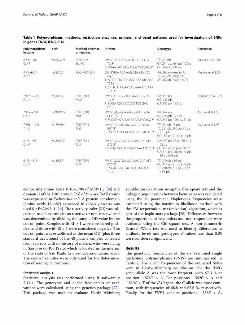
Page 3 of 8Furini et al. Malar J (2016) 15:374
comprising amino acids 1616–1704 of MSP-119 [24] and domain II of the DBP protein [25] of P. vivax (SAll strain) was expressed in Escherichia coli. A protein ectodomain (amino acids 43–487) expressed in Pichia pastoris was used for PvAMA-1 [26]. The reactivity index (RI) was cal-culated to define samples as reactive or non-reactive and was determined by dividing the sample OD value by the cut-off point. Samples with RI ≥ 1 were considered posi-tive, and those with RI < 1 were considered negative. The cut-off point was established as the mean OD (plus three standard deviations) of the 40 plasma samples collected from subjects with no history of malaria who were living in São José do Rio Preto, which is located in the interior of the state of São Paulo (a non-malaria-endemic area). The control samples were only used for the determina-tion of serological response.
Statistical analysisStatistical analysis was performed using R software v 2.11.1. The genotypic and allelic frequencies of each variant were calculated using the genetics package [27]. This package was used to evaluate Hardy–Weinberg
equilibrium deviations using the Chi square test and the linkage disequilibrium between locus pairs was calculated using the D′ parameter. Haplotypes frequencies were estimated using the maximum likelihood method with the EM (expectation–maximization) algorithm, which is part of the haplo stats package [28]. Differences between the proportions of responders and non-responders were evaluated using the Chi square test. A non-parametric Kruskal–Wallis test was used to identify differences in antibody levels and genotypes. P values less than 0.05 were considered significant.
ResultsThe genotypic frequencies of the six examined single nucleotide polymorphisms (SNPs) are summarized in Table 2. The allelic frequencies of the evaluated SNPs were in Hardy–Weinberg equilibrium. For the IFNG gene, allele A was the most frequent, with 67.3 % at position +874T > A. For positions −592C > A and −819C > T of the IL10 gene, the C allele was most com-mon, with frequencies of 68.8 and 65.6 %, respectively. Finally, for the TNFA gene at positions −238G > A,
Table 1 Polymorphisms, methods, restriction enzymes, primers, and band patterns used for investigation of SNPs in genes TNFA, IFNG, IL10
Polymorphisms in gene
SNP Method enzimas annealing
Primers Genotype Reference
IFN γ −183G > T
rs2069709 RFLP (53º)Eco47I
FW: 5′‑AAT GAT CAA TGT GCT TTG TG‑3′
R: 5′‑TAA GAT GAG ATG GTG ACAG‑3′
TT: 271 pbGT: 271 pb, 164 pb, 107pbGG: 164pb, 107 pb
Suxia Qi et al. [20]
IFN γ +874A > T
rs2430561 ASO‑PCR (53º) CP: ‑5′‑TCA ACA AAG CTG ATA CTC CA‑3′
T : 5′‑TTC TTA CAA CAC AAA ATC AAA TCT‑3′
A: 5′‑TTC TTA CAA CAC AAA ATC AAA TCA‑3′
AA: 262 pb (reação A)TT: 262 pb (reação T)AT: 262 pb (reação A, T)
Medina et al. [21]
TNF α −238G > A
rs 361525 RFLP (60º)MspI
FW: 5′‑ATC TGG AGG AAG CGG TAG TG‑3′
R: 5‑AGA AGA CCC CCC TCG GAA CC’ 3′
AA: 156 pbGG: 133 pbGA: 153 pb, 133 pb
Hedayati et al. [22]
TNFα −308G > A
rs 1800629 RFLP (59º)NcoI
FW: 5′‑GAG GCA ATA GGT TTT GAG GGC CAT‑3′
R: 5′‑GGG ACA CAC AAG CAT CAAG 3′
AA: 147 pbGG: 126 pb, 121 pbGA: 147 pb, 126 pb, 21 pb
Asghar et al. [23]
TNFα −1031T > C
rs1799964 RFLP (57º)BbsI
FW: 5′‑TAT GTG ATG GAC TCA CCA GGT‑3′
R: 5′‑CCT CTA CAT GGC CCT GTC TT 3′
TT: 251 pb, 13 pbTC: 251 pb, 180 pb, 71 pb
e 13 pbCC: 180 pb, 71 pb e 13 pb
Asghar et al. [23]
IL‑10 −592C > A
rs1800872 RFLP (54º)RsaI
FW: 5′‑GGG TGA GGA AAC CAA ATT CTC‑3′
R:5′‑GAG GGG GTG GGC TAA ATA TC 3′
AA: 240 pb, 77 pb, 36 pb e 08 pb
CC: 317 pb,36 pb e 08 pbCA: 317 pb, 240 pb, 77 pb,
36 pb e 08 pb
IL‑10 −819C > T
rs1800871 RFLP (54º)RseI
FW: 5′‑GGG TGA GGA AAC CAA ATT CTC‑3′
R: 5′‑GAG GGG GTG GGC TAA ATA TC 3′
TT: 270 pb e 91 pbCC: 217 pb, 91 pb e 53 pbTC: 270 pb, 217 pb, 91 pb
e 53 pb
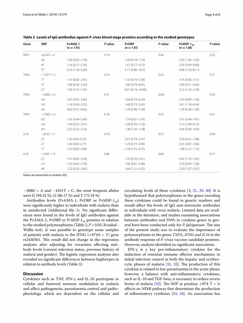
Page 4 of 8Furini et al. Malar J (2016) 15:374
−308G > A and −1031T > C, the most frequent alleles were G (94.32 %), G (86.17 %) and T (75.18 %).
Antibodies levels (PvAMA-1, PvDBP or PvMSP-119) were significantly higher in individuals with malaria than in uninfected (Additional file 1). No significant differ-ences were found in the levels of IgG antibodies against the PvAMA-1, PvDBP or PvMSP-119 proteins in relation to the studied polymorphisms (Table 2, P > 0.05, Kruskal–Wallis test). It was possible to genotype some samples of patients with malaria to the IFNG (+874A > T) gene rs2430561. This result did not change in the regression analyses after adjusting for covariates affecting anti-body levels (current infection status, previous history of malaria and gender). The logistic regression analyses also revealed no significant differences between haplotypes in relation to antibody levels (Table 3).
DiscussionCytokines such as TNF, IFN-γ and IL-10 participate in cellular and humoral immune modulation in malaria and affect pathogenesis, parasitaemia control and patho-physiology, which are dependent on the cellular and
circulating levels of these cytokines [3, 21, 29, 30]. It is hypothesized that polymorphisms in the genes encoding these cytokines could be found in genetic markers and would affect the levels of IgG anti-merozoite antibodies in individuals with vivax malaria. Limited data are avail-able in the literature, and studies examining associations between antibodies and SNPs in cytokine genes in gen-eral have been conducted only for P. falciparum. The aim of the present study was to evaluate the importance of polymorphisms in the genes TNFA, IFNG and IL10 in the antibody response of P. vivax vaccine candidate proteins. However, analysis identified no significant association.
IFN-γ is a key pro-inflammatory cytokine for the induction of essential immune effector mechanisms in initial infection control in both the hepatic and erythro-cytic phases of malaria [31, 32]. The production of this cytokine is related to low parasitaemia in the acute phase; however, a balance with anti-inflammatory cytokines, such as IL-10 and TGF-beta, is necessary to reduce severe forms of malaria [32]. The SNP at position +874 T > A affects an NFkB pathway that determines the production of inflammatory cytokines [33, 34]. An association has
Table 2 Levels of IgG antibodies against P. vivax blood stage proteins according to the studied genotypes
Values are presented as medians (IQ)
Gene SNP PvAMA-1 (n = 135)
P value PvDBP (n = 135)
P value PvMSP-119(n = 128)
P value
INFG +874T > A 0.19 0.40 0.42
AA 1.36 (0.63–2.78) 1.58 (0.78–7.29) 2.92 (1.00–7.63)
AT 1.14 (0.51–2.18) 1.41 (0.77–4.73) 2.05 (0.49–6.64)
TT 2.50 (1.18–3.00) 5.17 (0.86–18.5) 4.66 (1.50–8.11)
TNFA −1031T > C 0.74 0.42 0.71
TT 1.41 (0.60–2.97) 1.34 (0.79–5.38) 3.74 (0.96–7.57)
TC 1.49 (0.58–2.50) 1.66 (0.79–6.87) 2.60 (0.51–6.93)
CC 1.45 (1.01–1.52) 8.57 (0.73–14.89) 2.12 (1.32–2.59)
TNFA −308G > A 0.41 0.58 0.50
GG 1.67 (0.65–2.80) 1.66 (0.79–6.30) 2.92 (0.89–7.58)
GA 1.14 (0.58–2.23) 1.48 (0.73–3.42) 2.61 (1.18–6.54)
AA 0.62 (0.51–0.62) 1.39 (0.96–1.39) 1.30 (0.28–1.30)
TNFA −238G > A 0.76 0.97 0.61
GG 1.45 (0.64–2.66) 1.59 (0.81–5.59) 2.61 (0.94–7.47)
GA 1.34 (0.53–2.91) 1.48 (0.76–7.16) 5.15 (1.69–8.16)
AA 2.32 (2.32–2.32) 1.28 (1.28–1.28) 3.04 (3.04–3.04)
IL10 −819C > T 0.79 0.57 0.99
CC 1.35 (0.63‑2.53) 2.01 (0.79–5.91) 3.50 (0.55–7.48)
CT 1.45 (0.60‑2.77) 1.29 (0.77–5.06) 2.61 (0.87–7.60)
TT 2.22 (0.83–2.84) 2.18 (1.35–6.31) 1.98 (1.22–7.13)
IL10 −592C > A 0.86 0.86 0.77
CC 1.41 (0.62–2.50) 1.59 (0.76–5.91) 4.92 (1.10–7.63)
CA 1.52 (0.62–2.78) 1.46 (0.81–5.46) 2.59 (0.94–7.26)
AA 1.22 (0.52–2.85) 1.84 (1.21–4.32) 2.50 (1.07–5.97)
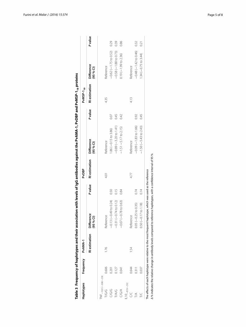
Page 5 of 8Furini et al. Malar J (2016) 15:374
Tabl
e 3
Freq
uenc
y of
hap
loty
pes
and
thei
r ass
ocia
tion
wit
h le
vels
of I
gG a
ntib
odie
s ag
ains
t the
PvA
MA
-1, P
vDBP
and
PvM
SP-1
19 p
rote
ins
The
effec
ts o
f eac
h ha
plot
ype
wer
e re
lativ
e to
the
mos
t fre
quen
t hap
loty
pe, w
hich
was
use
d as
the
refe
renc
e
Δ %
indi
cate
s th
e re
lativ
e ch
ange
in a
ntib
ody
leve
ls c
ompa
red
to re
fere
nce
hapl
otyp
es, w
ith a
con
fiden
ce in
terv
al o
f 95
%
Hap
loty
pes
Freq
uenc
yPv
AM
A-1
PvD
BPPv
MSP
-119
RI e
stim
atio
nD
iffer
ence
(95
% C
I)P
valu
eRI
est
imat
ion
Diff
eren
ce(9
5 %
CI)
P va
lue
RI e
stim
atio
nD
iffer
ence
(95
% C
I)P
valu
e
TNF −
1031
/−30
8/−
238
T/G
/G0.
606
1.76
Refe
renc
e4.
01Re
fere
nce
4.35
Refe
renc
e
C/G
/G0.
201
−0.
13 (−
0.49
to 0
.24)
0.50
1.86
(−0.
15 to
3.8
6)0.
07−
0.62
(−1.
75 to
0.5
2)0.
29
T/A
/G0.
127
−0.
31 (−
0.74
to 0
.12)
0.15
−0.
89 (−
3.20
to 1
.41)
0.45
−0.
58 (−
1.88
to 0
.73)
0.39
C/G
/A0.
041
−0.
07 (−
0.78
to 0
.63)
0.84
−1.
51 −
5.17
to 2
.15)
0.42
0.19
(−1.
99 to
2.3
6)0.
86
IL10
−81
9/−
592
C/C
0.64
41.
54Re
fere
nce
4.77
Refe
renc
e4.
13Re
fere
nce
T/A
0.31
10.
05 (−
0.25
to 0
.35)
0.74
−0.
09 (−
1.83
to 1
.66)
0.92
−0.
48 (−
1.42
to 0
.46)
0.32
T/C
0.03
70.
50 (−
0.17
to 1
.18)
0.14
−1.
50 (−
5.43
to 2
.43)
0.45
1.34
(−0.
75 to
3.4
4)0.
21

Page 6 of 8Furini et al. Malar J (2016) 15:374
been observed between the T allele of this SNP and high IFN-γ production [34]. The results of individuals carry-ing the TT (Table 2) genotype had higher levels of anti-PvAMA-1, -PvMSP-119 and -PvDBP IgG antibodies than those with the TA or AA genotypes, although the asso-ciation was not significant. Thus, the TT genotype may not be related to increased production of IFN-γ, which would negatively modulate the Th2 immune response and antibody production in the analysed samples, given that these patients had higher antibody levels.
TNF participates in the total IgG response, which is mediated by follicular dendritic cells and dependent on soluble TNFR1 signalling [35], and high levels of this cytokine are related to malarial paroxysm [32] and severe malaria [6]. The A allele of TNFA (−308, −238) has been associated with elevated levels of antibodies in falcipa-rum malaria [6, 35, 36]. At positions −863 and −857, the A and T alleles, respectively, have been associated with high levels of IgG3 and IgG4 antibodies in malaria [37]. In Tanzania, Carpenter et al. [11] reported a negative asso-ciation between levels of anti-P. falciparum IgG antibod-ies and the A allele (−308) in malaria patients. In a study of SNPs in the TNFA gene in Burkina Faso, the A (−863), T (−857) and G (−1304) alleles in particular were associ-ated with total IgG levels against P. falciparum; however, no association was found for positions −1031, −308 or −238 of this gene [6]. This prevalence of subjects indi-viduals with the A allele (TNF308G > A) had the lowest levels of anti-PvAMA-1, -PvMSP-119 and -PvDBP IgG antibodies, but the association was not significant.
Interleukin 4, IL-10 and IL-13 are anti-inflammatory cytokines involved in antibody production mechanisms [3]. IL-10 is a Th2-type immunoregulatory cytokine that negatively modulates the effects of pro-inflammatory cytokines produced by Th1 cells [29, 30, 38], partici-pates in the induction of immunoglobulin synthesis and promotes isotype class switching from IgM to IgG1 and from IgG1 to IgG3 [6]. The AA genotype (−1082) has been associated with high levels of anti-MSP-2/31D7 and -AMA-1 IgG antibodies in mothers and newborns with falciparum malaria in Uganda [3]. In patients with falci-parum malaria in Tanzania, the A allele (−592, −1082) was associated with low levels of IgE and IgG4 [11]. SNPs (−592 and −819) were associated with high levels of IgE and P. falciparum NANP (IgG) antigen antibodies in Sri Lanka [39]. Haplotypes IL10−1082/−819/−592 GCC, ACC and ATA were correlated with high, intermediate and low levels, respectively, of the IL-10 cytokine [29, 40]. It has been hypothesized that high cytokine concen-trations are associated with the presence of the C allele at positions −819 and −592 based on the observation that IL-10 participates in the immunological activa-tion of antibody production. However, this proposition
was supported for the analyses carried out only for anti-PvMSP-119 IgG for these genotypes. The opposite occurred in the presence of the TT (−819) and AA (−592) genotypes associated with low production of anti-MSP-119; however, there was no statistical significance in either case. This finding may be due to low serum con-centrations of the cytokine resulting from other factors or to the non-influence of these SNPS on IL-10 levels and antibodies in the patients evaluated in this study.
ConclusionsThis study revealed no association between genotypes and alleles with IgG class P. vivax anti-merozoite anti-body levels. Studies of possible associations between SNPs in cytokine genes and the humoral immune response to malaria are still incipient, and the results are contradictory. However, this was the first Brazilian study to examine this set of SNPs (IFNG-183G > T, +874A > T; TNFA −238G > A, −308G > A and −1031T > C; IL10-592C > A, −819C > T) in control cases of vivax malaria. Immunogenetic profile studies are needed to better understand the immunomodulation of P. vivax; this research will be essential for the development of new malaria vaccines and treatments.
AbbreviationsBL: lymphocytes B; BLYS: B‑lymphocyte stimulator; CD40: CD40 gene; CD86: CE86 gene; DARC: duffy antigen receptor for chemokines; ELISA: enzyme‑linked immunosorbent assay; HLA‑DR3: human leukocyte antigen‑DR3; HLA‑DR16: human leukocyte antigen‑DR16; IgE: immunoglobulin E; IgG: immunoglobulin G; IgG1: immunoglobulin G, subclass 1; IgG2: immunoglobu‑lin G, subclass 2; IgG4: immunoglobulin G, subclass 4; IL4: interleukin 4; IL‑10: interleukin 10; IL10: interleukin 10 gene; IL‑13: interleukin 13; INFG: interferon gamma gene; INFγ: cytokine interferon gamma; LTCD4: lymphocyte CD4 T; MSP‑2/31D7: merozoite surface protein–2/31D; PvAMA‑1: apical membrane antigen‑1; PvDBP: duffy binding protein; PvMSP‑119: merozoite surface protein‑1; RFLP: restriction fragment length polymorphism; RI: reactivity index; TNF: cytokine tumour necrosis factor; TNFA: tumour necrosis factor gene; VK247: variant of Plasmodium vivax.
Authors’ contributionsAACF, GCC and RLDM contributed to the design and conception of the study and wrote the manuscript. MPC participated in the molecular analyses. MGC and GCC performed the serum analyses. GCC performed the statistical analy‑ses. AACF, GCC and RLDM contributed to the data interpretation and reviewed the manuscript. All the authors read and approved of the final manuscript.
Author details1 Department of Dermatology, Infectious and Parasitic Diseases, São José do Rio Preto Medical School, São José do Rio Preto, São Paulo, Brazil. 2 Depart‑ment of Biology, São Paulo State University, São José do Rio Preto, São Paulo, Brazil. 3 Laboratory of Molecular Genetics and Biotechnology, Department of Biology, Federal University of Sergipe, São Cristóvão, SE, Brazil. 4 Laboratory of Microbiology and Immunology, Institute of Biological Sciences, Federal Uni‑versity of Pará (Universidade Federal do Pará‑UFPA), Belém, State of Pará (PA), Brazil. 5 Laboratory of Tropical Diseases‑Department of Genetics, Evolution
Additional file
Additional file 1. IgG antibody levels to PvAMA‑1, PvDBP and PvMSP‑119.
Page 7 of 8Furini et al. Malar J (2016) 15:374
and Bioagents, Universidade de Campinas, Campinas, SP, Brazil. 6 Laboratory of Basic Research in Malaria, Section of Parasitology, Evandro Chagas Institute, Belém, PA, Brazil.
AcknowledgementsWe thank the residents of the municipality of Goianésia do Pará. We are thankful to Valéria Fraga and Luciana Conceição for their help with laboratory analyses.
Competing interestsThe authors declare that they have no competing interests.
Availability of data and materialsBiological samples used for this project and preparation of the manuscript are frozen and can be used for further analysis if requested.
Ethics approval and consent to participateAll participants or guardians signed the consent form, and the project was approved by the Goianésia do Pará health authorities and by the Research Ethics Committee (CAAE 01774812.2.0000.5415) of the College of Medicine of São José do Rio Preto (Faculdade de Medicina de São José do Rio Preto).
FundingThis study was financed by the Brazilian National Council for Scientific and Technological Development (Conselho Nacional de Desenvolvimento Cientí‑fico e Tecnológico) process number 472135/2012, the Pará State Research Support Foundation (Fundação de Amparo à Pesquisa do Estado do Pará) and CAPPES. We thank Dr. Irene Soares and Luzia Carvalho for their help in serological analysis. This project was funded by CNPq and Capes.
Received: 19 May 2016 Accepted: 4 July 2016
References 1. WHO. Global Technical Strategy for Malaria 2016–2030. Geneva: World
Health Organization; 2016. 2. WHO. World malaria report. Geneva: World Health Organization; 2015. 3. Lokossou AG, Dechavanne C, Bouraïma A, Courtin D, Le Port A, Ladékpo
R, et al. Association of IL‑4 and IL‑10 maternal haplotypes with immune responses to P. falciparum in mothers and newborns. BMC Infect Dis. 2013;13:215.
4. Driss A, Hibbert JM, Wilson NO, Iqbal SA, Adamkiewicz TV, Stiles JK. Genetic polymorphisms linked to susceptibility to Malaria. Malar J. 2011;10:271.
5. Silva Santos S, Clark TG, Campino S, Suarez‑Mutis MC, Rockett KA, Kwiatkowski DP, et al. Investigation of host candidate malaria‑associated risk/protective SNPs in a Brazilian Amazonian population. PLoS One. 2012;7:e36692.
6. Afridi S, Atkinson A, Garnier S, Fumoux F, Rihet P. Malaria resistance genes are associated with the levels of IgG subclasses directed against Plasmodium falciparum blood‑stage antigens in Burkina Faso. Malar J. 2012;11:308.
7. Thomas AW, Narum D, Waters AP, Trape JF, Rogier C, Gonçalves A, et al. Aspects of immunity for the AMA‑1 family of molecules in humans and non‑human primates malarias. Mem Inst Oswaldo Cruz. 1994;89:67–70.
8. Zakeri S, Babaeekhou L, Mehrizi AA, Abbasi M, Djadid ND. Antibody responses and avidity of naturally acquired anti‑ Plasmodium vivax duffy binding protein (PvDBP) antibodies in individuals from an area with unstable malaria transmission. Am J Trop Med Hyg. 2011;84:944–50.
9. O’Donnell RA, de Koning‑Ward TF, Burt RA, Bockarie M, Reeder JC, Cow‑man AF, et al. Antibodies against merozoite surface protein (MSP)‑1(19) are a major component of the invasion‑inhibitory response in individuals immune to malaria. J Exp Med. 2001;193:1403–12.
10. Cassiano GC, Furini AA, Capobianco MP, Storti‑Melo LM, Cunha MG, Kano FS, et al. Polymorphisms in B cell co‑stimulatory genes are associated with IgG antibody responses against blood‑stage proteins of Plasmodium vivax. PLoS One. 2016;22:e0149581.
11. Carpenter D, Abushama H, Bereczky S, Färnert A, Rooth I, Troye‑Blomberg M, et al. Immunogenetic control of antibody responsiveness in a malaria endemic area. Hum Immunol. 2007;68:165–9.
12. Duah NO, Weiss HA, Jepson A. Heritability of antibody isotype and subclass responses to Plasmodium falciparum antigens. PLoS One. 2009;4:e7381.
13. Tangteerawatana P, Perlmann H, Hayano M, Kalambaheti T, Troye‑Blomberg M, Khusmith S. IL4 gene polymorphism and previous malaria experiences manipulate anti‑Plasmodium falciparum antibody isotype profiles in complicated and uncomplicated malaria. Malar J. 2009;8:286.
14. Beretta L, Cappiello F, Barili M, Scorza R. Proximal interleukin‑10 gene polymorphisms in Italian patients with systemic sclerosis. Tissue Antigens. 2007;69:305–12.
15. Cyktor JC, Turner J. Interleukin‑10 and immunity against prokaryotic and eukaryotic intracellular pathogens. Infect Immun. 2011;79:2964–73.
16. Oliveira‑Ferreira J, Pratt‑Riccio LR, Arruda M, Santos F, Ribeiro CT, Goldberg AC, et al. HLA class II and antibody responses to circumsporozoite protein repeats of P. vivax (VK210, VK247 and P. vivax‑like) in individuals naturally exposed to malaria. Acta Trop. 2004;92:63–9.
17. Storti‑Melo LM, da Costa DR, Souza‑Neiras WC, Cassiano GC, Couto VS, Póvoa MM, et al. Influence of HLA‑DRB‑1 alleles on the production of antibody against CSP, MSP‑1, AMA‑1, and DBP in Brazilian individuals naturally infected with Plasmodium vivax. Acta Trop. 2012;121:152–5.
18. Tomaz FMMB, Furini AAC, Capobianco MP, Póvoa MM, Trindade PCA, Fraga VD, et al. Humoral immune responses against the malaria vaccine candidate antigen Plasmodium vivax AMA‑1 and IL‑4 gene polymor‑phisms in individuals living in an endemic area of the Brazilian Amazon. Cytokine. 2015;74:273–8.
19. Snounou G, Viriyakosol S, Jarra W, Thaithong S, Brown KN. Identifica‑tion of the four human malaria parasite species in field samples by the polymerase chain reaction and detection of a high prevalence of mixed infections. Mol Biochem Parasitol. 1993;58:283–92.
20. Qi S, Cao B, Jiang M, Xu C, Dai Y, Li K, et al. Association of the ‑183 poly‑morphism in the IFN‑γ gene promoter with hepatitis B virus infection in the Chinese population. J Clin Lab Anal. 2005;19:276–81.
21. Medina TS, Costa SPT, Oliveira MD, Ventura AM, Souza JM, Gomes TF, et al. Increased interleukin‑10 and interferon‑g levels in Plasmodium vivax malaria suggest a reciprocal regulation which is not altered by IL‑10 gene promoter polymorphism. Malar J. 2011;10:264.
22. Hedayati M, Sharifi K, Rostami FMS, Daneshpour MS, Yeganeh MZ, Azizi F. Association between TNF‑a promoter G‑308A and G‑238A polymor‑phisms and obesity. Mol Biol Rep. 2012;39:825–9.
23. Asghar T, Yoshida S, Kennedy S, Negoro K, Zhuo W, Hamana S, et al. The tumor necrosis factor‑a promoter 21031C polymorphism is associated with decreased risk of endometriosis in a Japanese population. Hum Reprod. 2004;19:2509–14.
24. Cunha MG, Rodrigues MM, Soares IS. Comparison of the immunogenic properties of recombinant proteins representing the Plasmodium vivax vaccine candidate MSP1(19) expressed in distinct bacterial vectors. Vac‑cine. 2001;20:385–96.
25. Ntumngia FB, Schloegel J, Barnes SJ, McHenry AM, Singh S, King CL, et al. Conserved and variant epitopes of Plasmodium vivax Duffy binding protein as targets of inhibitory monoclonal antibodies. Infect Immun. 2012;80:1203–8.
26. Vicentin EC, Françoso KS, Rocha MV, Iourtov D, Dos Santos FL, Kubrusly FS, et al. Invasion‑inhibitory antibodies elicited by immunization with Plasmodium vivax apical membrane antigen‑1 expressed in Pichia pastoris yeast. Infect Immun. 2014;82:1296–307.
27. Warnes G, Gorjanc G, Leisch F, Man M (2011) Genetics: Popula‑tion Genetics. R package version 1.3.6. http://CRAN.R‑project.org/package=genetics. Accessed 30 July 2012.
28. Sinnwell JP, Schaid DJ (2009). haplo.stats: Statistical analysis of haplotypes with traits and covariates when linkage phase is ambiguous. R package version 1.4.4. http://CRAN.R‑project.org/package=haplo.stats. Accessed 29 Apr 2013.
29. Ouma C, Davenport GC, Were T, Otieno MF, Hittner VB, Vulule JN, et al. Haplotypes of IL‑10 promoter variants are associated with susceptibility to severe malarial anemia and functional changes in IL‑10 production. Hum Genet. 2008;124:515–24.
30. Sohail M, Kaul A, Bali P, Raziuddin M, Singh MP, Singh OP, et al. Alleles −308A and −1031C in the TNFα gene promoter do not increase the
Page 8 of 8Furini et al. Malar J (2016) 15:374
• We accept pre-submission inquiries
• Our selector tool helps you to find the most relevant journal
• We provide round the clock customer support
• Convenient online submission
• Thorough peer review
• Inclusion in PubMed and all major indexing services
• Maximum visibility for your research
Submit your manuscript atwww.biomedcentral.com/submit
Submit your next manuscript to BioMed Central and we will help you at every step:
risk but associated with circulating levels of TNFα and clinical features of vivax malarian in Indian patients. Mol Immunol. 2008;45:1682–92.
31. Walther M, Woodruff J, Edele F, Jeffries D, Tongren JE, King E, et al. Innate immune responses to human malaria: heterogeneous cytokine responses to blood‑stage Plasmodium falciparum correlate with parasito‑logical and clinical outcomes. J Immunol. 2006;177:5736–45.
32. Goncalves RM, Salmazi KC, Santos BAN, Bastos MS, Rocha SC, Boscardin SB, et al. CD4+ CD25+ Foxp3+ Regulatory T Cells, dendritic cells, and circulating cytokines in uncomplicated malaria: do different parasite spe‑cies elicit similar host responses? Infect Immun. 2010;78:4763–72.
33. Khatri R, Mukhopadhyay K, Verma KK, Sethuraman G, Sharma A. Genetic predisposition to parthenium dermatitis in an Indian cohort due to lower‑producing genotypes of interleukin‑10 (−)1082 G > A and (−) 819 C > T loci but no association with interferon‑γ (+) 874 A > T locus. Br J Dermatol. 2011;165:115–22.
34. MacMurray J, Comings DE, Napolioni V. The gene‑immune‑behavioral pathway: Gamma‑interferon (IFN‑c) simultaneously coordinates suscep‑tibility to infectious disease and harm avoidance behaviors. Brain Behav Immun. 2014;35:169–75.
35. Migot‑Nabias F, Mombo LE, Luty AJ, Dubois B, Nabias R, Bisseye C, et al. Human genetic factors related to susceptibility to mild malaria in Gabon. Genes Immun. 2000;1:435–41.
36. Carpenter D, Rooth I, Färnert A, Abushama H, Quinnell RJ, Shaw M‑A. Genetics of susceptibility to malaria related phenotypes. Infect Genet Evol. 2009;9:97–103.
37. Flori L, Delahaye NF, Iraqi FA, Hernandez‑Valladares M, Fumoux F, Rihet P. TNF as a malaria candidate gene: polymorphism‑screening and family‑based association analysis of mild malaria attack and parasitemia in Burkina Faso. Genes Immun. 2005;6:472–80.
38. Vafa M, Maiga B, Israelsson E, Dolo A, Doumbo OK, Troye‑Blomberg M. Impact of the IL‑4‑590 C/T transition on the levels of Plasmodium falcipa-rum specific IgE, IgG, IgG subclasses and total IgE in two sympatric ethnic groups living in Mali. Microbes Infect. 2009;11:779–84.
39. Dewasurendra RL, Suriyaphol P, Fernando SD, Carter R, Rockett K, Corran P, et al. Genetic polymorphisms associated with anti‑malarial antibody levels in a low and unstable malaria transmission area in southern Sri Lanka. Malar J. 2012;11:281.
40. Pereira VA, Sánchez‑Arcila JC, Teva A, Perce‑da‑Silva DC, Vasconcelos MPA, Lima CAM, et al. IL10 a genotypic association with decreased IL‑10 circulating levels in malaria infected individuals from endemic area of the Brazilian Amazon. Malar J. 2015;14:30.

CONCLUSÕES
49
CONCLUSÕES
As seguintes conclusões puderam ser obtidas a partir da amostragem
populacional avaliada:
-Os SNPs avaliados estão em Equilíbrio de Hardy-Weinberg.
- As frequências alélicas e genotípicas dos SNPs avaliados são compatíveis
com investigações de outras regiões brasileiras e não mostram diferenças
entre indivíduos com malária e os não infectados.
- O SNP no gene INFG-183G>A(rs2069709) foi monomórfico para a
amostragem avaliada.
- Maior frequência do genótipo AA para o SNP INFG+874T/A (rs2430561) foi
observada no grupo de indivíduos com malária, porém sem diferença
significante.
- Os SNPs nos genes de citocinas TNFA, INFG e IL10 e no receptor CD28
investigados não foram associados com nenhuma ancestralidade genômica na
amostra estudada.
-Os haplótipos nos genes TNFA e IL10 não são influenciados para os dois com
as ancestralidades europeia, africana e nativo americana para os dois grupos
avaliados no estudo.
- O alelo A do TNF-308 diminui progressivamente com o aumento da proporção
de ancestralidade europeia.
-Os polimorfismos de genes de citocinas e haplótipos avaliados não interferem
com níveis de anticorpos da classe IgG anti-merozoítos (PvDBP, Pv-AMA-1 e
Pv-MSP1-19) para o Plasmodium vivax.

50
- Indivíduos com o genótipo GG para o SNP CD28-372G/A (rs35593994)
tinham níveis mais baixos de anticorpos da classe IgM para a proteína
iumnogênica anti ICB2-5.
- Para o SNP CD28+17T/C (rs3116496) os indivíduos com o genótipo CC
apresentaram níveis mais elevados de IgG1 em relação aos portadores do
genótipo TT e TC.

REFERÊNCIAS BIBLIOGRÁFICAS

51
REFERÊNCIAS BIBLIOGRÁFICAS
1. Jain V, Singha PP, Silawata N, Patela R, Saxenaa A, Bhartia PK, et al. A
preliminary study on pro and anti-inflammatory cytokine profiles in
Plasmodium vivax malaria patients from central zone of India. Acta Trop
2010;113:263–8.
2. Maiga B, Dolo A, Touré O, Dara V, Tapily A, Campino S, et al. Human
candidate polymorphisms in sympatric ethnic groups differing in malaria
susceptibility in Mali. PLoS One 2013;9:e104358.
3. World Health Organization. Global Technical Strategy for Malaria 2016–
2030. Geneva: Disponível em:
http://www.who.int/malaria/areas/global_technical_strategy/en/. Acesso
em: 16 abr 2016.
4. World Health Organization. Country Profiles. Geneva: Disponível em
http://www.who.int/malaria/publications/world_malaria_report_2013/wmr2
013_country_profiles.pdf?ua=1. Acesso em 10 abr 2016.
5. Pina-Costa A, Brasil P, Di Santi SM, Araujo MP, Suárez-Mutis MC, Faria e
Silva Santelli AC, et al. Malaria in Brazil: what happens outside the
Amazonian endemic region. Mem Inst Oswaldo Cruz 2014;109:618-33.
52
6. Lorenz C, Virginio F, Aguiar BS, Suesdek L, Chiaravalloti-Neto F.
Spatial and temporal epidemiology of malaria in extra Amazonian
regions of Brazil. Malar J 2015;14:208.
7. Gazzinelli R, Parisa K, Fitzgerald KA, Golenbock DT. Innate sensing of
malaria parasites. Nat Rev Immunol 2014;14:744-57.
8. Mueller I, Galinski MR, Baird JK, Carlton JM, Kochar DK, Alonso PL, et
al. Key gaps in the knowledge of Plasmodium vivax, a neglected human
malaria parasite. Lancet Infect Dis 2009;9:555–66.
9. Lima-Júnior JC, Pratt-Riccio LR. Major Histocompatibility Complex and
Malaria: Focus on Plasmodium vivax infection. Front Immunol 2016; 7.
10. Brock PM, Fornace KM, Parmiter M, Cox J, Drakeley CJ, Ferguson HM,
et al. Plasmodium knowlesi transmission: integrating quantitative
approaches from epidemiology and ecology to understand malaria as a
zoonosis. Parasitology 2016; 143:389-400.
11. Andrade BB, Reis-Filho A, Souza-Neto SM, Clarêncio J, Camargo LMA,
Barral A, et al. Severe Plasmodium vivax malaria exhibits marked
inflammatory imbalance. Malar J 2010;9:13.
53
12. Gonçalves RM, Salmazi KC, Santos BAN, Bastos MS, Rocha SC,
Boscardin SB, et al. CD4+CD25+Foxp3+ Regulatory T Cells, Dendritic
Cells, and Circulating Cytokines in Uncomplicated Malaria: Do Different
Parasite Species Elicit Similar Host Responses? Infect Immun
2010;78:4763–72.
13. Medina TS, Costa SPT, Oliveira MD, Ventura AN, Souza JM, Gomes TF,
et al. Increased interleukin-10 and interferon-g levels in Plasmodium
vivax malaria suggest a reciprocal regulation which is not altered by IL-
10 gene promoter polymorphism. Malar J 2011; 10:264.
14. Gonçalves RM, Lima NF, Ferreira MU. Parasite virulence, co-infections
and cytokine balance in malaria. Pathog Glob Health 2014;108:173-8.
15. Rummel T, Batchelder J, Flaherty P, LaFleur G, Nanavati P, Burns JM, et
al. CD28 Costimulation Is Required for the Expression of T-Cell-
Dependent Cell-Mediated Immunity against Blood-Stage Plasmodium
chabaudi Malaria Parasites. Infect Immun 2004; 72: 5768–4.
16. Elias RM, Sardinha LR, Bastos KRB, Zago CA, Silva APF, Alvarez JM, et
al. Role of CD28 in Polyclonal and Specific T and B Cell Responses
Required for Protection against Blood Stage Malaria. J Immunol
2005;174:790–9.
54
17. Cassiano GC, Santos EJ, Maia MH, Furini Ada C, Storti-Melo LM, Tomaz
FM, et al. Impact of population admixture on the distribution of immune
response co-stimulatory genes polymorphisms in a Brazilian population.
Hum Immunol 2015;76: 836-2.
18. Mittrucker HWM, Kohler TW, Mak TH, Kaufmann SH. Critical role of
CD28 in protective immunity against Salmonella typhimurium. J Immunol
1999; 163:6769-76.
19. Miyahira Y, Katae M, Kobayashi S, Takeuchi T, Fukuchi Y, Abe R,
Okumura K, et al. Critical contribution of CD28-CD80/CD86
costimulatory pathway to protection from Trypanosoma cruzi infection.
Infect Immun 2003; 71: 3131–7.
20. Villegas EN, Elloso MM, Reichmann G, Peach R, Hunter CA. Role of
CD28 in the generation of effector and memory responses required for
resistance to Toxoplasma gondii. J Immunol. 1999;163: 3344–3.
21. Taylor-Robinson AW, Phillips RS. B cells are required for the switch from
Th1- to Th2-regulated immune responses to Plasmodium chabaudi
infections. Infect Immun 1994; 62:2490–8.
22. Kemp K, Akanmori BD , Kurtzhals JAL, Adabayeri V, Goka BQ, Hviid L.
Acute P. falciparum malaria induces a loss of CD28-T IFN-γ producing
cells. Parasite Immunol 2002;24:545-8.
55
23. Oliveira-Ferreira J, Lacerda M V, Brasil P, Ladislau JL, Tauil PL, Daniel-
Ribeiro, CT. Malaria in Brazil: an overview. Malar J 2010;9:115.
24. Ribeiro BP, Cassiano GC, Souza RM, Cysne DN, Grisotto MAG, Santos
APSA, et al. . Polymorphisms in Plasmodium vivax Circumsporozoite
Protein (CSP) Influence Parasite Burden and Cytokine Balance in a Pre-
Amazon Endemic Area from Brazil. PLoS Negl Trop Dis 2016;
4;10:e0004479.
25. Perez-Mazliah D, Langhorne J. CD4T-cell subsets in malaria:TH1/TH2
revisited. Front Immunol 2015;5:671.
26. Sohail M, Kaul A, Bali P, Raziuddin M, Singh MP, Singh OP, et al. Allels -
308A and -1031C in the TNFα gene promoter do not increase the risk
but associated with circulating levels of TNFα and clinical features of
vivax malaria in Indian patients. Mol Immunol 2008; 45:1682-2.
27. Pereira VA, Sánchez-Arcila JC, Teva A, Perce-da-Silva DC, Vasconcelos
MPA, Lima CAM, et al. IL10 a genotypic association with decreased IL-
10circulating levels in malaria infected individuals from endemic area of
the Brazilian Amazon. Malar J 2015;28:30.

56
28. Gimenez F, Lageriea SB, Fernandez C, Pinob P, Mazierb D. Tumor
necrosis factor a in the pathogenesis of cerebral malaria. Cell Mol Life
Sci 2003;60:1623–35.
29. Flori L, Delahaye NF, Iraqi FA, Hernandez-Valladares M, Fumoux F,
Rihet P. TNF as a malaria candidate gene: polymorphism-screening and
family-based association analysis of mild malaria attack and parasitemia
in Burkina Faso. Genes Immun 2005;6:472–0.
30. Migot-Nabias F, Mombo LE, Luty AJ, Dubois B, Nabias R, Bisseye C, et
al. Human genetic factors related to susceptibility to mild malaria in
Gabon. Genes Immun 2000;1:435–1.
31. Afridi S, Atkinson A, Garnier S, Fumoux F, Rihet P. Malaria resistance
genes are associated with the levels of IgG subclasses directed against
Plasmodium falciparum blood-stage antigens in Burkina Faso. Malar J
2012; 11:308.
32. Seoh JY, Khan M, Park HO, Park HK, Shin MH, Ha EH, et al. Serum
cytokine profiles in patients with Plasmodium vivax Malaria: a
comparison between those who presented with abd without
hyoeroyrexia. Am J Trop Med Hyg 2003; 6:102–6.

57
33. Walther M, Woodruff J, Edele F, Jeffries D, Tongren JE, King E, et al.
Innate Immune Responses to Human Malaria: Heterogeneous Cytokine
Responses to Blood-Stage Plasmodium falciparum correlate with
parasitological and Clinical Outcomes. J Immunol 2006;177:5736-5.
34. Hunt NH, Ball HJ, Hansen AM, Khaw LT, Guo J, Bakmiwewa S, et al.
Cerebral malaria: gamma interferon redux. Front Cell Infect Microbiol
2014;4:113.
35. Tangteerawatana P, Perlmann H, Hayano M, Kalambaheti T, Troye-
Blomberg M, Khusmith S. IL4 gene polymorphism and previous malaria
experiences manipulate anti-Plasmodium falciparum antibody isotype
profiles in complicated and uncomplicated malaria. Malar J 2009;8:286.
36. Wang C, Zhang X, Zhu B, Hu D, Wu J, Yu R, et al. Relationships
between tumour necrosis factor-a, interleukin-12B and interleukin-10
gene polymorphisms and hepatitis B in Chinese Han haemodialysis
patients. Nephrology 2012;17:167–4.
37. Ouma C, Davenport GC, Were T, Otieno MF, Hittner JB, Vulule JM, et al.
Haplotypes of IL-10 promoter variants are associated with susceptibility
to severe malarial anemia and functional changes in IL-10 production.
Hum Genet 2008;124: 515–4.
58
38. Vafa M, Maiga B, Israelsson E, Dolo A, Doumbo OK, Troye-Blomberg M.
Impact of the IL-4 _590 C/T transition on the levels of Plasmodium
falciparum specific IgE, IgG, IgG subclasses and total IgE in two
sympatric ethnic groups living in Mali. Microbes Infect 2009;11:779–4.
39. Lokossou AG, Dechavanne C, Bouraïma A, Courtin D, Le Port A,
Ladékpo R, et al. Association of IL-4 and IL-10 maternal haplotypes with
immune responses to P. falciparum in mothers and newborns. BMC
Infect Dis 2013;13:215.
40. Driss A, Hibbert JM, Wilson NO, Iqbal SA, Adamkiewicz TV, Stiles JK.
Genetic polymorphisms linked to susceptibility to Malaria. Malar J
2011,10:271.
41. Silva Santos S, Clark TG, Campino S, Suarez-Mutis MC, Rockett KA,
Kwiatkowski DP, et al. Investigation of host candidate malaria-associated
risk/protective SNPs in a Brazilian Amazonian population,” PLoS One
2012; 7: e36692
42. Dewasurendra RL, Suriyaphol P, Fernando SD, Carter R, Rockett K,
Corran P, Kwiatkowski D, et al. Genetic polymorphisms associated with
anti-malarial antibody levels in a low and unstable malaria transmission
area in southern Sri Lanka. Malar J 2012;11:281.
59
43. Mendonça VR, Souza LC, Garcia GC, Magalhães BM, Lacerda MV,
Andrande BB et al. DDX39B (BAT1), TNF and IL6 gene polymorphisms
and association with clinical outcomes of patients with Plasmodium vivax
malaria. Malar J 2014;13: 278.
44. Ihara K, Ahmed S, Nakao F, Kinukawa N, Kuromaru R.; Matsuura N, et
al. Association studies of CTLA-4, CD28, and ICOS gene polymorphisms
with type 1 diabetes in the Japanese population. Immunogenetics
2001;53:447-4.
45. Ledezma-Lozano IY, Padilla-Martínez JJ, Leyva-Torres SD, Parra-Rojas
I, Ramírez-Dueñas MG, Pereira-Suárez AL, et al. Association of CD28
IVS3 +17T/C polymorphism with soluble CD28 in rheumatoid arthritis.
Dis Markers 2011;30:25-9.
46. Cassiano GC, Furini AAC, Capobianco MP, Storti-Melo LM, Almeida
ME, Barbosa DRL, et al. Immunogenetic markers associated with a
naturally acquired humoral immune response against an N-terminal
antigen of Plasmodium vivax merozoite surface protein 1 (PvMSP-1).
Malar J. No prelo 2016.
47. Teutsch SM, Booth DR, Bennetts BH, Heard RN, Stewart GJ.
Association of commom T cell activation gene polymorphims with

60
multiple sclerosis in Australian patients. J Neuroimmunol 2004;148:218-
30.
48. Dalla-Costa R, Pincerati MR, Beltrame MH, Malheiros D, Petzl-Erler ML.
Polymorphisms in the 2q33 and 3q21 chromosome regions including T-
cell coreceptor and ligand genes may influence susceptibility to
pemphigus foliaceus. Hum Immunol 2010; 71: 809-7.
49. Moraes MO, Santos AR, Schonkeren JJM, Vanderborght PR, Ottenhoff
THM, Moraes ME, et al. Interleukin–10 promoter haplotypes are
differently distributed in the Brazilian versus the Dutch population.
Immunogenetics 2003; 54:896–9.
50. Carpenter D, Abushama H, Bereczky S, Färnert A, Rooth I, Troye-
Blomberg M, et al. Immunogenetic control of antibody responsiveness in
a malaria endemic area. Hum Immunol 2007;68:165-9.
51. Ben-Selma W, Harizi H, Boukadida J. Association of TNF-a and IL-10
polymorphisms with tuberculosis in Tunisian populations. Microbes Infect
2011;13: 837-3.
52. Cabantous S, Poudiougou B, Traore A, Keita M, Cisse MB, Doumbo O,
et al. Evidence that Interferon-gamma plays a protective role during
cerebral malaria. J Infect Dis 2005;192: 854-0.

61
53. Lee HC, Chang TZ, Yeung XY, Chan WT, Jiang CB, Chen WF, et al.
Association of Interferon-Gamma Gene Polymorphisms in Taiwanese
Children with Biliary Atresia. J Clin Immunol 2010;30:68–3.
54. MacMurray J, Comings DE, B, Napolioni V. The gene-immune-
behavioral pathway: Gamma-interferon (IFN-c) simultaneously
coordinates susceptibility to infectious disease and harm avoidance
behaviors. Brain Behav Immun 2014;35:169–5.
55. Ansari A, Hasan Z, Dawood G, Hussain R. Differential Combination of
Cytokine and Interferon- c+874 T/A Polymorphisms Determines Disease
Severity in Pulmonary Tuberculosis. PLoS One 2011;6:11.
56. Khatri R, Mukhopadhyay K, Verma KK, Sethuraman G, Sharma A.
Genetic predisposition to parthenium dermatitis in an Indian cohort due
to lower-producing genotypes of interleukin-10 ( -)1082 G>A and (-) 819
C>T loci but no association with interferon-γ (+) 874 A>T locus. Br J
Dermatol 2011;165: 115–2.
57. Qi S, Cao B, Jiang M, Xu C, Dai Y, Li K, et al. Association of the -183
Polymorphism in the IFN gamma gene promoter with hepatitis B virus
infection in the Chinese population. J Clin Lab Anal 2005;19:276–1.

62
58. Carpenter D, Rooth I, Färnert A, Abushama H, Quinnell RJ, Shaw MA.
Genetics of susceptibility to malaria related phenotypes. Infect Genet
Evol 2009;9:97–3.
59. McGuire W, Knight JC, Hill AV. Severe malarial anemia and cerebral
malaria are associated with different tumor necrosis factor promoter
alleles. J Infect Dis 1999;179:287-0.
60. Meyer CG, May J, Luty AJ, Lell B, Kremsner PG. TNFalpha-308A
associated with shorter intervals of Plasmodium falciparum reinfections.
Tissue Antigens 2002;59: 287-2.
61. Dunstan SJ , Rockett KA, Quyen NT, Teo YY, Thai CQ, Hang NT, et al.
Variation in human genes encoding adhesion and proinflammatory
molecules are associated with severe malaria in the Vietnamese. Genes
Immun 2012;13:503-8.
62. Gichohi Wainaina WNG, Boonstra AM, Feskens EJ, Demir AY,
Veenemans J, Verhoef H. Tumour necrosis factor allele variants and
their association with the occurrence and severity of malaria in African
children: a longitudinal study. Malar J 2015;14:249.
63. Silva GA, Ramasawmy R, Boechat AL, Morais AC, Carvalho BK, Sousa
KB, et al.. Association of TNF-1031 C/C as a potential protection marker

63
for leprosy development in Amazonas state patients, Brazil. Human
Immunol 2015;76:137-1.
64. The 1000 Genomes Project Consortium. An integrated map of genetic
variation from 1,092 human genomes. Nature 491:56-5.
65. Lee JY, Kim HY, Kim KH, Seong M K, Jang MK, Park JY, et al.
Association of polymorphism of IL-10 and TNF-A genes with gastric
cancer in Korea. Cancer Lett 2005; 225:207–4.
66. Ubalee R, Suzuki F, Kikuchi M, Tasanor O, Wattanagoon Y,
Ruangweerayut R, et al. Strong association of a tumor necrosis factor-
alpha promoter allele with cerebral malaria in Myanmar. Tissue Antigens
2001;58:407–0.
67. Grimaldi R, Acosta AX, Machado TMB, Bonfim TF, Galvão-Castro B.
Distribution of SDF1-3’A polymorphisms in three different ethnic groups
from Brazil. Braz J Infect Dis 2010;14:197-0.
68. Friedrich DC, Genro JP, Sortica VA, Suarez-Kurtz G, Moraes ME, Pena
SDJ, et al. Distribution of CYP2D6 Alleles and Phenotypes in the
Brazilian Population. PLoS One 2014;9:e110691.
64
69. Pena SD, Di Pietro G, Fuchshuber-Moraes M, Genro JP, Hutz MH,
Kehdy Fde S, et al. The genomic ancestry of individuals from different
geographical regions of Brazil is more uniform than expected. PLoS One
2011;16:e17063.
70. Van Dyke AL, Cote ML, Wenzlaff AS, Land S, Schwartz AG. Cytokine
SNPs: Comparison of Allele Frequencies by Race & Implications for
Future Studies. Cytokine 2009;46:236-4.
71. Sortica VA, Cunha MG, Ohnishi MDO, Souza JM, Ribeiro-dos-Santos
AKC,Santos NPC, et al. IL1B, IL4R, IL12RB1 and TNF gene
polymorphisms are associated with Plasmodium vivax malaria in Brazil.
Malar J 2012;11:409.
72. Capobianco MP, Cassiano GC, Furini AAC, Storti-Melo LM, Pavarino
EC, Galbiatti ALS, et al. No evidence for association of the CD40, CD40L
and BLYS polymorphisms, B-cell co-stimulatory molecules, with Brazilian
endemic Plasmodium vivax malaria. Trans R Soc Trop Med Hyg
2013;107:377-3.
73. Tarazona-Santos E, Castilho L, Amaral DR, Costa DC, Furlani NG,
Zuccherato LW, et al. Population genetics of GYPB and association
study between GYPB*S/s polymorphism and susceptibility to P.

65
falciparum infection in the Brazilian Amazon. PLoS One 2011;
24:e16123.
74. Zago MA, Silva Jr WA, Tavella MH, Santos SEB, Guerreiro JF,
Figueiredo MS. Interpopulational and intrapopulational genetic diversity
of Amerindians as revealed by six variable number of tandem repeats.
Hum Hered 1996; 46: 274-89.
75. Albuquerque AG, Moraes M, Vanderborght PR, Romero M, Santos AR,
Moraes MO, et al. Tumor Necrosis Factor and Interleukin-10 Gene
Promoter Polymorphisms in Brazilian Population and in Terena Indians.
Transplant Proc 2004;36:825-2.
76. Soares IS, Oliveira SG, Souza JM, Rodrigues MM. Antibody response to
the N and C-terminal regions of the Plasmodium vivax Merozoite Surface
Protein 1 in individuals living in an area of exclusive transmission of P.
vivax malaria in the north of Brazil. Acta Trop 1999;72: 13-24.
77. Nogueira PA, Alves FP, Fernandez-Becerra C, Pein O, Santos NR,
Pereira Da Silva LH, et al. A reduced risk of infection with Plasmodium
vivax and clinical protection against malaria are associated with
antibodies against the N terminus but not the C terminus of merozoite
surface protein 1. Infect Immun 2006;74:2726-3.
66
78. Storti-Melo LM, Da Costa DR, Souza-Neiras WC, Cassiano GC, Couto
VS, Póvoa MM, et al. Influence of HLA-DRB-1 alleles on the production
of antibody against CSP, MSP-1, AMA-1, and DBP in Brazilian
individuals naturally infected with Plasmodium vivax. Acta Trop 2012;
121:152-5.
79. Cassiano GC, Furini AA, Capobianco MP, Storti-Melo LM, Cunha MG,
Kano FS, et al. Polymorphisms in B Cell Co-Stimulatory Genes Are
Associated with IgG Antibody Responses against Blood-Stage Proteins
of Plasmodium vivax. PLoS One 2016;22: e0149581.
80. Oliveira-Ferreira J, Pratt-Riccio LR, Arruda M, Santos F, Ribeiro CT,
Goldberg AC, et al. HLA class II and antibody responses to
circumsporozoite protein repeats of P. vivax (VK210, VK247 and P.
vivax-like) in individuals naturally exposed to malaria. Acta Trop
2004;92:63-9.
81. Lau YL, Cheong FW, Chin LC, Mahmud R, Chen Y, Fong MY.
Evaluation of codon optimized recombinant Plasmodium knowlesi
Merozoite Surface Protein-119 (pkMSP-119) expressed in Pichia
pastoris. Trop Biomed 2014;31(4):749–9.
82. Cerávolo IP, Bruña-Romero O, Braga EM, Fontes CJ, Brito CF, Souza
JM, et al. Anti-Plasmodium vivax duffy binding protein antibodies

67
measure exposure to malaria in the Brazilian Amazon. Am J Trop Med
Hyg 2005;72: 675-1.
83. Arévalo-Herrera M, Solarte Y, Zamora F, Mendez F, Yasnot MF, Rocha
L, et al. Plasmodium vivax: transmission-blocking immunity in a malaria-
endemic area of Colombia. Am J Trop Med Hyg 2005;73:38-3.
84. Thomas AW, Narum D, Waters AP, Trape JF, Rogier C, Gonçalves A, et
al. Aspects of immunity for the AMA-1 family of molecules in humans and
non-human primates malarias. Mem Inst Oswaldo Cruz 1994;89:67-0.
85. Tomaz FMMB, Furini AAC, Capobianco MP, Póvoa MM, Trindade PCA,
Fraga VD, et al. Humoral immune responses against the malaria
vaccine candidate antigen Plasmodium vivax AMA-1 and IL-4 gene
polymorphisms in individuals living in an endemic area of the Brazilian
Amazon. Cytokine 2015;74:273-8.
86. Da Silva-Nunes M, Codeço CT, Malafronte RS, Da Silva NS, Juncansen
C, Muniz PT, et al. Malaria on the Amazonian frontier: transmission
dynamics, risk factors, spatial distribution, and prospects for control. Am
J Trop Med Hyg 2008;79:624-5.
87. Cavasini CE, Mattos LC, Couto AA, Bonini-Domingos CR, Valencia SH,
Neiras WC, et al. Plasmodium vivax infection among Duffy antigen-

68
negative individuals from the Brazilian Amazon region: an exception?
Trans R Soc Trop Med Hyg 2007;101:1042-4.
88. Souza-Neiras WC, Storti-Melo LM, Cassiano GC, Couto VS, Couto AA,
Soares IS, et al. Plasmodium vivax circumsporozoite genotypes: a
limited variation or new subspecies with major biological consequences?
Malar J 2010; 23:178.

APÊNDIDES

69
APÊNDICE I
Comitê de Ética em Pesquisa: Aprovação Projeto
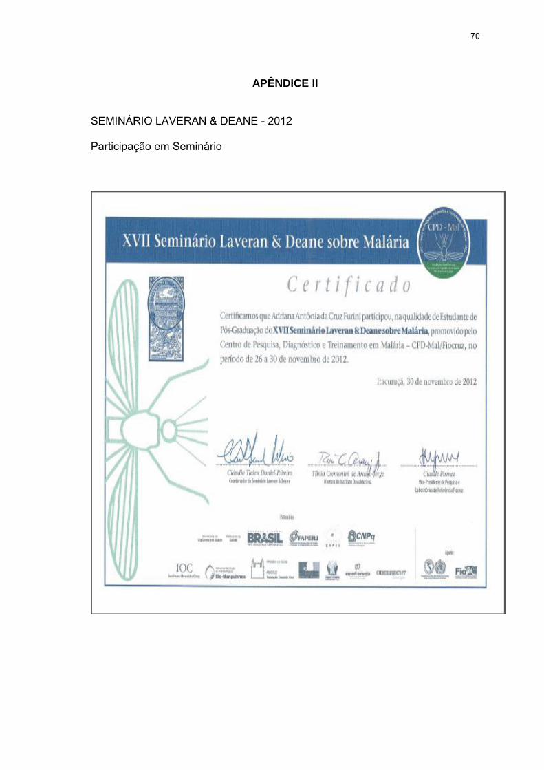
70
APÊNDICE II SEMINÁRIO LAVERAN & DEANE - 2012
Participação em Seminário

71
APÊNDICE III Curso: Citometria de Fluxo - 2012 CCAB: Centro de Conhecimento Avançado Becton Dickinson

72
APÊNDICE IV
Congresso Brasileiro de Medicina Tropical (2013)
Participação em Congresso

73
APÊNDICE V
Congresso Brasileiro de Medicina Tropical (2013)
Resumo em Anais de Congresso
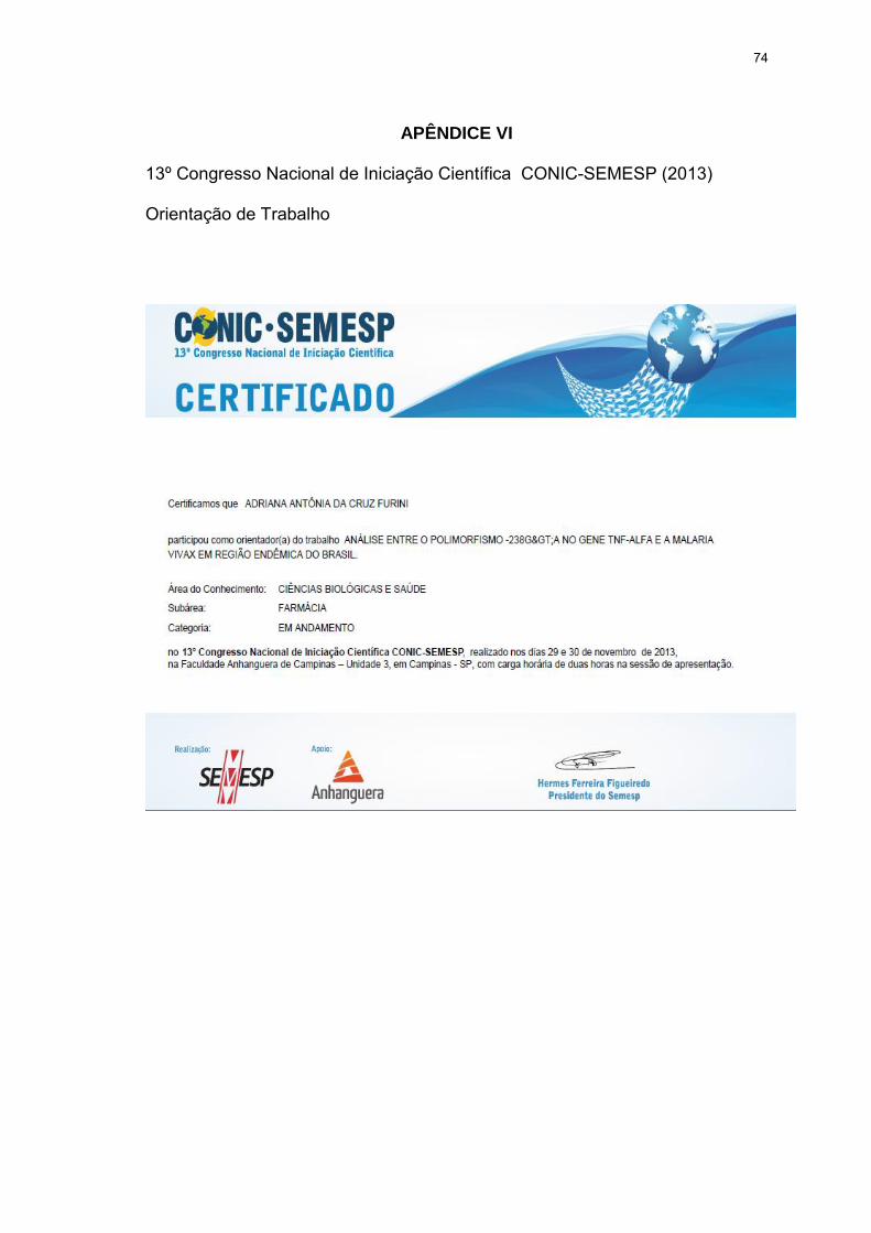
74
APÊNDICE VI 13º Congresso Nacional de Iniciação Científica CONIC-SEMESP (2013) Orientação de Trabalho

75
APÊNDICE VII
63 rd Annual Meeting – The American Society of Tropical Medicine and Hygiene
(2014)
Resumo em Anais de Congresso

76
APÊNDICE VIII
XXXIX Congress of the Brazilian Society of Immunology ( 2014) Participação em Congresso

77
APÊNDICE IX
XXXIX Congress of the Brazilian Society of Immunology
Apresentação de trabalho: pôster

78
APÊNDICE X Med Trop 2015 – 51º Congresso da Sociedade Brasileira de Medicina Tropical Apresentação de trabalho: pôster

79
APÊNDICE XI
Programa de Pós Graduação em Genética – UNESP/IBILCE Discussão de Seminários: Genética de Parasitos

80
APÊNDICE XII
FIOCRUZ – RIO DE JANEIRO Treinamento em Citometria de Fluxo

81
APÊNDICE XIII
XIV Reunião Nacional de Pesquisa em Malária (2015) Participação em Congresso
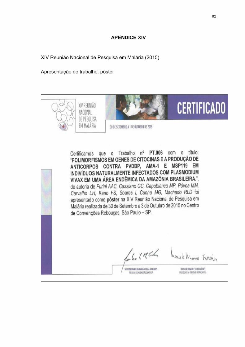
82
APÊNDICE XIV
XIV Reunião Nacional de Pesquisa em Malária (2015)
Apresentação de trabalho: pôster

83
APÊNDICE XV
Artigos publicados no Doutorado
1.Furini AAC, Lima TAM, Rodrigues LV, Fachina F, Galão EG, Santin MS,
Rossit ARB, Machado RLD. Prevalence of intestinal parasitosis in a population
of children of a daycare in Brazil. Parasitaria 2015; 21 (1):1-5.
2. Furini AAC, Guimaraes PM, Atique Netto HA, Castro KF, Silva BTOS, Atique,
TSC, Chiaparini J, Ramos MPSCM, Martins EA, Nardo CDD. Análise
epidemiológica, identificação e perfil de susceptibilidade a antimicrobianos
isolados de cães com infecção do trato urinário. Acta Veterinaria Brasilica
(UFERSA) 2014;7:288-93.
3.Furini AAC, Prates DC, Pezzini AP, Rabecini Junior LC, Regino BB, Schiesari
AJ, Machado RLD. Epidemiologia da coinfecção por HIV/HCV em um hospital
escola de Catanduva, São Paulo - Brasil. Revista do Instituto Adolfo Lutz 2014;
73: 106-12.
4.Furini AAC, Guimarães PM, Silva FSE, Lopes MASM, Castro KF, Atique,
TSC, Atique Netto H. Estudo da variação neutropênica pelo sulfato de
vincristina em cães com tvt tratados no Hospital Veterinário do Noroeste
paulista. Arquivos de Ciências Veterinárias e Zoologia da UNIPAR 2014; 17: 5-
9.
5.Furini AAC, Lima TAM, Faitarone NC, Verona JP, Silva LAM, Santos SS, Reis
AG, Guimarães PM, Atique TSC . Atenção Farmacêutica nas Interações
84
Medicamentosas e Indicadores de Prescrição em uma Unidade Básica de
Saúde. Arquivos de Ciências da Saúde 2014; 21: 99-06.
6.Furini AAC, Lima TAM, Rocha WM, Teixeira BCA, Martins AA, Feltrin J,
Seara MO, Gonçalves RR, Atique TSC. Acompanhamento farmacoterapêutico
em paciente idoso: relato de interações medicamentosas. Rev Eletr Pesq
UNIRP – Universitas 2004; 4:110-121.
7.Furini AAC, Pedro HSP, Schiesari AJ, Rodrigues JF, Faitarone NC, Borges,
MSB, Machado RLD, Rossit ARB. Tuberculose e micobacterioses no portador
do HIV: uma interação singular. Rev Eletr Pesq UNIRP – Universitas 2013;3: 1-
7.
8.Furini AAC, Rodrigues JF, Faitarone NC, Schiesari AJ, Barboza D, Santos
JRA, Schindler HS, Machado RLD. Terapia antirretroviral em crianças
soropositivas para o HIV em um hospital escola do Noroeste paulista:
correlação com dados imunológicos e infecções oportunistas. Arquivos de
Ciências da Saúde 2013; 20:43-8.
9.Furini AAC, Pedro HSP, Rodrigues JF, Montenego LML, Machado RLD,
Franco C, Schindler HS, Baptista IMFD, Rossit ARB. Detection of
Mycobacterium tuberculosis complex by nested polymerase chain reaction in
pulmonary and extrapulmonary specimens. Jornal Brasileiro de Pneumologia
2013; 39:711-18.
10.Furini AAC, Morelli Junior J, Atique TSC, Atique Netto H, Souza DB.
Isolamento de agentes da mastite bovina, perfil de sensibilidade e resíduos de
antimicrobianos em São José do Rio Preto/São paulo: mastite bovina,

85
sensibilidade e resíduos de antimicrobianos. Revistas Eletrônicas de Pesquisa
da UNIRP – Universitas 2013; 2:15-9.
11. Furini AAC, Atique TSC, Lima TAM. Sensibilidade à meticilina/oxacilina de
Staphylococcus aureus isolados da mucosa nasal de alunos do Centro
Universitário de Rio Preto. Revista Brasileira de Farmacia 2012; 93: 347-52.
Cassiano et al. Malar J (2016) 15:306 DOI 10.1186/s12936-016-1350-2
RESEARCH
Immunogenetic markers associated with a naturally acquired humoral immune response against an N-terminal antigen of Plasmodium vivax merozoite surface protein 1 (PvMSP-1)Gustavo Capatti Cassiano1,2*, Adriana A. C. Furini2, Marcela P. Capobianco1,2, Luciane M. Storti‑Melo3, Maria E. Almeida4, Danielle R. L. Barbosa5, Marinete M. Póvoa5, Paulo A. Nogueira4 and Ricardo L. D. Machado1,2,5
Abstract
Background: Humoral immune responses against proteins of asexual blood‑stage malaria parasites have been asso‑ciated with clinical immunity. However, variations in the antibody‑driven responses may be associated with a genetic component of the human host. The objective of the present study was to evaluate the influence of co‑stimulatory molecule gene polymorphisms of the immune system on the magnitude of the humoral immune response against a Plasmodium vivax vaccine candidate antigen.
Methods: Polymorphisms in the CD28, CTLA4, ICOS, CD40, CD86 and BLYS genes of 178 subjects infected with P. vivax in an endemic area of the Brazilian Amazon were genotyped by polymerase chain reaction‑restriction fragment length polymorphism (PCR‑RFLP). The levels of IgM, total IgG and IgG subclasses specific for ICB2‑5, i.e., the N‑terminal portion of P. vivax merozoite surface protein 1 (PvMSP‑1), were determined by enzyme‑linked immuno assay. The associations between the polymorphisms and the antibody response were assessed by means of logistic regression models.
Results: After correcting for multiple testing, the IgG1 levels were significantly higher in individuals recessive for the single nucleotide polymorphism rs3116496 in CD28 (p = 0.00004). Furthermore, the interaction between CD28 rs35593994 and BLYS rs9514828 had an influence on the IgM levels (p = 0.0009).
Conclusions: The results of the present study support the hypothesis that polymorphisms in the genes of co‑stimulatory components of the immune system can contribute to a natural antibody‑driven response against P. vivax antigens.
Keywords: Plasmodium vivax, MSP‑1, ICB2‑5, Immunogenetics, Antibodies
© 2016 The Author(s). This article is distributed under the terms of the Creative Commons Attribution 4.0 International License (http://creativecommons.org/licenses/by/4.0/), which permits unrestricted use, distribution, and reproduction in any medium, provided you give appropriate credit to the original author(s) and the source, provide a link to the Creative Commons license, and indicate if changes were made. The Creative Commons Public Domain Dedication waiver (http://creativecommons.org/publicdomain/zero/1.0/) applies to the data made available in this article, unless otherwise stated.
BackgroundAccording to the World Health Organization, there were an estimated 200 million cases of malaria in 2014 and an
estimated 584,000 resulting deaths worldwide [1]. Among the five species that can cause malaria in humans, Plas-modium vivax is the most widely distributed, accounting for most of the cases of malaria in South and Southeast Asia, Latin America and Oceania; there are an estimated 2.5 billion people living in areas at risk of transmission of the disease [1, 2]. Furthermore, although vivax malaria
Open Access
Malaria Journal
*Correspondence: [email protected] 1 Department of Biology, São Paulo State University, São José Do Rio Preto, São Paulo, BrazilFull list of author information is available at the end of the article
Page 2 of 11Cassiano et al. Malar J (2016) 15:306
has not been considered life-threatening for a long time, reports of severe cases associated with P. vivax have been increasingly numerous [3].
The blood stage of the Plasmodium lifecycle is respon-sible for the pathology associated with malaria. In this stage, merozoites released from schizont-infected eryth-rocytes invade non-infected erythrocytes, resulting in their destruction and the release of more merozoites into the bloodstream. During this brief extracellular period, these free merozoites are exposed to the host immune system, and proteins that are critical for parasite invasion are, therefore, important targets for the development of malaria vaccines. Merozoite surface proteins (MSPs) are among the most studied, especially MSP-1, which is nec-essary for merozoite attachment to erythrocytes [4] and normal parasite development [5].
The most widely accepted structure of the P. vivax mer-ozoite surface protein 1 (PvMSP-1) gene indicates that it consists of six polymorphic blocks (blocks 2, 4, 6, 8, 10 and 12) flanked by seven conserved blocks (blocks 1, 3, 5, 7, 9, 11 and 13) [6]. By analysing the primary structure of PvMSP-1, several seroepidemiological studies have been performed to evaluate its immunogenic potential [7–11]. Although the C-terminal portion of the protein (PvMSP-119) is the most immunogenic [7, 9, 12], a number of studies showed high prevalence of IgG against N-termi-nal PvMSP-1 in individuals exposed to P. vivax [9, 10, 13, 14]. Furthermore, specific IgG3 antibodies against the N-terminal portion of PvMSP-1 have been associ-ated with clinical protection in two riverine communities of the Brazilian Amazon [9, 11], similar to that observed in P. falciparum infection, where persistence of antibod-ies IgG3 against N-terminal of MSP-1 was related with prolongation time without malaria [15]. In fact, antibod-ies specific for the asexual blood stage are thought to be involved in clinical protection against malaria vivax. Lon-gitudinal cohort studies have shown correlations between magnitude of antibody responses to P. vivax merozoite antigens and protection from malaria [11, 16–18]. Due to the inability in maintaining P. vivax in continuous in vitro culture, it is difficult to define the role of antibodies to this species, but few evidences support that it may be related to inhibition of merozoite invasion [19, 20]. Fur-thermore, complement and FcR mediated mechanisms seem to be important in antibody-mediated protection [21].
The development of an adequate immune response depends on the fine regulation of lymphocyte activation. For this, in general, lymphocytes require two activation signals. The first signal is antigen-specific, whereas the second signal, called co-stimulation, is generated by the interaction between the surface molecules of T cells and
those of antigen-presenting cells, including B cells. The interaction between CD28 and its ligands, CD80 and CD86, provides the strongest costimulatory signal for T-cell proliferation, whereas CTLA-4 is a negative regula-tor that plays a key role in T cell homeostasis and in cen-tral tolerance [22]. Another member of the CD28 family, Inducible co-stimulator (ICOS), is an important immune regulatory molecule that participates in T-cell activation and T-cell dependent B-cell responses [23, 24]. CD40 is presented on the surface of B-cells and the CD40-CD40L interaction is the major costimulatory signal for B cells to mount a humoral response [25]. B lymphocyte stimulator (BLYS) is produced mainly by innate immune cells and is needed to provide signals for B cell survival and prolifera-tion [26]. Considering the importance of these molecules in development of immune response and because there are currently multiple lines of evidence showing that the genes involved in the immune response can influence antibody production during malaria infection [27–34], the authors hypothesised that polymorphisms in the genes of the co-stimulatory molecules CD28, CTLA-4, ICOS, CD86, CD40 and BLYS are involved in the magni-tude of the naturally acquired antibody-driven response against N-terminal PvMSP-1 in individuals infected with P. vivax in the Brazilian Amazon.
MethodsStudy area and subjectsThe present study was conducted in the municipality of Goianésia do Pará, Pará state, Brazil, which is a constitu-ent of the Brazilian Amazon region, where P. vivax is responsible for more than 80 % of all malaria cases [35]. Details of the study area have been described elsewhere [36]. Cross-sectional surveys conducted from February 2011 to August 2012 were used to recruit 178 subjects (125 men and 53 women) presenting with classic symp-toms of malaria, who sought treatment at the medical service of the municipality; the subjects had an average age of 29.8 years (varying from 14 to 68 years). Exclusion criteria included children under 10 years old, pregnancy, related individuals and anti-malarial treatment within the previous seven days. Diagnosis was performed by microscopy (thick film), and infection with P. vivax was subsequently confirmed by nested polymerase chain reaction (PCR) [37]. The geometric mean of parasiatemia was 718.4 parasites/mm3 (95 % CI 487.2–1059.2). All patients with microscopically confirmed infections were given standard treatment of 25 mg/kg of chloroquine in 3 days plus 0.5 mg/kg of primaquine during seven days. All participants were submitted to a questionnaire to obtain epidemiological information. The length of resi-dence in the municipality varied between one month and

Page 3 of 11Cassiano et al. Malar J (2016) 15:306
37 years (median of 5 years), and 91.9 % of the individu-als had previously contracted malaria. Samples from 40 malaria-naive individuals living in a non-endemic area (São José do Rio Preto, Brazil), who had never been to areas of malaria transmission, were used as controls.
Peripheral venous blood (~10 mL) was collected in EDTA tubes, and plasma samples were obtained by cen-trifugation and stored at −20 °C. The protocol of the present study was approved by the Research Ethics Com-mittee of the Medical School of São José do Rio Preto (CEP/FAMERP no. 4599/2011) and by the health author-ities of the municipality of Goianésia do Pará. All partici-pants or their legal guardians signed an informed consent form.
GenotypingDNA was extracted from peripheral blood samples with an Easy-DNA™ extraction kit (Invitrogen, California, USA). All single nucleotide polymorphisms (SNPs) were genotyped by PCR-restriction fragment length poly-morphism (RFLP). The SNPs −372G > A and +17T > C in CD28 (rs35593994 and rs3116496, respectively), −318C > T and +49A > G in CTLA4 (rs5742909 and rs231775, respectively), +1564T > C in ICOS (rs4404254), +1057G > A in CD86 (rs1129055), −1C > T in CD40 (rs1883832), and −871C > T in BLYS (rs9514828) were genotyped according to previously described protocols [36, 38, 39]. Primer sequences, restriction enzymes, and the restriction fragments obtained of each SNP are pre-sented in Additional file 1. PCR for the identification of −1722T > C and −1577G > A in CTLA4 (rs733618 and rs11571316, respectively) was performed with a final sample volume of 25 µL containing 1X Buffer (20 mM Tris-HCl pH 8.4, 50 mM KCl), 1.5 mM MgCl2, 0.2 mM of each dNTP, 0.6 pmol of each primer, and 0.5 U Plati-num Taq DNA Polymerase (Invitrogen, São Paulo, Bra-zil). The primers 5′ CTTCATGCCGTTTCCAACTT 3′ and 5′ CCTTTTCTGACCTGCCTGTT 3′ were used for the −1722T > C genotyping, whereas 5′ CTTCAT GCCGTTTCCAACTT 3′ and 5′ ATCTCCTCCAGGAA GCCTCTT 3′ were used to identify −1577G > A. Ampli-fications were performed under the following conditions: first step of 5 min at 94 °C, 35 cycles for 30 s at 94 °C, 30 s at 52 °C and 1 min at 72 °C, and a final step for 10 min at 72 °C. The PCR products of −1722T > C and −1577G > A were digested with the enzymes BbvI and MboII (Fer-mentas, Vilnius, Lithuania), respectively. Electrophoresis was performed in 2.5 % agarose and stained with Gel-Red™ (Biotium, Hayward, USA) with the exception of the rs5742909 and rs1883832 SNPs, which were performed in 12.5 % polyacrylamide gel after staining with ethidium bromide, and visualized in a UV transilluminator.
Antigen and antibody determinationThe glutathione-S-transferase (GST)-tagged recombinant ICB2-5 protein corresponds to the amino acids 170-675 of the N-terminal portion of MSP-1 from the Belém strain of P. vivax. The ICB2-5-GST fusion protein was puri-fied on a glutathione-Sepharose 4B column (Amersham Pharmacia), and the protein content was assessed with the Bio-Rad Protein Assay Kit I (Bio-Rad Laboratories, Inc.) [7]. The levels of IgM and IgG subclasses specific for ICB2-5 were measured as described previously with some modifications [7]. The plates were coated with GST-tagged ICB2-5 and GST alone, and all tests were done in duplicate. Briefly, 50 µL of ICB2-5-GST or GST alone (as a control) was coated on ELISA plates at 4 µg/mL (Costar, Corning Inc., New York, USA) and incubated overnight at 4 °C. After washing and blocking the plates with 0.05 % bovine serum albumin-PBS, 50 µL of plasma diluted 1/100 was added to the wells in duplicate and incubated for 1 h at 37 °C. Then, 50 µL of peroxidase-conjugated anti-human IgG or IgM antibodies (KPL, Maryland, USA) diluted 1/1000 were added to each well for the detection of total IgG or IgM, respectively. IgG subclasses were detected using mouse monoclonal antibodies specific for each isotype (Sigma, Missouri, USA), diluted accord-ing to the tested subclass (1/3000 for IgG1 clone HP6001; and IgG3 clone HP6050, 1/2500 for IgG2 clone HP6014, and 1/5000 for IgG4 clone HP6025). The immobilized monoclonal antibodies were detected with peroxidase-conjugated anti-mouse antibodies for 1 h at 37 °C (Sigma, Missouri, USA). Subsequently, the plates were washed and developed in the dark with 50 µL TMB substrate diluted 1/50 with 0.1 M phosphate-citrate buffer, pH 5.0, containing 0.03 % H2O2. The reaction was interrupted after 10 min by adding 50 µL of 2 N H2SO4 to each well and read at 450 nm. The determination of positivity for anti ICB2-5 was calculated as described previously, with some modifications [9]. Brief, firstly the average OD was calculated for each individual, and serum was considered positive if GST-tagged ICB2-5 OD was equal to or greater than cut off for this same protein, measured with sera of individuals who never had a past history of malaria. To confirm the positivity of a serum for anti ICB2-5, the aver-age of GST-tagged ICB2-5 OD was calculated to exclude reactivity against GST. For each serum, the GST-cut off was calculated as average of GST OD plus twice the standard deviations (SD). The positivity for anti ICB2-5 was determined when average of GST-tagged ICB2-5 OD was equal to or greater than its GST-cut off. The results are expressed as the reactivity index (RI), which was cal-culated by dividing the OD of the sample by the cut-off value. Only the samples that were positive for total IgG (RI > 1) were tested for IgG subclasses.

Page 4 of 11Cassiano et al. Malar J (2016) 15:306
Statistical analysisThe associations between the SNP genotypes and the antibody response were analysed by means of a logistic regression model, controlling for potential confound-ers, such as age and gender. Different genetic models (codominant, dominant, recessive and log-additive) were tested with the ‘SNPSassoc’ package [40] implemented for the R statistical software (version 3.1.1). A Bonfer-roni correction was used to adjust for multiple testing, and the significance was set at p < 0.005 (0.05/10). The sample size was calculated using GWAPower software [41]. The current sample size (n = 178) had 80 % power to detect a variant with about 6 % heritability. Since only 90 samples were assessed for IgG subclasses, the power to detect the same heritability was about 42 %. Geno-typic deviations from the Hardy–Weinberg equilibrium (HWE) were evaluated by using the exact test described by Wigginton et al. [42]. Statistically significant differ-ences between the means of continuous variables were assessed with one-way analysis of variance (ANOVA). Correlation coefficients of the antibody levels were esti-mated with the Pearson correlation. Interactions between SNP pairs were also assessed with the ‘SNPassoc’ pack-age, which determines the effects of interaction by means of log-likelihood ratio tests (LRTs).
Haplotype blocks were determined with Haploview 4.2 using the solid spine of linkage disequilibrium (LD) method [43], and the degree of LD between SNPs was estimated with the D’ parameter. The R package ‘haplo.stats’ [44] was used for association tests between hap-lotypes and antibody levels by means of the ‘haplo.glm’ function, which performs a regression that estimates the effect of each haplotype compared to a reference haplo-type by means of a general linear model.
ResultsSerological data and single‑marker associationsOf the 178 subjects analysed, all infected with P. vivax, 90 (50.6 %) exhibited IgG (RI > 1), whereas 53 (29.8 %) exhibited IgM specific for ICB2-5. The antibody levels were higher for IgG1 (mean = 1.08) compared to those of the other subclasses (means of 0.91, 0.79 and 0.79 for IgG2, IgG3 and IgG4, respectively) (p < 0.001, ANOVA). The highest correlations between levels of IgG subclasses were found for IgG1 and IgG2 (r = 0.48) and IgG1 and IgG3 (r = 0.43) (Additional file 2). The levels of ICB2-5-specific antibodies were not influenced by previous exposure to malaria (age, length of residence in the stud-ied area, and previous episodes of malaria) (Additional file 3), except for IgG2, which exhibited higher levels in individuals who reported to have had less than five previ-ous episodes of malaria, compared to those who reported to have had more than five previous episodes [mean
(confidence interval): 0.97 (0.84–1.10) vs. 0.88 (0.76–0.99), p = 0.01, ANOVA].
The allelic and genotype frequencies of all the analysed SNPs are listed in Table 1 and Additional file 4. The allele frequencies of these SNPs were previously presented in a larger set of samples (with exception of SNPs rs733618 and rs11571316) [45]. The success rate of SNP genotyp-ing was 100 % for eight of the studied SNPs, whereas rs733618 and rs11571316 were successfully genotyped in 88.2 and 82 % of the samples, respectively, and no devia-tion from HWE was observed (all p value >0.1). A sum-mary of the statistics for all the evaluated SNPs is listed in Table 1 and Additional file 4. There is not significant association between the polymorphisms and parasi-taemia (all p value >0.07, Kruskal–Wallis test, data not show). Three SNPs were significantly associated with the humoral immune response against P. vivax ICB2-5. IgM levels were associated with rs35593994 in CD28; individuals with the GG genotype had lower antibody levels (mean 0.67 vs. 0.88, p = 0.03). Based on a reces-sive model, individuals with a CC genotype for rs3116496 in CD28 exhibited higher IgG1 levels with respect to the other genotypes (mean 3.13 vs. 1.04, p = 0.00004). Individuals with a TC genotype for rs733618 in CTLA4 exhibited higher IgG2 levels with respect to homozy-gous individuals (mean 1.18 vs. 0.87, p = 0.04). However, after correction for multiple testing, only association that remained significant was the one with CD28 (re3116496).
Linkage disequilibrium analysis and haplotype associationLinkage disequilibrium analysis was performed for all possible pairwise combinations of seven SNPs that were analysed across 255 kb in the 2q33 chromosomal region, which encompasses CD28, CTLA4 and ICOS. Two hap-lotype blocks were defined using the criterion described by Barret et al. [43] (Fig. 1). Three haplotypes of block 1, which contains rs35593994 and rs3116496 in CD28, were built, with frequencies varying between 18.5 and 52.2 %, and for block 2, which contains rs733618, rs11571316 and rs5742909 in the promoter region of CTLA4, four haplotypes were built, with frequencies varying between 6.3 and 51.8 %. The effects of each haplotype on the anti-body levels were estimated and are shown in Table 2. The AT haplotype in block 1 was significantly associated with a 20 % increase in IgM levels (p = 0.027) with respect to the reference haplotype (specifically, the most frequent haplotype, i.e., CT). However, no association remained significant after correction for multiple testing.
Interaction analysisUsing the R project package ‘SNPassoc’ [40], the study explored all pairwise SNP−SNP interactions under the codominant model. These interactions are represented

Page 5 of 11Cassiano et al. Malar J (2016) 15:306
graphically in Fig. 2, in which the upper part contains the p values for the interaction LRT. Although several significant associations involving rs3116496 in CD28 and other SNPs (rs1129055, rs5742909 and rs231775) with IgG1 levels were observed, the level of significance was below that found for rs3116496 individually (all p values >0.0001). However, the interaction involving rs35593994 in CD28 with rs9514828 in BLYS was associ-ated with the IgM levels (p = 0.0009). A regression anal-ysis was performed to confirm this interaction, and the results of significant associations involving the above two SNPs are listed in Table 3.
DiscussionThe main strategies used for malaria control are based on prompt diagnosis and treatment and on vector con-trol. However, new resistant parasite strains arise as new drugs are applied, and vector control is also encountering great challenges due to the growing resistance to insec-ticides, thus justifying research on the development of a vaccine that is effective against malaria. Characterisation of the naturally acquired immune response in different populations is a useful tool for the identification of mol-ecules that can be targeted by anti-malarial vaccines.
The present study was to evaluate the naturally acquired immune response against the N-terminal por-tion of PvMSP-1. Although the C-terminal portion (MSP-119) is considered to be the most immunogenic region of the protein [7, 9, 12], there is evidence sug-gesting that antibodies targeting the N-terminal portion
Table 1 Levels of Plasmodium vivax ICB2-5-specific IgM, IgG and IgG subclasses with respect to the analysed genotypes from individuals infected with P. vivax
a Number of individuals evaluated for ICB2-5-specific IgM and total IgG (n = 178)b Number of individuals evaluated for ICB2-5-specific IgG subclasses (n = 90)c Significant according to the dominant model (p = 0.033)d Significant according to the recessive model (p = 0.00004)e Significant according to the overdominant model (p = 0.042)
SNP Gene Genotypes Genotypes (na)
Genotypes (nb)
IgG (RI) IgG1 (RI) IgG2 (RI) IgG3 (RI) IgG4 (RI) IgM (RI)
rs35593994 CD28 GG/GA/AA 86/80/12 42/39/8 1.02/1.03/1.11 1.11/1.10/0.89 0.92/0.93/0.80 0.76/0.86/0.64 0.77/0.83/0.74 0.66/0.86/1.13c
rs3116496 CD28 TT/TC/CC 118/54/6 60/27/2 1.04/1.02/0.91 1.06/0.98/3.13d 0.91/0.92/0.95 0.82/0.72/0.75 0.82/0.75/0.67 0.83/0.70/0.65
rs733618 CTLA4 TT/TC/CC 135/20/2 72/7/1 1.06/0.89/1.05 1.11/0.97/0.81 0.87/1.18/0.64e 0.80/0.66/0.57 0.80/0.68/0.62 0.82/0.72/1.25
rs11571316 CTLA4 GG/GA/AA 59/75/12 29/39/4 1.04/1.03/0.97 1.17/1.10/0.84 0.96/0.95/0.68 0.74/0.86/0.56 0.73/0.82/0.66 0.81/0.75/0.79
rs5742909 CTLA4 CC/CT/TT 151/27/0 75/14/0 1.15/1.01/– 1.04/1.29/– 0.91/0.93/– 0.80/0.76/– 0.80/0.78/– 0.79/0.78/–
rs231775 CTLA4 AA/AG/GG 75/84/19 40/41/8 1.07/1.00/1.01 0.96/1.22/0.98 0.87/0.98/0.82 0.78/0.82/0.70 0.79/0.79/0.81 0.79/0.75/0.89
rs4675378 ICOS TT/TC/CC 77/73/27 35/38/16 1.00/1.05/1.09 1.08/1.12/0.99 0.86/0.94/0.96 0.73/0.90/0.67 0.80/0.78/0.80 0.80/0.73/0.85
rs1129055 CD86 GG/GA/AA 110/60/8 53/31/5 1.00/1.08/1.15 1.08/1.09/1.06 0.93/0.90/0.93 0.78/0.82/0.70 0.82/0.76/0.70 0.81/0.73/0.90
rs1883832 CD40 CC/CT/TT 135/37/6 65/20/4 1.01/1.06/1.25 1.11/1.03/0.86 0.92/0.95/0.68 0.81/0.77/0.65 0.81/0.75/0.79 0.80/0.69/1.13
rs9514828 BLYS CC/CT/TT 103/62/13 48/35/6 0.99/1.07/1.15 0.99/1.23/0.98 0.91/0.94/0.83 0.82/0.69/1.11 0.76/0.81/0.92 0.73/0.83/0.95
Fig. 1 Linkage disequilibrium of investigated polymorphisms in region 2q33, which comprises genes CD28, CTLA4 and ICOS. The num-bers within the squares represent the D’ value, expressed as per cent. Empty squares represent a D’ value of 1, which indicates complete linkage disequilibrium. Darker-shaded squares represent pairs with an LOD score = 2, light grey squares represent D’ = 1 and LOD < 2, and white squares represent LOD < 2 and D’ < 1. Two blocks were identified by means of the solid spine of linkage disequilibrium method [37]. Linkage Disequilibrium map was generated by Haploview 4.2

Page 6 of 11Cassiano et al. Malar J (2016) 15:306
Tabl
e 2
Hap
loty
pe fr
eque
ncie
s an
d th
eir a
ssoc
iati
on w
ith
leve
ls o
f ICB
2-5-
spec
ific
anti
bodi
es
The
effec
ts o
f eac
h ha
plot
ype
are
rela
tive
to th
e m
ost f
requ
ent h
aplo
type
, whi
ch w
as u
sed
as a
refe
renc
e. ∆
% in
dica
tes
chan
ges
rela
tive
to th
e an
tibod
y le
vels
com
pare
d to
refe
renc
e ha
plot
ypes
, with
95
% c
onfid
ence
in
terv
als.
Bloc
k 1
refe
rs to
the
SNPs
rs35
5939
94 a
nd rs
3116
496,
and
blo
ck 2
refe
rs to
rs73
3618
, rs1
1571
316
and
rs57
4290
9
Bloc
kH
aplo
type
Freq
uenc
yIg
GIg
G1
IgG
2Ig
G3
IgG
4Ig
M
∆%p
∆%p
∆%p
∆%p
∆%p
∆%p
Bloc
k 1
G‑T
0.52
2Re
fere
nce
A‑T
0.29
22
(−9;
13)
0.72
−3
(−27
; 21)
0.80
−3
(−16
; 9)
0.61
0 (−
15; 1
4)0.
950
(−10
; 10)
0.99
20 (2
; 38)
0.02
7
G‑C
0.18
5−
3 (−
16; 9
)0.
5921
(−9;
51)
0.17
0 (−
15; 1
6)0.
96−
8 (−
27; 1
0)0.
38−
7 (−
19; 5
)0.
26−
6 (−
27; 1
5)0.
58
Bloc
k 2
T‑G
‑C0.
518
Refe
renc
e
T‑A
‑C0.
330
−2
(−14
; 10)
0.72
−4
(−32
; 25)
0.81
−9
(−23
; 5)
0.21
2 (−
16; 1
9)0.
853
(−9;
16)
0.59
−7
(−28
; 13)
0.48
T‑G
‑T0.
076
12 (−
7; 3
1)0.
2249
(−23
; 121
)0.
183
(−21
; 27)
0.83
−4
(−34
; 25)
0.77
−3
(−23
; 17)
0.77
−3
(−34
; 27)
0.84
C‑G
‑C0.
063
−17
(−38
; 11)
0.27
−10
(−70
; 51)
0.75
−14
(−44
; 17)
0.38
−14
(−52
; 23)
0.45
−11
(−36
; 14)
0.38
−23
(−54
; 9)
0.16
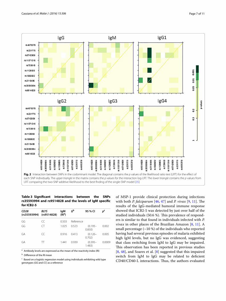
Page 7 of 11Cassiano et al. Malar J (2016) 15:306
of MSP-1 provide clinical protection during infections with both P. falciparum [46, 47] and P. vivax [9, 11]. The results of the IgG-mediated humoral immune response showed that ICB2-5 was detected by just over half of the studied individuals (50.6 %). This prevalence of respond-ers is similar to that found in individuals infected with P. vivax in other places of the Brazilian Amazon [8, 11]. A small percentage (~10 %) of the individuals who reported having had several previous episodes of malaria exhibited high IgM levels, but no IgG was evidenced, suggesting that class switching from IgM to IgG may be impaired. This observation has been reported in previous studies [8, 48], and Soares et al. [8] suggested that this impaired switch from IgM to IgG may be related to deficient CD40/CD40-L interactions. Thus, the authors evaluated
Fig. 2 Interaction between SNPs in the codominant model. The diagonal contains the p values of the likelihood ratio test (LRT) for the effect of each SNP individually. The upper triangle in the matrix contains the p values for the interaction log‑LRT. The lower triangle contains the p values from LRT comparing the two‑SNP additive likelihood to the best finding of the single‑SNP model [35]
Table 3 Significant interactions between the SNPs rs35593994 and rs9514828 and the levels of IgM specific for ICB2-5
a Antibody levels are expressed as the mean of the reactivity index (RI)b Difference of the RI meanc Based on a logistic regression model using individuals exhibiting wild type genotypes (GG and CC) as a reference
CD28 (rs35593994)
BLYS (rs9514828)
IgM (RIa)
∆b 95 % CI pc
GG CC 0.503 Reference
GG CT 1.025 0.523 (0.195–0.850)
0.002
GA CC 0.916 0.413 (0.126–0.702)
0.005
GA TT 1.441 0.939 (0.395–1.483)
0.0009
Page 8 of 11Cassiano et al. Malar J (2016) 15:306
whether co-stimulatory molecule gene polymorphisms are involved in the delayed class switch from IgM to IgG. However, no association was observed.
Regarding IgG subclasses, higher levels of IgG1 were found compared to the other IgG subclasses, which is in contrast to previous studies showing a predominance of IgG3 specific for ICB2-5 [8, 9, 11]. Although there is still no consensus regarding the role of antibody sub-classes in clinical protection, it has been suggested that only the cytophilic subclasses, i.e., IgG1 and IgG3, are protective [49, 50]. Two longitudinal studies performed in the Brazilian Amazon, specifically, one in Portu-chuello, near Porto Velho, and the other in a commu-nity of Rio Pardo, Amazonas state, have observed that ICB2-5-specific antibodies were associated with clini-cal protection against malaria caused by P. vivax and that IgG3 was detected in all asymptomatic individuals, whereas most symptomatic patients exhibited no IgG3 [9, 11]. If the above results were to be extrapolated to Goianésia do Pará, the site of this study, the fact that a passive collection was performed on patients exhibit-ing symptoms could explain the low IgG3 levels found. Furthermore, some studies have also shown that IgG2 may be associated with protection. Deloron et al. [51] found an association between high IgG2 levels and low risk of acquiring an infection by P. falciparum. Although the design of the present study does not allow for the association of the prevalence and levels of clinically protective antibodies, higher IgG2 levels were observed in subjects who reported having had fewer cases of malaria.
The development of an immune response against Plas-modium species is a complex process, and one of the main issues is understanding why individuals differ in their immune responses against the parasite. Thus, the objective of the present study was to investigate the influ-ence of co-stimulatory molecule gene polymorphisms on the production of antibodies specific for an important P. vivax vaccine candidate antigen. The most important result was that CD28 rs3116496 was associated with lev-els of IgG1 specific for ICB2-5. In addition to the impor-tant role of CD28 in T cell activation, the binding of this receptor to its ligands CD80 and CD86 on the surface of B cells provides bidirectional signals that appear to be important for IgG production by B cells [52]. Thus, CD28 may be involved in the immune response against malaria. In fact, CD28 knockout mice infected with Plasmodium chabaudi were unable to resolve the infection, main-taining low levels of parasitaemia for weeks after infec-tion [53, 54]. Furthermore, treatment of wild type mice with monoclonal anti-CD86 antibodies abolished IL-4
production and was significantly associated with reduced levels of P. chabaudi-specific IgG1 [55].
In the present study, individuals exhibiting the T allele for rs3116496 in CD28 were found to be associated with reduced IgG1 levels. Although the biological functions of this SNP, which is located in the third intron of the gene, are still unknown, it is located near a splice site, at which point mutations can induce abnormal splicing, thus affecting protein expression [56]. The relationship between rs3116496 in CD28 and susceptibility to several diseases has already been evaluated, and significant asso-ciations have been found in type 1 diabetes [57], cervi-cal [58] and breast cancer [59], and rheumatoid arthritis [60]. However, the role of this polymorphism in malaria has not yet been assessed, and even if the presence of the T allele, which is associated with lower levels of ICB2-5-specific IgG1, were implicated in higher susceptibil-ity to vivax malaria, further elucidation would still be necessary.
Although it seems likely that immunity against malaria is affected by several genes, the influence of combined polymorphisms is rarely ever investigated. The present study performed analyses of interactions between the SNP pairs and found that CD28 rs35593994 and BLYS rs9514828 together were associated with levels of IgM specific for ICB2-5, especially in individuals with GA and TT genotypes, respectively. Specifically, they were associated with a ~180 % increase in IgM levels com-pared to that of wild type genotype individuals. Although the biological mechanisms underlying this interaction are still unknown, CD28 and BLyS may be involved in the production of memory B cells and antibody isotype class switching [61, 62]. Both polymorphisms, i.e., CD28 rs35593994 and BLYS rs9514828, are located in the 5′ regulatory region of the gene; thus, differential expres-sion of these genes may change the regulation of the B cells involved in the production of ICB2-5-specific anti-bodies. Liu et al. [63] have shown that despite the genera-tion of memory B cells as a response to vaccination with MSP-119, the function of these cells was nullified due to a lack of BLyS expression in dendritic cells from mice infected with P. yoelii.
Although it was observed the influence of these two SNPs in the antibody response to ICB2-5, most of the evaluated polymorphisms did not show any significant differences. In fact, variations in the immune response to malaria can be attributed to several factors, including the environment, previous exposure to malaria and immuno-genicity of antigen. Thus, as a complex trait, it is likely that many polymorphisms have small effect in malaria immune response, but large sample size is required to detect it.

Page 9 of 11Cassiano et al. Malar J (2016) 15:306
ConclusionsIn summary, several genes involved in the control of the immune response were investigated, entailing the production of antibodies against a P. vivax vaccine can-didate antigen. Despite the growing number of stud-ies evaluating human genetic factors that control the immune response to malaria [27–34, 64], it is likely that many genes responsible for the wide inter-individ-ual variation observed in the immune response against malaria remain unknown; studies similar to the present work may help identify subjects who are more prone to respond to a specific vaccine [29]. Although the statis-tical evidence supporting the described associations is limited by the relatively small sample size and, although it is impossible to tell whether the SNPs studied here are truly causal or are in LD with the causal variants, the results suggest that genetic variations in CD28 and interactions between polymorphisms in BLYS and CD28 may be involved in the control of the immune response against vivax malaria.
Authors’ contributionsGCC, RLDM and PAN contributed to the design and conception of the study and wrote the manuscript. AACF, MPC and GCC participated in the molecular analyses. PAN, MEA, DRLB and GCC performed the serum analyses. GCC performed the statistical analyses. LMSM and MMP contributed to data inter‑pretation and reviewed the manuscript. RLDM, GCC and MMP participated in field data collection. All authors read and approved of the final manuscript.
Author details1 Department of Biology, São Paulo State University, São José Do Rio Preto, São Paulo, Brazil. 2 Department of Skin, Infectious and Parasitic Diseases, São José do Rio Preto Medical School, São José Do Rio Preto, São Paulo, Brazil. 3 Depart‑ment of Biology, Federal University of Sergipe, São Cristóvão, Sergipe, Brazil. 4 Leônidas and Maria Deane Institute, Oswaldo Cruz Foundation, Manaus, Amazonas, Brazil. 5 Laboratory of Malaria Basic Research, Division of Parasitol‑ogy, Evandro Chagas Institute, Belém, Pará, Brazil.
AcknowledgementsWe thank the residents of the municipality of Goianésia do Pará, who made this study possible. We are also thankful to Darci Rodrigues, who helped with sample collection, and Valéria Fraga and Luciana Conceição for their help with laboratory analyses. This study was financed by the Brazilian National Council for Scientific and Technological Development (Conselho Nacional de Desen‑volvimento Científico e Tecnológico) and the Pará State Research Support Foundation (Fundação de Amparo à Pesquisa do Estado do Pará).
Additional files
Additional file 1. Reaction conditions for the amplification and enzyme digestion of polymorphisms in the studied polymorphisms.
Additional file 2. Correlation between ICB2‑5‑specific IgG, IgM and IgG subclasses in individuals infected with Plasmodium vivax living in an endemic area of the Brazilian Amazon.
Additional file 3. IgM and IgG subclasses levels (RI) for ICB2‑5 according to age, length of residence in the studied area and previous episode of malaria.
Additional file 4. Association tests between genetic polymorphisms and Plasmodium vivax ICB2‑5‑specific antibody levels.
Competing interestsThe authors declare that they have no competing interests.
Received: 16 February 2016 Accepted: 17 May 2016
References 1. WHO. World malaria report. Geneva: World Health Organization; 2013. 2. Gething PW, Elyazar IR, Moyes CL, Smith DL, Battle KE, Guerra CA, et al. A
long neglected world malaria map: Plasmodium vivax endemicity in 2010. PLoS Negl Trop Dis. 2012;6:e1814.
3. Lacerda MV, Mourão MP, Alexandre MA, Siqueira AM, Magalhães BM, Martinez‑Espinosa FE, et al. Understanding the clinical spectrum of complicated Plasmodium vivax malaria: a systematic review on the contri‑butions of the Brazilian literature. Malar J. 2012;11:12.
4. Goel VK, Li X, Chen H, Liu SC, Chishti AH, Oh SS. Band 3 is a host receptor binding merozoite surface protein 1 during the Plasmodium falciparum invasion of erythrocytes. Proc Natl Acad Sci U S A. 2003;100:5164–9.
5. Child MA, Epp C, Bujard H, Blackman MJ. Regulated maturation of malaria merozoite surface protein‑1 is essential for parasite growth. Mol Micro‑biol. 2010;78:187–202.
6. Putaporntip C, Jongwutiwes S, Sakihama N, Ferreira MU, Kho WG, Kaneko A, et al. Mosaic organization and heterogeneity in frequency of allelic recombination of the Plasmodium vivax merozoite surface protein‑1 locus. Proc Natl Acad Sci U S A. 2002;10:16348–53.
7. Soares IS, Levitus G, Souza JM, Del Portillo HA, Rodrigues MM. Acquired immune responses to the N‑ and C‑terminal regions of Plasmodium vivax merozoite surface protein 1 in individuals exposed to malaria. Infect Immun. 1997;65:1606–14.
8. Soares IS, da Cunha MG, Silva MN, Souza JM, Del Portillo HA, Rodrigues MM. Longevity of naturally acquired antibody responses to the N‑ and C‑terminal regions of Plasmodium vivax merozoite surface protein 1. Am J Trop Med Hyg. 1999;60:357–63.
9. Nogueira PA, Alves FP, Fernandez‑Becerra C, Pein O, Santos NR, Pereira da Silva LH, et al. A reduced risk of infection with Plasmodium vivax and clini‑cal protection against malaria are associated with antibodies against the N terminus but not the C terminus of merozoite surface protein 1. Infect Immun. 2006;74:2726–33.
10. Storti‑Melo LM, Souza‑Neiras WC, Cassiano GC, Taveira LC, Cordeiro AJ, Couto VS, et al. Evaluation of the naturally acquired antibody immune response to the Pv200 L N‑terminal fragment of Plasmodium vivax mero‑zoite surface protein‑1 in four areas of the Amazon Region of Brazil. Am J Trop Med Hyg. 2011;84(2 Suppl):58–63.
11. Versiani FG, Almeida ME, Melo GC, Versiani FO, Orlandi PP, Mariúba LA, et al. High levels of IgG3 anti ICB2‑5 in Plasmodium vivax‑infected indi‑viduals who did not develop symptoms. Malar J. 2013;12:294.
12. Riccio EK, Totino PR, Pratt‑Riccio LR, Ennes‑Vidal V, Soares IS, Rodri‑gues MM, et al. Cellular and humoral immune responses against the Plasmodium vivax MSP‑119 malaria vaccine candidate in individuals living in an endemic area in north‑eastern Amazon region of Brazil. Malar J. 2013;12:326.
13. Levitus G, Mertens F, Speranca MA, Camargo LM, Ferreira MU, del Portillo HA. Characterization of naturally acquired human IgG responses against the N‑terminal region of the merozoite surface protein 1 of Plasmodium vivax. Am J Trop Med Hyg. 1994;51:68–76.
14. Fernandez‑Becerra C, Sanz S, Brucet M, Stanisic DI, Alves FP, Camargo EP, et al. Naturally‑acquired humoral immune responses against the N‑ and C‑termini of the Plasmodium vivax MSP1 protein in endemic regions of Brazil and Papua New Guinea using a multiplex assay. Malar J. 2010;55:e67–74.
15. Cavanagh DR, Dodoo D, Hviid L, Kurtzhals JA, Theander TG, Akanmori BD, et al. Antibodies to the N‑terminal block 2 of Plasmodium falciparum merozoite surface protein 1 are associated with protection against clini‑cal malaria. Infect Immun. 2004;72:6492–502.
16. King CL, Michon P, Shakri AR, Marcotty A, Stanisic D, Zimmerman PA, et al. Naturally acquired Duffy‑binding protein‑specific binding inhibitory anti‑bodies confer protection from blood‑stage Plasmodium vivax infection. Proc Natl Acad Sci U S A. 2008;105:8363–8.
Page 10 of 11Cassiano et al. Malar J (2016) 15:306
17. Cole‑Tobian JL, Michon P, Biasor M, Richards JS, Beeson JG, Mueller I, et al. Strain‑specific Duffy binding protein antibodies correlate with protec‑tion against infection with homologous compared to heterologous Plasmodium vivax strains in Papua New Guinean children. Infect Immun. 2009;77:4009–17.
18. Stanisic DI, Javati S, Kiniboro B, Lin E, Jiang J, Singh B, et al. Naturally acquired immune responses to P. vivax merozoite surface protein 3α and merozoite surface protein 9 are associated with reduced risk of P. vivax malaria in young Papua New Guinean children. PLoS Negl Trop Dis. 2013;7:e2498.
19. Grimberg BT, Udomsangpetch R, Xainli J, McHenry A, Panichakul T, Sattabongkot J, et al. Plasmodium vivax invasion of human erythrocytes inhibited by antibodies directed against the Duffy binding protein. PLoS Med. 2007;4:e337.
20. Vicentin EC, Françoso KS, Rocha MV, Iourtov D, Dos Santos FL, Kubrusly FS, et al. Invasion‑inhibitory antibodies elicited by immunization with Plasmodium vivax apical membrane antigen‑1 expressed in Pichia pastoris yeast. Infect Immun. 2014;82:1296–307.
21. Beeson JG, Drew DR, Boyle MJ, Feng G, Fowkes FJ, Richards JS. Merozo‑ite surface proteins in red blood cell invasion, immunity and vaccines against malaria. FEMS Microbiol Rev. 2016;40:343–72.
22. Gardner D, Jeffery LE, Sansom DM. Understanding the CD28/CTLA‑4 (CD152) pathway and its implications for costimulatory blockade. Am J Transplant. 2014;14:1985–91.
23. Coyle AJ, Gutierrez‑Ramos JC. The role of ICOS and other costimula‑tory molecules in allergy and asthma. Springer Semin Immunopathol. 2004;25:349–59.
24. Simpson TR, Quezada SA, Allison JP. Regulation of CD4 T cell activation and effector function by inducible costimulator (ICOS). Curr Opin Immu‑nol. 2010;22:326–32.
25. Chatzigeorgiou A, Lyberi M, Chatzilymperis G, Nezos A, Kamper E. CD40/CD40L signaling and its implication in health and disease. BioFactors. 2009;35:474–83.
26. Stadanlick JE, Cancro MP. BAFF and the plasticity of peripheral B cell toler‑ance. Curr Opin Immunol. 2008;20:158–61.
27. Carpenter D, Abushama H, Bereczky S, Färnert A, Rooth I, Troye‑Blomberg M, et al. Immunogenetic control of antibody responsiveness in a malaria endemic area. Hum Immunol. 2007;68:165–9.
28. Duah NO, Weiss HA, Jepson A, Tetteh KK, Whittle HC, Conway DJ. Heritability of antibody isotype and subclass responses to Plasmodium falciparum antigens. PLoS ONE. 2009;4:e7381.
29. Pandey JP, Morais CG, Fontes CJ, Braga EM. Immunoglobulin GM 3 23 5,13,14 phenotype is strongly associated with IgG1 antibody responses to Plasmodium vivax vaccine candidate antigens PvMSP1‑19 and PvAMA‑1. Malar J. 2010;9:229.
30. Afridi S, Atkinson A, Garnier S, Fumoux F, Rihet P. Malaria resistance genes are associated with the levels of IgG subclasses directed against Plasmodium falciparum blood‑stage antigens in Burkina Faso. Malar J. 2012;11:308.
31. Dewasurendra RL, Suriyaphol P, Fernando SD, Carter R, Rockett K, Corran P, et al. Genetic polymorphisms associated with anti‑malarial antibody levels in a low and unstable malaria transmission area in southern Sri Lanka. Malar J. 2012;11:281.
32. Lima‑Junior JC, Rodrigues‑da‑Silva RN, Banic DM, Jiang J, Singh B, Fabrí‑cio‑Silva GM, et al. Influence of HLA‑DRB1 and HLA‑DQB1 alleles on IgG antibody response to the P. vivax MSP‑1, MSP‑3α and MSP‑9 in individuals from Brazilian endemic area. PLoS ONE. 2012;7:e36419.
33. Storti‑Melo LM, da Costa DR, Souza‑Neiras WC, Cassiano GC, Couto VS, Póvoa MM, et al. Influence of HLA‑DRB‑1 alleles on the production of antibody against CSP, MSP‑1, AMA‑1, and DBP in Brazilian individuals naturally infected with Plasmodium vivax. Acta Trop. 2012;121:152–5.
34. Sabbagh A, Courtin D, Milet J, Massaro JD, Castelli EC, Migot‑Nabias F, et al. Association of HLA‑G 3’ untranslated region polymorphisms with antibody response against Plasmodium falciparum antigens: preliminary results. Tissue Antigens. 2013;82:53–8.
35. Ministério da Saúde. Secretaria de Vigilância em Saúde [Ministry of Health. Department of Health Surveillance]. Malária. Resumo epidemi‑ológico de malária no Brasil. 2013 [Malaria. Epidemiological report of malaria in Brazil. 2013]. Sistema de Informações de Vigilância Epidemi‑ológica (SIVEP). 2013. http://portalweb04.saude.gov.br/sivep_malaria. Accessed 27 Dec 2013.
36. Cassiano GC, Furini AC, Capobianco MP, Storti‑Melo LM, Cunha MG, Kano FS, et al. Polymorphisms in B cell co‑stimulatory genes are associated with IgG antibody responses against blood–stage proteins of Plasmo-dium vivax. PLoS ONE. 2016;11:e0149581.
37. Snounou G, Viriyakosol S, Jarra W, Thaithong S, Brown KN. Identifica‑tion of the four human malaria parasite species in field samples by the polymerase chain reaction and detection of a high prevalence of mixed infections. Mol Biochem Parasitol. 1993;58:283–92.
38. Guzman VB, Morgun A, Shulzhenko N, Mine KL, Gonçalves‑Primo A, Musatti CC, et al. Characterization of CD28, CTLA4, and ICOS polymor‑phisms in three Brazilian ethnic groups. Hum Immunol. 2005;66:773–6.
39. Malheiros D, Petzl‑Erler ML. Individual and epistatic effects of genetic polymorphisms of B‑cell co‑stimulatory molecules on susceptibility to pemphigus foliaceus. Genes Immun. 2009;10:547–58.
40. Gonzalez JR, Armengol L, Sole X, Guino E, Mercader JM, Estivill X, et al. SNPassoc: an R package to perform whole genome association studies. Bioinformatics. 2007;23:644–5.
41. Feng S, Wang S, Chen CC, Lan L. GWAPower: a statistical power calcula‑tion software for genome‑wide association studies with quantitative traits. BMC Genet. 2011;12:12.
42. Wigginton JE, Cutler DJ, Abecasis GR. A note on exact tests of Hardy–Weinberg equilibrium. Am J Hum Genet. 2005;76:887–93.
43. Barrett JC, Fry B, Maller J, Daly MJ. Haploview: analysis and visualization of LD and haplotype maps. Bioinformatics. 2005;21:263–5.
44. Sinnwell JP, Schaid DJ. Haplo.stats: Statistical analysis of haplotypes with traits and covariates when linkage phase is ambiguous. R package version 1.4.4. http://CRAN.R‑project.org/package=haplo.stats (2014). Accessed 12 Dec 2014.
45. Cassiano GC, Santos EJ, Maia MH, Furini Ada C, Storti‑Melo LM, Tomaz FM, et al. Impact of population admixture on the distribution of immune response co‑stimulatory genes polymorphisms in a Brazilian population. Hum Immunol. 2015;76:836–42.
46. Conway DJ, Cavanagh DR, Tanabe K, Roper C, Mikes ZS, Sakihama N, et al. A principal target of human immunity to malaria identified by molecular population genetic and immunological analyses. Nat Med. 2000;6:689–92.
47. Polley SD, Tetteh KK, Cavanagh DR, Pearce RJ, Lloyd JM, Bojang KA, et al. Repeat sequences in block 2 of Plasmodium falciparum merozoite surface protein 1 are targets of antibodies associated with protection from malaria. Infect Immun. 2003;71:1833–42.
48. Mertens F, Levitus G, Camargo LM, Ferreira MU, Dutra AP, Del Portillo HA. Longitudinal study of naturally acquired humoral immune responses against the merozoite surface protein 1 of Plasmo-dium vivax in patients from Rondonia, Brazil. Am J Trop Med Hyg. 1993;49:383–92.
49. Bouharoun‑Tayoun H, Oeuvray C, Lunel F, Druilhe P. Mechanisms underly‑ing the monocyte‑mediated antibody‑dependent killing of Plasmodium falciparum asexual blood stages. J Exp Med. 1995;182:409–18.
50. Nasr A, Hamid O, Al‑Ghamdi A, Allam G. Anti‑malarial IgG subclasses pattern and FcγRIIa (CD32) polymorphism among pregnancy‑associated malaria in semi‑immune Saudi women. Malar J. 2013;12:110.
51. Deloron P, Dubois B, Le Hesran JY, Riche D, Fievet N, Cornet M, et al. Iso‑typic analysis of maternally transmitted Plasmodium falciparum‑specific antibodies in Cameroon, and relationship with risk of P. falciparum infec‑tion. Clin Exp Immunol. 1997;110:212–8.
52. Rau FC, Dieter J, Luo Z, Priest SO, Baumgarth N. B7‑1/2 (CD80/CD86) direct signaling to B cells enhances IgG secretion. J Immunol. 2009;183:7661–71.
53. Rummel T, Batchelder J, Flaherty P, LaFleur G, Nanavati P, Burns JM, et al. CD28 costimulation is required for the expression of T‑cell‑dependent cell‑mediated immunity against blood‑stage Plasmodium chabaudi malaria parasites. Infect Immun. 2004;72:5768–74.
54. Elias RM, Sardinha LR, Bastos KR, Zago CA, da Silva AP, Alvarez JM, et al. Role of CD28 in polyclonal and specific T and B cell responses required for protection against blood stage malaria. J Immunol. 2005;174:790–9.
55. Taylor‑Robinson AW, Smith EC. Modulation of experimental blood stage malaria through blockade of the B7/CD28 T‑cell costimulatory pathway. Immunology. 1999;96:498–504.
56. Baralle D, Baralle M. Splicing in action: assessing disease causing sequence changes. J Med Genet. 2005;42:737–48.
Page 11 of 11Cassiano et al. Malar J (2016) 15:306
• We accept pre-submission inquiries
• Our selector tool helps you to find the most relevant journal
• We provide round the clock customer support
• Convenient online submission
• Thorough peer review
• Inclusion in PubMed and all major indexing services
• Maximum visibility for your research
Submit your manuscript atwww.biomedcentral.com/submit
Submit your next manuscript to BioMed Central and we will help you at every step:
57. Ihara K, Ahmed S, Nakao F, Kinukawa N, Kuromaru R, Matsuura N. Associa‑tion studies of CTLA‑4, CD28, and ICOS gene polymorphisms with type 1 diabetes in the Japanese population. Immunogenetics. 2001;53:447–54.
58. Ivansson EL, Juko‑Pecirep I, Gyllensten UB. Interaction of immunological genes on chromosome 2q33 and IFNG in susceptibility to cervical cancer. Gynecol Oncol. 2010;116:544–8.
59. Chen S, Zhang Q, Shen L, Liu Y, Xu F, Li D, et al. Investigation of CD28 gene polymorphisms in patients with sporadic breast cancer in a Chinese Han population in Northeast China. PLoS ONE. 2012;7:e48031.
60. Ledezma‑Lozano IY, Padilla‑Martínez JJ, Leyva‑Torres SD, Parra‑Rojas I, Ramírez‑Dueñas MG, Pereira‑Suárez AL, et al. Association of CD28 IVS3 +17T/C polymorphism with soluble CD28 in rheumatoid arthritis. Dis Markers. 2011;30:25–9.
61. Ferguson SE, Han S, Kelsoe G, Thompson CB. CD28 is required for germi‑nal center formation. J Immunol. 1996;156:4576–81.
62. Avery DT, Kalled SL, Ellyard JI, Ambrose C, Bixler SA, Thien M, et al. BAFF selectively enhances the survival of plasmablasts generated from human memory B cells. J Clin Invest. 2003;112:286–97.
63. Liu XQ, Stacey KJ, Horne‑Debets JM, Cridland JA, Fischer K, Narum D, et al. Malaria infection alters the expression of B‑cell activating factor resulting in diminished memory antibody responses and survival. Eur J Immunol. 2012;42:3291–301.
64. Capobianco MP, Cassiano GC, Furini AA, Storti‑Melo LM, Pavarino EC, Gal‑biatti AL, et al. No evidence for association of the CD40, CD40 L and BLYS polymorphisms, B‑cell co‑stimulatory molecules, with Brazilian endemic Plasmodium vivax malaria. Trans R Soc Trop Med Hyg. 2013;107:377–83.

Impact of population admixture on the distribution of immune responseco-stimulatory genes polymorphisms in a Brazilian population
Gustavo Capatti Cassiano a,b,⇑, Eduardo José Melo Santos c, Maria Helena Thomaz Maia c,Adriana da Cruz Furini b, Luciane Moreno Storti-Melo d, Franciele Maira Batista Tomaz b,Pamella Cristina Alves Trindade b, Marcela Petrolini Capobianco a,b, Marcos Antônio Trindade Amador e,Giselle Maria Rachid Viana f, Marinete Marins Póvoa f, Sidney Emanuel Batista Santos e,Ricardo Luiz Dantas Machado a,f
aUniversidade Estadual Paulista, Instituto de Biociências, Letras e Ciências Exatas, São José do Rio Preto, Brazilb Faculdade de Medicina de São José do Rio Preto, Centro de Investigação de Microrganismos, São José do Rio Preto, BrazilcUniversidade Federal do Pará, Instituto de Ciências Biológicas, Belém, BrazildUniversidade Federal do Sergipe, Departamento de Biologia, Aracaju, BrazileUniversidade Federal do Pará, Laboratório de Genética Humana e Médica, Belém, Brazilf Laboratório de Pesquisas Básicas em Malária, Instituto Evandro Chagas, Secretaria de Vigilância em Saúde, Ananindeua, Brazil
a r t i c l e i n f o
Article history:Received 15 April 2014Revised 8 September 2014Accepted 27 September 2015Available online 30 September 2015
Keywords:Admixture populationAncestry markersImmunogenetics
a b s t r a c t
Co-stimulatory molecules are essential in the orchestration of immune response and polymorphisms intheir genes are associated with various diseases. However, in the case of variable allele frequenciesamong continental populations, this variation can lead to biases in genetic studies conducted in admixedpopulations such as those from Brazil. The aim of this study was to evaluate the influence of genomicancestry on distributions of co-stimulatory genes polymorphisms in an admixed Brazilian population.A total of 273 individuals from the north of Brazil participated in this study. Nine single nucleotide poly-morphisms in 7 genes (CD28, CTLA4, ICOS, CD86, CD40, CD40L and BLYS) were determined by polymerasechain reaction-restriction fragment length polymorphism. We also investigated 48 insertion/deletionancestry markers to characterize individual African, European and Amerindian ancestry proportions inthe samples. The analysis showed that the main contribution was European (43.9%) but also a significantcontribution of African (31.6%) and Amerindian (24.5%) ancestry. ICOS, CD40L and CD86 polymorphismswere associated with genomic ancestry. However there were no significant differences in the proportionsof ancestry for the other SNPs and haplotypes studied. Our findings reinforce the need to apply AIMs ingenetic association studies involving these polymorphisms in the Brazilian population.� 2015 American Society for Histocompatibility and Immunogenetics. Published by Elsevier Inc. All rights
reserved.
1. Introduction
The development of an immune response depends on a com-plex network of cells and is essential to protect humans againstinfectious agents and the appearance of autoimmune diseasesand tumors. T cells have a critical role in the development of theimmune response however these cells require two independentsignals for them to become completely activated. The first signalis triggered by the binding of the T cell receptor (TCR) to an
antigenic peptide presented by a major histocompatibility complexmolecule (MHC). The second signal is provided by co-stimulatorymolecules; the binding of the CD28 receptor to CD80 and CD86molecules is essential for the activation of T cells. However,another molecule called CTLA-4 can also bind to CD80 and CD86molecules which, instead to providing a positive stimulation, exerta regulatory role by reducing the generated response. This processis crucial for homeostasis and immune tolerance [1].
Another stimulatory receptor expressed on the surface of T cellsis called ICOS; the gene of this molecule is located close to the CD28and CTLA4 genes in the 2q33 chromosomal region. The interactionsbetween B cells and activated T cells, mediated by CD40/CD40L sig-naling, also indirectly acts on T cell activation, but this signaling
http://dx.doi.org/10.1016/j.humimm.2015.09.0450198-8859/� 2015 American Society for Histocompatibility and Immunogenetics. Published by Elsevier Inc. All rights reserved.
⇑ Corresponding author at: Departamento de Doenças Dermatológicas, Infeciosase Parasitárias, Faculdade de Medicina de São José do Rio Preto, Avenida BrigadeiroFaria Lima, 5416, São José do Rio Preto, Brazil.
E-mail address: [email protected] (G.C. Cassiano).
Human Immunology 76 (2015) 836–842
Contents lists available at ScienceDirect
www.ashi-hla.org
journal homepage: www.elsevier .com/ locate/humimm

pathway is critical to B lymphocyte activation and so, in the devel-opment of the humoral immune response. Another molecule, BLyS,expressed on the surface of T cells and also in soluble form, mod-ulates the survival and proliferation of B cells through three differ-ent receptors: BR3, TACI and BCMA [2].
An adequate immune response must maintain a balancebetween the ability to respond to infectious agents and to suppressautoimmunity. Thus, polymorphisms associated with the modula-tion of gene expression of co-stimulatory molecules can influencethe development of several diseases. In recent years, several stud-ies have shown associations between polymorphisms in co-stimulatory genes and diseases [3–14]. However, studies in differ-ent populations have failed to reproduce the results [15–19]. Oneof the reasons may be due to variable allele frequencies in differentpopulations, which result in a lack of statistical power. For exam-ple, geographical gradients in the distribution of CTLA4 alleles havebeen well documented [20]. Population structure also has beenpresumed to cause many of the unreplicated disease-marker asso-ciations reported in the literature, particularly in admixedpopulations.
Brazil has one of the most diverse populations in the worldresulting from five centuries of interethnic breeding between Euro-peans, Africans and Amerindians. It has been shown that due to theintense miscegenation of the Brazilian population, indicators ofphysical appearance, such as skin color, are poor indicators ofgenomic ancestry [21,22]. Some studies have shown that the distri-bution of pharmacogenetic polymorphisms in the Brazilian popu-lation is best characterized using ancestry informative markers(AIMs) instead of self-declaration of ethnicity [23,24]. In fact,nowadays it is recognized that ethnicity can be better studied withAIMs, which enable a better understanding of the relationshipbetween the various ethnic components and the variability ofthese co-stimulatory genes. Thus, the objective of the presentstudy was to describe the allele frequencies of nine SNPs dis-tributed across seven co-stimulatory genes (CD28, CTLA4, ICOS,CD86, CD40, CD40L and BLYS) and assess the impact of Brazilian
population admixture on the distribution of these polymorphismsusing AIMs.
2. Materials and methods
2.1. Sample
The sample of this study was composed of 273 (175 men and 91women) unrelated subjects from the town of Goianésia do Pará(03�5003300 S; 49�0504900W), located in the southeastern region ofthe State of Pará in the north of Brazil. All the participants signedinformed consent forms. The project was approved by the ResearchEthics Committee of the Medicine School in São José do Rio Preto(FAMERP 45992011). The DNA was extracted from peripheralblood samples using the Easy-DNATM extraction kit (Invitrogen, Cal-ifornia, USA).
2.2. Genotyping
The following SNPs were genotyped by polymerase chainreaction-restriction fragment length polymorphism (PCR-RFLP):rs35593994 and rs3116496 in the CD28 gene; rs5742909 andrs231775 in the CTLA4 gene; rs4404254 in the ICOS gene;rs1129055 in the CD86 gene; rs3092945 in the CD40L gene;rs1883832 in the CD40 gene and; rs9514828 in the BLYS gene. AllPCR reactions were performed in a final volume of 25 lL contain-ing 1� Buffer (20 mM Tris–HCl pH 8.4, 50 mM KCl), 1.5 mM MgCl2,0.2 mM of each dNTP, 0.4 pmol of each primer and 0.5 U of TaqDNA Polymerase Platinum (Invitrogen, São Paulo, Brazil). Amplifi-cations were made in a MasterCycler DNA thermal cycler (Eppen-dorf, Hamburg, Germany) under the following conditions: an initialstep of 5 min at 94 �C, 35 cycles of 30 s at 94 �C, 30 s at an anneal-ing temperature depending on the primer and 1 min at 72 �C, and afinal step of 10 min at 72 �C. The sequences of the primers as wellas the annealing temperatures used in each reaction are shown inTable 1. The amplification products were viewed in agarose gel
Table 1Location of SNPs, primers, annealing temperatures, restriction enzymes used for genotyping and length of fragments resulting from PCR-RFLP.
SNP Gene(Chromosomeregion)
Location Primer 50–30 (forward) Primer 50–30 (reverse) Annealingtemperature(�C)
Restrictionenxyme
Fragmentslength (pb)
rs35593994 CD28 (2q33) 204570826 TTCTCATTCTGTTGCCCTGGC CACCATCCCCTTAGGGCACAT 62 HinfI G: 468 + 78A: 546
rs3116496 CD28 (2q33) 204594512 GAAACACCTTTGTCCAAGTC CTCAATGCCTTCTGGGAAATC 52 AciI T: 333C: 193 + 140
rs5742909 CTLA4 (2q33) 204732347 GGGATTTAGGAGGACCCTTG GTGCACACACAGAAGGCACT 48 MseI C: 244T: 179 + 65
rs231775 CTLA4 (2q33) 204732714 CTGAACACCGCTCCCATAAA CACTGCCTTTGACTGCTGAA 50 BbvI A: 215G: 159 + 56
rs4404254 ICOS (2q33) 204819570 TTACCAAGACTTTAGATGCTTTCTT GAATCTTTCTAGCCAAATCATATTC 55 AluI T: 385 + 339+ 99C: 339 + 289+ 99 + 96
rs1129055 CD86 (3q21) 121838319 CTGTTCCAATGGCAACCTCT GGTTGCCCAGGAACTTACAA 56 CviKI-1 G: 79 + 75+ 58 + 54A: 154 + 58+ 54
rs3092945 CD40L (Xq26) 135729609 ATCTTCACAGCAACCTAC CACTAAACTCAATGAAAGCC 56 LweI T: 251 + 195C: 446
rs1883832 CD40 (20q12-q13.2)
44746982 GAAACTCCTGCGCGGTGAAT GAAACTCCTGCGCGGTGAAT 56 StyI C: 133 + 96+ 74T: 207 + 96
rs9514828 BLYS (13q32-q34)
108921373 TGGCTCTTGTGTGATCAAGG GCCTGGTCTCAGCTTTTCTG 50 MbiI C: 162 + 48T: 210
Chromosome positions were referred to the sequence of NCBI database (GRCh37).
G.C. Cassiano et al. / Human Immunology 76 (2015) 836–842 837

after staining with 2% GelRedTM (Biotium, Hayward, USA) and weredigested using enzymes of the Fermentas company (Vilnius,Lithuania) according to manufacturer’s instructions. The enzymesused, as well as the size of the fragments resulting from the diges-tion of each polymorphism are shown in Table 1. The digestionproducts were stained with 2.5% GelRedTM (Biotium, Hayward,USA) and viewed in agarose gel with the exception of thers1883832 and rs5742909 polymorphisms, which were viewed in12.5% polyacrylamide gel after staining with ethidium bromide.
2.3. Determination of ancestry
Genotyping to determine ancestry was carried out using 48INDEL-type markers (insertion/deletion) that have been standard-ized and validated [25]. The markers were selected employing two
main criteria: significant differences in allele frequencies betweenAfricans, Europeans and/or Amerindians (P40%) and located ondifferent chromosomes or in distant physical regions when onthe same chromosome. Estimation of the parental ancestry of theBrazilian samples was performed considering three parental popu-lations, which was evaluated by Santos et al. [25]: Africans (fromAngola, Mozambique, Zaire, Cameroon, and the Ivory Coast), Euro-peans (mainly Portuguese), and Native Americans (individualsfrom indigenous tribes of the Brazilian Amazon region). The PCRreactions were carried out on three multiplex systems, each onecontaining 16 pairs of fluorescent-labeled primers. Electrophoresiswas carried out in an automatic sequencer (ABI PRISM 3130Genetic Analyzer: Applied Biosystems). The sequences of the pri-mers as well as the conditions of cycling and of capillary elec-trophoresis are described by Santos et al. [25].
Table 2Genotypic frequencies and proportion of the African, European and Amerindian ancestry according to genotype.
Genotypes Frequency (%) Africana Europeana Amerindiana
rs35593994 n = 273G/G 46.7 0.309 (0.29–0.33) 0.445 (0.42–0.47) 0.246 (0.23–0.26)G/A 45.8 0.320 (0.30–0.34) 0.440 (0.42–0.46) 0.239 (0.22–0.26)A/A 7.5 0.333 (0.29–0.37) 0.394 (0.35–0.44) 0.273 (0.24–0.31)p 0.54 0.2 0.33
rs3116496 n = 273T/T 65.0 0.317 (0.30–0.33) 0.434 (0.42–0.45) 0.248 (0.23–0.26)T/C 32.0 0.311 (0.29–0.33) 0.453 (0.43–0.48) 0.235 (0.21–0.26)C/C 3.0 0.349 (0.27–0.42) 0.380 (0.28–0.48) 0.271 (0.18–0.37)p 0.67 0.2 0.44
rs5742909 n = 271C/C 0.84 0.317 (0.30–0.33) 0.439 (0.42–0.45) 0.243 (0.23–0.26)C/T 0.16 0.310 (0.27–0.35) 0.435 (0.39–0.48) 0.255 (0.22–0.29)p 0.91 0.95 0.93
rs231775 n = 272A/A 41.0 0.317 (0.30–0.34) 0.432 (0.41–0.45) 0.251 (0.23–0.27)A/G 49.0 0.313 (0.30–0.33) 0.441 (0.42–0.46) 0.246 (0.23–0.26)G/G 9.0 0.335 (0.28–0.39) 0.453 (0.40–0.51) 0.212 (0.18–0.23)p 0.65 0.68 0.17
rs4404254 n = 269T/T 44.4 0.301 (0.28–0.32) 0.447 (0.42–0.47) 0.252 (0.23–0.27)T/C 42.3 0.314 (0.29–0.33) 0.445 (0.42–0.47) 0.240 (0.22–0.26)C/C 13.3 0.371 (0.33–0.41) 0.394 (0.36–0.43) 0.235 (0.20–0.27)p 0.003 0.054 0.53
rs1129055 n = 272G/G 61.9 0.322 (0.30–0.34) 0.426 (0.40–0.44) 0.251 (0.24–0.27)G/A 34.0 0.301 (0.29–0.33) 0.459 (0.44–0.48) 0.231 (0.21–0.25)A/A 4.1 0.278 (0.20–0.35) 0.459 (0.36–0.56) 0.263 (0.20–0.32)p 0.34 0.09 0.2
rs1883832 n = 272C/C 73.6 0.319 (0.30–0.33) 0.436 (0.42–0.45) 0.245 (0.23–0.26)C/T 22.6 0.301 (0.28–0.33) 0.450 (0.42–0.48) 0.241 (0.22–0.27)T/T 3.8 0.301 (0.22–0.40) 0.434 (0.38–0.50) 0.258 (0.21–0.31)p 0.81 0.72 0.87
rs9514828 n = 271C/C 55.3 0.320 (0.30–0.34) 0.432 (0.41–0.45) 0.248 (0.23–0.26)C/T 37.9 0.317 (0.30–0.34) 0.447 (0.42–0.47) 0.236 (0.22–0.25)T/T 6.8 0.289 (0.23–0.35) 0.450 (0.38–0.52) 0.261 (0.21–0.31)p 0.54 0.60 0.47
rs3092945Women n = 92T/T 78.0 0.300 (0.27–0.33) 0.453 (0.42–0.48) 0.247 (0.23–0.27)T/C 18.7 0.326 (0.27–0.38) 0.395 (0.34–0.45) 0.279 (0.24–0.32)C/C 3.3 0.408 (0.07–0.74) 0.424 (0.18–0.67) 0.169 (0.08–0.23)p 0.22 0.18 0.10
Men n = 180T 90.0 0.314 (0.30–0.33) 0.446 (0.43–0.46) 0.240 (0.22–0.26)C 10.0 0.373 (0.30–0.44) 0.368 (0.30–0.44) 0.259 (0.20–0.32)p 0.02 0.008 0.42
Bold value indicates p < 0.05.a Ancestry expressed as mean (95% CI).
838 G.C. Cassiano et al. / Human Immunology 76 (2015) 836–842

2.4. Statistical analysis
The program Structure version 2.3.4 (http://pritch.bsd.uchicago.edu/software.html) was used to estimate the individual interethnicadmixture. Allele and genotype frequencies for each variant wereobtained using the genetics package [26]. Using this package, devi-ations from Hardy-Weinberg equilibrium were evaluated by theChi-square test and the linkage disequilibrium between pairs ofloci was analyzed using parameter D0. Haplotype frequencies wereestimated by the maximum likelihood method which uses theexpectation-maximization algorithm which is part of the haplo.stats package [27]. Analysis of variance (ANOVA) and Student’s ttest were used to test differences in the proportions of each ofthe ancestries between different genotypes. A binary logisticregression model was built to graphically explore the associationof polymorphisms with individual estimated ancestry using theggplot2 package [28]. All packages were implemented employingthe R computer program, version 2.11.1 (http:www.r-project.org). P-values < 0.05 were considered statistically significant.
3. Results
3.1. Distribution of polymorphisms according to ancestry
The genotype frequencies of the nine SNPs studied are shown inTable 2. All polymorphisms are in Hardy–Weinberg equilibrium.Allele frequencies found in the current study, as well as in othergeographical populations are presented in Supplementary Table 1.
Variance analysis used to test the difference of each ancestrybetween the different genotypes demonstrated that the mean pro-portions of African ancestry differed significantly between thegenotypes of the rs4404254 SNP in the ICOS gene (p-value = 0.003). The Tukey post hoc test indicated that the meanAfrican ancestry was higher for the CC genotype than for the TC(p-value = 0.01) and TT genotypes (p-value = 0.002). As the CD40Lgene is on the X chromosome, analyses of the rs3092945 SNP inthis gene were performed separately for men and women. Themean proportion of African ancestry was higher in men with theC allele than those with the T allele (p-value = 0.02). Moreover,
Fig. 1. Fitted logistic regression describing the association between ancestry and polymorphisms. (a) ICOS rs4404254. Chance of having a T allele according to Africanancestry. (b) CD86 rs1129055. Chance of having a A allele according to European ancestry. (c) CD40L rs3092945. Chance of having a T allele according to African ancestry and(d) according to European ancestry. Gray shadows show 95% confidence intervals. Graphics were created using ggplot2 in R.
G.C. Cassiano et al. / Human Immunology 76 (2015) 836–842 839

men with the T allele had a higher average proportion of Europeanancestry compared to men with the C allele (p-value = 0.008).There were no significant differences in the proportions of ancestryfor the other SNPs studied (Table 2).
Binary logistic regression, using the generalized linear modeland implemented in the program R, was employed to graphicallyexplore the association between polymorphisms and ancestry.The results, presented in Fig. 1, show that the chance of havingat least one T allele for the rs3092945 of the CD40L gene continu-ously decreases as African ancestry increases (p-value = 0.008).Furthermore, the chance of having this allele increases as the Euro-pean ancestry increases (p-value = 0.01). In relation to thers4404254 SNP in the ICOS gene, the chance of having the T alleledecreases as African ancestry increases (p-value = 0.001). Thechance of an individual having the A allele (rs1129055) in theCD86 gene increases as the European ancestry increases (p-value = 0.02).
3.2. Linkage disequilibrium and haplotypes
Linkage disequilibrium were evaluated using the statisticalparameter D0, between all pairs of SNPs in the CD28, CTLA4 and ICOSgenes located in the chromosome 2q33 region. There was absolutelinkage disequilibrium (D0) only between the rs35593994 andrs3116496 SNPs and between the rs35593994 and rs5742909SNPs. The value of D0 varied for the other pairs of SNPs (Supple-mentary Table 2).
Eighteen haplotypes of the CD28, CTLA4 and ICOS genes werefound in the study sample with frequencies ranging from 0.002 to0.194 (Table 3). The haplo.stats computer program whose functionhaplo.score generates a score for each haplotype (hap.score), as wellas a p-value for each hap.score was used to assess whether a givenhaplotype is associatedwith differences in the proportions of ances-try. A positive/negative score for a given haplotype suggest that thehaplotype is associated with an increase/decrease in ancestry. Onlyhaplotypes with frequencies higher than 0.01 were included in theanalysis. The GTCGT, GTCGC and ATCGC haplotypes had significantassociations with African ancestry, and the GTCGT haplotype pre-sented a significant association with European ancestry. However,
when the Bonferroni correction was applied (correctedp-value < 0.004), these differences were no longer significant.
3.3. Estimate of ancestry
The genotypes of the sample population from Goianésia do Paráand parental populations (Europeans, Africans and Amerindians)were analyzed together, assuming K = 3. The analysis showed thatthe study sample is composed of individuals who possess an aver-age of 43.9% European ancestry (ranging from 16.2% to 70.5%),31.6% African (ranging from 11.4% to 66.4%) and 24.5% Amerindian(ranging from 8.3% to 57.3%). The results are shown in Fig. 2. Therewere no significant differences in the proportions of genomicancestry between males and females (Mann–Whitney test, all p-value > 0.14).
4. Discussion
Analysis using AIMs demonstrates that the population ofGoianésia do Pará, a town located in the north of Brazil, presentsgreater contribution from European ancestry (43.9%), and smaller,albeit significant, contributions from African and Amerindianancestries (31.6% and 24.5%, respectively). These values are similarto other Brazilian populations, although the proportion of Africanancestry in this study was higher than other populations of thenorthern region of Brazil with contributions ranging from 12% to25% [25,29,30]. This variation might be explained by the large pres-ence of individuals from the northeastern region of Brazil in thestudied population, where the contribution of African ancestry isadmittedly greater than in other regions of Brazil [21].
We report that ICOS, CD40L, and CD86 polymorphisms wereassociated with genomic ancestry. In fact, according to availabledata from 1000Genomes project, the T allele frequency forrs4404254 SNP in the ICOS gene is lower in populations of Africanorigin [31]. The rs3092945 SNP is also significantly associated withancestry; the T allele frequency has a reverse relationship withAfrican ancestry and its frequency increases with the increase inEuropean ancestry. These observations are consistent with avail-able data which show a lower prevalence of this allele in Africanpopulations compared to Europeans, specifically Italians and Iberi-ans, who were the largest source of Brazilian immigration [31].
Table 3Haplotype frequencies and association with African, European and Amerindianancestry.
African European Amerindian
Haplotypea Frequencyb hap.scorec
p hap.scorec
p hap.scorec
p
GTCGT 0.193 �2.06 0.03 2.18 0.02 �0.29 0.77GTCAT 0.153 �1.64 0.09 0.46 0.64 1.37 0.17ATCAT 0.136 �1.13 0.25 0.17 0.85 1.09 0.27GTCAC 0.102 1.08 0.27 �0.66 0.50 �0.43 0.66ATCAC 0.098 1.92 0.05 �1.71 0.08 0.01 0.98GCTAT 0.055 �0.98 0.32 0.03 0.97 1.11 0.26GTCGC 0.051 2.26 0.02 �1.07 0.28 �1.41 0.15GCCAT 0.050 1.15 0.24 �0.55 0.58 �0.69 0.48GCCAC 0.044 �0.29 0.77 0.44 0.65 �0.24 0.80ATCGT 0.032 0.07 0.93 0.16 0.87 �0.26 0.78ATCGC 0.027 2.57 0.01 �1.62 0.10 �0.92 0.35GCCGT 0.024 0.50 0.61 0.62 0.53 �1.30 0.19GTTAT 0.008 – – – – – –GCTAC 0.007 – – – – – –ATTAC 0.006 – – – – – –ACTGC 0.002 – – – – – –ATTGC 0.002 – – – – – –GCCGC 0.002 – – – – – –
a Order of variants in haplotype is as follows rs35593994, rs3116496, rs5742909,rs231775, rs4404254.
b Estimated frequency of each haplotype in the population.c The score for the haplotype, which is the statistical measurement of association
of each specific haplotype with the trait.
Fig. 2. Schematic representation of the individual admixture estimates. Each pointrepresents one individual and the correspondent admixture proportions areindicated by the distance to the edges of the triangle. European, African andAmerindian correspond to individuals from the parental populations. The figurewas made using Structure v. 2.3.4 software.
840 G.C. Cassiano et al. / Human Immunology 76 (2015) 836–842

Using a logistic regression model, we showed that the chance ofan individual possessing the A allele for rs1129055 SNP in the CD86gene is enhanced with the increase in European ancestry. Thispolymorphism has often been assessed in studies of associationswith autoimmune diseases and cancer in Asian populations [32–34], but information about the frequency of this SNP in other pop-ulations are scarce. Our results are in accordance with data whichreport a higher frequency of the allele in the European populationcompared to an African population [31]. Beltrame et al. [35] eval-uated this polymorphism in populations of different ancestriesand found that the G allele is more common, with the exceptionof the Japanese population, where an inversion of the allele fre-quencies exists with the A allele being the most prevalent. As someAmerindian groups have lower frequencies of the A allele, theauthors suggest that this change in allele frequencies occurredrecently on the Asian continent. Although the frequency of theallele is lower in Amerindian populations, we found no significantassociation in respect to this allele with Amerindian ancestry.
We found no association of ancestry with the other evaluatedSNPs. Previous studies on the Brazilian population compared allelefrequencies of polymorphisms between Euro-and Afro-Brazilians(Supplementary Table 1). Differences were only found with thers3116496 and rs1883832 SNPs [36,37]; this is not in accordancewith our results. These differences may be due to the fact that inthese studies, the classification of Euro- and Afro-Brazilians wasperformed using morphological features and/or self-declarationof ethnicity, indicators that have been demonstrated as poor todescribe genomic ancestry [21,22]. However, differences in allelefrequencies of these two SNPs, as well as the rs9514828 SNP inthe gene BLYS are evident when the 1000Genomes data areassessed. Hence, it is possible that these populations differ in theirallele frequencies compared to the parental populations thatformed the population of Goianésia do Pará, which could thusexplain the absence of any association of these polymorphismswith ancestry in our study.
The tests of associations between ancestry and haplotypesshowed no significant effect of the stratification of the populationon the distribution of haplotypes. This corroborates the study ofPincerati et al. [38], who also found no significant differences inthe frequencies in the haplotype frequencies of the CD28 and CTLA4genes between Euro- and Afro-Brazilians. Although Butty et al. [39]demonstrated differences in the distribution of haplotypes of theCD28, CTLA4 and ICOS genes between different geographical popu-lations, the intense process of miscegenation of the Brazilian pop-ulation may have eliminated patterns of linkage disequilibrium inparental populations and changed the haplotype frequencies.
Information on genotype and allele frequencies, as well as esti-mates of haplotype frequencies and their associations with thelevels of ancestry are fundamental in mixed populations, sincethe population structure can lead to spurious results in geneticassociation studies. In this study we describe the association ofSNPs in ICOS, CD40L, and CD86 genes with ancestry in the Brazilianpopulation. Our findings reinforce the need to apply AIMs ingenetic association studies involving these polymorphisms in theBrazilian population.
Conflict of interest
The authors declare no conflict of interest.
Acknowledgments
To David Andrew Hewitt for the English revision of thismanuscript. Financial support was provided by National Counsel
of Technological and Scientific Development (CNPq, proc.471605/2011-5).
Appendix A. Supplementary material
Supplementary data associated with this article can be found, inthe online version, at http://dx.doi.org/10.1016/j.humimm.2015.09.045.
References
[1] Greenwald RJ, Freeman GJ, Sharpe AH. The B7 family revisited. Annu. Rev.Immunol. 2005;23:515–48.
[2] Scholz JL, Oropallo MA, Sindhava V, Goenka R, Cancro MP. The role of Blymphocyte stimulator in B cell biology: implications for the treatment oflupus. Lupus 2013;22:350–60.
[3] Benmansour J, Stayoussef M, Al-Jenaidi FA, Rajab MH, Rayana CB, Said HB,Mahjoub T, Almawi WY. Association of single nucleotide polymorphisms incytotoxic T-lymphocyte antigen 4 and susceptibility to autoimmune type 1diabetes in Tunisians. Clin. Vaccine Immunol. 2010;17:1473–7.
[4] Si X, Zhang X, Tang W, Luo Y. Association between the CTLA-4 +49A/Gpolymorphism and Graves’ disease: a meta-analysis. Exp. Ther. Med.2012;4:538–44.
[5] Li X, Zhang C, Zhang J, Zhang Y, Wu Z, Yang L, Xiang Z, Qi Z, Zhang X, Xiao X.Polymorphisms in the CTLA-4 gene and rheumatoid arthritis susceptibility: ameta-analysis. J. Clin. Immunol. 2012;32:530–9.
[6] Dalla-Costa R, Pincerati MR, Beltrame MH, Malheiros D, Petzl-Erler ML.Polymorphisms in the 2q33 and 3q21 chromosome regions including T-cellcoreceptor and ligand genes may influence susceptibility to pemphigusfoliaceus. Hum. Immunol. 2010;71:809–17.
[7] Song GG, Lee YH. The CTLA-4 and MCP-1 polymorphisms and susceptibility tosystemic sclerosis: a meta-analysis. Immunol. Invest. 2013;42:481–92.
[8] Hughes T, Adler A, Kelly JA, Kaufman KM, Williams AH, Langefeld CD, BrownEE, Alarcón GS, Kimberly RP, Edberg JC, Ramsey-Goldman R, Petri M, BoackleSA, Stevens AM, Reveille JD, Sanchez E, Martín J, Niewold TB, Vilá LM, ScofieldRH, Gilkeson GS, Gaffney PM, Criswell LA, Moser KL, Merrill JT, Jacob CO, TsaoBP, James JA, Vyse TJ, Alarcón-Riquelme ME, BIOLUPUS Network, Harley JB,Richardson BC, Sawalha AH. Evidence for gene-gene epistatic interactionsamong susceptibility loci for systemic lupus erythematosus. Arthritis Rheum.2012;64:485–92.
[9] Sokolova EA, Malkova NA, Korobko DS, Rozhdestvenskii AS, Kakulya AV,Khanokh EV, Delov RA, Platonov FA, Popova TY, Aref’ eva EG, Zagorskaya NN,Alifirova VM, Titova MA, Smagina IV, El’ chaninova SA, Popovtseva AV, PuzyrevVP, Kulakova OG, Tsareva EY, Favorova OO, Shchur SG, Lashch NY, Popova NF,Popova EV, Gusev EI, Boyko AN, Aulchenko YS, Filipenko ML. Association ofSNPs of CD40 gene with multiple sclerosis in Russians. PLoS One 2013;8:e61032.
[10] Nieters A, Bracci PM, de Sanjosé S, Becker N, Maynadié M, Benavente Y,Foretova L, Cocco P, Staines A, Holly EA, Boffetta P, Brennan P, Skibola CF. Afunctional TNFRSF5 polymorphism and risk of non-Hodgkin lymphoma, apooled analysis. Int. J. Cancer 2011;128:1481–5.
[11] Pineda B, Tarín JJ, Hermenegildo C, Laporta P, Cano A, García-Pérez MÁ. Geneinteraction between CD40 and CD40L reduces bone mineral density andincreases osteoporosis risk in women. Osteoporos. Int. 2011:221451–8.
[12] Sabeti P, Usen S, Farhadian S, Jallow M, Doherty T, Newport M, Pinder M, WardR, Kwiatkowski D. CD40L association with protection from severe malaria.Genes Immun. 2002;3:286–91.
[13] Manjurano A, Clark TG, Nadjm B, Mtove G, Wangai H, Sepulveda N, CampinoSG, Maxwell C, Olomi R, Rockett KR, Jeffreys A, MalariaGen Consortium, RileyEM, Reyburn H, Drakeley C. Candidate human genetic polymorphisms andsevere malaria in a Tanzanian population. PLoS One 2012;7:e47463.
[14] Zayed RA, Sheba HF, Abo Elazaem MA, Elsaadany ZA, Elmessery LO, MahmoudJA, Abdel Rahman DR, Abdou FR. B-cell activating factor promoterpolymorphisms in egyptian patients with systemic lupus erythematosus.Ann. Clin. Lab. Sci. 2013;43:289–94.
[15] Buck D, Kroner A, Rieckmann P, Mäurer M, Wiendl H. Analysis of the C/T(-1)single nucleotide polymorphism in the CD40 gene in multiple sclerosis. TissueAntigens 2006;68:335–8.
[16] Çelmeli F, Türkkahraman D, Özel D, Akçurin S, Yegin O. CTLA-4 (+49A/G)polymorphism and type-1 diabetes in Turkish children. J. Clin. Res. Pediatr.Endocrinol. 2013;5:40–3.
[17] Kawasaki A, Tsuchiya N, Fukazawa T, Hashimoto H, Tokunaga K. Analysis onthe association of human BLYS (BAFF, TNFSF13B) polymorphisms withsystemic lupus erythematosus and rheumatoid arthritis. Genes Imunn.2002;3:424–9.
[18] Shojaa M, Javid N, Amoli M, Shakeri F, Samaei NM, Aghaie M, Khashayar P,Livani S. No evidence of association between CTLA-4 polymorphisms andsystemic lupus erythematosus in Iranian patients. Int. J. Rheum. Dis.2013;16:681–4.
[19] Toure O, Konate S, Sissoko S, Niangaly A, Barry A, Sall AH, Diarra E, PoudiougouB, Sepulveda N, Campino S, Rockett KA, Clark TG, Thera MA, Doumbo O,Collaboration with The MalariaGEN Consortium. Candidate polymorphismsand severe malaria in a Malian population. PLoS One 2012;9:e43987.
G.C. Cassiano et al. / Human Immunology 76 (2015) 836–842 841
[20] Holopainen P, Naluai AT, Moodie S, Percopo S, Coto I, Clot F, Ascher H, Sollid L,Ciclitira P, Greco L, Clerget-Darpoux F, Partanen J, Members of the EuropeanGenetics Cluster on Coeliac Disease. Candidate gene region 2q33 in Europeanfamilies with coeliac disease. Tissue Antigens 2004;63:212–22.
[21] Pena SD, Di Pietro G, Fuchshuber-Moraes M, Genro JP, Hutz MH, Kehdy Fde S,Kohlrausch F, Magno LA, Montenegro RC, Moraes MO, de Moraes ME, deMoraes MR, Ojopi EB, Perini JA, Racciopi C, Ribeiro-Dos-Santos AK, Rios-SantosF, Romano-Silva MA, Sortica VA, Suarez-Kurtz G. The genomic ancestry ofindividuals from different geographical regions of Brazil is more uniform thanexpected. PLoS One 2011;6:e17063.
[22] Cardena MM, Ribeiro-Dos-Santos A, Santos S, Mansur AJ, Pereira AC, FridmanC. Assessment of the relationship between self-declared ethnicity,mitochondrial haplogroups and genomic ancestry in Brazilian individuals.PLoS ONE 2013;8:e62005.
[23] Vargens DD, Almendra L, Struchiner CJ, Suarez-Kurtz G. Distribution of theGNB3 825C > T polymorphism among Brazilians: impact of populationstructure. Eur. J. Clin. Pharmacol. 2008;64:253–6.
[24] Suarez-Kurtz G, Pena SD, Struchiner CJ, Hutz MH. Pharmacogenomic diversityamong Brazilians: influence of ancestry, self-reported color, and geographicalorigin. Front Pharmacol. 2012;3:191.
[25] Santos NP, Ribeiro-Rodrigues EM, Ribeiro-Dos-Santos AK, Pereira R, Gusmão L,Amorim A, Guerreiro JF, Zago MA, Matte C, Hutz MH, Santos SE. Assessingindividual interethnic admixture and population substructure using a 48-insertion-deletion (INSEL) ancestry-informative marker (AIM) panel. Hum.Mutat. 2010;31:184–90.
[26] G. Warnes, G. Gorjanc, F. Leisch, M. Man, Genetics: population genetics, Rpackage version 1.3.6. <http://CRAN.R-project.org/package=genetics>, 2011(accessed 30.07.12).
[27] J.P. Sinnwell, D.J. Schaid, haplo.stats: Statistical analysis of haplotypes withtraits and covariates when linkage phase is ambiguous, R package version1.4.4. <http://CRAN.R-project.org/package=haplo.stats>, 2009 (accessed29.04.13).
[28] Wickham H. Ggplot2: Elegant Graphics for Data Analysis. New York: Springer;2009.
[29] Pedroza LS, Sauma MF, Vasconcelos JM, Takeshita LY, Ribeiro-Rodrigues EM,Sastre D, Barbosa CM, Chies JA, Veit TD, Lima CP, Oliveira LF, Henderson BL,Castro AP, Maia MH, Barbosa FB, Santos SE, Guerreiro JF, Sena L, Santos EJ.Systemic lupus erythematosus: association with KIR and SLC11A1
polymorphisms, ethnic predisposition and influence in clinicalmanifestations at onset revealed by ancestry genetic markers in an urbanBrazilian population. Lupus 2011;20:265–73.
[30] Tarazona-Santos E, Castilho L, Amaral DR, Costa DC, Furlani NG, ZuccheratoLW, Machado M, Reid ME, Zalis MG, Rossit AR, Santos SE, Machado RL,Lustigman S. Population genetics of GYPB and association study betweenGYPB⁄S/s polymorphism and susceptibility to P. falciparum infection in theBrazilian Amazon. PLoS One 2011;6:e16123.
[31] The 1000 Genomes Project Consortium. An integrated map of genetic variationfrom 1,092 human genomes. Nature 2012;491:56–65.
[32] Liao WL, Chen RH, Lin HJ, Liu YH, Chen WC, Tsai Y, Wan L, Tsai FJ. Theassociation between polymorphisms of B7 molecules (CD80 and CD86) andGraves’ ophthalmopathy in a Taiwanese population. Ophthalmology2011;118:553–7.
[33] Xiang H, Zhao W, Sun Y, Qian W, Xing J, Zhou Y, Yao J, Xu J, Wang Y, Yao H, HuZ. CD86 gene variants and susceptibility to pancreatic cancer. J. Cancer Res.Clin. Oncol. 2012;138:2061–7.
[34] Liu CP, Jiang JA, Wang T, Liu XM, Gao L, Zhu RR, Shen Y, Wu M, Xu T, Zhang XG.CTLA-4 and CD86 genetic variants and haplotypes in patients with rheumatoidarthritis in southeastern China. Genet. Mol. Res. 2013;12:1373–82.
[35] Beltrame MH, Pincerati MR, Dalla-Costa R, Wassem R, Köhler KF, Chautard-Freire-Maia EA, Tsuneto LT, Petzl-Erler ML. CD80 and CD86 polymorphisms inpopulations of various ancestries: 5 new CD80 promoter alleles. Hum.Immunol. 2012;73:111–7.
[36] Guzman VB, Morgun A, Shulzhenko N, Mine KL, Gonçalves-Primo A, MusattiCC, Gerbase-Delima M. Characterization of CD28, CTLA4, and ICOSpolymorphisms in three Brazilian ethnic groups. Hum. Immunol.2005;66:773–6.
[37] Malheiros D, Petzl-Erler ML. Individual and epistatic effects of geneticpolymorphisms of B-cell co-stimulatory molecules on susceptibility topemphigus foliaceus. Genes Immun. 2009;10:547–58.
[38] Pincerati MR, Dalla-Costa R, Pavoni DP, Petzl-Erler ML. Genetic polymorphismsof the T-cell coreceptors CD28 and CTLA-4 in Afro- and Euro-Brazilians. Int. J.Immunogenet. 2010;37:253–61.
[39] Butty V, Roy M, Sabeti P, Besse W, Benoist C, Mathis D. Signatures of strongpopulation differentiation shape extended haplotypes across the human CD28,CTLA4, and ICOS costimulatory genes. Proc. Natl. Acad. Sci. U.S.A.2007;104:570–5.
842 G.C. Cassiano et al. / Human Immunology 76 (2015) 836–842

104
APÊNDICE XVI
Polimorfismos no gene CD28
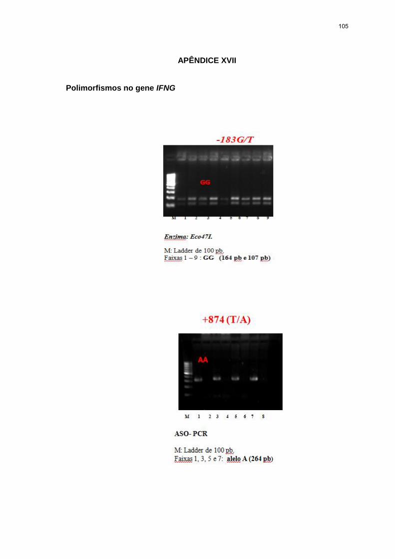
105
APÊNDICE XVII
Polimorfismos no gene IFNG

106
APÊNDICE XVIII
Polimorfismos no gene da IL10

107
APÊNDICE XIX
Polimorfismos no gene TNFA

108
Polimorfismos no gene TNFA

ANEXOS
109
ANEXO I
Comitê de Ética em Pesquisa: Utilização de amostras
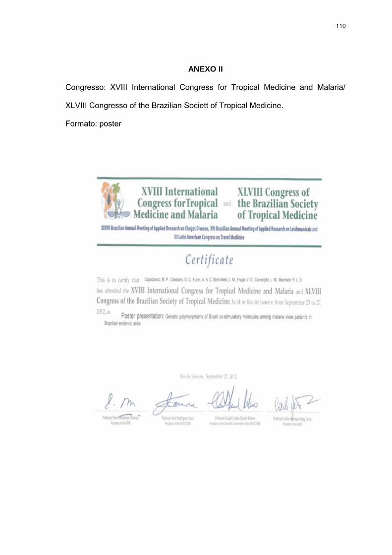
110
ANEXO II
Congresso: XVIII International Congress for Tropical Medicine and Malaria/
XLVIII Congresso of the Brazilian Societt of Tropical Medicine.
Formato: poster

111
ANEXO III ASTMH 61 st Annual Meeting – The American Journal of Tropical Medicine and
Hygiene (2012)
Resumo em Anais de Congresso

112
ANEXO IV
American Society of Tropical Medicine and Hygiene 62st Meeting (2013)
Resumo em Anais de Congresso

113
ANEXO V
XIII Reunião Nacional de Malária em Manaus (2013)
Resumo em Reunião

114
ANEXO V
Congresso Brasileiro de Medicina Tropical (2013)
Resumo em Anais de Congresso
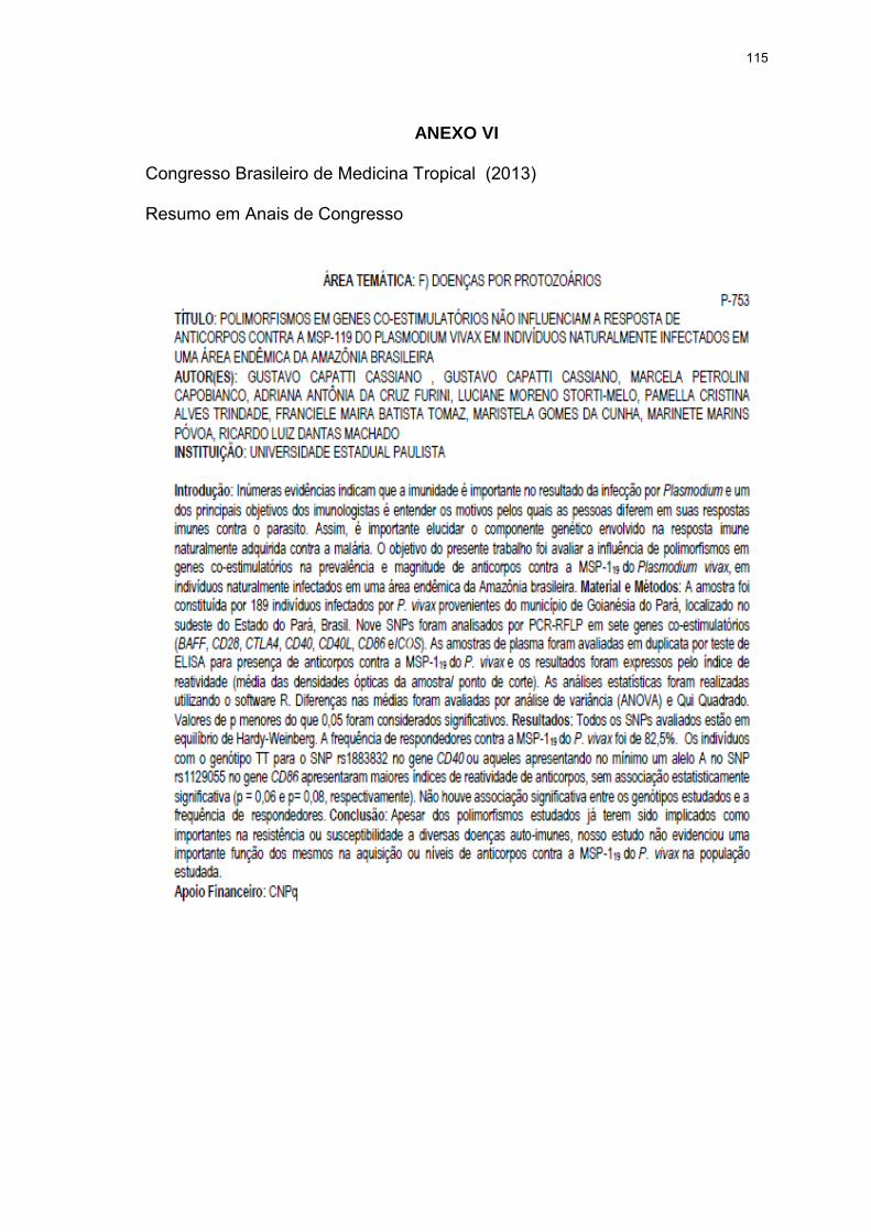
115
ANEXO VI Congresso Brasileiro de Medicina Tropical (2013)
Resumo em Anais de Congresso

116
ANEXO VII
63 rd Annual Meeting – The American Society of Tropical Medicine and Hygiene
(2014)
Resumo em Anais de Congresso
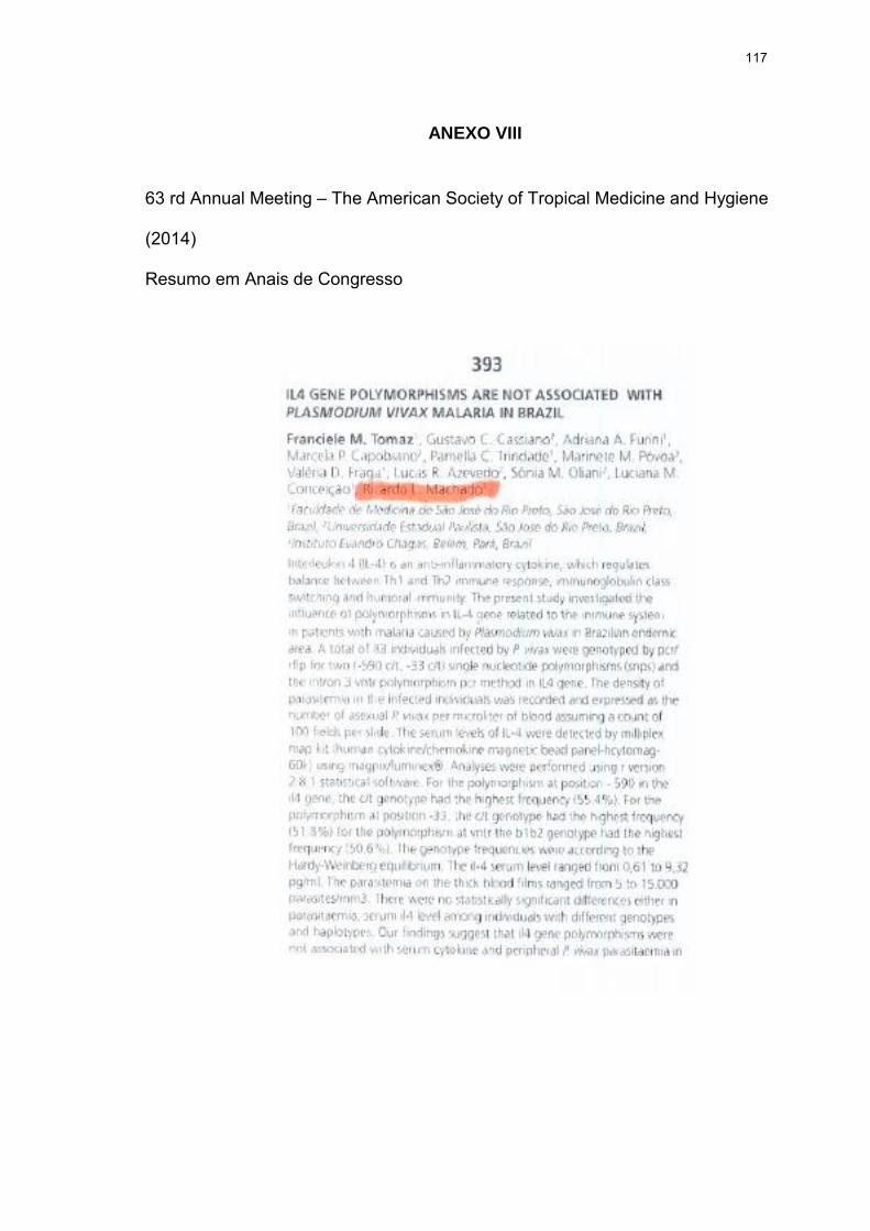
117
ANEXO VIII
63 rd Annual Meeting – The American Society of Tropical Medicine and Hygiene
(2014)
Resumo em Anais de Congresso

118
ANEXO IX
XXXIX Congress of the Brazilian Society of Immunology
Formato: poster

119
ANEXO X
Med Trop – 51º Congresso da Sociedade Brasileira de Medicina Tropical
Formato: pôster

120
ANEXO XI
XIV Reunião Nacional de pesquisa em Malária
Formato: pôster

121
ANEXO XII
XIV Reunião Nacional de pesquisa em Malária
Formato: pôster

122
ANEXO XIII
Participação em artigos publicados durante o doutorado
1-Galisteu KJ, Cardoso LV, Furini AAC, Schiesari Júnior A, Cesarino CB, Franco C,
Baptista ARS, Machado RLD. Opportunistic infections among individuals with HIV-
1/AIDS in the highly active antiretroviral therapy era at a Quaternary Level Care
Teaching Hospital. Rev Soc Bras Med Trop 2015; 48:149-56.
2. Cavasini CE, Cimmernan S, Barbosa DRL, Silva MCME, Furini AAC, Machado
RLD. Consensos e controvérsias sobre os microrganismos comensais intestinais.
Revista Panamericana de Infectología 2015; 17: 26-9.
3. Tomaz FMMB, Furini AAC, Capobianco MP, Póvoa MM, Trindade PCA, Fraga
VD, Conceição LM, De Azevedo LR, Oliani SM, Cassiano GC, Cavasini CE, Dos
Santos SEB, Machado RLD. Humoral immune responses against the malaria
vaccine candidate antigen Plasmodium vivax AMA-1 and IL-4 gene polymorphisms
in individuals living in an endemic area of the Brazilian Amazon. Cytokine 2015; 74:
273-78.
4. Rodrigues JF, Minari SH, Oliveira GC, Almeida VM, Jorge LS, Braz MM, Machado
RLD, Schiesari AJ, Furini AAC. HAART versus pacientes com HIV/AIDS em unidade
de referência do Noroeste paulista. Revista Panamericana de Infectología 2014; 16:
169-73.
5. Lima TAM, Nakazone MA, Furini AAC. Avaliação preliminar de prescrições para
idosos em serviço de cardiologia de um Hospital de Ensino. Revista Brasileira de
Cardiologia 2014; 27: 333-41.
123
6. Verona JP, Verona JAOB, Furini AAC. Gerenciamento de Resíduos de Serviços
de Saúde em Indústria de Produtos Hospitalares. Revista Eletrônica de Pesquisa da
UNIRP – Universitas 2013; 3: 1-10.
7.Capobianco MP, Cassiano GC, Furini AAC, Storti-Melo LM, Pavarino EC, Galbiatti
ALS, Fraga VD, Conceição LM, Couto VSC, Couto AARA, Machado RLD. No
evidence for association of the CD40, CD40L and BLYS polymorphisms, B-cell co-
stimulatory molecules, with Brazilian endemic Plasmodium vivax malaria. Trans R
Soc Trop Med Hyg 2013;107: 377-83.
8. Schiesari AJ,Galisteu KJ, Cardoso LV, Schiesari VMB, Furini AAC, Rossit ARB,
Machado RLD. Epidemiological Patterns of AIDS in a Reference Center from
Catanduva, São Paulo State, Brazil. Open J Medical Microbiol 2012; 2: 47-53.
9. Atique TSC , Lima TAM, Souza VA, Pacheco PFS, Furini AAC. Sensibilidade à
meticilina/oxacilina de Staphylococcus aureus isolados da mucosa nasal de alunos
do Centro Universitário de Rio Preto.. Revista Brasileira de Farmacia 2012; 93: 347-
52.
10. Reis AGR, Guimarães PM, Atique TSC, Furini AAC. Análise de potenciais
interações medicamentosas em prescrições de um Hospital Veterinário do noroeste
paulista como ferramenta do serviço de farmácia hospitalar para reduzir suas reais
manifestações. Revista de Ciências Farmacêuticas Básica e Aplicada 2012; 33:
291-98.
11. Atique TSC, Atique Netto HÁ, Morelli JJ, Momesso CAS, Furini AAC.
Isolamento de agentes da Mastite Bovina, perfil de sensibilidade e resíduos de

124
antimicrobianos em São José do Rio Preto - São Paulo, Mastite Bovina e resíduos
de antimicrobianos. Revista Eletrônica de Pesquisa da UNIRP – Universitas 2012;
2:29-8.
12. Atique TSC, Furini AAC, Reis AG, Guimarães PM, Atique Netto HA. Indicadores
de prescrição e interações medicamentosas no Hospital Veterinário Dr. Halim
Atique-UNIRP. Revista de Ciências Farmacêuticas Básica e Aplicada 2012; 33: 291-
96.
.