Embed Size (px)
Citation preview

October 2014
Universidade do MinhoEscola de Engenharia
Francisco Duarte da Cunha Ventura
Characterization of gene or geneclusters responsible for theproduction of antimicrobialcompounds in Pseudoalteromonasatlantica
UM
inho
|201
4Fr
anci
sco
Dua
rte
da C
unha
Ven
tura
Ch
ara
cte
riza
tio
n o
f g
en
e o
r g
en
e c
lust
ers
re
spo
nsi
ble
fo
r th
e p
rod
uct
ion
o
f a
nti
mic
rob
ial c
om
po
un
ds
in P
seudoal
tero
monas
atlan
tica

Dissertation for the M.Sc. degree in BiomedicalEngineering
Supervisors:Maria João Vieira, PhDCarina Almeida, PhD
October 2014
Universidade do MinhoEscola de Engenharia
Francisco Duarte da Cunha Ventura
Characterization of gene or geneclusters responsible for theproduction of antimicrobialcompounds in Pseudoalteromonasatlantica


Acknowledgements!! !
! I would like to thank:!
! ! Professor Maria João Vieira for having me on board.!
! ! Carina Almeida, for all the supervision throughout the entire work.
You have gone way beyond your duty. For always pointing me in the right
direction, for all the time I made you lose, for the great support in the laboratory. I
can’t thank you enough! It’s been an honor and a privilege to work with you for
the past year!!
!
iii

Abstract!!! The intensive and unconscious use of antibiotics alongside with the
decrease of investment in research of novel molecules (since the mid 1960s),
has led to a rise of highly resistant bacteria. These microorganisms have been
developing mechanisms that enable them to survive to the aggressions of
classical antibiotics. Also, these self-defense mechanisms are easily transmitted
between bacteria, which is a worrisome panorama.!
! It is necessary to get back to a proactive fight against these
microorganisms. Living organisms have long proven to be a rich source for
antimicrobial compounds due to their need to fight for their place in ecological
niches. The marine environment is known to be prolific with microbial
communities and thus a great diversity of bioactive compounds was already
discovered.!
! In this thesis work, I searched for novel antimicrobial compounds in a
marine bacteria, Pseudoalteromonas atlantica. A genome mining approach was
followed. A search for clusters coding for secondary metabolites with
antimicrobial characteristics was done using softwares such as AntiSmash,
ClustScan, HHpred or BLASTx. This process resulted in the finding of 17 putative
clusters coding for polyketide synthase and also 1 putative cluster coding for a
bacteriocin.!
! To assess if P. atlantica is, in fact, capable of producing antimicrobial
compounds, and in the positive case, to further enhance the production of such
iv

compounds, a compound production optimization procedure was performed.
Optimal culture conditions for production of antibiotic compounds in P. atlantica
were met for Marine Broth culture medium at a temperature of 23ºC, a pH of 8
and a 120 rpm agitation. Bacteria-free spent media proved to inhibit the growth of
Escherichia coli K12. Moreover, a bacteria-free spent media from cultures grown
in the presence of a competitor (E. coli K12; Staphylococcus aureus;
Pseudomonas aeruginosa or Vibrio harveyi) has also resulted in the inhibition of
Salmonella enteritidis.!
! These results indicate that the P. atlantica genome might be a source for
novel antimicrobial compounds. In fact, under the described conditions, P.
atlantica was capable of producing antimicrobial molecules with a narrow activity
spectrum.!
!
v

Sumário!!! O uso intensivo e inconsciente de antibióticos e a quebra do investimento
na investigação de novas moléculas (desde meados dos ano 60), levou ao
aparecimento de bactérias altamente resistentes. Estes microorganismos têm
desenvolvido mecanismos que os permite sobreviver às agressões provocadas
pelos antibióticos clássicos. Além disso, estes mecanismos de auto-defesa são
facilmente transmitidos entre diferentes espécies de bactérias, o que é um
cenário muito preocupante.!
! É então necessário voltar a uma atitude proactiva na luta contra estes
microorganismos. Os microorganismos já há muito provaram ser uma fonte rica
em compostos antimicrobianos, devido à sua necessidade de lutar por um lugar
nos respectivos nichos ecológicos. O ambiente marinho é conhecido por ser fértil
em comunidades microbianas e portanto uma grande diversidade de compostos
bioactivos foram já descobertos.!
! Nesta tese procurei por compostos antimicrobianos numa bactéria
marinha, a Pseudoalteromonas atlantica. Seguiu-se uma estratégia de “genome
mining”. Foi realizada uma procura de clusters de metabolitos secundários de
cariz antimicrobiano, através do recurso a ferramentas como o AntiSmash,
ClustScan, HHpred ou BLASTx. Todo este processo resultou na descoberta de
17 putativos clusters de poliquétidos sintetases e 1 putativo cluster de
bacteriocina.!
vi

! De forma a avaliar se a P. atlantica é capaz de produzir compostos
antimicrobianos, e, em caso positivo, para aumentar a produção desses
compostos, foi realizado um procedimento de optimização da produção do
antimicrobiano. As condições óptimas para a produção de compostos
antimicrobianos em P. atlantica revelaram ser o uso de meio marinho (Marine
Broth), a uma temperatura de 23ºC, um pH de 8 e uma agitação de 120 rpm. O
meio de cultura gasto desprovido de bactérias provou inibir o crescimento de
Escherichia coli K12. Além disso, na presença de um competidor (E. coli K12;
Staphylococcus aureus; Pseudomonas aeruginosa or Vibrio harveyi), o meio de
cultura de P. atlantica, também resultou na inibição de Salmonella enteritidis.!
! Estes resultados indicam que o genoma da P. atlantica pode ser uma
fonte de compostos antimicrobianos inauditos. De facto, nas condições referidas
no presente trabalho, a P. atlantica foi capaz de produzir moléculas
antimicrobianas, com um espectro de actividade aparentemente muito
específico.!
!
vii
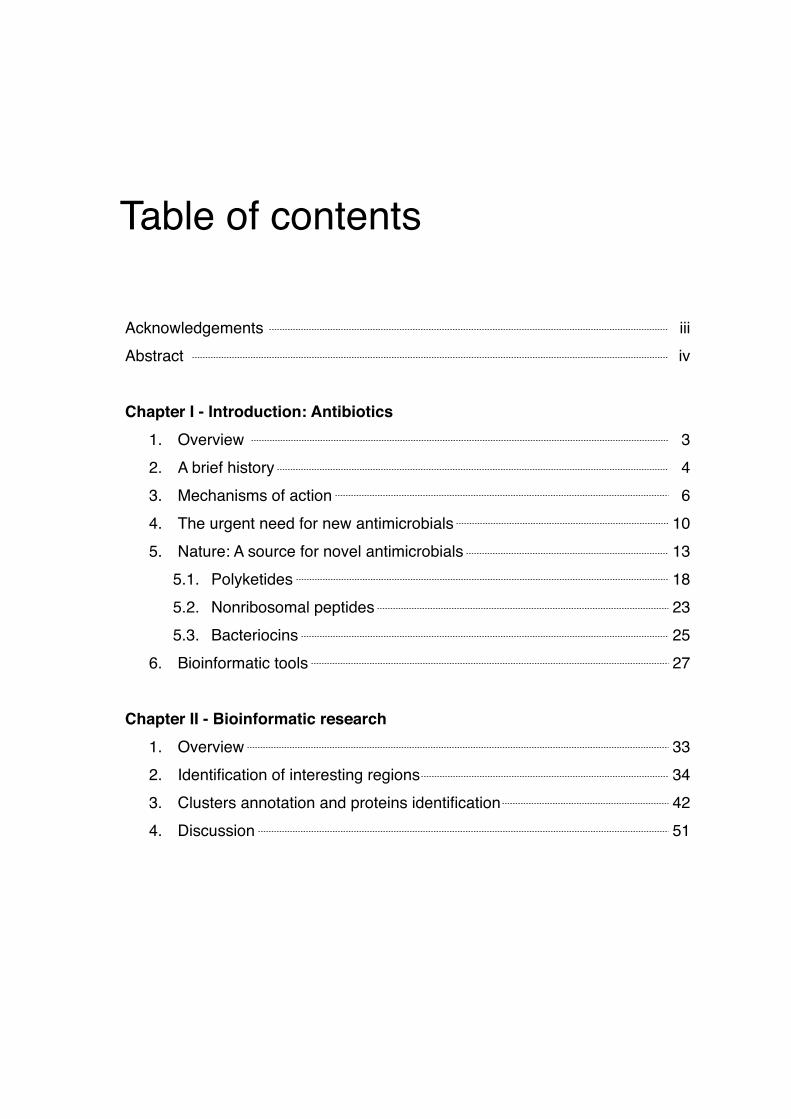
Table of contents!!
!!Acknowledgements!
Abstract!!Chapter I - Introduction: Antibiotics!
1. Overview !2. A brief history!3. Mechanisms of action!4. The urgent need for new antimicrobials!5. Nature: A source for novel antimicrobials!
5.1. Polyketides!5.2. Nonribosomal peptides!5.3. Bacteriocins!
6. Bioinformatic tools!!Chapter II - Bioinformatic research!
1. Overview!2. Identification of interesting regions!3. Clusters annotation and proteins identification!4. Discussion
iii!iv!!!
3!4!6!
10!13!18!23!25!27!!!
33!34!42!51

!Chapter III - Optimization of antimicrobial compounds production!
1. Overview!2. Materials and methods!
2.1. Strains and cultures conditions optimization!2.1.1. Effect of the growth medium!2.1.2. Temperature, pH and agitation!2.1.3. Addition of extracellular ATP!2.1.4. Competitors!
2.2. Assessment of antimicrobial activity!2.3. Determination of arbitrary units!
3. Results and discussion!3.1. Effect of culture media on cell growth and antimicrobial
activity!3.2. Effect of temperature, pH and agitation on cell growth and
antimicrobial activity!3.3. Effect of addition of extracelullar ATP!3.4. Effect of competitors!
!Chapter IV - General conclusions and future work!
1. General conclusions and future work!!Chapter V - References and appendix!
1. References!2. Appendix
!!
57!58!58!59!60!61!62!64!66!67!!
67!!
70!75!76!!!
83!!!
89!96

List of figures and tables!!
Table 1 - Main antibiotic families and mechanisms of action. From Levy S. and Marshall B. (2004).Abstract!!Figure 1 - New antibacterial agents approved in the US per 5-year period from 1983 to 2002. Adapted from Shlaes, D. et al (2004).!!Figure 2 - Compounds with new antibacterial templates divided into development phases and their lead derivation source. Adapted from Butler M. and Cooper M. (2011).!!Table 2 - Bioactivity of Pseudoalteromonas species. Adapted from Bowman (2007)!!Figure 3 - Example of type I PKS (lovastatin), type II PKS (doxorubicin) and type III PKS (naringenin chalcone) From Hertweck (2009).!!Table 3 - Examples of Polyketides and their bioactivity. Adapted from Pfeifer and Khosla (2001).!!Figure 4 - Repartition of six main biological activities displayed by curated peptides in the Norine database (790 NRPs). From Caboche S. et al (2010).!!Figure 5 - Parameters selected in AntiSmash.!!Table 4 - Position of all clusters detected by AntiSmash!!Figure 6 - Screenshot of ClustScan workspace.!
14
20
21
24
34
35
36
6
11
13

Figure 7 - Script used for detection of nearby clusters!!Table 5 - Position of all PKS genes and their main specifications given by ClustScan!!Table 6 - Position of clusters detected in AntiSmash, ClustScan similarity and ClustScan E-value.!!Figure 8 - Screenshot of CLC Sequence Viewer workspace.!!Figure 9 - Screenshot of AntiSmash annotations.!!Figure 10 - Screenshot of InterPro search.!!Table 7 - PKS related ORFs detected near AntiSmash detection sites!!FIgure 11 - Schematic representation of a hypothetical Polyketide Synthase in P. atlantica T6c composed by 4 distinct domains: AT - acyltransferase ; KS - ketosynthase ; KR - ketoreductase!!Figure 12 - Graphic of homologous gene clusters, for PKS #8 cluster - AntiSmash.!!Figure 13 - Schematic representation of a hypothetical bacteriocin in P. atlantica T6c.!!Figure 14 - Graphic of homologous gene clusters, for bacteriocin cluster - AntiSmash.!!Figure 15 - Representation of the culture conditions used for the competition assays.!!Table 8 - Effect of culture media in P. atlantica growth and the production of inhibitory compounds.!!Figure 16 - Inhibition halos in an E.coli K12 lawn.!
37
38
40
42
43
44
45
47
48
49
50
62
68
69
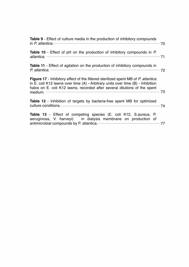
Table 9 - Effect of culture media in the production of inhibitory compounds in P. atlantica.!!Table 10 - Effect of pH on the production of inhibitory compounds in P. atlantica.!!Table 11 - Effect of agitation on the production of inhibitory compounds in P. atlantica.!!Figure 17 - Inhibitory effect of the filtered sterilized spent MB of P. atlantica in E. coli K12 lawns over time (A) - Arbitrary units over time (B) - Inhibition halos on E. coli K12 lawns, recorded after several dilutions of the spent medium.!!Table 12 - Inhibition of targets by bacteria-free spent MB for optimized culture conditions.!!Table 13 - Effect of competing species (E. coli K12, S.aureus, P. aeruginosa, V. harveyi) in dialysis membrane on production of antimicrobial compounds by P. atlantica.
70
71
72
73
74
77

!!!

!
!
!
!
!
!
!
!
!
!
Chapter I!Introduction: Antibiotics

!

1. Overview!!! Antibiotics are compounds that can either kill (bactericidals), or inhibit the
growth (bacteriostatics) of bacteria. This categorization between bactericidals and
bacteriostatics isn’t as obvious as it may appear, since it depends on the drug
type, concentration and on the bacterial species. They can be of natural source,
when they are for instance produced by living organisms. Also, they can be
produced by chemical synthesis or derive from a biological source.!
! Since their discovery, antibiotics have revolutionized medicine in many
ways. Countless lives were saved by these compounds. Nevertheless, the use of
antibiotics has been accompanied by a growing number of resistant
microorganisms. It is feared that because of this, we may face a new era, like the
preantibiotic one.!
! The study of antibiotics has had its ups and downs all over the years, but it
has become of major interest in the last decade. It is important to understand how
this all began, in order to understand where we stand nowadays.!
!!
Introduction: Antibiotics Chapter I
3

2. A brief history!!! Sir Alexander Fleming set the beginning of antibiotics timeline. In 1929 Sir
Fleming was conducting experiments with Staphylococcus variants. Plates
containing Staphylococcus were left in contact with air and, by chance, they
became contaminated by a fungus. Sir Fleming noticed that the areas around the
mold became transparent, Staphylococcus were undergoing lysis. He concluded
that the contaminant was a Penicillum mold that was producing a powerful
bactericidal, the penicillin [1].!
! In 1940, years before the use of penicillin as a therapy, a bacterial
penicillinase was identified. After its mass production, several strains showed the
capacity to resist to penicillin. Just then, experiments were conducted so that the
penicillin could be modified chemically, in order to prevent cleavage by
penicillinases (β-lactamases) [2].!
! By 1944 Waksman, Feldman and Hinshaw discovered the streptomycin
from Streptomyces griseus, a bacteria commonly found in the soil [2]. They found
that streptomycin was effective against virulent human tubercle bacilli. This
discovery led to a growing interest in soil bacteria, which later proved to be the
main resource for the discovery of several antibiotics. Streptomycin was then
used to cure tuberculosis, but in the mean time, strains of Mycobacterium
tuberculosis resistant to the antibiotic arose during patient treatment [2].!
! As in penicillin and streptomycin, many powerful antibiotics were
discovered or synthesized throughout the years, but then again, many resistant
Introduction: Antibiotics Chapter I
4

strains appeared through a process that remained unknown until the mid 1950s
[3]. In Japan, a country devastated by war at the time, an epidemic of Shigella
dysenteriae turned to be particularly hard to irradiate, due to the growing number
of resistant strains. Sulfonamide was no longer effective in 80% of the cases [3].
Kitamoto wrote about S. dysenteriae strains that could resist to four different
antibiotics. Later on, it was proved that the resistance of these strains could be
transferred to other Enterobacteriaceae simply requiring a cell-to-cell contact,
indicating that bacterial conjugation was involved in the process [3].!
! In the mid 80s, Michael Syvanen proved that the uniformity of the gene
code among nature would allow organisms to use genes transposed from
organisms of different species (Horizontal Gene Transfer). This revealed to be of
particular interest when one tries to understand the rapid bursts in the evolution
of organisms, also helping to understand the way which pathogens gained
resistance to several antibiotics [2].!
! Since Sir Fleming found penicillin in 1929 till now, the development of
antibiotics has come up through different eras. It is safe to say that the majority of
the antibiotics used nowadays derive from the ones discovered during the so
called golden era, which took place between the 50s and the mid 60s. From then
on came the lean years, or the innovation gap. A gap that lasted until 2000, when
a decrease of the discovery rate was observed. The main approach for the
development of novel drugs has been the modification of the molecules
previously known [2].!
Introduction: Antibiotics Chapter I
5

3. Mechanisms of action!!! Antibiotics can be classified according to a different range of settings, such
as their spectrum of activity, their chemical structure, but most commonly,
antibiotics are categorized by their mechanism of action (Table 1). Each category,
class or family aims to destroy or defunctionalize essential physiological or
metabolic targets of the bacterial cell [4].!
!
!!
Table 1 - Main antibiotic families and mechanisms of action. From Levy S. and Marshall B. (2004).
Mechanism of action Antibiotic families
Inhibition of cell wall synthesis Penicillins; cephalosporins; daptomycin; monobactams; glycopeptides
Inhibition of protein synthesisTetracyclines; aminoglycosides;
oxazolidonones; streptogramins; ketolides; macrolides; lincosamides
Inhibition of DNA synthesis Fluoroquinolones
Competitive inhibition of folic acid synthesis Sulfonamides; trimethroprim
Inhibition of RNA synthesis Rifampin
Other Metronidazole
Introduction: Antibiotics Chapter I
6

• Inhibition of cell wall synthesis!
! Antibiotics that inhibit the cell wall synthesis act by interfering with the
formation of the peptidoglycan wall present in bacteria. The process by which that
occurs is what distinguishes the different families belonging to this mechanism of
action. For example, β-Lactams like penicillins, carbapenems and cephalosporins
act by inhibiting transpeptidase enzymes, whose function is to cross-link
peptidoglycan chains that compose the bacterial cell wall, causing it to lyse [5].!
! Another way for an antibiotic to affect cell wall integrity is by binding with
peptidoglycan units and by blocking transglycosylase (enzyme that adds
disaccharide pentapeptides to extend the glycan strands of existing
peptidoglycan molecules) and also blocking transpeptidase activity [5]. That is the
case of a glycopeptides, such as vancomycin. Nevertheless, this family of
antibiotics is ineffective against Gram-negative bacteria, due to their lower
permeability, by contrast with β-Lactams, which are effective against both Gram-
positive and Gram-negative bacteria. There are other ways to inhibit the cell wall
synthesis or the cell wall integrity, besides the ones used by β-Lactams and
glycopeptides, like affecting the transport of individual peptidoglycan (e.g.
Bacitracin), for example [5].!
!• Inhibition of protein synthesis!
! Translation is a crucial process in biological lifeforms. It is a series of
processes by which information coded in RNA is used to create proteins
constituted by aminoacids. One of these processes includes the presence of a
very important organelle - the ribosome. This organelle is divided into two
7
Introduction: Antibiotics Chapter I

subunits. In the case of prokaryotes, there is a small subunit (30s - responsible
for reading the mRNA) and the big subunit (50s - responsible for joining amino
acids to the growing peptide chain). Antibiotics that act through this mechanism
(inhibition of protein synthesis) are molecules that act by compromising the
function of one of these two subunits. Therefore, they can be divided into 30s
inhibitors or 50s inhibitors [6].!
! The 30s inhibitors like the tetracyclines act by compromising the function
of the small ribosome subunit. Tetracycline, for example, blocks the aminoacyl-
tRNA to the ribosome and therefore stops protein synthesis [5].!
! Antibiotics belonging to macrolides, lincosamides or oxazolidonones
families are considered to be 50s inhibitors, since they act upon the big subunit of
the bacterial ribosome. They stop protein synthesis by either blocking initiation or
elongation of the translation process. Also, some act by hindering translocation of
peptidyl-tRNA. Peptidyl is one of the binding sites in the ribosome for tRNA [5].!
!• Inhibition of DNA synthesis!
! Deoxyribonucleic acid or just DNA is an essential molecule of all living
organisms. It would be correct to assume that a living organism would be killed if
its DNA synthesis stopped. That is just how quinolones, a family of synthetic
broad-spectrum antibiotics, work. Fluoroquinolone (a quinolone with a fluorine
atom attached to the central ring system) targets the function of DNA-
topoisomerase and DNA-gyrase complexes [5]. Topoisomerases and gyrases are
enzymes that participate in DNA replication. By canceling their function,
fluoroquinolones lead to bacterial cell death. Thus, they’re considered
Introduction: Antibiotics Chapter I
8

bactericidal. There are other families belonging to this mechanism of action and
they target different components of the bacterial DNA machinery [5].!
!• Competitive inhibition of folic acid synthesis!
! Unlike humans, bacteria do not acquire folic acid through diet. Instead they
synthesize it. It is by affecting this mechanism that antibiotics like the sulfonamide
work. All cells require folate cofactors for the biosynthesis of diverse cellular
components, like the formation of the so important amino acid methionine, crucial
for starting protein translation. Sulfone inhibitors work by being analog of para-
aminobenzoic acid (pABA), which is a required intermediate of bacterial synthesis
of folate. Sulfonamides act as alternative substrates for dehydropteroate
synthase, an enzyme involved in the folate pathway. By doing so, they inhibit the
growth of the target microorganism, therefore they are considered to be
bacteriostatical antibiotics [7].!
!• Inhibition of RNA synthesis!
! Like the DNA, ribonucleic acid or RNA plays an important role in
expression of genes. Inhibition of RNA synthesis is a catastrophic event for
prokaryotic nucleic acid metabolism. Drugs belonging to the rifamycin group
inhibit or affect the normal behavior of RNA within bacterial cells. Rifampin, for
example, hinders DNA transcription by inhibiting RNA polymerase, leading, in
most cases, bacterial pathogens to death [5].!
9
Introduction: Antibiotics Chapter I

4. The urgent need for new antimicrobials!
!! As stated by Hiroshi Nikaido in 2004, about 100 000 tons of antibiotics are
produced per year [8]. Their intensive use has had a tremendous impact in
worldwide microbial resistance. Because of this, many antibiotics that were
effective many years ago, no longer produce a harmful effect in the same
pathogens. Methicilin-resistant Staphylococcus aureus (MRSA) is a glaring
example of a dangerous pathogen. It is resistant to methicilin and a wide range of
antibiotics like aminoglycosides, macrolides, tetracycline, chloramphenicol, and
lincosamides [8]. Since 2002 some strains of MRSA have also proved to be
resistant to vancomicyn (the main strategy of eliminating MRSA until then) [9].
The rise of multidrug resistant bacteria, the scarcity of new classes of
antibacterial drugs (Fig. 1) and the stagnation in antibiotics discovery is
demanding new ways of controlling these very resistant pathogens [10, 11].!
! Alongside the heavy health burden of this problem, also comes an
economic burden that hasn’t been correctly measured yet. As stated by WHO,
“All costs for infections caused by resistant strains were consistently greater than
those for infections caused by susceptible strains.” That is mainly due not only to
the increase in the intensity of care needed by infected patients, but also to an
increase in the length of stay of those patients in the hospitals. Moreover, as
expected, infections with resistant strains are associated with worse clinical
outcomes [11].!
Introduction: Antibiotics Chapter I
10

! The laborious legal procedures to approve new antimicrobials, especially
in the United States, have delayed the introduction of new antibiotics and large
pharmaceutical companies are fending themselves from this area of investigation
[10]. This obviously results in a declining number of new antimicrobials in
development. Although all of this may appear to be a dantesque scenario, there
are some good news as well. State of the art techniques, like genomics,
proteomics, structure based design, high-throughput screening and combinatorial
chemistry are paving their own path into new antimicrobial compounds [10].!
!
0 4 8 12 16
1998 - 2002
1993 - 1997
1988 - 1992
1983 - 1987
Figure 1 - New antibacterial agents approved in the US per 5-year period from 1983 to 2002. Adapted from Shlaes, D. et al (2004).
11
Introduction: Antibiotics Chapter I

5. Nature: A source for novel antimicrobials!
!! Today, there is an understanding that the method for finding new
antibiotics used in the last 50 years is no longer effective. While 2nd, 3rd, etc.
generations of antibiotics fail to deliver a continuous harm to pathogens along
time, some researchers think that natural compounds might be the solution. Many
antibiotics discovered until nowadays are secondary metabolites that some
microorganisms naturally produce. Thinking that microorganisms have found their
way against many pathogens long before they even become a known threat for
humans, can be a successful contemplation [12].!
! Nowadays, efforts are being made in order to find novel antimicrobials
from old sources, like Streptomyces [13]. Also, many are studying the capacity of
other unstudied microorganisms (cyanobacteria or uncultured bacteria) to
synthesize antimicrobials compounds. This is proving to be a successful strategy,
since several novel scaffolds with antibiotic potential are being discovered. We
can also think that many targets within pathogens haven’t been explored yet (like
the fatty acid synthesis) and if novel scaffolds can act upon these targets, a new
era may be arriving. Naturally occurring antibiotics can benefit from the fact that
they have features which are not present in libraries of synthetic drugs molecules,
like those found via high-throughput screening. It is not surprising that rather than
chemically synthesized, the majority of antibiotics in the last stages of clinical
trials derive from natural sources (Fig. 2) [13].!
Introduction: Antibiotics Chapter I
12

!
! The marine environment is known to be proliferous with microbial
communities. Also, as many marine communities are highly specific to particular
ecological niches (sponge or algae environments), there is a vast biodiversity
among these microbial communities that inhabit seawater. This statement is
useful when one tries to understand the implications of natural products
discovery, such as naturally produced antibiotics [14].!
! It is estimated that about 0,5% to 6 % of the oceanic bacterioplancton
belongs to the Pseudoalteromonas genus. Some species of this genus have
0
11
22
NDA Phase III Phase II Phase I
1
1
5
10
41
6
11
1
Syn-derivedNP-derivedUnknown
Figure 2 - Compounds with new antibacterial templates divided into development phases and their lead derivation source. Adapted from Butler M. and Cooper M. (2011).
13
Introduction: Antibiotics Chapter I

proved to be efficient in producing bioactive compounds, such as antimicrobials,
anti-fouling or algicidals compounds [15, 16].!
! Table 2 summarizes the bioactivity of compounds produced by
Pseudoalteromonas species. Although many bioactive compounds have already
been detected, the synthesis of such compounds are quorum-sensitive. Thus, the
ecosystem in which these species grow, influence the biosynthesis of these
compounds. As many aspects of ecological networks remain unexplored, there is
room for the discovery of novel natural products [15].!
!Table 2 - Bioactivity of Pseudoalteromonas species. Adapted from Bowman (2007).
Species Source Bioactive compounds Bioactivity
P. aliena seawater Unknown compound(s)Anti-tumorigenic activity - Ehrlich ascites carcinoma
cell line inhibited
P. agarivorans seawater, ascidians Not Available degrades algal
polysaccharides
P. antarcticaseawater, sea-ice,
muddy soils, sediment
Not AvailableNone observedpolysaccharides, cold-
active enzymes]
P. atlantica seawater, marine alga Not Available
May cause opportunistic disease in crabs [strong
degrader of algal polysaccharides]
P. aurantia surface of Ulva lactuca, seawater Unknown compound(s)
Antimicrobial activity; inhibits settlement of invertebrate larvae
P. byunsanensis tidal flat sediment Not Available —
Introduction: Antibiotics Chapter I
14
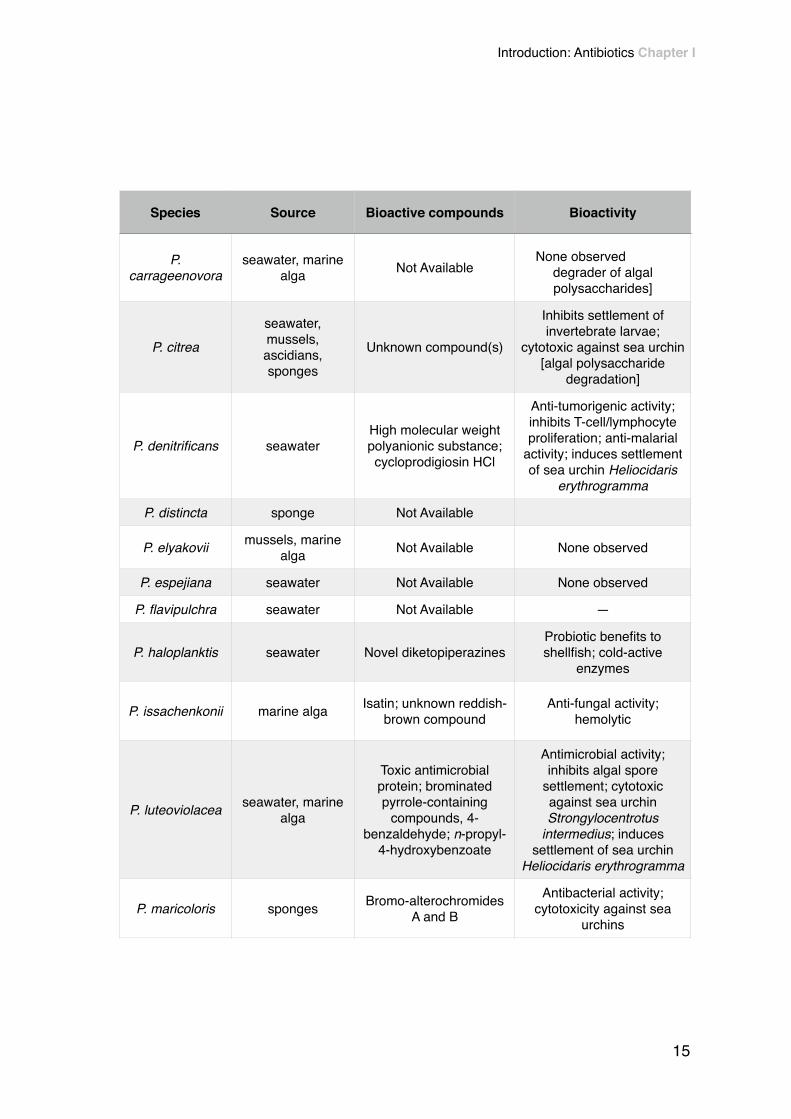
P. carrageenovora
seawater, marine alga Not Available
None observeddegrader of algal polysaccharides]
P. citrea
seawater, mussels, ascidians, sponges
Unknown compound(s)
Inhibits settlement of invertebrate larvae;
cytotoxic against sea urchin [algal polysaccharide
degradation]
P. denitrificans seawaterHigh molecular weight polyanionic substance; cycloprodigiosin HCl
Anti-tumorigenic activity; inhibits T-cell/lymphocyte proliferation; anti-malarial
activity; induces settlement of sea urchin Heliocidaris
erythrogramma
P. distincta sponge Not Available
P. elyakovii mussels, marine alga Not Available None observed
P. espejiana seawater Not Available None observed
P. flavipulchra seawater Not Available —
P. haloplanktis seawater Novel diketopiperazinesProbiotic benefits to shellfish; cold-active
enzymes
P. issachenkonii marine alga Isatin; unknown reddish- brown compound
Anti-fungal activity; hemolytic
P. luteoviolacea seawater, marine alga
Toxic antimicrobial protein; brominated pyrrole-containing
compounds, 4- benzaldehyde; n-propyl-
4-hydroxybenzoate
Antimicrobial activity; inhibits algal spore
settlement; cytotoxic against sea urchin Strongylocentrotus
intermedius; induces settlement of sea urchin
Heliocidaris erythrogramma
P. maricoloris sponges Bromo-alterochromides A and B
Antibacterial activity; cytotoxicity against sea
urchins
Species Source Bioactive compounds Bioactivity
15
Introduction: Antibiotics Chapter I
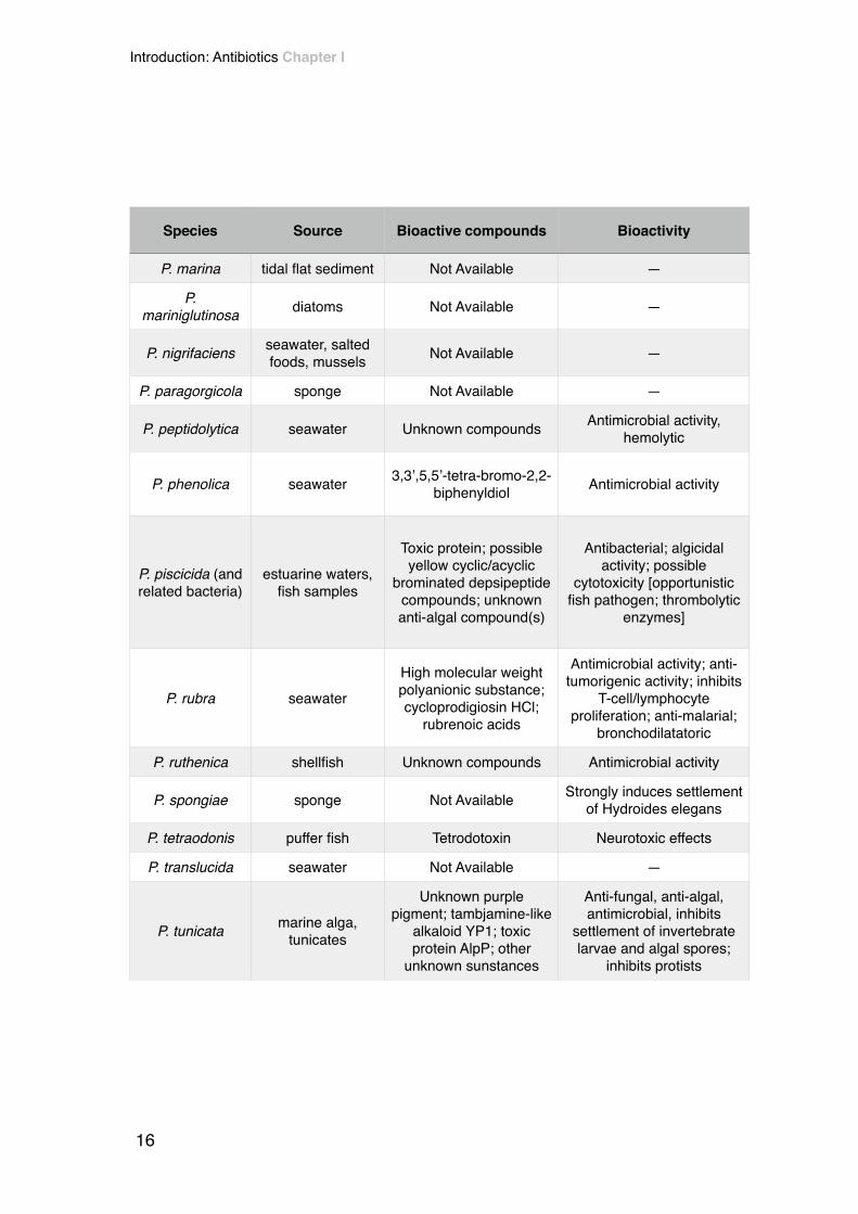
P. marina tidal flat sediment Not Available —
P. mariniglutinosa diatoms Not Available —
P. nigrifaciens seawater, salted foods, mussels Not Available —
P. paragorgicola sponge Not Available —
P. peptidolytica seawater Unknown compounds Antimicrobial activity, hemolytic
P. phenolica seawater 3,3’,5,5’-tetra-bromo-2,2- biphenyldiol Antimicrobial activity
P. piscicida (and related bacteria)
estuarine waters, fish samples
Toxic protein; possible yellow cyclic/acyclic
brominated depsipeptide compounds; unknown anti-algal compound(s)
Antibacterial; algicidal activity; possible
cytotoxicity [opportunistic fish pathogen; thrombolytic
enzymes]
P. rubra seawaterHigh molecular weight polyanionic substance; cycloprodigiosin HCl;
rubrenoic acids
Antimicrobial activity; anti- tumorigenic activity; inhibits
T-cell/lymphocyte proliferation; anti-malarial;
bronchodilatatoric
P. ruthenica shellfish Unknown compounds Antimicrobial activity
P. spongiae sponge Not Available Strongly induces settlement of Hydroides elegans
P. tetraodonis puffer fish Tetrodotoxin Neurotoxic effects
P. translucida seawater Not Available —
P. tunicata marine alga, tunicates
Unknown purple pigment; tambjamine-like
alkaloid YP1; toxic protein AlpP; other
unknown sunstances
Anti-fungal, anti-algal, antimicrobial, inhibits
settlement of invertebrate larvae and algal spores;
inhibits protists
Species Source Bioactive compounds Bioactivity
Introduction: Antibiotics Chapter I
16

!! Because there are many strains belonging to the Pseudoalteromonas
genus, many of them live in the wild under different environmental conditions,
which leaves room for a vast spectrum of different bioactive compounds
produced by different strains. Although some of these compounds were already
described and tested, there is a believe that there is an enormous potential for
the discovery of novel compounds [15, 16].!
! Among the naturally occurring antibiotics, polyketides and non-ribossomal
peptides have gained their own status in antimicrobials research and production.
Some of them are even known and widely used as antibiotics. Nevertheless, their
synthase is yet to be fully understood. Besides, the ability for many
microorganisms to produce these compounds is being tested in different species
of bacteria and fungus [13].!
!
P. ulvae marine alga Unknown substancesInhibits invertebrate larval settlement and algal spore germination and settlement
P. undina seawater, fish Not Availablehemolytic; [probiotic benefits; possible
opportunistic fish pathogen]
Species Source Bioactive compounds Bioactivity
17
Introduction: Antibiotics Chapter I
5.1. Polyketides!!! Polyketides are small secondary metabolites that microorganisms
produce. Their vast diversity in terms of structure and function is well known.
They are produced by polyketide synthases, which are enzymatic assembly lines
that determine the final structure of the produced polyketide. These metabolites
can be of major interest in the clinical area (Table 3). For example, erythromycin
A is a potent antibiotic used as a therapy against bacterial infections. Rapamycin
is an immunosuppressant used in various surgery techniques. This is to say that
polyketides may present very interesting characteristics. Some of them have
already been useful, others may help us win the war against pathogens in the
future. It is then important to try to understand how these powerful molecules are
produced by some microorganisms [17].!
! Polyketides, are synthesized due to repetitive condensation reactions, in a
process that is very similar to the synthesis of fatty acids. In these reactions that
link carbon precursors, coenzyme A thioesters play an important role as they
constitute the core of the molecule. However, polyketides are found in much more
diverse structures than fatty acids. This diversity is of great usefulness since they
also present different modes of action, thus can be used in different applications
[18].!
! Polyketide synthases (PKS) are the enzymes of large dimension with
specific catalytic domains that catalyze the referred condensations. Their core
domains are ketosynthase (KS), acyltransferase (AT) and thiolation (T). PKS are
Introduction: Antibiotics Chapter I
18

categorized into three classes (I, II and III) (Fig. 3) which differs slightly from fatty
acid nomenclatures [18, 19].!
!• Type I - best exemplified by the PKS responsible for building the backbone
of erythromycin ( 6-deoxyerythronolide B or 6-DEB). These PKS’s are
constituted by multidomains resembling type I fatty acid synthases (FAS).!
!• Type II - as one would imagine, this PKS resembles type II FAS. The growth
of the polyketide is iterative and KS, AT and T domains are re-used during
polyketide synthesis.!
!• Type III - They differ from other types by using an acyl-carrier-protein
independent mechanism. Besides, they typically lack multiple catalytic
domains.!
!
19
Introduction: Antibiotics Chapter I

!
Figure 3 - Example of type I PKS (lovastatin), type II PKS (doxorubicin) and type III PKS (naringenin chalcone) From Hertweck (2009).
Introduction: Antibiotics Chapter I
20

!! As polyketides are small metabolites hard to isolate, its correct
characterization has been an hard quest for many researchers. Traditional
methods used on the identification of polyketides usually involve phenotype
screening, followed by the use of analytical chemistry techniques. For instance,
Marinho and his colleagues [20] reported the presence of citreorosein (1), emodin
(2), janthinone (3), citrinin (4), citrinin H1 (5) and dicitrinoln (6), six known
polyketides produced by an endophytic fungi, Penicillum herquei. To evaluate the
presence of these compounds, they have used classical methods of
chromatography. They were identified by 1D and 2D Nuclear Magnetic
Resonance spectroscopy (NMR) and Mass Spectrum analysis (MS), results were
then compared to previous identifications of the referred compounds [20].!
! However, phenotype screening approaches are usually very time
consuming and depend on the availability of large libraries of organisms.
Besides, the production of some metabolites might not be induced under the
testing conditions [21] and important compounds might be lost. Actually, the
Table 3 - Examples of Polyketides and their bioactivity. Adapted from Pfeifer and Khosla (2001).
Polyketide Bioactivity
Actinorhodin Antibiotic
Doxorubicin Antitumor agent
Erytrhomicin A Antibiotic
Epothilone A Anti-cancer agent
6-methylsalicilic acid Antibiotic precursor
Lovastatin Cholesterol-lowering agent
21
Introduction: Antibiotics Chapter I

advances in genomics and genome sequencing have shown that the bacteria
potential to produce molecules of pharmacological interest has been greatly
underestimated. Nowadays, the development of bioinformatic tools and the
increasing update in genome databases are being helpful to finally understand
the steps behind the synthesis of these small metabolites. It is now possible to
follow a genome mining approach to identify regions with potential interest before
proceeding with further laboratorial testing [21].!
!
Introduction: Antibiotics Chapter I
22

5.2. Nonribosomal peptides!!! Nonribosomal peptides are a class of potent antibiotics (like penicillin) and
other important pharmaceuticals of great economic interest. These molecules are
synthesized by a process that is independent of the ribosome and nucleic-acids,
unlike the classical pathway for metabolites synthesis. They are assembled by
nonribosomal peptide synthetases (NRPS), which are multimodular
megaenzymes. Their biosynthesis relies not only in the 20 canonical amino acids,
but also in some different building blocks, such as “d-configured and β-amino
acids, methylated, glycosylated and phosphorylated residues, heterocyclic
elements and even fatty acid building blocks” [22]. Due to this diversity of building
blocks, there are generally a large number of active sites which are essential to
the bioactive purposes of these compounds [22].!
! NRP synthetases are modularly organized enzymes, which comprise
multiple catalytic domains. Each module is responsible for adding one amino acid
to the peptide and, therefore, their order in the chain influences the final product.
The process of adding amino acids to the elongating chain continues until the
final molecule is released by a thioesterase domain [23].!
! Norine is a database entirely dedicated to NRPs, from where it is possible
to perform analysis of NRP-related peptides, like predicting functions. Biological
activities presented by NRPs mainly range from immunomodulating, iron
chelating, antibiotics, toxins, surfactants to anti-tumor (Fig. 4) [24].!
23
Introduction: Antibiotics Chapter I

!
!
!!!!
Figure 4 - Repartition of six main biological activities displayed by curated peptides in the Norine database (790 NRPs). From Caboche S. et al (2010).
Introduction: Antibiotics Chapter I
24

5.3. Bacteriocins!!! Bacteriocins are antimicrobial peptides/proteins produced by bacteria that
helps the producing bacteria to proliferate within an environment by eliminating
other rival bacterial species that compete for the same environment. Unlike
polyketides and non ribossomal peptides (NRP’s), which are synthesized by non
ribossomal pathways, bacteriocins follow the classical ribosomal pathway.
Because of this, bacteriocins are structurally different from either PKS and
NRP’s, and thus some authors do not consider them as antibiotics. Also, unlike
traditional antibiotics, bacteriocins usually restrict their activity to related species
of the producing bacteria, and particularly to strains of the same species [25, 26].!
! As bacteriocins may exhibit significant potency against pathogens (some
can even compromise antibiotic-resistant strains), they may be seen as a viable
alternative to classic antibiotics and so help to solve the multi-resistant pathogens
problem [27]. There are already many useful applications of these metabolites in
some industries. For instance, bacteriocins are already used in the food industry
to prevent the colonization by pathogens [25, 26]. On the other hand, as some
pathogens produce their own bacteriocins that help them conquer unwanted
places, such as the human nasopharynx (in the case of Streptococcus
pneumoniae), they may also play a negative role by competing with commensal
flora [26-28].
Bacteriocins include a very heterogeneous group of molecules, and so
their classification is largely based on their molecular weight differences [29].
Their composition can consist of only 19 amino acids, while large bacteriocins
25
Introduction: Antibiotics Chapter I

can have molecular weights up to 90 000 Da. Additionally, some small
bacteriocins can also present some pos-translation modifications.!
! Generally, bacteriocins attack pathogens by compromising their
membrane. They act by binding to cell’s surface receptors that are recognized by
that particular bacteriocin. In a microbial environment, there are usually three
types of cells: the bacteriocinogenic (produce bacteriocins); the sensitive ones;
the resistant ones. That diversity results in an ecological balance since each type
has a stronger and weaker opponent. In spite of bacteriocin producers tend to kill
strains belonging to their same species, there are some remarkable exceptions,
for example, E. coli bacteriocins are proved to eliminate strains of the distant
related Hafnia alvei [26].
As in polyketides, the growing number of genome databases is helping to
improve the understanding of the genes or gene clusters involved in the
production of bacteriocins, giving way for a regulation and functionalization of
these powerful metabolites.!
!
Introduction: Antibiotics Chapter I
26

6. Bioinformatic tools!!! Although bioinformatics is a cross-disciplinary field that began to emerge in
the mid 60’s, it was only by the beginning of the last decade that it gained
relevant advantage in microbiology. That is due to the massive amount of data
generated by genomic research in the 90’s. Since then, bioinformatic has proved
to be an indispensable field of knowledge in order to store and organize genomic
sequences or predict metabolic behaviors, to refer some of the immense
applications [30].!
! One of the most approaches to predict a gene or protein function is data-
mining or genome mining. It relies on the fact that different species may present a
local similarity in some genes (closely related species present higher similarity).
Using this technique, a researcher can search for patterns in databases of
different species and if those patterns look alike with patterns that belong to a
known sequence, this researcher finds evidence for what that sequence or gene
might do [30]. Since 2000’s, genome sequencing of various microorganisms has
allowed many researchers to identify new genes and pathways for the production
of powerful antibiotics. Genome mining of actinomycetes has already resulted in
the finding of new natural products, i.e., strambomycins A-D [31]. !
! The list below shortly describes some bioinformatic tools that can be useful
for searching specific clusters/genes:!
!
27
Introduction: Antibiotics Chapter I

• BLASTx: BLAST stands for Basic Local Alignment Search Tools, and
particularly, BLASTx searches proteins giving a translated nucleotide query.!
!• AntiSmash: It is a library with secondary metabolites produced by microbes
which can be a powerful source of antibiotics and other pharmaceuticals. It
uses a genome mining approach for biosynthetic clusters, to do so.
Antismash can be used to find putative PKS, bacteriocins, lantibiotics, homo
serine lactones or other types of secondary metabolites. AntiSmash is
based on profile hidden Markov models (HMM) of genes that are specific for
certain types of gene clusters. Profile HMM analysis complements standard
pairwise search models used in BLAST, for example, for a better
identification of gene clusters encoding secondary metabolites [32, 33].
!• ClustScan: Or Cluster Scanner is a rapid, semi-automatic software for DNA
sequences annotations. The main goal for the use of ClustScan is the
discovery of novel biosynthetic gene clusters. ClustScan is particularly
efficient in finding regions of a given genome that can possibly code
interesting secondary metabolites, such as PKS, NRPS, immuno-
supressants, etc. As in AntiSmash, ClustScan also uses HMM analysis to
detect more accurately sequence alignments [34].
!• CLC Sequence Viewer: Among a large amount of features, CLC sequence
viewer was used as a genome browser. This software can be used to track
Introduction: Antibiotics Chapter I
28

and retrieve sequences of the targets given by other tools, such as
ClustScan.!
!• HHpred: It is an interactive server for protein homology. It allows to search
for protein homology within a wide choice of databases. Various single
queries sequences (retrieved, for instance, from CLC sequence viewer or
from NCBI) can be run one by one in HHpred, in order to see similarities
between these queries and proteins coded by other organisms.!
!• InterPro: It is a documentation resource for finding protein domains, families
and functional sites of proteins. As each InterPro entry includes a
description, annotations and relevant literature, it is a powerful tool for
predicting a protein’s derivatives and its function. !!
! As these tools help in the prediction and understanding of the biosynthesis
of interesting metabolites, they are of major relevance when one aims to discover
and try to induce the production of novel interesting compounds.!
!
29
Introduction: Antibiotics Chapter I

!!

!
!
!
!
!
!
!
!
Chapter II!Bioinformatic research!
!

!!
1. Overview!!! In order to evaluate if Pseudoalteromonas atlantica T6c possessed
clusters with potential antibiotic activity, a genome mining approach was required.
First, P. atlantica T6c genome was downloaded in raw format from NCBI
database. A search for polyketides synthases (PKS), nonribosomal peptides
synthases (NRPS) and bacteriocins was done using AntiSmash (an antibiotics
and secondary metabolites analyzer). In addition to AntiSmash, results were
confirmed by using a second bioinformatic tool, ClustScan (a powerful tool for
scanning clusters).!
! CLC Sequence Viewer was then used as a resource for clusters
annotation. First, each ORF within the genomic regions identified was copied and
then pasted into BLASTx (Basic Local Alignment Search Tool), which searches
proteins database using a translated nucleotide query. For each ORF within the
interest region, its size, name, direction of translation and domains were
captured.!
! Several software tools were used to search each protein’s catalytic
domains - HHpred, Interpro and BLAST. From all the clusters annotated, only one
seems to be a completed conserved PKS, with all the domains present in most of
the PKS found in the literature.!
!!
Bioinformatic research Chapter II
33

2. Identification of interesting regions!
!! A search for PKSs in the genome of P. atlantica (accession number:
NC_008228) was done using AntiSmash (ANTIbiotics & Secondary Metabolite
Analysis SHell). Because PKSs regions are usually of a great diversity or
variability, in order to detect these clusters it is needed to select “Detect putative
genes clusters based on PFAM domain probabilities” in search parameters. All
the other parameters were set as default (Fig. 5).!
Figure 5 - Parameters selected in AntiSmash.
Bioinformatic research Chapter II
34

! A fasta format file of P. atlantica T6c genome was once again retrieved
from NCBI database and then loaded to the “Nucleotide input” area. AntiSmash
searches resulted in the finding in 17 putative PKS clusters and a cluster coding
for a bacteriocin (Table 4). Unfortunately, this effort didn’t result in the finding of
any NRP cluster.!
! To maintain explicitness, bacteriocin cluster will be analyzed in a section
ahead.!
!Table 4 - Position of all clusters detected by AntiSmash
Cluster From To
#1 240399 252932
#2 1133011 1138638
#3 1306704 1333830
#4 1591638 1596044
#5 1689023 1702894
#6 1716663 1730405
#7 2316179 2333946
#8 2561287 2572936
#9 2588568 2597466
#10 2799741 2809626
#11 3092399 3106725
#12 3541734 3557681
#13 3692166 3714982
#14 3841002 3874237
35
Bioinformatic research Chapter II
!! In order to confirm these results and to better identify interesting regions, a
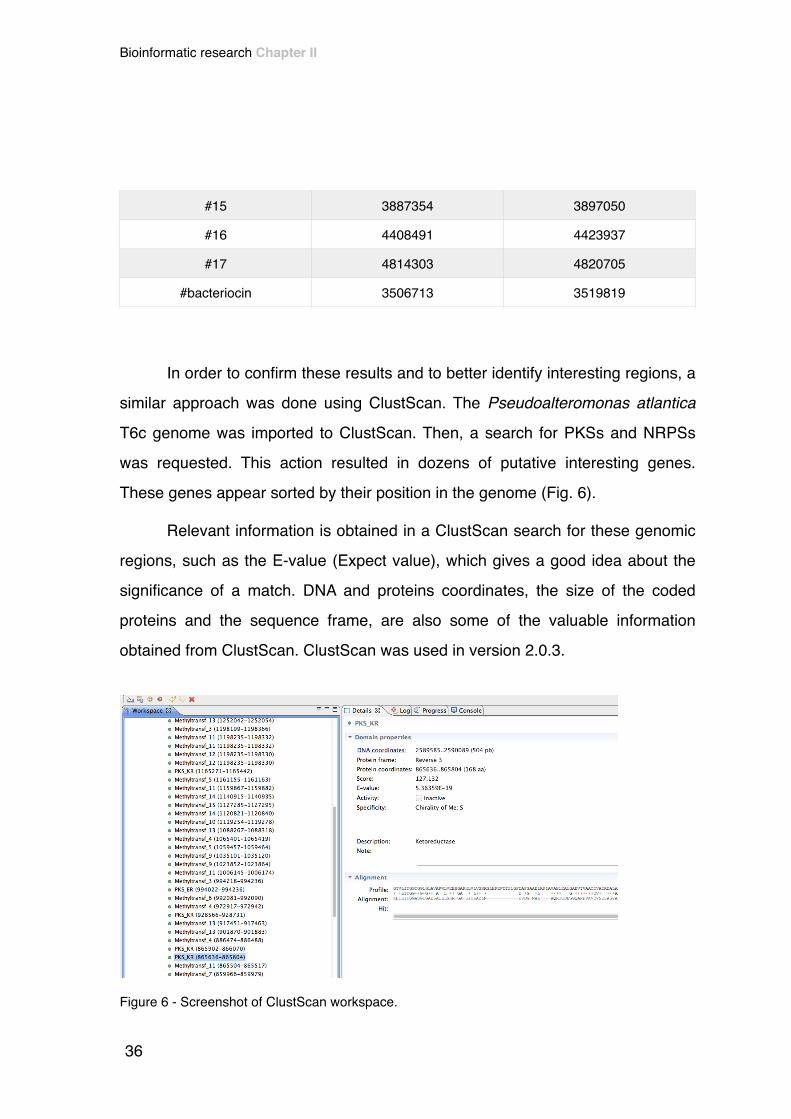
similar approach was done using ClustScan. The Pseudoalteromonas atlantica
T6c genome was imported to ClustScan. Then, a search for PKSs and NRPSs
was requested. This action resulted in dozens of putative interesting genes.
These genes appear sorted by their position in the genome (Fig. 6).!
! Relevant information is obtained in a ClustScan search for these genomic
regions, such as the E-value (Expect value), which gives a good idea about the
significance of a match. DNA and proteins coordinates, the size of the coded
proteins and the sequence frame, are also some of the valuable information
obtained from ClustScan. ClustScan was used in version 2.0.3.!
#15 3887354 3897050
#16 4408491 4423937
#17 4814303 4820705
#bacteriocin 3506713 3519819
Figure 6 - Screenshot of ClustScan workspace.
36
Bioinformatic research Chapter II
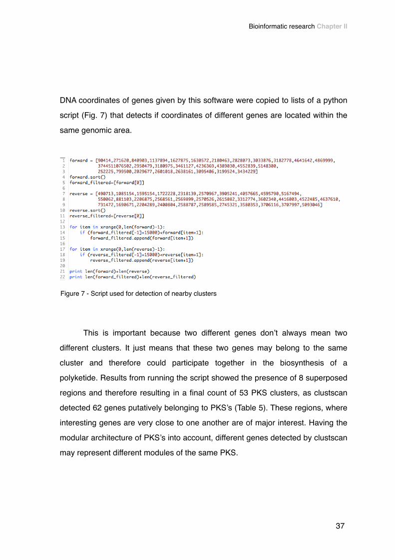
DNA coordinates of genes given by this software were copied to lists of a python
script (Fig. 7) that detects if coordinates of different genes are located within the
same genomic area.!
!! This is important because two different genes don’t always mean two
different clusters. It just means that these two genes may belong to the same
cluster and therefore could participate together in the biosynthesis of a
polyketide. Results from running the script showed the presence of 8 superposed
regions and therefore resulting in a final count of 53 PKS clusters, as clustscan
detected 62 genes putatively belonging to PKS’s (Table 5). These regions, where
interesting genes are very close to one another are of major interest. Having the
modular architecture of PKS’s into account, different genes detected by clustscan
may represent different modules of the same PKS.
Figure 7 - Script used for detection of nearby clusters
37
Bioinformatic research Chapter II
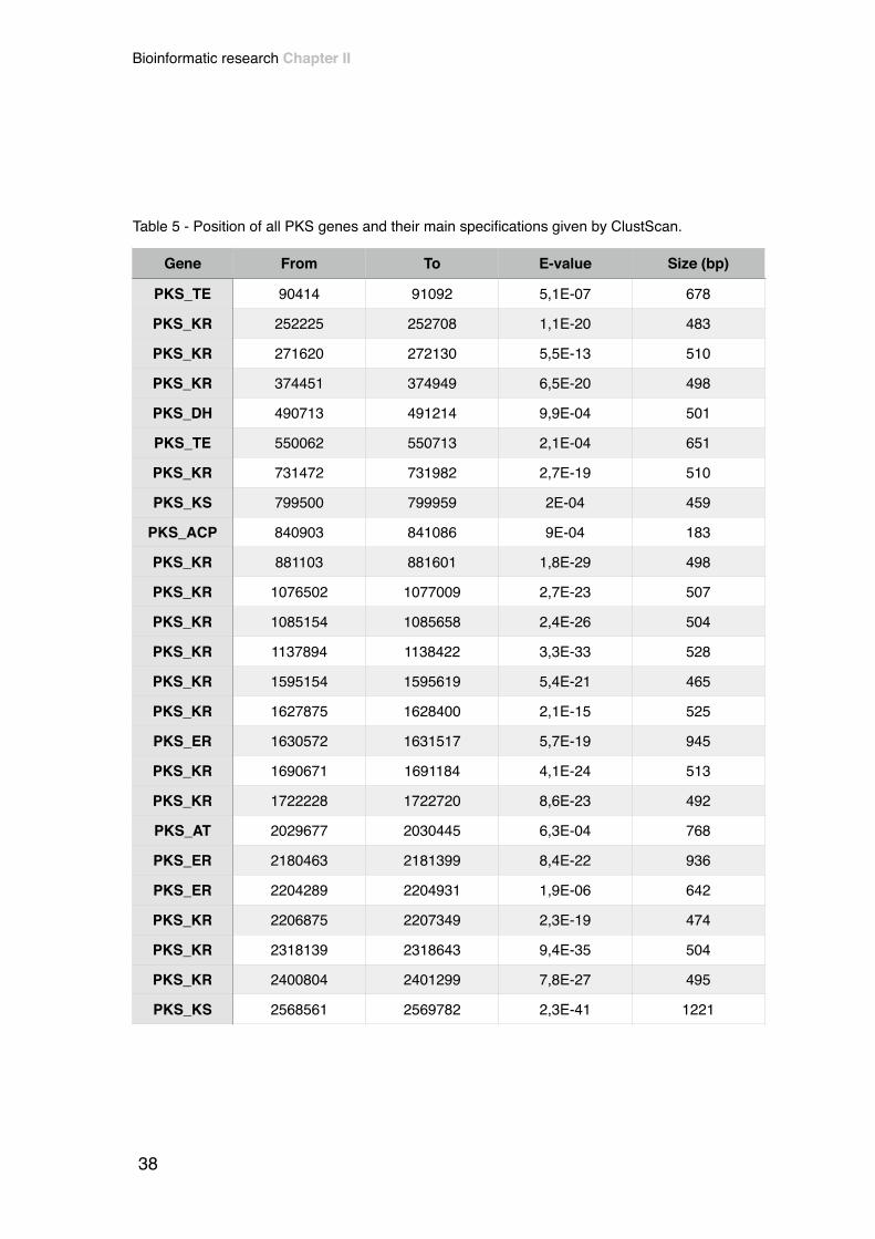
Table 5 - Position of all PKS genes and their main specifications given by ClustScan.
Gene From To E-value Size (bp)
PKS_TE 90414 91092 5,1E-07 678
PKS_KR 252225 252708 1,1E-20 483
PKS_KR 271620 272130 5,5E-13 510
PKS_KR 374451 374949 6,5E-20 498
PKS_DH 490713 491214 9,9E-04 501
PKS_TE 550062 550713 2,1E-04 651
PKS_KR 731472 731982 2,7E-19 510
PKS_KS 799500 799959 2E-04 459
PKS_ACP 840903 841086 9E-04 183
PKS_KR 881103 881601 1,8E-29 498
PKS_KR 1076502 1077009 2,7E-23 507
PKS_KR 1085154 1085658 2,4E-26 504
PKS_KR 1137894 1138422 3,3E-33 528
PKS_KR 1595154 1595619 5,4E-21 465
PKS_KR 1627875 1628400 2,1E-15 525
PKS_ER 1630572 1631517 5,7E-19 945
PKS_KR 1690671 1691184 4,1E-24 513
PKS_KR 1722228 1722720 8,6E-23 492
PKS_AT 2029677 2030445 6,3E-04 768
PKS_ER 2180463 2181399 8,4E-22 936
PKS_ER 2204289 2204931 1,9E-06 642
PKS_KR 2206875 2207349 2,3E-19 474
PKS_KR 2318139 2318643 9,4E-35 504
PKS_KR 2400804 2401299 7,8E-27 495
PKS_KS 2568561 2569782 2,3E-41 1221
Gene
38
Bioinformatic research Chapter II

PKS_ACP 2569899 2570100 5,7E-27 201
PKS_KR 2570526 2571018 4,1E-45 492
PKS_AT 2570967 2571972 5,5E-19 1005
PKS_KR 2588787 2589291 2,3E-33 504
PKS_KR 2589585 2590089 5,4E-39 504
PKS_TE 2601018 2601741 2,4E-05 723
PKS_ER 2615082 2616027 8,5E-20 945
PKS_KR 2638161 2638674 7,5E-23 513
PKS_AT 2745321 2746107 2,6E-04 786
PKS_KR 2828073 2828580 2,6E-32 507
PKS_TE 2950479 2951073 5E-04 594
PKS_KR 3033876 3034380 2E-10 504
PKS_KR 3095406 3095910 3,5E-28 504
PKS_DH 3180975 3181362 1,8E-04 387
PKS_KR 3182778 3183207 1,2E-10 429
PKS_ER 3199524 3200481 9,7E-15 957
PKS_KR 3312774 3313329 6,9E-24 555
PKS_ACP 3434229 3434406 9,5E-04 177
PKS_ER 3461127 3462114 3,9E-16 987
PKS_KR 3580353 3580860 6,4E-36 507
PKS_KR 3602340 3602886 4,6E-10 546
PKS_KR 3706116 3706611 2,9E-33 495
PKS_ACP 3707997 3708192 3,9E-06 195
PKS_ER 3905241 3905994 2,8E-04 753
PKS_KS 4057665 4058862 4,2E-30 1197
PKS_KR 4236369 4236873 1,6E-25 504
PKS_KR 4389030 4389540 6,1E-22 510
From To E-value Size (bp)Gene
39
Bioinformatic research Chapter II

!! From the 17 PKS clusters detected by AntiSmash, 11 were also detected
by ClustScan (Table 6). Due to the fact that these two softwares work in a
different manner (while ClustScan finds genes and allows the user to build
clusters based on that information, AntiSmash provides the entire clusters found
in the genome), the discrepancy between these two results was expected.!
!
PKS_KR 4416003 4416507 1,1E-24 504
PKS_AT 4522485 4523424 7,9E-04 939
PKS_KS 4552839 4553919 1,5E-09 1080
PKS_TE 4595790 4596426 2,7E-05 636
PKS_KR 4637610 4638126 6,6E-28 516
PKS_KS 4641642 4642215 8,4E-04 573
PKS_KR 4869999 4870473 1E-16 474
PKS_TE 5093046 5093772 1E-06 726
PKS_AT 5148300 5149071 5,7E-04 771
PKS_ACP 5167494 5167692 8,3E-04 198
From To E-value Size (bp)Gene
Table 6 - Position of clusters detected in AntiSmash, ClustScan similarity and ClustScan E-value.
# DNA coordinates (From - To) Detected by ClustScan E-Value
1 240399 252932 ✔ 1,1E-20
2 1133011 1138638 ✔ 3,3E-33
3 1306704 1333830 ✘ NA*
4 1591638 1596044 ✔ 5,4E-21
5 1689023 1702894 ✔ 4,1E-24
40
Bioinformatic research Chapter II

!To further evaluate the potential of the selected clusters, it is necessary to
analyze and annotate each ORF within the different clusters. This analysis will
allow to understand which core domains are present in a specific cluster and
which clusters might be functional.!
(See appendix for more detailed information about the ORFs identified for each
cluster.)!
!!
6 1716663 1730405 ✔ 8,6E-23
7 2316179 2333946 ✔ 9,4E-35
8 2561287 2572936 ✔ 4,1E-45
9 2588568 2597466 ✔ 5,4E-39
10 2799741 2809626 ✘ NA
11 3092399 3106725 ✔ 3,5E-28
12 3541734 3557681 ✘ NA
13 3692166 3714982 ✘ 2,9E-33
14 3841002 3874237 ✘ NA
15 3887354 3897050 ✘ NA
16 4408491 4423937 ✔ 1,1E-24
17 4814303 4820705 ✘ NA
*NA - Not available
41
Bioinformatic research Chapter II

3. Clusters annotation and proteins identification!
!! Positions of interesting regions given by AntiSmash were then used to
build a map of the most interesting clusters. Since PKS clusters are usually very
large, ORF’s within a ± 15 000 bp range of the target genes identified by
AntiSmash and ClustScan were annotated. CLC Sequence Viewer (version 6.9)
was used to locate these genes within P. atlantica genome (FIG. 8). To get to the
target location, it is only needed to type the location desired (in bp) desired and
then press the “find” button. This software also shows the direction of
transcription of each ORF, and by using “selection” mode, one can click on the
“arrows” and copy each ORF sequence for further analysis.!
Figure 8 - Screenshot of CLC Sequence Viewer workspace.
42
Bioinformatic research Chapter II

! Sequences of each ORF within the range selected were copied to the
blastx of NCBI. This blast (basic local alignment search tool) allows to “Search
protein database using a translated nucleotide query”. Default parameters with
BLOSUM 62 (BLOcks of Amino Acid SUbstitution Matrix) were used. This lagging
action resulted in several hits or sequences producing significant alignments. Hits
with the best scores were considered for further studying. For example, from
position 2568566 bp to 2569804 bp in P. atlantica genome, there is a 1239 bp
long sequence that codes a 412 aa enzyme which is identified as a 3-oxoacyl-
ACP synthase [Pseudoalteromonas atlantica], also known as Beta-ketoacyl-acyl-
carrier-protein synthase I, typically involved in polyketide synthesis. [19]!
! Besides this synergy between CLC Sequence Viewer and blastx,
AntiSmash is also of great utility when identifying proteins present in each cluster.
By hovering the mouse over the arrows representing each ORF, a translated
protein sequence appears with its identification as shown in a blastp (blast for
proteins) of NCBI (Fig. 9). These annotations were confirmed with NCBI
database.!
!Figure 9 - Screenshot of AntiSmash annotations.
43
Bioinformatic research Chapter II
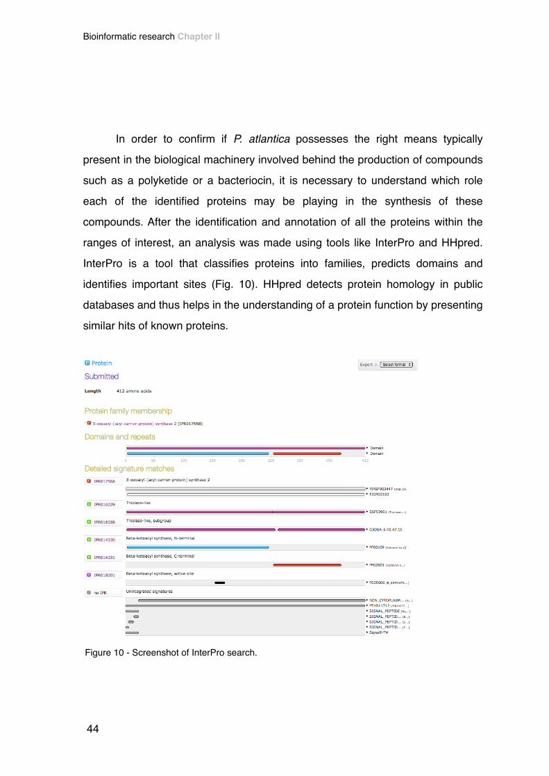
! In order to confirm if P. atlantica possesses the right means typically
present in the biological machinery involved behind the production of compounds
such as a polyketide or a bacteriocin, it is necessary to understand which role
each of the identified proteins may be playing in the synthesis of these
compounds. After the identification and annotation of all the proteins within the
ranges of interest, an analysis was made using tools like InterPro and HHpred.
InterPro is a tool that classifies proteins into families, predicts domains and
identifies important sites (Fig. 10). HHpred detects protein homology in public
databases and thus helps in the understanding of a protein function by presenting
similar hits of known proteins.!
Figure 10 - Screenshot of InterPro search.
44
Bioinformatic research Chapter II

! Biosynthesis of polyketides is catalyzed by a series of enzymes (PKS) that
accomplish sequential decarboxylative condensations and reductive reactions in
order to produce vast polyketide products. PKSs are characterized by having
core domains that contain active sites [35]. These core domains are: AT
(acyltransferase), ACP (acyl carrier protein) and KS (ketosynthase). A starter unit
(acetyl) is loaded by the AT domain onto the KS domain, through a process that
is mediated by ACP. Other domains, like KR (ketoreductase), ER
(enoylreductase), DH (dehydratase) have been characterized as modifying
domains. When present, these domains perform a modification of the initial
carbonyl group, and therefore play a role in the structure of the final product. TE
(thioesterase) domain catalyzes the release of the final product when it reaches
its full length [35].!
! Table 7 shows the most glaring PKS-related ORFs for each annotated
cluster.!
Table 7 - PKS related ORFs detected near AntiSmash detection sites.
Cluster ORF Name Predicted function
#1Patl_0202 3-ketoacyl-CoA thiolase Acyltransferase
Patl_0209 short-chain dehydrogenase/reductase SDR Ketoreductase
#2 Patl_0952 3-ketoacyl-ACP reductase Ketoreductase
#3
Patl_1087 NAD-dependent epimerase/dehydratase Dehydratase
Patl_1093 acyltransferase 3 Acyltransferase
Patl_1095 acyltransferase 3 Acyltransferase
Patl_1096 acyltransferase 3 Acyltransferase
#4 Patl_1329 short-chain dehydrogenase/reductase SDR Ketoreductase
45
Bioinformatic research Chapter II

!The majority of the regions only presents one ORF with a PKS-related function or
domain. A functional region should have ORFs with multiple domains, or multiple
ORFs with PKS-related domains. This situation appears to happen only in one
particular case, from Patl_2120 to Patl_2125.!
!
#5 Patl_1046 3-oxoacyl-ACP reductase Ketoreductase
#6 Patl_1432 short-chain dehydrogenase/reductase SDR Ketoreductase
#7 Patl_1914 short-chain dehydrogenase/reductase SDR Ketoreductase
#8
Patl_2120 beta-ketoacyl synthase Ketosynthase
Patl_2122 3-oxoacyl-(acyl-carrier-protein) reductase Ketoreductase
Patl_2123 malonyl CoA-acyl carrier protein transacylase Acyltransferase
Patl_2125 3-oxoacyl-(acyl-carrier-protein) synthase III Ketosynthase
Patl_2125 fatty acid/phospholipid synthesis protein PlsX Acyltransferase
#9 Patl_2139 short-chain dehydrogenase/reductase SDR Ketoreductase
#11 Patl_2552 short-chain dehydrogenase/reductase SDR Ketoreductase
#12Patl_2923 acetyl-CoA acetyltransferase Acyltransferase
Patl_2935 TesB family acyl-CoA thioesterase Thioesterase
#13 Patl_3071 short-chain dehydrogenase/reductase SDR Ketoreductase
#16 Patl_3661 3-oxoacyl-[acyl-carrier protein] reductase Ketoreductase
#17 Patl_3994 aldo/keto reductase Ketoreductase
Cluster ORF Name Predicted function
46
Bioinformatic research Chapter II

• PKS #8 CLUSTER!
Out of the 17 PKS clusters previously selected (Table 7 and appendix), one was
identified as having all the core domains typically present in a PKS. From position
2568566 to 2574049 in the genome (Fig. 11), there is a transcripted cluster in
reverse direction (complement) with possible two AT domains, one KS domains
and one KR domain.!
!!! The first protein of the cluster is a fatty acid/phospholipid synthesis protein
PlsX (ORF Patl_2125), which has a 332 aa region referred in NCBI as a putative
acyltransferase. Besides the InterPro scan for this protein reports a molecular
function “transferase activity, transferring acyl groups other than amino-acyl
groups”.!
! The second protein in the chain is a beta-ketoacyl-ACP synthase.
Biosynthesis of PKS pressuposes that the ketoacyl and ACP modules work
together in the catalyzation of the chain elongation. [35]!
FIgure 11- Schematic representation of a hypothetical Polyketide Synthase in P. atlantica T6c composed by 4 distinct domains: AT - acyltransferase ; KS - ketosynthase ; KR - ketoreductase
47
Bioinformatic research Chapter II

After the beta-ketoacyl-ACP synthase there is a malonyl CoA-acyl carrier protein
transacylase. InterPro detects an acyltransferase domain and reports that these
domains are involved PKS synthesis, as previous literature stated. [35]!
! The next protein in this cluster is a 3-oxoacyl-(acyl-carrier-protein)
reductase (from NCBI blastx). This protein, also known as beta-Ketoacyl
reductase. Although a small number of ketoreductases has been deeply studied,
and the mechanistic basis of their function is poorly understood, KR domains are
typically present in PKS, as they catalyze reduction of 2- methyl-3-ketoacyl-ACP
substrates. [36]!
! The last protein of this cluster is again a beta-ketoacyl synthase, one of
the core domains of a PKS.!
! This PKS cluster presents relative similarity with clusters in the genome of
other species (Fig. 12). Blastp over proteins of this cluster detected that
Methylomonas methanica, Edwarsiella ictaluri and Xenorhabdus nematophila
presented homology with a sequence set of six ORFs of the entire cluster
detected by AntiSmash.!
! Like in the case of P. atlantica there isn’t (yet) any reported proof that PKS
are naturally produced by these bacteria.!
Figure 12 - Graphic of homologous gene clusters, for PKS #8 cluster - AntiSmash
48
Bioinformatic research Chapter II

• BACTERIOCIN CLUSTER!
! Detected by AntiSmash, this cluster (Fig. 13) is 13 106 bp long, and is
constituted by 12 different proteins, two of them have a biosynthetic function.!
!! The first protein of the cluster has a TonB-dependent receptor, and a
TonB-dependent plug domains, which have a transport-related function.!
! The fourth protein in this cluster is detected by AntiSmash as having a
biosynthetic role in the production of the bacteriocin. Surprisingly, a search
(HHpred, AntiSmash, blastp) for this protein detects a beta-lactamase (typically
antibiotic-resistance related). Nevertheless, BioGraph, a web tool that searches
functional paths between different biomedical entities [37], detects a relation in
the metabolic pathway of bacteriocins and Beta-lactamase-type transpeptidase
fold proteins, just like the one present in this cluster.!
! The sixth protein in the cluster chain, catalogued as a “hypothetical
protein” in NCBI database is identified by InterPro as a Xylose isomerase-like,
TIM barrel domain, with none molecular function predicted. Although AntiSmash
detects this protein as participatory in the biosynthesis of bacteriocins, there is no
Figure 13 - Schematic representation of a hypothetical bacteriocin in P. atlantica T6c.
49
Bioinformatic research Chapter II

evidence in the literature that links this kind of protein with the synthesis of
bacteriocins.!
! Once again, AntiSmash presents an homology graphic. However, for this
bacteriocin cluster, Blastp detected a lower similarity (less ORFs in the same
position) between this cluster and sequences of other species (Fig. 14).!
! Once again, evidences that these bacteria produce bacteriocins are not
available in the literature.!
!!
Figure 14 - Graphic of homologous gene clusters, for bacteriocin cluster - AntiSmash.
50
Bioinformatic research Chapter II

4. Discussion!!! The intense work over the analysis of the data retrieved by bioinformatic
tools found in the literature is really impressive. The use of bioinformatic skills has
led researchers to narrow genomic regions of interest and fully investigate over a
smaller set of clusters.!
! Because Streptomyces is a genus known to be prolific in the production of
secondary metabolites, many have already tried to detect PKS and NRPS
clusters in genomes of different strains. For example, bioinformatic research
using ClustScan and also AntiSmash has also resulted in the finding of 4 modular
PKS clusters and 6 NRPS clusters in Streptomyces tsukubaensis. In this work
over the S. tsukubaensis genome, ClustScan detected 60 putative KS domains,
of which 38 were assigned to the referred clusters [38]. Moreover, genome
analysis of Streptomyces turgidiscabies resulted in the detection of 17 PKS/
NRPS clusters, using different bioinformatic tools (e.g. MiGAP - a microbial
genome annotation software) [39].!
! Regarding Pseudoalteromonas species, the production of novel and
powerful bioactive compounds among Pseudoalteromonas genus has already
been recognized. Previous studies have already identified putative type I PKS or
hybrid PKS/NRPS clusters in other Pseudoalteromonas species [40].!
! Although genome mining generally relies on the identification of gene
clusters of known compounds such as NRPS or PKS, other important molecules
were also found using this technique. For example, works over the genome of
51
Bioinformatic research Chapter II

Pseudoalteromonas tunicata led to the discovery of a 3-Formyl-Tyrosine, via
searching for homologous ATP-grasp enzymes, which may possess antimicrobial
properties [41].!
! AntiSmash has already been used to search clusters of antimicrobial
interest. An AntiSmash analysis of the genome of Pseudoalteromonas
flavipulchra resulted in the finding of four bacteriocin-type gene clusters,
lantipeptide biosynthesis genes, four type I hybrid PKS/NRPS clusters and three
NRPS clusters. Only one type I PKS gene was found in P. flavipulchra [42].!
! Although some of the state of the art techniques, like predicting
compounds structure using bioinformatic tools, were not explored in this work, the
number of antimicrobial clusters detected in P. atlantica T6c seems to be a sign
that the genome of this strain might be prolific in regions responsible for
antimicrobial compounds production [38, 43].!
! From the analysis of the results obtained here by bioinformatic research, it
is presumable that if a PKS is really being translated through P. atlantica T6c
genome, it must be type II PKS. That is because the PKS related proteins found
in the genome, are monofunctional proteins, in contrast with the highly modular
proteins typically present in type I PKSs. Also, an Acyl-Carrier-Protein (ACP) was
found in various clusters, which eliminates the type III PKSs, since they are ACP
independent. In spite of being shortly described, the presence of type II PKS
clusters in marine bacteria has already been confirmed. However, no information
is available for Pseudoalteromonas. BLAST searches over the genome
Streptomyces and Micromonospora strains isolated from the soft coral tissue in
the East China Sea detected the presence of type II PKS clusters [44]. Besides,
52
Bioinformatic research Chapter II

the occurrence of bacteriocin clusters in marine bacteria was already verified, as
several gene clusters were found in many cyanobacteria strains. The
identification of such clusters was done by searching for the best hits for proteins’
sequences using BLASTp and Artemis (a genome browser and annotation tool),
in a similar process used in this work [45]. Bacteriocin gene clusters normally
comprise a region coding for an immunity protein against the bacteriocin itself
[46], such protein was not detected through bioinformatic research in this work.!
! Regarding the most complete PKS cluster found (PKS #8 cluster),
although it possesses the core domains of a PKS, it lacks a TE domain, which is
essential to facilitate the release of the molecule from the enzyme. Nevertheless,
it is possible that a particular domain may be positioned relatively far away (a few
ORFs from the center of the cluster) [38], and thus escaping from the thorough
analysis of that particular region (over the center of the cluster).!
!!
53
Bioinformatic research Chapter II

!!
!
!
!
!
!
!
!
!
Chapter III!Optimization of
antimicrobial compounds production!
!

!

1. Overview!!! Many factors influence the efficiency of antibiotic production by living
organisms. In this work, some culturing conditions were varied in order to
understand their role in the biosynthesis of antimicrobial compounds. A one-
factor-at-a-time method was used, each parameter was tested individually, which
is a very time-consuming and expensive method, if large amount of trials are
required. Nevertheless, it is an easy and simple method and that is why it is
widely used in such experiments. !
! The following parameters were changed in the process of compound
production optimization:!
• culturing medium!
• temperature!
• agitation!
• pH!
• addition of extracellular ATP!
• presence of competitors!
! The optimization of the referred parameters will be addressed in the next
sections.!
57
Optimization of antimicrobial compounds production Chapter III

2. Materials and methods!!
2.1. Strains and culture conditions optimization!
!! P. atlantica T6c (ATCC BAA-1087) was gently provided by Professor Anna
Karls from University of Georgia - Department of Microbiology.!
! In order to evaluate if P. atlantica T6c produced antimicrobial compounds,
and to test their activity spectrum, twelve target species, both reference strains
and isolates, were assayed:!
• Escherichia coli K12!
• Escherichia coli K12 ΔimpA!
• Staphylococcus aureus CECT 229!
• Pseudomonas aeruginosa PA01!
• Pseudomonas fluorescens ATCC 27663!
• Salmonella enteritidis!
• Listeria monocytogenes CECT 4031!
• Enterococcus faecalis V583!
• Bacillus subtilis!
• Bacillus cereus!
Optimization of antimicrobial compounds production Chapter III
58
• Klebsiella pneumoniae VK 089 RIFR!
• Vibrio harveyi!
!! E. coli K12 is a debilitated strain of E. coli commonly used in lab
experimentations, because it does not normally colonize the human intestine [47].
The E. coli K12 ΔimpA is a weaker strain that does not possess the impA gene,
a gene that codes for an inner membrane protein [48] and thus it possesses a
disrupted membrane.!
! All the bacteria isolates were retrieved from cryopreserved cultures. They
were then grown overnight in a LB agar plate at 37º C and maintained at 4º C. P.
fluorescens and B. cereus, which were grown overnight at 25º C.!
!!
2.1.1. Effect of the growth medium!!! In order to mimic the natural sea environment, in which P. atlantica
inhabits, two culturing media related to this niche were assessed: DIFCO™
Marine Broth (MB) and Vatanen Nine Salts Solution (VNSS) [49]. Also, a Minimal
Medium (MM) was tested, as it is common practice to optimize the production of
antimicrobials in such medium, to determine optimal nutritional and culture
conditions [50, 51]. This last medium was tested with four different and separate
carbon sources: glucose, glycerol, lactose or galactose. At this stage, culturing
59
Optimization of antimicrobial compounds production Chapter III

conditions were tried at 25ºC, 120 rpm and a pH of 7. Compositions of lab-made
VNSS and MM media are shown below.!
! VNSS medium composition (all percentages are w/v): 0,1% peptone from
soymeal; 0,05% yeast extract; 0,05% glucose; 0,5% soluble starch; 0,001%
FeSO4·7H2O; 0,001% NasHPO4·2H2O; 1,76% NaCl; 0,147% Na2SO4; 0,008%
NaHCO3; 0,025% KCL; 0,004% KBr; 0,187% MgCl2·6H2O; 0,041% CaCl2·2H2O;
0,001% SrCl2·6H2O and 0,001% H3BO3.!
! MM composition (all percentages are w/v): 0,6% Na2HPO4 0,3% K2HPO4; 0,005% NH4Cl; 0,006% MgSO4; 0,75% Na2HPO4·2H2O; 0,55% C6H12O6·H2O and
0,5% glucose / glycerol / lactose / galactose (only one of these carbon sources)!
!!
2.1.2. Temperature, pH and agitation!!! Once again, in order to mimic the natural habitat conditions of P. atlantica,
and in accordance to the literature [14], temperatures between 20ºC and 30ºC
were tested in a refrigerated incubator (Shel Lab®). These parameters are of
utmost importance in the optimization of the production of antimicrobial
compounds, as they affect organisms and have the ability to induce or inhibit the
production of such compounds [51].!
! For the optimization of pH, MB medium and a temperature of 23ºC were
used. In sea water, pH normally ranges between 8 and 8,5 [52]. Levels of pH
between 5 and 9 were assayed.!
Optimization of antimicrobial compounds production Chapter III
60

! For aeration optimization, MB medium, 23ºC, and a pH of 8 were used.
Agitations of 100 to 150 RPM were then tested. Moreover, the dimensions of the
flask and the media to flask size ratio are also important aspects to have into
account, since they influence the aeration of the culture. Because of this, 250 mL
of media were poured into 1000 mL sterile flasks. Cultures were grown during
120 hours.!
!!
2.1.3. Addition of extracellular ATP!!! As previous studies indicated that the addition of extracellular ATP
enhances the production of antibiotics in Streptomyces coelicolor [53], the same
was tried with P. atlantica. Ten μM (final concentration) of ATP were added to MB
cultures at 23º C, 120 rpm agitation and pH of 8 in order to see if this factor
influenced antimicrobial metabolites production. Cultures were grown during 120
hours.!
!
61
Optimization of antimicrobial compounds production Chapter III

2.1.4. Competitors!!! As the presence of a foreign (competitor) genus, or the metabolites
produced by this competitor could affect antimicrobial metabolites production
[15], three different configurations were assayed, as represented in Fig. 15.!
!• Fig. 15 - A represents the simplest configuration. This configuration consists
in a single culture of P. atlantica T6c with the selected conditions chosen in
the previous steps in optimization phase; A small portion of biomass from a
fresh culture of P. atlantica in marine agar was added aseptically to the
Marine Broth medium. This flask served as a control condition.!
Figure 15 - Representation of the culture conditions used for the competition assays.
Optimization of antimicrobial compounds production Chapter III
62

• Fig. 15 - B represents a co-culture of P. atlantica T6c and E. coli K12. Three
different sub-configurations (1:1, 10:1 and 100:1 ratios of P. atlantica/E.coli)
were tested; One hundred µL from a pre-inoculum of P. atlantica grown
overnight in Marine Broth were added to three eppendorfs cointaining 900
µL of phosphate buffered saline (PBS). After vortexing, the mix was poured
onto the 1000 mL culture flasks containing 250 mL of Marine Broth. One
hundred µL of a E. coli K12 pre-inoculum grown overnight in LB were added
to an eppendorf containing 900 µL of PBS and three ten-fold dilutions were
made and poured into the three different culture flasks.!
!• Fig. 15 - C represents a configuration in which a dialysis membrane (with a
molecular weight cutoff of 14 000 Daltons) with a 2 mL culture of the
competitor genus was put inside the culture flask. Four different sub-
configurations were tested (variation of foreign genus inside membrane): E.
coli K12, P. aeruginosa, S. aureus, V. harveyi; Ten centimeters of a dialysis
membrane were cut using sterile scissors. One tip of this small portion was
secured with a sterile clamp. Two milliliters of the foreign genus grown
overnight at the respective conditions were added to the membrane with the
help of a micropipette. The other tip was then secured with another sterile
clamp and the apparatus was seamlessly put inside the large culture flask
(as shown in Fig. 15 - C).!
!! Cultures were incubated during 120 hours.
63
Optimization of antimicrobial compounds production Chapter III

2.2. Assessment of antimicrobial activity!
!! In order to measure the antimicrobial activity of the metabolites produced
by P. atlantica, each 24 hours until a maximum of 120 hours of incubation, 10 mL
of the culture were retrieved from the flasks under aseptic conditions. The spent
media was then centrifuged at 15 000 RPM (refrigerated Sigma™ 3-16K) so that
cell particles could deposit in the bottom of a falcon tube. The supernatant was
then filtrated using a syringe filter with a 0,22 µm pore size (sterile Q-MAX®
Syringe Filter from Frisenette) to further eliminate the presence of cells (obtaining
bacteria-free spent media).!
! Target strains were grown overnight in LB. One hundred µL of the target
cultures were shed into a Petri dish containing a thin layer of the respective
media with solid agar. 4 mL of soft agar (0,5% agar) at 50º C were thereafter
shed over the 100 µL culture. A slight shake of the Petri dish was made in order
to improve an equitable distribution over the dish. This mix was allowed air dry for
about 30 minutes. After that, 10 µL of the bacteria-free spent media and, when
applicable, several dilutions were spotted into these bacterial lawns. These plates
were incubated for 24h at 37ºC. Their ability to form an inhibition halo was
recorded.!
! For experiments over medium, temperature, agitation and pH optimization,
the antimicrobial activity was only assayed on E. coli K12 lawns. Only after the
Optimization of antimicrobial compounds production Chapter III
64

optimization of the referred parameters, the entire array of strains was used to
evaluate the compound activity spectrum.
65
Optimization of antimicrobial compounds production Chapter III

2.3. Determination of arbitrary units!!! Arbitrary Units (AU) are a simple way of quantifying the ability of a
compound to inhibit the growth of a target culture. They measure the strength of
a compound. Arbitrary units are measured having the premise that the last
dilution presenting inhibition has 1 compound unit. For example, if the last dilution
presenting inhibition was a dilution of 1/16, then that sample would have 16
arbitrary units per mL [54].!
!!
Optimization of antimicrobial compounds production Chapter III
66

3. Results and discussion!!
3.1. Effect of culture media on cell growth and antimicrobial activity!
!! As culturing conditions may affect the antimicrobial capacity of
Pseudoalteromonas, a set of default parameters found in literature was followed
[55], varying the culture media to see what effect in cell growth and antimicrobial
activity was being induced. Marine broth and the complex marine medium VNSS
are culture media for the marine organisms and both are already linked to the
successful production of bioactive secondary metabolites produced by
Pseudoalteromonas. The Minimal Medium is a basic culturing medium usually
used as a control in order to assay if a given nutrient is necessary for the growth
of the culture or the production of a metabolite. [49, 56, 57]!
! P. atlantica showed no significant growth in MM medium in any of the
chosen carbon sources, meaning that P. atlantica may be a demanding bacteria
that requires some of the nutrients or amino-acids that are not present in such a
poor medium. The standard approach is to culture in a Marine Minimal Medium
(which contains a set micro and macronutrients required by the marine microbial
consortia) [58], however, such medium was not tried. Otherwise, bacterial growth
was observed in VNSS and MB media, which confirms that the richer marine
67
Optimization of antimicrobial compounds production Chapter III

related media have a positive effect on P. atlantica growth. In fact, the use of
Marine Broth and VNSS has already been related to successful growth of other
strains of the Pseudoalteromonas genus [16, 59].!
! Table 8 summarizes the effect of chosen media in the bacterial growth and
in the production of inhibitory compounds. As expected, the lack of growth
verified with MM (with any of the carbon sources) led to an absence of effect on
target. The strain tested (E. coli K12) grew over the drops of bacteria-free spent
media of MM flasks. Not so expected, metabolites present in VNSS medium
flasks were found innocuous, as an evident growth of P. atlantica was observed,
but no harm for target variants was observed. In fact, the use of VNSS medium
has already been linked to a successful production of antifouling agents [59], but
any proof of the use of VNSS inducing the production of antimicrobial compounds
in Pseudoalteromonas was found in the literature.!
!
!
Table 8 - Effect of culture media in P. atlantica growth and the production of inhibitory compounds.
Media Growth Effect on target (E. coli K12)
MM - glucose - -MM - glycerol - -MM - lactose - -
MM - galactose - -VNSS + -
MB + +
- No growth or no inhibition; + Growth or inhibition
Optimization of antimicrobial compounds production Chapter III
68

! On the other hand, bacteria-free spent media from MB flasks proved to
inhibit the growth of target (E. coli K12) (FIG. 15), as clear inhibition halos were
recorded. Marine Broth proved to be a medium where a notorious growth of P.
atlantica was observed, with the formation of a thick biofilm on the walls of the
flasks. From the three media tested, Marine Broth was the one where an
antimicrobial activity was recorded. Hence, Marine Broth was the chosen medium
for further assays. Other authors have recognized that the use of Marine Broth
leads to a successful production of a potent antibiotic in Pseudoalteromonas
piscicida, a compound that attacks the cell wall of MRSA pathogens [60]. Also, it
was reported that a strain of Pseudoalteromonas closely related to
Pseudoalteromonas rubra produces antimicrobial compounds of unknown
structure with a broad spectral activity, when cultured in MB [61].!
!
Figure 16 - Inhibition halos in an E.coli K12 lawn.
69
Optimization of antimicrobial compounds production Chapter III

3.2. Effect of temperature, pH and agitation on cell growth and antimicrobial activity!
!! The right set of culturing parameters can lead to a better production of
some secondary metabolites. Parameters such as temperature, pH and agitation
are of utmost importance when one tries to optimize the production of
antimicrobial compounds. Living organisms have long proven to react to these
exterior factors and thus they influence the production of such compounds [60,
62].!
! To test the effect of temperature, agitation and pH on cell growth, each
parameter was tested individually. First, cultures of P. atlantica in Marine Broth
were submitted to five different temperatures (20, 23, 25, 27, and 30ºC) (Table 9)
at the standard pH medium (around 7,5).!
Table 9 - Effect of culture media in the production of inhibitory compounds in P. atlantica.
Temperature Inhibition
20ºC +
23ºC ++
25ºC ++
27ºC +
30ºC +
+ Slight inhibition; ++ Good inhibition
Optimization of antimicrobial compounds production Chapter III
70

! Antimicrobial activity was stronger for 23ºC and 25ºC, but a slight
advantage was observed for the 23 ºC incubation, since a more vehement
inhibition halo was recorded in the E. coli K12 lawns. Once again, the 23 ºC
incubation temperature was elected for further analysis.!
! To test the pH effect, 5 different pHs were assayed in Marine Broth,
ranging from pH 5 to 9 (Table 10). A clear advantage was attributed to the pH 8
(which is close to normal sea water levels) [52], since clearer inhibition halos
were recorded. Previous works have already found a pH of around 7.5 to be ideal
for the production of a target metabolite (a novel alginate-lyase) from
Pseudoalteromonas. [63] Also, a pH of 7,5 was also found ideal for the
production of antibiotics of unknown structure (not proteinaceus) by
Pseudoalteromonas piscicida. The best conditions were also achieved in MB, but
at a temperature of 37ºC [60].!
!
!
Table 10 - Effect of pH on the production of inhibitory compounds in P. atlantica.
pH Inhibition
5 -*
6 +
7 +
8 ++
9 -
-* No growth; + Slight inhibition ; ++ Good inhibition
71
Optimization of antimicrobial compounds production Chapter III

! The same process was done to find which agitation provided the highest
antimicrobial activity. Agitations varying from 80 rpm to 170 rpm (80, 100, 120,
150 and 170 rpm) were assayed (Table 11). Because there was no difference in
antimicrobial activity nor bacterial growth, a 120 RPM agitation was chosen,
according to previous works on Pseudoalteromonas, where rotations of about
120 rpm proved to be ideal for the production of secondary metabolites [55, 60].!
!
!! Also, previous experiments on how oxygen limitation influences the
production of antimicrobial metabolites produced by Pseudoalteromonas,
suggested that the antimicrobial activity is at a maximum for agitations up to 150
RPM. Antimicrobial activity was proved to be negligible for agitations greater than
250 RPM, which suggests that the RPM tried in this work were within the
working window of agitations [64].!
Table 11 - Effect of agitation on the production of inhibitory compounds in P. atlantica.
Agitation (rpm) Inhibition
80 ++
100 ++
120 ++
150 ++
170 ++
+ Slight inhibition ; ++ Good inhibition
Optimization of antimicrobial compounds production Chapter III
72

! Antimicrobial assays on lawns of E. coli K12 (Fig 17 - B) for the best
culture conditions were made in order to find the best time of incubation. (Fig. 17
- A) shows the arbitrary units (a compound strength measure) for antimicrobial
assays on E. coli K12 of the bacteria-free spent media, from cultures of Marine
Broth, at 23 ºC, 120 RPM and pH 8.!
! It is easily seen that the compound strength increases over time until 96
hours.!
!! Besides the positive tests on both E. coli strains, the other targets were not
harmed by this bacteria-free spent media (Table 12).!
Arbi
trary
Uni
ts p
er m
L
0
17,5
35
52,5
70
Time in Hours0 24 48 72 96 120
Figure 17 - Inhibitory effect of the filtered sterilized spent MB of P. atlantica in E. coli K12 lawns over time (A) - Arbitrary units over time (B) - Inhibition halos on E. coli K12 lawns, recorded after several dilutions of the spent medium.
A B
73
Optimization of antimicrobial compounds production Chapter III

! Further tests on the spectrum of activity of the antimicrobial compounds
were performed on a set of gram-positive and gram-negative bacteria. The fact
that only E. coli strains were affected by bacteria-free spent media, may be due
to the fact that these two strains are weak variants of E. coli and thus are more
easily affected. Besides, only direct samples of bacteria-free spent media were
assayed. The concentration of the samples, for example via solvent extraction,
could lead to a stronger and eventually broader inhibitory activity.!
!Table 12 - Inhibition of targets by bacteria-free spent MB for optimized culture conditions.
Strains Inhibition
E. coli K12 ++
E. coli K12 ΔimpA +
S. aureus -
P. aeruginosa -
P. fluorescens -
S. enteritidis -
L. monocytogenes -
E. faecalis -
B. subtilis -
B. cereus -
K. pneumoniae -
V. harveyi -
+ Slight inhibition ; ++ Good inhibition; - No inhibition
Optimization of antimicrobial compounds production Chapter III
74

3.3. Effect of addition of extracellular ATP!
!! Evidences that the addition of extracellular ATP could promote the
production of antimicrobial metabolites in Pseudoalteromonas are not available in
the literature. However, this approach was experienced in Streptomyces
coelicolor and resulted in an improved production of the known polyketide
antibiotic actinorhodin [53]. The same approach was tried in this work.!
! The addition of extracellular ATP did not influence nor potentiate the
production of antimicrobial compounds, since halos from cultures with
extracellular ATP were not clearer than those from cultures without the ATP.
Hence, the addition of extracellular ATP was ignored for further assays.
75
Optimization of antimicrobial compounds production Chapter III

3.4. Effect of competitors!!! The battle for resources in a “tight economy” only leaves space for the
strongest. It is known that the presence of competition induces the production of
secondary metabolites such as antibiotics in some bacteria [65, 66]. It is then
important to figure out if competition also affects the production of antimicrobial
compounds in Pseudoalteromonas atlantica.!
• Co-cultures of P. atlantica and E. coli K12!
! Co-cultures of P. atlantica and E. coli K12 in Marine Broth at 23 ºC, 120
RPM and pH 8 were tested for three different ratios (1:1, 10:1, 100:1) of P.
atlantica : E.coli K12. Although cell growth was observed for the three
configurations, antimicrobial activity was not registered. Bacteria-free spent
media was spotted over lawns of all target variants (E. coli, S. aureus, P.
fluorescens, P. aeruginosa, S. enteritidis, L. monocytogenes, E. faecalis, B.
subtilis, B. cereus, K. pneuomoniae and V. harveyi), but in any of the plates an
inhibition halo was perceptible nor evident. These results may be due to the fact
that the used competitors may overgrow and therefore inhibit the growth of P.
atlantica and thus there isn’t production of antimicrobial compounds.!
!• Cultures of P. atlantica and E. coli K12 / V. harveyi / P. aeruginosa / S.
aureus in dialysis membrane!
! To avoid the overgrowth of the competing species, a new strategy was
used to confine and limit the growth of the competitors.!
Optimization of antimicrobial compounds production Chapter III
76

! Competing species were placed inside of a dialysis membrane. They were
then placed in a culture flask of P. atlantica with Marine Broth at 23 ºC, 120 RPM
and pH 8. Significant cell growth was observed for all the flasks/competing
species. The antimicrobial assays showed a great similarity for all the
configurations, since all proved to inhibit the growth of E. coli K12 ΔimpA and S.
enteritidis (Table 13) for an incubation time superior to 72 hours. These results
suggest that the metabolites produced by the competing species, which manage
to cross the dialysis membrane due to their reduced size, could influence or
induce the production of antimicrobial metabolites by P. atlantica.!
Table 13 - Effect of competing species (E. coli K12, S.aureus, P. aeruginosa, V. harveyi) in dialysis membrane on production of antimicrobial compounds by P. atlantica.
Competing species E. coli K12 S. aureus P. aeruginosa V. harveyi
Targets
E. coli K12 - - - -
E. coli K12 Δ imp A + + + +
S. aureus - - - -
P. aeruginosa - - - -
P. fluorescens - - - -
S. enteritidis + + + +
L. monocytogenes - - - -
E. faecalis - - - -
B. subtilis - - - -
B. cereus - - - -
K. pneumoniae - - - -
V. harveyi - - - -
77
Optimization of antimicrobial compounds production Chapter III

! Because the first configuration (culture without competing species)
managed to inhibit the growth of E. coli K12 and E. coli K12 ΔimpA, while the
dialysis membrane configuration inhibited the growth of E. coli K12 ΔimpA and S.
enteritidis, it is presumable that a different antimicrobial compound could be
synthesized under different culture conditions. However, it was impossible to infer
which metabolic switch might have been involved in the production of a different
compound, and which compound is being produced under each particular
conditions. Further chemical techniques would be necessary to isolate and
characterize the compounds.!
!
Optimization of antimicrobial compounds production Chapter III
78

!

!

!
!
!
!
!
!
!
!
Chapter IV!General conclusions and
future work!!

!
1. General conclusions and future work!
!! The use of microorganisms as a source of powerful antibiotics has been
recognized as a successful approach [2]. The search for species capable of
producing metabolites with antibiotic activity has called the attention of many
towards the marine environment [14, 15, 26]. The Pseudoalteromonas genus is
known to colonize a vast range of habitats, which may be due to its capacity of
producing powerful bioactive compounds that enable strains of this genus to
conquer others’ habitats. The high diversity of strains among this genus and the
chemical diversity of the compounds already identified make the
Pseudoalteromonas genus a very interesting target for the discovery of novel
molecules [14, 15].!
! In this work, bioinformatic tools were used to detect regions of the P.
atlantica genome with clusters of antimicrobial compounds, to further investigate
these regions, detect ORFs and identify proteins domains and functions.
Research over the genome of P. atlantica resulted in the finding of many clusters
of PKSs. Although they all seem to miss one or more domains of a typical
complete PKS, some of them could be working together in the synthesis of a
single polyketide. Also, a PKS cluster possessing the core domains of a PKS was
found and its ORFs were described. This cluster may in fact be an active
producer of a polyketide. Besides the PKS clusters, a bacteriocin cluster was
found. It is possible that these two very different metabolites could be
General conclusions and future work Chapter IV
83

synthesized by P. atlantica under different culturing conditions, and thus explain,
at some extent, some of the results obtained in laboratory testing. Nevertheless,
at this point, there isn’t any proof that supports such argument, since we have not
further investigated the nature of the compounds.!
! Pseudoalteromonas atlantica is a marine bacterium that proved to inhibit
the growth of other bacteria, under the right set of culturing parameters. While the
VNSS or MM media have not induced the production of antimicrobial molecules,
the MB has provided a clear inhibition of E. coli strains. Optimal conditions for
antimicrobial production were found for cultures in Marine Broth, at a temperature
of 23ºC and a pH of 8, at an agitation of 120 rpm). Also, P. atlantica showed
different inhibition patterns in the presence of competing species, which might
suggest that different conditions induce the production of different molecules.!
! It is possible to continue the work over P. atlantica antibacterial
metabolites. A deeper investigation over the referred clusters could lead to the
finding of more PKS domains working together in the assembly of a polyketide.
The interruption of the identified clusters could be done to evaluate their
involvement in the production of the detected antimicrobials. The purification, for
instance by High-Performance Liquid Chromatography (HPLC), of the bacteria-
free spent media would be a step forward in the definition of the antibacterial
strength of the referred metabolites. Finally, if the purified molecule presented a
potent effect, the work could move towards the characterization of the molecule
structure.!
!
General conclusions and future work Chapter IV
84

!

!

!
!
!
!
!
!
!
!
Chapter V!References and appendix!!

!

1. References!!
1.! Fleming, A., On the Antibacterial Action of Cultures of a Penicillium, with Special Reference to Their Use in the Isolation of B. influenzae. British Journal of Experimental Pathology, 1929. 10: p. 226-236.!
2.! Davies, J. and D. Davies, Origins and Evolution of Antibiotic Resistance. 2010. 74(3): p. 417-433.!
3.! Davies, J., Vicious Circles: Looking Back on Resistance Plasmids. Genetics, 1995: p. 1465-1468.!
4.! Levy, S.B. and B. Marshall, Antibacterial resistance worldwide: causes, challenges and responses. nature medicine, 2004. 10(12): p. 122-129.!
5.! Kohanski, M.A., D.J. Dwyer, and J.J. Collins, How antibiotics kill bacteria: from targets to networks. Nature Reviews Microbiology, 2010. 8: p. 423-435.!
6.! Alberts, B., et al., From RNA to Protein. Molecular Biology of the Cell. Vol. 4. 2002.!
7.! Bermingham, A. and J.P. Derrick, The folic acid biosynthesis pathway in bacteria: evaluation of potential for antibacterial drug discovery. BioEssays, 2002. 24(7): p. 637-648.!
8.! Nikaido, H., Multidrug Resistance in Bacteria. Annual Review of Biochemistry, 2009. 78(1): p. 119-146.!
9.! erichon, B.P. and P. Courvalin, VanA-Type Vancomycin-Resistant Staphylococcus aureus. ANTIMICROBIAL AGENTS AND CHEMOTHERAPY, 2009. 53(11).!
10.! Shlaes, D.M., S.J. Projan, and J.E. Edwards, Antibiotic Discovery: State of the State. ASM News, 2004. 70: p. 270-280.!
References and appendix Chapter V
89

11.! Organization, W.H., Antimicrobial Resistance: Global Report on Survaillance. 2014.!
12.! Hayashi, M.A.F., F.C. Bizerra, and P.I. Da Silva Junior, Antimicrobial compounds from natural sources. frontiers in MICROBIOLOGY, 2013. 4.!
13.! Clardy, J., M. Fischbach, and C. Walsh, New antibiotics from bacterial natural products. Nat Biotech, 2006. 24(12): p. 1541-1550.!
14.! Vynne, N.G., Bioactivity and phylogeny of the marine bacterial genus Pseudoalteromonas, in National Food Institute!
Division of Industrial Food Research. 2011, Technical University of Denmark.!
15.! Bowman, J.P., Bioactive Compound Synthetic Capacity and Ecological Significance of Marine Bacterial Genus Pseudoalteromonas. Marine Drugs, 2007: p. 22.!
16.! Vynne, N.G., Bioactivity and phylogeny of the marine bacterial genus Pseudoalteromonas, in National Food Institute Division of Industrial Food Research. 2011, Technical University of Denmark.!
17.! Piasecki, S.K., Investigations into the biocatalytic potential of modular polyketide synthase ketoreductases. 2013, University of Texas.!
18.! Pfeifer, B. and C. Khosla, MICROBIOLOGY AND MOLECULAR BIOLOGY REVIEWS : Biosynthesis of Polyketides in Heterologous Hosts. Vol. 65. 2001.!
19.! Chan, Y., et al., Biosynthesis of Polyketide Synthase Extender Units. National Institute of Health, 2009.!
20.! Marinho, A.M.R., et al., Active polyketides isolated from Penicillium herquei. Anais da Academia Brasileira de Ciências, 2013. 85: p. 909-912.!
21.! Arias, A.A.e., M. Craig, and P. Fickers, Science against microbial pathogens: communicating current research!
and technological advances (Gram-positive antibiotic biosynthetic clusters: a review). Vol. 1. 2011.!
References and appendix Chapter V
90

22.! Marahiel, M.A., Working outside the protein-synthesis rules: insights into non-ribosomal peptide synthesis. Journal of Peptide Science, 2009. 15(12): p. 799–807.!
23.! Sundlov, J.A., et al., Structural and functional investigation of the intermolecular interaction between NRPS adenylation and carrier protein domains. Chem Biol., 2013.!
24.! Caboche, S., et al., Diversity of Monomers in Nonribosomal Peptides: towards the Prediction of Origin and Biological Activity. JOURNAL OF BACTERIOLOGY, 2010. 192(19): p. 5143–5150.!
25.! Cotter, P., C. Hill, and P. Ross, Bacteriocins: developing innate immunity for food. Nature Reviews Microbiology, 2005. 3.!
26.! Bakkal, S., S. Robinson, and M. Riley, Bacteriocins of Aquatic Microorganisms and Their Potential Applications in the Seafood Industry. 2012.!
27.! Cotter, P., P. Ross, and C. Hill, Bacteriocins - a viable alternative to antibiotics? Nature Reviews Microbiology, 2013. 11.!
28.! Lux, T., et al., Diversity of Bacteriocins and Activity Spectrum in Streptococcus pneumoniae. Journal of Bacteriology, 2007. 189(21).!
29.! Klaenhammer, T.R., Genetics of bacteriocins produced by lactic acid bacteria. FEMS Microbiology Reviews, 1993. 12: p. 39–85.!
30.! Biology, N.R.C.U.B.o., Bioinformatics: Converting Data to Knowledge: Workshop Summary. 2000.!
31.! Scheffler, R.J., et al., Antimicrobials, drug discovery, and genome mining. Appl Microbiol Biotechnol, 2012.!
32.! Eddy, S.R., Profile Hidden Markov Models. Bioinformatics Review, 1998. 14(9): p. 755-763.!
33.! Blin, K., et al., antiSMASH 2.0--a versatile platform for genome mining of secondary metabolite producers. Nucleic Acids Res, 2013. 41(Web Server issue): p. W204-12.!
91
References and appendix Chapter V

34.! Starcevic, A., et al., ClustScan: an integrated program package for the semi- automatic annotation of modular biosynthetic gene clusters and in silico prediction of novel chemical structures. Nucleic Acids Res, 2008. 36(21): p. 6882-92.!
35.! Yadav, G., R. Gokhale, and D. Mohanty, SEARCHPKS: a program for detection and analysis of polyketide synthase domains. Nucleic Acids Research, 2003. 31(13): p. 3654–3658.!
36.! You, Y.-O., C. Khosla, and D.E. Cane, Stereochemistry of Reductions Catalyzed by Methyl-Epimerizing Ketoreductase Domains of Polyketide Synthases. J Am Chem Soc, 2013.!
37.! Liekens, A.M., et al., BioGraph: unsupervised biomedical knowledge discovery via automated hypothesis generation. Genome Biology, 2011.!
38.! Blažicˇ, M., et al., Annotation of the Modular Polyketide Synthase and Nonribosomal Peptide Synthetase Gene Clusters in the Genome of Streptomyces tsukubaensis NRRL18488. Applied and Environmental Microbiology, 2012. 78(23): p. 8183–8190.!
39.! Komaki, H., et al., Genome-wide survey of polyketide synthase and nonribosomal peptide synthetase gene clusters in Streptomyces turgidiscabies NBRC 16081. The Journal of General and Applied Microbiology, 2012. 58: p. 363‒372.!
40.! Zhu, P., et al., Molecular Phylogeny and Modular Structure of Hybrid NRPS/ PKS Gene Fragment of Pseudoalteromonas sp. NJ6-3-2 Isolated From Marine Sponge Hymeniacidon perleve. Journal of Microbiology and Biotechnology, 2009.!
41.! Blasiak, L.C. and J. Clardy, Discovery of 3-Formyl-Tyrosine Metabolites from Pseudoalteromonas tunicata through Heterologous Expression. American Chemical Society, 2009: p. 926-927.!
42.! Yu, M., et al., Genome analysis of Pseudoalteromonas flavipulchra JG1 reveals various survival advantages in marine environment. BMC Genomics, 2013.!
References and appendix Chapter V
92

43.! Challis, G.L., Mining microbial genomes for new natural products and biosynthetic pathways. Microbiology, 2008. 154: p. 1555–1569.!
44.! Sun, W., et al., Functional Gene-Guided Discovery of Type II Polyketides from Culturable Actinomycetes Associated with Soft Coral Scleronephthya sp. PLoS ONE, 2012. 7(8).!
45.! Wang, H., D.P. Fewer, and K. Sivonen, Genome Mining Demonstrates the Widespread Occurrence of Gene Clusters Encoding Bacteriocins in Cyanobacteria. PLoS ONE, 2011. 6(7).!
46.! Desriac, F., et al., Bacteriocin as Weapons in the Marine Animal-Associated Bacteria Warfare: Inventory and Potential Applications as an Aquaculture Probiotic. Marine Drugs, 2010. 8: p. 1153-1177.!
47.! Escherichia coli K-12 Derivatives Final Risk Assessment. 2012 [cited 2014; Available from: http://epa.gov/biotech_rule/pubs/fra/fra004.htm.!
48.! MINTZ, K.P. and P.M. FIVES-TAYLOR, impA, a Gene Coding for an Inner Membrane Protein, Influences Colonial Morphology of Actinobacillus actinomycetemcomitans. INFECTION AND IMMUNITY, 2000. 68(12): p. 6580–6586.!
49.! Rao, D., J.S. Webb, and S. Kjelleberg, Competitive Interactions in Mixed- Species Biofilms Containing the Marine Bacterium Pseudoalteromonas tunicata. APPLIED AND ENVIRONMENTAL MICROBIOLOGY, 2005. 71(4).!
50.! Neetia, D., et al., Optimization of the Production of the Lantibiotic Mutacin 1140 in Minimal Media. Process Biochem, 2011.!
51.! Jafarzade, M., et al., Influence of Culture Conditions and Medium Composition on theProduction of Antibacterial Compounds by Marine Serratia sp.WPRA3. Journal of Microbiology, 2013.!
52.! Kulthanan, K., P. Nuchkull, and S. Varothai, The pH of water from various sources: an overview for recommendation for patients with atopic dermatitis. Asia Pacific Association of Allergy, Asthma and Clinical Immunology, 2013.!
93
References and appendix Chapter V

53.! Li, M., et al., Effects of extracellular ATP on the physiology of Streptomyces coelicolor A3(2). Federation of European Microbiological Societies, 2008.!
54.! GOEBEL, W., G. BARRY, and T. SHEDLOVSKY, The Production of Colicin K in Media Maintained at Constant pH. The Rockefeller Institute for Medical Research, 1956.!
55.! Jung, C., et al., Optimization of Pseudoalteromonas sp. JYBCL 1 Culture Conditions, Medium Composition and Extracellular β-agarase Activity. Biotechnology and Bioprocess Engineering, 2012.!
56.! Lodish, H., et al., Molecular Cell Biology. 2000.!
57.! Fehér, D., et al., Highly Brominated Antimicrobial Metabolites from a Marine Pseudoalteromonas sp. Journal of Natural Products, 2010. 73(11).!
58.! Kim, S.K., Marine Cosmeceuticals: Trends and Prospects. 2011: Taylor & Francis.!
59.! Dheilly, A., et al., Antibiofilm Activity of the Marine Bacterium Pseudoalteromonas sp. Strain 3J6. APPLIED AND ENVIRONMENTAL MICROBIOLOGY, 2010. 76(11): p. 3452–3461.!
60.! Darabpour, E., et al., Isolation of a potent antibiotic producer bacterium, especially against MRSA, from northern region of the Persian Gulf. Bosnian Journal of Basic Medical Sciences, 2012: p. 108-121.!
61.! Chellarama, C., et al., Bioactive Peptides from Epibiotic Pseudoalteromonas Strain P1. APCBEE Procedia, 2012. 2: p. 37-42.!
62.! Song, Q., Y. Huang, and H. Yang, Optimization of Fermentation Conditions for Antibiotic Production by Actinomycetes YJ1 Strain against Sclerotinia sclerotiorum. Journal of Agricultural Science, 2012. 4(7).!
63.! Ma, Y., et al., Culture optimization and characterization of an alginate-lyase from Pseudoalteromonas sp. LJ1. 2009.!
64.! López, R., et al., OXYGEN LIMITATION FAVORS THE PRODUCTION OF PROTEIN WITH ANTIMICROBIAL ACTIVITY IN
References and appendix Chapter V
94

PSEUDOALTEROMONAS SP. Brazilian Journal of Microbiology, 2012: p. 1206-1212.!
65.! Slattery, M., I. Rajbhandari, and K. Wesson, Competition-Mediated Antibiotic Induction in the Marine Bacterium Streptomyces tenjimariensis. Microbial Ecology, 2001. 41: p. 90-96.!
66.! Cornforth, D.M. and K.R. Foster, Competition sensing: the social side of bacterial stress responses. Nature Reviews Microbiology, 2013. 11.!
67.! Jung, C., et al., Optimization of Pseudoalteromonas sp. JYBCL 1 Culture Conditions, Medium Composition and Extracellular β-agarase Activity!
. Biotechnology and Bioprocess Engineering, 2012.!
!
!!!
95
References and appendix Chapter V

2. Appendix!!
#1
ORFs Name ⇆ Ident % e-value From To Size Dom
ains
1 peptidase S9, prolyl oligopeptidase active site region ← 100 0,00E+00 231294 233234 1941
2 hypothetical protein Patl_0195 ← 100 0,00E+00 233297 234433 11373 glutathione S-transferase-like protein ➠ 100 9,00E-149 234572 235192 6214 hypothetical protein ← 100 2,00E-84 235224 235691 4685 hypothetical protein Patl_0198 ← 100 0,00E+00 235660 236664 10056 proline dipeptidase ← 100 0,00E+00 236751 238118 13687 lytic transglycosylase ← 100 0,00E+00 238178 240124 1947
8 multifunctional fatty acid oxidation complex subunit alpha ➠ 100 0,00E+00 240363 242558 2196
9 3-ketoacyl-CoA thiolase ➠ 100 0,00E+00 242559 243740 1182 AT10 TonB-dependent receptor, plug ← 100 0,00E+00 243936 246056 2121
11 major facilitator superfamily transporter ➠ 100 0,00E+00 246350 247648 1299
12 hypothetical protein ➠ 100 1,00E-113 247678 248220 54313 hypothetical protein Patl_0206 ← 100 0,00E+00 248297 250078 1782
14 twin-arginine translocation pathway signal protein ← 100 0,00E+00 250329 251312 984
15 TetR family transcriptional regulator ← 100 2,00E-144 251449 252081 633
16 short-chain dehydrogenase/reductase SDR ➠ 100 8,00E-175 252090 252932 843 KR
17 hypothetical protein Patl_0210 ➠ 100 0,00E+00 253285 254175 89118 patatin ← 100 0,00E+00 254179 255222 104419 alanine--glyoxylate aminotransferase ← 100 0,00E+00 255445 256740 129620 hypothetical protein Patl_0213 ➠ 100 4,00E-132 256969 257700 732
21 glyoxalase/bleomycin resistance protein/dioxygenase ← 100 2,00E-97 257682 258134 453
22 adenylosuccinate synthetase ← 100 0,00E+00 258225 259526 130223 sensor signal transduction histidine kinase← 100 0,00E+00 259847 261367 1521
References and appendix Chapter V
96

!
!#2
ORFs Name ⇆ Iden
t % e-value From To Size
Domains
1 TonB-dependent receptor ← 100 0,00E+001120789112309223032 TonB-dependent receptor ➠ 100 0,00E+001123519112598724683 peptidase M19 ➠ 100 0,00E+001125957112723112744 hypothetical protein Patl_0942 ← 100 7,00E-13011273251127897 5725 hypothetical protein Patl_0943 ➠ 99 5,00E-15211281691128711 5426 rhodanese-like protein ← 100 1,00E-9211289661129391 4257 hypothetical protein Patl_0945 ← 100 5,00E-11111293751130202 8278 beta-lactamase-like protein ➠ 100 0,00E+0011302441131113 8699 response regulator receiver protein ← 100 0,00E+00113112411327731649
10 methylmalonate-semialdehyde dehydrogenase ➠ 100 0,00E+00113299911345011502
11 acyl-CoA dehydrogenase-like protein ➠ 100 0,00E+0011344711135670119912 enoyl-CoA hydratase/isomerase ➠ 99 0,00E+0011357611136894113313 3-hydroxyisobutyrate dehydrogenase ➠ 100 0,00E+0011368461137817 97114 3-ketoacyl-ACP reductase ➠ 100 8,00E-15511378651138638 773 KR15 hypothetical protein Patl_0953 ➠ 100 0,00E+00113898411405431559
16 peptidoglycan binding domain-containing protein ➠ 100 3,00E-13911406761141278 602
17 hypothetical protein Patl_0955 ← 100 4,00E-5611412791141581 30218 glutamine amidotransferase, class-II ← 100 0,00E+0011418641142646 78219 hypothetical protein Patl_0957 ➠ 99 2,00E-9011427751143269 49420 hypothetical protein Patl_0958 ➠ 100 0,00E+0011434631145889242621 hypothetical protein Patl_0959 ➠ 100 0,00E+0011458021146761 95922 hypothetical protein Patl_0960 ➠ 100 0,00E+0011467621148213145123 serine/threonin protein kinase ← 100 0,00E+00114822411509862762
24 ECF subfamily RNA polymerase sigma-24 factor ➠ 100 4,00E-13711510441151646 602
25 hypothetical protein Patl_0963 ← 100 5,00E-7411517321152079 34726 hypothetical protein Patl_0964 ➠ 100 0,00E+00115232611534681142
97
References and appendix Chapter V

!!!
#3
ORFs Name ⇆ Ident % e-value From To Size Domai
ns1 hypothetical protein ← 100 0,00E+001305039 1306028 990
2 NAD-dependent epimerase/dehydratase ➠ 100 0,00E+001306662 1307696 1035 DH
3 WecB/TagA/CpsF family glycosyl transferase ➠ 99 0,00E+001307922 1308770 849
4 group 1 glycosyl transferase ➠ 100 0,00E+001308771 1309871 1101
5 group 1 glycosyl transferase ➠ 100 0,00E+001309872 1311002 1131
6 exopolysaccharide production protein ➠ 100 0,00E+001311008 1312306 1299
7 glycosyl transferase family protein ➠ 100 0,00E+001312297 1313250 954
8 acyltransferase 3 ← 100 0,00E+001313415 1314416 1002 AT
9 GumL protein ➠ 99 0,00E+001315152 1315976 825
10 acyltransferase 3 ➠ 100 0,00E+001316015 1317169 1155 AT
11 acyltransferase 3 ← 100 0,00E+001317430 1319280 1851 AT
12 hypothetical protein Patl_1097 ← 100 8,00E-1591319374 1320060 687
13 hypothetical protein ➠ 100 0,00E+001320692 1321690 999
14 hypothetical protein Patl_1099 ➠ 99 0,00E+001321961 1322884 924
15 xanthan biosynthesis pyruvyltransferase GumL ➠ 99 0,00E+001323322 1324146 825
16 hypothetical protein Patl_1101 ➠ 100 0,00E+001324575 1326056 1482
17 hypothetical protein Patl_1102 ➠ 100 0,00E+001326335 1328806 2472
18 hypothetical protein Patl_1103 ← 100 8,00E-991329011 1329448 438
19 coproporphyrinogen III oxidase ← 100 0,00E+001329512 1330900 1389
20 hypothetical protein ← 100 0,00E+001331029 1332498 1470
21 phosphodiesterase I ← 100 0,00E+001332556 1333884 1329
22 catalase ➠ 100 0,00E+001334375 1336039 1665
References and appendix Chapter V
98

!!!
#4
ORFs Name ⇆ Ident %
e-value From To Size Dom
ains1 riboflavin biosynthesis protein RibD ➠ 99 0,00E+0015788751580002 11282 riboflavin synthase subunit alpha ➠ 100 2,00E-15715801371580811 675
3 3,4-dihydroxy-2-butanone 4-phosphate synthase ➠ 100 0,00E+0015808801582004 1125
4 transcription antitermination protein NusB ➠ 99 2,00E-9415827311583153 423
5 thiamine monophosphate kinase ➠ 99 0,00E+0015832171584236 10206 phosphatidylglycerophosphatase ➠ 100 2,00E-10915842181584724 5077 hypothetical protein Patl_1318 ← 100 5,00E-10415848121585270 459
8 deoxyxylulose-5-phosphate synthase ← 100 0,00E+0015853551587265 1911
9 polyprenyl synthetase ← 100 0,00E+0015872661588183 91810 flagellar motor protein MotB ➠ 100 2,00E-17715895101590430 92111 putative lipoprotein ← 100 1,00E-10515905441591131 58812 hypothetical protein Patl_1325 ← 100 9,00E-11015911101591637 528
13 cyclopropane-fatty-acyl-phospholipid synthase ← 100 0,00E+0015916381592936 1299
14 hypothetical protein ← 100 0,00E+0015928931593630 73815 amine oxidase ← 100 0,00E+0015936211594919 1299
16 short-chain dehydrogenase/Reductase SDR ← 100 7,00E-16215949091595640 732 KR
17 hypothetical protein Patl_1330 ← 100 1,00E-9715956221596047 42618 peptidase S16 ➠ 100 4,00E-13815961501596773 62419 RNA polymerase sigma factor ➠ 100 5,00E-14915967691597443 67520 anti-ECF sigma factor ChrR ➠ 100 2,00E-16515974301598107 678
21 outer membrane biogenesis protein BamD ← 100 3,00E-17715981871598975 789
22 RluA family pseudouridine synthase ➠ 100 0,00E+0015991151600110 99623 hypothetical protein ➠ 100 0,00E+0016001111600851 74124 protein disaggregation chaperone ➠ 100 0,00E+0016009601603548 258925 hypothetical protein ← 100 0,00E+0016036571606594 293826 hypothetical protein Patl_1339 ← 100 1,00E-12216066101607425 81627CDP-diacylglycerol--serine O-phosphatidyltransferase➠ 100 2,00E-15516077441608577 834
99
References and appendix Chapter V

!!
!
#5ORFs Name ⇆ Iden
t % e-value From To Size
Domains
1 glutaredoxin-like protein ➠ 100 2,00E-7316810081681349 3422 BFD-like (2Fe-2S)-binding protein ← 100 5,00E-3816815351681906 3723 alkyl hydroperoxide reductase ➠ 100 3,00E-4516819451682601 6574 ribonuclease T ← 100 4,00E-15616827161683357 6425 OmpA/MotB protein ➠ 100 0,00E+0016835341684424 891
6 glycerophosphodiester phosphodiesterase ← 100 0,00E+00168455916855631005
7 glyoxalase I ← 100 6,00E-8816855851686070 4868 multidrug transporter ← 100 2,00E-6116860711686448 3789 cellulase ← 100 0,00E+0016868081688208140110 2-alkenal reductase ← 100 0,00E+0016890231690057103511 3-oxoacyl-ACP reductase ← 100 0,00E+0016904521691222 771 KR12 acyl-CoA dehydrogenase ➠ 100 0,00E+0016919161693760184513 hypothetical protein ← 100 1,00E-6716940361694803 76814 hypothetical protein ← 100 9,00E-6516948301695150 32115 hypothetical protein Patl_1411 ← 99 4,00E-8616954821695883 40216 XRE family transcriptional regulator ← 100 2,00E-6716965421696880 33917 hypothetical protein ← 100 6,00E-17316986071699413 80718 hypothetical protein ← 99 0,00E+0016994071700396 99019 2-methylisocitrate lyase ➠ 100 0,00E+0017020041702894 89120 methylcitrate synthase ➠ 100 0,00E+0017030011704134113421 aconitate hydratase ➠ 100 0,00E+0017041351706873273922 3-methylitaconate isomerase ➠ 100 0,00E+0017068741708112123923 major facilitator superfamily transporter ➠ 100 0,00E+0017083211709511119124 diguanylate cyclase ➠ 100 0,00E+00170959617107171122
References and appendix Chapter V
100

!!
!
#6ORFs Name ⇆ Iden
t % e-value From To Size
Domains
1 major facilitator superfamily transporter ➠ 100 0,00E+001708321170951111912 diguanylate cyclase ➠ 100 0,00E+001709596171071711223 NADH:flavin oxidoreductase ➠ 100 0,00E+001711068171230612394 alcohol dehydrogenase GroES-like protein ➠ 100 0,00E+001712442171353910985 ethanolamine ammonia lyase large subunit ← 100 1,00E-0317139771714285 3096 ethanolamine ammonia lyase large subunit ➠ 100 6,00E-571714223171562614047 ethanolamine ammonia-lyase small subunit ➠ 100 0,00E+0017155511716534 9848 hypothetical protein ← 100 0,00E+001716663171843517739 putative lipoprotein ← 100 0,00E+0017186741720206153310 lyase -like protein ← 100 0,00E+0017206321721957132611 short-chain dehydrogenase/reductase SDR ← 100 6,00E-15117220551722777 723 KR12 hypothetical protein ➠ 99 0,00E+0017230481724295124813 glutamate synthase ➠ 100 0,00E+0017243421725979163814 glyoxalase ← 100 2,00E-8417261511726546 39615 LysR family transcriptional regulator ← 100 0,00E+0017267141727640 92716 quaternary ammonium transporter ➠ 100 1,00E-5817278201728155 33617 S-(hydroxymethyl)glutathione dehydrogenase ➠ 100 0,00E+0017282821729424114318 S-formylglutathione hydrolase ➠ 99 0,00E+0017294911730405 91519 LysR family transcriptional regulator ← 100 0,00E+0017304831731466 98420 hypothetical protein ➠ 100 0,00E+0017315871732456 87021 hypothetical protein Patl_1442 ← 99 2,00E-5517326671733038 37222 TonB-dependent siderophore receptor ➠ 100 0,00E+0017332931735563227123 peptidase ➠ 100 0,00E+0017356401736914127524 short chain fatty acid transporter ← 100 0,00E+00173713217384481317
101
References and appendix Chapter V

!
!
#7ORFs Name ⇆ Iden
t % e-value From To Size
Domains
1 agarase ➠ 100 0,00E+002302140230424221022 beta-lactamase ➠ 100 0,00E+002305159230628011213 thioredoxin domain-containing protein ➠ 100 4,00E-7423072942307635 3414 hypothetical protein Patl_1908 ➠ 99 0,00E+002307655230903113765 AraC family transcriptional regulator ← 100 0,00E+0023091532309947 7946 major facilitator superfamily transporter ➠ 99 0,00E+002310113231148913767 aldehyde dehydrogenase ➠ 100 0,00E+00231168023131431463
8 peptidase M1, membrane alanine aminopeptidase ← 100 0,00E+00231335123159542603
9 AMP-dependent synthetase and ligase ← 100 0,00E+00231617923178281649
10 short-chain dehydrogenase/reductase SDR ← 100 1,00E-17523179062318712 806 KR
11 haloalkane dehalogenase ← 100 0,00E+0023186882319674 98612 3-hydroxyacyl-CoA dehydrogenase ➠ 100 0,00E+0023198632321980211713 acetyl-CoA acetyltransferase ➠ 100 0,00E+0023220002323196119614 acyl-CoA dehydrogenase-like protein ➠ 100 0,00E+0023232142325046183215 alpha/beta hydrolase fold protein ➠ 100 0,00E+0023250952326057 96216 diguanylate cyclase ← 100 0,00E+0023261602327218105817 hypothetical protein Patl_1921 ← 100 1,00E-7523275352327981 44618 phytanoyl-CoA dioxygenase ← 100 0,00E+0023282192329451123219 TetR family transcriptional regulator ➠ 100 5,00E-16323294822330186 70420 hypothetical protein Patl_1924 ➠ 100 9,00E-16523304192331174 75521 hypothetical protein Patl_1925 ➠ 99 1,00E-9223312542331676 42222 hypothetical protein Patl_1926 ➠ 100 0,00E+0023317292332709 98023 major facilitator superfamily transporter ← 100 0,00E+00233272023339701250
References and appendix Chapter V
102

!
!!!
#8ORFs Name ⇆ Iden
t % e-value From To Size
Domains
1 6-phosphogluconate dehydrogenase ← 100 0,00E+002555257255677715202 phage integrase ➠ 100 0,00E+002557215255845312383 Pyrrolo-quinoline quinone ← 100 0,00E+002558550256099424444 alpha amylase ← 100 0,00E+002561287256309518085 hypothetical protein Patl_2114 ➠ 100 6,00E-10625634612563985 5246 TatD family hydrolase ← 99 0,00E+0025641582564940 7827 DNA-directed DNA polymerase ← 99 0,00E+0025649942565968 9748 thymidylate kinase ← 99 3,00E-15225658982566530 6329 aminodeoxychorismate lyase ← 100 0,00E+0025665952567611101610 class IV aminotransferase ← 100 0,00E+0025676182568469 85111 beta-ketoacyl synthase ← 99 0,00E+00256856625698371271 KS12 3-oxoacyl-(acyl-carrier-protein) reductase ← 100 1,00E-15825703062571052 746 KR
13 malonyl CoA-acyl carrier protein transacylase ← 100 0,00E+0025710582572032 974 AT
14 3-oxoacyl-(acyl-carrier-protein) synthase III ← 100 0,00E+0025720492573008 959 KS
15 fatty acid/phospholipid synthesis protein PlsX ← 99 0,00E+00257301225740491037 AT
16 hypothetical protein Patl_2127 ← 100 3,00E-12325742312574788 55717 maf protein ➠ 100 5,00E-13725748252575466 64118 dTDP-4-dehydrorhamnose reductase ← 99 0,00E+0025755322576416 88419 dTDP-4-dehydrorhamnose 3,5-epimerase ← 100 1,00E-13225764392576996 557
20 glucose-1-phosphate thymidylyltransferase ← 100 0,00E+0025770152577938 923
21 dTDP-glucose 4,6-dehydratase ← 100 0,00E+0025779112579008109722 ribonuclease ➠ 100 0,00E+0025805072583743323623 DEAD/DEAH box helicase ➠ 100 0,00E+0025839232585233131024 hypothetical protein Patl_2136 ➠ 99 0,00E+00258530225879562654
103
References and appendix Chapter V

!
!
9ORFs Name ⇆ Iden
t % e-value From To Size
Domains
1 hypothetical protein Patl_2127 ← 100 3,00E-12325742312574788 5572 maf protein ➠ 100 5,00E-13725748252575466 6413 dTDP-4-dehydrorhamnose reductase ← 99 0,00E+0025755322576416 8844 dTDP-4-dehydrorhamnose 3,5-epimerase ← 100 1,00E-13225764392576996 557
5 glucose-1-phosphate thymidylyltransferase ← 100 0,00E+0025770152577938 923
6 dTDP-glucose 4,6-dehydratase ← 100 0,00E+002577911257900810977 RluA family pseudouridine synthase ← 100 0,00E+0025790542580052 9988 ribonuclease ➠ 100 0,00E+002580507258374332369 DEAD/DEAH box helicase ➠ 100 0,00E+0025839232585233131010 hypothetical protein Patl_2136 ➠ 99 0,00E+0025853022587956265411 hypothetical protein Patl_2138 ➠ 100 9,00E-5925881332588471 33812 short chain dehydrogenase ← 100 2,00E-17325885682589332 764 KR14 acyl-CoA dehydrogenase-like protein ← 100 0,00E+0025902822592189190715 AraC family transcriptional regulator ➠ 99 0,00E+0025922742593287101316 glycoside hydrolase ← 100 0,00E+0025938592594176 31717 beta-galactosidase ➠ 100 0,00E+00259423325962091976
18 electron transfer flavoprotein subunit alpha ← 100 0,00E+0025965402597493 953
19 electron transfer flavoprotein subunit beta ← 100 0,00E+0025974662598281 815
20 electron-transferring-flavoprotein dehydrogenase ➠ 99 0,00E+00259861726002931676
21 histone-like nucleoid-structuring protein H-NS ➠ 100 2,00E-7226004602600876 416
22 alpha/beta hydrolase fold protein ➠ 100 3,00E-16526009492601746 79723 flavodoxin FldA ➠ 100 1,00E-12226020512602599 54824 ferric uptake regulator ➠ 100 3,00E-9626028132603244 431
25 molybdate ABC transporter inner membrane subunit ← 100 6,00E-12326035492604247 698
26 molybdenum ABC transporter periplasmic molybdate-binding protein ➠ 100 9,00E-3226040422604413 371
References and appendix Chapter V
104

!
!
#10ORFs Name ⇆ Iden
t % e-value From To Size
Domains
1 phosphorylase kinase alphabeta ➠ 100 0,00E+002787408279082734202 threonine transporter RhtB ← 100 5,00E-12527911422791795 6543 LuxR family transcriptional regulator ← 100 5,00E-15427918892792566 6784 ATPase ← 100 0,00E+00279253327939631431
5 TRAP dicarboxylate transporter subunit DctP ➠ 100 0,00E+00279416227952441083
6 C4-dicarboxylate ABC transporter ➠ 100 4,00E-11527953552795906 5527 C4-dicarboxylate ABC transporter ➠ 100 4,00E-1152795904279719612938 hypothetical protein Patl_2295 ➠ 100 0,00E+002797206279824610419 hypothetical protein Patl_2296 ➠ 100 4,00E-8527980172798730 71410 superoxide dismutase ← 100 1,00E-11527987962799359 56411 hypothetical protein ➠ 100 7,00E-9027997292800136 40812 hypothetical protein ← 100 5,00E-8928007622801229 46813 chemotaxis protein CheY ➠ 100 2,00E-17628013642802107 74414 sensor signal transduction histidine kinase ➠ 100 0,00E+00280210128034621362
15 pyridine nucleotide-disulfide oxidoreductase ← 100 0,00E+00280353828047971260
16 2Fe-2S ferredoxin ← 100 2,00E-6628048832805194 31217 cytochrome P450 ← 100 0,00E+0028052882806538125118 AraC family transcriptional regulator ← 99 0,00E+0028066522807551 90019 histidine kinase ➠ 100 0,00E+0028076102808800119120 ATPase ➠ 99 0,00E+0028087122809626 91521 signal peptidase I ← 100 5,00E-14828097392810437 69922 diguanylate cyclase ➠ 99 0,00E+0028106262812869224423 alcohol dehydrogenase GroES-like protein ➠ 100 0,00E+0028128502814148129824 LysR family transcriptional regulator ➠ 99 0,00E+0028143532815264 91125 response regulator receiver protein ← 100 0,00E+0028153232816189 86626 dentin sialophosphoprotein ➠ 100 3,00E-4928167722818733196127 hypothetical protein Patl_2315 ➠ 100 2,00E-5228187702819162 39228 peptidase C39, bacteriocin processing ➠ 100 9,00E-15428191702819853 683
105
References and appendix Chapter V

!!!
#11ORFs Name ⇆ Iden
t % e-value From To Size
Domains
1 periplasmic sensor hybrid histidine kinase ➠ 99 0,00E+003084696308642317282 protein SapC protein ← 99 8,00E-16430865333087279 7473 tryptophan halogenase ← 100 0,00E+003087311308889715874 TonB-denpendent receptor ← 100 0,00E+003089048309187028235 GntR family transcriptional regulator ← 99 2,00E-14330923993093019 6216 uroporphyrinogen decarboxylase ➠ 100 0,00E+003093130309431111827 molecular chaperone GroES ➠ 100 0,00E+00309431230953461035
8 short-chain dehydrogenase/reductase SDR ➠ 100 0,00E+0030953403096137 798 KR
9 betaine aldehyde dehydrogenase ➠ 100 0,00E+0030960773097606153010 dethiobiotin synthase ← 100 0,00E+0030978363098786 95111 SAM-dependent methlyltransferase ← 99 0,00E+0030987673099753 98712 8-amino-7-oxononanoate synthase ← 100 0,00E+0030987283099996126913 biotin synthase ← 100 0,00E+00310096031020841125
14 adenosylmethionine--8-amino-7-oxononanoate aminotransferase ➠ 99 0,00E+00310210531034481344
15 hypothetical protein ← 99 0,00E+0031034963104434 93916 invasion protein ← 100 8,00E-8231047093105104 39617 metal-dependent phosphohydrolase ← 100 0,00E+00310517231068301659
18 N5-glutamine S-adenosyl-L-methionine-dependent methyltransferase ← 100 0,00E+0031068313107742 912
19 MULTISPECIES: peptide chain release factor 1 ← 100 0,00E+00310773331088661134
20 shikimate/quinate 5-dehydrogenase ← 100 0,00E+0031089233110281135921 outer membrane lipoprotein LolB ➠ 100 1,00E-15031102723110940 669
22 4-diphosphocytidyl-2C-methyl-D-erythritol kinase ➠ 100 0,00E+0031108533111785 933
23 MULTISPECIES: ribose-phosphate pyrophosphokinase ➠ 99 0,00E+00311187731128931017
24 mechanosensitive ion channel protein ➠ 100 0,00E+00311302631141201095
References and appendix Chapter V
106

!
!
#12ORFs Name ⇆ Iden
t % e-value From To Size
Domains
1 peptidase M48, Ste24p ➠ 99 0,00E+003533970353498610172 hypothetical protein ← 100 2,00E-9235350333535443 4113 glutaminyl-tRNA synthetase ← 100 0,00E+003536654353811714644 chemotaxis protein CheY ➠ 100 0,00E+003539793354141816265 kynureninase ← 100 0,00E+003541734354298112486 lipase ← 100 0,00E+003542978354403610597 acetyl-CoA acetyltransferase ← 100 0,00E+00354407835452951218 AT8 MerR family transcriptional regulator ➠ 100 4,00E-7735455153545937 4239 isovaleryl-CoA dehydrogenase ➠ 100 0,00E+0035459193547103118510 methylcrotonoyl-CoA carboxylase ➠ 99 0,00E+0035471043548801169811 enoyl-CoA hydratase/isomerase ➠ 100 0,00E+0035488023549635 83412 carbamoyl-phosphate synthase subunit L ➠ 100 0,00E+0035496203551659204013 hydroxymethylglutaryl-CoA lyase ➠ 100 0,00E+0035516443552573 93014 hypothetical protein ← 100 9,00E-8235526133553044 43215 polyketide cyclase ← 100 2,00E-12735531693553720 55216 glucose-1-phosphate adenylyltransferase ➠ 100 0,00E+0035541013555423132317 alpha/beta hydrolase ← 99 0,00E+0035554893556355 86718 MFS transporter ← 100 0,00E+0035563563557696134119 TesB family acyl-CoA thioesterase ➠ 100 0,00E+0035579263558750 825 TE20 histidine kinase ← 100 0,00E+0035588573559753 89721 hypothetical protein Patl_2937 ← 100 1,00E-17235605313561328 79822 flagellar motor protein MotB ← 100 2,00E-11135612983561861 56423 translocation protein TolB ← 100 0,00E+0035619053563284138024 hypothetical protein ← 100 5,00E-7035632603564189 930
107
References and appendix Chapter V

!#13
ORFs Name ⇆ Iden
t % e-value From To Size
Domains
1 hypothetical protein Patl_3056 ← 99 2,00E-1733690757369146773822252 formyl transferase-like protein ← 100 2,00E-1683691468369219973836683 type 12 methyltransferase ← 100 1,00E-1543692166369280173849684 DegT/DnrJ/EryC1/StrS aminotransferase ← 100 0,00E+003692981369409373870755 C-methyltransferase ← 100 0,00E+003694090369533173894226 dTDP-4-dehydrorhamnose 3,5-epimerase ← 99 3,00E-1343695307369588873911967 CDP-glucose 4,6-dehydratase ← 100 0,00E+003695867369694973928178 glucose-1-phosphate cytidylyltransferase ← 100 0,00E+003696925369772273946489 glycosyl transferase family protein ← 100 0,00E+0036976893699008739669810 glycosyl transferase family protein ← 100 0,00E+0036990603699929739899011 glycosyl transferase family protein ← 100 0,00E+0037000603701337740139812 type 11 methyltransferase ← 99 0,00E+0037012793702415740369513 capsule polysaccharide biosynthesis ← 100 0,00E+0037023853703725740611114 UDP-galactopyranose mutase ← 100 0,00E+0037036883704863740855215 glycosyl transferase family protein ← 100 0,00E+00370486437058237410688
16 short-chain dehydrogenase/reductase SDR ← 100 5,00E-138370590337066437412547
17 hypothetical protein Patl_3072 ← 100 0,00E+0037066213708048741467018 hypothetical protein Patl_3074 ← 100 2,00E-150370823037088417417072
19 spore coat polysaccharide biosynthesis protein glycosyltransferase-like protein ← 100 0,00E+00370883237099777418810
20 N-acylneuraminate-9-phosphate synthase ← 100 0,00E+0037099433711043742098721 hypothetical protein Patl_3077 ← 100 0,00E+0037110493712164742321422 acylneuraminate cytidylyltransferase ← 100 3,00E-17237121273712828742495623 DegT/DnrJ/EryC1/StrS aminotransferase ← 100 0,00E+0037128393713996742683624 polysaccharide biosynthesis protein CapD ← 100 0,00E+0037139843714988742897325 hypothetical protein Patl_3081 ← 99 4,00E-5437151143715452743056726 flagellar protein FliS ← 100 6,00E-10037154193715865743128527 flagellar hook-associated 2-like protein ← 100 0,00E+0037158703717339743321028 flagellar protein FlaG protein ← 100 7,00E-9837173553717798743515429 flagellin-like protein ← 100 0,00E+0037179193718803743672330 hypothetical protein Patl_3086 ➠ 99 5,00E-7137187383719160743789931 flagellin-like protein ← 100 0,00E+0037201473720998744114632 flagellin-like protein ← 100 0,00E+00372137337222067443580
References and appendix Chapter V
108

#14ORFs Name ⇆ Iden
t % e-value From To Size
Domains
1 putative signal transduction protein ➠ 99 0,00E+00383746938392711803
2 isocitrate dehydrogenase ➠ 100 0,00E+00383944438404541011
3 competence protein ComEA ➠ 100 1,00E-5338405983840921 324
4 UTP--glucose-1-phosphate uridylyltransferase subunit GalU ← 100 0,00E+0038410023841907 906
5 UDP-glucose 6-dehydrogenase ← 100 0,00E+00384198738433751389
6 hypothetical protein ➠ 100 8,00E-0838436563844024 369
7 hypothetical protein Patl_3202 ➠ 100 1,00E-16438440253844786 762
8 hypothetical protein Patl_3203 ➠ 100 1,00E-18038448023845548 747
9 hypothetical protein Patl_3204 ➠ 100 0,00E+00384547038476742205
10 rhamnosyltransferase ← 99 0,00E+0038478633848735 873
11 glycosyl transferase ← 100 0,00E+0038487263849667 942
12 glycosyl transferase family protein ← 100 0,00E+00384983938516051767
13 hypothetical protein Patl_3208 ← 99 0,00E+00385136938524901122
14 hypothetical protein Patl_3209 ← 100 0,00E+00385292838541271200
15 hypothetical protein Patl_3210 ← 100 0,00E+00385419238553071116
16 glycerol-3-phosphate cytidylyltransferase ← 100 6,00E-9338552883855701 414
17 coagulation factor 5/8 type-like protein ← 100 0,00E+00385570238570061305
18 hypothetical protein Patl_3213 ← 100 0,00E+00385700738589771971
19 hypothetical protein Patl_3215 ← 99 0,00E+00386038238618001419
20 glycosyl transferase family 1 ← 99 0,00E+00386108138623551275
21 glycosyl transferase family protein ← 99 0,00E+00386304238644961455
22 glycosyl transferase family protein ➠ 100 0,00E+00386462938673402712
23 hypothetical protein Patl_3219 ➠ 100 0,00E+0038673633868361 999
24 group 1 glycosyl transferase ← 100 0,00E+00386838438704292046
25 hypothetical protein Patl_3221 ← 100 0,00E+0038704053871316 912
26 hypothetical protein Patl_3222 ← 99 0,00E+0038713733872362 990
27 hypothetical protein Patl_3223 ➠ 100 0,00E+0038724463873228 783
28 hypothetical protein Patl_3224 ← 100 0,00E+0038733503874252 903
29 hypothetical protein Patl_3225 ← 100 0,00E+00387432038756841365
109
References and appendix Chapter V

!#15
ORFs Name ⇆ Iden
t % e-value From To Size
Domains
1 prepilin-type cleavage/methylation protein ← 100 3,00E-14438770133877672 660
2 prepilin-type cleavage/methylation protein ← 100 1,00E-7838776733878113 441
3 prepilin-type cleavage/methylation protein ← 100 8,00E-6738780763878525 450
4 type II secretion system protein G ← 100 9,00E-8338785063878952 447
5 secretion system protein ← 99 0,00E+00387904938802781230
6 type II secretion system protein ← 100 0,00E+00388023638820231788
7 hypothetical protein Patl_3233 ➠ 100 0,00E+0038820723883031 960
8 hypothetical protein Patl_3234 ➠ 100 2,00E-9338829773883609 633
9 hypothetical protein Patl_3235 ➠ 100 2,00E-9038835843884063 480
10 type II and III secretion system protein ➠ 100 0,00E+00388410338862412139
11 hypothetical protein Patl_3237 ← 100 9,00E-10438863803886904 525
12 lipopolysaccharide biosynthesis protein ← 100 0,00E+00388735438888441491
13 ABC transporter ATP-binding protein ← 100 4,00E-14938888083889509 702
14 hypothetical protein Patl_3237 ← 100 9,00E-10438894553890255 801
15 sugar transporter ← 100 0,00E+00389025938920311773
16 hypothetical protein ← 99 1,00E-8338920193892411 393
17undecaprenyl-phosphate alpha-N-
acetylglucosaminyl 1-phosphatetransferase
← 100 0,00E+00389248638935681083
18 amino acid/peptide transporter ➠ 100 0,00E+00389404638955931548
19 hypothetical protein Patl_3245 ➠ 100 7,00E-3638957153896029 315
20 UDP-galactose-4-epimerase ➠ 100 0,00E+00389596238970501089
21 VacJ-like lipoprotein ← 99 5,00E-17138971763897952 777
22 hypothetical protein Patl_3248 ← 99 0,00E+00389794038992411302
23 cytochrome C biogenesis protein ← 100 7,00E-10538992383899687 450
24 thiol:disulfide interchange protein DsbE ← 100 3,00E-13538996843900319 636
25 heme lyase subunit CcmF ← 100 0,00E+00390025339022952043
26 cytochrome C biogenesis protein CcdA ← 100 9,00E-11439022883902839 552
27 heme exporter protein CcmC ← 100 3,00E-17739030193903963 945
ORFs
References and appendix Chapter V
110
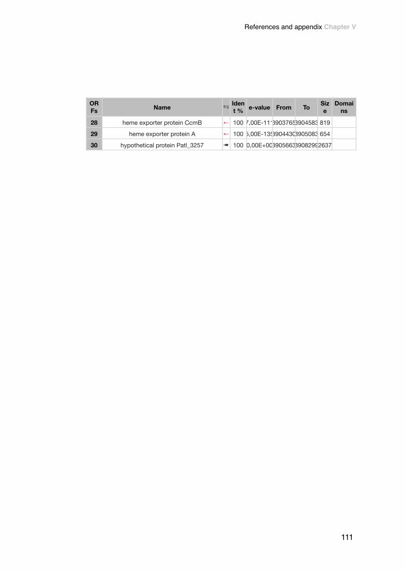
!
28 heme exporter protein CcmB ← 100 7,00E-11139037653904583 819
29 heme exporter protein A ← 100 5,00E-13539044303905083 654
30 hypothetical protein Patl_3257 ➠ 100 0,00E+00390566339082992637
Name ⇆ Ident % e-value From To Siz
eDomai
nsORFs
111
References and appendix Chapter V

!#16
ORFs Name ⇆ Iden
t % e-value From To Size
Domains
1 TonB-denpendent receptor ➠ 100 0,00E+00439936744024143048
2 heparinase II/III-like protein ➠ 100 0,00E+00440267644049372262
3 cation transporter ← 100 0,00E+00440507644062451170
4 hypothetical protein Patl_3653 ➠ 100 0,00E+0044061944406658 465
5 NAD(P) transhydrogenase subunit beta ← 100 0,00E+00440618244075791398
6 NAD(P) transhydrogenase subunit alpha ← 100 5,00E-4944082104408536 327
7 NAD(P) transhydrogenase subunit alpha ← 100 0,00E+00440849144097411251
8 PfkB protein ← 100 0,00E+0044098164410772 957
9 MFS transporter ← 95 0,00E+00441080444121591356
10 alginate lyase ← 100 0,00E+00441216044144722313
11 GntR family transcriptional regulator ➠ 100 5,00E-15844149184415685 768
12 3-oxoacyl-[acyl-carrier protein] reductase ← 98 4,00E-16044157834416553 771
13 monooxygenase ← 100 0,00E+00441690144180971197
14 SAM-dependent methyltransferase ← 100 0,00E+00441828444205332250
15 hypothetical protein Patl_3664 ← 100 0,00E+00442047044216121143
16 CorA-like Mg2+ transporter protein ← 100 0,00E+00442179044228271038
17 ribosomal protein S6 modification protein ➠ 100 0,00E+0044229964423937 942
18 hypothetical protein Patl_3667 ➠ 100 1,00E-10444239134424458 546
19 hypothetical protein Patl_3668 ➠ 100 2,00E-12944246404425263 624
20 diguanylate cyclase ← 100 0,00E+0044253284426284 957
21 TetR family transcriptional regulator ➠ 100 5,00E-14344267494427411 663
22 mechanosensitive ion channel protein ➠ 100 9,00E-16544277014428546 846
23 aldehyde oxidoreductase ← 100 0,00E+00442876144300621302
ORFs
References and appendix Chapter V
112

!
24 XRE family transcriptional regulator ➠ 100 0,00E+0044300674430984 918
25 glycosyl transferase ← 100 0,00E+00443097044327391770
Name ⇆ Ident % e-value From To Siz
eDomai
nsORFs
113
References and appendix Chapter V

!#17
ORFs Name ⇆ Iden
t % e-value From To Size
Domains
1 OmpA/MotB protein ➠ 100 0,00E+00480196048037291770
2 small GTP-binding protein ← 100 1,00E-11848037414804250 510
3 diguanylate phosphodiesterase ➠ 100 0,00E+00480424448064032160
4 acyl-CoA dehydrogenase ← 100 0,00E+00480636048075531194
5 aldo/keto reductase ← 100 0,00E+00480760948086611053
6 hypothetical protein Patl_3995 ← 100 2,00E-9648087154809146 432
7 elongation factor Tu ← 100 0,00E+00480974148109281188
8 pantothenate kinase ← 99 2,00E-15848115114812251 741
9bifunctional biotin--[acetyl-CoA-
carboxylase] synthetase/biotin operon repressor
← 100 0,00E+00481223548132691035
10 UDP-N-acetylenolpyruvoylglucosamine reductase ← 99 0,00E+00481321848142941077
11 protein-tyrosine kinase ← 100 0,00E+00481430348164622160
12 sugar ABC transporter substrate-binding protein ← 100 2,00E-11648164324817019 588
13 polysaccharide biosynthesis protein CapD ← 100 0,00E+00481702048188941875
14 aminotransferase ← 100 0,00E+00481886448200691206
15 sugar transferase ← 100 1,00E-13348206954821411 717
16 glycosyl transferase family 1 ← 100 0,00E+00482130848224981191
17 group 1 glycosyl transferase ← 100 0,00E+00482248948237511263
18 hypothetical protein Patl_4008 ← 100 0,00E+00482366748250461380
19 group 1 glycosyl transferase ← 100 0,00E+00482500948261181110
ORFs
References and appendix Chapter V
114

!
20 hypothetical protein Patl_4010 ← 100 0,00E+00482600948273101302
21 hypothetical protein Patl_4011 ← 100 2,00E-15448278084828584 777
22 carbonic anhydrase ← 100 3,00E-16048303634831028 666
Name ⇆ Ident % e-value From To Siz
eDomai
nsORFs
115
References and appendix Chapter V

#bacteriocin
ORFs Name ⇆ Ident % e-value From To Siz
eDomai
ns
1 recombinase RecR ← 100 5,00E-144 3506713 3507315 603
2 hypothetical protein Patl_2896 ← 100 9,00E-52 3507354 3507710 357
3 ATPase AAA ← 100 0,00E+00 3507711 3510464 2754
4 adenine phosphoribosyltransferase ← 99 2,00E-123 3510465 3511025 561
5 conserved hypothetical protein ← 100 9,00E-172 3511242 3511991 750
6 hypothetical protein Patl_2901 ← 100 2,00E-24 3512956 3513528 573
7 beta-lactamase ← 100 0,00E+00 3513966 3515288 1323
8 anhydro-N-acetylmuramic acid kinase ← 100 0,00E+00 3515568 3516876 1309
9 TonB-denpendent receptor ← 100 2,00E-95 3516945 3517634 690
References and appendix Chapter V
116