Embed Size (px)
Citation preview

PONTIFÍCIA UNIVERSIDADE CATÓLICA DO RIO GRANDE DO SUL
FACULDADE DE BIOCIÊNCIAS
PROGRAMA DE PÓS-GRADUAÇÃO EM BIOLOGIA CELULAR E MOLECULAR
GUSTAVO NASCENTE IGANSI
EFEITO DA EXPOSIÇÃO AO CÁDMIO SOBRE DANO OXIDATIVO, MORTE
CELULAR E COMPORTAMENTO DE ZEBRAFISH
PORTO ALEGRE
2012
PONTIFÍCIA UNIVERSIDADE CATÓLICA DO RIO GRANDE DO SUL
FACULDADE DE BIOCIÊNCIAS
PROGRAMA DE PÓS-GRADUAÇÃO EM BIOLOGIA CELULAR E MOLECULAR
EFEITO DA EXPOSIÇÃO AO CÁDMIO SOBRE DANO OXIDATIVO, MORTE
CELULAR E COMPORTAMENTO DE ZEBRAFISH
Dissertação de mestrado apresentada
ao Programa de Pós-Graduação em
Biologia Celular e Molecular da
Faculdade de Biociências da
Pontifícia Universidade Católica do
Rio Grande do Sul.
Aluno: Gustavo Nascente Igansi
Orientadora: Profª. Drª. Monica Ryff Moreira Vianna
Porto Alegre
2012
i
RESUMO
O cádmio é considerado a sétima substância mais perigosa presente no ambiente e é
classificado como carcinogênico tipo I, potencialmente afetando uma grande quantidade
de seres vivos, incluindo os humanos. Ademais, o cádmio tem sido associado a defeitos
neurocomportamentais que podem comprometer o status ecológico e a sobrevivência de
animais. Apesar do potencial impacto, a compreensão dos mecanismos celulares e
moleculares por trás de seus efeitos deletérios no comportamento de animais é ainda
escassa. Este estudo teve por objetivo avaliar os efeitos comportamentais do cádmio
por 1 e 7 dias a concentrações relevantes no ambiente (10 µg/L, 100 µg/L and 1000
µg/L) em zebrafish e analisar o estresse oxidativo e marcadores de apoptose no
encéfalo. Animais expostos ao cádmio apresentaram aumento na atividade locomotora
após 1 e 7 dias de tratamento em todas as concentrações e parâmetros avaliados,
incluindo distância percorrida num período de 5 minutos, velocidade média e períodos
móveis. A hiperlocomoção afetou significativamente o desempenho dos animais em
explorar um novo ambiente em todos os grupos tratados, evidenciado por uma
diminuição na eficiência de percurso e alterada distribuição na coluna d’água.
Adicionalmente, nossos resultados confirmaram estudos prévios sobre aumento no dano
oxidativo em peixes quando expostos ao cádmio e especialmente demonstraram níveis
mais elevados de dano a proteínas em amostras de encéfalo em animais tratados a 100
µg/L por 1 dia e a 10 µg/L por 7 dias quando comparados aos seus respectivos
controles. Lipoperoxidação aumentou significativamente no encéfalo de animais
expostos por 1 dia a 100 µg/L. Análises dos marcadores p53 e bax por Real-time PCR
apresentaram nenhuma alteração após 1 dia de exposição, mas significativamente
aumentaram após 7 dias. Nossos resultados apresentam evidências dos efeitos deletérios
do cádmio no comportamento de zebrafish e chama atenção para o fato de que a
manifestação de seus efeitos aparece a partir de 1 dia de exposição a 10 µg/L, uma
concentração aceita por muitas agências de regulamentação internacional.

ii
ABSTRACT
Cadmium is considered the seventh most dangerous substance in the environment
and is classified as carcinogen type I, potentially affecting a wide range of living
organisms, including humans. In addition to its wide systemic impact and potential
lethality, cadmium has been associated to neurobehavioral defects that may also
compromise animals’ ecological status and survival. Despite its potential impact, the
comprehension of cellular and molecular mechanisms underlying cadmium deleterious
effects on animals’ behavior is still scarce. This study aimed to evaluate the behavioral
effects of cadmium for 1 or 7 days at environmentally relevant concentrations (10 µg/L,
100 µg/L and 1000 µg/L) on zebrafish and to analyze brain oxidative stress and
apoptotic markers. Cadmium-exposed zebrafish exhibited a generalized increase in
locomotor activity after 1 and 7 days of treatment at all doses in all parameters
evaluated, including distance travelled in a 5-min. evaluation period, mean speed and
mobile periods. This hyperlocomotory effect significantly compromised animals’
general performance in exploring a new environment, which was evident in all
cadmium exposed animals decreased path efficiency and altered distribution on the
water column. Additionally, our results confirmed previous reports of increased
oxidative damage in fishes exposed to cadmium and specifically demonstrated higher
levels of damaged proteins in brain samples of animals exposed to cadmium at 100
µg/L for 1 day and at 10 µg/L for 7 days when compared to their respective control
groups. Lipid peroxidation was also significantly increased in animals’ brain after 1 day
cadmium exposure at 100 µg/L. Real-Time PCR analysis of transcripts for p53 and bax
were not altered after 1 day cadmium exposure, but significantly increased after 7 days.
Our results present evidence of cadmium deleterious effects on zebrafish cognitive
functions and raise attention to the fact that its manifestation appears already after a one
day exposure to 10 µg/L, a concentration accepted by most international regulating
agencies.
iii
LISTA DE ILUSTRAÇÕES
FIGURA 01 – Esquema ilustrando as vias de absorção e locais de
acúmulo..........................................................................
10
FIGURA 02 – Cádmio e vias de sinalização intracelular........................ 11
FIGURA 03 – Envolvimento do Cádmio em várias vias de sinalização
intracelular anti e pro apoptóticos...................................
12
FIGURA 04 – Esquema geral sobre as consequências da intoxicação
pelo Cádmio na mitocôndria...........................................
13
FIGURA 05 – Zebrafish como modelo animal........................................ 15

iv
LISTA DE TABELAS
TABELA1 – Níveis considerados naturais de Cádmio no ambiente................
8
v
SUMÁRIO
CAPÍTULO 1
1.INTRODUÇÃO......................................................................................... 7
1.1. Ecotoxicologia de Metais Pesados................................................. 7
1.2. Ecotoxicologia do Cádmio.............................................................. 8
1.3. Cádmio e sua Interação com Vias de Sinalização Intracelular...... 11
1.4. Zebrafish como Modelo Animal...................................................... 14
2. JUSTIFICATIVA..................................................................................... 16
3. OBJETIVOS........................................................................................... 16
3.1. Objetivo Geral................................................................................. 16
3.2. Objetivos Específicos...................................................................... 16
CAPÍTULO 2
ARTIGO CIENTÍFICO............................................................................ 20
CAPÍTULO 3
CONSIDERAÇÕES FINAIS................................................................... 46
REFERÊNCIAS........................................................................................... 49
ANEXOS.................................................................................................... 56

CAPÍTULO 1

7
1. INTRODUÇÃO 1.1 - Ecotoxicologia de Metais Pesados
A ação antrópica tem gerado uma grande variedade de substâncias que
atuam diretamente sobre o meio ambiente, incluindo, por exemplo, herbicidas,
pesticidas, detergentes, metais pesados e substâncias químicas de natureza
diversa. A capacidade do homem de explorar e manipular metais proporcionou
um importante marco no desenvolvimento de nossa sociedade (Wilson,1996).
Os metais, em especial metais pesados, possuem efeitos negativos quando
liberados em elevadas concentrações no meio ambiente (Han et al., 2002),
sendo classificados como poluentes e causando impactos tanto na saúde
humana (Duruibe et al., 2007) quanto em ecossistemas aquáticos e terrestres
(Sánchez, 2008).
A maior parte dos estudos com metais pesados, como o cádmio, o
mercúrio e o cobre, atenta para os efeitos negativos diretos sobre os
organismos, ou seja, na sintomatologia toxicológica e mortalidade (Lefcort et
al., 2002), enquanto pouco apontam os efeitos indiretos, por exemplo, sobre a
estrutura de comunidades e de cadeias alimentares (Fleeger et al., 2003;
Sloman, 2007). Ademais, há muitos estudos a respeito dos efeitos sobre
alterações no comportamento e adaptações de organismos, que vão desde
procariotos e eucariotos basais (Lass & Spaak, 2003; Challis, 2005; Schertzer
et al., 2009) até plantas e animais (Koricheva et al., 1998; Greger, 2004;
McPherson et al., 2004; Lürling & Scheffer, 2007; Klaschka, 2008; Liu et al.,
2009).
Apesar dos conhecidos efeitos negativos dos metais pesados sobre o
comportamento animal, os mecanismos através dos quais os metais impactam
as funções cerebrais superiores não são conhecidos, especialmente do ponto
de vista celular e molecular. Esta informação, quando disponível, permitirá o
melhor entendimento de seus impactos e potencialmente proporcionará
substratos para estratégias terapêuticas e preventivas dos efeitos deletérios
duradouros que tais elementos têm sobre o sistema nervoso central.
Finalmente, Boyd (2010) alerta que para se entender o comportamento de
organismos, devem ser considerados os múltiplos fatores interligados que
8
atuam sobre eles, como disponibilidade de recursos, estresse, condições
ambientais entre outros, tornando o seu estudo realista.
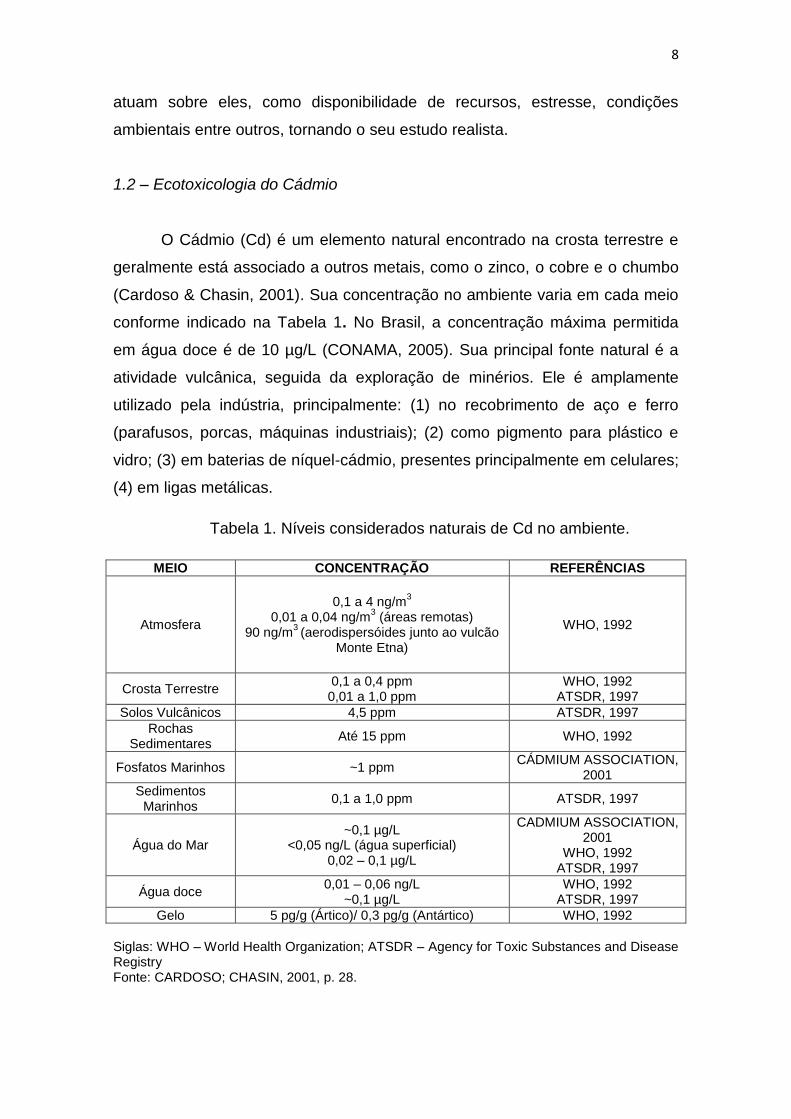
1.2 – Ecotoxicologia do Cádmio
O Cádmio (Cd) é um elemento natural encontrado na crosta terrestre e
geralmente está associado a outros metais, como o zinco, o cobre e o chumbo
(Cardoso & Chasin, 2001). Sua concentração no ambiente varia em cada meio
conforme indicado na Tabela 1. No Brasil, a concentração máxima permitida
em água doce é de 10 µg/L (CONAMA, 2005). Sua principal fonte natural é a
atividade vulcânica, seguida da exploração de minérios. Ele é amplamente
utilizado pela indústria, principalmente: (1) no recobrimento de aço e ferro
(parafusos, porcas, máquinas industriais); (2) como pigmento para plástico e
vidro; (3) em baterias de níquel-cádmio, presentes principalmente em celulares;
(4) em ligas metálicas.
Tabela 1. Níveis considerados naturais de Cd no ambiente.
MEIO CONCENTRAÇÃO REFERÊNCIAS
Atmosfera
0,1 a 4 ng/m3
0,01 a 0,04 ng/m3 (áreas remotas)
90 ng/m3 (aerodispersóides junto ao vulcão
Monte Etna)
WHO, 1992
Crosta Terrestre 0,1 a 0,4 ppm 0,01 a 1,0 ppm
WHO, 1992 ATSDR, 1997
Solos Vulcânicos 4,5 ppm ATSDR, 1997
Rochas Sedimentares
Até 15 ppm WHO, 1992
Fosfatos Marinhos ~1 ppm CÁDMIUM ASSOCIATION,
2001
Sedimentos Marinhos
0,1 a 1,0 ppm ATSDR, 1997
Água do Mar ~0,1 µg/L
<0,05 ng/L (água superficial) 0,02 – 0,1 µg/L
CADMIUM ASSOCIATION, 2001
WHO, 1992 ATSDR, 1997
Água doce 0,01 – 0,06 ng/L
~0,1 µg/L WHO, 1992
ATSDR, 1997
Gelo 5 pg/g (Ártico)/ 0,3 pg/g (Antártico) WHO, 1992
Siglas: WHO – World Health Organization; ATSDR – Agency for Toxic Substances and Disease Registry Fonte: CARDOSO; CHASIN, 2001, p. 28.
9
A contaminação do ambiente por Cd ocorre por fontes naturais, como as
erupções vulcânicas (WHO, 1992), ou por atividade antrópica. A mineração, a
produção industrial e o descarte de resíduos urbanos e industriais não tratados
que contenham cádmio contaminam o ambiente (Teves, 2001). As fontes de
contaminação das águas incluem a atividade de mineração de metais não
ferrosos, fundições de minério não ferroso, extração de rochas fosfatadas e
manufatura de fertilizantes fosfatados e a acidificação de solos e lagos (WHO,
1992 e 1998). O Cd proveniente de indústrias é rapidamente adsorvido e
acumula-se no sedimento (Calmano & Förstner, 1996), associando-se a
carbonatos e formando depósitos que contaminam o meio aquático e o solo
entorno (WHO, 1992). No sedimento, o Cd pode sofrer ação de bactérias, que
o transformam em compostos orgânicos tóxicos como, por exemplo,
dimetilcádmio (Yannai & Berdicevsky, 1995). Dependendo das condições
físico-químicas do ambiente, estes compostos são liberados do sedimento e
podem entrar em contato com outros organismos, comprometendo toda a
cadeia alimentar (Calmano & Förstner, 1996). Em água doce, o Cd está
presente na forma Cd+2, hidróxido de cádmio e carbonato de cádmio (ATSDR,
1997).
Cd bioacumula-se no fitoplâncton e, através de complexas teias
alimentares, em animais aquáticos, tais como moluscos, peixes e crustáceos.
Peixes possuem a capacidade de bioacumular Cd principalmente nas
brânquias e paredes intestinais, além de outros órgãos como fígado e rins
(Cardoso & Chasin, 2001). Além disso, o Cd, ao passar pelas brânquias, é
transportado para as células através de vias de transporte de Ca+2 (Sloman et
al., 2007). Em resposta à contaminação, o peixe passa a expressar a proteína
metalotioneína (MT), que se liga ao metal, inativando-o, embora esta estratégia
tenha sua eficiência limitada e dependente das concentrações do metal (De
Conto Cinier et al., 1998).
A dieta é a principal fonte de contaminação no homem, e além dos
organismos aquáticos citados acima, outros alimentos são importantes fontes
potenciais de contaminação, como óleos de sementes, cereais, vegetais e
raízes (Järup & Akesson, 2009). Mais de 80% do Cd em humanos vem de
cereais e vegetais (Olsson et al., 2002), e a concentração média de ingestão
diária varia de 8 a 25 μg por dia em um indivíduo adulto (Berglund et al., 1994;
10
MacIntosh et al.,1996; Thomas et al., 1999; Ysart et al., 2000; Larsen et al.,
2002; Olsson et al., 2002; Llobet et al., 2003; Egan et al., 2007). A fumaça de
tabaco é outra fonte importante de contaminação humana, sendo que crianças
são mais suscetíveis (Willers et al., 2005; Arora et al., 2008), enquanto que
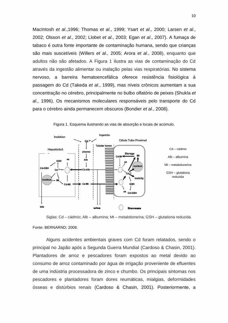
adultos não são afetados. A Figura 1 ilustra as vias de contaminação do Cd
através da ingestão alimentar ou inalação pelas vias respiratórias. No sistema
nervoso, a barreira hematoencefálica oferece resistência fisiológica à
passagem do Cd (Takeda et al., 1999), mas níveis crônicos aumentam a sua
concentração no cérebro, principalmente no bulbo olfatório de peixes (Shukla et
al., 1996). Os mecanismos moleculares responsáveis pelo transporte do Cd
para o cérebro ainda permanecem obscuros (Bondier et al., 2008).
Figura 1. Esquema ilustrando as vias de absorção e locais de acúmulo.
Siglas: Cd – cádmio; Alb – albumina; Mt – metalotioneína; GSH – glutationa reduzida.
Fonte: BERNARND; 2008.
Alguns acidentes ambientais graves com Cd foram relatados, sendo o
principal no Japão após a Segunda Guerra Mundial (Cardoso & Chasin, 2001).
Plantadores de arroz e pescadores foram expostos ao metal devido ao
consumo de arroz contaminado por água de irrigação proveniente de efluentes
de uma indústria processadora de zinco e chumbo. Os principais sintomas nos
pescadores e plantadores foram dores reumáticas, mialgias, deformidades
ósseas e distúrbios renais (Cardoso & Chasin, 2001). Posteriormente, a
urina lisossomo
Inalation Ingestão
Célula Tubo Proximal
Hepatócito\o
Cd – cádmio
Alb – albumina
Mt – metalotioneína
GSH – glutationa
reduzida
11
sintomatologia foi chamada de Itai-Itai (ouch-ouch). Além destes, outros
sintomas foram relacionados à exposição do Cd em outros tecidos, como: (1)
decréscimo da função pulmonar e enfisema; (2) aumento da pressão arterial e
doenças cerebrovasculares; (3) anemia; (4) comprometimento dos ossos; (5)
cálculos renais e danos nos túbulos renais (WHO, 1992; ATSDR, 1997).
1.3 – Cádmio e sua Interação com Vias de Sinalização Intracelular
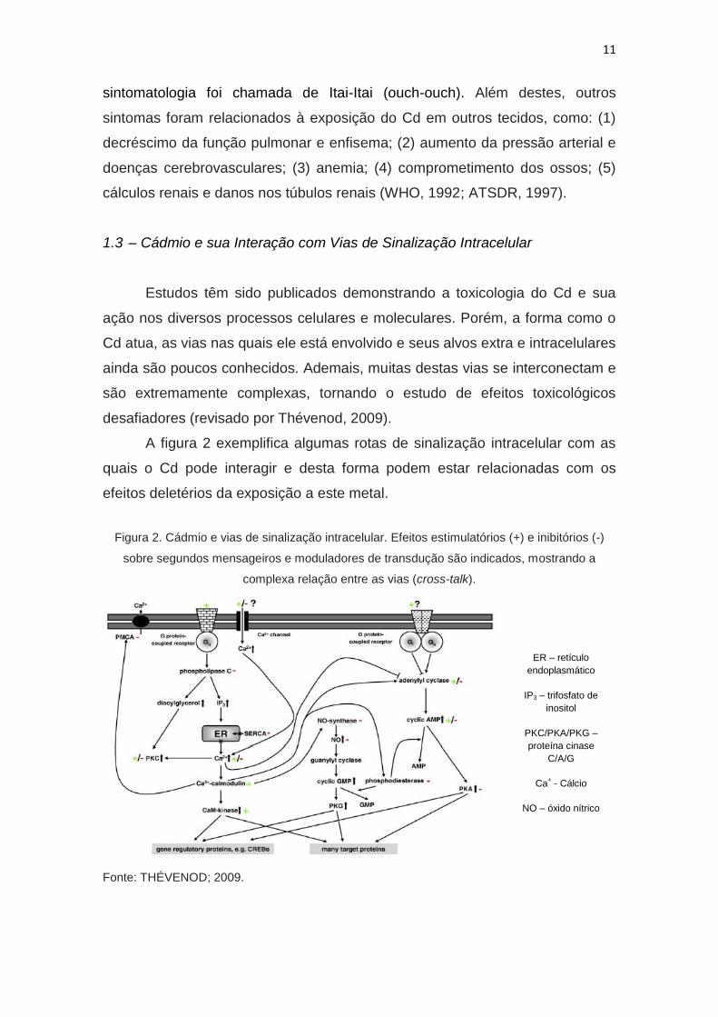
Estudos têm sido publicados demonstrando a toxicologia do Cd e sua
ação nos diversos processos celulares e moleculares. Porém, a forma como o
Cd atua, as vias nas quais ele está envolvido e seus alvos extra e intracelulares
ainda são poucos conhecidos. Ademais, muitas destas vias se interconectam e
são extremamente complexas, tornando o estudo de efeitos toxicológicos
desafiadores (revisado por Thévenod, 2009).
A figura 2 exemplifica algumas rotas de sinalização intracelular com as
quais o Cd pode interagir e desta forma podem estar relacionadas com os
efeitos deletérios da exposição a este metal.
Figura 2. Cádmio e vias de sinalização intracelular. Efeitos estimulatórios (+) e inibitórios (-)
sobre segundos mensageiros e moduladores de transdução são indicados, mostrando a
complexa relação entre as vias (cross-talk).
Fonte: THÉVENOD; 2009.
ER – retículo
endoplasmático
IP3 – trifosfato de
inositol
PKC/PKA/PKG –
proteína cinase
C/A/G
Ca+ - Cálcio
NO – óxido nítrico
12
Por exemplo, Cd altera os níveis de Ca+2 nas células, afetando diversas
vias, principalmente a ativação/inibição de proteínas cinases (Mazzei et al.,
1984; Kostrzewska & Sobieszek, 1990; Chao et al., 1995; Liu & Templeton,
2007). Ademais, Thévenod (2009) apresenta uma excelente revisão sobre as
vias do cAMP, oxido nítrico, NF-kB, entre outras, apontando contradições nas
evidências disponíveis e perspectivas futuras de investigação da influência do
Cd nestas vias.
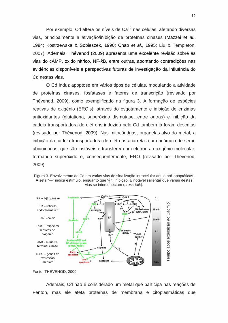
O Cd induz apoptose em vários tipos de células, modulando a atividade
de proteínas cinases, fosfatases e fatores de transcrição (revisado por
Thévenod, 2009), como exemplificado na figura 3. A formação de espécies
reativas de oxigênio (ERO’s), através do esgotamento e inibição de enzimas
antioxidantes (glutationa, superóxido dismutase, entre outras) e inibição da
cadeia transportadora de elétrons induzida pelo Cd também já foram descritas
(revisado por Thévenod, 2009). Nas mitocôndrias, organelas-alvo do metal, a
inibição da cadeia transportadora de elétrons acarreta a um acúmulo de semi-
ubiquinonas, que são instáveis e transferem um elétron ao oxigênio molecular,
formando superóxido e, consequentemente, ERO (revisado por Thévenod,
2009).
Figura 3. Envolvimento do Cd em várias vias de sinalização intracelular anti e pró-apoptóticas. A seta “→” indica estímulo, enquanto que “┤”, inibição. É notável salientar que várias destas
vias se interconectam (cross-talk).
Fonte: THÉVENOD, 2009.
Ademais, Cd não é considerado um metal que participa nas reações de
Fenton, mas ele afeta proteínas de membrana e citoplasmáticas que
IKK – Iĸβ quinase
ER – retículo
endoplasmático
Ca+ - cálcio
ROS – espécies
reativas de
oxigênio
JNK - c-Jun N-
terminal cinase
IEGS – genes de
expressão
imediata
Tem
po
ap
ós e
xposiç
ão a
o C
ádm
io
13
contenham outros metais, a exemplo do ferro. Este último, quando livre no
interior da célula, é capaz de formar ERO através da quebra de H2O2, formando
o radical OH (revisado por Matovic et al, 2011). Por fim, exposição a longo
prazo ao metal pode produzir mediadores inflamatórios, como IL-6 e IL-8, que
induzem a produção de ERO (revisado por Matovic et al, 2011).
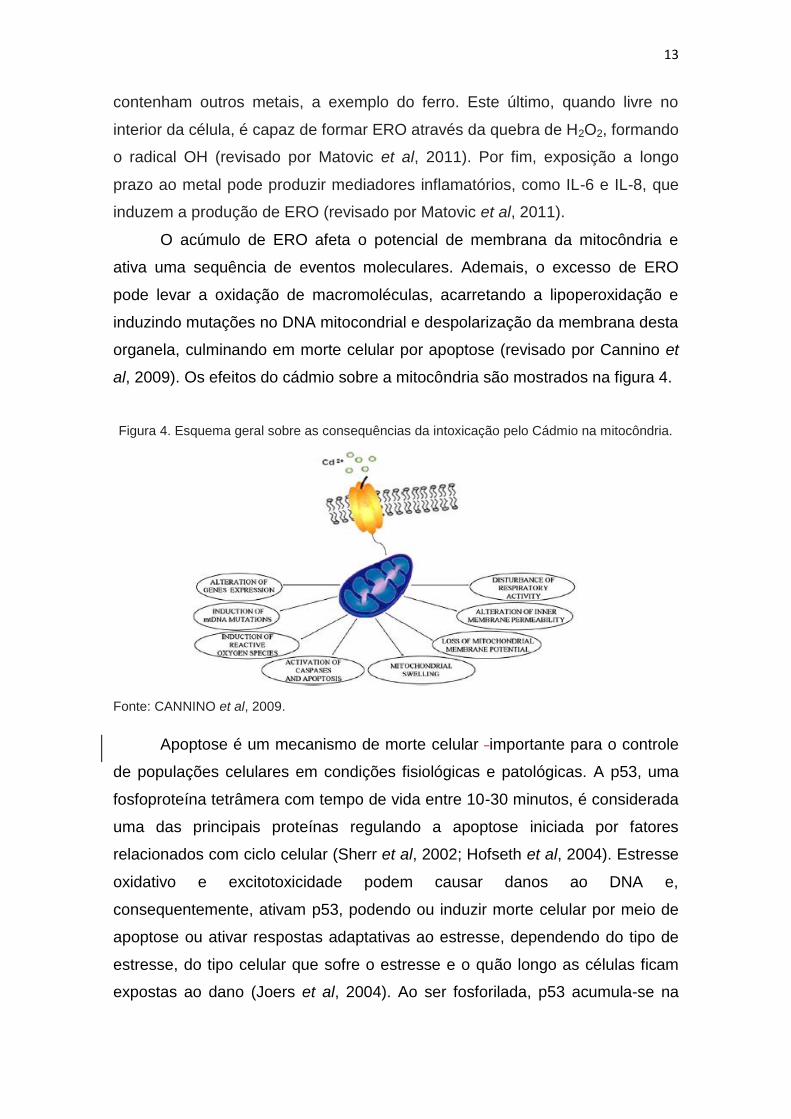
O acúmulo de ERO afeta o potencial de membrana da mitocôndria e
ativa uma sequência de eventos moleculares. Ademais, o excesso de ERO
pode levar a oxidação de macromoléculas, acarretando a lipoperoxidação e
induzindo mutações no DNA mitocondrial e despolarização da membrana desta
organela, culminando em morte celular por apoptose (revisado por Cannino et
al, 2009). Os efeitos do cádmio sobre a mitocôndria são mostrados na figura 4.
Figura 4. Esquema geral sobre as consequências da intoxicação pelo Cádmio na mitocôndria.
Fonte: CANNINO et al, 2009.
Apoptose é um mecanismo de morte celular importante para o controle
de populações celulares em condições fisiológicas e patológicas. A p53, uma
fosfoproteína tetrâmera com tempo de vida entre 10-30 minutos, é considerada
uma das principais proteínas regulando a apoptose iniciada por fatores
relacionados com ciclo celular (Sherr et al, 2002; Hofseth et al, 2004). Estresse
oxidativo e excitotoxicidade podem causar danos ao DNA e,
consequentemente, ativam p53, podendo ou induzir morte celular por meio de
apoptose ou ativar respostas adaptativas ao estresse, dependendo do tipo de
estresse, do tipo celular que sofre o estresse e o quão longo as células ficam
expostas ao dano (Joers et al, 2004). Ao ser fosforilada, p53 acumula-se na
14
célula e começa a transcrever genes específicos para morte celular, tais como
Bax, NOXA, PUMA e Apaf-1 (Fridman and Lowe, 2003; Slee et al, 2004; Ward
et al, 2004). Bax é uma proteina capaz de formar canais em membranas. Uma
vez expressa, Bax é translocada para mitocôndria, formando poros. Este
processo perturba o potencial de membrana da organela, induzindo a liberação
de citocromo c (Dargusch et al, 2001; Polster & Fiskum, 2004). O citocromo c, a
apaf-1 e a caspase-9 formam o apoptosoma, que induz apoptose ao ativar
caspases efetoras, como a caspase-3 (Jayanthi et al, 2004). Vários estudos
têm mostrado que o Cd não apenas possui a habilidade de induzir morte
celular em vário tipos celulares, incluindo encéfalo e brânquias de peixes
(Azzouqiel et al, 1994; Habeebu et al, 1998; Dong et al, 2001; López et al,
2003, Liu et al, 2011) mas também é capaz de induzir desordens
neurodegenerativas em humanos, como Doença de Parkinson (O’Callaghan
and Miller, 1986; Okuda et al, 1997) e esclerose lateral amiotrófica (Bar-Sela et
al., 2001).
Por fim, Cd ocupa o sétimo lugar entre as substâncias mais perigosas
(ATSDR, 2007) e é classificado como carcinogênico tipo 1 (IARC, 1993),
estando associado ao câncer de pulmão, rim, próstata e pâncreas (Bernard &
Lauwerys, 1984; Foulkes, 1986; Friberg et al., 1986; Morselt, 1991; Goering et
al., 1995; Jarup et al., 1998).
1.4 – Zebrafish como Modelo Animal
Diversos animais são utilizados para testes toxicológicos, incluindo
desde invertebrados, como Caenorhabditis elegans e Drosophila melanogaster,
até mamíferos, como ratos e primatas. Mamíferos sempre foram utilizados para
avaliar os efeitos de tais substâncias devido a sua semelhança aos humanos.
O grande número de substâncias tóxicas produzidas e liberadas pelo homem
no ambiente, impactando todos os componentes dos ecossistemas, tem
desviado a visão antropocêntrica do impacto de substâncias liberadas no meio,
valorizando o uso de outros animais como indicadores. Adicionalmente,
questões éticas quanto ao uso de animais na pesquisa e recursos limitados
para manutenção e cuidados tornaram o uso de mamíferos menos atrativo.
Recentemente, novos animais modelos vertebrados com conhecimento
15
genético e fisiológico tem sido propostos, e dentre eles tem se destacado o
zebrafish (revisado por Peterson et al., 2008).
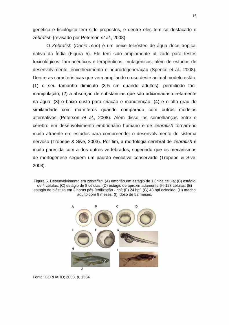
O Zebrafish (Danio rerio) é um peixe teleósteo de água doce tropical
nativo da Índia (Figura 5). Ele tem sido amplamente utilizado para testes
toxicológicos, farmacêuticos e terapêuticos, mutagênicos, além de estudos de
desenvolvimento, envelhecimento e neurodegeneração (Spence et al., 2008).
Dentre as características que vem ampliando o uso deste animal modelo estão:
(1) o seu tamanho diminuto (3-5 cm quando adultos), permitindo fácil
manipulação; (2) a absorção de substâncias que são adicionadas diretamente
na água; (3) o baixo custo para criação e manutenção; (4) e o alto grau de
similaridade com mamíferos quando comparado com outros modelos
alternativos (Peterson et al., 2008). Além disso, as semelhanças entre o
cérebro em desenvolvimento embrionário humano e de zebrafish tornam-no
muito atraente em estudos para compreender o desenvolvimento do sistema
nervoso (Tropepe & Sive, 2003). Por fim, a morfologia cerebral de zebrafish é
muito parecida com a dos outros vertebrados, sugerindo que os mecanismos
de morfogênese seguem um padrão evolutivo conservado (Tropepe & Sive,
2003).
Figura 5. Desenvolvimento em zebrafish. (A) embrião em estágio de 1 única célula; (B) estágio de 4 células; (C) estágio de 8 células; (D) estágio de aproximadamente 64-128 células; (E)
estágio de blástula em 3 horas pós-fertilização - hpf; (F) 24 hpf; (G) 48 hpf eclodido; (H) macho adulto com 8 meses; (I) Idoso de 52 meses.
Fonte: GERHARD; 2003, p. 1334.
16
Estudos anteriores sobre o efeito do Cd em zebrafish concentraram-se
em avaliar o efeito desde metal sobre o desenvolvimento inicial (Chan &
Cheng, 2003; Chow et al., 2008; Chow et al., 2009). Poucos são os estudos
que discutem a ação do cádmio em peixes adultos, e praticamente não há
trabalhos que caracterizem os efeitos comportamentais e cognitivos do Cd em
zebrafish.
2 - JUSTIFICATIVA
Considerando os pontos apresentados até aqui, acreditamos ser
relevante utilizar o zebrafish como modelo para avaliar os efeitos da exposição
ao Cd na vida adulta sobre parâmetros comportamentais e tentar identificar as
vias celulares envolvidas nos eventuais efeitos desta exposição. Muito
recentemente, estudos começaram a ser direcionados para a integração entre
o ambiente no qual os animais estão e os diversos poluentes que interagem
com eles e as implicações na fisiologia e vias de sinalização. Isto nos oferece
um grande espectro para melhor entendermos como, de fato, as interações
ocorrem e que são múltiplos os fatores que atuam nelas. Portanto, esta
dissertação tem por finalidade caracterizar o efeito da exposição ao cádmio por
1 dia e 7 dias sobre o comportamento, dano tecidual e interação com as vias
de apoptose.
3 – OBJETIVOS
3.1 – Objetivo Geral
Caracterizar o efeito da exposição ao cádmio por 1 ou 7 dias sobre o
comportamento, dano tecidual e via de morte celular em zebrafish adultos.
3.2 – Objetivos Específicos
Avaliar parâmetros comportamentais em animais adultos tratados com
Cd por 1 ou 7 dias através de avaliação da capacidade exploratória;

17
Avaliar o dano oxidativo ocasionado pela exposição ao Cd no encéfalo
de animais adultos tratados por 1 ou 7 dias através da mensuração de Carbonil
e TBARS;
Avaliar a morte celular por apoptose no encéfalo de animais adultos
tratados com Cd por 1 ou 7 dias através da alteração nos níveis de expressão
de p53 e bax por Real-Time PCR.

CAPÍTULO 2
20
ARTIGO CIENTÍFICO A SER SUBMETIDO PARA “AQUATIC TOXICOLOGY”
Título: Cadmium exposure at environmentally relevant concentrations compromises
zebrafish behavior and induces cell damage and apoptosis in the brain
Autores: Gustavo N. Igansi, Marcelo N. Gonçalves, Laura R. Nery, Larissa S.
Constantino, Renata C. Gonçalves, Felipe Dal-Pizzol, Monica R. M. Vianna
Revista: Aquatic Toxicology (IF JCR 2010: 3,333) – Elsevier Science, San Diego,
California, USA.
21
Cadmium exposure at environmentally relevant concentrations compromises zebrafish
behavior and increases oxidative cell damage and apoptosis markers in the brain
Gustavo N. Igansia, Marcelo N. Gonçalves
a, Laura R. Nery
a, Larissa S. Constantino
b, Renata
C. Gonçalvesb, Felipe Dal-Pizzol
b, Monica R.M. Vianna
a,c,d,*
aLaboratório de Biologia e Desenvolvimento do Sistema Nervoso, Faculdade de Biociências,
Pontifícia Universidade Católica do Rio Grande do Sul, 90619-900, Porto Alegre, RS, Brazil
bLaboratório de Fisiopatologia Experimental, Programa de Pós-Graduação em Ciências da
Saúde, Unidade Acadêmica de Ciências da Saúde, Universidade do Extremo Sul Catarinense,
88806-000 Criciúma, SC, Brazil
cNational Institute for Translational Medicine (INCT-TM) 90035-003, Porto Alegre, RS,
Brazil
dZebrafish Neuroscience Research Consortium (ZNRC)
*Corresponding author
Prof. Monica R. M. Vianna
Correspondence address:
Faculdade de Biociências, Pontifícia Universidade Católica do Rio Grande do Sul
Av. Ipiranga 6681, Prédio 12
90619-900, Porto Alegre, RS, Brazil
Phone: +55 51 33534743
FAX: + 55 51 33203612
E-mail address: [email protected]
22
Abstract
Cadmium is considered the seventh most dangerous substance in the environment and is
classified as carcinogen type I, potentially affecting a wide range of living organisms,
including humans. In addition to its wide systemic impact and potential lethality, cadmium
has been associated to neurobehavioral defects that may also compromise animals’ ecological
status and survival. Despite its potential impact, the comprehension of cellular and molecular
mechanisms underlying cadmium deleterious effects on animals’ behavior is still scarce. This
study aimed to evaluate the behavioral effects of cadmium for 1 or 7 days at environmentally
relevant concentrations (10 µg/L, 100 µg/L and 1000 µg/L) on zebrafish and to analyze brain
oxidative stress and apoptotic markers. Cadmium-exposed zebrafish exhibited a generalized
increase in locomotor activity after 1 and 7 days of treatment at all doses in all parameters
evaluated, including distance travelled in a 5-min. evaluation period, mean speed and mobile
periods. This hyperlocomotory effect significantly compromised animals’ general
performance in exploring a new environment, which was evident in all cadmium exposed
animals decreased path efficiency and altered distribution on the water column. Additionally,
our results confirmed previous reports of increased oxidative damage in fishes exposed to
cadmium and specifically demonstrated higher levels of damaged proteins in brain samples of
animals exposed to cadmium at 100 µg/L for 1 day and at 10 µg/L for 7 days when compared
to their respective control groups. Lipid peroxidation was also significantly increased in
animals’ brain after 1 day cadmium exposure at 100 µg/L. Real-Time PCR analysis of
transcripts for p53 and bax were not altered after 1 day cadmium exposure, but significantly
increased after 7 days. Our results present evidence of cadmium deleterious effects on
zebrafish cognitive functions and raise attention to the fact that its manifestation appears
already after a one day exposure to 10 µg/L, a concentration accepted by most international
regulating agencies.
Keywords: Cadmium, behavior, oxidative stress, apoptosis, zebrafish
23
1. INTRODUCTION
Cadmium is a heavy metal found naturally in the crust, considered one of the most toxic
substances in the environment and has no known biological function in animals (ATSDR,
1997; Soares et al, 2008). Environmental contamination occurs through either natural sources
(WHO, 1992) or anthropogenic action (reviewed by Bernard, 2008). Aquatic ecosystems are
especially vulnerable (Bhakta et al, 2008) and fish tend to accumulate metals on tissues such
as gills, liver and kidneys (Cinier et al, 1999; Soares et al, 2008). Humans can be exposed to
cadmium from different sources, including contaminated fish diet or accidental occupational
exposure, with significant deleterious effects even at trace quantities (Bernard, 2008; Järup
and Akesson, 2009).
Previous reports have shown evidences of the deleterious effects of cadmium exposure to
brain functions in fish (reviewed by Atchison, 1987; Giusi et al, 2005; Scott and Sloman,
2004). Gills and olfactory rosettes are considered routes of cadmium uptake, since both
structures are directly exposed to the environment. Cadmium is able to enter gills chloride
cells through calcium channels (Verbost et al, 1987, 1989) being subsequently transported to
other organs such as liver and kidneys (Szebedinsky et al, 2001). Cadmium crosses the
olfactory epithelium and is transported through the olfactory nerves’ axons, reaching the
olfactory bulb. (Gottofrey and Tjälve, 1991; Tjälve and Gottofrey, 1986; Tjälve and
Henriksson, 1999). Cadmium transportation to other areas of the central nervous system
remains unclear (Bondieret al, 2008) but it reaches the striatum in rodents and humans
(Fernández-Pérez et al, 2010; Okuda et al, 1997;) and telencephalic and mesencephalic areas
of fish brain (Giusiet al, 2005).The fish and mammalian blood-brain barrier offers resistance
to cadmium entrance in the brain but the olfactory route has no influence of it, being this route
considered a pathway to cadmium neurotoxicity (Takeda et al., 1999; Tallkvist, 2002).
Several studies have quantified cadmium accumulation on specific fish organs after acute and
chronic exposures, demonstrating its accumulation in gills, brain and muscles (Gonzalez et al,
2006; Kargin and Çogun, 1999; Pretto et al, 2010; Rashed, 2001).
Cadmium is known to induce reactive oxygen species (ROS) in a wide range of tissues,
such as brain (Kumar et al, 1996), gills (Pretto et al, 2011) and muscle (Yano et al, 2005)
through depletion and inhibition of antioxidant enzymes, disturbances on the electron transfer
chain and competition with other ions (e.g. calcium, zinc) and therefore culminating in
membrane permeability alterations (reviewed by Thévenod, 2009). High levels of ROS may
24
induce damage of macromolecules including proteins, DNA and lipids, depolarization of
mitochondrial membrane and, finally, apoptosis (reviewed by Cannino et al, 2009).
DNA damage caused by oxidative stress or excitotoxicity can trigger cell cycle arrest and
apoptosis. The phosphoprotein p53 may induce cell death through apoptosis depending on the
stress and cellular type (Joers et al, 2004), by activation of specific genes, including Bax
(Fridman and Lowe, 2003; Slee et al, 2004; Ward et al, 2004). Once expressed, Bax is
translocated to mitochondria, forming pores, disrupting mitochondrial membrane potential,
releasing cytochrome c and triggering caspase activation and apoptosome formation
(Dargusch et al, 2001; Jayanthi et al, 2004; Polster and Fiskum, 2004).
Fish have been used as excellent biomarkers for toxicological studies, since their
ecological behaviors are easily observed and monitored (Scott and Sloman, 2004). Zebrafish
(Danio rerio) is a fresh water teleost employed in many areas to different studies, including
toxicology (Komjarova and Blust, 2009) and behavior (Piato et al, 2011). In addition to its
prominent advantages, including fast development and low cost, zebrafish brain is not only
neuroanatomically and functionally comparable to mammals (Guo, 2004) but also presents
identical neurotransmitter systems (Kastenhuber et al., 2010; Lillesaar et al., 2007;
Rosemberg et al., 2007; Yamamoto et al., 2010) and a complex range of behaviors (Buske and
Gerlai, 2011; Piatoet al, 2011).
Despite previous reports of cadmium negative effects, the mechanisms by which the metal
impacts cerebral functions are poorly understood and will benefit by use of a model organism
such as zebrafish. Few reports have used automated systems to evaluate effects of cadmium
on fish behavior (Beauvais et al, 2001; Giusiet al, 2005) and the only two studies that used
zebrafish have evaluated larvae behavior (Blechingeret al, 2007; Kuschet al, 2007). The aim
of this study was to evaluate cadmium effects on adult zebrafish behavior using an automated
system and analyze oxidative stress and apoptotic markers in brain samples when exposed to
cadmium for 1 or 7 days.
2 MATERIALS AND METHODS
2.1. Animals and Housing
A total of 240 adult male/female wild-type zebrafish (Danio rerio) were purchased from a
local supplier (Redfish, Porto Alegre, Brazil). Fish were acclimated for at least two weeks and
housed in groups of 50 fish in 15 L tanks with reverse osmosis water equilibrated with Instant
Ocean and constantly aerated. Water parameters were daily monitored (temperature: 28 +1°C;
25
pH: 7-7.5; hardness: 300-450 ppm CaCO3; nitrites and nitrates: 0,05 a 0,1 mg/L) and kept
constant in housing tanks and in tanks used for behavioral analysis in order to preserve
animals’ welfare. Fish were kept on a 14–10h day/night cycle and fed three times a day with
commercial flakes (Tetramin, Tetra, Melle FRG) supplemented with brine shrimp (Artemia
salina). All protocols were approved by the Institutional Animal Care Committee (10/00214,
CEUA-PUCRS) and followed Brazilian legislation, the guidelines of the Brazilian Collegium
of Animal Experimentation (COBEA), Canadian Council for Animal Care (CCAC) guide on
the care and use of fish in research, teaching, and testing and the recommendations by
Westerfield (2000).
2.2. Treatments
Animals were assigned into eight groups, according to exposure times (1 day or 7 days)
and cadmium carbonate (CdCO3 – Sigma Aldrich) concentrations (0, 10 µg/L, 100 µg/L and
1000 µg/L). Animals were kept in 15 L tanks, each one containing 60 animals. CdCO3 was
dissolved in conditioned water as mentioned on item 2.1 and control animals were exposed to
cadmium-free conditioned-water. Cadmium concentrations were based on the literature and
chosen to represent environmentally tolerated concentrations (10 µg/L, according to the
Brazilian Environmental Council (CONAMA, 2005) and to model release accidents (100
µg/L and 1000 µg/L) (Ansari et al., 2010; Bhagure et al, 2009). Neither continuous exposure
to the metal nor water exchange were made during treatments. Furthermore, cadmium
concentrations were not measured in each treatment tank. Fish were observed twice a day
during treatment (8 a.m. and 18 p.m.) for mortality and other abnormal symptoms including
aggressiveness. At the end of each treatment, fish were placed in cadmium-free tanks for
behavioral analysis or euthanasia for sample collection, in order to perform oxidative stress
essays and Quantitative Real-Time PCR.
2.3. Behavioral Analysis
Twenty-four hours after cadmium exposure, animals were subjected during 5 min to
behavioral analysis. Individual animals were placed and recorded in a 2.7 L tank (24 cm × 8
cm × 20 cm, length × width × height) with 15 cm high water level, divided equally in three
portions (bottom, medium and top) (adapted from Egan, 2004). Videos were analyzed using
the software ANY-maze (Stoelting Co, Wood Dale, IL, USA) for the following parameters:
26
traveled distance (m), mean speed (m/s), mobile time (s), number of rotations, path efficiency
(defined as an index of efficiency of the path taken by the animal to get from the entrance in
the test tank to the last position during the 5 min task, expressed as an arbitrary unit) and time
spent (s) in the bottom, middle and top portions of the test tank.
2.4. Oxidative Stress
Whole brains (pool of 5, in triplicate) were dissected, frozen in liquid nitrogen and stored
at -80°C until analysis. The oxidative damage to proteins was assessed by the determination
of carbonyl groups based on the reaction with dinitrophenylhydrazine as previously described
(Levine et al., 1990). Briefly, proteins were precipitated by the addition of 20% trichloroacetic
acid and redissolved in dinitrophenylhydrazine and the absorbance read at 370 nm.
As an index of lipid peroxidation we used the formation of Thiobarbituric Acid Reactive
Species (TBARS) during an acid-heating reaction as previously described (Draper and
Hadley, 1990). Briefly, the samples were mixed with 1 ml trichloroacetic acid 10% and 1 ml
thiobarbituric acid 0.67%, then heated in a boiling water bath for 15 min. TBARS were
determined by the absorbance at 535 nm.
All the results were normalized by protein concentration measured by the Lowry assay
(Lowry et al., 1951).
2.5. Quantitative Real-Time PCR
RNA isolation and cDNA synthesis were performed as described in Rosemberg and
collaborators (2007). Briefly, whole brains (pool of 5, in duplicate) were frozen in liquid
nitrogen and RNA was isolated with TRIzol reagent (Invitrogen), according to the
manufacturer’s instruction. RNA purity was quantified spectrophotometrically calculating the
ratio between the absorbance values at 260nm and 280nm and the integrity of the samples
were tested by electrophoresis in a 1.0% agarose gel with gelRed nucleic acid stain (Biotium).
cDNA species were synthesized with SuperScriptTM
III First-Strand Synthesis SuperMix
(Invitrogen, USA) and we used random hexamers for the priming method (Rosemberg, 2007).
For all genes, qRT-PCRs were performed using SYBR green (Tang et al., 2007).
Standard reactions (25 µl) were assembled as follows: 4 µl of SYBR green qPCRsupermix-
UDG (Invitrogen), 0.25 µl of forward primer (10 µM), 0.25 µl of reverse primer (10 µM),
0.25µl of dNTPs (10mM), 1.5µl of MgCl (50mM), 2.5µl of PCR buffer (10X), 3.7 µl DEPC
27
water, 0.05µl of Platinum TaqDNA 0.5U (Invitrogen) and 12.5µl of template. Templates were
1:50 diluted cDNA samples, and in the case of negative controls, cDNAs were replaced by
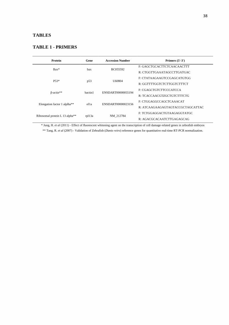
DEPC water. The primers for the constitutive genes beta actin1, ef1a and rpl13a (Table 1)
were according from Tang (2007). Zebrafish gene transcripts were obtained from Jung et al
(2011).
All real time assays were carried out in triplicate using an Applied Biosystems 7500 real-
time PCR system. Forty amplification cycles were performed, with each cycle consisting of
94ºC for 15 seconds followed by 60ºC for 35 seconds. Dissociation curves were used for the
analysis of reaction specificity, and amplification curves generated by the software Applied
Biosystems 7500 were used for gene expression analysis.
Threshold cycle (Ct) values were obtained for each gene. Following the removal of
outliers, raw fluorescence data were exported to the LinRegPCR 12.x
(http://LinRegPCR.HFRC.nl) to determine the PCR amplification efficiency of each sample.
PCR efficiency of each sample, together with Ct values, was used to calculate a relative gene
expression value for each transcript, according to the equation: R = (Eref)CT Target
X (ETarget)-CT
Target X (ETarget)
CT Ref X ( Eref)
-CT Ref , where ERef refers to PCR efficiency of the reference
Gene, ETarget refers to PCR efficiency of the target gene, CtRef is the Ct value for each
amplification of the reference gene and CtTarget to the Ct value for each amplification of the
target gene in question (Pfaffl, 2001). This equation was calculated for each gene with each
reference gene, and a mean of this values was obtained for the evaluation of target gene
expression.
2.6. Statistical Analysis
All data were parametrically analyzed using One-Way ANOVA followed by Tukey’s
post-hoc test. A p<0.05 was considered to be significant and all data are expressed as Mean +
S.D.
3. RESULTS
3.1. Survival
Cadmium-treated groups showed high survival after 1-day exposure to cadmium
(control: 93.33%; 10 µg/L: 96.66%; 100 µg/L: 100.0%; 1000 µg/L: 96.66%). Similarly,
28
survival was high after 7 days of cadmium exposure (control: 100.0%; 10 µg/L: 96.66%; 100
µg/L: 100.0%; 1000 µg/L: 100.0%).
3.2. Behavioral Analysis
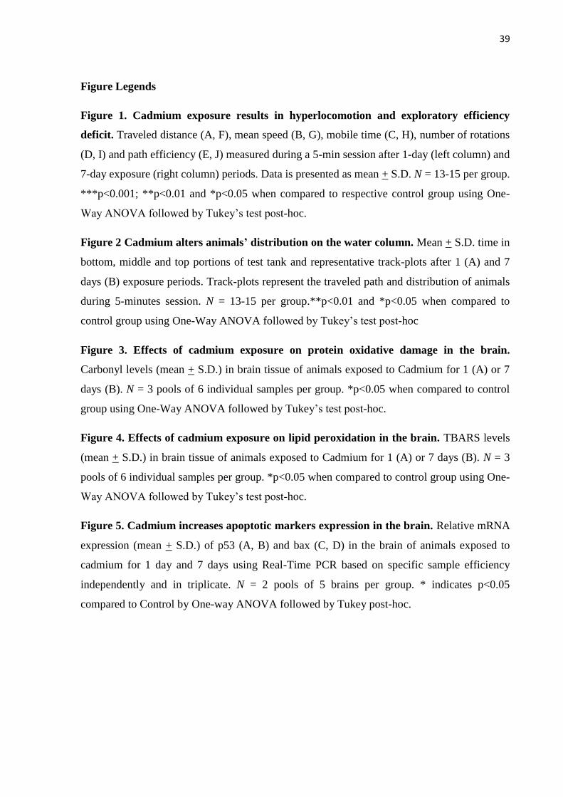
Results for the behavioral analysis are presented in Figure 1. Cadmium-treated groups
demonstrated a general increase in locomotion when compared to controls. Animals exposed
for 1 day to 10 and 1000 µg/L showed increased traveled distance and mean speed when
compared to their control group (p<0.05 and p<0.05, respectively; Figures 1A and 1B). All 1-
day cadmium-treated animals spent significantly more time mobile (p<0.001; Figure 1C) and
made more full body turns (p<0.01; Figure 1D) during the analyzed 5-minutes period than
their control. Interestingly, despite hyperlocomotion, animals path efficiency was decreased in
all 1-day treated groups when compared to the control group (p<0.001; Figure 1E).
Accordingly, after 7-day cadmium exposure at 10, 100 and 1000 µg/L, a significantly
dose-dependent increase in locomotion accompanied by a dose-dependent decrease in path
efficiency was observed in treated animals when compared to controls. All 7-day cadmium-
treated groups showed a dose-dependent increase in traveled distance and mean speed when
compared to controls (p<0.001 and p<0.001, respectively; Figures 1F and 1G). Besides, all 7-
day cadmium-treated animals spent more time mobile (p<0.001; Figure 1H) and made more
full body turns (p<0.001; Figure 1I) while path efficiency was dose-dependently decreased
(p<0.001; Figure 1J).
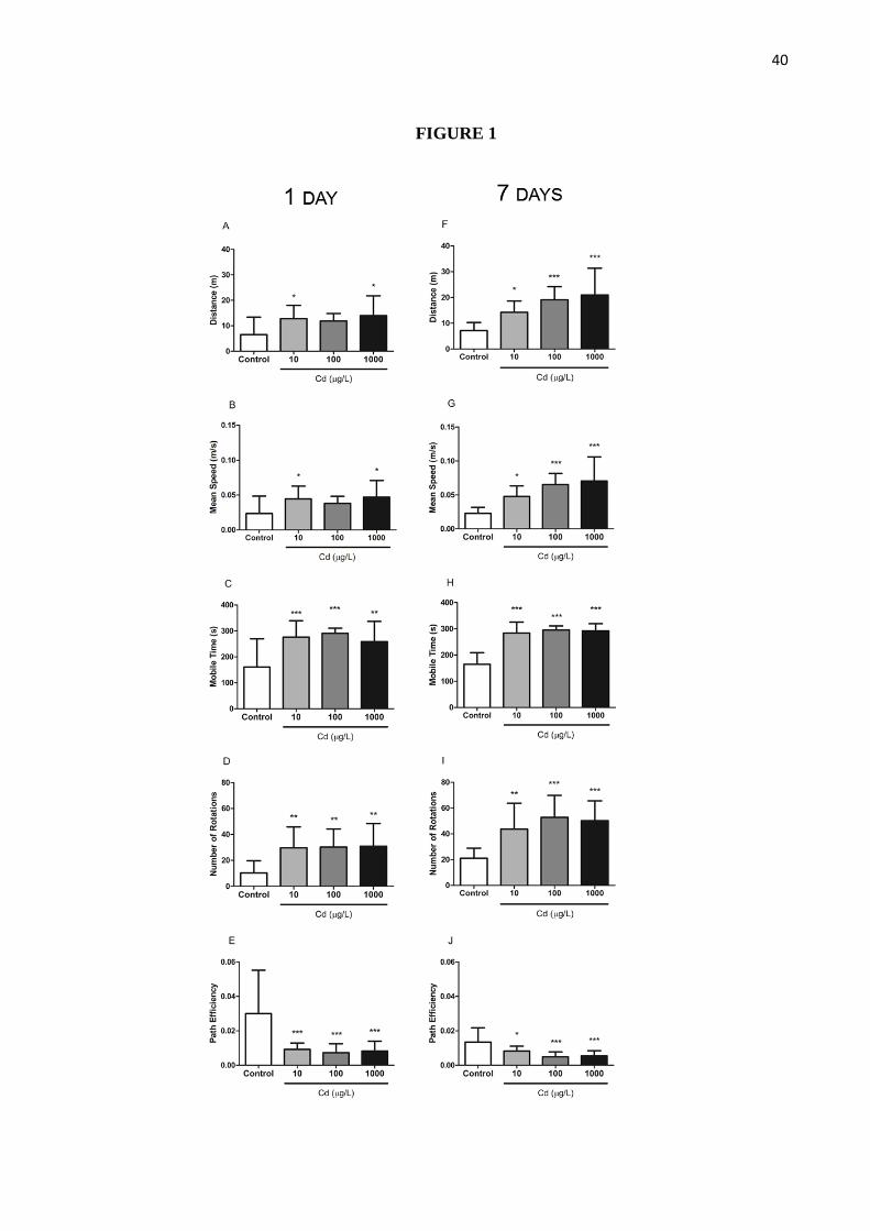
Figure 2 shows mean time spent in the bottom, middle and top portions of the test tank
during the 5-minutes session and representative trackplots. Animals exposed for 1 day to
different concentrations of cadmium showed no significant differences regarding to mean
time spent in the bottom, middle and top portions when compared to their respective control
(p=0.35; Figure 2A). Distinctively, 7 days cadmium-treated groups tended to spend less time
in the bottom and more in the middle and top portions of the tank. Animals treated with 10
µg/L spent significantly less time in the bottom (p<0.01) and more on the top (p<0.05).
Animals exposed to 100 and 1000 µg/L spent in average less time in the top portion (p=0.43
for the 1000 µg/L treated group) and consequently more time in the middle portion (p<0.05
for both groups; Figure 2B).
29
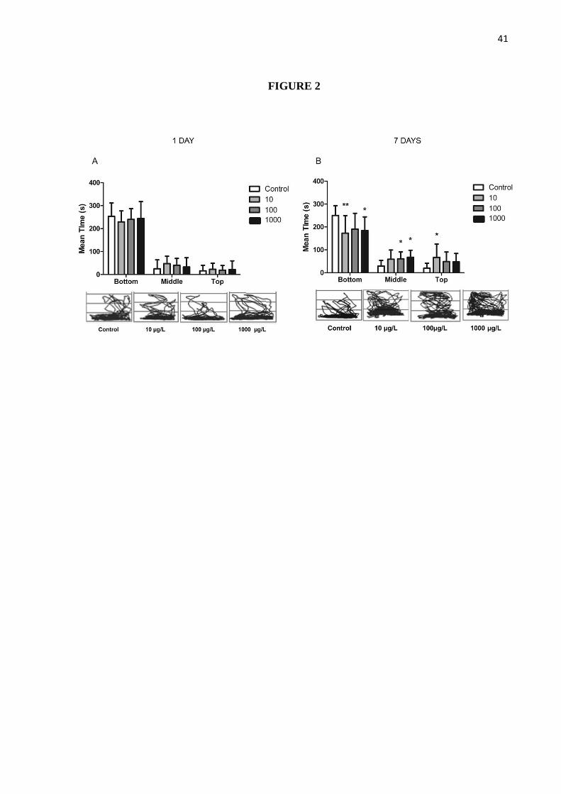
3.3. Oxidative Stress
Cadmium-exposed animals’ brain samples showed increased carbonyl levels and TBARS
depending on dose and treatment period (Figure 3). Brain samples collected from animals
exposed to 100 µg/L for 1 day exhibited an increase in carbonyl levels when compared to
control (p<0.05; Figure 3A) while those of animals treated for 7 days at 10 µg/L were also
increased (Figure 3B).
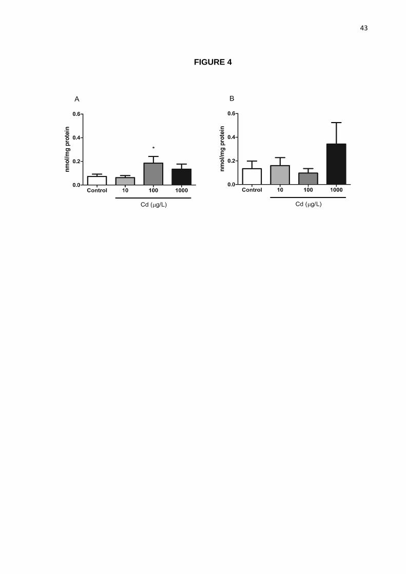
TBARS levels in brain samples from 1-day treated animals at 100 µg/L were increased
when compared to controls (p<0.05; Figure 4A) while no significant difference was observed
at 7 days of exposure.
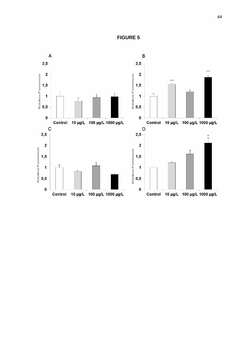
3.4. Real-Time PCR
While no significant effects of cadmium exposure were observed in animals treated for 1
day (Figures 5A and 5C), significant increases of p53 were observed in animals treated with
10 and 1000 µg/L (p<0.001 when compared to controls; Figure 5B), and of bax in animals
exposed to 1000 µg/L (p<0.05; Figure5D).
4. DISCUSSION
The increased locomotor activity observed has been previously described in fish, such as
rainbow trout (Giusiet al, 2005) as well in rats (Fernández-Pérez et al, 2010; Rastogiet al,
1977). It has been shown that cadmium increases levels of catecholamines and amino acid
neurotransmitters in rat central nervous system, culminating in augmented locomotor activity
(Fernández-Pérez et al, 2010; Rastogi et al, 1977), which could account for the observed
effects in our study. Our data also demonstrates, in addition to a robust increase in all
locomotor parameters, decreased path efficiency, suggesting that cadmium-treated animals
have impaired orientation and exploratory performance. Many studies in humans have shown
the deleterious effects of cadmium on central nervous system, such as learning disability and
hyperactivity (Pihl and Parkes, 1977; reviewed by Schoeters et al, 2006) in children as well
neuropsychological disturbances in adults (Hart et al, 1989; Viaene et al, 2000). Furthermore,
it is known that cadmium is capable of inducing aggressiveness in rats (Galbiati et al, 2011;
Salvatoriet al, 2004), which could be related to hyperactivity.
30
The deleterious effects of cadmium on overall animals’ performance were also evident by
animals altered distribution on the water column. When introduced to a novel environment,
zebrafish tend to spend more time at the bottom portion of the test tank and gradually start to
explore the top portions of the tank (Levin et al, 2007). Longer time spent in the bottom
portion and less time spent in higher levels indicates anxiety (Gebauer et al, 2011; Levin et al,
2007; Piato et al, 2011). Thus, our data suggest that animals were less anxious in relation to
controls, increasing their exploratory activity in the novel environment (Figure 2B). This can
be considered a negative effect of cadmium exposure, since zebrafish naturally spends more
time in the bottom portion as a protective behavior to escape predation. Furthermore, it is
known that cadmium affects olfaction on fish, impairing the ability of fish to detect predators
(reviewed by Scott and Sloman, 2004). Thus, increased exploratory activity caused by
diminished anxiety and impaired olfaction may impair animal’s survival, leading to increased
mortality due to predation.
Importantly, the lowest concentration of 10 µg/L, which is considered to be the
maximum, tolerated concentration of the metal in fresh water in Brazil (CONAMA, 2005),
and lies close to other international limits, including 1 µg/L in the U.S.A. (EPA, 2001) and 5
µg/L in Europe (EEC, 1983) caused deleterious behavioral effects to the animals’ behavior
starting at 1-day exposure. This suggests that even concentrations considered safe may impair
animals’ health and survival through disturbing locomotion and exploratory parameters,
which may culminate in disruption of behaviors such as foraging, social interaction (Sloman
et al, 2003) and reproduction (Baker and Montgomery, 2001) and predator avoidance (Scott et
al, 2003).
Several studies have characterized the cellular and molecular mechanisms of cadmium
toxicity (reviewed by Cannino, 2009; reviewed by Thévenod, 2009). Carbonyl is the most
general marker of protein oxidation(Dalle-Donne et al, 2003) while lipid peroxidation
inferred by TBARS is another traditional marker of the deleterious effects of increased
oxidative damage. The increase in the carbonyl and TBARS levels at 1-day exposure in brain
samples may be explained due to cadmium’s ability to decrease antioxidant activity (reviewed
by Thévenod, 2009) and this organ being sensitive to cadmium toxicity. Different from other
tissues such as muscle in which efficient mechanisms of protection and detoxification are
present (Gonzalez et al, 2005), cadmium tends to accumulate in brain (Giusiet al, 2005),
explaining the increased susceptibility of the latter.
Extended exposure to cadmium might enhance activity of antioxidant proteins, as a
consequence of adaptative induction of genes (reviewed by Thévenod, 2009), compensating
31
the cellular damage. Or, maybe, as long-term exposure disturbs the blood-brain barrier,
accumulating more cadmium in the brain, the injury has been so intense that cadmium
disrupted completely the mitochondrial status, decreasing O2 consumption and, subsequently,
decreasing the formation of ROS. Despite the lack of statistically significant difference at 7
days on TBARS levels at the highest cadmium concentration, probably due the high data
variability, Cd tends to increase lipid peroxidation through interaction with proteins, mainly
the ones with SH-groups, and Fenton reactions in the brain, which contains a great pool of
Fe+2
and Cu+2
(Lópezet al, 2006; Thévenod, 2009). Extended exposure to the metal and/or
high doses disturb several intracellular pathways involved in lipid metabolism, culminating in
a wide range of damage to cell, especially to the cell membrane.
Cellular injury caused through ROS may trigger apoptosis. Many reports have shown
cadmium’s ability to induce apoptosis in several cell types, including fish gills and brain
(Azzouqielet al, 1994;Dong et al, 2001;Habeebuet al, 1998; Lópezet al, 2003, Liu et al,
2011). After 1-day cadmium exposure, p53 and Bax levels were not altered, suggesting that
this short treatment, despite other effects, was not capable of inducing detectable levels of
apoptosis in the brain. Tokumotoet al (2011) showed that NRK-52E cells treated with
cadmium induce activation of p53 after 3h of treatment but p53 mRNA levels were
downregulated until 12h of treatment in the study. When exposure time was increased, p53
and Bax gene expression were increased, suggesting a sustained activation of the cell
apoptotic machinery. One may speculate that the depletion of antioxidant enzymes and/or the
intense damage caused to mitochondria leading to DNA damage could have activated the
expression of p53 in order to protect the brain against mutagenesis. Besides, it has been
shown that cadmium not only induces cell death through apoptosis but also through necrosis
(Krumschnabelet al, 2010), since cadmium is able to deplete the intracellular levels ATP and
inhibit caspase-3 activity at elevated doses (Lópezet al, 2003). Finally, cadmium-induced
apoptosis in the central nervous system has been linked to neurodegenerative disorders, such
as Parkinson’s Disease (Okuda et al, 1997) and amyotrophic lateral sclerosis (Bar-Sela et al.,
2001), being accumulated in the striatum in rodents (Fernández-Pérez et al, 2010) and regions
responsible for motor activity in fish (Giusiet al, 2005), which could explain the
hyperlocomotion.
In conclusion, we have shown a robust increase in fish locomotor behavior in all tested
concentrations and treatment periods, starting after 1 day exposure of cadmium at 10 µg/L.
The hyperlocomotion was accompanied by decreased path efficiency and a disrupted
distribution on the water column. Oxidative stress was observed in brains tissue of treated

32
animals, and, consequently, an increased expression of apoptotic markers after 7 days of
exposure suggest a persistent deleterious effect of cadmium exposure.
Taken together, these effects suggest that, despite the lack of acute lethal effects,
cadmium exposure at currently legally tolerated concentrations and those observed in release
accidents, even for short periods, may lead to behavioral effects that could later compromise
animals’ behavioral and cognitive performance and survival.
Competing interests
There are no competing interests.
Acknowledgement
This study was supported by DECIT/SCTIE-MS through Conselho Nacional de Pesquisa
e Desenvolvimento (CNPq - 567483/2008-8) and Fundação de Amparo à Pesquisa do Estado
do Rio Grande do Sul (FAPERGS) (Proc. 10/0036-5, Conv. n. 700545/2008). We are thankful
for received fellowships (M.R.V.: CNPq 305060/2009-0; G.N.I: CAPES).
References
Ansari, M.I., Malik, A., 2010. Seasonal variation of different microorganisms with nickel and cadmium in the
industrial wastewater and agricultural soils. Environ. Monit. Assess. 167(1-4), 151-63.
Atchison, G.J., Henry, M.G., Sandheinrich, M.B., 1987. Effects of metals on fish behavior: a review.
Environmental Biology of Fishes. 18(1), 11-25.
ATSDR. Agency for Toxic Substance and Disease Registry. Toxicological Profile for cadmium. Atlanta:
ATSDR, p. 347, 1997.
Azzouqiel, B., Tsangaris, G.T., Pellegrini, O., Manuel, Y., Benvieniste, J., Thomas, Y., 1994. Cadmium induces
apoptosis in a human T cell line. Toxicology. 88, 127-39.
Bhagure, G.R., Mirgane, S.R., 2009. Heavy metal concentrations in groundwaters and soils of Thane Region of
Maharashtra, India. Environ. Monit. Assess. DOI: 10.1007/s10661-010-1412-9.
Baker, C.F., Montgomery, J.C., 2001. Sensory deficits induced by cadmium in banded kokopu, Galaxias
fasciatus, juveniles. Environ. Biol. Fish. 62, 455–464.
Bar-Sela, S., Reingold, S., Richter, E.D., 2001. Amyotrophic lateral sclerosis in a battery-factory worker exposed
to cadmium. Int. J. Occup. Environ. Health. 7, 109–112.
Beauvais, S.L., Jones, S.B., Parris, J.T., Brewer, S.K., Little, E.E., 2001. Cholinergic and behavioral
neurotoxicity of carbaryl and cadmium to larval rainbow trout (Oncorhynchus mykiss). Ecotoxicol.
Environ. Saf. 49(1), 84-90.
Bernard, A., 2008. Cadmium and its adverse effects on human health. Indian. J. Med. Res. 128, 557-564.
33
Bhakta, J.N., Munekage, Y., 2008. Role of ecosystem components in Cd removal process of aquatic ecosystem.
Ecol. Eng. 32, 274–280.
Blechinger, S. R., Kusch, R. C., Haugo, K., Matz, C., Chivers, D. P., Krone, P. H., 2007. Brief embryonic
cadmium exposure induces a stress response and cell death in the developing olfactory system followed by
long-term olfactory deficits in juvenile zebrafish. Toxicology and Applied Pharmacology. 224(1), 72-80.
Bondier, J.R.; Michel, G.; Propper, A.; Badot, P.M. Harmful effects of cadmium on olfactory system in mice.
Inhal Toxicol. 20(13), p. 1169-77, 2008.
Buske, C., Gerlai, R., 2011. Maturation of shoaling behavior is accompanied by changes in the dopaminergic and
serotoninergic systems in zebrafish. Dev Psychobiol. 54(1), 28-35.
Cannino, G., Ferruggia, E., Luparello, C., Rinaldi, A.M., 2009. Cadmium and Mitochondria. Mitochondrion. 9,
377-384.
Cinier, C.C., Petit-Ramel, M., Faure, R., Garin, D., Bouvet, Y., 1999. Kinetics of cadmium accumulation and
elimination in carp (Cyprinus carpio) tissues. Comp. Biochem. Physiol. C. 122, 345–352.
CONAMA. Brazilian Environmental Conciul. Resolution number 357
<http://www.mma.gov.br/port/conama/res/res05/res35705.pdf>. Last accession: Jan.12, 2012.
Dalle-Donne, I., Rossi, R., Giustarini, D., Milzani, A., Colombo, R., 2003. Protein carbonyl groups as
biomarkers of oxidative stress. Clin. Chim. Acta. 329:23–38.
Dargusch, R., Piasecki, D., Tan, S., Liu, Y., Schubert, D., 2001. The role of Bax in glutamate-induced nerve cell
death, J. Neurochem. 76, 295–301.
Dong, S., Shen, H.M., Ong, C.N., 2001. Cadmium induced apoptosis and phenotypic changes in mouse
thymocytes. Mol. Cell Biochem. 222, 11-20.
Draper, H.H., Hadley, M., 1990. Malondialdehyde determination as index of lipid peroxidation. Methods
Enzymol. 186:421–431.
EEC. European Environment Council., 1983. On limit values and quality objectives for cadmium discharges.
<http://eur-lex.europa.eu/LexUriServ/LexUriServ.do?uri=OJ:L:1983:291:0001:0008:EN:PDF>. Last
accession: Jan. 12, 2012.
EPA. United States Environmental Protection Agency., 2001. Update of ambient water quality criteria for
cadmium.
<http://water.epa.gov/scitech/swguidance/standards/upload/2001_04_13_criteria_cadmium_cad2001upd.pd
f>. Last accession: Jan. 12, 2012.
Egan, R.J., Bergner, C.L., Hart, P.C., Cachat, J.M., Canavello, P.R., Elegante, M.F., Elkhayat, S.I., Bartels, B.K.,
Tien, A.K., Tien, D.H., Mohnot, S., Beeson, E., Glasgow, E., Amri, H., Zukowska, Z., Kalueff, A.V., 2009.
Understanding behavioral and physiological phenotypes of stress and anxiety in zebrafish. Behavioral
Brain Research. 205(1), 38-44.
Fernández-Pérez B, Caride A, Cabaleiro T, Lafuente A. 2010. Cadmium effects on 24h changes in glutamate,
aspartate, glutamine, GABA and taurine content of rat striatum. J Trace Elem Med Biol. 24 (3), 212-8.
Fridman, J.S., Lowe, S.W., 2003. Control of apoptosis by p53. Oncogene. 22, 9030–9040.
Galbiati, S.T., Almeida, A.A., Godinho, A.F., 2011.Cadmium and exposure to stress increase aggressive
behavior. Environmental Toxicology and Pharmacology. 32, 40–45.
34
Gebauer, Daiane L. ; Pagnussat, Natália ; Piato, Ângelo L. ; Schaefer, Isabel C. ; Bonan, Carla D. ; Lara, Diogo
R. . Effects of anxiolytics in zebrafish: Similarities and differences between benzodiazepines, buspirone
and ethanol. Pharmacology, Biochemistry and Behavior, v. 99, p. 480-486, 2011.
Giusi, G., Facciolo, R.M., Alò, R., Carelli, A., Madeo, M., Brandmayr, P., Canonaco, M., 2005. Some
environmental contaminants influence motor and feeding behaviors in the ornate wrasse (Thalassoma pavo)
via distinct cerebral histamine receptor subtypes. Environ. Health. Perspect. 113(11), 1522-9.
Gonzalez, P., Baudrimont, M., Boudou, A., Bourdineaud, J.P., 2006. Comparative effects of direct cadmium
contamination on gene expression in gills, liver, skeletal muscles and brain of the zebrafish (Danio rerio).
BioMetals. 19:225–235.
Gottofrey, J., Tjälve, H., 1991. Axonal transport of cadmium in the olfactory nerve of the pike. Pharmacol.
Toxicol. 69, 242-252.
Guo, S., 2004. Linking genes to brain, behavior and neurological diseases: what can we learn from zebrafish?
Genes Brain Behav. 3(2), 63–74.
Habeebu, S.S., Liu, J., Klassen, C.D., 1998. Cadmium induced apoptosis in mouse liver. Toxicol Appl
Pharmacol. 149, 203-9.
Hart, R.P., Rose, C.S., Hamer, R.M., 1989. Neuropsychological effects of occupational exposure to cadmium. J.
Clin. Exp. Neuropsychol. 11,933–943.
Järup, L., Akasson, A., 2009. Current status of cadmium as an environmental health problem. Toxicology and
Applied Pharmacology. 238, 201–208.
Jayanthi, S., Deng, X., Noailles, P.A., Ladenheim, B., Cadet, J.L., 2004. Methamphetamine induces neuronal
apoptosis via cross-talks between endoplasmic reticulum and mitochondria-dependent death cascades,
FASEB J. 18, 238–251.
Joers, A., Jaks. V., Kase, J., Toivo, M., 2004. p53-dependent transcription can exhibit both on/off and graded
response after genotoxic stress. Oncogene. 23, 6175–6185.
Jung, H., Kim, T.H., Lee, C.R., Seok, S.H., Park, J.H., Chang, S.N., Seo, J.E., Han, H., Ko, A.S., Byun, S.H.,
Abdelkader, T.S., Choi, S.K., Kim, J.A., 2011. Effect of fluorescent whitening agent on the transcription of
cell damage-related genes in zebrafish embryos. J. Appl. Toxicol. DOI 10.1002/jat.1665.
Kargin, F., Çogun, H.I., 1999. Metal Interactions During Accumulation and Elimination of Zinc and Cadmium in
Tissues of the Freshwater Fish Tilapia nilotica. Bull. Environ. Contam. Toxicol. 63, 511-519.
Kastenhuber, E., Kratochwil, C.F., Ryu, S., Schweitzer, J., Driever, W., 2010. Genetic dissection of
dopaminergic and noradrenergic contributions to catecholaminergic tracts in early larval zebrafish. J.
Comp. Neurol. 518(4), 439–58.
Komjarova, I., Blust, R., 2009. Multimetal interactions between Cd, Cu, Ni, Pb, and Zn uptake from water in the
zebrafish Danio rerio. Environ Sci Technol 43(19), 7225–9.
Krumschnabel, G., Ebner, H. L., Hess, M. W., Villunger, A., 2010. Apoptosis and necroptosis are induced in
rainbow trout cell lines exposed to cadmium. Aquatic Toxicology. 99, 73-85.
Kumar, R., Asic, K., Agarwal, K., Seth, P.K., 1996. Oxidative stress mediated neurotoxicity of cadmium.
Toxicol. Lett. 89, 65–69. M during embryonic and larval development results in the long-term
hindrance of antipredator behavior in zebrafish. Environmental Toxicology and Chemistry. 27(3),
705-11.
35
Kusch, R. C., Krone, P. H., Chivers, D. P., 2007. Chronic exposure to low concentrations of waterbone cadmiu
Levin, E.D., Bencan, Z., Cerutti, D.T., 2007. Anxiolytic effects of nicotine in zebrafish. Physiol Behav, 90, 54–
58.
Levine, R.L., Garland, D., Oliver, C.N., 1990. Determination of carbonyl content in oxidatively modified
proteins. Methods Enzymol. 186:464–478.
Lillesaar, C., Tannhäuser, B., Stigloher, C., Kremmer, E., Bally-Cuif, L., 2007. The serotonergic phenotype is
acquired by converging genetic mechanisms within the zebrafish central nervous system. Dev. Dyn. 236(4),
1072–84.
Liu, M., Tee, C., Zeng, F., Sherry, J.P., Dixon, B., Bols, N.C., Duncker, B.P., 2011. Characterization of p53
expression in rainbow trout. Comp. Biochem. Physiol. C., Toxicol Pharmacol. 154(4), 326-32.
López, E.; Figueroa, S.; Oset-Gasque, M.J.; González, M.P., 2003. Apoptosis and necrosis: two distinct events
induced by cadmium in cortical neurons in culture. British Journal of Pharmacology. 138, 901-11.
Lowry, O.H., Rosebrough, N.J., Farr, A.L., Randall, R.J., 1951. Protein measurement with the Folin phenol
reagent. The Journal of Biological Chemistry 193(1), 265–275.
O’Callaghan, J. P., Miller, D., 1986. Diethyldithiocarbamate increases distribution of cadmium to brain but
prevents cadmium-induced neurotoxicity. Brain Res. 370, 354–358.
Okuda, B., Iwamoto, Y., Tachibana, H., Sugita, M., 1997. Parkinsonism after cadmium poisoning. Clinical
Neurology and Neurosurgery. 99, 263-265.
Pfaffi, M.W., 2001. A new mathematical model for relative quantification in real-time PT-PCR. Nucleic Acid
Res. 29, 2003-2007.
Piato, Â.L., Capiotti, K.M., Tamborski, A.R., Oses, J.P., Barcellos, L.J., Bogo, M.R., Lara, D.R., Vianna, M.R.,
Bonan, C.D., 2011. Unpredictable chronic stress model in Zebrafish (Danio rerio): Behavioral and
physiological responses, Prog Neuro-Psychopharmacol Biol Psychiatry. doi:10.1016/j.pnpbp.2010.12.018
Pihl, R.O., Parkes, M., 1977. Hair element content in learning disabled children. Science. 198(4313), 204-6.
Polster, B.M., Fiskum, G., 2004. Mitochondrial mechanisms of neural cell apoptosis, J. Neurochem. 90, 1281–
1289.
Pretto, A., Loro, V.L., Moraes, B.S., Clasen, B., Baldisserotto. B. Isabela, C.M., Finamor, A., Cattaneo, R.,
Dressler, V., 2011. Effects of Water Cadmium Concentrations on Bioaccumulation and Various Oxidative
Stress Parameters in Rhamdia quelen. Arch. Environ. Contam. Toxicol. 60, 309–318.
Rashed, M.N., 2001. Cadmium and lead levels in fish (Tilapia nilotica) tissues as biological indicator for lake
water pollution. Environmental Monitoring and Assessment. 68, 75–89.
Rastogi, R.B., Merali, Z., Singhal, R.L., 1977. Cadmium alters behaviour and the biosynthetic capacity for
catecholamines and serotonin in neonatal rat brain. J. Neurochem. 28(4), 789-94.
Rosemberg, D.B., Rico, E.P., Guidoti, M.R., Dias, R.D., Souza, D.O., Bonan, C.D., 2007. Adenosine deaminase-
related genes: molecular identification, tissue expression pattern and truncated alternative splice isoform in
adult zebrafish (Danio rerio). Life Sci. 81(21–22), 1526–34.
Salvatori, F., Talassi, C.B., Salzgeber, S.A., Spinosa, H.S., Bernardi, M.M., 2004. Embryotoxic and long-term
effects of cadmium exposure during embryogenesis in rats. Neurotoxicol. Teratol. 26 (5), 673–680.
Schoeters, G., Den Hond, E., Zuurbier, M., Naginiene, R., van den Hazel, P., Stilianakis, N., Ronchetti, R.,
Koppe, J.G., 2006. Cadmium and children: exposure and health effects. Acta Paediatr Suppl. 95(453), 50-4.
36
Scott, G.R., Sloman, K.A., 2004. The effects of environmental pollutants on complex fish behaviour: integrating
behavioural and physiological indicators of toxicity. Aquatic Toxicology. 68, 369–392..
Slee, E.A., O’Connor, D.J., Lu, X., 2004. To die or not to die: how does p53 decide? Oncogene 23, 2809–2818.
Sloman, K.A., Scott, G.R., Diao, Z., Rouleau, C., Wood, C.M., McDonald, D.G., 2003. Cadmium affects the
social behavior of rainbow trout, Oncorhynchus mykiss. Aquat. Toxicol. 65, 171–185.
Soares, S.S., Martins, H., Gutiérrez-Merino, C., Aureliano, M., 2008. Vanadium and cadmium in vivo effects in
teleost cardiac muscle: metal accumulation and oxidative stress markers. Comp Bochem Physiol C.
147,168–178.
Szebedinsky, C., McGeer, J.C., McDonald, D.G. and Wood, C.M., 2001. Effects of chronic Cd exposure via the
diet or water on internal organspecific distribution and subsequent gill Cd uptake kinetics in juvenile
rainbow trout (Oncorhynchus mykiss). Environ. Toxicol. Chem. 20, 597-607.
Takeda, A.; Takefuta, S.; Ijiro, H.; Okada, S.; Oku, N. 1999. 109Cd transport in rat brain. Brain Res Bull. 49 (6),
453-7.
Tallkvist, J., Persson, E., Henriksson, J., Tjälve, H., 2002. Cadmium-metallothionein interactions in the olfactory
pathways of rats and pikes. Toxicol Sci. 67(1),108-13.
Tang, R., Dodd, A., Lai, D., McNabb, W., Love, D., 2007. Validation of Zebrafish (Danio rerio) reference genes
for quantitative real-time RT-PCR normalization. Acta Biochime. Biophys. Sin. 38, 384-90.
Thévenod, F., 2009. Cadmium and cellular signaling cascades: To be or not to be? Toxicology and Applied
Pharmacology. 238, 221–239.
Tjälve, H., Gottofrey, J., 1986. Tissue disposition of in the brown trout (Salmo trutta) studied by autoradiography
and impulse counting. Toxicol. Environ. Chem. 12, 31-45.
Tjälve, H., Henriksson, J., 1999. Uptake of metals in the brain via olfactory athways. Neurotoxicology 20, 181-
196.
Tokumoto, M., Fujiwara, Y., Shimada, A., Hasegawa, T., Seko, Y., Nagase, H., Satoh, M., 2011. Cadmium
toxicity Is caused by accumulation of p53 through the down-regulation of Ube2d family genes in vitro and
in vivo. The Journal of Toxicological Sciences, 36 (2), 191-200.
Tropepe, V.; Sive, H.L., 2003. Can zebrafish be used as a model to study the neurodevelopmental causes of
autism? Genes Brain Behav. 2, 268– 281.
Verbost, P.M., Flik, G., Lock, R.A.C., Wendelaar Bonga, S. E., 1987. Cadmium inhibition of Ca2+ uptake in
rainbow trout gills. Am. J. Physiol. 253, 216-221
Verbost, P.M., Rooij, J.V., Flik, G., Lock, R.A.C., Wendelaar Bonga, B.S.E., 1989. The movement of cadmium
through freshwater trout branchial epithelium and its interference with calcium transport. J. Exp. Biol. 145,
185-197.
Viaene, M.K., Massachelein, R., Leenders, J., De Groof, M., Swerts, L.J.V.C., Roels, H.A., 2000.
Neurobehavioural effects of occupational exposure to cadmium: a cross sectional epidemiological study.
Occup. Environ. Med. 57:19–27 19
Ward, M.W., Kogel, D., Prehn, J.H., 2004. Neuronal apoptosis: BH3 only proteins the real killers? J. Bioenerg.
Biomembr. 36, 295–298.
Westerfield, M., 2000. The zebrafish book. A guide for the laboratory use of zebrafish (Danio rerio). 4th ed.
Eugene: Univ. of Oregon Press.

37
WHO. World Health Organization. Cadmium. Geneva, 1992. (Environment Health Criteria 134).
Yamamoto, K., Ruuskanen, J.O., Wullimann, M.F., Vernier, P., 2010. Two tyrosine hydroxylase genes in
vertebrates new dopaminergic territories revealed in the zebrafish brain. Mol. Cell Neurosci. 43(4), 394–
402.
Yano, C.L., Marcondes, M.C.C.G., 2005. Cadmium chloride-induced oxidative stress in skeletal muscle cells in
vitro. Free Radical Biology & Medicine 39, 1378 – 1384.
38
TABLES
TABLE 1 - PRIMERS
Protein Gene Accession Number Primers (5'-3')
Bax* bax BC055592 F: GAGCTGCACTTCTCAACAACTTT
R: CTGGTTGAAATAGCCTTGATGAC
P53* p53 U60804 F: CTATAAGAAGTCCGAGCATGTGG
R: GGTTTTGGTCTCTTGGTCTTTCT
β-actin** bactin1 ENSDART00000055194 F: CGAGCTGTCTTCCCATCCA
R: TCACCAACGTZGCTGTCTTTCTG
Elongation factor 1 alplha** ef1a ENSDART00000023156 F: CTGGAGGCCAGCTCAAACAT
R: ATCAAGAAGAGTAGTACCGCTAGCATTAC
Ribosomal protein L 13 alpha** rpl13a NM_212784 F: TCTGGAGGACTGTAAGAGGTATGC
R: AGACGCACAATCTTGAGAGCAG
* Jung, H. et al (2011) - Effect of fluorescent whitening agent on the transcription of cell damage‐related genes in zebrafish embryos
** Tang, R. et al (2007) - Validation of Zebrafish (Danio reiro) reference genes for quantitative real-time RT-PCR normalization.
39
Figure Legends
Figure 1. Cadmium exposure results in hyperlocomotion and exploratory efficiency
deficit. Traveled distance (A, F), mean speed (B, G), mobile time (C, H), number of rotations
(D, I) and path efficiency (E, J) measured during a 5-min session after 1-day (left column) and
7-day exposure (right column) periods. Data is presented as mean + S.D. N = 13-15 per group.
***p<0.001; **p<0.01 and *p<0.05 when compared to respective control group using One-
Way ANOVA followed by Tukey’s test post-hoc.
Figure 2 Cadmium alters animals’ distribution on the water column. Mean + S.D. time in
bottom, middle and top portions of test tank and representative track-plots after 1 (A) and 7
days (B) exposure periods. Track-plots represent the traveled path and distribution of animals
during 5-minutes session. N = 13-15 per group.**p<0.01 and *p<0.05 when compared to
control group using One-Way ANOVA followed by Tukey’s test post-hoc
Figure 3. Effects of cadmium exposure on protein oxidative damage in the brain.
Carbonyl levels (mean + S.D.) in brain tissue of animals exposed to Cadmium for 1 (A) or 7
days (B). N = 3 pools of 6 individual samples per group. *p<0.05 when compared to control
group using One-Way ANOVA followed by Tukey’s test post-hoc.
Figure 4. Effects of cadmium exposure on lipid peroxidation in the brain. TBARS levels
(mean + S.D.) in brain tissue of animals exposed to Cadmium for 1 (A) or 7 days (B). N = 3
pools of 6 individual samples per group. *p<0.05 when compared to control group using One-
Way ANOVA followed by Tukey’s test post-hoc.
Figure 5. Cadmium increases apoptotic markers expression in the brain. Relative mRNA
expression (mean + S.D.) of p53 (A, B) and bax (C, D) in the brain of animals exposed to
cadmium for 1 day and 7 days using Real-Time PCR based on specific sample efficiency
independently and in triplicate. N = 2 pools of 5 brains per group. * indicates p<0.05
compared to Control by One-way ANOVA followed by Tukey post-hoc.
40
FIGURE 1
41
FIGURE 2
42
FIGURE 3
43
FIGURE 4
44
FIGURE 5

CAPÍTULO 3
46
CONSIDERAÇÕES FINAIS
Cádmio representa um risco à saúde humana, bem como de ecossistemas
em geral. Desde a descoberta de sua toxicidade na década de 60, os níveis de
emissão do metal no ambiente, seja aquático, aéreo ou terrestre, têm diminuído,
devido a melhorias no manejo de resíduos e surgimento de novas tecnologias que
poluem menos (Cardoso e Chasin, 2001). Atualmente, o metal é encontrado
principalmente em baterias de níquel-cádmio e em aparelhos eletrônicos, sendo que
o descarte irregular destes itens e a utilização de fertilizantes fosfatados são as
principais fontes de contaminação do ambiente (revisado por Chen et al, 2011). Uma
vez no ambiente, o cádmio contamina diversos organismos, desde plantas e
invertebrados até vertebrados, inclusive o ser humano, participando em complexas
cadeias alimentares.
Peixes são considerados excelentes bioindicadores de toxicidade aquática.
Muitos estudos já foram feitos expondo-se peixes a diferentes concentrações de
cádmio e de tempo, a fim de avaliar comportamentos simples e complexos (revisado
por Scott e Sloman, 2004), dano oxidativo (Singhal et al, 1987; Jihen et al, 2011) e
apoptose (Krumschnabel, 2010). Todavia, estudos que integrem grandes áreas de
estudo como comportamento, biologia celular e molecular são ainda escassos, o que
ajudaria a compreender de forma mais específica os efeitos toxicológicos do cádmio
sobre os animais, principalmente sobre o sistema nervoso central, cuja
compreensão é ainda menor.
A fim de contribuir para um melhor entendimento, nós avaliamos o
comportamento de zebrafish adultos tratados com cádmio por 1 dia e 7 dias. Os
animais apresentaram distância percorrida e velocidade média elevadas em 1 dia,
exceto aqueles tratados com a dose de 100 µg/L. Ademais, os animais
permaneceram mais tempo em movimento bem como realizaram mais rotações em
torno do corpo. Após 7 dias de exposição ao cádmio, os animais apresentaram uma
crescente resposta dose-dependente em relação a distância percorrida (r=0,63) e
velocidade média (r=0,64), assim como tempo móvel (r=0,69) e número rotações
(r=0,55). Atividade locomotora elevada tem sido avaliada em peixes (Giusi et al,
2005) bem como em roedores (Rastogi et al, 1976; Beauvais et a,l 2001; Fernández-
Pérez et al, 2010). Muitos estudos em humanos têm mostrado os efeitos deletérios
do cádmio sobre o sistema nervoso central, tais como déficits de aprendizagem e
47
hiperatividade (Pihl and Parkes, 1977; revisado por Schoeters et al, 2006) em
crianças, bem como perturbações neuropsicológicas em adultos (Hart et al, 1989;
Viaene et al, 2000). Portanto, exposição cádmio induz hiperlocomoção em animais
tratados por curtos e longos períodos.
Animais tratados com cádmio por 7 dias com 10 e 1000 µg/L permaneceram
menos tempo na porção inferior do aquário-teste, sugerindo que os animais estavam
menos ansiosos, elevando a atividade exploratória dos mesmos no novo ambiente.
Em seu ambiente natural, zebrafish passa mais tempo em locais profundos, típico
comportamento para evitar predadores. Além disto, sabe-se que cádmio prejudica o
olfato de peixes (revisado por Scott & Sloman, 2004). Assim, a hiperlocomoção, que
poderia ser causada por menor ansiedade, e o olfato prejudicado podem culminar na
mortalidade dos animais devido à predação.
O Conselho Nacional do Meio Ambiente (CONAMA) estabelece o limite de 10
µg/L de cádmio em água doce. Inesperadamente, esta dose apresentou efeitos
comportamentais deletérios a partir do primeiro dia de exposição. Desta forma, a
dose mais baixa pode implicar na sobrevivência dos animais através de alterações
na locomoção e ansiedade, que culminam em perturbações comportamentais como
forrageio, interação social (Sloman et al, 2003) e reprodução (Baker and
Montgomery, 2001), bem como comportamentos antipredatórios (Scott et al, 2003),
e consequentemente, desestruturando ecossistemas.
Com base nos resultados obtidos, decidimos verificar o possível dano
oxidativo em amostras de encéfalo. A exposição ao cádmio por 1 dia elevou os
níveis de TBARS e carbonil em amostras de encéfalo. Exposição a curto prazo ao
metal tende a diminuir a atividade das enzimas antioxidantes, culminando no
aumento de espécies reativas de oxigênio (revisado por Thévenod, 2009). A
exposição ao cádmio por 7 dias diminuiu os níveis de carbonil quando comparados
com 1 de tratamento. Exposições a longo prazo e doses elevadas de cádmio tendem
a diminuir os níveis de espécies reativas de oxigênio, ou por um mecanismo de
adaptação, induzindo maior expressão das enzimas antioxidantes (revisado por
Thévenod, 2009), ou pela inibição da cadeia transportadora de elétrons, reduzindo o
consumo de oxigênio e, consequentemente, de espécies reativas de oxigênio
(revisado por Cannino et al, 2009). Por fim, os mecanismos protetores e
desintoxicantes no encéfalo são menos eficientes do que em outros tecidos, como o
48
músculo (Gonzalez et al, 2006), sendo assim, considerado um órgão-alvo pelo
cádmio.
Os níveis de expressão gênica de p53 e bax no encéfalo, medidos por Real-
Time PCR, não foram alterados após 1 dia de tratamento com cádmio. Células
possuem um pool de p53 e, caso elas sofram algum dano, este pool é recrutado até
que ocorra transcrição gênica, sendo assim, um mecanismo de proteção agudo.
Exposição ao cádmio por 7 dias aumentou os níveis de p53 e bax significativamente.
O esgotamento de enzimas antioxidantes e excessivo dano causado ao DNA podem
ter ativado a expressão de p53, de maneira a proteger o encéfalo contra
mutagênese. Tokumoto et al (2011) demonstraram que células NRK-52E tratadas
com cádmio induzem a ativação de p53 após 3h de tratamento mas os níveis de
RNAm estavam diminuídos até 12h de tratamento. Ademais, cádmio não apenas
induz morte celular por apoptose, mas também por necrose, pois ele é capaz de
perturbar os níveis intracelulares de ATP e inibir caspase-3 em doses elevadas
(López et al, 2003). Finalmente, evidências sugerem uma ligação entre apoptose
induzida por cádmio no sistema nervosa central e doenças neurodegenerativas, tais
como Parkinson (Okuda et al, 1997), acumulando-se principalmente no estriado, em
roedores, (Fernández-Pérez et al, 2010) e regiões responsáveis pela atividade
motora em peixes (Giusi et al, 2005), que poderia explicar a hiperlocomoção
observada neste estudo.
A grande variedade de efeitos deletérios provocados pelo cádmio ressalta a
complexa interação entre este metal com vias intracelulares, culminando em
alterações comportamentais. A compreensão destes efeitos nos permitiria
desenvolver estratégias preventivas e terapêuticas. Por exemplo, uma alternativa
seria o tratamento com quelantes, que diminuem tais efeitos, inclusive no encéfalo
(Gonçalves et al, 2010). Contudo, mais estudos são necessários para a
compreensão das vias, pois pontos mais básicos, como a permeabilidade da
barreira hematoencefálica e as próprias características do metal, ainda permanecem
obscuros.
49
REFERÊNCIAS
ARORA, M., WEUVE, J., SCHWARTZ, J., WRIGHT, R. O. Association of environmental cadmium exposure with
pediatric dental caries. Environmental Health Perspectives. v. 116, p. 821–825, 2008.
[ATSDR] Agency for Toxic Substance and Disease Registry. Toxicological Profile for cadmium. Atlanta: ATSDR,
p. 347, 1997.
_______. 2007 CERCLA Priority List of Hazardous Substances. U.S, 2007.
AZZOUQIEL, B.; TSANGARIS, G.T.; PELLEGRINI, O.; MANUEL, Y.; BIENVIENISTE, J.; THOMAS, Y. Cadmium
induces apoptosis in a human T cell line. Toxicology. 88, 127-39, 1994.
BAKER, C.F., MONTGOMERY, J.C. Sensory deficits induced by cadmium in banded kokopu, Galaxias fasciatus,
juveniles. Environ. Biol. Fish. 62, 455–464, 2001.
BAR-SELA, S., REINGOLD, S., RICHTER, E.D. Amyotrophic lateral sclerosis in a battery-factory worker exposed
to cadmium. Int. J. Occup. Environ. Health. 7, 109–112, 2001.
BEAUVAIS, S.L., JONES, S.B., PARRIS, J.T., BREWER, S.K., LITTLE, E.E., 2001. Cholinergic and behavioral
neurotoxicity of carbaryl and cadmium to larval rainbow trout (Oncorhynchus mykiss). Ecotoxicol. Environ. Saf.
49(1), 84-90.
BERGLUND, M., ÅKESSON, A., NERMELL, B., VAHTER, M. Intestinal absorption of dietary cadmium in women
depends on body iron stores and fiber intake. Environmental Health Perspectives. v. 102, p. 1058–1066, 1994.
BERNARD, A.; LAUWERYS, R. Cadmium in human population. Experientia. v. 40, p. 143–152, 1984.
BERNARD, A. Cadmium and its adverse effects on human health. Indian Journal of Medical Research. v. 128, p.
557-564, 2008.
BONDIER, J.R.; MICHEL, G.; PROPPER, A.; BADOT, P.M. Harmful effects of cadmium on olfactory system in
mice. Inhal Toxicol. 20(13), p. 1169-77, 2008.
BOYD, R. S. Heavy metal pollutants and chemical toxicology: exploring new frontiers. Journal of Chemical
Ecology. v. 36, p. 46-58, 2010.
BRASIL. Conselho Nacional do Meio Ambiente. Resolução CONAMA nº 357: dispõe sobre a classificação dos
corpos de água e diretrizes ambientais para o seu enquadramento, bem como estabelece as condições e
padrões de lançamento de efluentes, e dá outras providências.
http://www.mma.gov.br/port/conama/res/res05/res35705.pdf>. Acesso em: 15 de abril de 2010.
CANNINO, G.; FERRUGGIA, E.; LUPARELLO, C.; RINALDI, A. M. Cadmium and Mitochondria. Mitochondrion.
v. 9, p. 377-384, 2009, doi: 10.1016/j.mito.2009.08.009.
50
CALMANO, W.; FÖRSTNER, U. Sediments and toxic substances. Springer-Verlag, Berlin, Germany, 1996.
CARDOSO, L. M. N.; CHASIN, A. A. M. 2001. Ecotoxicologia do cádmio e seus compostos. Série Cadernos de
Referência Ambiental. v. 6, 2001.
CHALLIS, G. L. A widely distributed bacterial pathway for siderophore biosynthesis independent of nonribosomal
peptide synthetases. ChemBioChem v. 6, p. 601–611, 2005.
CHAN, P. K.; CHENG, S. H. Cadmium-induced ectopic apoptosis in zebrafish embryos. Archives of Toxicology.
v. 77, p. 69–79, 2003.
CHAO, S. H.; BU, C. H.; CHEUNG, W. Y. Stimulation ofmyosin light-chain kinase by Cd2 + and Pb2+. Archives of
Toxicology. v. 69, p. 197–203, 1995.
CHEN, W.-Y.; JOHN, J. A. C.; LIN, C.-H.; LIN, H.-F.; WU, S.-C.; LIN, C.-H.; CHANG, C.-Y. Expression of
metallothionen gene during embryonic and early larval development in zebrafish. Aquatic Toxicology. v. 69, p.
215–227, 2004.
CHOW, E. S. H.; HUI, M. N. Y.; LIN, C. C.; CHENG, S. H. Cadmium inhibits neurogenesis in zebrafish embryonic
brain development. Aquatic Toxicology. v. 87, p. 157–169, 2008.
CHOW, E.S.; Hui, M.N.; Cheng, C.W.; Cheng, S.H. Cadmium affects retinogenesis during zebrafish embryonic
development. Toxicol Appl Pharmacol. 235(1), p. 68-76, 2009.
DARGUSCH, R.; PIASECKI, D.; TAN, S.; LIU, Y.; SCHUBERT, D.; The role of Bax in glutamate-induced nerve
cell death, J. Neurochem. 76, 295–301, 2001.
DE CONTO CINIER, C.; PETIT-RAMEL, R.; FAURE, R.; BORTOLATO, M. Cadmium accumulation and
metallothionein biosynthesis in Cyprinus carpio tissues. Bulletin of Environmental Contamimantion and
Toxicology, v. 61, p. 793-799, 1998.
DONG, S.; SHEN, H.M.; ONG, C.N. Cadmium induced apoptosis and phenotypic changes in mouse thymocytes.
Mol. Cell Biochem. 222, 11-20, 2001.
DURUIBE, J. O., OGWOEGBU, M. O. C., and EGWURUGWU, J. N. Heavy metal pollution and human biotoxic
effects. International. Journal of Physical Sciences, v. 2, p. 112–118, 2007.
EGAN, S.K., BOLGER, P.M., CARRINGTON, C.D. Update of US FDA's Total Diet Study food list and diets.
Journal of Exposure Science and Environmental Epidemiology, 17 (6), p. 573–582, 2007.
EGAN RJ, BERGNER CL, HART PC, CACHAT JM, CANAVELLO PR, ELEGANTE MF, ELKHAYAT SI,
BARTELS BK, TIEN AK, TIEN DH, MOHNOT S, BEESON E, GLASGOW E, AMRI H, ZUKOWSKA Z, KALUEFF
AV. Understanding behavioral and physiological phenotypes of stress and anxiety in zebrafish. Behavioral Brain
Research. v. 205 (1), p. 38-44, 2009.
51
FERNÁNDEZ-PÉREZ, B., CARIDE, A., CABALEIRO, T., LAFUENTE, A. Cadmium effects on 24h changes in
glutamate, aspartate, glutamine, GABA and taurine content of rat striatum. J Trace Elem Med Biol. 24 (3), p. 212-
8, 2010.
FLEEGER, J. W., CARMAN, K. R., and NISBET, R. M. Indirect effects of contaminants in aquatic ecosystems.
Science of the Total Environment, v. 317, p. 207–233, 2003.
FOULKES, E. C. Cadmium. Handbook Experimental Pharmacology, v. 80, Springer-Verlag, New York, 1986.
FRIBERG, L.; ELINDER, C.-G.; KJELLSTRÖM, T.; NORDBERG, G. Cadmium and Health: a Toxicological and
Epidemiological Appraisal. CRC Press, Inc., Boca Raton, FL. v. 1-2, 1986
FRIDMAN, J.S.; LOWE, S.W. Control of apoptosis by p53. Oncogene. 22, 9030–9040, 2003.
GIUSI, G., FACCIOLO, R.M., AlÒ, R., CARELLI, A., MADEO, M., BRANDMAYR, P., CANONACO, M. Some
environmental contaminants influence motor and feeding behaviors in the ornate wrasse (Thalassoma pavo) via
distinct cerebral histamine receptor subtypes. Environ. Health. Perspect. 113(11), 1522-9, 2005.
GOERING, P. L.; WAALKES, M. P.; KLAASSEN, C. D. Toxicology of Cadmium. In Toxicology of Metals,
Biochemical Aspects, Handbook Experimental Pharmacology, v. 115, p. 189–214, Springer Verlag, New York,
1995.
GONÇALVES, J. F.; FIORENZA, A. M.; SPANEVELLO, R. M.; MAZZANNTI, C. M.; BOCHI, G. V.; ANTES, F. G.;
STEFANELLO, N.; RUBIN, M. A.; DRESSLER, V.; MORSCH, V. M.; SCHETINGER, M. R. C. N-acetylcysteine
prevents memory deficits, the decrease in acetylcholinesterase activity and oxidative stress in rats exposed to
cadmium. Chemico-Biological Interactions. 186, p. 53–60, 2010.
GONZALEZ, P.; BAUDRIMONT, M.; BOUDOU, A.; BOURDINEAUD, J. P. Comparative effects of direct cadmium
contamination on gene expression in gills, liver, skeletal muscles and brain of the zebrafish (Danio rerio).
BioMetals v. 19, p. 225–235, 2006. DOI 10.1007/s10534-005-5670-x
GREGER, M. Metal availability, uptake, transport and accumulation in plants, pp. 1–27, in M. N. V. Prasad (ed.).
Heavy Metal Stress in Plants: From Biomolecules to Ecosystems. Springer, Berlin, 2004.
HABEEBU, S.S.; LIU, J.; KLASSEN, C.D. Cadmium induced apoptosis in mouse liver. Toxicol Appl Pharmacol.
149, 203-9, 1998.
HAN, F. X., BANIN, A., SU, Y., MONTS, D. L., PLODINEC, M. J., KINGERY, W. L., and TRIPLETT, G. E.
Industrial age anthropogenic inputs of heavy metals into the pedosphere. Naturwissenschaften, v. 89, p. 497–
504, 2002.
HANSEN, J.M., ZHANG, H., JONES, D.P. Differential oxidation of thioredoxin-1,-2, and glutathione by metal ions.
Free Radical Biology & Medicine, v. 40, p. 138–145, 2006.
52
HOFSETH, L.J.; HUSSAIN, S.P.; HARRIS, C.C. p53: 25 years after its discovery. Trends Pharmacol. Sci. 25,
177–181, 2004.
[IARC] International Agency for Research on Cancer. Monographs on cancer risks. Beryllium, cadmium, mercury,
and exposures in the glass manufacturing industry. IARC Scientific Publications, Lyon, v. 58, p. 1–415, 1994.
JÄRUP, L.; BERGLUND, M.; ELINDER, C. G.; NORDBERG, G.; VAHTER, M. Health effects of cadmium
exposure–a review of the literature and a risk estimate. Scandinavian Journal of Work, Environment and Health,
v. 24 (Suppl. 1), p. 1–51, 1998.
JÄRUP, L., AKASSON, A. Current status of cadmium as an environmental health problem. Toxicology and
Applied Pharmacology. v. 238, p. 201–208, 2009.
JAYANTHI, S.; DENG, X.; NOAILLES, P.A.; LADENHEIM, B.; CADET, J.L. Methamphetamine induces neuronal
apoptosis via cross-talks between endoplasmic reticulum and mitochondria-dependent death cascades, FASEB
J. 18, 238–251, 2004.
JIHEN, E.H., et al., Interrelationships between cadmium, zinc and antioxidants in the liver of the rat exposed
orally to relatively high doses of cadmium and zinc. Ecotoxicol. Environ. Saf. (2011),
doi:10.1016/j.ecoenv.2011.06.008.
JOERS, A.; JAKS, V.; KASE, J.; TOIVO, M. p53-dependent transcription can exhibit both on/off and graded
response after genotoxic stress. Oncogene. 23, 6175–6185, 2004.
KLASCHKA, U. The infochemical effect—a new chapter in ecotoxicology. Environmental Science and Pollution.
Research, v. 15, p. 452–462, 2008.
KORICHEVA, J., LARSSON, S., and HAUKIOJA, E. Insect performance on experimentally stressed woody
plants: A metaanalysis. Annual Review of Entomology, v. 43, p. 195–216, 1998.
KOSTRZEWSKA, A., SOBIESZEK, A. Diverse actions of cadmium on the smooth muscle myosin
phosphorylation system. FEBS Letters, v. 263, p. 381–384, 1990.
KRUMSCHNABEL, G., EBNER, H. L.; HESS, M. W.; VILLUNGER, A.; Apoptosis and necroptosis are induced in
rainbow trout cell lines exposed to cadmium. Aquatic Toxicology. 99 (1), p73-85, 2010.
KUSCH, R. C.; KRONE, P. H.; CHIVERS, D. P. Chronic exposure to low concentrations of waterborne cadmium during embryonic and larval development results in the long-term hindrance of antipredator behavior in zebrafish., v. 27(3), p. 705-10, 2008.
LARSEN, E.H., ANDERSEN, N.L., MOLLER, A., PETERSEN, A., MORTENSEN, G.K., PETERSEN, J. Monitoring the content and intake of trace elements from food in Denmark. Food Additives and Contaminants, 19 (1), p. 33–46, 2002.
LASS, S., SPAAK, P. Chemically induced anti-predator defences in plankton: a review. Hydrobiologia. v. 491, p.
221–239, 2003.
53
LEFCORT, H., AGUON, M. Q., BOND, K. A., CHAPMAN, K. R., CHAQUETTE, R., CLARK, J., KORNACHUK, P.,
LANG, B. Z., and MARTIN, J. C. Indirect effects of heavy metals on parasites may cause shifts in snail species
composition. Archives of Environmental Contamiantion and Toxicology, v. 43, p. 34–41, 2002.
LIU, Y.; TEMPLETON, D.M. Cadmium activates CaMK-II and initiates CaMK-IIdependent apoptosis in mesangial
cells. FEBS Letters, v. 581, p. 1481–1486, 2007.
LIU. Y.; WANG, J.; WEI, Y.; ZHANG, H.; XUA, M.; DAI, J. Induction of time-dependent oxidative stress and
related transcriptional effects of perfluorododecanoic acid in zebrafish liver. Aquatic Toxicology. v. 89, p. 242–
250, 2008.
LIU, F., TANG, Y., DU, R., YANG, H., WU, Q., and RONGLIANG, Q. Root foraging for zinc and cadmium
requirement in the Zn/ Cd hyperaccumulator plant Sedum alfredii. Plant Soil. (2009), doi:10.1007/s11104-009-
0060-8.
LIU, M.; TEE, C.; ZENG, F.; SHERRY, J.P.; DIXON, B.; BOLS, N.C.; DUNCKER, B.P. Characterization of p53
expression in rainbow trout. Comp. Biochem. Physiol. C., Toxicol Pharmacol. 154(4), 326-32, 2011.
LLOBET, J.M., FALCO, G., CASAS, C., TEIXIDO, A., DOMINGO, J.L. Concentrations of arsenic, cadmium,
mercury, and lead in common foods and estimated daily intake by children, adolescents, adults, and seniors of
Catalonia, Spain. Journal of Agricultural and Food Chemistry, 51 (3), p. 838–842, 2003.
LÓPEZ, E.; FIGUEROA, S.; OSET-GASQUE, M.J.; GONZÁLEZ, M.P. Apoptosis and necrosis: two distinct
events induced by cadmium in cortical neurons in culture. British Journal of Pharmacology. 138, 901-11, 2003.
LÜRLING, M., and SCHEFFER, M. Info-disruption: pollution and the transfer of chemical information between
organisms. Trends in Ecology & Evolution. v. 22, p. 374–379, 2007.
MACINTOSH, D.L., SPENGLER, J.D., OZKAYNAK, H., TSAI, L., RYAN, P.B. Dietary exposures to selected
metals and pesticides. Environmental Health Perspectives. 104 (2), p. 202–209, 1996.
MATOVIĆ, V., BUHA, A., BULAT, Z., ĐUKIĆ-ĆOSIĆ, D. Cadmium toxicity revisited: focus on oxidative stress
induction and interactions with zinc and magnesium. Arh Hig Rada Toksikol. 62, p. 65-76, 2011.
MAZZEI, G.J., GIRARD, P.R., KUO, J.F. Environmental pollutant Cd2+ biphasically and differentially regulates
myosin light chain kinase and phospholipid/Ca2+- dependent protein kinase. FEBS Letters. v. 173, p. 124–128,
1984.
MCPHERSON, T. D., MIRZA, R. S., and PYLE, G. G. Responses of wild fishes to alarm chemicals in pristine and
metalcontaminated lakes. Canadian Journal of Zoology, v. 82, p. 694–700, 2004.
MORSELT, A. W. F. Environmental pollutants and diseases: A cell biological approach using chronic cadmium
exposure in the animal model as a paradigm case. Toxicology, v. 70, p. 1–84, 1991.
O’CALLAGHAN, J. P., MILLER, D. Diethyldithiocarbamate increases distribution of cadmium to brain but
prevents cadmium-induced neurotoxicity. Brain Res. 370, 354–358, 1986.
54
OKUDA, B., IWAMOTO, Y., TACHIBANA, H., SUGITA, M. Parkinsonism after cadmium poisoning. Clinical
Neurology and Neurosurgery. 99, 263-265, 1997.
OLSSON, I.M., BENSRYD, I., LUNDH, T., OTTOSSON, H., SKERFVING, S., OSKARSSON, A. Cadmium in
blood and urine–impact of sex, age, dietary intake, iron status, and former smoking–association of renal effects.
Environ. Health Perspect. 110, 1185–1190, 2002.
PETERSON, R.T., NASS, R., BOYD, W.A., FREEDAN, J.H., DONG, K., NARAHASHI, T. Use of non-mammalian
alternative models for neurotoxicological study. Neurotoxicology. 29(3), p. 546-55, 2008.
POLSTER, B.M.; FISKUM, G. Mitochondrial mechanisms of neural cell apoptosis, J. Neurochem. 90, 1281–1289,
2004.
SÁNCHEZ, M. L. (ed.). Causes and Effects of Heavy Metal Pollution, Nova Science Publishers, Hauppauge, p.
392, 2008
SCHERTZER, J. W., BOULETTE, M. L., and WHITELEY, M. More than a signal: non-signaling properties of
quorum sensing molecules. Trends in Microbiology, (2009), doi:10.1016/j.tim.2009.02.001.
SHERR, C.J., MCCORMICK, F. The RB and p53 pathways in cancer. Cancer Cell. 2, 103–112, 2002.
SHUKLA, A., SHUKLA, G.S, SRIMAL, R.C. Cadmium-induced alterations in blood- brain barrier permeability and its possible correlation with decreased microvessel antioxidant potential in rat. Human & Experimental Toxicology, v. 15, p. 400-405, 1996. doi: 10.1177/096032719601500507
SINGHAL, R.K., ANDERSON, M.E., MEISTER, A. Glutathione, a first line of defense against cadmium toxicity.
FASEB J. 1(3), p. 220-3, 1987.
SLEE, E.A.; O’CONNOR, D.J.; LU, X. To die or not to die: how does p53 decide? Oncogene 23, 2809–2818,
2004.
SLOMAN, K. A.; SCOTT, G. R.; DIAO, Z.; ROULEAU, C.; WOOD, C. M.; MCDONALD, D. G. Cadmium affects
the social behaviour of rainbow trout, Oncorhynchus mykiss. Aquatic Toxicology, v. 65, p. 171–185, 2003.
SLOMAN, K. A. Effects of trace metals on salmonid fish: the role of social hierarchies. Applied Animal Behavior
Science, v. 104, p. 326–345, 2007.
SPENCE, R.; GERLACH, G.; LAWRENCE, C.; SMITH, C. The behavior and ecology of the zebrafish, Danio
rerio. Biological Reviews of the Cambridge Philosophical Society, v. 83 (1), p. 13-34, 2008.
TAKEDA, A.; TAKEFUTA, S.; IJIRO, H.; OKADA, S.; OKU, N. 109Cd transport in rat brain. Brain Res Bull. 49(6),
p. 453-7, 1999.
55
TEVES, M. L. U. Lixo urbano: contaminação por resíduos de tintas e vernizes. São Paulo: Fundacentro, 2001.
THÉVENOD, F. Cadmium and cellular signaling cascades: To be or not to be? Toxicology and Applied
Pharmacology. 238, 221–239, 2009.
THOMAS, K.W., PELLIZZARI, E.D., BERRY, M.R. Population-based dietary intakes and tap water concentrations
for selected elements in the EPA region V National Human Exposure Assessment Survey (NHEXAS). Journal of
Exposure Analysis and Environmental Epidemiology, 9 (5), p. 402–413, 1999.
TOKUMOTO, M., FUJIWARA, Y., SHIMADA, A., HASEGAWA, T., SEKO, Y., NAGASE, H., SATOH, M.
Cadmium toxicity Is caused by accumulation of p53 through the down-regulation of Ube2d family genes in vitro
and in vivo. The Journal of Toxicological Sciences, 36 (2), p. 191-200, 2011.
TROPEPE, V.; SIVE, H. L. Can zebrafish be used as a model to study the neurodevelopmental causes of
autism? Genes Brain Behavior, v. 2, p. 268– 281, 2003.
WARD, M.W.; KOGEL, D.; PREHN, J.H. Neuronal apoptosis: BH3 only proteins the real killers? J. Bioenerg.
Biomembr. 36, 295–298, 2004.
[WHO] World Health Organization. Cadmium. Geneva, 1992. (Environment Health Criteria 134).
WILLERS, S., GERHARDSSON, L., LUNDH, T. Environmental tobacco smoke (ETS) exposure in children with
asthma — relation between lead and cadmium, and cotinine concentrations in urine. Respiratory Medicine, v. 99,
p. 1521–1527, 2005.
WILSON, A. J. The Living Rock: The Story of Metals Since Earliest Time and Their Impact on Civilization.
Whitehurst & Clark, Flemington. p. 272, 1996.
YANNAI, S., BERDICEVSKY, I. Formation of Organic Cadmium by Marine Microorganisms. Ecotoxicology and
Environmental Safety. v. 32, p. 209-214, 1994.
YSART, G., MILLER, P., CROASDALE, M., CREWS, H., ROBB, P., BAXTER, M., DE L'ARGY, C., HARRISON,
N. 1997 UK Total Diet Study — dietary exposures to aluminium, arsenic, cadmium, chromium, copper, lead,
mercury, nickel, selenium, tin and zinc. Food Additives and Contaminants, 17 (9), p. 775–786, 2000.
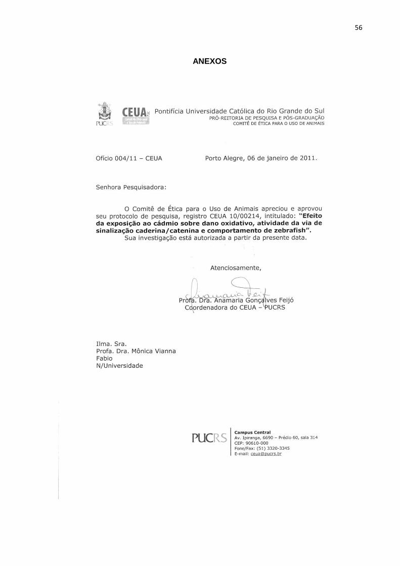
56
ANEXOS
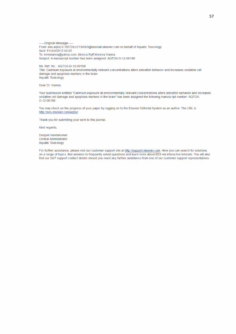
57