Embed Size (px)
Citation preview

UNIVERSIDADE FEDERAL DE SANTA MARIA CENTRO DE CIÊNCIAS RURAIS
PROGRAMA DE PÓS-GRADUAÇÃO EM MEDICINA VETERINÁRIA
IMUNOTERAPIA COM IGY AVIÁRIA EM RATOS EXPERIMENTALMENTE INFECTADOS POR
Trypanosoma evansi
TESE DE DOUTORADO
Luzia Cristina Lencioni Sampaio
Santa Maria, RS, Brasil
2014

IMUNOTERAPIA COM IGY AVIÁRIA EM RATOS EXPERIMENTALMENTE INFECTADOS POR Trypanosoma
evansi
Luzia Cristina Lencioni Sampaio
Tese apresentada ao Curso de Doutorado do Programa de Pós-Graduação em Medicina Veterinária, Área de Concentração em Medicina Veterinária Preventiva, da Universidade Federal de Santa Maria (UFSM, RS), como requisito parcial para obtenção de grau de
Doutor em Medicina Veterinária
Orientadora: Profª Drª Silvia Gonzalez Monteiro
Santa Maria, RS, Brasil
2014
Universidade Federal de Santa Maria Centro de Ciências Rurais
Programa de Pós-Graduação em Medicina Veterinária
A Comissão Examinadora, abaixo assinada, aprova a Tese de Doutorado
IMUNOTERAPIA COM IGY AVIÁRIA EM RATOS EXPERIMENTALMENTE INFECTADOS POR Trypanosoma evansi
Elaborada por
Luzia Cristina Lencioni Sampaio
Como requisito parcial para obtenção do grau de Doutor em Medicina Veterinária
Comissão Examinadora:
Silvia Gonzalez Monteiro, Dr.ª. (UFSM) (Presidente/Orientadora)
Daniel Roulim Stainki, Dr. (UFSM) (Coorientador)
Aleksandro Schafer da Silva, Dr. (UDESC)
Carmen Lucia Garcez Ribeiro, Dr.ª (UFPel)
Luciana Maria Fontanari Krause, Dr.ª (UNIFRA)
Santa Maria, 17 de abril de 2014

Ao meu Pai... que sempre
me apoiou, que sempre me
incentivou e acreditou em mim...
que nunca me abandonou ... e que
continua me conduzindo em todos
os momentos da minha vida,
dedico ...

AGRADECIMENTOS
Sem dúvida alguma esta é uma das páginas de grande importância neste
trabalho. Uma página que não obedece normas e que jamais será resumida em
parágrafo único e vinte linhas. Aqui tenho a oportunidade de apresentar sem objetivos
previsíveis e sem nenhuma metodologia os resultados desta inesquecível experiência
vivida durante minha passagem por esta Universidade: amizade e coleguismo
verdadeiro, sem os quais nada aqui descrito seria possível. Pessoas realmente
importantes, as quais serei eternamente agradecida ...
Inicio agradecendo a minha colega e amiga Maria Elizabeth Berne, professora
da UFPel, que não me permitiu desistir de realizar o doutorado, se dispôs a ser minha
orientadora e me incentivou a ingressar como aluna especial do Programa de Pós
Graduação daquela Universidade. Minha eterna admiração e reconhecimento. Com
certeza, aqui está o primeiro passo para que tenha chegado até este momento.
Também não foi o acaso que trouxe para o nosso convívio na Universidade
Federal de Pelotas o professor Daniel Roulim Stainki. Durante o curto período em que
trabalhamos juntos não podia imaginar o grande amigo que havia conquistado. Admiro
sua tranquilidade, disciplina, seriedade, honestidade e senso de justiça. Ao ser
transferido para a Universidade Federal de Santa Maria nos deparamos com a alegria
de vê-lo retornar para casa e a tristeza de perder o convívio com um colega especial.
Não poderia sequer imaginar que seria justamente ele quem abriria as portas para a
realização deste sonho. Com muito orgulho o tenho ainda como meu coorientador.
Impossível mensurar meu agradecimento Daniel !
Ao ser selecionada no Programa de Pós graduação da Universidade Federal
de Santa Maria, ninguém vibrou e torceu tanto por mim como a minha grande amiga,
colega e Chefe do nosso Departamento: Carmen Lucia Garcez Ribeiro, nossa Kalu!
Amiga de verdade, que durante todo este período esteve ao meu lado, me
incentivando, orientando e não mediu esforços para agilizar toda a burocracia
relacionada ao meu afastamento da UFPel dando o suporte necessário. Impossível
não lembrar de pessoas especiais assim ....
Aqui chegando conheci minha orientadora, Professora Silvia Gonzalez
Monteiro. Não existe palavras que expressem a magnitude da minha admiração,

reconhecimento, gratidão e carinho por alguém que me recebeu de braços abertos e
que me permitiu a realização deste sonho. Ao término desta etapa, levo comigo a
lembrança de uma orientadora incondicional, crítica, firme, que nos mostrou toda sua
capacidade e qualidade de uma verdadeira educadora; mas sobretudo uma grande
amiga e conselheira.
E como esquecer dos meus colegas Camila Belmonte, Luciana Dalla Rosa,
Lucas Trevisan Gressler, Thirssa Helena Grando, Mariângela Facco de Sá, Dianni
Menezes Capeleto, Matheus Dellaméa Baldissera? Espero um dia ter a oportunidade
de retribuir todo o carinho, consideração e respeito com que fui tratada por vocês!
Contem comigo sempre ... Também a todos os bolsistas e estagiários do LAPAVET
meu eterno agradecimento. E aos colegas da UFPel, Fabio Leivas Leite, Everton
Fagonde da Silva, Andreia Anciuti, Karina Colonetti, Amilton Seixas Neto, Alceu
Gonçalves dos Santos e Tiago Heres ... todos vocês fazem parte desta história!
E como não falar nos filhos, em especial a minha filha Daniele, extremamente
responsável e determinada, iniciando sua vida profissional também longe de casa,
minha maior incentivadora. Obrigada minha filha pelos momentos em que você se
comportou como minha mãe....
Não foi fácil viver longe de casa, longe da família, dos filhos e dos amigos ...
mas alguém muito importante para mim, me segurou pela mão, me abraçou, me
carregou para cá e para lá, enxugou minhas lágrimas e me convenceu que o tempo e
a distância eram muito pequenos perto da grandeza desta conquista. Muito obrigado
Getulio !!!!!!!! só o amor de verdade sobrevive nas adversidades!
Do fundo do coração, agradeço a minha mãe: nunca esquecerei todo o
incentivo, apoio e as palavras de carinho, desde o momento que decidi ir atrás deste
sonho e durante estes dois anos longe da família. Obrigado por estar sempre ao meu
lado durante toda esta etapa da minha vida ....
Finalmente, aquele que meu deu toda a força para chegar até aqui:
Muito Obrigada MEU DEUS!

“Que não falte bons sentimentos. Que nos falte
egoísmo. Que nos sobre paciência. Que não nos
falte esperança. Que cada caminho escolhido nos
reserve boas surpresas. Que cada um de nós saiba
ouvir cada conselho dado por uma pessoa mais
velha. Que não nos falte vontade de sorrir. Que
nenhum de nós se esqueça da força que possui.
Que não nos falte fé e amor”.
(Caio Fernando Abreu)

RESUMO
Tese de Doutorado
Programa de Pós-Graduação em Medicina Veterinária
Universidade Federal de Santa Maria
IMUNOTERAPIA COM IGY AVIÁRIA EM RATOS
EXPERIMENTALMENTE INFECTADOS POR Trypanosoma evansi
AUTORA: LUZIA CRISTINA LENCIONI SAMPAIO
ORIENTADORA: SILVIA GONZALEZ MONTEIRO
Data e Local da Defesa: Santa Maria, 17 de abril de 2014
Trypanosoma evansi é um protozoário flagelado que acomete bovinos, ovinos, caprinos, asininos, felinos e suínos. A enfermidade tem especial importância em equinos, sendo conhecida como “mal das cadeiras”, devido aos déficits de locomoção característicos em animais infectados. A doença tem distribuição mundial, com vários relatos de infecções naturais em diversas regiões no Brasil. Várias drogas terapêuticas têm sido recomendadas para a profilaxia e controle do protozoário, porém ao longo dos anos estes medicamentos tem perdido a eficácia e o protozoário parece desenvolver resistência. A produção e uso de anticorpos aviários tem despertado grande interesse na comunidade científica devido à diversidade de aplicações diagnósticas e terapêuticas. Devido à distância filogenética, mecanismos de diversificação imune e capacidade de transferência das imunoglobulinas séricas para a gema do ovo, atualmente reconhece-se uma série de vantagens ao utilizar anticorpos aviários ao invés de anticorpos de mamíferos. Neste estudo foi produzido uma imunoglobulina específica contra o T. evansi a partir de imunização de galinhas usando um isolado do protozoário. Após a extração e purificação a partir da gema do ovo, foram avaliadas a citotoxicidade e genotoxicidade destes anticorpos em cultura de leucócitos humanos. A eficácia terapêutica foi testada em Rattus norvegicus. O ensaio também avaliou o uso do anticorpo aviário associado ao dipropionato de imidocarb e diaceturato de diminazene. Os testes de toxicidade mostraram que a imunoglobulina não produz lesões celulares e genéticas. Testes in vivo mostraram aumento da longevidade e período pré-patente, principalmente em ratos tratados com a imunoglobulina aviária associada ao tratamento farmacológico. O protocolo de imunização e extração usados nesta pesquisa permitiu a obtenção de anticorpos específicos contra o protozoário e inócuo para o hospedeiro. Além disso, ao ser administrado com fins terapêuticos, reforça o sistema imunológico e prolonga a sobrevida de pacientes infectados.
Palavras – chave: imunoglobulina aviária, IgY, tripanosomose

ABSTRACT
Doctoral Thesis
Postgraduate Program in Veterinary Medicine
Universidade Federal de Santa Maria
IgY IMMUNOTHERAPY IN RATS EXPERIMENTALLY INFECTED
WITH Trypanosoma evansi
AUTHORESS: LUZIA CRISTINA LENCIONI SAMPAIO
ADVISOR: SILVIA GONZALEZ MONTEIRO
Place and date: Santa Maria, April 17, 2014.
Trypanosoma evansi is a protozoan flagellate that affect cattle, sheep, goats, donkeys, cats and pigs. The disease is particularly important in horses, known as "Mal das Cadeiras", due to deficits characteristic of locomotion in animals infected. The disease has a worldwide distribution, with several reports of natural infections in different regions in Brazil. Several pharmaceutical drugs have been recommended for the prophylaxis and control of the parasite, but over the years, these drugs have lost their effectiveness and protozoan looks developing resistance. The production and use of avian antibodies has aroused great interest in the scientific community because of the diversity of diagnostic and therapeutic applications. Due to the phylogenetic distance, immune diversification mechanisms and transferability of serum immunoglobulin to the egg yolk, currently recognizes a number of advantages to using avian antibodies rather than antibodies of mammals. This study produced a specific immunoglobulin against Trypanosoma evansi from immunization of chickens using an isolate of the parasite. After extraction and purification from egg yolk, cytotoxicity and genotoxicity of these antibodies in cultures of human leukocytes were evaluated. The therapeutic efficacy was tested in Rattus norvegicus. The trial also assessed the use of avian antibody associated with imidocarb dipropionate and diaceturate of diminazene. The toxicity tests showed that immunoglobulin does not produce gene and cell injury. In vivo tests showed increased in longevity and pre-patent period, mainly in rats treated with avian immunoglobulin associated with pharmacological treatment. The immunization and the extraction protocol used in this study allowed obtaining specific antibodies against protozoan and innocuous to the host. Moreover, when administered for therapeutic purposes, strengthens the immune system and prolongs the survival of infected patients.
Keywords: avian immunoglobulin, IgY, trypanosomosis.

LISTA DE TABELAS
ARTIGO 1
Tabela 1 - Karyotypic changes (%) in mononuclear cells of human peripheral blood exposed to different concentrations of IgY….......
52
ARTIGO 2
Tabela 1 - Pre-patent period, longevity and mortality of different treatments throughout the experimental period …………………………………
88

LISTA DE FIGURAS
REVISÃO DE LITERATURA
Figura 1- Formas tripomastigotas de T. evansi em esfregaço sanguíneo de ratos experimentalmente infectados ..............................................
18
Figura 2- Sinais neurológicos encefálicos. Incoordenação motora, afastamento membros anteriores, cruzamento dos membros pélvicos, andar em círculos e déficits de propriocepção ................
22
Figura 3- Estrutura das imunoglobulinas de mamífero (IgG) e de galinhas (IgY).................................................................................................
32
ARTIGO 1
Figura 1 - IgY MTT after 72 hours of incubation …..........................................
50
Figura 2- Lymphocyte in metaphase by the karyotype technique, without chromosomal alteration ………………………………………………
51
Figura 3 - TBARS assay after 72 h of incubation. Results are expressed as nmol of MDA/106 cells ………………………………………………
53
ARTIGO 2
Figura 1 - Total protein concentration obtained by photometry at 280 nm. Five doses of antigen were given at 14 day intervals with the production of total protein assessed for 70 days. The values were obtained using 1:50 dilution of samples purified by the PEG-6000
85
Figura 2 - (A) Electrophoresis in polyacrylamide gel (10%) under reducing conditions, stained with Coomassie Blue R250 reagent (Bio-Rad) from samples of three immunized hens. (B) immunoblot showing the recognition of IgY after addition of secondary antibody conjugated with peroxidase. In the center, the scale of molecular weight expressed in kDa ………………………………………………
85

Figura 3 - Immunodiffusion in agarose gel. (A) Central wells filled with antigen; cavities 1, 3 and 5 with the negative control; wells 2, 4 and 6 filled with extracted samples (IgY). (B) Characterization of the antigen antibody reaction through the formation of precipitation lines ………………………………………………………………………
86
Figura 4 - Kinetics of the immune response in hens immunized with T. evansi and detected by ELISA. The optical density values obtained were expressed at dilution of 1:100 of samples purified by PEG-6000, from the egg yolk of immunized hens …………………………………
86
Figura 5 - Avidity index (AI) of IgY antibodies produced during immunization of the hens with T. evansi antigens. AI<40% were considerate of low avidity; AI between 41 and 70% represented a medium avidity; and AI> 70% was considered as high avidity …................................
87
Figura 6 - Kinetics of specific IgY production of anti-T. evansi detected by the Coomassie Blue method. The values were obtained by photometry at 595 nm ……………………………………………………………….
87

LISTA DE ANEXOS
Anexo 1 - Artigo intitulado “In vitro cytotoxicity and genotoxicity of chicken egg yolk antibodies (IgY) against Trypanosoma evansi in human lymphocytes” publicado na revista International Journal of Pharmacy and Pharmaceuthical Sciences em abril de 2014
108
Anexo 2 - Comprovante de submissão do artigo intitulado “Production, purification and therapeutic potential of egg yolk antibodies for treating Trypanosoma evansi infection” enviado para Revista Veterinary Parasitology ………………………………………………
112
Anexo 3 - Parecer Comitê Ética da Universidade Federal de Santa Maria ... 113

SUMÁRIO
1 INTRODUÇÃO .............................................................................................. 2 REVISÃO DE LITERATURA ........................................................................ 2.1 Trypanosoma evansi ................................................................................ 2.1.1 Transmissão e ciclo biológico .................................................................. 2.1.2 Distribuição da doença ............................................................................ 2.1.3 Patogenia e Sinais Clínicos ..................................................................... 2.1.4 Diagnóstico ............................................................................................. 2.1.5 Tratamento .............................................................................................. 2.2 Imunoglobulina Y ..................................................................................... 2.2.1 Características estruturais, imunológicas e bioquímicas ......................... 2.2.2 Transferência passiva de imunidade em galinhas ................................... 2.2.3 Composição da gema do ovo .................................................................. 2.2.4 Vantagens do uso de anticorpos aviários ................................................ 2.2.5 Importância da tecnologia IgY ................................................................. 2.2.6 Produção e purificação de IgY ................................................................. 3 ARTIGOS …………………………………………………………………………. 3.1 Artigo 1 – In vitro cytotoxicity and genotoxicity of chicken egg yolk antibodies (IgY) against Trypanosoma evansi in human lymphocytes … Resumo ........................................................................................................... Introdução ........................................................................................................ Material e Métodos ........................................................................................... Resultados e Discussão ................................................................................... Conclusão ........................................................................................................ Referências ...................................................................................................... 3.2 Artigo 2 – Production, purification and therapeutic potential of egg yolk antibodies for treating Trypanosoma evansi infection ……………… Resumo ........................................................................................................... Introdução ........................................................................................................ Material e Métodos ........................................................................................... Resultados ....................................................................................................... Discussão ........................................................................................................ Conclusão ........................................................................................................ Referências ...................................................................................................... 4 DISCUSSÃO ................................................................................................. 5 CONCLUSÃO ............................................................................................... 6 REFERÊNCIAS ............................................................................................ 7 ANEXOS .......................................................................................................
16 17 17 18 20 21 23 25 29 30 32 33 34 35 40 42
43 44 45 47 49 53 54
59 61 62 63 69 72 75 76 89 93 94
108

1 INTRODUÇÃO
A “Tecnologia IgY” baseia-se na imunização de galinhas (Gallus gallus
domesticus) e extração de anticorpos IgY-específicos a partir da gema do ovo. O
procedimento é aceito internacionalmente como método alternativo a favor do bem
estar animal por se tratar de procedimento não invasivo. Inúmeras vantagens são
enumeradas na literatura em relação a produção e utilização de anticorpos aviários
em detrimento aos anticorpos de mamíferos. Nas últimas décadas, a busca por novos
métodos diagnósticos e recursos terapêuticos nas pesquisas biomédicas aumentou
consideravelmente os estudos nesta área. Pesquisas envolvendo métodos de
imunização, extração, purificação, caracterização e aplicabilidade contra as mais
diversas enfermidades tem sido alvo de pesquisas.
Trypanosoma evansi (T. evansi) é um protozoário emergente, que infecta a
maioria das espécies domésticas, algumas espécies silvestres e o homem. A
enfermidade produzida por este parasito adquire grande importância na espécie
equina, causando sintomatologia grave e levando os animais a óbito. No Brasil, várias
regiões já registraram casos de tripanossomose equina nos últimos anos. A situação
é preocupante, uma vez que a enfermidade causa sérios prejuízos econômicos,
principalmente quando afeta animais de alto valor zootécnico. A queda da
produtividade e óbito são consequências da forma aguda da doença. No entanto, a
manifestação crônica adquire maior importância, pois além da queda de performance
ou produtividade do animal, a condição de portador da enfermidade e recidivas do
quadro clínico são situações agravantes. Embora existam vários protocolos
quimioterápicos relatados na literatura, tanto para profilaxia como para o tratamento
da enfermidade diagnosticada, constata-se que até o presente momento ainda não se
conseguiu um tratamento realmente eficaz para o controle desse protozoário.
Nesse contexto, nossos estudos tiveram como objetivos a produção e
caracterização de anticorpos IgY anti- T.evansi a partir de inoculações periódicas com
formas tripomastigotas do parasito em galinhas poedeiras, avaliação in vitro da cito e
genotoxicidade e avaliação in vivo da eficácia profilática e terapêutica dos anticorpos
produzidos.

2 REVISÃO DE LITERATURA
2.1 Trypanosoma evansi
Trypanosoma evansi (T. evansi) é o agente etiológico da enfermidade
conhecida como Mal das Cadeiras, Tripanossomose equina, Surra, Murina,
Derrengadeira, Peste Boba, ou Quebra-bunda, que acomete equinos de todas as
raças e tem distribuição mundial. Na África é denominada Abori, Salaf, Debad, Tahaga
e Diufar (DARLING, 1910; HOARE, 1972; LEVINE, 1973). Bovinos, ovinos, caprinos,
asininos, felinos e suínos também são hospedeiros susceptíveis. Caninos, capivaras
(Hydrochaeris hydrochaeris), quatis (Nasua nasua) e morcegos hematófagos
(Desmodus rotundus) são reservatórios e ocasionalmente podem manifestar
sintomatologia clínica; sendo que os morcegos ainda são vetores da doença (NUNES,
1993; SILVA et al., 2002). Em equinos, camelos e caninos a doença é frequentemente
fatal. Em 2005, foi relatado o primeiro caso de infecção humana em um fazendeiro na
Índia (JOSHI et al., 2005).
A classificação taxonômica inclui este parasito ao Filo Protozoa, subfilo
Sarcomastigophora, classe Mastigophora, ordem Cinetoplastida, família
Trypanosomatidae. Foi descrito pela primeira vez em 1880 por Griffith Evans, um
médico veterinário do exército do Reino Unido, ao examinar ao microscópio lâminas
com sangue de equinos e camelos na região de Punjab (Índia). Após identificar a
presença do protozoário nestes animais, Evans inoculou o sangue de animais doentes
em animais sadios e após seis dias observou os protozoários no sangue dos equinos
inoculados (HOARE, 1972; FALLIS, 1986). A primeira observação da doença no Brasil
ocorreu entre os anos de 1827 e 1830 na Ilha de Marajó, atingindo equinos da região.
A partir deste local a enfermidade disseminou-se pelo restante do País e América do
Sul, atingindo a Guiana, Bolívia, Venezuela e Colômbia (HOARE, 1972).
Este protozoário possui um corpo alongado (Figura 1), com comprimento
variando entre 14-33 µm e largura entre 1,5-2,2 µm, extremidades afiladas, um flagelo
terminal, um núcleo central e uma membrana ondulante que permeia toda a extensão
do parasito (BRUN et al., 1998; SILVA et al., 2002).

18
Figura 1 - Formas tripomastigotas de T. evansi em esfregaço sanguíneo de ratos experimentalmente infectados, observados sob microscopia óptica, sob imersão (1.000 x). (Fonte: arquivo pessoal).
O cinetoplasto pode ou não estar presente, dependendo da origem da cepa.
Cepas sul-americanas são preferencialmente acinetoplásticas. Análises de cepas
brasileiras de T. evansi em cavalos e cães (AQUINO et al., 1999; NUNES; 1993),
quatis, capivaras, ratos silvestres, cães e equinos (MASIGA & GIBSON, 1990;
VENTURA et al., 2002), resultaram ser desprovidas de cinetoplasto. As cepas que os
possuem apresentam-no incompleto, sem os maxicírculos (BORST et al., 1987).
2.1.1 Transmissão e Ciclo Biológico
A transmissão das tripanossomíases ocorre através da inoculação dos
parasitos pela saliva dos vetores (seção Salivaria), ou pela contaminação da mucosa
ou da pele lesionada por tripanosomas presentes no material fecal do vetor (seção
Stercoraria) (CONNOR e VAN DEN BOSSCHE, 2004). T. evansi pertence à secção

19
Salivaria, sendo a transmissão essencialmente mecânica. As formas tripomastigotas
são transferidas diretamente de um hospedeiro para outro através de insetos
hematófagos (moscas das famílias Tabanidae e Muscidae) ou artificialmente por
agulhas contaminadas com sangue infectado (SILVA et al., 2002).
Na América do Sul, T. evansi pode também ser transmitido por morcegos
hematófagos (Desmodus rotundus), onde os parasitos podem se multiplicar e
sobreviver por longos períodos. Nestes vetores, o parasito não desenvolve nenhuma
fase do ciclo (SILVA et al., 2002). Dessa maneira, morcegos hematófagos atuam
tanto como vetores como reservatórios (HOARE, 1972). DUNN (1932), JOHNSON
(1936) e OLIVEIRA (1943) relataram a transmissão de T. evansi utilizando este vetor
no Panamá e no Brasil, além de registrarem que T. evansi e morcegos hematófagos
tem a mesma distribuição geográfica.
Pesquisas realizadas no Laboratório de Imunomodulação do Departamento de
Protozoologia do Instituto Oswaldo Cruz demonstraram que as sanguessugas são
capazes de transmitir o T. evansi. Os parasitos foram encontrados dispersos em todo
o corpo das sanguessugas: aparelho bucal, estômago, intestino e glândulas. No
intestino eles formam grumos arredondados, o que os pesquisadores supõem ser um
mecanismo de defesa. Nas glândulas, os tripanosomas foram encontrados no interior
de células epiteliais. O estudo sugere que a transmissão ocorra através da ingestão
do parasito. Capivaras são mamíferos semi-aquáticos que se alimentam de algas e
do capim à margem dos rios. É possível que engulam sanguessugas junto com esses
vegetais, o que também pode acontecer com os cavalos. O estudo conclui que a
abundância destes vetores associada à presença de animais selvagens ou
domésticos, que servem como reservatório do parasito, como capivaras, quatis e
cães, seja responsável pela alta prevalência da doença na região do Pantanal no
Brasil (CARREIRA, 2005).
Carnívoros, principalmente caninos, são naturalmente infectados pelo
protozoário, mas podem se infectar ainda através da ingestão de carne de animais
recentemente mortos com a doença ou carcaça de animais infectados (RAMIREZ et
al., 1979; BARRIGA, 1997; HERRERA et al., 2004). A transmissão via oral foi
comprovada experimentalmente em camundongos e caninos (RAINA et al., 1985;
BAZZOLI et al., 2002), sendo importante fonte de contaminação em cães, quatis e
capivaras após brigas entre animais infectados e não infectados.

20
A transmissão venérea ainda não foi constatada, porém já foi detectada a
presença do protozoário na mucosa vaginal de coelhas experimentalmente infectadas
(UCHE & JONES,1992). Há relatos de casos de infecção natural via transplacentária
em ruminantes (OGWU & NURU,1981; MURALEEDHARAN & SRINIVAS,1985). Esta
via também foi comprovada em camundongos experimentalmente infectados
(SARMAH, 1998).
Tripanosomas são parasitos digenéticos, ou seja, o ciclo biológico envolve
apenas dois hospedeiros: um animal vertebrado que é o hospedeiro final, e diversos
invertebrados hematófagos representando os hospedeiros intermediários ou vetores.
Alguns autores usam o termo “inoculadores mecânicos” em vez de “vetores” para se
referir à transmissão mecânica de tripanosomas.
No caso do T. evansi a transmissão é mecânica e as formas sanguíneas dos
tripanosomas são transferidas diretamente de um mamífero para outro por insetos
hematófagos ou artificialmente por agulhas contaminadas com sangue infectado. Não
ocorre desenvolvimento do T. evansi no inseto. Quando transmitido por tabanídeo a
transmissão é facilitada, pois a picada é dolorosa e leva o hospedeiro a se coçar. A
habilidade para transmitir os tripanosomas mecanicamente é de curta duração
(geralmente mensuradas em minutos), dependendo do tempo de sobrevivência dos
parasitos no aparelho bucal do inseto (BRUN et al., 1998; SILVA et al.,2002).
2.1.2 Distribuição da doença
A doença tem distribuição cosmopolita, atingindo principalmente regiões
tropicais e subtropicais, com relatos na África do Norte, América Central, América do
Sul, China, Filipinas, Índia, Indonésia, Malásia, Oriente Médio e Rússia (HORNIBY,
1953; BAJYANA SONGA et al., 1987; LUCKINS, 1988; MONZÓN & COLMAN, 1988;
BAZOLLI et al., 2002).
Acredita-se que o T. evansi tenha origem no continente africano e foi
introduzido nas Américas através dos primeiros colonizadores europeus. Os primeiros
casos de tripanossomíase equina na América do Sul surgiram no final no século XIX,
após a importação de cavalos da Espanha (HOARE, 1972; SANTOS et al., 1992).

21
Nestes continentes o T. evansi é responsável por infecções agudas em várias
espécies animais, desde o sul da Argentina até o norte do Panamá (WELLS, 1984).
Surtos ou casos isolados de tripanossomíases têm sido relatados em diversas
regiões brasileiras (FRANKE et al., 1994; SILVA et al., 1995; HERRERA et al., 2005).
Já foram descritos casos de infecção natural em animais domésticos e silvestres no
Rio Grande do Sul (COLPO et al., 2005; CONRADO et al., 2005; RODRIGUES et al.,
2005; FRANCISCATO et al., 2007; ZANETTE et al.; 2008), Mato Grosso do Sul
(MOREIRA & MACHADO, 1985; BRANDÃO et al., 2002), Santa Catarina (DA SILVA
et al., 2007), Paraná (KUBIAK & MOLFI, 1954), Minas Gerais (NUNES et al., 2011) e
no Pantanal, onde a doença é endêmica, com recorrentes casos (SILVA et al., 2002).
2.1.3 Patogenia e Sinais Clínicos
Os principais fatores que influem na patogenicidade dos Tripanosomas
referem-se ao tipo de cepa, espécie animal susceptível, fatores concomitantes à
infecção (estado nutricional, estado imunológico, estresse, outras doenças) e
epizootiologia (HOARE, 1972). Os tripanosomas patogênicos de importância pecuária
estão localizados na seção salivaria e incluem o Trypanosoma vivax, Trypanosoma
equiperdum e Trypanosoma evansi. (SILVA et al.; 2002).
Em seguida à infecção inicia-se a multiplicação do protozoário no local da
picada e a sua migração para a corrente sanguínea e sistema linfático. A reprodução
do T. evansi ocorre por fissão binária, sendo que esta divisão se processa em uma
sequência definitiva envolvendo sucessivamente o corpo basal, flagelo, cinetoplasto
e núcleo, culminando na clivagem do citoplasma (SILVA et al., 2002). O processo
ocorre no sangue do hospedeiro (BRUN et al., 1998), sendo que a medida que
aumenta a parasitemia a resposta inflamatória é ativada e surgem picos de febre. A
infecção tende a cronificação, com períodos aparasitêmicos e afebris. Estes
protozoários possuem em sua superfície glicoproteínas com alta capacidade mutante,
capazes de driblar o sistema de defesa do hospedeiro (CONNOR & VAN DEN
BOSSCHE, 2004; PAYS et al., 2004).
A doença pode se manifestar de forma aguda ou crônica. Infecções agudas
podem causar a morte rápida em equinos e cães não tratados. Os sintomas incluem

22
emagrecimento, palidez de mucosas, febre intermitente, tosse, edema nas partes
baixas do corpo, aumento dos linfonodos superficiais, fraqueza muscular, atrofia da
musculatura dos membros pélvicos, incoordenação motora, paresia de trem posterior,
dificuldade para levantar, hiperexcitabilidade, andar em círculo, déficit proprioceptivo
(Figura 2), cegueira e quedas constantes (LEVINE, 1973; AQUINO et al.; 1999;
HERRERA et al.; 2005; RODRIGUES et al., 2009). As infecções crônicas podem durar
anos (BRUN et al., 1998) e nesta fase ocorre o agravamento dos sinais clínicos e
caquexia (BRANDÃO et al., 2002; SILVA et al., 2002; RODRIGUES et al., 2005).
Figura 2 - Sinais neurológicos encefálicos. Incoordenação motora, afastamento membros anteriores, cruzamento dos membros pélvicos, andar em círculos e déficits de propriocepção (Rodrigues et al., 2009).
Os sinais neurológicos têm sido descritos na fase terminal da doença,
principalmente em equinos e bovinos (TUNTASUVAN et al., 1997; RODRIGUES et
al., 2009; DA SILVA et al., 2007). Sintomas reprodutivos tais como aborto, infertilidade,
esterilidade, atrofia ou degeneração de testículos, epidídimo e túbulos seminíferos,

23
assim como alterações na concentração, morfologia e volume espermático foram
relatados por Sekoni et al (2004), Bezerra et al.(2006); Batista et al. (2007, 2008).
A infecção por T. evansi em bovinos cursa com anorexia, emagrecimento,
apatia, hipertermia (41ºC), mucosas pálidas ou ictéricas e hemorragias cutâneas após
a morte (DA SILVA et al., 2007). A análise hematológica de animais infectados com
T. evansi mostra acentuada anemia, caracterizada por decréscimo no hematócrito,
concentração de hemoglobina e contagem total de eritrócitos (SILVA et al.; 2002). Em
caninos foi demonstrada a anemia microcítica e hipocrômica e em equinos a anemia
microcítica e normocrômica. Em ambas as espécies foram observadas anormalidades
eritrocitárias tais como: microesferócitos, acantócitos, dacriócitos, micrócitos,
vacuolização eritrocitária, policromasia, poiquilocitose. A eritrofagocitose e a adesão
do tripanosoma ao eritrócito também são achados descritos nestas espécies (ANOSA
& KANEKO, 1983; CONRADO et al. 2005).
Logo após a infecção ocorre uma anemia hemolítica devido ao processo de
eritrofagocitose. Uma segunda fase anêmica ocorre em torno de 4 a 6 semanas após.
Jain (1993) e Connor & Van Den Bossche (2004) referem-se a liberação de
hemolisinas e enzimas pelos tripanosomas capazes de induzir lesões na membrana
e aumentar a fragilidade destas, como principal mecanismo de origem da anemia.
Além disto, a adesão do complexo antígeno-anticorpo e componentes do sistema
complemento aos eritrócitos também contribui para anemia, pois promove a
eritrofagocitose. Atualmente sugere-se que a anemia também seja consequente ao
estresse oxidativo. Desta forma, o aumento de radicais livres de oxigênio promove a
peroxidação lipídica e consequente dano a membrana eritrocitária (WOLKMER et al.,
2009).
2.1.4 Diagnóstico
O diagnóstico definitivo da infecção por T. evansi é obtido através do somatório
dos dados epidemiológicos, sinais clínicos e exames laboratoriais. Os sintomas da
doença não são patognomônicos e apresentam-se comuns a várias outras
enfermidades. Portanto, a análise laboratorial é imprescindível no estabelecimento do
diagnóstico (SILVA et al., 2002). Os métodos diagnósticos mais frequentemente

24
usados são os esfregaços corados pelo Giemsa (GSS), método de concentração de
Strout (SCM), método do microhematócrito (HCT), método "Buffy Coat" (BCM) e
inoculação em camundongos (MONZÓN, 1993).
Nas infecções agudas é possível a visualização do T. evansi através de
esfregaços sanguíneos. Em bovinos a amostra pode ser coletada a partir de aspiração
do linfonodo pré-escapular. A técnica baseia-se em colocar uma gota do sangue numa
lâmina a uma distância de aproximadamente 2-3 cm de uma das extremidades,
empurrando a gota com outra lâmina num ângulo de 45º até o lado oposto. São
recomendáveis esfregaços de camada fina, pois facilitam a leitura ao apresentarem
as células mais dispersas. O material é analisado entre lâmina e lamínula, após
fixação em álcool metílico e corado com Giemsa. A leitura é realizada sob microscopia
ótica
As técnicas do microhematócrito e Buffy Coat (WOO, 1970) são indicadas
quando a parasitemia é baixa e é necessário o uso de técnicas de concentração. Após
preencher aproximadamente 2/3 do volume do tubo capilar com o sangue a ser
testado, este deve ser selado com chama ou com plastilina em uma das extremidades
e centrifugado para realização da leitura. Os protozoários podem ser observados na
junção entre a camada de células brancas e o plasma. Para o Buffy Coat monta-se
uma lâmina, quebrando o tubo capilar na parte onde se divide a parte líquida com a
parte celular, colocando assim uma ou duas gotas deste material numa lâmina para
confecção do esfregaço.
A inoculação em camundongos com amostras de sangue suspeito permite a
visualização do protozoário. A parasitemia deve ser acompanhada a cada 48h através
de esfregaço sanguíneo dos roedores e o período pré-patente geralmente é curto (5
dias), variando conforme a patogenicidade da cepa. A inoculação em camundongo se
faz geralmente por via intraperitoneal, mas também pode ser feita por via
intramuscular. O inóculo de sangue infectado é de aproximadamente 0,20 ml.
Segundo Lanham & Godfrey (1970) a mini-anion exchange centrifugation
technique (mAECT) é um método diagnóstico bastante sensível. Consiste na
realização de uma cromatografia de troca iônica em DEAE (dietilaminoetil)-Celulose,
onde os protozoários são isolados do material sanguíneo, e depois de centrifugados
são visualizados em microscópio óptico.
Vários métodos sorológicos são descritos na literatura para diagnóstico da
infecção por T. evansi. A limitação destes métodos reside no fato de que após o

25
tratamento os anticorpos podem persistir por mais de um ano, o que dificulta saber se
é de uma nova infecção ou são anticorpos residuais de uma infecção passada e já
curada. Silva et al. (2002) descreve as principais técnicas sorológicas indicadas,
dando destaque a imunofluorescência indireta (RIFI), Teste de aglutinação direta
(MONZÓN, 1993), Enzyme-linked Immunosorbent Assay (ELISA) e o Card
agglutination test for trypanosomiasis (CATT).
A identificação e caracterização do protozoário são fundamentais para
estabelecer o diagnóstico diferencial entre as espécies de tripanosomas. Durante
décadas o diagnóstico baseou-se em diferenças morfológicas, as quais permitiam
distinguir parasitos de diferentes subgêneros, mas mostravam falhas na distinção
entre variantes biológicos. Atualmente, métodos de detecção específica de DNA
através da reação em cadeia da polimerase (PCR) oferecem alto nível de
especificidade. Ventura et al. (2002) desenvolveram um diagnóstico por PCR do tipo
espécie-específico para o T. evansi.
2.1.5 Tratamento
Vários protocolos terapêuticos têm sido estudados com o objetivo de prevenir
ou tratar a infecção por T. evansi. Neste sentido, uma ampla variedade de drogas
químicas e extratos de plantas medicinais têm sido recomendados, embora até o
momento não se tenha obtido um tratamento plenamente eficaz e com mínima
toxicidade ao hospedeiro.
A terapia química ainda é o método de eleição no controle da doença em
animais domésticos, sendo recomendado na literatura drogas como suramina,
quinapiramina, melarsomina, homidium, isometamidium, aceturato de diminazene e
dipropionato de imidocarb. Os grandes problemas encontrados no tratamento são a
alta toxicidade destas drogas para o hospedeiro e o surgimento de cepas resistentes,
visto que grande parte destes compostos vem sendo utilizados no campo há mais de
40 anos (SILVA et al., 2002). Os três primeiros grupos químicos são primariamente
indicados como drogas terapêuticas para infecções por T. evansi, enquanto,
homidium, isometamidium e diminazene são primariamente usados para o tratamento
e profilaxia da tripanossomíase em bovinos, ovinos e caprinos (SILVA et al., 2004).

26
A diferença entre terapia curativa e preventiva está na dependência da droga
escolhida e, em alguns casos da dose prescrita. Neste sentido, normalmente os
tratamentos profiláticos recomendam doses inferiores ao limiar mínimo terapêutico,
podendo induzir à resistência parasitária. Drogas capazes de se armazenarem nos
tecidos e serem liberadas gradualmente para a corrente circulatória conseguem
manter uma concentração de princípio ativo por um maior tempo circulante
(PEREGRINE, 1994). Segundo Boyt (1986) recomenda-se drogas curativas quando a
prevalência da parasitose é baixa ou quando poucos casos atingem um rebanho.
Segundo o mesmo autor, a terapia preventiva é indicada em áreas de alto risco e
quando a prevalência da enfermidade é alta durante o ano.
A suramina pertence ao grupo das naftilamidas e foi a primeira droga
tripanocida usada em animais domésticos, sendo comercializada desde 1920. Para
infecções por T. evansi foi usada por muitos anos como droga de escolha para
tratamento em equinos e camelos por apresentar menor toxicidade que as demais.
Também foi utilizada com fins profiláticos, uma vez que possui forte ligação com as
proteínas plasmáticas, permanecendo na circulação sistêmica por mais de três
meses. Atualmente esta droga não é mais comercializada (SILVA et al., 2004).
Quinapiramina pertence ao grupo das piramidinas, sendo usado com fins
curativos ou profiláticos. Comercialmente são apresentadas nas formas de sulfato e
cloreto. Foi amplamente utilizada na África entre 1950 e 1970, sendo proibido seu uso
em 1974 devido à toxicidade destes compostos. Em 1984 a droga retornou ao
mercado para uso em camelos e cavalos, não sendo indicado para bovinos devido à
resistência cruzada com outras drogas tripanocidas (HAROUN et al., 2003). Em áreas
endêmicas, o cloreto de quinapiramina é indicado como terapia preventiva, sendo
recomendada a administração a cada dois ou três meses (SILVA et al, 2004).
Homidium é o princípio ativo pertencente ao grupo das fenantridinas cuja
atividade tripanocida foi primeiramente reconhecida por Browning et al. em 1938. O
brometo de homidium é um ánalogo do brometo de dimidium, tendo sido amplamente
utilizado a partir de 1952 no tratamento da tripanossomíase bovina no leste e oeste
da África. É uma droga efetiva na cura de tripanossomíases e com menor toxicidade
em relação ao dimidium (FORD et al., 1953). A administração da dose de 1 mg/kg
com fins profiláticos mostrou-se eficaz por um período de 4-6 semanas (LEACH, 1955;
MAWAMBU, 1971). Dolan et al. (1990) testaram o brometo de homidium após
tratamento com o aceturato de diminazeno, e concluiram que os tripanosomas
27
presentes foram sensíveis a concentração da droga, ou seus metabólitos ativos por
até 17 semanas pós administração.
A inexistência de apresentações comerciais do produto, custo, resistência por
parte dos protozoários ao princípio ativo e relatos de toxidade, são fatores limitantes
ao uso da droga no Brasil.
O isometamidium é um derivado da fenandridina resultante da combinação do
homidium com a p-aminobenzamida diazotizada do dimenazene. Tem ação profilática
e curativa. A dose recomendada para fins profiláticos é de 0,5-1,0 mg/kg. A atividade
profilática em bovinos dura entre 2 a 22 semanas. Pelegrine et al (1988) afirma haver
uma relação direta entre a dose administrada e o tempo de profilaxia, não havendo
diferenças entre animais infectados e não infectados.
A melarsomina foi desenvolvida há pouco mais de 20 anos (RAYNAUD et al.,
1989), e é o único composto que apresenta eficácia comprovada em diversas
espécies (PAYNE et al., 1994), sendo indicado em infecções agudas, subagudas e
crônicas. Pertence ao grupo dos tioarsenitos, tendo como apresentação comercial o
Cymelarsan®, também conhecido como Mel Cy ou RM 110. A dose recomendada
para infecções por T. evansi em camelos é de 0,2-1,25 mg/kg e em búfalos é de 0,25-
3,0 mg/kg. Para equinos a dose é de 0,25 mg/kg via intramuscular profunda. O produto
não é usado com fins profiláticos (BRUN et al., 1998).
Diminazene pertence ao grupo das diamidinas aromáticas, sendo
comercializado na forma de aceturato, em soluções a 4% ou 7%, associadas ou não
a vitamina B12. As preparações comerciais estão associadas com antipirinas, um
estabilizador que prolonga a atividade do composto em solução. São indicados a
administração na dose de 3,5 mg/kg, via intramuscular profunda, no tratamento das
babesioses e tripanossomíases de bovinos, ovinos, equinos e caprinos. Recomenda-
se o volume máximo de 10 ml no local da aplicação, sendo que quando necessárias
doses superiores estas devem ser subdivididas em dois ou mais pontos de aplicação.
Segundo os fabricantes, o tratamento com subdoses predispõe ao desenvolvimento
de resistência parasitária e ineficácia do tratamento. Normalmente uma única dose é
suficiente, podendo ser administrada uma segunda dose 48 a 72 horas após, quando
a infecção não for controlada.
Verma et al. (1976) ao administrarem a dose de 10 mg/kg por via intramuscular
em bovinos experimentalmente infectados por T. evansi constataram a ação
tripanocida do diminazene após 12 horas da aplicação. Silva et al. (2004) indicam a

28
dose de 7 mg/kg via intramuscular profunda ou 0,5 mg/kg via endovenosa lenta no
tratamento das infecções por T. evansi em equinos. Da Silva et al. (2008) ao
administrarem as doses únicas de 3,5 e 7 mg/kg via intramuscular em ratos,
constataram que a droga não controlou a infecção, havendo recidiva após 25 e 37
dias respectivamente. No entanto, ao administrar as mesmas doses durante 5 dias
consecutivos obtiveram a cura da infecção durante o período do experimento (120
dias).
Tonin et al (2011) ao associarem o aceturato de diminazene com selênio em
ratos experimentalmente infectados obtiveram aumento da longevidade, incremento
na taxa de leucócitos e linfócitos e redução da peroxidação lipídica. Por outro lado, a
associação com vitamina E não mostrou os mesmos efeitos protetores e tripanocidas.
O período de carência para abate é de 30 dias após a última aplicação em bovinos,
ovinos, caprinos e equinos. No caso de utilização do leite para consumo humano ou
industrialização o período de carência é de 72 horas. Segundo Peregrine & Mamman
(1993), é a droga mais utilizada no Brasil contra tripanossomíases de animais
domésticos, tendo boa eficácia terapêutica e menor índice de resistência quando
comparado a outras drogas tripanocidas. Estudos de Tuntasuvan et al. (2003) e Colpo
et al. (2005) demonstraram que a administração de dose única de aceturato de
diminazene nem sempre leva a cura total da infecção pelo T. evansi, resultando em
recidivas da enfermidade. Isto pode ocorrer devido à administração de subdoses e,
também, a incapacidade do medicamento em atravessar a barreira hematoencefálica
(JENINGS et al., 1977).
O dipropionato de imidocarb pertence ao grupo das carbanilidas, cujo nome
químico é 3,3’-bis (2-imidazolina-2-yl) dipropionato de carbanilida. Apresenta-se na
forma de um pó esbranquiçado, odor de ácido propiônico, altamente solúvel em água
e álcool metílico. Quando administrado por via subcutânea ou intramuscular atinge
níveis terapêuticos plasmáticos rapidamente. Atua impedindo a absorção pelo
protozoário de hipoxantina e ácido oróico, necessários à síntese das purinas e
pirimidinas. Bloqueia desta forma a síntese de ácido nucléico do parasito (RONCATI,
2007).
Estudos sobre a farmacocinética do dipropionato de imidocarb em cavalos
demonstraram que esta droga tem um pico de concentração sanguínea após a
primeira hora de aplicação, pico de concentração urinária após 36 horas e fecal em
até 10 dias após a administração. Esta eliminação fecal longa indica eliminação biliar

29
da droga e consequente armazenamento hepático (BELLOLI et al., 2002). Zobba et
al. (2008) compararam a administração de oxitetraciclina associada a uma dose de
dipropionato de imidocarb com a administração de duas doses de dipropionato de
imidocarb com intervalo de 24 horas no tratamento da piroplasmose equina.
Concluíram que todos os animais tratados com duas doses de imidocarb se
recuperaram, enquanto no grupo tratado com oxitetraciclina + dipropionato de
imidocarb houve recidiva em alguns animais do experimento, obtendo-se a melhora
clínica somente após sucessivas administrações de dipropionato de imidocarb. Em
alguns casos, surgiram efeitos tóxicos colinérgicos após a segunda dose de
imidocarb, os quais foram resolvidos após a administração de sulfato de atropina. A
DL50 e efeitos tóxicos em diferentes dosagens foram determinados por Adams (1981).
Ao testar o princípio ativo nas doses de 0 - 2 - 4 - 8 - 16 e 32 mg/kg, em duas doses
com intervalo de 24 horas, concluiu-se que a DL50 é de 15.99 ± 1,49, sendo a principal
causa mortis a falência hepato-renal. A dose de 4 mg/kg, produz discretos sinais e
pouca alteração nas dosagens enzimáticas hepáticas e renais. A partir de 8 mg/kg
ocorrem reações sistêmicas evidentes e elevações significantes nos níveis
enzimáticos, assim como lesões histopatológicas renais após 21 dias de tratamento,
sugerindo que nesta dose ou superior há riscos em equinos.
Nogueira et al. (2005) ao administrarem as doses de 1, 2 e 2,4 mg/kg a equinos
imunossuprimidos com babesiose crônica, concluiram que as doses testadas foram
eficientes na prevenção da reagudização da infecção. Da Silva et al. (2008) ao
tratarem ratos experimentalmente infectados com T. evansi nas doses de 2 e 4 mg/kg
não conseguiram controlar a infecção.
2.2 Imunoglobulina Y ( IgY)
A produção e uso de anticorpos aviários tem despertado grande interesse na
comunidade científica devido a diversidade de aplicações diagnósticas e terapêuticas
nas pesquisas biomédicas. Devido a distância filogenética, mecanismos de
diversificação imune e capacidade de transferência das imunoglobulinas séricas para
a gema do ovo, atualmente reconhece-se uma série de vantagens ao utilizar
anticorpos aviários ao invés de anticorpos de mamíferos.

30
A denominação IgY refere-se as imunoglobulinas da gema do ovo (Y = yolk). É
uma imunoglobulina sérica de baixo peso molecular predominante em anfíbios, répteis
e aves. A transferência de imunidade da galinha para o embrião através de anticorpos
presentes na gema foi demonstrada pela primeira vez em 1893 por Klemperer.
2.2.1 Características estruturais, imunológicas e bioquímicas
A anticorpogênese é o principal mecanismo de defesa natural dos vertebrados,
e é o processo pelo qual ocorre a formação de anticorpos específicos sempre que o
indivíduo se depara com agentes estranhos. Assim também ocorre com as galinhas,
cujo sistema imunológico é ativado na presença dos mais diversos antígenos.
Entre os três isotipos aviários conhecidos (IgY, IgM e IgA), IgY é a
imunoglobulina mais abundante no soro. Os órgãos na galinha responsáveis pela
produção de anticorpos diferem daqueles dos mamíferos. Os órgãos linfoides
primários das aves são o timo e a Bursa de Fabricius, enquanto os órgãos secundários
incluem baço, glândulas Harderianas, medula óssea e tecidos linfoides conjuntival,
bronquial e intestinal associados.
Nas aves as principais células envolvidas no processo são as células B. No
entanto, o desenvolvimento das células B nas aves ocorre de maneira diferente
quando comparado aos mamíferos. Células B precursoras originadas das células
tronco, iniciam o desenvolvimento no saco vitelino, continuam na medula óssea e
finalmente migram desta para a Bursa de Fabricius. Neste local, células B imaturas
sofrem um processo de amadurecimento. Células B maduras migram para os órgãos
linfoides secundários, onde ocorre a produção de anticorpos. Dependendo do
antígeno e do método diagnóstico utilizado, os anticorpos podem ser detectados no
soro até 8 dias após o estímulo antigênico (BERNARDO, 2009).
A Bursa de Fabricius é um órgão exclusivo das aves, tem localização próxima
à cloaca e alcança seu tamanho máximo entre 8 e 10 semanas. A remoção cirúrgica
deste órgão repercute em queda significativa da concentração de anticorpos
circulantes e desaparecimento das células B e células plasmáticas. Quando a
bursectomia é realizada no estágio embrionário ocorre a formação de poucas células

31
precursoras incapazes de produzir anticorpos (CIRIACO et al., 2003; BERNARDO,
2009).
Define-se anticorpos como glicoproteínas altamente conservadas, com
estrutura geral composta por 4 cadeias peptídicas (2 leves e pesadas), pertencentes
a superfamília das imunoglobulinas. Cada anticorpo é formado por 2 pares de cadeia
pesada e 2 pares de cadeia leve, sendo que cada cadeia é codificada pelo seu próprio
lócus gênico específico. Enquanto os mamíferos possuem dois diferentes loci para a
cadeia leve, as galinhas apresentam um único lócus para esta cadeia (tipo lambda).
Em relação a cadeia pesada, nas aves há apenas um lócus e três genes (μ, α e υ)
para a região constante. A codificação das cadeias pesadas por estes genes origina
as três classes de imunoglobulinas: IgM, IgA e IgY.
A imunoglobulina M tem estrutura e propriedades semelhantes à dos
mamíferos, sendo encontrada preferencialmente no soro. Também pode ser
detectada na bile, conteúdo intestinal, liquido seminal e clara do ovo. É formada por
cinco monômeros, originando uma estrutura polimérica com cerca de 900 kDa.
A imunoglobulina A é produzida nos tecidos linfoides da vesícula biliar e trato
intestinal, sendo encontrada na bile, fluídos intestinais, secreções respiratórias e clara
do ovo. É encontrada nas formas dimérica (340 kDa) e monomérica (170 kDa).
A imunoglobulina Y é o anticorpo predominante do soro das galinhas,
combinando as funções da IgG e IgE dos mamíferos. A concentração sérica média
corresponde a 4,5 a 5 mg/ml. Estruturalmente é composta por duas cadeias leves e
duas cadeias pesadas (Figura 3), com peso molecular em torno de 180 kDa. A cadeia
leve tem peso em torno de 18 kDa, o fragmento Fab pesa em torno de 45 kDa (SUN
et al., 2001). A cadeia pesada tem peso molecular entre 65-105 kDa e apresenta um
domínio constante a mais do que a IgG dos mamíferos. Além disso, a região da
dobradiça não é desenvolvida, sendo atribuído aos resíduos de prolina e glicina a
flexibilidade limitada do fragmento Fab da IgY (NARAT, 2003; BIZANOV &
JONAUSKIEN, 2003).

32
Figura 3 - Estrutura das imunoglobulinas de mamífero (IgG) e de galinhas (IgY) (http://www.mcmaster.ca/inabis98/immunology/szabo0509/two.html).
Contreras et al (2005) ao produzirem IgY anti epimastigotas de Trypanosoma
cruzi, obtiveram duas cadeias pesadas com peso molecular igual a 57 e 70 kDa e
duas cadeias leves de peso igual a 35 e 37 kDa. Ferella et al. (2012) obtiveram IgY
específica contra o vírus sincicial respiratório bovino com peso molecular de 70 kDa
na cadeia pesada e 25 kDa na cadeia leve. Observa-se que o peso molecular é
variável de acordo com a metodologia e antígeno utilizado. A molécula IgY é mais
hidrofóbica que a IgG, tem ponto isoelétrico entre 5,7 a 7,6, é sensível a desnaturação
ácida e resistente a altas temperaturas (JARADAT & MARQUARDT, 2000). Em
temperatura ambiente a meia vida desta imunoglobulina é superior a 6 meses. Em
temperaturas superiores a 37 °C a meia vida pode ser igual ou inferior a 30 dias. O
congelamento a -70°C por 12 meses provoca a perda de 50% de sua atividade,
enquanto a -20°C, pelo mesmo período, a perda é mínima (STAAK et al., 2001; LEE
et al., 2002).
2.2.2 Transferência passiva de imunidade em galinhas
A transferência de imunoglobulinas da mãe para o filho é um processo comum
a todas as espécies e é denominada imunidade passiva. Enquanto nos mamíferos a

33
IgG é transferida no ambiente uterino ou após o nascimento via colostro, nas aves
ocorre a transferência de IgY do soro para a gema do ovo (BERNARDO, 2009).
A IgY é transferida para o ovo durante sua formação no ovário da ave, sendo
transportada ativamente para a gema através do epitélio folicular. Este processo é
mediado por receptores. Para que ocorra a transferência é necessário que a fração
Fc da imunoglobulina esteja intacta, uma vez que é necessária a ligação desta fração
com receptores específicos no epitélio do oócito (MORISSON et al., 2001; DE SOUSA,
2008).
Durante o desenvolvimento embrionário o pinto absorve a IgY da gema. As IgA
e IgM da mãe se difundem através do líquido amniótico e são ingeridas pelo embrião.
Ao nascer, os pintos já apresentam a IgY no soro e as demais na mucosa intestinal
(BERNARDO, 2009).
A quantidade de imunoglobulina transferida depende da concentração sérica,
sendo toda a concentração transferida (MOHAMMED et al., 1998). Já a IgM e a IgA
são incorporadas juntamente com a albumina à clara do ovo durante a passagem do
ovo pelo oviduto, em quantidades bastante inferiores quando comparada a IgY. A
quantidade de IgY transferida independe do tamanho do ovo (PATTERSON et al.,
1962). A passagem transovariana leva aproximadamente 5 dias (SCHADE et al.,
2005). A meia vida circulante em aves adultas é de 36-65 horas (MORRISON et al.,
2001).
A quantidade de IgY na gema é dependente da sua concentração no soro.
Segundo Carlander et al. (2002) a concentração varia entre 10 e 20 mg/ml, podendo
ser obtido entre 100 e 400 mg por ovo. Tini et al. (2002) encontraram valores entre 6
e 13 mg/ml, o que permite obter até 200 mg de anticorpo em uma única gema. A
linhagem genética, raça e biorritmo da ave são fatores que influenciam nestes
parâmetros.
2.2.3 Composição da gema do ovo
A gema possui em torno de 51% de matéria seca e 49% de água,
correspondendo a 36% do peso total de um ovo fresco. É constituída
predominantemente de lipídios e proteínas. As proteínas encontram-se na forma livre

34
ou conjugadas aos lipídios (apoproteínas). A fração lipídica é constituída por
triglicerídeos, fosfolipídios, colesterol e carotenoides. A interação entre estes dois
constituintes originam lipoproteínas de baixa densidade e alta densidade.
As proteínas podem ser separadas através de centrifugação, originando duas
frações principais: a granular (precipitado) e o plasma (fluido sobrenadante claro)
(KOVACS-NOLAN et al., 2005).
A fração granular constitui 22% das proteínas totais da gema, sendo composta
por lipoproteínas de alta densidade (HDL: α e β-lipovitelinas) e lipoproteínas de baixa
densidade (LDL). A fração de plasma possui 78% das proteínas totais da gema, sendo
constituída por LDL e livetinas (BURLEY & COOK, 1961; ANTON, 2007).
As livetinas são divididas em frações α-, β-, e γ, sendo as proteínas séricas. O
principal componente da α-livetina é a albumina, sendo a α-2-glicoproteína o principal
constituinte das β-livetinas. Nas γ-livetinas predominam as IgY (SCHADE &
CHACANA, 2007).
2.2.4 Vantagens do uso de anticorpos aviários
As vantagens em se produzir anticorpos em galinhas em vez de mamíferos são
enumeradas por Contreras et al. (2005) e Schade et al. (2005), e incluem:
IgY aviária combina as funções da IgG e IgE de mamíferos. Nos mamíferos a
IgG forma imunocomplexos e facilita a opsonização, ativa o sistema
complemento e dá proteção ao feto através da passagem através da placenta.
IgE pode sensibilizar células efetoras e mediar reações anafiláticas. IgY não
só promove proteção contra infecções, como também pode ser mediadora da
anafilaxia;
Os anticorpos da galinha não tem reações cruzadas com a IgG de mamíferos
(fator reumatoide) e não atacam os glóbulos vermelhos desta espécie. Os
fatores reumatoides podem causar reações inespecíficas em testes
imunológicos, podendo determinar resultados falso-positivos em testes ELISA.
A IgY não tem afinidade com fatores reumatoides;

35
Não ativam o sistema complemento e a cascata de coagulação. Durante a
ativação do sistema complemento, o componente C4 ativado pode ligar-se ao
fragmento Fab da IgG, interferindo na ligação com o antígeno;
Permite a obtenção de maior quantidade de anticorpos devido à distância
filogenética entre aves e mamíferos. Os anticorpos produzidos possuem alta
afinidade e avidez;
O sistema imunitário das aves pode produzir anticorpos contra antígenos de
mamíferos altamente complexos, podendo reconhecer partes de uma molécula
que a IgG não o faria;
Podem ser liofilizadas, possibilitando longa vida nas prateleiras. A IgY é
bastante estável frente as variações de temperatura. Pode ser armazenada em
solução salina a 0,85% associada à azida sódica a 0,02% a 4ºC por até 10
anos. Sua atividade no ovo in natura estocado a 4ºC se mantém por no mínimo
seis meses (OLOVSSON & LARSSON, 1993);
Podem ser produzidos a baixo custo e em larga escala. O manejo das aves é
simples e relativamente mais barato quando comparado a experimentos com
mamíferos. Além de produzir IgY rapidamente e em grandes quantidades, as
galinhas mantêm altos níveis de anticorpos específicos por um longo período;
Quando comparado a IgG dos mamíferos, o rendimento da IgY pode ser 5 a 10
vezes superior, dependendo do adjuvante utilizado (GOTTSTEIN &
HEMMELER, 1985), protocolo de imunização e purificação (SVENDSEN et
al.,1996);
Não é necessário sangrar a ave porque os anticorpos são extraídos da gema
do ovo. A coleta de ovos é um método simples, não invasivo e reduz o número
de animais utilizados na produção de anticorpos.
2.2.5 Importância da tecnologia IgY
É importante ressaltar que, embora os conhecimentos sobre estes anticorpos
datem desde o século XIX, sua aplicação nos meios científicos ressurgiu a partir do
final da década de 1950 como técnica alternativa para minimizar o sofrimento de

36
animais utilizados na pesquisa (RUSSEL & BURCH, 1992). A partir de 1980 houve
um incremento na utilização de IgY nos ensaios laboratoriais, principalmente devido a
disponibilidade de reagentes comerciais para purificação, venda de padrão IgY e
anticorpos específicos anti IgY marcados com fluoresceína, fosfatase alcalina ou
peroxidase (CHACANA et al., 2004). Conforme Karlsson et al. (2004) outros fatores
que vem estimulando as pesquisas com estas imunoglobulinas referem-se ao custo
de produção, facilidade de manejo e aumento da produção de ovos por ave (cerca de
280 ovos/ave/ano).
A tecnologia IgY é recomendada pelo Centro Europeu para Validação de
Métodos Alternativos em substituição a IgG de mamíferos. Também foi aprovada
como recurso alternativo a favor do bem estar animal pelo Office Vétérinaire Federal
da Suíça (CHACANA et al., 2004; SCHADE et al., 2005).
Garcia et al. (2005) desenvolveram anticorpos IgY anti Giardia duodenalis e
avaliaram diferentes métodos de purificação a partir da gema do ovo. A imunização
foi realizada com trofozoítos do parasito, por via intramuscular aos 0, 15, 30, 45, 60,
90 e 120 dias. A pureza da IgY foi avaliada através do SDS-PAGE, o qual na ausência
de redutor mostrou uma banda de 180 kDa e na sua presença bandas de 30 e 68 kDa
(referente as cadeias leve e pesada respectivamente). As maiores concentrações (4,6
mg/ml) de imunoglobulina foram obtidas pela deslipidação com dextran sulfato-cloreto
de cálcio e precipitação com sulfato de amônio.
A produção e purificação de anticorpos IgY contra Trypanosoma cruzi foi
relatada por Contreras et al. (2005). Os autores imunizaram as galinhas com formas
epimastigotas do protozoário, via intradérmica, utilizando 10 mg do parasito na
primeira dose seguido por três injeções de reforço (5 mg/ave) a cada dez dias. O
ensaio avaliou três métodos de purificação da imunoglobulina, sendo o método do
clorofórmio-PEG o de melhor rendimento, facilidade de execução e baixo custo.
Relatos de Ferreira Junior et al. (2012) descrevem a produção, caracterização
e aplicações da IgY contra o Toxoplasma gondii. Os autores referem-se à alta
especificidade e avidez dos anticorpos produzidos e demonstram a capacidade da
imunoglobulina em detectar formas de T. gondii em seções embebidas em parafina e
cultura celular.
Palaniyappan et al. (2012) desenvolveram um sistema de detecção quantitativa
para diagnóstico da síndrome respiratória aguda causada pelo coronavírus. Segundo
os autores, o método é sensível, efetivo e permite um diagnóstico precoce.

37
Antígenos circulantes de Trichinella spiralis no soro de ratos experimentalmente
infectados foram detectados através da tecnologia IgY. A zoonose é transmitida
principalmente pela ingestão de carne crua ou mal cozida contendo larvas do
nematódeo. ELISA realizado com IgG de coelhos é o exame sorológico usado para
diagnóstico, porém é impreciso até o terceiro mês pós-infecção. O uso de anticorpos
IgY associado ao ELISA permitiu a detecção de menos que 1 ng/ml do antígeno no
soro de ratos. Ao compararem a sensibilidade do método, os autores concluíram que
a IgY de galinhas além de ligar-se especificamente ao antígeno, foi mais sensível que
a IgG de coelhos (WANG et al., 2012).
Cai et al. (2012) utilizaram a tecnologia IgY associada ao ELISA, como método
diagnóstico para infecções por Schistosoma japonicum e compararam a sensibilidade
e especificidade do método com a técnica tradicional utilizando anticorpos de
mamíferos. Ao testarem amostras de soro de 43 pacientes humanos infectados com
S. japonicum, constataram que todos os casos agudos e 91,3% das infecções
crônicas apresentaram reação positiva ao teste. Observaram também que 5% das
amostras controle de pacientes sadios resultaram em positividade no teste.
Concluíram que a método desenvolvido é razoavelmente sensível e específico.
Relatos de Rangel et al. (2010) descrevem o desenvolvimento de anticorpos
IgY e IgG em galinhas e coelhos respectivamente, contra proteínas extraídas de hifas
de Pythium insidiosum isoladas de equinos doentes. Os autores reconhecem os
anticorpos aviários como valioso instrumento diagnóstico e imunoterápico para o
controle da enfermidade.
Lee et al. (2012) ao compararem a administração de IgY por via oral em
apresentações encapsuladas e não encapsuladas, concluíram que a atividade do
anticorpo encapsulado no estômago é menor em relação a apresentação não
encapsulada. No entanto, no intestino delgado foi observada atividade significativa
nos grupos que receberam as formas encapsuladas. O ensaio permitiu concluir que o
encapsulamento é útil quando se quer preservar a atividade do anticorpo,
principalmente na prevenção ou tratamento de infecções bacterianas entéricas.
O uso de imunoglobulina aviária específica contra Helicobacter pylori em ratos
foi estudado por Malekshahi et al. (2011). Foi utilizada a dose de 60 mg de IgY, via
oral, por um período de 28 dias. Os autores obtiveram sucesso na inibição da bactéria
e considerável redução da inflamação nos tecidos estomacais, concluindo que a
imunoterapia pode ser uma importante alternativa ao uso de terapias antibióticas.

38
A atividade terapêutica dos anticorpos aviários contra infecções agudas e
recorrentes por Clostridium difficile foi relatada por Mulvey et al. (2011). O experimento
realizado em hamsters sírios avaliou o potencial das imunoglobulinas na prevenção
da morbidade e mortalidade. A dose administrada de IgY foi de 0,5 mg em 0,5 ml de
tampão bicarbonatado, diariamente por um período de 10 dias. Os autores sugerem
com base nos resultados obtidos que as preparações obtidas a partir da gema do ovo
de galinhas imunizadas podem representar uma alternativa segura e de baixo custo
para o tratamento destas infecções em humanos.
Vega et al. (2011) avaliaram o grau de proteção e a imunomodulação induzida
através da administração oral da gema do ovo contendo imunoglobulina específica
contra rotavírus bovino (BRV) em bezerros recém-nascidos experimentalmente
infectados. Estes animais, após serem separados da mãe nas primeiras horas após o
parto, receberam alimentação a base de leite suplementado com 6% de gema de ovo
contendo IgY anti BRV. No segundo dia de vida foram infectados oralmente com o
agente viral causador de diarreia. Os autores concluíram que a dieta suplementada
com gema de ovo enriquecida com IgY anti BRV oferecida a bezerros recém-nascidos,
por um período de 14 dias, representa uma importante estratégia na prevenção da
diarreia produzida por este vírus.
A avaliação da ação profilática de anticorpos IgY contra o rotavirus, agente
causador de diarreia em humanos, foi relatada por Vega et al. (2012). A pesquisa foi
realizada com leitões gnotobióticos experimentalmente infectados com o rotavírus
humano WaG1. A IgY foi administrada por via oral, como suplemento ao leite usado
na alimentação destes animais. Os autores relatam que a suplementação por nove
dias conferiu proteção contra a diarreia associada ao rotavírus e significativa redução
da taxa viral. Sugerem ainda, que a administração como suplemento lácteo é capaz
de proteger porcos recém-nascidos contra patógenos entéricos virais. Acreditam que
os achados estendem-se a espécie humana, tendo em vista a similaridade entre o
modelo experimental e crianças no que se refere à fisiologia gastrointestinal e sistema
imune.
Testes in vitro realizados por Ferella et al. (2012) comprovaram a habilidade
dos anticorpos aviários em neutralizar o vírus sincicial bovino (BRSV), responsável
por doença respiratória grave nesta espécie animal. Os autores testaram dois
protocolos de imunização das galinhas, usando como variáveis a dose de antígeno
administrado e número de administrações e concluíram que a quantidade e tempo de

39
detecção da IgY na gema do ovo é dose dependente. Segundo os autores a IgY é
uma ferramenta importante na profilaxia e/ou tratamento de infecções respiratórias
causadas por este agente.
Xu et al. (2012) avaliaram a eficácia in vitro e in vivo da imunoglobulina contra
a doença periodontal causada pelo Fusobacterium nucleatum. Os autores obtiveram
inibição da formação do biofilme (placa bacteriana) e do crescimento bacteriano após
administração de 10 e 20 mg/ml de IgY. Para os testes in vivo foram utilizados 2 mg
de IgY especifica em pó em 100 µl de PBS com 2% carboximetilcelulose, a cada 4
dias. Após seis semanas de tratamento, os ratos foram sacrificados para avaliação da
perda óssea. Concluiu-se que a IgY preveniu a evolução da doença periodontal por
diminuir a perda óssea alveolar.
Imunoglobulinas IgY produzidas na gema de ovos de avestruz foram
produzidas por Tobias et al. (2012). Os autores obtiveram sucesso na inibição do
crescimento de Staphylococcus aureus e Escherichia coli.
Mendonza et al. (2012) imunizaram galinhas com 500 µg de veneno de
Bothrops atrox emulsificado com adjuvante completo de Freund (ACF) em um volume
final de 1 ml, via intramuscular, em quatro pontos da zona peitoral. Foram realizados
reforços a cada duas semanas, utilizando o adjuvante incompleto de Freund (AIF). Foi
avaliada a capacidade neutralizante da imunoglobulina e a existência de reação
cruzada com outros venenos. Os ensaios de reatividade cruzada mostraram que o
veneno de Bothrops atrox compartilha mais epítopes comuns com o veneno de
Bothrops brazili do que com venenos de serpentes dos gêneros Lachesis e Crotalus.
Com base nos resultados, os autores comprovaram a eficácia dos anticorpos IgY
contra o veneno da serpente, assim como indicam seu uso como ferramenta
imunoanalítica para avaliação da reatividade cruzada com venenos de outras
espécies.
Sui et al. (2011) avaliaram a atividade antibacteriana do anticorpo da gema do
ovo contra Listeria monocytogenes, um importante patógeno para humanos e animais,
transmitido por alimentos contaminados como produtos lácteos e pescados. O efeito
inibitório sobre o crescimento bacteriano foi comprovado em amostras líquidas e
amostras de alimentos (salmão fresco e salmão defumado). Após oito horas de
incubação com a IgY específica obteve-se redução do crescimento da bactéria nas
amostragens líquidas. A IgY também inibiu a multiplicação bacteriana nas amostras
de salmão previamente contaminadas e armazenadas durante 15 dias a 6ºC. Os

40
autores sugerem a utilização de IgY específica como agente antimicrobiano seguro e
natural na indústria alimentícia como alternativa ao uso de conservantes químicos
sintéticos.
2.2.6 Produção e purificação de IgY
As imunoglobulinas aviárias podem ser usadas para a produção de anticorpos
a partir do momento em que as aves iniciam a postura. Para assegurar um alto nível
de produção de anticorpos as aves devem ser imunizadas periodicamente. Estes
anticorpos mostram grande avidez logo após a primeira imunização. Porém, este
resultado depende de algumas variáveis: tipo, dose e peso molecular do antígeno,
adjuvante, via de administração, genética do animal e tipo de criação. A concentração
do antígeno pode variar de 0,1 a 1 mg, em casos especiais 10 µg, de acordo com o
tipo de adjuvante escolhido. O volume das administrações varia entre 0,5 a 1 ml e a
via de aplicação usual é a intramuscular, preferencialmente no musculo peitoral. O
intervalo pode ser de 4 a 8 semanas e o número de imunizações depende do interesse
da produção de anticorpos (SCHADE et al., 2005). Intervalos de 14 dias também são
relatados (BERNARDO, 2009; MENDONZA et al., 2012).
Após a primeira inoculação as aves iniciam a produção de anticorpos, os quais
após alguns dias são encontrados na gema dos ovos.
O processo de extração sempre inicia pela separação da gema e da clara. Para
separar a fase aquosa da gema utilizam-se solventes orgânicos, substâncias hidrófilas
ou congelamento a – 20ºC. Este processo é denominado deslipidação (STAAK et al.,
2001).
O isolamento da imunoglobulina a partir da gema pode ser realizado por
técnicas de precipitação com sais ((DEIGNAN et al., 2000), técnicas cromatográficas
(MEULENAER & HUYGHEBAERT, 2001; CHACANA et al., 2003) e ultrafiltração (KIM
& NAKAI, 1998). A escolha do método está na dependência da quantidade, pureza e
atividade biológica desejada, assim como do custo da técnica.
Dentre os sais citados nas técnicas de precipitação destacam-se o sulfato de
sódio, ácido caprílico, dextransulfato, sulfato de amônio e polietilenoglicol. Bernardo
(2009), ao produzir anticorpos aviários contra Leishmania amazonenses, comparou

41
as extrações realizadas com sulfato de amônio e polietilenoglicol 6000 (PEG 6000), e
constatou que enquanto o primeiro apresentou maior rendimento, o segundo obteve
maior pureza. O polietilenoglicol 6000 (PEG-6000) é um polímero de alto peso
molecular formado a partir do etileno glicol. O uso desse polímero para extração de
IgY da gema foi introduzido por Polson et al. (1980). O método tem como vantagem a
possibilidade de manipulação em temperatura ambiente sem risco de desnaturação
da imunoglobulina (AKITA & NAKAI, 1993).
Embora a IgY seja uma imunoglobulina com características e funções
semelhantes a IgG dos mamíferos, ela possui uma estrutura levemente diferenciada,
a qual confere propriedades e características bioquímicas distintas. IgY tem uma
massa molecular um pouco mais alta do que a equivalente dos mamíferos
(aproximadamente 167 kDa). Não há um consenso sobre seu peso molecular,
havendo divergências na literatura, que pode variar entre 167 a 206 kDa (MOREIRA,
2007). Bernardo (2009) obteve uma banda proteica de aproximadamente 250 kDa.

42
3 ARTIGOS
Os resultados desta tese são apresentados na forma de artigos, com sua
formatação de acordo com as orientações das revistas ao quais foram submetidos:
3.1 Artigo 1
Autores: Luzia Cristina Lencioni Sampaio, Matheus Dellaméa Baldissera, Michele
Rorato Sagrillo, Tiago da Silva Heres, Camila Belmonte Oliveira, Daniel Roulim
Stainki, Silvia Gonzalez Monteiro
De acordo com normas para publicação em:
International Journal of Pharmacy and Pharmaceutical Sciences
Artigo publicado na Revista “International Journal of Pharmacy and
Pharmaceutical Sciences” em abril de 2014
(Anexo 1)

43
IN VITRO CYTOTOXICITY AND GENOTOXICITY OF CHICKEN EGG YOLK
ANTIBODIES (IGY) AGAINST TRYPANOSOMA EVANSI IN HUMAN
LYMPHOCYTES
Luzia Cristina Lencioni Sampaioa*, Matheus Dellaméa Baldisseraa,b, Michele Rorato
Sagrillob, Tiago da Silva Heresc, Camila Belmonte Oliveiraa, Daniel Roulim Stainkia,
Silvia Gonzalez Monteiroa.
a Departamento de Microbiologia e Parasitologia, Universidade Federal de Santa Maria
(UFSM), Brazil.
b Laboratório de Cultura Celular, Centro Universitário Franciscano, Santa Maria, Brazil.
c Centro de Biotecnologia, Universidade Federal de Pelotas (UFPel), Brazil.
*Corresponding author at: Departamento de Microbiologia e Parasitologia - UFSM.
Faixa de Camobi - Km 9, Campus Universitário, Santa Maria - RS, Brasil. 97105-900,
Prédio 20, Sala 4232
Tel: +55 55 3220-8958
E-mail: [email protected] , [email protected]

44
ABSTRACT
The present study aims to evaluate the cytotoxic and genotoxic effects of polyclonal
antibodies (IgY), produced from the immunization of chickens against protozoan
Trypanosoma evansi (T. evansi). For the tests a culture of human lymphocytes and
IgY samples (1, 2.5, 5 and 10 mg/mL) were used. Cell viability was assessed by
employing MTT assay, and the formation of reactive oxygen species (ROS) and lipid
peroxidation were measured by testing the 2-thiobarbituric acid (TBARS). Genotoxicity
was evaluated by chromosomal instability test. At the concentrations tested, the study
revealed that IgY anti-T.evansi showed no significant cytotoxic and genotoxic effects.
These findings demonstrate the safety of these antibodies to mammalian cells.
Keywords: MTT assay; chromosomal instability; avian immunoglobulin; passive
immunization.

45
INTRODUCTION
Trypanosoma evansi (T. evansi) is a protozoan that affects most of the domestic
animals, as well as some wild species [1, 2]. In 2005, it was reported the first case of
human infection in a farmer in India [3]. The disease has a worldwide distribution,
affecting mainly tropical and subtropical regions [4, 5, 6, 7].
The transmission of tripanosomosis occurs through inoculation of T. evansi by the
saliva of flies of Tabanidae and Muscidae families, or artificially by fomites, such as
needles, contaminated with infected blood [8]. In South America, vampire bats
(Desmodus rotundus) can also transmit it [9].
Following the infection is initiated the multiplication of the parasite (in the bite site) and
its migration into the bloodstream and lymphatic system. As the parasitemia increases,
the inflammatory response is activated and peaks of fever occur. The infection usually
tends to become chronic, with periods without blood parasites and/or fever [8, 10]. At
this stage, a hemolytic anemia occurs due to the release of hemolysins and enzymes
or directly by the trypanosomes, which are able to induce lesions in the membrane and
increase the fragility of these cells [11, 8]. By the other hand, acute infections can
cause rapid death in untreated horses and dogs [12, 13, 14]. Chronic infections can
last for years [15] and at this stage the worsening of clinical signs and cachexia usually
occurs [16, 9, 17]. Neurological signs have been described in the terminal phase of the
disease, especially in horses and cattle [18, 17, 19].
Chemical therapy is still the method of choice in controlling the disease in domestic
animals. The major problems observed in the treatment are the high host toxicity of
some drugs and the emergence of resistant strains, since most of these compounds
have been used in the field for over 40 years [9]. Specific avian antibodies
immunotherapy against this parasite may be a therapeutic alternative in controlling
and/or prevention of these infections.
Immunoglobulin Y (IgY) are polyclonal antibodies obtained from egg yolk (yolk=y) of
hens immunized. The production and uses of these immunoglobulins has drawing the
attention of the scientific community, mainly because of the diagnostic diversity and
therapeutic applications in biomedical research. Furthermore, it is considered an
alternative technique to reduce the suffering of animals used in researches [20], since
the antibodies are derived from egg yolk. Since 1980 there was an increase in IgY
application in laboratorial tests, mainly due to the availability of commercial reagents

46
for purification, availability of standard IgY and specific antibodies anti-IgY labeled with
fluorescein, alkaline phosphatase or peroxidase [21].
A considerable number of advantages to the use of avian antibodies, rather than
mammalian antibodies are recognized, such as: avian IgY aggregates the functions of
the mammalian IgG and IgE; hens antibodies do not have cross reactions with
mammalian IgG, red blood cells are not attacked by it, not activating the complement
system, as well as the coagulation cascade. IgY technology allows to obtain antibodies
with high affinity and avidity. Individually, a hen is able to produce more antibodies than
rabbits, goats, horses or rodents [22, 23].
The production and evaluation of the therapeutic activity of avian antibodies in parasitic
diseases [22, 24, 25], bacterins [26, 27, 28] and viral [29] are widely reported in the
scientific literature. The application of these immunoglobulins consists in an important
alternative for the prevention or treatment of several acute, chronic or recurrent
diseases. There is now a concern, not only with therapeutic efficacy, but also with
pharmacology safety, determined by cytotoxicity and genotoxicity tests. Toxic stimuli
threaten the cellular metabolic functions, and thus, the response of cells depends on
the toxicity generated, may allowing the cells to adapt to the environment in which they
were submitted [30]. The toxicity tests are performed to determine the potential risks
that can be generate by new products on health and environment [31]. Thus, the
cytotoxicity reflects the effects on cellular structures. Most of the cells should show a
similar response, and respond similarly when the toxicity is measured by several
criteria of viability [32].
In vitro cytotoxicity assay is the first test for evaluating the biocompatibility of any
material for uses in biomedicine. In vitro methods, for assessing the toxicity of
biomaterials, were standardized using cell cultures. In these tests the material
evaluated is directly or indirectly when placed in contact with mammalian culture cells,
in order to verify cellular changes may produced by different mechanisms [33]. Cell
viability is a general term, and it may be assessed by assays that determine one or
more cellular parameters such as: 3-(4,5)-dimetiltialzolil-2, 5-diphenyltetrazolium
bromide (MTT) (mitochondrial activity), lactate dehydrogenase enzyme (LDH), Trypan
Blue, among others. These assays are generally suitable for measuring acute toxic
effects of cultured cells [34].
Peripheral blood mononuclear cells (PBMC) have been applied for decades as
biomarkers of genotoxic and cytotoxic effects. Abundant in the bloodstream, these

47
cells are exposed to any mutagenic agent, being able to reflect recent damage.
Cultured PBMC have become the promising in vitro model for many studies,
highlighting the usefulness of this kind of cell line in studies of cyto-genotoxicity [35].
When a new agent is incorporated into the cellular environment, an important aspect
that deserves to be evaluated is whether this agent are able to cause genotoxicity.
Damage to genetic material can result in the induction or promotion of carcinogenesis,
influencing cell reproduction if the DNA of germ cells was compromised [36]. These
compounds can react with several structures and cellular organelles, including DNA,
proteins and lipids, affecting multiple cellular processes. The DNA, when exposed to
certain agents, can mutate, affecting the stability of the genome and leading to the
development of genetic diseases and cancer. It may also results in the formation of
reactive species, which involve cell dysfunction, mutations and aging [37]. Although all
cellular components are susceptible to the action of reactive oxygen species (ROS),
the membrane structure is generally the most affected by the lipid peroxidation, leading
to changes in the structure and permeability. Consequently, there is selectivity loss in
ion exchange and release of organelle contents, such as hydrolytic enzymes of
lysosomes, and formation of cytotoxic products (e.g., malondealdeído), culminating in
cell death [38].
Therefore, the present study aims to assess the cytotoxic and genotoxic effects
produced by antibodies IgY anti-T. evansi in cell-cultured human lymphocytes.
MATERIAL AND METHODS
IgY obtainment and characterization
Three New Hampshire hens, at 25 weeks of age, were immunized with trypomastigotes
of T. evansi. Extraction of IgY from egg yolk was carried out according to the method
previous described [39, 40]. After extraction, the immunoglobulins were analyzed by
electrophoresis on 10% polyacrylamide gel (SDS-PAGE), under reducing conditions
[41]. The antigen antibody reaction was detected by Western blot and ELISA avidity.
The concentration of specific IgY anti-T.evansi extracted was assessed by Coomassie
blue method [42] using bovine serum albumin as standard.
Obtention of samples for cytotoxicity and genotoxicity tests
Peripheral blood samples were drawn (Vacutainer® - BD Diagnostics, Plymouth, UK),
from a healthy female volunteer, 21 years old, non-smoker, without drinking or under
48
chronic medication and under 12-h overnight fasting. Blood samples were stored in
heparin tubes and 5 mL were used to obtain cultured lymphocytes in culture media
composed by 1 mL of RPMI 1640 (with 10% of fetal calf serum - FCS), 1% of
penicillin/streptomycin and phytohemagglutinin (PHA). The cells were maintained in
suspension culture at 37 °C in a humidified 5% CO2 atmosphere in RPMI 1640 growth
medium during 72 hours. Further, the cells were exposed to 72 hours treatment and
the cell viability, mitotic index and chromosomal instability were assessed. All the
treatments were carried out in triplicate.
Treatments
In order to test the damage effects of IgY on cell viability, as well as its protective effects
on chromosomes damage in human lymphocytes, a similar protocol as described by
Wilms et al. (2005)[43] was performed. The concentrations of 1, 2.5, 5 and 10 µg/mL
were tested. The medium culture was used as a negative control, while the medium
added of 25 µM of hydrogen peroxide was used a positive control group.
Evaluation of IgY cytotoxicity
MTT assay was based on the metabolism of MTT reagent (yellow color) in formazan
crystals (violet color). The reaction occurs through the mitochondrial enzyme succinate
dehydrogenase, which remains active only in viable cells. Cells were treated at the
given concentrations (1, 2.5, 5 and 10 mg/mL) and, after 72 hours of incubation (at 37
°C, CO2/5%), 10 μL of MTT was added to each well (5 mg/mL), diluted in phosphate
buffered saline (PBS-1X). Then, the plate was homogenized (150 rpm/5 minutes) and
still maintained at the same conditions (37 °C and CO2/5%) for 4 hours. Later, the
supernatant was removed and 150µL of dimethylsulfoxide (DMSO) was added. Finally,
the plate was shaken at 150 rpm/5 minutes and the reading was performed in an ELISA
reader (wavelength of 570 nm). The results were expressed as "%" of cell viability
compared with the controls.
Evaluation of IgY genotoxicity
After exposure treatments, one replicate of each treatment was used to investigate the
mitotic index, as well as the chromosomal instability, using the G-band cytogenetic
analysis. At least 50 mitoses were analyzed in each sample.

49
Lipid peroxidation of IgY
Lipid peroxidation was assessed by TBARS (thiobarbituric acid reactive species)
method [44]. After treatment, the cells were centrifuged for 10 minutes at 2000 rpm, in
order to remove the culture medium. The supernatant was discarded and 2 additional
centrifugations (10 minutes at 2000 rpm) were made using saline (0.9 % NaCl). After
these steps, the supernatant was discarded and 100 µL of Butylated hydroxytoluene
(BHT/10 mM), plus 500 µL of trichloroacetic acid (TCA 20 %) were added for a final
centrifugation (5 minutes at 2000 rpm). After centrifugation, 900 µL of the supernatant
was mixed with a reaction medium containing thiobarbituric acid (TBA at 0.8 %), being
incubated at 95 °C during 1 hour. Absorbance was measured at 532 nm wavelength
with results expressed in nmol MDA/106 cells.
Data analysis
Assay results were submitted to analysis of variance (ANOVA) and Dunnett test in
order to verify the accuracy of the data. Values with p < 0.05 were considered
statistically different. (*) P <0.05 when compared with the control (#) P <0.05 when
compared with phytohemagglutinin.
RESULTS AND DISCUSSION
Our immunization protocol stimulated the production of immunoglobulins anti-T.evansi,
since this immunoglobulin was detected and extracted from egg yolks from the fourth
week post immunization. A well-defined peptide band, stained by SDS-PAGE, was
observed by Western blot, revealing in the light (25-20kDa) and heavy (75-50 kDa)
chains of the extracted IgY. The binding capacity of the antibody produced against the
antigen was detected through the high avidity levels obtained by ELISA avidity. The
average production of IgY anti-T. evansi was 2.64 ± 0.15 (4th week) and 2.87± 0.14
(10th week) [t (3) = -4.31, p <0.05].
The IgY antibodies, when subjected to in vitro culture medium containing human
lymphocytes, did not produce damage in the cellular structures. The percentage of
cellular viability did not decrease in any of the tested concentrations. The cytotoxicity
test showed that the IgY produced did not cause damage when in direct contact with
the blood cell, especially evidenced by preservation of mitochondrial physiology. It was
also observed a slight increase for cell viability as the concentration of the samples
increased, although it was not statistically significant. However, when the concentration

50
of 10 mg/mL was reached, the increase in the percentage of viability was statistically
significant compared to the control group, H2O2 group, as well as with the lowest
concentration groups (Figure 1); it did not differ from group treated with
phytohemagglutinin (PHA), a substance that stimulates mitosis of lymphocytes.
Fig. 1: IgY MTT after 72 hours of incubation.
These findings suggest that the concentration increase of IgY can enhance the
proliferation of lymphocytes. Lymphocytosis is a late cellular response and is related
to the production of specific and lasting antibodies. Antibodies are a major opsonins
produced by the host defense system and they are essential for the stimulation of
antigen phagocytosis [38]. On T. evansi infection, it is desirable that the host has an
immediate cellular immune response, in order to neutralize or minimize the action of
the parasite. Our finding suggests that IgY binds on T. evansi and, additionally, it also
stimulates the proliferation of lymphocytes, inducing an immune response against this
specific and long-lasting parasite. However, only in vivo tests, preferably parenterally,
may evaluate the effectiveness, or not, of these cells on the protozoan infection.

51
Our results did not detect structural changes in metaphase of the chromosomes
(chromatid breaks, centromeric, acentric and dicentric fragments, as well as the
formation of tri and tetra radial figures), which could indicate the occurrence of
chromosomal aberrations (Figure 2).
Fig. 2: Lymphocyte in metaphase by the karyotype technique, without
chromosomal alteration.
The analysis of chromosomal instability test (Table 1) demonstrated, at all
concentrations tested, 100 % of metaphase integrity, as evidenced in the control group.
Similarly, the interphase nuclei were normal and without any metanuclear changes.
The dependent dose effect on the mitotic index was well evidenced (data shown in
Table 1), where concomitant, as the concentration of IgY increased, also the number
of metaphases enhanced. The concentration of 10 mg/mL showed a significant
difference when compared with the control (p< 0,05). The proliferative activity observed
in the MTT assay, at the concentration of 10 mg/mL, was also observed by the mitotic
index in the chromosomal instability test. Our findings suggest that the therapeutic use

52
of avian antibodies do not promote change in cell reproduction or change the DNA
structure in human lymphocytes.
Table 1: Karyotypic changes (%) in mononuclear cells of human peripheral
blood exposed to different concentrations of IgY.
Concentration Metaphases Intact
metaphases
(%)
Morphology of
the nucleus
Markers of
Chromosomal
Instability
Control 700 ± 5 100 Normal -
H2O2 350 ± 4 *** 5 Karyopyknosis Karyopyknosis
1 mg/mL 730 ± 3 100 Normal -
2.5 mg/mL 760 ± 6 100 Normal -
5 mg/mL 793 ± 2 100 Normal -
10 mg/mL 810 ± 5*** 100 Normal -
According to TBARS test, the MDA values found in different concentrations of IgY, did
not differ statistically from the control group (p> 0,05). The dose-dependent effect, on
the formation of MDA, is evidenced in figure 3, where it was observed MDA growth
following the increasing concentration of IgY. However, the amount of ROS produced
did not result in the peroxidation of the cell membrane in any tested concentration (p>
0.05).

53
Fig. 3: TBARS assay after 72 h of incubation. Results are expressed as nmol of
MDA/106 cells.
Lipid peroxidation is the process in which the ROS act on phospholipids of cell
membranes, causing their disintegration. Lipid peroxides formation results in the
disruption of the cell membranes, DNA mutations, oxidation of unsaturated lipids and
formation of chemical waste [38]. No report has been found in the literature about the
occurrence of lipid peroxidation in the presence of avian antibodies.
CONCLUSION
IgY antibodies anti-T. evansi, at concentrations of 1, 2.5, 5 and 10 mg/mL did not cause
damage to the cell membrane of human lymphocytes, did not affect the cell viability,
as well as did not produce DNA damage at the chromosomal level; thus, not presenting
cytotoxicity and genotoxicity. At 10 mg/mL of concentration, IgY had a proliferative
effect, consisting in the normal cell proliferation, as evidenced by the chromosomal
instability test, a result that can be seen in cellular viability test and mitotic index.

54
REFERENCES
1. Darling ST. Equine trypanosomiasis in the canal zone. Bull Soc Pathol Exot 1910;
3: 381-84.
2. Hoare HE. The trypanosomiases of mammals: A zoological monograph. Oxford &
Edinburgh: Blackwell Scientific Publications; 1972.
3. Joshi PP, Shegokar VR, Powar RM, Herder S, Katti R, Salkar HR et al. Human
Trypanosomosis Caused by Trypanosoma evansi in India: The First Case Report. Am
J Trop Med Hyg 2005; 73: 491-95.
4. Hornby HE. Animal trypanosomiasis in eastern Africa, 1949. H. M. Stationery Office;
1952.
5. Bajyana Songa E, Kageruka P, Hamers R. The use of the card, agglutination test
(Testryp CATT) for the serodiagnosis of T. evansi infection. Ann Soc Belg Med Trop
1987; 67: 51-7.
6. Luckins AG. Trypanosoma evansi in Asia. Parasitol Today 1988; 4: 137142.
7. Bazolli RS, Marques LC, Machado RZ, Aquino LPC, Alessi AC, Camacho AA.
Transmissão oral de Trypanosoma evansi em cães. ARS Veterinaria 2002; 18: 148-
52.
8. Connor RJ, Van Den Boosche P. African animal trypanosomoses. In: Coetzer JAW,
Tustin RC (Eds.). Infectious diseases of livestock. 2nd ed. South Africa: Oxford
University Press 2004; 1: 251-96.
9. Silva RAMS, Seidl A, Ramirez L, Dávila AMR. Trypanosoma evansi e Trypanosoma
vivax – Biologia diagnóstico e controle. Corumbá, MS (Brazil): Embrapa; 2002.
10. Pays E, Vanhamme L, Pérez-Morga D. Antigenic variation in Trypanosoma brucei:
facts, challenges and mysteries. Curr Opin Microbiol 2004; 7: 369-74.

55
11. Jain NC. Hemolytic anemia associated with some infectious agents. In: Jain NC.
Essentials of Veterinary Hematology. Philadelphia: Lea & Febiger, 1993. p.177-92.
12. Levine ND. Protozoan parasites of domestic animals and man. 2nd ed.
Minneapolis: Burgers; 1973.
13. Aquino LPCT, Machado RZ, Alessi AC, Marques LC, Castro MB, Malheiros EB.
Clinical, parasitological and immunological aspects of experimental infection with
Trypanosoma evansi in dogs. Mem Inst Oswaldo Cruz 1999; 94: 255-60.
14. Herrera HM, Norek A, Freitas TPT, Rademaker V, Fernandes O, Jansen AM.
Domestic and wild mammals infection by Trypanosoma evansi in a pristine area of the
Brazilian Pantanal region. Parasitol Res 2005; 96: 121-26.
15. Brun R, Hecke, H, Lun ZR. Trypanosoma evansi and Trypanosoma equiperdum:
distribution, biology, treatment and phylogenetic relationship (a review). Vet Parasitol
1998; 79: 95-107.
16. Brandão LP, Larsson MHMA, Birgel EH, Hagiwara MK, Ventura RM, Teixeira MMA.
Infecção natural pelo Trypanosoma evansi em cão – Relato de caso. Clínica
Veterinária 2002; 36: 23-6.
17. Rodrigues A, Fighera RA, Souza TM, Schild AL, Soares MP, Milano J et al.
Outbreaks of Trypanosomiasis in horses by Trypanosoma evansi in the state of Rio
Grande do Sul, Brazil: epidemiological, clinical, hematological, and pathological
aspects. Pesqui Vet Bras 2005; 25: 239-49.
18. Tuntasuvan, D, Sarataphan N, Nishikawa H. Cerebral trypanosomiasis in native
cattle. Vet Parasitol 1997; 73: 357-63.
19. Da Silva AS, Oliveira CB, Zanette RA, Soares CDM, Coradini G, Polenz CH et al.
Ocorrência de Trypanosoma evansi em bovinos de uma propriedade leiteira no
município de Videira - SC, Brasil. Acta Sci Vet 2007; 35: 373-76.

56
20. Russel WMS, Burch RL. Principles of Humane Experimental Technique. Special
ed. Herts,
U.K.:Universities Federation for Animal Welfare 1992.
21. Chacana PA, Terzolo HR, Calazado EG, Schade R. Tecnologia IgY e aplicaciones
de los anticuerpos de yema de huevo de gallina. Rev Med Vet 2004; 85: 179-89.
22. Contreras VT, Lima AR, Navarro AC, Arteaga RY, Graterol D, Cabello L et al.
Produción y purificación de anticuerpos (IgY) a partir de huevos de gallinas
inmunizadas com epimastigotas de Trypanosoma cruzi. Salus online 2005; 9: 21-7.
23. Schade R, Calzado EG, Sarmiento R, Chacana PA, Asplund JP, Terzolo HR.
Chicken egg yolk antibodies (IgY-technology): A review of progress in production and
use in research and human and veterinary medicine. Altern Lab Anim 2005; 33: 1-
26.
24. Garcia DA, Nicholls RS, Arévalo A, Torres O, Duque S. Obtención, purificación y
caracterización de anticuerpos policlonales IgY desarrolados em gallina, dirigidos
contra aislamientos colombianos de Giardia duodenalis. Biomedica 2005; 25: 451-63
25. Ferreira Junior A, Santiago FM, Silva MV, Ferreira FB, Macêdo Júnior AG.
Production, Characterization and Applications for Toxoplasma gondii-Specific
Polyclonal Chicken Egg Yolk Immunoglobulins. PLoS One 2012; 7.
26. Malekshahi ZV, Gargari SLM, Rasooli I, Ebrahimizadeh W, Treatment of
Helicobacter pylory infection in mice with oral administration of egg yolk-driven anti-
UreC immunoglobulin. Microb Pathog 2011; 51: 66-372.
27. Mulvey GL, Dingle TC, Fang L, Strecker J, Armstrong GD. Therapeutic potential of
egg yolk antibodies for treating Clostridium difficile infection. J Med Microbiol 2011;
60: 1181-87.
57
28. Xu Y, Li X, Jin L, Zhen Y, Lu Y., Li S et al. Application of chicken egg yolk
immunoglobulins in the control of terrestrial and aquatic animal diseases: A review.
Byotechnol Adv 2011; 29: 860-68.
29. Vega CG, Bok M, Vlasova AN, Chatta KS, Fernández FM, Wigdorovitz A et al. IgY
Antibodies Protects against Human Rotavirus Induced Diarrhea in the Neonatal
Gnotobiotic Piglet Disease Model. PloS One 7; 2012.
30. Yang A, Cardona DL, Barile FA. Subacute cytotoxicity testing with cultured human
lung cells. Toxicol in Vitro 2002; 16: 33–9.
31. Castaño A, Gómez-Lechón MJ. Comparison of basal cytotoxicity data between
mammalian and fish cell lines: A literature survey. Toxicol in Vitro 2005; 19: 695-705.
32. Babich H, Rosenberg DW, Borenfreud E. In vitro cytotoxicity studies with the fish
hepatoma cell line, PLHC-1 (Poeciliopsis lucida). Ecotoxicol Environ Saf 1991; 21:
327-36.
33. Rogero SO, Lugão AB, Ikeda TI, Cruz AS. Teste in vitro de Citotoxicidade: Estudo
Comparativo entre Duas Metodologias. Mat Res 2003; 6: 3-31.
34. Soenen SJ, De Cuyper M. Assessing cytotoxicity of (iron oxide-based)
nanoparticles: an overview of different methods exemplified with cationic
magnetoliposomes. Contrast Media Mol Imaging 2009; 4: 207-19.
35. Maluf SW, Riegel M. Citogenética Humana. Porto Alegre: Artmed; 2011.
36. Doak SH, Manshian B, Jenkins GJS, Singh N. In vitro genotoxicity testing strategy
for nanomaterials and the adaptation of current OECD guidelines. Mutat Res 2011;
745: 104-11
37. Riley T, Sontag E, Chen PP, Levine A. Transcriptional control of human p53-
regulated genes. Nat Rev Mol Cell Biol 2008; 9:402–12.
58
38. Vajdovich P. Free Radicals and Antioxidants in Inflammatory Processes and
Ischemia-Reperfusion Injury. Vet Clin North Am Small Anim Pract 2008; 38: 31–123
39. Polson A, Von Vechmar MB, Van Regenmortel MH. Isolation of viral IgY antibodies
from yolks of immunized hens. Immunol Commun 1980; 9: 475-93.
40. Pauly D, Chacana PA, Calzado EG, Brembs B, Schade R. IgY Technology:
Extraction of Chicken Antibodies from Egg Yolk by Polyethylene Glycol (PEG)
Precipitation. J Vis Exp 2011; 51: 1-5.
41. Laemmli UK. Cleavage of structural proteins during the assembly of the head of
bacteriophage T4. Nature 1970; 227: 680-85.
42. Bradford MM. A rapid and sensitive method for the quantification of microgram
quantities of protein utilizing the principle of proteins-dye binding. Anal Biochem 1976;
72:248-54.
43. Wilms LC, Hollman PC, BootsAW, Kleinjans JC. Protection by quercetin and
quercetin-rich fruit juice against induction of oxidative DNA damage and formation of
BPDE-DNA adducts in human lymphocytes. Mutat Res 2005; 582:155-62.
44. Ohkawa H, Ohishi N, Yag K. Assay for lipid peroxides in animal tissues by
thiobarbituric acid reaction. Anal Biochem 1979; 95:351-58.

59
3.2 Artigo 2
Autores: Luzia Cristina Lencioni Sampaio, Thirssa Helena Grando, Lucas Trevisan
Gressler, Matheus Dellaméa Baldissera, Dianni de Menezes Capeleto, Mariângela
Facco de Sá, Francielli Pantella Kuns de Jesus, Alceu Gonçalves dos Santos Junior,
Andreia Nobre Anciuti, Karina Colonetti, Daniel Roulim Stainki, Silvia Gonzalez
Monteiro*.
De acordo com normas para publicação em: Veterinary Parasitology
Artigo submetido para publicação na Revista “Veterinary Parasitology”
(Anexo 2)

60
Production, purification and therapeutic potential of egg yolk antibodies for
treating Trypanosoma evansi infection
Luzia Cristina Lencioni Sampaioa*, Thirssa Helena Grandoa, Lucas Trevisan
Gresslera, Matheus Dellaméa Baldisseraa, Dianni de Menezes Capeletoa,
Mariângela Facco de Saa, Francielli Pantella Kuns de Jesusa, Alceu Gonçalves
dos Santos Juniorb, Andreia Nobre Anciutib, Karina Colonettib, Daniel Roulim
Stainkia, Silvia Gonzalez Monteiroa*.
a Department of Microbiology and Parasitology, Universidade Federal de Santa Maria
(UFSM), Brazil.
b Biotechnology Center, Universidade Federal de Pelotas (UFPel), Brazil.
*Corresponding author at: Departamento de Microbiologia e Parasitologia - UFSM.
Faixa de Camobi - Km 9, Campus Universitário, Santa Maria - RS, Brasil. 97105-900,
Prédio 20, Sala 4232
Tel: +55 55 3220-8958
E-mail: [email protected], [email protected]

61
Abstract
The use of avian antibodies has aroused interest in biomedical research due to the
numerous advantages compared to mammal’s antibodies. Our study aimed to produce
and purify IgY immunoglobulins in order to use as an alternative therapy against
Trypanosoma evansi. Every 14 days, four New Hampshire chickens were immunized
with trypomastigotes of T. evansi, totaling five inoculations. Eggs were collected during
70 days and the extraction of IgY was performed by precipitation through the PEG-
6000 method. Characterization and purification of IgY anti-T. evansi was carried out
by SDS-PAGE and Western blot, where heavy and light chains were detected. The
dosage of total proteins determined by photometry at 280 nm, obtaining an average
yield production of 9,46 mg/mL. Sample´s titration allowed the quantification of specific
IgY anti-T.evansi, with antibodies produced showing high avidity indexes. The results
indicated that T. evansi is able to generate an immune response in poultry, resulting in
a production of specific antibodies. In the in vivo study, IgY treatment resulted in
increase of pre-patent period and in the longevity, when compared with the positive
control, demonstrating an initial, but no curative, trypanocidal activity.
Keywords: immunotherapy, avian immunoglobulin, IgY, tripanosomosis.

62
Introduction
Trypanosoma evansi (T. evansi) is the etiologic agent of the disease known in Brazil
as trypanosomosis, “Mal das Cadeiras” or “Surra”, which affects horses of all breeds
and has worldwide distribution (Darling, 1910; Hoare, 1972). Cattle, sheep, goats,
donkeys, cats and pigs are also susceptible hosts. Canines, capybaras (Hydrochaeris
hydrochaeris), coatis (Nasua nasua) and vampire bats (Desmodus rotundus) are
reservoirs and occasionally may manifest clinical symptoms; additionally, bats are
considered vectors of this disease (Nunes et al., 1993; Silva et al., 2002). The
transmission of the parasite is essentially mechanical, since trypomastigotes are
transferred from one host to another through blood-sucking insects (flies of the families
Tabanidae and Muscidae) or artificially by needles contaminated with infected blood
(Silva et al., 2002). Clinical signs include weight loss, pale mucous membranes,
intermittent fever, cough, swelling in the lower parts of the body, superficial lymph
nodes increased, muscle atrophy, incoordination, hindquart’s paresis, difficulty to get
up and proprioceptive deficits (Levine, 1973; Aquino et al., 1999; Herrera et al.; 2005).
Chronic infections can last for years (Brun et al., 1998), usually occurring at this stage
the worsening of the clinical signs and cachexia (Brandão et al., 2002; Silva et al.,
2002; Rodrigues et al., 2005).
In Brazil, the most commonly drug used in trypanosomosis treatment in domestic
animals is the diminazene aceturate, which is able to provide an elimination of the
trypanosomes in bloodstream, just few hours after its administration (Peregrine and
Mamman, 1993). However, it do not presents curative efficacy, sometimes occurring
parasitemia relapses, mainly because most of the trypanocidal drugs do not cross the
blood brain barrier (Lonsdale-Eccles and Grab, 2002; Masocha et al., 2007).
The production of polyclonal antibodies, by poultry immunization, has aroused interest
in scientific community. In addition to the numerous advantages described in literature
(Olovsson and Larsson, 1993; Svendsen et al., 1996; Contreras et al., 2005; Schade
et al., 2005) for antibodies produced in mammals, the IgY technology is an ethical
experimental procedure by replacing the bleeding for collecting eggs. The process
consists in the immunization of chickens at regular intervals, with subsequent
production of serum immunoglobulin (IgG). These proteins are transferred to the yolk
and are named IgY (Y=yolk). The amount of IgG in the yolk is dependent upon its

63
concentration in serum (Carlander et al., 2002; Tini et al., 2002). The time between
immunization and detection of antibodies in yolks is variable, depending on the
biological assay performed (Patterson et al., 1962; Schade et al., 2005). The extraction
and purification of it from yolk involves two mechanisms: delipidation and aqueous
extract purification (Staak et al., 2001).
The present study aims to produce highly effective and pure antibodies by immunizing
chickens with trypomastigotes of T. evansi; evaluate the therapeutic efficacy of these
antibodies in rats experimentally infected with this protozoan; compare the
therapeutical responses among rats treated only with IgY-anti T. evansi and rats
treated with this antibody in association with diminazene aceturate and imidocarb
dipropionate
Material and Methods
All procedures in this study were approved by the Animal Welfare Committee of Ethics
in Animal Experimentation of Universidade Federal de Santa Maria (UFSM), number
018/2013.
2.1 IgY production and characterization
The antigen
For this experiment, it was used a strain of T. evansi obtained from a dog naturally
infected (Colpo et al., 2005), which is kept in liquid nitrogen. Prior of each
immunization, the parasites were thawed and injected into rats, intraperitoneally (Silva
et al., 2003). When the rats presented high parasitemia, their blood was collected and
the parasites were counted using a Neubauer chamber (Wolkmer et al., 2007).
Chicken immunization
Four 30-days-old egg-laying New Hampshire hens were kept in individual cages,
receiving food and water ad libitum until the beginning of the egg laying. For

64
inoculation, a solution containing at least 107 tripomastigotes of T. evansi was used,
added to incomplete Freund’s adjuvant (Sigma-Aldrich®) at a 1:1 ratio, with a final
volume of 1.0 mL. Inoculations were performed in three chickens, intramuscularly, at
five different sites of the pectoral muscle (200 μL/site). The first immunization was
performed so that the hens began posture. The interval between immunizations was
set as 14 days (days 0, 14, 28, 42 and 56), totaling five inoculations. The fourth hen
was not immunized, serving as the negative control.
Selection and initial processing of eggs
From the fourth week post-immunization (day 21), the eggs were collected daily until
the 70th day. They were all identified and stored at 4 ºC for further purification and
antibodies characterization.
Extraction of immunoglobulin IgY anti-T. evansi
Extraction was performed by the method of precipitation of polyethylene glycol 6000
(PEG-6000) as described by Polson et al. (1980) and Pauly et al. (2011). After
separation of white and yolk, the egg yolk was transferred to a filter paper and after to
a 250 mL tube. The delipidation was performed through the emulsion of the yolk in
PBS at 2:1 proportion. After this step PEG-6000 at 3,5 % was added. The mixture was
then centrifuged at 4 ºC, 13,000 x g for 20 minutes. The supernatant obtained was
transferred to a new tube, while the lipid layer was discarded. The process was
repeated twice using PEG-6000 at 8,5 % and 12 %, respectively. After dialysis, the
obtained extract was transferred to a 2 mL eppendorf tubes and stored at -20 ºC.
Total protein concentration
The determination of protein content was performed by photometry at 280 nm, with the
samples diluted in PBS (1:50). To estimate the amount of IgY anti-T. evansi produced
a calculation according to the Lambert-Beer law was applied, using an extinction
coefficient of 1,33 to IgY (Pauly et al., 2011).

65
SDS-PAGE - Polyacrylamide gel electrophoresis
Purified IgY was electrophoresed on SDS-PAGE using 10 % polyacrylamide gel to
check the purity, according to Laemmli (1970). The extracted samples were applied to
the stacking gel wells in a final volume of 10 μL. The electrophoresis, subjected to an
initial current of 80V and 120V, was stopped when the dye used in the samples
reached the base of the separating gel. One removed from the system, the gel was
stained with Comassie blue R 250 (Sigma-Aldrich®) for at least 1 hour and, then,
treated with a decolorizing solution (glacial acetic acid 100 mL + methanol 400 mL +
distilled water 500 mL) for visualization of protein bands (Bernardo, 2009). In another
assay, the sample with trypomastigotes forms of T. evansi (antigen) were subjected to
SDS-PAGE under the same conditions described above.
Western blot
The IgY extracted was subjected to polyacrylamide gel of electrophoresis as described
above, being electro transferred to a nitrocellulose membrane 0,45 µm (Bio-Rad®). For
the transfer it was used electroblotting mini tank (30V), which remained in cold
chambering “overnight”. The membrane was blocked with 5 % skim milk in PBS-T for
1 hour. The membrane was washed 3 times with PBS-T and incubated for 1 hour with
rabbit anti-chicken IgY peroxidase conjugate (1:2000) (Sigma-Aldrich®, USA). Finally,
the membrane was washed with PBS-T 3 more times. The reaction was revealed with
diaminobenzidine prepared in Tris HCl + nickel sulphate + hydrogen peroxide. The
membrane remained under constant stirring until visualization of the reactive bands.
In the assay, in which antigen was subjected to electrophoresis in polyacrylamide gel,
electro transfer to nitrocellulose membrane was identical as described above. After
blocking the membrane, it was washed 3 times with PBS-T and incubated for 1 hour
with IgY extracted from the samples (1:50). The membrane was washed 3 times again
and incubated with rabbit anti-chicken IgY peroxidase conjugate (1:2000) for more 1
hour. All incubations were carried out at room temperature. The reaction was revealed
as described previously.

66
Double Radial Immunodiffusion in agarose gel
In order to perform the test, initially it was prepared a borate (boric acid 0,09 g +
potassium chloride 0,36 g + water 50 mL), and the agarosis gel (agarose Sigma 0,4 g
+ borate buffer 2,5 mL + saline 0,85 % 42,5 mL). In a Petri dish of 85 mm diameter 15
mL of agarose gel was placed. After this, was prepared a test pattern of seven wells
for drilling the gel to the bottom of the plate (a central cavity around which six wells
arranged in a circle). In the central cavity antigen was placed. The peripheral wells 1,
3 and 5 were filled with negative control (Milli-Q water) and the cavities 2, 4 and 6 with
the extracted sample (IgY). The cavities were filled until the disappearance of the
meniscus. The system was kept in BOD incubator, with reading at 24, 48 and 72 hours.
The formation of a precipitation line shows the antigen recognition by the antibody.
ELISA (Enzyme Linked Immune Sorbent Assay)
96-well ELISA plates (Corning Costar Corporation, Cambridge, MA - USA) were
sensitized with soluble antigen (10 µg/well) diluted in 0.05 M carbonate buffer, pH 9.6,
and incubated "overnight" at 4 ºC. After incubation, the plates were washed 4x with
saline buffer solution (SBS) (pH 7.2). For testing, the plates were first blocked with 100
µL per well, Fetal Bovine Serum - FBS - (Cripion®, SP, Brazil) (FBS + SBS) and
incubated at 37 °C. After this incubation, the plates were washed again 4x with SBST
(SBS pH 7.4 with 0.05 % of Tween 20). The test sera were diluted at 1:10, 1:100,
1:500, 1:2000, 1:3000, 1:5000 in FBS + SBS, distributed on the plates (100 µL/well),
and incubated for one hour at 37 ºC. Followed this last incubation, the plates were
washed 4x. Each test sample was subjected to three replicates per plate and 2 inter-
plate repetitions. After incubation with the primary antibody the plates were washed 4x
with SBST and subjected to incubation with a secondary antibody (IgY anti-peroxidase
conjugate) (Sigma Aldrich®, USA) at 1:3000 dilution and incubated for one hour at 37
°C. Finally, the plates were washed again to, then, receive 100 mL of chromogenic
substrate (orthophenylene-diamine, OPD). After fifteen minutes, the reaction was
blocked with 10 µL of H2SO4 (4N), with reading held in microplate spectrophotometer
with 490 nm filter.

67
ELISA avidity
To perform the test, 96-well ELISA plates (Corning Costar Corporation, Cambridge,
MA - USA) were sensitized with antigen and incubated with IgY (1:3000), as described
in the previous section. Then, in each well it was added 100 μL of 6M urea in SST, pH
7.2 for five minutes. Plates were washed three times with SSTT and the secondary
antibody added. Revelation was performed as described above.
The results were expressed as the avidity index (AI)determined by the ratio between
optical density values of samples treated with urea (U+) and the optical density of
untreated samples (U-), and expressed in percentage (AI = U+/U- x 100). The values
(AI) < 40 % were considerate of low avidity, AI between 41 and 70 % of medium avidity
and AI > 70 % of high avidity.
Specific IgY concentration
Specific IgY anti-T.evansi content was measured by the Coomassie Blue method
according to Bradford (1976), using bovine serum albumin as the standard. This assay
is based on the binding of the dye Coomassie Blue G-250 to the protein. This binding
is accompanied by measuring the absorbance maximum of the solution at 595 nm.
In vivo test
Experimental animals
Sixty rats (Rattus norvegicus), male, 90 days old and weighing an average of 346 (±
27.53) grams were used as the experimental model. They were kept in cages, six rats
each, housed on a light/dark cycle of 12 h, and in an experimental room with
temperature and humidity controlled (25 ºC; 70 % respectively). They were fed with
commercial ration and water ad libitum. All animals were submitted to a period of 10
days for adaptation.
68
Animal’s infection
During the first step one rat (R1) were infected intraperitoneally with infected blood,
kept cryopreserved in liquid nitrogen. This procedure was performed to obtain a large
amount of viable parasite for infection of experimental groups. Animals in the groups
A, C, D, E, F, G, H, I and J were inoculated intraperitoneally with 0.1 mL of blood from
R1 containing 104 trypanosomes (Day 0). The animals were daily monitored through
peripheral blood smear (Silva et al., 2006), and when it was observed an average of
eight protozoan/field (microscopically), the treatment was started
Experimental design
Immunoglobulin IgY anti-T. evansi was administered intraperitoneally (IP), while
imidocarb dipropionate and diaceturate of diminazene were administered
intramuscularly (IM).
The rats were divided into a ten groups (groups A to J) with six animals each group.
They were named and treated as follows: Grup A (positive control - PC): infected and
not-treated animals; Grup B (negative control - NC): uninfected and not-treated
animals; Grup C (IgY): infected and treated (IgY anti-T. evansi at 10 mg.kg-1), during
five days; Grup D (IgY-Imid): infected and treated animals (imidocarb dipropionate 12
% [Imizol®1] at 3 mg.kg-1; plus IgY anti-T. evansi at 10 mg.kg-1), also during Five
days; Grup E (IgY-Dim): infected and treated animals (diminazene diaceturate 7 %
[Ganaseg®]2 at 3,5 mg.kg-1; and specific IgY anti-T. evansi at 10 mg.kg-1), during five
days; Grup F (Imid): infected and treated animals (imidocarb dipropionate 12 % at 3
mg.kg-1), during five days; Grup G (Dim): infected and treated animals (diminazene
diaceturate 7 % at 3,5 mg.kg-1), during five days; Grup H (Prev) or prevention: treated
with specific IgY at 10 mg.kg-1 during the five days prior to the infection; Grup I (Tox)
or toxicity: uninfected and treated with specific IgY anti-T. evansi (10 mg.kg-1) for five
days; Grup J (30 mg): animals treated with IgY anti-T. evansi (30 mg.kg-1) , during five
days (prior infection) and ten days (after de infection).
The animals were daily monitored during 90 days, following the technique described
by Silva et al. (2006).

69
Data analysis
Data from experiments were analyzed by the Student t test and by one-way ANOVA,
followed by the Tukey test when F was significant (P < 0.05)
Results
Total protein concentration
Considering the date obtained between the 4th and 6th and 7th and 10th weeks post-
immunization (WPI), among all the chickens, significant difference was observed in
protein production after the 7th WPI (Figure 1). Between the 4th and 6th WPI the average
yield observed was 5.56 ± 1.5 mg/mL over the 7th and 10th weeks, where the average
yield observed was 12.26 ± 2.37 mg/mL [t (3) = - 4.56; P<0.05]. Throughout the
experimental period, the average concentration of total protein was 9.46 ± 1.58 mg/mL.
It was observed a significant increase in protein production between the 6th and 7th
weeks [t (3) = - 5.61; P<0.05]. From the 7th week on, the values remained increased
until the end of the experiment (10th week).
When the chickens were evaluated separately, at the 4th to 6th weeks, the hens 1, 2
and 3 yielded an average of 7.16 mg/mL ± 2.28, 3.84 ± 1,02 mg/mL and 6.14 ± 0,82
mg/mL respectively, while from the 7th week their respective average was 10.59 ± 5.03
mg/mL, 11.23 ± 2.09 mg/mL and 14.96 ± 5.66 mg/mL. When evaluating protein
production individually, no significant differences were observed among the three hens
between the 4th and 6th weeks, as well as between the 7th and 10th weeks (P>0.05).
Gel electrophoresis SDS-PAGE e Western blot
The characterization of IgY was performed by SDS-PAGE and Western blot. By
electrophoresis in 10% polyacrylamide, under reducing conditions, it was possible to
observe that the three immunized chickens produced a well-stained peptide band, with
molecular weight ranging between 75-50 KDa (Figure 2a). The recognition of peptide
bands was performed by Western blot, where the IgY antibodies anti-T.evansi reacted
with secondary antibody conjugated with peroxidase, showing a strong and specific

70
peptide band, with molecular weight ranging between 75-50 KDa, (heavy chain of IgY)
and 25-20 KDa (light chain of IgY) (Figure 2b). The assay in that the antigen was
electro transferred to a nitrocellulose membrane, the antigen-antibody reaction was
characterized after addition of a secondary antibody. Due to the high concentration of
antigen in sample, the reaction was identified immediately bellow the line of the
stacking gel.
Double Radial Immunodiffusion in agarose gel
The samples taken from all immunized chickens responded positively to the presence
of the antigen, forming well defined precipitation bands (figure 3). In the wells, where
were deposited negative controls (non-immunized chicken sample), no reactivity with
the antigen was observed.
ELISA
At all tested dilutions, positive results and producing IgY were observed throughout the
study period (figure 4). Significant differences were observed in O.D. at 1:10 dilution
between the 4th (1.04 ± 0.26) and 10th weeks (1.56 ± 0.12) [t (3) = -5.72; P<0.05]. There
was also a significant difference between the 4th (1.20 ± 0.31) and 10th week (1.39 ±
0.22) [t (3) = - 5.04; P<0.05]. at a dilution of 1:100. In other tested dilutions, no
significances were observed (P>0.05).
ELISA avidity
This test aims to determine the strength of binding of the antibody to the antigen at
both sites. The analysis of avidity index revealed a significant difference between the
antibodies produced on the 4th week (52.53 ± 13.2) compared to those produced on
the 10th week (72.58 ± 5.1) at 1:100 dilution [t (3) = - 4.77; P<0.05]. It was also observed
that increasing avidity occurred as the immunizations were performed (figure 5). All
chickens showed increased rates of avidity, without statistical difference between

71
them, in the last week of the experiment. The other dilution showed no significant
difference between the 4th and 10th weeks (P>0.05).
IgY anti- T.evansi concentration
It was noted the production of IgY during the whole period (figure 6). There was
significant difference between 4th and 10th week at 1:500 dilution. The average
production for the period was 2.64± 0.15 (4th week) and 2.87 ± 0.14 (10th week) [t (3)
= -4.31; P<0.05].
In vivo tests
Table 1 shows the results regarding to the pre-patent period, longevity and time of
deaths. Among all the T. evansi infected groups, it was found statistically significant
differences in the pre-patent period for the group J, when compared with all the other
groups. While the average detection of the parasite in the bloodstream was 1 to 1.6
days PI, in group J the parasitemia was detected between the 12th and 17th days (P
<0.05)
The longevity analysis of all the infected and treated groups showed a statistically
significant difference, compared with the positive control group, exception observed for
the H group (prevention). The C, D, F and J groups did not control the infection, but
significantly increased the longevity of their animals. The H group did not control the
infection, showing no increase in its longevity. Only in the groups E and G the
parasitemia was fully controlled during the monitoring period, did not occuring
recurrence of infection throughout the observation period. Comparison of longevity
among rats treated with imidocarb and rats treated with the combination imidocarb +
specific IgY, also resulted in a statistically significant difference (P <0.05). The same
result was not found between groups E and G, respectively treated with the association
of antibody with diminazene aceturate and chemical drug alone (P> 0.05). The
experimental protocols used in groups C and J resulted in a significant statistical
difference between them, which was evidenced by the increase in the pre-patent
period and longevity.
72
Discussion
The experimental production of antibodies depends on a correct immunization
protocol. Ferella et al. (2012) tested two immunization protocols in poultry using
variable dose of the antigen and the number of immunizations, concluding that the
amount and timing of detecting the yolk IgY is dose-dependent. In our study we used
the antigen at concentrations above 2 x 10⁷ associated with the FIA, every 14 days. In
none of inoculations was observed pain, distress, dermatitis or granulomatous lesions
as described by Steiner et al. (1960) e Leenaars et al. (1998). According Stills Jr (2005)
the lesions produced by Freund’s adjuvants can be minimized by using formulations
with limited mycobacterial contents and minimizing total injection site volumes. The
fractionation of the total volume of antigen in five different points of inoculations
prevented the occurrence of local reactions. Likewise, the range and number of
immunizations adopted in this study allowed the detection of yolk IgY after the second
immunization and maintenance throughout the experimental period.
In our study, using the method described by Polson et al. (1980), we obtained an
average protein yield of 9.46mg/mL. The average obtained in this experiment was
similar to that described by Contreras et al. (2005), Malekshahi et al. (2011) and Paula
et al. (2011) using PEG-6000 in the same manner as Mendoza et al. (2012) and Vega
et al. (2012) which used the technique of precipitation with ammonium sulfate The
protein yield of our sample was higher than those reported by Garcia et al. (2005) and
Ferreira Junior et al. (2012), using sodium and ammonium salts to precipitation.
Therefore, our results suggest that the method described by Polson et al. (1980) has
similar performance than the methods of sodium and ammonium precipitation, the
opposite of the findings reported by Bernardo (2009). Additionally, our data are
contrary to the results of Schwarzkopf (1994), who claimed that the extraction with
PEG 6000 has a lower yield compared to the methods of PEG/ethanol (6.45 mg/mL)
and dextransulfate (7.05 mg/mL).
The characterization and specificity of IgY anti-T.evansi were confirmed by
polyacrylamide gel electrophoresis and by Western blot. Analyzing the proteins by
electrophoresis regarding to the molecular weight, three well marked protein bands
were visualized. The recognition of heavy and light chains of IgY was performed after
nitrocellulose membrane transfer, where the bands 75-50 KDa and 25-20 KDa were
73
revealed following the addition of secondary antibody. The presence of other proteins
in electrophoresis is common, and depends of the extraction method employed. The
molecular weight of the heavy chain found in our study was similar to most studies
described in literature (Contreras et al., 2005; Garcia et al., 2005; Malekshahi et al.,
2011; Mulvey et al., 2011; Ferella et al., 2012; Vega et al., 2012). There are
divergences in the literature as to the weight of light chain. However, our date agree
with the findings reported by Garcia et al. (2005), Bernardo (2009), Mulvey et al.
(2011), Cai et al. (2012), Ferella et al. (2012 and Vega et al. (2012). The IgY antibodies
active against T. evansi was also demonstrated by ELISA and Immunodiffusion.
Titration of purified egg yolk was made with the objective of quantifying the specific IgY
anti-T. evansi during the trial period. Thus, our results showed the production of IgY
throughout the experimental period. The immunoglobulin production and transfer to
egg yolk also occurred steadily during the study. These results confirm the reports of
Mohamed et al. (1998) and Morrisson et al. (2001), when referring to transfer of
passive immunity in poultry. Although the statistical analysis did not show significant
difference from one week to another, it was found that there was considerable
difference in the production of IgY between the initial and final periods of the study.
The strength of the antigen-antibody was clearly demonstrated by avidity-ELISA, as
well as the increase in avidity index was observed as the new immunizations were
carried out. This result shows the immunogenic potential of T. evansi in the production
of antibodies with high avidity. The spectrophotometry of purified sample was
performed to quantify the total protein in the yolk egg; however, this method does not
identify the specific IgY. The average total protein in our study was similar to findings
in Paula et al. (2011), Mendonza et al. (2012) and Vega et al. (2012). We also observed
a significant increase in the production of proteins from the 7th week, as reported by
Garcia et al. (2005), Ferreira Junior et al. (2012), Mendonza et al. (2012) and
Pallaniyappan et al. (2012). The quantification of pure IgY obtained in 10th week was
equivalent to 30,33% of the total protein in the sample. These data suggest that the
extracted method applied resulted in a considerable degree in the purity of the sample.
In the protocol treatments used in groups C, D, E, F and G, where the rats were infected
and treated 24 hours PI, it was observed that the pre patent of all the groups were
similar to the positive control group. Analyzing the groups H and J, in which it was
performed treatments with specific IgY, at different doses before experimental

74
infection, we observed that the J group showed statistically higher pre-patent period.
Whereas the average life of IgY in the bloodstream ranges between from 36 up to 65
hours (Patterson et al, 1962;. Woolley and Landon, 1995), and that, in both treatments
the IgY was administered during all the five days prior to the infection, we found that
the dose of 30 mg.kg-1 kept its the plasma levels sufficiently active to prevent the
development of the parasite after the infection, which was evidenced by the pre patent
± 14.8 2.1 days.
Except for group H, all the other treatment protocols, administered in infected animals,
differed from the positive control group in longevity. The administration of IgY
intraperitoneally for therapeutic purposes was described by Lee et al. (2000), Liou et
al. (2010), Zhen et al. (2011) and Feng et al. (2013), however there are no consensus
about the dose and duration of treatment. In our study avian IgY was administered at
10 mg.kg-1 (group C) and 30 mg.kg-1 (group J), and we observed that both
significantly differed from the control group. However, comparison between these two
protocols showed that the dose of 30 mg.kg-1 is significantly higher, regarding to the
longevity, if compared with the dose of 10 mg.kg-1. Furthermore, administration of IgY,
before and after infection in the animals of group J, resulted in a significant increase in
the pre-patent period and longevity, when compared with all other groups. There are
reports describing the efficacy of the passive immunization with IgY with prophylactic
and therapeutic purposes (Malekshahi et al., 2011; Mulvey et al., 2011; Vega et al.,
2012). Our study suggests that prophylaxis strengthens the immune system of the
animals against the infection by T. evansi, as well as it increases the pre-patent period,
while the continuity of the treatment increases the longevity of these animals.
Imidocarb dipropionate is indicated for the prophylaxis and treatment of babesiosis in
cattle and horses (Farias, 1998). In our study, an administration similar to the one
described by Silva et al (2008), who reached 30 days in longevity in rats, did not control
the infection and the animals died between the 12th and 14th day. The drug
administration, associated with the specific antibody, increased the survival rate of the
animals; however the infection recurred from day 9 PI, leading to the death of all the
animals until the 25th day PI. It is important to notice, that in this protocol, when we
tested a drug with little therapeutic effectiveness against T. evansi, it was evidenced
the effect of the avian immunoglobulin on the longevity of infected animals. The
comparison between the protocols used in groups E and G (diminazene aceturate)

75
showed no statistical significant differences between them. Our results correspond to
those described by Silva et al. (2008), regarding to the infection control and the
longevity, justifying the lack of statistical difference between groups E and G. It can be
stated that, in both groups, the infection was controlled by the drug, and it did not allow
evaluating the effect of IgY antibodies in group E.
By submitting healthy rats to treatment with IgY (Group I), adverse reactions "in vivo"
were not found, maintaining in this group the same experimental behavior when
compared with the negative control group.
Conclusion
The immunizing protocol used in this assay resulted in the detection of antibodies after
the second immunization. The extraction method used is of low cost, easy to perform,
allowing the extraction of samples with considerable purity. Chickens immunized
against T. evansi are capable of producing highly avid antibodies against the parasite.
The administration of these antibodies intraperitoneally, at a dose of 10 mg.kg-1,
showed no prophylactic action, and did not control the infection; however it increased
the longevity, when combined with the treatment with anti-hematozoa drugs. At 30
mg.kg-1, administered prophylactically and after the experimental infection, it promoted
an increased pre patent period and longevity in the infected animals. Association
between anti-T. evansi antibodies and pharmacological treatment, strengthens the
immune system of the infected animals, increasing longevity, as well as developing a
better therapeutic response to the treatment.
Acknowledgements
We gratefully acknowledge the Doctors Everton Fagonde da Silva, Fabio Pereira
Leivas Leite and Odir Antonio Dellagostin, coordinators of Biotechnology Center,
Universidade Federal de Pelotas, for the guidance and technical support to carry out
our tests in their laboratories. To Gustavo Marçal Schimidt Garcia Moreira, Maria
Elizabeth Aires Berne, and Carmen Lucia Garcez Ribeiro the technical assistance.
Also, we thank to financial support to CNPq – Conselho Nacional de Pesquisa.

76
References
Aquino, L.P.C.T., Machado, R.Z., Alessi, A.C., Marques, L.C., Castro, M.B.,
Malheiros, E.B., 1999. Clinical, parasitological and immunological aspects of
experimental infection with Trypanosoma evansi in dogs. Mem I Oswaldo Cruz 94,
255-60.
Bernardo, A.R., 2009. Tecnologia IgY: Produção de anticorpos aviários para
Leishmania (Leishmania) amazonensis com o uso ético dos animais de
experimentação. Dissertação (Mestrado em Ciências) – Universidade Federal Rural
do Rio de Janeiro, Seropédica-RJ, 60p.
Bradford, M.M., 1976. A rapid and sensitive method for the quantification of
microgram quantities of protein utilizing the principle of proteins-dye binding. Anal
Biochem 72, 248-254.
Brandão, L.P., Larsson, M.H.M.A, Birgel, E.H., Hagiwara, M.K., Ventura, R.M.,
Teixeira, M.M.A., 2002. Infecção natural pelo Trypanosoma evansi em cão – Relato
de caso. Clínica Vet 36, 23-26.
Brun, R., Hecker, H., Lun, Z.R., 1998. Trypanosoma evansi and Trypanosoma
equiperdum: distribution, biology, treatment and phylogenetic relationship (a review).
Vet Parasitol 79, 95-107.
Cai, Y.C., Guo, J., Chen, S.H., Tian, L.G., Steinmann, P., Chen, M.X., Li, H., Ai, L.,
Chen, J.X., 2012. Chicken egg yolk antibodies (IgY) for detecting circulating antigens
of Schistosoma japonicum. Parasitol Int 61, 385-390.
77
Carlander, D., Kollberg, H., Larsson, A., 2002. Retention of specific yolk IgY in the
human oral cavity. BioDrugs 16, 433-437.
Colpo, C.B., Monteiro, S.G., Stainki, D.R., Colpo, E.T.B., Henriques, G.B., 2005.
Infecção Natural por Trypanosoma evansi em cão no Rio Grande do Sul. Cienc
Rural 35, 717-719.
Contreras, V.T., Lima, A.R., Navarro, A.C., Arteaga, R.Y., Graterol, D., Cabello, L.,
Farias, M., 2005. Produción y purificación de anticuerpos (IgY) a partir de huevos de
gallinas inmunizadas com epimastigotas de Trypanosoma cruzi. Revista F.C.S. 9,
21-27.
Darling, S.T., 1910. Equine trypanosomiasis in the canal zone. B Soc Pathol Exot 3,
381-385.
Farias, N.A., 1998. Doenças parasitárias - Babesiose eqüina. In: Correa, F.R., Shild,
A.L., Mendez, M. D.C. Doenças de ruminantes e eqüinos. Pelotas: UFPel, pp. 356-
360.
Feng Y., Liu, W., Shi, D., 2013. Effectiveness of Egg Yolk Antibody Against Shiga
Toxin II Variant Toxicity In Vitro and In Vivo. Curr Microbiol 67, 448–453.
Ferella, A., Bellido, D., Chacana, P., Wigdorovitz, A., Dus Santos, M.J. Mozgovoj,
M.V., 2012. Chicken egg yolk antibodies against bovine respiratory syncytial virus
neutralize the virus in vitro. Procedia in Vaccinology 6, 33-38.

78
Ferreira Junior, A., Santiago, F.M., Silva, M.V., Ferreira, F.B., Macêdo Júnior, A.G.,
2012. Production, Characterization and Aplications for Toxoplasma gondii-Specific
Polyclonal Chicken Egg Yolk Immunoglobulins. PloS ONE 7.
Garcia, D.A., Nicholls, R.S., Arévalo, A., Torres, O., Duque, S., 2005. Obtención,
purificación y caracterización de anticuerpos policlonales IgY desarrolados em
gallina, dirigidos contra aislamientos colombianos de Giardia duodenalis. Biomédica
25, 451-463
Herrera, H.M., Norek, A., Freitas, T.P.T., Rademaker, V., Fernandes, O., Jansen,
A.M., 2005. Domestic and wild mammals infection by Trypanosoma evansi in a
pristine area of the Brazilian Pantanal region. Parasitol Res 96, 121-126.
Hoare, H.E., 1972. The trypanosomiases of mammals: A zoological monograph.
Oxford: Blackwell, p.555-593.
Laemmli, U.K., 1970. Cleavage of structurals proteins during the assembly of the
head of bacteriófago T4. Nature 227, 680-685.
Lee, S.B., Mine, Y., Stevenson, R.M.W., 2000. Effects of Hen Egg Yolk
Immunoglobulin in Passive Protection of Rainbow Trout against Yersinia ruckeri. J.
Agric. Food Chem. 48, 110-115.
Leenaars, P., Koedam, M., Wester, P., Baumans, V., Claassen, E., Hendriksen, C.F.,
1998. Assessment of side effects induced by injection of different adjuvant/antigen
combinations in rabbits and mice. Lab Anim 32, 387-406.

79
Levine, N.D., 1973. Protozoan parasites of domestic animals and man. 2. ed.
Minneapolis: Burgers, 48-49.
Liou, J.F., Chang, C.W., Tailiu, J.J., Yu, C.K., Lei, H.Y., Chen, L.R., Tai, C., 2010.
Passive protection effect of chicken egg yolk immunoglobulins on enterovirus 71
infected mice. Vaccine 28, 8189-8196.
Lonsdale-Eccles, J.D., Grab D.J., 2002. Trypanosome hidrolases and the blood-brain
barrier. Trends Parasitol 18, 17-19
Malekshahi, Z.V., Gargari, S.L.M., Rasooli, I., Ebrahimizadeh, W., 2011. Treatment of
Helicobacter pylory infection in mice with oral administration of egg yolk-driven anti-
UreC immunoglobulin. Microbial Pathogenesis 51, 66-372.
Masocha, W., Rottenberg, M.E., Kristensson, K., 2007. Migration of African
Trypanosomes across the blood-brain barrier. Phy. Beh 92, 110-114
Mendonza, J.C., Vivas, D., Rodríguez, E., Inga, R., Sandoval, G., Lazo, F., Yarlequé,
A., 2012. Eficácia experimental de anticuerpos IgY producidos em huevos, contra el
veneno de la serpiente peruana Botrops atrox. Rev Peru Med Exp Salud Publica 29,
69-75
Mohammed, S.M., Morrison, S., Wims, L., Trinh, K.R., Wildeman, A .G., Bonselaar,
J., Etches, R.J., 1998. Deposition of genetically engineered humam antibodies into
the egg yolk of hens. Immunotechnology 4, 115-125.

80
Morrison, S.L., Mohammed, S.M., Wims, L.A., Trinh, R., Etches, R., 2001.
Sequences in antibody molecules important for receptor-mediated transport into the
chicken egg yolk. Mol Immunol 38, 619-625.
Mulvey, G.L., Dingle, T.C., Fang, L., Strecker, J., Armstrong, G.D., 2011. Therapeutic
potential of egg yolk antibodies for treating Clostridium difficile infection. J Med
Microbiol 60, 1181-1187.
Nunes, V.L B., Oshiro, E.T., Dorval, M E C., Garcia, L.A.M., Silva, A.A P., Bogliolo, A
R., 1993. Investigação epidemiológica sobre Trypanosoma (Trypanozoon) evansi no
pantanal sul-matogrossense. Estudo de reservatórios. Rev Bras de Parasitol 2, 41-
44.
Olovsson, M., Larsson, A., 1993. Biotin labelling of chicken antibodies and their
subsequent use in ELISA and immunohistochemistry. Comp Immunol Microb 16,
145-152.
Palaniyappan, A., Das, D., Kammila, S., Suresh, M.R. Sunwoo, H.H., 2012.
Diagnostic of severe acute respiratory syndrome-associated coronavirus (SARS-
CoV) nucleocapside antigen using chicken immunoglobulin Y. Poultry Sci 91, 636-
642.
Patterson, R., Youngner, J.S., Weigle, W.O., Dixon, F.J., 1962. Antibody Production
and Transfer to Egg Yolk in Chickens. J Immunol 89, 2722-2728.
Paula, V.S., Silva, A.S., Vasconcelos, G.A.L.B.M., Iff, E.T., Silva, M.E.M., Kappel,
L.A., Cruz, P.B., Pinto, A.A., 2011. Applied biotechnology for production of
immunoglobulin Y specific to hepatitis A virus. J Virol Methods 171, 102-106.

81
Pauly, D., Chacana, P.A, Calzado, E.G., Brembs, B., Schade, R., 2011. IgY
Technology: Extraction of Chicken Antibodies from Egg Yolk by Polyethylene Glycol
(PEG) Precipitation. J Vis Exp 51, 1-5.
Peregrine, A.S., Mamman, M., 1993. Pharmacology of Diminazene: a review. Acta
Trop 54, 185-203.
Polson, A., Von Vechmar, M.B., Van Regenmortel, M.H., 1980. Isolation of viral IgY
antibodies from yolks of immunized hens. Immunol Commun 9, 475-93.
Rodrigues, A., Fighera, R.A., Souza, T.M., Schild, A.L., Soares, M.P., Milano, J.,
Barros, C.S.L,. 2005. Outbreaks of Trypanosomiasis in horses by Trypanosoma
evansi in the state of Rio Grande do Sul, Brazil: epidemiological, clinical,
hematological, and pathological aspects. Pesquisa Vet Brasil 25, 239-249.
Schade, R., Calzado, E.G., Sarmiento, R., Chacana, P.A., Asplund, J.P., Terzolo,
H.R., 2005. Chicken egg yolk antibodies (IgY-technology): A review of progress in
production and use in research and human and veterinary medicine. Alternatives to
Laboratory Animals: ATLA 33, 1-26.
Schwarzkopf, C., Staak, C., Behn, I., Erhard, M. Immunisation. 1994 In: Schade, R.;
Behn, I.; Erhard, M. Hlinak, A.; Staak, C. 2001. Chicken egg yolk antibodies,
production and application IgY-technology. Berlim. Springer, p. 25-64.
Silva, A.S., Doyle, R. L., Monteiro, S.G., 2006. Métodos de contenção e confecção
de esfregaço sanguíneo para pesquisa de hemoparasitos em ratos e camundongos.
Revista da Faculdade de Zootecnia, Veterinária e Agronomia 13, 83-87.

82
Silva, A.S., Tochetto, C., Zanette, R.A., Pierezan, F., Rissi, D.R., Santurio, J.M.,
Monteiro, S.G., 2008. Diminazene aceturate and imidocarb dipropionate in the
control of Trypanosoma evansi infection in Rattus norvegicus experimentally
infected. Cienc Rural 38, 1357-1362.
Silva, R.A.M.S., Seidl, A., Ramirez, L., Dávila, A.M.R., 2002. Trypanosoma evansi e
Trypanosoma vivax – Biologia diagnóstico e controle, EMBRAPA. Corumbá, MS,
Brazil, 137p.
Silva, R.A.M.S., Sanchez, V., Dávila, A.M.R., 2003. Metodologia da Criopreservação
dos Trypanosoma evansi e Trypanosoma vivax. Embrapa Pantanal Corumbá, 2p.
Staak, C., Schwarzkopf, C., Behn, I., Hommel, U., Hlinak, A., Schade, R., Erhard, M.,
2001. Isolation of IgY from yolk. In: Schade, R.; Behn, I.; Erhard, M.; Hlinak, A.;
Staak, C., 2001. Chicken egg yolk antibodies, production and application. IgY
technology. Berlin. Springer, p.65-10.
Steiner, J.W., Langer, B., Schatz, D.L., 1960. The local and systemic effects of
Freund’s adjuvant and its fractions. Arch. Pathol. 70, 42-424.
Stills, H.F. Jr., 2005. Adjuvants and Antibody Production: Dispelling the Myths
Associated with Freund’s Complete and Other Adjuvants. ILAR J 46, 280-293.
Svendsen, L.B., Crowley, A., Stodulski, G., Hau, J., 1996. Antibody production in
rabbits and chickens immunized with human IgG A comparison of titre and avidity

83
development in rabbit serum, chicken serum and egg yolk using three different
adjuvants. J Immunol Methods 191, 113-120.
Tini, M., Jewell, U.R, Camenisch, G., Chilov, D., Gassmann, M., 2002. Generation
and application of chicken egg-yolk antibodies. Comp Biochem Phys A 131, 569-
574.
Vega, C.G., Bok, M., Vlasova, A.N., Chatta, K.S., Fernández, F.M., Wigdorovitz, A.,
Parrenõ, V.G., Saif, L.J., 2012. IgY Antibodies Protects against Human Rotavirus
Induced Diarrhea in the Neonatal Gnotobiotic Piglet Disease Model. Plos ONE 7.
Wolkmer, P., Silva, A.S., Cargneelutti, J.F., Costa, M.M., Traesel, C.K., 2007.
Resposta eritropoiética de ratos em diferentes graus de parasitemia por
Trypanosoma evansi. Ciência Rural 37, 1682-1687.
Woolley, A., Landon, J., 1995. J. Immunol. Methods 178, 253-265
Zhen, Y.H., Fang, R., Ding, C., Jin, L.J., Li, X.Y., Diao, Y.P., Shu, X.H., Ma, X.C., Xu,
Y.P., 2011. Efficacy of specific IgY for treatment of lipopolysaccharideinduced
endotoxemia using a mouse model. J Appl Microbiol. 11, 1524-1532.
Legends of figures
Figure 1. Total protein concentration obtained by photometry at 280 nm. Five
doses of antigen were given at 14 day intervals with the production of total
protein assessed for 70 days. The values were obtained using 1:50 dilution of
samples purified by the PEG-6000.
84
Figure 2. (A) Electrophoresis in polyacrylamide gel (10%) under reducing conditions,
stained with Coomassie Blue R250 reagent (Bio-Rad) from samples of three
immunized hens. (B) immunoblot showing the recognition of IgY after addition of
secondary antibody conjugated with peroxidase. In the center, the scale of molecular
weight expressed in kDa.
Figure 3. Immunodiffusion in agarose gel. (A) Central wells filled with antigen;
cavities 1, 3 and 5 with the negative control; wells 2, 4 and 6 filled with extracted
samples (IgY). (B) Characterization of the antigen antibody reaction through the
formation of precipitation lines.
Figure 4. Kinetics of the immune response in hens immunized with T. evansi and
detected by ELISA. The optical density values obtained were expressed at dilution of
1:100 of samples purified by PEG-6000, from the egg yolk of immunized hens.
Figure 5. Avidity index (AI) of IgY antibodies produced during immunization of the
hens with T. evansi antigens. AI<40% were considerate of low avidity; AI between 41
and 70% represnted a medium avidity; and AI> 70% was considered as high avidity.
Figure 6. Kinetics of specific IgY production of anti-T. evansi detected by the
Coomassie Blue method. The values were obtained by photometry at 595 nm.
Table 1. Pre-patent period, longevity and mortality of different treatments throughout
the experimental period.

85
Figure 1. Total protein concentration obtained by photometry at 280 nm. Five
doses of antigen were given at 14 days intervals with the production of total
protein assessed for 70 days. The values were obtained using 1:50 dilution of
samples purified by the PEG-6000.
Figure 2. (A) Electrophoresis in polyacrylamide gel (10%) under reducing conditions,
stained with Coomassie Blue R250 reagent (Bio-Rad) from samples of three
immunized hens. (B) immunoblot showing the recognition of IgY after addition of
secondary antibody conjugated with peroxidase. In the center, the scale of molecular
weight expressed in kDa.

86
Figure 3. Immunodiffusion in agarose gel. (A) Central wells filled with antigen;
cavities 1, 3 and 5 with the negative control; wells 2, 4 and 6 filled with extracted
samples (IgY). (B) Characterization of the antigen antibody reaction through the
formation of precipitation lines.
Figure 4. Kinetics of the immune response in hens immunized with T. evansi and
detected by ELISA. The optical density values obtained were expressed at dilution of
1:100 of samples purified by PEG-6000, from the egg yolk of immunized hens.

87
Figure 5. Avidity index (AI) of IgY antibodies produced during immunization of the
hens with T. evansi antigens. AI<40% were considerate of low avidity; AI between 41
and 70% represented a medium avidity; and AI> 70% was considered as high avidity.
Figure 6. Kinetics of specific IgY production of anti-T. evansi detected by the
Coomassie Blue method. The values were obtained by photometry at 595 nm.

88
Table 1. Pre-patent period, longevity and mortality of different treatments throughout
the experimental period.
Means followed by the same letters in the same column do not differ statistically from
each other at 5% probability by Tukey test
Groups
(n=6)
Treatment
Prepatent
Period
(day)
Longevity
(day)
Mortality (n) for periods (day)
5 10 15 20 25 30 60 90 A Positive
control 1.0a (±0.0) 4.3a (± 0.5) 4/6 6/6
B Negative
control 0/6 0/6 0/6 0/6 0/6 0/6 0/6 0/6
C IgY 1.1a (±0.4) 11.3b (± 1.3) 0/6 0/6 6/6 D IgY-Imidocarb 1.3a (±0.8) 22.8c (± 1.3) 0/6 0/6 0/6 0/6 5/6 6/6 E IgY-
Diminazene 1.0a (±0.0) 90.0d (± 0.0) 0/6 0/6 0/6 0/6 0/6 0/6 0/6 0/6
F Imidocarb 1.0a (±0.0) 13.0b (± 1.2) 0/6 0/6 6/6 G Diminazene 1.0a (±0.0) 90.0d (± 0.0) 0/6 0/6 0/6 0/6 0/6 0/6 0/6 0/6 H Preventive 1.6a (±0.8) 4.8a (± 2.0) 5/6 6/6 I Toxicity 0/6 0/6 0/6 0/6 0/6 0/6 0/6 0/6 J 30 mg 14.8b (±2.1) 28.0e (± 1.0) 0/6 0/6 0/6 0/6 0/6 5/6 6/6

89
4 DISCUSSÃO
Estudos envolvendo a produção, extração e purificação de anticorpos
policlonais aviários como recurso diagnóstico e terapêutico nas mais diversas
enfermidades, tem despertado o interesse dos pesquisadores nas últimas décadas. A
quantidade e qualidade do anticorpo produzido é dependente do adjuvante utilizado,
intervalo e tempo de inoculações e sobretudo da técnica de extração utilizada a partir
da gema do ovo (FERELLA et al., 2012). Em nosso estudo, a administração do
antígeno associado ao adjuvante incompleto de Freund, em intervalos de 14 dias, em
um total de 5 inoculações resultou na obtenção de imunoglobulinas anti T. evansi a
partir da 4ª semana, atingindo as maiores concentrações a partir da 7ª semana. Em
nenhuma das inoculações foi observado dor, desconforto, dermatite ou lesão
granulomatosas conforme descrições de Steiner et al (1960) e Leenars et al (1998).
De acordo com Stills Jr (2005) as lesões produzidas pelos adjuvantes podem ser
minimizadas através do uso de formulações com uso limitado de microbactérias e
menor volume do conteúdo injetado no sítio de inoculação. O fracionamento do
volume total de antígeno em cinco diferentes pontos de inoculação preveniu a
ocorrência de reações locais. A dose e intervalo entre as imunizações adotada neste
estudo também permitiram a detecção da IgY anti-T.evansi a partir da segunda
imunização. Nossos resultados concordam com as constatações de Ferella et al.
(2012) ao afirmar que a concentração da imunoglobulina produzida é dose-
dependente e a especificidade aumenta à medida que são realizadas as inoculações.
Embora as técnicas de precipitação não apresentem rendimento comparável
as técnicas cromatográficas e de ultra filtração em relação a quantidade, pureza e
atividade biológica desejada (KIM & NAKAI, 1998; MEULENAER & HUYGHEBAERT,
2001), em nosso estudo, usando o método descrito por Polson et al. (1980), obtivemos
em média 9,46 mg/mL de proteína total e 2,87 mg/mL de imunoglobulina específica
anti-T.evansi. A média obtida neste experimento foi similar aos valores descritos por
Contreras et al. (2005) Malekshahi et al. (2011) e Paula et al. (2011) usando PEG-
6000, assim como Mendoza et al. (2012) e Vega et al. (2012) ao usarem a técnica de
precipitação com sulfato de amônio. O rendimento proteico de nossas amostras foi
superior aos encontrados por Garcia et al. (2005) e Ferreira Junior et al (2012) ao

90
utilizarem sais de sódio e amônio para precipitação. Nossos resultados sugerem que
o método descrito por Polson et al. (1980) tem rendimento proteico similar aos
métodos de precipitação com sulfato de sódio e sulfato de amônio, contrariando os
achados de Schwarzkopf (1994) (apud STAAK et al., 2001) e Bernardo (2009).
A purificação e caracterização da imunoglobulina extraída através das técnicas
de eletroforese em gel de poliacrilamida e Western Blot identificaram cadeias pesadas
com peso molecular similar a maioria dos resultados descritos na literatura
(CONTRERAS et al., 2005; GARCIA et al., 2005; MALEKSHAHI et al., 2011; MULVEY
et al., 2011; FERELLA et al., 2012; VEGA et al., 2012). Embora haja divergências com
relação ao peso molecular das cadeias leves, nossos resultados confirmaram os
achados descritos por Garcia et al. (2005), Bernardo (2009), Mulvey et al. (2011), Cai
et al. (2012), Ferella et al. (2012) e Vega et al. (2012).
A atividade funcional do anticorpo aviário ficou bem demonstrada através do
ELISA, ELISA avidez e da formação das bandas de precipitação resultantes da reação
antígeno anticorpo na reação de imunodifusão radial em gel de agarose, confirmando
os relatos de Contreras et al. (2005) e Schade et al. (2005) ao referirem-se a
especificidade e avidez dos anticorpos produzidos. Observamos ainda o incremento
gradativo do índice de avidez a medida que as imunizações eram realizadas. A
detecção das imunoglobulinas a partir da 4ª semana pós imunização, a constatação
de que as maiores concentrações de proteína total e IgY específica são obtidas a partir
da 7ª semana e que a produção destes anticorpos foi constante durante todo o período
experimental confirmam os relatos de Mohamed et al. (1998) e Morrison et al. (2001)
ao referirem-se a transferência de imunidade passiva em aves.
Segundo Castaño e Gómez-Lechón (2005) os testes de toxicidade devem ser
realizados para testar riscos potenciais de novos produtos sobre a saúde de humanos
e animais, principalmente quando produzidos com fins terapêuticos. Testes in vitro
utilizando cultura de células de linfócitos humanos permitem avaliar os efeitos sobre a
estrutura e fisiologia celular (ROGERO et al., 2003). Ao colocarmos os anticorpos IgY
em contato direto com cultura de células de mamíferos, nenhuma alteração
anatômica, fisiológica e da viabilidade celular foi constatada, o que os tornam
biocompatíveis para uso na biomedicina. Testes de genotoxicidade são importantes
para avaliação de danos ao material genético, com consequente indução de
carcinogênese e impactos na reprodução celular (DOAK et al., 2011). Ao
submetermos as imunoglobulinas IgY ao teste de instabilidade cromossômica e índice
91
mitótico, não foram observadas alterações da estrutura do DNA e da reprodução
celular. Também não constatamos formação de radicais livres de oxigênio pelo teste
TBARS. A formação de peróxidos lipídicos resulta na ruptura das membranas
celulares, mutações do DNA, oxidação dos lipídeos insaturados e formação de
resíduos químicos (VAJDOVICH, 2008).
Nos testes in vivo, ao avaliarmos a ação profilática destes anticorpos nas doses
de 10 mg.kg-1 e 30 mg.kg-1, observamos que na maior dose o período pré-patente foi
superior estatisticamente. Considerando que a vida média da IgY na circulação
sanguínea varia entre 36 a 65 horas (PATTERSON et al., 1962; WOOLLEY e
LANDON, 1995) e que em ambos os tratamentos a IgY foi administrada durante os
cinco dias anteriores à infecção, constatamos que a dose de 30 mg.kg-1 manteve
níveis plasmáticos suficientes para impedir o desenvolvimento do protozoário logo
após infecção, o que ficou evidenciado pelo período pré patente de 14.8 ± 2.1 dias.
Com exceção do grupo que recebeu somente a dose de10 mg.kg-1 cinco dias
antes da infecção experimental, todos os demais protocolos de tratamento
administrados em ratos infectados, diferiram estatisticamente do grupo controle
positivo em relação a longevidade. A administração de imunoglobulinas IgY por via
intraperitoneal com fins terapêuticos é descrita por Lee et al. (2000), Liou et al. (2010),
Zhen et al. (2011) e Feng et al. (2013), entretanto não há um consenso em relação a
dose administrada e duração do tratamento com estes anticorpos. Em nosso estudo
foi administrado IgY aviária nas doses de 10 mg.kg-1 e 30 mg.kg-1, e constatamos que
ambas diferiram significativamente do grupo controle. Igualmente, a longevidade do
grupo que recebeu a dose de 30 mg.kg-1 foi significativamente superior em relação ao
grupo que recebeu a menor dose. Atribuímos a diferença de longevidade entre os dois
grupos principalmente devido a continuidade da administração do anticorpo em
seguida a infecção experimental, protocolo usado somente no grupo que recebeu 30
mg.kg-1. Relatos descrevem a eficácia da imunização passiva com anticorpos IgY
com fins profiláticos e terapêuticos (MALEKSHAHI et al., 2011; MULVEY et al., 2011;
VEGA et al., 2012). Nosso estudo sugere que a profilaxia reforça o sistema
imunológico dos animais frente a infecção pelo protozoário e aumenta o período pré-
patente; e, a continuidade do tratamento aumenta a longevidade destes animais.
Dipropionato de imidocarb é indicado para profilaxia e tratamento de
babesioses em bovinos e equinos (FARIAS, 1998). Em nosso estudo, a administração
de protocolo semelhante ao descrito por Da Silva et al. (2008), que obteve 30 dias de

92
longevidade em ratos, não controlou a infecção e os animais vieram a óbito entre o
12º e 14º dia. A administração da droga associada ao anticorpo específico aumentou
a sobrevida dos animais, mas houve recidiva a partir do 9º dia pós infecção e a morte
de todos os ratos até o 25º dia. Neste protocolo, ao testarmos uma droga com pouca
eficácia terapêutica contra o T. evansi, ficou evidenciado o efeito das imunoglobulinas
aviárias sobre a longevidade de animais infectados.
A comparação entre o grupo tratado com aceturato de diminazene e o grupo
tratado com a associação anticorpo-aceturato de diminazene, não mostrou diferença
estatística significativa entre eles. Nossos resultados coincidem aos descritos por Da
Silva et al. (2008) em relação ao controle da infecção e longevidade, e justificam a
ausência de diferença estatística entre os grupos. Pode-se afirmar que em ambos os
grupos a infecção foi controlada pelo fármaco, não sendo possível avaliar o efeito dos
anticorpos IgY.

93
5 CONCLUSÃO
O protocolo de imunização e técnica de extração usados neste estudo
resultaram na detecção de anticorpos IgY anti T.evansi a partir da segunda
inoculação. Galinhas imunizadas contra T. evansi produziram anticorpos funcionais
específicos e de alta avidez contra o protozoário.
Ao serem submetidas a testes de toxicidade in vitro, as imunoglobulinas IgY
não mostraram danos em membrana celular de linfócitos humanos, não interferiram
na viabilidade celular e não produziram alterações no DNA em nível cromossômico;
não apresentando portanto, citotoxidade e genotoxicidade, nas concentrações
testadas. A concentração de 10 mg/mL teve um efeito proliferativo, sendo está
proliferação de células normais, evidenciada pelo teste de instabilidade
cromossômica, resultado este que pode ser verificado no teste de viabilidade celular
e índice mitótico.
Nos testes in vivo, a administração destes anticorpos, por via intraperitoneal,
na dose de 10 mg.kg-1, não apresentou ação profilática e não controlou a infecção,
mas aumentou a longevidade quando associada ao tratamento com fármacos anti-
hematozoários. A dose de 30 mg.kg-1, administrada profilaticamente e após a infecção
experimental, promoveu aumento do período pré patente e longevidade em ratos
infectados com T. evansi. A associação de anticorpos anti T. evansi ao tratamento
farmacológico reforçou o sistema imunológico de animais infectados, aumentando a
longevidade, desenvolvendo uma melhor resposta terapêutica ao tratamento
instituído.

94
6 REFERÊNCIAS
ADAMS, L. G. Clinicopathological aspects of imidocarb dipropionate toxicity in horse. Research in Veterinary Science, v. 31, p. 54-61, 1981 AKITA, E.M., NAKAI, S. Comparison of four purification methods for the production of immunoglobulin from eggs laid by hens immunized with an enterotoxigenic E. coli strain. Journal of Immunological Methods, Netherlands, v. 160, p.207–214, 1993. ANOSA, V. O.; KANEKO, J. J. Pathogenesis of Trypanosoma brucei infection in deer mice (Peromyscus maniculatus): light and electron microscopic studies on erythrocyte pathologic changes and phagocytosis. American Journal of Veterinary Research, v. 44, n. 4, p. 645-651, 1983. ANTON, M. Composition and structure of hen egg yolk. In: HUOPALAHTI, R.; LÓPEZ-FANDINO, R.; ANTON, M.; SCHADE, R. Bioactive Egg Compunds. Berlim: Springer, 2007. p. 1-6. AQUINO, L.P.C.T. et al. Clinical, parasitological and immunological aspects of experimental infeccion with Trypanosoma evansi in dogs. Mem. Inst. Oswaldo Cruz, v.94, p.255-260, 1999 BAJYANA SONGA, E. et al. The use of the card agglutination test (Testryp Catt) for the serodiagnosis of T. evansi infection. Annales des Societes Belges de Medicine Tropicale de Parasitologie et de Mycologie, v. 67, p. 51-7, 1987. BARRIGA, O.O. The class mastigophora or flagellates. In:____. Veterinary parasitology for practioners. 2. ed. Minessota: Burgers International Group, cap. 25, p. 1-25, 1997. BATISTA, J. S. et al. Trypanossomiasis by Trypanosoma vivax in cattle in the Brazilian semiarid: Description of an outbreak and lesions in the nervous system. Veterinary Parasitology, v.143, p. 174-181, 2007 BATISTA, J. S. et al. Aspectos clínicos, epidemiológicos e patológicos da infecção natural em bovinos por Trypanosoma vivax na Paraíba. Pesquisa Veterinária Brasileira.v. 28, p. 63-69, 2008

95
BAZZOLI, R. S. et al. Transmissão oral de Trypanosoma evansi em cães. ARS Veterinária, Jaboticabal, SP, v. 18, n. 2, p. 148-152, 2002. BEZERRA, F. S. B. et al. Aspectos clínicos e reprodutivos da infecção natural por Trypanosoma vivax em bovinos leiteiros no Alto Sertão da Paraíba. In: Congresso Nordestino de Produção Animal, Petrolina. PB, p. 1135-1137, 2006 BELLOLI et al. Pharmacokinetics of imidocarb dipropionate in horses after intramuscular administration. Equine Veterinary Journal, v.34, p. 625-629, 2002. BERNARDO, A.R. Tecnologia IgY: Produção de anticorpos aviários para Leishmania (Leishmania) amazonensis com o uso ético dos animais de experimentação. 2009. 60 p. Dissertação (Mestrado em Ciências) – Universidade Federal Rural do Rio de Janeiro, Seropédica-RJ, 2009 BIZANOV, G.; JONAUSKIEN, I. Production and purification of IgY from egg yolk after immunization of hens with pig IgG. Bulletin of the Veterinary Institute in Pulawy, v. 47, n. 1, p. 403-410, 2003. BORST, P. et al. Kinetoplast DNA of Trypanosoma evansi. Molecular and Biochemical Parasitology, v. 23, p. 31-38, 1987. BOYT, W. P. A Field guide for diagnosis, treatment and prevention of African animal Trypanosomiasis. [S.l.]: FAO, 139 p., 1986. BRANDÃO, J. P. et al. Natural infection by Trypanosoma evansi em dog – Case report. Clínica Veterinária, v. 36, p. 23-26, 2002. BROWNING, C.H. The trypanocidal action of certain phenanthridinium compounds. Journal of Pathology and Bacteriology, v. 46, p.203-204, 1938. BRUN, R. et al. Trypanosoma evansi and T. equiperdum: distribution, biology, treatment phylogenetic relationship (a review). Veterinary Parasitology, v. 79, p. 95-107, 1998.
BURLEY, R.W.; COOK, W.H. Isolation and composition of avian egg yolk granules and their constituents alpha and beta-lipovitellins. Canadian Journal of Biochemistry and Physiology, v. 39, p. 1295-1307, 1961 CAI, Y.et al. Chicken egg yolk antibodies (IgY) for detecting circulating antigens

96
of Schistosoma japonicum. Parasitology International, v. 61, p. 385-390, 2012. CARLANDER, D. et al. Retention of specific yolk IgY in the human oral cavity. BioDrugs: clinical immunotherapeutics, biopharmaceuticals and gene therapy, New Zealand, v. 16, p. 433-437, 2002. CARREIRA, J.C. Sanguessugas podem transmitir o Mal de Cadeiras, doença de eqüinos que tem grande importância econômica no Brasil, 2005. Disponível em: http://www.fiocruz.br/ccs/novidades/abr05/sanguessuga_adr.htm. Acesso em 10 nov. 2012. CASTAÑO, A.; GÓMEZ-LECHÓN, M.J. Comparison of basal cytotoxicity data between mammalian and fish cell lines: A literature survey. Toxicology in Vitro, v. 19, p. 695-705, 2005. CHACANA, P. A. et al. A new bacterium suitable for egg yolk immunoglobulin (IgY) large-scale chromatographic purification. ALTEX: Alternativen zu Tierexperimenten, Germany, v.3, n. 5, p. 165, 2003. CHACANA, P.A. et al. Tecnologia IgY e aplicaciones de los anticuerpos de yema de huevo de gallina. Revista de Medicina Veterinaria, Buenos Aires, v. 85, n. 2, p. 179-189, 2004. CIRIACO, E. et al. Age-related changes in the avian primary lymphoid organs (thymus and bursa of Fabricius). Microscopy Research and Technique, United States, v. 62, n. 6, p.482-487, 2003. COLPO, C. B. et al. Infecção Natural por Trypanosoma evansi em cão no Rio Grande do Sul. Ciência Rural, v. 35, n. 3, p. 717-719, 2005. CONNOR, R. J.; VAN DEN BOSSCHE, P. African animal trypanosomoses. In: COETZER, J. A. W.; TUSTIN, R. C. (Eds.). Infectious diseases of livestock. 2nd ed. South Africa: Oxford University Press, 2004. v. 1. cap. 12, p. 251-296. CONRADO, A. C. et al. Infecção natural por Trypanosoma evansi em cavalos na região central do Estado do Rio Grande do Sul. Ciência Rural, v. 35, n. 4, p. 928-931, 2005.

97
CONTRERAS, V. T. et al. Produción y purificación de anticuerpos (IgY) a partir de huevos de gallinas inmunizadas com epimastigotas de Trypanosoma cruzi. Salus online, v.9, n. 2, p.33-44, 2005 DARLING, S. T. Equine trypanosomiasis in the canal zone. Bulletin de la Societe de Pathologie Exotique v. 3, p.381-385, 1910. DA SILVA, A. S. et al. Ocorrência de Trypanosoma evansi em bovinos de uma propriedade leiteira no município de Videira - SC, Brasil. Acta Scientiae Veterinariae, v. 35, n. 3, p. 373-376, 2007. DA SILVA, A. S. et al. Aceturato de diminazeno e dipropionato de imidocarb no controle de infecção por Trypanosoma evansi em Rattus norvegicus infectados experimentalmente. Ciência Rural, Santa Maria, v.38, n.5, p.1357-1362, ago, 2008. DEIGNAN, T.et al. Comparative analysis of methods of purification of egg yolk immunoglobulin. Food Agricultural Immunology, United States, v. 12, n. 1, p. 77-85, 2000. DE SOUSA, S.M.M. Conjugados fluorescentes produzidos com IgY de galinhas hiperimunizadas, 2008. Dissertação (Mestrado em Ciência Animal) – Universidade Federal de Minas Gerais, Belo Horizonte, 2008. DOAK, S.H. et al. In vitro genotoxicity testing strategy for nanomaterials and the adaptation of current OECD guidelines. Mutation Research, v. 745, p. 104-111, 2011. DOLAN, R.B. et al. Homidium bromide as a chemoprophylactic for cattle trypanosomiasis in Kenya. Acta Tropica, Kenya, v.47, p.137-144, 1990 DUNN, L. H. Experiments in the transmission of Trypanosoma hyppicum Darling with the vampire bat, Desmodus rotundus Wagner, as a vector in Panama. Journal Preventive Medicine, v. 6, p.415-24, 1932. FALLIS, A. M. Discoverer of the first pathogenic trypanosome - Griffith Evans 1835-1935. Canadian Veterinary Journal, v. 27, p. 336-338, 1986. FARIAS, N.A. Doenças Parasitárias – Babesiose equine. In: CORREA, F.R; SCHILD, A.L.; MENDEZ, M.D.C. Doenças de Ruminantes. Pelotas, UFPel, p. 356-360.

98
FENG, Y.; LIU, W.; SHI, D. 2013. Effectiveness of Egg Yolk Antibody Against Shiga Toxin II Variant Toxicity In Vitro and In Vivo. Current. Microbiology, v. 67, p. 448–453, 2013. FERELLA, A.et al. Chicken egg yolk antibodies against bovine respiratory syncytial vírus neutralize the virus in vitro. Procedia in Vaccinology, v. 6, p. 33-38, 2012. FERREIRA JÚNIOR, A. et al. Production, Characterization and Applications for Toxoplasma gondii-Specific Polyclonal Chicken Egg Yolk Immunoglobulins. PLoS ONE v.7, n. 7, 2012. FRANCISCATO, C. et al. Cão naturalmente infectado por Trypanosoma evansi em Santa Maria, RS, Brasil. Ciência Rural, v. 37, n. 1, p. 288-291, 2007. FRANKE, C. R. et al. Investigation on naturally ocurring T. evansi infections in horses, cattle, dogs and capybaras (Hydrochaeris hydrochaeris) in Pantanal de Poconé (Mato Grosso, Brazil). Acta Tropica, v. 58, n. 2, p. 159-69, 1994. FORD, E.J.H et al. Studies on ethidium bromide. The treatment of early T. vivax infections in cattle. Veterinary Record, v. 65, p.589-590, 1953. GARCIA, D. A. et al. Obtención, purificación y caracterización de anticuerpos policlonales IgY desarrollados em gallina, dirigidos contra aislamientos comlombianos de Giardia duodenalis. Biomédica, v. 25, p. 451-463, 2005. GOTTSTEIN, B.; HEMMELER, E. Egg yolk immunoglobulin Y as an alternative antibody in the serology of echinococcosis. Zeitschrift für Parasitenkunde, Germany, v. 71, p. 273–278, 1985. HAROUN, E. M. et al. A preliminary comparative study on the efficacy of quinapyramine sulphate/chloride and melarsoprol in rats, experimentally infected with Trypanosoma evansi. Bulgarian Journal of Veterinary Medicine, v.6, n.4, p.215-221, 2003. HERRERA, H. M. et al. Enzootiology of Trypanosoma evansi in Pantanal, Brazil. Veterinary Parasitology, v. 125, n. 3-4, p. 263-275, 2004. HERRERA, H. M. et al. Domestic and wild mammals infection by Trypanosoma evansi in a prisitine area of the Brazilian Pantanal region. Parasitology Research, v. 96, n. 2, p. 121-126, 2005.

99
HOARE, C. A. The Trypanosomes of mammals: a zoological monograph. Oxford: Blackwell, p.555-593, 1972. HORNIBY, H. E. Animal trypanosomiasis in eastern Africa. H. M. Stationery Office, p. 10-11, 1953. p. JAIN, N.C. Hemolytic anemias associated with some infectious agents. Essentials of veterinary hematology. Philadelphia: Lea & Febiger, cap.10, p.177-192, 1993. JARADAT, Z.W. & MARQUARDT, R.R. Studies on the stability of chicken IgY in different sugars, complex carbohydrates and food materials. Food and Agricultural Immunology, United States, v.12, n.4, p.263-272, 2000. JENINGS, F. W. et al. The relationship between duration of infection with T. brucei in mice and the efficacy of chemotherapy. Parasitology, v. 75, p. 145-153, 1977. JOHNSON, C. M. A natural infection of Trypanosoma hippicum Darling in the vampire bat Desmodus rotundus murinus. American Journal of Tropical Medicine and Hygiene, v.16 p. 59-62, 1936. JOSHI, P. P. et al. Human Trypanosomosis Caused by Trypanosoma evansi in India: The First Case Report. American of Journal Tropical Medicine and Hygiene, v. 73, n. 3, p. 491-495, 2005. KARLSSON, M.; LARSSON, A.; KOLLBERG, H. Chicken IgY: utilizing the evolutionary advantage. World’s Poultry Science Journal, v.60, p. 341-348, 2004. KIM, H., NAKAI, S. Simple separation of immunoglobulin from egg yolk by ultrafiltration. Journal of Food Science, United States, v. 63, p. 485–490, 1998. KOVACS-NOLAN, J., PHILLIPS, M., MINE, Y. Advances in the value of eggs and egg components for human health. Journal of Agricultural and Food Chemistry., v. 53, n. 22, p.8421-8431, 2005 KUBIAK, G. V. L.; MOLFI, A. Tripanosomíase eqüina (Mal das Cadeiras). Boletim n.33. Instituto de Biologia e Pesquisas Tecnológicas do Estado do Paraná. Tip. João Haupt & Cia. Ltda. – Curitiba, 51p., 1954.

100
LANHAM, S. M.; GODFREY, D. G. Isolation of salivarian trypanosomes from man and other animals using DEAE-cellulose. Experimental Parasitology, v. 28, n. 3, p. 521-534, 1970. LEACH, T.M. Studies on ethidium bromide. The prophylactic properties of the drug. Journal of Comparative Pathology, v. 65, p.130-142, 1955. LEE, J.et al. Stability of orally administered immunoglobulin in the gastrointestinal tract. Journal of Immunological Methods, v.384, p. 143-147, 2012. LEE, K.A. et al. Acid stability of anti-Helicobacter pylori IgY in aqueous polyol solution. Journal of Biochemistry and Molecular Biology, Korea (South), v. 35, n. 5, p. 488-493, 2002. LEE, S.B.; MINE, Y.; STEVENSON, R.M.W. Effects of Hen Egg Yolk Immunoglobulin in Passive Protection of Rainbow Trout against Yersinia ruckeri. Journal of Agricultural and Food Chemistry, v. 48, p.110-115, 2000. LEENAARS, P. et al. Assessment of side effects induced by injection of different adjuvant/antigen combinations in rabbits and mice. Laboratory Animals, v.32, p.387-406, 1998. LEVINE, N. D. Protozoan parasites of domestic animals and of man. 2.ed. Minneapolis: Burgess Publishing Company, 1973, 406p. LIOU, J.F. et al. Passive protection effect of chicken egg yolk immunoglobulins on enterovirus 71 infected mice. Vaccine, v.28, p. 8189-8196, 2010. LUCKINS, A. G. Trypanosoma evansi in Asia. Parasitology Today, v. 4, p. 137-42, 1988. MALEKSHAHI, Z.V.et al. Treatment of Helicobacter pylori infection in mice with oral administration of egg yolk-driven anti-UreC immunoglobulin. Microbial Pathogenesis, v. 51, p. 366-372, 2011. MASIGA, D.K.; GIBSON, W.C. Specific DNA probes for Trypanosoma (Trypanozoon) evansi based on kinetoplast DNA minicircles. Molecular and Biochemical Parasitology, v.40, p.279-284, 1990.

101
MAWAMBU, P.M. The effect of a block treatment regimen, using ethidium, on cattle trypanosomiasis in an endemic area. East African Agricultural and Forestry Journal, v.36, p.414-418, 1971. MENDOZA, J.C. et al. Eficácia experimental de anticuerpos IgY producidos em huevos, contra el veneno de la serpiente peruana Botrops atrox. Revista Peruana de Medicina Experimental y Salud Publica, v. 29, n. 1, p. 69-75, 2012. MEULENAER, B.; HUYGHEBAERT, A. Isolation and purification of chicken egg yolk immunoglobulins: a review. Food and Agricultural Immunology, United States, v. 13, n. 1, p. 275-288, 2001. MOHAMMED, S.M.et al. Deposition of genetically enginerred human antibodies into the egg yolk of hens. Immunotechnology, Netherlands, v. 4, p. 115-125, 1998. MOREIRA, A.S.A.S. Produção antiveneno botrópico em ovos de galinha. 2007. 48 p.Dissertação (Mestrado em Ciência Animal) - Universidade Federal de Minas Gerais, Belo Horizonte, 2007. MOREIRA, R. D., MACHADO, R. Z. Identificação e isolamento do Trypanosoma equinum em um cão do município de Camapuã-MS. In: Encontro de Pesquisas Veterinárias, 10, 1985, Jaboticabal. Resumos, p.66. MONZÓN, C. M., COLMAN, O. L. R. Estudio seroepidemiologico de la tripanosomiasis equina (Mal de caderas) mediante la prueba de Inmunofluorescencia Indireta en la Provincia de Formosa (Argentina). Años 1983-1987. Arquivo Brasileiro de Medicina Veterinária e Zootecnia, v.40, p. 279-85, 1988. MONZÓN, C. M. Serological diagnosis of Trypanosoma evansi (Steel, 1885) in horses using a direct agglutination test. Veterinary Parasitology, Amsterdam, v.47, p.25-35, 1993. MORRISON, S.L.et al. Sequences in antibody molecules important for receptor-mediated transport into the chincken egg yolk. Molecular Immunology, England, v. 38, p. 619-625, 2001. MULVEY, G.L.et al. Therapeutic potencial of egg yolk antibodies for treating Clostridium difficile infection. Journal of Medical Microbiology, v. 60, p. 1181-1187, 2011. doi: 10.1099/jmm.0.029835-0.

102
MURALEEDHARAN, K.; SRINIVAS, P. M. A. Report on the observation of Trypanosoma evansi in the aborted foetus of a cow. Indian Veterinary Journal, v. 62, p. 16-18, 1985. NARAT, M. Production of Antibodies in Chickens. Food Technology and Biotechnology, Croatia, v.41, n. 3, p.259–267, 2003. NOGUEIRA, C. E. W. et al. Efeito quimioprofilático do dipropionato imidocarb na prevenção da agudização de babesiose eqüina em cavalos portadores da infecção. A Hora Veterinária, v. 146, p. 17-20, 2005. NUNES, J. T. S. et al. Occurrence of Trypanosoma evansi in Horses in the State of Minas Gerais, Brazil. Journal of Equine Veterinary Science, In Press, 2011. NUNES, V. L. B. Investigação epidemiológica sobre Trypanosoma (Trypanozoon) evansi no Pantanal sul-matogrossense. Estudos de reservatórios. Brazilian Journal of Veterinary Parasitology, v. 2, p. 41-44, 1993. OGWU, D.; NURU, S. Transplacental transmission of trypanosomes in animals and man: a review. Veterinary Bulletin, v. 51, p. 381-384, 1981. OLIVEIRA, M. A. O Desmodus rotundus na transmissão do Trypanosoma equinum. In: CONGRESSO BRASILEIRO DE VETERINÁRIA, 2, 1943, São Paulo. Resumos...p. 157. OLOVSSON, M.; LARSSON, A. Biotin labelling of chicken antibodies and their subsequent use in ELISA and immunohistochemistry. Comparative Immunology, Microbiology and Infectious Diseases, England, v. 16, n. 2, p. 145-152, 1993. PALANIYAPPAN, A. et al. Diagnostics of severe acute respiratory syndrome-associated coronavirus (SARS-CoV) nucleocapsid antigen using chicken immunoglobulin Y. Poultry Science, v. 91, p.636–642, 2012. PATTERSON, R. et al. Antibody Production and Transfer to Egg Yolk in Chickens. The Journal of Immunology, v. 89, p. 2722-278, 1962. PAULA, V.S. et al. Applied biotechnology for production of immunoglobulin Y specific to hepatitis A virus. Journal of Virological Methods, v.171, p.102-106, 2011.

103
PAYNE, R. C. et al. Efficacy of Cymelarsan in Friesian Holstein calves infected with Trypanosoma evansi. Tropical Animal Health and Production, v. 26, n. 4, p. 219-226, 1994. PAYS, E. et al. Antigenic variation in Trypanosoma brucei: facts, challenges and mysteries. Current Opinion in Microbiology. v. 7, p. 369-374, 2004. PELEGRINE et al. Factors Influencing the Duration of Isometamidium Cloride (Samorin) Profilaxis against Experimental Challenge with Metacyclic Forms of Trypanossoma congolense. Veterinary Parasitology, v.28, p.53-64, 1988. PEREGRINE, A.S.; MAMMAN, M. Pharmacology of Diminazene: A Review. Acta Tropica, v. 54, n. 2, p. 185-203, 1993. PEREGRINE, A. S. Chemotherapy and Delivery Systems: Haemoparasites. Veterinary Parasitology, v. 54, n. 1-3, p. 223-248, 1994. POLSON, A. et al. Isolation of viral IgY antibodies from yolks of immunized hens. Immunological Communications, United States, v. 9, n. 5, p. 475-93, 1980. RAINA, A. K. et al. Oral transmission of Trypanosoma evansi infection in dogs and mice. Veterinary Parasitology, v. 18, n. 1, p.67-69, 1985. RAMIREZ, L. E. et al. La tripanosomiasis in los animals domesticos em Colombia. Colombia: Centro Internacional de Agricultura Tropical, 1979. RANGEL, M.F.N.et al. Development of IgY antibodies in chickens and IgG in rabbits immunized against proteins of Pythium insidiosum isolated from horses i n the state of Rio de Janeiro. Pesquisa Veterinária Brasileira., v. 30, n. 1, p. 87-93, 2010. RAYNAUD, J. P. et al. A review of Cymelarsan - a new treatment proposed for animal trypanosomiasis due to T. evansi and other trypanosomes of the T. brucei group. ISCTRC, 20th Meeting, Mombasa, Kenya, Pub. N°. 115. 1989. RODRIGUES, A. et al. Outbreaks of trypanosomiasis in horses by Trypanosoma evansi in the state of Rio Grande do Sul, Brazil: epidemiological, clinical, hematological, and pathological aspects. Pesquisa Veterinária Brasileira, v. 25, n. 4, p. 239-249, 2005. RODRIGUES, A. et al. Neuropathology of Naturally Occurring Trypanosoma evansi infection on Horses. Veterinary Pathology, v. 46, p. 251-258, 2009.

104
ROGERO, S. O. et al. Teste in vitro de Citotoxicidade: Estudo Comparativo entre Duas Metodologias. Materials Research, v. 6, p. 6-31, 2003. RONCATI, N.V - Babesiose eqüina – Aspectos gerais e terapêutica - Vet News®- Schering-Plough- Ano XIII Nº 86, - revista eletrônica, p 9, MAR/ABR 2007. RUSSEL, W.M.S.& BURCH, R.L. Principles of Human Experimental Technique. Special Ed. Herts, U.K.:Universities Federation for Animal Welfare, 1992. SANTOS, A. S. et al. Origin of the Pantaneiro horse in Brazil. Archives of Zootecnia, v. 41, n. 154, p. 371-381, 1992. SARMAH, P. C. Transplacental transmission of Trypanosoma evansi in mice. Indian Journal of Animal Sciences, v. 68, n. 4, p. 344-345, 1998. SCHADE, R. et al. Chicken egg yolk antibodies (IgY-technology): A review of progress in production and use in research and human and veterinary medicine. Alternatives to Laboratory Animals: ATLA, England, v.33, p. 1-26. 2005. SCHADE, R. & CHACANA, P.A. Livetin fractions. In: HOUPALAHTI, R.; LOPEZ-FANDINO, R.; ANTON, M.; SCHADE, R. Bioactive egg compunds. Berlim: Springer, p. 25-32, 2007.
SCHWARZKOPF, C.; STAAK, C.; BEHN, I.; ERHARD, M. Immunisation. 1994 In: SCHADE, R. et al. Chicken egg yolk antibodies, production and application IgY-technology. Berlim: Springer, 2001. p. 25-64. SEKONI, V. O. et al. Effects of Trypanosoma vivax and Trypanosoma congolense infections on the reaction time and semen characteristics of Zebu (Bunaji) x Friesian crossbred bulls. Theriogenology, v. 61, p. 174-194, 2004 SILVA, R. A. M. S. et al. Trypanosomosis outbreaks due to Trypanosoma evansi in the Pantanal, Brazil: a preliminary approach on risk factors. Revue D’Élevage et de Médicine Véterinaire des Pays Tropicaux, v. 4, n. 3, p. 315-319, 1995. SILVA, R. A. M. S. et al. Trypanosoma evansi e Trypanosoma vivax – Biologia, Diagnóstico e Controle. Embrapa, 2002. Disponível em:

105
http://www.cpap.embrapa.br/publicacoes/online/Livro015.pdf. Acesso em: 09 out. 2012. SILVA, R. A. M. S. et al. Quimioterapia das Tripanossomoses ocorrentes no Pantanal. Corumbá: Embrapa Pantanal, 2004, 26 p. STAAK, C.et al. Isolation of IgY from yolk. In: SCHADE, R et al. Chicken egg yolk antibodies, production and application. IgY technology. Berlin: Springer, 2001. p.65-107. STEINER, J.W.; LANGER, B.; SCHATZ, D.L. The local and systemic effects of Freund’s adjuvant and its fractions. Archives of. Pathology, v. 70, p. 424-434, 1960. STILLS, H.F. Jr. Adjuvants and Antibody Production: Dispelling the Myths Associated with Freund’s Complete and Other Adjuvants. ILAR Journal. v.46, p.280-293, 2005. SUI, J.et al. Antibacterial activity of egg yolk antibody (IgY) against Listeria monocytogenes and preliminar evaluation of its potencial for food preservation. Journal of the Science of Food and Agricultural, v. 91, p. 1946-1950, 2011. SUN, S. et al. Preparation and mass spedtrometric study of egg yolk antibody (IgY) against rabies virus. Rapid Communications in Mass Spectrometry: RCM, England, v.15, p. 708-712, 2001. SVENDSEN, L.B. et al. Antibody production in rabbits and chickens immunized with human IgG. A comparison of titre and avidity development in rabbit serum, chicken serum and egg yolk using three different adjuvants. Journal of Immunological Methods, Netherlands, v. 191, n. 2, p. 113-120, 1996. TINI, M.et al. Generation and application of chicken egg-yolk antibodies Comparative Biochemistry and Physiology - Part A: Molecular & Integrative Physiology, United States, v.131, n. 6, p.569-574. 2002. TOBIAS, F.L.et al. Growth inhibition of staphylococcus aureus and escherichia coli strains by neutralizing IgY antibodies from ostrich egg yolk. Brazilian Journal of Microbiology, v. 43, p. 544-551, 2012. TONIN, A. A. et al. Diminazene aceturate associated with sodium selenite and vitamin E in the treatment of Trypanosoma evansi infection in rats. Experimental Parasitology, v.128, p.243–249, 2011.

106
TUNTASUVAN, D. et al. Cerebral trypanosomiasis in native cattle. Veterinary Parasitology, v. 73, n. 4, p. 357-363, 1997. TUNTASUVAN, D. et al. Detection of Trypanosoma evansi in brains of the naturally infected hog deer by immunohistochemistry. Veterinary Parasitology, v. 87, n. 3, p.223-230, 2003. UCHE, U. E.; JONES, T. W. Pathology of experimental Trypanosoma evansi infection in rabbits. Journal of Comparative Pathology, v. 106, n. 3, p. 299-309, 1992.
VAJDOVICH, P. Free Radicals and Antioxidants in Inflammatory Processes and Ischemia-Reperfusion Injury. The Veterinary Clinics of North America. Small Animal Practice, v. 38, p. 31-123, 2008. VEGA, C. G. et al. Egg yolk IgY: Protection against rotavirus induced diarrhea and modulatory effect on the systemic and mucosal antibody responses in newborn calves. Veterinary Immunology and Immunopathology, v. 142, p. 156-169, 2011. VEGA, C.G. et al. IgY Antibodies Protect against Human Rotavirus Induced Diarrhea in the Neonatal Gnotobiotic Piglet Disease Model. PLoS ONE, v.7, n.8, 2012. VENTURA, R. M. et al. Genetic relatedness among Trypanosoma evansi stocks by random amplification of polymorphic DNA and evaluation of a synapomorphic DNA fragment for species-specific diagnosis. International Journal for Parasitology, v. 32, p. 53-63, 2002. VERMA, B. B. et al. Trypanosoma evansi: Therapeutic Efficacy of Diminazine Aceturate in Crossbred Calves, Bos tourus and B. indicus. Experimental Parasitology, v. 40, p. 406-410, 1976. WANG, Z.et al. Detection of Trichinella spiralis circulating antigens in serum of experimentally infected mice by an IgY-mAb sandwich ELISA. Foodborne Pathogens and Disease, v. 9, n. 8, 2012. WELLS, E.A. Animal Tripanosomiases in South America. In: RIEMANN, H.P.; BURRIDGE, M.J. Impact of diseases on livestock production in the tropics, Amsterdam: Elsevier, 1984, p. 31-41.

107
WOLKMER, P. et al. Lipid peroxidation associated with anemia in rats experimentally infected with Trypanosoma evansi. Veterinary Parasitology, v. 165, p. 41-46, 2009. WOO, P. T. K. The haematocrit centrifuge technique for the diagnosis of African trypanosomosis. Acta Tropica, v. 27, p. 384-386, 1970. WOOLLEY, A.; LANDON, J. Comparison of antibody production to human interleukin-6 (IL-6) by sheep and chickens. Journal of Immunological Methods, v. 178, p. 253-265, 1995. XU, F.X.et al. Effectiveness of egg yolk immunoglobulin (IgY) against periodontal disease-causing Fusobacterium nucleatum. Journal of Applied Microbiology, 2012 ZANETTE, R.A et al; Ocorrência de Tripanosoma evansi em equinos no município de Cruz Alta, RS, Brasil. Ciência Rural, v. 38, n. 5, p. 1468-1471, 2008.
ZHEN, Y.H. et al. Efficacy of specific IgY for treatment of lipopolysaccharide induced endotoxemia using a mouse model. Journal of Applied Microbiology, v. 11, p. 1524-1532, 2011. ZOBBA, R. et al. Clinical and Laboratory Findings in Equine Piroplasmosis. Journal of Equine Veterinary Science, vol. 28, n.5, 2008.

108
7 ANEXOS
7.1 Artigo “In vitro cytotoxicity and genotoxicity of chicken egg yolk antibodies
(IgY) against Trypanosoma evansi in human lymphocytes” publicado na revista
Journal of Pharmacy and Pharmaceuthical Sciences em abril 2014.

109
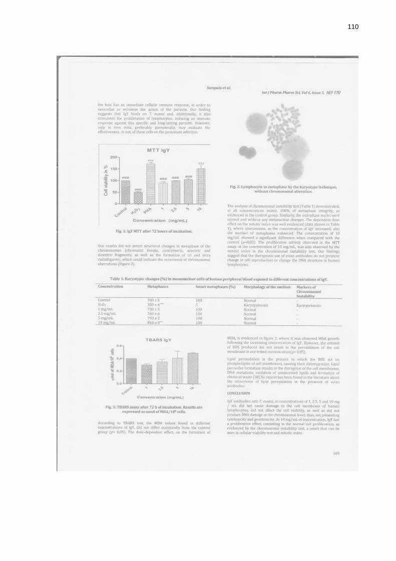
110

111

112
7.2 Comprovante submissão artigo “Production, purification and therapeutic
potential of egg yolk antibodies for treating Trypanosoma evansi infection”
enviado para Revista Veterinary Parasitology.

113
7.3 Parecer Comitê de Ética da Universidade Federal de Santa Maria (UFSM)


![tc ub igy& rc ub ØkfUr · lkxoku ugha mx ldrs ga] ogk¡ egksxuh¼ Swetenia Mahogani ,S. humilis, S.macrophylla½] tSVksc ¼Hymenia caurbaril ½] flYoj vksd¼Gravielia robusta dqN](https://img.document.onl/doc/110x75/6004cb8f66b8b05a0153e36c/tc-ub-igy-rc-ub-kfur-lkxoku-ugha-mx-ldrs-ga-ogk-egksxuh-swetenia-mahogani.jpg)