Embed Size (px)
Citation preview
www.jcol.org.br
Journal ofColoproctology
J C O L O P R O C T O L . 2 0 1 3 ; 3 3 ( 3 ) : 1 1 8 – 1 2 5
Original article
APC protein immunoexpression in colorectal adenoma and adenocarcinoma☆
Vivian Sati Oba Bourroula,b,*, Guilherme Muniz Bourroulc, Giovanna Canato Toloid, Rogério Tadeu Palmaa,e, Celina Tizuko Fujiyama Oshimaf, Thiago Simão Gomesg, Sílvia Saiuli Miki Iharag, Jaques Waisbergh a Service of Digestive System Surgery of Hospital do Servidor Público Estadual (HSPE), São Paulo, SP, Brazil b Service of Digestive System Surgery of Hospital Estadual Mário Covas, Santo André, SP, Brazilc Service of Digestive System Surgery of Hospital Estadual Mário Covas, Santo André, SP, Brazild Faculdade de Medicina do ABC, São Paulo, SP, Brazile Discipline of Service of Digestive System Surgery of Faculdade de Medicina do ABC, São Paulo, SP, Brazilf Laboratory of Molecular Pathology of Escola Paulista de Medicina da Universidade Federal de São Paulo (EPM-UNIFESP), São Paulo, SP, Brazilg Laboratory of Molecular Pathology, Departament of Pathology, EPM-UNIFESP, São Paulo, SP, Brazil h General and Digestive System Surgery of Faculdade de Medicina do ABC, São Paulo, SP, Brazil
a r t i c l e i n f o
Article history:
Received 12 May 2013
Accepted 10 June 2013
Keywords:
Colorectal neoplasms
Carcinoma
Adenoma
APC Gene
Immunohistochemistry
Biological tumor markers
a b s t r a c t
Background: activation of the Wnt pathway by mutated APC gene is considered the initial
event in colorectal carcinogenesis. The identifi cation of these mutations can improve the
specifi c treatment of the adenocarcinoma.
Objective: detect and evaluate wild-type APC protein in tissue from colorectal adenoma,
adenocarcinoma and adjacent mucosa.
Methods: 42 patients that underwent surgery for adenocarcinoma and 53 patients with re-
sected adenomas were studied. Tissue samples from the adenocarcinoma were obtained
from the tumor and from adjacent non-neoplastic mucosa located 10 cm from the proxi-
mal margin of the tumor. Adenoma tissue was obtained from representative areas. Blocks
of tissue microarray (TMA) were submitted to immunohistochemistry with anti-APC, with
readings of positivity and intensity of immunostaining and the score of immune expres-
sion of APC protein was obtained.
Results: the APC protein immune expression score showed a signifi cantly lower expression
of APC protein in the adenoma when compared with the adenocarcinoma (p < 0.0001) and
adjacent mucosa (p < 0.0001). The APC protein immune expression score in the colorectal
mucosa and adjacent to the adenocarcinoma showed no signifi cant difference (p = 0.24).
Conclusions: the fi nding of decreased expression of APC protein in adenoma tissue may indi-
cate that the mutated APC gene may contribute to the changes in the adenoma-carcinoma
process of carcinogenesis sequence. The strong expression of protein APC in tissues from
☆ Support: Coordenação de Aperfeiçoamento de Pessoal de Nível Superior (CAPES) and Fundação de Amparo a Pesquisa do Estado de São Paulo
(FAPESP) 2009/17420-7.
* Corresponding author.
E-mail: [email protected] (V.S.O. Bourroul)
2237-9363/$ - see front matter. © 2013 Elsevier Editora Ltda. All rights reserved.
http://dx.doi.org/10.1016/j.jcol.2013.06.002
JCOL Vol 33 Ed 03 - Arquivo-livro.indb 118JCOL Vol 33 Ed 03 - Arquivo-livro.indb 118 02/10/2013 12:50:2402/10/2013 12:50:24

J C O L O P R O C T O L . 2 0 1 3 ; 3 3 ( 3 ) : 1 1 8 – 1 2 5 119
Palavras-chave:
Neoplasias colorretais
Carcinoma
Adenoma
Gene APC
Imunoistoquímica
Marcadores biológicos de tumor
r e s u m o
Imunoexpressão da proteína APC nos tecidos de adenoma e de adenocarcinoma colorretais
Racional: a ativação da via Wnt pelo gene APC mutado é considerado evento inicial da car-
cinogênese colorretal. A identifi cação dessas mutações pode tornar o tratamento do ade-
nocarcinoma mais específi co.
Objetivo: detectar e avaliar a proteína APC não mutada em tecidos de adenoma, adenocar-
cinoma e mucosa adjacente.
Método: estudados 42 doentes operados de adenocarcinoma e 53 com adenomas resseca-
dos. Tecidos de adenocarcinoma foram obtidas da neoplasia e da mucosa adjacente não
neoplásica situadas a 10 cm da margem proximal do tumor. Tecidos do adenoma foram
obtidas de área representativa. Blocos de tissue microarray (TMA) foram submetidos a
imuno-histoquímica com anticorpo anti-APC. Avaliadas a positividade e intensidade da
expressão e obtidos escores da imunoexpressão da proteína APC.
Resultados: o escore da imunoexpressão da proteína APC no adenoma foi signifi cantemente
menor do que no adenocarcinoma (p < 0,0001) e na mucosa adjacente (p < 0,0001). O escore
da imunoexpressão da proteína APC na mucosa adjacente e no adenocarcinoma não mos-
traram diferença signifi cante (p = 0,24).
Conclusões: a menor expressão da proteína APC no adenoma pode indicar que o gene APC
mutado participa das alterações do processo adenoma-carcinoma. A forte expressão da
proteína APC no CCR e na mucosa adjacente sugerem que a mutação do gene APC não
participou da oncogênese.
© 2013 Elsevier Editora Ltda. Todos os direitos reservados.
the carcinoma and adjacent mucosa suggests that in most patients in this series, the muta-
tion of the APC gene did not participate in the oncogenesis mechanism.
© 2013 Elsevier Editora Ltda. All rights reserved.
Introduction
Colorectal cancer (CRC) is the second most common malig-nancy in the West. Although 15-20% of CRC cases occur in the context of positive family history of the disease, specifi c genetic alterations in familial and sporadic adenocarcinomas are not yet completely known. 1
In sporadic CRC, mutations in the APC and k-ras genes and p53 protein are evident, but mutations in all three genes are rarely found in the same tumor. 2
APC gene comprises 108,352 bp at position 21q in chromo-some 5 and has 21 exons encoding a protein with multiple functional domains that interact with proliferation and apop-tosis regulators.3
The gene is mutated in 63% of sporadic adenomas4 and in over 80% of sporadic CRC cases5 being inherent as heterozygous mutation in all cases of familial adenomatous polyposis (FAP).3
The APC gene encodes a protein of 312 kDa with 2843 ami-no acids. The APC gene inhibits the members of Wnt signaling pathway that promotes β-catenin expression as a stimulator of cell division within the intestinal crypts.6 A central region of APC protein is involved in β-catenin downregulation. This central region contains four segments with 15-amino acid re-peats and seven segments with 20 amino-acid repeats.7
A characteristic of the Wnt pathway activation is the nuclear accumulation of its main effector, the β-catenin, an
activation component of the transcriptional complex that in-cludes members of the TCF/LEF (T-cell transcription factor/Lymphoid enhancer-binding factor), and consequently acti-vating target genes in carcinogenesis.4,8,9,10
In detail, the activation of the Wnt pathway occurs when Wnt proteins bind to frizzled receptors and act as paracrine proteins starting transduction of several signaling pathways. The canonical Wnt signaling pathway stabilizes β-catenin transcription.11 The several domains of APC protein allow its interaction with numerous partner proteins, including β-catenin and axin, and is involved in several cellular pro-cesses.10 The truncated APC protein disrupts the regulation of β-catenin-proteasome cellular concentration, blocking its degradation. The critical point is the β-catenin phosphory-lation into Ser 33 and Ser 37 by phosphoglycerate dehydro-genase, which catalyzes the fi rst step in the biosynthesis of glycine inside the APC/axin complex, known as the “β-catenin destruction complex.” The Phospho-Ser33/37-β-catenin com-plex comprises the recognition sequence for the ubiquitin subtype called E3 ligase βTrCP (β transduction repeat con-taining protein), resulting in “ubiquitination” and subsequent degradation of β-catenin.9,10,11,12
A functioning APC protein is thus vital in maintaining low levels of cytosolic β-catenin in the absence of Wnt signaling pathway, thereby preventing excessive cell proliferation.13 Al-though the role of APC in the regulation of Wnt signaling is more important in the prevention of tumor initiation, its in-
JCOL Vol 33 Ed 03 - Arquivo-livro.indb 119JCOL Vol 33 Ed 03 - Arquivo-livro.indb 119 27/09/2013 12:47:4627/09/2013 12:47:46

J C O L O P R O C T O L . 2 0 1 3 ; 3 3 ( 3 ) : 1 1 8 – 1 2 5120
volvement in apoptosis and chromosomal stability also has an effect on adenoma growth progression.14
Although surgery and chemotherapy are useful methods for the treatment of patients with CRC, additional strategies for the treatment of such patients are needed. It has become apparent that the accumulation of gene mutations in a clonal cell results in the transition from a normal colon epithelial cell into carcinoma.15 By defi ning the molecular alterations involved in the development of neoplasia, it is possible to ex-pect the achievement of specifi c molecular targeting for the treatment of already established tumors, as well as for che-moprophylaxis interventions.16
Method
Ethics
The study was approved by the Research Ethics Committee of Instituto de Assistência Médica ao Servidor Público Estadual de São Paulo (IAMSPE) (protocol N. 043/09) (Annex 10), by the Research Ethics Committee of Faculdade de Medicina do ABC (protocol N. 291/2007) (Annex 11) and by the Research Ethics Committee of Universidade Federal de São Paulo (UNIFESP) (protocol N. 815/09) (Annex 12). Permission to obtain tissue samples was obtained from the Department of Pathological Anatomy of Hospital do Servidor Público Estadual (HSPE), In-stituto de Assistência Médica ao Servidor Público Estadual de São Paulo (IAMSPE) (Annex 13) and permission to perform immunohistochemical tests at the Molecular Pathology Lab-oratory, Department of Pathology, EPM/UNIFESP (Annex 14). Permission to send the paraffi n blocks to the Laboratory of Pathology, Faculdade de Medicina do ABC (Annex 15).
Sample
A total of 64 patients with CRC and 53 patients with colorec-tal adenoma were analyzed retrospectively. Patients with CRC were submitted to curative or palliative surgery at the Surgical Gastroenterology Service of HSPE and patients with adenoma underwent lesion removal by colonoscopy at the Endoscopy Sector of Faculdade de Medicina do ABC (Santo André, São Paulo, Brazil).
Patients were divided into Group 1, comprising 64 patients submitted to surgery for CRC, whose tissue samples were ob-tained from the tumor and adjacent non-neoplastic colorec-tal mucosa anteriorly located 10 cm from the upper margin of the tumor, and Group 2, comprising 53 patients undergoing colonoscopic removal of 71 adenomas.
Inclusion criteria were adult patients with CRC confi rmed by histological analysis of the resected tumor with curative intention or palliative removal of the lesion.
Exclusion criteria were patients younger than 18 years of age, patients with familial CRC or CRC associated with infl am-matory bowel disease and patients submitted to emergency surgery and defi ciency of the histological material used in the immunohistochemistry analysis. A total of 22 patients with CRC were excluded due to insuffi cient histological material for immunohistochemistry analysis, or due to detachment of the tissue. Thus, group 1 consisted of 42 patients with CRC
that provided material considered adequate for immunohis-tochemical analysis. In patients from group 2 that underwent resection of more than one adenoma, only the tumor with more marked histological alterations was considered for the analysis, totaling 53 adenomas.
In group 1, 24 (57.1%) patients were males and 18 (42.9%) females. Mean age was 69.2 ± 7.4 years (51-90 years). The loca-tion of the CRC was the colon in 27 (64.3%) cases and the rec-tum in 15 (35.7%). The diameter of the tumors was ≥ 5 cm in 21 (50%) patients and < 5 cm in 21 (50%). Of the 42 patients, 7 (16.7%) with distal rectal adenocarcinoma underwent neoad-juvant therapy. In group 2, regarding the adenomas obtained from 53 patients, 27 (50.9%) were males and 26 (49.1%) fe-males. The mean age was 60.7 ± 3.4 years (29-88 years).
Staging was performed by complete clinical and anorectal examination, serum measurement of CEA and CA19-9, colo-noscopy with lesion biopsy and respective histopathological analysis, opaque enema when indicated, and total abdominal and chest CT. The clinicopathological staging classifi cation used was the TNM (UICC, 2010).17
Resection with curative intent followed the pattern of on-cologic surgeries. In palliative resections, the colorectal lesion was removed and oncologic procedures were not performed.
Clinical characteristics were obtained from the sample for all groups of individuals. In the group of patients with colorec-tal adenoma, the morphological characteristics of the lesion were recorded (location, histological type and degree of cell atypia). In patients with CRC, the following information was obtained: macroscopic characteristics of the lesion (location, size along the major axis), microscopic characteristics (level of invasion, infl ammatory infi ltrate, surgical margins, lymph node involvement, degree of tumor differentiation, lymphat-ic/vascular invasion and neural infi ltration), TNM classifi ca-tion of UICC (2010),17 presence of synchronous metastases and immunostaining (staining intensity scores and percent-age of stained cells) of anti-APC antibody in colorectal tissue.
Anatomopathological study
CRC tissues were fi xed in formalin and routinely processed us-ing the paraffi n-embedding method for histological analysis. All pathological reports were analyzed to characterize macro-scopic parameters. Histological sections with 3-μm thickness were obtained from each block. All slides were stained with hematoxylin-eosin (HE), revised by the pathologist and the diagnosis was confi rmed.
In the stained slides, the best preserved areas that were representative of the tumor to obtain the building block (cyl-inder) to be used in the preparation of tissue microarray (TMA) were identifi ed. For the construction of TMA, areas with ne-crosis, hemorrhage, desmoplasia, and areas of low cellularity or even acellular areas were excluded.
TMA preparation
The TMA block was prepared using a Beecher ™ (Beecher In-struments, Silver Spring, MD, USA) equipment, followed by the following steps: identifi cation of the selected area in the re-spective paraffi n block; construction of the casela in the recipi-ent block; extraction of 1 mm of tissue of the donor block from
JCOL Vol 33 Ed 03 - Arquivo-livro.indb 120JCOL Vol 33 Ed 03 - Arquivo-livro.indb 120 27/09/2013 12:47:4727/09/2013 12:47:47
J C O L O P R O C T O L . 2 0 1 3 ; 3 3 ( 3 ) : 1 1 8 – 1 2 5 121
the previously selected area of interest; obtaining cylinder tis-sue from the donor block and placing it in the previously cre-ated casela in the recipient block; progression into 1-mm frag-ments to the new positions in the recipient block, thus creating a set of tissue samples obtained from the array arrangement and quality assessment of the fi nal block for storage.
For adhesion of the TMA block section onto the slides, an adhesive tape system was used (Instrumedics, Hackensack, N.J., USA). Samples were cut at a thickness of 4 μm and a small cylinder was used to fi x the tape. The histological section was subsequently joined, placed on a slide coated with resin and pressed with a cylinder to obtain better tissue section adhe-sion. Then the slides with the histological cuts adhered to the tape were placed under UV light for 20 min, followed by im-mersion in TPC solvent TM solution for another 20 min. After drying, the tapes were removed and the slides stored at -80 °C until completion of immunohistochemistry analysis.
The antibody receptor blocks were obtained from two blocks, in which the fi rst (A-TMA) was added to the adenoma tissue. In the second block (TMA-B), CRC and adjacent mu-cosa cylinders were included. HE staining was performed in order to evaluate the quality of the sections and possible loss of sample representativeness.
Immunohistochemical analysis
For the immunohistochemical study, 4-μm histological frag-ments were obtained, placed on glass slides pretreated with 3-aminopropyltriethoxysilane adhesive (Sigma Chemical Co, St. Louis, MO, USA). Initially, histological slides were placed in an oven at 60 °C for 24 hours to obtain better tissue adhesion and deparaffi nization. Deparaffi nization was performed in three xylene baths at room temperature for 15 min and placed after each of the three baths, in absolute ethanol baths in for 1 min each. The slides were washed in running water for 5 min and submitted to heat induced antigen recovery by steam in a 10 mM citrate buffer solution with pH 6.0 for 30 min.
After cooling for 20 min at room temperature, the slides were washed in running water for 5 min and endogenous per-oxidase blocking was performed using a hydrogen peroxide solution at 3%, in four baths of 5 min each. The slides were again washed in running water for 5 min and then washed with phosphate buffered saline (PBS), pH 7.2-7.6, also during 5 min.
Rabbit polyclonal anti-APC antibody C-20 (batch C1709) (Santa Cruz Biotechnology, Inc., CA, USA) was used, which de-tects the wild-type APC protein and is recommended for de-tection of the respective protein in human tissue. Incubation was carried out at a concentration of 1:100 in a humidifi ed chamber at 4 ° C for at least 16-18 hours (overnight).
Subsequently, after three washes in PBS at pH 7.2 to 7.6, the incubation was performed with the streptavidin-biotin peroxidase kit (LSAB, DakoCytomation, CA, USA) in a humidi-fi ed chamber at room temperature for 30 min. This step was followed by washes with PBS at pH 7.2-7.6 and development with liquid DAB (DakoCytomation, CA, USA) at room tempera-ture for 5 min.
After washing in running water for 3 min, counter-staining was performed with Harris hematoxylin for 1 min. The sec-tions were dehydrated in three baths with absolute ethanol
and three baths of xylene and then mounted using cover slips with Entellan resin (Sigma Chemical Co., St. Louis, MO, USA) for analysis by optical microscopy.
As positive control, slides with histological sections pre-viously demonstrated as being positive for these antibodies were used. A similar slide was used as a negative control, sub-tracting the primary antibody from the reaction.
Immunoexpression score was prepared using the method by Hao.18 Slide positivity was considered zero score if there were less than 5% of epithelial cells in the lesion; score 1, with 5% to 25%, score 2, with 26% to 50%, score 3, 51% to 75% and score 4, more than 75% of epithelial cells in the lesion. Inten-sity was considered zero score when negative; score 1, when weak, score 2 when moderate and score 3, strong intensity.
The fi nal score of the immunoexpression varied between zero and 12 and was obtained by multiplying the scores of in-tensity and positivity. The immunoexpression was considered reduced when the fi nal score was between zero and 8, and strong if the fi nal score was between 9 and 12.
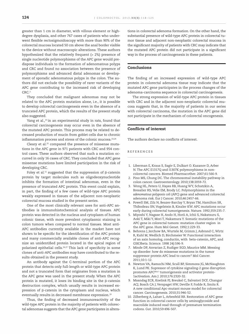
Fig. 1 shows the adenoma tissue cells stained with anti-APC antibody. Positivity in these slides was greater than 50% and the intensity was moderate to strong.
Fig. 2 shows adenocarcinoma tissue cells stained with anti-APC antibody. Positivity in these slides was greater than 75% and the intensity was strong.
Statistical analysis
The quantitative results were described as mean and stan-dard deviation. Qualitative data was described as frequencies. The correlation between the scores of the immunohistochem-ical expression of the APC protein with clinicopathological parameters was assessed by Spearman’s correlation coef-fi cient. Student’s t test, chi-square (χ2), Mann-Whitney test and McNemar test for paired variables were used to test the signifi cance of differences in clinicopathological parameters.Associations between positivity in protein labeling and clini-copathological features of interest were evaluated by chi-square or Fisher’s exact test (very small tables with frequen-cies) or chi-square (χ2) of likelihood ratio (tables of variables with more than two categories). The univariate logistic regres-sion analysis (ANOVA) and multivariate analysis were used to identify the dependent and independent variables. Signifi -cance level was set at 5% (p ≤ 0.05). The statistical software SPSS, version 15.0 (The Predictive Analytics Company, Chica-go, IL, USA) was used for statistical calculations.
Results
Regarding the anatomopathological characteristics of CRC, 21 (50%) patients had lymph node metastases, 13 (31%) had ve-nous invasion, 13 (31%) had lymphatic vessel invasion and 6 (14.3%) had neural and/or perineural infi ltration. Thirty-four (81%) cases were classifi ed as moderately differentiated, 7 (16.7%) were well differentiated and 1 (2.3%) was poorly differ-entiated. In relation to the depth of tumor invasion, 29 (69%) tumors were classifi ed as T3, 12 (28.6%) as T2, and 1 (2.4%) as T4. Nine (21.4%) patients had metastases to the liver, perito-neum, or both locations.
JCOL Vol 33 Ed 03 - Arquivo-livro.indb 121JCOL Vol 33 Ed 03 - Arquivo-livro.indb 121 27/09/2013 12:47:4727/09/2013 12:47:47

J C O L O P R O C T O L . 2 0 1 3 ; 3 3 ( 3 ) : 1 1 8 – 1 2 5122
Of the 42 CRC cases submitted to surgery, 39 (92.9%) under-went curative resection and the median survival of these pa-tients was 18.5 months. Neoplastic recurrence occurred in 10 (23.8%) patients (2 colorectal adenocarcinomas and 8 colon ad-enocarcinomas) and fi ve of them (11.9%) died due to neoplasia.
Of the 71 adenomas removed from 53 patients, 64 (90.1%) were located in the descending colon, sigmoid colon and rectum and in the ascending colon or transverse colon in 7 (9.9%). As for the histological type, the tumors were clas-sifi ed as tubular in 49 (69%) and tubulovillous lesions in 22 (31%). Mild atypia was found in 39 (55%) adenomas and 32 adenomas (45%) had no atypia. A total of 18 adenomas from patients with more than one adenoma were excluded at the immunohistochemical analysis.
The positivity and intensity of the immunohistochemical expression of APC protein observed in the cytoplasm of 53 colorectal adenomas studied are described in Tables 1 and 2.
The intensity of the immunohistochemical expression of the APC protein observed in the cytoplasm in cases of adjacent non-neoplastic colorectal mucosa is described in
Table 3. The intensity of the immunohistochemical expres-sion of the APC protein found in the cytoplasm of the 42 CRC cases is described in Table 4.
The positivity of the immunohistochemical expression of the APC protein in CRC and adjacent non-neoplastic mucosa was strong in all patients.
The frequency of the APC protein in the colorectal ade-noma tissues, of CRC and adjacent non-neoplastic colorectal mucosa is shown in Table 5. The frequency of strong expres-sion was higher in tumor samples and adjacent non-neoplas-tic colorectal mucosa than in samples of colorectal adenoma. The comparison of wild-type APC protein immunoexpression scores between adenoma and adenocarcinoma tissue showed that the immunoreactivity was signifi cantly higher (p <0.0001) in adenocarcinoma tissue. The comparison of the scores of the wild-type APC protein immunoexpression between ad-enoma tissue and adjacent non-neoplastic colorectal mucosa showed that the immunoexpression was signifi cantly higher (p < 0.0001) in the adjacent non-neoplastic colorectal mucosa. The comparison of the scores of the wild-type APC protein immunoexpression in adenocarcinoma tissue and adjacent
Fig. 2 – Photomicrograph of positive immunoexpression of anti-APC antibody represented by the brownish color in the cytoplasm of neoplastic cells from colorectal adenocarcinoma (immunohistochemistry; (A) 40x and (B) 400x).
Fig. 1 – Photomicrograph of positive immunoexpression of anti-APC antibody represented by the brownish color in the cytoplasm of cells from colorectal adenoma (immunohistochemistry; (A) 40× and (B) 400×).
JCOL Vol 33 Ed 03 - Arquivo-livro.indb 122JCOL Vol 33 Ed 03 - Arquivo-livro.indb 122 27/09/2013 12:47:4727/09/2013 12:47:47

J C O L O P R O C T O L . 2 0 1 3 ; 3 3 ( 3 ) : 1 1 8 – 1 2 5 123
Regarding gender, in cases of adenomas with strong ex-pression there were 13 (24.5%) women and 11 (20.7%) men, whereas in cases of adenomas with reduced expression there were 13 (24.5%) women and 16 (31.3% ) men (p = 0.58).
Regarding the histological type, in adenomas with strong expression, 21 (39.6%) were tubular and 3 (5.6%) were tubulovil-lous, whereas in adenomas with reduced expression, 16 (30.3%) were tubular and 13 (24.5%) were tubulovillous (p = 0.01).
In cases with adjacent non-neoplastic colorectal mucosa, there was one case with the reduced APC protein immunoex-pression, without the possibility of statistical analysis regard-ing age and sex.
In tumor tissue there were 38 (90.5%) patients with strong APC protein immunoexpression and 4 (9.5%) patients with reduced immunoexpression, and statistical analysis regard-ing age and sex was not possible. In cases with strong APC protein immunoexpression, there were 22 (52.3%) women and 16 (38.1%) men, whereas in tissues with CRC, reduced immu-noexpression was found in 2 (4.8%) men and 2 (4.8%) women.
Discussion
Luo et al.19 studied the formation of aberrant crypt foci as pre-cursor lesions of colorectal carcinogenesis in normal human tissue. These authors observed normal expression of APC and β-catenin proteins, suggesting that loss of heterozygosity of the APC tumor suppressor gene, commonly seen in CRC, may occur earlier and perhaps even before the APC mutation. The fi ndings of this study showed that the strong expression of wild-type APC protein in CRC tissues and adjacent non-neoplastic colorectal mucosa were found in most samples (90.4% and 97.6%, respec-tively), a result that is in accordance with those by Luo et al.19
A study on the infl uence of the Wnt signaling pathway in adenomas was performed by Wang et al.,20 who associ-ated the presence of Wnt pathway activation to the process of carcinogenesis of laterally spreading tumors (LST). These authors studied 15 LST lesions and 54 adenomas and com-pared the expression of GSK3-β, phospho-GSK3-β, axin and β-catenin proteins of the Wnt pathway in LST lesions and colorectal adenomas.There was an increase in the expression of phospho-GSK3-β and β-catenin in LST lesions, but there was no difference in the expression of axin and GSK3-β proteins. These fi ndings suggested that activation of the Wnt / β-catenin pathway seems to be more intense in LST lesions, when compared to colorectal adenoma. Hashimoto et al.21 studied 42 LST lesions, including seven colorectal adenomas, 25 intramucosal adeno-carcinomas and 10 submucosal invasive colorectal adenocar-cinomas. They found that the presence of the methylated APC protein was inversely proportional to the presence of submu-cosal invasion in LST lesions. APC protein hypermethylation is signifi cantly associated with the presence of APC muta-tions.21,22 There was no association between APC gene muta-tions and tendency to malignant LST lesions.21 In the present study, the wild-type APC protein expression was reduced in colorectal adenoma tissue, which may indicate tendency to malignant adenomas.20
Wong et al.3 studied 758 cases of colorectal adenomas lo-cated in the distal large intestine represented by adenomas
Table 5 – Frequency of scores of immunohistochemical expression of wild-type APC protein in adenoma tissue in adjacent non-neoplastic colorectal mucosa and colorectal adenocarcinoma.
Scores Adenoma Adjacent Mucosa
Tumor
Strong expression 24 (45.2%) 41 (97.6%) 38 (90.4%)Decreased expression 29 (54.7%) 1 (2.4%) 4 (9.5%)
Table 1 – Intensity of immunohistochemical expression of wild-type APC protein in colorectal adenomas.
Intensity n (%)
Low 0 (0%)Moderate 22 (41.5%)Strong 31 (58.5%)
n, number of cases.
Table 3 – Intensity of immunohistochemical expression of wild-type APC protein in non-neoplastic adjacent colorectal mucosa in 42 patients with colorectal adenocarcinoma.
Intensity n (%)
Low 0 (0%)Moderate 1 (2.4%)Strong 41 (97.6%)
n, number of cases.
Table 4 – Intensity of immunohistochemical expression of wild-type APC protein in the 42 cases of colorectal adenocarcinoma.
Intensity n (%)
Low 0 (0%)Moderate 2 (4.8%)Strong 40 (95.2%)
n, number of cases.
Table 2 – Positivity of immunohistochemical expression of wild-type APC protein in 53 colorectal adenomas.
Positivity n (%)
< 5% 0 (0%)5 to 25% 3 (5.7%)26 to 50% 9 (17%)51 to 75% 9 (17%)> 75% 32 (60.3%)
n, number of cases.
non-neoplastic colorectal mucosa showed that immunoex-pression was not signifi cantly different (p = 0.24).
In adenomas with strong expression (n = 24), the mean age was 62.3 ± 2.7 years, and in those with reduced expression, the mean age was 58.8 ± 1.94 (p = 0.29).
JCOL Vol 33 Ed 03 - Arquivo-livro.indb 123JCOL Vol 33 Ed 03 - Arquivo-livro.indb 123 27/09/2013 12:47:4727/09/2013 12:47:47
J C O L O P R O C T O L . 2 0 1 3 ; 3 3 ( 3 ) : 1 1 8 – 1 2 5124
greater than 1 cm in diameter, with villous element or high-degree dysplasia, and other 767 cases of patients who under-went fl exible rectosigmoidoscopy with more than 90% of the colorectal mucosa located 50 cm above the anal border visible to the device without macroscopic alterations. These authors hypothesized that the relatively frequent (> 2%) presence of single nucleotide polymorphisms of the APC gene would pre-dispose individuals to the formation of adenomatous polyps and CRC and found no association between the presence of polymorphisms and advanced distal adenomas or develop-ment of sporadic adenomatous polyps in the colon. The au-thors did not exclude the possibility of rarer variants of the APC gene contributing to the increased risk of developing CRC.3
They concluded that malignant adenomas may not be related to the APC protein mutation alone, i.e., it is possible to develop colorectal carcinogenesis even in the absence of a truncated APC protein, which the results of the present study also suggested.
Yang et al.,23 in an experimental study in rats, found that colorectal carcinogenesis may occur even in the absence of the mutated APC protein. This process may be related to de-creased production of mucin from goblet cells due to chronic infl ammation process and stress of the colonic mucosa.23,24
Cleary et al.25 compared the presence of missense muta-tions in the APC gene in 971 patients with CRC and 954 con-trol cases. These authors observed that such a mutation oc-curred in only 16 cases of CRC. They concluded that APC gene missense mutations have limited participation in the risk of developing CRC.
Foley et al.15 suggested that the suppression of β-catenin protein by target molecules such as oligodeoxynucleotide inhibits the formation of intestinal adenomas, even in the presence of truncated APC protein. This event could explain, in part, the fi nding of a few cases of wild-type APC protein weakly expressed in tissues of the adjacent non-neoplastic colorectal mucosa studied in the present series.
One of the most clinically relevant uses for anti-APC an-tibodies is immunohistochemical analysis of tissues. APC protein was detected in the nucleus and cytoplasm of human colonic tissue, with more prevalent cytoplasmic staining in colon tumors when compared to normal tissues. Most anti-APC antibodies currently available in the market have not shown to be specifi c for the identifi cation of the APC protein and many commercially available clones of anti-APC recog-nize an unidentifi ed protein located in the apical region of polarized epithelial cells.26,27 This lack of specifi city in some clones of anti-APC antibodies may have contributed to the re-sults obtained in the present study.
An antibody against the C-terminal portion of the APC protein that detects only full-length or wild-type APC protein and not a truncated form that originates from a mutation in the APC gene was used in the present study. When the APC protein is mutated, it loses its binding site in the β-catenin destruction complex, which usually results in increased ex-pression of β-catenin in the cytoplasm and nucleus, which eventually results in decreased membrane expression.19
Thus, the fi nding of decreased immunoreactivity of the wild-type APC protein in the majority of patients with colorec-tal adenomas suggests that the APC gene participates in altera-
tions in colorectal adenoma formation. On the other hand, the substantial presence of wild-type APC protein in colorectal tu-mor tissue and adjacent non-neoplastic colorectal mucosa in the signifi cant majority of patients with CRC may indicate that the mutated APC protein did not participate in a signifi cant way in the process of carcinogenesis in these patients.
Conclusions
The fi nding of an increased expression of wild-type APC protein in colorectal adenoma tissue may indicate that the mutated APC gene participates in the process changes of the adenoma-carcinoma sequence in colorectal carcinogenesis.
The strong expression of wild-type APC protein in tissues with CRC and in the adjacent non-neoplastic colorectal mu-cosa suggests that, in the majority of patients in our series with colorectal carcinoma, the mutation in the APC gene did not participate in the mechanism of colorectal oncogenesis.
Confl icts of interest
The authors declare no confl icts of interest.
R E F E R E N C E S
1. Liberman E, Kraus S, Sagiv E, Dulkart O. Kazanov D, Arber N. The APC E1317Q and I1307K polymorphisms in non-colorectal cancers. Biomed Pharmacother. 2007;61:566-9.
2. Pino MS, Chung DC. The chromosomal instability pathway in colon cancer. Gastroenterology. 2010;138:2059-72.
3. Wong HL, Peters U, Hayes RB, Huang WY, Schatzkin A, Bresalier RS, Velie EM, Brody LC. Polymorphisms in the adenomatous polyposis coli (APC) gene and advanced colorectal adenoma risk. Eur J Cancer. 2010;46:2457-66.
4. Powell SM, Zilz N, Beazer-Barclay Y, Bryan TM, Hamilton SR, Thibodeau SN, Vogelstein B, Kinzler KW. APC mutations occur early during colorectal tumorigenesis. Nature. 1992;359:235-7.
5. Miyoshi Y, Nagase H, Ando H, Horii A, Ichii S, Nakatsuru S, Aoki T, Miki Y, Mori T, Nakamura Y. Somatic mutations of the APC gene in colorectal tumors: mutation cluster region in the APC gene. Hum Mol Genet. 1992;1:229-33.
6. Behrens J, Jerchow BA, Wurtele M, Grimm J, Asbrand C, Wirtz R, Kuhl M, Wedlich D, Birchmeier W. Functional interaction of an axin homolog, conductin, with beta-catenin, APC, and GSK3beta. Science. 1998 24):596-9.
7. Minde DP, Anvarian Z, Rudiger SGD, Maurice MM. Messing up disorder: how do missense mutations in the tumor suppressor protein APC lead to cancer? Mol Cancer. 2011;10:1-12.
8. Newton VA, Ramochi NM, Scull BP, Simmons JG, McNaughton K, Lund PK. Supressor of cytokine signaling-2 gene disruption promotes APCMin/+ tumorigenesis and activator protein-1activation. Am J. 2010;176:2320-32.
9. Maandag ECR, Koelink PJ, Breukel C, Salvatori DCF, Changur ACJ, Bosch CA J, Verspaget HW, Deville P, Fodde R, Smits R. A new conditional Apc-mutant mouse model for colorectal cancer. Carcinogenesis. 2010;31:946-52.
10. Zilberberg A, Lahav L, Arbesfeld RR. Restoration of APC gene function in colorectal cancer cells by aminoglycoside and macrolide induced read through of premature termination codons. Gut. 2010;59:496-507.
JCOL Vol 33 Ed 03 - Arquivo-livro.indb 124JCOL Vol 33 Ed 03 - Arquivo-livro.indb 124 27/09/2013 12:47:4827/09/2013 12:47:48
J C O L O P R O C T O L . 2 0 1 3 ; 3 3 ( 3 ) : 1 1 8 – 1 2 5 125
11. Zhanker HH, Walker F, Kifl emariam S, Whitehead RH, Williams D, Phillips WA, Mikeska T, Dobrovic A, Burgess AW. Selective inhibition of proliferation in colorectal carcinoma cell lines expressing mutant APC or activated B-Raf. Int J Cancer. 2009;125:297-307.
12. Dhir M, Montgomery EA, Glöckner SC, Schuebel KE, Hooker CM, Herman JG, Baylin SB, Gearhart SL, Ahuja N. Epigenetic regulation of WNT signaling pathway genes in infl ammatory bowel disease (IBD) associated neoplasia. J Gastrointest Surg. 2008;12:1745-53.
13. Dumas YR, He X. Wnt signaling: What the X@# is WTX! EMBO J. 2011;30:1415-7.
14. Vincan E, Barker N. The upstream components of the Wnt signalling pathway in the dynamic EMT and MET associated with colorectal cancer progression. ClinExp Metastasis. 2008;25:657-63.
15. Samowitz WS. Genetic and epigenetic changes in colon cancer. Exp Mol Pathol. 2008;85:64-7.
16. Foley PJ, Scheri RP, Smolock CJ, Pippin J, Green DW, Drebin JA. Targeted supression of β-catenin blocks intestinal adenoma formation in APC Min mice. J Gastrointest Surg. 2008;12:1452-8.
17. TMN Classifi cation of Malignant Tumours. International Union Against Cancer(UICC)/American Joint Committee on Cancer (AJCC). 7th ed. New York, NY: Springer, 2010, pp 143-164.
18. Hao XP, Pretlow TG, Rao JS, Pretlow TP. β-catenin expression is altered in human colon aberrant cript foci. Cancer Res. 2002;61:8085-8.
19. Luo L, Shen GQ, Stiffl er KA, Wang QK, Pretlow TG, Pretlow T. Loss of the heterozygosity in human aberrant crypt foci (ACF), a putative precursor of colon cancer. Carcinogenesis. 2006;27:1153-9.
20. Wang J, Wang X, Gong W, Mi B, Liu S, Jiang B. Increased expression of β-catenin, phosphorylated glycogen synthase
kinase 3β, cyclin d1, and c-mycin laterally spreading colorectal tumors. J HistochemCytochem. 2009;57:363-71.
21. Hashimoto K, Shimizu Y, Suehiro Y, Okayama N, Hashimoto S, Okada T, Hiura M, Ueno K, Hazama S,Higaki S, Hamanaka Y, Oka M, Sakaida I, Hinoda Y. Hypermethylation status of APC inversely correlates with the presence of submucosal invasion in laterally spreading colorectal tumors. Mol Carcinogenesis. 2008;47:1-8.
22. Lee BB, Lee EJ, Jung H, Chun HK, Chang DK, Song SY, Park J, Kim DH. Aberrant methylation of APC, MGMT, RASSF2A, and Wif-1 genes in plasma as a biomarker for easy detection of colorectal cancer. Clin Cancer Res. 2009;15 :6185-91.
23. Yang K, Popova NV, Yang WC, Lozonschi I, Tadesse S, Kent S, Bancroft L, Matise I, Cormier RT, Scherer SJ,Edelmann W, Lipkin M, Augenlicht L, Velcich A. Interaction of Muc 2 and APC on Wnt signaling and in intestinal tumorigenesis: potential role of chronic infl ammation. Cancer Res. 2008;68:7313-22.
24. Burgess AW, Faux MC, Layton MJ, Ramsay RG. Wnt signaling and colon tumorigenesis- a view from the periphery. Exp Cell Res. 2011; 317:2748-58.
25. Cleary SP, Lim H, Croitoru ME, Redston M, Knight JA, Gallinger S, Gryfe R. Missense polymorphisms in the adenomatous polyposis coli gene and colorectal cancer risk. Dis Colon Rectum. 2008;51:1467-74.
26. Weeraratna AT, Jiang Y, Hostetter G, Rosenblatt K, Duray P, Bittner M, Trent JM. Wnt5a signaling directly affects cell motility and invasion of metastatic melanoma. Cancer Cell. 2002;1:279-88.
27. Wang Y, Azuma Y, Friedman DB, Coffey RJ, Neufeld KL. Novel association of APC with intermediate fi laments identifi ed using a new versatile APC antibody. BMC Cell Biology. 2009;10:1-13.
JCOL Vol 33 Ed 03 - Arquivo-livro.indb 125JCOL Vol 33 Ed 03 - Arquivo-livro.indb 125 27/09/2013 12:47:4827/09/2013 12:47:48


![MANUAL DE INSTRUÇÕES 3D Blu-ray™/DVD Home Theater … · Selecionando um arquivo de legenda Ouvindo músicas durante o Slideshow 33 Definições [3D] Funções Avançadas Desfrutando](https://img.document.onl/doc/110x75/5c043c5609d3f296388b61f1/manual-de-instrucoes-3d-blu-raydvd-home-theater-selecionando-um-arquivo.jpg)
![Relatório de Abril 2012 [Modo de Compatibilidade]cuiaba.mt.gov.br/upload/arquivo/rel_04_30_2012.pdf20,00% 30,00% 40,00% 50,00% Jornal Telefone Online Presencial 0% 9% 2% 37% 13% 33%](https://img.document.onl/doc/110x75/5faa4ee87305f3131b0e591f/relatrio-de-abril-2012-modo-de-compatibilidade-2000-3000-4000-5000-jornal.jpg)