Embed Size (px)
Citation preview
Londrina 2016
CENTRO DE PESQUISA EM CIÊNCIAS DA SAÚDE
MESTRADO EM CIÊNCIAS DA REABILITAÇÃO
LEANDRO VAZ TOFFOLI
EFEITOS DO ESTRESSE E DA PRÁTICA DE EXERCÍCIO
FÍSICO SOBRE O MECANISMO DE METILAÇÃO DO DNA
EM CÉLULAS DO PULMÃO DE RATOS
Londrina 2016
LEANDRO VAZ TOFFOLI
EFEITOS DO ESTRESSE E DA PRÁTICA DE EXERCÍCIO
FÍSICO SOBRE O MECANISMO DE METILAÇÃO DO DNA
EM CÉLULAS DO PULMÃO DE RATOS
Dissertação apresentada ao Programa de Pós-
Graduação em Ciências da Reabilitação (Programa
Associado entre Universidade Estadual de Londrina -
UEL e Universidade Norte do Paraná - UNOPAR),
como requisito para a obtenção do título de Mestre em
Ciências da Reabilitação.
Orientador: Prof. Dr. Marcus Vinícius de Matos
Gomes

LEANDRO VAZ TOFFOLI
EFEITOS DO ESTRESSE E DA PRÁTICA DE EXERCÍCIO
FÍSICO SOBRE O MECANISMO DE METILAÇÃO DO DNA
EM CÉLULAS DO PULMÃO DE RATOS
Dissertação apresentada ao Programa de Pós-
Graduação em Ciências da Reabilitação
(Programa Associado entre Universidade
Estadual de Londrina [UEL] e Universidade
Norte do Paraná [UNOPAR]), como requisito
para a obtenção do título de Mestre em Ciências
da Reabilitação.
BANCA EXAMINADORA
____________________________________
Prof. Dr. Marcus Vinícius de Matos Gomes
Universidade Norte do Paraná
____________________________________
Prof. Drª Regina Célia Poli Frederico
Universidade Norte do Paraná
____________________________________
Prof. Dr. Henrique César Santejo Silveira
Hospital do Câncer de Barretos
Londrina, 30 de Abril de 2016.

AGRADECIMENTOS
Ao meu pai Valdecir Aparecido Toffoli, e minha mãe Elenice Vaz Toffoli e
minha irmã Michelle Vaz Toffoli pelo incentivo, pelo amor e apoio incondicional.
Ao meu parceiro e amigo, Thiago Rennan Gomes que esteve presente
durante todo esse percurso, sempre me apoiando e comemorando junto comigo as minhas
vitórias.
Ao meu Profº Orientador Dr. Marcus Vinicius de Matos Gomes pelo
conhecimento transmitido e todo o suporte durante estes anos.
Aos funcionários e amigos do Laboratório Cetel e Laboratório Cellmais por
acompanhar essa trajetória.
Aos colaboradores do Laboratório de Análise de Materiais e Moléculas
(LAMM) da Universidade Estadual de Londrina (UEL) pela disponibilidade e prontidão.
Aos colaboradores e colegas do Laboratório de Neurociência e
Farmacologia Cardiovascular da Universidade Estadual de Londrina (UEL) pelo suporte e
disponibilidade.
À Funadesp, pelo apoio financeiro na execução do projeto.
A todos que direta ou indiretamente contribuíram para este trabalho.

Pouco conhecimento faz com que as pessoas se
sintem orgulhosas. Muito conhecimento, que se
sintam humildes. – Leonardo da Vinci
TOFFOLI, Leandro Vaz. Efeitos do estresse e da prática de exercício físico sobre o
mecanismo de metilação do DNA em células do pulmão de ratos. 2016. 61 fls. Dissertação
(Mestrado em Ciências da Reabilitação) – Universidade Norte do Paraná, Londrina, 2016.
RESUMO
Dados recentes da literatura sugerem a participação de mecanismos epigenéticos nas respostas
celulares adaptativas ao estresse. Similarmente, dados consistentes revelam o potencial do
exercicio fisico em modular os efeitos do estresse comportamental. Neste contexto,
objetivamos avaliar no presente estudo o efeito do estresse sobre a metilação global do DNA
em células do pulmão de ratos e sua relação com a expressão dos genes Dnmt1, Dnmt3a e
Dnmt3b. Adicionalmente avaliamos o potencial da prática de exercício físico (natação) em
modular os efeitos moleculares relacionados ao estresse. Ratos Wistar foram distribuidos em
04 grupos contendo 05 animais cada: um grupo foi submetido ao protocolo validado de
estresse (grupo ST) do 67º dia pós-natal (DPN) ao 80º DPN. Para o grupo de praticantes de
exercício físico (grupo EX) foram considerados ratos submetidos a natação no período que
compreende 53º e 79º DPN. O grupo de exercício físico e estresse (grupo EX-ST) foi
constituído por animais submetidos à natação no período do 53º ao 79º DPN e ao protocolo de
estresse do 67º ao 80º DPN. O grupo controle (CTL) foi constituído por animais que foram
mantidos nas mesmas condições ambientais porém não foram submetidos aos protocoos de
estresse ou exercício físico. No último dia do protocolo (80º DPN) os animais foram
sacrificados por decaptação para obtenção de amostra do pulmão. Para a avaliação
quantitativa do perfil de metilação global do DNA foi utilizado o Imprint® Methylated DNA
Quantification kit (Sigma-Aldrich). A expressão dos genes Dnmt1, Dnmt3a e Dnmt3b foi
avaliada por PCR em tempo real utilizando o sistema de detecção Taqman e o gene Gapdh
como controle endógeno. Diminuição estatisticamente significativa da metilação global do
DNA foi observada quando comparados os grupos ST e CTL (p=0.0159). Entretanto, um
aumento significativo na metilação global do DNA foi observado quando comparados os
grupos EX e EX-ST com ST (p=0.0018). No tocante aos genes das DNA metiltranferases,
aumento estatisticamente significativo foi observado na expressão do gene Dnmt1 no grupo
ST quando comparado com CTL (p=0.0159) e uma diminuição na expressão quando
comparados os grupos EX-ST e ST (p=0.0042). Diminuição significativa na expressão dos
genes Dnmt3a e Dnmt3b foi observada quando comparados os grupos EX e ST (p=0.0018)
além de uma diminuição da expressão do gene Dnmt3b quando comparados EX e CTL
(p=0.0357). Em suma, nossos dados demonstram que o estresse comportamental induz uma
diminuição no perfil de metilação global do DNA em células do pulmão de ratos associada ao
aumento na expressão do gene Dnmt1. Além disso, nossas evidências sugerem que a prática
de exercício fisico pode potencialmente modular as respostas induzidas pelo estresse em
células do pulmão de ratos no que diz respeito a variações na metilação global do DNA e na
expressão dos genes Dnmt1, Dnmt3 e Dnmt3b.
Palavras-Chave: Estresse; Exercício Físico; Metilação do DNA; Epigenética; DNA
metiltransferase
TOFFOLI, Leandro Vaz. Effects of stress and physical activity on the DNA methylation
mechanism of rat lung cells. 2016. 61 fls. Dissertação (Mestrado em Ciências da
Reabilitação) – Universidade Norte do Paraná, Londrina, 2016.
ABSTRACT
Recent data from the literature suggest the involvement of epigenetic mechanisms in adaptive
cellular responses to stress. Similarly, consistent data show the potential of physical exercise
in modulation the effects of behavioral stress. In this context, we aimed to evaluate in the
study the effect of the stress on the global DNA methylation in rat lung cells and its
relationship with the expression of Dnmt1, Dnmt3a and Dnmt3b genes. Furthermore, we
evaluated the potential of physical activity (swimming) to modulate the molecular effects
evoked by stress. Wistar rats were divided into four experimental groups containing 05
animals each: one group was submitted to the chronic stress protocol (ST) from the 67th
postnatal day (PND) to the 80 th PND. For the group of physical activity (EX) we considered
rats that were submitted to swimming sessions in the period of 53th to 79th PND. Physical
activity and stress group (EX-ST) consisted of animals submitted to swimming in the period
of 53 th to 79 th PND and to the stress protocol from 67th to 80th PND. The control group (CTL)
consisted of animals that were not submitted to interventions. On the last day of the protocol
(80th PND), the animals were sacrificed by decapitation and samples from lung were obtained.
For quantitative assessment of the global DNA methylation profile was quantified using the
Imprint Methylated DNA Quantification kit (Sigma-Aldrich®). The expression of Dnmt1,
Dnmt3a and Dnmt3b genes was evaluated by real-time PCR using the Taqman detection
system and the Gapdh as endogenous control. A statistically significant decrease in the global
DNA methylation profile was observed in the lung of animals from ST group when compared
to the CTL group (p=0.0159). There was no statistically significant difference in the global
DNA methylation profile when compared the animals from EX and CTL groups. A
statistically significant increase in the global DNA methylation profile was observed when
compared the EX and the EX-ST groups with the ST group (p=0.0018). In addition, a
statistically significant increase in the expression of the Dnmt1 gene was observed when
compared the ST group with CTL (p=0.0159). A statistically significant decrease in the
expression on the Dnmt1 gene was observed in the EX-ST group when compared to the ST
group (p=0.0042). Statistically significant decrease in the expression of the Dnmt3a and
Dnmt3b genes was observed when compared EX and ST group (p=0.0018) and statistically
significant decrease in the expression of the Dnmt3b gene was observed in the EX group
when compared to CTL group (p=0.0357). In summary, our data show that stress induces a
decrease in the global DNA methlyation profile accompanied by an increase in the expression
of the gene Dnmt1. In addition, our evidence suggests that physical exercise practice can
potentially attenuate the effects of stress on the global DNA methylation and modulate the
effects induced by behavioral stress on the expression of Dnmt1, Dnmt3a e Dnmt3b genes.
Key words: Stress; Physical Activity; DNA methylation; Epigenetics; DNA
methyltransferase
LISTA DE FIGURAS
Figura 1.A – Representação Ilustrativa dos Mecanismos Epigenéticos de Controle da
Expressão Gênica. .................................................................................................................... 15
Figura 1 – Efeitos do Estresse e do Exercicio Fisico Sobre a Metilação Global do DNA em
Células do Pulmão de Ratos ..................................................................................................... 28
Figura 2 – Efeitos do Estresse e do Exericio Fisico Sobre a Expressão do Gene Dnmt1 em
Células do Pulmão de Ratos ..................................................................................................... 28
Figura 3 – Efeitos do Estresse e do Exericio Fisico Sobre a Expressão do Gene Dnmt3a em
Células do Pulmão de Ratos. .................................................................................................... 29
Figura 4 – Efeitos do Estresse e do Exericio Fisico Sobre a Expresão do Gene Dnmt3b em
Células do Pulmão de Ratos. .................................................................................................... 30

LISTA DE ABREVIATURAS E SIGLAS
BDNF Fator Neurotrófico Derivado do Cérebro (Brain Derived Neurotrophic Factor)
CTL Controle
DNA Ácido Desoxirribonucleico (Deoxyribonucleic Acid)
DNMT DNA metiltransferase
DPN Dias pós-natal
EX-ST Exercício-estresse
EX Exercício Físico
Gapdh Gliceraldeído 3-fosfato Desidrogenase
PAG Substância cinzenta periaquedutal
PCR Reação em Cadeia da Polimerase (Polymersase Chain Reaction)
ST Estresse (Stress)
UEL Universidade Estadual de Londrina
UNOPAR Universidade Norte do Paraná

SUMÁRIO
1 INTRODUÇÃO ................................................................................................................... 11
2 REVISÃO DE LITERATURA – CONTEXTUALIZAÇÃO .......................................... 14
3 OBJETIVOS ........................................................................................................................ 21
4 ARTIGO ............................................................................................................................... 22
5 CONCLUSÃO GERAL ...................................................................................................... 39
6 REFERÊNCIAS .................................................................................................................. 40
7 ANEXOS .............................................................................................................................. 46
ANEXO A – NORMAS DE FORMATAÇÃO DO PERIÓDICO BEHAVIOURAL
BRAIN RESEARCH ............................................................................................................... 47
11
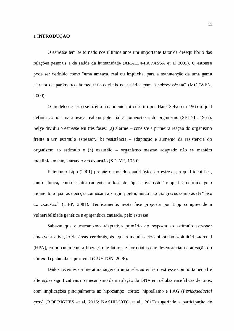
1 INTRODUÇÃO
O estresse tem se tornado nos últimos anos um importante fator de desequilíbrio das
relações pessoais e de saúde da humanidade (ARALDI-FAVASSA et al 2005). O estresse
pode ser definido como "uma ameaça, real ou implícita, para a manutenção de uma gama
estreita de parâmetros homeostáticos vitais necessários para a sobrevivência” (MCEWEN,
2000).
O modelo de estresse aceito atualmente foi descrito por Hans Selye em 1965 o qual
definiu como uma ameaça real ou potencial a homeostasia do organismo (SELYE, 1965).
Selye dividiu o estresse em três fases: (a) alarme – consiste a primeira reação do organismo
frente a um estimulo estressor, (b) resistência – adaptação e aumento da resistência do
organismo ao estimulo e (c) exaustão – organismo mesmo adaptado não se mantém
indefinidamente, entrando em exaustão (SELYE, 1959).
Entretanto Lipp (2001) propõe o modelo quadrifásico do estresse, o qual identifica,
tanto clinica, como estatisticamente, a fase de “quase exaustão” o qual é definida pelo
momento o qual as doenças começam a surgir, porém, ainda não tão graves como as da “fase
de exaustão” (LIPP, 2001). Teoricamente, nesta fase proposta por Lipp compreende a
vulnerabilidade genética e epigenética causada. pelo estresse
Sabe-se que o mecanismo adaptativo primário de resposta ao estímulo estressor
envolve a ativação de áreas cerebrais, às quais inclui o eixo hipotálamo-pituitária-adrenal
(HPA), culminando com a liberação de fatores e hormônios que desencadeiam a ativação do
córtex da glândula suprarrenal (GUYTON, 2006).
Dados recentes da literatura sugerem uma relação entre o estresse comportamental e
alterações significativas no mecanismo de metilação do DNA em células encefálicas de ratos,
com implicações pincipalmente ao hipocampo, córtex, hipotálamo e PAG (Periaqueductal
gray) (RODRIGUES et al, 2015; KASHIMOTO et al., 2015) sugerindo a participação de

12
mecanismos epigenéticos na neurobiologia do estresse (TROLLOPE et al, 2012; HUNTER et
al, 2015).
Definem-se como epigenéticos os mecanismos capazes de controlar a atividade e a
expressão gênica das células, sem que a sequência do DNA seja modificada. Entre as
principais modificações epigenéticas conhecidas inclui a metilaçao do DNA e as modificações
pós-traducionais de histonas (JONES et al., 2001; JENUWEIN et al., 2001).
A metilação do DNA é certamente a modificação epigenética mais estudada nos
últimos anos. Basicamente, a inserção do radical metil (CH3) no carbono 5 de citosinas é
catalisada por um grupo de enzimas denominadas DNA metiltransferases (DNMT). As
principais atividades metiltranferases de estabelecimento e manutenção de padrões de
metilação do DNA genômicotêm sido atribuídas às enzimas DNMT1 e DNMT3
(principalmente DNMT3A e DNMT3B). Às enzimas DNMT3A e DNMT3B, expressas
principalmente durante o desenvolvimento embrionário, tem sido atribuída a atividade de
metilação de novo, ou seja, a inserção de CH3 em regiões do DNA não metiladas previamente
(OKANO et al., 1999), enquanto que à enzima DNMT1 tem sido considerado o papel crucial
da manutenção das marcas de metilação do DNA pós replicação celular (LI E et al., 1993).
O exercicio fisico é conceituado como qualquer atividade que se associa à um
movimento corporal produzido por músculos o qual resulta em maior dispêndio de energia,
desde que seja estruturada, repetitiva e aplicada de forma proposital (NEDER e NERY, 2004).
A atividade física tem sido evidenciada como intervenção profilática e terapêutica para
diversas disfunções, onde a sua prática regular parece estar associada com menor estresse
psicológico como demonstrado em estudos transversais, prospectivos e experimentais
(MATTHEW et al., 2014).
Oportunamente, estudos têm demonstrado a vulnerabilidade das marcas epigenéticas
diante do exercicio físico, fornecendo assim uma explicação viável de como a pratica de

13
exercicio fisico pode ter impacto na função celular e promover beneficios a saúde (DENHAM
et al, 2013; PAREJA-GALEANO et al, 2014). Neste contexto, os estudos atuais têm abordado
o impacto do exercicio fisico em células cerebrais, do sangue, músculo esquelético e cardiaco,
tecido adiposo e mucosa oral (PAREJA-GALEANO et al, 2014; ZHANG et al, 2013),
entretanto muito pouco se sabe sobre esses efeitos em outros tecidos.
A despeito das evidências dos efeitos do estresse e da prática de exercício físico sobre
a programação epigenética de células encefálicas, muito pouco se sabe a respeito da
vulnerabilidade dos mecanismos epigenéticos, como a metilação do DNA, de células não
encefálicas, como as células pulmonares.
Diante disso, objetivamos no presente estudo identificar o efeito do estresse sobre o
mecanismo de metilação do DNA de células do pulmão de ratos, além de identificar as
variações na expressão dos genes Dnmt1, Dnmt3a e Dnmt3b consequentes a esse efeito. Além
disso, objetivamos avaliar o potencial da prática de exercício físico em modular os efeitos
induzidos pelo estresse sobre a metilação do DNA, bem como sobre variações na expressão
dos genes Dnmt1, Dnmt3a e Dnmt3b em células do pulmão de ratos.
Acreditamos que a identificação de variações dos padrões de metilação do DNA em
células pulmonares possa gerar conhecimentos adicionais sobre o mecanismo molecular
associado aos efeitos somáticos do estresse comportamental. Além disso, considerando a
inacessibilidade da obtenção de amostras de pulmão de humanos higidos, consideramos que o
presente estudo, em modelo experimental, possa gerar dados adicionais sobre a influência do
estresse e do exercício físico sobre o mecanismo de metilação do DNA, dados esse
tangivelmente transponíveis para a promoção de saúde humana.

14
2 REVISÃO DE LITERATURA – CONTEXTUALIZAÇÃO
O estresse tem se tornado nos últimos anos um importante fator de desequilíbrio das
relações pessoais e de saúde da humanidade (ARALDI-FAVASSA et al., 2005).
Diante de uma sociedade moderna que exige grande capacidade de adaptação física,
mental e social, os indivíduos estão frequentemente expostos a situações de conflitos,
ansiedades, angústias e desestabilizações emocionais. Neste contexto, o estresse emerge como
uma consequência direta aos persistentes esforços adaptativos da pessoa à sua situação
existencial. O estresse pode ser definido como um desequilíbrio substancial entre a
capacidade de demanda (física ou psicológica) e a capacidade de resposta, que repercute em
consequências importantes tais como alterações cognitivas e comportamentais, além de gerar
uma predisposição a estados patológicos. (MCGRATH, 1970).
No Brasil, estima-se uma incidência populacional de estresse em torno de 32%,
estando à intensidade de sua manifestação associada à atividade profissional (LIPP;
TANGANELLI, 2002).
No que se diz respeito a fisiologia do estresse, sabe-se que entre as principais áreas
cerebrais ativadas no mecanismo adaptativo inclui o eixo hipotálamo-hipófise, sendo essa
área responsável pela liberação de fatores e hormônios que desencadeiam a ativação do córtex
da glândula supra-renal (GUYTON, 2006). Entretanto pouco se sabe a respeito da
vulnerabilidade das marcações epigenéticas em células não encefálicas, como do pulmão, ao
estresse.
Denominam-se epigenéticas as modificações que ocorrem na molécula de DNA ou nas
histonas (proteínas onde o DNA se enovela para constituir a cromatina) capazes de afetar a
expressão gênica sem que a sequência original do DNA seja alterada (JABLONKA, LAMB
2002).

15
Os mecanismos epigenéticos podem ser divididos em três tipos: 1) modificações
covalentes na cauda N terminal de histonas, 2) metilação do carbono 5 de citosinas,
geralmente seguidas por guaninas (dinucleotideos CpGs) na molécula de DNA e 3) RNAs não
codificantes (Fig. 1.A) (PORTELA, ESTELLER 2010, KOUZARIDES et al., 2007,
HAGOOD et al., 2014).
Figura 1A – Representação ilustrativa dos Mecanismos Epigenéticos de Controle da Expressão Gênica
(HAGOOD et al., 2014)
A metilação do DNA é certamente a modificação epigenética mais estudada nas
últimas décadas. Dinâmica e com potencial modificação ao longo do desenvolvimento, a

16
metilação do DNA desenvolve um importante papel no controle de vários processos
biológicos, tais quais, diferenciação celular embrionária, inativação do cromossomo X em
fêmeas de mamíferos, imprinting genômico, supressão de retrovírus endógenos e estabilidade
cromossômica (REIK, WALTER 2001; LI et al 1992; BESTOR 1998; PANNING,
JAENISCH 1998; THOMPSON et al 2010).
A inserção deste radical no carbono 5 de citosinas é catalisada por enzimas
denominadas DNA metiltransferases (DNMT1, DNMT2, DNMT3A, DNMT3B e DNMT3L).
As DNMT3A e DNMT3B são, predominantemente, expressas durante o desenvolvimento
embrionário e catalisam a chamada metilação de novo. Similarmente, a DNMT3L (DNMT3-
Like) compartilham homologia com as DNMT3s, desempenhando um papel essencial sobre a
metilação tecido-especifico. (OKANO et al., 1999). Já a DNMT1 tem papel crucial na
metilação hereditária, pois age reconhecendo os sítios CpG hemi-metilados estabelecidos,
anteriormente, pela DNMT3A e DNMT3B, o qual converte para um estado totalmente
metilado. Assim, a DNMT1 é responsável pela metilação de manutenção célula-célula (LI E
et al., 1993). Finalmente, a DNMT2 parece ter pouca atividade de DNA metiltransferase,
sendo descrita como tendo um papel chave na metilação e estabilizações de tRNA, aumentado
a síntese proteica (TUORTO et al, 2012).
A vulnerabilidade dos padrões epigenéticos frente a exposições ambientais é um tema,
atualmente, muito estudado em virtude de inúmeras implicações à saúde (GOMES; PELOSI,
2013). Durante os últimos anos, vários grupos de pesquisadores têm relatado os efeitos do
estresse, tanto agudo como crônico, no epigenoma (CHAKRAVARTY et al, 2014;
KENWORTHY et al, 2014; SCHOUTEN et al, 2013).
Estudo realizado por Unternaehrer et al (2012) demonstrou a presença de alterações
loci especificas de metilação do DNA em células do sangue periférico humano associadas ao
estresse, No entanto, análises envolvendo outros tecidos não foram realizadas neste estudo em
17
virtude da inacessibilidade para a obtenção de material em indivíduos hígidos
(UNTERNAEHRER et al, 2012).
Roth et al (2011) demonstraram, em modelo experimental, a associação entre o
estresse psicossocial e o aumento dos níveis de metilação do gene BDNF (Brain Derived
Neurotrophic Factor) acompanhado de diminuição da expressão desse gene no hipocampo
dorsal de ratos adultos (ROTH et al, 2011)
Dados recentes do nosso grupo de pesquisa revelaram a participação do mecanismo de
metilação do DNA nas respostas celulares adaptativas de células cerebrais de ratos como o
hipocampo, córtex, hipotálamo e PAG (Periaqueductal gray) mediante estresse
comportamental (RODRIGUES et al, 2015; KASHIMOTO et al., 2015).
Muito pouco se sabe ainda a respeito dos efeitos do estresse comportamental em
células não encefálicas, como do pulmão. No entanto, com base na característica
vulnerabilidade da manutenção (transmissão célula-célula) do padrão epigenético frente a
variações ambientais, acredita-se que alterações dos padrões de metilação do DNA possam ser
induzidas por fatores externos e, consequentemente estarem associadas à origem de várias
doenças humanas, como o câncer (FRAGA, 2009).
O câncer é certamente a doença em que a associação com alterações epigenéticas está
mais bem caracterizada (PORTELA; ESTELLER 2010). De uma forma geral, o padrão de
metilação de células tumorais está consideravelmente alterado em relação às células normais
(PORTELA; ESTELLER 2010). Entre as principais alterações relacionadas ao câncer incluem
a perda de metilação global do DNA e hipermetilação de regiões promotoras (ilhas CpGs) de
genes supressores tumorais. Particularmente, enquanto as células tumorais são caracterizadas
por uma perda significativa da metilação do DNA (20 a 60% menos 5-metil-citosinas) é
observado um aumento de metilação em ilhas CpGs específicas (SHEN et al., 2007;
ESTELLER, 2008).

18
Estudos epidemiológicos prévios indicaram uma relação significativa entre o estresse
crônico e a origem, progressão e mortalidade de câncer (LILIBERG et al., 2003; BUCCHERI
et al., 1998; STOMMEL et al., 2002; CHIDA et al., 2008).
Dados obtidos por metanálise envolvendo 165 estudos longitudinais correlacionaram a
influência de fatores psicossociais com a incidência e diminuição na sobrevida de pacientes
com câncer (CHIDA et al., 2008). Além disso, a análise destes dados pemite a observação de
que o câncer de pulmão é o tipo de câncer no qual a associação com o estresse está
estabelecida de forma mais significativa (CHIDA et al., 2008), o que torna este o modelo
determinado para as análises subsequentes. Por se tratar de um estudo longitudinal, há que se
ponderar que os resultados obtidos por Chida et al (2008) se limitam à observação dos
elementos amostrais sem manipular fatores que possam alterar as variáveis de interesse, como
os hábitos alimentares, estilo de vida, consumo de álcool e tabagismo, fatores hereditários e
histórico de doenças pulmonares em casos de câncer pulmonar (CHIDA et al., 2008).
Além disso, estudos envolvendo modelos experimentais validados demonstraram que
o estresse crônico pode aumentar in vivo a capacidade de crescimento tumoral e de metástase
em modelos xenotransplantados de tumores (FENG et al 2012; HASSAN et al 2013).
A atividade física é definida como um movimento corporal produzido pelos músculos
esqueléticos que requer gasto de energia acima dos níveis de repouso (BAMMAN et al.,
2014; COLPANI et al., 2013), enquanto o exercício físico é conceituado como qualquer
atividade que se associa à um movimento corporal produzido por músculos que resulta em
maior dispêndio de energia que ocorra de forma estruturada, repetitiva e aplicada de forma
proposital (NEDER e NERY, 2004).
De acordo com a Organização Mundial da Saúde estima-se que a inatividade física
possa ser responsável por 5,3 milhões de mortes em todo o mundo/ano e associada a 10% dos
19
cânceres de mama, 10% dos canceres do cólon e 6% das doenças cardíacas coronarianas
(OMS, 2010).
Estudos prévios têm buscado a elucidação da relação entre a prática de exercício físico
a promoção da saúde via mecanismos epigenéticos (PAREJA-GALEANO et al., 2014).
Entretanto tal relação é pouco compreendida emergindo dessa forma a necessidade de estudos
tecidos específicos.
Zeng et al (2012) observaram, a partir de um ensaio clinico envolvendo pacientes com
câncer de mama e análise genômica em larga escala (microrray) a associação entre a prática
de atividade física aeróbica moderada por um período de seis meses e mudanças nos perfis de
metilação do DNA de 43 genes. Destes, alterações significativas na expressão de três genes
foram correlacionadas positivamente com a sobrevida dos pacientes, entre eles o gene
L3MBTL1, um gene de conhecida função de supressão tumoral (ZENG et al., 2012).
Similarmente, outro estudo realizado por Bryan et al (2012) demonstrou que o aumento da
atividade física durante 12 meses diminuiu a metilação do DNA em 45 ilhas CpGs de genes
relacionados com o inicio e progressão do mesmo tipo de câncer (BRYAN et al., 2012).
Em idosos, Luttrop et al (2012) demonstraram que a prática de atividade física está
relacionada com um aumento dos níveis de metilação global do DNA, e que esses níveis
permanecem significativos após a correção com fatores de risco cardiovasculares (LUTTROP
et al., 2012).
Similarmente, Roon et al (2013) demonstraram um aumento nos níveis de metilação
global do DNA, bem como alterações em 17.975 ilhas CpGs em 7663 genes em tecido
adiposo de homens adultos saudáveis sugerindo o potencial da intervenção de seis meses de
exercício físico na indução de alterações no mecanismo de metilação do DNA em células
desse tecido (RONN et al, 2013).
20
Em virtude da quantidade ainda limitada de informações na literatura, emerge a
necessidade de estudos em modelos experimentais focados na compreensão, ao nível
molecular, do quanto e como o estresse pode influenciar as atividades biológicas normais de
controle da expressão gênica celular. Especulamos que alterações epigenéticas importantes
podem ocorrer em consequência à exposição ambiental, indicando dessa forma uma possível
associação molecular entre o estresse e a origem de doenças (KASHIMOTO et al., 2015).

21
3 OBJETIVOS
3.1 Objetivo Geral
Diante do expoto acima, o presente trabalho tem como objetivo geral identificar o
efeito do estresse crônico sobre a metilação do DNA e expressão gênica de células do pulmão
de ratos.
3.2 Objetivo Específico
1) Identificar o efeito do estresse crônico sobre o perfil de metilação global do DNA
em células do pulmão de ratos
2) Identificar o efeito do estresse crônico sobre a expressão dos genes Dnmt1, Dnmt3a
e Dnmt3b em células do pulmão de ratos.
3) Avaliar os efeitos da prática de exercício (natação) sobre a metilação global do
DNA e a expressão dos genes Dnmt1, Dnmt3a e Dnmt3b em células do pulmão de ratos.
4) Verificar o possível potencial do exercicio físico em modular os efeitos moleculares
induzidos pelo estresse sobre a metilação do DNA e a expressão dos genes Dnmt1, Dnmt3a e
Dnmt3b em células do pulmão de ratos.

22
4 ARTIGO
EFFECTS OF THE BEHAVIORAL STRESS AND THE PRACTICE OF PHYSICAL
EXERCISE ON THE MECHANISM OF DNA METHYLATION IN RAT LUNG
CELLS
Toffoli LV1, Volpini VL2, Silva WR1, Magnoni LN1, Pelosi GG2, Gomes MV1.
1 Centro de Pesquisa em Ciências da Saúde, Universidade Norte do Paraná (Unopar),
Londrina, Paraná, Brasil,
2 Centro de Ciências Biológicas, Departamento de Ciências Fisiológicas, Universidade
Estadual de Londrina (UEL), Paraná, Brasil
Telefone: +55 43 3371 9859
Email: [email protected]; [email protected]
URL: http://www.unopar.br

23
ABSTRACT
Increasing recent evidences has proposed the participation of epigenetics mechanisms in the
molecular adaptive cellular responses to stress and physical exercise practicing. In the present
study we assessed the effects of the chronic stress and physical exercise (swimming) on the
global DNA methylation profile of rat lung cells and addressed the respective relationships
with the expression of the DNA methyltransferases (Dnmt) genes Dnmt1, Dnmt3a and
Dnmt3b. Furthermore, we evaluated the potential of physical activity to modulate the
molecular effects induced by the behavioral stress. Male Wistar rats were divided into four
experimental groups: (1) animals that were submitted to the chronic stress protocol (ST)
during the period that comprehends the 67th and 80th postnatal day (PND); (2) animals that
were submitted to physical exercise (swimming sessions) (EX) in the period of 53th -79th
PND; (3) animals that were submitted to swimming in the period of 53th -79th PND and to
stress during the 67-80 PND (EX-ST); and (4) animals that were not submitted to stress or
swimming (CTL). The animals were sacrificed at the end of stress protocol at the 80th PND
and the lung removed. The global DNA methylation profile was quantified using the Imprint
Methylated DNA Quantification kit (Sigma-Aldrich®) and the expression of genes was
obtained by real time PCR. A statistically significant decrease in the global DNA methylation
profile was observed when compared the lung of animals from ST and CTL groups
(p=0.0159) which was accompanied by a statistically significant increase in the expression of
the Dnmt1 gene in the ST group (p=0.0159). A statistically significant decrease in the
expression of the Dnmt3b gene was observed in the EX group in comparison to CTL group
(p=0.0357). Furthermore, a statistically significant decrease in the expression of the Dnmt3a
and Dnmt3b genes was observed in the EX group when compared with the ST group
(p=0.0018). Based on these findings we concluded that chronic stress evokes a global DNA
hypomethylation and an increase in expression Dnmt1 in lung cells. Furthermore, our data
indicated that physical exercise might potentially attenuate the effects of behavioral stress on
the global DNA methylation profile and modulate the impact of stress on the expression of
Dnmt1, Dnmt3a e Dnmt3b genes.
Key words: Stress; Physical Activity; Lung; Epigenetics; DNA methylation; DNMT´s

24
INTRODUCTION
Stress has become, in the last years, an important imbalance factor of
personal and health relationships of humanity [1,2]. Consistent data associate behavioral
stress with modulation epigenetic mechanisms, such as DNA methylation, in brain cells of the
hippocampus, cortex, hypothalamus and PAG (Periaquectual gray) [3,4] thus suggesting
implications of epigenetic mechanisms in the adaptive cellular responses to stress [5-7].
DNA methylation is certainly the most studied epigenetic modification in
the latest decades. Dynamic and potentially modifiable throughout the development, the
methylation process consists in the introduction of a methyl radical on the carbon 5 of
cytosine, usually followed by guanines (dinucleotide CpG) in DNA molecule, by the action
of enzymes called DNA methyltransferase (DNMT1, DNMT3a and Dnmt3b) [8-10].
Cancer is certainly the disease in which the association with epigenetic
changes is most well characterized. Among these changes related to cancer include loss of
global methylation and hypermethylation of promoter regions (CpG islands) [10].
It is currently accepted that the regular practice of physical exercise
adaptively benefits several body systems, including the cardiopulmonary system, which can
be evidenced in lung capacity by the increase of alveolar openings, significant increase in the
partial pressure of oxygen in arterial blood and the reduction in the partial pressure of carbon
dioxide (CO2) [11].
Previous studies have sought to elucidate the vulnerability of epigenetic
mechanisms to physical exercise [12]. For example, in healthy adult males initially with
physical inactivity, Roon et al (2013) demonstrated that a six months program of physical
exercise results in adipose tissue in significant changes in global methylation and loci-specific
levels [13]. However this relationship (physical exercise versus epigenetic mechanisms) is
poorly understood in several other tissues such as lung.
Despite the enormous progress obtained on the field of neurobiology of
stress and the benefits of the practice of physical exercise in the promotion of health, little is
still known about the molecular molecular mechanisms associated to the behavioral stress and
the practice of the physical exercise in non-brain cells [3,4].
In this context, the present study aimed to identify the effects of chronic
stress and the practice of physical exercise on the global DNA methylation profile in rat lung
cells, as well as the correlation with the expression of Dnmt1, Dnmt3a and Dnmt3b genes.
Furthermore, the present study aimed to evaluate the potential of the practice of physical

25
exercise in modulating the effects of the behavioral stress effects on the global DNA
methylation profile and the expression of Dnmt1, Dnmt3a and Dnmt3b genes in rat lung cells.
MATERIALS AND METHODS
Animals and experimental groups:
Experimental procedures were carried out following protocols approved by
the ethical review committee of the University of Lon-drina, Londrina, Parana, Brazil (CEUA
no. 14441.2013.18) and all efforts were made to minimize suffering.
Non-related male Wistar rats were kept under standard condi-tions
(temperature 25 ± 1◦C, photoperiod 12 h light/12 h dark) andwith water and food ad libitum
in the Central Animal House of theUniversity of Londrina.
The animals were distributed into four groups containing 5 animals each: 1)
stress group (ST): animals submitted to the restriction stress protocol from the 67 postnatal
day (PND) to 80 PND; 2) physical exercise group (EX): animals submitted to physical
exercise (swimming for 60 minutes / day) from 53 PND to 79 PND; 3) physical exercise and
stress group (EX-ST): animals submitted to physical exercise (swimming for 60 minutes /
day) 53 PND to 79 PND and also to chronic stress by restriction of 67 PND to 80 PND,
respectively; and 4) control group (CTL): animals that were maintained in the same
conditions as other animals but were not submitted to stress or exercise protocols.
The animals from groups one and three (EX e EX-ST) were assisted in all
swimming sessions, to avoid the passive rat floating and minimize bias related to different
exercise intensities of each animal. All the experiment protocol was performed in the morning
between 07:00 and 12:00.
Chronic stress by restriction of movement:
For the analyzes of the effects of behavioral stress on epigenetic patterns
was used the validated test of chronic stress by restriction as previously described [14].
In the morning (7:00 to 09:00) the animals were transported to the
experimental room in their cages and allowed to adapt to this environment for at least 30
minutes. After this period, the animals were submitted to the stress protocol, being placed in a
metal cylinder with 6.5 cm in diameter and 15 cm long with holes, that allow ventilation,
which remained closed for 60 min.

26
Physical exercise:
The animals from EX and EX-ST group were submitted to physical training
(swimming), according to Martins-Pinge et al. Swim sessions were realized in the morning
(8:00 to 12:00) in a glass tank filled with warm water (31 ± 1 ° C) of 4000 cm2 surface area
and 60 cm in depth. The training consisted of 4 weeks (20 sessions) of swimming being held
5 days a week and 60 min/day.
During the first week, the training was graded, begin-ning with 15 min on
the first day, 30 min on the second day, and45 min on the third day, for adaptation to the
training process. Fromthe fourth day on, each session consisted of 60 min of swimminguntil
the 74th PND. After each swimming session, the animals weredried with a towel and returned
to their cages.
Samples:
Samples were collected in the morning (between 08:00 and 12:00) in order
to maintain the same circadian rhythm for all groups. The animals were killed by decapitation
and the right lower lobe of the lung was removed. All samples were washed immediately in
saline solution after obtaining and frozen at -80 ° C for further molecular analysis.
Global DNA Methylation Profile:
Genomic DNA was obtained by phenol chloroform after soaking the tissue
in a grail and pistillate in the presence of liquid nitrogen [16]. Evaluation of the purity and
concentration of DNA was performed by analysis of absorbance in a spectrophotometer
(NanoDrop ND-2000 - Thermo Scientific) at 260nm and 280nm.
The global DNA methylation profile was evaluated by dosage of methyl
groups (CH3) by Imprint® Methylated DNA Quantification Kit Kit (Sigma-Aldrich®) using
the concentration of 200ng / ul DNA according to the manufacturer's recommendations and as
described previously [17]. Briefly, the percentage of each sample methylation was calculated
by the amount of cytosines methylated in the sample (5mC) relative to overall cytidine (5mC
+ AD) in a positive control previously methylated (100% methylated) and a negative control
(0 % methylated). The absorbance readings were performed at 450 nm in a microplate reader
and the percentage of DNA methylation was obtained following the formula: A450sample-
A450NTC / (A450met-A450NTC) x100. All samples were analyzed in duplicate.

27
Gene Expression Levels:
RNA samples were obtained using Trizol (Invitrogen®) and the reverse
transcription was performed using the High Capacity Kit (Applied Biosystems®).
The gene expression levels were quantitatively assessed by RT-PCR, Step
One Plus (Applied Biosystems) and used the detection system specifies Company Taqman
Applied Biosystems with the probes (on demand) to the Dnmt1 gene (ID: Rn00709664_M1)
Dnmt3a (ID: Rn01027162_G1) and Dnmt3b (ID: Rn01536418_G1). For the normalization of
the difference in the amount of cDNA used in each experiment we used GAPDH (ID:
Rn01775763_G1) as endogenous control. It used the comparative CT method (2-ΔΔCt) for
assessment of gene expression levels. The experiments were performed in triplicate.
Statistical Analysis:
Graph Prism 6.0 software for statistical analysis was used. The data were
submitted to a descriptive analysis, and the normality and homogeneity of variance of the data
evaluated by the Shapiro-Wilk test. To analyze the effects of stress and physical exercise on
DNA methylation and expression of Dnmt1 genes, Dnmt3a and Dnmt3b the values observed
in the groups ST and EX were analyzed by non-parametric Mann-Whitney in the values
presented CTL group.
For evaluating the potential exercise-physical to modulate cellular responses
triggered by stress profile of global DNA methylation and the values for the relative
expression of DNMT1 genes, Dnmt3a and Dnmt3b of ST groups, EX and EX-ST were
analyzed from the Kruskal-Wallis statistical test (post-test: Dunn). Confidence interval of
95% and a significance level of 5% (p <0.05) in all the tests used was adopted.
RESULTS
Effects of stress on global DNA methylation:
A statistically significant decrease was observed in the global DNA
methylation in lung cells (p = 0.0159) of animals ST group when compared with animals CTL
group.
There was no statistically significant difference in the percentage of global
DNA methylation when compared the EX and CTL groups (p = 0.3095).
A statistically significant increase in the percentage of global DNA

28
methylation was observed when comparing the EX and EX-ST groups with the ST group (p =
0.018). (Fig. 1).
Figure 1 – The effects of stress and physical exercise on the global DNA methylation in rats lung cells. A) CTL
group versus ST; B) CTL group versus EX; C) ST group versus EX-ST versus EX. CTL: control group; ST:
stress group; EX: physical exercise group and EX-ST: physical exercice and stress group. *p<0.05. Mann-
Whitney and Kruskal-Wallis test. The error bar represents the standard error of average.
Effects of stress on expression of Dnmt1 gene:
A statistically significant increase was observed in the expression of the
Dnmt1 gene in ST group when compared to CTL group (p = 0.0159).
There was no statistically significant difference when compared the EX
group with CTL group (p = 0.3095).
A statistically significant decreae in Dnmt1 gene expression was observed
when comparing the values of the EX-ST and ST groups (p = 0.0042). There was no
statistically significant difference when compared the ST and EX values (Fig. 2).
Figure 2 – Effects of stress and physical exercise on the expression of Dnmt1 gene in rat lung cells. A) CTL
group versus ST; B) CTL group versus EX; C) ST group versus EX-ST versus EX. CTL: control group; ST:
stress group; EX: physical exercise group and EX-ST: physical exercice and stress group; RQ: relative
quantification according to expression of the Gapdh gene.. *p<0.05. Mann-Whitney e Kruskal-Wallis test. The
error bar represents the standard error of average.

29
Effects of stress on expression of Dnmt3a gene:
No statistically significant difference in the expression of the Dnmt3a gene
was observed when compared ST and CTL groups (p = 0.6667), as well as when compared
the EX and CTL groups (p = 0.0556).
Statistically significant decrease in expression of the Dnmt3a gene was
observed between the EX and ST groups (p = 0.0018). (Fig. 3)
Figure 3 – Effects of stress and physical exercise on the expression of Dnmt3a gene in rat lung cells. A) CTL
group versus ST; B) CTL group versus EX; C) ST group versus EX-ST versus EX. CTL: control group; ST:
stress group; EX: physical exercise group and EX-ST: physical exercice and stress group; RQ: relative
quantification according to expression of the Gapdh gene. *p<0.05. Mann-Whitney e Kruskal-Wallis test. The
error bar represents the standard error of average.
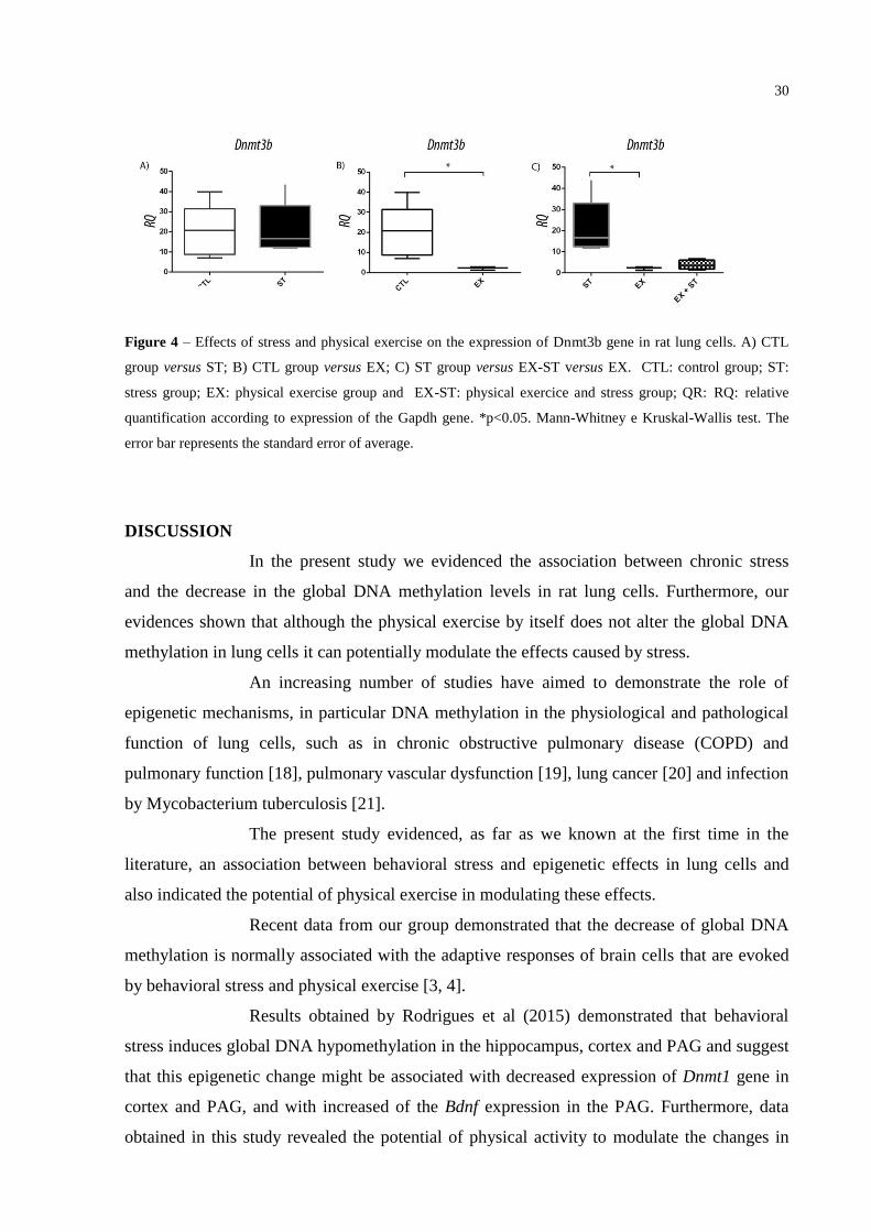
Effects of stress on expression of Dnmt3b gene:
No statistically significant difference in the expression of the Dnmt3b gene
was observed when compared ST and CTL groups (p = 0.8016). However, a statistically
significant decrease was observed when compared the Dnmt3b gene expression values in EX
and CTL groups (p = 0.0357). In addition, statistically significant decrease in expression of
Dnmt3b gene was also observed when compared the EX and ST groups (p = 0.0018). (Fig. 4)
30
Figure 4 – Effects of stress and physical exercise on the expression of Dnmt3b gene in rat lung cells. A) CTL
group versus ST; B) CTL group versus EX; C) ST group versus EX-ST versus EX. CTL: control group; ST:
stress group; EX: physical exercise group and EX-ST: physical exercice and stress group; QR: RQ: relative
quantification according to expression of the Gapdh gene. *p<0.05. Mann-Whitney e Kruskal-Wallis test. The
error bar represents the standard error of average.
DISCUSSION
In the present study we evidenced the association between chronic stress
and the decrease in the global DNA methylation levels in rat lung cells. Furthermore, our
evidences shown that although the physical exercise by itself does not alter the global DNA
methylation in lung cells it can potentially modulate the effects caused by stress.
An increasing number of studies have aimed to demonstrate the role of
epigenetic mechanisms, in particular DNA methylation in the physiological and pathological
function of lung cells, such as in chronic obstructive pulmonary disease (COPD) and
pulmonary function [18], pulmonary vascular dysfunction [19], lung cancer [20] and infection
by Mycobacterium tuberculosis [21].
The present study evidenced, as far as we known at the first time in the
literature, an association between behavioral stress and epigenetic effects in lung cells and
also indicated the potential of physical exercise in modulating these effects.
Recent data from our group demonstrated that the decrease of global DNA
methylation is normally associated with the adaptive responses of brain cells that are evoked
by behavioral stress and physical exercise [3, 4].
Results obtained by Rodrigues et al (2015) demonstrated that behavioral
stress induces global DNA hypomethylation in the hippocampus, cortex and PAG and suggest
that this epigenetic change might be associated with decreased expression of Dnmt1 gene in
cortex and PAG, and with increased of the Bdnf expression in the PAG. Furthermore, data
obtained in this study revealed the potential of physical activity to modulate the changes in

31
global DNA methylation that are evoked by stress in the hippocampus, cortex and PAG as
well as modulate the effects on the expression of Dnmt1 and Bdnf gene in the brain rats [3].
Similarly, in another study from our research group, Kashimoto et al (2015)
identified significant changes in global DNA methylation in brain cells, mainly in the
hypothalamus, associated with physical exercise practice. In addition the data obtained by
Kashimoto et al (2015) demonstrated the potential of physical exercise to modulate the
adaptative responses to repeated stress in regards to DNA methylation in the hippocampus,
cortex, hypothalamus and PAG and in the expression of Dnmt1 gene in the hippocampus and
hypothalamus [4].
Contextualizing with the previously results obtained in brain cells [3, 4] the
data from the present study corroborate the vulnerability of epigenetic markings to stress and
to physical exercise and show that stress might also induce a global DNA hypomethylation in
lung cells.
Changes in DNA methylation profiles have been associated with the
etiology of various diseases, including cancer [22]. Global DNA hypomethylation
accompanied loci-specific hypermethylation are normally epigenetic characteristics described
in various tumor types, indicating that the DNA methylation mechanism participates
significantly in the tumor development process [23, 24].
Concurrently, recent data demonstrate the potential of considering the global
hypomethylation in peripheral blood leukocytes as a biomarker for the risk of various cancers
such as kidney cancer, breast cancer and bladder cancer [25-28].
Epidemiological studies have suggested a relation between chronic stress
and the origin, progression and mortality of cancer [29-32], and lung cancer being the type of
cancer in which the association with stress is established more significantly [32].
In this way, considering the results of this study that link stress to global
DNA hypomethylation in lung cells to the literature data that correlate epigenetic change to
the molecular mechanism of tumor development, we hypothesized that somehow the change
of DNA methylation can justify a possible relationship between stress and lung cancer, as
previously reported in epidemiological studies [32]. However, further studies in this area are
certainly needed to confirm this hypothesis.
DNA methylation, which consists in introducing methyl group to the
cytosine (CpG dimers) is usually catalyzed by DNA methyltransferase enzymes, DNMT1,
DNTMT3A and DNMT3B. It is believed that DNMT1 is involved in the maintenance of
DNA methylation after cell division, acting on the recognition of CpG hemi-methylated sites
32
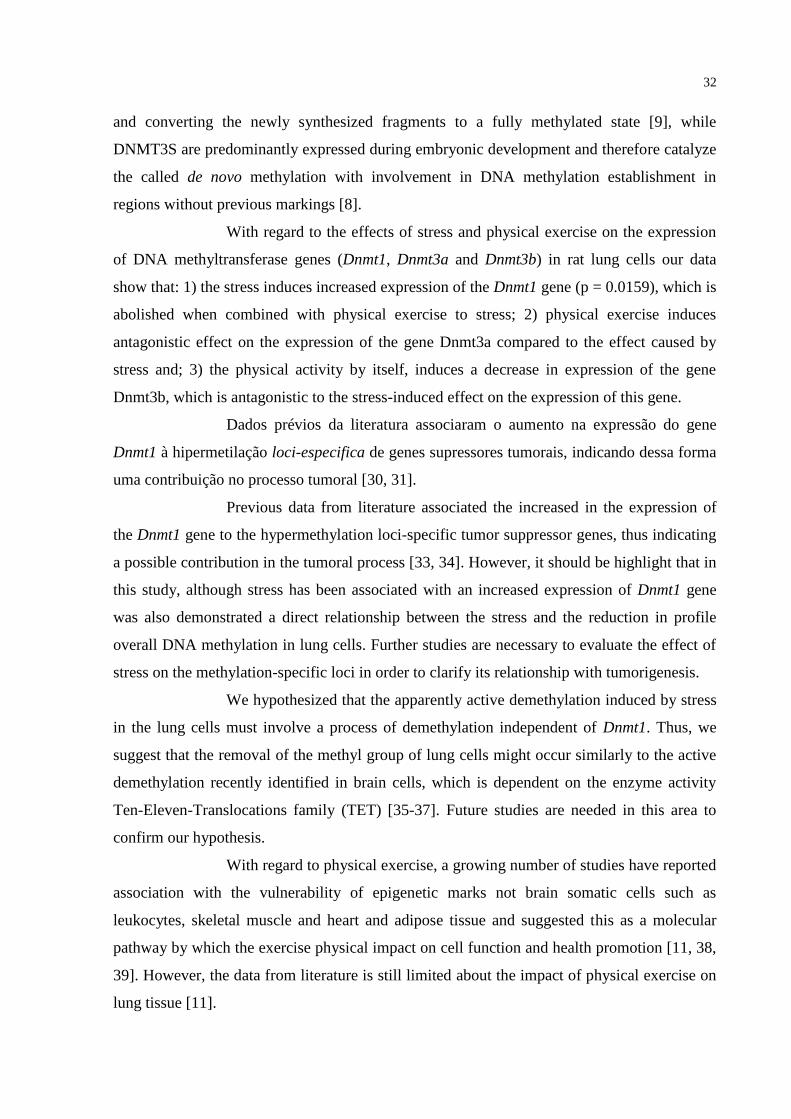
and converting the newly synthesized fragments to a fully methylated state [9], while
DNMT3S are predominantly expressed during embryonic development and therefore catalyze
the called de novo methylation with involvement in DNA methylation establishment in
regions without previous markings [8].
With regard to the effects of stress and physical exercise on the expression
of DNA methyltransferase genes (Dnmt1, Dnmt3a and Dnmt3b) in rat lung cells our data
show that: 1) the stress induces increased expression of the Dnmt1 gene (p = 0.0159), which is
abolished when combined with physical exercise to stress; 2) physical exercise induces
antagonistic effect on the expression of the gene Dnmt3a compared to the effect caused by
stress and; 3) the physical activity by itself, induces a decrease in expression of the gene
Dnmt3b, which is antagonistic to the stress-induced effect on the expression of this gene.
Dados prévios da literatura associaram o aumento na expressão do gene
Dnmt1 à hipermetilação loci-especifica de genes supressores tumorais, indicando dessa forma
uma contribuição no processo tumoral [30, 31].
Previous data from literature associated the increased in the expression of
the Dnmt1 gene to the hypermethylation loci-specific tumor suppressor genes, thus indicating
a possible contribution in the tumoral process [33, 34]. However, it should be highlight that in
this study, although stress has been associated with an increased expression of Dnmt1 gene
was also demonstrated a direct relationship between the stress and the reduction in profile
overall DNA methylation in lung cells. Further studies are necessary to evaluate the effect of
stress on the methylation-specific loci in order to clarify its relationship with tumorigenesis.
We hypothesized that the apparently active demethylation induced by stress
in the lung cells must involve a process of demethylation independent of Dnmt1. Thus, we
suggest that the removal of the methyl group of lung cells might occur similarly to the active
demethylation recently identified in brain cells, which is dependent on the enzyme activity
Ten-Eleven-Translocations family (TET) [35-37]. Future studies are needed in this area to
confirm our hypothesis.
With regard to physical exercise, a growing number of studies have reported
association with the vulnerability of epigenetic marks not brain somatic cells such as
leukocytes, skeletal muscle and heart and adipose tissue and suggested this as a molecular
pathway by which the exercise physical impact on cell function and health promotion [11, 38,
39]. However, the data from literature is still limited about the impact of physical exercise on
lung tissue [11].

33
Using the large scale genomic analysis, Kanzleiter et al (2015) recently
demonstrated that 2.762 CpG island became differently methylated in skeletal muscle of rats
practitioners of physical exercise when compared with animals not practitioners. Moreover,
these authors associated a negative correlation with the expressions of approximately 200
genes [40]. In the same segment, another recent study reviewed the role of DNA methylation
in myogenesis, addressing physiological and pathological conditions [41].
Although physical exercise practice is known to be beneficial to the
cardiopulmonary system, somewhat enlightening are still evidence of the molecular
mechanisms associated with such benefits. Addressing recent research related the role of
physical activity in the prevention of pathophysiological changes in cells and cardiovascular
tissues, Zimmer et al (2015) suggested the involvement of epigenetic mechanisms in the
biological pathways that culminate with the benefits induced by exercise practice [42].
In relation to the expression of DNMTs genes, our results demonstrated no
statistically significant difference in global DNA methylation and gene expression of Dnmt1
and Dnmt3a compared animals who practiced physical exercise and the control group (p>
0.05). However, our data show a significant decrease in expression of the gene Dnmt3b (p =
0.0357) compared physical exercise and control groups.
Still considering the evidence of this study, we note that in practicing animal
physical exercise effects induced by stress on the expression of Dnmt1 genes, Dnmt3a and
Dnmt3b are somewhat modified. This modulating effect can be plain observed when
comparing animals subjected to stress (ST) and animals subjected to stress and exercise (EX-
ST). In such cases we may infer the existence of an important biological effect of modulating
the effects of stress caused by physical exercise, although statistically significant difference
was observed only with respect to the expression of the gene Dnmt1.
In summary, our data show the vulnerability of methylation cell lung DNA
to behavioral stress and suggest the existence of an active demethylation mechanism of the
lung induced by stress, which is independent of the expression of Dnmt3a and Dnmt3b genes,
although a Dnmt1 increase in gene expression is observed. In addition, our evidence shows
the potential of physical exercise in attenuate the effects induced by stress in the lung cells
with implications thus in global DNA methylation as the expression of Dnmt1 genes, Dnmt3a
and Dnmt3b.
34
REFERENCES
1. Araldi-Favassa CT, Armiliato N, Kalinine I. Aspectos fisiológicos e psicológicos do
estresse. Revista de Psicologia da UnC 2005; 2: 84-92.
2. Mcgrath, J.E. Social and psychological factors in stress. Illinois: Air Force, 1970.
3. Rodrigues GM Jr, Toffoli LV, Manfredo MH, Francis-Oliveira J, Silva AS, Raquel
HA, Martins-Pinge MC, Moreira EG, Fernandes KB, Pelosi GG,Gomes MV. Acute stress
affects the global DNA methylation profile in rat brain: modulation by physical exercise.
Behav Brain Res 2015; 15;279: 123-128.
4. Kashimoto RK, Toffoli LV, Manfredo MH, Volpini VL, Martins-Pinge MC, Pelosi
GG, Gomes MV. Physical exercise affects the epigenetic programming of rat brain and
modulates the adaptive response evoked by repeated restraint stress. Behav Brain Res
2016; 1; 296-289
5. Guyton, AC.; Hall, JE. Tratado de Fisiologia Médica. 9 ed. Rio de Janeiro: Elsevier,
2006.
6. Collins A, Hill LE, Chandramohan Y, Whitcomb D, Droste SK, Reul JMHM. Exercise
improves cognitive responses to psychological stress through enhancement of epigenetic
mechanisms and gene expression in the dentate gyrus. PLoS One 2009;4(1):e4330.
7. Brandão ML, Vianna DM, Masson S. et al. Organização neural de diferentes tipos de
medo e suas implicações na ansiedade. Rev Bras Psiquiatria 2003 25: 36-41.
8. Okano MDW, Bell DA. Haber, and E. Li. 1999. DNA methyltransferases Dnmt3a and
Dnmt3b are essential for de novo methylation and mammalian development. Cell
99:247–257.
9. Li E, C. Beard, and R. Jaenisch. 1993. Role for DNA methylation in genomic imprinting.
Nature 366:362–365.

35
10. Portela A, Esteller M. Epigenetic modifications and human disease. Nat Biotechnol, 2010
28: 1057-1068.
11. Abrahão, LMB. Efeitos da atividade física aeróbia no complexo vasculo-alveolar no
pulmão de ratos. Dissertação (mestrado) USP, Faculdade de Medicina Veterinária e
Zootecnia 2009; 78fl.
12. Pareja-Galeano H, Sanchis-Gomar F, García-Giménez JL. Physical Exercise and
Epigenetic Modulation: Elucidating Intricate Mechanisms. Sports Med 2014; 44: 429-
436.
13. Ronn T, Volkov P, Davegårdh, C, Dayeh T, Hall E, Olsson AH, Nilsson E, Tornberg A,
Dekker Nitert M, Eriksson KF, Jones HA, Groop L, Ling C. A six months exercise
intervention influences the genome-wide DNA methylation pattern in human adipose
tissue. PLoS Genetic, 2013 9(6): e1003572.
14. Babenko O, Golubov A, Ilnytskyy Y, Kovalchuk I, Metz GA. Genomic and Epigenomic
Responses to Chronic Stress Involve miRNA-Mediated Programming. PLoS One 2012
7(1): e29441.
15. Martins-Pinge MC, Becker LK, Garcia MRL, Zoccal DB, Neto RV, Basso LS et al.
Attenuated pressor responses to amino acids in the rostral ventrolateral medulla after
swimming training in conscious rats. Auton Neurosci 2005;122: 21–28.
16. Sambrook J, Fritschi EF, Maniatis T. Molecular cloning, a laboratory manual. 3rd ed.
New York: Cold Spring Harbor Laboratory Press; 1989.
17. Toffoli LV, Rodrigues GM Jr, Oliveira JF, Silva AS, Moreira EG, Pelosi GG et al.
Maternal exposure to fluoxetine during gestation and lactation affects the DNA
methylation programming of rat's offspring: modulation by folic acid supplementation.
Behav Brain Res 2014;265:142-147.
18. Qiu W, Baccarelli A, Carey VJ, Boutaoui N, Bacherman H, Klanderman B, Rennard S,
Agusti A, Anderson W, Lomas DA, DeMeo DL. Variable DNA methylation is associated

36
with chronic obstructive pulmonary disease and lungfunction. Am J Respir Crit Care Med
2012; 15;185(4): 373-381.
19. Rexhai E, Bloch J, Jayet PY, Rimoldi SF, Dessen P, Mathieu C, Tolsa JF, Nicod P,
Scherrer U, Sartori C. Fetal programming of pulmonary vascular dysfunction in mice:
role of epigenetic mechanisms. Respir Crit Care Med 2011;301(1): 247-252
20. Na YK, Lee SM, Hong HS, Kim JB, Park JY, Kim DS. Hypermethylation of growth
arrest DNA-damage-inducible gene 45 in non-small cell lung cancer and its relationship
with clinicopathologic features. Mol Cells 2010; 30(1): 89-92.
21. Shell SS, Prestwich EG, Baek SH, Shah RR, Sassetti CM, Dedon PC, Fortune SM.
DNA methylation impacts gene expression and ensures hypoxic survival of
Mycobacterium tuberculosis. PLoS Pathog 2013;9(7): e1003419
22. Portela A, Esteller M. Epigenetic modifications and human disease. Nat Biotechnol,
2010; 28(10): 1057–1068.
23. Shen L, Kondo Y, Guo Y, Zhang J, Zhang L, Ahmed S, Shu J, Chen X, Waterland R,
Issa JP. Genome-Wide Profiling of DNA Methylation Reveals a Class of Normally
Methylated CpG Island Promoters. PlosGenetics 2007, 3: 2023-2036.
24. Esteller, M. Epigenetics in cancer. N Engl J Med 2008; 358: 1148-1159.
25. Liao LM, Brennan P, van Bemmel DM, Zaridze D, Matveev V, et al. LINE-1
methylation levels in leukocyte DNA and risk of renal cell cancer. PLoSOne, 2011;
6(11): e27361.
26. Choi JY, James SR, Link PA, McCann SE, Hong CC, et al. Association between global
DNA hypomethylation in leukocytes and risk of breast cancer. Carcinogenesis, 2009;
30(11): 1889–1897.
27. Cash HL, Tao L, Yuan JM, Marsit CJ, Houseman EA, et al. LINE-1 hypomethylation is
associated with bladder cancer risk among nonsmoking Chinese. Int J Cancer, 2012;
130(5): 1151–1159.

37
28. Woo HD, Kim J. Global DNA hypomethylation in peripheral blood leukocytes as a
biomarker for cancer risk: a meta-analysis. PLoS One, 2012; 7(4): e34615.
29. Lillberg K, et al. (2003) Stressful life events and risk of breast cancer in 10,808 women:
A cohort study. Am J Epidemiol, 2003; 157: 415–423.
30. Buccheri G (1998) Depressive reactions to lung cancer are common and often followed
by a poor outcome. Eur Respir J, 1998; 11: 173–178.
31. Stommel M, Given Ba, Given Cw Depression and functional status as predictors of death
among cancer patients. Cancer , 2002; 94: 2719–2727
32. Chida Y, Hamer M, Wardle J, Steptoe A. Do stress-related psychosocial factors
contribute to cancer incidence and survival? Nat Clin Pract Oncol, 2008; 5: 466–475.
33. Herman JG, Merlo A, Mao L, Lapidus RG, Issa JP, Davidson NE, Sidransky D, Baylin
SB. Inactivation of the CDKN2/p16/MTS1 Gene Is Frequently Associated with Aberrant
DNA Methylation in All Common Human Cancers. Cancer Res 1995; 55: 4525-4530.
34. Merlo A, Herman JG, Mao L, Lee DJ, Gabrielson E, Burger PC, Baylin SB, Sidransky D.
5′ CpG island methylation is associated with transcriptional silencing of the tumour
suppressor p16/CDKN2/MTS1 in human cancers. Nat Med 1995; 686-692.
35. Kriaucionis S, Heintz N. The nuclear DNA base 5-hydroxymethylcytosine is present in
Purkinje neurons and the brain. Science 2009;324:929–930.
36. Tahiliani M, Koh KP, Shen Y, Pastor WA, Bandukwala H, Brudno Y et al. Conversion of
5-methylcytosine to 5-hydroxymethylcytosine in mammalian DNA by MLL partner
TET1. Science 2009;324:930–935.
37. Ito S, D'Alessio AC, Taranova OV, Hong K, Sowers LC, Zhang Y. Role of Tet proteins
in 5mC to 5hmC conversion, ES-cell self-renewal and inner cell mass specification.
Nature 2010;466:1129–1133.
38
38. Hargreaves M. Exercise and Gene Expression. Prog Mol Biol Transl Sci 2015; 135: 457-
469.
39. Deham J, Marquez FZ,O’Brien BJ, Charchar FJ. Exercise: putting action into our
epigenome: Sports Med 2013; 44:189-209.
40. Kanzleiter T, Jahnert M, Schulze G, Selbig K, Hallahan N, Schwenk RW, Schurmann A.
Exercise training alters DNA methylation patterns in genes related to muscle growth and
differentiation in mice. Am J Physiol Endocrinol Metab 2015; 15;308(10).
41. Carrio E, Suelves M. DNA methylation dynamics in muscle development and disease.
Front Aging Neurosci 2015; 5;7-19.
42. Zimmer P, Bloch W. Physical exercise and epigenetic adaptations of the cardiovascular
system. Herz 2015; 40(3) 353-360

39
5 CONCLUSÃO GERAL
Com base em nossos dados podemos concluir que:
1) O estresse comportamental crônico induz hipometilação global do DNA
no pulmão de ratos além de induzir um aumento na expressão do gene Dnmt1, embora não
afete de forma significativa a expressão dos genes Dnmt3a e Dnmt3b;
2) A prática de exercício física não modifica o perfil de metilação global
do DNA, assim como não afeta a expressão dos genes Dnmt1 e Dnmt3a no pulmão de ratos,
embora induza uma diminuição na expressão do gene Dnmt3b;
3) A prática de exercício física modula os efeitos do estresse crônico sobre
a metilação global do DNA, assim como sobre a expressão dos genes Dnmt1, Dnmt3a e
Dnmt3b em células do pulmão de ratos.
40
6 REFERÊNCIAS
ARALDI-FAVASSA, C.T.; ARMILIATO, N.; KALININE, I. Aspectos
fisiológicos e psicológicos do estresse. Revista de Psicologia da UnC, 2, 2, 84-92, 2005.
BAMMAN, M. M.; COOPER, D. M.; BOOTH, F. W.; CHIN, E. R.;
NEUFER, P. D.; TRAPPE, S.; LIGHTFOOT, J. T.; KRAUS, W. E.; JOYNER, M.J.
Exercise biology and medicine: innovative research to improve global health. Mayo Clin.
Proc. 89: 148–153, 2014.
BRYAN, D. A.; MAGNAN R. E.; HOOPER CALDWELL, A. E.;
HARLAAR, N.; HUTCHISON, K .E. Physical Activity and Differential Methylation of
Breast Cancer Genes Assayed from Saliva: A Preliminary Investigation. Ann Behav Med.
45: 89-98, 2013.
BUCCHERI, G. Depressive reactions to lung cancer are common and
often followed by a poor outcome. Eur Respir J 11: 173–178, 1998.
CHAKRAVARTY, S.; PATHAK, S. S.; MAITRA, S.;
KHANDELWAL, N.; CHANDRA, K. B.; KUMAR, A. Epigenetic regulatory
mechanisms in stressinduced behavior. Int Rev Neurobiol, 115:117–154, 2014
doi:10.1016/ B978-0-12-801311-3.00004-4 73.
CHIDA, Y.; HAMER, M.; WARDLE, J.; STEPTOE, A. Do stress-
related psychosocial factors contribute to cancer incidence and survival? Nat Clin Pract
Oncol, 5: 466–475, 2008.
COLPANI, V.; OPPERMANN, K.; AND SPRITZER, P. M. Association
between habitual physical activity and lower cardiovascular risk in premenopausal,
perimenopausal, and postmenopausal women: a population-based study. Menopause 20,
525–531, 2013.
ESTELLER, M. Epigenetics in cancer. N Engl J Med, 358, 1148-1159,
2008.
41
FENG Z.; LIU L.; ZHANG C.; ZHENG T.; WANG J.; LIN M.; ZHAO
Y.; WANG X.; LEVINE A. J.; HU W. Chronic restraint stress attenuates p53 function and
promotes tumorigenesis. Proc Natl Acad Sci U S A., 1;109(18):7013-8, 2012.
FRAGA, M. F. Genetic and epigenetic regulation of aging. Curr
Opinion Immunol, 21, 446-453, 2009.
GOMES, M. V; PELOSI, G. G. Epigenetic vulnerability and the
environamental influence on heatlh. Exp Biol Med (Maywood). 238: 859-865, 2013 .
GUYTON, A. C.; HALL, J. E. Tratado de fisiologia médica. 11ed. Rio
de Janeiro: Elsevier, 2006.
HAGOOD, J. S. Beyond the Genome: Epigenetic Mechanisms in Lung
Remodeling. Physiology, 3: 177-185, 2014.
HASSAN, S.; KARPOVA, Y.; BAIZ, D.; YANCEY, D.; PULLIKUTH,
A.; FLORES, A.; REGISTER, T.; CLINE, J. M.; D'AGOSTINO, R. JR.; DANIAL, N.;
DATTA, S. R.; KULIK G. Behavioral stress accelerates prostate cancer development in
mice. J Clin Invest, 1;123(2):874-86, 2013.
HERMANN, A.; GOWHER, H.; JELTSCH, A.; DENHAM, J.;
MARQUES, F. Z.; O’BRIEN, B. J.; CHARCHAR, F. J. Exercise: putting action into our
epigenome. Sports Med, 44: 189-209, 2013.
HUNTER, R. G.; GAQNIDZE, K.; MCEWEN, B. S.; PFAFF, D. W. Stress
and the dynamicgenome: Steroids, epigenetics, and the transposome. Proc. Natl. Acad. Sci
U.S.A, 112: 6828-6833, 2015
JABLONKA, E. ; LAMB M. J. The changing concept of epigenetics. Ann N
Y Acad Sci 981: 82-96, 2002.
42
KASHIMOTO, R. K.; TOFFOLI, L. V.; MANFREDO, M. H.; VOLPINI V.
L.; MARTINS-PINGE M. C.; PELOSI G. G.; GOMES M. V. Physical exercise affects the
epigenetic programming of rat brain and modulates the adaptive response evoked by repeated
restraint stress. Behav Brain Res 1: 296-289, 2016.
KENWORTHY, C. A.; SENGUPTA, A.; LUZ, S. M.; HOEVE, E. S.;
MEDA, K.; BHATNAGAR, S.; ABEL, T. Social defeat induces changes in histone
acetylation and expression of histone modifying enzymes in the ventral hippocampus,
prefrontal cortex, and dorsal raphe nucleus. Neurosci, 264: 88–98, 2014 doi:10.1016/j.
neuroscience.2013.01.024
KOUZARIDES, T. Chromatin modifications and their function. Cell, 128:
693-705, 2007.
LI, E.; BEARD, C.; JAENISCH, R. Role for DNA methylation in genomic
imprinting. Nature, 366:362–365, 1993.
LI, E.; BESTOR, T. H.; JAENISCH R. Targeted mutation of the DNA
methyltransferase gene results in embryonic lethality. Cell, 69: 915-26, 1992.
LILLBERG, K; et al. Stressful life events and risk of breast cancer in
10,808 women: A cohort study. Am J Epidemiol, 157: 415–423, 2003.
LIPP, M. N.; MALAGRIS L. N. O Stress Emocional e seu Tratamento. In
Bernard Range (Org). São Paulo: Artes Medicas. 2001.
LIPP M. N.; TANGANELLI, M. S. Stress e qualidade de vida em
magistrados da justiça do trabalho: diferenças entre homens e mulheres. Psicologia: Reflexão
e Crítica, 3: 537-548, 2002.
LUTTROP, K.; NORDFORS, L.; EKSTROM T. J.; LIND, L. Physical
Activity is associated with decrease global DNA methylation in Swedish older individuals.
Scand J Clin Lab Invest. 73: 184-185, 2013

43
MCEWEN, B.S. The neurobiology of stress: from serendipity to clinical
relevance. Brain Res, 886: 172–189, 2000.
NEDER, J. A.; NERY, L. E. Fisiologia clínica do exercício: teoria e prática.
1st ed: São Paulo: Artes Médicas, 2004.
OKANO, M. D. W.; BELL, D. A.; HABER; LI, E. DNA methyltransferases
Dnmt3a and Dnmt3b are essential for de novo methylation and mammalian development.
Cell, 99:247–257, 1999.
World Health Organization: Global Recommendations on Physical Activity
for Health. Geneva 2010: 60
PAREJA-GALEANO, H.; SANCHIS-GOMAR, F.; GARCÍA-GIMÉNEZ,
J. L. Physical Exercise and Epigenetic Modulation: Elucidating Intricate Mechanisms Sports
Med. 44:429-436, 2014.
PORTELA A, ESTELLER M. Epigenetic modifications and human disease.
Nat Biotechnol 28: 1057-68, 2010.
REIK, W.; WALTER J. Genomic imprinting: parental influence on the
genome. Nat Rev Genet, 2: 21-32, 2001.
RONN, T.; VOLKOV, P.; DAVEGÅRDH, C.; DAYEH, T.; HALL, E.;
OLSSON, A. H.; NILSSON, E.; TORNBERG, A.; DEKKER NITERT, M.; ERIKSSON, K.
F.; JONES H. A.; GROOP, L.; LING, C. A six months exercise intervention influences the
genome-wide DNA methylation pattern in human adipose tissue. PLoS Genetic 9(6):
e1003572, 2013
ROTH, T. L.; ZOLADZ, P. R.; SWEATT, J. D.; DIAMOND, D. M.
Epigenetic modification of hippocampal Bdnf DNA in adult rats in an animal model of post-
traumatic stress disorder. J Psychiatr Res, 45(7): 919– 926, 2011.
doi:10.1016/j.jpsychires.2011.01.013
44
SCHOUTEN, M.; ASCHRAFI, A.; BIELEFELD, P.; DOXAKIS, E.;
FITZSIMONS, C. P. microRNAs and the regulation of neuronal plasticity under stress
conditions. Neurosci, 241: 188–205, 2013. doi:10.1016/j. neuroscience.2013.02.065
SELYE H. Stress: a Tensao da Vida. 2nd ed. São Paulo: IBRASA; 1965. p.
380.
SELYE H. Stress: a tensão da vida. São Paulo: IBRASA; 1959.
SHEN, L.; KONDO, Y.; GUO, Y.; ZHANG, J.; ZHANG, L.; AHMED, S.;
SHU, J.; CHEN, X.; WATERLAND, R.; ISSA, J. P. Genome-Wide Profiling of DNA
Methylation Reveals a Class of Normally Methylated CpG Island Promoters. PlosGenetics, 3,
2023-2036, 2007.
STOMMEL, M.; GIVEN, B. A.; GIVEN C. W. Depression and functional
status as predictors of death among cancer patients. Cancer 94: 2719–2727, 2002.
THOMPSON, R. F.; ATZMON, G.; GHEORGHE, C.; LIANG, H. Q.;
LOWES, C.; et al. Tissue-specific dysregulation of DNA methylation in aging. Aging Cell, 9:
506-18, 2010.
TROLLOPE, A. F.; GUTIÉRREZ-MECINAS, M.; MIFSUD, K. R.;
COLLINS, A.; SAUNDERSON, E. A.; REUL, J. M. Stress, epigenetic control of gene
expression and memoryformation. Exp Neurol, 233: 3-11, 2012.
TUORTO, F.; LIEBERS, R.; MUSCH, T. et al. RNA cytosine methylation
by Dnmt2 and NSun2 promotes tRNA stability and protein synthesis. Nat Struct Mol Biol
19:900–905, 2012.
UNTERNAEHRER, E.; LUERS, P.; MILL, J.; DEMPSTER. E.; MEYER,
A. H.; et al. Dynamic changes in DNA methylation of stress-associated genes (OXTR,
BDNF) after acute psychosocial stress. Translational Psychiatry, 2, e150, 2012.
45
ZENG, H.; IRWIN, M. L.; RISCH, H.; MAYNE, S.; MU, L.; DENG, Q.;
SCARAMPI, L.; MITIDIERI, M.; KATSAROS, D.; YU, H. Physical Activity and breast
cancer survival: an epigenetic link through reduced methylation of a tumor suppressor gene
L3MBTL1. Breast Cancer Res Treat. 133:1: 127-135, 2012.
ZHANG, B.; ZHOU, Y.; LIN, N.; LOWDON, R. F.; HONG, C.;
NAGARAJAN, R. P.; CHENG, J. B.; LI, D.; STEVENS, M.; LEE, HJ.; XING, X.; ZHOU,
J.; SUNDARAM, V.; ELLIOT, G.; GU, J.; SHI, T.; GASCARD, P.; SIGAROUDINIA, M.;
TLSTY, T. D.; KADLECEK, T.; WEISS, A.; O´GREEN, H.; FARNHAM, P. J.; MAIRE, C.
L.; LIGON, K. L.; MADDEN, P. A.; TAM, A.; MOORE, R.; HIRST, M.; MARRA, M. A.;
ZHANG, B.; COSTELLO, J. F.; WANG, T.; Functional DNA methylation differences
between tissues, cell types, and across individuals discovered using the M&M algorithm.
Genome Res, 23: 1522–1540, 2013.

46
ANEXOS

47
ANEXO A – NORMAS DE FORMATAÇÃO DO PERIÓDICO BEHAVIOURAL
BRAIN RESEARCH
DESCRIPTION
Behavioural Brain Researchis an international, interdisciplinary journal dedicated to the publication of articles in the field of behavioural neuroscience, broadly defined. Contributions from the entire range of disciplines that comprise the neurosciences, behavioural sciencesor cognitive sciences are appropriate, as long as the goal is to delineate the neural mechanisms underlying behaviour. Thus, studies may range from neurophysiological, neuroanatomical, neurochemical or neuropharmacological analysis of brain-behaviour relations, including the use of molecular genetic or behavioural genetic approaches, to studies that involve the use of brain imaging techniques, to neuroethological studies. Reports of original research, of major methodological advances, or of novel conceptual approaches are all encouraged. The journal will also consider critical reviews on selected topics.
Benefits to authors
We also provide many author benefits, such as free PDFs, a liberal copyright policy, special discounts on Elsevier publications and much more. Please click here for more information on our author services. Please see our Guide for Authors for information on article submission. If you require any further information or help, please visit our support pages: http://support.elsevier.com
AUDIENCE
Neuroscientists, Neurophysiologists, Neuropharmacologists, Psychologists, Psychiatrists, Behavioral Scientists and Neurologists.
IMPACT FACTOR
2013: 3.391 © Thomson Reuters Journal Citation Reports 2014
ABSTRACTING AND INDEXING
Animal Behaviour Abstracts
BIOSIS
Elsevier BIOBASE
Chemical Abstracts
Current Contents/Life Sciences
MEDLINE®
EMBASE
PsycINFO Psychological Abstracts

48
Reference Update
Scopus
EDITORIAL BOARD
Editors-in-Chief
J.P. Huston, Center for Behavioral Neuroscience, Heinrich-Heine-Universität Düsseldorf, Universitätsstr.1, 40225, Düsseldorf, Germany, Fax: +49 211 811 2024
S. Maren, Dept. of Psychology, Texas A&M University, Mailstop 4235 College Station, TX 77843-4235, Texas, USA, Fax: (979) 458-7960
Editorial Board
J.P. Aggleton, Cardiff University, Cardiff, UK
M. Ammassari-Teule, National Research Council of Italy (CNR), Rome, Italy
A.K. Braun, Otto-von-Guericke-Universität Magdeburg, Magdeburg, Germany
D. J. Bucci, Dartmouth College, Hanover, New Hampshire, USA
G. Buszaki, Rutgers University, Newark, New Jersey, USA
R.M. Carelli, University of North Carolina at Chapel Hill, Chapel Hill, North Carolina, USA
R.J. Carey, Veterans Affairs (VA) Medical Center, Syracuse, New York, USA
J.C. Crabbe, Veterans Affairs (VA) Medical Center, Portland, Oregon, USA
J.N. Crawley, University of California, Davis, Sacramento, California, USA
H. Crombag, University of Sussex, Brighton, UK
M.A. de Souza Silva, Heinrich-Heine-Universität Düsseldorf, Dresden, Germany
G. Di Chiara, Università di Cagliari, Cagliari, Italy
S.B. Dunnett, Cardiff University, Cardiff, UK
H. Eichenbaum, Boston University, Boston, Massachusetts, USA
R. Jaffard, Institut Universitaire de Technologie, Talence Cedex, France
P.W. Kalivas, Medical University of South Carolina (MUSC), Charleston, South Carolina, USA
B.E. Kolb, University of Lethbridge, Lethbridge, Alberta, Canada
J.E. LeDoux, New York University, New York, New York, USA
L. Lenard, University of Pécs (Pécs Tudományegyetem), Pecs, Hungary
B. Moghaddam, Yale University School of Medicine, New Haven, Connecticut, USA
C.P. Müller, Friedrich-Alexander-Universität Erlangen-Nürnberg, Erlangen, Germany
T. Nabeshima, Nagoya University, Nagoya, Japan
M.S. Oitzl, Universiteit van Amsterdam, Amsterdam, Netherlands
49
A.D. Phillips, University of British Columbia, Vancouver, British Columbia, Canada
B. Poucet, Université de Provence, Marseille, France
G.V. Rebec, Indiana University, Bloomington, Indiana, USA
G. Riedel, University of Aberdeen, Foresterhill, Scotland, UK
T.W. Robbins, University of Cambridge, Cambridge, UK
T.E. Robinson, University of Michigan, Ann Arbor, Michigan, USA
R.J. Rodgers, University of Leeds, Leeds, UK
A.G. Sadile, Seconda Università degli Studi di Napoli, Napoli, Italy
J.D. Salamone, University of Connecticut, Storrs, Connecticut, USA
T. Schallert, University of Texas at Austin, Austin, Texas, USA
W. Schultz, University of Cambridge, Cambridge, UK
D. Schulz, Brookhaven National Laboratory, Upton, New York, USA
R.K.W. Schwarting, Philipps-Universität Marburg, Gutenberstr 18, Germany
R. Spanagel, Ruprecht-Karls-Universität Heidelberg, Mannheim, Germany
D. van der Kooy, Toronto Western Hospital, Toronto, Ontario, Canada
M. Watanabe, Tokyo Metropolitan Institute of Medical Science (Rinshoken), Fuchu, Japan
I.Q. Whishaw, University of Lethbridge, Lethbridge, Canada
L. Xu, Chinese Academy of Sciences (CAS), Yunnan, China
GUIDE FOR AUTHORS
BEFORE YOU BEGIN
Ethics in publishing
For information on Ethics in publishing and Ethical guidelines for journal publication see http://www.elsevier.com/publishingethicsand http://www.elsevier.com/journal-authors/ethics.
Conflict of interest
All authors are requested to disclose any actual or potential conflict of interest including any financial, personal or other relationships with other people or organizations within three years of beginning the submitted work that could inappropriately influence, or be perceived to influence, their work. See also http://www.elsevier.com/conflictsofinterest. Further information and an example of a Conflict of Interest form can be found at: http://help.elsevier.com/app/answers/detail/a_id/286/p/7923.
Submission declaration and verification
Submission of an article implies that the work described has not been published previously (except in the form of an abstract or as part of a published lecture or academic thesis or as
50
an electronic preprint, see http://www.elsevier.com/postingpolicy), that it is not under consideration for publication elsewhere, that its publication is approved by all authors and tacitly or explicitly by the responsible authorities where the work was carried out, and that, if accepted, it will not be published elsewhere in the same form, in English or in any other language, including electronically without the written consent of the copyright-holder. To verify originality, your article may be checked by the originality detection service CrossCheck http://www.elsevier.com/editors/plagdetect.
Changes to authorship
This policy concerns the addition, deletion, or rearrangement of author names in the authorship of accepted manuscripts: Before the accepted manuscript is published in an online issue: Requests to add or remove an author, or to rearrange the author names, must be sent to the Journal Manager from the corresponding author of the accepted manuscript and must include: (a) the reason the name should be added or removed, or the author names rearranged and (b) written confirmation (e-mail, fax, letter) from all authors that they agree with the addition, removal or rearrangement. In the case of addition or removal of authors, this includes confirmation from the author being added or removed. Requests that are not sent by the corresponding author will be forwarded by the Journal Manager to the corresponding author, who must follow the procedure as described above. Note that: (1) Journal Managers will inform the Journal Editors of any such requests and (2) publication of the accepted manuscript in an online issue is suspended until authorship has been agreed. After the accepted manuscript is published in an online issue: Any requests to add, delete, or rearrange author names in an article published in an online issue will follow the same policies as noted above and result in a corrigendum.
Copyright
This journal offers authors a choice in publishing their research: Open access and Subscription.
For subscription articles
Upon acceptance of an article, authors will be asked to complete a 'Journal Publishing Agreement' (for more information on this and copyright, see http://www.elsevier.com/copyright). An e-mail will be sent to the corresponding author confirming receipt of the manuscript together with a 'Journal Publishing Agreement' form or a link to the online version of this agreement. Subscribers may reproduce tables of contents or prepare lists of articles including abstracts for internal circulation within their institutions. Permission of the Publisher is required for resale or distribution outside the institution and for all other derivative works, including compilations and translations (please consult http://www.elsevier.com/permissions). If excerpts from other copyrighted works are included, the author(s) must obtain written permission from the copyright owners and credit the source(s) in the article. Elsevier has preprinted forms for use by authors in these cases: please consult http://www.elsevier.com/permissions.
For open access articles
Upon acceptance of an article, authors will be asked to complete an 'Exclusive License Agreement' (for more information see http://www.elsevier.com/OAauthoragreement). Permitted reuse of open access articles is

51
determined by the author's choice of user license (see http://www.elsevier.com/openaccesslicenses).
Retained author rights
As an author you (or your employer or institution) retain certain rights. For more information on author rights for: Subscription articles please see http://www.elsevier.com/journal-authors/author-rights-and-responsibilities. Open access articles please see http://www.elsevier.com/OAauthoragreement.
Role of the funding source
You are requested to identify who provided financial support for the conduct of the research and/or preparation of the article and to briefly describe the role of the sponsor(s), if any, in study design; in the collection, analysis and interpretation of data; in the writing of the report; and in the decision to submit the article for publication. If the funding source(s) had no such involvement then this should be stated.
Funding body agreements and policies
Elsevier has established agreements and developed policies to allow authors whose articles appear in journals published by Elsevier, to comply with potential manuscript archiving requirements as specified as conditions of their grant awards. To learn more about existing agreements and policies please visit http://www.elsevier.com/fundingbodies.
Open access
This journal offers authors a choice in publishing their research:
Open access
• Articles are freely available to both subscribers and the wider public with permitted reuse
• An open access publication fee is payable by authors or their research funder
Subscription
• Articles are made available to subscribers as well as developing countries and patient groups through our access programs (http://www.elsevier.com/access)
• No open access publication fee
All articles published open access will be immediately and permanently free for everyone to read and download. Permitted reuse is defined by your choice of one of the following Creative Commons user licenses:
Creative Commons Attribution (CC BY): lets others distribute and copy the article, to create extracts, abstracts, and other revised versions, adaptations or derivative works of or from an article (such as a translation), to include in a collective work (such as an anthology), to text or data mine the article, even for commercial purposes, as long as they credit the author(s), do not represent the author as endorsing their adaptation of the article, and do not modify the article in such a way as to damage the author's honor or reputation.
Creative Commons Attribution-NonCommercial-ShareAlike (CC BY-NC-SA): for noncommercial purposes, lets others distribute and copy the article, to create extracts,

52
abstracts and other revised versions, adaptations or derivative works of or from an article (such as a translation), to include in a collective work (such as an anthology), to text and data mine the article, as long as they credit the author(s), do not represent the author as endorsing their adaptation of the article, do not modify the article in such a way as to damage the author's honor or reputation, and license their new adaptations or creations under identical terms (CC BY-NC-SA).
Creative Commons Attribution-NonCommercial-NoDerivs (CC BY-NC-ND): for noncommercial purposes, lets others distribute and copy the article, and to include in a collective work (such as an anthology), as long as they credit the author(s) and provided they do not alter or modify the article.
To provide open access, this journal has a publication fee which needs to be met by the authors or their research funders for each article published open access.
Your publication choice will have no effect on the peer review process or acceptance of submitted articles.
The open access publication fee in this journal is $2200, excluding taxes. Learn more about Elsevier's pricing policy: http://www.elsevier.com/openaccesspricing.
Language (usage and editing services)
Please write your text in good English (American or British usage is accepted, but not a mixture of these). Authors who feel their English language manuscript may require editing to eliminate possible grammatical or spelling errors and to conform to correct scientific English may wish to use the English Language Editing service available from Elsevier's WebShop (http://webshop.elsevier.com/languageediting/) or visit our customer support site (http://support.elsevier.com) for more information.
Submission
Submission to this journal proceeds totally online and you will be guided stepwise through the creation and uploading of your files. The system automatically converts source files to a single PDF file of the article, which is used in the peer-review process. Please note that even though manuscript source files are converted to PDF files at submission for the review process, these source files are needed for further processing after acceptance. All correspondence, including notification of the Editor's decision and requests for revision, takes place by e-mail removing the need for a paper trail.
Submission Address
http://ees.elsevier.com/bbr/
PREPARATION
Use of word processing software
It is important that the file be saved in the native format of the word processor used. The text should be in single-column format. Keep the layout of the text as simple as possible. Most formatting codes will be removed and replaced on processing the article. In particular, do not use the word processor's options to justify text or to hyphenate words. However, do use bold face, italics, subscripts, superscripts etc. When preparing

53
tables, if you are using a table grid, use only one grid for each individual table and not a grid for each row. If no grid is used, use tabs, not spaces, to align columns. The electronic text should be prepared in a way very similar to that of conventional manuscripts (see also the Guide to Publishing with Elsevier: http://www.elsevier.com/guidepublication). Note that source files of figures, tables and text graphics will be required whether or not you embed your figures in the text. See also the section on Electronic artwork. To avoid unnecessary errors you are strongly advised to use the 'spell-check' and 'grammar-check' functions of your word processor.
Article structure
Length of Article
Original Research Articles should not exceed 12,000 words (inclusive of abstract, references, and figure legends). Short communications should not exceed 3500 words (inclusive of abstract, references, and figure legends) and should not be divided into sections. No more than 25 references and four figures or tables should be included.
Subdivision - numbered sections
Divide your article into clearly defined and numbered sections. Subsections should be numbered 1.1 (then 1.1.1, 1.1.2, ...), 1.2, etc. (the abstract is not included in section numbering). Use this numbering also for internal cross-referencing: do not just refer to 'the text'. Any subsection may be given a brief heading. Each heading should appear on its own separate line.
Introduction
State the objectives of the work and provide an adequate background, avoiding a detailed literature survey or a summary of the results.
Material and methods
Provide sufficient detail to allow the work to be reproduced. Methods already published should be indicated by a reference: only relevant modifications should be described.
Results
Results should be clear and concise.
Discussion
This should explore the significance of the results of the work, not repeat them. A combined Results and Discussion section is often appropriate. Avoid extensive citations and discussion of published literature.
Conclusions
54
The main conclusions of the study may be presented in a short Conclusions section, which may stand alone or form a subsection of a Discussion or Results and Discussion section.
Appendices
If there is more than one appendix, they should be identified as A, B, etc. Formulae and equations in appendices should be given separate numbering: Eq. (A.1), Eq. (A.2), etc.; in a subsequent appendix, Eq. (B.1) and so on. Similarly for tables and figures: Table A.1; Fig. A.1, etc.
Essential title page information
• Title. Concise and informative. Titles are often used in information-retrieval systems. Avoid abbreviations and formulae where possible.
• Author names and affiliations. Where the family name may be ambiguous (e.g., a double name), please indicate this clearly. Present the authors' affiliation addresses (where the actual work was done) below the names. Indicate all affiliations with a lower-case superscript letter immediately after the author's name and in front of the appropriate
address. Provide the full postal address of each affiliation, including the country name and, if available, the e-mail address of each author.
• Corresponding author. Clearly indicate who will handle correspondence at all stages of refereeingand publication, also post-publication. Ensure that phone numbers (with country and area code) are provided in addition to the e-mail address and the complete postal address. Contact details must be kept up to date by the corresponding author.
• Present/permanent address. If an author has moved since the work described in the article was done, or was visiting at the time, a 'Present address' (or 'Permanent address') may be indicated as a footnote to that author's name. The address at which the author actually did the work must be retained as the main, affiliation address. Superscript Arabic numerals are used for such footnotes.
Abstract
A concise and factual abstract is required. The abstract should state briefly the purpose of the research, the principal results and major conclusions. An abstract is often presented separately from the article, so it must be able to stand alone. For this reason, References should be avoided, but if essential, then cite the author(s) and year(s). Also, non-standard or uncommon abbreviations should be avoided, but if essential they must be defined at their first mention in the abstract itself. The Abstract should not exceed 250 words
Graphical abstract
Although a graphical abstract is optional, its use is encouraged as it draws more attention to the online article. The graphical abstract should summarize the contents of the article in a concise, pictorial form designed to capture the attention of a wide readership. Graphical abstracts should be submitted as a separate file in the online submission system. Image size: Please provide an image with a minimum of 531 × 1328 pixels (h × w) or proportionally more. The image should be readable at a size of 5 × 13 cm using a regular screen resolution of 96 dpi. Preferred file types: TIFF, EPS, PDF or MS Office files. See http://www.elsevier.com/graphicalabstractsfor examples.
55
Authors can make use of Elsevier's Illustration and Enhancement service to ensure the best presentation of their images and in accordance with all technical requirements: Illustration Service.
Highlights
Highlights are mandatory for this journal. They consist of a short collection of bullet points that convey the core findings of the article and should be submitted in a separate file in the online submission system. Please use 'Highlights' in the file name and include 3 to 5 bullet points (maximum 85 characters, including spaces, per bullet point). See http://www.elsevier.com/highlightsfor examples.
Keywords
Immediately after the abstract, provide a maximum of 6 keywords, using American spelling and avoiding general and plural terms and multiple concepts (avoid, for example, 'and', 'of'). Be sparing with abbreviations: only abbreviations firmly established in the field may be eligible. These keywords will be used for indexing purposes.
Abbreviations
Define abbreviations that are not standard in this field in a footnote to be placed on the first page of the article. Such abbreviations that are unavoidable in the abstract must be defined at their first mention there, as well as in the footnote. Ensure consistency of abbreviations throughout the article.
Acknowledgements
Collate acknowledgements in a separate section at the end of the article before the references and do not, therefore, include them on the title page, as a footnote to the title or otherwise. List here those individuals who provided help during the research (e.g., providing language help, writing assistance or proof reading the article, etc.).
Database linking
Elsevier encourages authors to connect articles with external databases, giving their readers oneclick access to relevant databases that help to build a better understanding of the described research. Please refer to relevant database identifiers using the following format in your article: Database: xxxx (e.g., TAIR: AT1G01020; CCDC: 734053; PDB: 1XFN). See http://www.elsevier.com/databaselinking for more information and a full list of supported databases.
Footnotes
Footnotes should be used sparingly. Number them consecutively throughout the article, using superscript Arabic numbers. Many wordprocessors build footnotes into the text, and this feature may be used. Should this not be the case, indicate the position of footnotes in the text and present the footnotes themselves separately at the end of the article. Do not include footnotes in the Reference list.

56
Table footnotes
Indicate each footnote in a table with a superscript lowercase letter.
Artwork Electronic artwork
General points
• Make sure you use uniform lettering and sizing of your original artwork.
• Embed the used fonts if the application provides that option.
• Aim to use the following fonts in your illustrations: Arial, Courier, Times New Roman, Symbol, or use fonts that look similar.
• Number the illustrations according to their sequence in the text.
• Use a logical naming convention for your artwork files.
• Provide captions to illustrations separately.
• Size the illustrations close to the desired dimensions of the printed version.
• Submit each illustration as a separate file.
A detailed guide on electronic artwork is available on our website: http://www.elsevier.com/artworkinstructions You are urged to visit this site; some excerpts from the detailed information are given here.
Formats
If your electronic artwork is created in a Microsoft Office application (Word, PowerPoint, Excel) then please supply 'as is' in the native document format. Regardless of the application used other than Microsoft Office, when your electronic artwork is finalized, please 'Save as' or convert the images to one of the following formats (note the resolution requirements for line drawings, halftones, and line/halftone combinations given below):
EPS (or PDF): Vector drawings, embed all used fonts.
TIFF (or JPEG): Color or grayscale photographs (halftones), keep to a minimum of 300 dpi.
TIFF (or JPEG): Bitmapped (pure black & white pixels) line drawings, keep to a minimum of 1000 dpi.
TIFF (or JPEG): Combinations bitmapped line/half-tone (color or grayscale), keep to a minimum of 500 dpi.
Please do not:
• Supply files that are optimized for screen use (e.g., GIF, BMP, PICT, WPG); these typically have a low number of pixels and limited set of colors;
• Supply files that are too low in resolution;
• Submit graphics that are disproportionately large for the content.
Color artwork
Please make sure that artwork files are in an acceptable format (TIFF (or JPEG), EPS (or PDF), or MS Office files) and with the correct resolution. If, together with your accepted article, you submit usable color figures then Elsevier will ensure, at no additional charge, that these figures will appear in color on the Web (e.g., ScienceDirect and other sites) regardless of whether or not these illustrations are reproduced in color in the printed version. For color reproduction in print, you will receive information regarding the costs from Elsevier after

57
receipt of your accepted article. Please indicate your preference for color: in print or on the Web only. For further information on the preparation of electronic artwork, please see http://www.elsevier.com/artworkinstructions.
Please note: Because of technical complications that can arise by converting color figures to 'gray scale' (for the printed version should you not opt for color in print) please submit in addition usable black and white versions of all the color illustrations.
Figure captions
Ensure that each illustration has a caption. Supply captions separately, not attached to the figure. A caption should comprise a brief title (noton the figure itself) and a description of the illustration. Keep text in the illustrations themselves to a minimum but explain all symbols and abbreviations used.
Tables
Number tables consecutively in accordance with their appearance in the text. Place footnotes to tables below the table body and indicate them with superscript lowercase letters. Avoid vertical rules. Be sparing in the use of tables and ensure that the data presented in tables do not duplicate results described elsewhere in the article.
References
Citation in text
Please ensure that every reference cited in the text is also present in the reference list (and vice versa). Any references cited in the abstract must be given in full. Unpublished results and personal communications are not recommended in the reference list, but may be mentioned in the text. If these references are included in the reference list they should follow the standard reference style of the journal and should include a substitution of the publication date with either 'Unpublished results' or 'Personal communication'. Citation of a reference as 'in press' implies that the item has been accepted for publication.
Reference links
Increased discoverability of research and high quality peer review are ensured by online links to the sources cited. In order to allow us to create links to abstracting and indexing services, such as Scopus, CrossRef and PubMed, please ensure that data provided in the references are correct. Please note that incorrect surnames, journal/book titles, publication year and pagination may prevent link creation. When copying references, please be careful as they may already contain errors. Use of the DOI is encouraged.
Web references
As a minimum, the full URL should be given and the date when the reference was last accessed. Any further information, if known (DOI, author names, dates, reference to a source publication, etc.), should also be given. Web references can be listed separately (e.g., after the reference list) under a different heading if desired, or can be included in the reference list.
References in a special issue
58
Please ensure that the words 'this issue' are added to any references in the list (and any citations in the text) to other articles in the same Special Issue.
Reference management software
This journal has standard templates available in key reference management packages EndNote (http://www.endnote.com/support/enstyles.asp) and Reference Manager (http://refman.com/support/rmstyles.asp). Using plug-ins to wordprocessing packages, authors only need to select the appropriate journal template when preparing their article and the list of references and citations to these will be formatted according to the journal style which is described below.
Reference formatting
There are no strict requirements on reference formatting at submission. References can be in any style or format as long as the style is consistent. Where applicable, author(s) name(s), journal title/book title, chapter title/article title, year of publication, volume number/book chapter and the pagination must be present. Use of DOI is highly encouraged. The reference style used by the journal will be applied to the accepted article by Elsevier at the proof stage. Note that missing data will be highlighted at proof stage for the author to correct. If you do wish to format the references yourself they should be arranged according to the following examples:
Reference style
Text: Indicate references by number(s) in square brackets in line with the text. The actual authors can be referred to, but the reference number(s) must always be given.
List: Number the references (numbers in square brackets) in the list in the order in which they appear in the text.
Examples:
Reference to a journal publication:
[1] Van der Geer J, Hanraads JAJ, Lupton RA. The art of writing a scientific article. J Sci Commun 2010;163:51–9.
Reference to a book:
[2] Strunk Jr W, White EB. The elements of style. 4th ed. New York: Longman; 2000.
Reference to a chapter in an edited book:
[3] Mettam GR, Adams LB. How to prepare an electronic version of your article. In: Jones BS, Smith RZ, editors. Introduction to the electronic age, New York: E-Publishing Inc; 2009, p. 281–304.
Note shortened form for last page number. e.g., 51–9, and that for more than 6 authors the first 6 should be listed followed by 'et al.' For further details you are referred to 'Uniform Requirements for Manuscripts submitted to Biomedical Journals' (J Am Med Assoc 1997;277:927–34) (see also http://www.nlm.nih.gov/bsd/uniform_requirements.html).

59
Video data
Elsevier accepts video material and animation sequences to support and enhance your scientific research. Authors who have video or animation files that they wish to submit with their article are strongly encouraged to include links to these within the body of the article. This can be done in the same way as a figure or table by referring to the video or animation content and noting in the body text where it should be placed. All submitted files should be properly labeled so that they directly relate to the video file's content. In order to ensure that your video or animation material is directly usable, please provide the files in one of our recommended file formats with a preferred maximum size of 50 MB. Video and animation files supplied will be published online in the electronic version of your article in Elsevier Web products, including ScienceDirect: http://www.sciencedirect.com. Please supply 'stills' with your files: you can choose any frame from the video or animation or make a separate image. These will be used instead of standard icons and will personalize the link to your video data. For more detailed instructions please visit our video instruction pages at http://www.elsevier.com/artworkinstructions. Note: since video and animation cannot be embedded in the print version of the journal, please provide text for both the electronic and the print version for the portions of the article that refer to this content.
AudioSlides
The journal encourages authors to create an AudioSlides presentation with their published article. AudioSlides are brief, webinar-style presentations that are shown next to the online article on ScienceDirect. This gives authors the opportunity to summarize their research in their own words and to help readers understand what the paper is about. More information and examples are available at http://www.elsevier.com/audioslides. Authors of this journal will automatically receive an invitation e-mail to create an AudioSlides presentation after acceptance of their paper.
Supplementary data
Elsevier accepts electronic supplementary material to support and enhance your scientific research. Supplementary files offer the author additional possibilities to publish supporting applications, highresolution images, background datasets, sound clips and more. Supplementary files supplied will be published online alongside the electronic version of your article in Elsevier Web products, including ScienceDirect: http://www.sciencedirect.com. In order to ensure that your submitted material is directly usable, please provide the data in one of our recommended file formats. Authors should submit the material in electronic format together with the article and supply a concise and descriptive caption for each file. For more detailed instructions please visit our artwork instruction pages at http://www.elsevier.com/artworkinstructions.
3D neuroimaging
You can enrich your online articles by providing 3D neuroimaging data in NIfTI format. This will be visualized for readers using the interactive viewer embedded within your article, and will enable them to: browse through available neuroimaging datasets; zoom, rotate and pan the 3D brain reconstruction; cut through the volume; change opacity and color mapping; switch between 3D and 2D projected views; and download the data. The viewer supports both single (.nii) and dual (.hdr and .img) NIfTI file formats. Recommended size of a single uncompressed dataset is 100 MB or less. Multiple datasets can be submitted. Each dataset

60
will have to be zipped and uploaded to the online submission system via the '3D neuroimaging data' submission category. Please provide a short informative description for each dataset by filling in the 'Description' field when uploading a dataset.
Note: all datasets will be available for downloading from the online article on ScienceDirect. If you have concerns about your data being downloadable, please provide a video instead. For more information see: http://www.elsevier.com/3DNeuroimaging.
Submission checklist
The following list will be useful during the final checking of an article prior to sending it to the journal for review. Please consult this Guide for Authors for further details of any item.
Ensure that the following items are present:
One author has been designated as the corresponding author with contact details:
• E-mail address
• Full postal address
• Phone numbers
All necessary files have been uploaded, and contain:
• Keywords
• All figure captions
• All tables (including title, description, footnotes)
Further considerations
• Manuscript has been 'spell-checked' and 'grammar-checked'
• References are in the correct format for this journal
• All references mentioned in the Reference list are cited in the text, and vice versa
• Permission has been obtained for use of copyrighted material from other sources (including the Web)
• Color figures are clearly marked as being intended for color reproduction on the Web (free of charge) and in print, or to be reproduced in color on the Web (free of charge) and in black-and-white in print
• If only color on the Web is required, black-and-white versions of the figures are also supplied for printing purposes
For any further information please visit our customer support site at http://support.elsevier.com.
AFTER ACCEPTANCE
Use of the Digital Object Identifier
The Digital Object Identifier (DOI) may be used to cite and link to electronic documents. The DOI consists of a unique alpha-numeric character string which is assigned to a document by the publisher upon the initial electronic publication. The assigned DOI never changes. Therefore, it is an ideal medium for citing a document, particularly 'Articles in press' because

61
they have not yet received their full bibliographic information. Example of a correctly given DOI (in URL format; here an article in the journal Physics Letters B): http://dx.doi.org/10.1016/j.physletb.2010.09.059
When you use a DOI to create links to documents on the web, the DOIs are guaranteed never to change.
Online proof correction
Corresponding authors will receive an e-mail with a link to our online proofing system, allowing annotation and correction of proofs online. The environment is similar to MS Word: in addition to editing text, you can also comment on figures/tables and answer questions from the Copy Editor. Web-based proofing provides a faster and less error-prone process by allowing you to directly type your corrections, eliminating the potential introduction of errors. If preferred, you can still choose to annotate and upload your edits on the PDF version. All instructions for proofing will be given in the e-mail we send to authors, including alternative methods to the online version and PDF. We will do everything possible to get your article published quickly and accurately - please upload all of your corrections within 48 hours. It is important to ensure that all corrections are sent back to us in one communication. Please check carefully before replying, as inclusion of any subsequent corrections cannot be guaranteed. Proofreading is solely your responsibility. Note that Elsevier may proceed with the publication of your article if no response is received.
Offprints
The corresponding author, at no cost, will be provided with a personalized link providing 50 days free access to the final published version of the article on ScienceDirect. This link can also be used for sharing via email and social networks. For an extra charge, paper offprints can be ordered via the offprint order form which is sent once the article is accepted for publication. Both corresponding and co-authors may order offprints at any time via Elsevier's WebShop (http://webshop.elsevier.com/myarticleservices/offprints). Authors requiring printed copies of multiple articles may use Elsevier WebShop's 'Create Your Own Book' service to collate multiple articles within a single cover (http://webshop.elsevier.com/myarticleservices/booklets).
AUTHOR INQUIRIES
You can track your submitted article at http://help.elsevier.com/app/answers/detail/a_id/89/p/8045/. You can track your accepted article at http://www.elsevier.com/trackarticle. You are also welcome to contact Customer Support via http://support.elsevier.com.