Embed Size (px)
Citation preview
Agrotrópica 28(2): 151 - 158. 2016.Centro de Pesquisas do Cacau, Ilhéus, Bahia, Brasil
A NEW EPIFOLIAR SPECIES OF Neopestalotiopsis FROM BRAZIL
Michelline Lins Silvério, Maria Auxiliadora de Queiroz Calvacanti, Gladstone Alves da Silva,
Rafael José Vilela de Oliveira, José Luiz Bezerra
Neopestalotiopsis pernambucana, a new Ascomycota species found on leaves of Vismia guianensis in AtlanticRain Forest of Pernambuco State, Brazil, is described and illustrated. The species is characterized by havingmorphological and DNA data different from the other species of the genus. A key for identification ofNeopestalotiopsis and Pestalotiopsis with sexual morph (previously known as Pestalosphaeria) species is alsoprovided. The phylogenetic relationship between N. pernambucana and other related species is discussed.
Key words: Amphisphaeriaceae, ITS, taxonomy, tef1, tropical fungi.
Uma nova espécie epifoliar de Neopestalotiopsis do Brasil. Neopestalotiopsis
pernambucana, uma nova espécie de Ascomycota encontrada sobre folhas de Vismia guianensis na Mata Atlânticade Pernambuco, Brasil é descrita e ilustrada. A espécie é caracterizada por ter dados morfológicos e de DNAdiferentes das outras espécies do gênero. Uma chave para identificação das espécies teleomorficas deNeopestalotiopsis e Pestalotiopsis (previamente alocadas em Pestalosphaeria) é apresentada. As relaçõesfilogenéticas entre N. pernambucana e outras espécies são discutidas.
Palavras-chave: Amphisphaeriaceae, ITS, taxonomia, tef1, fungos tropicais.
Universidade Federal de Pernambuco - UFPE. Av. Prof. Moraes Rego, 1235 - Cidade Universitária, Recife, Pernambuco,Brasil. 50670-901. [email protected]; [email protected].
Recebido para publicação em 07 de dezembro de 2015. Aceito em 28 de julho de 2016. 151DOI: 10.21757/0103-3816.2016v28n2p151-158
Agrotrópica 28(2) 2016
152
Introduction
Vismia guianensis (Aubl.) Pers. is a small tree ofthe Angiosperms group (Angiospermae), Hypericaceaefamily (Reichardt, 1878) (previously Guttiferae orClusiaceae). It presents cosmopolitan and neotropicaldistribution, occurring in Amazonia, Cerrado and AtlanticForest biomes and almost all Brazilian territory (CRIA,2007). Viégas (1961) published a catalogue of fungiassociated with higher plants from Brazil and SouthAmerica which lists 10 ascomycetes on Vismia species.Later, Mendes et al. (1998) listed the foliicolous fungistudied in Brazil, mentioning only Hypocrella
camerunensis (Aubl.) Pers., Micropeltella vismiae Bat.,Peres & Holanda and Sphaerulina vismiae Bat. & J.L.Bezerra on Vismia spp. However, Farr and Rossman(2016) recorded Pestalotiopsis spp. on V. guianensis,V. obtusa Spruce ex Reichardt, V. baccifera (L.) Planch.& Triana and V. baccifera subsp. ferruginea (Kunth)Ewan in Ecuador and Venezuela.
Pestalotiopsis Steyaert is a cosmopolitan genusfrequently reported in many states of Brazil comprisingsaprobic, pathogenic and endophytic species. The 211species of Pestalotiopsis recorded in Brazil werecataloged in association with 53 host plants(Kruschewsky; Luz; Bezerra 2014). According toMaharachchikumbura et al. (2011), most species ofPestalotiopsis lack sexual morphs and only 13 specieshave been recorded to reproduce sexually, and they werepreviously treated as belonging to the genusPestalosphaeria. The sexual morph of Pestalotiopsis
has not been reported in Brazil. The genusNeopestalotiopsis was segregated from Pestalotiopsis
by Maharachchikumbura et al. (2014) based onphylogenetic analysis and morphological differences,such as versicolorous median cells and conidiophoresindistinct, often reduced to conidiogenous cells, andaccording to Index Fungorum currently has 25recognized species (CABI, 2016).
A new species of Neopestalotiopsis, found on Vismia
guianensis from the Atlantic Rain Forest of PernambucoState, Brazil, is here described and illustrated.
Materials and Methods
Morphological study - collection, isolation and
characterization
During a mycological expedition to the ‘ReservaEcológica de Dois Irmãos’ (08°00’36.9'’S and34°56’57.2'’W), an important remnant of the AtlanticRain Forest of Pernambuco State, Northeast of Brazil,attached and fallen spotted leaves of Vismia guianensis
were collected. Some of the leaves were incubated forabout 30 days in moist chambers consisting of Petridishes lined with wet filter paper. Handmade transversalsections of leaves with the fungus colonies, using a razorblade were mounted between slides and cover slideswith PVLG plus cotton blue, Melzer’s reagent or waterand examined under a light microscope Leica DM500equipped with a drawing tube and a digital camera.Ascospores obtained ‘in nature’ were transferred toPotato Dextrose Agar (PDA) culture medium andincubated at temperature about 28°C and 12 hours light/dark regime. The fungal morphological features ‘innature’ and artificial medium were described, measuredand illustrated. The exsiccates and cultures weredeposited in the Herbarium URM and URM CultureCollection of the Universidade Federal de Pernambuco(UFPE), respectively.
Molecular analyses
The fungal biomass was obtained from culturesgrown on malt agar contained in test tubes and kept at28°C for up to six days. All mycelium was removedfrom the test tube with the aid of a platinum loop, thematerial was transferred to 2 mL micro-tubes withscrew caps, being added in each tube 0.5 g of glassbeads with two different diameters in the 1:1 ratio(acid-washed, 150-212 ìm and 425-600 ìm; Sigma,U.S. sieve). The material was crushed by stirring athigh speed in a FastPrep.
The genomic DNA extraction procedures followedGóes-Neto; Loguercio-Leite; Guerreiro (2005). Themycelium was washed with chloroform:isoamyl alcohol(24:1), followed by a homogenization in CTAB bufferat 2%, isopropanol precipitation, wash in 70% ethanol,and re-suspension in 50 ìL of ultrapure water.
For ITS rDNA amplifications the primers ITS5/ITS4 (White et al., 1990) were used. The polymerasechain reactions were carried as described by Oliveiraet al. (2014). For tef1 amplifications the primers EF1-526F/EF1-1567R (Rehner, 2001) were used. Thepolymerase chain reactions were carried asMaharachchikumbura et al. (2012).
Silvério et al.

Agrotrópica 28(2) 2016
153
The final amplicons were purified with the PureLinkPCR Purification Kit (Invitrogen). Sequencing wasperformed by the Human Genome Research Center(São Paulo, Brazil). Sequence data were comparedwith similar sequences available on EMBL andGenBank databases on through BLASTn. The obtainedsequences were deposited in the NCBI database underthe accession numbers KJ792466 and KJ792467 (ITSrDNA), KU306739 and KU306740 (tef1).
Phylogenetic analyses
The phylogeny was reconstructed by analyses fromsequences of the ITS1, 5.8s and ITS2 of the rDNAand tef1 gene. The fungal sequences were aligned inClustalX (Larkin et al., 2007) and edited with theBioEdit program (Hall, 1999). Prior to phylogeneticanalysis, the model of nucleotide substitution wasestimated using Topali 2.5 (Milne et al., 2004). Bayesian(two runs over 1 x 10⁶ generations with a burnin valueof 2500) and maximum likelihood (1,000 bootstrap)analyses were performed, respectively, in MrBayes3.1.2 (Ronquist & Huelsenbeck, 2003) and PhyML(Guindon & Gascuel, 2003), launched from Topali 2.5.
ResultsTaxonomy
Neopestalotiopsis pernambucana M.L. Silvério,M.A.Q. Cavalcanti et J.L. Bezerra, sp. nov.
Figures 1–2, 3
Mycobank MB814915Etymology – name reflects the original place of
the species, Pernambuco State, Brazil.Foliicola. Sexual morph: Ascomata perithecial,
epiphyllous, abundant, isolated, immersed in the hosttissue, with neck slightly erumpent, subglobose, 170–205 × 182.5–202.5 µm, unilocular, glabrous, ostiolate,dark brown, stromata none; peridium 15–27.5 µm thick,inner stratum hyaline to subhyaline, composed ofelongated, thin-walled, compressed cells; outer stratummore developed, dark brown, with bigger and thicker-walled cells; ostiolar canal periphysate, 32.5–45 × 25–37.5 µm. Asci unitunicate, 8-spored, cylindrical toclavate-cylindrical, 52.5–100 × 7.5–10 µm, short-stipitate, stipe 5–7.5 µm high, apical ring amyloid,flattened; paraphyses flexuous, vacuolated, simple, thin,intertwined, smooth, septate, hyaline, semi-evanescent,
Figures. 1–2 – Neopestalotiopsis pernambucana on Vismia
guianensis. 1. Vertical section of ascomata. 2. a) Asci; b)Paraphyses; c) Ascospores. Bars = 20 µm.
Figure 3 – Neopestalotiopsis pernambucana. Conidia withtwo, three and four apical appendages. Bar = 10 µm.
A new species of Neopestalotiopsis from Brazil
Agrotrópica 28(2) 2016
154
2.5 µm wide about the same height of the asci or slightlysmaller. Ascospores uniseriate, smooth, usuallyellipsoidal, sometimes irregularly oblong, straight toslightly curved, 8–17 (–22) × 4–7.5 (–10) µm, usually2septate, occasionally with 1 supramedian septum,slightly constricted in the septa, hyaline while young,becoming olivaceous to brown when mature,concoloured or the middle cell slightly darker.
Asexual morph: Conidiomata acervular,epiphyllous, black, abundant, isolated, sub-epidermal,irregularly distributed on the leaf surface, erumpent atmaturity. Conidiophores (conidiogenous cells) hyaline,short. Conidia fusiform to subclavate, smooth, straight,18–32 × 6–10 µm, 4septate; three median cellsversicoloured, the middle cell or the two upper cellsdarker; apical cell hyaline to subhyaline, 2–3 (–4)appendages, 7–33 µm long, filiform, simple or branched;basal cell hyaline, 1–2 appendages, 3–15 µm long,simple or occasionally branched.
Colonies on PDA fast-growing, 6.5 cm diam. afterfive days at about 28°C, white, cottony, odorless,without exudate, with black dots in the centercorresponding to the conidiomata (acervuli); reversesmooth, pale cream. Mycelium hyaline, septate, smoothhyphae, 13–22 × 2–6 µm; acervuli isolated oraggregated. Conidia 5celled (4septate), fusiform tosubclavate, versicoloured, usually with the two uppermedian cells dark brown, 14–24 × 5–7 µm; apical cellhyaline, with 2–3 appendages, filiform, simple orbranched, 8–32 µm long; median cells 11–20 µm long;basal cell hyaline, with 1 (–2) appendage, filiform,simple or scarcely branched, 3–11 µm long.
Material examined – Brazil, Pernambuco, Recife,Reserva Ecológica de Dois Irmãos, 08°00’36.9'’S,34°56’57.2'’W, elev. 30 m, on living and fallen leavesof Vismia guianensis (Aubl.) Pers. (Clusiaceae), 24Apr 2009, M.L. Silvério (holotype, UFPEHerbariumURM, 80210; UFPE-URM Culture Collection, 7148).
Notes - The new proposed species, N.
pernambucana, differs from other congeneric speciesby size, septation and form of its ascospores, as shownin the key of the species with sexual morph below. Inthis study, both conidiomata and conidia developed onPDA presented characters similar to those found innature. According to Misaghi et al. (1978), conidiaobtained from nature are usually more uniform in sizeand morphology than those from artificial media.
1. Ascospores strictly 2septate............................................... 2
1’. Ascospores 1–3 septate....................................................... 8
2. Asci clavate............................................................................ 3
2’. Asci cylindrical..................................................................... 5
3. Ascospores not constricted at the septa................ P. eugeniae
3’. Ascospores constricted at the septa.................................... 4
4. Asci 120–134 × 10–15 µm; ascospores verruculose......................................................................................P. austroamericana
4’. Asci 67–90 × 9–15 µm; ascospores smooth........... P. alpiniae
5. Asci more than 9 µm wide................................. P. concentrica
5’. Asci less than 9 µm wide...................................................... 6
6. Perithecia solitary, up to 150 µm diameter.............. P. elaeidis
6’. Perithecia aggregated, more than 150 µm diameter................ 7
7. Ascospores pale brown, ellipsoidal; conidiomata acervular........................................................................................P. leucospermi
7’. Ascospores brown, fusoid; conidiomata pycnidial...........................................................................................P. maculiformans
8. Ascospores verruculose............................................... P. varia
8’. Ascospores smooth.............................................................. 9
9. Ascospores 1–2 septate, slightly constricted at the septa..... 10
9’. Ascospores without the combination of characteristicsabove........................................................................................ 11
10. Perithecia 210.5–294.7 µm diameter; asci clavate.............................................................................................P. jinggangensis
10’. Perithecia 170–205 µm diameter; asci cylindrical toclavatecylindrical........................................... N. pernambucana
11. Ascospores 2–3 septate..................................................... 12
11’. Ascospores may be 1septate........................... P. accidenta
12. Asci oblongcylindrical to oblongclavate, 10–12.5 µmwide............................................................................... P. gubae
12’. Asci cylindrical, 7.7–9 µm wide.......................... P. hansenii
Phylogenetic analyses
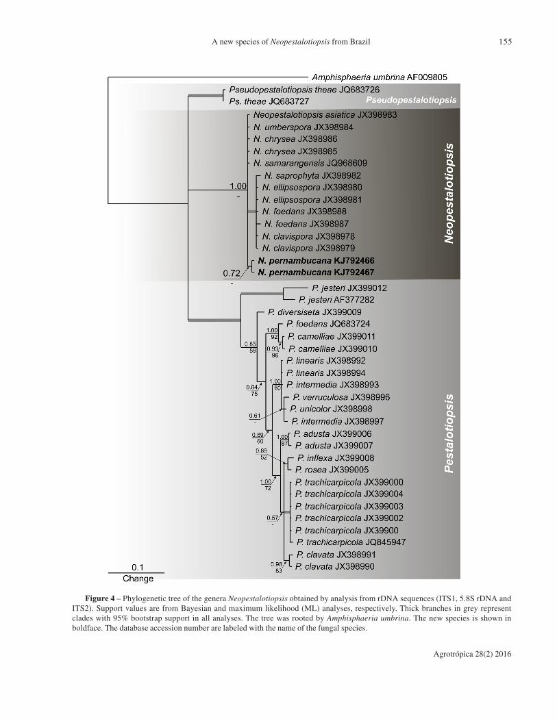
Phylogenetic analyses from sequences of the ITSgene showed that Neopestalotiopsis pernambucana
forms a distinct clade (Figure 4) and phylogeneticanalyses from sequences of the tef1 gene separatedN. pernambucana from other species used in thisstudy, with high bootstrap support (Figure 5).
Key to current species of Pestalotiopsis and
Neopestalotiopsis with sexual morphs
Silvério et al.
Agrotrópica 28(2) 2016
155
Figure 4 – Phylogenetic tree of the genera Neopestalotiopsis obtained by analysis from rDNA sequences (ITS1, 5.8S rDNA andITS2). Support values are from Bayesian and maximum likelihood (ML) analyses, respectively. Thick branches in grey representclades with 95% bootstrap support in all analyses. The tree was rooted by Amphisphaeria umbrina. The new species is shown inboldface. The database accession number are labeled with the name of the fungal species.
A new species of Neopestalotiopsis from Brazil

Agrotrópica 28(2) 2016
156
Figure 5 – Phylogenetic tree of the genera Neopestalotiopsis obtained by analysis tef1. Support values are from Bayesian andmaximum likelihood (ML) analyses, respectively. Thick branches in grey represent clades with 95% bootstrap support in all analyses.The tree was rooted by Seiridium sp. The new species is shown in boldface. The database accession number are labeled with the nameof the fungal species.
Silvério et al.

Agrotrópica 28(2) 2016
157
Discussion
In the review of the genus Pestalotiopsis,
Maharachchikumbura et al. (2014) made aphylogenetic reconstruction of the Amphisphaeriaceaebased on analysis of LSU of the rRNA sequence data.Furthermore, two novel genera were segregated fromPestalotiopsis, namely Neopestalotiopsis andPseudopestalotiopsis based on combinedmorphological and DNA data.
The genus Pestalosphaeria (Amphisphaeriaceae,Xylariales) was established in 1975 by Margaret E.Barr, as the sexual morph of Pestalotiopsis, to allocatethe pathogenic species P. concentrica, found on livingleaves of Rhododendron maximum L., ornamentalplant normally cultivated in North-American gardens.The features of the type species included peritheciaglobose, immerse in the host tissues and with erumpentostiole, asci cylindrical, 70-95 × 9-12 µm, unitunicate,with short to elongated stipe and apical ring amyloidand ascospores ovoid-elliptical, 13.5-20 × 7-10 µm,brown when mature and 2septate.
Later, Van der Aa (1976) proposed the transferof Leptosphaeria elaeidis C. Booth & J.S.Robertson for the genus Pestalosphaeria, aftercomparing structural features of the asci. This newcombination, P. elaeidis, was redescribed andillustrated by Hyde (1996).
In 1982, Shoemaker & Simpson describedPestalosphaeria hansenii on Pinus caribaea var.hondurensis, while Nag Raj (1979; 1985) found thespecies P. austroamericana, on dead leaves ofHarknessa americana in Chile and P. varia, on pods ofAcacia koa in Hawaii. The first record of P. leucospermi
was done by Samuels; Muller; Petrini (1987) in NewZealand and P. gubae was discovered in Japan byKobayashi; Ishihara; Ono (2001). Pestalosphaeria
accidenta, P. jinggangensis, P. alpiniae and P.
eugeniae were reported in China (Zhu et al., 1991; Chi,1994) and P. maculiformans was found in South Africa,on dead leaves of several hosts (Marincowitz et al., 2008).
According to the Amsterdam Declaration on FungalNomenclature, only one name can be applied to anyfungal species (Hawksworth et al., 2011).Maharachchikumbura et al. (2011) suggested thatPestalotiopsis should be adopted instead of
Pestalosphaeria because it is an older and more commonname. The genus Neopestalotiopsis was introduced byMaharachchikumbura et al. (2014) as sexual morph notobserved. The new species presently described isreferred to as Neopestalotiopsis pernambucana andit is the first species of the genus encountered with thesexual morph. Phylogenetic analysis of the sequencesof ITS and tef1 genes showed that Neopestalotiopsis
pernambucana forms a distinct clade (Figures 4 and5) with high bootstrap support. The versicolorousmorphology of the conidia, a feature of the genusNeopestalotiopsis, corroborate this molecular result.
Acknowledgements
The authors are grateful to the Programa de Pós-Graduação em Biologia de Fungos of the UniversidadeFederal de Pernambuco (PPGBFUFPE) for theopportunity given to the first author to obtain her Doctordegree, CEPLAC/CEPEC for the permission to use itslaboratories during part of the work, CNPqBrasil forfinancial support, Dr. Harry Evans and Dr. SeonjuMarincowitz for help with literature acquisition, Dr.Marcondes Albuquerque de Oliveira (PPGBVUFPE)for botanical identification and MSc. LarissaTrierveilerPereira (PPGBFUFPE) for drawings.
Literature Cited
BARR, M. E. 1975. Pestalosphaeria, a new genus inthe Amphisphaeriaceae. Mycologia 67:187-193.
CABI Bioscience. 2016. The CABI Bioscience and CBSDatabase of Fungal Names. <http://www.indexfungorum.org/names/Names.asp>.Access: 06 Jul 2016.
CHI, P. 1994. Fungal diseases of cultivated medicinalplants in Guangdong Province. GuangdongAcademy Press, Guangdong, China.
CENTRO DE RERERÊNCIA EM INFORMAÇÃOAMBIENTAL - CRIA. 2007. Flora brasiliensisrevisitada. CRIA/UNICAMP/Missouri BotanicalG a r d e n . < h t t p : / / f l o r a . c r i a . o r g . b r /taxonCard?id=FBR1519>. Access: 02 Mar 2009.
FARR, D. F.; ROSSMAN, A.Y. 2016. Fungal databases,systematic mycology and microbiology laboratory,ARS, USDA. <http://nt.ars-grin.gov/fungaldatabases/>. Access: 06 Jun 2016.
A new species of Neopestalotiopsis from Brazil

Agrotrópica 28(2) 2016
158
GÓES-NETO, A.; LOGUERCIO-LEITE, C.;GUERRERO, R. T. 2005. DNA extraction fromfrozen field-collected and dehydrated herbariumfungal basidiomata: performance of SDS andCTAB-based methods. Biotemas (Brasil)18:19-32.
GUINDON, S.; GASCUEL, O. 2003. A simple, fast, andaccurate algorithm to estimate large phylogenies bymaximum likelihood. Systematic Biology 52:696-704.
HALL, T. A. 1999. BioEdit: a user-friendly biologicalsequence alignment editor and analysis program forWindows 95/98/NT. Nucleic Acids SymposiumSeries 41:95-98.
HAWKSWORTH, D. L. et al. 2011. The Amsterdamdeclaration on fungal nomenclature. IMA Fungus2:105-112.
HYDE, K. D. 1996. Fungi from palms XXV.Pestalosphaeria elaeidis. Mycotaxon 57: 353-357.
KOBAYASHI, T.; ISHIHARA, M.; ONO, Y. 2001. Anew species of Pestalosphaeria, the teleomorphof Pestalotiopsis neglecta. Mycoscience 42: 211-216.
KRUSCHEWSKY, M. C.; LUZ, E. D. M. N.; BEZERRA,J. L. 2014. O gênero Pestalotiopsis (Ascomycota,‘Coelomycetes’) no Brasil. Agrotrópica (Brasil)26(2):89-98.
LARKIN, M. A. et al. 2007. Clustal W and Clustal Xversion 2.0. Bioinformatics 23:2947-2948.
MAHARACHCHIKUMBURA, S. S. N. et al. 2011.Pestalotiopsis – morphology, phylogeny,biochemistry and diversity. Fungal Diversity 50:167-187.
MAHARACHCHIKUMBURA, S. S. N. et al. 2012. Amulti-locus backbone tree for Pestalotiopsis, witha polyphasic characterization of 14 new species.Fungal Diversity 56: 95-129.
MAHARACHCHIKUMBURA, S. S. N. et al. 2014.Pestalotiopsis revisited. Studies in Mycology 79:121-186.
MARINCOWITZ, S. et al. 2008. Microfungi occurringon Proteaceae in the fynbos. Utrecht, TheNetherlands, CBS Biodiversity Series 7:75–77.
MENDES, M. A. S. et al. 1998. Fungos em plantas noBrasil. Brasília, DF, EMBRAPASPI/EMBRAPACENARGEN.
MILNE, I. et al. 2004. TOPALi: Software for AutomaticIdentification of Recombinant Sequences within DNAMultiple Alignments. Bioinformatics 20:1806-1807.
MISAGHI, I. J. et al. 1978. Influence of environmentand culture media on spore morphology ofAlternaria alternata. Phytopathology 68:29-34.
NAG RAJ, T. R. 1979. Miscellaneous microfungi III.Canadian Journal of Botany 57:2489-2496.
NAG RAJ, T. R. 1985. Redisposals and redescriptionsin the Monochaetia-Seiridium , Pestalotia-
Pestalotiopsis complexes II. Pestalotiopsis
besseyii (Guba) comb. nov. and Pestalosphaeria
varia sp. nov. Mycotaxon 22:52-63.
OLIVEIRA, R. J. V. 2014. Corniculariella brasiliensis,a new species of coelomycetes in the rhizosphereof Caesalpinia echinata (Fabaceae,Caesalpinioideae) in Brazil. Phytotaxa 178:197-204.
REHNER, S. A. 2001. Primers for Elongation Factor 1-alpha (EF1-alpha). <http://ocid.nacse.org/research/deephyphae/EF1primer.pdf>.
REICHARDT, H. G. 1878. Hypericaceae. Flora brasiliensis,vol. XII, part I, fasc. 81, colunas 180–212. <http://florabrasiliensis.cria.org.br/search?taxon_id=2493>.Access: 25 Jan 2012.
RONQUIST, F.; HUELSENBECK, J. P. 2003. MrBayes3: Bayesian phylogenetic inference under mixedmodels. Bioinformatics 19:1572-1574.
SAMUELS, G. J.; MÜLLER, E.; PETRINI, O. 1987.Studies in the Amphisphaeriaceae (sensu lato) 3.New species of Monographella andPestalosphaeria, and two new genera. Mycotaxon28(2):473-499.
SHOEMAKER, R. A.; SIMPSON JA. 1982. A newspecies of Pestalosphaeria on pine with commentson the generic placement of the anamorph.Canadian Journal of Botany 59:986-991.
VAN DER. A A, H. A. 1976. Progress Report 1975. RoyalNetherlands Academy of Arts and Sciences/Verhandelingen der Koninklijke NederlandischeAkademie van Wetenschappen. Tweede Reeks2(67):86-87.
VIÉGAS, A. P. 1961. Índice de fungos da América doSul. Campinas, SP, Instituto Agronômico.
WHITE, T. J. et al. 1990. Amplification and directsequencing of fungal ribosomal RNA genes forphylogenetics. In Innis, M.A. eds. PCR protocols:a guide to methods and applications. New York,Academic Press Inc. pp. 315–322.
ZHU, P.; GE, Q.; XU, T. 1991. The perfect stage ofPestalotiopsis from China. Mycotaxon 40:129-140.
l
Silvério et al.
![[BIOLOGIA MAIS] [] [VOLUME …biologiamais.com.br/download-304/exercicios-pteridofitas.pdf · o aguapé (Angiospermae), e Salvinia sp. (Pteridophyta). a) 'Eichornia crassipes' é](https://img.document.onl/doc/110x75/5d041b3388c99322638c50ba/biologia-mais-volume-biologiamaiscombrdownload-304exercicios-pteridofitaspdf.jpg)