Embed Size (px)
Citation preview
MSPMESTRADO EMSAÚDE PÚBLICA
UNIVERSIDADE DO PORTOFACULDADE DE MEDICINAINSTITUTO DE CIÊNCIAS BIOMÉDICAS ABEL SALAZAR
Relationship between Helicobacter pylori
infection and gastric cardia cancer
Marlene Vanessa Brandão Lima Cavaleiro Pinto
Porto, 2010

I
Investigação realizada no Serviço de Higiene e Epidemiologia da Faculdade de Medicina
da Universidade do Porto e no Instituto de Saúde Pública da Universidade do Porto
(ISPUP), sob orientação do Professor Doutor Nuno Lunet.
O trabalho teve apoio financeiro através de Bolsas da Fundação Para a Ciência e
Tecnologia (PTDC/SAL-ESA/715117/2006 e SFRH/BD/36818/2007).
II
A dissertação tem como base dois artigos, no primeiro colaborei activamente na
operacionalização das hipóteses, na recolha, armazenamento, análise e interpretação dos
dados e fui responsável pela redacção da primeira versão do manuscrito. No segundo
artigo colaborei activamente na definição das hipóteses, na análise e interpretação dos
dados e na redacção inicial do manuscrito:
- Marlene Cavaleiro-Pinto, Bárbara Peleteiro, Nuno Lunet, Henrique Barros. Helicobacter
pylori infection and gastric cardia cancer: systematic review and meta-analysis.
[Submetido].
- Bárbara Peleteiro, Marlene Cavaleiro-Pinto, Rita Barros, Henrique Barros, Nuno Lunet. Is
cardia cancer etiologically different from distal stomach cancer? [Submetido].

III
Agradecimentos
IV
Ao Professor Doutor Nuno Lunet pela confiança depositada em mim e pelos
conhecimentos partilhados durante a elaboração desta dissertação.
Aos Professores, com quem tive o privilégio de aprender ao longo do meu percurso
académico, pela confiança depositada, o estímulo e a transmissão de conhecimentos.
Aos colegas do Serviço de Higiene e Epidemiologia pelo apoio, o espaço e o tempo
concedidos durante a elaboração desta dissertação.
À Bárbara pelo acolhimento e valioso auxílio durante a elaboração desta
dissertação.
Aos meus Avós, pelo seu carinho.
Aos meus pais, pela afeição, apoio incondicional e fundamental neste trilho da
minha vida.
Ao meu irmão, pela sua afeição sem a qual esta caminhada não seria com o mesmo
alento.
1
Table of contents
Page
1. Gastric cancer incidence, mortality and trends 2
1.1. Frequency and trends by cancer location 4
2. Aetiological epidemiology of gastric cardia cancer 5
2.1. Environmental factors 5
2.1.1. Lifestyles 5
2.1.2. Helicobacter pylori infection 6
2.1.2.1. Bacterial properties and pathogenic mechanisms 6
2.1.2.2. Strains variations and the cag pathogenicity island 7
2.1.2.3. Diagnostic methods 9
2.1.2.4. Epidemiology of infection 9
2.1.2.5. Helicobacter pylori infection and gastric cardia cancer 10
2.2. Host factors 16
2.2.1. Response to H. pylori infection determined by host genetic
polymorphisms
16
3. Aims 19
Paper I Helicobacter infection and gastric cardia cancer: a systematic
review and meta-analysis
20
Paper II Is cardia cancer etiologically different from distal stomach cancer? 88
4. General discussion and conclusions 103
5. References 105
6. Summary 119
7. Sumário 122
2
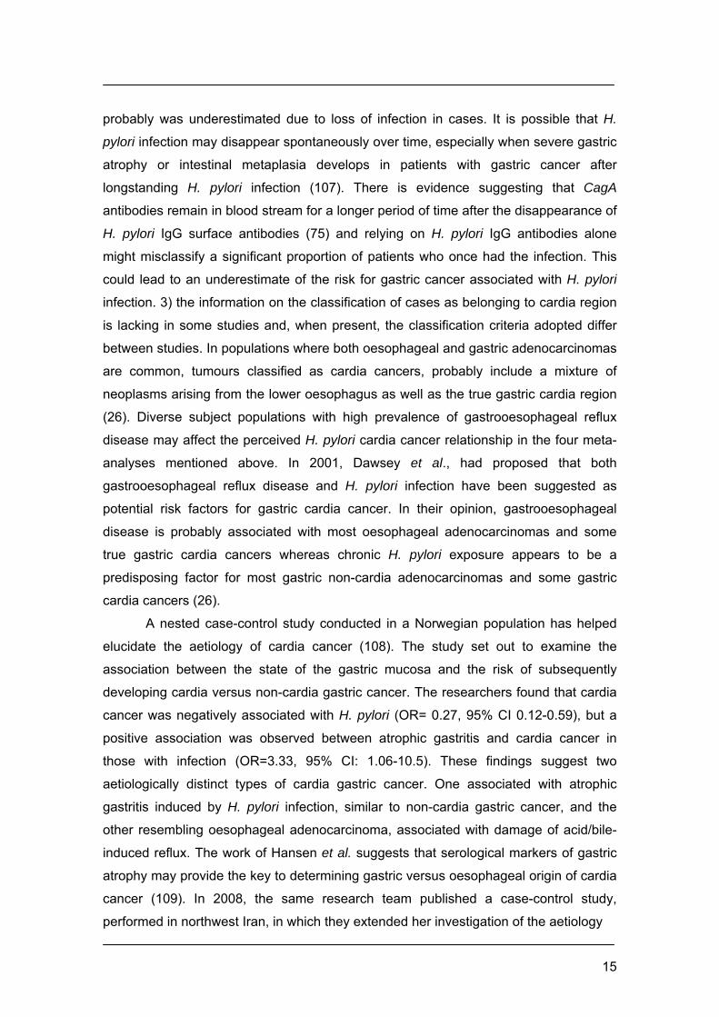
1. Gastric cancer incidence, mortality and trends
Gastric cancer is now the fourth most common cancer worldwide, with around
989 000 new cases estimated for 2008 (1).
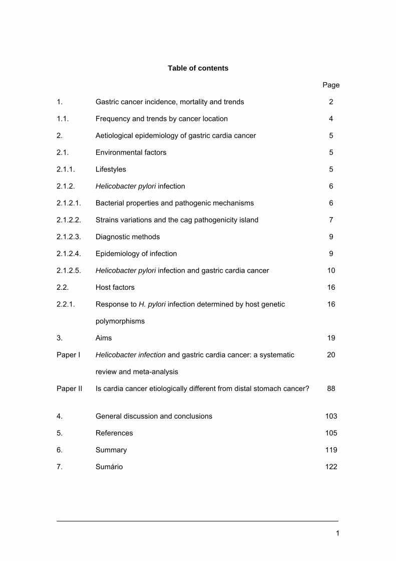
On the American continent, sharp regional differences are observed in the
gastric cancer age-standardized incidence rates. North America has the lowest
incidence, in Central America the rates are approximately twice higher and in South
America about three-fold higher (figure 1) (1). In Europe, Northern and Western
countries have the lowest incidence followed by the Southern and Central/Eastern
regions (1). Worldwide, the highest age-standardized incidence rates are observed in
Eastern Asia, and the lowest in Western Africa (figure 1) (1).
Figure 1. Incidence of the gastric cancer by sex by region (Age-Standardised rates
(world reference population)), Source: Globocan-2008 (1).
The incidence rates from gastric cancer in generation migrants tend to move
toward to the rates of host country (2-4).
Gastric cancer is the second most common cause of oncological death, with
approximately 737 000 deaths annually (1).
3
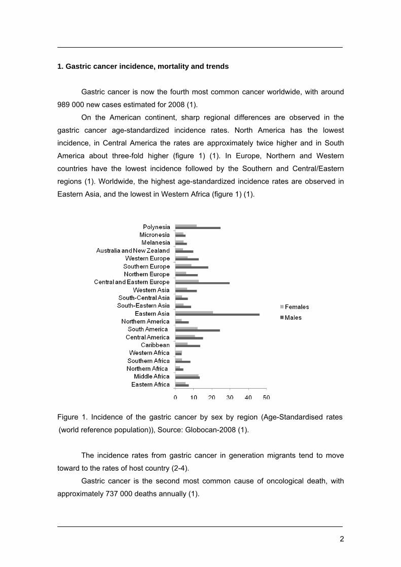
In America, there is a gradual increase in mortality rates as we move to the
south (figure 2), similarly to what is observed in Europe, with North and West regions
presenting the lowest mortality rates, and Southern and Central/Eastern regions the
highest (figure 2) (1). In Eastern Asia, the age-standardised mortality rates are among
the highest worldwide, with South Eastern, South Central and Western Asian regions
presenting mortality rates similar to the observed in Northern and Western Europe
(figure 2) (1). Africa is the continent with the lowest age-standardised mortality rates,
the lowest being observed in the North and the highest in Middle Africa (figure2) (1).
Figure 2. Gastric cancer mortality by region, according to gender (age-standardised
rates; world reference population). Source: Globocan-2008 (1).
Since the mid 1980s, incidence rates of gastric cancer have fallen in all high-
income countries, and overall rates are now about 15% lower than in 1985 (5-7), with a
wide variation in incidence rates across geographical areas (5). There is also a large
heterogeneity between countries in the incidence of the two major sites of gastric
cancer, namely the proximal (cancer of the cardia) and distal (noncardia cancer)
locations (8-11).
4
1.1. Frequency and trends by cancer location
Despite the overall decline in gastric cancer, most published papers showed an
increasing (9, 10, 12-16) or stable incidence in gastric cardia cancer (8, 17-21), in the
past 30 years. In the last decade’s population-based studies in western countries like
United States, Australia, New Zealand and several countries of Europe, reported an
increasing incidence of gastric cardia cancer (22), while the incidence of tumours in the
non-cardia localization is decreasing (23). An increase in incidence was observed in
Northern Europe (Denmark), Southern Europe (Italy, Varese), Eastern Europe
(Slovakia) and Western Europe (England and Wales, Scotland) during 1968-1995. In
Central Europe (Switzerland, Basel) and in Northern Europe (Iceland), Western Europe
(France, Bas-Rhin and Calvados, Southern Ireland), and in Western Europe
(Netherlands, Eindhoven) no rise in incidence was observed (11). In Japan, national
data reported significant increases in gastric cardia cancer, in both absolute number
and in proportion to other gastric cancers over a 36-year period (24). In China, a
statistically significant increasing trend of gastric cardia cancer was observed during in
the last 16 years (25).
The gastric cardia represents only the proximal 2-3 cm of the stomach. This
small anatomical region can easily be overgrown by tumours that originate from
adjacent mucosal sites (26).
The definition of the anatomical cardia region has changed over the years and
currently differs between countries. Anatomically the gastric cardia is defined as the
part of the stomach adjacent to the orifice of the oesophagus (27), but this orifice can
also be defined as the oesophagogastric junction (EGJ). As the EGJ forms the border
between oesophagus and stomach, the classification of tumors in this region is
inherently complex. While some investigators classify all oesophagogastric tumours as
oesophageal carcinomas, others consider them to be gastric carcinomas and yet
others regard them as separate entities (27). The EGJ is differently defined by
anatomists, physiologists, endoscopists and pathologists.
Physiologists define EGJ as the distal border of the lower oesophageal
sphincter (LES) as determined by manometry. This location, however, is difficult to
identify by endoscopic techniques and there is a large discrepancy between
manometric and endoscopic estimation of the LES. As the distal oesophagus is a
dynamic structure with peristaltic activity, which moves during respiration, it is a difficult
task for accurate endoscopic judgments of the EGJ. Histologically, the gastric cardia
has a distinct pattern of loosely packed mucous glands in which an occasional parietal
5
cell can be present. Histology, however, needs to be combined with the endoscopic
appearance, as cardiac type mucosa can be present in the oesophagus in the setting
of columnar lined oesophagus (27).
Adenocarcinomas of the EGJ, in China, are generally referred to as gastric
cardia adenocarcinomas. Therefore, adenocarcinomas arising from the EGJ/cardia
have been referred to as oesophageal cancers for the past several decades. As most
of the adenocarcinomas arising from the cardia are diagnosed at an advanced stage, it
is very difficult to accurately define whether these tumours have a primary oesophageal
or gastric origin (28). Imprecise clinical and pathological definitions of
adenocarcinomas of the lower oesophagus, EGJ, and gastric cardia may be one of the
reasons underlying inconsistencies across studies (27). In populations where both
oesophageal and gastric adenocarcinomas are common, tumours described as cardia
cancers undoubtedly include a mixture of neoplasms arising from the lower
oesophagus as well as the gastric cardia and distal stomach (26).
2. Aetiological epidemiology of gastric cardia cancer 2.1. Environmental factors
2.1.1. Lifestyles
The geographic variation, time trend, and the migratory effect on gastric cancer
incidence suggest that environmental factors are the major determinants of this
disease.
A protective effect from diets rich in fresh fruit and vegetables has been
suggested (29, 30). The World Cancer Research Fund (WCRF) published a report in
which a substantial amount of evidence is available for vegetables and fruit that protect
against stomach cancer (29). Similar results were documented in published cohort and
case-control studies evaluating the association between fruit and vegetables
consumption and the occurrence of gastric cardia cancer (30-33).
Vitamin C and other anti-oxidant nutrients have attracted a lot of attention as the
potential mediators of a dietary influence on gastric cancer risk. Vitamin C seems a
promising candidate since its is a free radical scavenger, it reduces the formation of
potentially carcinogenic N-nitroso compounds (34). Data from the EPIC-EUROGAST
show a negative correlation between gastric cancer risk and serum vitamin C (35). A
Cochrane Collaboration review, which included a number of high-quality randomized
trials, has concluded that there is no evidence that dietary supplementation with anti-
6
oxidants, including vitamin C, reduces gastric cancer risk (36). Dietary vitamin C has
been shown inversely associated with both gastric cancer subsites (37, 38).
Salt and nitrite are other dietary components that have been implicated in
gastric cancer risk. Pickled and smoked foods may also contain potential carcinogens
such as N-nitroso compounds and benzapyrene. Carcinogenic N-nitroso compunds
can be formed directly from nitrite and nitrate in the diet, or by the metabolic action of
anaerobic bacteria colonizing the stomach (39). Meta-analyses have shown a positive
association between gastric cancer risk and both salt and dietary nitrate/nitrite intake
(29).
Smoking is an independent risk factor for gastric cancer, which is involved in
neoplastic transformation of gastric mucosa. It was found to nearly double the risk of
transition from atrophic gastritis to dysplasia in a high-risk population (40). A systematic
review and meta-analysis of cohort studies presented an overall summary RR estimate
for current smokers in the highest categories of consumption compared to never
smokers of 1.66 (95% CI: 1.46-1.88) (41), and prospective studies have demonstrated
a significant dose-dependent association between tobacco smoke and gastric cancer
(42, 43). In the EPIC-EUROGAST study it was estimated that 17.6% of the gastric
cancer cases occurring in this European population were attributable to cigarette
smoking (41, 44). Published studies yielded similar conclusions for cardia cancer,
supporting that smoking is a risk factor also for this gastric cancer subtype (41, 45).
2.1.2. Helicobacter pylori infection
The discovery of H. pylori in 1983 has proved to be pivotal in our understanding
of the aetiology of gastric cancer (46). In 1994, the World Health Organization and the
International Agency for research on Cancer consensus group stated that there was
sufficient evidence to classify H. pylori as a Class I human carcinogen (47).
2.1.2.1. Bacterial properties and pathogenic mechanisms
H. pylori is a gram-negative bacillus and when observed in vivo is a spiral-
shaped or curved rod, a few micrometers long and actively motile. The bacteria can be
found in a horse-shoe like U-form and a round, or coccoid forms, in older cultures. The
four to six unipolar sheathed flagella are important for bacterial motility (48, 49).
7
A number of bacterial factors have been suggested to be responsible for
successful infection by H. pylori: high urease production, which increases the pH and
activates the host immune defence; flagella, which facilitate the movement within the
mucus layer; adherence to gastric epithelium by different adhesins using
hemagglutinins, laminin and Lewis b antigens as receptors (48, 50).
There is accumulated evidence that acid secretory capacity is important in
determining the distribution, and natural history of H. pylori infection (51, 52). In hosts
with low secretory capacity (genetically determined or by pharmacologic inhibition) the
organisms are capable of colonising a wider niche than would be possible in the
presence of high volumes of acid (51, 52). Colonization of a wider niche including the
corpus mucosa leads to corpus gastritis with resultant functional inhibition of acid
secretion (51). This inhibition is mediated by H. pylori induced inflammatory cytokines
(such as IL-1β and TNF-α) and the net effect is the establishment of a more aggressive
gastritis that accelerates the development of gastric atrophy. The infected subjects
under long term proton pump inhibitors have a higher risk of developing gastric
atrophy, a precursor lesion of gastric neoplasia (53). Gastric atrophy seems to be a
morphological change hard to reverse, although this issue is still open to debate.
2.1.2.2. Strains variation and the cag pathogenicity island
Early investigations of the differential pathogenic properties of H. pylori strains
indicated that pathogenicity was associated with the ability of these more virulent
strains to induce morphological changes, vacuolization, and successive degeneration
of in vitro-cultured cells (54). This activity was then linked to the presence of a protein
with a molecular mass of approximately 140 kDa that was named CagA (for "cytotoxin-
associated gene A") and a highly immunogenic 95-kDa protein that was named VacA
(VacA vacuolating cytotoxin) (55).
The CagA protein is a highly immunogenic protein encoded by the cagA gene
(55). The cagA gene is present in approximately 50 to 70% of H. pylori strains (56-58)
and is a marker for the presence of a genomic PAI (cag pathogenicity island) of about
40 kb that, depending on the strain analyzed, encodes between 27 and 31 proteins (55,
59, 60). H. pylori’s CagA protein is now regarded as having direct oncogenic potential
(61). The CagA is delivered into gastric epithelial cells by the bacterial type IV secretion
system. Once injected into gastric epithelial cells, CagA undergoes tyrosine
phosphorylation by SRC family Kinases. Phosphorilated CagA specifically binds and
8
activates SHP2 (protein-tyrosine phosphatase), that acts as a human oncoprotein.
SHP2 transmits positive signals for cell growth and motility and deregulation of SHP2
by CagA is thought to be an important mechanism by which CagA-positive H. pylori
strains may promote gastric carcinogenesis (61). H. pylori is capable of subverting cell
physiology towards several proneoplastic process through CagA and other proteins
(e.g. activation of growth factor receptors, increased proliferation, sustained
angiogenesis and cell dissociation and tissue invasion) (61). H. pylori and its citotoxins
mediate these proneoplastic mechanisms and it is very likely that the inflammatory
process unleashed by the presence of the bacteria on the gastric mucosa also
contributes to the neoplastic impel (61). Many of the mediators and products of
inflammation are mitogenic and mutagenic (62). Release of pro-inflamatory cytokines,
reactive oxygen species and upregulation of cyclooxygenase-2 (Cox-2) all contribute to
an intra-gastric environment conductive to neoplastic transformation (62). The
mechanisms involve direct DNA damage, inhibition of apoptosis, subversion of
immunity, and stimulation of angiogenesis (62). In addition, chronic inflammation in the
gastrointestinal tract is also known to affect proliferation, adhesion and cellular
transformation (62). However, the clinical outcome of H. pylori infection is varied and
includes gastric cancer and non-neoplastic conditions (peptic ulcer, non-ulcer
dyspepsia and simple asymptomatic gastritis).
The VacA protein plays an important role in the pathogenesis of both peptic
ulceration and gastric cancer (63). The activities of VacA include membrane channel
formation, disruption of endosomal and lysosomal activity, effects on integrin receptor-
induced cell signaling, interference with cytoskeleton-dependent cell functions,
induction of apoptosis, and immune modulation (63). The VacA protein is produced as
a 140-kDa protoxin that is cleaved into the 95-kDa mature form when secreted.
Although all strains carry a functional vacA gene, there is considerable variation in
vacuolating activities among strains. This is due to the sequence heterogeneity within
the vacA gene at the signal region (s) and the middle region (m) (63). The s region of
the gene, which encodes the signal peptide, occurs as either an s1 or s2 type, whereas
the m region, which contains the p58 cell binding domain, exists as an m1 or m2 type.
Vacuolating activity is high in s1/m1 genotypes, intermediate in s1/m2 genotypes, and
absent in s2/m2 genotypes (63, 64). In line with this, vacA s1/m1 genotypes are more
frequently associated with peptic ulceration and gastric carcinoma (64). The vacA
s2/m1 strain, however, is noncytotoxic (65, 66).
The genes cagA and vacA were the most intensively studied over the years and
have been used as the most common markers for strains with enhanced virulence.
9
2.1.2.3. Diagnostic methods
Currently there are several methods for detection of H. pylori infection and can
be grouped into 2 categories, depending on the technique used to collect the biological
material used: invasive tests (require endoscopy) and non-endoscopy tests (67, 68).
The 13C-urea breath test (13C UBT), is a technique used to detect H. pylori colonization
in the stomach, without an endoscopical examination. The high urease activity of H.
pylori turns urea into ammonia and produce 13C-labeled CO2 which can be measured
with biochemical tests kits in market. Other non-endoscopy technique is based on the
collection of saliva, serum, or stool samples that need processing before the test can
be performed (e.g. enzyme immunoassay (EIA), enzyme-linked immunosorbent assay
(ELISA), Western blot, complement fixation, latex agglutination assays) (68, 69). A
large number of enzyme immunoassay (EIA) and enzyme-linked immunosorbent assay
(ELISA) tests to detect anti-H. pylori Immunoglobulins (e.g. IgG, IgM and IgA) are
available in the market. Comparisons between sensitivity and specificity of these tests
are published regularly (70-72). The median sensitivity and specificity in 36
commercially available kits were 92% and 83% respectively. The immunoblotting
techniques are useful method to detect infections caused by more virulent strains (e.g.:
VacA and CagA positive) (73-77).
The invasive tests which require biopsy material are: rapid urease test (RUT–
CLO-test), direct Gram stain, culture (in non-selective or selective agar), histology (by
haematoxylin and eosin, Warthin Starry Silver, modified Giemsa stain or acridine-
orange stains), and immunohistochemistry (by immunoperoxidase or immuno-
fluorescent techniques using anti-H. pylori monoclonal or polyclonal antibodies) (67).
For specific detection of H. pylori in environmental samples, stool samples,
gastric juice and biopsies, and for detection of H. pylori genes, polymerase chain
reaction techniques are also used. The most frequently genes that are used for PCR-
techniques to distinguish between H. pylori and other microorganisms are the adhesin
encoding gene hap, the urease encoding gene ureA and 16S ribosomal RNA gene
(78).
2.1.2.4. Epidemiology of infection
The prevalence of H. pylori infection varies across geographical regions. In
developed countries, the improvement of sanitation and hygiene has been responsible
10
for the decrease in transmission during the last few decades, and the epidemiology of
the bacterium at present follows a birth cohort model (older people infected decades
ago still frequently carry the bacterium while children rarely do) (79). As the acquisition
of H. pylori occurs predominantly in childhood and, if untreated, persists throughout life,
a lower seroprevalence of H. pylori in older age-groups is expected in the future.
In general, infection is acquired during childhood, and so the prevalence
gradually increases, at a higher rate in developing than in developed countries, to
reach the maximum in middle age (80). The overall estimate prevalence of infection in
middle-aged adults is 74% in developing countries and 58% in developed countries
(80). In 2002, the proportion of gastric cancers worldwide attributable to H. pylori
infection was 63.4%, corresponding to 592 000 cases. Regarding developed and
developing countries the estimated numbers of stomach cancer cases attributable to H.
pylori infection was 61.4% and 64.4%, respectively (80).
From the many risk factors for infection that have been investigated, age and
lower socioeconomic status (SES) are the ones considered most important (81).
Socioeconomic deprivation in childhood is associated with a high prevalence of H.
pylori colonisation. While the incidence of H. pylori may be declining, it remains
common in poor families (82, 83). Low socioeconomic status, poor sanitary indications,
and crowded families in childhood were related to high prevalence of H. pylori infection
(83).
2.1.2.5. Helicobacter pylori infection and gastric cardia cancer
Numerous cohort and case-control studies were published demonstrating an
association between serological evidence of H. pylori infection and increased risk of
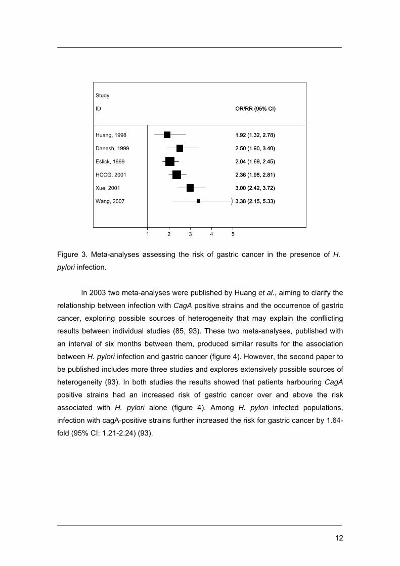
gastric cancer, which have been summarized in meta-analyses (84-91).
The first meta-analysis on this subject was published in 1998 and included 19
epidemiological studies (cohort and case-control studies) that diagnosed H. pylori
infection by serology. The researchers concluded that H. pylori infection is a risk factor
for gastric cancer. They suggested that the heterogeneity of reported results was
caused by differences in the selection of controls, patient age and stage of gastric
cancer (figure3) (90).
In the following year, a meta-analysis based in prospective studies (10 nested
case-control studies) was published, suggesting that gastric cancer was 2 or 3 times
more frequent in those chronically infected by H. pylori (figure 3) (92). In the same
11
year, Eslick et al. published a meta-analysis performed with 42 studies (8 cohort
studies and 34 case-control studies), which confirmed the positive association between
H. pylori infection and gastric carcinoma. According to the authors, an effort was made
to perform an analysis as inclusive as possible and the quality of studies included was
explored as a source of heterogeneity. The results suggested that the majority of
studies classified as excellent found positive association and the very poor to moderate
studies gave mixed results (figure 3) (88).
In 2001, the Helicobacter and Cancer Collaborative Group published a study
based in a collaborative reanalysis using individual subject data from 12 nested case-
control studies intending to clarify the magnitude of the association. The inclusion of
data from published and unpublished studies was the main difference in relation to
previous meta-analyses. The results support the idea that when H. pylori status is
assessed close to cancer diagnosis, the magnitude of the association may be
underestimated (figure 3) (91).
In the same year, a meta-analysis conducted only with studies published in
Chinese populations (11 case-control studies conducted in China) was published. The
results showed that H. pylori infection is a risk factor for gastric carcinoma, increasing
the risk to 3-fold more in Chinese population infected (figure 3) (87).
The most recent meta-analysis on this topic was published in 2007, and it has
reviewed systematically the relationship between H. pylori infection and the occurrence
of early gastric cancer (EGC) and in the advanced gastric cancer (AGC). In the fifteen
studies included, the prevalence of H. pylori infection in EGC and in non-neoplasm
controls was 87.3% (2,377/2,722) and 61.4% (8,588/13,976) respectively,
corresponding to a summary OR of 3.38 (95% CI, 2.15–5.33), but the results from the
individual studies were substantially heterogeneous (I2=83.5%, p<0.00001). Sensitivity
analyses were conducted by separating studies with factors that could have possible
impact on heterogeneity (e.g.: H. pylori detection methods, different publication years,
geography, and different sample sizes); and the prevalence of infection in EGC and
controls remained essentially unchanged. The OR was around 3 in all the strata and
the heterogeneity was not eliminated. The prevalence of H. pylori in EGC group was
87.6% (1,780/2,032), significantly higher than the 77.3% (874/1,130) in the advanced
gastric cancer (AGC) group, with an OR of 2.13 (95% CI; 1.75-2.59). No significant
heterogeneity was observed (p=0.87) (figure 3) (84).
12
Huang, 1998
Danesh, 1999
Eslick, 1999
HCCG, 2001
Xue, 2001
Wang, 2007
ID
Study
1.92 (1.32, 2.78)
2.50 (1.90, 3.40)
2.04 (1.69, 2.45)
2.36 (1.98, 2.81)
3.00 (2.42, 3.72)
3.38 (2.15, 5.33)
OR/RR (95% CI)
1.92 (1.32, 2.78)
2.50 (1.90, 3.40)
2.04 (1.69, 2.45)
2.36 (1.98, 2.81)
3.00 (2.42, 3.72)
3.38 (2.15, 5.33)
OR/RR (95% CI)
11 2 3 4 5
Figure 3. Meta-analyses assessing the risk of gastric cancer in the presence of H.
pylori infection.
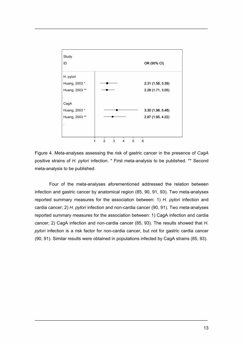
In 2003 two meta-analyses were published by Huang et al., aiming to clarify the
relationship between infection with CagA positive strains and the occurrence of gastric
cancer, exploring possible sources of heterogeneity that may explain the conflicting
results between individual studies (85, 93). These two meta-analyses, published with
an interval of six months between them, produced similar results for the association
between H. pylori infection and gastric cancer (figure 4). However, the second paper to
be published includes more three studies and explores extensively possible sources of
heterogeneity (93). In both studies the results showed that patients harbouring CagA
positive strains had an increased risk of gastric cancer over and above the risk
associated with H. pylori alone (figure 4). Among H. pylori infected populations,
infection with cagA-positive strains further increased the risk for gastric cancer by 1.64-
fold (95% CI: 1.21-2.24) (93).
13
.
.
H. pylori
Huang, 2003 *
Huang, 2003 **
CagA
Huang, 2003 *
Huang, 2003 **
ID
Study
2.31 (1.58, 3.39)
2.28 (1.71, 3.05)
3.30 (1.98, 5.48)
2.87 (1.95, 4.22)
OR (95% CI)
2.31 (1.58, 3.39)
2.28 (1.71, 3.05)
3.30 (1.98, 5.48)
2.87 (1.95, 4.22)
OR (95% CI)
11 2 3 4 5 6
Figure 4. Meta-analyses assessing the risk of gastric cancer in the presence of CagA
positive strains of H. pylori infection. * First meta-analysis to be published. ** Second
meta-analysis to be published.
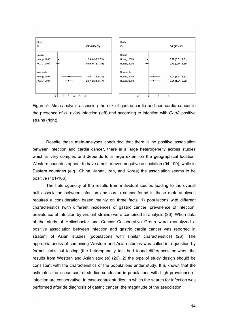
Four of the meta-analyses aforementioned addressed the relation between
infection and gastric cancer by anatomical region (85, 90, 91, 93). Two meta-analyses
reported summary measures for the association between: 1) H. pylori infection and
cardia cancer; 2) H. pylori infection and non-cardia cancer (90, 91). Two meta-analyses
reported summary measures for the association between: 1) CagA infection and cardia
cancer; 2) CagA infection and non-cardia cancer (85, 93). The results showed that H.
pylori infection is a risk factor for non-cardia cancer, but not for gastric cardia cancer
(90, 91). Similar results were obtained in populations infected by CagA strains (85, 93).
14
.
.
Cardia
Huang, 1998
HCCG, 2001
Noncardia
Huang, 1998
HCCG, 2001
ID
Study
1.23 (0.56, 2.71)
0.99 (0.72, 1.35)
3.08 (1.78, 5.31)
2.97 (2.34, 3.77)
OR (95% CI)
1.23 (0.56, 2.71)
0.99 (0.72, 1.35)
3.08 (1.78, 5.31)
2.97 (2.34, 3.77)
OR (95% CI)
1.5 1 2 3 4 5 6
.
.
Cardia
Huang, 2003
Huang, 2003
Noncardia
Huang, 2003
Huang, 2003
ID
Study
0.82 (0.51, 1.31)
0.70 (0.44, 1.10)
2.01 (1.21, 3.32)
2.01 (1.21, 3.32)
OR (95% CI)
0.82 (0.51, 1.31)
0.70 (0.44, 1.10)
2.01 (1.21, 3.32)
2.01 (1.21, 3.32)
OR (95% CI)
1-1 1 3 5
Figure 5. Meta-analysis assessing the risk of gastric cardia and non-cardia cancer in
the presence of H. pylori infection (left) and according to infection with CagA positive
strains (right).
Despite these meta-analyses concluded that there is no positive association
between infection and cardia cancer, there is a large heterogeneity across studies
which is very complex and depends to a large extent on the geographical location.
Western countries appear to have a null or even negative association (94-100), while in
Eastern countries (e.g.: China, Japan, Iran, and Korea) the association seems to be
positive (101-106).
The heterogeneity of the results from individual studies leading to the overall
null association between infection and cardia cancer found in these meta-analyses
requires a consideration based mainly on three facts: 1) populations with different
characteristics (with different incidences of gastric cancer, prevalence of infection,
prevalence of infection by virulent strains) were combined in analysis (26). When data
of the study of Helicobacter and Cancer Collaborative Group were reanalyzed a
positive association between infection and gastric cardia cancer was reported in
stratum of Asian studies (populations with similar characteristics) (26). The
appropriateness of combining Western and Asian studies was called into question by
formal statistical testing (the heterogeneity test had found differences between the
results from Western and Asian studies) (26). 2) the type of study design should be
consistent with the characteristics of the populations under study. It is known that the
estimates from case-control studies conducted in populations with high prevalence of
infection are conservative. In case-control studies, in which the search for infection was
performed after de diagnosis of gastric cancer, the magnitude of the association
15
probably was underestimated due to loss of infection in cases. It is possible that H.
pylori infection may disappear spontaneously over time, especially when severe gastric
atrophy or intestinal metaplasia develops in patients with gastric cancer after
longstanding H. pylori infection (107). There is evidence suggesting that CagA
antibodies remain in blood stream for a longer period of time after the disappearance of
H. pylori IgG surface antibodies (75) and relying on H. pylori IgG antibodies alone
might misclassify a significant proportion of patients who once had the infection. This
could lead to an underestimate of the risk for gastric cancer associated with H. pylori
infection. 3) the information on the classification of cases as belonging to cardia region
is lacking in some studies and, when present, the classification criteria adopted differ
between studies. In populations where both oesophageal and gastric adenocarcinomas
are common, tumours classified as cardia cancers, probably include a mixture of
neoplasms arising from the lower oesophagus as well as the true gastric cardia region
(26). Diverse subject populations with high prevalence of gastrooesophageal reflux
disease may affect the perceived H. pylori cardia cancer relationship in the four meta-
analyses mentioned above. In 2001, Dawsey et al., had proposed that both
gastrooesophageal reflux disease and H. pylori infection have been suggested as
potential risk factors for gastric cardia cancer. In their opinion, gastrooesophageal
disease is probably associated with most oesophageal adenocarcinomas and some
true gastric cardia cancers whereas chronic H. pylori exposure appears to be a
predisposing factor for most gastric non-cardia adenocarcinomas and some gastric
cardia cancers (26).
A nested case-control study conducted in a Norwegian population has helped
elucidate the aetiology of cardia cancer (108). The study set out to examine the
association between the state of the gastric mucosa and the risk of subsequently
developing cardia versus non-cardia gastric cancer. The researchers found that cardia
cancer was negatively associated with H. pylori (OR= 0.27, 95% CI 0.12-0.59), but a
positive association was observed between atrophic gastritis and cardia cancer in
those with infection (OR=3.33, 95% CI: 1.06-10.5). These findings suggest two
aetiologically distinct types of cardia gastric cancer. One associated with atrophic
gastritis induced by H. pylori infection, similar to non-cardia gastric cancer, and the
other resembling oesophageal adenocarcinoma, associated with damage of acid/bile-
induced reflux. The work of Hansen et al. suggests that serological markers of gastric
atrophy may provide the key to determining gastric versus oesophageal origin of cardia
cancer (109). In 2008, the same research team published a case-control study,
performed in northwest Iran, in which they extended her investigation of the aetiology
16
of cardia cancer by examining the association of both serologic evidence of gastric
atrophy and reflux symptoms with adenocarcinoma of the oesophagus, cardia, and
non-cardia region of the stomach. They had concluded that there are not only two
distinct aetiologies of cardia cancers but that the structural and functional state of the
stomach associated with them was profoundly different. One type is associated with a
non-atrophic healthy gastric mucosa producing sufficient acid and pepsin to damage
the mucosa of the gastro-oesophageal junction and lead to columnar intestinal
metaplasia and intestinal subtype cancer. The other is associated with atrophic gastritis
of sufficient severity and extent to involve the proximal stomach leading to the
development of intestinal or diffuse subtype cancer from the atrophic gastric mucosa
(105).
2.2. Host factors
2.2.1. Response to H. pylori infection determined by host genetic polymorphisms
Individual differences in the host response to H. pylori infection, determined by
host genetic polymorphisms, might, in part, explain why some individuals are more
likely to develop the gastric cancer phenotype than others. H. pylori cause damage by
initiating chronic inflammation in the gastric mucosa. This inflammation is mediated by
an array of pro and anti-inflammatory cytokines. Genetic polymorphisms directly
influence inter-individual variation in the magnitude of cytokine response and this
clearly contributes to an individual’s ultimate clinical outcome (108). It was speculated
that the most relevant candidate genes implicated in the host response to infection
would be ones whose products were involved in handling the H. pylori attack (innate
and adaptive immune responses) and ones that mediated the resulting inflammation
(108). The most relevant and consistent genetic factors uncovered thus far are in the
interleukin-1 and tumor necrosis factor-α gene clusters. Interleukin-1 beta (IL–1β) is a
pro-inflammatory cytokine and also a potent inhibitor of gastric acid secretion. The IL–1
gene was therefore a potential candidate for host genetic polymorphisms that may
influence gastric cancer risk. Individuals with pro-inflammatory IL–1 gene cluster
polymorphisms (IL-1B encoding IL-1β and IL-1RN encoding its naturally occurring
receptor antagonist) are at increased risk of developing mucosal atrophy and
hypochlorhydria in response to H. pylori infection, and this is reflected in a 2- to 3-fold
increase in the risk of gastric cancer(110, 111).
17
Numerous studies were published demonstrating an association between
Interleukin-1 (IL-1) gene cluster polymorphisms and increased risk of gastric cancer,
which have been summarized in meta-analyses (112-115). Camargo et al., had
published, in 2006, the first meta-analysis on the association between Interleukin-1beta
(IL1B) and/or interleukin-1 receptor antagonist (IL1RN) gene polymorphisms and
gastric. The authors concluded that IL1B-511T and IL1RN*2 polymorphisms were
associated with gastric cancer risk in Caucasians, but not in Asians. No significant
association of IL1B+3954T and gastric cancer risk was detected (112). In the same
year, Kamangar et al. published a meta-analysis where the overall associations
between IL-1B or IL-1RN proinflammatory polymorphisms and gastric were null (113).
However, the contradiction between the two articles was clarified in a subsequent
publication, which stated that: 1) Kamangar et al. combined Central/South American
studies with those from European/North American populations and contrasted them to
Asian studies. Camargo et al. examined European/North American populations
separately. Asian, Central American, and South American populations have an IL1B-
511T allele frequency of >50%, in contrast to about 33% in Europeans/North
Americans. Therefore, grouping populations that vary in the relevant allele frequencies
may unsure the association (116); 2) the two meta-analyses used different genetic
models (116); 3) in three European studies; Kamangar et al. noted overlapping
samples, a duplication that Camargo et al. overlooked (116).
In 2009, Al-Moundhri et al., had published a meta-analysis that was the first
report on the combined analysis of polymorphisms GSTM1/G1 (glutathione S-
transferase (GST) M1 and G1) and IL-1B/IL-1RN genes in gastric adenocarcinoma.
The author’s suggested that the individual variation in both the cellular inflammatory
modulator IL-1RN and the antioxidative property of GSTM1 may predispose individuals
to an increased risk of gastric cancer (117).
Individuals with the IL-1B-31*C or IL-1B-511*T and IL-1RN*2/*2 genotypes are
at an increased risk of developing hypochlorhydria and gastric atrophy in response to
H. pylori infection (108). The IL1RN*22 genotype seems to consistently increase the
risk of gastric precancerous lesions, supporting a role for this polymorphism in the early
stages of gastric carcinogenesis (118).
Figueiredo et al., showed an interaction between host and bacterium in the
pathogenesis of gastric cancer, for each combination of bacterial/host genotype, the
odds ratio were greatest in those with both high-risk genotypes (the combined effects
of pro-inflammatory IL-1 genotypes and bacterial virulence factors, such as, CagA
genotypes) (119).
18
TNF–α is another pro-inflammatory cytokine whose expression is up regulated
in the gastric mucosa in response to H. pylori infection. IL-10 is an anti-inflammatory
cytokine that suppresses expression of pro-inflammatory cytokines including IL-1β,
TNF–α and interferon – gamma (IFN-γ) (39). Pro-inflammatory genotypes of tumour
necrosis factor-α (TNF–α) and interleukin-10 (IL–10) were described as independent
risk factors for non-cardia gastric cancer (110).
The IFNGR1-56C/T polymorphisms, in the gene encoding the interferon gamma
receptor 1 (IFNGR1), are associated with increased susceptibility to H. pylori infection
and were also shown to be a relevant host susceptibility factor for gastric cancer
development (120).
Approximately 10% of gastric cancer cases show familial clustering but only 1 to
3% of gastric carcinomas arise as a result of inherited gastric cancer predisposition
syndromes (121). An increased risk of gastric cancer is associated with recognized
dominantly inherited cancer predisposition syndromes, such as familial adenomatous
polyposis, hereditary non-polyposis colon cancer and Peutz-Jeghers syndrome (122,
123). Hereditary Gastric Cancer is a genetic disease with a germline gene defect,
demonstrated by co-segregation of germline E-cadherin (CDH1) mutations with early
onset diffuse gastric cancer in families with an autosomal dominant pattern of
inheritance (HDGC) (121). Hereditary Diffuse Gastric Cancer is also inherited as a
dominant trait, and in around a third of affected kindred is caused by inactivating
mutations in the CDH1 gene, which encodes the epithelial cell adhesion protein E-
cadherin (122, 123). E-cadherin is a transmembrane calcium-dependent cell-adhesion
molecule involved in cell-junction formation and the maintenance of epithelial integrity
(121).
Gao et al., in 2008, published a meta-analysis on tumour invasion-related gene
polymorphisms (namely the most widely-studied polymorphism CDH1-160C>A
polymorphism) and gastric cancer susceptibility. The results showed an inverse
association with gastric cancer among Asians (OR=0.76; 95% CI: 0.55-1.05) and a
positive association among Caucasians (OR=1.40; 95% CI: 0.95-2.04). The authors
had concluded that genetic polymorphisms in tumour invasion could be candidate
biomarkers of gastric cancer risk, however, differences between populations and
stages of cancer at diagnosis need to be considered and may explain some of the
inconsistencies found in previous studies (124).
19
3. Aims
The aim of the present dissertation was to examine the relationship between
infection with H. pylori and the occurrence of gastric cardia cancer, through the
accomplishment of the following specific objectives:
1) to quantify the association between infection and gastric cardia cancer through
meta-analysis, and to provide an explanation for the expected heterogeneity of results.
2) to compare gastric cardia and non-cardia cancers regarding the frequency of H.
pylori infection, the histological characteristics of the non-neoplastic gastric mucosa,
and the tumor histological type.
The methods, results and discussion of the investigations conducted are
presented in the articles included in this dissertation:
Paper I: Helicobacter pylori infection and gastric cardia cancer: a systematic review
and meta-analysis.
Paper II: Is cardia cancer etiologically different from distal stomach cancer?

20
Helicobacter pylori infection and gastric cardia cancer: systematic review and meta-analysis
21
ABSTRACT
Objective: Helicobacter pylori infection is the most important risk factor for gastric
cancer, but no association with cardia cancer has been recognised. However, a
heterogeneous distribution of aetiologically distinct types of cardia cancer may contribute
to explain conflicting findings between studies in high- and low-risk settings. We aimed to
quantify the association between H. pylori infection and gastric cardia cancer through
meta-analysis, and to provide an explanation for the expected heterogeneity of results.
Methods: We systematically reviewed published studies addressing the association
between H. pylori infection and gastric cardia cancer (up to June 2009), and extracted
relative risk (RR) estimates for the association with cardia and non-cardia cancers.
Summary RR estimates and 95% confidence intervals (95%CI) were computed using
random-effects models. Subgroup analyses were conducted, namely according to gastric
cancer risk settings.
Results: Thirty-four articles were considered for meta-analysis. For cardia cancer,
summary RR was 1.08 (95%CI:0.83-1.40;I2=52.8%), higher in high-risk
(RR=1.98;95%CI:1.38-2.83;I2=18.4%) than in low-risk settings (RR=0.78;95%CI:0.63-
0.97;I2=11.6%). For non-cardia cancer, RR estimates were similar in high-
(RR=3.02;95%CI:1.92-4.74;I2=90.7%) and low-risk settings (RR=2.56;95%CI:1.99-
3.29;I2=46.6%). These observations were consistent across different inclusion criteria
and when accounting for the virulence of the infecting strains.
Conclusions: In high-risk settings a positive association between H. pylori infection and
gastric cancer was observed both for cardia and non-cardia cancers. The results support
the hypothesis of a heterogeneous distribution of etiologically distinct types of cardia
cancer.
22
INTRODUCTION
Gastric cancer incidence and mortality have fallen dramatically over the past
decades in all developed countries [1]. Evidence of a decrease in cardia cancer
frequency along the years, however, is not so abundant [2-4], and most published papers
show increasing [5-9] or stable incidence [10-15] in the past 30 years.
Helicobacter pylori infection is known to increase the risk of non-cardia gastric
cancer [16-19] but apparently there is no positive association between infection and
gastric cardia adenocarcinomas [19-22]. However, the latter relation was evaluated in
fewer and smaller studies [21-23] and the evidence is heterogeneous, depending to a
large extent on the geographical location where the investigations took place [21].
Studies conducted in Western countries tend to show a neutral or even negative
association [24-27] while in Eastern populations with high gastric cancer incidence,
namely China, Korea and Japan, there is evidence of a higher risk of cardia cancer
among the infected [21, 22, 28-31].
A positive association between gastric cardia cancer and gastric atrophy has been
observed, despite no relation between H. pylori infection and cardia cancer [32, 33].
However, this may be explained by the coexistence of two aetiologically distinct types of
cardia cancer, as recently proposed [34], one associated with H. pylori-induced atrophic
gastritis, etiologically similar to non-cardia cancer, more frequent in populations with high
frequency of gastric cancer, and the other arising in non-atrophic gastric mucosa,
associated with acid/bile-induced damage to the distal oesophagus, resembling
oesophageal adenocarcinoma [35], and likely to have a higher relative frequency in
settings with low overall gastric cancer risk. This hypothesis is according to the available
evidence on the relation between H. pylori infection and gastric cancer and may provide
an explanation for the heterogeneous findings reported so far.
We systematically reviewed the published evidence on the association between H.
pylori infection and gastric cancer, aiming to quantify the association between infection
and gastric cardia cancer through meta-analysis, across settings with different gastric
cancer risk.
23
MATERIALS AND METHODS
Literature search
We identified cohort, nested case-control, case-cohort and case-control studies
presenting data on the association between H. pylori infection and gastric cardia cancer
through the following search strategy:
1) PubMed® search (http://www.ncbi.nlm.nih.gov/entrez) using the expression
((“systematic review” OR meta analysis OR “combined analysis” OR “pooled analysis”)
AND Helicobacter pylori AND (gastric OR stomach) AND cancer), from inception to June
30, 2009, aiming to identify published systematic reviews and meta-analyses addressing
the association between H. pylori infection and gastric cancer. The reference lists of the
identified systematic reviews and meta-analyses were searched to identify original
research reports on this topic.
2) PubMed® search using the expression (gastric OR stomach OR cardia) AND
cancer AND Helicobacter pylori between March 1, 2003 (corresponding to the publication
date of the most recent original research article [36] included in the most recent
systematic review [20], among those identified in the previous step of the search
strategy) and June 30, 2009, aiming to identify published articles not included in the
previous step of the search strategy.
We evaluated full papers published in English, Spanish, Portuguese, French and
Italian. English abstracts of full papers written in other languages were considered when
providing the necessary information.
The searching, elaboration of references lists, and selection of articles for
systematic review was accomplished independently by two researchers (MP, BP) and
discrepancies were discussed until consensus or resolved involving a third researcher
(NL).
Data extraction
Data extraction was performed independently by two researchers (MP, BP),
following a priori defined protocol, and discrepancies resolved by consensus or involving
a third researcher (NL).
From each study we collected information on the following items: country/region of
origin, study design (cohort, nested case-control, case-cohort, population- or hospital-
based case-control studies; follow-up time, when applicable), sample characteristics
24
(number, age and gender of the participants, anatomic location of gastric
adenocarcinomas, and prevalence of H. pylori infection across participants’ subgroups),
criteria used to define gastric cardia cancer, assessment of infection status (methods and
criteria), relative risk (RR) estimates for the association between H. pylori infection and
gastric cancer (according to tumour topography) with the respective precision estimates,
or the necessary information to compute the RR and variance estimates, and
confounding control.
When similar information from the same study was provided in more than one
report only one was selected for systematic review, according to the following criteria
(applied consecutively): 1) availability of adjusted RR estimates for cardia and non-cardia
cancers; 2) longer follow-up period (applicable to cohort, nested case-control and case-
cohort analyses); 3) larger sample size; 4) availability of RR estimates for cardia cancer
according to H. pylori infection status and according to the virulence of the H. pylori
infecting strains.
When the risk of gastric cancer according to H. pylori infection and CagA status
was reported in different articles referring to the same study, both were considered
eligible for systematic review, but not included in the same meta-analyses.
Meta-analysis
Meta-analyses were conducted to obtain summary measures for the association
between: 1) H. pylori infection and cardia cancer; 2) H. pylori infection and non-cardia
cancer; 3) CagA infection and cardia cancer; 4) CagA infection and non-cardia cancer.
Relative risks (cumulative incidence ratios or incidence density ratios), hazard ratios
and odds ratios were treated the same and are referred to as relative risks (RR).
When a study provided RR estimates defining infection with CagA using different
criteria, we preferred the ones referring to the comparison of those infected with CagA
strains (positive status for both H. pylori infection and H. pylori CagA [HP positive/CagA
positive]) with non-infected subjects (negative status for H. pylori infection and CagA [HP
negative/CagA negative]). We also considered eligible for meta-analysis: 1) four studies
[22, 31, 37, 38] comparing subjects with positive results for infection with CagA
regardless of overall H. pylori infection status (HP negative or positive/CagA positive)
with non-infected subjects (HP negative/CagA negative), 2) one study [39] comparing
subjects infected with CagA (HP positive/CagA positive) with those with negative results
for infection with CagA regardless of overall H. pylori infection status (HP positive or
negative/CagA negative). Three studies [40-42] were excluded from the meta-analyses
25
on the association between infection and cancer according to the virulence of the H.
pylori strains because: 1) two studies compared subjects with positive results for infection
with H. pylori CagA strains (HP positive/CagA positive) with those with negative results
for infection with CagA strains (HP positive/CagA negative) [41], one of which was
included in the systematic review but excluded from the meta-analyses [40] since it was
not comparable with the other studies included, and 2) one study compared subjects
infected with positive CagA (positive status for infection with CagA or VacA or UreA
strains) with those with negative results for infection with CagA strains (negative status
for infection with CagA, VacA and UreA strains) [42].
When a study provided crude and adjusted RRs, the adjusted estimates were
selected for the analyses. Lin et al [43] provided adjusted estimates for the comparison
between cardia and gastric antrum cancers and between corpus and antrum cancers,
and we computed the crude odds ratios for the risk of cardia and non-cardia cancers in
comparison with controls to be used in meta-analysis. Lee et al [44] and Brenner et al
[24] provided the crude and adjusted odds ratios for the risk of non-cardia cancer but only
crude odds ratios for the cardia cancer, and only the crude estimates were used for
meta-analyses. Kikuchi et al. [45] provided crude estimates for the association between
H. pylori infection and cardia cancer and adjusted estimates for the association between
CagA infection and cardia cancer. The crude and adjusted estimates were used for meta-
analyses of H. pylori infection and CagA infection, respectively.
When a study provided different RR estimates regarding confounding control, we
selected the one adjusting for the largest number of variables. For studies that presented
risk estimates for different follow-up times (applicable to cohort, nested case-control and
cohort analyses), we opted for the longest follow-up period reported.
Data referring to “cardia”, “upper third” or “proximal” stomach cancers were taken
as equivalent to cancer of the gastric cardia, and “distal” or “non-cardia” stomach cancers
were taken as equivalent to cancers not located in the cardia region.
Combined RR estimates and corresponding 95%CI were computed with STATA®,
version 9.0, using the DerSimonian-Laird random effects method. Heterogeneity was
quantified using the I2 statistic [46]. Stratified analyses were performed according to the
risk of gastric cancer in the settings where the studies were conducted, and according to
study design.
The epidemiologic knowledge of the gastric cancer incidence was used to define
high- and low-risk settings according to the risk of gastric cancer [47-49], for stratified
meta-analyses. China, Japan and Korea were classified as high-risk settings. Australia,
Finland, Germany, Norway, Sweden and USA were classified as low-risk settings. A
26
multicenter prospective study including 10 European countries (Denmark, France,
Germany, Greece, Italy, Netherland, Norway, Spain, Sweden and United Kingdom) was
also considered as conducted in low-risk settings. The gastric cancer incidence (age-
standardized, world reference population) was above 41/100.000 inhabitants in regions
classified as high-risk and was below 19/100.000 inhabitants in the low-risk ones [47, 50].
27
RESULTS
Systematic Review
We identified 59 articles reporting information on the association between H. pylori
infection and gastric cardia cancer and, after exclusion of 15 duplicates [22, 34, 39, 51-
62] and 9 studies [63-71] not providing RR and no suitable information to compute RR
(Annex 1), 35 studies were considered eligible, as described in the systematic review
flow-chart (Figure 1).
The 35 selected reports referred to studies conducted predominantly in Asian
(n=17, 5 in China, 8 in Japan, 3 in Korea, and 1 Iran) and European countries (n=11, 3 in
Finland, 2 in Germany, 1 in Norway, 4 in Sweden, and 1 in 10 different European
countries (Denmark, France, Germany, Greece, Italy, Netherland, Norway, Spain,
Sweden and United Kingdom)). Five studies were from the United States, 1 of which
evaluated participants from 6 races/ethnicities, including Chinese, Filipino, Japanese,
Korean, Native Hawaiian or White [25]. One study was conducted in Australia, and 1 in
Brazil (Annex 2).
Regarding the method used to assess H. pylori infection status, 30 studies used
serologic tests (enzime immunoassay [EIA], enzime-linked immunosorbent assay
[ELISA], or immunobloting [IB]), [23-27, 29, 31, 32, 36-38, 41-43, 45, 72-86] 2 used non-
serologic tests (microbial culture, immunohistochemistry, rapid urease test, Giemsa stain,
Wartin-Stary stain, or carbolfuchsin stain) [30, 44], and a combination of results from
different methods, either serologic or non-serologic, was used in 3 studies [28, 40, 87].
Meta-analyses
The meta-analyses were conducted over results presented in 34 articles. Thirty
studies provided RR estimates for the association between infection and cardia cancer
(only crude RRs were available from 6 studies [24, 38, 43-45, 79], from which 29 also
provided RR estimates for the risk of non-cardia cancer (only crude RRs were available
from 6 studies [24, 38, 43-45, 79, 85]. Fourteen articles provided RR estimates for the
relation between infection with CagA-positive strains and cardia cancer (only crude RRs
were available from 4 studies [24, 37, 38, 85], from which 13 also addressed the risk of
non-cardia gastric cancer (only crude RRs were available from 4 studies [24, 37, 38, 85]
(Figure 1).
28
HP positive vs. HP negative
For cardia cancer, the summary RR was 1.08 (95%CI: 0.83-1.40; I2=452.8%; 30
studies). Stratifying the analysis by gastric cancer incidence in the setting where the
studies were conducted, the summary RR was 0.78 (95%CI: 0.63-0.97; I2=11.6%; 16
studies) in the low-risk settings, but a significant positive association was observed in the
high-risk regions (summary RR=1.98; (95%CI: 1.38-2.83; I2=18.4%; 14 studies) (Figure
2). Similar results were obtained for the adjusted RR estimates (Table 1).
Within the group of studies describing the criteria used to classify cases as
belonging to the cardia region, non statistically significant positive associations were
observed between infection and cardia cancer in the subgroups of studies that described
the tumour as: 1) centred within 1 cm proximal and 2-3 cm distal to the origin of the
gastroesophageal junction, and 2) located in the upper third of the stomach (Table 1).
Within the group of studies from countries with high incidence of gastric cancer,
positive associations were also observed between infection and cardia cancer in the
subgroups of cohort/nested case-control and hospital-based case-control studies (Table
1).
For non-cardia gastric cancer the results were much more heterogeneous, and the
summary RR was 2.81 (95%CI: 2.14-3.68; I2=84.4%; 29 studies), similar in both low- and
high-risk settings (Figure 3), and for the adjusted RR estimates (Table1).
Statistically significant positive associations were observed between infection and
cardia cancer in the subgroups of studies which described the tumour as: 1) centred
within 1 cm proximal and 2-3 cm distal to the origin of the gastroesophageal junction, 2)
in the upper third of the stomach, and 3) involving cardioesophageal junction (Table 1).
No meaningful differences were observed in the summary estimates according to
study design.
CagA positive vs. CagA negative
For cardia cancer, the summary RR was 0.85 (95%CI: 0.55–1.33; I2=73.0%; 14
studies), with point estimates lower in low- (summary RR=0.74; 95%CI: 0.51-1.08;
I2=37.3%; 10 studies) than in high gastric cancer incidence settings (summary RR=1.47;
95%CI: 0.44-4.87; I2=86.3%; 4 studies) (Figure 4). Similar results were obtained for
adjusted RR estimates (Table 1).
For non-cardia cancer, the summary RR was 3.63 (95%CI: 1.93-6.84; I2=91.4%; 13
studies), with a positive association being observed both in low- (summary RR=4.59;
95%CI: 2.78-7.57; I2=76.4%; 9 studies) and high-risk settings (summary RR=2.08;
29
95%CI: 0.40-10.69; I2=95.7%; 4 studies) (Figure 5). Similar results were obtained for
adjusted RR estimates (Table 1).
A sensitivity analyses including the three studies [40-42] using different criteria to
define the risk of gastric cancer according to the virulence of the H. pylori infecting strains
yielded summary RRs of 0.89 (95%CI: 0.60-1.31; I2=66.8%; 17 studies) and 3.98
(95%CI: 2.22-7.14; I2=90.6%; 16 studies), respectively for cardia and non-cardia cancers
(results not tabulated). Similar results were obtained for adjusted RR estimates (not
tabulated).
The small number of studies precludes a sound interpretation of the summary
estimates according to study design in low- and high-risk settings (Table 1).
Selection bias
The visual inspection of the funnel plots suggests an asymmetric distribution for
gastric cardia cancer and gastric non-cardia cancer; however the corresponding
statistical tests for asymmetry show no evidence of publication bias (Figure 6).
30
DISCUSSION
No overall association was found between H. pylori infection and cardia cancer,
while a 2.8-fold increased risk was observed for non-cardia cancer. However, when
stratifying the data according to the gastric cancer incidence in each population, positive
associations were observed in high-risk settings for both cardia (summary RR=1.98,
95%CI: 1.38-2.83) and non-cardia (summary RR=3.02, 95%CI: 1.92-4.74) cancers, and
similar results were obtained for infection with CagA strains.
The conclusions reached by systematic reviews and meta-analyses depend on the
comprehensiveness of the search strategy and on the criteria for study inclusion and
selection of data for quantitative synthesis, in addition to the quality of the evidence being
reviewed. These issues have implications in the validity of our findings and deserve
further discussion.
We reviewed the original studies addressing the relation between H. pylori infection
and gastric cancer included in several previous systematic reviews and conducted a
comprehensive PubMed® search to identify the most recent reports on this topic.
Furthermore, we considered eligible for our meta-analyses all the studies providing the
necessary information to quantify the relation between infection and cardia cancer, even
if that was not a specific aim of the original studies, which contributed for the identification
of a much larger number of reports than those included in the previous meta-analyses
and to overcome selection bias. However, some selection bias could have been
introduced by having considered English abstracts of publications written in others
languages, especially for studies conducted in Asia. Still, funnel plot analysis confirms
that our sample of studies assessing the relation between infection and cardia cancer is
unbiased, both for low- and high-risk settings. On the other hand, there is the suggestion
of underrepresentation of studies yielding stronger association between infection and
non-cardia cancer, especially in the high-risk regions. This pattern is probably explained
by methodological difficulties in assessing this relation in these settings due to the high
prevalence of infection in the general populations [88], which contributes for conservative
estimates regarding the differences between cardia and non-cardia cancers in their
association with infection.
Sensitivity analysis excluding the studies according to different inclusion criteria
yielded results leading to the same conclusions. Also, the conclusions remained
unchanged when the analyses were stratified by factors that could have a possible
impact on heterogeneity, or if Hawaii was classified as high-risk setting (data not shown).
31
Another possible source of heterogeneity could be the mixture of RRs both
unadjusted and adjusted with different covariates, but no significant differences in the
results were found when including only adjusted estimates.
The clinical and pathological interpretation of the anatomical limits of gastric cardia
region has changed over the years and currently differs between countries [89].
Imprecise clinical and pathological delimitation of adenocarcinomas of the lower
oesophagus, EGJ, and gastric cardia may be one of the reasons underlying inconsistent
results across studies. Seventeen (44.7%) of the 38 studies included in meta-analysis,
described the definition used to classify cases as belonging to the cardia region and the
conclusions remained unchanged when analysis was stratified by presence/absence of
gastric cardia definition in studies. When the analysis was stratified by different criteria
used to classify cardia cases we found a positive association between infection and
cardia cancer in two of four criteria, although the small sample size precludes a sound
interpretation of this stratified analysis.
In conclusion, the present study is the most comprehensive assessment of the
association between H. pylori infection and gastric cardia cancer, adding to previous
knowledge an update in understanding the role of low and high-gastric cancer risk
settings on this subject. Our results support the hypothesis of different aetiologies for
gastric cardia cancer.
32
REFERENCES
1. Parkin DM WS, Ferlay J, Teppo L, Thomas BD. Cancer Incidence in Five Continents.
Lyon: IARC: IARC Sci Publ No. 155; 2002.
2. Bertuccio P, Chatenoud L, Levi F et al. (2009) Recent patterns in gastric cancer: A
global overview. Int J Cancer 125:666-673
3. Armstrong RW, Borman B (1996) Trends in incidence rates of adenocarcinoma of
the oesophagus and gastric cardia in New Zealand, 1978-1992. Int J Epidemiol
25:941-947
4. Hansen S, Wiig JN, Giercksky KE, Tretli S (1997) Esophageal and gastric carcinoma
in Norway 1958-1992: incidence time trend variability according to morphological
subtypes and organ subsites. Int J Cancer 71:340-344
5. Pera M, Cameron AJ, Trastek VF, Carpenter HA, Zinsmeister AR (1993) Increasing
incidence of adenocarcinoma of the esophagus and esophagogastric junction.
Gastroenterology 104:510-513
6. Moller H (1992) Incidence of cancer of oesophagus, cardia and stomach in Denmark.
Eur J Cancer Prev 1:159-164
7. Harrison SL, Goldacre MJ, Seagroatt V (1992) Trends in registered incidence of
oesophageal and stomach cancer in the Oxford region, 1974-88. Eur J Cancer Prev
1:271-274
8. Levi F, La Vecchia C, Te VC (1990) Descriptive epidemiology of adenocarcinomas of
the cardia and distal stomach in the Swiss Canton of Vaud. Tumori 76:167-171
9. Thomas RJ, Lade S, Giles GG, Thursfield V (1996) Incidence trends in oesophageal
and proximal gastric carcinoma in Victoria. Aust N Z J Surg 66:271-275
10. Correa P, Haenszel W, Cuello C, Tannenbaum S, Archer M (1975) A model for
gastric cancer epidemiology. Lancet 2:58-60
11. Popiela T, Kulig J, Kolodziejczyk P, Sierzega M (2002) Changing patterns of gastric
carcinoma over the past two decades in a single institution: clinicopathological
findings in 1557 patients. Scand J Gastroenterol 37:561-567
12. Lee JY, Kim HY, Kim KH et al. (2003) No changing trends in incidence of gastric
cardia cancer in Korea. J Korean Med Sci 18:53-57
13. Blot WJ, Devesa SS, Kneller RW, Fraumeni JF, Jr. (1991) Rising incidence of
adenocarcinoma of the esophagus and gastric cardia. JAMA 265:1287-1289
14. Bouvier AM, Esteve J, Mitry E, Clinard F, Bonithon-Kopp C, Faivre J (2002) Trends in
gastric cancer incidence in a well-defined French population by time period and birth
cohort. Eur J Cancer Prev 11:221-227
33
15. Crane SJ, Richard Locke G, 3rd, Harmsen WS et al. (2007) The changing incidence
of oesophageal and gastric adenocarcinoma by anatomic sub-site. Aliment
Pharmacol Ther 25:447-453
16. Huang JQ, Hunt RH (2003) The evolving epidemiology of Helicobacter pylori
infection and gastric cancer. Can J Gastroenterol 17 Suppl B:18B-20B
17. Eslick GD, Lim LL, Byles JE, Xia HH, Talley NJ (1999) Association of Helicobacter
pylori infection with gastric carcinoma: a meta-analysis. Am J Gastroenterol 94:2373-
2379
18. Huang JQ, Sridhar S, Chen Y, Hunt RH (1998) Meta-analysis of the relationship
between Helicobacter pylori seropositivity and gastric cancer. Gastroenterology
114:1169-1179
19. (2001) Gastric cancer and Helicobacter pylori: a combined analysis of 12 case
control studies nested within prospective cohorts. Gut 49:347-353
20. Huang JQ, Zheng GF, Sumanac K, Irvine EJ, Hunt RH (2003) Meta-analysis of the
relationship between cagA seropositivity and gastric cancer. Gastroenterology
125:1636-1644
21. Dawsey SM, Mark SD, Taylor PR, Limburg PJ (2002) Gastric cancer and H pylori.
Gut 51:457-458
22. Limburg P, Qiao Y, Mark S et al. (2001) Helicobacter pylori seropositivity and
subsite-specific gastric cancer risks in Linxian, China. J Natl Cancer Inst 93:226-233
23. Chow WH, Blaser MJ, Blot WJ et al. (1998) An inverse relation between cagA+
strains of Helicobacter pylori infection and risk of esophageal and gastric cardia
adenocarcinoma. Cancer Res 58:588-590
24. Brenner H, Arndt V, Stegmaier C, Ziegler H, Rothenbacher D (2004) Is Helicobacter
pylori infection a necessary condition for noncardia gastric cancer? Am J Epidemiol
159:252-258
25. Nomura AM, Kolonel LN, Miki K et al. (2005) Helicobacter pylori, pepsinogen, and
gastric adenocarcinoma in Hawaii. J Infect Dis 191:2075-2081
26. Kamangar F, Dawsey SM, Blaser MJ et al. (2006) Opposing risks of gastric cardia
and noncardia gastric adenocarcinomas associated with Helicobacter pylori
seropositivity. J Natl Cancer Inst 98:1445-1452
27. Talley NJ, Zinsmeister AR, Weaver A et al. (1991) Gastric adenocarcinoma and
Helicobacter pylori infection. J Natl Cancer Inst 83:1734-1739
28. Shibata T, Imoto I, Ohuchi Y et al. (1996) Helicobacter pylori infection in patients with
gastric carcinoma in biopsy and surgical resection specimens. Cancer 77:1044-1049
34
29. Fukuda H, Saito D, Hayashi S et al. (1995) Helicobacter pylori infection, serum
pepsinogen level and gastric cancer: a case-control study in Japan. Jpn J Cancer
Res 86:64-71
30. Kim HY, Cho BD, Chang WK et al. (1997) Helicobacter pylori infection and the risk of
gastric cancer among the Korean population. J Gastroenterol Hepatol 12:100-103
31. Kamangar F, Qiao YL, Blaser MJ et al. (2007) Helicobacter pylori and oesophageal
and gastric cancers in a prospective study in China. Br J Cancer 96:172-176
32. Palli D, Masala G, Del Giudice G et al. (2007) CagA+ Helicobacter pylori infection
and gastric cancer risk in the EPIC-EURGAST study. Int J Cancer 120:859-867
33. Zhang ZF, Kurtz RC, Klimstra DS et al. (1999) Helicobacter pylori infection on the
risk of stomach cancer and chronic atrophic gastritis. Cancer Detect Prev 23:357-367
34. Hansen S, Vollset SE, Derakhshan MH et al. (2007) Two distinct aetiologies of cardia
cancer; evidence from premorbid serological markers of gastric atrophy and
Helicobacter pylori status. Gut 56:918-925
35. Lochhead P, El-Omar EM (2007) Helicobacter pylori infection and gastric cancer.
Best Pract Res Clin Gastroenterol 21:281-297
36. Wu AH, Crabtree JE, Bernstein L et al. (2003) Role of Helicobacter pylori CagA+
strains and risk of adenocarcinoma of the stomach and esophagus. Int J Cancer
103:815-821
37. Shen J, Wang RT, Wang LW, Xu YC, Wang XR (2004) A novel genetic
polymorphism of inducible nitric oxide synthase is associated with an increased risk
of gastric cancer. World J Gastroenterol 10:3278-3283
38. Kokkola A, Louhimo J, Puolakkainen P, Alfthan H, Haglund C, Rautelin H (2005)
Helicobacter pylori infection and low serum pepsinogen I level as risk factors for
gastric carcinoma. World J Gastroenterol 11:1032-1036
39. Held M, Engstrand L, Hansson LE, Bergstrom R, Wadstrom T, Nyren O (2004) Is the
association between Helicobacter pylori and gastric cancer confined to CagA-positive
strains? Helicobacter 9:271-277
40. Queiroz DM, Mendes EN, Rocha GA et al. (1998) cagA-positive Helicobacter pylori
and risk for developing gastric carcinoma in Brazil. Int J Cancer 78:135-139
41. Yamaoka Y, Kodama T, Kashima K, Graham DY (1999) Antibody against
Helicobacter pylori CagA and VacA and the risk for gastric cancer. J Clin Pathol
52:215-218
42. Mitchell H, English DR, Elliott F et al. (2008) Immunoblotting using multiple antigens
is essential to demonstrate the true risk of Helicobacter pylori infection for gastric
cancer. Aliment Pharmacol Ther 28:903-910
35
43. Lin JT, Wang JT, Wang TH, Wu MS, Lee TK, Chen CJ (1993) Helicobacter pylori
infection in a randomly selected population, healthy volunteers, and patients with
gastric ulcer and gastric adenocarcinoma. A seroprevalence study in Taiwan. Scand
J Gastroenterol 28:1067-1072
44. Lee BM, Jang JJ, Kim JS et al. (1998) Association of Helicobacter pylori infection
with gastric adenocarcinoma. Jpn J Cancer Res 89:597-603
45. Kikuchi S, Crabtree JE, Forman D, Kurosawa M (1999) Association between
infections with CagA-positive or -negative strains of Helicobacter pylori and risk for
gastric cancer in young adults. Research Group on Prevention of Gastric Carcinoma
Among Young Adults. Am J Gastroenterol 94:3455-3459
46. Higgins JP, Thompson SG (2002) Quantifying heterogeneity in a meta-analysis. Stat
Med 21:1539-1558
47. GLOBOCAN 2002: Cancer Incidence, Mortality and Prevalence Worlwide. [database
on the Internet]. Lyon: IARC Press. 2004.
48. Society AC. Hawaii Cancer Facts & Figures 2003-2004 2004.
49. Crew KD, Neugut AI (2006) Epidemiology of gastric cancer. World J Gastroenterol
12:354-362
50. Curado. M. P., Edwards, B., Shin. H.R., Storm. H., Ferlay. J., Heanue. M. and Boyle.
P., eds (2007). Cancer Incidence in Five Continents, Vol. IX. IARC Scientific
Publications No. 160, Lyon, IARC.
51. Parsonnet J, Friedman GD, Vandersteen DP et al. (1991) Helicobacter pylori
infection and the risk of gastric carcinoma. N Engl J Med 325:1127-1131
52. Lin JT, Wang JT, Wang TH, Wu MS, Chen CJ (1993) Helicobacter pylori infection in
early and advanced gastric adenocarcinoma: a seroprevalence study in 143
Taiwanese patients. Hepatogastroenterology 40:596-599
53. Hansson LE, Engstrand L, Nyren O et al. (1993) Helicobacter pylori infection:
independent risk indicator of gastric adenocarcinoma. Gastroenterology 105:1098-
1103
54. Kikuchi S, Wada O, Nakajima T et al. (1995) Serum anti-Helicobacter pylori antibody
and gastric carcinoma among young adults. Research Group on Prevention of
Gastric Carcinoma among Young Adults. Cancer 75:2789-2793
55. Webb PM, Yu MC, Forman D et al. (1996) An apparent lack of association between
Helicobacter pylori infection and risk of gastric cancer in China. Int J Cancer 67:603-
607
56. Siman JH, Forsgren A, Berglund G, Floren CH (1997) Association between
Helicobacter pylori and gastric carcinoma in the city of Malmo, Sweden. A
prospective study. Scand J Gastroenterol 32:1215-1221
36
57. Brenner H, Arndt V, Sturmer T, Stegmaier C, Ziegler H, Dhom G (2000) Individual
and joint contribution of family history and Helicobacter pylori infection to the risk of
gastric carcinoma. Cancer 88:274-279
58. Brenner H, Arndt V, Bode G, Stegmaier C, Ziegler H, Stumer T (2002) Risk of gastric
cancer among smokers infected with Helicobacter pylori. Int J Cancer 98:446-449
59. Enroth H, Kraaz W, Rohan T, Nyren O, Engstrand L (2002) Does the method of
Helicobacter pylori detection influence the association with gastric cancer risk?
Scand J Gastroenterol 37:884-890
60. Gonzalez CA, Jakszyn P, Pera G et al. (2006) Meat intake and risk of stomach and
esophageal adenocarcinoma within the European Prospective Investigation Into
Cancer and Nutrition (EPIC). J Natl Cancer Inst 98:345-354
61. Gonzalez CA, Pera G, Agudo A et al. (2006) Fruit and vegetable intake and the risk
of stomach and oesophagus adenocarcinoma in the European Prospective
Investigation into Cancer and Nutrition (EPIC-EURGAST). Int J Cancer 118:2559-
2566
62. Ren JS, Qiao YL (2008) [Helicobacter pylori infection and gastric cardia cancer: a
nested case-control study]. Zhonghua Zhong Liu Za Zhi 30:428-431
63. Estevens J, Fidalgo P, Tendeiro T et al. (1993) Anti-Helicobacter pylori antibodies
prevalence and gastric adenocarcinoma in Portugal: report of a case-control study.
Eur J Cancer Prev 2:377-380
64. Blaser MJ, Perez-Perez GI, Kleanthous H et al. (1995) Infection with Helicobacter
pylori strains possessing cagA is associated with an increased risk of developing
adenocarcinoma of the stomach. Cancer Res 55:2111-2115
65. Aromaa A, Kosunen TU, Knekt P et al. (1996) Circulating anti-Helicobacter pylori
immunoglobulin A antibodies and low serum pepsinogen I level are associated with
increased risk of gastric cancer. Am J Epidemiol 144:142-149
66. Martin-de-Argila C, Boixeda D, Redondo C et al. (1997) Relation between histologic
subtypes and location of gastric cancer and Helicobacter pylori. Scand J
Gastroenterol 32:303-307
67. Erkisi M, Colakoglu S, Koksal F et al. (1997) Relationship of Helicobacter pylori
infection to several malignant and non-malignant gastrointestinal diseases. J Exp
Clin Cancer Res 16:289-293
68. Whiting JL, Hallissey MT, Fielding JW, Dunn J (1998) Screening for gastric cancer by
Helicobacter pylori serology: a retrospective study. Br J Surg 85:408-411
69. Louw JA, Kidd MS, Kummer AF, Taylor K, Kotze U, Hanslo D (2001) The relationship
between Helicobacter pylori infection, the virulence genotypes of the infecting strain
and gastric cancer in the African setting. Helicobacter 6:268-273
37
70. Watabe H, Mitsushima T, Yamaji Y et al. (2005) Predicting the development of
gastric cancer from combining Helicobacter pylori antibodies and serum pepsinogen
status: a prospective endoscopic cohort study. Gut 54:764-768
71. Suzuki G, Cullings H, Fujiwara S et al. (2007) Low-positive antibody titer against
Helicobacter pylori cytotoxin-associated gene A (CagA) may predict future gastric
cancer better than simple seropositivity against H. pylori CagA or against H. pylori.
Cancer Epidemiol Biomarkers Prev 16:1224-1228
72. Knekt P, Teppo L, Aromaa A, Rissanen H, Kosunen TU (2006) Helicobacter pylori
IgA and IgG antibodies, serum pepsinogen I and the risk of gastric cancer: changes
in the risk with extended follow-up period. Int J Cancer 119:702-705
73. Ekstrom AM, Held M, Hansson LE, Engstrand L, Nyren O (2001) Helicobacter pylori
in gastric cancer established by CagA immunoblot as a marker of past infection.
Gastroenterology 121:784-791
74. Siman JH, Engstrand L, Berglund G, Forsgren A, Floren CH (2007) Helicobacter
pylori and CagA seropositivity and its association with gastric and oesophageal
carcinoma. Scand J Gastroenterol 42:933-940
75. Rudi J, Muller M, von Herbay A et al. (1995) Lack of association of Helicobacter
pylori seroprevalence and gastric cancer in a population with low gastric cancer
incidence. Scand J Gastroenterol 30:958-963
76. Hansen S, Melby KK, Aase S, Jellum E, Vollset SE (1999) Helicobacter pylori
infection and risk of cardia cancer and non-cardia gastric cancer. A nested case-
control study. Scand J Gastroenterol 34:353-360
77. Ye W, Held M, Lagergren J et al. (2004) Helicobacter pylori infection and gastric
atrophy: risk of adenocarcinoma and squamous-cell carcinoma of the esophagus and
adenocarcinoma of the gastric cardia. J Natl Cancer Inst 96:388-396
78. Yamagata H, Kiyohara Y, Aoyagi K et al. (2000) Impact of Helicobacter pylori
infection on gastric cancer incidence in a general Japanese population: the
Hisayama study. Arch Intern Med 160:1962-1968
79. Kato M, Asaka M, Shimizu Y, Nobuta A, Takeda H, Sugiyama T (2004) Relationship
between Helicobacter pylori infection and the prevalence, site and histological type of
gastric cancer. Aliment Pharmacol Ther 20 Suppl 1:85-89
80. Shin A, Shin HR, Kang D, Park SK, Kim CS, Yoo KY (2005) A nested case-control
study of the association of Helicobacter pylori infection with gastric adenocarcinoma
in Korea. Br J Cancer 92:1273-1275
81. Derakhshan MH, Malekzadeh R, Watabe H et al. (2008) Combination of gastric
atrophy, reflux symptoms and histological subtype indicates two distinct aetiologies
of gastric cardia cancer. Gut 57:298-305
38
82. Sasazuki S, Inoue M, Iwasaki M et al. (2006) Effect of Helicobacter pylori infection
combined with CagA and pepsinogen status on gastric cancer development among
Japanese men and women: a nested case-control study. Cancer Epidemiol
Biomarkers Prev 15:1341-1347
83. Komoto K, Haruma K, Kamada T et al. (1998) Helicobacter pylori infection and
gastric neoplasia: correlations with histological gastritis and tumor histology. Am J
Gastroenterol 93:1271-1276
84. Yuan JM, Yu MC, Xu WW, Cockburn M, Gao YT, Ross RK (1999) Helicobacter pylori
infection and risk of gastric cancer in Shanghai, China: updated results based upon a
locally developed and validated assay and further follow-up of the cohort. Cancer
Epidemiol Biomarkers Prev 8:621-624
85. Yang KC, Chu A, Liao CS, Lin YM, Wang GM (2006) Evaluation of the role of H
pylori infection in pathogenesis of gastric cancer by immunoblot assay. World J
Gastroenterol 12:7029-7032
86. Parsonnet J, Samloff IM, Nelson LM, Orentreich N, Vogelman JH, Friedman GD
(1993) Helicobacter pylori, pepsinogen, and risk for gastric adenocarcinoma. Cancer
Epidemiol Biomarkers Prev 2:461-466
87. Enroth H, Kraaz W, Engstrand L, Nyren O, Rohan T (2000) Helicobacter pylori strain
types and risk of gastric cancer: a case-control study. Cancer Epidemiol Biomarkers
Prev 9:981-985
88. Parkin DM (2006) The global health burden of infection-associated cancers in the
year 2002. Int J Cancer 118:3030-3044
89. Marsman WA, Tytgat GN, ten Kate FJ, van Lanschot JJ (2005) Differences and
similarities of adenocarcinomas of the esophagus and esophagogastric junction. J
Surg Oncol 92:160-168
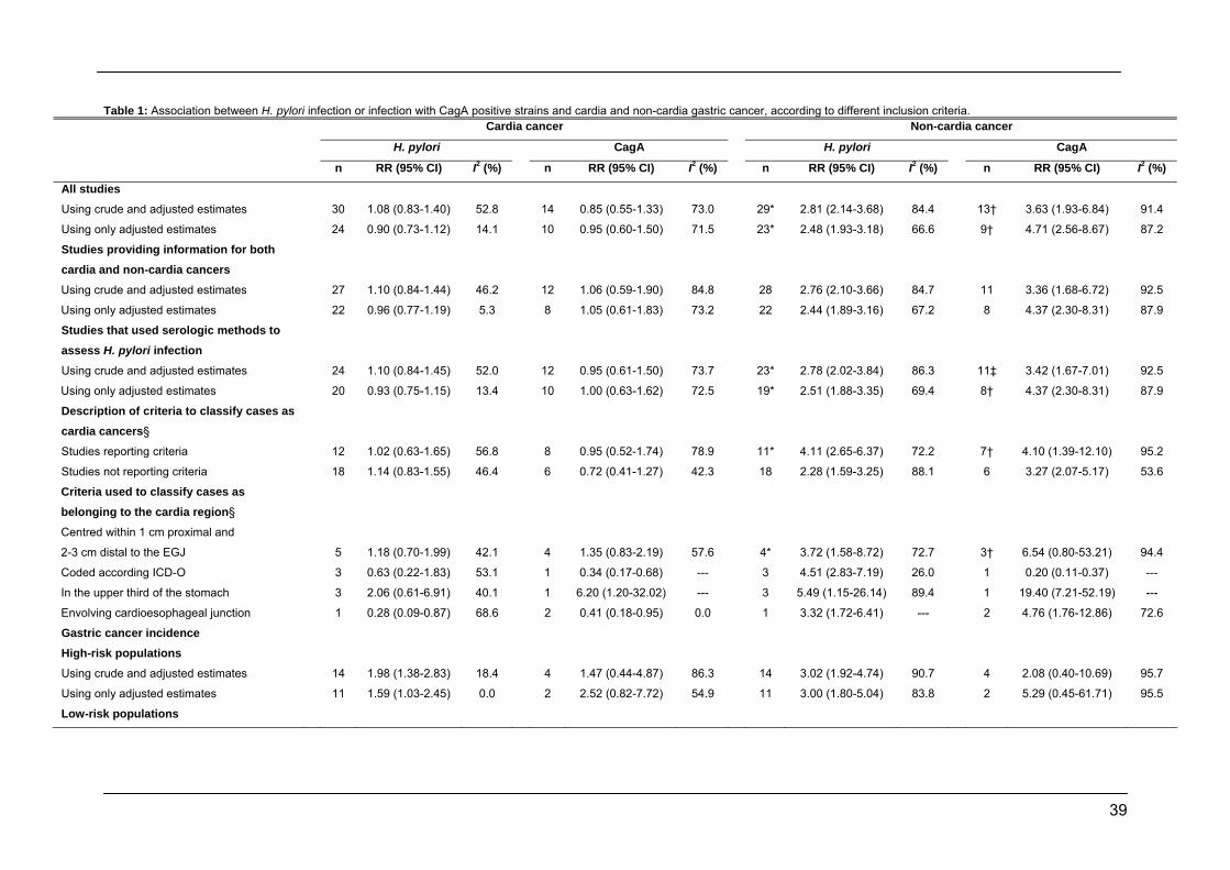
39
Table 1: Association between H. pylori infection or infection with CagA positive strains and cardia and non-cardia gastric cancer, according to different inclusion criteria. Cardia cancer Non-cardia cancer
H. pylori CagA H. pylori CagA
n RR (95% CI) I2 (%) n RR (95% CI) I2 (%) n RR (95% CI) I2 (%) n RR (95% CI) I2 (%)
All studies
Using crude and adjusted estimates 30 1.08 (0.83-1.40) 52.8 14 0.85 (0.55-1.33) 73.0 29* 2.81 (2.14-3.68) 84.4 13† 3.63 (1.93-6.84) 91.4
Using only adjusted estimates 24 0.90 (0.73-1.12) 14.1 10 0.95 (0.60-1.50) 71.5 23* 2.48 (1.93-3.18) 66.6 9† 4.71 (2.56-8.67) 87.2
Studies providing information for both cardia and non-cardia cancers
Using crude and adjusted estimates 27 1.10 (0.84-1.44) 46.2 12 1.06 (0.59-1.90) 84.8 28 2.76 (2.10-3.66) 84.7 11 3.36 (1.68-6.72) 92.5
Using only adjusted estimates 22 0.96 (0.77-1.19) 5.3 8 1.05 (0.61-1.83) 73.2 22 2.44 (1.89-3.16) 67.2 8 4.37 (2.30-8.31) 87.9
Studies that used serologic methods to assess H. pylori infection
Using crude and adjusted estimates 24 1.10 (0.84-1.45) 52.0 12 0.95 (0.61-1.50) 73.7 23* 2.78 (2.02-3.84) 86.3 11‡ 3.42 (1.67-7.01) 92.5
Using only adjusted estimates 20 0.93 (0.75-1.15) 13.4 10 1.00 (0.63-1.62) 72.5 19* 2.51 (1.88-3.35) 69.4 8† 4.37 (2.30-8.31) 87.9
Description of criteria to classify cases as cardia cancers§
Studies reporting criteria 12 1.02 (0.63-1.65) 56.8 8 0.95 (0.52-1.74) 78.9 11* 4.11 (2.65-6.37) 72.2 7† 4.10 (1.39-12.10) 95.2
Studies not reporting criteria 18 1.14 (0.83-1.55) 46.4 6 0.72 (0.41-1.27) 42.3 18 2.28 (1.59-3.25) 88.1 6 3.27 (2.07-5.17) 53.6
Criteria used to classify cases as belonging to the cardia region§
Centred within 1 cm proximal and
2-3 cm distal to the EGJ 5 1.18 (0.70-1.99) 42.1 4 1.35 (0.83-2.19) 57.6 4* 3.72 (1.58-8.72) 72.7 3† 6.54 (0.80-53.21) 94.4
Coded according ICD-O 3 0.63 (0.22-1.83) 53.1 1 0.34 (0.17-0.68) --- 3 4.51 (2.83-7.19) 26.0 1 0.20 (0.11-0.37) ---
In the upper third of the stomach 3 2.06 (0.61-6.91) 40.1 1 6.20 (1.20-32.02) --- 3 5.49 (1.15-26.14) 89.4 1 19.40 (7.21-52.19) ---
Envolving cardioesophageal junction 1 0.28 (0.09-0.87) 68.6 2 0.41 (0.18-0.95) 0.0 1 3.32 (1.72-6.41) --- 2 4.76 (1.76-12.86) 72.6
Gastric cancer incidence�
High-risk populations
Using crude and adjusted estimates 14 1.98 (1.38-2.83) 18.4 4 1.47 (0.44-4.87) 86.3 14 3.02 (1.92-4.74) 90.7 4 2.08 (0.40-10.69) 95.7
Using only adjusted estimates 11 1.59 (1.03-2.45) 0.0 2 2.52 (0.82-7.72) 54.9 11 3.00 (1.80-5.04) 83.8 2 5.29 (0.45-61.71) 95.5
Low-risk populations

40
Table 1 (continuation): Association between H. pylori infection or infection with CagA positive strains and cardia and non-cardia gastric cancer, according to different inclusion criteria. Cardia cancer Non-cardia cancer
H. pylori CagA H. pylori CagA
n RR (95% CI) I2 (%) n RR (95% CI) I2 (%) n RR (95% CI) I2 (%) n RR (95% CI) I2 (%)
Using crude and adjusted estimates 16 0.78 (0.63-0.97) 11.6 10 0.74 (0.51-1.08) 37.3 15* 2.56 (1.99-3.29) 46.6 9† 4.59 (2.79-7.57) 76.4
Using only adjusted estimates 14 0.80 (0.63-1.02) 18.9 8 0.74 (0.52-1.06) 30.5 13* 2.50 (1.88-3.32) 52.7 7† 4.67 (2.46-8.86) 81.0
High-risk populations§
Cohort/nested case-control 4 1.22 (0.60-2.48) 0.0 1 1.75 (1.33-2.31) --- 4 2.61 (0.98-6.95) 88.6 1 1.58 (1.13-2.22) ---
Case-control (PB) 1 1.16 (0.47-2.86) --- 2 0.70 (0.10-4.93) 63.1 1 1.01 (0.58-1.77) --- 2 0.81 (0.05-14.14) 94.0
Case-control (HB) 9 2.54 (1.75-3.54) 0.0 1 6.20 (1.20-32.02) --- 9 3.60 (2.23-5.79) 84.8 1 19.40 (7.21-52.19) ---
Low-risk populations§
Cohort/nested case-control 6 0.62 (0.37-1.06) 39.2 3 0.72 (0.39-1.36) 0.0 6 3.47 (2.13-5.66) 55.8 3 5.82 (3.44-9.84) 0.0
Case-control (PB) 6 0.86 (0.66-1.11) 2.0 6 0.67 (0.40-1.14) 56.5 5* 2.42 (1.46-4.00) 0.0 5† 3.70 (1.79-7.64) 85.2
Case-control (HB) 4 0.90 (0.54-1.48) 0.0 1 1.48 (0.47-4.65) --- 4 2.42 (1.46-4.01) 43.4 1 6.80 (3.11-14.86) ---
* One study did not report RR estimates for the association between H. pylori infection and non-cardia cancer; † One study did not report RR estimates for the association between CagA infection
and non-cardia cancer; ‡ Two studies did not report RR estimates for the association between CagA infection and non-cardia cancer; § The analyses included studies that provided only crude
estimates and the studies that provided adjusted estimates; � The epidemiologic knowledge of the gastric cancer incidence was used to define high- and low-risk populations (the gastric cancer
incidence (age-standardized, world reference population) was above 41/100.000 inhabitants in regions classified as high-risk and was below 19/100.000 inhabitants in the low-risk ones); EGJ –
Oesophagogastric junction; ICD-O – International Classification of Diseases for Oncology; PB – Population-based; HB – Hospital-based.
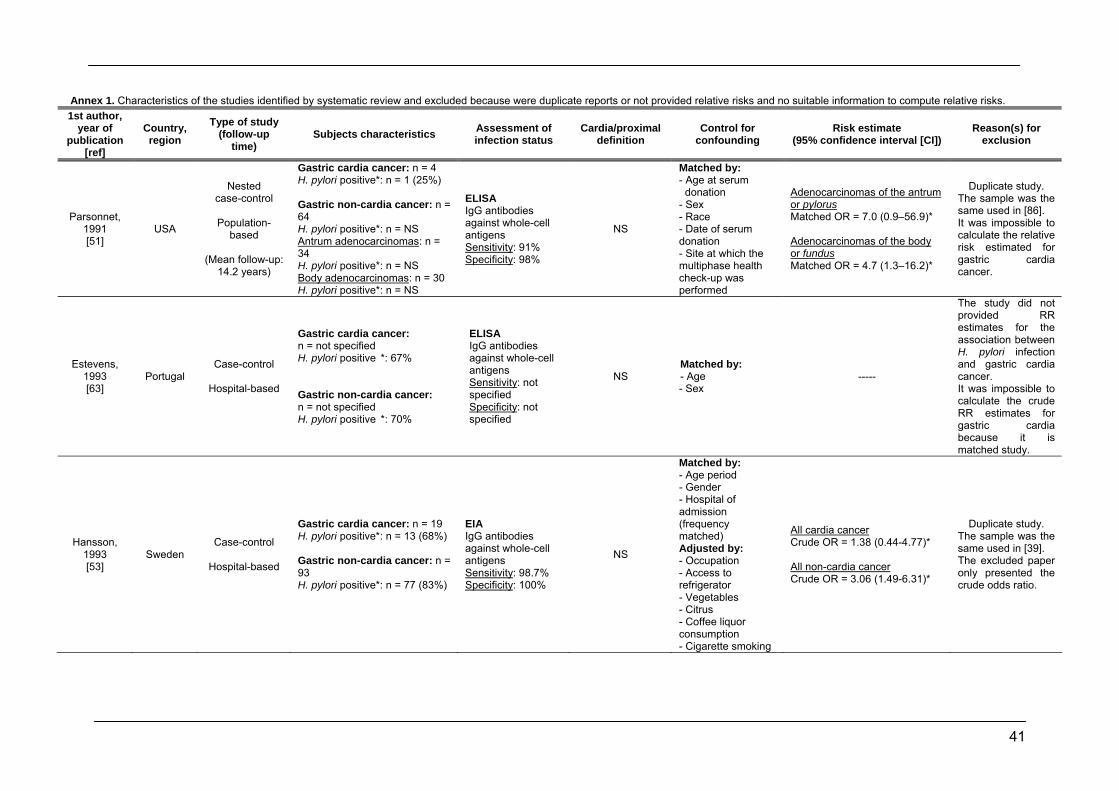
41
Annex 1. Characteristics of the studies identified by systematic review and excluded because were duplicate reports or not provided relative risks and no suitable information to compute relative risks. 1st author,
year of publication
[ref]
Country, region
Type of study (follow-up
time) Subjects characteristics Assessment of
infection status Cardia/proximal
definition Control for
confounding Risk estimate
(95% confidence interval [CI]) Reason(s) for
exclusion
Parsonnet, 1991 [51]
USA
Nested case-control
Population-
based
(Mean follow-up: 14.2 years)
Gastric cardia cancer: n = 4 H. pylori positive*: n = 1 (25%) Gastric non-cardia cancer: n = 64 H. pylori positive*: n = NS Antrum adenocarcinomas: n = 34 H. pylori positive*: n = NS Body adenocarcinomas: n = 30 H. pylori positive*: n = NS
ELISA IgG antibodies against whole-cell antigens Sensitivity: 91% Specificity: 98%
NS
Matched by: - Age at serum donation - Sex - Race - Date of serum donation - Site at which the multiphase health check-up was performed
Adenocarcinomas of the antrum or pylorus Matched OR = 7.0 (0.9–56.9)* Adenocarcinomas of the body or fundus Matched OR = 4.7 (1.3–16.2)*
Duplicate study. The sample was the same used in [86]. It was impossible to calculate the relative risk estimated for gastric cardia cancer.
Estevens, 1993 [63]
Portugal Case-control
Hospital-based
Gastric cardia cancer: n = not specified H. pylori positive *: 67% Gastric non-cardia cancer: n = not specified H. pylori positive *: 70%
ELISA IgG antibodies against whole-cell antigens Sensitivity: not specified Specificity: not specified
NS Matched by: - Age - Sex
-----
The study did not provided RR estimates for the association between H. pylori infection and gastric cardia cancer. It was impossible to calculate the crude RR estimates for gastric cardia because it is matched study.
Hansson, 1993 [53]
Sweden Case-control
Hospital-based
Gastric cardia cancer: n = 19 H. pylori positive*: n = 13 (68%) Gastric non-cardia cancer: n = 93 H. pylori positive*: n = 77 (83%)
EIA IgG antibodies against whole-cell antigens Sensitivity: 98.7% Specificity: 100%
NS
Matched by: - Age period - Gender - Hospital of admission (frequency matched) Adjusted by: - Occupation - Access to refrigerator - Vegetables - Citrus - Coffee liquor consumption - Cigarette smoking
All cardia cancer Crude OR = 1.38 (0.44-4.77)* All non-cardia cancer Crude OR = 3.06 (1.49-6.31)*
Duplicate study. The sample was the same used in [39]. The excluded paper only presented the crude odds ratio.
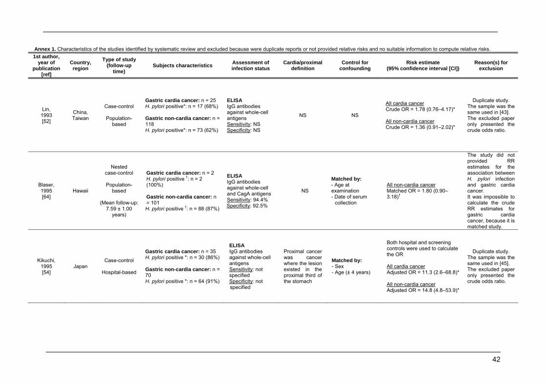
42
Annex 1. Characteristics of the studies identified by systematic review and excluded because were duplicate reports or not provided relative risks and no suitable information to compute relative risks. 1st author,
year of publication
[ref]
Country, region
Type of study (follow-up
time) Subjects characteristics Assessment of
infection status Cardia/proximal
definition Control for
confounding Risk estimate
(95% confidence interval [CI]) Reason(s) for
exclusion
Lin, 1993 [52]
China, Taiwan
Case-control
Population-based
Gastric cardia cancer: n = 25 H. pylori positive*: n = 17 (68%) Gastric non-cardia cancer: n = 118 H. pylori positive*: n = 73 (62%)
ELISA IgG antibodies against whole-cell antigens Sensitivity: NS Specificity: NS
NS NS
All cardia cancer Crude OR = 1.78 (0.76–4.17)* All non-cardia cancer Crude OR = 1.36 (0.91–2.02)*
Duplicate study. The sample was the same used in [43]. The excluded paper only presented the crude odds ratio.
Blaser, 1995 [64]
Hawaii
Nested case-control
Population-
based
(Mean follow-up: 7.59 ± 1.00
years)
Gastric cardia cancer: n = 2 H. pylori positive †: n = 2 (100%) Gastric non-cardia cancer: n = 101 H. pylori positive †: n = 88 (87%)
ELISA IgG antibodies against whole-cell and CagA antigens Sensitivity: 94.4% Specificity: 92.5%
NS
Matched by: - Age at examination - Date of serum
collection
All non-cardia cancer Matched OR = 1.80 (0.90–3.18)†
The study did not provided RR estimates for the association between H. pylori infection and gastric cardia cancer. It was impossible to calculate the crude RR estimates for gastric cardia cancer, because it is matched study.
Kikuchi, 1995 [54]
Japan Case-control
Hospital-based
Gastric cardia cancer: n = 35 H. pylori positive *: n = 30 (86%) Gastric non-cardia cancer: n = 70 H. pylori positive *: n = 64 (91%)
ELISA IgG antibodies against whole-cell antigens Sensitivity: not specified Specificity: not specified
Proximal cancer was cancer where the lesion existed in the proximal third of the stomach
Matched by: - Sex - Age (± 4 years)
Both hospital and screening controls were used to calculate the OR All cardia cancer Adjusted OR = 11.3 (2.6–68.8)* All non-cardia cancer Adjusted OR = 14.8 (4.8–53.9)*
Duplicate study. The sample was the same used in [45]. The excluded paper only presented the crude odds ratio.
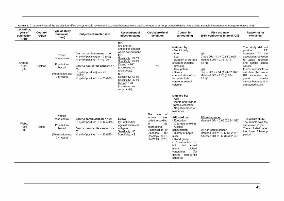
43
Annex 1. Characteristics of the studies identified by systematic review and excluded because were duplicate reports or not provided relative risks and no suitable information to compute relative risks. 1st author,
year of publication
[ref]
Country, region
Type of study (follow-up
time) Subjects characteristics Assessment of
infection status Cardia/proximal
definition Control for
confounding Risk estimate
(95% confidence interval [CI]) Reason(s) for
exclusion
Aromaa, 1996 [65]
Finland
Nested case-control
Population-
based
(Mean follow-up: 9.5 years)
Gastric cardia cancer: n = 9 H. pylori positive§: n = 0 (0%) H. pylori positive*: n = 0 (0%) Gastric non-cardia cancer: n = 75 H. pylori positive§: n = 75 (100%) H. pylori positive*: n = 73 (97%)
EIA IgG and IgA antibodies against whole-cell antigens IgG Sensitivity: 93.7% Specificity: 93.9% Cut-off: ≥ 700 (expressed as reciprocals) IgA Sensitivity: 73.1% Specificity: 95.1% Cut-off: ≥ 70 (expressed as reciprocals)
NS
Matched by: - Municipality - Age - Sex - Duration of storage of serum samples - Smoking - Occupation - Serum concentration of: α-tocopherol, ß-carotene, retinol, selenium
IgA Crude OR = 1.37 (0.64-2.95)§ Matched OR = 2.76 (1.11–6.87)§ IgG Crude OR = 7.54 (1.74-32.78)* Matched OR = 1.75 (0.80–3.81)*
The study did not provided RR estimates for the association between H. pylori infection and gastric cardia cancer. It was impossible to calculate the crude RR estimates for gastric cardia cancer because it is a matched study.
Webb, 1996 [55]
China
Nested case-control
Population-
based
(Mean follow-up: 2.4 years)
Gastric cardia cancer: n = 27 H. pylori positive*: n = 12 (44%) Gastric non-cardia cancer: n = 52 H. pylori positive*: n = 30 (58%)
ELISA IgG antibodies against whole-cell antigens Sensitivity: NS Specificity: NS
The site of tumour was coded according to the International Classification of Diseases for Oncology (ICD-O) (WHO, 1976)
Matched by: - Age
- Month and year of sample collection - Neighbourhood of residence Adjusted by: - Education - Cigarette smoking - Alcohol consumption - History of peptic ulcer - Blood group - Consumption of: bok choi, cured meats, pickled vegetables (for gastric non-cardia cancers)
All cardia cancer Matched OR = 0.65 (0.25-1.59)* All non-cardia cancer Matched OR =1.10 (0.57-2.14)* Adjusted OR =1.17 (0.54-2.54)*
Duplicate study. The sample was the same used in [84]. The excluded paper has lower follow-up period.
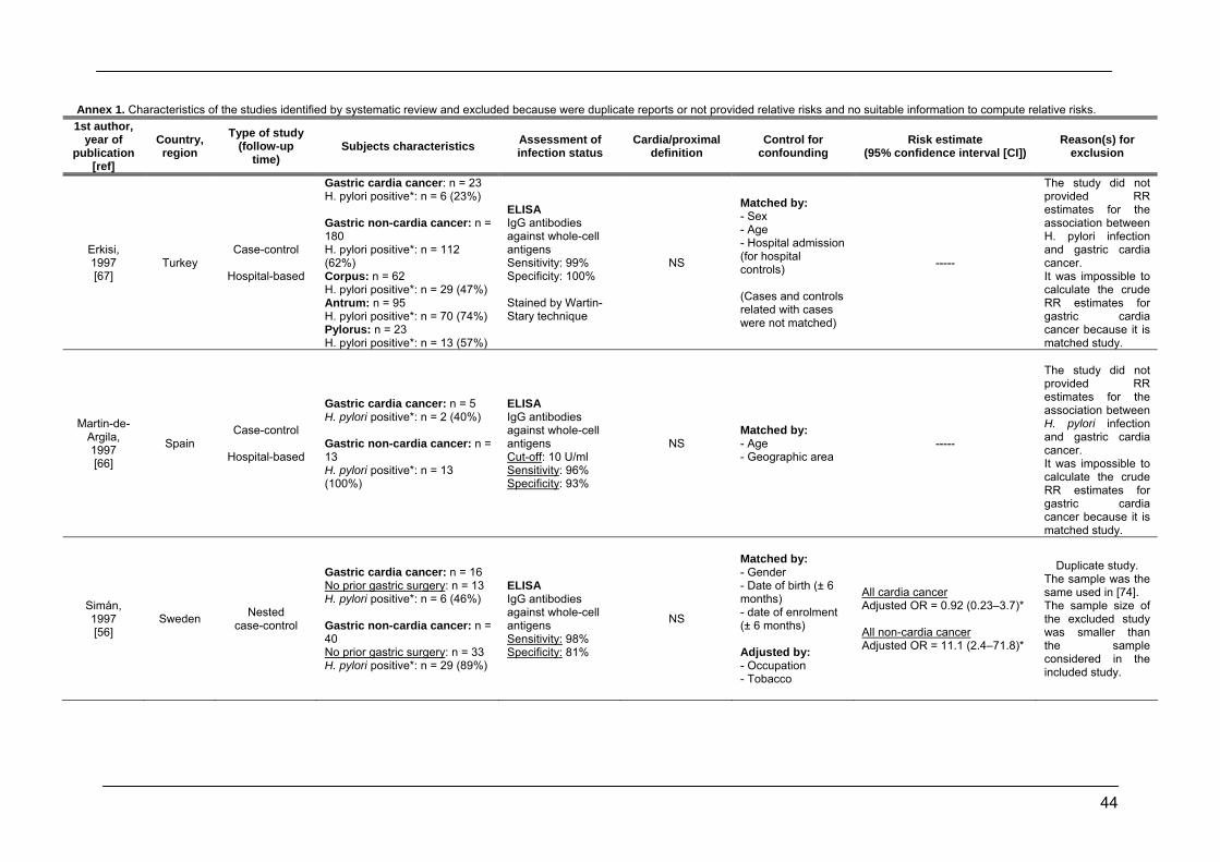
44
Annex 1. Characteristics of the studies identified by systematic review and excluded because were duplicate reports or not provided relative risks and no suitable information to compute relative risks. 1st author,
year of publication
[ref]
Country, region
Type of study (follow-up
time) Subjects characteristics Assessment of
infection status Cardia/proximal
definition Control for
confounding Risk estimate
(95% confidence interval [CI]) Reason(s) for
exclusion
Erkisi, 1997 [67]
Turkey Case-control
Hospital-based
Gastric cardia cancer: n = 23 H. pylori positive*: n = 6 (23%) Gastric non-cardia cancer: n = 180 H. pylori positive*: n = 112 (62%) Corpus: n = 62 H. pylori positive*: n = 29 (47%) Antrum: n = 95 H. pylori positive*: n = 70 (74%) Pylorus: n = 23 H. pylori positive*: n = 13 (57%)
ELISA IgG antibodies against whole-cell antigens Sensitivity: 99% Specificity: 100% Stained by Wartin-Stary technique
NS
Matched by: - Sex - Age - Hospital admission (for hospital controls) (Cases and controls related with cases were not matched)
-----
The study did not provided RR estimates for the association between H. pylori infection and gastric cardia cancer. It was impossible to calculate the crude RR estimates for gastric cardia cancer because it is matched study.
Martin-de-Argila, 1997 [66]
Spain Case-control
Hospital-based
Gastric cardia cancer: n = 5 H. pylori positive*: n = 2 (40%) Gastric non-cardia cancer: n = 13 H. pylori positive*: n = 13 (100%)
ELISA IgG antibodies against whole-cell antigens Cut-off: 10 U/ml Sensitivity: 96% Specificity: 93%
NS Matched by: - Age - Geographic area
-----
The study did not provided RR estimates for the association between H. pylori infection and gastric cardia cancer. It was impossible to calculate the crude RR estimates for gastric cardia cancer because it is matched study.
Simán, 1997 [56]
Sweden Nested case-control
Gastric cardia cancer: n = 16 No prior gastric surgery: n = 13 H. pylori positive*: n = 6 (46%) Gastric non-cardia cancer: n = 40 No prior gastric surgery: n = 33 H. pylori positive*: n = 29 (89%)
ELISA IgG antibodies against whole-cell antigens Sensitivity: 98% Specificity: 81%
NS
Matched by: - Gender - Date of birth (±/6 months) - date of enrolment (±/6 months) Adjusted by: - Occupation - Tobacco
All cardia cancer Adjusted OR = 0.92 (0.23–3.7)*
All non-cardia cancer Adjusted OR = 11.1 (2.4–71.8)*
Duplicate study. The sample was the same used in [74]. The sample size of the excluded study was smaller than the sample considered in the included study.
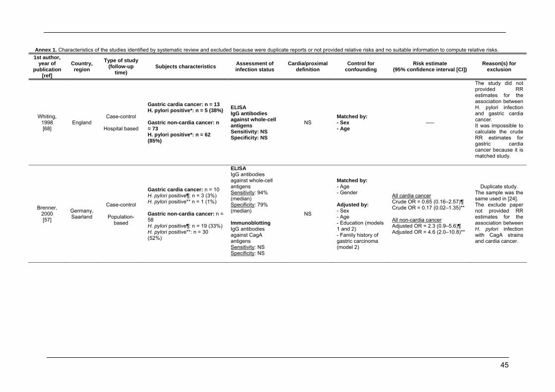
45
Annex 1. Characteristics of the studies identified by systematic review and excluded because were duplicate reports or not provided relative risks and no suitable information to compute relative risks. 1st author,
year of publication
[ref]
Country, region
Type of study (follow-up
time) Subjects characteristics Assessment of
infection status Cardia/proximal
definition Control for
confounding Risk estimate
(95% confidence interval [CI]) Reason(s) for
exclusion
Whiting, 1998 [68]
England Case-control
Hospital based
Gastric cardia cancer: n = 13 H. pylori positive*: n = 5 (38%) Gastric non-cardia cancer: n = 73 H. pylori positive*: n = 62 (85%)
ELISA IgG antibodies against whole-cell antigens Sensitivity: NS Specificity: NS
NS Matched by: - Sex - Age
-----
The study did not provided RR estimates for the association between H. pylori infection and gastric cardia cancer. It was impossible to calculate the crude RR estimates for gastric cardia cancer because it is matched study.
Brenner, 2000 [57]
Germany, Saarland
Case-control
Population-based
Gastric cardia cancer: n = 10 H. pylori positive¶: n = 3 (3%) H. pylori positive** n = 1 (1%) Gastric non-cardia cancer: n = 58 H. pylori positive¶: n = 19 (33%) H. pylori positive**: n = 30 (52%)
ELISA IgG antibodies against whole-cell antigens Sensitivity: 94% (median) Specificity: 79% (median) Immunoblotting IgG antibodies against CagA antigens Sensitivity: NS Specificity: NS
NS
Matched by: - Age - Gender Adjusted by: - Sex - Age - Education (models 1 and 2) - Family history of gastric carcinoma (model 2)
All cardia cancer Crude OR = 0.65 (0.16–2.57)¶ Crude OR = 0.17 (0.02–1.35)** All non-cardia cancer Adjusted OR = 2.3 (0.9–5.6)¶ Adjusted OR = 4.6 (2.0–10.8)**
Duplicate study. The sample was the same used in [24]. The exclude paper not provided RR estimates for the association between H. pylori infection with CagA strains and cardia cancer.
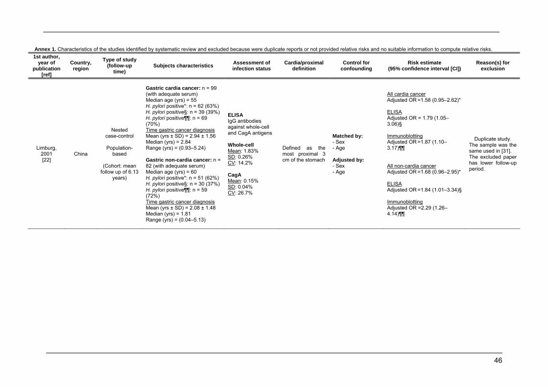
46
Annex 1. Characteristics of the studies identified by systematic review and excluded because were duplicate reports or not provided relative risks and no suitable information to compute relative risks. 1st author,
year of publication
[ref]
Country, region
Type of study (follow-up
time) Subjects characteristics Assessment of
infection status Cardia/proximal
definition Control for
confounding Risk estimate
(95% confidence interval [CI]) Reason(s) for
exclusion
Limburg, 2001 [22]
China
Nested case-control
Population-
based
(Cohort: mean follow up of 6.13
years)
Gastric cardia cancer: n = 99 (with adequate serum) Median age (yrs) = 55 H. pylori positive*: n = 62 (63%) H. pylori positive§: n = 39 (39%) H. pylori positive¶¶: n = 69 (70%) Time gastric cancer diagnosis Mean (yrs ± SD) = 2.94 ± 1.56 Median (yrs) = 2.84 Range (yrs) = (0.93–5.24) Gastric non-cardia cancer: n = 82 (with adequate serum) Median age (yrs) = 60 H. pylori positive*: n = 51 (62%) H. pylori positive§: n = 30 (37%) H. pylori positive¶¶: n = 59 (72%) Time gastric cancer diagnosis Mean (yrs ± SD) = 2.08 ± 1.48 Median (yrs) = 1.81 Range (yrs) = (0.04–5.13)
ELISA IgG antibodies against whole-cell and CagA antigens Whole-cell Mean: 1.83% SD: 0.26% CV: 14.2% CagA Mean: 0.15% SD: 0.04% CV: 26.7%
Defined as the most proximal 3 cm of the stomach
Matched by: - Sex - Age Adjusted by: - Sex - Age
All cardia cancer Adjusted OR =1.58 (0.95–2.62)* ELISA Adjusted OR = 1.79 (1.05–3.06)§ Immunoblotting Adjusted OR =1.87 (1.10–3.17)¶¶ All non-cardia cancer Adjusted OR =1.68 (0.96–2.95)* ELISA Adjusted OR =1.84 (1.01–3.34)§ Immunoblotting Adjusted OR =2.29 (1.26–4.14)¶¶
Duplicate study. The sample was the same used in [31]. The excluded paper has lower follow-up period.
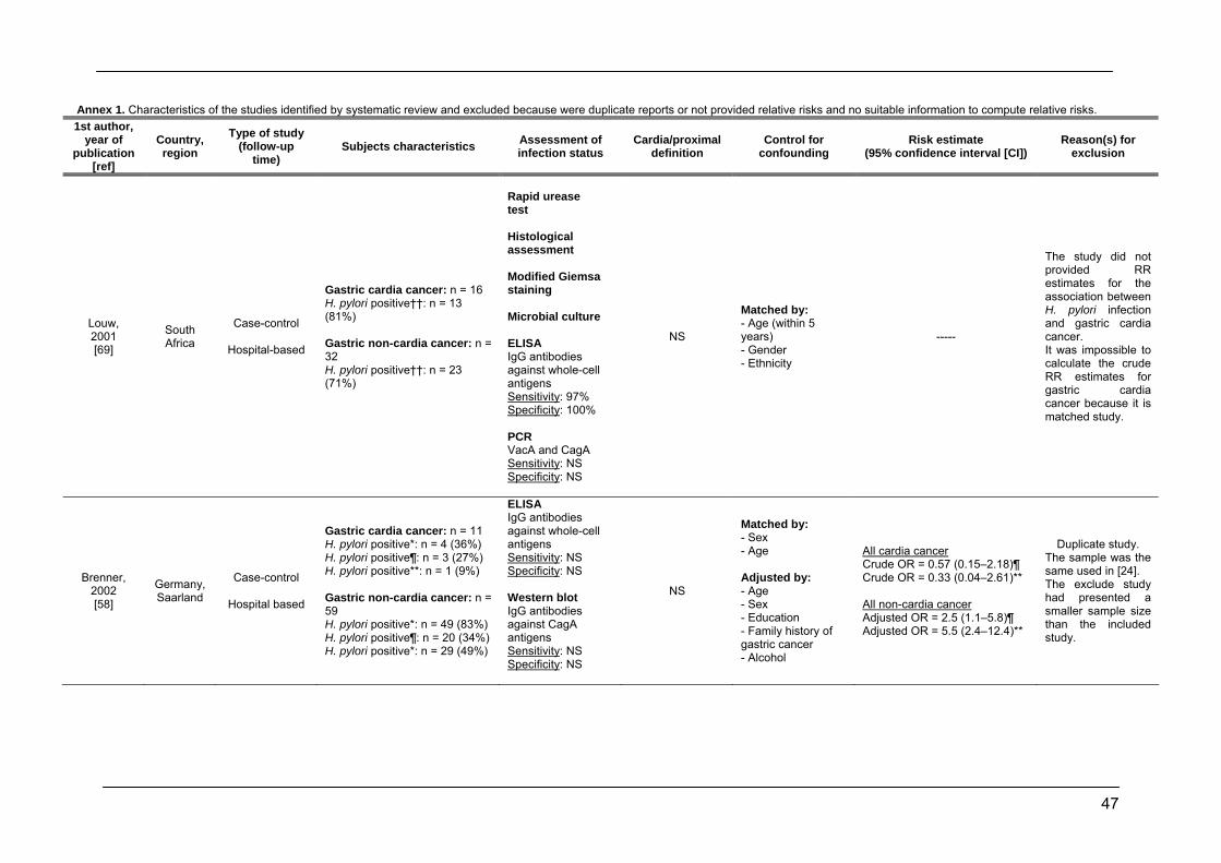
47
Annex 1. Characteristics of the studies identified by systematic review and excluded because were duplicate reports or not provided relative risks and no suitable information to compute relative risks. 1st author,
year of publication
[ref]
Country, region
Type of study (follow-up
time) Subjects characteristics Assessment of
infection status Cardia/proximal
definition Control for
confounding Risk estimate
(95% confidence interval [CI]) Reason(s) for
exclusion
Louw, 2001 [69]
South Africa
Case-control
Hospital-based
Gastric cardia cancer: n = 16 H. pylori positive††: n = 13 (81%) Gastric non-cardia cancer: n = 32 H. pylori positive††: n = 23 (71%)
Rapid urease test Histological assessment Modified Giemsa staining Microbial culture ELISA IgG antibodies against whole-cell antigens Sensitivity: 97% Specificity: 100% PCR VacA and CagA Sensitivity: NS Specificity: NS
NS
Matched by: - Age (within 5 years) - Gender - Ethnicity
-----
The study did not provided RR estimates for the association between H. pylori infection and gastric cardia cancer. It was impossible to calculate the crude RR estimates for gastric cardia cancer because it is matched study.
Brenner, 2002 [58]
Germany, Saarland
Case-control
Hospital based
Gastric cardia cancer: n = 11 H. pylori positive*: n = 4 (36%) H. pylori positive¶: n = 3 (27%) H. pylori positive**: n = 1 (9%) Gastric non-cardia cancer: n = 59 H. pylori positive*: n = 49 (83%) H. pylori positive¶: n = 20 (34%) H. pylori positive*: n = 29 (49%)
ELISA IgG antibodies against whole-cell antigens Sensitivity: NS Specificity: NS Western blot IgG antibodies against CagA antigens Sensitivity: NS Specificity: NS
NS
Matched by: - Sex - Age Adjusted by: - Age - Sex - Education - Family history of gastric cancer - Alcohol
All cardia cancer Crude OR = 0.57 (0.15–2.18)¶ Crude OR = 0.33 (0.04–2.61)** All non-cardia cancer Adjusted OR = 2.5 (1.1–5.8)¶ Adjusted OR = 5.5 (2.4–12.4)**
Duplicate study. The sample was the same used in [24]. The exclude study had presented a smaller sample size than the included study.
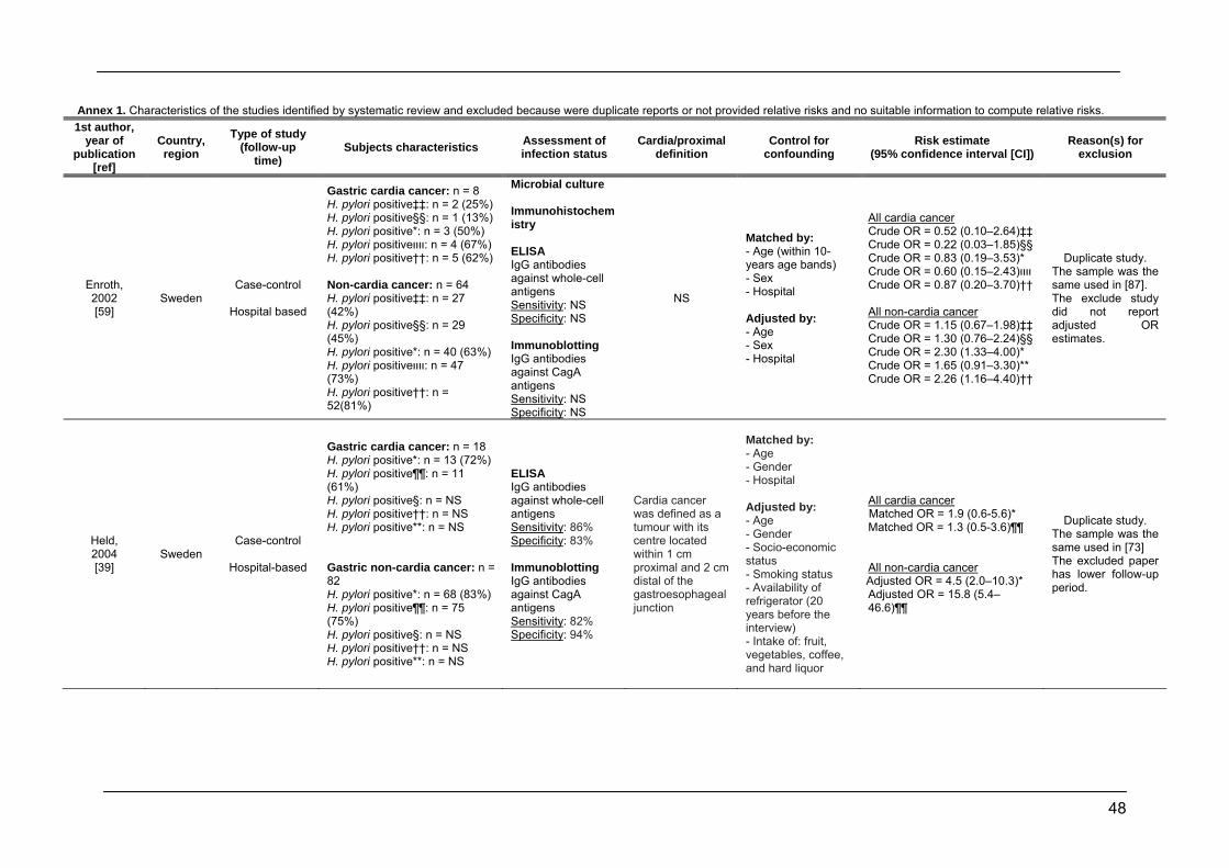
48
Annex 1. Characteristics of the studies identified by systematic review and excluded because were duplicate reports or not provided relative risks and no suitable information to compute relative risks. 1st author,
year of publication
[ref]
Country, region
Type of study (follow-up
time) Subjects characteristics Assessment of
infection status Cardia/proximal
definition Control for
confounding Risk estimate
(95% confidence interval [CI]) Reason(s) for
exclusion
Enroth, 2002 [59]
Sweden Case-control
Hospital based
Gastric cardia cancer: n = 8 H. pylori positive‡‡: n = 2 (25%) H. pylori positive§§: n = 1 (13%) H. pylori positive*: n = 3 (50%) H. pylori positiveװװ: n = 4 (67%) H. pylori positive††: n = 5 (62%) Non-cardia cancer: n = 64 H. pylori positive‡‡: n = 27 (42%) H. pylori positive§§: n = 29 (45%) H. pylori positive*: n = 40 (63%) H. pylori positiveװװ: n = 47 (73%) H. pylori positive††: n = 52(81%)
Microbial culture Immunohistochemistry ELISA IgG antibodies against whole-cell antigens Sensitivity: NS Specificity: NS Immunoblotting IgG antibodies against CagA antigens Sensitivity: NS Specificity: NS
NS
Matched by: - Age (within 10-years age bands) - Sex - Hospital Adjusted by: - Age - Sex - Hospital
All cardia cancer Crude OR = 0.52 (0.10–2.64)‡‡ Crude OR = 0.22 (0.03–1.85)§§ Crude OR = 0.83 (0.19–3.53)* Crude OR = 0.60 (0.15–2.43)װװ Crude OR = 0.87 (0.20–3.70)†† All non-cardia cancer Crude OR = 1.15 (0.67–1.98)‡‡ Crude OR = 1.30 (0.76–2.24)§§ Crude OR = 2.30 (1.33–4.00)* Crude OR = 1.65 (0.91–3.30)** Crude OR = 2.26 (1.16–4.40)††
Duplicate study. The sample was the same used in [87]. The exclude study did not report adjusted OR estimates.
Held, 2004 [39]
Sweden Case-control
Hospital-based
Gastric cardia cancer: n = 18 H. pylori positive*: n = 13 (72%) H. pylori positive¶¶: n = 11 (61%) H. pylori positive§: n = NS H. pylori positive††: n = NS H. pylori positive**: n = NS Gastric non-cardia cancer: n = 82 H. pylori positive*: n = 68 (83%) H. pylori positive¶¶: n = 75 (75%) H. pylori positive§: n = NS H. pylori positive††: n = NS H. pylori positive**: n = NS
ELISA IgG antibodies against whole-cell antigens Sensitivity: 86% Specificity: 83% Immunoblotting IgG antibodies against CagA antigens Sensitivity: 82% Specificity: 94%
Cardia cancer was defined as a tumour with its centre located within 1 cm proximal and 2 cm distal of the gastroesophageal junction
Matched by: - Age - Gender - Hospital Adjusted by: - Age - Gender - Socio-economic status - Smoking status - Availability of refrigerator (20 years before the interview) - Intake of: fruit, vegetables, coffee, and hard liquor
All cardia cancer Matched OR = 1.9 (0.6-5.6)* Matched OR = 1.3 (0.5-3.6)¶¶ All non-cardia cancer Adjusted OR = 4.5 (2.0–10.3)* Adjusted OR = 15.8 (5.4–46.6)¶¶
Duplicate study. The sample was the same used in [73] The excluded paper has lower follow-up period.
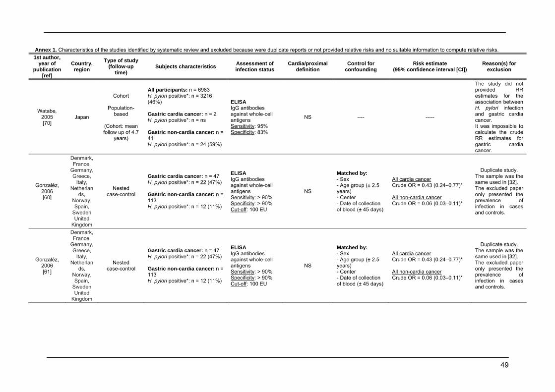
49
Annex 1. Characteristics of the studies identified by systematic review and excluded because were duplicate reports or not provided relative risks and no suitable information to compute relative risks. 1st author,
year of publication
[ref]
Country, region
Type of study (follow-up
time) Subjects characteristics Assessment of
infection status Cardia/proximal
definition Control for
confounding Risk estimate
(95% confidence interval [CI]) Reason(s) for
exclusion
Watabe, 2005 [70]
Japan
Cohort
Population-based
(Cohort: mean follow up of 4.7
years)
All participants: n = 6983 H. pylori positive*: n = 3216 (46%) Gastric cardia cancer: n = 2 H. pylori positive*: n = ns Gastric non-cardia cancer: n = 41 H. pylori positive*: n = 24 (59%)
ELISA IgG antibodies against whole-cell antigens Sensitivity: 95% Specificity: 83%
NS ---- -----
The study did not provided RR estimates for the association between H. pylori infection and gastric cardia cancer. It was impossible to calculate the crude RR estimates for gastric cardia cancer.
Gonzaléz, 2006 [60]
Denmark, France,
Germany, Greece,
Italy, Netherlan
ds, Norway, Spain,
Sweden United
Kingdom
Nested case-control
Gastric cardia cancer: n = 47 H. pylori positive*: n = 22 (47%) Gastric non-cardia cancer: n = 113 H. pylori positive*: n = 12 (11%)
ELISA IgG antibodies against whole-cell antigens Sensitivity: > 90% Specificity: > 90% Cut-off: 100 EU
NS
Matched by: - Sex - Age group (± 2.5 years) - Center - Date of collection of blood (± 45 days)
All cardia cancer Crude OR = 0.43 (0.24–0.77)* All non-cardia cancer Crude OR = 0.06 (0.03–0.11)*
Duplicate study. The sample was the same used in [32]. The excluded paper only presented the prevalence of infection in cases and controls.
Gonzaléz, 2006 [61]
Denmark, France,
Germany, Greece,
Italy, Netherlan
ds, Norway, Spain,
Sweden United
Kingdom
Nested case-control
Gastric cardia cancer: n = 47 H. pylori positive*: n = 22 (47%) Gastric non-cardia cancer: n = 113 H. pylori positive*: n = 12 (11%)
ELISA IgG antibodies against whole-cell antigens Sensitivity: > 90% Specificity: > 90% Cut-off: 100 EU
NS
Matched by: - Sex - Age group (± 2.5 years) - Center - Date of collection of blood (± 45 days)
All cardia cancer Crude OR = 0.43 (0.24–0.77)* All non-cardia cancer Crude OR = 0.06 (0.03–0.11)*
Duplicate study. The sample was the same used in [32]. The excluded paper only presented the prevalence of infection in cases and controls.
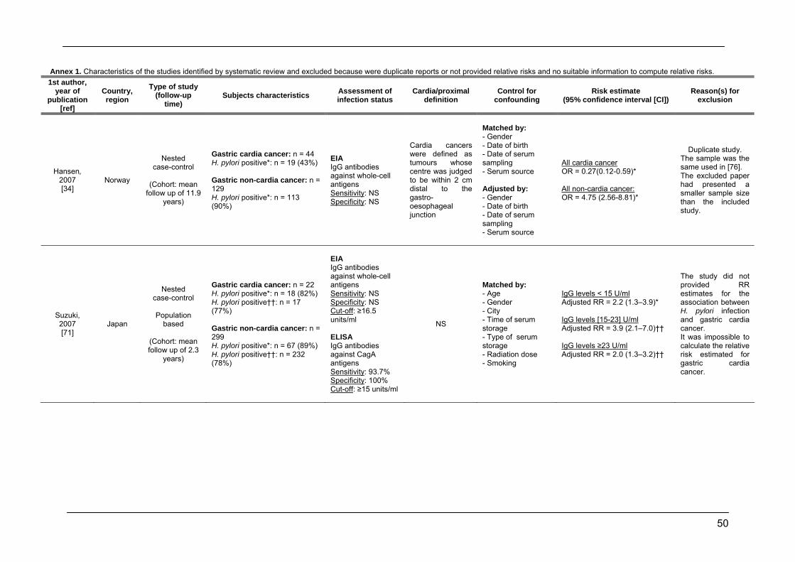
50
Annex 1. Characteristics of the studies identified by systematic review and excluded because were duplicate reports or not provided relative risks and no suitable information to compute relative risks. 1st author,
year of publication
[ref]
Country, region
Type of study (follow-up
time) Subjects characteristics Assessment of
infection status Cardia/proximal
definition Control for
confounding Risk estimate
(95% confidence interval [CI]) Reason(s) for
exclusion
Hansen, 2007 [34]
Norway
Nested case-control
(Cohort: mean
follow up of 11.9 years)
Gastric cardia cancer: n = 44 H. pylori positive*: n = 19 (43%) Gastric non-cardia cancer: n = 129 H. pylori positive*: n = 113 (90%)
EIA IgG antibodies against whole-cell antigens Sensitivity: NS Specificity: NS
Cardia cancers were defined as tumours whose centre was judged to be within 2 cm distal to the gastro-oesophageal junction
Matched by: - Gender - Date of birth - Date of serum sampling - Serum source Adjusted by: - Gender - Date of birth - Date of serum sampling - Serum source
All cardia cancer OR = 0.27(0.12-0.59)* All non-cardia cancer: OR = 4.75 (2.56-8.81)*
Duplicate study. The sample was the same used in [76]. The excluded paper had presented a smaller sample size than the included study.
Suzuki, 2007 [71]
Japan
Nested case-control
Population
based
(Cohort: mean follow up of 2.3
years)
Gastric cardia cancer: n = 22 H. pylori positive*: n = 18 (82%) H. pylori positive††: n = 17 (77%) Gastric non-cardia cancer: n = 299 H. pylori positive*: n = 67 (89%) H. pylori positive††: n = 232 (78%)
EIA IgG antibodies against whole-cell antigens Sensitivity: NS Specificity: NS Cut-off: ≥16.5 units/ml ELISA IgG antibodies against CagA antigens Sensitivity: 93.7% Specificity: 100% Cut-off: ≥15 units/ml
NS
Matched by: - Age - Gender - City - Time of serum storage - Type of serum storage - Radiation dose - Smoking
IgG levels < 15 U/ml Adjusted RR = 2.2 (1.3–3.9)* IgG levels [15-23] U/ml Adjusted RR = 3.9 (2.1–7.0)†† IgG levels ≥23 U/ml Adjusted RR = 2.0 (1.3–3.2)††
The study did not provided RR estimates for the association between H. pylori infection and gastric cardia cancer. It was impossible to calculate the relative risk estimated for gastric cardia cancer.
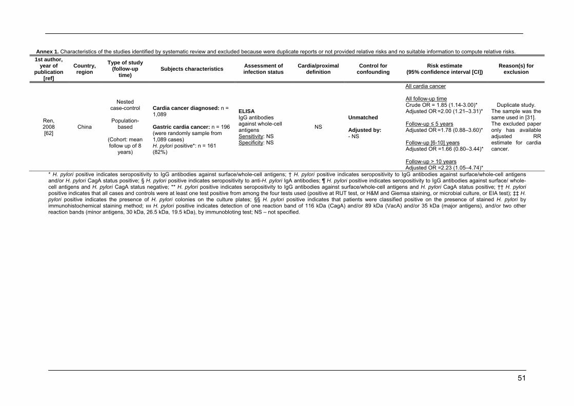
51
Annex 1. Characteristics of the studies identified by systematic review and excluded because were duplicate reports or not provided relative risks and no suitable information to compute relative risks. 1st author,
year of publication
[ref]
Country, region
Type of study (follow-up
time) Subjects characteristics Assessment of
infection status Cardia/proximal
definition Control for
confounding Risk estimate
(95% confidence interval [CI]) Reason(s) for
exclusion
Ren, 2008 [62]
China
Nested case-control
Population-
based
(Cohort: mean follow up of 8
years)
Cardia cancer diagnosed: n = 1,089 Gastric cardia cancer: n = 196 (were randomly sample from 1,089 cases) H. pylori positive*: n = 161 (82%)
ELISA IgG antibodies against whole-cell antigens Sensitivity: NS Specificity: NS
NS
Unmatched Adjusted by: - NS
All cardia cancer All follow-up time Crude OR = 1.85 (1.14-3.00)* Adjusted OR =2.00 (1.21–3.31)* Follow-up ≤ 5 years Adjusted OR =1.78 (0.88–3.60)* Follow-up [6-10] years Adjusted OR =1.66 (0.80–3.44)* Follow-up > 10 years Adjusted OR =2.23 (1.05–4.74)*
Duplicate study. The sample was the same used in [31]. The excluded paper only has available adjusted RR estimate for cardia cancer.
* H. pylori positive indicates seropositivity to IgG antibodies against surface/whole-cell antigens; † H. pylori positive indicates seropositivity to IgG antibodies against surface/whole-cell antigens and/or H. pylori CagA status positive; § H. pylori positive indicates seropositivity to anti-H. pylori IgA antibodies; ¶ H. pylori positive indicates seropositivity to IgG antibodies against surface/ whole-cell antigens and H. pylori CagA status negative; ** H. pylori positive indicates seropositivity to IgG antibodies against surface/whole-cell antigens and H. pylori CagA status positive; †† H. pylori positive indicates that all cases and controls were at least one test positive from among the four tests used (positive at RUT test, or H&M and Giemsa staining, or microbial culture, or EIA test); ‡‡ H. pylori positive indicates the presence of H. pylori colonies on the culture plates; §§ H. pylori positive indicates that patients were classified positive on the presence of stained H. pylori by immunohistochemical staining method; װװ H. pylori positive indicates detection of one reaction band of 116 kDa (CagA) and/or 89 kDa (VacA) and/or 35 kDa (major antigens), and/or two other reaction bands (minor antigens, 30 kDa, 26.5 kDa, 19.5 kDa), by immunobloting test; NS – not specified.
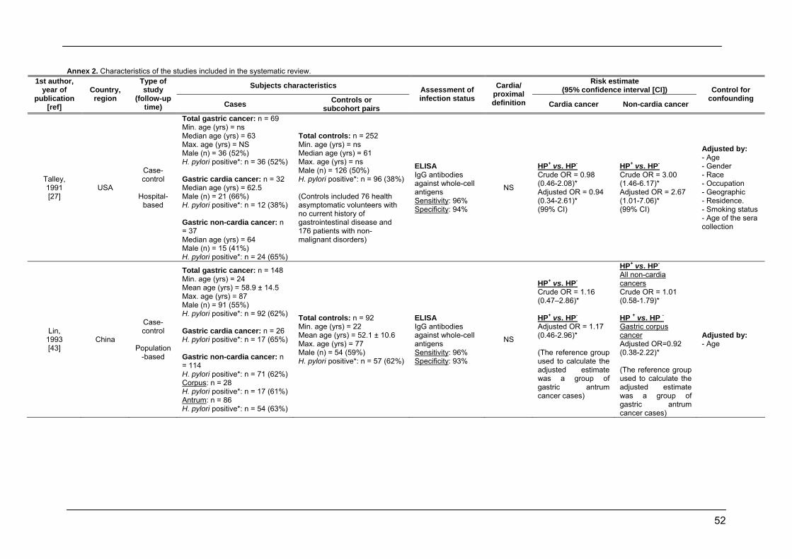
52
Annex 2. Characteristics of the studies included in the systematic review.
Subjects characteristics Risk estimate (95% confidence interval [CI])
1st author, year of
publication [ref]
Country, region
Type of study
(follow-up time) Cases Controls or
subcohort pairs
Assessment of infection status
Cardia/ proximal definition Cardia cancer Non-cardia cancer
Control for confounding
Talley, 1991 [27]
USA
Case-control
Hospital-
based
Total gastric cancer: n = 69 Min. age (yrs) = ns Median age (yrs) = 63 Max. age (yrs) = NS Male (n) = 36 (52%) H. pylori positive*: n = 36 (52%) Gastric cardia cancer: n = 32 Median age (yrs) = 62.5 Male (n) = 21 (66%) H. pylori positive*: n = 12 (38%) Gastric non-cardia cancer: n = 37 Median age (yrs) = 64 Male (n) = 15 (41%) H. pylori positive*: n = 24 (65%)
Total controls: n = 252 Min. age (yrs) = ns Median age (yrs) = 61 Max. age (yrs) = ns Male (n) = 126 (50%) H. pylori positive*: n = 96 (38%) (Controls included 76 health asymptomatic volunteers with no current history of gastrointestinal disease and 176 patients with non-malignant disorders)
ELISA IgG antibodies against whole-cell antigens Sensitivity: 96% Specificity: 94%
NS
HP+ vs. HP- Crude OR = 0.98 (0.46-2.08)* Adjusted OR = 0.94 (0.34-2.61)* (99% CI)
HP+ vs. HP- Crude OR = 3.00 (1.46-6.17)* Adjusted OR = 2.67 (1.01-7.06)* (99% CI)
Adjusted by: - Age - Gender - Race - Occupation - Geographic - Residence. - Smoking status - Age of the sera collection
Lin, 1993 [43]
China
Case-control
Population
-based
Total gastric cancer: n = 148 Min. age (yrs) = 24 Mean age (yrs) = 58.9 ± 14.5 Max. age (yrs) = 87 Male (n) = 91 (55%) H. pylori positive*: n = 92 (62%) Gastric cardia cancer: n = 26 H. pylori positive*: n = 17 (65%) Gastric non-cardia cancer: n = 114 H. pylori positive*: n = 71 (62%) Corpus: n = 28 H. pylori positive*: n = 17 (61%) Antrum: n = 86 H. pylori positive*: n = 54 (63%)
Total controls: n = 92 Min. age (yrs) = 22 Mean age (yrs) = 52.1 ± 10.6 Max. age (yrs) = 77 Male (n) = 54 (59%) H. pylori positive*: n = 57 (62%)
ELISA IgG antibodies against whole-cell antigens Sensitivity: 96% Specificity: 93%
NS
HP+ vs. HP- Crude OR = 1.16 (0.47–2.86)* HP+ vs. HP- Adjusted OR = 1.17 (0.46-2.96)* (The reference group used to calculate the adjusted estimate was a group of gastric antrum cancer cases)
HP+ vs. HP- All non-cardia cancers Crude OR = 1.01 (0.58-1.79)* HP + vs. HP - Gastric corpus cancer Adjusted OR=0.92 (0.38-2.22)* (The reference group used to calculate the adjusted estimate was a group of gastric antrum cancer cases)
Adjusted by: - Age
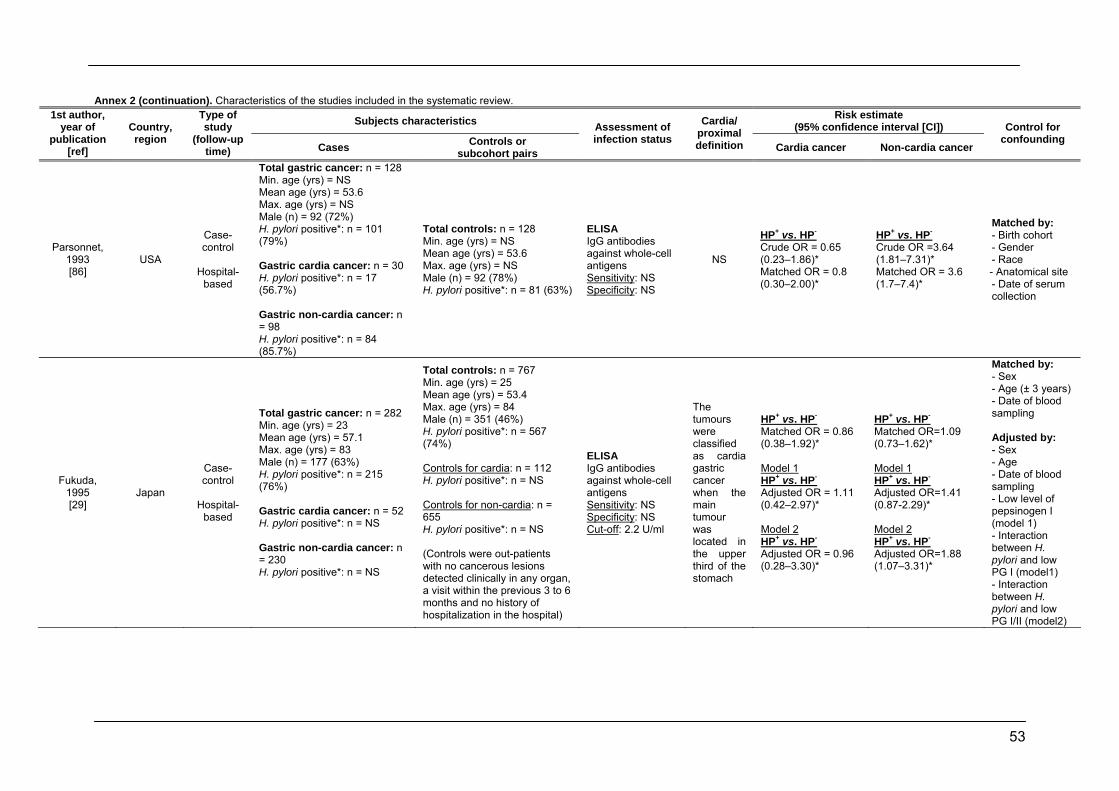
53
Annex 2 (continuation). Characteristics of the studies included in the systematic review.
Subjects characteristics Risk estimate (95% confidence interval [CI])
1st author, year of
publication [ref]
Country, region
Type of study
(follow-up time) Cases Controls or
subcohort pairs
Assessment of infection status
Cardia/ proximal definition Cardia cancer Non-cardia cancer
Control for confounding
Parsonnet, 1993 [86]
USA
Case-control
Hospital-
based
Total gastric cancer: n = 128 Min. age (yrs) = NS Mean age (yrs) = 53.6 Max. age (yrs) = NS Male (n) = 92 (72%) H. pylori positive*: n = 101 (79%) Gastric cardia cancer: n = 30 H. pylori positive*: n = 17 (56.7%) Gastric non-cardia cancer: n = 98 H. pylori positive*: n = 84 (85.7%)
Total controls: n = 128 Min. age (yrs) = NS Mean age (yrs) = 53.6 Max. age (yrs) = NS Male (n) = 92 (78%) H. pylori positive*: n = 81 (63%)
ELISA IgG antibodies against whole-cell antigens Sensitivity: NS Specificity: NS
NS
HP+ vs. HP- Crude OR = 0.65 (0.23–1.86)* Matched OR = 0.8 (0.30–2.00)*
HP+ vs. HP- Crude OR =3.64 (1.81–7.31)* Matched OR = 3.6 (1.7–7.4)*
Matched by: - Birth cohort - Gender - Race - Anatomical site - Date of serum collection
Fukuda, 1995 [29]
Japan
Case-control
Hospital-
based
Total gastric cancer: n = 282 Min. age (yrs) = 23 Mean age (yrs) = 57.1 Max. age (yrs) = 83 Male (n) = 177 (63%) H. pylori positive*: n = 215 (76%) Gastric cardia cancer: n = 52 H. pylori positive*: n = NS Gastric non-cardia cancer: n = 230 H. pylori positive*: n = NS
Total controls: n = 767 Min. age (yrs) = 25 Mean age (yrs) = 53.4 Max. age (yrs) = 84 Male (n) = 351 (46%) H. pylori positive*: n = 567 (74%) Controls for cardia: n = 112 H. pylori positive*: n = NS Controls for non-cardia: n = 655 H. pylori positive*: n = NS (Controls were out-patients with no cancerous lesions detected clinically in any organ, a visit within the previous 3 to 6 months and no history of hospitalization in the hospital)
ELISA IgG antibodies against whole-cell antigens Sensitivity: NS Specificity: NS Cut-off: 2.2 U/ml
The tumours were classified as cardia gastric cancer when the main tumour was located in the upper third of the stomach
HP+ vs. HP- Matched OR = 0.86 (0.38–1.92)* Model 1 HP+ vs. HP- Adjusted OR = 1.11 (0.42–2.97)* Model 2 HP+ vs. HP- Adjusted OR = 0.96 (0.28–3.30)*
HP+ vs. HP- Matched OR=1.09 (0.73–1.62)* Model 1 HP+ vs. HP- Adjusted OR=1.41 (0.87-2.29)* Model 2 HP+ vs. HP- Adjusted OR=1.88 (1.07–3.31)*
Matched by: - Sex - Age (± 3 years) - Date of blood sampling Adjusted by: - Sex - Age - Date of blood sampling - Low level of pepsinogen I (model 1) - Interaction between H. pylori and low PG I (model1) - Interaction between H. pylori and low PG I/II (model2)
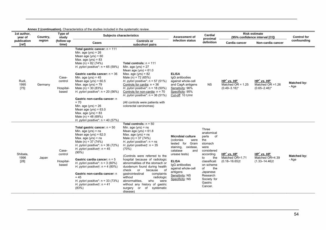
54
Annex 2 (continuation). Characteristics of the studies included in the systematic review.
Subjects characteristics Risk estimate (95% confidence interval [CI])
1st author, year of
publication [ref]
Country, region
Type of study
(follow-up time) Cases Controls or
subcohort pairs
Assessment of infection status
Cardia/ proximal definition Cardia cancer Non-cardia cancer
Control for confounding
Rudi, 1995 [75]
Germany
Case-control
Hospital-
based
Total gastric cancer: n = 111 Min. age (yrs) = 26 Mean age (yrs) = 60 Max. age (yrs) = 83 Male (n) = 82 (74%) H. pylori positive*: n = 65 (59%) Gastric cardia cancer: n = 36 Min. age (yrs) = 45 Mean age (yrs) = 60.5 Max. age (yrs) = 79 Male (n) = 30 (83%) H. pylori positive*: n = 20 (56%) Gastric non-cardia cancer: n = 70 Min. age (yrs) = 26 Mean age (yrs) = 63.0 Max. age (yrs) = 83 Male (n) = 48 (69%) H. pylori positive*: n = 40 (57%)
Total controls: n = 111 Min. age (yrs) = 27 Mean age (yrs) = 61.0 Max. age (yrs) = 82 Male (n) = 72 (65%) H. pylori positive*: n = 57 (51%) Controls for cardia: n = 36 H. pylori positive*: n = 18 (50%) Controls for non-cardia: n = 70 H. pylori positive*: n = 36 (51%) (All controls were patients with colorectal carcinomas)
ELISA IgG antibodies against whole-cell and CagA antigens Sensitivity: 96% Specificity: 95% Cut-off: 10 U/ml
NS HP+ vs. HP- Matched OR = 1.25 (0.49–3.16)*
HP+ vs. HP- Matched OR =1.26 (0.65–2.46)*
Matched by: - Age
Shibata, 1996 [28]
Japan
Case-control
Hospital-
based
Total gastric cancer: n = 50 Min. age (yrs) = ns Mean age (yrs) = 62.0 Max. age (yrs) = ns Male (n) = 37 (74%) H. pylori positive*: n = 36 (72%) H. pylori positive†: n = 45 (90%) Gastric cardia cancer: n = 5 H. pylori positive*: n = 3 (60%) H. pylori positive†: n = 4 (80%) Gastric non-cardia cancer: n = 45 H. pylori positive*: n = 33 (73%) H. pylori positive†: n = 41 (83%)
Total controls: n = 50 Min. age (yrs) = ns Mean age (yrs) = 61.8 Max. age (yrs) = ns Male (n) = 37 (74%) H. pylori positive*: n = ns H. pylori positive†: n = 35 (70%) (Controls were referred to the hospital because of radiologic abnormalities of the stomach or duodenum found during health check or because of gastrointestinal complaints without radiologic abnormalities, who were without any history of gastric surgery or of systematic disease)
Microbial culture (colonies were tested for Gram staining, oxidase, catalase and urease tests) ELISA IgG antibodies against whole-cell antigens Sensitivity: NS Specificity: NS
Three anatomical parts of the stomach were considered according to the classification scheme of the Japanese Research Society for Gastric Cancer.
HP+ vs. HP- Matched OR=1.71 (0.18–16.65)†
HP+ vs. HP- Matched OR=4.39 (1.33–14.46)†
Matched by: - Age
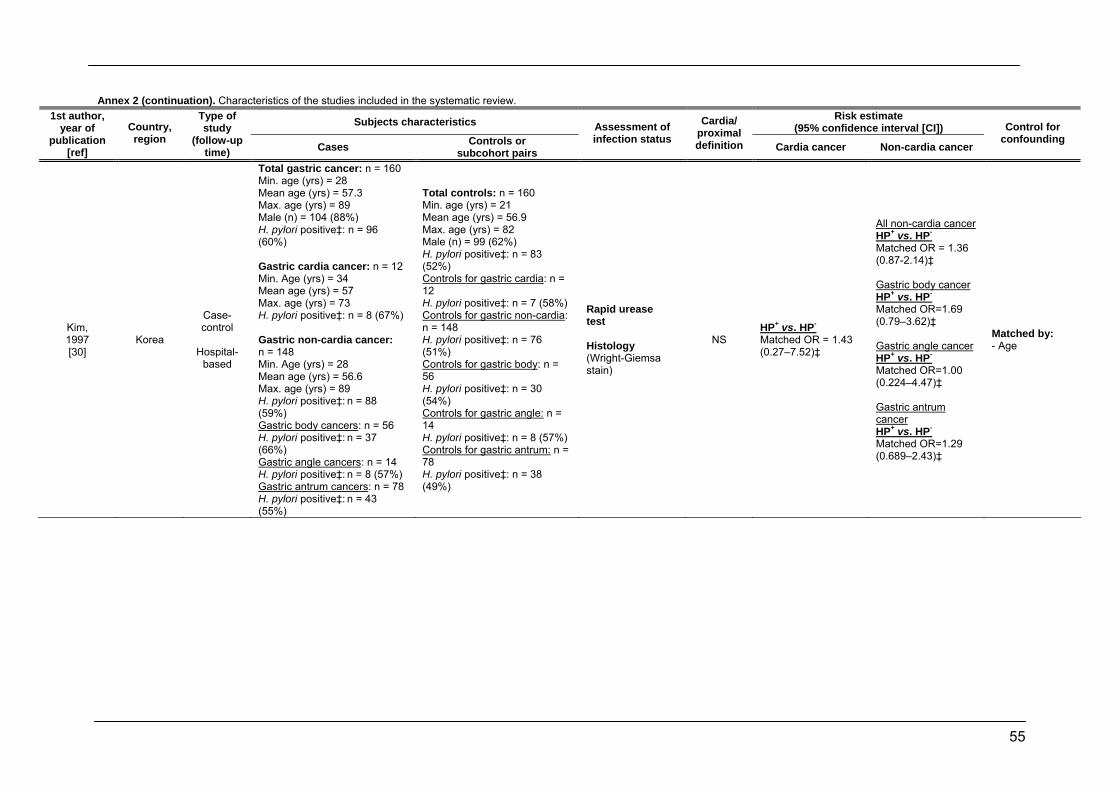
55
Annex 2 (continuation). Characteristics of the studies included in the systematic review.
Subjects characteristics Risk estimate (95% confidence interval [CI])
1st author, year of
publication [ref]
Country, region
Type of study
(follow-up time) Cases Controls or
subcohort pairs
Assessment of infection status
Cardia/ proximal definition Cardia cancer Non-cardia cancer
Control for confounding
Kim, 1997 [30]
Korea
Case-control
Hospital-
based
Total gastric cancer: n = 160 Min. age (yrs) = 28 Mean age (yrs) = 57.3 Max. age (yrs) = 89 Male (n) = 104 (88%) H. pylori positive‡: n = 96 (60%) Gastric cardia cancer: n = 12 Min. Age (yrs) = 34 Mean age (yrs) = 57 Max. age (yrs) = 73 H. pylori positive‡: n = 8 (67%) Gastric non-cardia cancer: n = 148 Min. Age (yrs) = 28 Mean age (yrs) = 56.6 Max. age (yrs) = 89 H. pylori positive‡: n = 88 (59%) Gastric body cancers: n = 56 H. pylori positive‡: n = 37 (66%) Gastric angle cancers: n = 14 H. pylori positive‡: n = 8 (57%) Gastric antrum cancers: n = 78 H. pylori positive‡: n = 43 (55%)
Total controls: n = 160 Min. age (yrs) = 21 Mean age (yrs) = 56.9 Max. age (yrs) = 82 Male (n) = 99 (62%) H. pylori positive‡: n = 83 (52%) Controls for gastric cardia: n = 12 H. pylori positive‡: n = 7 (58%) Controls for gastric non-cardia: n = 148 H. pylori positive‡: n = 76 (51%) Controls for gastric body: n = 56 H. pylori positive‡: n = 30 (54%) Controls for gastric angle: n = 14 H. pylori positive‡: n = 8 (57%) Controls for gastric antrum: n = 78 H. pylori positive‡: n = 38 (49%)
Rapid urease test Histology (Wright-Giemsa stain)
NS HP+ vs. HP- Matched OR = 1.43 (0.27–7.52)‡
All non-cardia cancer HP+ vs. HP- Matched OR = 1.36 (0.87-2.14)‡ Gastric body cancer HP+ vs. HP- Matched OR=1.69 (0.79–3.62)‡ Gastric angle cancer HP+ vs. HP- Matched OR=1.00 (0.224–4.47)‡ Gastric antrum cancer HP+ vs. HP- Matched OR=1.29 (0.689–2.43)‡
Matched by: - Age
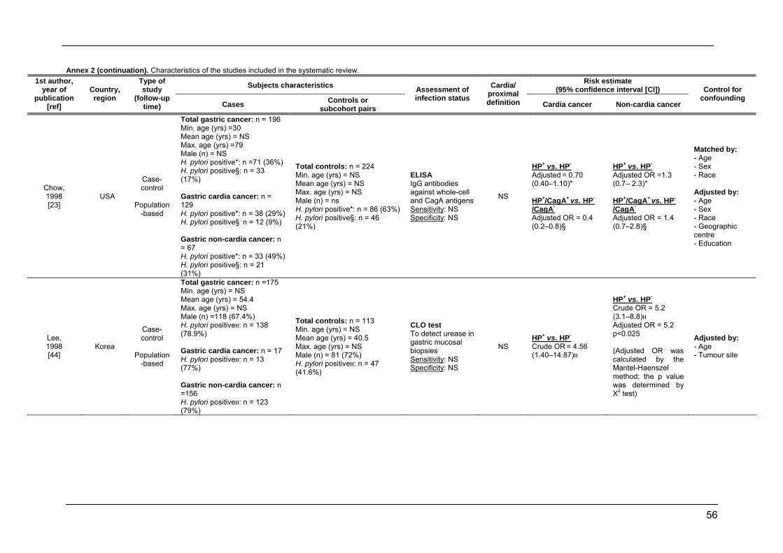
56
Annex 2 (continuation). Characteristics of the studies included in the systematic review.
Subjects characteristics Risk estimate (95% confidence interval [CI])
1st author, year of
publication [ref]
Country, region
Type of study
(follow-up time) Cases Controls or
subcohort pairs
Assessment of infection status
Cardia/ proximal definition Cardia cancer Non-cardia cancer
Control for confounding
Chow, 1998 [23]
USA
Case-control
Population
-based
Total gastric cancer: n = 196 Min. age (yrs) =30 Mean age (yrs) = NS Max. age (yrs) =79 Male (n) = NS H. pylori positive*: n =71 (36%) H. pylori positive§: n = 33 (17%) Gastric cardia cancer: n = 129 H. pylori positive*: n = 38 (29%) H. pylori positive§: n = 12 (9%) Gastric non-cardia cancer: n = 67 H. pylori positive*: n = 33 (49%) H. pylori positive§: n = 21 (31%)
Total controls: n = 224 Min. age (yrs) = NS Mean age (yrs) = NS Max. age (yrs) = NS Male (n) = ns H. pylori positive*: n = 86 (63%) H. pylori positive§: n = 46 (21%)
ELISA IgG antibodies against whole-cell and CagA antigens Sensitivity: NS Specificity: NS
NS
HP+ vs. HP- Adjusted = 0.70 (0.40–1.10)* HP+/CagA+ vs. HP-
/CagA- Adjusted OR = 0.4 (0.2–0.8)§
HP+ vs. HP- Adjusted OR =1.3 (0.7– 2.3)* HP+/CagA+ vs. HP-
/CagA- Adjusted OR = 1.4 (0.7–2.8)§
Matched by: - Age - Sex - Race Adjusted by: - Age - Sex - Race - Geographic centre - Education
Lee, 1998 [44]
Korea
Case-control
Population
-based
Total gastric cancer: n =175 Min. age (yrs) = NS Mean age (yrs) = 54.4 Max. age (yrs) = NS Male (n) =118 (67.4%) H. pylori positiveװ: n = 138 (78.9%) Gastric cardia cancer: n = 17 H. pylori positiveװ: n = 13 (77%) Gastric non-cardia cancer: n =156 H. pylori positiveװ: n = 123 (79%)
Total controls: n = 113 Min. age (yrs) = NS Mean age (yrs) = 40.5 Max. age (yrs) = NS Male (n) = 81 (72%) H. pylori positiveװ: n = 47 (41.6%)
CLO test To detect urease in gastric mucosal biopsies Sensitivity: NS Specificity: NS
NS HP+ vs. HP- Crude OR = 4.56 װ(14.87–1.40)
HP+ vs. HP- Crude OR = 5.2 װ(8.8–3.1)Adjusted OR = 5.2 p<0.025 (Adjusted OR was calculated by the Mantel-Haenszel method; the p value was determined by X2 test)
Adjusted by: - Age - Tumour site

57
Annex 2 (continuation). Characteristics of the studies included in the systematic review.
Subjects characteristics Risk estimate (95% confidence interval [CI])
1st author, year of
publication [ref]
Country, region
Type of study
(follow-up time) Cases Controls or
subcohort pairs
Assessment of infection status
Cardia/ proximal definition Cardia cancer Non-cardia cancer
Control for confounding
Queiroz, 1998 [40]
Brazil
Case-control
Hospital-
based
Total gastric cancer: n = 119 Min. age (yrs) = 32 Mean age (yrs ± SD) = NS Max. age (yrs) = 96 Male (n) = 84 (71%) H. pylori positive¶: n =119 (100%) H. pylori positive§: n =113 (95%) Gastric cardia cancer: n = 15 H. pylori positive¶: n = 15 (100%) H. pylori positive§: n =11 (73%) Gastric non-cardia cancer: n=104 H. pylori positive¶: n = 104 (100%) H. pylori positive§: n =102 (98%)
Total controls: n = 119 Min. age (yrs) = 32 Mean age (yrs ± SD) = 61.3 ± 13.6 Max. age (yrs) = 96 Male (n) = 84 (71%) H. pylori positive¶: n =119 (100%) H. pylori positive§: n =79 (66%)
ELISA IgG antibodies against whole-cell antigens Sensitivity: 95.4% Specificity: 100% Histology Urease test Carbolfuchsin stain Microbial culture PCR (for UreA and CagA genes)
NS
HP+/CagA+ vs. HP+/CagA- Matched OR = 1.3 (0.1–4.1)§
HP+/CagA+ vs. HP+/CagA- Matched OR = 25.9 (5.8–75.3)§
Matched by: - Sex - Age
Komoto, 1998 [83]
Japan
Case-control
Hospital-
based
Total gastric cancer: n = 141 Carcinomas: 105 Min. age (yrs) = NS Mean age (yrs) = 64.9 ± 1.2 Max. age (yrs) = NS Male (n) = 82 H. pylori positive*: n = 98 (93%) Gastric cardia cancer: n = 14 H. pylori positive*: n = 13 (93%) Gastric non-cardia cancer: n = 91 H. pylori positive*: n = 85 (93%)
Total controls: n = 105 Min. age (yrs) = NS Mean age (yrs ± SD) = 62.4 ± 1.1 Max. age (yrs) = NS Male (n) = 75 (71.43%) H. pylori positive*: n = 75 (71%)
ELISA IgG antibodies against whole-cell antigens Sensitivity: NS Specificity: NS Cut-off: ≥1.0 (ratio) Giemsa staining
Cardia was defined as the area in the stomach within 20 mm distance from the esophagogastric junction
HP+ vs. HP- Matched OR = 5.20 (0.65–41.68)*
HP+ vs. HP- Matched OR = 5.67 (2.25–14.44)*
Matched by: - Sex - Age
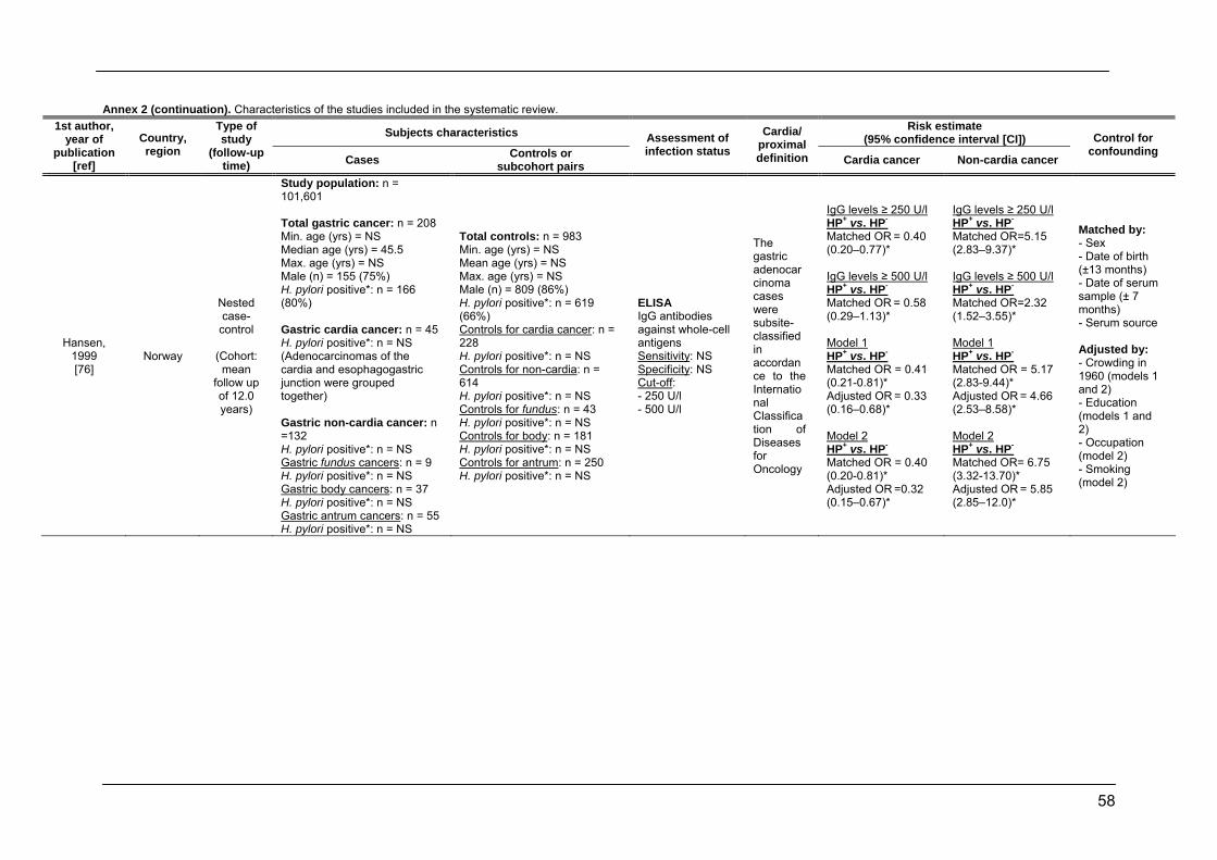
58
Annex 2 (continuation). Characteristics of the studies included in the systematic review.
Subjects characteristics Risk estimate (95% confidence interval [CI])
1st author, year of
publication [ref]
Country, region
Type of study
(follow-up time) Cases Controls or
subcohort pairs
Assessment of infection status
Cardia/ proximal definition Cardia cancer Non-cardia cancer
Control for confounding
Hansen, 1999 [76]
Norway
Nested case-
control
(Cohort: mean
follow up of 12.0 years)
Study population: n = 101,601 Total gastric cancer: n = 208 Min. age (yrs) = NS Median age (yrs) = 45.5 Max. age (yrs) = NS Male (n) = 155 (75%) H. pylori positive*: n = 166 (80%) Gastric cardia cancer: n = 45 H. pylori positive*: n = NS (Adenocarcinomas of the cardia and esophagogastric junction were grouped together) Gastric non-cardia cancer: n =132 H. pylori positive*: n = NS Gastric fundus cancers: n = 9 H. pylori positive*: n = NS Gastric body cancers: n = 37 H. pylori positive*: n = NS Gastric antrum cancers: n = 55 H. pylori positive*: n = NS
Total controls: n = 983 Min. age (yrs) = NS Mean age (yrs) = NS Max. age (yrs) = NS Male (n) = 809 (86%) H. pylori positive*: n = 619 (66%) Controls for cardia cancer: n = 228 H. pylori positive*: n = NS Controls for non-cardia: n = 614 H. pylori positive*: n = NS Controls for fundus: n = 43 H. pylori positive*: n = NS Controls for body: n = 181 H. pylori positive*: n = NS Controls for antrum: n = 250 H. pylori positive*: n = NS
ELISA IgG antibodies against whole-cell antigens Sensitivity: NS Specificity: NS Cut-off: - 250 U/l - 500 U/l
The gastric adenocarcinoma cases were subsite-classified in accordance to the International Classification of Diseases for Oncology
IgG levels ≥ 250 U/l HP+ vs. HP- Matched OR = 0.40 (0.20–0.77)*
IgG levels ≥ 500 U/l HP+ vs. HP- Matched OR = 0.58 (0.29–1.13)* Model 1 HP+ vs. HP- Matched OR = 0.41 (0.21-0.81)* Adjusted OR = 0.33 (0.16–0.68)*
Model 2 HP+ vs. HP- Matched OR = 0.40 (0.20-0.81)* Adjusted OR =0.32 (0.15–0.67)*
IgG levels ≥ 250 U/l HP+ vs. HP- Matched OR=5.15 (2.83–9.37)*
IgG levels ≥ 500 U/l HP+ vs. HP- Matched OR=2.32 (1.52–3.55)* Model 1 HP+ vs. HP- Matched OR = 5.17 (2.83-9.44)* Adjusted OR = 4.66 (2.53–8.58)*
Model 2 HP+ vs. HP- Matched OR= 6.75 (3.32-13.70)* Adjusted OR = 5.85 (2.85–12.0)*
Matched by: - Sex - Date of birth (±13 months) - Date of serum sample (± 7 months) - Serum source Adjusted by: - Crowding in 1960 (models 1 and 2) - Education (models 1 and 2) - Occupation (model 2) - Smoking (model 2)
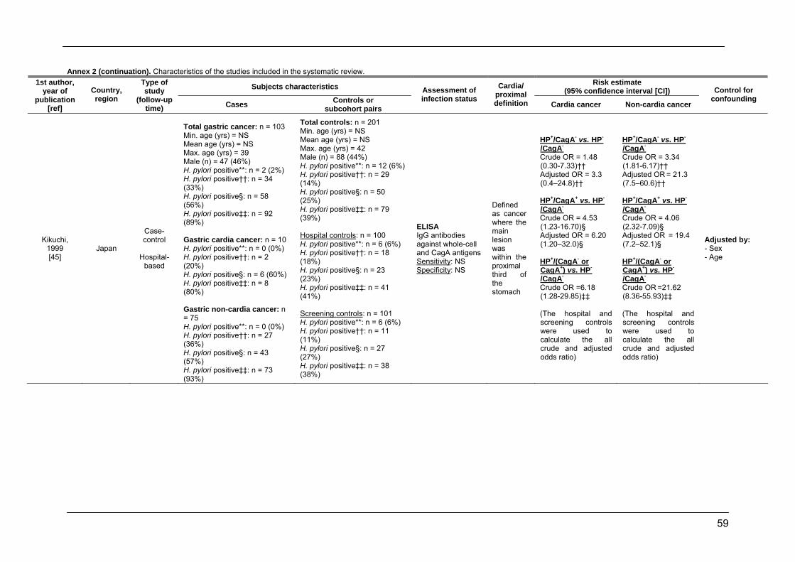
59
Annex 2 (continuation). Characteristics of the studies included in the systematic review.
Subjects characteristics Risk estimate (95% confidence interval [CI])
1st author, year of
publication [ref]
Country, region
Type of study
(follow-up time) Cases Controls or
subcohort pairs
Assessment of infection status
Cardia/ proximal definition Cardia cancer Non-cardia cancer
Control for confounding
Kikuchi, 1999 [45]
Japan
Case-control
Hospital-
based
Total gastric cancer: n = 103 Min. age (yrs) = NS Mean age (yrs) = NS Max. age (yrs) = 39 Male (n) = 47 (46%) H. pylori positive**: n = 2 (2%) H. pylori positive††: n = 34 (33%) H. pylori positive§: n = 58 (56%) H. pylori positive‡‡: n = 92 (89%) Gastric cardia cancer: n = 10 H. pylori positive**: n = 0 (0%) H. pylori positive††: n = 2 (20%) H. pylori positive§: n = 6 (60%) H. pylori positive‡‡: n = 8 (80%) Gastric non-cardia cancer: n = 75 H. pylori positive**: n = 0 (0%) H. pylori positive††: n = 27 (36%) H. pylori positive§: n = 43 (57%) H. pylori positive‡‡: n = 73 (93%)
Total controls: n = 201 Min. age (yrs) = NS Mean age (yrs) = NS Max. age (yrs) = 42 Male (n) = 88 (44%) H. pylori positive**: n = 12 (6%) H. pylori positive††: n = 29 (14%) H. pylori positive§: n = 50 (25%) H. pylori positive‡‡: n = 79 (39%) Hospital controls: n = 100 H. pylori positive**: n = 6 (6%) H. pylori positive††: n = 18 (18%) H. pylori positive§: n = 23 (23%) H. pylori positive‡‡: n = 41 (41%) Screening controls: n = 101 H. pylori positive**: n = 6 (6%) H. pylori positive††: n = 11 (11%) H. pylori positive§: n = 27 (27%) H. pylori positive‡‡: n = 38 (38%)
ELISA IgG antibodies against whole-cell and CagA antigens Sensitivity: NS Specificity: NS
Defined as cancer where the main lesion was within the proximal third of the stomach
HP+/CagA- vs. HP-
/CagA- Crude OR = 1.48 (0.30-7.33)†† Adjusted OR = 3.3 (0.4–24.8)†† HP+/CagA+ vs. HP-
/CagA- Crude OR = 4.53 (1.23-16.70)§ Adjusted OR = 6.20 (1.20–32.0)§
HP+/(CagA- or CagA+) vs. HP-
/CagA- Crude OR =6.18 (1.28-29.85)‡‡ (The hospital and screening controls were used to calculate the all crude and adjusted odds ratio)
HP+/CagA- vs. HP-
/CagA- Crude OR = 3.34 (1.81-6.17)†† Adjusted OR = 21.3 (7.5–60.6)†† HP+/CagA+ vs. HP-
/CagA- Crude OR = 4.06 (2.32-7.09)§ Adjusted OR = 19.4 (7.2–52.1)§ HP+/(CagA- or CagA+) vs. HP-
/CagA- Crude OR =21.62 (8.36-55.93)‡‡ (The hospital and screening controls were used to calculate the all crude and adjusted odds ratio)
Adjusted by: - Sex - Age
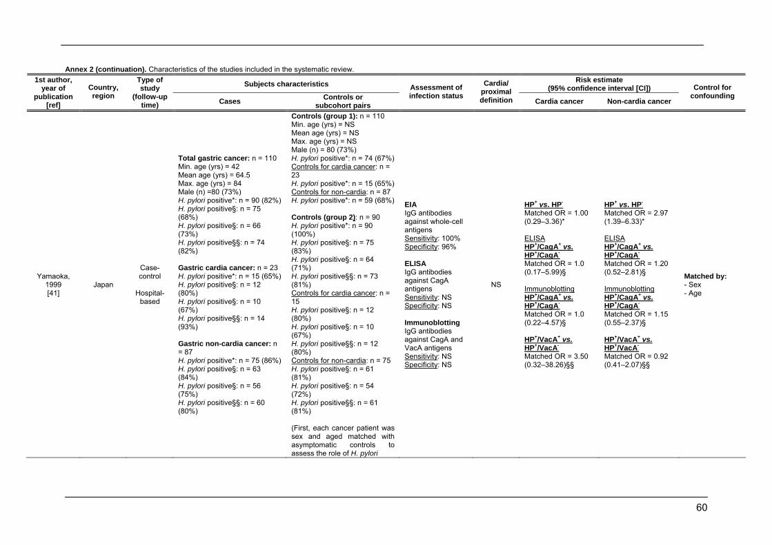
60
Annex 2 (continuation). Characteristics of the studies included in the systematic review.
Subjects characteristics Risk estimate (95% confidence interval [CI])
1st author, year of
publication [ref]
Country, region
Type of study
(follow-up time) Cases Controls or
subcohort pairs
Assessment of infection status
Cardia/ proximal definition Cardia cancer Non-cardia cancer
Control for confounding
Yamaoka, 1999 [41]
Japan
Case-control
Hospital-
based
Total gastric cancer: n = 110 Min. age (yrs) = 42 Mean age (yrs) = 64.5 Max. age (yrs) = 84 Male (n) =80 (73%) H. pylori positive*: n = 90 (82%) H. pylori positive§: n = 75 (68%) H. pylori positive§: n = 66 (73%) H. pylori positive§§: n = 74 (82%) Gastric cardia cancer: n = 23 H. pylori positive*: n = 15 (65%) H. pylori positive§: n = 12 (80%) H. pylori positive§: n = 10 (67%) H. pylori positive§§: n = 14 (93%) Gastric non-cardia cancer: n = 87 H. pylori positive*: n = 75 (86%) H. pylori positive§: n = 63 (84%) H. pylori positive§: n = 56 (75%) H. pylori positive§§: n = 60 (80%)
Controls (group 1): n = 110 Min. age (yrs) = NS Mean age (yrs) = NS Max. age (yrs) = NS Male (n) = 80 (73%) H. pylori positive*: n = 74 (67%) Controls for cardia cancer: n = 23 H. pylori positive*: n = 15 (65%) Controls for non-cardia: n = 87 H. pylori positive*: n = 59 (68%) Controls (group 2): n = 90 H. pylori positive*: n = 90 (100%) H. pylori positive§: n = 75 (83%) H. pylori positive§: n = 64 (71%) H. pylori positive§§: n = 73 (81%) Controls for cardia cancer: n = 15 H. pylori positive§: n = 12 (80%) H. pylori positive§: n = 10 (67%) H. pylori positive§§: n = 12 (80%) Controls for non-cardia: n = 75 H. pylori positive§: n = 61 (81%) H. pylori positive§: n = 54 (72%) H. pylori positive§§: n = 61 (81%) (First, each cancer patient was sex and aged matched with asymptomatic controls to assess the role of H. pylori
EIA IgG antibodies against whole-cell antigens Sensitivity: 100% Specificity: 96% ELISA IgG antibodies against CagA antigens Sensitivity: NS Specificity: NS Immunoblotting IgG antibodies against CagA and VacA antigens Sensitivity: NS Specificity: NS
NS
HP+ vs. HP- Matched OR = 1.00 (0.29–3.36)* ELISA HP+/CagA+ vs. HP+/CagA- Matched OR = 1.0 (0.17–5.99)§ Immunoblotting HP+/CagA+ vs. HP+/CagA- Matched OR = 1.0 (0.22–4.57)§ HP+/VacA+ vs. HP+/VacA- Matched OR = 3.50 (0.32–38.26)§§
HP+ vs. HP- Matched OR = 2.97 (1.39–6.33)* ELISA HP+/CagA+ vs. HP+/CagA- Matched OR = 1.20 (0.52–2.81)§ Immunoblotting HP+/CagA+ vs. HP+/CagA- Matched OR = 1.15 (0.55–2.37)§ HP+/VacA+ vs. HP+/VacA- Matched OR = 0.92 (0.41–2.07)§§
Matched by: - Sex - Age
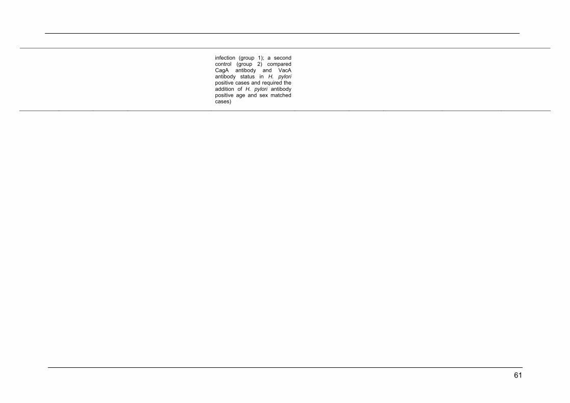
61
infection (group 1); a second control (group 2) compared CagA antibody and VacA antibody status in H. pylori positive cases and required the addition of H. pylori antibody positive age and sex matched cases)
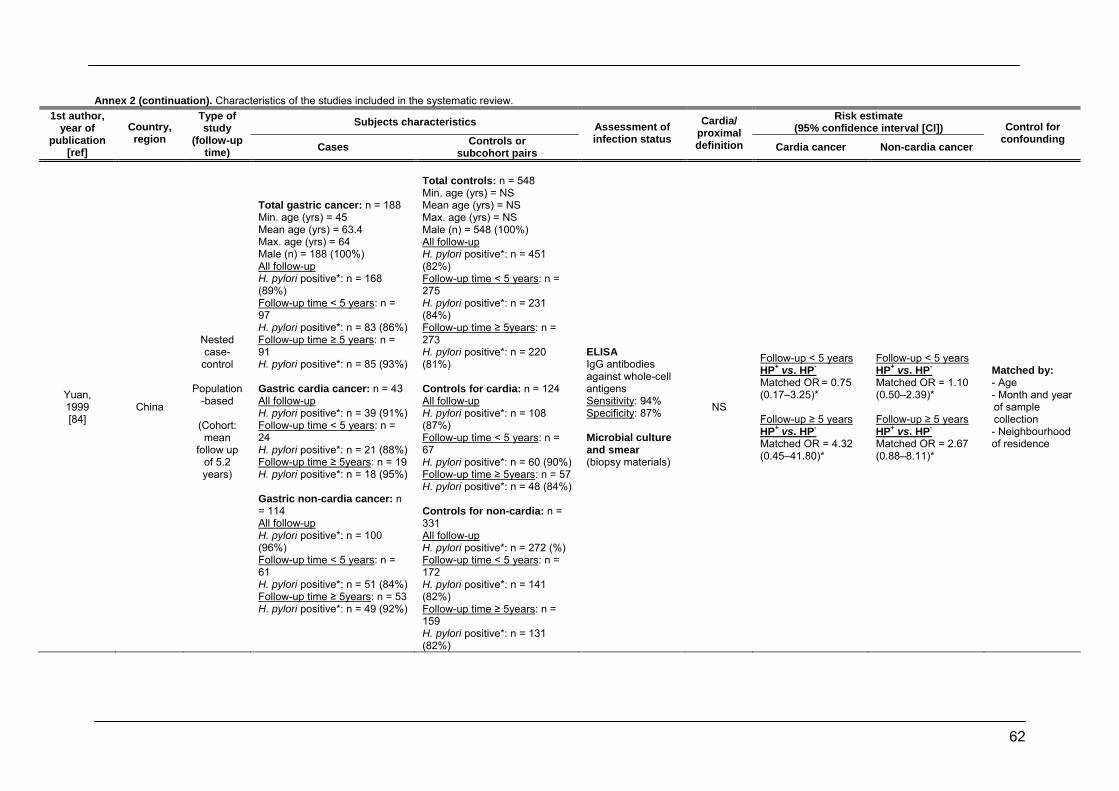
62
Annex 2 (continuation). Characteristics of the studies included in the systematic review.
Subjects characteristics Risk estimate (95% confidence interval [CI])
1st author, year of
publication [ref]
Country, region
Type of study
(follow-up time) Cases Controls or
subcohort pairs
Assessment of infection status
Cardia/ proximal definition Cardia cancer Non-cardia cancer
Control for confounding
Yuan, 1999 [84]
China
Nested case-
control
Population-based
(Cohort:
mean follow up
of 5.2 years)
Total gastric cancer: n = 188 Min. age (yrs) = 45 Mean age (yrs) = 63.4 Max. age (yrs) = 64 Male (n) = 188 (100%) All follow-up H. pylori positive*: n = 168 (89%) Follow-up time < 5 years: n = 97 H. pylori positive*: n = 83 (86%) Follow-up time ≥ 5 years: n = 91 H. pylori positive*: n = 85 (93%) Gastric cardia cancer: n = 43 All follow-up H. pylori positive*: n = 39 (91%) Follow-up time < 5 years: n = 24 H. pylori positive*: n = 21 (88%) Follow-up time ≥ 5years: n = 19 H. pylori positive*: n = 18 (95%) Gastric non-cardia cancer: n = 114 All follow-up H. pylori positive*: n = 100 (96%) Follow-up time < 5 years: n = 61 H. pylori positive*: n = 51 (84%) Follow-up time ≥ 5years: n = 53 H. pylori positive*: n = 49 (92%)
Total controls: n = 548 Min. age (yrs) = NS Mean age (yrs) = NS Max. age (yrs) = NS Male (n) = 548 (100%) All follow-up H. pylori positive*: n = 451 (82%) Follow-up time < 5 years: n = 275 H. pylori positive*: n = 231 (84%) Follow-up time ≥ 5years: n = 273 H. pylori positive*: n = 220 (81%) Controls for cardia: n = 124 All follow-up H. pylori positive*: n = 108 (87%) Follow-up time < 5 years: n = 67 H. pylori positive*: n = 60 (90%) Follow-up time ≥ 5years: n = 57 H. pylori positive*: n = 48 (84%) Controls for non-cardia: n = 331 All follow-up H. pylori positive*: n = 272 (%) Follow-up time < 5 years: n = 172 H. pylori positive*: n = 141 (82%) Follow-up time ≥ 5years: n = 159 H. pylori positive*: n = 131 (82%)
ELISA IgG antibodies against whole-cell antigens Sensitivity: 94% Specificity: 87% Microbial culture and smear (biopsy materials)
NS
Follow-up < 5 years HP+ vs. HP- Matched OR = 0.75 (0.17–3.25)* Follow-up ≥ 5 years HP+ vs. HP- Matched OR = 4.32 (0.45–41.80)*
Follow-up < 5 years HP+ vs. HP- Matched OR = 1.10 (0.50–2.39)* Follow-up ≥ 5 years HP+ vs. HP- Matched OR = 2.67 (0.88–8.11)*
Matched by: - Age - Month and year of sample collection - Neighbourhood of residence
63
Annex 2 (continuation). Characteristics of the studies included in the systematic review.
Subjects characteristics Risk estimate (95% confidence interval [CI])
1st author, year of
publication [ref]
Country, region
Type of study
(follow-up time) Cases Controls or
subcohort pairs
Assessment of infection status
Cardia/ proximal definition Cardia cancer Non-cardia cancer
Control for confounding
Yamagata, 2000 [78]
Japan
Cohort
Population-based
(Mean
follow up of 4.9 years)
Cohort: n = 2602 Min. age (yrs) = 40 Mean age (yrs) = 58 Max. age (yrs) = 58.4 Male (n) = 1070 (41%) Total gastric cancer: n = 67 Min. age (yrs) = NS Mean age (yrs) = NS Max. age (yrs) = NS Male (n) = 48 (72%) H. pylori positive*: n = 51 (76%) Male (all follow-up time): H. pylori positive*: n = 40 (83%) Male (follow-up ≤ 5 years): H. pylori positive*: n = NS Male (follow-up [6-9] years): H. pylori positive*: n = NS Gastric cardia cancer: n = 10 (data only given for male) H. pylori positive*: n = NS Gastric non-cardia cancer: n = 40 (data only given for male) H. pylori positive*: n = NS
Cohort: n = 2602 Min. age (yrs) = 40 Mean age (yrs) = 58 Max. age (yrs) = NS Male (n) = 1070 (41%) Subcohort pairs: n = 2535 Min. age (yrs) = NS Mean age (yrs) = NS Max. age (yrs) = NS Male (n) = 1022 (40%) H. pylori positive*: n = 1670 (64%) Subcohort pairs for cardia: n = NS H. pylori positive*: n = NS Subcohort pairs for non-cardia: n = NS H. pylori positive*: n = NS
EIA IgG antibodies against whole-cell antigens Sensitivity: [95.0-98.7]% Specificity: [96.4-100]%
NS
All follow-up time HP+ vs. HP- Adjusted RR = 1.29 (0.28-6.09)*
(The RR estimates were reported only for males)
All follow-up time HP+ vs. HP- Adjusted RR = 3.66 (1.12-11.92)*
(The RR estimates were reported only for males)
Matched by: - NS Adjusted by: - Age
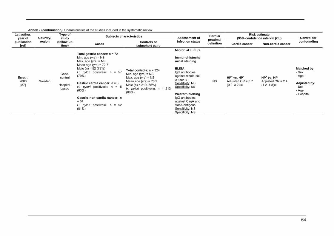
64
Annex 2 (continuation). Characteristics of the studies included in the systematic review. Subjects characteristics Risk estimate
(95% confidence interval [CI]) 1st author,
year of publication
[ref]
Country, region
Type of study
(follow-up time) Cases Controls or
subcohort pairs
Assessment of infection status
Cardia/ proximal definition Cardia cancer Non-cardia cancer
Control for confounding
Enroth, 2000 [87]
Sweden
Case-control
Hospital-
based
Total gastric cancer: n = 72 Min. age (yrs) = NS Max. age (yrs) = NS Mean age (yrs) = 72.7 Male (n) = 52 (72%) H. pylori positiveװװ: n = 57 (79%) Gastric cardia cancer: n = 8 H. pylori positiveװװ: n = 5 (63%) Gastric non-cardia cancer: n = 64 H. pylori positiveװװ: n = 52 (81%)
Total controls: n = 324 Min. age (yrs) = NS Max. age (yrs) = NS Mean age (yrs) = 70.9 Male (n) = 210 (65%) H. pylori positiveװװ: n = 213 (66%)
Microbial culture Immunohistochemical staining ELISA IgG antibodies against whole-cell antigens Sensitivity: NS Specificity: NS Western blotting IgG antibodies against CagA and VacA antigens Sensitivity: NS Specificity: NS
NS HP+ vs. HP- Adjusted OR = 0.7 װװ(3.2–0.2)
HP+ vs. HP- Adjusted OR = 2.4 װװ(4.8–1.2)
Matched by: - Sex - Age Adjusted by: - Sex - Age - Hospital
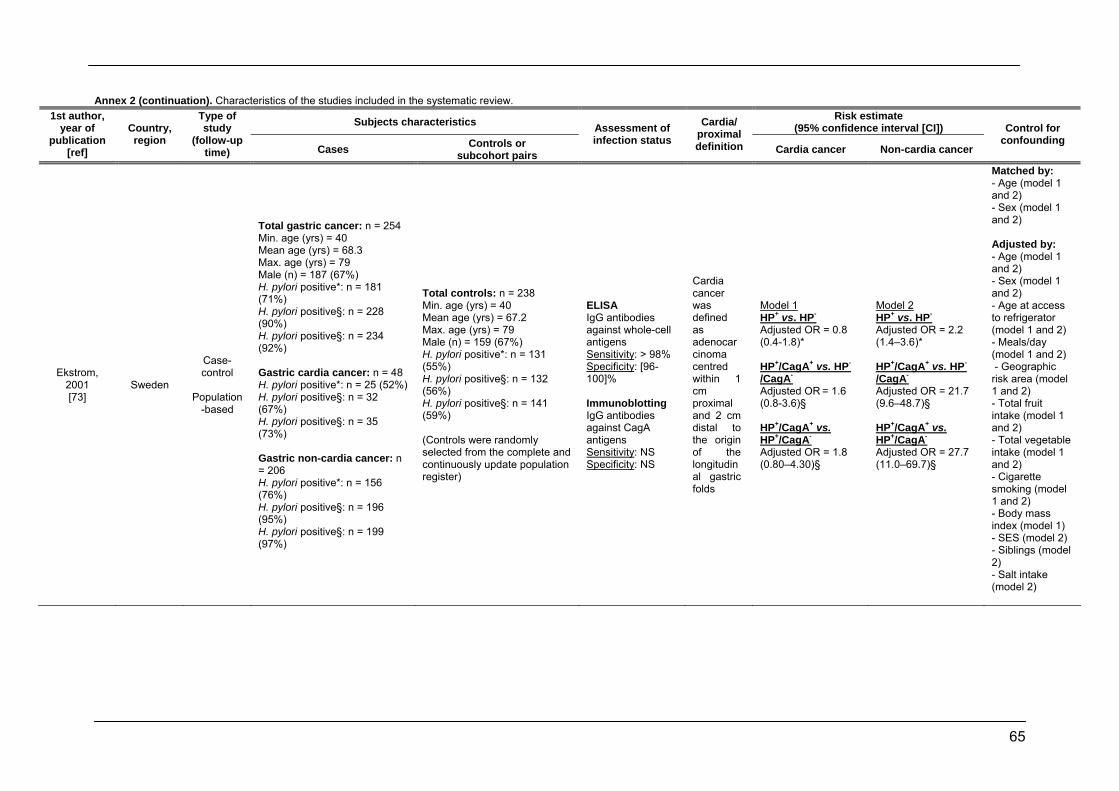
65
Annex 2 (continuation). Characteristics of the studies included in the systematic review.
Subjects characteristics Risk estimate (95% confidence interval [CI])
1st author, year of
publication [ref]
Country, region
Type of study
(follow-up time) Cases Controls or
subcohort pairs
Assessment of infection status
Cardia/ proximal definition Cardia cancer Non-cardia cancer
Control for confounding
Ekstrom, 2001 [73]
Sweden
Case-control
Population
-based
Total gastric cancer: n = 254 Min. age (yrs) = 40 Mean age (yrs) = 68.3 Max. age (yrs) = 79 Male (n) = 187 (67%) H. pylori positive*: n = 181 (71%) H. pylori positive§: n = 228 (90%) H. pylori positive§: n = 234 (92%) Gastric cardia cancer: n = 48 H. pylori positive*: n = 25 (52%) H. pylori positive§: n = 32 (67%) H. pylori positive§: n = 35 (73%) Gastric non-cardia cancer: n = 206 H. pylori positive*: n = 156 (76%) H. pylori positive§: n = 196 (95%) H. pylori positive§: n = 199 (97%)
Total controls: n = 238 Min. age (yrs) = 40 Mean age (yrs) = 67.2 Max. age (yrs) = 79 Male (n) = 159 (67%) H. pylori positive*: n = 131 (55%) H. pylori positive§: n = 132 (56%) H. pylori positive§: n = 141 (59%) (Controls were randomly selected from the complete and continuously update population register)
ELISA IgG antibodies against whole-cell antigens Sensitivity: > 98% Specificity: [96-100]% Immunoblotting IgG antibodies against CagA antigens Sensitivity: NS Specificity: NS
Cardia cancer was defined as adenocarcinoma centred within 1 cm proximal and 2 cm distal to the origin of the longitudinal gastric folds
Model 1 HP+ vs. HP- Adjusted OR = 0.8 (0.4-1.8)* HP+/CagA+ vs. HP-
/CagA- Adjusted OR = 1.6 (0.8-3.6)§ HP+/CagA+ vs. HP+/CagA- Adjusted OR = 1.8 (0.80–4.30)§
Model 2 HP+ vs. HP- Adjusted OR = 2.2 (1.4–3.6)* HP+/CagA+ vs. HP-
/CagA- Adjusted OR = 21.7 (9.6–48.7)§ HP+/CagA+ vs. HP+/CagA- Adjusted OR = 27.7 (11.0–69.7)§
Matched by: - Age (model 1 and 2) - Sex (model 1 and 2) Adjusted by: - Age (model 1 and 2) - Sex (model 1 and 2) - Age at access to refrigerator (model 1 and 2) - Meals/day (model 1 and 2) - Geographic risk area (model 1 and 2) - Total fruit intake (model 1 and 2) - Total vegetable intake (model 1 and 2) - Cigarette smoking (model 1 and 2) - Body mass index (model 1) - SES (model 2) - Siblings (model 2) - Salt intake (model 2)
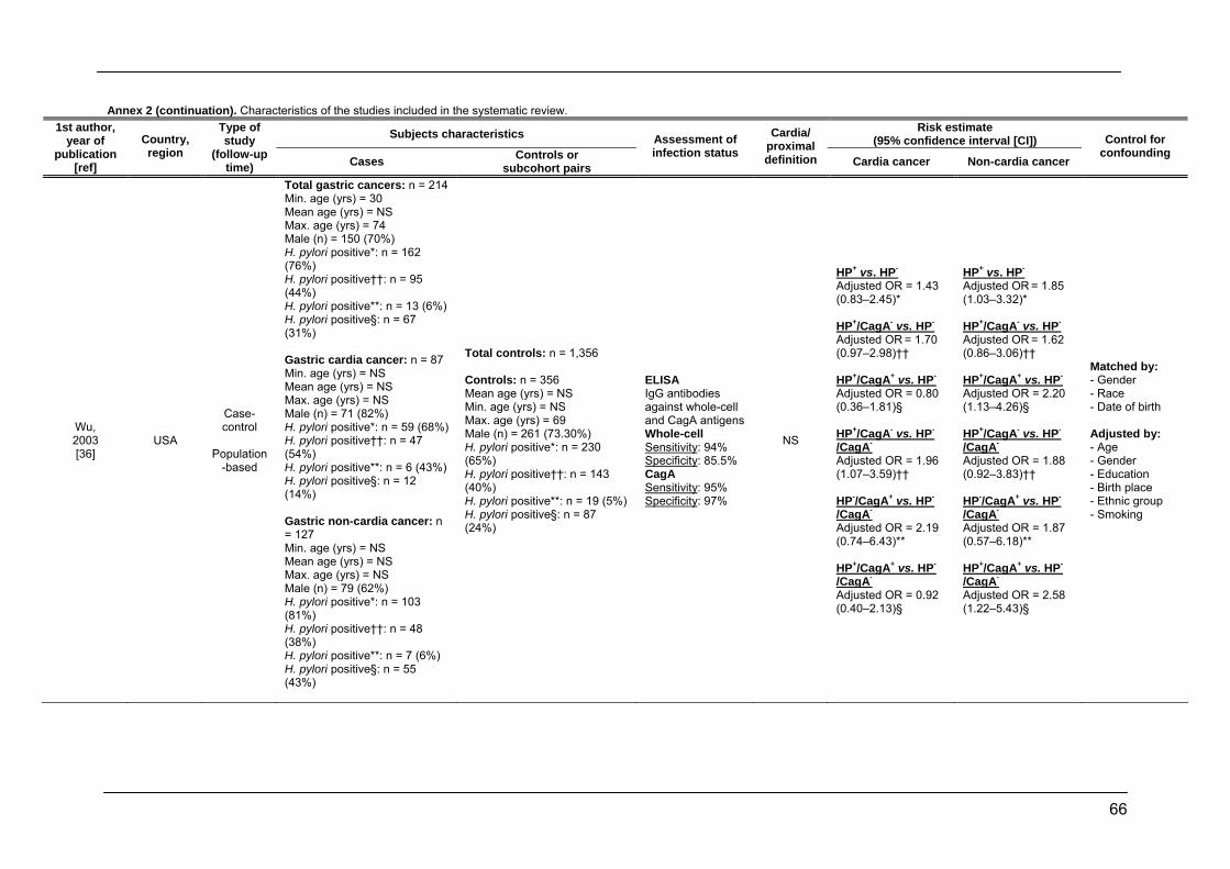
66
Annex 2 (continuation). Characteristics of the studies included in the systematic review. Subjects characteristics Risk estimate
(95% confidence interval [CI]) 1st author,
year of publication
[ref]
Country, region
Type of study
(follow-up time) Cases Controls or
subcohort pairs
Assessment of infection status
Cardia/ proximal definition Cardia cancer Non-cardia cancer
Control for confounding
Wu, 2003 [36]
USA
Case-control
Population
-based
Total gastric cancers: n = 214 Min. age (yrs) = 30 Mean age (yrs) = NS Max. age (yrs) = 74 Male (n) = 150 (70%) H. pylori positive*: n = 162 (76%) H. pylori positive††: n = 95 (44%) H. pylori positive**: n = 13 (6%) H. pylori positive§: n = 67 (31%) Gastric cardia cancer: n = 87 Min. age (yrs) = NS Mean age (yrs) = NS Max. age (yrs) = NS Male (n) = 71 (82%) H. pylori positive*: n = 59 (68%) H. pylori positive††: n = 47 (54%) H. pylori positive**: n = 6 (43%) H. pylori positive§: n = 12 (14%) Gastric non-cardia cancer: n = 127 Min. age (yrs) = NS Mean age (yrs) = NS Max. age (yrs) = NS Male (n) = 79 (62%) H. pylori positive*: n = 103 (81%) H. pylori positive††: n = 48 (38%) H. pylori positive**: n = 7 (6%) H. pylori positive§: n = 55 (43%)
Total controls: n = 1,356 Controls: n = 356 Mean age (yrs) = NS Min. age (yrs) = NS Max. age (yrs) = 69 Male (n) = 261 (73.30%) H. pylori positive*: n = 230 (65%) H. pylori positive††: n = 143 (40%) H. pylori positive**: n = 19 (5%) H. pylori positive§: n = 87 (24%)
ELISA IgG antibodies against whole-cell and CagA antigens Whole-cell Sensitivity: 94% Specificity: 85.5% CagA Sensitivity: 95% Specificity: 97%
NS
HP+ vs. HP- Adjusted OR = 1.43 (0.83–2.45)* HP+/CagA- vs. HP-
Adjusted OR = 1.70 (0.97–2.98)†† HP+/CagA+ vs. HP-
Adjusted OR = 0.80 (0.36–1.81)§
HP+/CagA- vs. HP-
/CagA- Adjusted OR = 1.96 (1.07–3.59)††
HP-/CagA+ vs. HP-
/CagA- Adjusted OR = 2.19 (0.74–6.43)**
HP+/CagA+ vs. HP-
/CagA- Adjusted OR = 0.92 (0.40–2.13)§
HP+ vs. HP- Adjusted OR = 1.85 (1.03–3.32)* HP+/CagA- vs. HP-
Adjusted OR = 1.62 (0.86–3.06)†† HP+/CagA+ vs. HP-
Adjusted OR = 2.20 (1.13–4.26)§
HP+/CagA- vs. HP-
/CagA- Adjusted OR = 1.88 (0.92–3.83)†† HP-/CagA+ vs. HP-
/CagA- Adjusted OR = 1.87 (0.57–6.18)**
HP+/CagA+ vs. HP-
/CagA- Adjusted OR = 2.58 (1.22–5.43)§
Matched by: - Gender - Race - Date of birth Adjusted by: - Age - Gender - Education - Birth place - Ethnic group - Smoking
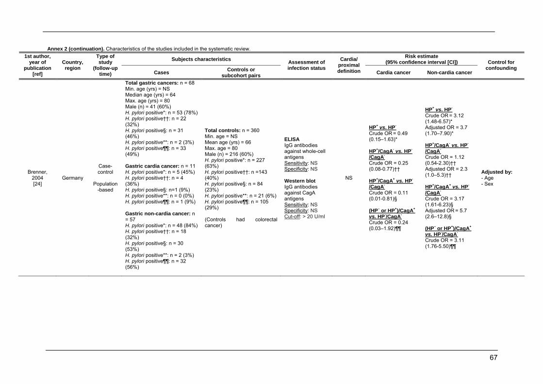
67
Annex 2 (continuation). Characteristics of the studies included in the systematic review.
Subjects characteristics Risk estimate (95% confidence interval [CI])
1st author, year of
publication [ref]
Country, region
Type of study
(follow-up time) Cases Controls or
subcohort pairs
Assessment of infection status
Cardia/ proximal definition Cardia cancer Non-cardia cancer
Control for confounding
Brenner, 2004 [24]
Germany
Case-control
Population
-based
Total gastric cancers: n = 68 Min. age (yrs) = NS Median age (yrs) = 64 Max. age (yrs) = 80 Male (n) = 41 (60%) H. pylori positive*: n = 53 (78%) H. pylori positive††: n = 22 (32%) H. pylori positive§: n = 31 (46%) H. pylori positive**: n = 2 (3%) H. pylori positive¶¶: n = 33 (49%) Gastric cardia cancer: n = 11 H. pylori positive*: n = 5 (45%) H. pylori positive††: n = 4 (36%) H. pylori positive§: n=1 (9%) H. pylori positive**: n = 0 (0%) H. pylori positive¶¶: n = 1 (9%) Gastric non-cardia cancer: n = 57 H. pylori positive*: n = 48 (84%) H. pylori positive††: n = 18 (32%) H. pylori positive§: n = 30 (53%) H. pylori positive**: n = 2 (3%) H. pylori positive¶¶: n = 32 (56%)
Total controls: n = 360 Min. age = NS Mean age (yrs) = 66 Max. age = 80 Male (n) = 216 (60%) H. pylori positive*: n = 227 (63%) H. pylori positive††: n =143 (40%) H. pylori positive§: n = 84 (23%) H. pylori positive**: n = 21 (6%) H. pylori positive¶¶: n = 105 (29%) (Controls had colorectal cancer)
ELISA IgG antibodies against whole-cell antigens Sensitivity: NS Specificity: NS Western blot IgG antibodies against CagA antigens Sensitivity: NS Specificity: NS Cut-off: > 20 U/ml
NS
HP+ vs. HP- Crude OR = 0.49 (0.15–1.63)*
HP+/CagA- vs. HP-
/CagA- Crude OR = 0.25 (0.08-0.77)†† HP+/CagA+ vs. HP-
/CagA- Crude OR = 0.11 (0.01-0.81)§ (HP– or HP+)/CagA+ vs. HP-/CagA- Crude OR = 0.24 (0.03–1.92)¶¶
HP+ vs. HP- Crude OR = 3.12 (1.48-6.57)* Adjusted OR = 3.7 (1.70–7.90)* HP+/CagA- vs. HP-
/CagA- Crude OR = 1.12 (0.54-2.30)†† Adjusted OR = 2.3 (1.0–5.3)†† HP+/CagA+ vs. HP-
/CagA- Crude OR = 3.17 (1.61-6.23)§ Adjusted OR = 5.7 (2.6–12.8)§
(HP– or HP+)/CagA+ vs. HP-/CagA- Crude OR = 3.11 (1.76-5.50)¶¶
Adjusted by: - Age - Sex
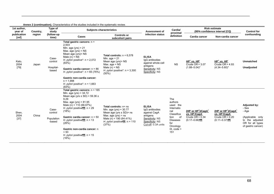
68
Annex 2 (continuation). Characteristics of the studies included in the systematic review.
Subjects characteristics Risk estimate (95% confidence interval [CI])
1st author, year of
publication [ref]
Country, region
Type of study
(follow-up time) Cases Controls or
subcohort pairs
Assessment of infection status
Cardia/ proximal definition Cardia cancer Non-cardia cancer
Control for confounding
Kato, 2004 [79]
Japan
Case-control
Hospital-
based
Total gastric cancers: n = 2,503 Min. age (yrs) = 21 Max. age (yrs) = NS Mean age (yrs)= NS Male (n) = NS H. pylori positive*: n = 2,072 (83%) Gastric cardia cancer: n = 86 H. pylori positive*: n = 65 (76%) Gastric non-cardia cancer: n = 1,998 H. pylori positive*: n = 1,663 (83%)
Total controls: n = 6,578 Min. age = 21 Mean age (yrs)= NS Max. age = NS Male (n) = NS H. pylori positive*: n = 3,300 (50%)
ELISA IgG antibodies against whole-cell antigens Sensitivity: NS Specificity: NS
NS HP+ vs. HP- Crude OR = 3.07 (1.88–5.04)*
HP+ vs. HP- Crude OR = 4.93 (4.34–5.60)*
Unmatched Unadjusted
Shen, 2004 [37]
China
Case-control
Population
-based
Total gastric cancers: n = 165 Min. age (yrs) = 34.72 Mean age (yrs ± SD) = 59.36 ± 9.29 Max. age (yrs) = 81.95 Male (n) = 110 (66.67%) H. pylori positive¶¶: n = 29 (18%) Gastric cardia cancer: n = 50 H. pylori positive¶¶: n = 13 (26%) Gastric non-cardia cancer: n = 93 H. pylori positive¶¶: n = 15 (16%)
Total controls: n= ns Min. age (yrs) = 30.77 Mean age (yrs ± SD)= ns Max. age (yrs) = ns Male (n) = 190 (64.41%) H. pylori positive¶¶: n = 110 (37%)
ELISA IgG antibodies against CagA antigens Sensitivity: NS Specificity: NS Cut-off: 0.3A units
The authors used the International Classification of Diseases for Oncology IX, code = 151
(HP- or HP+)/CagA+ vs. HP-/CagA- Crude OR = 0.34 (0.17–0.68)¶¶
(HP- or HP+)/CagA+ vs. HP-/CagA- Crude OR = 0.20 (0.11–0.37)¶¶
Adjusted by: - Sex - Age (Applicable only to the adjusted OR for all types of gastric cancer)
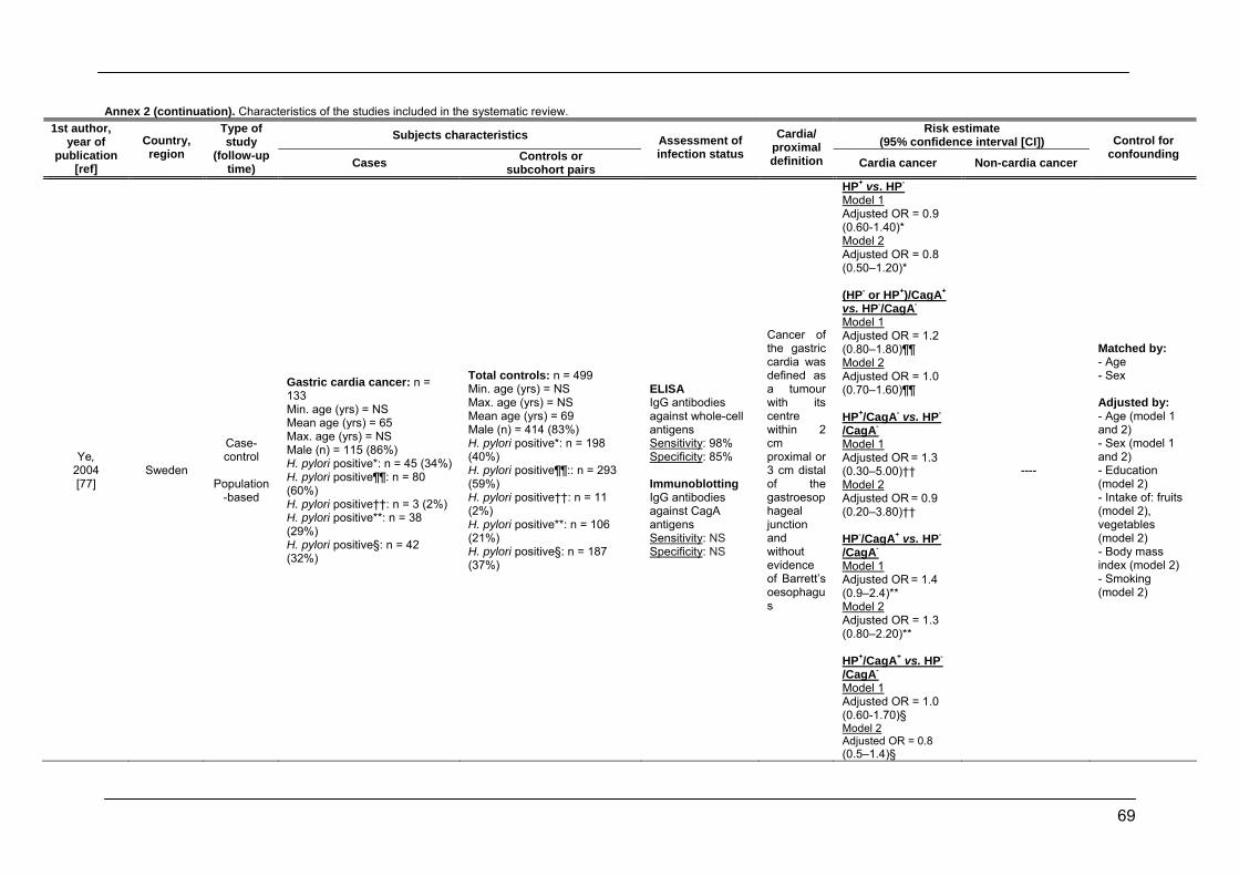
69
Annex 2 (continuation). Characteristics of the studies included in the systematic review.
Subjects characteristics Risk estimate (95% confidence interval [CI])
1st author, year of
publication [ref]
Country, region
Type of study
(follow-up time) Cases Controls or
subcohort pairs
Assessment of infection status
Cardia/ proximal definition Cardia cancer Non-cardia cancer
Control for confounding
Ye, 2004 [77]
Sweden
Case-control
Population
-based
Gastric cardia cancer: n = 133 Min. age (yrs) = NS Mean age (yrs) = 65 Max. age (yrs) = NS Male (n) = 115 (86%) H. pylori positive*: n = 45 (34%) H. pylori positive¶¶: n = 80 (60%) H. pylori positive††: n = 3 (2%) H. pylori positive**: n = 38 (29%) H. pylori positive§: n = 42 (32%)
Total controls: n = 499 Min. age (yrs) = NS Max. age (yrs) = NS Mean age (yrs) = 69 Male (n) = 414 (83%) H. pylori positive*: n = 198 (40%) H. pylori positive¶¶:: n = 293 (59%) H. pylori positive††: n = 11 (2%) H. pylori positive**: n = 106 (21%) H. pylori positive§: n = 187 (37%)
ELISA IgG antibodies against whole-cell antigens Sensitivity: 98% Specificity: 85% Immunoblotting IgG antibodies against CagA antigens Sensitivity: NS Specificity: NS
Cancer of the gastric cardia was defined as a tumour with its centre within 2 cm proximal or 3 cm distal of the gastroesophageal junction and without evidence of Barrett’s oesophagus
HP+ vs. HP- Model 1 Adjusted OR = 0.9 (0.60-1.40)* Model 2 Adjusted OR = 0.8 (0.50–1.20)* (HP- or HP+)/CagA+ vs. HP-/CagA- Model 1 Adjusted OR = 1.2 (0.80–1.80)¶¶ Model 2 Adjusted OR = 1.0 (0.70–1.60)¶¶ HP+/CagA- vs. HP-
/CagA- Model 1 Adjusted OR = 1.3 (0.30–5.00)††
Model 2 Adjusted OR = 0.9 (0.20–3.80)†† HP-/CagA+ vs. HP-
/CagA- Model 1 Adjusted OR = 1.4 (0.9–2.4)** Model 2 Adjusted OR = 1.3 (0.80–2.20)** HP+/CagA+ vs. HP-
/CagA- Model 1 Adjusted OR = 1.0 (0.60-1.70)§ Model 2 Adjusted OR = 0.8 (0.5–1.4)§
----
Matched by: - Age - Sex Adjusted by: - Age (model 1 and 2) - Sex (model 1 and 2) - Education (model 2) - Intake of: fruits (model 2), vegetables (model 2) - Body mass index (model 2) - Smoking (model 2)
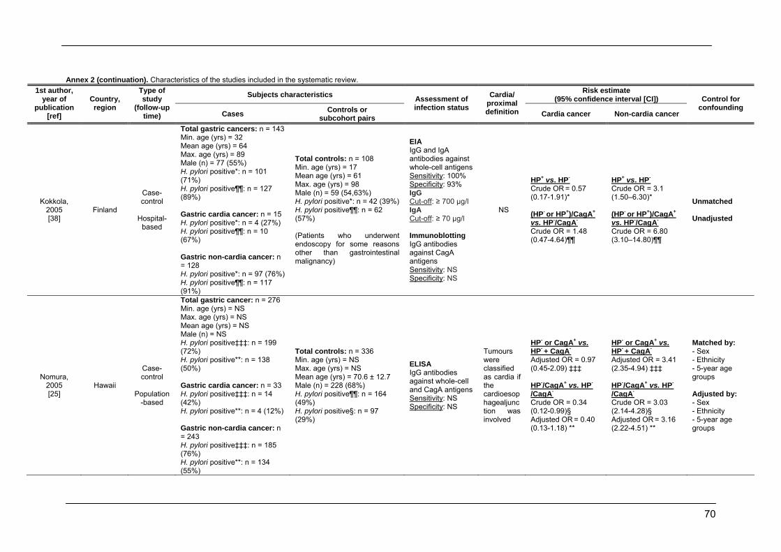
70
Annex 2 (continuation). Characteristics of the studies included in the systematic review.
Subjects characteristics Risk estimate (95% confidence interval [CI])
1st author, year of
publication [ref]
Country, region
Type of study
(follow-up time) Cases Controls or
subcohort pairs
Assessment of infection status
Cardia/ proximal definition Cardia cancer Non-cardia cancer
Control for confounding
Kokkola, 2005 [38]
Finland
Case-control
Hospital-
based
Total gastric cancers: n = 143 Min. age (yrs) = 32 Mean age (yrs) = 64 Max. age (yrs) = 89 Male (n) = 77 (55%) H. pylori positive*: n = 101 (71%) H. pylori positive¶¶: n = 127 (89%) Gastric cardia cancer: n = 15 H. pylori positive*: n = 4 (27%) H. pylori positive¶¶: n = 10 (67%) Gastric non-cardia cancer: n = 128 H. pylori positive*: n = 97 (76%) H. pylori positive¶¶: n = 117 (91%)
Total controls: n = 108 Min. age (yrs) = 17 Mean age (yrs) = 61 Max. age (yrs) = 98 Male (n) = 59 (54,63%) H. pylori positive*: n = 42 (39%) H. pylori positive¶¶: n = 62 (57%) (Patients who underwent endoscopy for some reasons other than gastrointestinal malignancy)
EIA IgG and IgA antibodies against whole-cell antigens Sensitivity: 100% Specificity: 93% IgG Cut-off: ≥ 700 µg/l IgA Cut-off: ≥ 70 µg/l Immunoblotting IgG antibodies against CagA antigens Sensitivity: NS Specificity: NS
NS
HP+ vs. HP- Crude OR = 0.57 (0.17-1.91)*
(HP- or HP+)/CagA+ vs. HP-/CagA- Crude OR = 1.48 (0.47-4.64)¶¶
HP+ vs. HP- Crude OR = 3.1 (1.50–6.30)*
(HP- or HP+)/CagA+ vs. HP-/CagA- Crude OR = 6.80 (3.10–14.80)¶¶
Unmatched Unadjusted
Nomura, 2005 [25]
Hawaii
Case-control
Population
-based
Total gastric cancer: n = 276 Min. age (yrs) = NS Max. age (yrs) = NS Mean age (yrs) = NS Male (n) = NS H. pylori positive‡‡‡: n = 199 (72%) H. pylori positive**: n = 138 (50%) Gastric cardia cancer: n = 33 H. pylori positive‡‡‡: n = 14 (42%) H. pylori positive**: n = 4 (12%) Gastric non-cardia cancer: n = 243 H. pylori positive‡‡‡: n = 185 (76%) H. pylori positive**: n = 134 (55%)
Total controls: n = 336 Min. age (yrs) = NS Max. age (yrs) = NS Mean age (yrs) = 70.6 ± 12.7 Male (n) = 228 (68%) H. pylori positive¶¶: n = 164 (49%) H. pylori positive§: n = 97 (29%)
ELISA IgG antibodies against whole-cell and CagA antigens Sensitivity: NS Specificity: NS
Tumours were classified as cardia if the cardioesophagealjunction was involved
HP- or CagA+ vs. HP- + CagA- Adjusted OR = 0.97 (0.45-2.09) ‡‡‡ HP-/CagA+ vs. HP-
/CagA- Crude OR = 0.34 (0.12-0.99)§ Adjusted OR = 0.40 (0.13-1.18) **
HP- or CagA+ vs. HP- + CagA- Adjusted OR = 3.41 (2.35-4.94) ‡‡‡ HP-/CagA+ vs. HP-
/CagA- Crude OR = 3.03 (2.14-4.28)§ Adjusted OR = 3.16 (2.22-4.51) **
Matched by: - Sex - Ethnicity - 5-year age groups Adjusted by: - Sex - Ethnicity - 5-year age groups
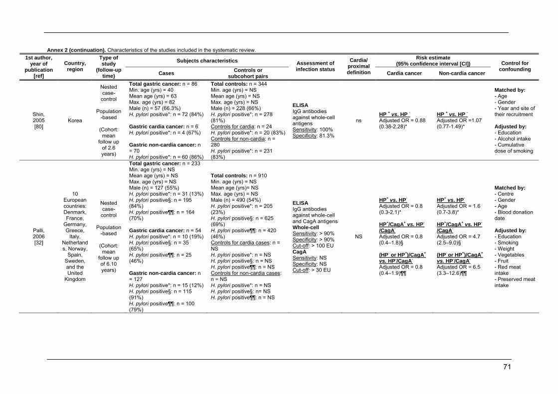
71
Annex 2 (continuation). Characteristics of the studies included in the systematic review.
Subjects characteristics Risk estimate (95% confidence interval [CI])
1st author, year of
publication [ref]
Country, region
Type of study
(follow-up time) Cases Controls or
subcohort pairs
Assessment of infection status
Cardia/ proximal definition Cardia cancer Non-cardia cancer
Control for confounding
Shin, 2005 [80]
Korea
Nested case-
control
Population-based
(Cohort:
mean follow up
of 2.6 years)
Total gastric cancer: n = 86 Min. age (yrs) = 40 Mean age (yrs) = 63 Max. age (yrs) = 82 Male (n) = 57 (66.3%) H. pylori positive*: n = 72 (84%) Gastric cardia cancer: n = 6 H. pylori positive*: n = 4 (67%) Gastric non-cardia cancer: n = 70 H. pylori positive*: n = 60 (86%)
Total controls: n = 344 Min. age (yrs) = NS Mean age (yrs) = NS Max. age (yrs) = NS Male (n) = 228 (66%) H. pylori positive*: n = 278 (81%) Controls for cardia: n = 24 H. pylori positive*: n = 20 (83%) Controls for non-cardia: n = 280 H. pylori positive*: n = 231 (83%)
ELISA IgG antibodies against whole-cell antigens Sensitivity: 100% Specificity: 81.3%
ns HP + vs. HP - Adjusted OR = 0.88 (0.38-2.28)*
HP + vs. HP - Adjusted OR =1.07 (0.77-1.49)*
Matched by: - Age - Gender - Year and site of their recruitment Adjusted by: - Education - Alcohol intake - Cumulative dose of smoking
Palli, 2006 [32]
10 European countries: Denmark, France,
Germany, Greece,
Italy, Netherlands, Norway,
Spain, Sweden, and the United
Kingdom
Nested case-
control
Population-based
(Cohort:
mean follow up of 6.10 years)
Total gastric cancer: n = 233 Min. age (yrs) = NS Mean age (yrs) = NS Max. age (yrs) = NS Male (n) = 127 (55%) H. pylori positive*: n = 31 (13%) H. pylori positive§: n = 195 (84%) H. pylori positive¶¶: n = 164 (70%) Gastric cardia cancer: n = 54 H. pylori positive*: n = 10 (19%) H. pylori positive§: n = 35 (65%) H. pylori positive¶¶: n = 25 (46%) Gastric non-cardia cancer: n = 127 H. pylori positive*: n = 15 (12%) H. pylori positive§: n = 115 (91%) H. pylori positive¶¶: n = 100 (79%)
Total controls: n = 910 Min. age (yrs) = NS Mean age (yrs)= NS Max. age (yrs) = NS Male (n) = 490 (54%) H. pylori positive*: n = 205 (23%) H. pylori positive§: n = 625 (69%) H. pylori positive¶¶: n = 420 (46%) Controls for cardia cases: n = NS H. pylori positive*: n = NS H. pylori positive§: n = NS H. pylori positive¶¶: n = NS Controls for non-cardia cases: n = NS H. pylori positive*: n = NS H. pylori positive§: n= NS H. pylori positive¶¶: n = NS
ELISA IgG antibodies against whole-cell and CagA antigens Whole-cell Sensitivity: > 90% Specificity: > 90% Cut-off: > 100 EU CagA Sensitivity: NS Specificity: NS Cut-off: > 30 EU
NS
HP+ vs. HP- Adjusted OR = 0.8 (0.3-2.1)* HP+/CagA+ vs. HP-
/CagA- Adjusted OR = 0.8 (0.4–1.8)§
(HP- or HP+)/CagA+ vs. HP-/CagA- Adjusted OR = 0.8 (0.4–1.9)¶¶
HP+ vs. HP- Adjusted OR = 1.6 (0.7-3.8)* HP+/CagA+ vs. HP-
/CagA- Adjusted OR = 4.7 (2.5–9.0)§
(HP- or HP+)/CagA+ vs. HP-/CagA- Adjusted OR = 6.5 (3.3–12.6)¶¶
Matched by: - Centre - Gender - Age - Blood donation date Adjusted by: - Education - Smoking - Weight - Vegetables - Fruit - Red meat intake - Preserved meat intake
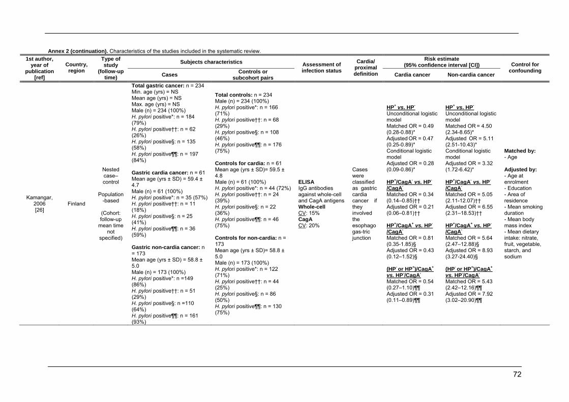
72
Annex 2 (continuation). Characteristics of the studies included in the systematic review.
Subjects characteristics Risk estimate (95% confidence interval [CI])
1st author, year of
publication [ref]
Country, region
Type of study
(follow-up time) Cases Controls or
subcohort pairs
Assessment of infection status
Cardia/ proximal definition Cardia cancer Non-cardia cancer
Control for confounding
Kamangar, 2006 [26]
Finland
Nested case–control
Population
-based
(Cohort: follow-up
mean time not
specified)
Total gastric cancer: n = 234 Min. age (yrs) = NS Mean age (yrs) = NS Max. age (yrs) = NS Male (n) = 234 (100%) H. pylori positive*: n = 184 (79%) H. pylori positive††: n = 62 (26%) H. pylori positive§: n = 135 (58%) H. pylori positive¶¶: n = 197 (84%) Gastric cardia cancer: n = 61 Mean age (yrs ± SD) = 59.4 ± 4.7 Male (n) = 61 (100%) H. pylori positive*: n = 35 (57%) H. pylori positive††: n = 11 (18%) H. pylori positive§: n = 25 (41%) H. pylori positive¶¶: n = 36 (59%) Gastric non-cardia cancer: n = 173 Mean age (yrs ± SD) = 58.8 ± 5.0 Male (n) = 173 (100%) H. pylori positive*: n =149 (86%) H. pylori positive††: n = 51 (29%) H. pylori positive§: n =110 (64%) H. pylori positive¶¶: n = 161 (93%)
Total controls: n = 234 Male (n) = 234 (100%) H. pylori positive*: n = 166 (71%) H. pylori positive††: n = 68 (29%) H. pylori positive§: n = 108 (46%) H. pylori positive¶¶: n = 176 (75%) Controls for cardia: n = 61 Mean age (yrs ± SD)= 59.5 ± 4.8 Male (n) = 61 (100%) H. pylori positive*: n = 44 (72%) H. pylori positive††: n = 24 (39%) H. pylori positive§: n = 22 (36%) H. pylori positive¶¶: n = 46 (75%) Controls for non-cardia: n = 173 Mean age (yrs ± SD)= 58.8 ± 5.0 Male (n) = 173 (100%) H. pylori positive*: n = 122 (71%) H. pylori positive††: n = 44 (25%) H. pylori positive§: n = 86 (50%) H. pylori positive¶¶: n = 130 (75%)
ELISA IgG antibodies against whole-cell and CagA antigens Whole-cell CV: 15% CagA CV: 20%
Cases were classified as gastric cardia cancer if they involved the esophagogas-tric junction
HP+ vs. HP- Unconditional logistic model Matched OR = 0.49 (0.28-0.88)* Adjusted OR = 0.47 (0.25-0.89)* Conditional logistic model Adjusted OR = 0.28 (0.09-0.86)* HP+/CagA- vs. HP-
/CagA- Matched OR = 0.34 (0.14–0.85)†† Adjusted OR = 0.21 (0.06–0.81)†† HP+/CagA+ vs. HP-
/CagA- Matched OR = 0.81 (0.35-1.85)§ Adjusted OR = 0.43 (0.12–1.52)§ (HP- or HP+)/CagA+ vs. HP-/CagA- Matched OR = 0.54 (0.27–1.10)¶¶ Adjusted OR = 0.31 (0.11–0.89)¶¶
HP+ vs. HP- Unconditional logistic model Matched OR = 4.50 (2.34-8.65)* Adjusted OR = 5.11 (2.51-10.43)* Conditional logistic model Adjusted OR = 3.32 (1.72-6.42)* HP+/CagA- vs. HP-
/CagA- Matched OR = 5.05 (2.11-12.07)†† Adjusted OR = 6.55 (2.31–18.53)†† HP+/CagA+ vs. HP-
/CagA- Matched OR = 5.64 (2.47–12.88)§
Adjusted OR = 8.93 (3.27-24.40)§
(HP- or HP+)/CagA+ vs. HP-/CagA- Matched OR = 5.43 (2.42–12.16)¶¶ Adjusted OR = 7.92 (3.02–20.90)¶¶
Matched by: - Age Adjusted by: - Age at enrolment - Education - Area of residence - Mean smoking duration - Mean body mass index - Mean dietary intake: nitrate, fruit, vegetable, starch, and sodium
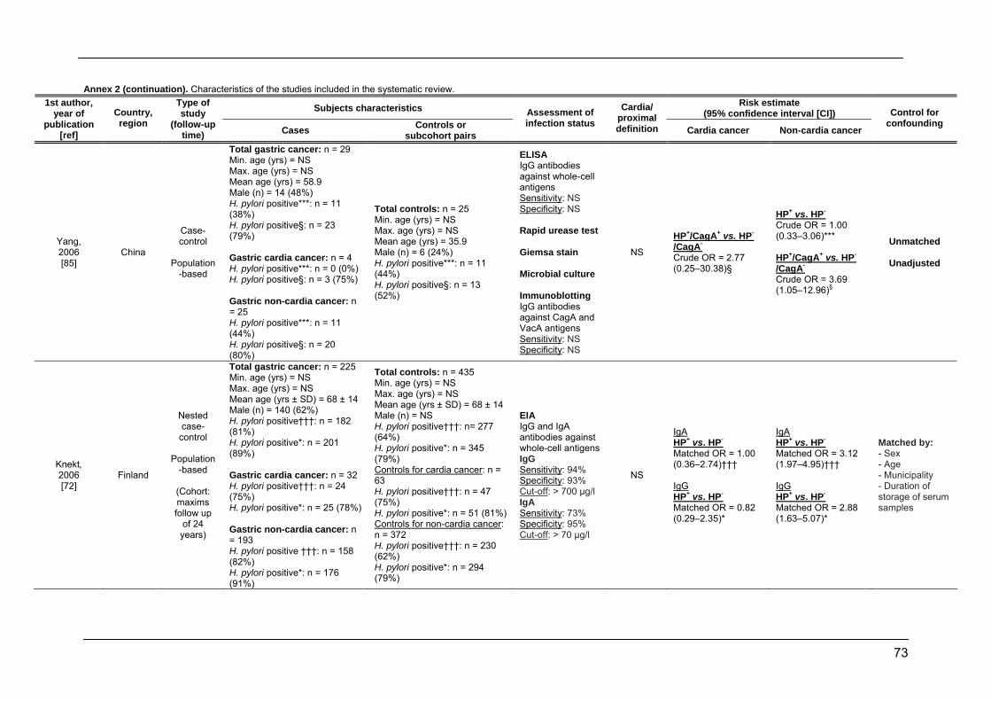
73
Annex 2 (continuation). Characteristics of the studies included in the systematic review.
Subjects characteristics Risk estimate (95% confidence interval [CI])
1st author, year of
publication [ref]
Country, region
Type of study
(follow-up time) Cases Controls or
subcohort pairs
Assessment of infection status
Cardia/ proximal definition Cardia cancer Non-cardia cancer
Control for confounding
Yang, 2006 [85]
China
Case-control
Population
-based
Total gastric cancer: n = 29 Min. age (yrs) = NS Max. age (yrs) = NS Mean age (yrs) = 58.9 Male (n) = 14 (48%) H. pylori positive***: n = 11 (38%) H. pylori positive§: n = 23 (79%) Gastric cardia cancer: n = 4 H. pylori positive***: n = 0 (0%) H. pylori positive§: n = 3 (75%) Gastric non-cardia cancer: n = 25 H. pylori positive***: n = 11 (44%) H. pylori positive§: n = 20 (80%)
Total controls: n = 25 Min. age (yrs) = NS Max. age (yrs) = NS Mean age (yrs) = 35.9 Male (n) = 6 (24%) H. pylori positive***: n = 11 (44%) H. pylori positive§: n = 13 (52%)
ELISA IgG antibodies against whole-cell antigens Sensitivity: NS Specificity: NS Rapid urease test Giemsa stain Microbial culture Immunoblotting IgG antibodies against CagA and VacA antigens Sensitivity: NS Specificity: NS
NS
HP+/CagA+ vs. HP-
/CagA- Crude OR = 2.77 (0.25–30.38)§
HP+ vs. HP- Crude OR = 1.00 (0.33–3.06)*** HP+/CagA+ vs. HP-
/CagA- Crude OR = 3.69 (1.05–12.96)§
Unmatched
Unadjusted
Knekt, 2006 [72]
Finland
Nested case-
control
Population-based
(Cohort: maxims follow up
of 24 years)
Total gastric cancer: n = 225 Min. age (yrs) = NS Max. age (yrs) = NS Mean age (yrs ± SD) = 68 ± 14 Male (n) = 140 (62%) H. pylori positive†††: n = 182 (81%) H. pylori positive*: n = 201 (89%) Gastric cardia cancer: n = 32 H. pylori positive†††: n = 24 (75%) H. pylori positive*: n = 25 (78%) Gastric non-cardia cancer: n = 193 H. pylori positive †††: n = 158 (82%) H. pylori positive*: n = 176 (91%)
Total controls: n = 435 Min. age (yrs) = NS Max. age (yrs) = NS Mean age (yrs ± SD) = 68 ± 14 Male (n) = NS H. pylori positive†††: n= 277 (64%) H. pylori positive*: n = 345 (79%) Controls for cardia cancer: n = 63 H. pylori positive†††: n = 47 (75%) H. pylori positive*: n = 51 (81%) Controls for non-cardia cancer: n = 372 H. pylori positive†††: n = 230 (62%) H. pylori positive*: n = 294 (79%)
EIA IgG and IgA antibodies against whole-cell antigens IgG Sensitivity: 94% Specificity: 93% Cut-off: > 700 µg/l IgA Sensitivity: 73% Specificity: 95% Cut-off: > 70 µg/l
NS
IgA HP+ vs. HP- Matched OR = 1.00 (0.36–2.74)††† IgG HP+ vs. HP- Matched OR = 0.82 (0.29–2.35)*
IgA HP+ vs. HP- Matched OR = 3.12 (1.97–4.95)††† IgG HP+ vs. HP- Matched OR = 2.88 (1.63–5.07)*
Matched by: - Sex - Age - Municipality - Duration of storage of serum samples

74
Annex 2 (continuation). Characteristics of the studies included in the systematic review.
Subjects characteristics Risk estimate (95% confidence interval [CI])
1st author, year of
publication [ref]
Country, region
Type of study
(follow-up time) Cases Controls or
subcohort pairs
Assessment of infection status
Cardia/ proximal definition Cardia cancer Non-cardia cancer
Control for confounding
Sasazuki, 2006 [82]
Japan
Nested case-
control
Population-based
(Cohort:
maximum follow up
was 9 years)
Total gastric cancer: n = 511 Min. age (yrs) = 49 Mean age (yrs ± SD) = 57.4 ± 0.32 Max. age (yrs) = 69 Male (n) = 342 (67%) H. pylori positive*: n = 478 (94%) H. pylori positive¶¶: n = 390 (76%) H. pylori positive††: n = 115 (23%) H. pylori positive**: n = 27 (5%) H. pylori positive‡‡‡: n = 505 (99%) H. pylori positive§: n = 363 (71%) Gastric cardia cancer: n = 39 H. pylori positive*: n = 37 (99%) Gastric non-cardia cancer: n = 368 H. pylori positive*: n = 344 (93%)
Total controls: n = 511 Min. age (yrs) = 49 Mean age (yrs ± SD) = 57.4 ± 0.32 Max. age (yrs) = 69 Male (n) = 342 H. pylori positive*: n = 383 (75%) H. pylori positive¶¶: n = 358 (70%) H. pylori positive††: n = 102 (20%) H. pylori positive**: n = 77 (15%) H. pylori positive‡‡‡: n = 460 (90%) H. pylori positive§: n = 281 (55%) Controls for cardia cases: n = 39 H. pylori positive*: n = 33 (85%) Controls for non-cardia cases: n = 368 H. pylori positive*: n = 274 (74%)
ELISA IgG antibodies against whole-cell and CagA antigens Whole-cell Sensitivity: 100% Specificity: 80% Cut-off: > 492 nm CagA Sensitivity: 100% Specificity: 93.7% Cut-off: NS
The authors combined tumours located in the esophagogastric junction and in the upper third of the stomach into one group for analysis in this study (ICD-O code C160-161)
HP+ vs. HP- Adjusted OR = 3.7 (0.2–68.4)*
HP+ vs. HP- Adjusted OR = 5.1 (3.0–8.6)*
Matched by: - Gender - Age - Study area - Blood donation date - Fasting time at blood donation Adjusted by: - Smoking - Intake of: fish gut, green/yellow other vegetables, fruit, green tea - Body mass index - Family history of gastric cancer
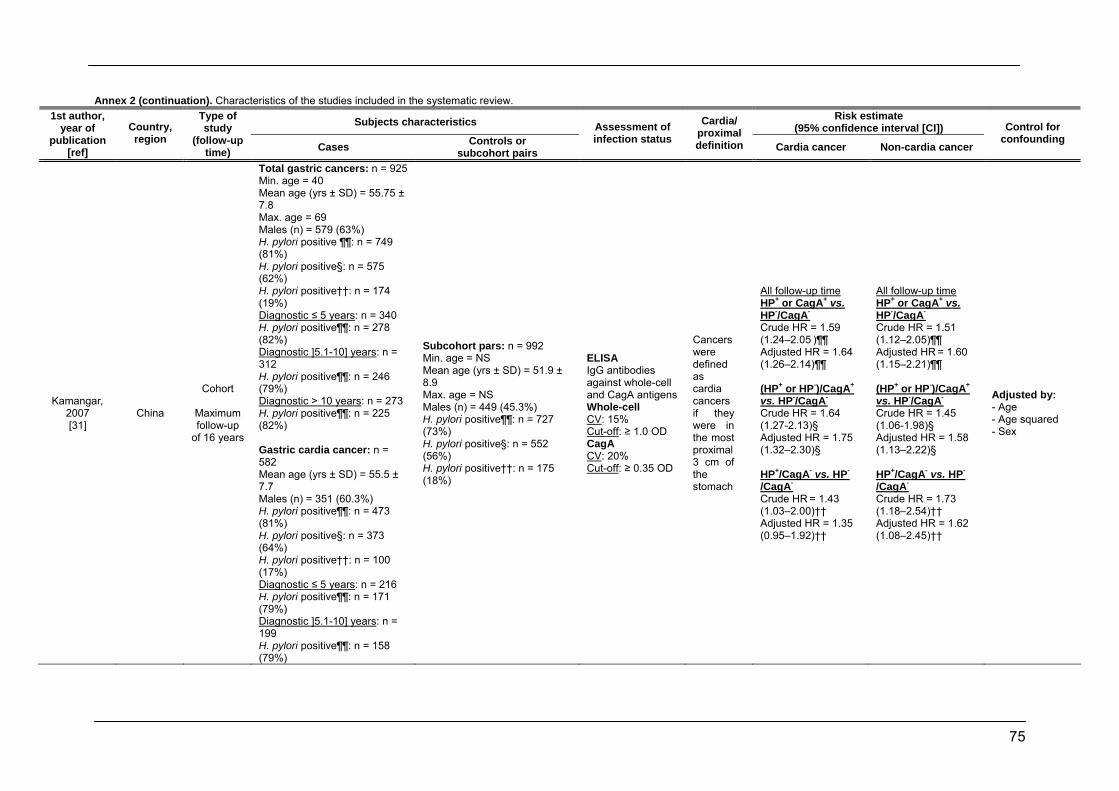
75
Annex 2 (continuation). Characteristics of the studies included in the systematic review.
Subjects characteristics Risk estimate (95% confidence interval [CI])
1st author, year of
publication [ref]
Country, region
Type of study
(follow-up time) Cases Controls or
subcohort pairs
Assessment of infection status
Cardia/ proximal definition Cardia cancer Non-cardia cancer
Control for confounding
Kamangar, 2007 [31]
China
Cohort
Maximum follow-up
of 16 years
Total gastric cancers: n = 925 Min. age = 40 Mean age (yrs ± SD) = 55.75 ± 7.8 Max. age = 69 Males (n) = 579 (63%) H. pylori positive ¶¶: n = 749 (81%) H. pylori positive§: n = 575 (62%) H. pylori positive††: n = 174 (19%) Diagnostic ≤ 5 years: n = 340 H. pylori positive¶¶: n = 278 (82%) Diagnostic ]5.1-10] years: n = 312 H. pylori positive¶¶: n = 246 (79%) Diagnostic > 10 years: n = 273 H. pylori positive¶¶: n = 225 (82%) Gastric cardia cancer: n = 582 Mean age (yrs ± SD) = 55.5 ± 7.7 Males (n) = 351 (60.3%) H. pylori positive¶¶: n = 473 (81%) H. pylori positive§: n = 373 (64%) H. pylori positive††: n = 100 (17%)
Diagnostic ≤ 5 years: n = 216 H. pylori positive¶¶: n = 171 (79%)
Diagnostic ]5.1-10] years: n = 199 H. pylori positive¶¶: n = 158 (79%)
Subcohort pars: n = 992 Min. age = NS Mean age (yrs ± SD) = 51.9 ± 8.9 Max. age = NS Males (n) = 449 (45.3%) H. pylori positive¶¶: n = 727 (73%) H. pylori positive§: n = 552 (56%) H. pylori positive††: n = 175 (18%)
ELISA IgG antibodies against whole-cell and CagA antigens Whole-cell CV: 15% Cut-off: ≥ 1.0 OD CagA CV: 20% Cut-off: ≥ 0.35 OD
Cancers were defined as cardia cancers if they were in the most proximal 3 cm of the stomach
All follow-up time HP+ or CagA+ vs. HP-/CagA- Crude HR = 1.59 (1.24–2.05 )¶¶ Adjusted HR = 1.64 (1.26–2.14)¶¶ (HP+ or HP-)/CagA+ vs. HP-/CagA- Crude HR = 1.64 (1.27-2.13)§ Adjusted HR = 1.75 (1.32–2.30)§ HP+/CagA- vs. HP-
/CagA- Crude HR = 1.43 (1.03–2.00)†† Adjusted HR = 1.35 (0.95–1.92)††
All follow-up time HP+ or CagA+ vs. HP-/CagA- Crude HR = 1.51 (1.12–2.05)¶¶ Adjusted HR = 1.60 (1.15–2.21)¶¶ (HP+ or HP-)/CagA+ vs. HP-/CagA- Crude HR = 1.45 (1.06-1.98)§ Adjusted HR = 1.58 (1.13–2.22)§ HP+/CagA- vs. HP-
/CagA- Crude HR = 1.73 (1.18–2.54)†† Adjusted HR = 1.62 (1.08–2.45)††
Adjusted by: - Age - Age squared - Sex
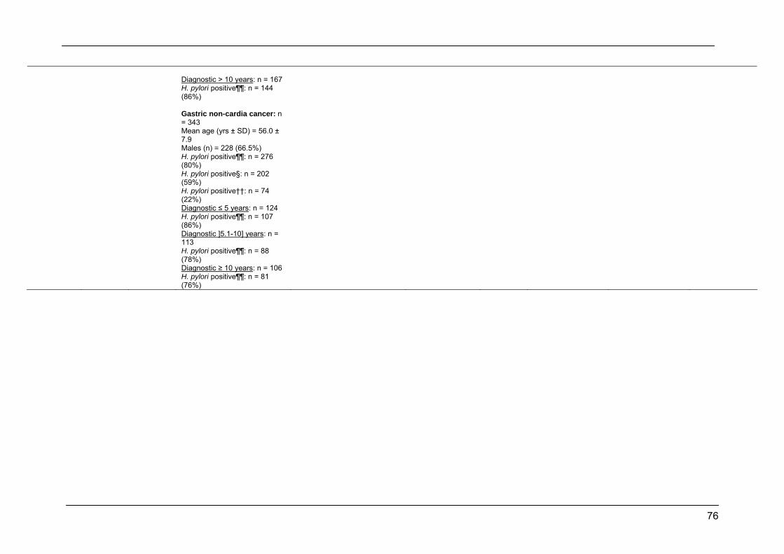
76
Diagnostic > 10 years: n = 167 H. pylori positive¶¶: n = 144 (86%) Gastric non-cardia cancer: n = 343 Mean age (yrs ± SD) = 56.0 ± 7.9 Males (n) = 228 (66.5%) H. pylori positive¶¶: n = 276 (80%) H. pylori positive§: n = 202 (59%) H. pylori positive††: n = 74 (22%)
Diagnostic ≤ 5 years: n = 124 H. pylori positive¶¶: n = 107 (86%)
Diagnostic ]5.1-10] years: n = 113 H. pylori positive¶¶: n = 88 (78%)
Diagnostic ≥ 10 years: n = 106 H. pylori positive¶¶: n = 81 (76%)
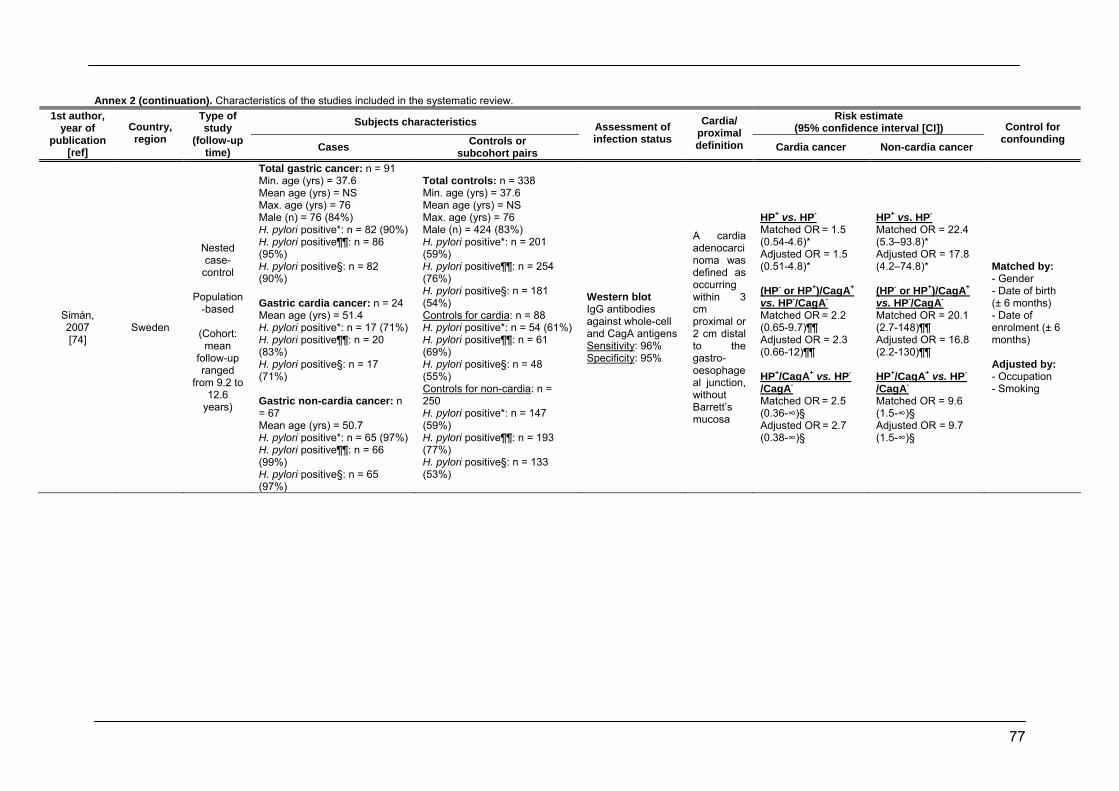
77
Annex 2 (continuation). Characteristics of the studies included in the systematic review.
Subjects characteristics Risk estimate (95% confidence interval [CI])
1st author, year of
publication [ref]
Country, region
Type of study
(follow-up time) Cases Controls or
subcohort pairs
Assessment of infection status
Cardia/ proximal definition Cardia cancer Non-cardia cancer
Control for confounding
Simán, 2007 [74]
Sweden
Nested case-
control
Population-based
(Cohort:
mean follow-up ranged
from 9.2 to 12.6
years)
Total gastric cancer: n = 91 Min. age (yrs) = 37.6 Mean age (yrs) = NS Max. age (yrs) = 76 Male (n) = 76 (84%) H. pylori positive*: n = 82 (90%) H. pylori positive¶¶: n = 86 (95%) H. pylori positive§: n = 82 (90%) Gastric cardia cancer: n = 24 Mean age (yrs) = 51.4 H. pylori positive*: n = 17 (71%) H. pylori positive¶¶: n = 20 (83%) H. pylori positive§: n = 17 (71%) Gastric non-cardia cancer: n = 67 Mean age (yrs) = 50.7 H. pylori positive*: n = 65 (97%) H. pylori positive¶¶: n = 66 (99%) H. pylori positive§: n = 65 (97%)
Total controls: n = 338 Min. age (yrs) = 37.6 Mean age (yrs) = NS Max. age (yrs) = 76 Male (n) = 424 (83%) H. pylori positive*: n = 201 (59%) H. pylori positive¶¶: n = 254 (76%) H. pylori positive§: n = 181 (54%) Controls for cardia: n = 88 H. pylori positive*: n = 54 (61%) H. pylori positive¶¶: n = 61 (69%) H. pylori positive§: n = 48 (55%) Controls for non-cardia: n = 250 H. pylori positive*: n = 147 (59%) H. pylori positive¶¶: n = 193 (77%) H. pylori positive§: n = 133 (53%)
Western blot IgG antibodies against whole-cell and CagA antigens Sensitivity: 96% Specificity: 95%
A cardia adenocarcinoma was defined as occurring within 3 cm proximal or 2 cm distal to the gastro-oesophageal junction, without Barrett’s mucosa
HP+ vs. HP- Matched OR = 1.5 (0.54-4.6)* Adjusted OR = 1.5 (0.51-4.8)* (HP- or HP+)/CagA+ vs. HP-/CagA- Matched OR = 2.2 (0.65-9.7)¶¶ Adjusted OR = 2.3 (0.66-12)¶¶ HP+/CagA+ vs. HP-
/CagA- Matched OR = 2.5 (0.36-∞)§ Adjusted OR = 2.7 (0.38-∞)§
HP+ vs. HP- Matched OR = 22.4 (5.3–93.8)* Adjusted OR = 17.8 (4.2–74.8)* (HP- or HP+)/CagA+ vs. HP-/CagA- Matched OR = 20.1 (2.7-148)¶¶ Adjusted OR = 16.8 (2.2-130)¶¶ HP+/CagA+ vs. HP-
/CagA- Matched OR = 9.6 (1.5-∞)§ Adjusted OR = 9.7 (1.5-∞)§
Matched by: - Gender - Date of birth (±/6 months) - Date of enrolment (±/6 months) Adjusted by: - Occupation - Smoking
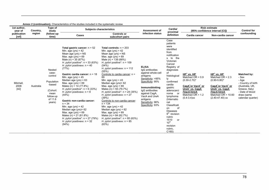
78
Annex 2 (continuation). Characteristics of the studies included in the systematic review.
Subjects characteristics Risk estimate (95% confidence interval [CI])
1st author, year of
publication [ref]
Country, region
Type of study
(follow-up time) Cases Controls or
subcohort pairs
Assessment of infection status
Cardia/ proximal definition Cardia cancer Non-cardia cancer
Control for confounding
Mitchell, 2008 [42]
Australia
Nested case-
control
Population-based
(Cohort:
mean follow-up of 11.6 years)
Total gastric cancer: n = 52 Min. age (yrs) = 42 Mean age (yrs) = NS Max. age (yrs) = 69 Male (n) = 35 (67%) H. pylori positive*: n = 33 (63%) H. pylori positiveװװ: n = 40 (77%) Gastric cardia cancer: n = 18 Min. age (yrs) = 45 Median age (yrs) = 63 Max. age (yrs) = 69 Males (n) = 14 (77.8%) H. pylori positive*: n = 6 (33%) H. pylori positiveװװ: n = 8 (44%) Gastric non-cardia cancer: n = 34 Min. age (yrs) = 42 Median age (yrs)= 62 Max. age (yrs) = 69 Males (n) = 21 (61.8%) H. pylori positive*: n = 27 (79%) H. pylori positiveװװ: n = 32 (94%)
Total controls: n = 203 Min. age (yrs) = 42 Mean age (yrs) = NS Max. age (yrs) = 69 Male (n) = 139 (68%) H. pylori positive*: n = 109 (54%) H. pylori positiveװװ: n = 112 (55%) Controls to cardia cancer: n = 69 Min. age (yrs) = 43 Median age (yrs)= 62 Max. age (yrs) = 69 Males (n) = 55 (79.7%) H. pylori positive*: n = 24 (35%) H. pylori positiveװװ: n = 27 (39%) Controls to non-cardia cancer: n = 134 Min. age (yrs) = 42 Median age (yrs) = 62 Max. age (yrs) = 69 Males (n) = 84 (62.7%) H. pylori positive*: n = 85 (63%) H. pylori positiveװװ: n = 85 (63%)
ELISA IgG antibodies against whole-cell antigens Sensitivity: >95% Specificity: 95% Immunoblotting IgG antibodies against CagA, VacA and UreA antigens Sensitivity: 96% Specificity: 93%
Case patients were identified from notifications to the Victorian Cancer Registry of diagnoses of histologically confirmed cardia gastric adenocarcinoma or lymphoma (International Classification of Diseases 9th revision rubric 1510 or 10th revision rubric, C160)
HP+ vs. HP- Matched OR = 0.9 (0.30-2.70)* CagA+or VacA+ or UreA+ vs. CagA-
/VacA-/UreA- Matched OR = 1.2 װװ(0.4-3.4)
HP+ vs. HP- Matched OR = 2.3 (0.90-5.80)* CagA+or VacA+ or UreA+ vs. CagA-
/VacA-/UreA- Matched OR = 10.60 װװ (2.40-47.40)
Matched by: - Sex - Age - Country of birth (Australia, UK, Greece, Italy) - Date of blood draw (same calendar quarter)
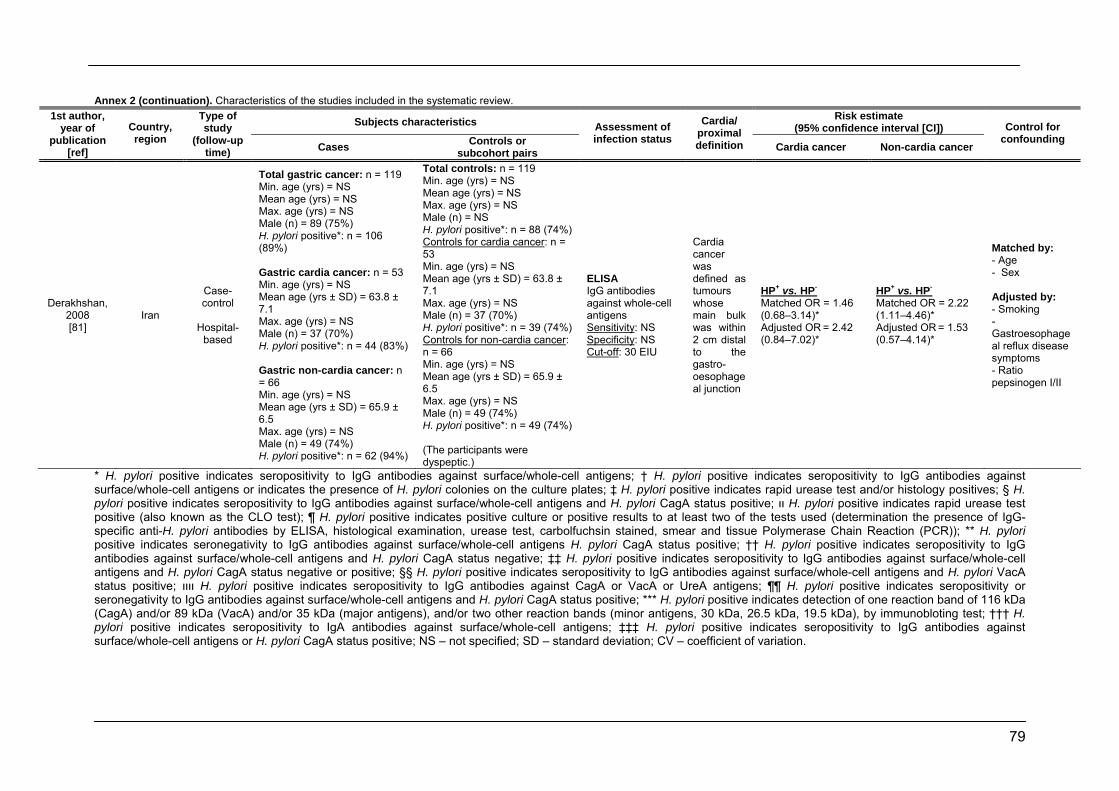
79
Annex 2 (continuation). Characteristics of the studies included in the systematic review.
Subjects characteristics Risk estimate (95% confidence interval [CI])
1st author, year of
publication [ref]
Country, region
Type of study
(follow-up time) Cases Controls or
subcohort pairs
Assessment of infection status
Cardia/ proximal definition Cardia cancer Non-cardia cancer
Control for confounding
Derakhshan, 2008 [81]
Iran
Case-control
Hospital-
based
Total gastric cancer: n = 119 Min. age (yrs) = NS Mean age (yrs) = NS Max. age (yrs) = NS Male (n) = 89 (75%) H. pylori positive*: n = 106 (89%) Gastric cardia cancer: n = 53 Min. age (yrs) = NS Mean age (yrs ± SD) = 63.8 ± 7.1 Max. age (yrs) = NS Male (n) = 37 (70%) H. pylori positive*: n = 44 (83%) Gastric non-cardia cancer: n = 66 Min. age (yrs) = NS Mean age (yrs ± SD) = 65.9 ± 6.5 Max. age (yrs) = NS Male (n) = 49 (74%) H. pylori positive*: n = 62 (94%)
Total controls: n = 119 Min. age (yrs) = NS Mean age (yrs) = NS Max. age (yrs) = NS Male (n) = NS H. pylori positive*: n = 88 (74%) Controls for cardia cancer: n = 53 Min. age (yrs) = NS Mean age (yrs ± SD) = 63.8 ± 7.1 Max. age (yrs) = NS Male (n) = 37 (70%) H. pylori positive*: n = 39 (74%) Controls for non-cardia cancer: n = 66 Min. age (yrs) = NS Mean age (yrs ± SD) = 65.9 ± 6.5 Max. age (yrs) = NS Male (n) = 49 (74%) H. pylori positive*: n = 49 (74%) (The participants were dyspeptic.)
ELISA IgG antibodies against whole-cell antigens Sensitivity: NS Specificity: NS Cut-off: 30 EIU
Cardia cancer was defined as tumours whose main bulk was within 2 cm distal to the gastro-oesophageal junction
HP+ vs. HP- Matched OR = 1.46 (0.68–3.14)* Adjusted OR = 2.42 (0.84–7.02)*
HP+ vs. HP- Matched OR = 2.22 (1.11–4.46)* Adjusted OR = 1.53 (0.57–4.14)*
Matched by: - Age - Sex Adjusted by: - Smoking - Gastroesophageal reflux disease symptoms - Ratio pepsinogen I/II
* H. pylori positive indicates seropositivity to IgG antibodies against surface/whole-cell antigens; † H. pylori positive indicates seropositivity to IgG antibodies against surface/whole-cell antigens or indicates the presence of H. pylori colonies on the culture plates; ‡ H. pylori positive indicates rapid urease test and/or histology positives; § H. pylori positive indicates seropositivity to IgG antibodies against surface/whole-cell antigens and H. pylori CagA status positive; װ H. pylori positive indicates rapid urease test positive (also known as the CLO test); ¶ H. pylori positive indicates positive culture or positive results to at least two of the tests used (determination the presence of IgG-specific anti-H. pylori antibodies by ELISA, histological examination, urease test, carbolfuchsin stained, smear and tissue Polymerase Chain Reaction (PCR)); ** H. pylori positive indicates seronegativity to IgG antibodies against surface/whole-cell antigens H. pylori CagA status positive; †† H. pylori positive indicates seropositivity to IgG antibodies against surface/whole-cell antigens and H. pylori CagA status negative; ‡‡ H. pylori positive indicates seropositivity to IgG antibodies against surface/whole-cell antigens and H. pylori CagA status negative or positive; §§ H. pylori positive indicates seropositivity to IgG antibodies against surface/whole-cell antigens and H. pylori VacA status positive; װװ H. pylori positive indicates seropositivity to IgG antibodies against CagA or VacA or UreA antigens; ¶¶ H. pylori positive indicates seropositivity or seronegativity to IgG antibodies against surface/whole-cell antigens and H. pylori CagA status positive; *** H. pylori positive indicates detection of one reaction band of 116 kDa (CagA) and/or 89 kDa (VacA) and/or 35 kDa (major antigens), and/or two other reaction bands (minor antigens, 30 kDa, 26.5 kDa, 19.5 kDa), by immunobloting test; ††† H. pylori positive indicates seropositivity to IgA antibodies against surface/whole-cell antigens; ‡‡‡ H. pylori positive indicates seropositivity to IgG antibodies against surface/whole-cell antigens or H. pylori CagA status positive; NS – not specified; SD – standard deviation; CV – coefficient of variation.
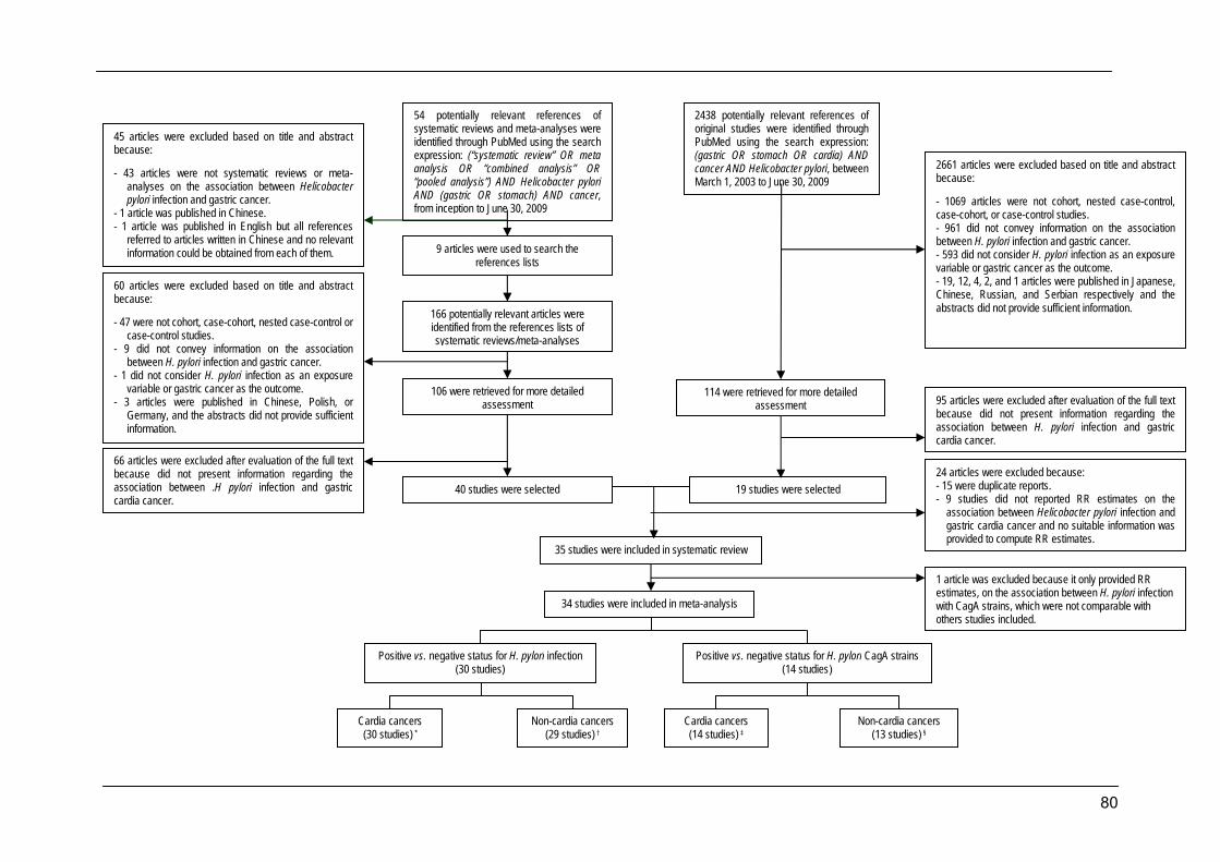
80
54 potentially relevant references of systematic reviews and meta-analyses were identified through PubMed using the search expression: (“systematic review” OR meta analysis OR “combined analysis” OR “pooled analysis”) AND Helicobacter pylori AND (gastric OR stomach) AND cancer, from inception to June 30, 2009
9 articles were used to search the references lists
166 potentially relevant articles were identified from the references lists of systematic reviews/meta-analyses
106 were retrieved for more detailed assessment
40 studies were selected
34 studies were included in meta-analysis
2438 potentially relevant references of original studies were identified through PubMed using the search expression: (gastric OR stomach OR cardia) AND cancer AND Helicobacter pylori, between March 1, 2003 to June 30, 2009
19 studies were selected
45 articles were excluded based on title and abstract because: - 43 articles were not systematic reviews or meta-
analyses on the association between Helicobacter pylori infection and gastric cancer.
- 1 article was published in Chinese. - 1 article was published in English but all references
referred to articles written in Chinese and no relevant information could be obtained from each of them.
60 articles were excluded based on title and abstract because: - 47 were not cohort, case-cohort, nested case-control or
case-control studies. - 9 did not convey information on the association
between H. pylori infection and gastric cancer. - 1 did not consider H. pylori infection as an exposure
variable or gastric cancer as the outcome. - 3 articles were published in Chinese, Polish, or
Germany, and the abstracts did not provide sufficient information.
66 articles were excluded after evaluation of the full text because did not present information regarding the association between .H pylori infection and gastric cardia cancer.
95 articles were excluded after evaluation of the full text because did not present information regarding the association between H. pylori infection and gastric cardia cancer.
114 were retrieved for more detailed assessment
2661 articles were excluded based on title and abstract because: - 1069 articles were not cohort, nested case-control, case-cohort, or case-control studies. - 961 did not convey information on the association between H. pylori infection and gastric cancer. - 593 did not consider H. pylori infection as an exposure variable or gastric cancer as the outcome. - 19, 12, 4, 2, and 1 articles were published in Japanese, Chinese, Russian, and Serbian respectively and the abstracts did not provide sufficient information.
Positive vs. negative status for H. pylori infection (30 studies)
Positive vs. negative status for H. pylori CagA strains (14 studies)
35 studies were included in systematic review
Cardia cancers (30 studies) *
Cardia cancers (14 studies) ‡
Non-cardia cancers (29 studies) †
Non-cardia cancers (13 studies) §
24 articles were excluded because: - 15 were duplicate reports. - 9 studies did not reported RR estimates on the
association between Helicobacter pylori infection and gastric cardia cancer and no suitable information was provided to compute RR estimates.
1 article was excluded because it only provided RR estimates, on the association between H. pylori infection with CagA strains, which were not comparable with others studies included.
81
Figure 1. Systematic review flow-chart.
* From 30 studies 24 provided adjusted RR estimates; † From 29 studies 23 provided adjusted RR estimates; ‡ From 14 studies 10 provided
adjusted RR estimates; § From 13 studies 9 provided adjusted RR estimates.
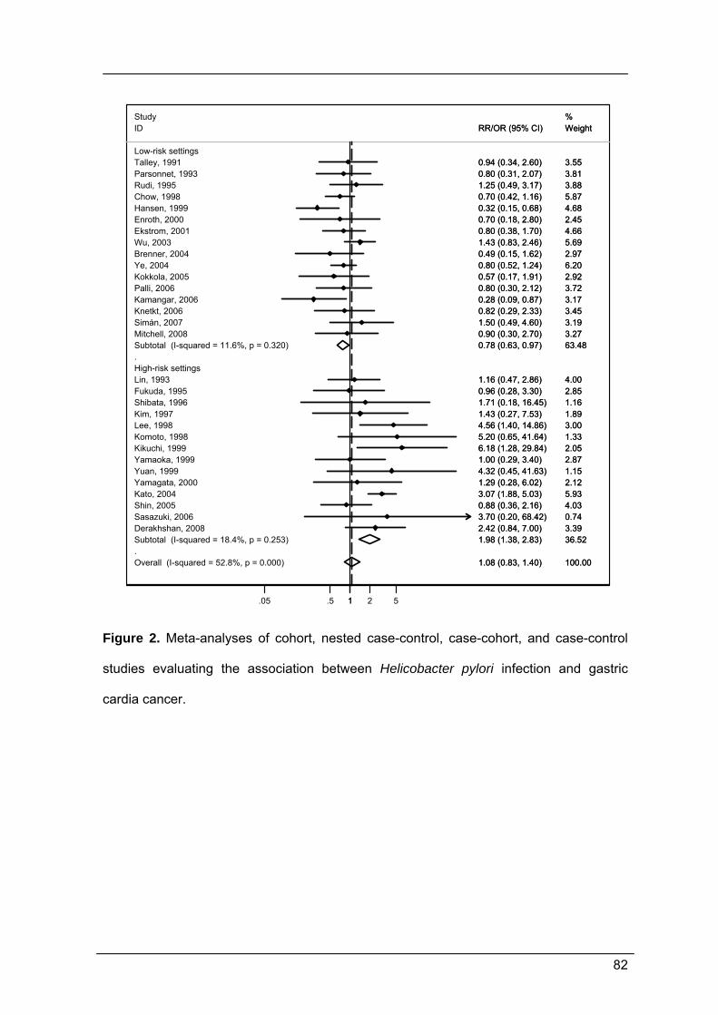
82
.
.
Overall (I-squared = 52.8%, p = 0.000)
Ye, 2004
Wu, 2003
Kim, 1997
Chow, 1998
Lin, 1993
Kamangar, 2006
Parsonnet, 1993
Knetkt, 2006
Study
Sasazuki, 2006
Rudi, 1995
Enroth, 2000
Yuan, 1999
ID
Shin, 2005
Yamagata, 2000
High-risk settings
Subtotal (I-squared = 11.6%, p = 0.320)
Yamaoka, 1999
Simán, 2007
Komoto, 1998
Palli, 2006
Ekstrom, 2001
Talley, 1991
Shibata, 1996
Subtotal (I-squared = 18.4%, p = 0.253)
Brenner, 2004
Fukuda, 1995
Derakhshan, 2008
Hansen, 1999
Kokkola, 2005
Lee, 1998
Mitchell, 2008
Kato, 2004
Low-risk settings
Kikuchi, 1999
1.08 (0.83, 1.40)
0.80 (0.52, 1.24)
1.43 (0.83, 2.46)
1.43 (0.27, 7.53)
0.70 (0.42, 1.16)
1.16 (0.47, 2.86)
0.28 (0.09, 0.87)
0.80 (0.31, 2.07)
0.82 (0.29, 2.33)
3.70 (0.20, 68.42)
1.25 (0.49, 3.17)
0.70 (0.18, 2.80)
4.32 (0.45, 41.63)
RR/OR (95% CI)
0.88 (0.36, 2.16)
1.29 (0.28, 6.02)
0.78 (0.63, 0.97)
1.00 (0.29, 3.40)
1.50 (0.49, 4.60)
5.20 (0.65, 41.64)
0.80 (0.30, 2.12)
0.80 (0.38, 1.70)
0.94 (0.34, 2.60)
1.71 (0.18, 16.45)
1.98 (1.38, 2.83)
0.49 (0.15, 1.62)
0.96 (0.28, 3.30)
2.42 (0.84, 7.00)
0.32 (0.15, 0.68)
0.57 (0.17, 1.91)
4.56 (1.40, 14.86)
0.90 (0.30, 2.70)
3.07 (1.88, 5.03)
6.18 (1.28, 29.84)
100.00
6.20
5.69
1.89
5.87
4.00
3.17
3.81
3.45
%
0.74
3.88
2.45
1.15
Weight
4.03
2.12
63.48
2.87
3.19
1.33
3.72
4.66
3.55
1.16
36.52
2.97
2.85
3.39
4.68
2.92
3.00
3.27
5.93
2.05
1.05 .5 1 2 5
Figure 2. Meta-analyses of cohort, nested case-control, case-cohort, and case-control
studies evaluating the association between Helicobacter pylori infection and gastric
cardia cancer.
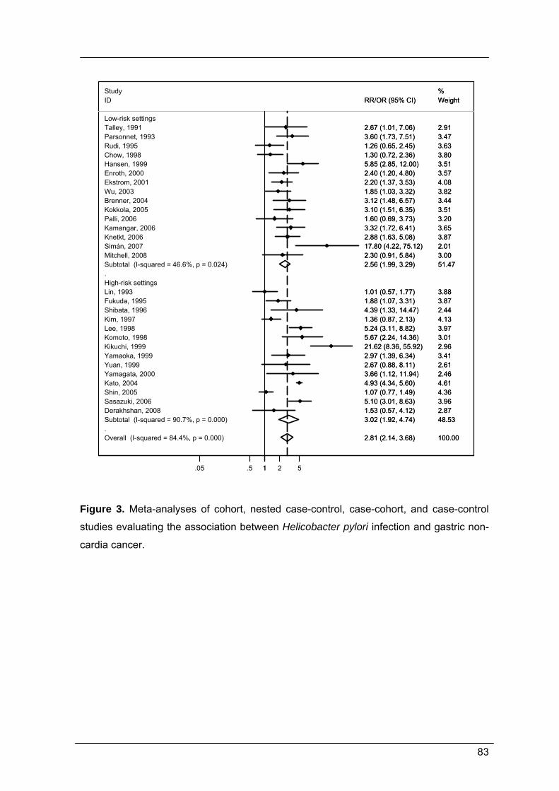
83
.
.Overall (I-squared = 84.4%, p = 0.000)
High-risk settings
Talley, 1991
Kato, 2004
Mitchell, 2008
Lee, 1998
ID
Yuan, 1999
Subtotal (I-squared = 90.7%, p = 0.000)
Knetkt, 2006Kamangar, 2006
Brenner, 2004
Shibata, 1996
Enroth, 2000
Kikuchi, 1999
Sasazuki, 2006
Hansen, 1999
Lin, 1993
Yamagata, 2000
Ekstrom, 2001
Chow, 1998
Subtotal (I-squared = 46.6%, p = 0.024)
Study
Kim, 1997
Fukuda, 1995
Palli, 2006
Low-risk settings
Simán, 2007
Parsonnet, 1993
Kokkola, 2005
Derakhshan, 2008
Shin, 2005
Rudi, 1995
Wu, 2003
Komoto, 1998
Yamaoka, 1999
2.81 (2.14, 3.68)
2.67 (1.01, 7.06)
4.93 (4.34, 5.60)
2.30 (0.91, 5.84)
5.24 (3.11, 8.82)
RR/OR (95% CI)
2.67 (0.88, 8.11)
3.02 (1.92, 4.74)
2.88 (1.63, 5.08)3.32 (1.72, 6.41)
3.12 (1.48, 6.57)
4.39 (1.33, 14.47)
2.40 (1.20, 4.80)
21.62 (8.36, 55.92)
5.10 (3.01, 8.63)
5.85 (2.85, 12.00)
1.01 (0.57, 1.77)
3.66 (1.12, 11.94)
2.20 (1.37, 3.53)
1.30 (0.72, 2.36)
2.56 (1.99, 3.29)
1.36 (0.87, 2.13)
1.88 (1.07, 3.31)
1.60 (0.69, 3.73)
17.80 (4.22, 75.12)
3.60 (1.73, 7.51)
3.10 (1.51, 6.35)
1.53 (0.57, 4.12)
1.07 (0.77, 1.49)
1.26 (0.65, 2.45)
1.85 (1.03, 3.32)
5.67 (2.24, 14.36)
2.97 (1.39, 6.34)
100.00
2.91
4.61
3.00
3.97
Weight
2.61
48.53
3.873.65
3.44
2.44
3.57
2.96
3.96
3.51
3.88
2.46
4.08
3.80
51.47
%
4.13
3.87
3.20
2.01
3.47
3.51
2.87
4.36
3.63
3.82
3.01
3.41
1.05 .5 1 2 5
Figure 3. Meta-analyses of cohort, nested case-control, case-cohort, and case-control
studies evaluating the association between Helicobacter pylori infection and gastric non-
cardia cancer.
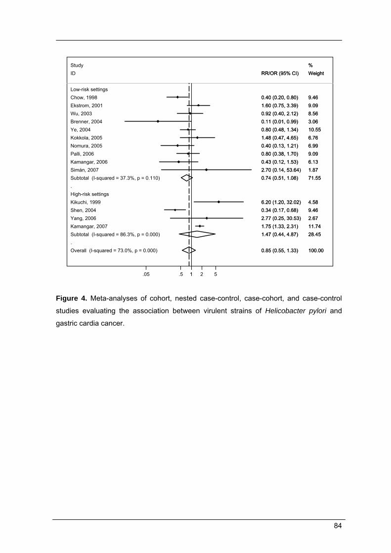
84
.
.Overall (I-squared = 73.0%, p = 0.000)
Study
Kikuchi, 1999
Subtotal (I-squared = 86.3%, p = 0.000)
High-risk settings
Kamangar, 2007
Ekstrom, 2001
Palli, 2006
ID
Subtotal (I-squared = 37.3%, p = 0.110)
Wu, 2003
Nomura, 2005
Ye, 2004
Low-risk settings
Yang, 2006
Brenner, 2004
Shen, 2004
Simán, 2007
Kokkola, 2005
Chow, 1998
Kamangar, 2006
0.85 (0.55, 1.33)
6.20 (1.20, 32.02)
1.47 (0.44, 4.87)1.75 (1.33, 2.31)
1.60 (0.75, 3.39)
0.80 (0.38, 1.70)
RR/OR (95% CI)
0.74 (0.51, 1.08)
0.92 (0.40, 2.12)
0.40 (0.13, 1.21)
0.80 (0.48, 1.34)
2.77 (0.25, 30.53)
0.11 (0.01, 0.99)
0.34 (0.17, 0.68)
2.70 (0.14, 53.64)
1.48 (0.47, 4.65)
0.40 (0.20, 0.80)
0.43 (0.12, 1.53)
100.00
%
4.58
28.4511.74
9.09
9.09
Weight
71.55
8.56
6.99
10.55
2.67
3.06
9.46
1.87
6.76
9.46
6.13
1.05 .5 1 2 5
Figure 4. Meta-analyses of cohort, nested case-control, case-cohort, and case-control
studies evaluating the association between virulent strains of Helicobacter pylori and
gastric cardia cancer.
85
.
.
Overall (I-squared = 91.4%, p = 0.000)
Kamangar, 2007
Brenner, 2004
Ekstrom, 2001
Subtotal (I-squared = 95.7%, p = 0.000)
Chow, 1998
Kokkola, 2005
Wu, 2003
High-risk settings
Kamangar, 2006
Subtotal (I-squared = 76.4%, p = 0.000)
Nomura, 2005
Palli, 2006
Yang, 2006
Simán, 2007
Kikuchi, 1999
ID
Shen, 2004
Low-risk settings
Study
3.63 (1.93, 6.84)
1.58 (1.13, 2.21)
3.17 (1.61, 6.24)
21.70 (9.63, 48.87)
2.08 (0.40, 10.69)
1.40 (0.70, 2.80)
6.80 (3.11, 14.86)
2.58 (1.22, 5.44)
8.93 (3.27, 24.39)
4.59 (2.79, 7.57)
3.16 (2.22, 4.50)
4.70 (2.48, 8.92)
3.69 (1.05, 12.96)
9.70 (0.97, 97.00)
19.40 (7.21, 52.19)
RR/OR (95% CI)
0.20 (0.11, 0.37)
100.00
8.81
8.20
7.87
31.24
8.16
7.95
8.03
7.37
68.76
8.79
8.28
6.67
4.12
7.41
Weight
8.35
%
1.05 .5 1 2 5
Figure 5. Meta-analyses of cohort, nested case-control, case-cohort, and case-control
studies evaluating the association between virulent strains of Helicobacter pylori and
gastric non-cardia cancer.
86
3.0%
6.9%
2.7%2.6%
15.0%
2.0%
6.8%
4.1%
20.0%
4.3%3.5%
3.2%3.7%
4.4%
13.1%
3.1%
0.2
.4.6
.8St
anda
rd e
rror
of R
R
.2 .5 1 2 4RR
Funnel plot with pseudo 95% confidence limits
4.8%
2.5%2.6%
4.7%
1.6%1.4%
0.7%
3.4%
15.8%
0.5%
0.7%
2.8%
0.9%
5.3%
0.5
11.
5St
anda
rd e
rror
of R
R
.5 1 2 5 10 20RR
Funnel plot with pseudo 95% confidence limits
8.7%10.9%
5.4%
11.2%
17.2%
4.4%
8.0%
4.1%
11.9%
7.5%6.9%8.9%
7.1% 7.4%
1.9%
0.2
.4.6
.8St
anda
rd e
rror
of R
R
1 2 5 10 20RR
Funnel plot with pseudo 95% confidence limits
12.1%
35.3%
19.0%
3.9%
12.0%
3.1%
6.7%
2.7%2.7%
236.5%
13.9%14.1%
4.4% 3.5%
0.2
.4.6
Stan
dard
err
or o
f RR
1 2 5 10 25RR
Funnel plot with pseudo 95% confidence limits
87
Figure 6. Funnel plot of cohort, nested case-control, case-cohort, and case-control studies evaluating the association between Helicobacter
pylori infection and gastric cardia cancer in low-risk settings (top left) and in high-risk settings (top right). Begg adjusted rank correlation test
(low-risk settings: p=0.499; high-risk settings: p=0.324), Egger’s regression asymmetry test (low-risk settings: p=0.498; high-risk settings:
p=0.842). Funnel plot of cohort, nested case-control, case-cohort, and case-control studies evaluating the association between Helicobacter
pylori infection and gastric non-cardia cancer in low-risk settings (bottom left) and in high-risk settings (bottom right). Begg adjusted rank
correlation test (low-risk settings: p=0.198; high-risk settings: p=0.661), Egger’s regression asymmetry test (low-risk settings: p=0.082; high-risk
settings: p=0.288).

88
Is cardia cancer etiologically different from distal stomach cancer?
89
ABSTRACT
Objective: The coexistence of two etiologically distinct types of cardia cancer, with
distinctive histological characteristics of the neoplastic and non-neoplastic gastric
mucosa may explain the heterogeneous evidence on its association with H. pylori
infection. We compared gastric cardia and non-cardia cancers regarding the frequency
of H. pylori infection, the histological characteristics of the non-neoplastic gastric
mucosa, and the tumor histological type.
Methods: We evaluated 41 cardia and 229 non-cardia cancer cases undergoing
gastrectomy, and 270 community controls. H. pylori infection and CagA infection status
were assessed by ELISA and Western Blot, respectively. Histological characteristics of
the neoplastic and non-neoplastic mucosa were obtained from pathology reports. The
association between infection and cancers with different location was quantified in a
case-control analysis and cardia and non-cardia cancers were further compared.
Results: No positive relation was found for H. pylori infection, but CagA-positive strains
were associated with an increased risk of non-cardia cancer (OR=1.97, 95%CI: 1.35-
2.87). Thirty-one (75.6%) cardia cancer cases, predominantly of the intestinal type
(67.7%), had non-neoplastic atrophic mucosa, as well as 173 (75.6%) non-cardia
cancers (54.3% of the intestinal type). Among cases occurring in non-atrophic patients,
the proportion of cancers of the Laurén’s intestinal type was 70.0% for cardia and
39.3% for non-cardia gastric cancers.
Conclusions: Cardia and non-cardia cancers were similar regarding the relation with
infection, histological type and condition of the non-neoplastic mucosa, supporting the
predominance of cardia cancers determined by H. pylori infection in this Western high-
risk setting.
90
INTRODUCTION
The discovery of Helicobacter pylori [1], and its recognition as a human
carcinogen [2] proved to be a key point in the understanding of the gastric cancer
etiology. Both cohort and case-control studies show an average three-fold increased
risk of distal gastric cancer among the infected, but apparently no positive relation
between H. pylori infection and cardia cancer. The evidence supporting this lack of
association, however, is based on contradictory findings [3, 4, 5]. Studies from Western
countries support a negative association between H. pylori infection and cardia cancer
[6, 7, 8, 9, 10, 11, 12], whereas there is a trend towards positive association in studies
from Eastern Asia [9, 12, 13, 14, 15].
The gastric cardia is a small anatomical region, which can be overgrown by
tumors that originate from adjacent mucosal sites. Tumors described as “cardia
cancers” include a mixture of neoplasms arising from the lower esophagus as well as
the gastric cardia, to a higher or lesser extent depending on the frequency of
esophageal and gastric adenocarcinomas on the populations under study [16]. The
observation of a positive association with gastric atrophy [17, 18, 19], despite no overall
relation with H. pylori infection, supports the coexistence of two etiologically distinct
types of cardia cancer [20], one associated with H. pylori-induced atrophic gastritis,
etiologically similar to non-cardia cancer, more frequent in populations with higher
overall gastric cancer incidence; and the other arising in non-atrophic gastric mucosa,
associated with acid/bile-induced damage to the distal esophagus, resembling
esophageal adenocarcinoma, and likely to have a higher relative frequency in settings
with low-gastric cancer risk.
The potential for misclassification of gastric cancer topography brings important
challenges to the study of the cardia cancer etiology, as it is usually impossible to
determine the origin of these cancers by examining them grossly or microscopically
[20]. However, the hypothesis described above provides a basis for a surrogate
definition of two subtypes of gastric cardia cancer, and may be useful for a proper
assessment of its etiology [20].
This report comprises a case-control study, in which cardia and non-cardia
cancers are compared with community controls regarding the frequency of infection
with H. pylori, taking into account the virulence of the infecting strains, and a case-case
analysis for the comparison of cardia and non-cardia cancers regarding the histological
characteristics of the neoplastic and non-neoplastic mucosas. The latter comparison is
stratified according to the infection status of the cases, aiming to provide evidence on
91
the frequency of etiologically distinct cases of cardia cancer, either resembling distal
stomach cancer or esophageal adenocarcinoma.
92
METHODS
Cases
From 2001 to 2006, we identified 709 incident cases of gastric adenocarcinoma
with no previous cancer diagnosis (except for skin non-melanoma) and not having
performed a subtotal gastrectomy for benign conditions. As previously described [21,
22], patients were admitted to the surgery wards of the two largest public hospitals
providing care to cancer patients in the North of the country, Hospital de S. João and
Instituto Português de Oncologia Francisco Gentil, both in Porto. Cancer diagnosis was
based on gastrectomy specimens, endoscopic biopsy material or the evaluation of
metastasis.
A blood sample was drawn from 477 cases and serum was kept frozen at -20ºC.
In addition, 207 cases were excluded because no gastrectomy specimen was
available, precluding the proper assessment of tumor histological type and
characterization of gastric lesions in the non-neoplastic mucosa. The remaining 270
cases were not significantly different from those excluded, regarding gender (58.9% vs.
60.5% males, p=0.682), age (median: 66 vs. 65 years, p=0.542) or education (median
number of school years: 4 vs. 4 years, p=0.426).
Controls
Controls were part of a representative sample of the adult population of Porto [23,
24]. Participants were recruited by random digit dialing using households as the
sampling frame, followed by simple random sampling to select one eligible person
among permanent residents in each household that was invited to visit our department
for interview and blood collection. For each of the 270 gastric cancer cases, we
selected age and gender frequency-matched controls.
Assessment of H. pylori infection status
H. pylori infection status was assessed using an anti-H. pylori IgG antibody
Enzyme Linked Immunoassay (ELISA) (Anti-Helicobacter pylori ELISA, Eurolmmun®,
Lubeck, Germany) on serum samples. Participants were classified according to
manufacturer instructions as negative if they had less than 16 RU/ml, as borderline if
their antibody concentration was between 16 and 22 RU/ml and as positive if the
antibody level was equal or greater than 22 RU/ml. For analysis, subjects having
borderline results were considered infected.
93
Further testing of H. pylori infection status was performed by Western Blot
(Helico Blot 2.1, Genelabs Diagnostics®, Singapore). The assay was conducted as
proposed by the manufacturer, and the results were interpreted following the
recommended criteria for CagA seropositivity: presence of the 116kD band (CagA) with
one or more of the following bands: 89kD (VacA); 37kD; 35kD; 30kD (UreA) and
19.5kD together.
Assessment of the histological characteristics of the neoplastic and non-neoplastic
mucosas of cases
For each case, the anatomic site was classified following image or pathology
descriptions. Cardia cancer was defined as a tumor in the cardioesophageal junction,
esophagogastric junction or gastroesophageal junction [16]. Information on the tumor’s
histological type, according to Lauren´s criteria (intestinal, diffuse, or mixed) [25], was
obtained for cases submitted to gastrectomy. The presence of gastric lesions in the
non-neoplastic mucosa was registered in the surgical specimen pathology reports. In
this study, we considered that atrophy was present in individuals having histological
evidence of chronic atrophic gastritis regardless of the presence of other gastric
lesions.
Statistical analysis
Odds Ratios (OR) and the corresponding 95% confidence intervals (95%CI) were
computed by multinomial logistic regression to quantify the association of between H.
pylori infection and gastric cardia and non-cardia cancers and unconditional logistic
regression to compare cardia and non-cardia cancer cases. All analyses were
conducted using STATA®, version 9.2.
Ethics
The ethics committees of the involved hospitals approved the study, and all
participants provided written informed consent.
94
RESULTS
Case-control study
No positive association was observed between H. pylori infection and gastric
cancer, neither for cardia (OR=1.72, 95%CI: 0.50-5.90) or non-cardia cancers
(OR=0.93, 95%CI: 0.55-1.58). Infection with CagA-positive strains was associated with
increased risk of non-cardia cancer (OR=1.97, 95%CI: 1.35-2.87) but not with cardia
cancer (OR=1.69, 95%CI: 0.84-3.42) (Table 1).
Case-case analysis
The frequency of infection with CagA-positive strains was similar in cardia and
non-cardia cancers (age- and gender-adjusted OR=0.84, 95%CI: 0.40-1.73).
Approximately three quarters of the cases classified as H. pylori-infected had
non-neoplastic atrophic mucosa. Nearly half of the cardia and non-cardia cancers were
of the intestinal histological type, regardless of the characteristics of the non-neoplastic
mucosa (Table 2). Similar results were observed among subjects infected with CagA-
positive strains (Table 3).
Among cases classified as H. pylori-negative, a similar proportion of both cardia
and non-cardia cancers had non-neoplastic atrophic mucosa. All cardia and 30.0% of
non-cardia cancers were of the Laurén’s intestinal type for cases occurring in non-
atrophic patients. The same trend was found for cases presenting non-neoplastic
atrophic mucosa (Table 2). Similar results were observed among subjects not H. pylori-
infected or infected with CagA-negative strains (Table 3).
95
DISCUSSION
The infection with H. pylori CagA-positive strains was associated with an
increased risk of both cardia and non-cardia cancers. Three-quarters of the gastric
cardia cancer cases observed in this high-gastric cancer risk setting had non-
neoplastic atrophic mucosa, and most of them were of the intestinal histological type,
similarly to what was observed for non-cardia cancers. Among cases occurring in non-
infected patients, cardia cancers were predominantly of the intestinal type whereas
only one third of non-cardia cancers presented with Laurén’s intestinal type, regardless
of the characteristics of the non-neoplastic mucosa.
As in other high-risk settings, we found no association between H. pylori infection
and gastric cancer. The high prevalence of infection observed in the general
Portuguese population contributes to weak associations between infection and gastric
cancer, namely at distal locations [4, 26]. In the general population the prevalence of
infection increases with age [27] and infection tends to persist during adulthood, while
cancer cases are more likely to have been infected early in life and to lack evidence of
infection as cancer progresses [28]. In high-prevalence settings, the difference
between cancer cases and controls regarding the proportion of subjects infected later
in life tends to be clearer and the potential for underestimation of the association
between infection and cancer in case-control designs is even higher [26]. In our study,
however, infection with CagA-positive strains was associated with an increased risk of
gastric cancer, in accordance with previous studies reporting that CagA antibodies
reflect past infection more accurately than surface antibodies detected by conventional
IgG ELISA [29].
The positive association between infection and gastric cardia cancer observed in
our study, although divergent from the mainstream opinion that there is no such
relation [4, 5, 30], is consistent with the results obtained in samples from populations
with high-risk for gastric cancer and prevalence of infection [31], and probably reflects
the predominance of the gastric cardia cancer subtype arising from H pylori-induced
atrophic gastritis. These tumors are expected to be similar to the non-cardia cancers
regarding the relation with infection and histological type, which is also supported by
our results. Among H. pylori-infected cases, a 1:1 ratio of intestinal and diffuse/mixed
histological subtypes was observed for both cardia and non-cardia cancers. However,
unlike non-cardia cancers, the cardia cases occurring in non-infected patients had a
much higher proportion of intestinal versus diffuse/mixed histological subtype. This
96
predominant intestinal histological subtype is similar to that reported in esophageal
adenocarcinoma [32] and supports an etiological resemblance with the latter.
The exclusion of cases not submitted to gastrectomy, as all histological
characteristics were assessed in surgical specimens, precludes the generalization of
our results to all gastric cancer cases. Since diffuse cancers have a worse prognosis
than those of the Laurén’s intestinal type [33, 34], this selection bias may have
contributed to an oversampling of the latter, among which the presence of an atrophic
non-neoplastic mucosa is more likely [35].
The interpretation of these results, although limited by the small sample size is
driven by a sound and well documented hypothesis. Our findings support the existence
of two etiologically different types of gastric cardia cancer, one of which is positively
associated with H. pylori infection and therefore more frequent in high-risk settings.
These results contribute to explain the heterogeneous results across high and low
gastric cancer risk settings regarding the association with H. pylori infection.
97
REFERENCES
1 Unidentified curved bacilli on gastric epithelium in active chronic gastritis. Lancet
1983;1:1273-5.
2 Schistosomes, liver flukes and Helicobacter pylori. IARC Working Group on the
Evaluation of Carcinogenic Risks to Humans. Lyon, 7-14 June 1994. IARC
Monogr Eval Carcinog Risks Hum 1994;61:1-241.
3 Dawsey SM, Mark SD, Taylor PR, Limburg PJ. Gastric cancer and H pylori. Gut
2002;51:457-8.
4 Huang JQ, Sridhar S, Chen Y, Hunt RH. Meta-analysis of the relationship
between Helicobacter pylori seropositivity and gastric cancer. Gastroenterology
1998;114:1169-79.
5 Huang JQ, Zheng GF, Sumanac K, Irvine EJ, Hunt RH. Meta-analysis of the
relationship between cagA seropositivity and gastric cancer. Gastroenterology
2003;125:1636-44.
6 Brenner H, Arndt V, Stegmaier C, Ziegler H, Rothenbacher D. Is Helicobacter
pylori infection a necessary condition for noncardia gastric cancer? Am J
Epidemiol 2004;159:252-8.
7 Enroth H, Kraaz W, Rohan T, Nyren O, Engstrand L. Does the method of
Helicobacter pylori detection influence the association with gastric cancer risk?
Scand J Gastroenterol 2002;37:884-90.
8 Kamangar F, Dawsey SM, Blaser MJ, Perez-Perez GI, Pietinen P, Newschaffer
CJ, et al. Opposing risks of gastric cardia and noncardia gastric
adenocarcinomas associated with Helicobacter pylori seropositivity. J Natl
Cancer Inst 2006;98:1445-52.
9 Kato M, Asaka M, Shimizu Y, Nobuta A, Takeda H, Sugiyama T. Relationship
between Helicobacter pylori infection and the prevalence, site and histological
type of gastric cancer. Aliment Pharmacol Ther 2004;20 Suppl 1:85-9.
10 Knekt P, Teppo L, Aromaa A, Rissanen H, Kosunen TU. Helicobacter pylori IgA
and IgG antibodies, serum pepsinogen I and the risk of gastric cancer: changes
in the risk with extended follow-up period. Int J Cancer 2006;119:702-5.
11 Nomura AM, Kolonel LN, Miki K, Stemmermann GN, Wilkens LR, Goodman MT,
et al. Helicobacter pylori, pepsinogen, and gastric adenocarcinoma in Hawaii. J
Infect Dis 2005;191:2075-81.
98
12 Shin A, Shin HR, Kang D, Park SK, Kim CS, Yoo KY. A nested case-control
study of the association of Helicobacter pylori infection with gastric
adenocarcinoma in Korea. Br J Cancer 2005;92:1273-5.
13 Kamangar F, Qiao YL, Blaser MJ, Sun XD, Katki H, Fan JH, et al. Helicobacter
pylori and oesophageal and gastric cancers in a prospective study in China. Br J
Cancer 2007;96:172-6.
14 Suzuki G, Cullings H, Fujiwara S, Hattori N, Matsuura S, Hakoda M, et al. Low-
positive antibody titer against Helicobacter pylori cytotoxin-associated gene A
(CagA) may predict future gastric cancer better than simple seropositivity against
H. pylori CagA or against H. pylori. Cancer Epidemiol Biomarkers Prev
2007;16:1224-8.
15 Yang KC, Chu A, Liao CS, Lin YM, Wang GM. Evaluation of the role of H pylori
infection in pathogenesis of gastric cancer by immunoblot assay. World J
Gastroenterol 2006;12:7029-32.
16 Sobin LH, Wittekind C, eds. UICC, TNM classification of malignant tumors. New
York: Wiley-Liss, 2002.
17 Palli D, Masala G, Del Giudice G, Plebani M, Basso D, Berti D, et al. CagA+
Helicobacter pylori infection and gastric cancer risk in the EPIC-EURGAST
study. Int J Cancer 2007;120:859-67.
18 Ye W, Held M, Lagergren J, Engstrand L, Blot WJ, McLaughlin JK, et al.
Helicobacter pylori infection and gastric atrophy: risk of adenocarcinoma and
squamous-cell carcinoma of the esophagus and adenocarcinoma of the gastric
cardia. J Natl Cancer Inst 2004;96:388-96.
19 Zhang ZF, Kurtz RC, Klimstra DS, Yu GP, Sun M, Harlap S, et al. Helicobacter
pylori infection on the risk of stomach cancer and chronic atrophic gastritis.
Cancer Detect Prev 1999;23:357-67.
20 Hansen S, Vollset SE, Derakhshan MH, Fyfe V, Melby KK, Aase S, et al. Two
distinct aetiologies of cardia cancer; evidence from premorbid serological
markers of gastric atrophy and Helicobacter pylori status. Gut 2007;56:918-25.
21 Lunet N, Valbuena C, Carneiro F, Lopes C, Barros H. Antioxidant vitamins and
risk of gastric cancer: a case-control study in Portugal. Nutr Cancer 2006;55:71-
7.
22 Lunet N, Valbuena C, Vieira AL, Lopes C, David L, Carneiro F, et al. Fruit and
vegetable consumption and gastric cancer by location and histological type:
case-control and meta-analysis. Eur J Cancer Prev 2007;16:312-27.
99
23 Gal DL, Santos AC, Barros H. Leisure-time versus full-day energy expenditure: a
cross-sectional study of sedentarism in a Portuguese urban population. BMC
Public Health 2005;5:16.
24 Ramos E, Lopes C, Barros H. Investigating the effect of nonparticipation using a
population-based case-control study on myocardial infarction. Ann Epidemiol
2004;14:437-41.
25 Lauren P. The Two Histological Main Types of Gastric Carcinoma: Diffuse and
So-Called Intestinal-Type Carcinoma. an Attempt at a Histo-Clinical
Classification. Acta Pathol Microbiol Scand 1965;64:31-49.
26 Peleteiro B, Lunet N, Barros R, La Vecchia C, Barros H. Factors contributing to
the underestimation of Helicobacter pylori-associated gastric cancer risk in a
high-prevalence population. Cancer Causes Control 2010.
27 Lunet N, Barros H. Helicobacter pylori infection and gastric cancer: facing the
enigmas. Int J Cancer 2003;106:953-60.
28 Hansson LR, Engstrand L, Nyren O, Lindgren A. Prevalence of Helicobacter
pylori infection in subtypes of gastric cancer. Gastroenterology 1995;109:885-8.
29 Ekstrom AM, Held M, Hansson LE, Engstrand L, Nyren O. Helicobacter pylori in
gastric cancer established by CagA immunoblot as a marker of past infection.
Gastroenterology 2001;121:784-91.
30 Gastric cancer and Helicobacter pylori: a combined analysis of 12 case control
studies nested within prospective cohorts. Gut 2001;49:347-53.
31 Cavaleiro-Pinto M, Peleteiro B, Lunet N, Barros H. Helicobacter pylori infection
and gastric cardia cancer: systematic review and meta-analysis. 2010
(manuscript submitted).
32 Reid BJ, Li X, Galipeau PC, Vaughan TL. Barrett's oesophagus and
oesophageal adenocarcinoma: time for a new synthesis. Nat Rev Cancer
2010;10:87-101.
33 Adachi Y, Yasuda K, Inomata M, Sato K, Shiraishi N, Kitano S. Pathology and
prognosis of gastric carcinoma: well versus poorly differentiated type. Cancer
2000;89:1418-24.
34 Tahara E. Genetic pathways of two types of gastric cancer. IARC Sci Publ
2004:327-49.
35 Correa P, Haenszel W, Cuello C, Tannenbaum S, Archer M. A model for gastric
cancer epidemiology. Lancet 1975;2:58-60.
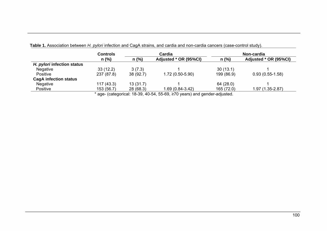
100
Table 1. Association between H. pylori infection and CagA strains, and cardia and non-cardia cancers (case-control study).
Controls
Cardia
Non-cardia
n (%) n (%) Adjusted * OR (95%CI) n (%) Adjusted * OR (95%CI) H. pylori infection status
Negative 33 (12.2) 3 (7.3) 1 30 (13.1) 1 Positive 237 (87.8) 38 (92.7) 1.72 (0.50-5.90) 199 (86.9) 0.93 (0.55-1.58)
CagA infection status Negative 117 (43.3) 13 (31.7) 1 64 (28.0) 1 Positive 153 (56.7) 28 (68.3) 1.69 (0.84-3.42) 165 (72.0) 1.97 (1.35-2.87)
* age- (categorical: 18-39, 40-54, 55-69, ≥70 years) and gender-adjusted.
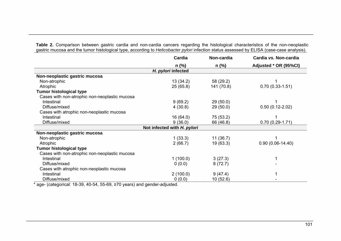
101
Table 2. Comparison between gastric cardia and non-cardia cancers regarding the histological characteristics of the non-neoplastic gastric mucosa and the tumor histological type, according to Helicobacter pylori infection status assessed by ELISA (case-case analysis). Cardia Non-cardia Cardia vs. Non-cardia n (%) n (%) Adjusted * OR (95%CI)
H. pylori infected Non-neoplastic gastric mucosa
Non-atrophic 13 (34.2) 58 (29.2) 1 Atrophic 25 (65.8) 141 (70.8) 0.70 (0.33-1.51)
Tumor histological type Cases with non-atrophic non-neoplastic mucosa
Intestinal 9 (69.2) 29 (50.0) 1 Diffuse/mixed 4 (30.8) 29 (50.0) 0.50 (0.12-2.02)
Cases with atrophic non-neoplastic mucosa Intestinal 16 (64.0) 75 (53.2) 1 Diffuse/mixed 9 (36.0) 66 (46.8) 0.70 (0.29-1.71)
Not infected with H. pylori Non-neoplastic gastric mucosa
Non-atrophic 1 (33.3) 11 (36.7) 1 Atrophic 2 (66.7) 19 (63.3) 0.90 (0.06-14.40)
Tumor histological type Cases with non-atrophic non-neoplastic mucosa
Intestinal 1 (100.0) 3 (27.3) 1 Diffuse/mixed 0 (0.0) 8 (72.7) -
Cases with atrophic non-neoplastic mucosa Intestinal 2 (100.0) 9 (47.4) 1 Diffuse/mixed 0 (0.0) 10 (52.6) -
* age- (categorical: 18-39, 40-54, 55-69, ≥70 years) and gender-adjusted.
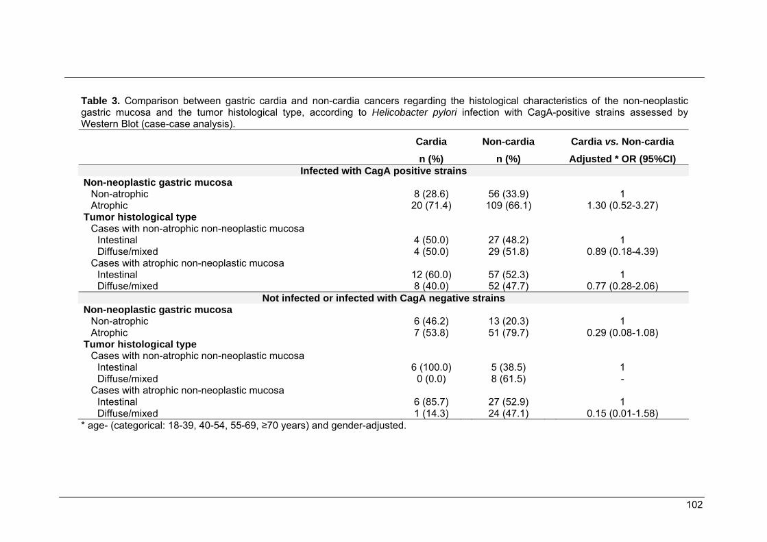
102
Table 3. Comparison between gastric cardia and non-cardia cancers regarding the histological characteristics of the non-neoplastic gastric mucosa and the tumor histological type, according to Helicobacter pylori infection with CagA-positive strains assessed by Western Blot (case-case analysis). Cardia Non-cardia Cardia vs. Non-cardia n (%) n (%) Adjusted * OR (95%CI)
Infected with CagA positive strains Non-neoplastic gastric mucosa
Non-atrophic 8 (28.6) 56 (33.9) 1 Atrophic 20 (71.4) 109 (66.1) 1.30 (0.52-3.27)
Tumor histological type Cases with non-atrophic non-neoplastic mucosa
Intestinal 4 (50.0) 27 (48.2) 1 Diffuse/mixed 4 (50.0) 29 (51.8) 0.89 (0.18-4.39)
Cases with atrophic non-neoplastic mucosa Intestinal 12 (60.0) 57 (52.3) 1 Diffuse/mixed 8 (40.0) 52 (47.7) 0.77 (0.28-2.06)
Not infected or infected with CagA negative strains Non-neoplastic gastric mucosa
Non-atrophic 6 (46.2) 13 (20.3) 1 Atrophic 7 (53.8) 51 (79.7) 0.29 (0.08-1.08)
Tumor histological type Cases with non-atrophic non-neoplastic mucosa
Intestinal 6 (100.0) 5 (38.5) 1 Diffuse/mixed 0 (0.0) 8 (61.5) -
Cases with atrophic non-neoplastic mucosa Intestinal 6 (85.7) 27 (52.9) 1 Diffuse/mixed 1 (14.3) 24 (47.1) 0.15 (0.01-1.58)
* age- (categorical: 18-39, 40-54, 55-69, ≥70 years) and gender-adjusted.
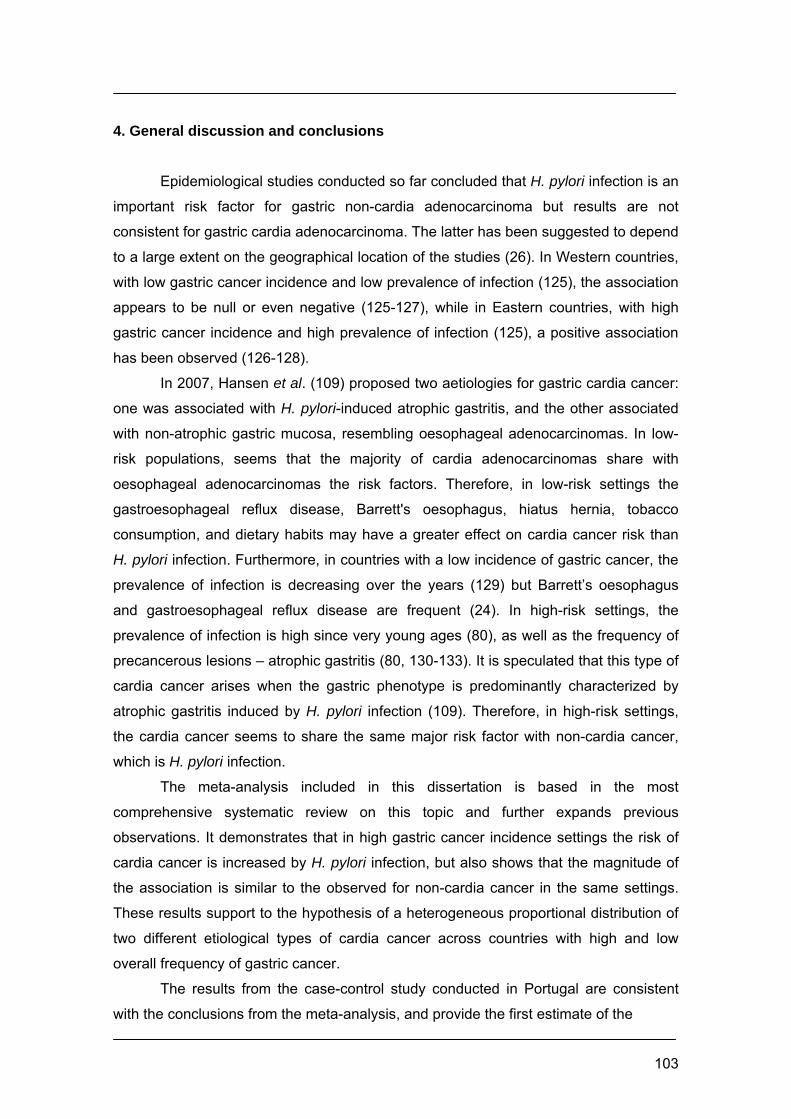
103
4. General discussion and conclusions
Epidemiological studies conducted so far concluded that H. pylori infection is an
important risk factor for gastric non-cardia adenocarcinoma but results are not
consistent for gastric cardia adenocarcinoma. The latter has been suggested to depend
to a large extent on the geographical location of the studies (26). In Western countries,
with low gastric cancer incidence and low prevalence of infection (125), the association
appears to be null or even negative (125-127), while in Eastern countries, with high
gastric cancer incidence and high prevalence of infection (125), a positive association
has been observed (126-128).
In 2007, Hansen et al. (109) proposed two aetiologies for gastric cardia cancer:
one was associated with H. pylori-induced atrophic gastritis, and the other associated
with non-atrophic gastric mucosa, resembling oesophageal adenocarcinomas. In low-
risk populations, seems that the majority of cardia adenocarcinomas share with
oesophageal adenocarcinomas the risk factors. Therefore, in low-risk settings the
gastroesophageal reflux disease, Barrett's oesophagus, hiatus hernia, tobacco
consumption, and dietary habits may have a greater effect on cardia cancer risk than
H. pylori infection. Furthermore, in countries with a low incidence of gastric cancer, the
prevalence of infection is decreasing over the years (129) but Barrett’s oesophagus
and gastroesophageal reflux disease are frequent (24). In high-risk settings, the
prevalence of infection is high since very young ages (80), as well as the frequency of
precancerous lesions – atrophic gastritis (80, 130-133). It is speculated that this type of
cardia cancer arises when the gastric phenotype is predominantly characterized by
atrophic gastritis induced by H. pylori infection (109). Therefore, in high-risk settings,
the cardia cancer seems to share the same major risk factor with non-cardia cancer,
which is H. pylori infection.
The meta-analysis included in this dissertation is based in the most
comprehensive systematic review on this topic and further expands previous
observations. It demonstrates that in high gastric cancer incidence settings the risk of
cardia cancer is increased by H. pylori infection, but also shows that the magnitude of
the association is similar to the observed for non-cardia cancer in the same settings.
These results support to the hypothesis of a heterogeneous proportional distribution of
two different etiological types of cardia cancer across countries with high and low
overall frequency of gastric cancer.
The results from the case-control study conducted in Portugal are consistent
with the conclusions from the meta-analysis, and provide the first estimate of the
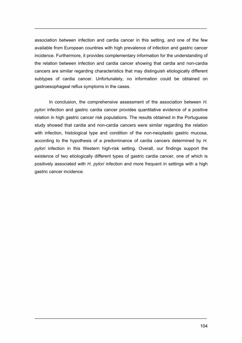
104
association between infection and cardia cancer in this setting, and one of the few
available from European countries with high prevalence of infection and gastric cancer
incidence. Furthermore, it provides complementary information for the understanding of
the relation between infection and cardia cancer showing that cardia and non-cardia
cancers are similar regarding characteristics that may distinguish etiologically different
subtypes of cardia cancer. Unfortunately, no information could be obtained on
gastroesophageal reflux symptoms in the cases.
In conclusion, the comprehensive assessment of the association between H.
pylori infection and gastric cardia cancer provides quantitative evidence of a positive
relation in high gastric cancer risk populations. The results obtained in the Portuguese
study showed that cardia and non-cardia cancers were similar regarding the relation
with infection, histological type and condition of the non-neoplastic gastric mucosa,
according to the hypothesis of a predominance of cardia cancers determined by H.
pylori infection in this Western high-risk setting. Overall, our findings support the
existence of two etiologically different types of gastric cardia cancer, one of which is
positively associated with H. pylori infection and more frequent in settings with a high
gastric cancer incidence.
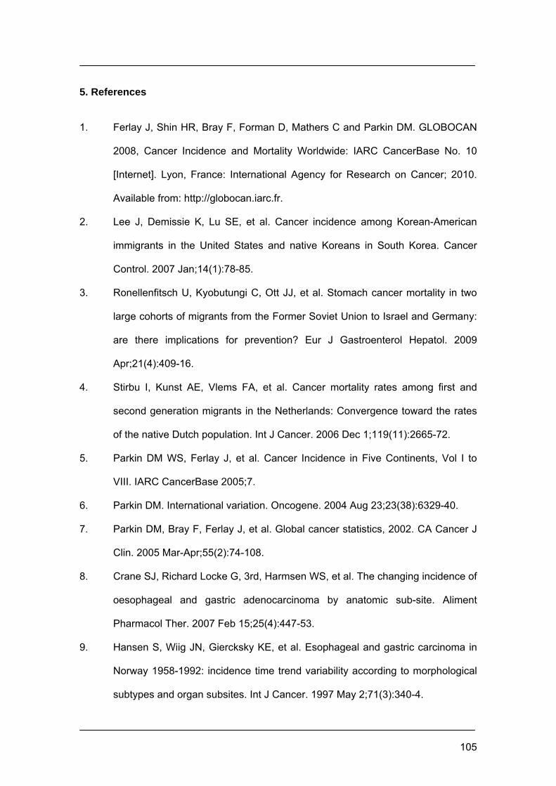
105
5. References
1. Ferlay J, Shin HR, Bray F, Forman D, Mathers C and Parkin DM. GLOBOCAN
2008, Cancer Incidence and Mortality Worldwide: IARC CancerBase No. 10
[Internet]. Lyon, France: International Agency for Research on Cancer; 2010.
Available from: http://globocan.iarc.fr.
2. Lee J, Demissie K, Lu SE, et al. Cancer incidence among Korean-American
immigrants in the United States and native Koreans in South Korea. Cancer
Control. 2007 Jan;14(1):78-85.
3. Ronellenfitsch U, Kyobutungi C, Ott JJ, et al. Stomach cancer mortality in two
large cohorts of migrants from the Former Soviet Union to Israel and Germany:
are there implications for prevention? Eur J Gastroenterol Hepatol. 2009
Apr;21(4):409-16.
4. Stirbu I, Kunst AE, Vlems FA, et al. Cancer mortality rates among first and
second generation migrants in the Netherlands: Convergence toward the rates
of the native Dutch population. Int J Cancer. 2006 Dec 1;119(11):2665-72.
5. Parkin DM WS, Ferlay J, et al. Cancer Incidence in Five Continents, Vol I to
VIII. IARC CancerBase 2005;7.
6. Parkin DM. International variation. Oncogene. 2004 Aug 23;23(38):6329-40.
7. Parkin DM, Bray F, Ferlay J, et al. Global cancer statistics, 2002. CA Cancer J
Clin. 2005 Mar-Apr;55(2):74-108.
8. Crane SJ, Richard Locke G, 3rd, Harmsen WS, et al. The changing incidence of
oesophageal and gastric adenocarcinoma by anatomic sub-site. Aliment
Pharmacol Ther. 2007 Feb 15;25(4):447-53.
9. Hansen S, Wiig JN, Giercksky KE, et al. Esophageal and gastric carcinoma in
Norway 1958-1992: incidence time trend variability according to morphological
subtypes and organ subsites. Int J Cancer. 1997 May 2;71(3):340-4.
106
10. Moller H. Incidence of cancer of oesophagus, cardia and stomach in Denmark.
Eur J Cancer Prev. 1992 Feb;1(2):159-64.
11. Botterweck AA, Schouten LJ, Volovics A, et al. Trends in incidence of
adenocarcinoma of the oesophagus and gastric cardia in ten European
countries. Int J Epidemiol. 2000 Aug;29(4):645-54.
12. Pera M, Cameron AJ, Trastek VF, et al. Increasing incidence of
adenocarcinoma of the esophagus and esophagogastric junction.
Gastroenterology. 1993 Feb;104(2):510-3.
13. Harrison SL, Goldacre MJ, Seagroatt V. Trends in registered incidence of
oesophageal and stomach cancer in the Oxford region, 1974-88. Eur J Cancer
Prev. 1992 Apr;1(3):271-4.
14. Levi F, La Vecchia C, Te VC. Descriptive epidemiology of adenocarcinomas of
the cardia and distal stomach in the Swiss Canton of Vaud. Tumori. 1990 Apr
30;76(2):167-71.
15. Armstrong RW, Borman B. Trends in incidence rates of adenocarcinoma of the
oesophagus and gastric cardia in New Zealand, 1978-1992. Int J Epidemiol.
1996 Oct;25(5):941-7.
16. Thomas RJ, Lade S, Giles GG, et al. Incidence trends in oesophageal and
proximal gastric carcinoma in Victoria. Aust N Z J Surg. 1996 May;66(5):271-5.
17. Correa P, Haenszel W, Cuello C, et al. A model for gastric cancer
epidemiology. Lancet. 1975 Jul 12;2(7924):58-60.
18. Popiela T, Kulig J, Kolodziejczyk P, et al. Changing patterns of gastric
carcinoma over the past two decades in a single institution: clinicopathological
findings in 1557 patients. Scand J Gastroenterol. 2002 May;37(5):561-7.
19. Lee JY, Kim HY, Kim KH, et al. No changing trends in incidence of gastric
cardia cancer in Korea. J Korean Med Sci. 2003 Feb;18(1):53-7.
20. Blot WJ, Devesa SS, Kneller RW, et al. Rising incidence of adenocarcinoma of
the esophagus and gastric cardia. JAMA. 1991 Mar 13;265(10):1287-9.
107
21. Bouvier AM, Esteve J, Mitry E, et al. Trends in gastric cancer incidence in a
well-defined French population by time period and birth cohort. Eur J Cancer
Prev. 2002 Jun;11(3):221-7.
22. Liabeuf A, Faivre J. Time trends in oesophageal cancer incidence in Cote d'Or
(France), 1976-93. Eur J Cancer Prev. 1997 Feb;6(1):24-30.
23. Hansson LE, Bergstrom R, Sparen P, et al. The decline in the incidence of
stomach cancer in Sweden 1960-1984: a birth cohort phenomenon. Int J
Cancer. 1991 Feb 20;47(4):499-503.
24. Blaser MJ, Saito D. Trends in reported adenocarcinomas of the oesophagus
and gastric cardia in Japan. Eur J Gastroenterol Hepatol. 2002 Feb;14(2):107-
13.
25. He YT, Hou J, Chen ZF, et al. Trends in incidence of esophageal and gastric
cardia cancer in high-risk areas in China. Eur J Cancer Prev. 2008
Apr;17(2):71-6.
26. Dawsey SM, Mark SD, Taylor PR, et al. Gastric cancer and H pylori. Gut. 2002
Sep;51(3):457-8.
27. Marsman WA, Tytgat GN, ten Kate FJ, et al. Differences and similarities of
adenocarcinomas of the esophagus and esophagogastric junction. J Surg
Oncol. 2005 Dec 1;92(3):160-8.
28. Wang LD, Zheng S, Zheng ZY, et al. Primary adenocarcinomas of lower
esophagus, esophagogastric junction and gastric cardia: in special reference to
China. World J Gastroenterol. 2003 Jun;9(6):1156-64.
29. World Cancer Research Fund (WCRF), American Institute for Cancer Research
(2007). Food, Nutrition, Physical Activity, and the prevention of cancer: a Global
Perspective. Washington: American Institute for Cancer Research.
30. Lunet N, Lacerda-Vieira A, Barros H. Fruit and vegetables consumption and
gastric cancer: a systematic review and meta-analysis of cohort studies. Nutr
Cancer. 2005;53(1):1-10.

108
31. Gonzalez CA, Pera G, Agudo A, et al. Fruit and vegetable intake and the risk of
stomach and oesophagus adenocarcinoma in the European Prospective
Investigation into Cancer and Nutrition (EPIC-EURGAST). Int J Cancer. 2006
May 15;118(10):2559-66.
32. Kobayashi M, Tsubono Y, Sasazuki S, et al. Vegetables, fruit and risk of gastric
cancer in Japan: a 10-year follow-up of the JPHC Study Cohort I. Int J Cancer.
2002 Nov 1;102(1):39-44.
33. Freedman ND, Subar AF, Hollenbeck AR, et al. Fruit and vegetable intake and
gastric cancer risk in a large United States prospective cohort study. Cancer
Causes Control. 2008 Jun;19(5):459-67.
34. Woodward M, Tunstall-Pedoe H, McColl K. Helicobacter pylori infection reduces
systemic availability of dietary vitamin C. Eur J Gastroenterol Hepatol. 2001
Mar;13(3):233-7.
35. Jenab M, Riboli E, Ferrari P, et al. Plasma and dietary vitamin C levels and risk
of gastric cancer in the European Prospective Investigation into Cancer and
Nutrition (EPIC-EURGAST). Carcinogenesis. 2006 Nov;27(11):2250-7.
36. Bjelakovic G, Nikolova D, Simonetti RG, et al. Antioxidant supplements for
prevention of gastrointestinal cancers: a systematic review and meta-analysis.
Lancet. 2004 Oct 2-8;364(9441):1219-28.
37. Ekstrom AM, Serafini M, Nyren O, et al. Dietary antioxidant intake and the risk
of cardia cancer and noncardia cancer of the intestinal and diffuse types: a
population-based case-control study in Sweden. Int J Cancer. 2000 Jul
1;87(1):133-40.
38. Mayne ST, Risch HA, Dubrow R, et al. Nutrient intake and risk of subtypes of
esophageal and gastric cancer. Cancer Epidemiol Biomarkers Prev. 2001
Oct;10(10):1055-62.
39. Lochhead P, El-Omar EM. Gastric cancer. Br Med Bull. 2008;85:87-100.
109
40. Kneller RW, You WC, Chang YS, et al. Cigarette smoking and other risk factors
for progression of precancerous stomach lesions. J Natl Cancer Inst. 1992 Aug
19;84(16):1261-6.
41. Ladeiras-Lopes R, Pereira AK, Nogueira A, et al. Smoking and gastric cancer:
systematic review and meta-analysis of cohort studies. Cancer Causes Control.
2008 Sep;19(7):689-701.
42. Nishino Y, Inoue M, Tsuji I, et al. Tobacco smoking and gastric cancer risk: an
evaluation based on a systematic review of epidemiologic evidence among the
Japanese population. Jpn J Clin Oncol. 2006 Dec;36(12):800-7.
43. Koizumi Y, Tsubono Y, Nakaya N, et al. Cigarette smoking and the risk of
gastric cancer: a pooled analysis of two prospective studies in Japan. Int J
Cancer. 2004 Dec 20;112(6):1049-55.
44. Gonzalez CA, Pera G, Agudo A, et al. Smoking and the risk of gastric cancer in
the European Prospective Investigation Into Cancer and Nutrition (EPIC). Int J
Cancer. 2003 Nov 20;107(4):629-34.
45. Ye W, Ekstrom AM, Hansson LE, et al. Tobacco, alcohol and the risk of gastric
cancer by sub-site and histologic type. Int J Cancer. 1999 Oct 8;83(2):223-9.
46. Marshall BJ WJ. Unidentified curved bacilli on gastric epithelium in active
chronic gastritis. Lancet. 1983;1:1273-5.
47. International Agency for Research on Cancer. Schistosomes
lfaHpImoteocrthVL, France: IARC, 1994.
48. Dunn BE, Cohen H, Blaser MJ. Helicobacter pylori. Clin Microbiol Rev. 1997
Oct;10(4):720-41.
49. Owen RJ. Helicobacter--species classification and identification. Br Med Bull.
1998;54(1):17-30.
50. Boren T, Falk P, Roth KA, et al. Attachment of Helicobacter pylori to human
gastric epithelium mediated by blood group antigens. Science. 1993 Dec
17;262(5141):1892-5.
110
51. McColl KE, el-Omar E, Gillen D. Helicobacter pylori gastritis and gastric
physiology. Gastroenterol Clin North Am. 2000 Sep;29(3):687-703, viii.
52. Axon AT. Personal view: to treat or not to treat? Helicobacter pylori and gastro-
oesophageal reflux disease - an alternative hypothesis. Aliment Pharmacol
Ther. 2004 Feb 1;19(3):253-61.
53. Kuipers EJ, Meuwissen SG. Helicobacter pylori and gastric carcinogenesis.
Scand J Gastroenterol Suppl. 1996;218:103-5.
54. Leunk RD. Production of a cytotoxin by Helicobacter pylori. Rev Infect Dis. 1991
Jul-Aug;13 Suppl 8:S686-9.
55. Covacci A, Censini S, Bugnoli M, et al. Molecular characterization of the 128-
kDa immunodominant antigen of Helicobacter pylori associated with cytotoxicity
and duodenal ulcer. Proc Natl Acad Sci U S A. 1993 Jun 15;90(12):5791-5.
56. Ching CK, Wong BC, Kwok E, et al. Prevalence of CagA-bearing Helicobacter
pylori strains detected by the anti-CagA assay in patients with peptic ulcer
disease and in controls. Am J Gastroenterol. 1996 May;91(5):949-53.
57. Cover TL, Glupczynski Y, Lage AP, et al. Serologic detection of infection with
cagA+ Helicobacter pylori strains. J Clin Microbiol. 1995 Jun;33(6):1496-500.
58. Tummuru MK, Cover TL, Blaser MJ. Cloning and expression of a high-
molecular-mass major antigen of Helicobacter pylori: evidence of linkage to
cytotoxin production. Infect Immun. 1993 May;61(5):1799-809.
59. Akopyants NS, Clifton SW, Kersulyte D, et al. Analyses of the cag pathogenicity
island of Helicobacter pylori. Mol Microbiol. 1998 Apr;28(1):37-53.
60. Censini S, Lange C, Xiang Z, et al. cag, a pathogenicity island of Helicobacter
pylori, encodes type I-specific and disease-associated virulence factors. Proc
Natl Acad Sci U S A. 1996 Dec 10;93(25):14648-53.
61. Hatakeyama M. Oncogenic mechanisms of the Helicobacter pylori CagA
protein. Nat Rev Cancer. 2004 Sep;4(9):688-94.
111
62. Macarthur M, Hold GL, El-Omar EM. Inflammation and Cancer II. Role of
chronic inflammation and cytokine gene polymorphisms in the pathogenesis of
gastrointestinal malignancy. Am J Physiol Gastrointest Liver Physiol. 2004
Apr;286(4):G515-20.
63. Atherton JC, Peek RM, Jr., Tham KT, et al. Clinical and pathological importance
of heterogeneity in vacA, the vacuolating cytotoxin gene of Helicobacter pylori.
Gastroenterology. 1997 Jan;112(1):92-9.
64. Atherton JC, Cao P, Peek RM, Jr., et al. Mosaicism in vacuolating cytotoxin
alleles of Helicobacter pylori. Association of specific vacA types with cytotoxin
production and peptic ulceration. J Biol Chem. 1995 Jul 28;270(30):17771-7.
65. Letley DP, Lastovica A, Louw JA, et al. Allelic diversity of the Helicobacter pylori
vacuolating cytotoxin gene in South Africa: rarity of the vacA s1a genotype and
natural occurrence of an s2/m1 allele. J Clin Microbiol. 1999 Apr;37(4):1203-5.
66. Cover TL, Krishna US, Israel DA, et al. Induction of gastric epithelial cell
apoptosis by Helicobacter pylori vacuolating cytotoxin. Cancer Res. 2003 Mar
1;63(5):951-7.
67. Versalovic J. Helicobacter pylori. Pathology and diagnostic strategies. Am J Clin
Pathol. 2003 Mar;119(3):403-12.
68. Megraud F, Lehours P. Helicobacter pylori detection and antimicrobial
susceptibility testing. Clin Microbiol Rev. 2007 Apr;20(2):280-322.
69. Loffeld RJ, Stobberingh E, Arends JW. A review of diagnostic techniques for
Helicobacter pylori infection. Dig Dis. 1993;11(3):173-80.
70. Reilly TG, Poxon V, Sanders DS, et al. Comparison of serum, salivary, and
rapid whole blood diagnostic tests for Helicobacter pylori and their validation
against endoscopy based tests. Gut. 1997 Apr;40(4):454-8.
71. Vaira D, Holton J, Menegatti M, et al. Blood tests in the management of
Helicobacter pylori infection. Italian Helicobacter pylori Study Group. Gut. 1998
Jul;43 Suppl 1:S39-46.
112
72. Laheij RJ, Straatman H, Jansen JB, et al. Evaluation of commercially available
Helicobacter pylori serology kits: a review. J Clin Microbiol. 1998
Oct;36(10):2803-9.
73. Klaamas K, Held M, Wadstrom T, et al. IgG immune response to Helicobacter
pylori antigens in patients with gastric cancer as defined by ELISA and
immunoblotting. Int J Cancer. 1996 Jul 3;67(1):1-5.
74. Nilsson I, Ljungh A, Aleljung P, et al. Immunoblot assay for serodiagnosis of
Helicobacter pylori infections. J Clin Microbiol. 1997 Feb;35(2):427-32.
75. Sorberg M, Engstrand L, Strom M, et al. The diagnostic value of enzyme
immunoassay and immunoblot in monitoring eradication of Helicobacter pylori.
Scand J Infect Dis. 1997;29(2):147-51.
76. Aucher P, Petit ML, Mannant PR, et al. Use of immunoblot assay to define
serum antibody patterns associated with Helicobacter pylori infection and with
H. pylori-related ulcers. J Clin Microbiol. 1998 Apr;36(4):931-6.
77. Lamarque D, Gilbert T, Roudot-Thoraval F, et al. Seroprevalence of eight
Helicobacter pylori antigens among 182 patients with peptic ulcer, MALT gastric
lymphoma or non-ulcer dyspepsia. Higher rate of seroreactivity against CagA
and 35-kDa antigens in patients with peptic ulcer originating from Europe and
Africa. Eur J Gastroenterol Hepatol. 1999 Jul;11(7):721-6.
78. el-Zaatari FA, Oweis SM, Graham DY. Uses and cautions for use of
polymerase chain reaction for detection of Helicobacter pylori. Dig Dis Sci. 1997
Oct;42(10):2116-9.
79. de Martel C, Franceschi S. Infections and cancer: established associations and
new hypotheses. Crit Rev Oncol Hematol. 2009 Jun;70(3):183-94.
80. Parkin DM. The global health burden of infection-associated cancers in the year
2002. Int J Cancer. 2006 Jun 15;118(12):3030-44.
81. Brown LM. Helicobacter pylori: epidemiology and routes of transmission.
Epidemiol Rev. 2000;22(2):283-97.
113
82. Malcolm CA, MacKay WG, Shepherd A, et al. Helicobacter pylori in children is
strongly associated with poverty. Scott Med J. 2004 Nov;49(4):136-8.
83. Nouraie M, Latifi-Navid S, Rezvan H, et al. Childhood hygienic practice and
family education status determine the prevalence of Helicobacter pylori infection
in Iran. Helicobacter. 2009 Feb;14(1):40-6.
84. Wang C, Yuan Y, Hunt RH. The association between Helicobacter pylori
infection and early gastric cancer: a meta-analysis. Am J Gastroenterol. 2007
Aug;102(8):1789-98.
85. Huang JQ, Hunt RH. The evolving epidemiology of Helicobacter pylori infection
and gastric cancer. Can J Gastroenterol. 2003 Jun;17 Suppl B:18B-20B.
86. Eslick GD. Helicobacter pylori infection causes gastric cancer? A review of the
epidemiological, meta-analytic, and experimental evidence. World J
Gastroenterol. 2006 May 21;12(19):2991-9.
87. Xue FB, Xu YY, Wan Y, et al. Association of H. pylori infection with gastric
carcinoma: a Meta analysis. World J Gastroenterol. 2001 Dec;7(6):801-4.
88. Eslick GD, Lim LL, Byles JE, et al. Association of Helicobacter pylori infection
with gastric carcinoma: a meta-analysis. Am J Gastroenterol. 1999
Sep;94(9):2373-9.
89. Danesh J. Helicobacter pylori infection and gastric cancer: systematic review of
the epidemiological studies. Aliment Pharmacol Ther. 1999 Jul;13(7):851-6.
90. Huang JQ, Sridhar S, Chen Y, et al. Meta-analysis of the relationship between
Helicobacter pylori seropositivity and gastric cancer. Gastroenterology. 1998
Jun;114(6):1169-79.
91. Gastric cancer and Helicobacter pylori: a combined analysis of 12 case control
studies nested within prospective cohorts. Gut. 2001 Sep;49(3):347-53.
92. Danesh J. Coronary heart disease, Helicobacter pylori, dental disease,
Chlamydia pneumoniae, and cytomegalovirus: meta-analyses of prospective
studies. Am Heart J. 1999 Nov;138(5 Pt 2):S434-7.
114
93. Huang JQ, Zheng GF, Sumanac K, et al. Meta-analysis of the relationship
between cagA seropositivity and gastric cancer. Gastroenterology. 2003
Dec;125(6):1636-44.
94. Chow WH, Blaser MJ, Blot WJ, et al. An inverse relation between cagA+ strains
of Helicobacter pylori infection and risk of esophageal and gastric cardia
adenocarcinoma. Cancer Res. 1998 Feb 15;58(4):588-90.
95. Wu AH, Crabtree JE, Bernstein L, et al. Role of Helicobacter pylori CagA+
strains and risk of adenocarcinoma of the stomach and esophagus. Int J
Cancer. 2003 Mar 1;103(6):815-21.
96. Whiting JL, Hallissey MT, Fielding JW, et al. Screening for gastric cancer by
Helicobacter pylori serology: a retrospective study. Br J Surg. 1998
Mar;85(3):408-11.
97. Hansen S, Melby KK, Aase S, et al. Helicobacter pylori infection and risk of
cardia cancer and non-cardia gastric cancer. A nested case-control study.
Scand J Gastroenterol. 1999 Apr;34(4):353-60.
98. Ekstrom AM, Held M, Hansson LE, et al. Helicobacter pylori in gastric cancer
established by CagA immunoblot as a marker of past infection.
Gastroenterology. 2001 Oct;121(4):784-91.
99. Kokkola A, Louhimo J, Puolakkainen P, et al. Helicobacter pylori infection and
low serum pepsinogen I level as risk factors for gastric carcinoma. World J
Gastroenterol. 2005 Feb 21;11(7):1032-6.
100. Siman JH, Engstrand L, Berglund G, et al. Helicobacter pylori and CagA
seropositivity and its association with gastric and oesophageal carcinoma.
Scand J Gastroenterol. 2007 Aug;42(8):933-40.
101. Lee BM, Jang JJ, Kim JS, et al. Association of Helicobacter pylori infection with
gastric adenocarcinoma. Jpn J Cancer Res. 1998 Jun;89(6):597-603.
115
102. Kato M, Asaka M, Shimizu Y, et al. Relationship between Helicobacter pylori
infection and the prevalence, site and histological type of gastric cancer.
Aliment Pharmacol Ther. 2004 Jul;20 Suppl 1:85-9.
103. Suzuki G, Cullings H, Fujiwara S, et al. Low-positive antibody titer against
Helicobacter pylori cytotoxin-associated gene A (CagA) may predict future
gastric cancer better than simple seropositivity against H. pylori CagA or
against H. pylori. Cancer Epidemiol Biomarkers Prev. 2007 Jun;16(6):1224-8.
104. Ren JS, Qiao YL. [Helicobacter pylori infection and gastric cardia cancer: a
nested case-control study]. Zhonghua Zhong Liu Za Zhi. 2008 Jun;30(6):428-
31.
105. Derakhshan MH, Malekzadeh R, Watabe H, et al. Combination of gastric
atrophy, reflux symptoms and histological subtype indicates two distinct
aetiologies of gastric cardia cancer. Gut. 2008 Mar;57(3):298-305.
106. Kamangar F, Qiao YL, Blaser MJ, et al. Helicobacter pylori and oesophageal
and gastric cancers in a prospective study in China. Br J Cancer. 2007 Jan
15;96(1):172-6.
107. Kuipers EJ. Review article: exploring the link between Helicobacter pylori and
gastric cancer. Aliment Pharmacol Ther. 1999 Mar;13 Suppl 1:3-11.
108. Lochhead P, El-Omar EM. Helicobacter pylori infection and gastric cancer. Best
Pract Res Clin Gastroenterol. 2007;21(2):281-97.
109. Hansen S, Vollset SE, Derakhshan MH, et al. Two distinct aetiologies of cardia
cancer; evidence from premorbid serological markers of gastric atrophy and
Helicobacter pylori status. Gut. 2007 Jul;56(7):918-25.
110. El-Omar EM. Role of host genes in sporadic gastric cancer. Best Pract Res Clin
Gastroenterol. 2006;20(4):675-86.
111. Smith MG, Hold GL, Tahara E, et al. Cellular and molecular aspects of gastric
cancer. World J Gastroenterol. 2006 May 21;12(19):2979-90.
116
112. Camargo MC, Mera R, Correa P, et al. Interleukin-1beta and interleukin-1
receptor antagonist gene polymorphisms and gastric cancer: a meta-analysis.
Cancer Epidemiol Biomarkers Prev. 2006 Sep;15(9):1674-87.
113. Kamangar F, Cheng C, Abnet CC, et al. Interleukin-1B polymorphisms and
gastric cancer risk--a meta-analysis. Cancer Epidemiol Biomarkers Prev. 2006
Oct;15(10):1920-8.
114. Wang P, Xia HH, Zhang JY, et al. Association of interleukin-1 gene
polymorphisms with gastric cancer: a meta-analysis. Int J Cancer. 2007 Feb
1;120(3):552-62.
115. Vincenzi B, Patti G, Galluzzo S, et al. Interleukin 1beta-511T gene (IL1beta)
polymorphism is correlated with gastric cancer in the Caucasian population:
results from a meta-analysis. Oncol Rep. 2008 Nov;20(5):1213-20.
116. Camargo MC, Mera R, Correa P, et al. IL1B polymorphisms and gastric cancer
risk. Cancer Epidemiol Biomarkers Prev. 2007 Mar;16(3):635; author reply -6.
117. Al-Moundhri MS, Alkindy M, Al-Nabhani M, et al. Combined polymorphism
analysis of glutathione S-transferase M1/G1 and interleukin-1B (IL-
1B)/interleukin 1-receptor antagonist (IL-1RN) and gastric cancer risk in an
Omani Arab Population. J Clin Gastroenterol. 2009 Feb;43(2):152-6.
118. Peleteiro B, Lunet N, Carrilho C, et al. Association between cytokine gene
polymorphisms and gastric precancerous lesions: systematic review and meta-
analysis. Cancer Epidemiol Biomarkers Prev. Mar;19(3):762-76.
119. Figueiredo C, Machado JC, Pharoah P, et al. Helicobacter pylori and interleukin
1 genotyping: an opportunity to identify high-risk individuals for gastric
carcinoma. J Natl Cancer Inst. 2002 Nov 20;94(22):1680-7.
120. Canedo P, Corso G, Pereira F, et al. The interferon gamma receptor 1
(IFNGR1) -56C/T gene polymorphism is associated with increased risk of early
gastric carcinoma. Gut. 2008 Nov;57(11):1504-8.
117
121. Oliveira C, Suriano G, Ferreira P, et al. Genetic screening for familial gastric
cancer. Hered Cancer Clin Pract. 2004;2(2):51-64.
122. Machado JC, Oliveira C, Carvalho R, et al. E-cadherin gene (CDH1) promoter
methylation as the second hit in sporadic diffuse gastric carcinoma. Oncogene.
2001 Mar 22;20(12):1525-8.
123. Carneiro F, Huntsman DG, Smyrk TC, et al. Model of the early development of
diffuse gastric cancer in E-cadherin mutation carriers and its implications for
patient screening. J Pathol. 2004 Jun;203(2):681-7.
124. Gao L, Nieters A, Brenner H. Meta-analysis: tumour invasion-related genetic
polymorphisms and gastric cancer susceptibility. Aliment Pharmacol Ther. 2008
Sep 1;28(5):565-73.
125. Konturek PC, Konturek SJ, Brzozowski T. Gastric cancer and Helicobacter
pylori infection. J Physiol Pharmacol. 2006 Sep;57 Suppl 3:51-65.
126. Shibata T, Imoto I, Ohuchi Y, et al. Helicobacter pylori infection in patients with
gastric carcinoma in biopsy and surgical resection specimens. Cancer. 1996
Mar 15;77(6):1044-9.
127. Fukuda H, Saito D, Hayashi S, et al. Helicobacter pylori infection, serum
pepsinogen level and gastric cancer: a case-control study in Japan. Jpn J
Cancer Res. 1995 Jan;86(1):64-71.
128. Kim HY, Cho BD, Chang WK, et al. Helicobacter pylori infection and the risk of
gastric cancer among the Korean population. J Gastroenterol Hepatol. 1997
Feb;12(2):100-3.
129. Crew KD, Neugut AI. Epidemiology of gastric cancer. World J Gastroenterol.
2006 Jan 21;12(3):354-62.
130. Asaka M. [Helicobacter pylori infection--from gastritis to stomach cancer].
Nippon Naika Gakkai Zasshi. 1996 Sep 10;85(9):1540-5.

118
131. Kato S, Nakajima S, Nishino Y, et al. Association between gastric atrophy and
Helicobacter pylori infection in Japanese children: a retrospective multicenter
study. Dig Dis Sci. 2006 Jan;51(1):99-104.
132. Namekata T, Miki K, Kimmey M, et al. Chronic atrophic gastritis and
Helicobacter pylori infection among Japanese Americans in Seattle. Am J
Epidemiol. 2000 Apr 15;151(8):820-30.
133. Aoki K, Kihaile PE, Wenyuan Z, et al. Comparison of prevalence of chronic
atrophic gastritis in Japan, China, Tanzania, and the Dominican Republic. Ann
Epidemiol. 2005 Sep;15(8):598-606.
119
6. Summary
Relationship between Helicobacter pylori infection and gastric cardia cancer
The discovery of H. pylori in 1983 has proved to be pivotal in our understanding
of the aetiology of gastric cancer. In 1994, the World Health Organization and the
International Agency for research on Cancer consensus group stated that there was
sufficient evidence to classify H. pylori as a Class I human carcinogen. The overall
prevalence of H. pylori infection in middle-aged adults is estimated in 74% in
developing and 58% in developed countries, and worldwide 63.4% of the gastric
cancer cases are attributable to infection, corresponding to 592,000 cases.
Epidemiological studies conducted so far concluded that H. pylori infection is an
important risk factor for gastric non-cardia adenocarcinoma but results are not
consistent for gastric cardia adenocarcinoma. The latter depends to a large extent on
the geographical location of the studies, with those conducted in Western countries
showing predominantly a null or negative association and those from Eastern countries
suggesting a positive association. Moreover, two aetiologically distinct types of cardia
gastric cancer were recently proposed, one associated with H. pylori-induced atrophic
gastritis and other occurring in non-atrophic gastric mucosa. The heterogeneous
geographic distribution of these two types of cardia cancer may contribute to explain
the conflicting findings between studies conducted in populations with different gastric
cancer risk.
The aim of the present dissertation was to examine the relationship between
infection with H. pylori and the occurrence of gastric cardia cancer, through the
accomplishment of the following specific objectives:
1) to quantify the association between infection and gastric cardia cancer through
meta-analysis, and to provide an explanation for the expected heterogeneity of results.
(Paper I)
2) to compare gastric cardia and non-cardia cancers regarding the frequency of H.
pylori infection, the histological characteristics of the non-neoplastic gastric mucosa,
and the tumor histological type. (Paper II)
120
Paper I: Helicobacter pylori infection and gastric cardia cancer: a systematic review
and meta-analysis
Published studies addressing the association between H. pylori infection and
gastric cardia cancer (up to June 2009) were systematically reviewed, and extracted
relative risk (RR) estimates for the association with cardia and non-cardia cancers.
Summary RR estimates and 95% confidence intervals (95%CI) were computed using
random-effects models. Subgroup analyses were conducted, namely according to
gastric cancer risk settings.
Thirty-four articles were considered for meta-analysis. For cardia cancer,
summary RR was 1.08 (95%CI: 0.83-1.40; I2=52.8%), higher in high-risk (RR=1.98;
95%CI: 1.38-2.83; I2=18.4%) than in low-risk settings (RR=0.78; 95%CI: 0.63-0.97;
I2=11.6%). For non-cardia cancer, RR estimates were similar in high-risk settings
(RR=3.02; 95%CI: 1.92-4.74; I2=90.7%) and low-risk settings (RR=2.56; 95%CI: 1.99-
3.29; I2=46.6%). These observations were consistent across different inclusion criteria
and when accounting for the virulence of the infecting strains.
Paper II: Is cardia cancer etiologically different from distal stomach cancer?
This report comprises a case-control study and a case-case analysis. In the
former, 41 cardia and 229 non-cardia cancers were compared with 270 community
controls regarding the frequency of infection with H. pylori, taking into account the
virulence of the infecting strains. Patients were admitted to the surgery wards of
Hospital de S. João and Instituto Português de Oncologia Francisco Gentil, both in
Porto. Controls were part of a representative sample of the adult population of Porto. A
case-case analysis was performed for the comparison of cardia and non-cardia
cancers regarding the histological characteristics of the neoplastic and non-neoplastic
mucosas and was stratified according to the infection status of the cases, to provide
evidence on the frequency of etiologically distinct cases of cardia cancer. H. pylori
infection and CagA infection status were assessed by ELISA and Western Blot,
respectively. Odds Ratios (OR) and the corresponding 95% confidence intervals
(95%CI) were computed by multinomial logistic regression to quantify the association
of between H. pylori infection and gastric cardia and non-cardia cancers and
unconditional logistic regression to compare cardia and non-cardia cancer cases.
121
A negative association was found for H. pylori infection, but CagA-positive strains
were associated with an increased risk of non-cardia cancer (OR=1.97, 95%CI: 1.35-
2.87). Thirty-one (75.6%) cardia cancer cases, predominantly of the intestinal type
(67.7%), had non-neoplastic atrophic mucosa, as well as 173 (75.6%) non-cardia
cancers (54.3% of the intestinal type). Among cases occurring in non-atrophic patients,
the proportion of cancers of the Laurén’s intestinal type was 70.0% for cardia and
39.3% for non-cardia gastric cancers.
Conclusions
The comprehensive assessment of the association between H. pylori infection
and gastric cardia cancer provides quantitative evidence of a positive relation in high
gastric cancer risk populations. The results obtained in the Portuguese study showed
that cardia and non-cardia cancers were similar regarding the relation with infection,
histological type and condition of the non-neoplastic gastric mucosa, according to the
hypothesis of a predominance of cardia cancers determined by H. pylori infection in
this Western high-risk setting.
Overall, our findings support the existence of two etiologically different types of
gastric cardia cancer, one of which is positively associated with H. pylori infection and
more frequent in settings with a high gastric cancer incidence.
122
7. Sumário
Relação entre infecção por Helicobacter pylori e cancro gástrico do cardia
A descoberta da H. pylori em 1983 revelou-se fundamental para a compreensão
da etiologia do cancro gástrico. Em 1994, a Organização Mundial de Saúde e a
Agência Internacional para investigação em Cancro reconheceram existir evidência
suficiente para classificar a H. pylori como carcinogénico de classe I. A estimativa
global da prevalência de infecção por H. pylori em adultos de meia-idade é de 74% em
países em desenvolvimento e 58% nos países desenvolvidos e, a nível mundial,
63,4% dos casos de cancro gástrico são atribuíveis à infecção, correspondendo a 592
000 casos.
Os estudos epidemiológicos realizados até ao momento concluíram que a
infecção por H. pylori é um factor de risco importante para o adenocarcinoma gástrico
não-cardia, mas não se demonstrou associação consistente com o adenocarcinoma
gástrico do cardia. Contudo, a relação entre a infecção e o cancro do cardia depende
em larga medida da localização geográfica dos estudos, com uma associação neutra
ou negativa na maioria das investigações conduzidas em países Ocidentais enquanto
nas realizadas em países Orientais é sugerida uma associação positiva.
Adicionalmente, foram propostos recentemente dois tipos etiologicamente distintos de
cancro gástrico do cardia, um associado a gastrite atrófica induzidas pela infecção e o
outro ocorrendo na mucosa gástrica sem atrofia. A distribuição heterogénea destes
tipos de cancro do cardia poderá contribuir para explicar os resultados discrepantes
entre estudos realizados em populações com risco diferente de cancro gástrico.
O objective da presente dissertação foi avaliar a relação entre a infecção por H.
pylori e a ocorrência de cancro gástrico do cardia, através da realização dos seguintes
objectivos específicos:
1) quantificar a associação entre a infecção e o cancro gástrico do cardia através de
meta-análise, e fornecer explicações para a esperada heterogeneidade de resultados
(Artigo I).
123
2) comparar cancros gástricos do cardia e não-cardia relativamente à frequência da
infecção por H. pylori, às características histológicas da mucosa gástrica não
neoplásica e ao tipo histológico do tumor (Artigo II).
Artigo I: Infecção por Helicobacter pylori e o cancro gástrico do cardia: revisão
sistemática e meta-análise
Os estudos publicados sobre a associação entre a infecção por H. pylori e o
cancro do cardia (até Junho de 2009) foram sistematicamente revistos e as
estimativas do risco relativo (RR) para a associação com o cancro do cardia e não-
cardia foram extraídas. As estimativas sumárias do RR e intervalos de confiança a
95% (IC95%) foram calculados usando modelos de efeitos aleatórios. Foram
realizadas análises estratificadas de acordo com a incidência de cancro gástrico nos
locais em que foram conduzidos os estudos.
Trinta e quatro artigos foram considerados para a meta-análise. Para o cancro do
cardia o RR sumário foi 1.08 (IC95%: 0.83-1.40; I2=52.8%), maior nos estudos
realizados em países com elevado risco de cancro gástrico (RR=1.98; IC95%: 1.38-
2.83; I2=18.4%) do que nos de baixo risco (RR=0.78; IC95%: 0.63-0.97; I2=11.6%).
Para o cancro não-cardia, as estimativas do RR foram semelhantes nos estudos de
países de elevado risco (RR=3.02; IC95%: 1.92-4.74; I2=90.7%) e nos de baixo risco
(RR=2.56; IC95%: 1.99-3.29; I2=46.6%). Estas observações foram consistentes entre
os diferentes critérios de inclusão e quando considerada a virulência das estirpes.
Artigo II: O cancro do cardia é etiologicamente diferente do cancro do estômago
distal?
Este artigo compreende um estudo caso-controlo e uma análise caso-caso. No
primeiro, foram comparados 41 cancros gástricos do cardia e 229 cancros não-cardia
com 270 controlos comunitários em relação à frequência de infecção por H. pylori,
tendo em conta a virulência das estirpes infectantes. Os casos foram internados nos
Serviços de Cirurgia do Hospital de S. João e do Instituto Português de Oncologia
Francisco Gentil, ambos no Porto. Os controlos foram parte de uma amostra
representativa da população adulta do Porto. A análise caso-caso, foi realizada para
124
comparação dos cancros do cardia e não-cardia no que respeita às características
histológicas da mucosa neoplásica e não neoplásica e foi estratificada por estado de
infecção nos casos, para fornecer evidência sobre a frequência de casos de cancro do
cardia de tipos etiologicamente distintos. A infecção por H. pylori e o estado de
infecção CagA foram avaliados por ELISA e Western Blot, respectivamente. Foram
calculados OR e respectivos IC 95% por regressão logística multinomial para
quantificar a associação entre infecção por H. pylori e o cancro do cardia e não-cardia
e a regressão logística não condicional para comparar casos de cancro do cardia e
não-cardia.
Foi observada uma associação negativa para a infecção por H. pylori, mas a
infecção com estirpes cagA-positivas associou-se a um risco aumentado de cancro
não-cardia (OR=1,97, IC95%: 1,35-2,87). Trinta e um (75,6%) dos casos de cancro do
cardia, predominantemente do tipo intestinal (67,7%), tinham atrofia da mucosa não-
neoplásica, bem como os 173 (75,6%) casos de cancro não-cardia (54,3% do tipo
intestinal). Entre os casos que ocorrem em doentes sem atrofia da mucosa, a
proporção dos cancros do tipo intestinal de Laurén’s foi de 70,0% para o cardia e
39,3% para o cancro não-cardia.
Conclusões
A avaliação exaustiva da associação entre a infecção por H. pylori e o cancro
gástrico do cardia fornece evidência quantitativa de uma relação positiva nas
populações com elevado risco de cancro gástrico. Os resultados obtidos no estudo
conduzido em Portugal demonstraram que os cancro do cárdia e não-cárdia eram
semelhantes relativamente à relação com a infecção, tipo histológico e características
da mucosa gástrica não neoplásica, de acordo com a hipótese do predomínio de
cancros do cárdia determinados pela infecção nesta população Ocidental com elevado
risco de cancro gástrico.
Os nossos resultados apoiam a existência de dois tipos etiologicamente distintos
de cancro gástrico do cardia, um dos quais positivamente associado com a infecção
por H. pylori e mais frequente em populações com elevada incidência de cancro
gástrico.