Embed Size (px)
Citation preview
i
PONTIFÍCIA UNIVERSIDADE CATÓLICA DO RIO GRANDE DO SUL
FACULDADE DE BIOCIÊNCIAS
PROGRAMA DE PÓS-GRADUAÇÃO EM BIOLOGIA CELULAR E MOLECULAR
Fernanda Cattani
Detecção e quantificação de células viáveis de Bacillus sporothermodurans e
de Bacillus cereus em leite através de PCR convencional e de PCR em
tempo real associadas ao propídio monoazida
Porto Alegre
2012
ii
Fernanda Cattani
Detecção e quantificação de células viáveis de Bacillus sporothermodurans e
de Bacillus cereus em leite através de PCR convencional e de PCR em
tempo real associadas ao propídio monoazida
Tese de Doutorado apresentada ao Programa de Pós-Graduação em Biologia Celular e
Molecular, da Faculdade de Biociências da Pontifícia Universidade Católica do Rio Grande
do Sul.
Orientadora: Prof. Dra. Sílvia Dias de Oliveira
Co-orientador: Prof. Dr. Carlos Alexandre Sanchez Ferreira
Porto Alegre
2012

iii
FERNANDA CATTANI
Detecção e quantificação de células viáveis de Bacillus sporothermodurans e
de Bacillus cereus em leite através de PCR convencional e de PCR em
tempo real associadas ao propídio monoazida
Tese de Doutorado apresentada ao Programa de Pós-Graduação em Biologia Celular e
Molecular, da Faculdade de Biociências da Pontifícia Universidade Católica do Rio Grande
do Sul.
Aprovado em 31 de outubro de 2012.
BANCA EXAMINADORA:
Marisa da Costa
Marjo Cadó Bessa
Eraldo Luiz Batista Júnior
Fernanda Bueno Morrone
Porto Alegre
2012
iv
AGRADECIMENTOS
A realização desta tese de doutorado não teria sido possível sem a colaboração de
diversas pessoas e instituições, às quais serei eternamente grata.
A Pontifícia Universidade Católica do Rio Grande do Sul (PUCRS), pela bolsa
concedida via programa PROBOLSAS que possibilitou a realização deste doutorado.
A minha orientadora, professora Sílvia Dias de Oliveira pela oportunidade de
desenvolver este projeto, pela atenção, orientação, ética, competência, incentivo e por sempre
fazer com que eu buscasse o meu aperfeiçoamento. Agradeço também pelos valiosos
ensinamentos transmitidos nesses anos de convívio.
Ao co-orientador deste trabalho, professor Carlos Alexandre Sanchez Ferreira, pelo
apoio, incentivo e contribuições.
Ao Conselho Nacional de Desenvolvimento Científico e Tecnológico (CNPq) pela
concessão da bolsa, fundamental para a execução deste trabalho.
A Coordenação de Aperfeiçoamento de Pessoal de Nível Superior (CAPES), pela
bolsa concedida para realização do doutorado sanduíche na Universidad Politécnica de
Cartagena (UPTC), em Cartagena, Espanha.
A UPCT e ao professor Pablo S. Fernandez, pela oportunidade de desenvolvimento
de uma parte da pesquisa, além de todo apoio recebido durante minha estada em Cartagena,
Espanha.
Aos colegas do laboratório, pelos momentos de discussão, troca de conhecimento,
por toda ajuda fornecida na realização desta pesquisa e, principalmente, pela amizade.
Aos amigos, pela confiança e compreensão da importância dessa etapa em minha
vida.
A toda minha família, que sempre apoiou meus estudos, pelo incentivo e paciência
durante esta jornada.
Ao meu namorado Augusto Moro, meu mais especial agradecimento, pelo apoio
incondicional em todas as minhas decisões, por estar comigo em todos os momentos, sempre
me dando força e coragem, pelo amor e carinho em tempo integral.
A todos aqueles que, direta ou indiretamente, colaboraram com a realização deste
trabalho, meus sinceros agradecimentos.
v
RESUMO
A presença de Bacillus spp. em leite representa um importante problema para a indústria de
laticínios devido à sua capacidade de esporulação e à possibilidade de resistência do esporo ao
tratamento térmico por ultra alta temperatura (UAT). O Bacillus sporothermodurans
sobrevive ao sistema UHT, germinando e se multiplicando no leite estocado e, caso não seja
corretamente quantificado e identificado, pode ultrapassar o limite estabelecido pela
legislação para microrganismos mesófilos aeróbios, além de alterar a qualidade dos produtos
lácteos quando em altas concentrações. Por outro lado, a contaminação de leite por Bacillus
cereus constitui não somente uma importante causa de deterioração, mas também está
associada com a ocorrência das síndromes emética e diarreica. Tradicionalmente, estes
microrganismos são identificados e quantificados em alimentos através de técnicas clássicas
de cultivo, mas métodos baseados na Reação em Cadeia pela Polimerase (PCR) também têm
sido amplamente utilizados. Entretanto, a PCR não distingue células mortas de células
viáveis, o que pode ser contornado com o emprego de intercalantes de DNA, como o propídio
monoazida (PMA). O PMA se liga ao DNA derivado de células com membranas rompidas,
impedindo suas amplificações na PCR, permitindo, assim, a detecção seletiva de células
viáveis. Portanto, a presente tese teve por objetivo caracterizar a resistência térmica de B.
sporothermodurans, bem como desenvolver métodos de detecção e quantificação de células
viáveis de B. sporothermodurans e de B. cereus em amostras de leite através de PCR
associada ao PMA. Tratamentos isotérmicos e não isotérmicos permitiram a determinação do
perfil de resistência térmica de esporos de B. sporothermodurans ao processo UHT,
predizendo que a 121°C foi encontrado um valor D entre 2 a 4 min. A detecção e
quantificação seletivas de B. sporothermodurans e de B. cereus através de PMA-qPCR foram
desenvolvidas utilizando o gene RNAr 16S e o gene da hemolisina como alvos,
respectivamente. O tratamento com PMA a partir de cultura pura e leite UHT artificialmente
contaminado foi padronizado através da PCR convencional para a detecção de células viáveis
destes microrganismos. A inibição da amplificação de DNA de células mortas foi obtida na
concentração de 30µg/mL de PMA. A padronização dos ensaios de qPCR foram realizados
utilizando sondas de hidrólise (sistema TaqMan®) específicas para cada gene alvo. O limite
de quantificação a partir de leite UHT artificialmente contaminado foi de 2,2 x 10² UFC/mL
para B. sporothermodurans e de 7,5 x 10² UFC/mL para B. cereus. As técnicas foram
aplicadas a 135 amostras de leite UHT de diferentes marcas comerciais, comparando com a
metodologia clássica de cultivo para cada microrganismo. B. sporothermodurans e B. cereus
foram, respectivamente, detectados em 14 (10,4%) e 44 (32,6%) das amostras analisadas
pelos métodos moleculares desenvolvidos, e em 11 (8,1%) e 15 (11,1%) pelos métodos
convencionais de cultivo. Os métodos de PMA-qPCR desenvolvidos neste estudo foram
específicos e sensíveis para a detecção e quantificação de células viáveis de B.
sporothermodurans e de B. cereus, mostrando-se aplicáveis para serem utilizados na
avaliação de amostras de leite, reduzindo o tempo de análise deste produto. Além disso, os
resultados demonstraram que B. cereus pode ser encontrado em leite tratado pelo sistema de
UHT.

vi
Palavras-chave: Bacillus sporothermodurans, Bacillus cereus, viabilidade, Propídio
Monoazida (PMA), PCR em tempo real, leite UHT.
vii
ABSTRACT
The presence of Bacillus spp. in milk is an important problem for the dairy industry due to
their capability of sporulation and the possibility of spore resistance to heat treatment by ultra
high temperature (UHT). Bacillus sporothermodurans survive to the UHT system,
germinating and growing in stored milk and, if not correctly identified and quantified, can
exceed the criterion established for mesophilic aerobic, besides altering the quality of dairy
products when in high concentrations. On the other hand, contamination of milk by Bacillus
cereus is not only an important cause of deterioration, but is also associated with the
occurrence of diarrhea and emetic syndromes. Traditionally, these microorganisms are
identified and quantified in food using conventional microbiological techniques, but the
Polymerase Chain Reaction (PCR) based methods have been widely used for the same
purpose. However, PCR cannot distinguish between viable and dead cells, which can be
overcame with the use of DNA intercalating, such as propidium monoazide (PMA). PMA
binds to DNA derived from cells with damaged membranes, preventing their amplification by
PCR, allowing, thus, the selective detection of viable cells. Therefore, this thesis aimed to
characterize the thermal resistance of B. sporothermodurans and to develop methods of
detection and quantificatification of viable cells of B. sporothermodurans and B. cereus in
milk samples by qPCR associated with PMA. Isothermal and non-isothermal treatments
allowed the determination of the profile of heat resistance of B. sporothermodurans spores to
heat UHT process, predicting that to 121°C was found a D value between 2 a 4 min. The
selective detection and quantification of B. sporothermodurans and B. cereus by PMA-qPCR
were developed targeting 16S rRNA gene and hemolysin gene, respectively. The treatment
with PMA from pure culture and artificially contaminated UHT milk were standardized by
end-point PCR for the detection of viable cells of these microorganisms. The inhibition of
amplification of DNA from dead cells was obtained at a concentration of 30μg/mL PMA. The
standardization of qPCR assays were performed using hydrolysis probes (TaqMan® system)
specific to each target gene. The quantification limit from UHT milk artificially contaminated
was 2.5 x 10² CFU/mL for B. sporothermodurans and 7.5 x 10
2 CFU/mL for B. cereus. The
assays were applied to 135 samples of UHT milk of different commercial brands, comparing
with the conventional method of cultivation for each microorganism. B. sporothermodurans
and B. cereus were respectively detected in 14 (10.4%) and 44 (32.6%) of the samples by
molecular methods developed, and in 11 (8.1%) and 15 (11.1%) by conventional culturing
methods. The PMA-qPCR methods developed in this study were specific and sensitive for the
detection and quantification of viable B. sporothermodurans and B. cereus cells, being
applicable for the evaluation of milk samples, reducing the time for the analysis of this
product. Furthermore, the results showed that B. cereus can be found in UHT milk.
Keywords: Bacillus sporothermodurans, Bacillus cereus, viability, propidium monoazida
(PMA), real time PCR, UHT milk.

viii
LISTA DE SIGLAS
CAPES – Coordenação de Aperfeiçoamento de Pessoal de Nível Superior
DNA – Ácido Desoxirribonucléico (Deoxyribonucleic Acid)
EMA – Etídio Monoazida (Ethidium Monoazide)
HHRS – Esporos altamente resistentes ao calor (Highly Heat Resistant Spores)
MAPA – Ministério da Agricultura, Pecuária e Abastecimento
PCR – Reação em Cadeia pela Polimerase (Polymerase Chain Reaction)
PMA – Propídio Monoazida (Propidium Monoazide)
qPCR – Reação em Cadeia pela Polimerase quantitativa (quantitative Polymerase Chain
Reaction)
RAPD – DNA polimórfico amplificado ao acaso (Random Amplified Polymorphic DNA)
REP-PCR – Reação em Cadeia pela Polimerase tendo como alvo sequências Palindrômicas
Extragênicas Repetidas (Repetitive Element Palindromic PCR)
RNA – Ácido Ribonucléico (Ribonucleic Acid)
rRNA – Ácido Ribonucléico ribossomal (Ribosomal Ribonucleic Acid)
UAT – Ultra Alta Temperatura
UFC – Unidades Formadoras de Colônia
UHT – Ultra High Temperature
VBNC – Viável mas não Cultivável (Viable But Non-Culturable)

ix
SUMÁRIO
Capítulo 1 ................................................................................................................................. 10
1.1 Introdução ........................................................................................................................... 11
1.2.1 Objetivo Geral ............................................................................................................. 21
1.2.2 Objetivos específicos .................................................................................................. 21
Capítulo 2 ................................................................................................................................. 22
Artigo Científico 1 ............................................................................................................... 22
Capítulo 3 ................................................................................................................................. 41
Artigo Científico 2 ............................................................................................................... 41
Capítulo 4 ................................................................................................................................. 69
Artigo Científico 3 ............................................................................................................... 69
Capítulo 5 ................................................................................................................................. 90
Artigo Científico 4 ............................................................................................................... 90
Capítulo 6 ............................................................................................................................... 122
Depósito de Patente ........................................................................................................... 122
Capítulo 7 ............................................................................................................................... 125
Considerações Finais ......................................................................................................... 125
Referências ............................................................................................................................. 132

10
Capítulo 1
Introdução
Objetivos
11
1.1 Introdução
O leite é um alimento de alto valor nutritivo, que apresenta um importante papel no
desenvolvimento e na manutenção da saúde. Contudo, devido à sua composição, torna-se
suscetível à colonização por um grande número de microrganismos, que pode causar
modificações físico-químicas e sensoriais, além de representar problemas econômicos e de
saúde pública (1-3).
A produção de alimentos lácteos microbiologicamente seguros depende grandemente
da qualidade do leite in natura. No Brasil, o leite cru apresenta, de maneira geral, altas
contagens de microrganismos aeróbios mesófilos, psicrotróficos e coliformes, indicando,
assim, uma deficiência na higiene durante a sua produção, transporte e amazenamento (1, 4).
Segundo Carvalho (5), a qualidade do leite brasileiro está abaixo dos padrões verificados em
outros países, o que acaba refletindo em menor rendimento industrial dos derivados, redução
da vida de prateleira e, consequentemente, menor qualidade dos produtos.
Desta forma, o controle da contaminação do leite in natura deve ser efetuado desde a
ordenha até a obtenção do produto final, para garantir um alimento com qualidade higiênico-
sanitária que atenda aos limites estabelecidos pelas legislações nacionais vigentes. No Brasil,
as características microbiológicas e físico-químicas do leite, para controle do produto desde a
sua produção, transporte, até sua chegada à indústria, são definidas através da Instrução
Normativa n° 51 de 18 de setembro de 2002 (IN51) e da Instrução Normativa n° 62 de 29 de
dezembro de 2011 (IN62) (6, 7).
Além disso, o beneficiamento de laticínios tem empregado tratamentos térmicos para
contribuir com a segurança microbiológica desses produtos. Um dos processos térmicos
utilizados na indústria de laticínios é o sistema UHT (Ultra High Temperature) ou UAT
(Ultra Alta Temperatura), que visa à inativação de microrganismos presentes no leite,
minimizando os riscos à saúde e aumentando o tempo de vida útil do produto.
12
O leite UHT tornou-se um produto de destaque pela sua facilidade de
comercialização e de consumo. As indústrias produtoras de leite UHT conseguiram
expressivas taxas de crescimento de vendas no período de 1991 a 2000. A participação do
leite UHT no mercado de leite fluido subiu de 4,4% em 1990 para 68,8% em 2000 e, em
2005, representou 74% do leite fluido consumido no Brasil (3, 8). Em 2011, estimou-se que o
leite UHT foi consumido em 89% dos domicílios brasileiros (9).
O Ministério da Agricultura, Pecuária e Abastecimento (MAPA) define o leite UHT
como: “leite homogeneizado submetido, durante 2 a 4 segundos, a uma temperatura entre
130°C e 150°C, mediante um processo térmico de fluxo contínuo, imediatamente resfriado a
uma temperatura inferior a 32°C e envasado sob condições assépticas em embalagens estéreis
e hermeticamente fechadas” (10).
A eficácia do tratamento por UHT é influenciado pelo binômio tempo/temperatura,
bem como pela carga inicial de microrganismos presentes na matéria prima (leite cru) (11-
13). Dentre os vários microrganismos que podem contaminar o leite in natura por meio do
solo, poeira, fezes e camas dos animais, equipamentos e utensílios higienizados
inadequadamente, destacam-se as bactérias pertencentes ao gênero Bacillus (14, 15), que
apresentam espécies capazes de produzir esporos que resistem ao calor empregado no
processamento por UHT (16, 17).
A contaminação do leite UHT brasileiro por bactérias do gênero Bacillus tem sido
determinada. Na análise de 32 amostras de leite UHT comercializadas na região de Ribeirão
Preto (SP), foi observada a presença de Bacillus spp. em 10,5% (18). Em Belo Horizonte, 80
amostras de leite UHT foram analisadas, sendo que 33 (41,2%) apresentaram contagem de
bactérias mesófilas aeróbicas entre 104 e 10
5 UFC/mL, das quais 93,1% pertenceram ao
gênero Bacillus (19). Entre as espécies de Bacillus importantes na indústria de laticínios está o
Bacillus sporothermodurans, que é capaz de produzir esporos altamente resistentes ao calor

13
(HHRS), o que confere a esta bactéria a capacidade de sobreviver em alimentos tratados pelo
sistema UHT, podendo germinar e se multiplicar no produto estocado (16, 17, 20). A
multiplicação de B. sporothermodurans pode alcançar o limite de 105 UFC/mL, levando à
instabilidade do produto devido à produção de enzimas proteolíticas (17, 21). O B.
sporothermodurans produz um dos esporos mais termorresistentes, uma vez que possui valor
D140°C entre 3,4 a 7,9 segundos (20). A análise dos danos estruturais e da sobrevivência de
esporos de B. sporothermodurans tratados pelo calor demonstrou que a inativação completa
dos esporos ocorre somente após tratamento térmico a 130°C por 8 minutos (21). No entanto,
o aumento da temperatura e/ou tempo de espera na tentativa de inativar esporos de B.
sporothermodurans afeta as características organolépticas, bem como a qualidade nutricional
do leite UHT (22). Portanto, a caracterização da resistência térmica de esporos de B.
sporothermodurans através de tratamentos isotérmicos e não isotérmicos permite estimar a
letalidade dos processos térmicos aplicados pelo sistema UHT.
O B. sporothermodurans tem sido isolado no leite UHT em diferentes regiões
geográficas (16, 23-25), até mesmo em contagens que excedem aos critérios microbiológicos
de aceitação para o leite UHT em alguns países (10, 26). No Brasil, a legislação estabelece
que o leite UHT não deve conter microrganismos capazes de proliferar sob condições normais
de armazenamento e distribuição, sendo que após a incubação da embalagem fechada a 35-
37ºC, por 7 dias, a contagem de mesófilos aeróbios não poderá exceder ao limite de 102
UFC/mL (10). Apesar do B. sporothermodurans ser um mesófilo, de acordo com a IN 62 de
26 de agosto de 2003, quando ocorrer a identificação de B. sporothermodurans a contagem
deste microrganismo deve ser subtraída da contagem total de mesófilos aeróbios (27). Esta
diferenciação, provavelmente, se deva ao fato deste microrganismo não ser patogênico (16) e
causar alterações físico-químicas somente após uma densidade de 105 UFC/mL (17).
Entretanto, este bacilo tem sido isolado a partir de amostras de leite UHT produzido em várias
14
regiões brasileiras (18, 19, 28-30). Uma análise de amostras de leite UHT procedentes das
regiões Sul, Sudeste e Centro-oeste, revelou que 45% apresentavam B. sporothermodurans
acima de 102 UFC/mL, ou seja, acima dos padrões nacionais vigentes em relação à contagem
de bactérias mesófilas (28). Neumann e colaboradores (31) analisaram 511 amostras de leite
UHT de 11 diferentes marcas comercializadas no estado do Rio Grande do Sul, e 100%
estavam de acordo com o padrão legal vigente, entretanto, 10,8% das amostras estavam
contaminadas com B. sporothermodurans. A análise de 88 amostras de leite UHT de 11
diferentes marcas comercializadas na região do Alto Uruguai-RS revelou que 54,5% (seis
marcas) estavam contaminadas por B. sporothermodurans (29).
Portanto, o principal problema em relação à contaminação de leite UHT pelo B.
sporothermodurans está no aumento da contagem total de microrganismos mesófilos aeróbios
viáveis, caso não seja realizada a correta detecção e quantificação deste microrganismo para,
então, diferenciá-lo dos demais mesófilos, evitando que o limite estabelecido pela legislação
seja ultrapassado. Assim, fica clara a importância de se buscar métodos rápidos, sensíveis e
específicos que permitam a detecção e quantificação de B. sporothermodurans.
O Bacillus cereus é outra espécie de Bacillus que tem sido isolado de uma ampla
variedade de alimentos processados e in natura, entre eles, leite e produtos lácteos, cereais, e
alimentos prontos para consumo (32-37).
A presença de B. cereus em leite e produtos lácteos pode causar importantes
problemas para a indústria de laticínios, pois além de ser um importante deteriorante causador
de alterações sensoriais no leite, pode causar dois tipos de doenças de origem alimentar: a
síndrome emética, provocada pela toxina cerulida pré-formada no alimento, e a síndrome
diarréica, provocada por uma enterotoxina produzida no intestino do hospedeiro (38-40).
Acredita-se que a dose infectante de B. cereus necessária para causar doença de
origem alimentar seja de 105 a 10
8 UFC/g ou mL de alimento (41, 42), embora alguns estudos

15
relatem que o consumo de alimentos que contenham concentrações acima de 103 UFC/g ou
mL não é seguro (43-45).
A prevalência de B. cereus em leite e derivados tem sido reportada em diversos
países. Bahout (46), ao analisar 60 amostras de leite UHT comercializadas no Egito, constatou
a presença de B. cereus em 29,2% das 11 (18,3%) amostras positivas para Bacillus spp. Reyes
e colaboradores (47) constataram a presença de B. cereus em 45,9% das 381 amostras de
produtos lácteos secos utilizados pelo programa de alimentação escolar no Chile. Batchoun e
colaboradores (35), ao avaliarem 22 amostras de iogurte adquiridos no comércio da Jordânia,
demonstraram a presença de B. cereus em 61,3% das amostras analisadas. No Brasil, a
contaminação de leite UHT com B. cereus também tem sido reportada por diversos autores.
Rezende e colaboradores (48), ao avaliarem 120 amostras de leite UHT adquiridas no
comércio da região de Ribeiräo Preto (SP), detectaram a presença de B. cereus em 34,1% das
amostras analisadas. Vidal-Martins e colaboradores (3) detectaram a presença de B. cereus em
11,8% de 110 amostras de leite UHT comercializadas em São José do Rio Preto (SP).
Rezende-Lago e colaboradores (49) detectaram a presença de B. cereus em 13,3% das 30
amostras de leite UHT comercializadas na região de Ribeiräo Preto (SP). Montanhini e
colaboradores (37) observaram que 16,4% das 110 amostras de leite UHT coletadas no
comércio dos Estados do Paraná, Santa Catarina e São Paulo estavam contaminadas com B.
cereus.
A legislação brasileira não especifica limites para a presença de B. cereus no leite e
nos derivados lácteos, com exceção para o leite em pó, em que é estabelecido um limite
máximo de 5,0 x 10³ UFC/g (50). No entanto, a legislação estabelece que o leite UHT, após 7
dias de incubação a 35-37°C, não deve apresentar microrganismos patogênicos e causadores
de alterações físicas, químicas e organolépticas do produto, em condições normais de

16
armazenamento (50), a partir do que subentende-se que estes produtos devem estar livres de
B. cereus.
As técnicas tradicionais para identificação e enumeração de microrganismos em
alimentos envolvem o uso de meios de cultivo, além da confirmação através de testes
bioquímicos e/ou sorológicos (51). No Brasil, os métodos analíticos oficiais para o controle
microbiológico de produtos de origem animal e água, que inclui a contagem de
microrganismos mesófilos aeróbios viáveis em produtos lácteos líquidos UHT, são
estabelecidos pela Instrução Normativa n° 62/2003 (27). Porém, estes métodos podem ser
extremamente trabalhosos, requerendo dias ou até semanas para produzir um resultado
conclusivo. Adicionalmente, microrganismos podem estar em um estado fisiológico
denominado viável mas não cultivável (“viable but non-culturable” – VBNC), no qual as
células sofrem mudanças fisiológicas e morfológicas, que podem proporcionar um fenótipo de
resistência, além de perderem a sua capacidade de crescer nos meios de cultivo (52-54).
Apesar de apresentarem limitações, as técnicas clássicas de cultivo e identificação
são consideradas o padrão ouro para a detecção e quantificação de microrganismos em
alimentos (55-57). Entretanto, diversos métodos moleculares desenvolvidos para a análise de
DNA e RNA, especialmente utilizando a Reação em Cadeia pela Polimerase (PCR), vêm
sendo desenvolvidos para a análise microbiológica de alimentos (55, 58, 59). A PCR tem-se
mostrado útil para identificação de espécies bacterianas, principalmente para aqueles que
dependem de uma caracterização fenotípica laboriosa, como é o caso do B.
sporothermodurans e do B. cereus, pois a técnica pode ser utilizada sem a necessidade do
isolamento da bactéria (39, 60-62). Entretanto, a PCR convencional não proporciona
resultados quantitativos, somente qualitativos baseados na presença ou ausência de
amplificação da sequência-alvo após eletroforese em gel. Por outro lado, a PCR em tempo
real ou PCR quantitativa (qPCR) é capaz de monitorar as amplificações ciclo a ciclo através

17
de compostos fluorescentes presentes na reação que emitem fluorescência proporcional à
quantidade de produto amplificado (63,64). Desta forma, é possível quantificar de forma
precisa o número exato de moléculas presentes em determinada amostra. Os compostos
químicos utilizados na qPCR podem ser agrupados em dois grupos: corantes intercalantes de
DNA fita dupla, como por exemplo o SYBR® Green, e sonda com uma sequência específica,
como por exemplo o sistema TaqMan®.
PCR convencional e qPCR tendo como alvo o gene RNAr 16S foram desenvolvidas
para a detecção de B. sporothermodurans (25, 61). O protocolo desenvolvido por Scheldeman
e colaboradores (61) foi validado pela utilização de uma coleção de B. sporothermodurans
isolados de diversas fontes e também de uma coleção de outras espécies de Bacillus
geneticamente relacionados ao B. sporothermodurans. No entanto, posteriormente foi descrito
o B. acidicola (65), que apresenta total identidade com a região alvo dos oligonucleotídeos
iniciadores desenhados para a detecção de B. sporothermodurans, amplificando um fragmento
de mesmo tamanho daquele esperado para esta espécie. O mesmo par de oligonucleotídeos
iniciadores foi, posteriormente, utilizado para a confirmação de B. sporothermodurans em
colônias isoladas de leite cru e tratados por UHT através da qPCR empregando o SYBR®
Green (25). Ambos objetivaram apenas a detecção deste microrganismo em colônias isoladas,
sendo que a aplicabilidade dos métodos desenvolvidos na matriz leite não foi verificada pelos
autores.
Diferentes estudos têm descrito a detecção de B. cereus em alimentos através da
utilização de vários genes alvo, tais como genes que codificam para hemolisina, cereolida-ces,
e RNAr 16S (66-70). Mais recentemente, protocolos que empregam qPCR para a detecção e
quantificação de B. cereus em alimentos também têm sido relatados. Martínez-Blanch e
colaboradores (45) desenvolveram um método empregando qPCR para detecção e
quantificação deste microrganismo em ovos líquidos e em fórmula infantil contaminados

18
artificialmente. O ensaio teve como alvo o gene que codifica para a fosfolipase C específica
para fosfatidilcolina C (pc-plc) e apresentou um limite de detecção de 6,0 X 101 UCF/mL.
Posteriormente, outro protocolo de qPCR foi desenvolvido tendo gene RNAr 16S como alvo
para a detecção e quantificação de B. cereus em alimentos a base de peixe e outros
ingredientes, tais como clara de ovo, água, sal, amido de trigo e óleo de girassol, apresentando
um limite de detecção de 1,65 X 10² UFC/mL (71).
No entanto, os estudos que descreveram métodos baseados na amplificação de DNA
para a detecção e quantificação de B. sporothermodurans e de B. cereus, até o momento, não
verificaram a viabilidade destes microrganismos (25, 45). A determinação da viabilidade
bacteriana é uma questão fundamental para a aplicação de ferramentas baseadas em biologia
molecular para a detecção de microrganismos em alimentos, principalmente naqueles que
sofrem tratamentos térmicos e, ação antimicrobiana. A PCR não é capaz de distinguir entre
DNA oriundo de células mortas do DNA pertencente a células viáveis, uma vez que, após a
morte celular, o DNA pode permanecer íntegro no ambiente (72-74). Desta forma, métodos
moleculares baseados na detecção de DNA tendem a superestimar a presença de células
viáveis. Os agentes intercalantes de DNA, etídio monoazida (EMA) (75) e propídio
monoazida (PMA) (76), têm sido associados à técnica de PCR com o objetivo de detectar
seletivamente o DNA de células viáveis (77-81).
EMA e PMA possuem a capacidade de penetrar na membrana celular comprometida
de células mortas e se ligar covalentemente ao DNA após foto-indução do grupo azida,
inibindo a sua amplificação através da PCR. Por outro lado, o DNA de célula viável não sofre
ação do agente intercalante, uma vez que a célula possui membrana celular intacta (76-82).
Estes dois intercalantes têm-se mostrado úteis para diferenciação de células viáveis e mortas,
tanto de bactérias Gram-positivas quanto de Gram-negativas (76). No entanto, estudos
demonstraram que o EMA é um indicador limitado de viabilidade celular, pois é incorporado

19
também em células viáveis, levando à perda substancial de detecção de DNA oriundo de
célula viável (78, 83).
A utilização de PMA associado a qPCR para detecção de microrganismos em
alimentos tem sido relatada. Mamlouk e colaboradores (84) desenvolveram um ensaio
associando o PMA à qPCR para detecção e quantificação de Brochothrix thermosphacta
viáveis diretamente de camarões cozidos e salmão fresco. Os pesquisadores verificaram que o
ensaio desenvolvido foi sensível e específico, com limite de detecção de 1,2 x 10² UFC/mL.
Liang e colaboradores (74), empregando um método de PMA-qPCR para a detecção de
células viáveis de Salmonella spp., apresentaram limite de detecção de 103 UFC/mL.
Portanto, a presença de bactérias cultiváveis e de bactérias que estejam no estado
viável mas não cultivável pode ser detectada pela qPCR associada ao tratamento com PMA,
inibindo a detecção de DNA proveniente de células mortas. Desta forma, o tratamento com
PMA, tem o potencial de limitar a análise do DNA originário somente de células bacterianas
com membrana celular intacta. Contudo, a utilização deste intercalante de DNA pode
apresentar algumas limitações. Por exemplo, o tratamento com PMA pode não inibir
completamente a amplificação de DNA das células mortas pela PCR quando as sequências
alvo são curtas (79, 85, 86). Tal limitação pode ser contornada pela utilização do PMA
associado a duas etapas de amplificação através de uma nested-PCR (85). Além disso, um
grande número de variáveis deve ser levado em consideração na padronização do tratamento
com PMA, tais como: determinação da concentração de PMA, método para obtenção de
células mortas, tempo de incubação no escuro, tempo de foto-ativação pela luz halógena e
potência da luz halógena. Tais fatores podem ser dependentes da concentração celular
utilizada e da espécie microbiana analisada (87, 88). Adicionalmente, outro fator que
desempenha um papel importante na eficiência do tratamento com PMA é a turbidez da
amostra. Zhu e colaboradores (86) sugerem que o tratamento com PMA em amostras com

20
Unidades Nefelométricas de Turbidez (UNT) acima de 10 não inibem adequadamente a
amplificação de DNA proveniente de células mortas e pode, portanto, produzir resultados
falsos positivos devido a alterações na foto-ativação e/ou falhas na ligação do PMA ao DNA.
Os autores observaram também que, o tratamento com PMA não foi eficaz em amostras que
apresentaram concentração celular com densidade óptica (DO600nm) acima de 0.8 (86).
Tendo em vista o tempo necessário para a detecção e identificação de B.
sporothermodurans e de B. cereus a partir de amostras de leite através das técnicas
tradicionais de cultivo, bem como a importância do leite na alimentação humana, torna-se
indispensável garantir sua qualidade, principalmente em relação aos aspectos microbiológicos
pelos riscos de veiculação de microrganismos patogênicos e deteriorantes. Assim, para
superar possíveis desvantagens dos métodos de cultivo, incluindo a detecção e quantificação
de VBNC, a qPCR combinada com o PMA pode ser utilizada para detectar e quantificar
somente células viáveis de B. sporothermodurans e de B. cereus em leite UHT.

21
1.2 Objetivos
1.2.1 Objetivo Geral
Este trabalho teve como objetivo estabelecer métodos de detecção e quantificação de
células viáveis de B. sporothermodurans e de B. cereus em amostras de leite através de PCR
associada ao PMA, bem como caracterizar a resistência térmica de B. sporothermodurans a
fim de estimar a letalidade do processamento através de UHT para este microrganismo.
1.2.2 Objetivos específicos
a) Caracterizar a resistência térmica de endósporos de B. sporothermodurans em sopa de
legumes tratada pelo sistema UHT.
b) Padronizar uma semi-nested PCR para a detecção de células viáveis e endósporos de B.
sporothermodurans, utilizando o gene RNAr 16S como alvo;
c) Estabelecer os limites de detecção da técnica de semi-nested PCR para detectar células
viáveis e endósporos de B. sporothermodurans;
d) Detectar seletivamente células viáveis de B. sporothermodurans em leite através de semi-
nested PCR associada ao PMA;
e) Padronizar um método de quantificação de células viáveis de B. sporothermodurans
através de qPCR associada ao PMA;
f) Quantificar células viáveis de B. sporothermodurans por qPCR associada ao PMA em
amostras de leite UHT em comparação com a metodologia clássica de cultivo para este
microrganismo.
g) Quantificar células viáveis de B. cereus por qPCR associada ao PMA em amostras de
leite UHT em comparação com a metodologia clássica de cultivo para este
microrganismo.

22
Capítulo 2
Artigo Científico 1
Kinetic characterization of Bacillus sporothermodurans spores in liquid food under static
and dynamic heating regimes
Artigo científico a ser submetido como artigo completo.
23
Kinetic characterisation of Bacillus sporothermodurans spores in liquid food under static
and dynamic heating regimes
F. Cattani1, S. D. Oliveira
1, C. A. S. Ferreira
1, P. M. Periago
2, M. Muñoz
2, V.P. Valdramidis
3,
P. S. Fernandez2
1Laboratório de Imunologia e Microbiologia, Faculdade de Biociências, PUCRS, Brazil
2Department of Food Engineering and Agricultural Machinery, Institute of Vegetable
Biotechnology, Technical University of Cartagena (UPCT), P. Alfonso XIII, No. 48, 30203
Cartagena, Spain ([email protected])
3UCD Biosystems Engineering, School of Agriculture, Food Science and Veterinary
Medicine, University College Dublin, Dublin; Ireland ([email protected],

24
ABSTRACT
Bacillus sporothermodurans produces highly heat-resistant endospores, surviving to
ultra-high temperature processing. High heat resistant sporeforming bacteria are one of the
main threats for the stability of shelf-stable, low-acid heat processed foods. At the same time
they are the best indicators to establish the minimum requirements for heat processes, but in
order to reduce existing heat treatments it is necessary to have precise scientific knowledge of
the factors involved in their inactivation and good modelling tools that describe these kinetics.
The aim of the present work was to study the inactivation kinetics in static conditions
(isothermal) in a food substrate (vegetable soup) and to compare them with those performed
under dynamic realistic heating profiles (1.5 and 2.5oC/min) simulating processing conditions
in the food industry. A thermoresistometer Mastia and spores of Bacillus sporothermodurans
as sensor element have been used for this study. Results from these experiments have been
modelled using a regression analysis for both the static and dynamic conditions. Inactivation
parameters were estimated accurately and precisely when data of both dynamic profiles were
combined.
Keywords: Thermal technologies; high heat resistance; Bacillus sporothermodurans; spore
forming bacteria.
1 Introduction
The thermal inactivation of bacterial spores has been the topic of many studies due to
their high heat resistance. B. sporothermodurans has been characterized to produce highly
heat-resistant spores (HRS) that may survive UHT treatment (135 to 142°C for a few seconds)
(Hammer et al., 1995; Pettersson et al., 1996). Due to their high heat resistance, B.
sporothermodurans spores have been found to be resistant at temperature above 130°C with
D140 ranging from 3.4 – 7.9 s and z-values ranging from 13.1 – 14.2 °C (Huemer et al, 1998).
In general, the safety of food products, such as vegetable soups, depends on the
processes commonly applied to inactivate vegetative cells and highly heat resistant spores,
combined with good manufacturing practices. The final aim is to produce a stable product that
can be preserved for long periods at room temperature with the purpose of ensuring a high
level of protection of consumer health. The application of heat is both an important method of
preserving foods and a means of developing texture, flavour and colour of the treated product.
However, thermal treatments are not always sufficient to inactivate all sporeforming bacteria,
especially those that are highly heat resistant and non-pathogenic (Hornstra et al., 2009). An
essential issue for food manufacturers is the effective application of thermal technologies
without damaging other desirable sensory and nutritional qualities in a food product
(Richardson, 2004). Therefore, the need to secure the microbiological quality and safety of
food products has demonstrated interest in the use of mathematical models for quantifying
and predicting the microbial behavior.
These models can predict the microbiological safety and the shelf life of products
under commercial conditions. In this way, the intensity of the thermal treatments can be
calculated and, as a consequence, the microbiological safety and nutritional quality of the
obtained food products can be estimated. It is common practice that microbial model
parameters are obtained under isothermal conditions and they are then validated under

26
dynamic realistic conditions. For these validations the general method assumes that non-
isothermal treatments are composed of successive isothermal segments of short duration, each
one at a different temperature (Conesa et al., 2009). Nevertheless, recent reports have
highlighted that inactivation model equations and their associated parameter values obtained
under static conditions (e.g., acid, thermal, etc) cannot be used directly for predicting dynamic
conditions (Janssen et al., 2008; Valdramidis et al., 2006). This can be attributed either to the
model structure properties (Janssen et al., 2008), the induced stress resistance phenomena
(Valdramidis et al., 2007; Velliou et al., 2011) or the co-existence of stress-sensitive and
stress-resistant sub-populations (Van Derlinden et al., 2010). A way to tackle these
phenomena is by implementing approaches in which parameter estimates are obtained under
realistic dynamic environments (Peleg et al., 2003; Dolan et al., 2007; Valdramidis et al.,
2008; Dogan et al., 2009). These studies have been assessed on dynamic (simulated) data of
specific temperature profiles. Further establishment of parameter estimates that are product
and process specific is imperative especially in the area of spore inactivation that defines the
safety boundaries of a thermal process.
The main objective of this study was to characterize the microbial resistance of B.
sporothermodurans spores in vegetable soup under static and dynamic temperature conditions
and assess parameter accuracy and precision that can be used for designing optimal thermal
processes of soup products.

27
2 Materials and Methods
2.1 Microorganism and spore crop preparation
B. sporothermodurans IC4 (Unilever Netherlands Sourcing Unit Oss) was isolated
from Indian curry soup and was able to survive high heat treatments. In order to sporulate the
microorganism, a freeze-dried sample was rehydrated in Nutrient Broth (NB) (Scharlau,
Barcelona, Spain) and incubated at 37ºC for 12-14 h under continuous agitation. Once
turbidity was evident, the culture was streaked to check purity on Nutrient Agar (NA)
(Scharlau). Plates containing Fortified Nutrient Agar (FNA) (Mazas et al., 1995) were used as
sporulation media. A bacterial suspension was prepared by flooding the NA plates, once
incubated at 37ºC for 24 h, with buffered peptone water. This suspension was collected with
sterile pipettes and used as inoculum. Plates containing the sporulation medium were
inoculated with 0.2 mL of the suspension and allowed to dry under aseptic conditions in a
laminar-flow cabinet. The plates containing sporulation media were then incubated at 37°C
for at least 4 days, until a sporulation rate of at least 90% was accomplished. Spores were
harvested and then centrifuged three times (5000 g for 10 min, at 4ºC). Spores of B.
sporothermodurans were suspended in distilled water for thermal treatments. The spore
suspension was stored at – 20°C until further use.
2.2 Vegetable soup
Vegetable soup treated by ultra high temperature (UHT) processing was purchased
from a local supermarket. Ingredients were water, onions, carrots, leek, celery, olive oil and
salt. Nutritional information (100 mL): energy, 9kCal/ 40kj; protein content 0.5 g;
carbohydrates 1.4 g; fat 0.2 g; NaCl 0.4 g. The final pH was 6.2.
28
2.3 Heat treatment
All heat treatments were carried out in a thermoresistometer Mastia (Conesa et al.,
2003) that can be programmed to perform isothermal and non- isothermal experiments. The
instrument consists on a stainless steel vessel of 400 mL volume with a screw cap that
contains an agitation shaft with a propeller, and several ports with screw caps to hold the
pressure source, the inoculum injection port with a gas chromatography septum, the sampling
tube and a thermocouple (Pt 100). The main vessel is pressurised through a manometer
connected to the pressure source. The control of the temperature inside the thermoresistometer
was done by the PLC, by means of a PID (Proportional Integral Derivative). The vessel was
filled with 350 mL of the substrate, pressurized and then set to the treatment temperature
selected. For isothermal treatments, the vessel was filled with sterile distilled water and, once
the heating temperature had attained stability, 0.2 mL of the B. sporothermodurans spore
suspension was injected. When vegetable soup was used as heating medium, the instrument
was sterilized with distilled water, cooled, emptied and immediately filled with vegetable
soup and heated to the temperature of treatment. The temperature was kept constant through
all the experiment. The samples were collected into sterile test tubes at preset time intervals
(Table 1), appropriately diluted and immediately plated and incubated. Before sampling, the
contents of the sampling tube of the thermoresistometer were discarded. The temperatures
from isothermal treatments were 118, 121, 124, and 127°C. Experiments were performed in
triplicate for each temperature.
For non-isothermal treatments the procedure was similar, but the thermoresistometer
was programmed to perform the selected temperature profile. The non-isothermal treatments
for vegetable soup were run in a temperature range from 80 to 121oC at a rate of 1.5
oC/min

29
and from 75 to 121oC at a rate of 2.6
oC/min. These temperature profiles were selected to
simulate typical processing conditions used in the food industry to process low acid soups.
Population densities were determined by decimal serial dilutions of the samples in sterile
peptone water, and were pour plated on Brain Heart Infusion (BHI) agar (Merck). The plates
were incubated at 37°C for 48 h to determine the number of bacterial spores expressed in
CFU/mL. All experiments were performed in triplicate for each of the tested profile.
2.4 Model development
A global identification technique (Valdramidis et al., 2005; van Zuijlenet al., 2010)
was performed for both the isothermal and the dynamic data. Based on preliminary
assessment of the isothermal data, which included regression analysis with a set of non-linear
models, as those presented in GInaFiT (Geeraerd et al., 2005). The appropriate primary model
structure appeared to be the classical log-linear. After integration of the Bigelow model in the
log-linear model the following equation is obtained:
)(10ln
exp1)(log10
ref
ref
TTzDdt
tNd (1)
Herein, log10N(t) represents the microbial cell density [log (CFU/mL)], Dref is the
decimal reduction time and z the thermal resistance constant. In the case of the dynamic
temperature profiles, temperature evolution T, which was recorded every 5 s, was plugged
into Equation 1. Linear interpolation was performed for estimating temperatures between the
recorded values.
30
Three different types of parameter identification approaches were applied during the
regression analysis of Equation 1: (i) all isothermal data was treated at once, (ii) the two
dynamic experiments were studied separately and (iii) the two dynamic profiles were studied
together. For more details also refer to the approach described by Valdramidis et al. (2008).
2.5 Regression and statistical analysis
All regression analysis was implemented by using the MatLab Optimisation Toolbox
(The Mathworks Inc., Natick, MA, USA). The command of lsqnonlin was used to solve non-
linear leas-squares problems and ode23s was the solver of the differential equations.
Statistical analysis included the estimation of Sum of Squared Error (SSE), Mean Squared
Error (MSE), Root Mean Squared Error (RMSE), estimation of the parameters standard error and
the 95% confidence interval of the estimated parameters.
3 Results and discussion
Heat resistance of B. sporothermodurans was characterized over a wide range of
temperatures (both for isothermal and non-isothermal treatments) in vegetable soup. Survival
curves of B. sporothermodurans IC4 under isothermal treatment and dynamic conditions in
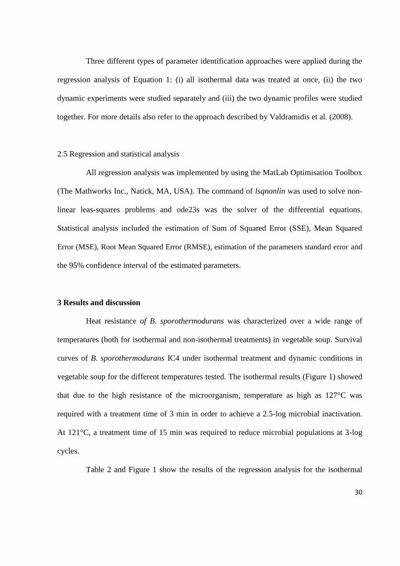
vegetable soup for the different temperatures tested. The isothermal results (Figure 1) showed
that due to the high resistance of the microorganism, temperature as high as 127°C was
required with a treatment time of 3 min in order to achieve a 2.5-log microbial inactivation.
At 121°C, a treatment time of 15 min was required to reduce microbial populations at 3-log
cycles.
Table 2 and Figure 1 show the results of the regression analysis for the isothermal

31
data. The application of a one-step regression analysis of all the isothermal data prevents
accumulation of fitting errors (Valdramidis et al., 2005). When the same spores were studied
in water, microbial non-linearities appeared to be evident and were expressed by the presence
of a shoulder effect (van Zuijlen et al., 2010). In the same work, the kinetic studies of the
inoculated commercial soups have shown a variation depending on the type of product
(mushroom, chicken, pea). The estimated parameter of the present work resulted in higher
D121 values than those obtained from distilled water in which B. sporothermodurans IC4 was
sporulated in mushroom soup agar. This is an expected result considering the protective effect
of nutrients that are present in the soup.
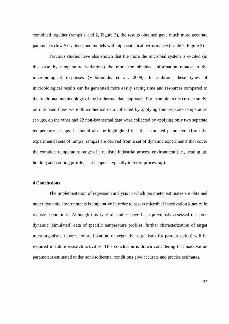
Based on the model structure selection of the isothermal data, regression analysis
was performed for the non-isothermal data by using the same model (Figure 2, Table 2). The
selection of the same Equation 1, is on the basis that dynamic data are assumed to exhibit
similar physiological responses, which will only be dependent on the specific applied
temperatures. Some authors have highlighted the importance of working with model
structures that incorporate the possible physiological adaptations, for example induced heat
resistance (Valdramidis et al., 2007). In the current study this was not assessed, as the
objective was to directly compare the regression methodologies applied as compared with the
use of different type of data, i.e., isothermal and non-isothermal.
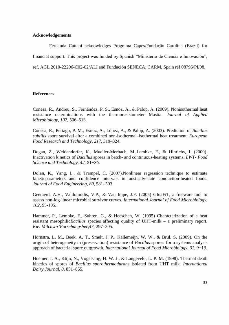
Initially, regression analysis of the dynamic data was performed separately, i.e.,
ramp1, ramp2. Despite the high fitting capacity of the model (ramp1 with RMSE=0.20, ramp2
with RMSE=0.10), the parameter estimates had very high standard errors and the D121 and z
values were significantly different between the two ramps. This could be attributed to the
experimental data and the information derived from them. Evidently, when these data are
32
combined together (ramps 1 and 2, Figure 3), the results obtained gave much more accurate
parameters (low SE values) and models with high statistical performance (Table 2, Figure 3).
Previous studies have also shown that the more the microbial system is excited (in
this case by temperature variations) the more the obtained information related to the
microbiological responses (Valdramidis et al., 2008). In addition, these types of
microbiological results can be generated more easily saving time and resources compared to
the traditional methodology of the isothermal data approach. For example in the current study,
on one hand there were 40 isothermal data collected by applying four separate temperature
set-ups, on the other had 22 non-isothermal data were collected by applying only two separate
temperature set-ups. It should also be highlighted that the estimated parameters (from the
experimental sets of ramp1, ramp2) are derived from a set of dynamic experiments that cover
the complete temperature range of a realistic industrial process environment (i.e., heating up,
holding and cooling profile, as it happens typically in retort processing).
4 Conclusions
The implementation of regression analysis in which parameter estimates are obtained
under dynamic environments is imperative in order to assess microbial inactivation kinetics in
realistic conditions. Although this type of studies have been previously assessed on some
dynamic (simulated) data of specific temperature profiles, further characterization of target
microorganisms (spores for sterilization, or vegetative organisms for pasteurization) will be
required in future research activities. This conclusion is drawn considering that inactivation
parameters estimated under non-isothermal conditions give accurate and precise estimates.
33
Acknowledgements
Fernanda Cattani acknowledges Programa Capes/Fundação Carolina (Brazil) for
financial support. This project was funded by Spanish “Ministerio de Ciencia e Innovación”,
ref. AGL 2010-22206-C02-02/ALI and Fundación SENECA, CARM, Spain ref 08795/PI/08.
References
Conesa, R., Andreu, S., Fernández, P. S., Esnoz, A., & Palop, A. (2009). Nonisothermal heat
resistance determinations with the thermoresistometer Mastia. Journal of Applied
Microbiology, 107, 506–513.
Conesa, R., Periago, P. M., Esnoz, A., López, A., & Palop, A. (2003). Prediction of Bacillus
subtilis spore survival after a combined non-isothermal–isothermal heat treatment. European
Food Research and Technology, 217, 319–324.
Dogan, Z., Weidendorfer, K., Mueller-Merbach, M.,Lembke, F., & Hinrichs, J. (2009).
Inactivation kinetics of Bacillus spores in batch- and continuous-heating systems. LWT- Food
Science and Technology, 42, 81−86.
Dolan, K., Yang, L., & Trampel, C. (2007).Nonlinear regression technique to estimate
kineticparameters and confidence intervals in unsteady-state conduction-heated foods.
Journal of Food Engineering, 80, 581–593.
Geeraerd, A.H., Valdramidis, V.P., & Van Impe, J.F. (2005) GInaFiT, a freeware tool to
assess non-log-linear microbial survivor curves. International Journal of Food Microbiology,
102, 95-105.
Hammer, P., Lembke, F., Suhren, G., & Heeschen, W. (1995) Characterization of a heat
resistant mesophilicBacillus species affecting quality of UHT-milk – a preliminary report.
Kiel MilchwirtForschungsber,47, 297–305.
Hornstra, L. M., Beek, A. T., Smelt, J. P., Kallemeijn, W. W., & Brul, S. (2009). On the
origin of heterogeneity in (preservation) resistance of Bacillus spores: for a systems analysis
approach of bacterial spore outgrowth. International Journal of Food Microbiology, 31, 9−15.
Huemer, I. A., Klijn, N., Vogelsang, H. W. J., & Langeveld, L. P. M. (1998). Thermal death
kinetics of spores of Bacillus sporothermodurans isolated from UHT milk. International
Dairy Journal, 8, 851–855.
34
Janssen, M., Verhulst, A., Valdramidis, V., Devlieghere, F., Van Impe, J.F., & Geeraerd, A.H.
(2008). Inactivation model equations and their associated parameter values obtained under
static acid stress conditions cannot be used directly for predicting inactivation under dynamic
conditions. International Journal of Food Microbiology, 128, 136-145.
Mazas, M., González, I., López, M., González, J., & Martín-Sarmiento, R. (1995). Effects of
sporulation media and strain on thermal resistance of Bacillus cereus spores. International
Journal of Food Science and Technology, 30, 71-78.
Peleg, M., Normand, M.D., & Campanella, O.H. (2003). Estimating microbial
inactivationparameters from survival curves obtained under varying conditions-the linear
case.Bulletin of Mathematical Biology, 65, 219–234.
Pettersson, B., Lembke, F., Hammer, P., Stackebrandt, E., & Priest, F.G. (1996). Bacillus
sporothermodurans, a new species producing highly heat-resistant endospores. International
Journal of Systematic Bacteriology, 46, 759–764.
Richardson, S. P. (2004). Improving the thermal processing of foods. CRC Press. USA.
Valdramidis, V. P., Belaubre, N., Zuniga, R., Foster, A. M., Havet, M., Geeraerd, A. H.,
Swain, M. J., Bernaerts, K.,van Impe, J. F., & Kondjoyan, A. (2005). Development of
predictive modeling approaches for surface temperature and associated microbiological
inactivation during hot dry air decontamination. International Journal of Food Microbiology,
100, 261−274.
Valdramidis, V. P., Geeraerd, A. H., & Van Impe, J. F. (2007). Stress adaptive responses by
heatunder the microscope of predictive microbiology. Journal of Applied Microbiology,103,
1922–1930.
Valdramidis, V.P., Geeraerd, A.H., Bernaerts, K., & Van Impe, J.F. (2006).Microbial
dynamicsversus mathematical model dynamics; the case of microbial heat
resistanceinduction.Innovative Food Science and Emerging Technologies, 7, 118–125.
Valdramidis, V.P., Geeraerd, A.H., Bernaerts, K., & Van Impe, J.F. (2008).Identification of
non-linear microbial inactivation kinetics under dynamic conditions. International Journal of
Food Microbiology, 128, 146−152.
van Derlinden, E., Lule, I., Bernaerts, K.,& Van Impe, J.F. (2010). Quantifying the
heterogeneous heat response of Escherichia coli under dynamic temperatures. Journal of
Applied Microbiology, 108,1123-1135.
vanZuijlen, A., Periago, P.M., Amézquita, A., Palop, A., Brul, S., & Fernandez, P.S. (2010).
Characterisation of Bacillus sporothermodurans IC4 spores; putative indicator microorganism
for optimization of thermal processes in food sterilization. Food Research International, 43,
1895-1901.

35
Velliou, E.G., Van Derlinden, E.,Cappuyns, A. M., Nikolaidou, E., Geeraerd, A.H.,
Devlieghere, F., & Van Impe, J.F. (2011).Towards the quantification of the effect of acid
treatment on the heat tolerance of Escherichia coli K12 at lethal temperatures. Food
Microbiology, 28, 702-711.

36
Table 1: Time sampling of isothermal treatments at each temperature.
Temperature (°C) Intervals of time (min) of sample collection
118 0, 5, 10, 15, 20, 25
121 0, 1, 2.5, 3.45, 5, 6.5, 8, 10, 12.5, 15
124 0, 1, 2, 3, 4, 6
127 0, 1, 1.5, 2, 2.5, 3

37
Table 2: Parameter estimates, standard erros, 95% confidence intervals and statistical indices
of the performed parameter identification techniques
All static data
Parameters estimate SE 95% CI 95% CI
D121 5.58 0.3412 4.89 6.28 z (degC) 10.37 0.6865 8.98 11.77
log10N(0)1 (log10(cfu/mL)) 4.66 0.1254 4.40 4.91
log10N(0)2 (log10(cfu/mL)) 4.80 0.1077 4.58 5.02
log10N(0)3 (log10(cfu/mL)) 4.54 0.0959 4.34 4.73 log10N(0)4 (log10(cfu/mL)) 4.54 0.1300 4.28 4.80 SSE, MSE, RMSE = 2.28, 0.07, 0.259
Ramp1
Parameters estimate SE 95% CI 95% CI
D121 6.14 0.6412 4.74 7.53 z (degC) 6.00 0.2627 5.42 6.57
log10N(0)1 (log10(cfu/mL)) 4.25 0.0684 4.10 4.40 SSE, MSE, RMSE = 0.50, 0.04, 0.204
Ramp2
Parameters estimate SE 95% CI 95% CI
D121 9.21 0.5253 8.04 10.38 z (degC) 21.91 0.7511 20.23 23.58
log10N(0)1 (log10(cfu/mL)) 4.46 0.0589 4.33 4.59 SSE, MSE, RMSE = 0.10, 0.01, 0.102
Ramp1 and Ramp2
Parameters estimate SE 95% CI 95% CI
D121 5.11 0.3554 4.38 5.84 z (degC) 8.23 0.1454 7.93 8.53
log10N(0)1 (log10(cfu/mL)) 4.29 0.0530 4.18 4.40
log10N(0)2 (log10(cfu/mL)) 4.3157 0.0562 4.1997 4.4318 SSE, MSE, RMSE = 0.73, 0.03, 0.175

38
Figure 1. Regression analysis of all the static data by the use of Equation (1) at the following
temperatures: 118 (●), 121 (●), 124 (○) and 127ºC (○).

39
Figure 2. Regression analysis of data collected in ramp 1 (A) for a temperature profile from
80 to 121oC and ramp 2 (B) for a temperature profile from 75 to 121
oC; by the use of
Equation (1) in both cases. Temperature profile appears with a thicker line.

40
Figure 3. Regression analysis of both data collected during ramps 1 (‒ , line blue) and 2 (--,
line black) by the use of Equation (1).Temperature profile appears with thick line.

41
Capítulo 3
Artigo Científico 2
Detection of viable cells of Bacillus sporothermodurans combining propidium monoazide
with semi-nested PCR
Artigo científico submetido ao periódico Food Microbiology.
Fator de impacto: 3.283

42

43
The detection of viable vegetative cells of Bacillus sporothermodurans using propidium
monoazide with semi-nested PCR
F. Cattani, C. A. S. Ferreira, S. D. Oliveira*
Laboratório de Imunologia e Microbiologia, Faculdade de Biociências, PUCRS, Brazil
*Corresponding author:
Sílvia Dias de Oliveira
Faculdade de Biociências, Pontifícia Universidade Católica do Rio Grande do Sul (PUCRS)
Av. Ipiranga 6681, 90619-900, Porto Alegre, Brasil
E-mail address: [email protected]
Tel.: +55-51-33534953; fax: +55-51-33203568

44
ABSTRACT
Bacillus sporothermodurans produces highly heat-resistant spores that can survive
ultra-high temperature (UHT) treatment in milk. Therefore, we developed a rapid, specific
and sensitive semi-nested touchdown PCR assay combined with propidium monoazide
(PMA) treatment for the detection of viable B. sporothermodurans vegetative cells. The semi-
nested touchdown PCR alone proved to be specific for B. sporothermodurans, and the
achieved detection limit was 4 CFU/mL from bacterial culture and artificially contaminated
UHT milk. This method combined with PMA treatment was shown to amplify DNA
specifically from viable cells and presented a detection limit of 102 CFU/mL in UHT milk.
The developed PMA-PCR assay shows applicability for the specific detection of viable cells
of B. sporothermodurans from UHT milk. This method is of special significance for
applications in the food industry by reducing the time required for the analysis of milk and
dairy products for the presence of this microorganism.
Keywords: Bacillus sporothermodurans; semi-nested PCR; milk; detection; viable cells;
propidium monoazide.
45
1. Introduction
Bacillus sporothermodurans is a Gram positive, aerobic and mesophilic bacterium,
which is characterized by the production of spores that are highly heat-resistant and capable
of surviving industrial ultra-high temperature (UHT) milk processing (140 °C for 4 s)
(Hammer et al., 1995; Pettersson et al., 1996; Scheldeman et al., 2006). Moreover, the spores
of B. sporothermodurans can germinate and grow up to 105 CFU/mL in stored UHT milk,
reaching concentrations that are above the maximum allowable thresholds for mesophilic
bacteria (Pettersson et al., 1996; Klijn et al., 1997; Herman et al., 1998), which can cause
product instability and therefore reduce both shelf life and acceptability to consumers (Tabit
and Buys, 2010). However, an increase in temperature and holding time in an attempt to
inactivate B. sporothermodurans spores can affect the organoleptic and nutritional qualities of
UHT products (Claeys et al., 2001).
The high resistance of B. sporothermodurans to the heat treatments used in the
processing of dairy products underscores the importance of its accurate detection. Phenotypic
tests for the identification of this microorganism can be complex and laborious. Additionally,
the highly competitive microbiota encountered in milk further increases the difficulties
encountered in isolating B. sporothermodurans with high sensitivity, specificity and in a short
period of time. Therefore, end-point and real time PCR targeting 16S rDNA have been
developed to detect B. sporothermodurans (Scheldeman et al., 2002; Tabit and Buys, 2011).
However, these molecular assays cannot discriminate between DNA from viable and dead B.
sporothermodurans, which can lead to false-positive results as well as to the overestimation
of cell numbers when evaluating food products (Josephson et al., 1993; Nogva et al., 2003). A
suggested approach to address this problem is to block the availability of DNA originating
46
from dead cells for PCR amplification, which can be achieved by using DNA-intercalating
dyes, such as propidium monoazide (PMA). PMA intercalates into DNA by a covalent
linkage induced by light exposure (Nocker et al., 2006). As PMA only penetrates membrane-
damaged cells, it has been widely used as an indicator of viability in a variety of bacteria,
protozoa, virus and fungi, including pathogenic, environmental and food strains (Nocker et
al., 2006; Nocker et al., 2007; Cawthorn and Witthuhn, 2008; Vesper et al., 2008; Bae and
Wuertz, 2009; Brescia et al., 2009; Josefsen et al., 2010; Taskin et al., 2011; Yánez et al.,
2011; Mamlouk et al., 2012).
In this context, the aim of this study was to develop a specific and sensitive PCR-based
method coupled to PMA treatment in order to detect only viable B. sporothermodurans
vegetative cells in the presence of dead cells from bacterial cultures and milk.
2. Materials and methods
2.1. Bacterial strains, media and culture conditions
B. sporothermodurans CBMAI 148 and CBMAI 155, obtained from the Brazilian
Collection of Microorganisms Environment and Industry UNICAMP-CPQBA, and Bacillus
cereus ATCC 33018, derived from the American Type Culture Collection, were cultivated in
BHI broth (Brain Heart Infusion) (Merck, Darmstadt, Germany) at 37 °C for 24 h. Bacillus
acidicola (NRRL B-23453), Bacillus lentus (NRRL NRS-1262), Bacillus firmus (NRRL B-
14307), Bacillus circulans (NRRL B-378), Bacillus coagulans (NRRL NRS-609),
Geobacillus stearothermophilus (NRRL B-11720) and Geobacillus kaustophilus (NRRL
NRS-81), provided by the United States Department of Agriculture (USDA), were cultivated

47
in TGY broth (5 g tryptone (Himedia, Mumbai, India),5 g yeast extract (Himedia), 1 g
glucose (Vetec, Rio de Janeiro, Brazil) and 1 g K2HPO4 (Vetec) in 1 liter dH2O).
2.2. DNA extraction
Bacterial genomic DNA from bacterial culture or milk was extracted as described by
Rademaker and de Bruijn (1997). DNA was eluted in a final volume of 50 μL MilliQ water,
and its concentration was determined using a fluorometer (Invitrogen, Van Allen Way
Carlsbad, USA) according to the manufacturer's specifications.
2.3. Semi-nested PCR assays
2.3.1. DNA amplification
In the first stage, the B. sporothermodurans primers BSPO-F2 and BSPO-R2 were used
to amplify a 664 bp fragment of the B. sporothermodurans 16S rRNA gene by PCR
(Scheldeman et al., 2002). PCR was performed in a total volume of 25 µL containing 1.5 U
Taq DNA polymerase (Invitrogen, São Paulo, Brazil), 1 X PCR buffer (Fermentas Life
Sciences, Germany), 2 mM MgCl2, 0.2 mM of each deoxynucleoside triphosphate (Fermentas
Life Sciences, Germany), 0.8 μM of each primer (Invitrogen) and 1 µL of genomic DNA.
Amplifications were carried out in a Thermocycler (MiniCyclerTM, MJ Research-Watertown,
MA–USA) using the following conditions: initial denaturation at 95 °C for 5 min followed by
30 cycles of denaturation at 95 °C for 15 s, annealing at 61 °C for 15 s, and extension at 72 °C
for 30 s, with a final extension at 72 °C for 8 min. To differentiate B. sporothermodurans
from B. acidicola, semi-nested touchdown PCR was performed using 1 µL of the first
amplification product in the same reaction mix. However, BSPO-F2 primer was replaced by

48
the forward internal primer designed in this study (5’AGAAGAGCGGAATTCCAC3’),
which shows a C as the last 3’ nucleotide, representing the only nucleotide different from B.
acidicola in this fragment, according to the sequences deposited in GenBank (ID: EU
231617.1; ID:AF329476.1; ID: 49080.1).
The semi-nested touchdown PCR produced a 613 bp fragment using the following
conditions: initial denaturation at 95 °C for 5 min, followed by 5 cycles consisting of
denaturation at 95 °C for 15 s, annealing at 67 °C for 15 s, and extension at 72 °C for 30 s, 10
cycles carried out under the same conditions (except for the annealing temperature at 66 ºC),
and 20 cycles with annealing at 65 °C, followed by a final extension at 72 °C for 8 min.
Amplification products were checked by agarose gel electrophoresis (1% w/v), 7.5 x 10 cm
(W x L) gel size, in 0.5 x TBE buffer, at a constant voltage of 100 V for 45 min; stained with
0.5 µg/mL ethidium bromide (Ludwig Biotecnologia) using 2 µL of 100 bp ladder (Ludwig
Biotecnologia) as the molecular mass ladder; and visualized under ultraviolet light using a Gel
Doc L-Pix image system (Loccus Biotecnologia, Brazil).
The amplification product obtained from the B. sporothermodurans CBMAI 148 and
CBMAI 155 was purified with PEG 8000 (USB, Cleveland, OH-USA) or with MicroSpinTM
S-400 HR Columns (Amershan Biosciences, Piscataway, N.J.), and then submitted to
nucleotide sequencing in an ABI 3130 XL Genetic Analyzer (Applied Biosystems, Lincoln
Centre Drive Foster City, USA) automated DNA sequencer. All Bacillus and Geobacillus
species described above were used to evaluate the specificity of this method.
49
2.3.2. Determination of detection limit
The detection limit of the semi-nested touchdown PCR was determined using a B.
sporothermodurans strain CBMAI 148 overnight culture of known concentration (4.0 x 10⁷
CFU/mL). Ten-fold dilutions of the original culture were prepared in 0.1% peptone saline and
commercial UHT milk. A 1 mL aliquot of each dilution of saline and artificially contaminated
milk, in quadruplicate, was subjected to DNA extraction. The CFU/mL number of the
dilutions was determined using the standard plate count method.
2.3.3. Detection of B. sporothermodurans in commercial UHT milk
The applicability of the semi-nested touchdown PCR was tested in ten samples of UHT
milk from different brands commercialized in the state of Rio Grande do Sul (Southern
Brazil). The procedure for the enumeration of total viable microorganisms in liquid UHT
dairy products followed the method of the Normative Instruction Nr. 62 of the Ministry of
Agriculture of Brazil (MAPA) (Brasil, 2003). Briefly, samples of UHT milk were incubated
at 37 °C for 7 days to observe visible changes, such as bloating and casting coagulation. Then,
1 mL aliquots of UHT milk and two decimal dilutions (10-1
and 10-2
) were spread on brain
heart infusion agar and nutrient agar (yeast extract-free) in duplicate. All plates were
incubated at 30 °C for 72 h for colony count. Two 1 mL aliquots of each UHT milk sample
were submitted to DNA extraction.

50
2.4. Discrimination of viable and dead B. sporothermodurans
2.4.1. Inactivation treatments
Two strategies were evaluated to kill B. sporothermodurans cells: (i) Heat treatment -
microtubes containing 500 µL of overnight grown cultures ( 107 CFU/mL) were heated at
100 °C in a water bath for 30 min. (ii) Isopropanol treatment - cells were killed by adding
1 mL of isopropanol (F. Maia, São Paulo, Brazil) to 500 μL of overnight grown cultures
followed by incubation for 30 min at room temperature. The isopropanol was removed by
harvesting the cells using centrifugation at 5,000 x g for 5 min and removing the supernatant.
Pellets of killed cells were resuspended in 500 μL of BHI broth (Merck). The viabilities of the
cells treated with both strategies were confirmed by plating on BHI agar.
2.4.2. PMA treatment
PMA (Biotium Inc., Hayward, California) was dissolved in 20% dimethyl sulfoxide
(DMSO) (Nuclear, São Paulo, Brazil), and added to 500 µL of B. sporothermodurans cell
suspension (viable and dead cells) at a concentration of approximately 107 cells/mL, to
achieve final concentrations of 2, 5, 10, 20, and 30 µg/mL. After 10 min of incubation in the
dark with occasional mixing, the samples were exposed to light for 10 min at a 15 cm distance
using a 500 W halogen light source (Osram, São Paulo, Brazil). After photo-activation, the
samples were centrifuged at 6,000 x g for 10 min prior to DNA extraction.
The effectiveness of the PMA treatment combined with semi-nested touchdown PCR
was further evaluated with mixtures containing different concentrations of viable and
isopropanol-killed B. sporothermodurans cells. The mixtures were prepared at pre-defined
ratios of 0, 25, 50, 75 and 100% viable cells. For example, the 100% viable cell mixtures,

51
named 100%, consisted of 500 µL of viable cells (107 CFU/mL), while the mixture named
25% was prepared by mixing 125 µL of viable cells with 375 µL of dead cells (isopropanol-
killed).
The band intensities were quantified and normalized using the band detection and
analysis tools of Quantity One 4.6.3 software (BioRad Laboratories) according to the
manufacturer's guidelines. The differences in band intensities between groups were analyzed
with Student’s t-test using IBM® SPSS® Statistics (version 2.0). A p-value of less than 0.05
was considered statistically significant.
2.4.3. Application of PMA associated to PCR in milk
Commercial UHT milk was purchased from a local supermarket. An aliquot of 500 µL
of each suspension (viable and dead cells) was diluted in commercial UHT milk to achieve
final concentrations ranging from 107 to 10
1 CFU/mL. The estimated number of CFU/mL was
determined by plating three 100 µL aliquots of the 10-5
, 10-6
and 10-7
dilutions onto BHI agar
followed by incubation for 24 h at 37 °C. Two aliquots of each dilution were removed and
one was submitted to DNA extraction without prior PMA treatment, and the other was treated
with PMA prior to the DNA extraction.
3. Results and discussion
An initial attempt to identify B. sporothermodurans cells by PCR amplification of the
16S rDNA was performed based on the method described by Scheldeman et al. (2002).
Unfortunately, the tested conditions produced amplification products for five other Bacillus
species (Fig. 1A). In this context, a semi-nested touchdown PCR method was successfully

52
developed to detect only B. sporothermodurans 16S rDNA using the same primers and an
additional internal primer, as shown in Fig. 1B. The fragments amplified from B.
sporothermodurans CBMAI 148 and CBMAI 155 were submitted to automated sequencing,
and the sequences were deposited in the GenBank database (GenBank ID: GU 238287 and JX
569192). Sequence alignment analysis showed 100% identity with the nucleotide sequence of
B. sporothermodurans strain LMG 17897 (GenBank ID: AJ302941.1), two nucleotide
alterations when compared with B. sporothermodurans strain LMG 17883 (GenBank ID:
AJ302942.1), and one different nucleotide when compared with B. acidicola strain
TCCC27037 (GenBank ID: EU231617.1) (see Supplementary data). The sequence
comparison ensured that specific detection of B. sporothermodurans was obtained and, as
expected, a high sequence identity was present even when comparing geographically distant
strains. However, although the B. acidicola sequence also presented a high identity when
compared to B. sporothermodurans sequences, the semi-nested touchdown PCR design
ensured specific detection of B. sporothermodurans. The sensitivity of the developed semi-
nested touchdown PCR method was evaluated using bacterial culture (Fig. 2A) and artificially
contaminated UHT milk (Fig. 2B), and a detection limit of 4.0 CFU/mL of B.
sporothermodurans was found from both sources. Therefore, the PCR-based method
developed here was shown to be highly specific and sensitive to detect B. sporothermodurans
vegetative cells, even in the presence of milk components, which are usually considered PCR
inhibitors (Rossen et al., 1992; Bickley et al., 1996). In addition, this method was shown to be
considerably less time-consuming than the classic procedures used for B. sporothermodurans
detection.

53
Although PCR-based methods can be sensitive, specific and applicable to food matrices,
they do not distinguish between DNA from viable and dead cells. To overcome this
limitation, treatment of samples with PMA prior to DNA extraction has been used to evaluate
the cellular viability of many different bacteria (Nocker et al., 2006; Nocker et al., 2007;
Cawthorn et al., 2008; Bae and Wuertz, 2009; Josefsen et al., 2010; Taskin et al., 2011; Yáñez
et al., 2011; Elizaquível et al., 2012; Mamlouk et al., 2012). Because, to the best of our
knowledge, no previous reports have described PMA treatment protocols for B.
sporothermodurans, experimental conditions were tested that involving bacterial-killing
strategies, PMA concentrations, and mixed viable and dead proportions of cells in order to
determine the optimal conditions for application to the semi-nested PCR method.
Two killing strategies were tested (heat and isopropanol treatments) and provided
similar results (Fig. 3), and isopropanol treatment was used throughout the remaining
experiments due to its ease of use. Possible PMA interference in the PCR amplification of
DNA from viable cells was initially evaluated and no significant difference could be found in
comparison to the controls at all concentrations used (Fig. 3, lanes 4, 7, 10, 13 and 16; p >
0.05). However, the amplification of DNA from PMA-treated dead cells (Fig. 3, lanes 5, 6, 8,
9, 11, 12, 14, 15, 17 and 18) was reduced with increasing PMA concentrations and was
completely inhibited at 30 µg/mL. PMA treatment at 30 µg/mL produced the same results
when mixing viable and dead B. sporothermodurans cells at different ratios prior to DNA
extraction (Fig. 4A) because a significant decrease in the band intensity (p < 0.05) was found
to correlate with the increase in the proportion of dead cells (Fig. 4B), although the total
amount of DNA in the reactions remained the same. Additionally, these results ensure that the
amplification of DNA from viable cells is not affected by different concentrations of

54
background dead cells in the presence of PMA. This finding is very important for the
potential applicability of the method developed here because raw milk may contain up to 107
CFU of bacteria per mL (Arenas et al., 2004; Chye et al., 2004; Torkar and Teger, 2008; Tabit
and Buys, 2011), and a considerable portion most likely dies during thermal processing. Thus,
the optimization of the PMA protocol to this bacterial density makes this method applicable
not only to UHT milk analyses but also to other food matrices with high bacterial loads.
Regarding the proportion of PMA used, it was expected that a high PMA concentration would
be required to inhibit the amplification signal because high concentrations of cells have been
suggested to inhibit the crosslinking step when PMA is light activated (Løvdal et al., 2011).
However, the optimized PMA concentration was not as high as those reported by other
authors who used high bacterial densities in their experiments (Cawthorn et al., 2008; Bae and
Wuertz, 2009; Chen et al., 2011; Taskin et al., 2011).
Another important point is the use of PMA as viability marker because its use is based
on the loss of membrane integrity, which can be considered a conservative viability criterion
when analyzing heat treated samples (Contreras et al., 2011). In this regard, Yang et al. (2011)
has reported that cells killed by heating to ≤ 72ºC may not allow PMA penetration, which can
limit the use of PMA-PCR for the analysis of some heat treated samples. Therefore, during
the design of PMA-PCR based procedures for the analysis of heat treated food, especially
when targeting milk contaminants, the fact that the pasteurization temperature may not exceed
72ºC in some instances must be considered. However, PMA can be considered a successful
viability marker to detect microorganisms in food treated at high temperatures, such as UHT
milk, as well as food exposed to treatments directly targeting membranes. The protocol
developed here tested the applicability of this method in milk and it was observed that after

55
addition of PMA in the milk samples, this dye reduced the intensity of false-positive signals
(Fig. 5, lanes 6 to 10), and its effect was not inhibited by milk components. The limit of
detection of PMA treatment associated with semi-nested touchdown PCR method in UHT
milk was determined to be 102 CFU/mL of viable cells, which is lower than or similar to those
described in other studies that have applied PMA-PCR assays to food analysis. For example, a
detection limit of 102 CFU/mL for Campylobacter jejuni (Josefsen et al., 2010) and
Brochothrix thermosphacta (Mamlouk et al., 2012) was found in chicken carcass rinse and
fresh salmon, respectively, while 103 CFU/g was described as the limit for Salmonella
Typhimurium in lettuce (Liang et al., 2011). The difference in sensibility of the PCR method
with or without the PMA pre-treatment can result from the loss of cells during the additional
PMA step and/or to the possible presence of B. sporothermodurans DNA in the late
exponential phase cultures used to determine the PCR detection limit, possibly originating
from cells that died during the bacterial growth phase. Additionally, the PMA-PCR method
detection limit of 102 CFU/mL for B. sporothermodurans alone meets the criteria of the
European Union (EU) and Brazilian legislation for the maximum count of mesophilic
microorganisms in UHT milk (Anonymous, 1992; Brasil, 1997).
In conclusion, the new molecular assay developed here for the rapid, sensitive and
specific detection of viable B. sporothermodurans cells could be a very useful tool for the
early identification of undesirable B. sporothermodurans vegetative cells in milk, dairy
products and additional food matrices. Thus, the application of this method could be of great
value for the quality control of food products by monitoring the level of viable B.
sporothermodurans during manufacture or storage and significantly reducing economic losses
to the industry.

56
Acknowledgments
The authors thank the United States Department of Agriculture (USDA) for supplying
seven Bacillus species strains and appreciate the financial support provided by Conselho
Nacional de Pesquisa (CNPq).
References
Anonymous. (1992). Council directive 92/46/EEC of 16 June 1992 laying down the health
rules for the production and placing on the market of raw milk, heat-treated milk and milk-
based products. Official Journal of the European Communities, L268, 4.9.1992, p. 1–32.
http://eurex.europa.eu/LexUriServ/LexUriServ.do?uri=CELEX:31992L0046: EN: HTML
Accessed 16.07.2012.
Arenas, R., Gonzales, L., Bernardo, A., Fresno, J. M., & Tornadijo, M. E. (2004).
Microbiological and physico-chemical changes in Genestoso cheese, a Spanish acid curd
variety, throughout ripening. Food Control, 15, 271–279.
Bae, S. & Wuertz, S. (2009) Discrimination of viable and dead fecal Bacteroidales bacteria
by quantitative PCR with propidium monoazide. Applied and Environmental
Microbiology, 75, 2940–2944.
Brescia, C.C., Griffin, S.M., Ware, M.W., Varughese, E.A., Egorov, A.I., Villegas, E.N.
(2009). Cryptosporidium propidium monoazide-PCR, a molecular biology-based technique
for genotyping of viable Cryptosporidium oocysts. Applied and Environmental
Microbiology, 75, 6856–6863.
Bickley, J., Short, J.K., McDowell, D.G., & Parkes, H.C. (1996). Polymerase chain reaction
(PCR) detection of Listeria monocytogenes in diluted milk and reversal of PCR inhibition
caused by calcium ions. Letters in Applied Microbiology, 22, 153–158.
Brasil. (1997). Ministério da Agricultura e do Abastecimento. Portaria n° 370, de 4 de
setembro de 1997. Regulamento técnico para fixação de identidade e qualidade do leite
UHT (UAT). Diário Oficial da União [República Federativa do Brasil], Brasília, 8 set.
1997. Seção I.
Brasil. (2003). Ministério da Agricultura, Pecuária e Abastecimento. Secretaria Nacional de
Defesa Agropecuária. Departamento de Defesa Animal. Métodos Analíticos Oficiais para
Análises Microbiológicas Para Controle de Produtos de Origem Animal. Diário Oficial da
União [República Federativa do Brasil], Brasília, 18 set. 2003.
57
Cawthorn, D. M., & Witthuhn, R. C. (2008). Selective PCR detection of viable Enterobacter
sakazakii cells utilizing propidium monoazide or ethidium bromide monoazide. Journal of
Applied Microbiology, 105, 1178–1185.
Chen, S., Wang, F., Beaulieu, J. C., Steins, R. E., & Ge, B. (2011). Rapid detection of viable
Salmonellae in produce by coupling propidium monoazide with Loup-Mediated isothermal
amplification. Applied and Environmental Microbiology, 77, 4008–4016.
Chye, F. Y., Abdullah, A., & Ayob, M. K. (2004). Bacteriological quality and safety of raw
milk in Malaysia. Food Microbiology, 21, 535−541.
Claeys, W. L., Ludikhuyze, L. R., & Hendrickx, M. E. (2001). Formation kinetics of
hydroxymethylfurfural, lactulose and furosine in milk heated under isothermal and non-
isothermal conditions. Journal of Dairy Research, 68, 287−301.
Contreras, P. J., Urrutia, H., Sossa, K., & Nocker, A. (2011). Effect of PCR amplicon lenght
on suppressing signals from membrane-compromissed cells by propidium monoazide
treatment. Journal of Microbiological Methods, 87, 89−95.
Elizaquível, P., Sánchez, G., Selma, M. V., & Aznar, R. (2012). Application of propidium
monoazide-qPCR to evaluate the ultrasonic inactivation of Escherichia coli O157:H7 in
fresh-cut vegetable wash water. Food Microbiology, 30, 316–320.
Hammer, P., Lembke, F., Suhren, G., & Heeschen, W. (1995). Characterization of a heat
resistant mesophilic Bacillus species affecting quality of UHT-milk – a preliminary report.
Kiel Milchwirt Forschungsber, 47, 297–305.
Herman, L., Heyndrickx, M., & Waes, G. (1998). Typing of Bacillus sporothermodurans and
other Bacillus species isolated from milk by repetitive element sequence based PCR.
Letters in Applied Microbiology, 26, 183–188.
Josefsen, M. H., Löfström, C., Hansen, T. B., Christensen, L. S., Olsen, J. E., & Hoorfar, J.
(2010). Rapid quantification of viable Campylobacter bacteria on chicken carcasses, using
real-time PCR and propidium monoazide treatment, as a tool for quantification risk
assessment. Applied and Environmental Microbiology, 76, 5097−5104.
Josephson, K. L., Gerba, C. P., Pepper, I. L. (1993). Polymerase chain reaction detection of
nonviable bacterial pathogens. Applied and Environmental Microbiology, 59, 3513–3515.
Klijn, N., Herman, L., Langeveld, L., Vaerewijck, M., Wagendorp, A. A., Huemer, I., &
Weerkamp, A. H. (1997). Genotypical and phenotypical characterization of Bacillus
sporothermodurans strains, surviving UHT sterilization. International Dairy Journal, 7,
421–428.

58
Liang, N., Dong, J., Luo, L., & Li, Y. (2011). Detection of viable Salmonella in lettuce by
propidium monoazide real-time PCR. Journal of Food Science, 76, M234–M237.
Løvdal, T., Hovda, M. B., Björkblom, B., & Møller, S. G. (2011). Propidium monoazide
combined with real-time quantitative PCR underestimates heat-killed Listeria innocua.
Journal of Microbiological Methods, 85, 164–169.
Mamlouk, K., Macé, S., Guilbaud, M., Jaffreès, E., Ferchichi, M., Prévost, H., Pilet, M., &
Dousset, X. (2012). Quantification of viable Brochothrix thermosphacta in cooked shrimp
and salmon by real-time PCR. Food Microbiology, 30, 173−179.
Nocker, A., Cheung, C. Y., & Camper, A. K. (2006). Comparison of propidium monoazide
with ethidium monoazide for differentiation of live vs. dead bacteria by selective removal
of DNA from dead cells. Journal of Microbiology Methods, 67, 310–320.
Nocker, A., Sossa, P., Burr, M., & Camper, A. K. (2007). Use of propidium monoazide for
live/dead distinction in microbial ecology. Applied and Environment Microbiology, 73,
5111–5117.
Nogva, H. K., Drømtorp, S.M., Nissen, H., & Rudi, K. (2003). Ethidium monoazide for
DNA-based differentiation of viable and dead bacteria by 5′-nuclease PCR. BioTechniques,
34, 804–813.
Pettersson, B., Lembke, F., Hammer, P., Stackebrandt, E., & Priest, F. G. (1996). Bacillus
sporothermodurans, a new species producing highly heat-resistant endospores.
International Journal of Systematic Bacteriology, 46, 759–764.
Rademaker, J. L. W., & de Bruijn, F. J. (1997). Characterization and classification of
microbes by REP-PCR genomic fingerprinting and computer-assisted pattern analysis. In
DNA markers: protocols, applications, and overviews. New York: Caetano-Anollés, G,
Gresshoff, P. M.
Rossen, L., Norskov, P., Holmstrom, K., & Rasmussen, O.F. (1992). Inhibition of PCR by
components of food samples, microbial diagnostic assays and DNA extraction solutions.
International Journal of Food Microbiology, 17, 37–45
Scheldeman, P., Herman, L., Foster S., & Heyndrickx, M. (2006). Bacillus
sporothermodurans and other highly heat-resistant spore formers in milk. Journal of
Applied Microbiology, 101, 542–555.
Scheldeman, P., Herman, L., Goris, J., De Vos, P., & Heyndrickx, M. (2002). Polymerase
chain reaction identification of Bacillus sporothermodurans from dairy sources. Journal of
Applied Microbiology, 92, 983–991.

59
Tabit, F. T., & Buys, E. (2010). The effects of wet heat treatment on the structural and
chemical components of Bacillus sporothermodurans spores. International Journal of
Food Microbiology, 140, 207−213.
Tabit, F. T., & Buys, E. (2011). Incidence and survival of Bacillus sporothermodurans during
processing of UHT milk. British Food Journal, 113, 505−518.
Taskin, B., Gozen, A. G., & Duran, M. (2011). Selective quantification of viable Escherichia
coli in biosolids by quantitative PCR with propidium monoazide modification. Applied and
Environmental Microbiology, 77, 4329−4335.
Torkar, K. G., & Teger, S. G. (2008). The Microbiological quality of raw milk after
introducing the two day’s milk collecting system. Acta agriculturae Slovenica, 92, 61–74.
van Zuijlen, A., Periago, P. M., Amézquita, A., Palop, A., Bruld, S., & Fernández, P. S.
(2010). Characterization of Bacillus sporothermodurans IC4 spores; putative indicator
microorganism for optimization of thermal process in food sterilization. Food Research
International, 43, 1895−1901.
Vesper, S., McKinstry, C., Hartmann, C., Neace, M., Yoder, S. & Vesper, A. (2008).
Quantifying fungal viability in air and water samples using quantitative PCR after
treatment with propidium monoazide (PMA). Journal of Microbiological Methods, 72,
180-184.
Yáñez, M. A., Nocker, A., Soria-Soria, E., Múrtula, R., Martínez, L., & Catalán, V. (2011).
Quantification of viable Legionella pneumophila cells using propidium monoazide
combined with quantitative PCR. Journal of Microbiology Methods, 85, 124−130.
Yang, X., Badoni, M., & Gill, C. O. (2011). Use of propidium monoazide and quantification
PCR for differentiation of viable Escherichia coli from E. coli killed by mild or
pasteurizing heat treatments. Food Microbiology, 28, 1478–1482.

60
Figures
Fig. 1. (A) Detection of Bacillus sporothermodurans by PCR. Amplicons originated from
genomic DNA of the following: (lane 1) B. sporothermodurans CBMAI 148; (lane 2) B.
sporothermodurans CBMAI 155; (lane 3) Bacillus cereus; (lane 4) Bacillus acidicola; (lane
5) Bacillus lentus; (lane 6) Bacillus firmus; (lane 7) Bacillus circulans; (lane 8) Bacillus
coagulans; (lane 9) Geobacillus stearothermophilus; (lane 10) Geobacillus kaustophilus; and
(lane 11) negative control (without template DNA); (M) 100 bp DNA ladder. (B) Detection of
B. sporothermodurans by semi-nested touchdown PCR. The 613 bp PCR products amplified
from genomic DNA of the following: (lane 1) B. sporothermodurans CBMAI 148; (lane 2) B.
sporothermodurans CBMAI 155; (lane 3) B. cereus; (lane 4) B. acidicola; (lane 5) B. lentus;
(lane 6) B. firmus; (lane 7) B. circulans; (lane 8) B. coagulans; (lane 9) G.
stearothermophilus; (lane 10) G. kaustophilus; and (lane 11) negative control (without
template DNA); (M) 100 bp DNA ladder.

61
Fig. 2. Detection limit of Bacillus sporothermodurans by semi-nested touchdown PCR.
Amplifications were performed from genomic DNA of (lane 1) B. sporothermodurans
CBMAI 148 culture (4.0 x 10⁷ CFU/mL); (lanes 2 to 9) ten-fold dilutions of a B.
sporothermodurans CBMAI 148 culture (4.0 x 10⁷ CFU/mL) until 10
0 CFU/mL in 1%
peptone saline (A) and UHT milk (B); and (lane 10) negative control (without template
DNA); (M) 100 bp DNA ladder.

62
Fig. 3. The effect of different concentrations of propidium monoazide (PMA) for the
detection of viable and dead (heat- or isopropanol-killed) Bacillus sporothermodurans
CBMAI 148 cells by PCR. Lanes 1-3: control samples, without PMA treatment; lanes 4-6: 2
µg/mL PMA; lanes 7-9: 5 µg/mL PMA; lanes 10-12: 10 µg/mL PMA; lanes 13-15: 20 µg/mL
PMA; and lanes 15-18: 30 µg/mL PMA; 1V= viable, H= heat-killed and I= isopropanol-killed.

63
Fig. 4. The effect of propidium monoazide (PMA) treatment for the detection by PCR of
viable and isopropanol-killed Bacillus sporothermodurans CBMAI 148 cells mixed at
different ratios. (A) PCR products visualized on agarose gel stained with 0.5 µg/mL ethidium
bromide under UV light. (B) Diagram representing the band intensities from amplicons
generated with or without PMA treatment using different ratios of viable:dead cells. *p <
0.001; **p < 0.005; ***p < 0.001.

64
Fig. 5. Detection limit of the PMA-PCR to detect viable and isopropanol-killed Bacillus
sporothermodurans cells in artificially contaminated milk. The ratio between viable and dead
cells is 50:50.
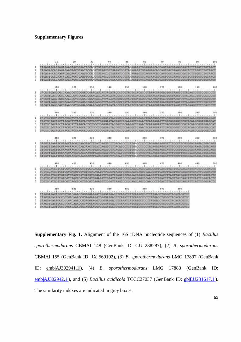
65
Supplementary Figures
Supplementary Fig. 1. Alignment of the 16S rDNA nucleotide sequences of (1) Bacillus
sporothermodurans CBMAI 148 (GenBank ID: GU 238287), (2) B. sporothermodurans
CBMAI 155 (GenBank ID: JX 569192), (3) B. sporothermodurans LMG 17897 (GenBank
ID: emb|AJ302941.1|), (4) B. sporothermodurans LMG 17883 (GenBank ID:
emb|AJ302942.1|), and (5) Bacillus acidicola TCCC27037 (GenBank ID: gb|EU231617.1|).
The similarity indexes are indicated in grey boxes.
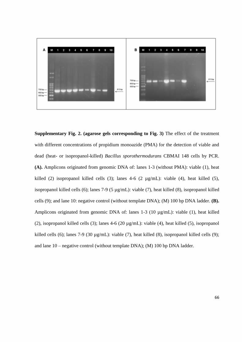
66
Supplementary Fig. 2. (agarose gels corresponding to Fig. 3) The effect of the treatment
with different concentrations of propidium monoazide (PMA) for the detection of viable and
dead (heat- or isopropanol-killed) Bacillus sporothermodurans CBMAI 148 cells by PCR.
(A). Amplicons originated from genomic DNA of: lanes 1-3 (without PMA): viable (1), heat
killed (2) isopropanol killed cells (3); lanes 4-6 (2 µg/mL): viable (4), heat killed (5),
isopropanol killed cells (6); lanes 7-9 (5 µg/mL): viable (7), heat killed (8), isopropanol killed
cells (9); and lane 10: negative control (without template DNA); (M) 100 bp DNA ladder. (B).
Amplicons originated from genomic DNA of: lanes 1-3 (10 µg/mL): viable (1), heat killed
(2), isopropanol killed cells (3); lanes 4-6 (20 µg/mL): viable (4), heat killed (5), isopropanol
killed cells (6); lanes 7-9 (30 µg/mL): viable (7), heat killed (8), isopropanol killed cells (9);
and lane 10 – negative control (without template DNA); (M) 100 bp DNA ladder.
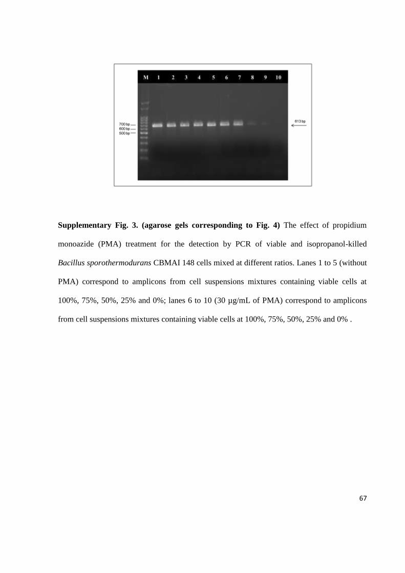
67
Supplementary Fig. 3. (agarose gels corresponding to Fig. 4) The effect of propidium
monoazide (PMA) treatment for the detection by PCR of viable and isopropanol-killed
Bacillus sporothermodurans CBMAI 148 cells mixed at different ratios. Lanes 1 to 5 (without
PMA) correspond to amplicons from cell suspensions mixtures containing viable cells at
100%, 75%, 50%, 25% and 0%; lanes 6 to 10 (30 µg/mL of PMA) correspond to amplicons
from cell suspensions mixtures containing viable cells at 100%, 75%, 50%, 25% and 0% .

68
Supplementary Fig. 4. (agarose gels corresponding to Fig. 5) Detection limit of the PMA-
PCR to detect viable and isopropanol-killed Bacillus sporothermodurans cells in artificially
contaminated milk. Lanes 1 to 5 correspond to amplicons from cell suspensions in milk (106
to 102 CFU/mL) without PMA treatment; Lanes 6 to 10 correspond to amplicons from cell
suspensions in milk (106 to 10
2 CFU/mL) with 30 µg/mL of PMA.

69
Capítulo 4
Artigo Científico 3
Detection and quantification of viable Bacillus sporothermodurans in milk by PMA-
qPCR
Artigo científico a ser submetido ao periódico Food Research International.
Fator de impacto: 3.150

70
Detection and quantification of viable Bacillus sporothermodurans in milk by PMA-
qPCR
Fernanda Cattani, Valdir Cristóvão Barth Junior, Jéssica Stephanie Rodrigues Nasário, Carlos
Alexandre Sanchez Ferreira, Sílvia Dias de Oliveira.
Laboratório de Imunologia e Microbiologia, Faculdade de Biociências, PUCRS, Brasil
*Corresponding author:
Sílvia Dias de Oliveira
Faculdade de Biociências, Pontifícia Universidade Católica do Rio Grande do Sul (PUCRS)
Av. Ipiranga 6681, 90619-900, Porto Alegre, Brasil
E-mail address: [email protected]
Tel.: +55-51-33534953; fax: +55-51-33203568
71
ABSTRACT
Bacillus sporothermodurans produces highly resistant spores that can survive ultra high
temperature (UHT) treatment in milk and germinate during the storage time. Currently, the
quantification of this microorganism is performed by the traditional culture-based method,
which is laborious and time consuming. Therefore, this study proposes a quantitative real-
time PCR (qPCR) associated with a viability marker as a faster alternative to enumeration of
viable B. sporothermodurans cells in UHT milk. Propidium monoazide (PMA) was chosen to
detect only viable cells because of its ability to bind DNA from dead cells, preventing its
amplification by PCR. The developed PMA-qPCR method showed to be specific for B.
sporothermodurans, and presented a quantification limit of 2.2 x 10² CFU/ml, taken into
account the losses that occur during the process of the milk samples. A total of 135 UHT milk
samples were analyzed and B. sporothermodurans was found in 14 (10.4%) and 11 (8.1%)
according PMA-qPCR and culturing method, respectively, not presenting a significant
difference between both methods (p = 0.083). The positive samples showed counts ranging
from 1.6 x 10² to 1.1 x 10
3 CFU/ml by PMA-qPCR and culturing method, not reaching
quantities able to cause spoilage of the product. Therefore, the PMA-qPCR method could be
of potential application in specific detection and accurate quantification of viable B.
sporothermodurans cells in milk.
Keywords: Bacillus sporothermodurans; viability; UHT milk; propidium monoazide; qPCR.
72
1. Introduction
Bacillus sporothermodurans is a bacterium able to produce highly heat resistant
spores, which are able to survive the industrial high temperature of UHT processing (130-140
°C for 4 s) (Hammer, Lembke, Suhren, & Heeschen, 1995; Pettersson, Lembke, Hammer,
Stackebrandt, & Priest, 1996). Once the spores germinate, they may grow in stored UHT milk
and cause instability due to their proteolytic activities and reduce shelf life (Klijn et al., 1997;
Tabit & Buyes, 2010). Furthermore, B. sporothermodurans spores have been found to be
more resistant than other heat resistant spores at temperatures above 130 °C with D140 ranging
from 3.4 – 7.9 s (Huemer, Klijn, Vogelsang, & Langeveld, 1998). Unfortunately, the
alternative of increasing the temperature and/or holding time to inactivate B.
sporothermodurans spores in UHT milk can affect the organoleptic and nutritional quality of
the product (Claeys, Ludikhuyze, & Hendrickx, 2001).
B. sporothermodurans is a non-pathogenic microorganism and, although it can affect
the quality of dairy products when in counts above 105 CFU/ml (Klijn et al., 1997), there is no
maximum limit for its presence in milk according to the legislations of several countries
(FSANZ, 2001; Mercosul, 1995; USPHS/FDA, 2003). However, the B. sporothermodurans
contamination can exceed the microbiological standard for viable aerobic mesophilic
microorganisms (maximum 102 CFU/ml) required by the legislation of some countries, such
as those belonging to European Union (EU) and Brazil (Anonymous, 1992; Brasil, 1997),
which can result in considerable economic losses.
The detection and enumeration of B. sporothermodurans in milk is usually performed
by traditional methods, including growth in culture media and confirmation by biochemical
tests for bacterial identification (Pettersson et al., 1996; Brasil, 2003; Scheldeman, Herman,

73
Foster & Heyndrickx, 2006). These traditional procedures are laborious and time-consuming,
and the phenotypic tests may lead to inaccurate results due to the poor growth characteristic of
the organism (Scheldeman, Herman, Goris, De Vos & Heyndrickx, 2002), what can be
circumvented by DNA-based PCR techniques that have been widely applied to detect
microorganisms in food (Beneduce, Fiocco & Spano, 2007; Naraveneni & Jamil, 2005;
Postollec, Falentin, Pavan, Combrisson & Sohier, 2011). However, DNA-based methods
cannot differentiate between viable and dead bacterial because DNA can remain intact after
cell death (Josephson, Gerba, & Pepper, 1993; Nogva, Drømtorp, Nissen, & Rudi, 2003). To
overcome this limitation, nucleic acid intercalating dyes, such as propidium monoazide
(PMA), have been used to treat samples prior to the PCR assays. PMA permeates through the
damaged cell membrane of dead cells and binds to DNA, avoiding its amplification, while it
has not been demonstrated to penetrate into viable cells (Nocker, Cheung, & Camper, 2006).
Several studies have demonstrated that PMA has been used to inhibit the DNA amplification
from a wide range of microorganisms including bacteria (Nocker, Sossa, Burr, & Camper,
2007; Cawthorn & Witthuhn, 2008; Taskin, Gozen, & Duran, 2011; Liang et al., 2011;
Mamlouk et al., 2012), fungi (Vesper et al., 2008), a protozoan (Brescia et al., 2009), and
viruses (Fittipaldi, Codony, Adrados, Camper, & Morató, 2010; Parshionikar, Laseke, &
Fout, 2010). In our previous study (Cattani, Ferreira & Oliveira, submitted manuscript), we
have presented the combination of PMA treatment with end-point PCR for the specific
detection of viable B. sporothermodurans cells and showed applicability for the specific
detection of viable cells from UHT milk. However, the quantitative analysis of the
contamination by viable B. sporothermodurans through qPCR has not yet been investigated.

74
Thus, the aim of this study was to develop a qPCR associated with PMA treatment to
quantify viable B. sporothermodurans cells, and determine its applicability in UHT milk.
2. Materials and methods
2.1 Bacterial strain and culture conditions
B. sporothermodurans CBMAI 148, obtained from the Brazilian Collection of
Microorganisms from the Environment and Industry UNICAMP-CPQBA, B. cereus ATCC
33018, and B. acidicola (NRRL B-23453), B. lentus (NRRL NRS-1262), B. firmus (NRRL B-
14307), B. circulans (NRRL B-378), B. coagulans (NRRL NRS-609) provided by the United
States Department of Agriculture (USDA), were grown in Brain Heart Infusion (BHI) broth
(Merck, Darmstadt, Germany) overnight at 37 °C.
2.2 Pre-treatment of samples with propidium monoazide
PMA (Biotium Inc., Hayward, California) was dissolved in 20% dimethyl sulfoxide
(DMSO) (Nuclear, São Paulo, Brazil), and added to 500 µL of UHT milk samples to make
final concentration of 30 µg/mL. The tubes were then placed in dark at room temperature for
10 min to allow the PMA to penetrate the dead cells and bind to the DNA. Next, the tubes
were placed in crushed ice, and exposed to light for 10 min at 15 cm distance using a 500 W
halogen light source (Osram, São Paulo, Brazil).
75
2.3 DNA isolation
Bacterial genomic DNA from pure culture or milk was extracted as described by
Rademaker and de Bruijn (1997). DNA was ressuspended in a final volume of 50 μl of MilliQ
water.
2.4 TaqMan-based qPCR assay
Specific probe and primers were designed with the Primer Express 3.0 software
(Applied Biosystems, Foster City, USA) targeting the 16S rRNA gene to amplify a 200 bp-
fragment. The forward primer (5’-AGAAGAGCGGAATTCCAC-3’) and reverse primer (5’-
AGCTGCAGCACTAAAGGGC-3’) were used in combination with the TaqMan probe (5’-
FAM- AGCGGTGAAATGC-MGBNQF- 3’). Amplification mixtures for qPCR contained 8
µl 2x Path-ID qPCR Master Mix (Ambion®, Life Technologies, USA), 0.4 µM of each primer
(Invitrogen), 0.15 µM of the TaqMan-probe (Applied Biosystems) and 2 µl of template DNA,
in a final volume of 20 µl. qPCR was performed on Applied Biosystems StepOne Real-Time
PCR System (Applied Biosystems) using an initial denaturation at 95 °C for 5 min followed
by 40 cycles at 95 °C for 15 s and 68 °C for 45 s. No-template negative controls and the
standard calibration curve was included in each run. Reactions were performed in duplicate.
The specificity of the qPCR developed assay was tested against six other different
species of Bacillus: B. cereus, B. acidicola, B. lentus, B. firmus, B. circulans and B.
coagulans.
In addition, the effectiveness of the PMA treatment combined with qPCR was further
evaluated with viable and dead cells treated with PMA as described by Cattani, Ferreira &
Oliveira (submitted manuscript). Briefly, B. sporothermodurans viable and isopropanol-killed

76
cells were treated with 30 µg/ml of PMA and, subsequently, submitted to DNA extraction.
After the qPCR analysis, ∆Ct values were calculated by the following equation: Ct value for
PMA treated cells − Ct values for untreated cells.
2.5 Standard calibration curves
The standard curve to calibrate qPCR was generated using ten-fold dilutions of
plasmid DNA harboring the target insert of B. sporothermodurans. The target fragment was
cloned into the pGEM-T Easy vector systems (Promega, Madison, Wi, USA) according to the
manufacturer’s instructions. The recombinant vectors were used to transform Top10
Escherichia coli strain by heat shock (42 °C for 45 s), that were spread on Luria Bertani (LB)
agar with ampicillin (100 µg/ml) and incubated at 37 °C for 12 h. The grown colonies were
selected and inoculated in LB broth with ampicillin (100 µg/ml) at 37 °C for 12 h with
shaking at 150 rpm. Plasmid DNA was purified using a PureYield™ Plasmid Miniprep kit
(Promega), separated by electrophoresis in 0.6% agarose gel stained with ethidium bromide
(0.5 µg/ml), and visualized under UV light. The corresponding DNA-fragment was isolated
from agarose gel and purified using Quick gel extraction kit (Promega). The concentration of
extracted plasmid DNA harboring the target insert was determined by fluorimetry (Qubit,
Invitrogen, Carlsbad, CA, USA), the absolute numbers of plasmid molecules calculated as
previously reported (Yun et al., 2006), and 10-fold serial dilutions were prepared, ranging
from 107 to 10
1 plasmid copies per PCR. A standard curve was generated by plotting the
DNA amount (mathematically adjusted to the supposed copies/ml) against the Ct value
exported from StepOne™ Real-Time PCR System (Applied Biosystems, Foster City, CA,
USA).

77
A second standard curve was constructed considering all steps required to perform the
PMA-qPCR assay to analyze B. sporothermodurans contamination in milk. Then, a B.
sporothermodurans CBMAI 148 overnight culture in BHI, containing 3.5 x 10⁷ CFU/ml, was
inoculated in 9 ml of milk and 10-fold dilutions were prepared in UHT milk. All milk
dilutions were treated with PMA, in duplicate. DNA from 500 µl aliquot of inoculated milk
was extracted as described above. The standard curve was generated by plotting the DNA
amount (expressed in its correspondent cell concentration in CFU/ml) against the Ct value
exported from the equipment.
Considering that the developed qPCR assay in this study is influenced by the fact that
the 16S rRNA gene is present in multiple copies of the genus Bacillus ranging from seven to
fourteen copies as revealed genomes sequenced to date
(http://ribosome.mmg.msu.edu/rrndb/search.php) and, to our knowledge, there is no data in
the literature concerning the exact number of 16S rRNA gene copies per cell of B.
sporothermodurans, we estimate the number of gene copies of B. sporothermodurans
comparing the amplification from the cell-based curve with the standard plasmid DNA-based
curve.
2.6 Sample preparation
The developed PMA-qPCR assay, in parallel with traditional microbiological method,
was used to analyze 135 UHT milk samples from different lots and brands. The samples were
bought from local supermarkets during the period of October 2011 to January 2012. All
samples were initially incubated for 7 days at 36 ± 1 °C and analyzed in parallel through
traditional microbiological method and qPCR associated to PMA treatment. For the molecular

78
analysis, two aliquots of each milk sample were collected and submitted to DNA extraction
with and without prior PMA treatment.
2.7 Detection of B. sporothermodurans in milk by culturing
The procedure for the enumeration of B. sporothermodurans in UHT milk was
performed according to Brasil (2003), with some modifications. Briefly, after the incubation
mentioned above, aliquots of 0.1 ml of each milk sample and two decimal dilutions (10-1
and
10-2
) were spread on BHI (Merck) agar modified by addition of 1 mg/ml of vitamin B12
(Vetec Química Fina Ltda, Rio de Janeiro, Brazil) (Pettersson, et al., 1996), in duplicate. All
plates were incubated at 30 °C for 72 h for colony count. Colonies reported as typical (small,
smooth, off-white to beige, and no soluble pigment) were enumerated and selected for further
biochemical (esculin hydrolysis, glucose fermentation, nitrate reduction, catalase, oxidase and
urease production) and morphological (Gram stain) identification.
2.8 Statistical analysis
In order to compare the frequencies of positive samples for the presence of B.
sporothermodurans obtained by the PMA-qPCR, qPCR without PMA and culturing method,
Cochran’s Q test was employed followed by pairwise comparisons using the Bonferroni
correction. One-way ANOVA, succeeded by Tukey’s post-hoc test, was used to evaluate
significant mean differences among PMA treatments in dead and viable cells. The data were
analyzed using IBM® SPSS® Statistics version 20 and the level of significance (α) was set to
0.05 for all tests.

79
3. Results and discussion
In the present study, a qPCR assay combined with PMA was developed and applied to
determine the prevalence of viable B. sporothermodurans cells in UHT milk. At first, the
qPCR was tested for specificity against other Bacillus species and positive results were
obtained only for B. sporothermodurans presence. In order to develop a method able to select
only viable cells, a PMA treatment was then associated to the qPCR assay and, thus, viable
and dead cells of B. sporothermodurans quantification experiments were evaluated. The ΔCt
value for viable cells was 0.25 (Table 1) indicating that PMA did not influence viable cells
detection. In contrast, the ΔCt value for dead cells was 15.92 (Table 1), demonstrating that
PMA reduced amplification of DNA derived from dead cells with efficiency of 99.99%, and
not inhibiting the amplification of target DNA from viable cells of B. sporothermodurans.
For the detection and quantification of B. sporothermodurans by PMA-qPCR method,
plasmid DNA- and cell-based standard curves were obtained. The plasmid DNA standard
curve showed an amplification efficiency of 87.5% with a good quantitative accuracy (r2=
0.9958) and allowed us to determine the quantification limit of the designed qPCR in 2.7 x
103 DNA copies/ml (Fig 1A). Nevertheless, to establish the real sensitivity of the PMA-qPCR
method, the second standard curve was constructed using artificially contaminated UHT milk
with a known number of B. sporothermodurans and submitted to the PMA treatment and
DNA extraction for qPCR analysis. The results showed a detection limit of 3.5 x 10² CFU/ml
with an amplification efficiency of 88.8% and determination coefficient (r2) of 0.9986 (Fig.
1A). Based on the data obtained, it was possible to estimate the number of copies of the16S
rRNA gene, which is consistent with twelve copies per cell, and, therefore, it could be
concluded that the actual detection limit of PMA-qPCR assay for detection and quantification

80
of viable B. sporothermodurans in milk was 2.2 x 10² CFU/ml (Fig. 2B). Such analysis took
into account the estimated loss of genetic material during the processing of milk samples,
which has been previously determined for another Bacillus species as approximately 38%
using the same PMA treatment and DNA extraction protocols (Cattani, Barth Jr, Nasário,
Ferreira & Oliveira, submitted manuscript).
In order to investigate the usefulness of the PMA-qPCR method, a total of 135 UHT
milk samples were evaluated for the presence of viable B. sporothermodurans. After
incubation at 36°C for 7 days, no UHT milk samples showed visible changes such as bloating,
casting or coagulation. Thus, all samples were submitted to molecular and culturing methods.
The PMA-qPCR and the traditional microbiological technique detected viable B.
sporothermodurans in 14 (10.4%) and 11 (8.15%) UHT milk samples, respectively, showing
no statistically significant difference (p = 0.083) between both methods. The occurrence of B.
sporothermodurans found in milk was similar to those described by Neuman, Salvatori,
Majolo and Froder (2010) evaluating 511 UHT milk samples by culturing methods. However,
a number quite larger than these was found previously by Busatta, Valduga and Cansian
(2005) in 88 UHT milk samples analyzed. The quantitative analysis allow us to find Ct values
for the 14 positive milk samples ranging between 36.0 and 39 cycles, which corresponded to
1.6 x 10² to 1.1 x 10
3 CFU/ml (Table 2), not reaching a quantity able to cause spoilage of the
product (Klijn et al., 1997).
Twenty-four (17.8%) samples were positive by qPCR for the detection and
quantification of B. sporothermodurans without PMA treatment, resulting in significant
difference between qPCR and PMA-qPCR methods (p = 0.002). These results clearly
illustrated that qPCR without a viability marker significantly overestimated the number of B.

81
sporothermodurans present in the milk samples in comparison with the PMA-qPCR assay.
The selective quantification of viable cells is of special concern for the samples that suffered
some antimicrobial treatment, such as UHT process. In this case, the milk is heated up to high
temperature for a few seconds killing vegetative bacterial cells. However, DNA from these
dead bacterial cells can serve as template during PCR amplification resulting in false positive
results (Josephson et al., 1993; Wolffs, Norling & Radstrom, 2005; Liang et al., 2011;
Mamlouk et al., 2012). Thus, the PMA treatment can inhibit the amplification of DNA from
dead cells and enable the qPCR to quantify specifically viable B. sporothermodurans cells
present in UHT milk.
In conclusion, the PMA-qPCR assay developed in this study was found to be a
sensitive and specific method, providing a good estimation of viable B. sporothermodurans
cells contamination in milk. This assay also presents the advantage of obtaining results in a
short time when compared to the traditional culture method, and it could be of great value for
the dairy industry to quantify this microorganism in UHT milk.
Acknowledgments
Financial support was provided by MAPA-SDA/CNPq (Conselho Nacional de
Pesquisa), Brazil, and Fernanda Cattani received a scholarship from PROBOLSAS/Pontifícia
Universidade Católica do Rio Grande do Sul (PUCRS).
82
References
Anonymous. (1992). Council directive 92/46/EEC of 16 June 1992 laying down the health
rules for the production and placing on the market of raw milk, heat-treated milk and milk-
based products. Official Journal of the European Communities, L268, 4.9.1992, p. 1–32.
http://eurex.europa.eu/LexUriServ/LexUriServ.do?uri=CELEX:31992L0046: EN: HTML
Accessed 16.07.2012.
Beneduce, L., Fiocco, D., & Spano, G. (2007). Development of PCR-based molecular tools
for the detection of emerging food- and water-borne pathogenic bacteria. In:
Communicating Current Research and Educational Topics and Trends in Applied
Microbiology. (ed. A. MendezVilas) pp. 569-576.
Brasil, (1997). Ministério da Agricultura, Pecuária e Abastecimento. Portaria nº 370, de 04 de
setembro de 1997. Regulamento Técnico para Fixação de Identidade e Qualidade de Leite
UHT (UAT). Diário Oficial da União. Brasília, 1997.
Brasil, (2003). Ministério da Agricultura, Pecuária e Abastecimento. Secretaria Nacional de
Defesa Agropecuária. Departamento de Defesa Animal. Métodos Analíticos Oficiais para
Análises Microbiológicas Para Controle de Produtos de Origem Animal. Diário Oficial da
União [República Federativa do Brasil], Poder Executivo, Brasília, DF, 18 set. 2003.
Brescia, C. C., Griffin, S. M., Ware, M. W., Varughese, E. A., Egorov, A. I., & Villegas, E.
N. (2009). Cryptosporidium propidium monoazide-PCR, a molecular biology-based
technique for genotyping of viable Cryptosporidium oocysts. Applied and Environmental
Microbiology, 75, 6856–6863.
Busatta, C., Valdruga, E., & Cansian, R. L. (2005). Ocorrência de Bacillus
sporothermodurans em leite UAT integral e desnatado. Ciência e Tecnologia de
Alimentos, 25, 408-11.
Cattani, F., Bath Jr, V. C., Nasário, J. S. R., Ferreira, C. A. S., & Oliveira, S. D. (submitted).
Real-time PCR associated with propidium monoazide for quantification of viable Bacillus
cereus in milk. International Journal of Food Microbiology.
Cattani, F., Ferreira, C. A. S., & Oliveira, S. D. (submitted).Detection of viable cells of
Bacillus sporothermodurans combining propidium monoazide with semi-nested PCR.
Food Microbiology.
Cawthorn, D. M., & Witthuhn, R.C. (2008). Selective PCR detection of viable Enterobacter
sakazakii cells utilizing propidium monoazide or ethidium bromide monoazide. Journal of
Applied Microbiology, 105, 1178–1185.
Claeys, W. L., Ludikhuyze, L. R., & Hendrickx, M. E. (2001). Formation kinetics of
hydroxymethylfurfural, lactulose and furosine in milk heated under isothermal and non-
isothermal conditions. Journal of Dairy Research, 68, 287–301.
83
Fittipaldi, M., Codony, F., Adrados, B., Camper, A., & Morató, J. (2010). Viable real time
PCR in environmental samples: can all data be interpreted directly? Microbial Ecology, 61,
7−12.
[FSANZ] Food Standards Australia New Zealand. PART 1.6 Microbiological and processing
requirements, standard 1.6.1 microbiological limits for food. 2001.
Hammer, P., Lembke, F., Suhren, G., & Heeschen, W. (1995). Characterization of a heat
resistant mesophilic Bacillus species affecting quality of UHT-milk – a preliminary report.
Kiel Milchwirt Forschungsber, 47, 297–305.
Huemer, I.A., Klijn, N., Vogelsang, H. W. J., & Langeveld, L. P. M. (1998). Thermal death
kinetics of spores of Bacillus sporothermodurans isolated from UHT milk. International
Dairy Journal, 8, 851−855.
Josephson, K. L., Gerba, C. P., & Pepper, I. L. (1993). Polymerase chain reaction detection of
nonviable bacterial pathogens. Applied and Environmental Microbiology, 59, 3513–3515.
Klijn, N., Herman, L., Langeveld, L., Vaerewijck, M., Wagendorp, A. A., Huemer, I., &
Weerkamp, A. H. (1997). Genotypical and phenotypical characterization of Bacillus
sporothermodurans strains, surviving UHT sterilization. International Dairy Journal, 7,
421–428.
Liang, N., Dong, J., Luo, L., & Li, Y. (2011). Detection of viable Salmonella in lettuce by
propidium monoazide real-time PCR. Journal of Food Science, 76, M234–M237.
Mamlouk, K., Macé, S., Guilbaud, M., Jaffreès, E., Ferchichi, M., Prévost, H., Pilet, M., &
Dousset, X. (2012). Quantification of viable Brochothrix thermosphacta in cooked shrimp
and salmon by real-time PCR. Food Microbiology, 30, 173−179.
Mercosul/Grupo Mercado Comum/ Resolução N° 78/94- Regulamento Técnico Mercosur de
Identidad y Calidad de la Leche UHT. Argentina, Brasil, Paraguai e Uruguai, 1995.
Naraveneni, R., & Jamil, K. (2005). Rapid detection of foodborne pathogens by using
molecular techniques. Journal of Medical Microbiology, 54, 51-54.
Neumann, F. D., Salvatori, R. U., Majolo, C., & Fröder, H. (2010). Occurence of Bacillus
sporothermodurans in UHT milk commercialized in the State of Rio Grande do Sul,
Brazil. Global Veterinaria, 4,156-159.
Nocker, A., Cheung, C. Y., & Camper, A. K. (2006). Comparison of propidium monoazide
with ethidium monoazide for differentiation of live vs. dead bacteria by selective removal
of DNA from dead cells. Journal of Microbiology Methods, 67, 310–320.

84
Nocker, A., Sossa, P., Burr, M., & Camper, A.K. (2007). Use of propidium monoazide for
live/dead distinction in microbial ecology. Applied and Environment Microbiology, 73,
5111–5117.
Nogva, H. K., Drømtorp, S.M., Nissen, H., & Rudi, K. (2003). Ethidium monoazide for
DNA-based differentiation of viable and dead bacteria by 5′-nuclease PCR. BioTechniques,
34, 804–813.
Parshionikar, S., Laseke, I., & Fout, G. S. (2010). Use of propidium monoazide in reverse
transcriptase PCR to distinguish between infectious and noninfectious enteric viruses in
water samples. Applied and Environmental Microbiology, 76, 4318–4326.
Pettersson, B., Lembke, F., Hammer, P., Stackebrandt, E., & Priest, F. G. (1996). Bacillus
sporothermodurans, a new species producing highly heat-resistant endospores.
International Journal of Systematic Bacteriology, 46, 759–764.
Postollec, F., Falentin, H., Pavan, S., Combrisson, J., & Sohier, D. (2011). Recent advances in
quantitative PCR (qPCR) applications in food microbiology. Food Microbiology. 28, 848-
861.
Rademaker, J. L. W., & Bruijn, F. J. (1997). Characterization and classification of microbes
by REP-PCR genomic fingerprinting and computer-assisted pattern analysis. In DNA
markers: protocols, applications, and overviews. New York: Caetano-Anollés, G,
Gresshoff, P. M.
Scheldeman, P., Herman, L., Foster S., & Heyndrickx, M. (2006). Bacillus
sporothermodurans and other highly heat-resistant spore formers in milk. Journal of
Applied Microbiology, 101, 542−555.
Scheldeman, P., Herman, L., Goris, J., De Vos, P., & Heyndrickx, M. (2002). Polymerase
chain reaction identification of Bacillus sporothermodurans from dairy sources. Journal of
Applied Microbiology, 92, 983–991.
Tabit, F. T., & Buys, E. (2010). The effects of wet heat treatment on the structural and
chemical components of Bacillus sporothermodurans spores. International Journal of
Food Microbiology, 140, 207−213.
Taskin, B., Gozen, A. G., & Duran, M. (2011). Selective quantification of viable Escherichia
coli in biosolids by quantitative PCR with propidium monoazide modification. Applied and
Environmental Microbiology, 77, 4329−4335.
USPHS/FDA, (2003) Grade “A” Pasteurized Milk Ordinance, including the Grade “A”
Condensed and Dry Milk Products and Condensed and Dry Whey Supplement I to the
Grade “A” Pasteurized Milk Ordinance.

85
Vesper, S., McKinstry, C., Hartmann, C., Neace, M., Yoder, S. & Vesper, A. (2008).
Quantifying fungal viability in air and water samples using quantitative PCR after
treatment with propidium monoazide (PMA). Journal of Microbiological Methods, 72,
180−184.
Wolffs, P., Norling, B., & Radstrom, P. (2005). Risk assessment of false-positive quantitative
real-time PCR results in food, due to detection of DNA originating from dead cells.
Journal of Microbiological Methods, 60, 315−323.
Yun, J. J., Heisler, L. E., Hwang, I. I. L., Wilkins, O., Lau, S. K., Hyrcza, M.,
Jayabalasingham, B., Jin, J., McLaurin, J., Tsao, M., Der, S. D. (2006). Genomic DNA
functions as a universal external standard in quantitative real-time PCR. Nucleic Acids
Research, 34, e85.
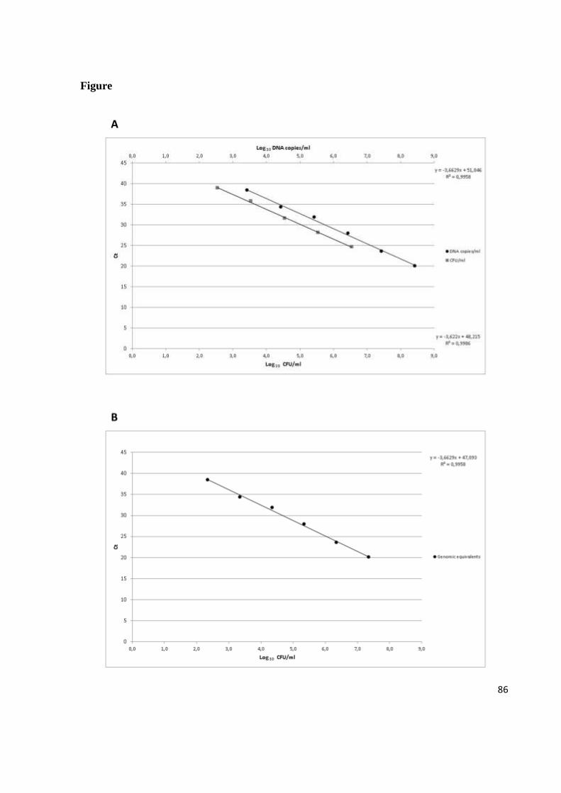
86
Figure

87
Figure 1. Standard curves used to calibrate the PMA-qPCR method. (A) Plasmid DNA and
cell-derived standard calibration curves for the B. sporothermodurans PMA-qPCR assay.
Standard curve of 10-fold serial dilution of: (●) plasmid DNA (from 2.7 x 10⁸ to 2.7 x 10
3 of
DNA copies/ml); (■) 10-fold serial dilution of B. sporothermodurans in artificially inoculated
milk (from 3.5 x 10⁶ to 3.5 x 10
2 CFU/ml). (B) Standard curve generated by the interpolation
of the cells-derived curve and the plasmid DNA curve.

88
Tables
Table 1
Effect of PMA treatment on cycle threshold (Ct) values obtained in quantitative PCR assay
from viable or dead Bacillus sporothermodurans derived from a culture with cell density of
107 CFU/ml.
Cells Ct values
NT SD T SD ∆Ct
Viable 22,20a*
0.56 22,45a 0.6 0.25
Dead 22,03a 0.58 37,95
b 0.35 15.92
*Different superscripts indicate statistically different values (p<0.05).
NT, samples not treated with PMA
T, samples treated with PMA
SD, standard deviations

89
Table 2
Quantification of viable Bacillus sporothermodurans in UHT milk by real time PCR
associated with PMA and conventional culture method.
CFU/ml Real-time PCR Traditional method
Cta range n° samples n° samples
≥ 4.0 x 10² - ≤ 1.1 x 10
3 36.0 ≤ Ct ≤ 38.0 6 6
≥ 1.6 x 10² - ≤ 3.9 x 10
2 38.1 ≤ Ct ≤ 39.0 8 5
not detectable > 39.0 or undeterminatedb 121 124
Total number of analyzed samples 135 135 aCt=cycle threshold;
bCt >39.0 or undetermined was assumed as absence of B. sporothermodurans detection.

90
Capítulo 5
Artigo Científico 4
Real-time PCR associated with propidium monoazide for quantification of viable
Bacillus cereus in milk
Artigo científico submetido ao periódico International Journal of Food Microbiology.
Fator de impacto: 3.327

91
92
Specific quantification of viable Bacillus cereus in milk by qPCR
F. Cattani, V. C. Barth Jr, J. S. R. Nasário, C. A. S. Ferreira, S. D. Oliveira
Laboratório de Imunologia e Microbiologia, Faculdade de Biociências, PUCRS, Brasil
Running title: Quantification of viable Bacillus cereus
Address correspondence to:
Sílvia Dias de Oliveira, Ph.D.
Faculdade de Biociências, Pontifícia Universidade
Católica do Rio Grande do Sul (PUCRS),
Av. Ipiranga 6681, 90619-900, Porto Alegre, Brasil
Tel.: +55-51-33534953; fax: +55-51-33203568
E-mail address: [email protected]

93
ABSTRACT
Bacillus cereus is an important spore-forming pathogen that is associated with foodborne
diseases and can be detected in food using culturing methods, which can be laborious and
time-consuming. The aim of this study was to develop a quantitative real-time PCR (qPCR)
combined with a propidium monoazide (PMA) treatment to specifically analyze the
contamination of UHT milk by viable B. cereus cells. Thirty µg/mL of PMA was shown to be
the most effective concentration for reducing the PCR amplification of extracellular DNA and
DNA from dead cells. The quantification limit of the PMA-qPCR assay was 7.5 x 10²
CFU/mL of milk. To evaluate the applicability of the developed assay, 135 UHT milk
samples were analyzed and compared to the culture-based method, and B. cereus was detected
in 44 (32.6%) and 15 (11.1%) samples, respectively. The evaluation of microorganism counts
in UHT milk using both methods indicates that infective dose may be reached depending on
the product storage conditions. Thus, the PMA-qPCR assay developed in this study was found
to be a sensitive method that was able to supply an accurate estimation of the contamination
of viable B. cereus. The application of this method in the food industry could provide great
value through the quantification of this foodborne pathogen, which represents an important
public health risk.
Keywords: Bacillus cereus; viability; propidium monoazide; UHT milk; real-time PCR.

94
1. Introduction
Bacillus cereus is a spore-forming Gram-positive bacterium that is ubiquitously found
in the environment and has been isolated from a wide variety of foods (Dierick et al., 2005;
Fricker et al., 2007; Ankolekar et al., 2009). These foods include raw, pasteurized and ultra
high temperature (UHT) milk (Notermans et al., 1997; Christiansson et al., 1999; Bahout,
2000; Banykó and Viletelová, 2009; Batchoun et al., 2011; Montanhini et al., 2012). This
microorganism is undesired in the food industry due to its spoilage capability and pathogenic
potential (Ankolekar et al., 2009). Consumption of food contaminated with B. cereus has been
implicated in outbreaks of emetic and diarrheal syndrome (Lund and Granum, 1996; Ehling-
Schulz et al., 2004; EFSA, 2005; Arnesen, et al., 2008).
Traditionally, enumeration of B. cereus from food samples is performed using culture
on selective media and biochemical identification to obtain definitive results, which are
laborious and time-consuming procedures (AOAC International, 1995; Brasil, 2003; ISO
7932, 2004). Molecular-based techniques have extensively been applied to detect foodborne
pathogens (Wang et al., 1997; Fricker et al., 2007; Ankolekar et al., 2009; Martínez-Blanch et
al., 2009; Alessandria et al., 2010; Ceuppens et al., 2010; Rantsiou et al., 2010; Fernández-No
et al., 2011; Oliwa-Stasiak et al., 2011; Rantsiou et al., 2012;), and real-time PCR has been a
reliable tool to detect and quantify B. cereus from pure culture (Fykse et al., 2003),
gastrointestinal matrix (Ceuppens et al., 2010) and food (Martínez-Blanch et al., 2009;
Fernández-No et al., 2011; Oliwa-Stasiak et al., 2011). Nevertheless, the major limitation of
any PCR-based assay is its inability to distinguish between DNA from viable cells and free
DNA, which are either available in the environment or originate from dead cells (Rudi et al.,

95
2005). If DNA from non-viable cells is amplified, then the bacteria counts will be
overestimated. Therefore, end-point and real-time PCR alone may not provide a reliable
indication of human health risk levels posed by the presence of viable pathogens. To
overcome this limitation, propidium monoazide (PMA) has been used as a nucleic acid-
intercalating dye to inhibit PCR amplification of extracellular DNA and DNA from dead
and/or membrane-compromised cells (Nocker et al., 2006; Nocker et al., 2007; Cawthorn and
Witthuhn, 2008; Josefsen et al., 2010; Liang et al., 2011; Yang et al., 2011; Mamlouk et al.,
2012; Martinon et al., 2012). However, to our knowledge, the effectiveness of PMA has not
yet been evaluated for B. cereus, which would be of value from a food safety perspective.
We describe a quantitative real-time PCR (qPCR) combined with PMA treatment that is
specific and sensitive in the detection and quantification of viable B. cereus directly from
naturally contaminated UHT milk samples.
2. Materials and methods
2.1. Bacterial strains and culture conditions
B. sporothermodurans CBMAI 148 was obtained from the Brazilian Collection of
Microorganisms from the Environment and Industry UNICAMP-CPQBA. B. acidicola
(NRRL B-23453), B. lentus (NRRL NRS-1262), B. firmus (NRRL B-14307), B. circulans
(NRRL B-378) and B. coagulans (NRRL NRS-609) were provided by the United States
Department of Agriculture (USDA). All strains were cultivated in Brain Heart Infusion (BHI)
broth (Merck, Darmstadt, Germany) at 37 °C for 24 h.
B. cereus ATCC 33018 was grown overnight in BHI broth (Merck) at 37 °C,
corresponding to a concentration of 108 cells/mL. Dead cells were obtained from two
96
treatments: (i) Heat - 500 µL of cell suspension was heated at 100 °C in a water bath for 30
min; and (ii) Isopropanol - cells were killed by adding 1 mL of isopropanol (F. Maia, São
Paulo, Brazil) to 500 µL of cell suspension followed by incubation for 30 min at room
temperature. The absence of viable cells from both strategies was confirmed by spread plating
100 µL aliquots of cells on BHI agar.
2.2. Optimization of PMA conditions
PMA (Biotium Inc., Hayward, USA) was dissolved in 20% dimethyl sulfoxide
(DMSO) (Nuclear, São Paulo, Brazil) and added to 500 µL of B. cereus cell suspension
(viable and dead cells) to achieve final concentrations of 2, 5, 10, 20 and 30 µg/mL. The tubes
were then placed in the dark for 10 min to allow PMA to penetrate into dead cells and bind to
the DNA. Afterwards, tubes were exposed to light for 10 min at a 15-cm distance using a
500 W halogen light source (Osram, São Paulo, Brazil). Additionally, to determine the PMA
effectiveness in selectively amplifying DNA from viable cells, mixtures of viable and dead
cells were evaluated. Mixtures were prepared so that viable cells corresponded to 100, 75, 50,
25 and 0% of the total bacterial cell concentration, using the same protocol for PMA
treatment.
2.3. DNA extraction
Bacterial genomic DNA from pure culture or milk was extracted as described by
Rademaker and de Bruijn (1997). The extracted DNA was resuspended in a final volume of
50 μL of MilliQ water.
97
2.4. End-point PCR
The optimization of PMA treatment was performed via end-point PCR using
oligonucleotides targeting the hemolysin gene of B. cereus (Wang et al., 1997). PCR was
performed in a solution containing 1.5 U of Taq DNA polymerase (Fermentas, St. Leon-Rot,
Germany), 1 X PCR buffer (Fermentas), 3 mM MgCl2, 0.2 mM of each deoxynucleoside
triphosphate (Fermentas), 0.8 µM of each primer (Invitrogen, São Paulo, Brazil) and 1 μL of
genomic DNA, in a final volume of 25 μL. The reactions were run on a Thermocycler
(MiniCycler MJ Research, Watertown, USA) at 95 °C for 3 min followed by 35 cycles at 95
°C for 10 s, 56 °C for 15 s, 72 °C for 15 s and 72 °C for 3 min. PCR products were visualized
on 1% agarose gel (Invitrogen) in TBE buffer with ethidium bromide (0.5 µg/mL) (Ludwig
Biotecnologia Ltda, Porto Alegre, Brazil) under UV light.
The band intensity was determined using Quantity One 4.6.3 Software (BioRad
Laboratories, Hercules, CA). The differences of band intensities between groups were
analyzed by the Student’s t-test using IBM® SPSS® Statistics version 20 (Somers, New
York, USA). A p-value of less than 0.05 was considered to be statistically significant.
2.5. Sample preparation
A total of 135 UHT milk samples from different lots and brands were purchased from
local retail markets during the period between October 2011 and January 2012.
All samples were analyzed in parallel using standard microbiological method and
qPCR. For the molecular analysis, two 500 µL aliquots were collected from each milk
sample; one aliquot was treated with PMA (30 µg/mL) before DNA extraction, whereas the
other was directly submitted to DNA extraction.
98
2.6. Detection of B. cereus in milk by culturing
Milk samples were incubated for 7 days at 36 ± 1 °C in order to favor spore
germination. Subsequently, aliquots of 25 mL of each sample were taken aseptically and then
diluted 10-fold in 0.1% peptone saline (pH 7.0). All dilutions were spread on B. cereus
selective agar base mannitol egg yolk polymyxin B (MYP) (Himedia, Mumbai, India) in
duplicate and incubated at 30 °C for 24-48 h. Colonies surrounded by a precipitate zone were
selected, transferred onto the stock agar and submitted for confirmation by Gram staining and
other phenotypic tests, which included motility, nitrate reduction, hemolytic activity on sheep
blood agar, tyrosine decomposition, rhizoid growth, and the absence of crystal parasporal
inclusion (Brasil, 2003). B. cereus ATCC 33018 was used as a reference culture.
2.7. Standard calibration curves
A standard curve was generated using 10-fold dilutions of plasmid DNA harboring the
target insert of B. cereus by qPCR. The 185-bp fragment of the hemolysin gene was cloned
into the pGEM-T Easy vector system (Promega, Madison, USA), according to the
manufacturer’s instructions. The recombinant vectors were used to transform Top10
Escherichia coli strain by heat shock (42 °C/45 s) and were then spread on Luria Bertani (LB)
agar with ampicillin (100 µg/mL) and incubated for 12 h at 37 °C. Colonies were selected and
inoculated in LB broth with ampicillin (100 µg/mL) for 12 h at 37 °C with shaking at 200
rpm. Plasmid DNA was purified using a PureYield™ Plasmid Miniprep kit (Promega),
separated by electrophoresis in 0.6% agarose gel, stained with ethidium bromide, and
visualized under UV light. The corresponding band was isolated from the agarose gel and
purified using a Quick gel extraction kit (Promega). The concentration of extracted plasmid

99
DNA harboring the target insert was determined using fluorimetry (Qubit, Invitrogen).
Subsequently, 10-fold serial dilutions of the extract were prepared, ranging from 106 to 10
0
plasmid copies per PCR. A standard curve was generated by plotting the DNA amount
(mathematically adjusted to the supposed copies/mL) against the Ct value exported from the
StepOne™ Real-Time PCR System (Applied Biosystems, Foster City, CA, USA).
Another standard curve to analyze B. cereus contamination in milk was constructed by
considering all steps required to perform the PMA-qPCR assay. Then, an overnight culture of
B. cereus ATCC 33018 in BHI containing 7.5 x 10⁸ CFU/mL was inoculated in 9 mL of milk.
Ten-fold dilutions were prepared in UHT milk and subsequently treated with PMA in
duplicate. DNA from 500 µL aliquots of inoculated milk was extracted as described above.
The standard curve was generated by plotting the DNA amount (expressed in the
corresponding cell concentration in CFU/mL) against the Ct value exported from the
equipment.
The determination of the quantification limit has taken into account that this hemolysin
(perfringolysin O) gene is present as a single copy in the B. cereus strains (B. cereus ATCC
14579, NC_004722.1; B. cereus ATCC 10987, NC_003909.8; B. cereus NC 7401,
NC_016771.1; B. cereus AH187, NC_011658.1; B. cereus AH820, NC_011773.1), as found
in the BLAST analysis (http://blast.ncbi.nlm.nhi.gov) of the genomes deposited in the
GenBank database (data not shown).
2.8. qPCR
A TaqMan qPCR method for the specific detection and quantification of B. cereus
based on the amplification of a 185-bp fragment of the hemolysin gene was performed on a

100
StepOne™ Real-Time PCR System (Applied Biosystems). The primers used were the same as
the end-point PCR primers, and the TaqMan MGB probe (5’-FAM- AGCTGTACAACTTGC
3’) was designed using the Primer Express 3.0 software from Applied Biosystems. qPCR
using the primers and the probe was optimized with Path-ID qPCR Master Mix (Ambion®,
Life Technologies, USA) in a final volume of 20 µL that contained 10 µL 2 X Path-ID qPCR
Master Mix, 0.6 µM of each primer, 0.2 µM of probe and 2 µL of template DNA.
Amplifications were performed with 1 cycle at 95 °C for 10 min, 40 cycles at 95 °C for 15 s
and 58 °C for 45 s. In every analysis, a negative control was included by using 2 µL of MilliQ
water instead of the DNA template, and the plasmid DNA standard calibration curve was
included. Reactions were performed in duplicate.
The specificity of the qPCR assay was tested against six different species of Bacillus:
B. sporothermodurans, B. acidicola, B. lentus, B. firmus, B. circulans and B. coagulans.
2.9. Statistical analysis
To compare the B. cereus positive sample frequencies obtained by the PMA-qPCR,
qPCR without PMA and culturing method, Cochran’s Q test was employed followed by
pairwise comparisons using the Bonferroni correction. One-way ANOVA, succeeded by
Tukey’s post-hoc test, was used to evaluate the significant mean differences among PMA
treatments in dead and viable cells. The data were analyzed using IBM® SPSS® Statistics
version 20, and the level of significance (α) was set to 0.05 for all tests.

101
3. Results and discussion
In the present study, we report a useful PMA-qPCR assay to allow the specific detection
of viable B. cereus, enabling the DNA-based method to accurately evaluate the microbial load
within foods, mainly those that are submitted to stressing conditions during processing and
storage. First, end-point PCR was used to evaluate the efficiency of PMA to exclude the DNA
from dead cells. The amplifications of DNA from PMA-treated dead cells gradually reduced
with increasing concentrations of PMA (p<0.05) (Fig. 1, lanes 5, 6, 8, 9, 11, 12, 14, 15, 17
and 18). Similar results were obtained using either heat or isopropanol as cell killing methods,
and isopropanol treatment was used throughout the remainder of this study because of its ease
of use. A PMA concentration of 30 µg/mL was used in the remaining experiments, as it was
shown to completely inhibit the PCR amplification from dead cell DNA (Fig. 1, lanes 17 and
18). By contrast, we detected specific amplifications from PMA-treated viable cell DNA, non-
treated viable cells (Fig. 1, lanes 1, 4, 7, 10, 13 and 16) and non-treated dead cells (Fig. 1,
lanes 2 and 3). Furthermore, we showed that the PMA associated to PCR could specifically
detect DNA from B. cereus viable cells in the presence of DNA from dead cells, which can be
observed from the direct relation between the relative intensity of the DNA bands (p<0.05)
and the percentage of viable cells in the range of cell mixtures (Fig. 2). Therefore, PMA
treatment effectively inhibited the PCR amplification of DNA from dead B. cereus cells
without having an effect on the amplifications of DNA from viable cells. Viable cells did not
interfere with the effect of PMA on dead cells, even in a background containing a high load of
bacterial cells. These findings indicate the potential application of the PMA-PCR method to
evaluate food exposed to treatments that injure bacterial cells, especially those treatments that
interfere with membrane integrity. However, end-point PCR is not an ideal method to
102
evaluate the B. cereus contamination in some foods because certain levels of this
microorganism may be acceptable, which indicates the need to use a quantitative method. We
associated the standardized PMA treatment with the qPCR to specifically quantify viable B.
cereus cells. To evaluate the effect of PMA on qPCR-amplifiable DNA from viable or dead
cells, ∆Ct (Ct value for PMA treated cells − Ct values for untreated cells) values were
calculated. The ∆Ct values for viable and dead cells were 0.83 and 14.93, respectively (Table
1), which indicates that PMA showed no interference in the detection of viable B. cereus
using the qPCR assay developed here.
Regarding the specificity of the qPCR method employed, the primers selected had
previously presented a high specificity when tested against a broad panel of bacterial species
(Wang et al., 1997). Furthermore, the TaqMan probe used in this study was designed to
specifically detect B. cereus, which was confirmed by the absence of amplification from the
DNA of six different strains and species of Bacillus.
The enumeration of B. cereus by qPCR was performed using cloned DNA- and cell-
based standard curves. The plasmid DNA standard curve showed an amplification efficiency
of 96.15%, with good quantitative accuracy (r2 = 0.9995), and allowed us to determine the
quantification limit of the designed qPCR in 1.6 x 10² DNA copies/mL (Fig. 3A). However,
to determine the actual sensitivity, which takes into account the possible losses that can occur
when processing the samples, we constructed another standard curve from milk that was
artificially contaminated with a known number of B. cereus. The contaminated milk was
submitted to PMA treatment and DNA extraction, and the curve generated from these cells
showed an amplification efficiency of 97.41% and a high coefficient of determination (r2 =
0.9971), providing us a quantification limit of 7.5 x 10² CFU/mL (Fig. 3A). Because both

103
standard curves had very similar efficiencies, we used the linear equation resulting from the
cell-derived curve and compared it to each point of the plasmid DNA curve, thus estimating
the differences in the final outcome for both curves (Fig. 3B). The comparison showed a
reduction of approximately 38% of genetic material in the cell-derived curve, indicating the
proportion of DNA loss during the process. This loss was then considered when determining
the quantity of viable B. cereus cells in UHT milk samples. The developed method was then
compared to the traditional culturing technique by analyzing 135 UHT milk samples. The
qPCR without PMA treatment detected B. cereus in 78 samples (57.8%), and the PMA-qPCR
method detected 44 positive samples (32.6%) (Ct ranging from 26.9 to 37.0), presenting a
significant difference (p<0.001) (Table 2). The PMA treatment likely inhibited the
amplification of a large amount of DNA from cells that were killed during the UHT
processing, which avoided an overestimation of B. cereus cells when using qPCR and, thus,
did not overvalue potential health risks. All 15 samples that were B. cereus-positive according
to the culture-based method were also detected by PMA-qPCR. Additionally, qPCR-PMA
was able to detect the presence of B. cereus in 29 additional samples, showing a significant
difference (p<0.001) when comparing both methods. Thus, these results indicate that the
PMA-qPCR method was far more sensitive than the culturing method, which, at least
partially, can be due to the lack of sensitivity of the selective media used to identify B. cereus
(Ehling-Schulz et al., 2004; Fricker et al., 2008; Reekmans et al., 2009; Ceuppens et al.,
2010). The MYP medium, which is commonly used to detect B. cereus, can present weak
reactions and is unable to detect B. cereus strains without lecithinase activity (ISO 2004).
Moreover, another difference among these methods is that the cultural assay can detect only
viable and replicating bacteria, whereas the molecular-based assays associated with PMA can
104
detect DNA from viable and also viable but non-culturable (VBNC) microorganisms. The
VBNC state has previously been described in B. stratosphericus (Cooper et al., 2010), which
enables us to consider that it can also occur in other Bacillus species, especially when
submitted to stress conditions, such as high temperature. This cellular condition is of special
concern because of the maintenance of infection potential, despite the inability of cells to
grow in culture media (McDougald et al., 1998, Wery et al., 2008).
Analyzing these data in a quantitative perspective, it was found that 44 UHT milk
samples contained B. cereus in concentrations ranging from approximately 102 to 10
5
CFU/mL (Table 2). In this context, it is important to note that the samples were incubated at
36°C for 7 days before the analyses, and, therefore, cell counts do not correspond to those
originally present in the samples. However, these data show that raw milk can be
contaminated with B. cereus spores that survive to the UHT treatment. Consequently, as
temperature and time of storage until consumption vary widely, it may be inferred that UHT
milk can provide growth conditions for B. cereus to reach infective dose required to cause
foodborne disease (Gilbert and Kramer, 1986; Granum and Lund, 1997; Granum, 2002;
Arnesen et al., 2008; Martínez-Blanch et al., 2009; Salustiano et al., 2010). Moreover,
according to European regulation (EC nº 2073/2005), foods in general should not contain
microorganisms, their toxins or metabolites in quantities that present an unacceptable risk for
human health. This guideline is similar to the Brazilian requirements for UHT milk, which
should not present pathogenic microorganisms (Brasil, 2001). Thus, at least 28.1% and 11.1%
of the UHT milk samples analyzed using the PMA-qPCR assay and traditional culture
methods, respectively, did not meet the established standards.
105
An important advantage of the method developed in this study is that it accomplishes a
rapid, specific and sensitive evaluation of B. cereus levels in UHT milk, detecting it at
numbers lower than the minimum infectious dose. This method represents a more realistic and
accurate analysis for food products, which is crucial to lower costs and improve food-health
security.
Acknowledgements
Financial support was provided by MAPA-SDA/CNPq (Conselho Nacional de
Pesquisa), Brazil, and Fernanda Cattani received a scholarship from PROBOLSAS/Pontifícia
Universidade Católica do Rio Grande do Sul (PUCRS).
References
Alessandria, V., Rantsiou, K.; Dolci, P.; Cocolin, L., 2010. Molecular methods to assess
Listeria monocytogenes route of contamination in a dairy processing plant. International
Journal of Food Microbiology 141, S156-S162.
Ankolekar, C., Rahmati, T., Labbé, R., 2009. Detection of toxigenic Bacillus cereus and
Bacillus thuringiensis spores in U.S. rice. International Journal of Food Microbiology 128,
460-466.
AOAC International, 1995. Microbiological Methods–Bacillus cereus in Foods–Enumeration
and Confirmation–Official Method, 980:31.
106
Arnesen, L. P., Fagerlund, A., Granum, P. E., 2008. From soil to gut: Bacillus cereus and its
food poisoning toxins. FEMS Microbiology Reviews 32, 579-606.
Bahout, A. A., 2000. Prevalence of Bacillus species in UHT milk. Assiut Veterinary Medical
Journal 42, 47-53.
Banykó, J., Vyletelová, M., 2009. Determining the source of Bacillus cereus and Bacillus
licheniformis isolated from raw milk, pasteurized milk and yoghurt. Letters in Applied
Microbiology 48, 318-321.
Batchoun, R., Al-Sha’er, A. I., Khabour, F., 2011. Molecular characterization of Bacillus
cereus toxigenic strains isolated from different food matrices in Jordan. Foodborne
Pathogens and Disease 8, 1153-1158.
Brasil Resolução – RDC n.12 jan. 2001. Revoga Portaria n.451, 19 set. 1997. Diário Oficial
da União, Brasília, DF, art. 4°, p.1-48.
Brasil, Ministério da Agricultura, Pecuária e Abastecimento. Secretaria Nacional de Defesa
Agropecuária. Departamento de Defesa Animal. Métodos Analíticos Oficiais para Análises
Microbiológicas Para Controle de Produtos de Origem Animal. Cap III, p.14- 15. Diário
Oficial da União [República Federativa do Brasil], Poder Executivo, Brasília, DF, 18 set.
2003.

107
Cawthorn, D. M., Witthuhn, R. C., 2008. Selective PCR detection of viable Enterobacter
sakazakii cells utilizing propidium monoazide or ethidium bromide monoazide. Journal of
Applied Microbiology 105, 1178-1185.
Ceuppens, S., Boon, N., Rajkovic, A., Heyndrickx, M., Van de Wiele, T., Uyttendaele, M.,
2010. Quantification methods for Bacillus cereus vegetative cells and spores in the
gastrointestinal environment. Journal of Microbiological Methods 83, 202-210.
Christiansson, A., Berttilsson, J., Svensson, B., 1999. Bacillus cereus spores in raw milk:
factors affecting the contamination of milk during the grazing period. Journal of Dairy
Science 82, 305-314.
Cooper, M., Fridman, G., Fridman, A., Joshi, S. G., 2010. Biologiacal responses of Bacillus
stratosphericus to floating electrode-dielectric barrier discharge plasma treatment. Journal
of Applied Microbiology 109, 2039-2048.
Dierick, K., Van, Coillie, E., Swiecicka, I., Meyfroidt, G., Devlieger, H., Meulemans, A.,
Hoedemaekers, G., Fourie, L., Heyndrickx, M., Mahillon, J., 2005. Fatal family outbreak
of Bacillus cereus–associated food poisoning. Journal of Clinical Microbiology 43, 4277-
4279.

108
EC nº 2073/2005. Comission regulation EC nº 2073/2005, of 15 November 2005 on
microbiological criteria for foodstuffs (Text with EEA relevance) (OJ L 338, 22.12.2005,
p. 1)
EFSA., 2005. Opinion of the scientific panel on biological hazards on Bacillus cereus and
other Bacillus spp in foodstuffs. EFSA Journal 175, 1-48.
Ehling-Schulz, M., Fricker, M., Scherer, S., 2004. Bacillus cereus, the causative agent of an
emetic type of food-borne illness. Molecular Nutrition and Food Research 48, 479-487.
Fernández-No, I.C., Guarddon, M., Böhme, K., Cepeda, A., Calo-Mata, P., Barros-Velázquez,
J., 2011. Detection and quantification of spoilage and pathogenic Bacillus cereus, Bacillus
subtilis and Bacillus licheniformis by real-time PCR. Food Microbiology 28, 605-610.
Fricker, M., Messelhäuber, U., Busch, U., Scherer, S., Ehling-Schulz, M., 2007. Diagnostic
real-time PCR assay for the detection of emetic Bacillus cereus strains in food and recent
food-borne outbreaks. Applied and Environmental Microbiology 73, 1892-1898.
Fricker, M., Reissbrodt, R., Ehling-Schulz, M., 2008. Evaluation of standard and new
chromogenic selective plating media for isolation and identification of Bacillus cereus.
International Journal of Food Microbiology 121, 27-34.

109
Fykse, E. M., Olsen, J. S., Skogan, G., 2003. Application of sonication to release DNA from
Bacillus cereus for quantitative detection by real-time PCR. Journal of Microbiology
Methods 55, 1-10.
Gilbert, R. J., Kramer, J. M., 1986. Bacillus cereus food poisoning. Progress in Food Safety
(Proceedings of Symposium) (Cliver DC & Cochrane BA, eds), 85-93. Food Research
Institute, University of Wisconsin-Madison, Madison, WI.
Granum, P. E., Lund, T., 1997. Bacillus cereus and its food poisoning toxins. FEMS
Microbiology Letters 157, 223-228.
Granum, P.E., 2002. Bacillus cereus and food poisoning. In applications and systematics of
Bacillus and relatives. In: Berkeley, R., Heyndrickx, M., Logan, N., De Vos, P. (Eds.),
Blackwell Publishing, Oxford, pp. 37–46.
ISO, 2004. International Organization for Standardization–Microbiology of Food and Animal
Feeding Stuffs–Horizontal Method for the Enumeration of Presumptive Bacillus cereus–
Colony Count Technique at 30 °C. Geneva: International Organization for Standardization.
ISO 7932:2004.
Josefsen, M. H., Löfström, C., Hansen, T. B., Christensen, L. S., Olsen, J. E., Hoorfar, J.,
2010. Rapid quantification of viable Campylobacter bacteria on chicken carcasses, using
110
real-time PCR and propidium monoazide treatment, as a tool for quantification risk
assessment. Applied and Environmental Microbiology 76, 5097-5104.
Liang, N., Dong, J., Luo, L., Yong, L., 2011. Detection of viable Salmonella in lettuce by
propidium monoazide real-time PCR. Journal of Food Science 76, M234-M237.
Lund, T., Granum, P.E., 1996. Characterisation of a non-haemolytic enterotoxin complex
from Bacillus cereus isolated after a foodborne outbreak. FEMS Microbiology Letters 141,
151–156.
Mamlouk, K., Macé, S., Guilbaud, M., Jaffreès, E., Ferchichi, M., Prévost, H., Pilet, M.,
Dousset, X., 2012. Quantification of viable Brochothrix thermosphacta in cooked shrimp
and salmon by real-time PCR. Food Microbiology, 30, 173−179.
Martínez-Blanch, J.F., Sánchez, G., Garay, E., Aznar, R., 2009. Development of a real-time
PCR assay for detection and quantification of enterotoxigenic members of Bacillus cereus
group in food samples. International Journal of Food Microbiology 135, 15-21.
Martinon, A., Cronin, U. P., Quealy, J., Stapleton, A., Wilkinson, M. G., 2012. Swab sample
preparation and viable real-time PCR methodologies for the recovery of Escherichia coli,
Staphylococcus aureus or Listeria monocytogenes from artificially contaminated food
processing surfaces. Food Control 24, 86-94.
111
McDougald, D., Rice, S. A., Weichart, D., Kjelleberg, S., 1998. Nonculturability: adaptation
or debilitation? FEMS Microbiology Ecology 25, 1-9.
Montanhini, M. T. M., Pinto, J. P. A. N., Bersot, L. S., 2012. Occurrence of Bacillus cereus in
Milks Traded in Paraná, Santa Catarina and São Paulo States. UNOPAR Científica.
Ciências Biológicas e da saúde 14, 155-158.
Nocker, A., Cheung, C. Y., Camper, A. K., 2006. Comparison of propidium monoazide with
ethidium monoazide for differentiation of live vs. dead bacteria by selective removal of
DNA from dead cells. Journal of Microbiological Methods 67, 310-320.
Nocker, A., Sossa, P., Burr, M., Camper, A.K., 2007. Use of propidium monoazide for
live/dead distinction in microbial ecology. Applied of Environmental Microbiology 73,
5111-5117.
Notermans, S., Dufrenne, J., Teunis, P., Beumes, R., te Giffel. M., Weem, P.P., 1997. A risk
assessment study of Bacillus cereus in pasteurized milk. Food Microbiology 14, 143-151.
Oliwa-Stasiak, K., Kolaj-Robin, O., Adley, C. C., 2011. Development of real-time PCR assay
for detection and quantification of Bacillus cereus group species: differentiation of B.
weihenstephanensis and Rhizoid B. pseudomycoides isolates from milk. Applied and
Environmental Microbiology 77, 80-88.

112
Rademaker, J. L. W., de Bruijn, F. J., 1997. Characterization and classification of microbes by
REP-PCR genomic fingerprinting and computer-assisted pattern analysis. In: DNA
markers: protocols, applications, and overviews. Caetano-Anollés G, and Gresshoff P M
(eds). New York: J. Wiley and Sons, 151-171.
Rantsiou, K.; Lamberti, C.; Cocolin, L., 2010. Survey of Campylobacter jejuni in retail
chicken meat products by application of a quantitative PCR protocol. International Journal
of Food Microbiology 141, S75-S79.
Rantsiou, K.; Alessandria. V.; Cocolin, L., 2012. Prevalence of Shiga toxin-producing
Escherichia coli in food products of animal origin as determined by molecular methods.
International Journal of Food Microbiology 154, 37-43.
Reekmans, R., Stevens, P., Versust, T., De Vos, P., 2009. Na alternative real-time PCR
method to detect the Bacillus cereus group in naturally contaminated food gelatin: a
comparison study. Letters in Applied Microbiology 48, 97-104.
Rudi, K., Moen, B., Dromtorp, S. M., Holck, A. L., 2005. Use of ethidium monoazide and
PCR in combination for quantification of viable and dead cells in complex samples.
Applied and Environmental Microbiology 71, 1018-1024.
Salustiano, V. C., Andrade, N. J., Ribeiro Junior, J. I., Fernandes, P. E., Lopes, J. P.,
Bernardes, P. C., Portugal, J. G., 2010. Controlling Bacillus cereus adherence to stainless

113
steel with different cleaning and sanitizing procedures used in dairy plants. Arquivo
Brasileiro de Medicina Veterinária e Zootecnia 62,1478-1483.
Wang, R. F., Cao, W. W., Cerniglla, C. E., 1997. A universal protocol for PCR detection of
13 species of foodborne pathogens in food. Journal of Applied Microbiology 83, 727-736.
Wery, N., Lhoutellier, C., Ducray, F., Delgenes, J. P., Godon, J. J., 2008. Behavior of
pathogenic and indicator bacteria during urban wastewater treatment and sludge
composting as revealed by quantitative PCR. Water Research 42, 53-62.
Yang, X., Badoni, M., Gill, C. O., 2011. Use of propidium monoazide and quantification PCR
for differentiation of viable Escherichia coli from E. coli killed by mild or pasteurizing
heat treatments. Food Microbiology 28, 1478-1482.
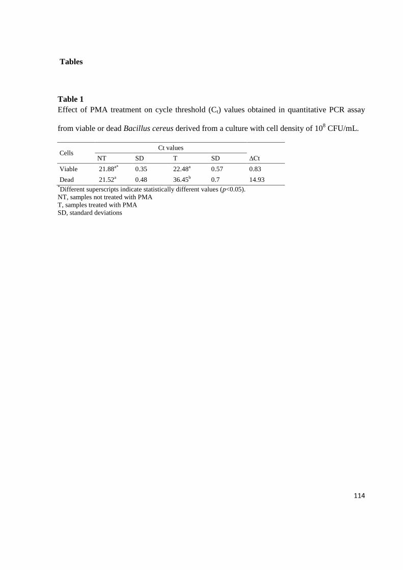
114
Tables
Table 1
Effect of PMA treatment on cycle threshold (Ct) values obtained in quantitative PCR assay
from viable or dead Bacillus cereus derived from a culture with cell density of 108 CFU/mL.
Cells Ct values
NT SD T SD ∆Ct
Viable 21.88a*
0.35 22.48a 0.57 0.83
Dead 21.52a 0.48 36.45
b 0.7 14.93
*Different superscripts indicate statistically different values (p<0.05).
NT, samples not treated with PMA
T, samples treated with PMA
SD, standard deviations

115
Table 2
Quantification of viable Bacillus cereus in UHT milk by real time PCR associated with PMA
and bacterial culture method.
CFU/mL Real-time PCR Traditional method
Cta range n° samples n° samples
≥ 3.9 x 10⁴ - ≤ 3.8 x 10
5 23.6 ≤ Ct ≤ 27.0 3 3
≥ 3.9 x 10³ - ≤ 3.8 x 10
4 27.1 ≤ Ct ≤ 30.8 10 9
≥ 3.8 x 10² - ≤ 3.8 x 10
3 30.9 ≤ Ct ≤ 34.4 25 3
≥ 1.8 x 10² - ≤ 3.7 x 10
2 34.5 ≤ Ct ≤ 37.0 6 0
not detectable > 37.0 or undeterminatedb 91 120
Total number of analyzed samples 135 135 aCt=cycle threshold;
bCt >37.0 or undetermined was assumed as absence of B. cereus detection.
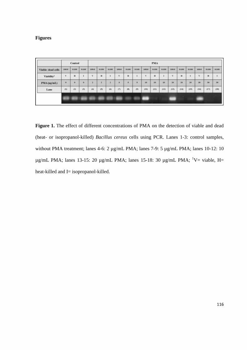
116
Figures
Figure 1. The effect of different concentrations of PMA on the detection of viable and dead
(heat- or isopropanol-killed) Bacillus cereus cells using PCR. Lanes 1-3: control samples,
without PMA treatment; lanes 4-6: 2 µg/mL PMA; lanes 7-9: 5 µg/mL PMA; lanes 10-12: 10
µg/mL PMA; lanes 13-15: 20 µg/mL PMA; lanes 15-18: 30 µg/mL PMA; 1V= viable, H=
heat-killed and I= isopropanol-killed.

117
Figure 2. The effect of PMA treatment on the PCR-based detection of viable and
isopropanol-killed Bacillus cereus cells mixed at different ratios.

118
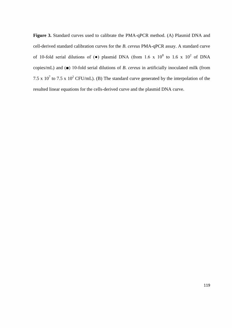
119
Figure 3. Standard curves used to calibrate the PMA-qPCR method. (A) Plasmid DNA and
cell-derived standard calibration curves for the B. cereus PMA-qPCR assay. A standard curve
of 10-fold serial dilutions of (●) plasmid DNA (from 1.6 x 10⁸ to 1.6 x 10
2 of DNA
copies/mL) and (■) 10-fold serial dilutions of B. cereus in artificially inoculated milk (from
7.5 x 10⁷ to 7.5 x 10
2 CFU/mL). (B) The standard curve generated by the interpolation of the
resulted linear equations for the cells-derived curve and the plasmid DNA curve.
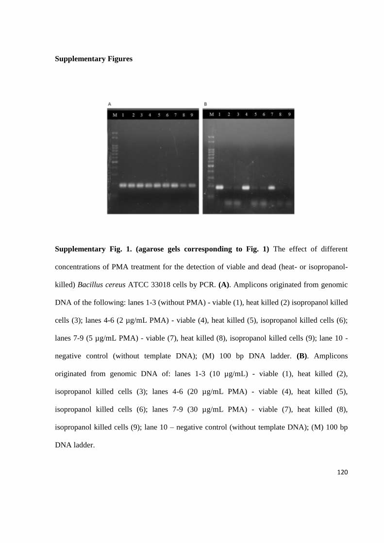
120
Supplementary Figures
Supplementary Fig. 1. (agarose gels corresponding to Fig. 1) The effect of different
concentrations of PMA treatment for the detection of viable and dead (heat- or isopropanol-
killed) Bacillus cereus ATCC 33018 cells by PCR. (A). Amplicons originated from genomic
DNA of the following: lanes 1-3 (without PMA) - viable (1), heat killed (2) isopropanol killed
cells (3); lanes 4-6 (2 µg/mL PMA) - viable (4), heat killed (5), isopropanol killed cells (6);
lanes 7-9 (5 µg/mL PMA) - viable (7), heat killed (8), isopropanol killed cells (9); lane 10 -
negative control (without template DNA); (M) 100 bp DNA ladder. (B). Amplicons
originated from genomic DNA of: lanes 1-3 (10 µg/mL) - viable (1), heat killed (2),
isopropanol killed cells (3); lanes 4-6 (20 µg/mL PMA) - viable (4), heat killed (5),
isopropanol killed cells (6); lanes 7-9 (30 µg/mL PMA) - viable (7), heat killed (8),
isopropanol killed cells (9); lane 10 – negative control (without template DNA); (M) 100 bp
DNA ladder.

121
Supplementary Fig. 2. (agarose gels corresponding to Fig. 2) The effect of PMA treatment
for the PCR detection of viable and isopropanol-killed Bacillus cereus ATCC 33018 cells
mixed at different ratios. Lanes 1 to 5 (without PMA) correspond to amplicons from cell
suspension mixtures containing viable cells at 100%, 75%, 50%, 25% and 0%; lanes 6 to 10
(30 µg/mL of PMA) correspond to amplicons from cell suspensions mixtures containing
viable cells at 100%, 75%, 50%, 25% and 0%.

122
Capítulo 6
Depósito de Patente
123
6.1 Depósito de Patente
Os resultados obtidos nesta tese foram objeto de pedido de patente sendo depositada
junto ao Instituto Nacional da Propriedade Industrial (INPI).
Título da Invenção: Métodos de detecção e quantificação de microrganismos viáveis em
amostras de alimentos.
Data Depósito: 03/08/2012
Número de Depósito: BR1020120195127.
Período de Sigilo: 18 meses

124

125
Capítulo 7
Considerações Finais

126
7.1 Considerações finais
Dentre os microrganismos que podem sobreviver no leite tratado pelo sistema de
UHT devido à produção de esporos resistentes ao calor, destacam-se o B. sporothermodurans
e o B. cereus. O B. sporothermodurans, embora não seja patogênico, pode causar deterioração
e, assim, reduzir a qualidade do leite tratado por UHT e de derivados lácteos quando
encontrado em altas concentrações. Por outro lado, a contaminação de leite por B. cereus
constitui não somente uma importante causa de deterioração, mas também está associada com
a ocorrência das síndromes diarreica e emética devido à ingestão de alimentos contaminados.
O B. sporothermodurans produz esporos altamente termorresistentes, possuindo
valores D140 de 3,4 a 7,9 segundos (20), o que possibilita que este microrganismo resista às
altas temperaturas utilizadas pelo tratamento térmico empregado para a conservação de
alimentos. A análise dos danos estruturais e da sobrevivência de esporos de B.
sporothermodurans tratados pelo calor demonstrou que a inativação completa do esporo
ocorre somente após tratamento térmico a 130°C por 8 minutos (21). Neste trabalho, a
resistência térmica do B. sporothermodurans IC4 foi caracterizada por diferentes temperaturas
(tratamentos isotérmicos e não isotérmicos) em água destilada, leite UHT e em sopa de
legumes tratada pelo sistema UHT. Os perfis de resistência observados tanto nos tratamentos
isotérmicos, como nos não isotérmicos aplicados a diferentes temperaturas permitiram
confirmar a elevada resistência dos esporos a altas temperaturas. Esta etapa do trabalho foi
viabilizada devido à aprovação do estágio de doutorado sanduíche em Cartagena, Espanha, no
período de novembro de 2010 a maio de 2011, junto à CAPES e à Fundação Carolina. Os
laboratórios da Universidade Politécnica de Cartagena possibilitaram o acesso a equipamentos
de alta tecnologia, com destaque para o Termoresistômetro Mastia (Patente espanhola

127
200302529), que não está disponível no Brasil, utilizado na área de Tecnologia de Alimentos
da Faculdade de Engenharia Agrícola para auxiliar a indústria de alimentos a melhorar os
tratamentos térmicos de seus produtos.
B. sporothermodurans pode germinar a partir dos esporos resistentes ao
processamento por UHT e se multiplicar no leite estocado e, caso não seja detectado
corretamente, pode ultrapassar o limite estabelecido pela legislação para microrganismos
mesófilos aeróbios. Desta forma, o estabelecimento de um método de detecção e
quantificação rápido e eficaz de células viáveis de B. sporothermodurans, bem como de B.
cereus, que ainda possui o agravante de ser patogênico, é relevante para uma melhor
qualidade e segurança do leite UHT destinado à população, além de evitar possíveis prejuízos
às indústrias.
Tradicionalmente, a identificação e a enumeração de B. sporothermodurans e de B.
cereus em alimentos são realizadas através de técnicas clássicas de cultivo. Estas técnicas são
baseadas nos aspectos morfológicos e bioquímicos dos microrganismos, podendo apresentar
variabilidade pela ação de fatores ambientais sobre a expressão gênica e risco de
interpretações errôneas devido à semelhança fenotípica e genotípica existente entre as
espécies do gênero Bacillus, bem como requerem vários dias para a obtenção dos resultados.
Adicionalmente, estas técnicas podem não detectar microrganismos que sejam
metabolicamente ativos, mas que não sejam cultiváveis (VBNC).
Métodos moleculares, como a PCR, podem constituir alternativas às técnicas
clássicas de cultivo, oferecendo vantagens sob alguns aspectos. Entretanto, os métodos
baseados em PCR podem não ser capazes de distinguir entre DNA de células viáveis e
mortas, levando a resultados falso-positivos e, consequentemente, a uma superestimação do

128
número total de células (75). A utilização do PMA pode contornar esta limitação, uma vez
que este se liga a moléculas de DNA oriundas de células com membranas danificadas,
impedindo sua amplificação na PCR e permitindo a detecção seletiva do DNA de células
viáveis (76).
Neste sentido, esse trabalho desenvolveu métodos baseados em PCR associada ao
PMA para detecção e quantificação de B. cereus e de B. sporothermodurans, utilizando o
gene da hemolisina e o gene RNAr 16S como alvos, respectivamente.
Na primeira etapa, foi desenvolvido um método para detecção específica de B.
sporothermodurans, utilizando-se como ponto de partida um protocolo baseado em PCR
convencional previamente descrito para identificação deste microrganismo em leite (61). No
entanto, como esse método não apresentou a especificidade esperada, foi desenhado um
oligonucleotídeo iniciador interno para ser utilizado em uma semi-nested touchdown PCR,
que detectou somente DNA oriundo da espécie alvo, mostrando-se um protocolo específico.
Este método também mostrou elevada sensibilidade para a detecção de células vegetativas e
esporos de B. sporothermodurans tanto em cultura pura, como em leite, o que nos indica a
possibilidade da utilização deste protocolo diretamente na matriz láctea, assim como a sua
utilização na etapa de identificação deste microrganismo durante a execução das técnicas
clássicas de cultivo. A partir do protocolo desenvolvido, procurou-se associar o PMA para a
detecção seletiva de células viáveis, sendo otimizada a concentração de 30 μg/mL como ideal
para a marcação de células mortas sem inibir a amplificação de DNA oriundo de células
viáveis.
Embora o método desenvolvido tenha se mostrado sensível para a análise direta do
leite, sem enriquecimento prévio, ele expressa resultados qualitativos, não sendo possível

129
realizar a quantificação precisa de microrganismos presentes em uma determinada amostra.
Tal limitação foi contornada com o desenvolvimento de um método que utilizou qPCR
associada ao PMA como marcador de viabilidade celular, mantendo a mesma sensibilidade
observada para a PCR convencional. Além disso, a qPCR permite a análise direta, em uma
única etapa, eliminando a necessidade da detecção dos produtos amplificados por eletroforese,
diminuindo a possibilidade de contaminações cruzadas. A utilização de qPCR para determinar
a ocorrência de B. sporothermodurans em leite tratado por UHT já havia sido descrita (25),
mas não foi associada à determinação de viabilidade, não distinguindo DNA de células
viáveis daquele de células mortas. A detecção seletiva de células viáveis é de suma
importância para a análise de leite tratado termicamente, como pode ser observado pelos
resultados obtidos a partir das análises das amostras de leite através de PMA-qPCR e qPCR
não associada ao PMA. A qPCR sem PMA detectou um número significativamente maior de
amostras positivas para B. sporothermodurans, provavelmente detectando também DNA
oriundo de células mortas presentes nas amostras analisadas, derivadas, provavelmente, do
tratamento por UHT aplicado ao leite.
O método desenvolvido foi aplicado em amostras comerciais de leite tratadas por
UHT, mostrando que 10% foram positivas com quantidade superior a 102 UFC/mL. Estes
resultados nos indicam que se o B. sporothermodurans não for corretamente identificado e
quantificado, pode ser erroaneamente incluído na contagem de bactérias mesófilas aeróbias
viáveis, o que poderia levar as amostras a estarem em desacordo com os padrões brasileiros
vigentes (10, 27). Cabe ressaltar que o método de cultivo utilizado para isolamento,
identificação e enumeração de B. sporothermodurans em leite foi conduzido conforme
preconizado por Brasil (27), porém, além da semeadura em profundidade prevista pelo

130
MAPA, paralelamente, foi realizada a semeadura em superfície, uma vez que este
microrganismo é aeróbio. Os dados apresentados neste trabalho que demonstraram a ausência
de diferença significativa entre PMA-qPCR e método clássico de cultivo para a quantificação
de B. sporothermodurans levaram em consideração somente a semeadura em superfície.
Quando comparamos a contagem em profundidade com a contagem através da semeadura em
superfície, bem como com a PMA-qPCR, observamos que a técnica de cultivo preconizada
pelo MAPA subestima significativamente a contagem de B. sporothermodurans, o que pode
levar ao aumento dos valores expressos na contagem de mesófilos aeróbios totais. Desta
forma, se tivéssemos estabelecido a comparação da PMA-qPCR desenvolvida neste estudo
para a análise de leite UHT com o cultivo em semeadura em profundidade, teríamos atribuído
uma eficácia comparativa irreal ao método molecular. Esta análise comparativa dos métodos
clássicos de cultivo não foi apresentada inicialmente neste trabalho, pois consta em um artigo
que está em fase de redação que apresenta interesse para publicação em revista de circulação
nacional.
O método desenvolvido para detecção e quantificação de B. cereus associando PMA à
qPCR mostrou-se altamente específico e sensível para a análise da contaminação de leite por
este microrganismo. A análise das amostras de leite tratado por UHT pelo método molecular
desenvolvido evidenciou um número considerável de amostras contaminadas por B. cereus
(32,6%). Destas, três amostras possivelmente atingiram contagens capazes de causar doenças
de origem alimentar. Considerando que a legislação brasileira para leite UHT não permite a
presença de microrganismos patogênicos, todas as amostras que apresentaram contaminação
estavam em desacordo com a legislação vigente. Além disso, o método molecular
desenvolvido e o método de cultivo determinaram a presença de células viáveis de B. cereus

131
em quantidades compatíveis com a dose infectante atribuída a este microrganismo após um
período de incubação de uma semana a 36ºC, condição, muitas vezes, encontrada durante a
estocagem deste produto nos pontos de venda. Desta forma, os resultados apresentados neste
trabalho podem ser utilizados como um alerta para as indústrias de laticínios e para os órgãos
oficiais de controle de produtos de origem animal, no sentido de que o B. cereus deva ser
investigado nos leites tratados por UHT, uma vez que é um microrganismo potencialmente
patogênico.
Adicionalmente, os resultados evidenciaram também que tanto a presença de B.
sporothermodurans, como de B. cereus em leite tratado por UHT reforça a necessidade de um
controle rigoroso na obtenção da matéria-prima e, em todas as etapas do processamento pelo
sistema UHT. Além disso, padrões microbiológicos específicos de leite cru destinado ao
processamento de leite por UHT, no que diz respeito à presença destes microrganismos,
contribuiriam para a melhoria da qualidade do produto final.
132
Referências
1 Santana EHW, Beloti V, Barros MAF, Moraes LB, Gusmão VV, Pereira MS.
Contaminação do leite em diferentes pontos do processo de produção: microrganismos
aeróbios mesófilos e psicotróficos. Semina: Ciências Agrárias. 2001; 22: 145-154.
2 Oliveira RPS. Condições microbiológicas e avaliação da pasteurização em amostras de
leite comercializadas no município de Piracicaba-SP [Dissertação]. São Paulo: Escola de
Superior de Agricultura “Luiz de Queiroz”, Universidade de São Paulo; 2005.
3 Vidal-Martins AMC, Rossi Jr OD, Rezende-Lago NCM. Microrganismos heterotróficos
mesófilos e bactérias do grupo do Bacillus cereus em leite integral submetido a ultra alta
temperatura. Arquivo Brasileiro de Medicina Veterinária e Zootecnia. 2005; 57: 396-400.
4 Pinto CLO, Martins ML, Vanetti MCD. Qualidade microbiológica de leite cru refrigerado
e isolamento de bactérias psicrotróficas proteolíticas. Ciência e Tecnologia de Alimentos.
2006; 26: 645-651.
5 Carvalho RG. A indústria de laticínios no Brasil: passado, presente e futuro. Circular
técnica 102, Juiz de Fora, MG: Embrapa Gado de Leite, 2010.
6 Brasil. Ministério da Agricultura, Pecuária e Abastecimento. Instrução Normativa nº 51,
de 18 de setembro de 2002. Aprova os Regulamentos Técnicos da Produção, Identidade e
Qualidade do Leite tipo A, do Leite tipo B, do Leite tipo C, do Leite Pasteurizado e do
Leite cru Refrigerado e o Regulamento Técnico da Coleta de Leite Cru Refrigerado e seu
Transporte a Granel. Diário Oficial da União. Brasília, 20 de setembro de 2002. Seção I,
p. 13-22.
7 Brasil. Ministério da Agricultura, Pecuária e Abastecimento. Instrução Normativa nº 62,
de 29 de dezembro de 2011. Aprova os Regulamentos Técnicos da Produção, Identidade
e Qualidade do Leite tipo A, do Leite tipo B, do Leite tipo C, do Leite Pasteurizado e do
Leite cru Refrigerado e o Regulamento Técnico da Coleta de Leite Cru Refrigerado e seu
Transporte a GranelDiário Oficial da União. Brasília, 30 de dezembro de 2011.
8 Rossi Junior OD, Vidal-Martins AMC, Salotti BM, Bürger KP, Cardozo MV, Cortez
ALL. Estudo das características microbiológicas do leite UAT ao longo de seu
processamento. Arquivos do Instituto Biológico. 2006; 73: 27-32.
9 Tetrapak. Leite longa vida está presente em 89% dos lares brasileiros. Disponível em
http://www.tetrapak.com.br Acesso em 29/12/2011.
10 Brasil. Ministério da Agricultura, Pecuária e Abastecimento. Portaria nº 370 de 04 de
setembro de 1997. Regulamento Técnico de Identidade e Qualidade de Leite UAT
(UHT). Diário Oficial da União. Brasília, 1997.

133
11 Prata LF. Leite UHT: solução ou problema? Uma análise da situação. Higiene Alimentar.
1998; 12: 10-15.
12 Vidal-Martins AMC, Salotti BM, Rossi Junior OD, Penna ALB. Evolução do índice
proteolítico e do comportamento reológico durante a vida de prateleira de leite
UAT/UHT. Ciências e Tecnologia de Alimentos. 2005; 25: 698-704.
13 Martins AMCV, Rossi Junior OD, Salotti BM, Bürger KP, Cortez ALL, Cardozo MV.
Efeito do processamento UAT (Ultra Alta Temperatura) sobre as características físico-
químicas do leite. Ciência e Tecnologia de Alimentos. 2008; 28: 295-298.
14 Christiansson A, Bertilsson J, Svensson B. Bacillus cereus spores in raw milk: factors
affecting the contamination of milk during the grazing period. Journal Dairy
Science.1999; 82: 305-314.
15 Bartoszewicz M, Hansen BM, Swiecicka I. The members of the Bacillus cereus group are
commonly present contaminants of fresh and heat–treated milk. Food Microbiology.
2008; 25: 588-296.
16 Pettersson B, Lembke F, Hammer P, Stackbrandt E, Priest FG. Bacillus
sporothermodurans, a new species producing highly heat-resistant endospores.
International Journal of Systematic Bacteriology. 1996; 46: 759-764.
17 Klijn N, Herman L, Langeveld L, Vaerewijck M, Wagendorp AA, Huemer I, Weerkamp
AH. Genotypical and phenotypical characterization of Bacillus sporothermodurans
strains, surviving UHT sterilisation. International Dairy Journal. 1997; 7: 421-428.
18 Schockhen-Iturrino RP, Nader Filho A, Dimenstein AR. Ocorrência de bactérias
esporuladas dos gêneros Bacillus e Clostridium em amostras de leite longa vida. Higiene
Alimentar. 1996; 10: 25-27.
19 Coelho OS, Silva N, Brescia MV, Siqueira AP. Avaliação da qualidade microbiológica
do leite UAT integral comercializado em Belo Horizonte. Arquivo Brasileiro de
Medicina Veterinário e Zootecnia. 2001; 53: 256-262.
20 Huemer IA, Klijn N, Vogelsang HWJ, Langeveld LPM. Thermal death kinetics of spores
of Bacillus sporothermodurans isolated from UHT milk. International Dairy Journal.
1998; 8: 851-855.
21 Tabit FT, Buys E. The effects of wet heat treatment on the structural and chemical
components of Bacillus sporothermodurans spores. International Journal of Food
Microbiology. 2010; 140: 207-213.
134
22 Claeys WL, Ludikhuyze LR, Hendrickx ME. Formation kinetics of
hydroxymethylfurfural, lactulose and furosine in milk heated under isothermal and non-
isothermal conditions. Journal of Dairy Research. 2001; 68: 287-301.
23 Vaerewijck MJM, De Vos P, Lebbe L, Scheldeman P, Hote B, Heyndrickx M.
Occurrence of Bacillus sporothermodurans and other aerobic spore-forming species in
feed concentrate for dairy cattle. Journal of Applied Microbiology. 2001; 91: 1074-1084.
24 Guillaume-Gentil O, Scheldeman P, Marugg J, Herman L, Joosten H, Heyndrickx M.
Genetic heterogeneity in Bacillus sporothermodurans as demonstrated by ribotyping and
repetitive extragenic palindromic PCR fingerprinting. Applied and Environmental
Microbiology. 2002; 68: 4216-4224.
25 Tabit FT, Buys E. Incidence and survival of Bacillus sporothermodurans during
processing of UHT milk. British Food Journal. 2011; 113: 505-518.
26 Anonymous. (1992). Council directive 92/46/EEC of 16 June 1992 laying down the
health rules for the production and placing on the market of raw milk, heat-treated milk
and milk-based products. Official Journal of the European Communities, L268, 4.9.1992,
p. 1–32. Accessed 16.07.2012.
27 Brasil. Ministério da Agricultura, Pecuária e Abastecimento. Instrução Normativa nº 62,
de 26 de agosto de 2003. Oficializa os Métodos Analíticos Oficiais para Análises
Microbiológicas para Controle de Produtos de Origem Animal e Água. Diário Oficial da
União. Brasília, 19 de setembro de 2003. Seção 1, página 14.
28 Zacarchenco PB, Leitão MFF, Destro MT, Andrigheto C. Ocorrência de Bacillus
sporothermodurans em leite UAT/UHT brasileiro e a influência do tratamento térmico.
Ciência e Tecnologia de Alimentos. 2000; 20: 363-368.
29 Busatta C, Valdruga E, Cansian RL. Ocorrência de Bacillus sporothermodurans em leite
UAT integral e desnatado. Ciência e Tecnologia de Alimentos. 2005; 25: 408-11.
30 Pereira FEV. Isolamento e caracterização de microrganismos em leite cru refrigerado e
leite UHT no Estado de Goiás e desenvolvimento de filme ativo antimicrobiano para
inibição de Bacillus sporothermodurans. [Dissertação] (Mestrado em Ciência e
Tecnologia de Alimentos) – Escola de Agronomia e Engenharia de Alimentos,
Universidade Federal de Goiás, Goiana, 2010.
31 Neumann FD, Salvatori RU, Majolo C, Fröder H. Occurence of Bacillus
sporothermodurans in UHT milk commercialized in the State of Rio Grande do Sul,
Brazil. Global Veterinaria. 2010; 4: 156-159.
32 Claus D, Berkeley CW. The genus Bacillus In: Bergey’s Manual of Systematic
Bacteriology. Volume 2. Sneath pHA (Ed.).Williams, Wilkins, Baltimore, ISBN: 0-683-
07893-934, 1986; 105-1139.

135
33 Notermans S, Dufrenne J, Teunis P, Beumer R, te Giffel M, Weem PP. A risk assessment
study of Bacillus cereus in pasteurized milk. Food Microbiology. 1997; 14: 143-151.
34 Banykó J, Vyletelová M. Determining the source of Bacillus cereus and Bacillus
licheniformis isolated from raw milk, pasteurized milk and yoghurt. Letters in Applied
Microbiology. 2009; 48: 318-321.
35 Batchoun R, Al-Sha’er AI, Khabour FO. Molecular characterization of Bacillus cereus
toxigenic strains isolated from different food matrices in Jordan. Foodborne Pathogens
and Disease. 2011; 8: 1153-1158.
36 Chaves JQ, Pires ES, Vivoni AM. Genetic diversity, antimicrobial resistance and
toxigenic profiles of Bacillus cereus isolated from food in Brazil over three decades.
International Journal of Food Microbiology. 2011; 147: 12-16.
37 Montanhini MTM, Pinto JPAN, Bersot LS. Ocorrência de Bacillus cereus em leite
comercializado nos estados do Paraná, Santa Catarina e São Paulo. UNOPAR Científica.
Ciências Biológicas e da saúde. 2012; 14: 155-158.
38 Granum PE, Lund T. Bacillus cereus enterotoxins. FEMS Microbiology Letters. 1997;
157: 223-228.
39 Ehling-Schulz M, Fricker M, Scherer S. Identification of emetic toxin producing Bacillus
cereus strains by a novel molecular assay. FEMS Microbiology Letters. 2004; 232: 189-
195.
40 Rajkovic A, Uyttendaele M, Vermeulen A, Andjelkovic M, Fitz-James I, in ´t Veld P,
Denon Q, Vérhe R, Debevere J. Heat resistance of Bacillus cereus emetic toxin,
cereulide. Letters in Applied Microbiology. 2008; 46: 536-541.
41 Arnesen LPS, Fagerlund A, Granum PE. From soil to gut: Bacillus cereus and its food
poisoning toxins. FEMS Microbiology Reviews. 2008; 32: 579-606.
42 Salustiano VC, Andrade NJ, Ribeiro Junior JI, Fernandes PE, Lopes JP, Bernardes PC,
Portugal JG. Controlling Bacillus cereus adherence to stainless steel with different
cleaning and sanitizing procedures used in dairy plants. Arquivo Brasileiro de Medicina
Veterinária e Zootecnia. 2010; 62: 1478-1483.
43 Gilbert RJ., Kramer JM. Bacillus cereus food poisoning. Progress in Food Safety
(Proceedings of Symposium) (Cliver DC & Cochrane BA, eds), 1986, pp. 85–93. Food
Research Institute, University of Wisconsin-Madison, Madison, WI.
44 Granum PE. Bacillus cereus and food poisoning. In applications and systematics of
Bacillus and relatives. In: Berkeley, R., Heyndrickx, M., Logan, N., De Vos, P. (Eds.),
Blackwell Publishing, Oxford, 2002; pp. 37–46.
136
45 Martínez-Blanch JF, Sanchez G, Garay E, Aznar R. Development of a Real-Time PCR
assay for detection and quantification of enterotoxigenic members of Bacillus cereus
group in food Samples. International Journal of Food Microbiology. 2009; 135: 15-21.
46 Bahout AA. Prevalence of Bacillus species in UHT milk. Assiut Veterinary Medical
Journal. 2000; 42: 47-53.
47 Reyes JE, Bastías JM, Gutiérrez MR, Rodríguez MO. Prevalence of Bacillus cereus in
dried milk products used by Chilean school feeding program. Food Microbiology. 2007;
24: 1-6.
48 Rezende NCM, Rossi Jr OD, Amaral LA. Ocorrência de bactérias do grupo do Bacillus
cereus em leite UHT integral (ultra-high-temperature). Revista Brasileira de Ciências
Veterinária. 2000; 7: 162-166.
49 Rezende-Lago NCM, Rossi OD, Vidal-Martins AMC, Amaral LA. Occurrence of
Bacillus cereus in whole milk and enterotoxigenic potential of the isolated strains.
Arquivos Brasileiros de Medicina Veterinária e Zootecnia. 2007; 59: 1563-1569.
50 Brasil, RDC 12. Regulamento Técnico sobre Padrões Microbiológicos para Alimentos, 2
de janeiro de 2001, Brasília, 2001.
51 Rodríguez-Lázaro D, Lombard B, Smith H, Rzezutka A, D’Agostino M, Helmuth R,
Schroeter A, Malorny B, Miko A, Guerra B. Trends in analytical methodology in food
safety and quality: monitoring microorganisms and genetically modified organisms.
Trends in Food Science and Technology. 2007; 18: 306-19.
52 McDougald D, Rice SA, Weichart D, Kjelleberg S. Nonculturability: adaptation or
debilitation? FEMS Microbiology Ecology. 1998; 25: 1-9.
53 Oliver JD. The viable but nonculturable state in bacteria. The Journal of Microbiology.
2005; 43: 93-100.
54 Cooper M, Fridman G, Fridman A, Joshi SG. Biologiacal responses of Bacillus
stratosphericus to floating electrode-dielectric barrier discharge plasma treatment.
Journal of Applied Microbiology. 2010; 109: 2039-2048.
55 Beneduce L, Fiocco D, Spano G. Development of PCR-based molecular tools for the
detection of emerging food- and water-borne pathogenic bacteria. In: Communicating
Current Research and Educational Topics and Trends in Applied Microbiology. (ed. A.
MendezVilas). 2007; pp. 569-576.
56 Mandal PK, Biswas AK, Choi K, Pal UK. Methods for rapid detection of foodborne
pathogens: an overview. American Journal of Food Technology. 2011; 6: 87-102.
137
57 Lungu B, Waltman WD, Berghaus RD, Hofacre CL. Comparison of a real-time PCR
method with a culture method for the detection of Salmonella enterica serotype
Enteritidis in naturally contaminated environmental samples from integrated poultry
houses. Journal of Food Protection. 2012; 75: 743-747.
58 Naraveneni R, Jamil K. Rapid detection of foodborne pathogens by using molecular
techniques. Journal of Medical Microbiology. 2005; 54; 51-54.
59 Postollec F, Falentin H, Pavan S, Combrisson J, Sohier D. Recent advances in
quantitative PCR (qPCR) applications in food microbiology. Food Microbiology. 2011;
28: 848-861.
60 Herman LMF, Vaerewijck MJM, Moermans RJB, Waes GMAVJ. Identification and
detection of Bacillus sporothermodurans spores in 1, 10, and 100 milliliters of raw milk
by PCR. Applied and Environmental Microbiology. 1997; 63: 3139-3143.
61 Scheldeman P, Herman L, Goris J, De Vos PD, Heyndrickx M. Polymerase chain
reaction identification of Bacillus sporothermodurans from dairy sources. Journal of
Applied Microbiology. 2002; 92: 983-991.
62 Fricker M, Reissbrodt R, Ehling-Schulz M. Evaluation of standard and new chromogenic
selective plating media for isolation and identification of Bacillus cereus. International
Journal of Food Microbiology. 2008; 121: 27-34.
63 Higuchi R, Dollinger G, Walsh PS, Griffith R. Simultaneous amplification and detection
of specific DNA sequences. Biotechnology. 1992; 10: 413-417.
64 Higuchi R, Fockler C, Dollinger G, Watson R. Kinetic PCR analysis: real-time
monitoring of DNA amplification reactions. Biotechnology. 1993; 11: 1026-1030.
65 Albert RA, Archambault J, Rosselló-Mora R, Tindall BJ, Matherny M. Bacillus acidicola
sp. nov., a novel mesophilic, acidophilic species isolated from acidic Sphagnum peat
bogs in Wisconsin. International Journal of Systematic and Evolutionary Microbiology.
2005; 55: 2125-2130.
66 Wang RF, Cao WW, Cerniglla CE. A universal protocol for PCR detection of 13 species
of foodborne pathogens in food. Journal of Applied Microbiology. 1997; 83: 727-736.
67 Fykse EM, Olsen JS, Skogan G. Application of sonication to release DNA from Bacillus
cereus for quantitative detection by real-time PCR. Journal of Microbiology Methods.
2003; 55: 1-10.
68 Fukushima H, Tsunomori Y, Seki R. Duplex real-time SYBR green PCR assays for
detection of 17 species of food- or waterborne pathogens in stools. Journal of Clinical
Microbiology. 2003; 41: 5134-5146.

138
69 De Clerck E, Van MK, Jannes G, Rossau R, De VP. Design of a 5' exonucleasebased
real-time PCR assay for simultaneous detection of Bacillus licheniformis, members of the
'B. cereus group' and B. fumarioli in gelatine. Letters in Applied Microbiology. 2004; 39:
109-115.
70 Fricker M, Messelhäuber U, Busch U, Scherer S, Ehling-Schulz M. Diagnostic real-time
PCR assay for the detection of emetic Bacillus cereus strains in food and recent food-
borne outbreaks. Applied and Environmental Microbiology. 2007; 73: 1892-1898.
71 Fernández-No IC, Guarddon M, Böhme K, Galo-Mata P, Barros-Velázquez J. detection
and quantification of spoilage and pathogenic Bacillus cereus, Bacillus subtilis and
Bacillus licheniformis by real-time PCR. Food Microbiology. 2011; 28: 605-610.
72 Josephson KL, Gerba CP, Pepper IL. Polymerase chain reaction detection of nonviable
bacterial pathogens. Applied and Environmental Microbiology. 1993; 59: 3513-3515.
73 Wolffs P, Norling B, Radstrom P. Risk assessment of false-positive quantitative real-time
PCR results in food, due to detection of DNA originating from dead cells. Journal of
Microbiological Methods. 2005; 60: 315-323.
74 Liang N, Dong J, Luo L, Yong L. Detection of viable Salmonella in lettuce by propidium
monoazide real-time PCR. Journal of Food Science. 2011; 76: M234-M237.
75 Rudi K, Moen B, Dromtorp SM, Holck AL. Use of ethidium monoazide and PCR in
combination for quantification of viable and dead cells in complex samples. Applied and
Environmental Microbiology. 2005; 71: 1018-1024.
76 Nocker A, Cheung CY, Camper AK. Comparison of propidium monoazide and ethidium
monoazide for differentiation of live vs. dead bacteria by selective removal of DNA from
dead cells. Journal of Microbiology Methods. 2006; 67: 310-320.
77 Nocker A, Sossa P, Burr M, Camper AK. Use of propidium monoazide for live–dead
distinction in microbial ecology. Applied and Environmental Microbiology. 2007; 73:
5111-5117.
78 Cawthorn DM, Witthuhn RC. Selective PCR detection of viable Enterobacter sakazakii
cells utilizing propidium monoazide or ethidium bromide monoazide. Journal of Applied
Microbiology. 2008; 105: 1178-1185.
79 Chen NT, Chang CW. Rapid quantification of viable legionellae in water and biofilm
using ethidium monoazide coupled with real-time quantitative PCR. Journal of Applied
Microbiology. 2010; 109: 624-634.
80 Wang L, Mustapha A. EMA-real-time PCR as reliable method for detection of viable
Salmonella in chicken and eggs. Journal of food science. 2010; 75: M134-M139.
139
81 Taskin B, Gozen AG, Duran M. Selective quantification of viable Escherichia coli in
biosolids by quantitative PCR with propidium monoazide modification. Applied and
Environmental Microbiology. 2011; 77: 4329-4335.
82 Yáñez MA, Nocker A, Soria-Soria E, Múrtula R, Martínez L, Catalán V. Quantification
of viable Legionella pneumophila cells using propidium monoazide combined with
quantitative PCR. Journal of Microbiology Methods. 2011; 85: 124−130.
83 Flekna G, Stefanic P, Wagner M, Smulders FJ, Mozina SS, Hein I. Insufficient
differentiation of live and dead Campylobacter jejuni and Listeria monocytogenes cells
by ethidium monoazide (EMA) compromises EMA/real-time PCR. Research
Microbiology. 2007; 158: 405-412.
84 Mamlouk K, Macé S, Guilbaud M, Jaffreès E, Ferchichi M, Prévost H, Pilet M, Dousset
X. Quantification of viable Brochothrix thermosphacta in cooked shrimp and salmon by
real-time PCR. Food Microbiology. 2012; 30: 173-179.
85 Luo JF, Lin WT, Guo Y. Method to detect only viable cells in micrbial ecology. Applied
and Microbiological Biotechnology. 2010; 86: 377-384.
86 Contreras PJ, Urrutia H, Sossa K, Nocker A. Effect of PCR amplicon lenght on
suppressing signals from membrane-compromissed cells by propidium monoazide
treatment. Journal of Microbiological Methods. 2011; 87: 89-95.
87 Loozen G, Boon N, Pauwels M, Quirynen M, Teughels W. Live/dead real-time
polymerase chain reaction to assess new therapies against dental plaque-related
pathologies. Molecular Oral Microbiology. 2011; 26: 253-261.
88 Zhu RG, Li TP, Jia YF, Song LF. Quantitative study of viable Vibrio parahaemolyticus
cells in raw seafood using propidium monoazide in combination with quantitative PCR.
Journal of Microbiological Methods. 2011; 90: 262-266.