Embed Size (px)
Citation preview

PONTIFÍCIA UNIVERSIDADE CATÓLICA DO RIO GRANDE DO S UL FACULDADE DE ODONTOLOGIA
PROGRAMA DE PÓS-GRADUAÇÃO EM ODONTOLOGIA NÍVEL: DOUTORADO
ÁREA DE CONCENTRAÇÃO: PRÓTESE DENTÁRIA
INFLUÊNCIA DA APLICAÇÃO LOCAL DE BISFOSFONATO NA
OSSEOINTEGRAÇÃO DE IMPLANTES DE TITÂNIO INSTALADOS EM
TÍBIA DE COELHO
MAGÁLI BECK GUIMARÃES
PORTO ALEGRE
2014

MAGÁLI BECK GUIMARÃES
INFLUÊNCIA DA APLICAÇÃO LOCAL DE BISFOSFONATO NA
OSSEOINTEGRAÇÃO DE IMPLANTES DE TITÂNIO INSTALADOS EM
TÍBIA DE COELHO
Tese apresentada ao Programa de Pós-Graduação em
Odontologia da Faculdade de Odontologia da Pontifícia
Universidade Católica do Rio Grande do Sul, como requisito
para obtenção do título de Doutor em Odontologia, área de
concentração: Prótese Dentária.
ORIENTADOR: Profª Drª LUCIANA MAYUMI HIRAKATA MARQUES
PORTO ALEGRE
2014

MAGÁLI BECK GUIMARÃES
INFLUÊNCIA DA APLICAÇÃO LOCAL DE BISFOSFONATO NA
OSSEOINTEGRAÇÃO DE IMPLANTES DE TITÂNIO INSTALADOS EM
TÍBIA DE COELHO
Linha de pesquisa: Técnicas e Aparelhos em Odontologia
Tese apresentada ao Programa de Pós-Graduação em
Odontologia da Faculdade de Odontologia da Pontifícia
Universidade Católica do Rio Grande do Sul, como requisito
para obtenção do título de Doutor em Odontologia, área de
concentração: Prótese Dentária.
BANCA EXAMINADORA:
______________________________________________________
Prof. Dr. Eduardo Rolim Teixeira
______________________________________________________
Prof. Dr. Diego Segatto Blaya
______________________________________________________
Profª Drª Letícia Borges Jacques
______________________________________________________
Profª Drª Tatiana Bernardon Silva
Dados Internacionais de Catalogação na Publicaçã o (CIP)
G963i Guimarães, Magáli Beck
Influência da aplicação local de bisfosfonato na osseointegração de implantes de titânio instalados em tíbia de coelho / Magáli Beck Guimarães. – Porto Alegre, 2014.
107 f.
Tese (Doutorado em Odontologia) – Faculdade de Odontologia, PUCRS.
Orientação: Profª. Drª. Luciana Mayumi Hirakata Marques.
1. Implantodontia. 2. Osseointegração. 3. Remodelação óssea. 4. Difosfonatos. 5. Coelhos. I. Marques, Luciana Mayumi Hirakata. II. Título.
CDD 617.69
Aline M. Debastiani
Bibliotecária - CRB 10/2199

EPÍGRAFE

“Somos todos anjos de uma asa só; e somente
podemos voar quando abraçados uns aos outros.”
Luciano de Crescenzo

DEDICATÓRIA

Ao meu filho Benício.
Você me proporcionou o maior título da vida.
Seu sorriso é motivação diária e permanente para
seguir em frente.

AGRADECIMENTOS
AGRADECIMENTOS
À Deus, pela dádiva da vida. Hoje percebo que todos os obstáculos que surgiram em
minha trajetória até aqui foram essenciais para meu crescimento interior, minha auto-
confiança e fortalecimento da minha fé. Agradeço imensamente às pessoas
maravilhosas que colocou no meu caminho - com certeza, elas são extensão de Sua mão
e benção neste mundo.
À Profª Drª Luciana Mayumi Hirakata Marques , pela orientação deste trabalho, pelo
exemplo profissional e humano e, sobretudo, pela amizade. Não poderia ter escolhido
melhor tutora em minha trajetória de mestrado e doutorado. Devo muito do que sou hoje
aos seus ensinamentos.
À Coordenação do Programa de Pós-Graduação em Odontologia Stricto-Sensu da
PUCRS, em nome da Profª Drª Ana Maria Spohr, agradeço a oportunidade
proporcionada ao me confiar uma vaga no nível de doutorado e o conhecimento
adquirido durante o decorrer do curso.
Ao Curso de Zootecnia da Universidade Federal de Santa Maria, na pessoa do Prof. Dr.
Paulo Santana Pacheco, por viabilizar a realização da parte experimental envolvendo
os animais. A disposição em ajudar o próximo é exemplar em sua pessoa.
Ao Colégio Politécnico da Universidade Federal de Santa Maria, na pessoa da Profª
Ione Denardin, pelo exemplo de amor e respeito aos animais, por ceder o espaço físico
para abrigo e cuidado deles e por disponibilizar de seu tempo e sabedoria para condução
desta pesquisa. Conhecê-la foi um grande presente que este estudo me proporcionou.
Às Médicas Veterinárias Luciana Wolle e Gabriele Serafini, pelo auxílio na condução
da anestesia, cirurgia, acompanhamento pós-operatório e eutanásia dos animais. Os
conhecimentos técnicos e científicos que me passaram, somados à boa vontade e a
disposição para o desenvolvimento deste trabalho foram essenciais para que tudo desse
certo.
À Farmácia Escola da Universidade Federal de Santa Maria, através da Profª Drª
Marta Alves e da Farmacêutica e amiga Marila Crivellaro Lay Marchiori , pela
elaboração do gel de bisfosfonato, conduzido de forma tão criteriosa e comprometida.
Ao Instituto Bioface, na pessoa do Prof. Dr. Sérgio Alexandre Gehrke, pela troca de
conhecimentos, pela acolhida calorosa, por dispor dos equipamentos e possibilitar a
aplicação da metodologia necessária à realização da análise histomorfométrica deste
estudo.
Ao corpo docente do Programa de Pós-Graduação em Odontologia da PUCRS.
Agradeço os ensinamentos técnico-científicos de tão alta qualidade recebidos, reflexo
da qualidade profissional que o caracteriza.
Aos funcionários da Secretaria de Pós-Graduação da Faculdade de Odontologia da
PUCRS, pela presteza e educação sempre presentes no convívio diário.
À Pontifícia Universidade Católica do Rio Grande do Sul, por viabilizar a realização
deste doutorado através de incentivo financeiro (ProBolsa).
Ao Centro Universitário Franciscano, por ter concedido permissão para realização
deste doutorado.
Aos membros da banca, por disponibilizarem de seu tempo e conhecimento na
avaliação deste trabalho. O convite é reflexo de minha admiração pelo seu
profissionalismo.
À minha família de berço e à que adquiri através do meu marido, agradeço
imensamente o suporte emocional e toda a ajuda para que eu pudesse realizar meus
sonhos.
Em especial, ao meu marido Rodrigo, por auxiliar na parte experimental deste estudo;
por ser motivação, compreensão, mão estendida, abraço que acolhe, companhia nas
horas difíceis e o aconchego do meu lar. Sua tranqüilidade completa minha inquietação;
sua presença equilibra minha vida; seu amor fortalece minha alma.

RESUMO

RESUMO
O objetivo deste estudo foi analisar, comparativamente, aspectos relacionados à
osseointegração de implantes de titânio, instalados com e sem a aplicação tópica de um
bisfosfonato, após 04 semanas in vivo. O estudo compreendeu a colocação de 50
implantes de titânio cp. no terço médio da tíbia de 10 coelhos, sendo a tíbia direita
usada como controle e a esquerda como teste. No grupo teste procedeu-se a
administração local de gel de alendronato de sódio e no grupo controle foi utilizada
solução salina estéril. Após a eutanásia, 10 implantes de cada grupo foram destinados à
análise de torque máximo de remoção. O restante da amostra foi processada para
obtenção de lâminas não descalcificadas de aproximadamente 30µm de espessura, onde
foram realizadas análises histomorfológica e histomorfométrica de contato osso-
implante (%BIC). Os dados foram analisados com nível de significância de 5%. Os
valores de torque máximo de remoção do grupo teste foram, em média, metade dos
valores do grupo controle. O grupo teste demonstrou menor %BIC, bem como
alterações notáveis em qualidade óssea. Conclui-se que os eventos iniciais de
osseointegração de implantes de titânio não são favorecidos pela aplicação local de gel
de alendronato de sódio em coelhos.
Palavras-chave: Difosfonatos. Osseointegração. Remodelação óssea. Implantes
Dentários.

ABSTRACT

ABSTRACT
The aim of this study was to make a comparative analysis of aspects related to the
osseointegration of titanium implants placed with and without local application of a
bisphosphonate, after 4 weeks, in vivo. The study comprised the placement of 50 cp.
titanium implants in the middle third of the tibia of 10 rabbits, with the right tibia being
used as control, and the left as test site. In the test group, local administration of sodium
alendronate gel was instituted, and in the control group, sterile saline solution was used.
After euthanasia, 10 implants from each group were analyzed for maximum removal
torque. The remainder of the sample was processed to obtain non decalcified slides with
approximately 30µm thick, in which histomorphological and histomorphometric
analyses of bone-implant contact (%BIC) were performed. Data were analyzed at a
level of significance of 5%. The removal torque of the test group, on an average, were
half of the values obtained in the control group. The test group showed lower
percentage of BIC, and notable changes in bone quality. It was concluded that the initial
events of osseointegration of titanium implants are not favored by the local application
of sodium alendronate gel in rabbits.
Keywords: Diphosphonates. Osseointegration. Bone remodeling. Dental Implants.

LISTA DE FIGURAS, GRÁFICOS E TABELAS
LISTA DE FIGURAS
Figura 1 – Monitoramento trans-operatório das funções vitais dos animais................. 42
Figura 2 - Aplicação local do gel de bisfosfonato......................................................... 43
Figura 3 - Instalação dos implantes imediatamente após a aplicação do gel de
bisfosfonato.................................................................................................................... 44
Figura 4 - Mini implante especialmente projetado para pesquisa científica em modelo
animal............................................................................................................................. 45
Figura 5 - a) Máquina de torque conectada ao computador (Software Dynaview Torque
Standard/Pro M); b) Teste de torque de remoção........................................................... 47
Figura 6 – a) Mensuração do perímetro total do implante e b) das áreas de contato
osso-implante para cálculo de BIC................................................................................. 48
Figura 7 – Posicionamento dos implantes em cortical e medular tibiana. Magnificação:
20X. ............................................................................................................................... 52
Figura 8 – Interface osso-implante do grupo controle na região de osso cortical,
demonstrando atividade óssea mais intensa próximo ao corpo do implante, com
presença de tecido ósseo recentemente formado (região de coloração mais intensa),
osteócitos volumosos em amplas lacunas (--›) e osteoblastos grande e volumosos em
contato com a superfície do implante (+). Magnificação: 40X.
........................................................................................................................................ 53
Figura 9 – Interface osso-implante do grupo teste na região de osso cortical,
demonstrando baixa atividade óssea localizada quase que somente em região de topo de
espiras e região mais cervical do implante, com presença de tecido granuloso
preenchendo o espaço entre as espiras (+). Magnificação: 40X. ................................... 54

LISTA DE GRÁFICOS
Gráfico 1 - Torque máximo de remoção segundo grupos (média ± dp) e resultado do
teste comparativo............................................................................................................ 51
Gráfico 2 - Gráfico de Bland-Altman entre as medidas dos examinadores.................. 55
Gráfico 3 - Valores médios de contato osso-implante e respectivos erros padrões
segundo grupos............................................................................................................... 56

LISTA DE TABELAS
Tabela 1 - Desenho experimental ................................................................................. 40
Tabela 2 - Torque máximo de remoção (Ncm) segundo grupos e resultado do teste
comparativo.................................................................................................................... 51
Tabela 3 - Descrição dos percentuais de contato osso-implante para cada examinador e
resultado da reprodutibilidade entre os examinadores................................................... 54
Tabela 4 - Descrição do contato osso-implante segundo grupos e resultado do teste
comparativo.................................................................................................................... 56

LISTA DE ABREVIATURAS, SIGLAS E SÍMBOLOS
LISTA DE ABREVIATURAS, SIGLAS E SÍMBOLOS
% - Por cento
ºC - Graus Celsius
® - Marca registrada
± - Mais ou menos
˂ - Menor que
µg - Micrograma
µm - Micrometro
ATP - Adenosina Trifosfato
BIC - Bone-Implant Contact
CEUA - Comissão de Ética no Uso de Animais
COBEA - Colégio Brasileiro de Experimentação Animal
COI - Contato Osso-Implante
cm - Centímetro
DP - Desvio-Padrão
FO - Faculdade de Odontologia
g - Grama
h - Horas
kg - Quilograma
mg - Miligrama
ml - Mililitro
mm - Milímetro
n - Número da amostra
Nº - Número
N.cm - Newton centímetro
p - Significância
P-C-P - Fósforo-Carbono-Fósforo
PO3 - Íon fosfato
PUCRS - Pontifícia Universidade Católica do Rio Grande do Sul
rpm - Rotação por minuto
SiC - Carbeto de Silício
SPSS - Statistical Package for Social Sciences

UFSM - Universidade Federal de Santa Maria
X - Vezes

SUMÁRIO
SUMÁRIO
1. INTRODUÇÃO .............................................................................................. 24
2. PROPOSIÇÃO …………………………………………………………….… 28 2.1 Problema ………………………………………………………………….. 29 2.2 Hipóteses nulas …………………………………………...…….………… 29
3. REVISÃO DE LITERATURA …………………………………………….. 30
3.1 Bisfosfonatos …………………………………………………………...… 31 3.2 Bisfosfonatos e Osseointegração …………………………………………. 33
4. METODOLOGIA …………………………………………………………… 38
4.1 Amostra………………………………………………………………...…. 39 4.2 Grupos experimentais …………………………………………………….. 39 4.3 Procedimento cirúrgico ……………………...……………………………. 40 4.4 Morte dos animais .……….………………………………………………. 46 4.5 Mensuração do torque de remoção ……………………………………….. 46 4.6 Análise histomorfológica e histomorfométrica ……………………….….. 47 4.7 Análise estatística ………………………………………………………… 49
5. RESULTADOS …………………………………………………………..…. 50
5.1 Torque máximo de remoção ……………………………………………… 51 5.2 Análise histomorfológica ………………………..…………...…………… 52 5.3 Análise histomorfométrica ……………………………………..………… 54
6. DISCUSSÃO ………………………………………………………………… 57
7. CONCLUSÕES ……………………………………………………………… 64
REFERÊNCIAS BIBLIOGRÁFICAS .................................................................. 66
ANEXOS .................................................................................................................. 72 A – Aprovação da Comissão Científica e de Ética- FO/PUCRS ....................... 73 B – Aprovação da Comissão de Ética no Uso de Animais – PUCRS ............... 74 C – Croqui do implante utilizado no estudo ...................................................... 75 D – Artigo científico .......................................................................................... 76 E – Normas do periódico Int J Oral Maxillofac Surg ...................................... 100 F – Submissão do artigo científico .................................................................. 107

24
1. INTRODUÇÃO

25
1. INTRODUÇÃO
Nas últimas décadas, a busca pela reposição de dentes perdidos através da
terapia com implantes dentários e próteses sobre implantes tem crescido
consideravelmente. Isto se deve, entre outras razões, ao fato de que o edentulismo, seja
parcial ou total, é uma condição bucal que afeta grande parte das pessoas de faixa etária
mais avançada, parcela da população que mais cresce no mundo (MULLER, 2014). Tais
pacientes apresentam um perfil de qualidade e quantidade óssea, muitas vezes, afetado
por doenças sistêmicas, como diabetes e osteoporose, além de apresentarem menor
potencial de regeneração óssea, fatores que podem contribuir para taxas reduzidas de
sucesso na terapia com implantes osseointegráveis (TRUHLAR et al, 1997). Em
contrapartida, o desejo de atingir-se osseointegração mais rapidamente é comum tanto
aos profissionais de saúde quanto aos pacientes, motivando pesquisas que busquem
melhorias no desenvolvimento de materiais e técnicas relacionados à otimização do
processo de remodelação óssea ao redor de implantes osseointegráveis.
Durante a cirurgia de instalação de implantes, a decisão sobre a aplicação ou não
de carga imediata é determinada, entre outros aspectos, pela estabilidade primária do
implante ao tecido ósseo. Nos casos em que não há estabilidade primária durante a
instalação do implante dentário, recomenda-se que o profissional aguarde o período de
osseointegração sem aplicação de carga funcional, seguindo protocolo de duas etapas
cirúrgicas (MISCH e WANG, 2003). Assim, é nesse cenário que se torna importante a
aceleração no processo de osseointegração, a fim de que menos tempo seja necessário
entre a cirurgia de instalação de implantes e a reabertura para conexão dos pilares
protéticos.

26
O interesse pelo uso dos bisfosfonatos como biomoduladores ósseos em
Implantodontia, surgiu pela conhecida habilidade deste fármaco em inibir a atividade de
osteoclastos, motivo pelo qual é amplamente utilizado no tratamento de doenças
caracterizadas pelo excesso de reabsorção óssea como osteoporose, hipercalcemia e
metástases ósseas (MUNDY e YONEDA, 1998). Além disso, sabe-se que um efeito na
formação óssea peri-implantar também pode ser esperado, promovendo uma redução no
turnover ósseo consideravelmente (ALLGROVE, 1997). Estudos sugerem que os
bisfosfonatos podem ter uma influência positiva na formação e remodelação óssea e
consequente melhora na fixação de implantes osseointegráveis em humanos (ABTAHI,
TENGVALL e ASPENBERG, 2010; ABTAHI, TENGVALL E ASPENBERG, 2012).
Em função dos severos efeitos colaterais que o uso sistêmico desta droga
provoca (DE GROEN et al, 1996; BEDOGNI et al, 2010; GOSS et al, 2010;
LAZAROVICI et al, 2010; VOHRA et al, 2014), os estudos têm voltado sua atenção
para o desenvolvimento de métodos de entrega local deste fármaco no sítio de interesse
(PETER et al, 2005; JAKOBSEN et al, 2009; LI et al, 2013; HARMANKAYA et al,
2013; BOBYN et al, 2014). A intenção é que o bisfosfonato influencie positivamente a
remodelação do tecido ósseo adjacente ao implante, sem promover efeitos colaterais
sistêmicos indesejados. Nesse sentido, tem sido proposta a imobilização do bisfosfonato
na superfície do implante como forma de entrega local do fármaco (YOSHINARI et al,
2001; LEE et al, 2011; MOON et al, 2012; GUIMARÃES et al, 2013; STADLINGER
et al, 2013) . Entretanto, essa imobilização necessita, muitas vezes, de uma metodologia
complexa e equipamentos sofisticados para ser realizada. A aplicação direta do
bisfosfonato no alvéolo cirúrgico, imediatamente antes da inserção do implante, parece
ser um procedimento mais simples e prático, mas, até agora, pouco testado
27
(SKOGLUND, HOLMERTZ e ASPENBERG, 2004; JAKOBSEN et al, 2007;
JAKOBSEN et al, 2009; CUAIRÁN et al, 2014)
Assim, o objetivo deste trabalho é propor a aplicação local de um bisfosfonato
(alendronato de sódio) em forma de gel, diretamente no alvéolo cirúrgico e avaliar,
comparativamente, aspectos relacionados à osseointegração de implantes de titânio
instalados imediatamente após essa aplicação, in vivo.

28
2. PROPOSIÇÃO

29
2. PROPOSIÇÃO
2.1 Problema
Dentro do contexto atual explicitado, surge o questionamento: a osseointegração
de implantes de titânio é favorecida pela aplicação local de um gel de bisfosfonato?
2.2 Hipóteses nulas
Duas hipóteses nulas foram testadas:
1) Não há diferença no torque de remoção de implantes osseointegráveis
instalados com e sem a aplicação local de um gel de alendronato de sódio;
2) Não há diferença na porcentagem de contato osso-implante quando
instalados com e sem aplicação local de um gel de alendronato de sódio.

30
3. REVISÃO DE LITERATURA

31
3. REVISÃO DE LITERATURA
3.1 BISFOSFONATOS
Os bisfosfonatos são compostos farmacologicamente simples, considerados
análogos dos pirofosfatos por possuírem uma estrutura fósforo-carbono-fósforo, em
comparação com a estrutura fósforo-oxigênio-fósforo do pirofosfato. Uma vez que o
pirofosfato sofre rápida degradação enzimática no organismo, o bisfosfonato funciona
como um análogo resistente a essa degradação capaz de ter a mesma função frente ao
tecido ósseo: controle dos mecanismos de reabsorção óssea (FLEISCH, 1998).
O termo “bisfosfonato” é derivado de sua estrutura farmacológica, a qual possui
dois grupos fosfato (PO3) covalentes ligados a um carbono central. A estrutura P-C-P do
bisfosfonato tem uma grande afinidade por hidroxiapatitas e íons de cálcio. Além dos
grupamentos fosfato, ligada ao carbono existe uma cadeia química curta, responsável
pela ligação do fármaco ao tecido ósseo. Também ligada ao carbono, existe uma cadeia
química longa, que determina as propriedades químicas, o modo de ação e a potência do
bisfosfonato (OTOMO-CORGEL, 2007).
Tais compostos funcionam como potentes inibidores da reabsorção óssea e têm
um largo uso no tratamento de uma variedade de doenças com excesso de reabsorção
óssea, como metástase óssea, hipercalcemia, osteoporose e doença de Paget (MUNDY e
YONEDA, 1998). Quando administrados oral ou parenteralmente, eles são absorvidos
por cristais de hidroxiapatita no mineral ósseo e, uma vez que sua estrutura os torna
resistentes a degradação enzimática, eles atuam principalmente inibindo a reabsorção
óssea, embora algum efeito na formação óssea também deva ocorrer. O efeito em rede é
promover acumulação mineral no osso enquanto, ao mesmo tempo, promover a redução
do turnover ósseo consideravelmente (ALLGROVE, 1997).

32
Um estudo afirma que os bisfosfonatos são seletivamente incorporados pelos
osteoclastos na matriz óssea contendo hidroxiapatita e têm vários efeitos diretos a nível
celular. Eles inibem o recrutamento e diferenciação de precursores de osteoclastos, além
de inibir a atividade de reabsorção dos osteoclastos maduros. Somado a isso, os
bisfosfonatos induzem a apoptose de macrófagos e osteoclastos maduros. É afirmado
que osteoclastos que sofrem influência de bisfosfonatos mostram mudanças
morfológicas, tais como deficiência da borda de absorção ou dilaceração do anel de
actina (MURAKAMI et al, 1995).
Evidências têm demonstrado que alguns efeitos desses compostos nos
osteoclastos são mediados diretamente via osteoblastos. Tais efeitos indiretos nos
osteoclastos parecem ter a ver com a secreção de fatores pelos osteoblastos, como a
interleucina-6, que regulam a diferenciação e ativação de osteoclastos (VITTE,
FLEISCH e GUENTHER, 1996). Além disso, bisfosfonatos têm demonstrado
capacidade de aumentar a proliferação, diferenciação e atividade ósseo-formadora de
osteoblastos diretamente. Assim, tais estudos sugerem que os bisfosfonatos afetam o
metabolismo ósseo através de osteoclastos e osteoblastos (GARCIA-MERCO et al,
1998).
Atualmente, é considerado que as propriedades de inibição da reabsorção óssea
por parte dos bisfosfonatos agem nos osteoclastos por dois mecanismos que são
dependentes da presença de nitrogênio na cadeia química longa ligada ao carbono.
Assim, os bisfosfonatos são classificados em dois grupos com diferentes mecanismos de
ação: (1) os bisfosfonatos nitrogenados e (2) os não-nitrogenados. Os bisfosfonatos que
não têm o nitrogênio presente em sua estrutura (etidronato, clodronato e tiludronato)
inativam análogos não-hidrolizáveis de ATP, que interferem na energia celular dos
osteoclastos, induzindo sua apoptose. Os bisfosfonatos mais potentes, que contém

33
nitrogênio em sua estrutura (pamidronato, alendronato, ibandronato, risedronato e
zoledronato), agem por quatro mecanismos distintos: (1) inativando o ATP; (2) inibindo
a síntese de farnesildifosfonato (parte do mecanismo do mevalonato na síntese do
colesterol), resultando em desestruturação citoesqueletal do osteoclasto, desregulamento
do transporte intracelular e inibição da proliferação celular; (3) reduzindo o
recrutamento de osteoclastos e (4) induzindo a produção por parte dos osteoblastos de
uma enzima de inibição da reabsorção óssea (OTOMO-CORGEL, 2007).
A afinidade de um bisfosfonato pelo tecido ósseo parece ser relacionada à sua
estrutura química, uma vez que os bisfosfonatos nitrogenados demonstraram apresentar
uma capacidade de inibição da dissolução mineral mais forte. Tais diferenças na
afinidade pela hidroxiapatita parecem influenciar no tempo necessário para que a
reabsorção óssea deixe de ser afetada pela ação dos bisfosfonatos mesmo após a
interrupção da terapia com essa droga (NANCOLLAS et al, 2006).
3.2 BISFOSFONATOS E OSSEOINTEGRAÇÃO
Um dos primeiros estudos a pesquisar a influência de bisfosfonatos nos eventos
relacionados à osseointegração foi o de Yoshinari e colaboradores (2001). Nesse estudo,
os autores modificaram a superfície de placas de titânio com a implantação de íons de
cálcio e imobilização de pamidronato. Nessas placas, foram cultivadas células
osteoblásticas e avaliada a atividade de fosfatase alcalina dessas células. Os autores
demonstraram que o bisfosfonato não teve efeito tóxico sobre os osteoblastos e, ainda,
concluíram que tal superfície oferece um micro-ambiente favorável ao crescimento
celular, com habilidades osteogênicas.
34
O mesmo grupo de pesquisa avaliou a resposta óssea a implantes de titânio
cobertos com fosfato de cálcio e modificados pela imobilização de pamidronato,
instalados em cachorros. A maior porcentagem de contato osso-implante foi encontrada
em implantes modificados pela presença de bisfosfonato após 12 semanas de
implantação, comparados ao grupo controle (YOSHINARI et al, 2002).
Mais tarde, Goto e colaboadores (2003) avaliaram a capacidade de osteoblastos
formarem nódulos calcificados sobre diferentes superfícies de titânio. Dentre essas
superfícies estavam titânio puro, impregnado por íons de cálcio, e modificado pela
imobilização de dois bisfosfonatos em diferentes concentrações – pamidronato e
incadronato. Os autores encontraram resultados quatro vezes melhores no grupo teste do
pamidronato em comparação com o controle titânio puro, concluindo que o bisfosfonato
pode ser uma alternativa útil na estimulação de formação de tecido mineralizado em
implantes de titânio.
Em estudo de Skoglund, Holmertz e Aspenberg (2004), foram avaliados os
efeitos da administração sistêmica e da aplicação local de ibandronato na fixação de
implantes de aço inoxidável em ratos. A administração sistêmica se dava através de
injeção subcutânea diária de ibandronato, enquanto a aplicação tópica deu-se através da
injeção de 0,1ml de ibandronato na loja cirúrgica antes da inserção do implante. Os
resultados revelaram que a aplicação sistêmica aumentou em 30% a força necessária
para arrancar o implante. A administração local aumentou em 60% a força de torque de
remoção, e em 68% a força necessária para girar o implante em ¼ de volta. Os autores
concluíram que o bisfosfonato administrado de forma sistêmica ou local pode ser de
grande valia no aumento da fixação de implantes em estágios iniciais de cicatrização.

35
Em 2005, Kajiwara e colaboradores analisaram a quantidade de tecido ósseo
novo formado ao redor de implantes com superfície modificada pela imobilização de
pamidronato, em tíbias de ratos. Após quatro semanas, houve significativamente mais
neoformação óssea ao redor dos implantes modificados com bisfosfonato do que
naqueles que não receberam esse tratamento, evidenciando que essa superfície estimula
a formação óssea ao redor do implante, podendo contribuir para a aumento no sucesso
com essa terapia.
Jakobsen e colaboradores (2006) também avaliaram o efeito da aplicação tópica
de alendronato na fixação de implantes, mas com técnica cirúrgica de compactação
óssea. Os autores encontraram um aumento no contato osso-implante e na densidade
total óssea ao redor dos implantes no grupo tratado com bisfosfonato, mas relataram que
esse aumento aconteceu basicamente às expensas de osso não-vital, o que atribuem ao
efeito inibidor de osteoclastos que o bisfosfonato possui. Os autores sugerem períodos
mais longos de observação para avaliar se o efeito preservativo do alendronato sobre
osso não-vital poderia aumentar a fixação de implantes por osteocondução.
Avaliando a influência da administração sistêmica de alendronato na
osseointegração de implantes de titânio instalados em coelhos, Chacon e colaboradores
(2006) não encontraram diferença estatística no torque de remoção entre os grupos teste
e controle. Os autores concluíram que a administração de doses de alendronato via oral
não teve efeito significativo nos valores de torque de remoção seis semanas após a
instalação dos implantes.
Eberhardt e colaboradores (2007) conduziram um estudo pré-clínico em ratos
para avaliar se o ibandronato poderia acelerar o processo de osseointegração, resultando
em estabilidade secundária mais precoce. Para tanto, os animais do grupo teste

36
receberam injeções subcutâneas de ibandronato em diferentes concentrações durante os
períodos pós-operatórios analisados. Os resultados indicaram que o grupo que recebeu
doses mais altas de ibandronato teve o tempo necessário para a osseointegração
reduzido em 60% se comparado com o grupo controle. Entretanto, para os grupos que
receberam baixas doses, não houve aceleração da osseointegração em comparação ao
grupo controle. Os autores concluíram que um tratamento contínuo com 5µg/kg por dia
de ibandronato pode ser um potente acelerador da osseointegração e, consequentemente,
estabilidade secundária mais precoce pode ser esperada.
Aspenberg e colaboradores (2008) avaliaram os efeitos da cobertura de
bisfosfonatos em implantes de aço inoxidável na fixação óssea. Em seus resultados,
encontraram que o bisfosfonato foi capaz de aumentar a força de remoção após duas
semanas de inserção.
Langhoff e colaboradores (2008) também avaliaram o efeito da cobertura de
bisfosfonato em implantes de titânio instalados em ovelhas. Os autores investigaram
histomorfometricamente a porcentagem de contato osso-implante nos diferentes grupos.
Os resultados apontaram que não houve diferenças estatísticas entre os grupos
analisados, embora todos tivessem alcançado resultados comparativamente bons.
Mais recentemente, Abtahi, Tengvall e Aspenberg (2010) realizaram um estudo
piloto em cinco pacientes, nos quais foram instalados implantes dentários cobertos com
bisfosfonato na maxila. Foram realizadas avaliações radiográficas e de estabilidade do
implante através de frequência de ressonância no momento da instalação do implante e
após seis meses, na cirurgia de reabertura. Além disso, os implantes cobertos com
bisfosfonato foram removidos em blocos em dois pacientes, para fins de avaliação
histológica. Em cada paciente, o implante coberto com bisfosfonato mostrou valores
37
mais altos de frequência de ressonância, teste quantitativo não-invasivo frequentemente
utilizado para medir estabilidade de implantes dentários. Histologicamente, não houve
anormalidades.
Tsetsenekou e colaboradores (2011) realizaram um estudo com coelhos
ovariectomizados a fim de avaliar se a administração sistêmica de alendronato interferia
na osseointegração de implantes instalados no fêmur, após seis e doze semanas da
inserção. Os resultados demonstraram que não houve diferença entre os grupos controle
e teste, indicando que a administração sistêmica de alendronato não afetou a
osseointegração de implantes em animais com status hormonal semelhante ao de
mulheres em idade pós-menopausa.

38
4. METODOLOGIA

39
4. METODOLOGIA
4.1 AMOSTRA
Após aprovação da Comissão Científica e de Ética da Faculdade de Odontologia
da PUCRS (Anexo A) e da Comissão de Ética no Uso de Animais (Anexo B), 10
coelhos da espécie Oryctolagus cuniculus, da linhagem New Zealand, machos, adultos,
e peso corporal médio de 4,0kg foram utilizados nesta pesquisa.
Os animais foram alojados em gaiolas individuais, regularmente higienizadas e
com serragem estéril, nas dependências do Colégio Politécnico do Centro de Ciências
Rurais da Universidade Federal de Santa Maria (UFSM), recebendo dieta ad libitum. Os
procedimentos experimentais seguiram os princípios da Lei n.º 6.638, de 08 de Maio de
1979, que estabelece normas para a prática didático-científico da vivissecção de animais
e os princípios éticos na experimentação animal, segundo o Colégio Brasileiro de
Experimentação Animal (COBEA).
4.2 GRUPOS EXPERIMENTAIS
Um total de 50 implantes foram instalados na amostra, sendo a tíbia direita
usada como controle e a esquerda como teste (INTERNATIONAL STANDARD ISSO
10993-6, 1994) (Tabela 1). O grupo controle, ou seja, os alvéolos cirúrgicos
confeccionados na tíbia direita de cada animal, recebeu aplicação de solução salina
estéril. O grupo teste, caracterizado pelos alvéolos cirúrgicos confeccionados na tíbia
esquerda de cada coelho, recebeu a aplicação tópica de 1ml de gel de alendronato de
sódio (10mg/g), confeccionado segundo estudo previamente publicado (Reddy e
Kumar, 2005). Dos 50 implantes instalados, 20 (n=10/grupo) foram avaliados em teste
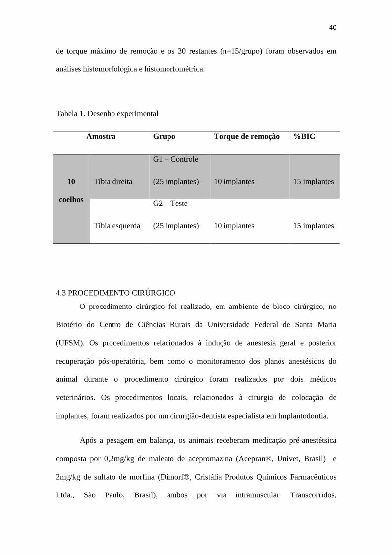
40
de torque máximo de remoção e os 30 restantes (n=15/grupo) foram observados em
análises histomorfológica e histomorfométrica.
Tabela 1. Desenho experimental
Amostra Grupo Torque de remoção %BIC
10
coelhos
Tíbia direita
G1 – Controle
(25 implantes)
10 implantes
15 implantes
Tíbia esquerda
G2 – Teste
(25 implantes)
10 implantes
15 implantes
4.3 PROCEDIMENTO CIRÚRGICO
O procedimento cirúrgico foi realizado, em ambiente de bloco cirúrgico, no
Biotério do Centro de Ciências Rurais da Universidade Federal de Santa Maria
(UFSM). Os procedimentos relacionados à indução de anestesia geral e posterior
recuperação pós-operatória, bem como o monitoramento dos planos anestésicos do
animal durante o procedimento cirúrgico foram realizados por dois médicos
veterinários. Os procedimentos locais, relacionados à cirurgia de colocação de
implantes, foram realizados por um cirurgião-dentista especialista em Implantodontia.
Após a pesagem em balança, os animais receberam medicação pré-anestétsica
composta por 0,2mg/kg de maleato de acepromazina (Acepran®, Univet, Brasil) e
2mg/kg de sulfato de morfina (Dimorf®, Cristália Produtos Químicos Farmacêuticos
Ltda., São Paulo, Brasil), ambos por via intramuscular. Transcorridos,

41
aproximadamente, 10 minutos, canulou-se a veia marginal da orelha do animal para
administração de fluidoterapia com solução de Ringer com Lactato (Baxter®, Baxter
Hospitalar Ltda, São Paulo, Brasil) e 10mhg/kg de enrofloxacina (Baytril®, Bayer S.A.,
São Paulo, Brasil), 20 minutos antes da cirurgia. Procedeu-se a indução anestésica
através da injeção intravenosa de 10mg/kg de cloridrato de cetamina (Ketamina®,
Agener Pharmaceutica, São Paulo, Brasil) e 1mg/kg de midazolam (Dormonid®
injetável, Roche Químicos e Farmacêuticos S.A., Rio de Janeiro, Brasil). Realizou-se a
anestesia epidural com 0,25ml/kg de lidocaína 2% (Lidocaína 2%, Geyer, Porto Alegre,
Brasil). Após a indução anestésica, os animais foram tricotomizados na região de tíbia
direita e esquerda. Realizou-se antissepsia da perna dos coelhos com digluconato de
clorexidina a 2% (Riohex Degermante 2%, Rioquímica, Brasil), incluindo a pelagem
adjacente a área tricotomizada.
Então, colocou-se o animal em decúbito dorsal e, sobre a área tricotomizada, foi
posicionado um campo fenestrado estéril, com dimensões de 40 X 40 cm,
confeccionado para expor a perna do animal e cobrir o restante do corpo, prevenindo
eventuais contaminações. Cada cirurgia ocorreu de forma independente, sobre uma
bancada térmica com 37°C, coberta com campo cirúrgico estéril descartável.
No trans-operatório, os animais receberam suporte com máscara de oxigênio e o
monitoramento dos planos anestésicos dos animais foi realizado analisando-se as
funções vitais (freqüência cardíaca e respiratória) (Figura 1) e o reflexo de dor. Quando
necessário, repetia-se ¼ da dose de indução para manutenção anestésica.

42
Figura 1 – Monitoramento trans-operatório das funções vitais dos animais.
A cirurgia teve início através de uma incisão linear, com lâmina de bisturi n°15
montada em cabo n.°3, medindo cerca de 2cm de extensão, em pele e músculo na
superfície diafisária medial da tíbia, sempre com apoio em base óssea. Após esse
procedimento, os tecidos moles foram afastados com o auxílio de dois afastadores Senn
Müller, permitindo a visualização do periósteo, que subseqüentemente foi incisado,
divulsionado com um descolador de Molt e afastado juntamente com os outros tecidos,
expondo a superfície externa da tíbia. Com uma seringa descartável de 20 ml foi feita
irrigação da região com soro fisiológico a 0,9%, secando-se posteriormente com gaze
estéril.
Os locais de confecção das lojas cirúrgicas foram previamente demarcados com
o auxílio de uma sonda exploradora. As perfurações foram posicionadas 10 mm abaixo
do côndilo da tíbia, com 10mm de distância entre cada perfuração, medida com o
auxílio de um instrumento de medição, e confeccionadas com o auxílio de contra-
ângulo cirúrgico 16:1 (Contra-ângulo Kavo, Kavo do Brasil, Brasil) conectado ao

43
micromotor cirúrgico (Kavo Koncept Surg, Kavo do Brasil, Brasil) previamente
ajustado para 1.200 rpm e a bomba peristáltica ajustada para o máximo de vazão, sob
irrigação abundante de solução fisiológica. A fresagem para instalação dos implantes foi
realizada com brocas apropriadas (Kit Cirúrgico, Conexão Sistemas de Prótese, São
Paulo, Brasil), até a profundidade de 4mm, na sequência broca lança e broca helicoidal
2 mm.
Após a confecção das cavidades, estas foram irrigadas abundantemente com
solução fisiológica para remoção dos resíduos gerados no processo de perfuração. Uma
gaze estéril foi introduzida e mantida por compressão no alvéolo cirúrgico por 1 a 2
minutos, com a finalidade de absorver e coibir o sangramento, processo que garantiu
que o gel de bisfosfonato entrasse em contato direto com toda a parede do alvéolo, sem
a interposição de sangue. A quantidade de 1ml de gel de alendronato de sódio (grupo
teste) ou 1ml de solução salina estéril (grupo controle) foi injetada no alvéolo cirúrgico
imediatamente antes da inserção do implante (SKOGLUND, HOLMERTZ e
ASPENBERG, 2004; JAKOBSEN et al, 2006) (Figura 2).
Figura 2 – Aplicação local do gel de bisfosfonato.
44
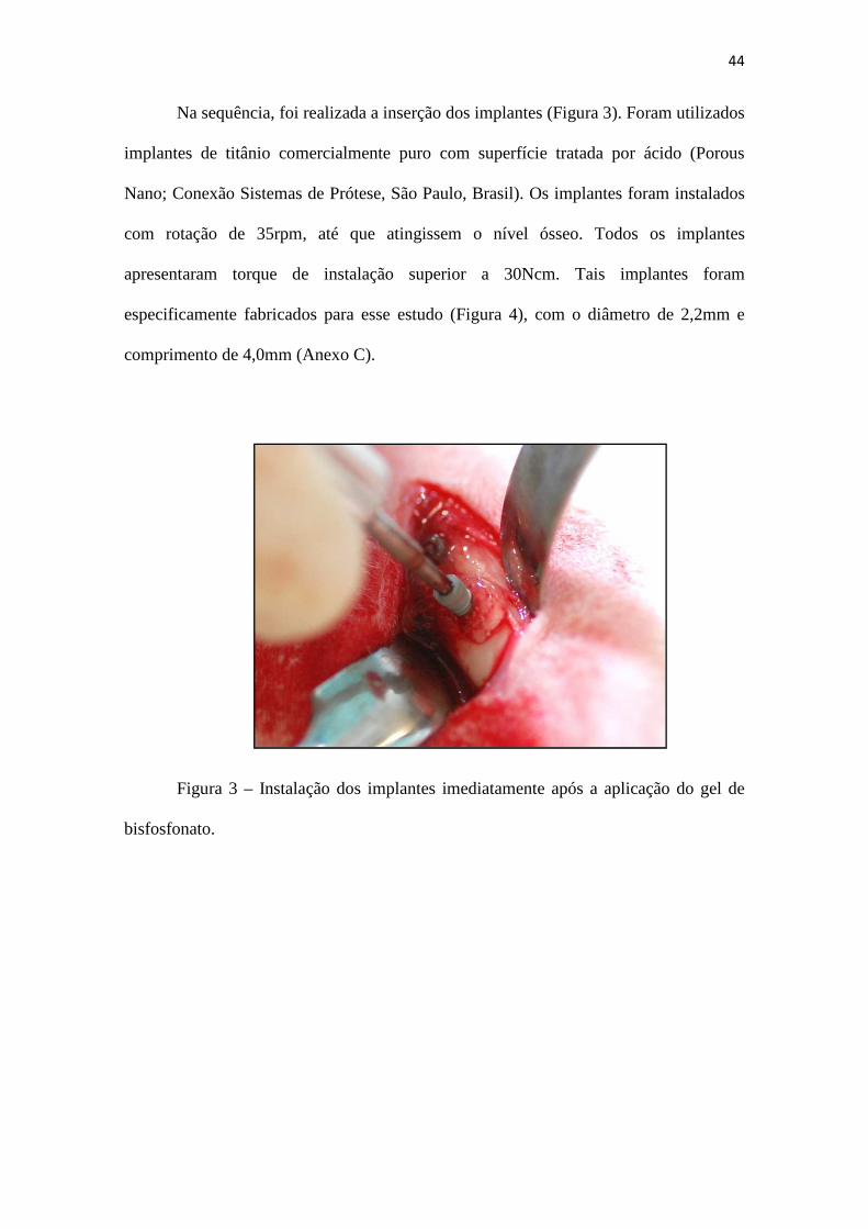
Na sequência, foi realizada a inserção dos implantes (Figura 3). Foram utilizados
implantes de titânio comercialmente puro com superfície tratada por ácido (Porous
Nano; Conexão Sistemas de Prótese, São Paulo, Brasil). Os implantes foram instalados
com rotação de 35rpm, até que atingissem o nível ósseo. Todos os implantes
apresentaram torque de instalação superior a 30Ncm. Tais implantes foram
especificamente fabricados para esse estudo (Figura 4), com o diâmetro de 2,2mm e
comprimento de 4,0mm (Anexo C).
Figura 3 – Instalação dos implantes imediatamente após a aplicação do gel de
bisfosfonato.
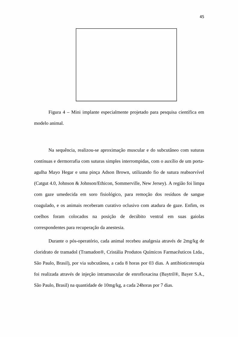
Figura 4 – Mini implante
modelo animal.
Na sequência, realizou
contínuas e dermorrafia com suturas s
agulha Mayo Hegar e uma pinça Adson Brown, utilizando fio de sutura reabsorvível
(Catgut 4.0, Johnson & Johnson/Ethicon, Sommerville, New Jersey)
com gaze umedecida em soro fisiológico, para remoção dos resíduos de sangue
coagulado, e os animais receber
coelhos foram colocados na posição de decúbito ventral em suas gaiolas
correspondentes para recuperação da anestesia.
Durante o pós-operatório, cada
cloridrato de tramadol (Tramadon®,
São Paulo, Brasil), por via subcutânea, a cada
foi realizada através de injeção intramuscular de
São Paulo, Brasil) na quantidade de 1
Mini implante especialmente projetado para pesquisa científica em
Na sequência, realizou-se aproximação muscular e do subcutâneo com suturas
ntínuas e dermorrafia com suturas simples interrompidas, com o auxílio de um porta
agulha Mayo Hegar e uma pinça Adson Brown, utilizando fio de sutura reabsorvível
(Catgut 4.0, Johnson & Johnson/Ethicon, Sommerville, New Jersey). A região foi
e umedecida em soro fisiológico, para remoção dos resíduos de sangue
coagulado, e os animais receberam curativo oclusivo com atadura de gaze. Enfim, os
colocados na posição de decúbito ventral em suas gaiolas
correspondentes para recuperação da anestesia.
operatório, cada animal recebeu analgesia através de 2mg/kg de
(Tramadon®, Cristália Produtos Químicos Farmacêuti
via subcutânea, a cada 8 horas por 03 dias. A antibioticoterapia
realizada através de injeção intramuscular de enrofloxacina (Baytril®, Bayer S.A.,
na quantidade de 10mg/kg, a cada 24horas por 7 dias.
45
projetado para pesquisa científica em
se aproximação muscular e do subcutâneo com suturas
s, com o auxílio de um porta-
agulha Mayo Hegar e uma pinça Adson Brown, utilizando fio de sutura reabsorvível
. A região foi limpa
e umedecida em soro fisiológico, para remoção dos resíduos de sangue
curativo oclusivo com atadura de gaze. Enfim, os
colocados na posição de decúbito ventral em suas gaiolas
analgesia através de 2mg/kg de
Cristália Produtos Químicos Farmacêuticos Ltda.,
dias. A antibioticoterapia
Baytril®, Bayer S.A.,
.

46
4.4 MORTE DOS ANIMAIS
Para eutanásia, cada animal recebeu medicação pré-anestésica composta por
1mg/kg de maleato de acepromazina (Acepran®, Univet, Brasil), 15mg/kg de cloridrato
de cetamina (Ketamina®, Agener Pharmaceutica, São Paulo, Brasil) e 2mg/kg de
cloridrato de xilazina (Rompum®, Bayer, São Paulo, Brasil), todos por via
intramuscular. Em torno de 10 minutos eram necessários até que os reflexos palpebrais,
corneal e de dor estivessem ausentes. Com o animal em plano anestésico profundo,
administrou-se, por via intravenosa, solução de cloreto de potássio a 10% (Ariston, São
Paulo, Brasil) até a parada cardiorrespiratória. A morte dos animais ocorreu após o
período de vida pós-operatório previsto de 28 dias.
4.5 MENSURAÇÃO DO TORQUE DE REMOÇÃO
Para mensuração do torque máximo de remoção de cada implante, os espécimes
foram processados imediatamente após a remoção da tíbia. Primeiramente foram
mantidos em solução de formalina neutra tamponada a 10% e, após 1 hora, foram
submetidos ao teste de torque de remoção, não sofrendo, assim, desidratação. A peça
anatômica foi cuidadosamente posicionada em equipamento de teste de torque – CME
(Técnica Industrial Oswaldo Filizola, Guarulhos, Brasil), o qual é totalmente controlado
pelo software DynaView Torque Standard/Pro M (Figura 5), gerando os valores
automaticamente com velocidade de 1rpm e medição angular do sistema com resolução
de 0.002º. As medições de torque máximo para iniciar a rotação inversa foram
registrados e os valores de torque médio foram calculados para cada grupo.

47
Figura 5 – a) Máquina de torque conectada ao computador (Software Dynaview Torque
Standard/Pro M); b) Teste de torque de remoção.
4.6 ANÁLISE HISTOMORFOLÓGICA E HISTOMORFOMÉTRICA
Após exame macroscópico local, as peças anatômicas foram imediatamente
armazenadas em recipientes de vidro, mergulhados em formalina neutra tamponada a
10%, com a finalidade de evitar as alterações post mortem dos tecidos.
A etapa de confecção e análise das lâminas histológicas foi realizada no Bioface
Institute (Santa Maria, RS, Brasil). Os blocos ósseos contendo os implantes foram
gradualmente sendo desidratados em concentrações sucessivas de álcool (de 50% a
100%). Após a desidratação, a amostra foi embebida em resina a base de metacrilato
(EMBed-812, Embedding Kit, EMS, Hatfield, PA) de acordo com as instruções do
fabricante. Os blocos foram, então, cortados em fatias de, aproximadamente 300µm de
espessura, objetivando o centro do implante no sentido do seu longo eixo, com disco de
diamante em cortadeira metalográfica (Modelo DTQTM5, Pantec®, São Paulo, Brasil).
A seguir, as amostras foram coladas a uma placa de acrílico com cimento a base de
acrilato e deixadas secar por 24h previamente aos processos de desgaste e acabamento.
As secções foram reduzidas a espessura final de, aproximadamente, 30µm por meio de
uma série de lixas d’água abrasivas (400, 600, 800, 1200 e 2400) (3M do Brasil, São

48
Paulo, Brasil) em uma politriz (Polipan 2, Pantec®, São Paulo, Brasil) sob irrigação
com água. Finalmente, a amostra foi corada com fucsina e levada à análise em
microscópio óptico (Nikon Eclipse E200, Nikon Corporation, Tóquio, Japão).
Todos os cortes histológicos de osso-implante foram analisados
histomorfológicamente de modo a se estabelecer as características teciduais gerais no
processo de osseointegração em cada grupo por meio da observação do tecido ósseo
neoformado e seus elementos celulares típicos. Procurou-se registrar as regiões dos
implantes com mais forte evidência de osseointegração, bem como avaliar o processo de
recobrimento das espiras pelo tecido ósseo.
A análise histomorfométrica foi realizada através do contato osso-implante
(%BIC), determinado em uma magnificação de 50-200X por meio de um software
(Image Tool® for Windows, versão 5.02). As regiões de contato osso-implante ao longo
do perímetro do implante foram subtraídas do perímetro total do implante (Figura 6), e
os cálculos foram realizados para determinar a porcentagem de BIC. As medições foram
realizadas por dois examinadores, treinados e calibrados. A amostra foi codificada
previamente à analise do examinador, caracterizando o processo de cegamento da
avaliação.
Figura 6 – a) Mensuração do perímetro total do implante e b) das áreas de contato osso-
implante para cálculo de BIC.
a b
49
4.7 ANÁLISE ESTATÍSTICA
Os valores de torque máximo de remoção foram comparados entre os grupos
através de Teste T-Student pareado. Para os resultados de contato osso-implante,
primeiramente foram calculados os coeficientes de correlação intraclasse (CCI) (Fleiss,
1986), a fim de que fosse verificada a reprodutibilidade da avaliação (Altman e Bland,
1983). Após, as porcentagens de contato osso-implante foram comparadas entre os
grupos com uso de Equações de Estimação Generalizadas (EEGs) com matriz de
correlações permutável nas medidas em um mesmo coelho (McCullagh e Nelder, 1989).
Todos os testes foram realizados com nível de significância de 5% (SPSS, Versão 20.0).

50
5. RESULTADOS

51
5. RESULTADOS
5.1 TORQUE MÁXIMO DE REMOÇÃO
A Tabela 2 e Gráfico 1 mostram que os valores de torque máximo de remoção
no grupo teste foi, em média, a metade que no grupo controle (p < 0,001).
Tabela 2. Torque máximo de remoção (Ncm) segundo grupos e resultado do teste comparativo.
Gráfico 1. Torque máximo de remoção segundo grupos (média ± dp) e resultado do teste comparativo.
0
5
10
15
20
25
Controle TesteGrupo
p < 0,001
Grupo Média DP Mediana Mínimo Máximo N p
Controle 17,86 5,15 17,9 10,1 25,3 10 Teste 8,73 2,86 7,9 5,9 14,2 10 <0,001
Tor
que
máx
imo
(N.c
m)

52
5.2 ANÁLISE HISTOMORFOLÓGICA
A avaliação qualitativa das lâminas histológicas demonstrou que todos os
implantes tiveram sua porção mais cervical atravessando a cortical tibiana, e a porção
apical em contato com a medular óssea (Figura 7).
Figura 7 – Posicionamento dos implantes em cortical e medular tibiana. Magnificação: 20X.
No grupo controle, a análise histológica mostrou neoformação óssea nas
adjacências da superfície dos implantes, com locais de remodelamento ósseo
evidenciando arranjo estrutural semelhante ao lamelar. Próximo ao implante, observou-
se osteócitos volumosos em grande quantidade localizados dentro de amplas lacunas.
Conforme se aproximava do implante, trabéculas ósseas imaturas com inúmeros
osteoblastos grandes e volumosos foram observados. A diferença da coloração – áreas
mais intensamente coradas – revelam tecido ósseo formado mais recentemente, o que

53
pode ser constatado principalmente nas regiões entre as espiras dos implantes (Figura
8).
Figura 8 – Interface osso-implante do grupo controle na região de osso cortical,
demonstrando atividade óssea mais intensa próximo ao corpo do implante, com
presença de tecido ósseo recentemente formado (região de coloração mais intensa),
osteócitos volumosos em amplas lacunas (--›) e osteoblastos grandes e volumosos em
contato com a superfície do implante (+). Magnificação: 40X.
No grupo teste, a análise histológica mostrou, em sua maioria, ausência de
neoformação óssea nas adjacências da superfície dos implantes, com locais de
remodelamento ósseo próximo ao topo das espiras e à porção mais cervical do implante.
Preenchendo os espaços entre as espiras, observou-se, na maioria das amostras, tecido
granular (Figura 9).

54
Figura 9 – Interface osso-implante do grupo teste na região de osso cortical,
demonstrando baixa atividade óssea localizada quase que somente em região de topo de
espiras e região mais cervical do implante, com presença de tecido granular
preenchendo o espaço entre as espiras (+). Magnificação: 40X.
5.3 ANÁLISE HISTOMORFOMÉTRICA
A reprodutibilidade das medidas entre os examinadores foi analisada e
demonstrou ser alta, pois o coeficiente de correlação intraclasse apresentou-se muito
próxima de 1 (CCI = 0,993), sendo que a repetibilidade (erro) entre os avaliadores foi
menor de 1% (0,90%) (Tabela 3).
Tabela 3. Descrição dos percentuais de contato osso-implante para cada examinador e
resultado da reprodutibilidade entre os examinadores.
Inferior Superior1 17,2 10,6 302 17,2 10,1 30
CCIIC (95%)
Repetibilidade
0,993 0,985 0,997 0,90
Examinador Média DP N

55
O Gráfico 2 confirma o resultado apresentado pela Tabela 2, demonstrando que
não há tendência evidente nas diferenças entre as avaliações dos examinadores e que a
diferença média entre eles foi bastante próxima de zero.
Gráfico 2. Gráfico de Bland-Altman entre as medidas dos examinadores.
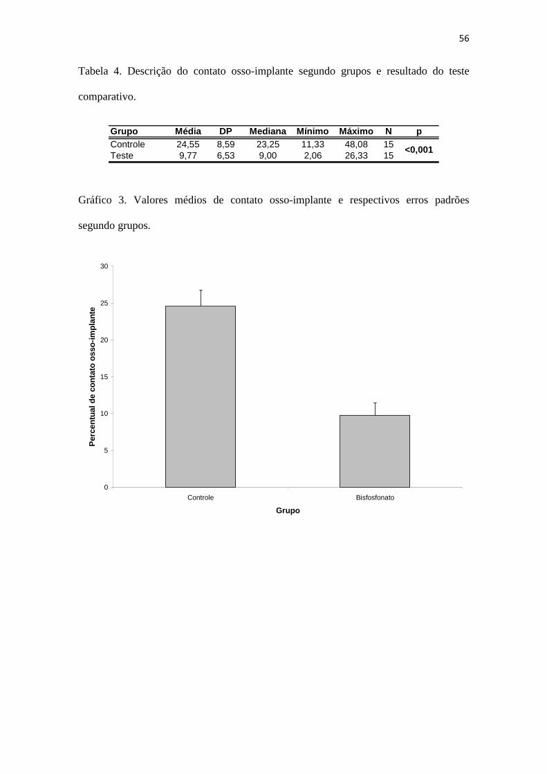
A média dos valores de BIC no grupo controle foi aproximadamente 2,5 vezes
maior que do grupo teste, mostrando uma diferença estatisticamente significante (p <
0,001) (Tabela 4 e Gráfico 3).
-3
-2
-1
0
1
2
3
0 5 10 15 20 25 30 35 40 45 50
Média das avaliações
Dife
renç
a en
tre
as a
valia
ções
(E
x.1
- E
x.2)
56
Tabela 4. Descrição do contato osso-implante segundo grupos e resultado do teste
comparativo.
Gráfico 3. Valores médios de contato osso-implante e respectivos erros padrões
segundo grupos.
0
5
10
15
20
25
30
Controle Bisfosfonato
Grupo
Per
cent
ual d
e co
ntat
o os
so-im
plan
te
Grupo Média DP Mediana Mínimo Máximo N pControle 24,55 8,59 23,25 11,33 48,08 15Teste 9,77 6,53 9,00 2,06 26,33 15
<0,001

57
6. DISCUSSÃO

58
6. DISCUSSÃO
A avaliação de interfaces entre tecido ósseo e implantes osseointegráveis é
imprescindível no desenvolvimento de novas técnicas e materiais relacionados à
Implantodontia. Resultados de estudos in vitro não podem ser extrapolados para a
situação in vivo; ao mesmo tempo, modelos animais são necessários para testes pré-
clínicos destes materiais antes de seu uso em humanos. Seguindo este pensamento, o
presente estudo foi delineado em um modelo animal, com objetivo de avaliar
parâmetros relacionados à osseointegração de implantes de titânio na presença de um
bisfosfonato.
A escolha do coelho como modelo animal partiu da observação de alguns fatos
importantes, já reconhecidos no meio científico: facilidade de manuseio, tamanho do
animal, maturidade óssea precoce e composição óssea moderadamente similar aos
humanos em ossos de crescimento primário (PEARCE et al, 2007). Além disso, este
modelo animal é tradicionalmente aceito e utilizado para pesquisa de osseointegração
(Branemark, 1983).
Nestes animais, o padrão internacional para avaliação biológica de dispositivos
médicos recomenda o máximo de 6 implantes (3 controles e 3 testes) por coelho, sendo
que estes dispositivos devem ter aproximadamente, no máximo, 2mm de diâmetro e
6mm de comprimento (INTERNATIONAL STANDARD ISO 10993-6, 1994). Tais
recomendações foram observadas neste estudo.
Este estudo teve como objetivo avaliar a osseointegração de implantes de titânio
instalados com e sem a aplicação local de um bisfosfonato. Os resultados encontrados
levaram à rejeição das duas hipóteses nulas admitidas inicialmente, uma vez que houve
diferença entre os grupos testados, com melhores resultados para o grupo controle.

59
A influência do uso de bisfosfonatos em implantodontia vem sendo
exaustivamente pesquisada atualmente, tanto através do uso sistêmico (AYAN et al,
2012; CHEN et al, 2013; DE OLIVEIRA et al, 2014), como da uso local, seja por meio
de aplicação direta no alvéolo cirúrgico (SKOGLUND, HOLMERTZ e ASPENBERG,
2004; JAKOBSEN et al, 2007; JAKOBSEN et al, 2009; CUAIRÁN et al, 2014) ou
através da imobilização na superfície do implante (ABTAHI, TENGVALL e
ASPENBERG, 2012; MOON et al, 2012; GUIMARÃES et al, 2013; HARMANKAYA
et al, 2013; STADLINGER et al, 2013; BOBYN et al, 2014). A proposta de aplicação
local do bisfosfonato neste estudo justifica-se pela intenção de beneficiar-se de sua
influência biomoduladora na osseointegração sem, contudo, sofrer os possíveis efeitos
sistêmicos desta droga (DE GROEN et al, 1996; BEDOGNI et al, 2010; GOSS et al,
2010; LAZAROVICI et al, 2010; VOHRA et al, 2014), uma vez que o bisfosfonato
localmente aplicado permanece concentrado nos arredores da aplicação, não sendo
distribuído sistemicamente (MCKENZIE et al, 2011).
Os implantes teste foram instalados imediatamente após a aplicação local de um
gel de bisfosfonato. Alguns estudos realizaram a análise da influência local deste
fármaco na osseintegração através de sua aplicação na forma de solução (SKOGLUND,
HOLMERTZ e ASPENBERG, 2004; JAKOBSEN et al, 2007; JAKOBSEN et al, 2009;
CUAIRÁN et al, 2014). A escolha pela apresentação em gel se deve ao cuidado em
conter o fármaco no alvéolo cirúrgico e mantê-lo em contato com as paredes ósseas
durante a instalação do implante, evitando que exista um viés no caso do resultado ser
igual nos dois grupos: a possibilidade do fármaco não ter sido realmente testado por ter
extravasado completamente.
A formulação do gel aplicado nesta pesquisa seguiu minuciosamente a descrita
por Reddy e Kumar (2005), a qual teve suas propriedades adequadamente testadas.
60
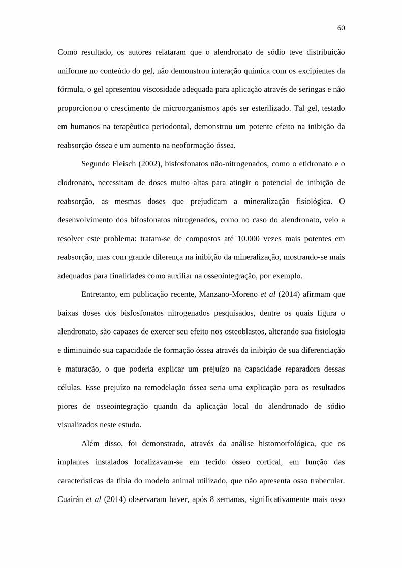
Como resultado, os autores relataram que o alendronato de sódio teve distribuição
uniforme no conteúdo do gel, não demonstrou interação química com os excipientes da
fórmula, o gel apresentou viscosidade adequada para aplicação através de seringas e não
proporcionou o crescimento de microorganismos após ser esterilizado. Tal gel, testado
em humanos na terapêutica periodontal, demonstrou um potente efeito na inibição da
reabsorção óssea e um aumento na neoformação óssea.
Segundo Fleisch (2002), bisfosfonatos não-nitrogenados, como o etidronato e o
clodronato, necessitam de doses muito altas para atingir o potencial de inibição de
reabsorção, as mesmas doses que prejudicam a mineralização fisiológica. O
desenvolvimento dos bifosfonatos nitrogenados, como no caso do alendronato, veio a
resolver este problema: tratam-se de compostos até 10.000 vezes mais potentes em
reabsorção, mas com grande diferença na inibição da mineralização, mostrando-se mais
adequados para finalidades como auxiliar na osseointegração, por exemplo.
Entretanto, em publicação recente, Manzano-Moreno et al (2014) afirmam que
baixas doses dos bisfosfonatos nitrogenados pesquisados, dentre os quais figura o
alendronato, são capazes de exercer seu efeito nos osteoblastos, alterando sua fisiologia
e diminuindo sua capacidade de formação óssea através da inibição de sua diferenciação
e maturação, o que poderia explicar um prejuízo na capacidade reparadora dessas
células. Esse prejuízo na remodelação óssea seria uma explicação para os resultados
piores de osseointegração quando da aplicação local do alendronado de sódio
visualizados neste estudo.
Além disso, foi demonstrado, através da análise histomorfológica, que os
implantes instalados localizavam-se em tecido ósseo cortical, em função das
características da tíbia do modelo animal utilizado, que não apresenta osso trabecular.
Cuairán et al (2014) observaram haver, após 8 semanas, significativamente mais osso
61
cortical ao redor dos implantes do grupo controle do que naqueles onde o bisfosfonato
foi aplicado, ao contrário do observado em osso trabecular. Os autores concluíram que o
bisfosfonato teria um efeito positivo no osso trabecular e negativo em osso cortical. Este
fato corrobora os resultados negativos de torque de remoção para o grupo bisfosfonato
no presente estudo.
Quando implantes osseointegráveis são instalados em tecido ósseo, um dano
neste tecido é causado pela utilização das brocas indicadas, e microfraturas e necrose
ósseas podem ser geradas em todo área tecidual adjacente. Este tecido ósseo danificado
deve ser remodelado através de eventos de reabsorção e neoformação (BERGLUNDH
et al, 2003). Segundo Russel et al (2008), é esperado que o bisfosfonato injetado cause
inibição ou restrição da atividade osteoclástica; uma vez que a atividade osteoblástica
está intimamente relacionada com a osteoclástica, é razoável assumir que o
remodelamento ósseo pode ser prejudicado pela presença do fármaco nestas condições
(RUSSEL et al, 2008), fato que pode explicar resultados significativamente piores de
histomorfometria e visivelmente alterados de histomorfologia encontrados no grupo
testado, nesta pesquisa.
Um estudo de Jakobsen et al (2010) testou a osseointegração de implantes
endósseos instalados com um gap de 2,5mm, o qual foi preenchido com enxerto alógeno
embebido em um bisfosfonato, com diferentes concentrações. Em seus resultados, tanto
o grupo controle quanto o grupo de baixa concentração do bisfosfonato tiveram melhor
fixação do que comparados com os demais grupos; enquanto que o grupo de maior
concentração teve o maior potencial inibidor de reabsorção óssea. Assim, um aumento
na fração de volume ósseo não pode ser correlacionado com um aumento na fixação
biomecânica. Este contraste indica claramente que o efeito benéfico do fármaco é dose-
dependente e que a manutenção de tecido ósseo inviável pelo efeito inibidor da

62
reabsorção pode não resultar no objetivo esperado, uma vez que a remodelação óssea
necessita fundamentalmente do efeito de reabsorção para ocorrer.
A literatura concernente, embora vasta, não utiliza metodologia padronizada
para comparação de resultados, variando tanto o tipo de bisfosfonato utilizado, como a
forma de apresentação, via administrativa, modelo animal, variáveis secundárias e testes
avaliativos. É importante ressaltar que Stadelmann et al (2009) desenvolveram um
modelo de remodelação óssea ao redor de implantes carreadores de bisfosfonatos para
liberação local, levando em consideração o estímulo mecânico e o estímulo do fármaco,
a fim de obter um resultado previsível. Os autores relataram que, muito embora os
resultados científicos atuais sejam baseados em escolhas empíricas, é possível obter-se
previsibilidade se for levado em consideração o tipo de bisfosfonato, a dose e o modelo
animal adotado.
Visivelmente, há um contraste dos resultados encontrados na literatura atual com
aqueles relatados neste estudo. A grande maioria dos estudos que testaram a aplicação
local de bisfosfonatos relataram haver melhora na fixação do implante, com aumento da
densidade óssea peri-implantar (PETER et al, 2005; KAJIWARA et al, 2005; LEE et al,
2011; MOON et al, 2012; STADLINGER et al, 2013; HARMANKAYA et al, 2013;
BOBYN et al, 2014; CUAIRÁN et al, 2014), resultados positivos para atividade de
células osteoblásticas (GOTO et al, 2003; BEUVELOT et al, 2009), aumento da
estabilidade primária (ABTAHI, TENGVALL e ASPENBERG, 2010; ABTAHI,
TENGVALL e ASPENBERG, 2012; CUAIRÁN et al, 2014) e maior contato osso-
implante (YOSHINARI et al, 2002; JAKOBSEN et al, 2007; STADLINGER et al,
2013). Entretanto, acredita-se que isso possa ser devido a um viés de publicação,
favorável a resultados positivos ao grupo testado. Faz-se importante que todos os relatos
científicos, sem qualquer distinção, baseados em estudos corretamente delineados,
63
sejam publicados em periódicos de maior fator de impacto, a fim de que todas as
informações a respeito de um produto testado possam ser acessadas pelo maior número
possível de leitores, facilitando, também, a análise por parte de pesquisadores que
busquem a realização de revisões sistemáticas e metanálises sobre este tópico.
Neste estudo, tanto a avaliação de torque de remoção, quanto a análise
histomorfométrica e histomorfológica mostraram resultados piores para o grupo que
sofreu a intervenção farmacológica. Esta coerência entre os achados biomecânicos e
histológicos reforça os resultados como um todo e embasa a conclusão gerada. Além
disso, o baixo desvio padrão encontrado na análise estatística demonstra o critério de
execução e a confiabilidade nos procedimentos metodológicos, principalmente no que
diz respeito ao procedimento cirúrgico, indicando uma padronização adequada da
amostra avaliada. Entretanto, é importante ressaltar que se trata de um estudo pré-
clínico, em que foi testado somente um tipo de bisfosfonato em uma única
concentração, pontualmente após 4 semanas in vivo, objetivando a análise da
osseointegração nos seus eventos mais iniciais. Ainda há muito o que ser esclarecido a
respeito deste tema, principalmente no que diz respeito a aplicabilidade em humanos,
uma vez que o cenário clínico ainda é pouco explorado na literatura científica atual
(ABTAHI, TENGVALL e ASPENBERG, 2010; ABTAHI, TENGVALL e
ASPENBERG, 2012).

64
7. CONCLUSÕES

65
7. CONCLUSÕES
A metodologia empregada na realização deste experimento, a partir das análises
realizadas, permite concluir que a aplicação local do gel de alendronato de sódio
prejudicou a osseointegração de implantes de titânio instalados em tíbia de coelho,
negando as hipóteses nulas. A aplicação local deste fármaco atuou diminuindo a
porcentagem de contato osso-implante e os valores de torque máximo de remoção, além
de, visivelmente, influenciar negativamente na remodelação óssea ao redor dos
implantes instalados.

66
REFERÊNCIAS BIBLIOGRÁFICAS
67
REFERÊNCIAS BIBLIOGRÁFICAS
Abtahi J, Tengvall P, Aspenberg P. Bisphosphonate coating might improve fixation of dental implants in the maxilla: A pilot study. Int J Oral Maxillofac Surg 2010;39:673-677.
Abtahi J, Tengvall P, Aspenberg P. A bisphosphonate-coating improves the fixation of metal implants in human bone. A randomized trial of dental implants. Bone 2012;50:1148-1151.
Allgrove J. Bisphosphonates. Arch Dis Child 1997;76:73-75.
Altman DG, Bland JM. (1983). Measurement in Medicine: The Analysis of Method Comparison Studies. The Statistician 1983;32(3):307-317.
Aspenberg P, Wermelin K, Tengwall P, Fahlgren A. Additive effects of PTH and bisphosphonates on the bone healing response to metaphyseal implants in rats. Acta Orthopaedica 2008;79(1):111-115.
Ayan M, Dolanmaz D, Mihmanli A, Ayan A, Kurkçu M. The effect of sistemically administrated zoledronic acid on the osseointegration of dental implants. Oral Diseases 2012;18:802-808.
Bedogni A, Bettini G, Totola A, Saia G, Nocini PF. Oral bisphosphonate-associated osteonecrosis of the jaw after implant surgery: a case report and literature review. J Oral Maxillofac Surg 2010;68:1662-66.
Berglundh T, Abrahamsson I, Lang NP, Lindhe J. De novo alveolar bone formation adjacent to endosseous implant. Clin Oral Impl Res 2003;14(3):251-62.
Beuvelot J, Portet D, Lecollinet G, Moreau MF, Baslé MF, Chappard D, Libouban H. In vitro kinetic study of growth and mineralization of osteoblast-like cells (Saos-2) on titanium surface coated with a RGD funtionalized bisphosphonate. J Biomed Mater Res Part B: Appl Biomater 2009;90B:873-881.
Bobyn JD, Thompson R, Lim L, Pura JA, Bobyn K, Tanzer M. Local alendronic acid elution increases net periimplant bone formation: a micro-CT analysis. Clin Orthop Relat Res 2014;472:687-694.
Branemark PI. Osseointegration and its experimental background. J Prosthet Dent 1983;50(3):399-410.
Chacon GE, Stine EA, Larsen PE, Beck M, McGlumphy EA. Effect of alendronate on endosseous implant integration: An in vivo study in rabbits. J Oral Maxillofac Surg 2006;64:1005-1009.
Chen B, Li Y, Yang X, Xu H, Xie D. Zoledronic acid enhances bone-implant osseointegration more than alendronate and strontium ranelate in ovariectomized rats. Osteoporos Int 2013;24:2115-2121.
Cuairán C, Campbell PM, Kontogiorgos E, Taylor RW, Melo AC, Buschang PH. Local application of zoledronate enhances miniscrew implant stability in dogs. Am J Orthod Dentofacial Orthop 2014;145:737-49.

68
De Groen PC, Lubbe DF, Hirsch LJ, Daifotis A, Stephenson W, Freedholm D et al. Esophagitis associated with the use of alendronate. N Engl J Med 1996;335(14):1016-21.
De Oliveira MA, Asahi DA, Silveira CAE, Lima LAPA, Glick M, Gallottini M. The effects of zoledronic acid and dexamethasone in osseointegration of endosseous implants: histological and histomorphometrical evaluation in rats. Clin Oral Impl Res 2014;0:1-5.
Eberhardt C, Habermann B, Müller S, Schwarz M, Bauss F, Kurth AHA. The bisphosphonate ibandronate accelerates osseointegration of hydroxyapatite-coated cementless implants in an animal model. J Orthop Sci 2007;12:61-66.
Fleisch H. Development of bisphosphonates. Breast Cancer Res 2002;4:30-34.
Fleiss JL. The design and analysis of clinical experiments. New York: Wiley. 432p. 1986.
García-Merco C, Serrano S, Nacher M, Farré M, Díez A, Marinoso ML, Carbonell J, Mallibovsky L, Nogués X, Ballester J, Aubía J. Effect of alendronate on cultured normal human osteoblasts. Bone 1998;22:233-9.
Goss A, Bartold M, Sambrook P, Hawker P. The nature and frequency of bisphosphonate-associated osteonecrosis of the jaws in dental implant patients: A south australian case series. J Oral Maxillofac Surg 2010;68:337-343.
Goto T, Kajiwara H, Yoshinari M, Fukuhara E, Kobayashi S, Tanaka T. In vitro assay of mineralized-tissue formation on titanium using fluorescent staining with calceinblue. Biomaterials 2003;24:3885-3892.
Guimarães MB, Bueno RS, Blaya MBG, Hirakata LM, Hubler R. Diphosphonate immobilization on hydroxyapatite-coated titanium – Method Description. Implant Dent 2013;22(4):356-359.
Harmankaya N, Karlsson J, Palmquist A, Halvarsson M, Igawa K, Andersson M et al. Raloxifene and alendronate containing thin mesoporous titanium oxide films improve implant fixation to bone. Acta Biomaterialia 2013;9:7064-7073.
International Standard ISO 10993-6. Biological evaluation of medical devices – Part 6. 1994:1-11.
Jakobsen T, Kold S, Bechtold JE, Elmengaard B, Soballe K. Effect of topical alendronate treatment on fixation of implants inserted with bone compaction. Clin Orthop Relat Res 2006;444:229-234.
Jakobsen T, Kold S, Bechtold JE, Elmengaard B, Soballe K. Local alendronate increases fixation of implants inserted with bone compaction: 12-week canine study. J Orthop Res 2007;25:432-441.
Jakobsen T, Baas J, Kold S, Bechtold JE, Elmengaard B, Soballe K. Local bisphosphonate treatment increases fixation of hydroxyapatite-coated implants inserted with bone compaction. J Orthop Res 2009;27(2):189-194.

69
Jakobsen T, Baas J, Bechtold JE, Elmengaard B, Soballe K. The effect of soaking allograft in bisphosphonate. A pilot dose-response study. Clin Orthop Relat Res 2010;468:867-874.
Kajiwara H, Yamaza T, Yoshinari M, Goto T, Iyama S, Atsuta I, Kido MA, Tanaka T. The bisphosphonate pamidronate on the surface of titanium stimulates bone formation around tibial implants in rats. Biomaterials 2005;26:581-587.
Langhoff JD, Voelter K, Scharnweber D, Schnabelrauch M, Schlottig F, Hefti T, Kalchofner K, Nuss K, Von Rechenberg B. Comparison of chemically and pharmaceutically modified titanium and zirconia implant surfaces in dentistry: a study in sheep. Int J Oral Maxillofac Surg 2008;37:1125-1132.
Lazarovici TS, Yahalom R, Taicher S, Schwartz-Arad D, Peleg O, Yarom N. Bisphosphonate-related osteonecrosis of the jaw associated with dental implants. J Oral Maxillofac Surg 2010;68:790-796.
Lee SJ, Oh TJ, Bae TS, Lee MH, Soh Y, Kim BI et al. Effect of bisphosphonates on anodized and heat-treated titanium surfaces: an animal experimental study. J Periodontol 2011;82:1035-1042.
Li YF, Li XD, Bao CY, Chen QM, Zhang H, Hu j. Promotion of peri-implant bone healing by sistemically administered parathyroid hormone (1-34) and zoledronic acid adsorbed onto the implant surface. Osteoporos Int 2013;24:1063-1071.
Manzano-Moreno FJ, Ramos-Torrecillas J, Luna-Bertos E, Reyes-Botella C, Ruiz C, García-Martínez O. Nitrogen-containing bisphosphonates modulate the antigenic profile and inhibit the maturation and biomineralization potencial of osteoblast-like cells. Clin Oral Invest 2014; [Epub ahead of print] DOI 10.1007/s00784-014-1309-z
McCullagh P, Nelder JA. Generalized linear models. 2nd ed. Chapman and Hall: New York, USA. p.511. 1989.
McKenzie K, Bobyn JD, Roberts J, Karabasz D, Tanzer M. Bisphosphonate remains highly localized after elution from porous implants. Clin Orthop Relat Res 2011;469:514-522.
Misch CE, Wang HL. Immediate occlusal loading for fixed prostheses in implant dentistry. Dent Today 2003;22(8):50-6.
Moon SH, Lee SJ, Park IS, Lee MH, Soh YJ, Bae TS et al. Bioactivity of Ti-6Al-4V alloy implants treated with ibandronate after the formation of the nanotube TiO2 layer. J Biomed Mater Res 2012;100B:2053-2059.
Muller F. Interventions for edentate elders - what is the evidence? Gerodontology 2014;31(Suppl.1):44-51.
Mundy GR, Yoneda T. Bisphosphonates as anticancer drugs. N Engl J Med 1998;339:398-400.
Murakami H, Takahashi N, Sasaki T, Udagawa N, Tanaka S, Nakamura I, Zhang D, Barbier A, Suda T. A possible mechanism of the specific action of bisphosphonates on

70
osteoclasts: tildronate preferentially affects polarized osteoclasts having ruffled borders. Bone 1995;17:137-44.
Nancollas GH, Tang R, Phipps RJ, Henneman Z, Gulde S, Wu W, Mangood A, Russell RGG, Ebetino FH. Novel insights into actions of bisphosphonates on bone: Differences in interactions with hydroxyapatite. Bone 2006;38:617-627.
Otomo-Corgel J. Implants and oral bisphosphonates: Risky Business? J Periodontol 2007;78(3):373-6.
Pearce AI, Richards RG, Milz S, Schneider E, Pearce SG. Animal models for implant biomaterial research in bone: A review. Eur Cell Mater 2007;13:1-10.
Peter B, Pioletti DP, Laib S, Bujoli B, Pilet P, Janvier P. et al. Calcium phosphate drug delivery system: influence of local zoledronate release on bone implant osteointegration. Bone 2005;36:52-60.
Reddy GT, Kumar TMP. Formulation and evaluation of alendronate sodium gel for the treatment of bone resorptive lesions in periodontitis. Drug Delivery 2005;12:217-222.
Russel RGG, Watts NB, Ebetino FH, Rogers MJ. Mechanisms of action of bisphosphonates: similarities and differences and their potencial influence on clinical efficacy. Osteoporos Int 2008;19:733-759.
Skoglund B, Holmertz J, Aspenberg P. Systemic and local ibandronate enhance screw fixation. J Orthop Res 2004;22:1108-1113.
Stadelmann VA, Terrier A, Gauthier O, Bouler JM, Pioletti DP. Prediction of bone density around orthopedic implants delivering bisphosphonate. J Biomech 2009;42:1206-1211.
Stadlinger B, Korn P, Todtmann N, Eckelt U, Range U, Burki A et al. Osseointegration of biochemically modified implants in an osteoporosis rodent model. Eur Cell Mater 2013;25:326-340.
Truhlar RS, Orenstein IH, Morris HF, Ochi S. Distribution of bone quality in patients receiving endosseous dental implants. Int J Oral Maxillofac Surg 1997;55(12):38-45.
Tsetsenekou E, Papadopoulos T, Kalyvas D, Papaioannou N, Tangl S, Watsek G. The influence of alendronate on osseointegration of nanotreated dental implants in New Zealand rabbits. Clin Oral Impl Res 2011;10.1111/j.1600-0501.2011.02189.x.
Vitte C, Fleisch H, Guenther HL. Bisphosphonates induce osteoblasts to secret an inhibitor of osteoclast-mediated resorption. Endocrinology 1996;137:2324-33.
Vohra F, Al-Rifaiy MQ, Almas K, Javed F. Efficacy of sistemic bisphosphonate delivery on osseointegration of implants under osteoporotic conditions: lessons from animal studies. Arch Oral Biol 2014;59:912-920.
Yoshinari M, Oda Y, Ueki H, Yokose S. Immobilization of bisphosphonates on surface modified titanium. Biomaterials 2001;22:709-715.

71
Yoshinari M, Oda Y, Inoue T, Matsuzaka K, Shimono M. Bone response to calcium phosphate-coated and bisphosphonate-immobilized titanium implants. Biomaterials 2002;23:2879-2885.

72
ANEXOS

73
ANEXO A – Aprovação da Comissão Científica e de Ética – FO/PUCRS

74
ANEXO B – Aprovação da Comissão de Ética no Uso de Animais – PUCRS.

ANEXO C – Croqui do implante utilizado.
Croqui do implante utilizado.
75

76
ANEXO D – Artigo científico
O artigo a seguir intitula-se “Influência da aplicação local de gel de
alendronato de sódio na osseointegração de implantes de titânio” e foi escrito nas
normas do Periódico International Journal of Oral and Maxillofacial Surgery – Qualis
A1 – Fator de Impacto: 1.359 - ISSN: 0901-5027.
77
INFLUENCE OF LOCAL APPLICATION OF SODIUM ALENDRONATE GEL ON
OSSEOINTEGRATION OF TITANIUM IMPLANTS
GUIMARÃES, Magáli Beck; BUENO, Rodrigo Salbego; BLAYA, Micéli Beck
Guimarães; SHINKAI, Rosemary Sadami Arai; MARQUES, Luciana Mayumi
Hirakata.
Department of Prosthodontics. Faculty of Dentistry. Pontifícia Universidade Católica do
Rio Grande do Sul. Avenida Ipiranga, 6681, Prédio 6. Zip Code: 90619-900. Porto
Alegre, RS – Brazil.
Corresponding Author:
Magáli Beck Guimarães. Pós-Graduação - Odontologia. Pontifícia Universidade
Católica do Rio Grande do Sul. Avenida Ipiranga, 6681, Prédio 6. Zip Code: 90619-
900. Porto Alegre, RS – Brazil. Telephone: +55.51.33203538. Fax: +55.51.33203626.
E-mail: [email protected]
Keywords: Diphosphonates; Osseointegration; Bone remodeling; Dental Implants.
Short Title: Bisphosphonate in Implant Dentistry.

78
ABSTRACT
The aim of this study was to make a comparative analysis of aspects related to the
osseointegration of titanium implants placed with and without local application of a
bisphosphonate agent, after 4 weeks, in vivo.The study comprised the placement of 50
cp. titanium implants in the middle third of the tibia of 10 rabbits, with the right tibia
being used as control, and the left as test site. In the test group, local administration of
sodium alendronate gel was instituted, and in the control group, sterile saline solution
was used. After euthanasia, 10 implants from each group were analyzed for maximum
removal torque. The remainder of the sample was processed to obtain non decalcified
slides with approximately 30µm thick, in which histomorphological and
histomorphometric analyses of bone-implant contact (%BIC) were performed. Data
were analyzed at a level of significance of 5%. The removal torque of the test group, on
an average, were half of the values obtained in the control group. The test group showed
lower percentage of BIC, and notable changes in bone quality. It was concluded that the
initial events of osseointegration of titanium implants are not favored by the local
application of sodium alendronate gel in rabbits.
79
INTRODUCTION
Over the last few decades, there has been considerable growth in the demand for
replacement of lost teeth by means of therapy with dental implants and implant-
supported dentures. Among other reasons, this is due to the fact that either partial or
complete edentulism affects a large portion of persons in the more advanced age group,
a steadily growing part of the population worldwide. These patients frequently present
a profile of bone quality and quantity affected by systemic diseases, such as diabetes
and osteoporosis, in addition to presenting lower potential bone regeneration, which
may contribute to lower success rates in therapy with dental implants2. On the other
hand, the desire to attain faster osseointegration is common in both health professionals
and patients. This motivates researches that seek improvements in the development of
materials and techniques related to optimization of the process of bone remodeling
around dental implants.
During implant placement surgery, the decision about whether or not to apply
immediate load is determined, among other aspects, by the primary stability of the
implant in bone tissue. In cases in which there is no primary stability during dental
implant placement, the professional is recommended to wait during the period of
osseointegration without application of functional load, following the protocol of two
surgical stages3. Therefore, in this scenario it becomes important to accelerate the
process of osseointegration, so that less time will be required between implant
placement surgery, and re-opening for connection of the prosthetic abutments.
The interest in using bisphosphonates as bone biomodulators in implant dentistry
arose by the known ability of this drug to inhibit the activity of osteoclasts, which is
why it is widely used in the treatment of diseases characterized by excessive bone
resorption, such as osteoporosis, hypercalcemia and bone metastases4. Furthermore, it is

80
known that an effect on bone formation may be expected, promoting a considerable
reduction in bone turnover5. Studies have suggested that bisphosphonates may have a
positive influence on bone formation and remodeling, and consequently an
improvement in the fixation of titanium implants in humans6,7.
Because of the severe side effects caused by the systemic use of this drug8-11,
researchers have turned their attention to developing methods of local delivery of this
drug to the site of interest12-16. The intention is for bisphosphonate to positively
influence the remodeling of bone adjacent to the implant, without causing undesirable
systemic side effects. In this sense, immobilization of the bisphosphonate on the implant
surface has been proposed, as a way of local delivery of the drug17-21. However, to
perform this immobilization, a complex methodology and sophisticated equipment are
frequently required. The direct application of bisphosphonate in the surgical alveolus
immediately before implant insertion, appears to be a simpler and more practical
procedure, however, up to now, it has not been extensively tested13,22,23.
Therefore, the aim of this study was to propose the local application of a
bisphosphonate drug (sodium alendronate) in gel form, directly in the surgical site, and
make a comparative evaluation of aspects related to osseointegration of titanium
implants inserted immediately after this application, in vivo. As a null hypothesis, it was
admitted that the local application of sodium alendronate gel did not favor the
osseointegration of titanium implants placed in rabbit tibias.
MATERIALS AND METHODS
Animals and Experimental Groups

81
Ten rabbits of the Oryctolagus cuniculus species, New Zealand lineage, adult
males, with a mean body weight of 4.0kg were used in this research. The study was
approved by the ethics committee of the university, and the animals received all the care
stipulated by the institution.
A total of 50 implants were inserted in the sample, with the right tibia being used
as control, and the left, as test site. In the control group, the surgical alveoli performed
in the right tibia of each animal, received an application of sterile saline solution. In the
test group, characterized by the surgical alveoli performed according to a previously
published study24, in the left tibia of each rabbit, a topical application of 1 ml of sodium
alendronate gel (10mg/g) was administered.
Surgical Procedure
After being weighed, the animals received pre-anesthesia medication, composed
of acepromazine maleate (0.2mg/kg) and morphine sulphate (2mg/kg), both
administered intramuscularly. After approximately 10 minutes had elapsed, the animals'
marginal ear vein was cannulated for the administration of fluid therapy with Lactated
Ringer solution and Enrofloxacin (10mg/kg), 20 minutes before surgery. Anesthesia
was induced by means of intravenous injection of ketamine chloride (10mg/kg) and
1mg midazolam (1mg/kg). Epidural anesthesia was administered with 2% lidocaine
(0.25 ml/kg). After anesthesia induction, the animals were trichotomized and antisepsis
of the region was performed, including the skin adjacent to the trichotomized area.
Surgery began with a linear incision, measuring approximately 2cm in
extension, on the medial diaphysal surface of the tibia. The sites where the surgical
recesses were performed were previously demarcated, with the perforations positioned
10 mm below the tibial condyle, with a distance of 10mm between each perforation.
Cutting for implant insertion was performed with appropriate burs, under irrigation, up

82
to a depth of 4 mm, in the sequence of using a 2mm lance-shaped bur and then a helical
bur.
After the cavities were prepared, sterile gauze was introduced and kept in the
surgical alveolus by compression for 1 to 2 minutes, with the purpose of absorbing and
stopping the bleeding. This process guaranteed that the bisphosphonate gel would come
into direct contact with the entire wall of the alveolus, without the interposition of
blood. The quantity of 1ml of sodium alendronate - test group - was injected in the
surgical alveolus immediately before placement of the implant22,25 (Figure 1a).
After this, the implants were inserted (Figure 1b). Commercially pure titanium
implants were used, with surface treated with acid (Porous Nano; ConexãoSistemas de
Prótese, Brazil), 2.2mm in diameter and 4.0mm long, specifically fabricated for this
study. The implants were inserted at a speed of 35 rpm, until they reached bone level.
To finalize, the muscle and subcutaneous tissues were approximated with
continuous sutures and the skin was approximated with simple, interrupted sutures,
using reabsorbable suture thread (Catgut 4.0, Johnson & Johnson/Ethicon, Sommerville,
USA). The region was cleaned with gauze dampened with physiological solution, to
remove the residues of blood clots, and the animals received an occlusive dressing with
a gauze bandage.
During the post-operative period, each animal received analgesic medication
with tramadol hydrochloride (2mg/kg) via the subcutaneous pathway, every 8 hours, for
3 hours. Antibiotic therapy was administered via intramuscular injection of enrofloxacin
(10mg/kg), every 24 hours for 7 days.
Euthanasia of Animals

83
To perform euthanasia each animal received pre-anesthetic medication
composed of Acepromazine maleate (l mg/kg) ketamine hydrochloride (15mg/kg), and
xylazin hydrochloride, all administered intramuscularly. Passed around 10 minutes, the
reflexes such as palpebral, corneal and pain were absent. With the animal in a plane of
deep anesthesia, a 10% potassium chloride solution was administered intravenously
until cardiorespiratory function ceased. Euthanasia occurred after the pre-established
period of post-operative life of 28 days.
Removal Torque Measurement
For measurement of the maximum removal torque of each implant, the
specimens were processed immediately after removal of the tibia. Firstly, the tibias were
kept in a 10% buffered neutral formalin solution, and after 1 hour, were submitted to the
torque removal test, thus they did not become dehydrated. The anatomic part was
carefully placed on the torque test equipment– CME (Técnica Industrial Oswaldo
Filizola, Guarulhos, Brazil), completely controlled by the software program DynaView
Torque Standard/Pro M, generating the values automatically at a speed of 1rpm and
angular measurement of the system with a resolution of 0.002º. The maximum torque
measurements to begin the inverse rotation were recorded, and the mean torque values
were calculated for each group.
Histomorphological and Histomorphometric Analyses
The bone blocks containing the implants were gradually dehydrated in
successive concentrations of alcohol, and the sample was imbibed in methacrylate-
based resin (EMBed-812, Embedding Kit, EMS, Hatfield, PA, USA) in accordance with
the manufacturer's instructions. The blocks were then cut into slices approximately
300µm thick. with the center of the implant facing in the direction of its long axis, using
a diamond-coated disc in a metallographic cutter (Model DTQTM5, Pantec®, São Paulo,

84
Brazil). After this, the samples were bonded to an acrylic plate with acrylate-based
cement, and left to dry for 24 h before the stripping and finishing processes. The
sections were reduced to a final thickness of approximately 30µm, with the use of a
series of water abrasive papers (400, 600, 800, 1200 and 2400 grit) (3M do Brasil, São
Paulo, Brazil) in a polishing machine (Polipan 2, Pantec®, São Paulo, Brazil) under
irrigation with water. Finally the sample was stained with fuchsin and taken for
analysis under an optical microscope (Nikon Eclipse E200, Nikon Corporation, Tokyo,
Japan).
All the bone-implant histological cuts were analyzed histomorphologically, in
order to establish the general tissue characteristics in the osseointegration process in
each group, by means of observing the neoformed bone tissue and its typical cell
elements. An endeavor was made to record the regions of the implants with the
strongest evidence of osseointegration and to evaluate the process of covering the
implant spirals with bone tissue.
The histomorphometrical analysis was performed according to bone-implant
contact (%BIC), determined at 50-200X magnification, by means of a software program
(Image Tool® for Windows, version 5.02). The bone implant contact regions along the
perimeter of the implant were subtracted from the total implant perimeter, and
calculations were made to determine the percentage of BIC.
Statistical Analysis
The maximum removal torque values were compared between the groups by
means of the paired Student's-t test. For the bone-implant contact results, the intraclass
correlation coefficients (ICC) were first calculated, in order to verify the reproducibility
of the evaluation. After this, the implant-bone contact percentages were compared
between the groups with the use of the Generalized Estimating Equations (GEEs) with

85
the correlations matrix permutable in the measurements in one and the same rabbit. All
the tests were considered with a level of significance of 5% (SPSS, version 20.0).
RESULTS
Maximum Removal Torque
Table 1 shows the maximum removal torque values in the test group were, on an
average, half the value they were in the control group (p < 0.001).
Histomorphological Analysis
The qualitative evaluation of the histological slides demonstrated that the most
cervical portion of all the implants passed through the tibial cortical bone, and the apical
portion was in contact with medullary bone (Figure 2).
In the control group, histological analysis showed bone neoformation in the
areas adjacent to the implant surfaces, with locations of bone remodeling, showing
evidence of a structural arrangement similar to that of the lamellar region. Close to the
implant, a large quantity of voluminous osteocytes were observed located within wide
gaps. Closer to the implant, immature bone trabeculae with innumerable large and
voluminous osteoblasts were observed. The difference in staining- more intensely
stained areas - revealed more recently formed bone tissue, which was found particularly
in the regions between the implant spirals (Figure 3).
In the majority of specimens in the test group, the histological analysis showed
absence of bone neoformation in the areas adjacent to the implant surfaces, with sites of
bone remodeling close to the top of the spirals and more cervical portion of the implant.
In the majority of samples, granular tissue was observed filling the spaces between the
spirals (Figure 4).
86
Histomorphometric Analysis
Reproducibility of the measurements between examiners was shown to be high
(ICC=0.993), with the repeatability (error) between evaluators being lower than 1%
(0.90%) (Table 2).
Graph 1 confirms the results presented in Table 2, demonstrating that there was
no evident trend in the differences between the evaluations of the examiners, and that
the mean difference among them was very close to zero.
The mean BIC values in the control group were approximately 2.5 times higher
than they were in the test group, showing a statistically significant difference (p <
0.001) (Table 3).
DISCUSSION
It is imperative to evaluate the interfaces between bone tissue and
osseointegratable implants, in the development of new techniques and material related
to implant dentistry. The results of in vitro studies cannot be extrapolated to the in vivo
situation; at the same time, animal models are necessary for the pre-clinical tests of
these materials before they are used in humans.
Following this line of thinking, the present study was designed on an animal model,
with the goal of evaluating parameters related to the osseointegration of titanium
implants in the presence of a bisphosphonate,
The choice of the rabbit as animal model was made, based on the observation of
some important facts already known in the scientific medium: ease of handling, size of

87
animal, early bone maturity, composition of the bone moderately similar to that of
humans in bones with primary growth26
In these animals, the international pattern for biologic evaluations of medical
devices recommends a maximum of 6 implants (3 controls and 3 tests) per rabbit, and
these devices must, approximately, be a maximum of 2 mm in diameter and and 6mm
long27. These recommendations were observed in this study.
The influence of the use of bisphosphonates in implant dentistry has been
exhaustively research at present, with both systemic28-30, and local use by means of
direct application in the surgical alveolus13,22,23,25 or by immobilization of the
medication on the implant surface 7,15,16,19-21. In this study, the proposal of local
application of bisphosphonate was justified by the intention to benefit from its
biomodulating influence on osseointegration without, however, suffering the possible
systemic effects of this drug8-11. This was done, considering that locally applied
bisphosphonate remains concentrated on the tissues around the application, and is not
systemically distributed31.
The test implants were inserted immediately after local application of a
bisphosphonate gel. Some studies have analyzed the local influence of this drug on
osseointegration by means of applying it in the form of a solution13,22,23. The choice of
the presentation in gel form was due to the care taken to contain the drug in the surgical
alveolus, and keep it in contact with the bone walls during implant placement. The
intention was to prevent bias in case the results were equal in the two groups: due to the
possibility of the drug not being really tested because it had completely extravasated.
The gel formulation applied in this test meticulously followed the one described
by Reddy and Kumar24, of which the properties were adequately tested. As a result, the
authors reported that sodium alendronate was uniformly distributed throughout the
88
content of the gel, and did not demonstrate chemical reaction with the excipients of the
formula. The gel presented adequate viscosity for application by means of syringes, and
the not favor the growth of microorganisms after being sterilized.
This gel, tested in humans in periodontal therapy, was shown to have a powerful effect
on the inhibition of bone resorption and increase in bone neoformation.
According to Fleisch32, non nitrogenated bisphosphonates, such as etidronate
and clodronate, require very high doses in order to attain the potential of resorption
inhibition; indeed, the same doses as those that harm physiological mineralization. The
development of nitrogenated bisphosphonates, as in the case of alendronate, has
resolved this problem: the are compounds up to 10,00 more powerful in resorption, but
with the great difference in inhibition of mineralization, and have been shown to be
more adequate for purposes such as helping with osseointegration, for example.
However, in a recent publication, Manzano-Moreno et al33 affirmedthat low
doses of the nitrogenated bisphosphonates researched, among them alendronate, are
capable of exerting their effect on osteoblasts, altering their physiology and diminishing
their bone formation capacity by inhibiting their differentiation and maturation, which
may explain the harm to the reparative capacity of these cells. This harm to bone
remodeling would be an explanation for the worse osseointegration results with the
local application of sodium alendronate found in this study.
Furthermore, by means of histomorphological analysis it was demonstrated that
the implants inserted were exclusively located in cortical bone tissue, because of the
characteristics of the animal model used. Cuairánet al23 , after 8 weeks, observed
significantly more cortical bone around the implants of the control group than around
those in which bisphosphonate was applied, contrary to the situation observed in
trabecular bone. The authors concluded that bisphosphonate would have a positive
89
effect on trabecular bone and a negative effect on cortical bone. This fact corroborates
the negative removal torque results for the bisphosphonate group in the present study.
When dental implants are inserted in bone tissue, damage is caused to this tissue
by the use of the burs indicated, and bone microfractures and necrosis may be generated
in the entire adjacent tissue area. This damaged bone tissue must be remodeled by
events of resorption and neoformation34. According to Russel et al35, the
bisphosphonate injected is expected to cause inhibition or restriction of osteoclastic
activity. Since osteoblastic activity is closely related to osteoclastic activity, it is
reasonable to assume that bone remodeling could be harmed by the presence of this
drug in these conditions35. This could explain the significantly worse
histomorphometrical and visibly altered histomorphological results found in the group
tested in this research.
The study of Jakobsen et al36 tested the osseointegration of endosseous implants
inserted with a gap of 2.5mm, which was filled with an allogenous graft imbibed in a
bisphosphonate, in different concentrations. In their results, both the control and the
group with a low concentration of bisphosphonate had better fixation when compared
with the other groups; while the group with the highest concentration had the highest
potential for inhibiting bone resorption. Thus, an increase in the fraction of bone
volume canto be correlated with an increase in biomechanical fixation. This contrast
clearly indicates that the beneficial effect of the drug is dose-dependent, and that the
unfeasible maintenance of bone tissue may not result in the expected objective, since
bone remodeling fundamentally requires the effect of resorption in order to occur.
The relevant literature, although vast, does not use standardized methodology for
comparison of results, and varies both in the type of bisphosphonate used and in the
form of presentation, administration pathway, animal model, secondary variables and

90
evaluative tests. It is important to point out that Stadelmann et al37 developed a model
of bone remodeling around implants, carriers of bisphosphonates for local release,
taking into consideration the mechanical stimulus of the drug, in order to obtain a
predictable result. The authors related that although the present scientific results are
based on empirical choices, it is possible to obtain predictability if the type of
bisphosphonate, dose and animal model adopted were taken into consideration.
Visibly there is contrast between the results found in current literature and those
reported in this study. The large majority of studies that tested local application of
bisphosphonates reported there was improvement in implant fixation with an increase in
peri-implant bone density12,15,16,18,19,21,23, positive results ostoblastic cell activity38,39,
increase in primary stability6,7,23, and a greater degree of bone-implant contact13,21,40.
However, it is believed that this may be due to a publication bias, favorable to positive
results in the group tested. It is important for all scientific reports, without any
distinction whatever, based on correctly designed studies, to be published in periodicals
of the highest impact factor, so that all the information with respect to a product tested
may be accessed by the largest possible number of reasons. In addition this would
facilitate analysis by researchers who seek to conduct systematic review and meta-
analyses about the subject.
In this study, both the evaluation of removal torque and the histomorphometric
and histomorphological analyses showed worse results for the group in which drug
intervention was performed. This coherence among the biomechanical and histological
findings reinforces the results as a whole and is the basis of the conclusion generated.
However, it is important to point out that this was a pre-clinical study, in which only
one type of bisphosphonate was tested in a single concentration, punctually after 4
weeks, in vivo. A great deal remains to be elucidated on this subject, particularly with

91
respect to the applicability in humans, since the clinical scenario has as yet been little
explored in the current scientific literature.
The methodology used for conducting this experiment, starting with the analyses
performed, allows one to conclude that the local application of sodium alendronate gel
harmed the osseointegration of titanium implants installed in rabbit tibias, confirming
the null hypothesis. The local application of this drug acted in diminishing the
percentage of bone-implant contact and maximum removal torque values, in addition to
having a visibly negative influence on bone remodeling around the implants inserted.

92
ACKNOWLEDGMENTS
No conflicts of interest.
All authors of this paper have read and approved the final version submitted. All
authors of this paper have agreed to the submission to International Journal of Oral and
Maxillofacial Surgery.
93
REFERENCES
1. Muller F. Interventions for edentate elders - what is the evidence?
Gerodontology 2014;31(Suppl.1):44-51.
2. Truhlar RS, Orenstein IH, Morris HF, Ochi S. Distribution of bone quality in
patients receiving endosseous dental implants. Int J Oral MaxillofacSurg
1997;55(12):38-45.
3. Misch CE, Wang HL. Immediate occlusal loading for fixed prostheses in
implant dentistry. Dent Today 2003;22(8):50-6.
4. Mundy GR, Yoneda T. Bisphosphonates as anticancer drugs. N Engl J Med
1998;339:398-400.
5. Allgrove J. Bisphosphonates. Arch Dis Child 1997;76:73-75.
6. Abtahi J, Tengvall P, Aspenberg P. Bisphosphonate coating might improve
fixation of dental implants in the maxilla: A pilot study. Int J Oral Maxillofac
Surg 2010;39:673-677.
7. Abtahi J, Tengvall P, Aspenberg P. A bisphosphonate-coating improves the
fixation of metal implants in human bone. A randomized trial of dental implants.
Bone 2012;50:1148-1151.
8. De Groen PC, Lubbe DF, Hirsch LJ, Daifotis A, Stephenson W, Freedholm D,
Pryor-Tillotson S, Seleznick NJ, Pinkas H, Wang KK. Esophagitis associated
with the use of alendronate. N Engl J Med 1996;335(14):1016-21.
9. Bedogni A, Bettini G, Totola A, Saia G, Nocini PF. Oral bisphosphonate-
associated osteonecrosis of the jaw after implant surgery: a case report and
literature review. J Oral MaxillofacSurg 2010;68:1662-66.
94
10. Goss A, Bartold M, Sambrook P, Hawker P. The nature and frequency of
bisphosphonate-associated osteonecrosis of the jaws in dental implant patients:
A south australian case series. J Oral Maxillofac Surg 2010;68:337-343.
11. Lazarovici TS, Yahalom R, Taicher S, Schwartz-Arad D, Peleg O, Yarom N.
Bisphosphonate-related osteonecrosis of the jaw associated with dental implants.
J Oral MaxillofacSurg 2010;68:790-796.
12. Peter B, Pioletti DP, Laib S, Bujoli B, Pilet P, Janvier P, Guicheux J, Zambelli
PY, Bouler JM, Gauthier O. Calcium phosphate drug delivery system: influence
of local zoledronate release on bone implant osteointegration. Bone 2005;36:52-
60.
13. Jakobsen T, Baas J, Kold S, Bechtold JE, Elmengaard B, Soballe K. Local
bisphosphonate treatment increases fixation of hydroxyapatite-coated implants
inserted with bone compaction. J Orthop Res 2009;27(2):189-194.
14. Li YF, Li XD, Bao CY, Chen QM, Zhang H, Hu J. Promotion of peri-implant
bone healing by sistemically administered parathyroid hormone (1-34) and
zoledronic acid adsorbed onto the implant surface. Osteoporos Int
2013;24:1063-1071.
15. Harmankaya N, Karlsson J, Palmquist A, Halvarsson M, Igawa K, Andersson
M, Tengvall P. Raloxifene and alendronate containing thin mesoporous titanium
oxide films improve implant fixation to bone. Acta Biomaterialia 2013;9:7064-
7073.
16. Bobyn JD, Thompson R, Lim L, Pura JA, Bobyn K, Tanzer M. Local alendronic
acid elution increases net periimplant bone formation: a micro-CT analysis. Clin
Orthop Relat Res 2014;472:687-694.

95
17. Yoshinari M, Oda Y, Ueki H, Yokose S. Immobilization of bisphosphonates on
surface modified titanium. Biomaterials 2001;22:709-715.
18. Lee SJ, Oh TJ, Bae TS, Lee MH, Soh Y, Kim BI, Kim HS. Effect of
bisphosphonates on anodized and heat-treated titanium surfaces: an animal
experimental study. J Periodontol 2011;82:1035-1042.
19. Moon SH, Lee SJ, Park IS, Lee MH, Soh YJ, Bae TS, Kim HS. Bioactivity of
Ti-6Al-4V alloy implants treated with ibandronate after the formation of the
nanotube TiO2 layer. J Biomed Mater Res 2012;100B:2053-2059.
20. Guimarães MB, Bueno RS, Blaya MBG, Hirakata LM, Hubler R.
Diphosphonate immobilization on hydroxyapatite-coated titanium – Method
Description. Implant Dent 2013;22(4):356-359.
21. Stadlinger B, Korn P, Todtmann N, Eckelt U, Range U, Burki A, Fergusson SJ,
Kramer I, Kautz A, Schnabelrauch M, Kneissel M, Schlottig F. Osseointegration
of biochemically modified implants in an osteoporosis rodent model. Eur Cell
Mater 2013;25:326-340.
22. Skoglund B, Holmertz J, Aspenberg P. Systemic and local ibandronate enhance
screw fixation. J Orthop Res 2004;22:1108-1113.
23. Cuairán C, Campbell PM, Kontogiorgos E, Taylor RW, Melo AC, Buschang
PH. Local application of zoledronate enhances miniscrew implant stability in
dogs. Am J Orthod Dentofacial Orthop 2014;145:737-49.
24. Reddy GT, Kumar TMP. Formulation and evaluation of alendronate sodium gel
for the treatment of bone resorptive lesions in periodontitis. Drug Delivery
2005;12:217-222.
96
25. Jakobsen T, Kold S, Bechtold JE, Elmengaard B, Soballe K. Effect of topical
alendronate treatment on fixation of implants inserted with bone compaction.
Clin Orthop Relat Res 2006;444:229-234.
26. Pearce AI, Richards RG, Milz S, Schneider E, Pearce SG. Animal models for
implant biomaterial research in bone: A review. Eur Cell Mater 2007;13:1-10.
27. International Standard ISO 10993-6. Biological evaluation of medical devices –
Part 6. 1994:1-11.
28. Ayan M, Dolanmaz D, Mihmanli A, Ayan A, Kurkçu M. The effect of
sistemically administrated zoledronic acid on the osseointegration of dental
implants. Oral Diseases 2012;18:802-808.
29. Chen B, Li Y, Yang X, Xu H, Xie D. Zoledronic acid enhances bone-implant
osseointegration more than alendronate and strontium ranelate in ovariectomized
rats. Osteoporos Int 2013;24:2115-2121.
30. De Oliveira MA, Asahi DA, Silveira CAE, Lima LAPA, Glick M, Gallottini M.
The effects of zoledronic acid and dexamethasone in osseointegration of
endosseous implants: histological and histomorphometrical evaluation in rats.
Clin Oral Impl Res 2014;0:1-5.
31. McKenzie K, Bobyn JD, Roberts J, Karabasz D, Tanzer M. Bisphosphonate
remains highly localized after elution from porous implants. Clin Orthop Relat
Res 2011;469:514-522.
32. Fleisch H. Development of bisphosphonates. Breast Cancer Res 2002;4:30-34.
33. Manzano-Moreno FJ, Ramos-Torrecillas J, Luna-Bertos E, Reyes-Botella C,
Ruiz C, García-Martínez O. Nitrogen-containing bisphosphonates modulate the
antigenic profile and inhibit the maturation and biomineralizationpotencial of

97
osteoblast-like cells. Clin Oral Invest 2014; [Epub ahead of print] doi:
10.1007/s00784-014-1309-z
34. Berglundh T, Abrahamsson I, Lang NP, Lindhe J. De novo alveolar bone
formation adjacent to endosseous implant. Clin Oral Impl Res 2003;14(3):251-
62.
35. Russel RGG, Watts NB, Ebetino FH, Rogers MJ. Mechanisms of action of
bisphosphonates: similarities and differences and their potencial influence on
clinical efficacy. Osteoporos Int 2008;19:733-759.
36. JakobsenT, Baas J, Bechtold JE, Elmengaard B, Soballe K. The effect of
soaking allograft in bisphosphonate. A pilot dose-response study. Clin Orthop
Relat Res 2010;468:867-874.
37. Stadelmann VA, Terrier A, Gauthier O, Bouler JM, Pioletti DP. Prediction of
bone density around orthopedic implants delivering bisphosphonate. J Biomech
2009;42:1206-1211.
38. Goto T, Kajiwara H, Yoshinari M, Fukuhara E, Kobayashi S, Tanaka T. In vitro
assay of mineralized-tissue formation on titanium using fluorescent staining with
calcein blue. Biomaterials 2003;24:3885-3892.
39. Beuvelot J, Portet D, Lecollinet G, Moreau MF, Baslé MF, Chappard D,
Libouban H. In vitro kinetic study of growth and mineralization of osteoblast-
like cells (Saos-2) on titanium surface coated with a RGD funtionalized
bisphosphonate. J Biomed Mater Res Part B: Appl Biomater 2009;90B:873-881.
40. Yoshinari M, Oda Y, Inoue T, Matsuzaka K, Shimono M. Bone response to
calcium phosphate-coated and bisphosphonate-immobilized titanium implants.
Biomaterials 2002;23:2879-2885.
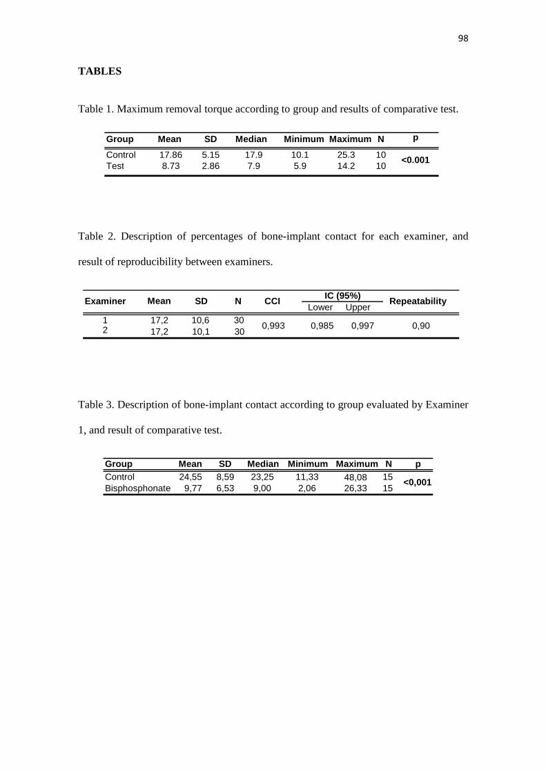
98
TABLES
Table 1. Maximum removal torque according to group and results of comparative test.
Table 2. Description of percentages of bone-implant contact for each examiner, and
result of reproducibility between examiners.
Table 3. Description of bone-implant contact according to group evaluated by Examiner
1, and result of comparative test.
Group Mean SD Median Minimum Maximum N p
Control 24,55 8,59 23,25 11,33 48,08 15Bisphosphonate 9,77 6,53 9,00 2,06 26,33 15
<0,001
Lower Upper 1 17,2 10,6 302 17,2 10,1 30
CCIIC (95%)
Repeatability
0,993 0,985 0,997 0,90
Examiner Mean SD N
Group Mean SD Median Minimum Maximum N p
Control 17.86 5.15 17.9 10.1 25.3 10 Test 8.73 2.86 7.9 5.9 14.2 10
<0.001
99
CAPTION TO ILLUSTRATIONS
Figure 1 - Local application of bisphosphonate gel and b) immediate insertion of
titanium implant.
Figure 2 – Positioning the implants in tibial cortical and medullary bone. Magnification
20X.
Figure 3 – Bone-implant interface of control group in cortical bone region,
demonstrating more intense bone activity close to the implant body, with presence of
recently formed bone (region with more intense coloring), voluminous osteocytes in
wide gaps (--›) and large and voluminous osteoblasts in contact with the implant
surface(+). Magnification 40X.
Figure 4 – Bone-implant interface of the test group in the cortical bone region,
demonstrating low bone activity located almost only in the region at the top of spirals,
and most cervical region of the implant, with presence of granular tissue filling the
space between spirals (+). Magnification: 40X.
Graph 1 - Bland-Altman Graph of measurements between examiners.

100
ANEXO E – Normas do Periódico International Journal of Oral and Maxillofacial
Implants.
Guide for Authors Would authors please note that the reference style for the journal has now changed. Please pay special attention to the guidelines unde r the heading "References" below Authors wishing to submit their work to the journal are urged to read this detailed guide for authors and comply with all the requirements, particularly those relating to manuscript length and format. This will speed up the reviewing process and reduce the time taken to publish a paper following acceptance. Online Submission Submission and peer-review of all papers is now conducted entirely online, increasing efficiency for editors, authors, and reviewers, and enhancing publication speed. Authors requiring further information on online submission are strongly encouraged to view the system, including a tutorial, at http://ees.elsevier.com/ijoms A comprehensive Author Support service is available to answer additional enquiries at [email protected]. Once a paper has been submitted, all subsequent correspondence between the Editorial Office ([email protected]) and the corresponding author will be by e-mail. Editorial Policy A paper is accepted for publication on the understanding that it has not been submitted simultaneously to another journal, has been read and approved by all authors, and that the work has not been published before. The Editors reserve the right to make editorial and literary corrections. Any opinions expressed or policies advocated do not necessarily reflect the opinions and policies of the Editors. Declarations Upon submission you will be required to complete and upload the declarations page (pdf version or word version) to declare funding, conflict of interest and to indicate whether ethical approval was sought. This information must also be inserted into your manuscript under the acknowledgements section with the headings below. Upon submission you will be required to complete and upload this form (pdf version or word version) to declare funding, conflict of interest, and to indicate whether ethical approval and patient consent were sought. Lastly you must confirm that all authors have agreed to the submission. PLEASE NOTE that all funding must be declared at fi rst submission, as the addition of funding at acceptance stage may invalidate the acce ptance of your manuscript. Authorship All authors should have made substantial contributions to all of the following: (1) the conception and design of the study, or acquisition of data, or analysis and interpretation of data (2) drafting the article or revising it critically for important intellectual content (3) final approval of the version to be submitted. Normally one or two, and no more than three, authors should appear on a short communication, technical note or interesting case/lesson learnt. Full length articles may contain as many authors as appropriate. Minor contributors and non-contributory clinicians who have allowed their patients to be used in the paper should be acknowledged at the end of the text and before the references. The corresponding author is responsible for ensuring that all authors are aware of their obligations. Before a paper is accepted all the authors of the p aper must sign the Confirmation of Authorship form. This form confirms that all the named authors agree to publication if the

101
paper is accepted and that each has had significant input into the paper. Please download the form and send it to the Editorial Office. (pdf version or word version) It is advisable that to prevent delay this form is submitted early in the editorial process. Acknowledgements All contributors who do not meet the criteria for authorship as defined above should be listed in an acknowledgements section. Examples of those who might be acknowledged include a person who provided purely technical help, writing assistance, or a department chair who provided only general support. Authors should disclose whether they had any writing assistance and identify the entity that paid for this assistance. Conflict of interest At the end of the main text, all authors must disclose any financial and personal relationships with other people or organisations that could inappropriately influence (bias) their work. Examples of potential conflicts of interest include employment, consultancies, stock ownership, honoraria, paid expert testimony, patent applications/registrations, and grants or other funding. If an author has no conflict of interest to declare, this should be stated. Role of the funding source All sources of funding should be declared as an acknowledgement at the end of the text. Authors should declare the role of study sponsors, if any, in the study design, in the collection, analysis and interpretation of data; in the writing of the manuscript; and in the decision to submit the manuscript for publication. If the study sponsors had no such involvement, the authors should so state. Open access This journal offers you the option of making your article freely available to all via the ScienceDirect platform. To prevent any conflict of interest, you can only make this choice after receiving notification that your article has been accepted for publication. The fee of $3,000 excludes taxes and other potential author fees such as color charges. In some cases, institutions and funding bodies have entered into agreement with Elsevier to meet these fees on behalf of their authors. Details of these agreements are available at http://www.elsevier.com/fundingbodies. Authors of accepted articles, who wish to take advantage of this option, should complete and submit the order form (available at http://www.elsevier.com/locate/openaccessform.pdf). Whatever access option you choose, you retain many rights as an author, including the right to post a revised personal version of your article on your own website. More information can be found here: http://www.elsevier.com/authorsrights. Ethics Any manuscript concerned with human subjects, medical records, or human tissue that is submitted to the International Journal of Oral and Maxillofacial Surgery should comply with the principles stated in the Declaration of Helsinki Ethical Principles for Medical Research Involving Human Subjects, adopted by the 18th World Medical Assembly, Helsinki, Finland, June 1964, and as amended most recently by the 64th World Medical Assembly, Fontaleza, Brazil, October 2013. The manuscript should contain a statement that the work has been approved by the appropriate Ethical Committee related to the institution(s) in which the work was performed, and that subjects gave informed consent to the work. The International Journal of Oral and Maxillofacial Surgery requires institutional Ethics Committee approval for all human studies. For retrospective studies of records either a statement of approval or a statement of exemption from the Committee is appropriate. This statement should be provided upon submission of the manuscript. Studies involving experiments with animals must state that their care was in accordance with institution guidelines.
102
Patient confidentiality Patients have a right to privacy. Therefore identifying information, including patients' images, names, initials, or hospital numbers, should not be included in videos, recordings, written descriptions, photographs, and pedigrees unless the information is essential for scientific purposes and you have obtained written informed consent for publication in print and electronic form from the patient (or parent, guardian or next of kin where applicable). If such consent is made subject to any conditions, The Editor and Publisher must be made aware of all such conditions. Written consents must be provided to the Editorial Office on request. Even where consent has been given, identifying details should be omitted if they are not essential. If identifying characteristics are altered to protect anonymity, such as in genetic pedigrees, authors should provide assurance that alterations do not distort scientific meaning and editors should so note. If consent for publication has not been obtained, personal details of patients included in any part of the paper and in any supplementary materials (including all illustrations and videos) must be removed before submission. Language Editing Services Papers will only be accepted when they are written in an acceptable standard of English. Authors, particularly those whose first language is not English, who require information about language editing and copyediting services pre- and post-submission should visit http://www.elsevier.com/wps/find/authorshome.authors/languagepolishing or contact [email protected] for more information. Please note, Elsevier neither endorses nor takes responsibility for any products, goods or services offered by outside vendors through our services or in any advertising. For more information please refer to our Terms and Conditions
http://www.elsevier.com/wps/find/termsconditions.cws_home/termsconditions. Article Types The following contributions will be accepted for publication. Please take careful note of the maximum length where applicable. Overlength articles will be returned to the authors without peer review: • editorials (commissioned by the editor) • clinical papers: no more than 5000 words and 30 references • research papers: no more than 6000 words and 40 references • review papers - no limit on length or number of references • technical notes (surgical techniques, new instruments, technical innovations) - no more than 2000 words, 10 references and 4 figures • case reports - no more than 2000 words, 10 references and 2 figures • book reviews • letters to the editor - please see detailed guidelines provided at the end of the main guide for authors • IAOMS announcements • general announcements. Please note: Case reports will be considered for publication only if they add new information to the existing body of knowledge or present new points of view on known diseases. All authors must have contributed to the paper, not necessarily the patient treatment. Technical notes and case reports are limited to a maximum of 4 authors, in exceptional circumstances, 5. Criteria for Publication Papers that will be considered for publication should be: • focused • based on a sound hypothesis and an adequate investigation method analysing a statistically relevant series, leading to relevant results that back the conclusion • well written in simple, scientific English grammar and style • presented with a clear message and containing new information that is relevant for the readership of the journal • Note the comment above relating to case reports. Following peer-review, authors are required to resubmit their revised paper within 3 months; in exceptional circumstances, this timeline may be extended at the editor's discretion.
103
Presentation of Manuscripts General points Papers should be submitted in journal style. Failure to do so will result in the paper being immediately returned to the author and may lead to significant delays in publication. Spelling may follow British or American usage, but not a mixture of the two. Papers should be double-spaced with a margin of at least 3 cm all round. Each line must be numbered. Format Observational or Case Cohort Studies, as well as Case Series must be presented in conformance with STROBE guidelines: www.strobe-statement.org Randomized Controlled Trials must be presented in conformance with CONSORT guidelines:
www.consort-statement.org Systematic Reviews and Meta-Analyses must be presented according to PRISMA guidelines:
www.prisma-statement.org Papers should be set out as follows, with each section beginning on a separate page:
• title page • abstract • text • acknowledgements • references • tables • captions to illustrations. Please note that the qualifications of the authors will not be included in the published paper and should not be listed anywhere on the manuscript. Title page The title page should give the following information: • title of the article • full name of each author • name and address of the department or institution to which the work should be attributed • name, address, telephone and fax numbers, and e-mail address of the author responsible for correspondence and to whom requests for offprints should be sent • sources of support in the form of grants • key words. If the title is longer than 40 characters (including spaces), a short title should be supplied for use in the running heads. Abstract 200 words maximum. Do not use subheadings or abbreviations; write as a continuous paragraph. Must contain all relevant information, including results and conclusion. Text Please ensure that the text of your paper conforms to the following structure: Introduction, Materials and Methods, Results, Discussion. There is no separate Conclusion section. There should be no mention of the institution where the work was carried out, especially in the Materials and Methods section. Introduction • Present first the nature and scope of the problem investigated • Review briefly the pertinent literature • State the rationale for the study • Explain the purpose in writing the paper • State the method of investigation and the reasons for the choice of a particular method •; Should be written in the present tense Materials and Methods • Give the full details, limit references • Should be written in the past tense • Include exact

104
technical specifications, quantities and generic names • Limit the number of subheadings, and use the same in the results section • Mention statistical method • Do not include results in this section Results • Do not describe methods • Present results in the past tense • Present representations rather than endlessly repetitive data • Use tables where appropriate, and do not repeat information in the text Discussion • Discuss - do not recapitulate results • Point out exceptions and lack of correlations. Do not try to cover up or 'fudge' data • Show how results agree/contrast with previous work • Discuss the implications of your findings • State your conclusions very clearly Headings: Headings enhance readability but should be appropriate to the nature of the paper. They should be kept to a minimum and may be removed by the Editors. Normally only two categories of headings should be used: major ones should be typed in capital letters; minor ones should be typed in lower case (with an initial capital letter) at the left hand margin. Quantitative analysis: If any statistical methods are used, the text should state the test or other analytical method applied, basic descriptive statistics, critical value obtained, degrees of freedom, and significance level, e.g. (ANOVA, F=2.34; df=3,46; P<0.001). If a computer data analysis was involved, the software package should be mentioned. Descriptive statistics may be presented in the form of a table, or included in the text. Abbreviations, symbols, and nomenclature: Only standardized terms, which have been generally accepted, should be used. Unfamiliar abbreviations must be defined when first used. For further details concerning abbreviations, see Baron DN, ed. Units, symbols, and abbreviations. A guide for biological and medical editors and authors, London, Royal Society of Medicine, 1988 (available from The Royal Society of Medicine Services, 1 Wimpole Street, London W1M 8AE, UK). The minus sign should be -. If a special designation for teeth is used, a note should explain the symbols. Scientific names of organisms should be binomials, the generic name only with a capital, and should be italicised in the typescript. Microorganisms should be named according to the latest edition of the Manual of Clinical Microbiology, American Society of Microbiology. Drugs: use only generic (non-proprietary) names in the text. Suppliers of drugs used may be named in the Acknowledgments section. Do not use 'he', 'his' etc where the sex of the person is unknown; say 'the patient' etc. Avoid inelegant alternatives such as 'he/she'. Patients should not be automatically designated as 'she', and doctors as 'he'. References The journal's reference style has changed. References should be numbered consecutively throughout the article, beginning with 1 for the first-cited reference. References should be listed at the end of the paper in the order in which they appear in the text (not listed alphabetically by author and numbered as previously). The accuracy of references is the responsibility of the author. References in the text should be numbered with superscript numerals inside punctuation: for example "Kenneth and Cohen14 showed..."; "each technique has advantages and disadvantages5-13." Citations in the text to papers with more than two authors should give the name of the first author followed by "et al."; for example: "Wang et al37 identified..." All references cited in the text must be included in the list of references at the end of the paper. Each reference listed must include the names of all authors. Please see section "Article Types" for guidance on the maximum number of reference for each type of article. Titles of journals should be abbreviated according to Index Medicus (see www.nlm.nih.gov.uk) .

105
When citing papers from monographs and books, give the author, title of chapter, editor of book, title of book, publisher, place and year of publication, first and last page numbers. Internet pages and online resources may be included within the text and should state as a minimum the author(s), title and full URL. The date of access should be supplied and all URLs should be checked again at proof stage. Examples: Journal article: Halsband ER, Hirshberg YA, Berg LI. Ketamine hydrochloride in outpatient oral surgery. J Oral Surg 1971: 29: 472-476. When citing a paper which has a Digital Object Identifier (DOI), use the following style: Toschka H, Feifel H. Aesthetic and functional results of harvesting radial forearm flap. Int J Oral Maxillofac Surg 2001: 30: 45-51. doi: 10.1054/ijom.2000.0005 Book/monograph: Costich ER, White RP. Fundamentals of oral surgery. Philadelphia: WB Saunders, 1971: 201-220. Book chapter: Hodge HC, Smith FA. Biological properties of inorganic fluorides. In: Simons JH, ed.: Fluorine chemistry. New York: Academic Press, 1965: 135. Internet resource: International Committee of Medical Journal Editors. Uniform requirements for manuscripts submitted to biomedical journals. http://www.icmje.org [Accessibility verified March 21, 2008] Tables Tables should be used only to clarify important points. Double documentation in the form of tables and figures is not acceptable. Tables should be numbered consecutively with Arabic numerals. They should be double spaced on separate pages and contain only horizontal rules. Do not submit tables as photographs. A short descriptive title should appear above each table, with any footnotes suitably identified below. Care must be taken to ensure that all units are included. Ensure that each table is cited in the text. Figures All illustrations (e.g. graphs, drawings or photographs) are considered to be figures, and should be numbered in sequence with Arabic numerals. Each figure should have a caption, typed double-spaced on a separate page and numbered correspondingly. The minimum resolution for electronically generated figures is 300 dpi. Line illustrations: All line illustrations should present a crisp black image on an even white background (127 x 178 mm (5 x 7 in), or no larger than 203 x 254 mm (8 x 10 in). The size of the lettering should be appropriate, taking into account the necessary size reduction. Photographs and radiographs: Photomicrographs should show magnification and details of any staining techniques used. The area(s) of interest must be clearly indicated w ith arrows or other symbols. Colour images are encouraged, but the decision whether an illustration is accepted for reproduction in colour in the printed journal lies with the editor-in-chief. Figures supplied in colour will appear in colour in the online version of the journal. Size of photographs: The final size of photographs will be: (a) single column width (53 mm), (b) double column width (110 mm), (c) full page width (170 mm). Photographs should ideally be submitted at the final reproduction size based on the above figures. Funding body agreements and policies Elsevier has established agreements and developed policies to allow authors who publish in Elsevier journals to comply with potential manuscript archiving requirements as specified as conditions of their grant awards. To learn more about existing agreements and policies please visit http://www.elsevier.com/fundingbodies Proofs One set of page proofs in PDF format will be sent by e-mail to the corresponding author, which they are requested to correct and return within 48 hours. Elsevier now sends PDF proofs which can be annotated; for this you will need to download Adobe Reader version 7 available free

106
from http://www.adobe.com/products/acrobat/readstep2.html. Instructions on how to annotate PDF files will accompany the proofs. The exact system requirements are given at the Adobe site: http://www.adobe.com/products/acrobat/acrrsystemreqs.html#70win. If you do not wish to use the PDF annotations function, you may list the corrections (including replies to the Query Form) and return to Elsevier in an e-mail. Please list your corrections quoting line number. If, for any reason, this is not possible, then mark the corrections and any other comments (including replies to the Query Form) on a printout of your proof and return by fax, or scan the pages and e-mail, or by post. Please use this proof only for checking the typesetting, editing, completeness and correctness of the text, tables and figures. Significant changes to the article as accepted for publication will only be considered at this stage with permission from the Editor. We will do everything possible to get your article published quickly and accurately. Therefore, it is important to ensure that all of your corrections are sent back to us in one communication: please check carefully before replying, as inclusion of any subsequent corrections cannot be guaranteed. Proofreading is solely your responsibility. Note that Elsevier may proceed with the publication of your article if no response is received. Offprints The corresponding authorwill be provided , at no cost, with a PDF file of the article via e-mail. The PDF file is a watermarked version of the published article and includes a cover sheet with the journal cover image and a disclaimer outlining the terms and conditions of use. Additional paper offprints can be ordered by the authors. An order form with prices will be sent to the corresponding author. Accepted Articles For the facility to track accepted articles and set email alerts to inform you of when an article's status has changed, visit: http://authors.elsevier.com/TrackPaper.html There are also detailed artwork guidelines, copyright information, frequently asked questions and more. Contact details for questions arising after acceptance of an article, especially those related to proofs, are provided after registration of an article for publication. Instructions for Letters to the Editor The IJOMS welcomes Letters to the Editor. To facilitate submission of the highest quality of Letters to the Editor, the following guidelines should be followed: 1. Letters are meant to be focus pieces and, therefore, are limited to no more than 600 words, 6 references and a maximum of 2 figures. One reference should include a reference to the IJOMS article being addressed. 2. It is recommended that you limit your letter to one or two important and critical points to which you wish to provide a clear and precise discussion regarding the previously published article. 3. One should support all assertion by peer review literature which should be a primary research or large clinical studies rather than a case report. 4. Please include any financial disclosures at the end of the letter. This would include the potential conflicts of interest not just related to the specific content of your letter but also the content of the IJOMS article and other related areas. 5. Please recognize that letters that are essentially in agreement with the author's findings and offer no additional insights provide little new information for publication. Likewise, letters that highlight the writer's own research or are otherwise self promotional will receive a low publication priority. 6. There may be a need for additional editing. Should editing be required the letter will be sent back to the author for final approval of the edited version. 7. It is important to use civil and professional discourse. It is not advisable that one adopt a tone that may be misconstrued to be in anyway insulting. 8. Finally, it is not advisable to provide a letter that is anecdotal. While personal experiences can have great value in patient care, it is generally not strong evidence to be placed in a letter to the editor.

107
ANEXO F – Submissão do artigo científico.