Embed Size (px)
Citation preview

Rev. Inst. Med. top. São Paulo23(2) :61-67, ntarço-abril, 1981
THE INFLUENCE OF GYTOCHALASIN B ON THE INTERACTION OF T. CRUZIAND MOUSE PERITONEAL MACROPHAGES (")
tr. EBERT (1) ¿nd Hetene Srntos BARBOSA (p)
SUMMARY
Stimulated mouse peritoneal macrophages were cultured in M 19g containing20% f.etal bovine serum (FBS). Macrophage cultures weïe pre-incubated with cy-tochalasin B in final concentrations of l, 5 and. 10 pg,/ml (dissolved in DMSO 1
mg/ml) for two hours before the addition of r. cruzi. strains of r. cruzi withsignificant differences in the proportion of trypomastigotes were added in theratio 1:2 (peritoneal cell: parasite). The macrophage monolayers were fixed andstained at various time intervals. Both strains rvere found to attach to macropha-ges independent of the presence or absence of cytochalasin B. The uptake ofT. cruzi, however, was almost completely inhibited in macrophages treated r¡¡ithCytochalasin B at concentrations of. b pg/ml and. 10 pg/rrrl. Cultures exposed toI pg Cytochalasin B,/ml did not blook the ingestion but the rate of infection ofmacrophages was significantly reduced. In comparison macrophages incubated ina M 199 + 20% FBS control culture, with DMSO, did not change the uptake ofT. cruzi. No significant differences between the strains were found. Electron mi-croscopic examinations of untreated macrophage,s showed the parasites eitherinside phagocytic vacuoles or attached to the membrane and surround.ed by fin-ger-like pseudopodia. The results suggest that epimastigotes and trypomastigotesfrom a culture of r. cruzi enter macrophages by a process of phagocytosis.
UDC s93. 161.13616.937 .3
Trypanosoma ctuzi the agent of Chagas, di-sease is a parasite infecting a variety of mam-malian cell types including those of the mono-nuclear phagocytÍc system. The interaction ofT. cruzi (blood stream and culture forms) withtissue culturos and mouse peritoneal macropha-ges has been studied in several investigations(ALCANTARA & BRENEft,:; DVORAK & SCH-MUNIS 6; SOOKSRI & INOKI ts; TANOWITZ eral. r0). It seems that T. cruzi þlood stream formspenetrate actively into both non-phagocytic andphagocytic cells. On the other hand there areconflicting reports on the mechanism bywhich culture forms of T. cruzi gain access
INTRODUCTION
(*) This study was supported by BMZ, Research program - Fiocruz, Rio de Jâneiro, BNI, Hamburg
(1) Dr. rel. nat. - Centro de Microscopia Eletrônica, Fundação Oswaldo Ctu, Rio d.e Janeiro, BrazilPresent address: Tropical Institute Hamburg, Dept. of protozootogy, Getmany
(2) Pesquisadora Á,uxi1iar - Centro de Microscopiâ Eletlônica, Fundação Oswaldo Cruz, Rio de Janeiro, Brazil
to macrophages (ALEXANDER +; KIPNIS eraI. to' NOGUEIRA & COHN t2) treated with Cy-tochalasin B, a drug which inhibits phagocytosisby macrophages (ALLISON et al.2).
This paper reports the effect of Cytochala"sin B on the uptake, by mouse peritoneal ma-crophages, of culture forms of T. cruzi strains,having different proportions of trypomastigote,s.
MATERIALS AND METHODS
Parasites - The following T. cruzi strainswere used. The Rato strain. isolated from a
61

EBER'T, F. & BAR'BOSÄ, H. S, - The influence of Cytochalasin B on the interaction of T. cruzi and. mouse Þeritonealmacrophages, Rev, Iroú. Med. trop. São Paulo 23:61.67, 1981.
wild rat in Ribeirão Preto, SP., Brazil (R,IBEI-RO & BARRETTO14), was suppliedto usbyDr.Barretto in 19?8. The OPS-22 strain, isolated bymouse inoculation of feces from positive Pans.trongylus geniculatus in Cojedes, Venezuela,197?, was kindly provided by Prof. Mühlpfordt,Tropical Institute, Hamburg. Both strains weremaintained in NNN-medium at 2?"C at two\¡úeekly intervals and collected in the stationa-ry phase. The Rato and OPS-22 strain contained78% and 41% epimastigotes, respectively.
Macrophage culture - Albino mice wereinoculated intraperitoneally with 2 ml of 3.7o/oBrain-Heartinfusion (Difco). Three days latermacrophages were collected by peritoneal lava-ge with 2 ml of saline. The cells were centri-fuged for 5 minutes at 10009 and suspended inmedium 199 with 30 mM Hepes containing 20%fetal bovine serum (FBS), 200 pg/ml Strepto-rr¡ycin and 200 U,zml Penicillin. The concentra-tion was adjusted to 2x10s cells/l,5 ml. The cellswere then distributed into 9,5 x 3b mm Leightontubes with coverslips, incubated. for four hoursat 3?'C and washed twice with M 199 to removethe non-adherent cells. The adherent cells wereincubated overnight until used in experiments.
Cytochalasin B (Serva, Germany) was dis-solved in Dimethyl sulphoxide (DMSO) L mglmland diluted in M 199 + 20% FBS to final con-centrations of 1,5 and l0 pg/ml.
The macrophage cultures were pre-treatedwith 1,5 and 10 pg cytochalasin B for two hours.T. cruzi was then added at a ratio of T. cruzito peritoneal cells of 2:1. Control cultures con-tained M i99 * 20Vo FBS and M 199 + Z0%FBS and DMSO G,5 and L0 pt/mD.
Freparation of cells for microscopy
a) Light microscopy - The coverslips werefixed in 2% OsO,, in 0,1 M Cacodylate buffer, pH7,2, f.or a few seconds and. then stained in Giem_sa solution; b) Elecúron microscopy - Macro_phages were cultured in 100 ml flasks. Half anhour after addition of T, cruzi the cells werefixed by the method of HIRSCH & FEDORKO e.
The process was done in situ. Then, the celtswere gently scraped off with a rubber police_man. The procedure of embedding has been des_cribed previously (EBERT et al.1). Micro_graphs were taken with a Zeiss EM 108.
62
Statistical Analysis
The median values of intracellular parasiteswere calculated by counting of 400 macropha-ges,/time interval in each of the four experi-ments. The U-test of rffilcoxon, Mann and 1ühit-ney (SACHS 13) was used for testing the signifi-cance between the groups. A p-level of 0,005was chosen to indicate the statistical signific-ance.
RESULTS
Culture forms of T. cruzi of both strarnswere found to attach to the membrane of themacrophages a few minutes after the infectionindependent of the presence or absence of Cy-tochalasin B (Figs. 1 and 2). There was no pre-fered position of contact of T. cruzi to the pha-gocytic cells, either the flagellum, the body orthe posterior end of the parasite were seen incontact with the macrophages.
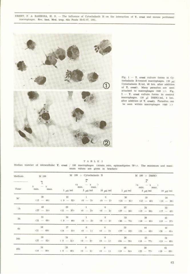
The engulfment of T. cruzi, ho.wever, wasalmost completely inhibited by the macrophagecultures treated with Cytochalasin B in theconcentrations of 5 pg/ml and I0 pg/ml (Table rand II). Cultures exposed to L pg/ml of Cyto-chalasin B did no block the ingestion of T. cruzito the extent seen in the higher concentrationsbut the rate of infection of macrophages wassignificantþ reduced.
In comparison with macrophages incubatedin M 199 + 20% FBS control cultures withDMSO did not change significantty the uptakeof T. cruzi.
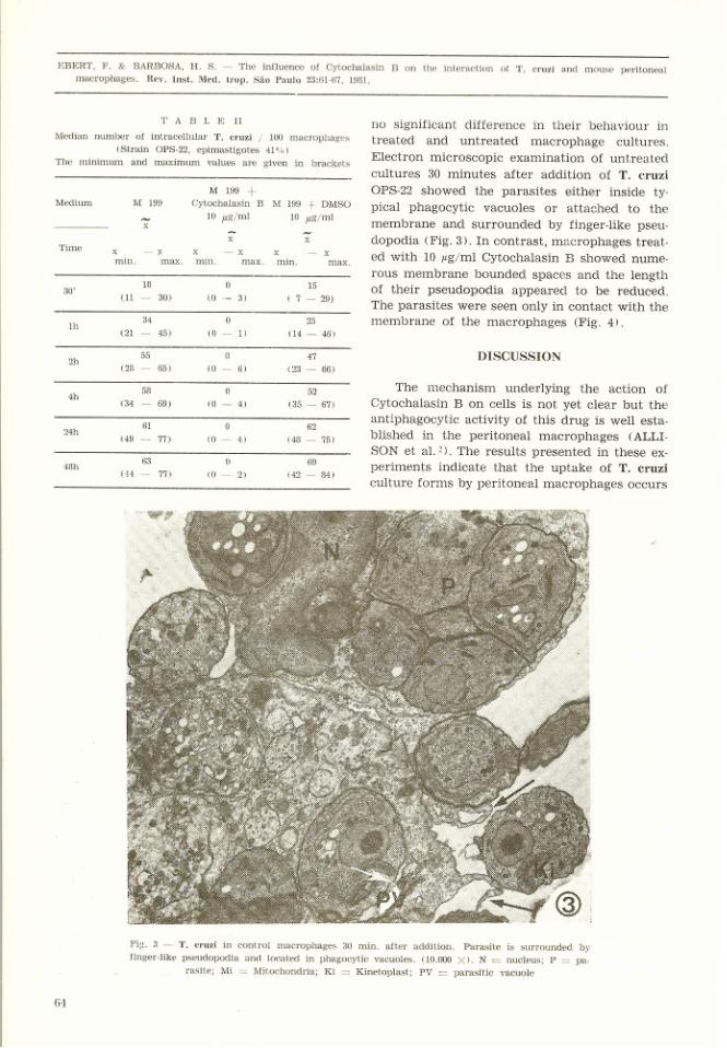
In all experiments it was found that the ma_crophage cultures treated with the higher con_centrations of Cytochalasin B (b pg,/ml and 10þC/ml) had a very low number of intracellularparasites, which were observed exclusively inmacrophages localized on the periphery of thestained coverslips. Furthermore, in these cultu_res the macrophages retracted and became cir-cular (Fig. 1). The effect of Cytochalasin Bappeared to be completely reversible. Cellsappeared normal within 20 - B0 minutes afterwashing with M 199 and replacement of growthmedium. The phagocytosis was continued andparasites were seen to be intracellular.
The statistical anaiysis of the resulüs withthe two strains tested indicated that there was

EBERT, F. & BARBOSA, H. S. - The influence of Cytochalasin B on the inte¡action of T. crui and mouse peritoneal
macrophages. Rev, Inst. Med, trop. São Paulo ?3:61-6?, 1981.
During the initial phase of interaction ofT. cruzi rilith peritoneal macrophages the para-sites were seen to attach to the membrane ofthe macrophages either by means of the flagel-Ium, the body or the posterior end correspond-ing to the findings of NOGUEIRA & COHN 12.
By direct observations, however, DVORAK &SCHMUNIS found that epimastigotes enteredinto macrophages with the flagellar end whe-reas trypomastigotes entered with the poste-rior end first. Similar "conflicting" results dur-ing the initial phase of studying host-parasiteinteraction are described for Leishmania (see
ALEXANDER4, EBERT et al.8).
It has been noted that T. cruzi as well asLeishmania show the ability to enter a cell ac-tively depending on the cell type used. In non-phagocytic cells it could be shown that T. cruziand Leishmania brasiliensis appear to penetratethese cells actively (CHANGS, TANOWITZ etal. r0). Hor,vever, it is not understood why stu-dies on T. cruzi macrophage interactions givesuch controversiai results and consequently se-
veral interpretations. If these discrepancies arebased on virulence, different strains should beexamined in further experiments.
RESI]MO
A influência de cytochalasina B na interação deT. cruzi e macrófagos peritoneais de
camundongos
Macrófagos peritoneais de camundongos fo-ram cultivados em Meio 199 contendo 20Vo desoro fetal bovino (SFB). Culturas de macrófa-gos foram pré-inculoadas com Cytochalasina Bnas concentrações finais de 1, 5 e 10 pglml (dis-solvida em 1 mg,/mt DMSO) 2 horas antes daadição de T. cruzi. Cepas de T. cruzi com dife-renças significantes na proporção de tripomas-tigotas foram adicionadas na relação 1:2 (célu-la peritoneal: parasita). As culturas de macró-fagos foram fixadas e coradas em vários inter-valos de tempo. Constatou-se que ambas as ce-pas aderiram aos macrófagois independente dapresença ou ausência da Cytochalasina B. O en-globamento de T. cruzi, porém, foi quase com-pletamente inibido em macrófagos tratados comCytochalasina B nas concentrações de 5 pglmle 1,0 pg/ml. Culturas expostas a 1 pglml Cyto-chalasina B,/mI não bloquearam a ingestáo, maso grau da infecção dos macrófagos foi signifi-cantemente reduzido. Em comparação aos ma-
66
crófagos incubados em M t99 + 20% SFB o¡controles com DMSO náo alterarâm o engloba'mento de T. cruzi. Exames em microscópio ele'
trônico de macrófagos náo tratados mostraramos parasitas dentro de vacúolos fagocíticos, ou
aderidos à membranas, e cercado,s por pseudó-podos em forma de dedo. Os resultados indi-cam que epimastigotas e tripomastigotas de
culturas de T. cruzi, entram em macrófagosatravés do processo de fagocitose.
REFERENCES
1. AKIYAMA, H. J. & MoQUILLEN, N. K. - fnteractioiland transformation of Leishmania donovaui within invitro cultured cells. Am. J. Trop, Med. Hyg. ?1: 8?3'
879, 1972.
2. ALLISON, A. C.; DAVIES, P. & DE PETR'IS, S. -RoIe of contractile microfilaments in macrophage mo-
vement ând endocytosis. N¿ture (London) 232: 153-155,
1971.
3, ALCANTARA, A. & BRENER', Z' - The in vitro inte-
raction of Trypanosoma cruzi bloodstream forms and
mouse peritoneal mâcrophages. Acta Trop. 35: 209-219'
1978.
4. ALEXANDER, J. - Effect of antiphagocytic ag¿rnt
cytochalasin B on macrophage invasion by Leishmania
mexicana promastigotes and Trypanosoma cruzi epi'
mastisotes. J. Protozool. 22: 237-240, 1975'
CHANG, K. P. - Leishmania infection of human skin
fibroblâsts in vitro, Absence of phagolysosomal fusion
after induced phagocytosis of promastigotes, and theilintracellular transformation. Am. J. Trop. Med. Hyg.2?: 1084-1096, 19?8.
DVORAK, A. J. & SCHMUNIS, G A - Trypanosomacruzi: Interaction with mouse pelitoneal macrophages.
Exp. Parasitol. 32: 289-300, 19?2.
EBERT, F.; ENR,IQUEZ, G, L. & MÜHLPFOR'DT, H.
- Electron microscopic studies of phagocytosis ofLeishmania donovani by hamster peritoneal macropha'ges and its lysosomal activity in vivo. Behring Inst.Mitt. 60: 65-74, 1976.
EBERT, F.; BUSE, E. & MÍJHLPFORDT, H. - Invitro light and electron microscopic studies of diffe-rent virulent promastigotes of Leishmania donovani inhamster peritoneal macrophages. Z. Parasitenk. 59: 31-
41, 79',19.
HIRSCH, J. G. & FEDOnKO, M. E. - Ultrastructureof human Leukocytes after simultaneous fixation wilbglutaraldehyde and osmium tetroxide and "postfixation"in uranyl acetate. J. Cell. Biol. 38: 615-627, 1968.
KIPNIS, T. L.; CALICH, V. L. c. & DIAS DA SILVA,W. - .A.ctive entry of bloodstream forms of Trypano.solna cruzi into macrophages. Parasitology l8: 98-99,
19?9.

EBERT' F' & BÁR'BosA' H' s' - The Ínfluence of cytochalasin B on the interaction of T, cruzi "nu
*ou.ulilãmacrophages. Rev. Inst, Med, trop. São paulo p3:61_6?, f981.
11 MILDER, R,.; KLOETZEL, J. & DEANE, M. P. -Observation on the intetaction of peritoneal macro-phages with Trypanosoma cruzi. I _ Initial phase ofrelationship with bloodstream and culture forms invitro. Rev. Insú. Med. trop. São paulo li: Ag6_892, 19?3.
NOGUEIRA, N. & COHN, Z. - Trypanosoma cruzi:Mechanism of entry and intracellular fate in mamma,lian cells. J. Exp. Med. t4}t I4O2-L4ZO, tg76.
SACHS, L. - Angewaudte Statistik. 5th ed. Bertin.New York, Springer Verlag, 19?8.
RIBEIRO, R,. Ð. & BARRETTO, M. p. _ Estudosobre reservató¡ios e vectores silvestres de T. cruzi.LVIII - Infecção nâtural do rato Akodon lasiotis (Lund.
12
1841) pelo T. cruzi. Rev. Inst. Med, trop. São paulo17: 247-252, 1975.
SOOKSRI, v. & INOKI, S. _ Electron microscoDicstudies on penetration and development of Trypanolo-ma cruzi in HeLa cells. Biken J. 15: 1?9-191, 19?2.
TANOWITZ, H.; WTTTNER,, M.; KRESS, y. & BLOOM,B. - Studies of in vitro infection by Trypanosgma cr¡_zi. I - Ultrastructural studies on the invasion of ma-crophages and L-cells. Am. J. Trop. Med. Hyg. ?,1:25-33.1975.
15
16
Recebido pâra publicação em 1g13/1980.
67


![FOUCAULT; CARUSO, P. Quem é você, professor Foucault (Entrevista, 1969, D.E. 1 [fr])](https://img.document.onl/doc/110x75/55cf9cdc550346d033ab509c/foucault-caruso-p-quem-e-voce-professor-foucault-entrevista-1969-de.jpg)