Embed Size (px)
Citation preview
S
GvAHP2
JSa
b
c
d
e
f
a
A
R
A
A
I
TMtcT
h1o
rev bras hematol hemoter. 2 0 1 6;3 8(2):147–157
www.rbhh.org
Revista Brasileira de Hematologia e HemoterapiaBrazilian Journal of Hematology and Hemotherapy
pecial article
uidelines on neonatal screening and painfulaso-occlusive crisis in sickle cell disease:ssociacão Brasileira de Hematologia,emoterapia e Terapia Celularroject guidelines: Associacão Médica Brasileira –016
osefina Aparecida Pellegrini Bragaa, Mônica Pinheiro de Almeida Veríssimob,ara Teresinha Olalla Saadc, Rodolfo Delfini Cancadod,e, Sandra Regina Loggettof,∗
Escola Paulista de Medicina, Universidade Federal de São Paulo (Unifesp), São Paulo, SP, BrazilCentro Infantil Boldrini, Campinas, SP, BrazilFaculdade de Ciências Médicas, Universidade Estadual de Campinas (Unicamp), Campinas, SP, BrazilFaculdade de Ciências Médicas da Santa Casa de São Paulo (FCMSCSP), São Paulo, SP, BrazilHospital Samaritano, São Paulo, SP, BrazilCentro de Hematologia de São Paulo (CHSP), São Paulo, SP, Brazil
r t i c l e i n f o
rticle history:
eceived 12 February 2016
ccepted 4 April 2016
vailable online 8 April 2016
ntroduction
he guidelines project is a joint initiative of the Associacão
édica Brasileira and the Conselho Federal de Medicina. It aimso bring together information in medicine to standardizeonduct in order to help decision-making during treatment.he data contained in this article were prepared by and
∗ Corresponding author at: Avenida Brigadeiro Luís Antônio, 2533, JardimE-mail address: [email protected] (S.R. Loggetto).
ttp://dx.doi.org/10.1016/j.bjhh.2016.04.001516-8484/© 2016 Associacão Brasileira de Hematologia, Hemoterapiapen access article under the CC BY-NC-ND license (http://creativecom
are recommended by the Associacão Brasileira de Hematolo-gia, Hemoterapia e Terapia Celular (ABHH). Even so, all possiblemedical approaches should be evaluated by the physicianresponsible for treatment depending on the patient’s charac-teristics and clinical status.
This article presents the guidelines on neonatal screeningand painful vaso-occlusive crisis in sickle cell disease (SCD).
América, 01401-000 São Paulo, SP, Brazil.
e Terapia Celular. Published by Elsevier Editora Ltda. This is anmons.org/licenses/by-nc-nd/4.0/).
oter.
In France, SCD prevalence tends to be higher in the pop-ulation of infants born to parents from Sub-Saharan Africa,
148 rev bras hematol hem
Description of the method used to gatherevidence
These Guidelines were prepared by elaborating eight clinicallyrelevant questions, two related to neonatal screening for SCDand six related to painful vaso-occlusive crisis in SCD. Thequestions were structured using the Patient/Problem, Inter-vention, Comparison and Outcome (PICO) system (AppendixA), allowing the generation of evidence search strategies inthe key scientific databases (MEDLINE PubMed, Lilacs, Sci-ELO, Embase, Cochrane Library, Premedline via OVID). Thedata recovered were critically analyzed using discriminatoryinstruments (scores) according to the type of evidence – JADADfor randomized clinical trials and the Newcastle Ottawa scalefor non-randomized studies. After identifying studies thatpotentially substantiate recommendations, the level of evi-dence and degree of recommendation were calculated usingthe Oxford Classification.1
Degree of recommendation and level ofevidence
A: Major experimental and observational studiesB: Minor experimental and observational studiesC: Case reports (non-controlled studies)D: Opinion without critical evaluation based on consensus,physiological studies or animal models
Background
SCD is a group of inherited diseases in which the synthesis ofhemoglobin (Hb) is impaired because of a mutation in the betaglobin chain of the Hb gene on chromosome 16. This muta-tion leads to the substitution of a glutamic acid for valine atposition 6 of the beta chain, resulting in the production of HbS whose expression causes sickling of red blood cells, poly-merizing Hb with resulting vaso-occlusion, pain and chronicorgan damage.1–3 (D) Hb S is the most common abnormal Hbin Brazil.2 (D)
Sickle cell anemia occurs when the patient is homozygousfor the Hb S gene (Hb SS). Moreover, Hb S can be associ-ated with other abnormal Hb, such as Hb S/beta-thalassemia,Hb SC, Hb SD, persistence of Hb fetal (Hb F) with Hb S,among others. The term SCD defines both sickle cell ane-mia and these associations. The combination of the Hb Sgene with normal Hb (Hb A) characterizes the sickle cell trait(Hb AS).1–3 (D)
The diagnosis of SCD, a condition with high morbidity andmortality rates, is made at birth with neonatal screening.The pathophysiology is complex and evolves with acute andchronic complications that affect different organs and sys-tems.
Because of the complexity of SCD, many questions wereasked during the development of these guidelines and so it
was decided to present them in three parts. The first partdiscusses diagnosis by neonatal screening and aspects ofvaso-occlusive crisis, the second part answers questions aboutsplenic sequestration and the central nervous system from2 0 1 6;3 8(2):147–157
diagnosis to treatment and the third part deals with theprevention of infections, diagnosis and treatment of fever, pri-apism and bone marrow transplantation.
Objective
The aim of the first part of these guidelines is the approach todiagnosis by neonatal screening and subsequent confirmationof SCD and questions related to the diagnosis and treatmentof the vaso-occlusive crisis.
What is the prevalence of sickle cell diseaseand how are the results of neonatal screeningfor hemoglobinopathies interpreted?
P: All newborn babiesI: Results of neonatal screeningC:O: Interpretation of the results
The laboratory techniques used to identify hemoglobinin the Newborn Screening Program are high-performanceliquid chromatography (HPLC) and isoelectric focusing (IEF)because these tests can quantify small amounts of Hb; themain Hb in the newborn is Hb F.4–16 (A),17 (B) Several dis-eases are investigated during neonatal screening, includingSCD, phenylketonuria and congenital hypothyroidism.4–8,12–16
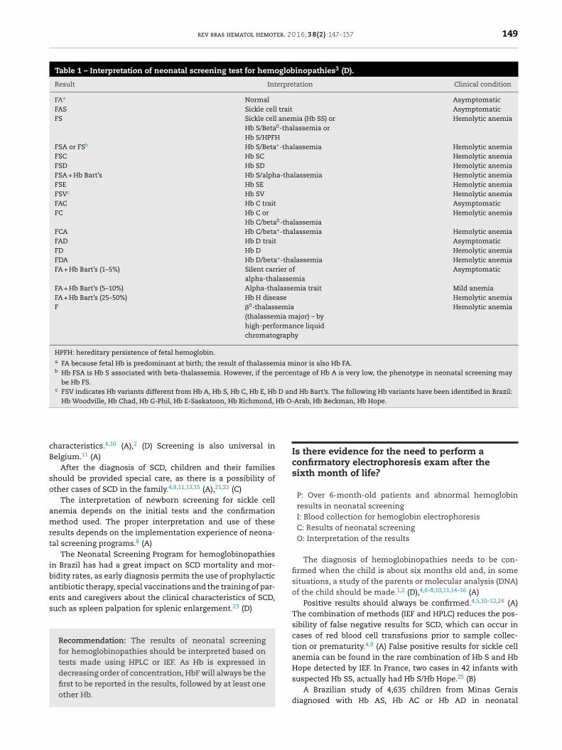
(A) The Hb with the highest concentration is shownfirst in the neonatal screening results and so Hb F willalways be followed by the other hemoglobins.4–6,14 (A)1–3 (D)Table 1 shows how to interpret neonatal screening results.The most common hemoglobinopathies in Brazil are HbFAS, Hb FS, Hb FSA, Hb FSC, Hb FSD and Hb FSA + HbBart’s.
The prevalence of Hb S in Brazil is from 1.2 to 10.9% depend-ing on the region of the country,5,6,13–16 (A),17–19 (B) while theprevalence of Hb FS (sickle cell anemia) ranges from <0.1to 0.2%6,7,13–16 (A)17,19 (B) and Hb FSC ranges from <0.1 to0.9%.5–7,14,15 (A)17,18 (B) It is noteworthy that the states with thehighest number of cases of SCD are Bahia and Rio de Janeiro,while Paraná and Rio Grande do Sul have the lowest rates.The prevalence of Hb C is from 0.15 to 7.4%.5–7,14,15 (A)17–19
(B). Hb S/beta-thalassemia has been identified in the statesof São Paulo, Minas Gerais, Paraná and Rio Grande do Sul, allwith prevalences <0.1%.5,6,14,16 (A) In the city of Ribeirão Preto,São Paulo the incidence for sickle cell anemia was shown tobe 1:7,358 and for Hb SC disease 1:9,365.7 (A) In Rio Grandedo Sul the incidence of SCD (Hb SS, Hb SC, Hb SD and HbS/beta-thalassemia) was 1:9,1206 (A) and in Minas Gerais theincidences of Hb FS and Hb FSC were 1:2,800 and 1:3,450,respectively.14 (A)
the Mediterranean, the Arabian Peninsula, French islandsand India compared to the general population (3:10,000 vs.1:10,000).20 (B) In Brazil, the USA and the United Kingdom,universal screening is considered ideal due to the population
rev bras hematol hemoter. 2 0 1 6;3 8(2):147–157 149
Table 1 – Interpretation of neonatal screening test for hemoglobinopathies3 (D).
Result Interpretation Clinical condition
FAa Normal AsymptomaticFAS Sickle cell trait AsymptomaticFS Sickle cell anemia (Hb SS) or
Hb S/Beta0-thalassemia orHb S/HPFH
Hemolytic anemia
FSA or FSb Hb S/Beta+-thalassemia Hemolytic anemiaFSC Hb SC Hemolytic anemiaFSD Hb SD Hemolytic anemiaFSA + Hb Bart’s Hb S/alpha-thalassemia Hemolytic anemiaFSE Hb SE Hemolytic anemiaFSVc Hb SV Hemolytic anemiaFAC Hb C trait AsymptomaticFC Hb C or
Hb C/beta0-thalassemiaHemolytic anemia
FCA Hb C/beta+-thalassemia Hemolytic anemiaFAD Hb D trait AsymptomaticFD Hb D Hemolytic anemiaFDA Hb D/beta+-thalassemia Hemolytic anemiaFA + Hb Bart’s (1–5%) Silent carrier of
alpha-thalassemiaAsymptomatic
FA + Hb Bart’s (5–10%) Alpha-thalassemia trait Mild anemiaFA + Hb Bart’s (25–50%) Hb H disease Hemolytic anemiaF �0-thalassemia
(thalassemia major) – byhigh-performance liquidchromatography
Hemolytic anemia
HPFH: hereditary persistence of fetal hemoglobin.a FA because fetal Hb is predominant at birth; the result of thalassemia minor is also Hb FA.b Hb FSA is Hb S associated with beta-thalassemia. However, if the percentage of Hb A is very low, the phenotype in neonatal screening may
be Hb FS.c FSV indicates Hb variants different from Hb A, Hb S, Hb C, Hb E, Hb D and Hb Bart’s. The following Hb variants have been identified in Brazil:
Hb Woodville, Hb Chad, Hb G-Phil, Hb E-Saskatoon, Hb Richmond, Hb O-Arab, Hb Beckman, Hb Hope.
cB
so
amrt
ibaes
haracteristics.4,10 (A),2 (D) Screening is also universal inelgium.11 (A)
After the diagnosis of SCD, children and their familieshould be provided special care, as there is a possibility ofther cases of SCD in the family.4,8,11,13,15 (A),21,22 (C)
The interpretation of newborn screening for sickle cellnemia depends on the initial tests and the confirmationethod used. The proper interpretation and use of these
esults depends on the implementation experience of neona-al screening programs.8 (A)
The Neonatal Screening Program for hemoglobinopathiesn Brazil has had a great impact on SCD mortality and mor-idity rates, as early diagnosis permits the use of prophylacticntibiotic therapy, special vaccinations and the training of par-nts and caregivers about the clinical characteristics of SCD,uch as spleen palpation for splenic enlargement.23 (D)
Recommendation: The results of neonatal screeningfor hemoglobinopathies should be interpreted based ontests made using HPLC or IEF. As Hb is expressed in
decreasing order of concentration, HbF will always be thefirst to be reported in the results, followed by at least oneother Hb.Is there evidence for the need to perform aconfirmatory electrophoresis exam after thesixth month of life?
P: Over 6-month-old patients and abnormal hemoglobinresults in neonatal screeningI: Blood collection for hemoglobin electrophoresisC: Results of neonatal screeningO: Interpretation of the results
The diagnosis of hemoglobinopathies needs to be con-firmed when the child is about six months old and, in somesituations, a study of the parents or molecular analysis (DNA)of the child should be made.1,2 (D),4,6–8,10,11,14–16 (A)
Positive results should always be confirmed.4,5,10–12,24 (A)The combination of methods (IEF and HPLC) reduces the pos-sibility of false negative results for SCD, which can occur incases of red blood cell transfusions prior to sample collec-tion or prematurity.4,9 (A) False positive results for sickle cellanemia can be found in the rare combination of Hb S and HbHope detected by IEF. In France, two cases in 42 infants with
suspected Hb SS, actually had Hb S/Hb Hope.25 (B)A Brazilian study of 4,635 children from Minas Geraisdiagnosed with Hb AS, Hb AC or Hb AD in neonatal

oter.
150 rev bras hematol hemscreening showed 0.6% of discordant results between theinitial screening and an IEF exam after six months of life.Seven cases had had blood transfusions before blood collec-tion, seven cases had problems in blood collection or in thetranscription of the exam results, there was difficulty to dif-ferentiate between Hb S and Hb D in eight cases and the reasonwas not identified in five cases.14 (A)
Recommendations: All children identified as havinghemoglobinopathies during neonatal screening shouldbe retested by hemoglobin electrophoresis after sixmonths of life.
Is there evidence on the factors that cause avaso-occlusive crisis?
P: Patients between 0 and 18 years old with sickle cell anemiaand painful crisisI: Fever, dehydration, infection, metabolic disorder, exposureto extreme cold and heat, alcoholism, osteomyelitis, illegaldrugs (marijuana, cocaine)C: Without symptomsO: Triggers of vaso-occlusive crisis
Several factors have been associated with the vaso-occlusive crisis in patients with SCD, such as infections,climate change, psychological factors, altitude, acidosis, sleepapnea, stress, dehydration, hypoxia and physical exhaustion.However, in most cases the triggering factor is not identified.26
(D)Pain crises in these patients have variable intensities
and frequencies, being higher in winter. Higher tempera-tures during winter were associated with less pain intensityand frequency.27 (B) However, another study did not confirmthe association between changes in temperature and painfrequency.28 (B) Moreover, the hospitalization rate for pain cri-sis increases with the intensity of wind and air humidity.29,30
(B)Some factors increase the likelihood of hospitalization for
painful crisis in patients with SCD. In a multivariate analysisof factors associated with crisis, increased risk of hospitaliza-tion was observed in subjects with Hb SS (hazard ratio: 3.1), inthose exposed to smoking (hazard ratio: 1.9) and those witha history of asthma (hazard ratio: 1.3).31 (B) Another studydemonstrated the importance of respiratory symptoms andasthma in pain crisis.32 (B) An assessment of nocturnal O2
saturation and painful crisis in 90 patients with sickle cell ane-mia concluded that a nocturnal O2 saturation <90% (p-value<0.0001), Hb below 8.8 g/dL (p-value <0.01) and a hematocrit
below 28% (p-value <0.0012) are associated with the onset ofsymptoms.33 (B)Other clinical situations are associated with pain crisis inpatients with SCD, including high blood viscosity34 (B) andmenstruation (61.5% of cases of crisis in women occur duringmenstruation).35 (C)
2 0 1 6;3 8(2):147–157
Recommendation: Factors associated with pain cri-sis can be environmental, such as temperature, windand humidity or clinical such as respiratory diseases,increased blood viscosity, anemia and menstruation.Smoking increases the risk of hospitalization and someinfections increase the risk of having a painful vaso-occlusive crisis.
Is there evidence that the use of painassessment scales is a good method to monitorpain related to vaso-occlusive crisis?
P: Patients between 0 and 18 years old with sickle cell anemiaand painful crisisI: Following up treatment using pre-established pain scalesC: Following up treatment without using pre-establishedpain scalesO: Adequate pain control
There are several challenges to pain management in sicklecell anemia, such as disregarding the level of pain felt bypatients, the difficulty to ‘quantify’ this pain, the best instru-ment to assess pain, the discrepancy between pain and patientbehavior, the inadequate prescription of analgesia and the fearthat the patient becomes dependent on opioids. Only by eval-uating the true intensity of the pain, is it possible to offer thebest treatment.36 (B)
Three methods are used to evaluate pain intensity. TheAfrican-American Oucher scale is designed for childrenbetween 3 and 12 years and gives scores of 0–100 for theintensity of pain; it consists of a series of pictures of childrenexpressing different levels of pain. The Wong-Baker FACESscale uses pain scores between 0 and 5, and can be used inover 3-year-old children; it is comprised of a series of drawingsof faces expressing different levels of pain. The visual analogscale (VAS) uses a horizontal 10-cm line on paper, where oneend is designated as no pain and the other is designated asextreme pain; the patient indicates at what level his/her painis on a scale of 0–10.37 (B)
A critical issue in the management of pain in sickle cellanemia is precisely which scale is most appropriate. Anassessment of the pain of 100 children with sickle cell anemiaand vaso-occlusive crisis using the three methods (African-American Oucher scale, VAS and the Wong-Baker FACES scale)showed that the FACES and Oucher scales were equally validand reliable as instruments to evaluate pain, but 56% of chil-dren and adolescents preferred the FACES scale. The visualanalog scale had the lowest degree of reliability.37 (B)
A retrospective study of 3- to 21-year-old patients used theVAS to compare 152 episodes of pain due to vaso-occlusionin 77 patients with SCD versus 221 episodes of pain in 219patients with long bone fracture. The pain scores were sig-
nificantly higher in the children with painful vaso-occlusivecrisis (7.7 ± 2.5 vs. 6.7 ± 3.0; p-value = 0.005). In SCD patients,there was no relationship between any pain assessment scaleand time to analgesic administration.38 (B)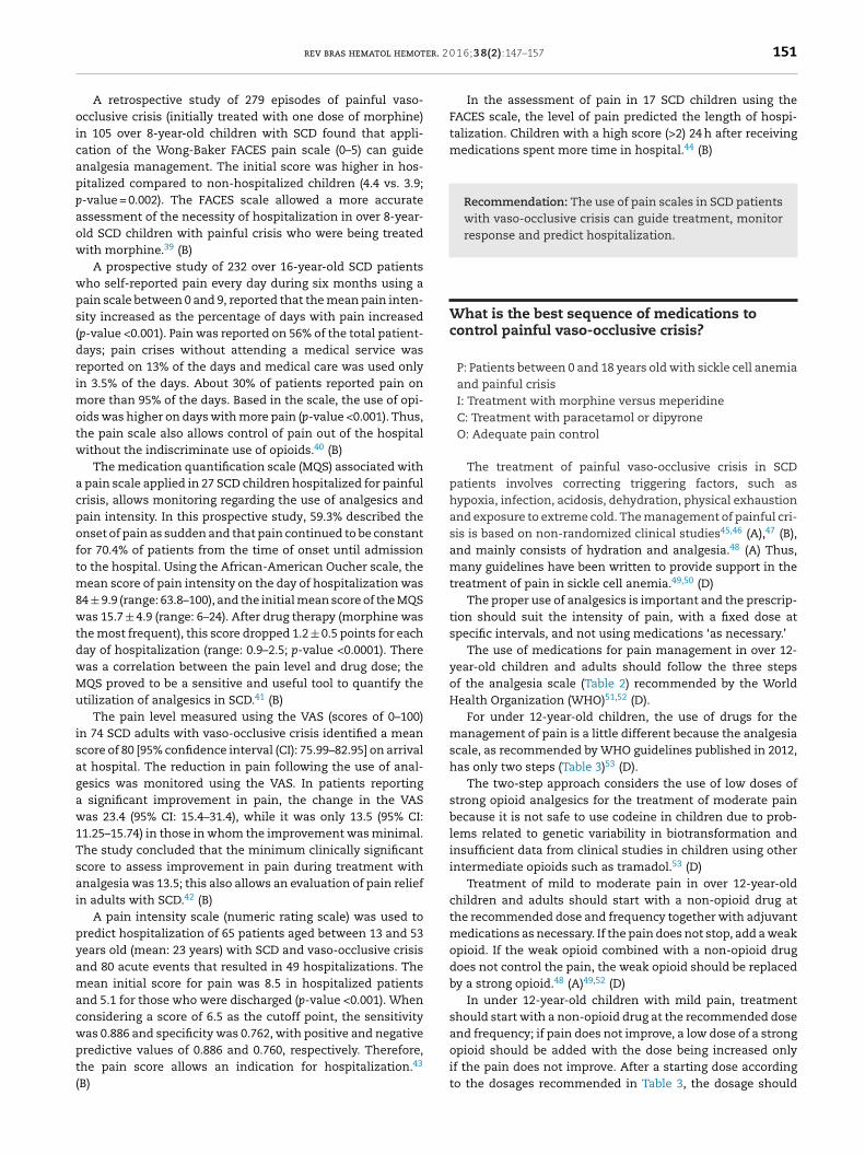
er. 2 0
oicappaow
wps(drimotw
acpoftm8wtdwMu
isagaw1Tsai
pyamacwpt(
rev bras hematol hemot
A retrospective study of 279 episodes of painful vaso-cclusive crisis (initially treated with one dose of morphine)
n 105 over 8-year-old children with SCD found that appli-ation of the Wong-Baker FACES pain scale (0–5) can guidenalgesia management. The initial score was higher in hos-italized compared to non-hospitalized children (4.4 vs. 3.9;-value = 0.002). The FACES scale allowed a more accuratessessment of the necessity of hospitalization in over 8-year-ld SCD children with painful crisis who were being treatedith morphine.39 (B)
A prospective study of 232 over 16-year-old SCD patientsho self-reported pain every day during six months using aain scale between 0 and 9, reported that the mean pain inten-ity increased as the percentage of days with pain increasedp-value <0.001). Pain was reported on 56% of the total patient-ays; pain crises without attending a medical service waseported on 13% of the days and medical care was used onlyn 3.5% of the days. About 30% of patients reported pain on
ore than 95% of the days. Based in the scale, the use of opi-ids was higher on days with more pain (p-value <0.001). Thus,he pain scale also allows control of pain out of the hospitalithout the indiscriminate use of opioids.40 (B)
The medication quantification scale (MQS) associated with pain scale applied in 27 SCD children hospitalized for painfulrisis, allows monitoring regarding the use of analgesics andain intensity. In this prospective study, 59.3% described thenset of pain as sudden and that pain continued to be constantor 70.4% of patients from the time of onset until admissiono the hospital. Using the African-American Oucher scale, the
ean score of pain intensity on the day of hospitalization was4 ± 9.9 (range: 63.8–100), and the initial mean score of the MQSas 15.7 ± 4.9 (range: 6–24). After drug therapy (morphine was
he most frequent), this score dropped 1.2 ± 0.5 points for eachay of hospitalization (range: 0.9–2.5; p-value <0.0001). Thereas a correlation between the pain level and drug dose; theQS proved to be a sensitive and useful tool to quantify thetilization of analgesics in SCD.41 (B)
The pain level measured using the VAS (scores of 0–100)n 74 SCD adults with vaso-occlusive crisis identified a meancore of 80 [95% confidence interval (CI): 75.99–82.95] on arrivalt hospital. The reduction in pain following the use of anal-esics was monitored using the VAS. In patients reporting
significant improvement in pain, the change in the VASas 23.4 (95% CI: 15.4–31.4), while it was only 13.5 (95% CI:
1.25–15.74) in those in whom the improvement was minimal.he study concluded that the minimum clinically significantcore to assess improvement in pain during treatment withnalgesia was 13.5; this also allows an evaluation of pain reliefn adults with SCD.42 (B)
A pain intensity scale (numeric rating scale) was used toredict hospitalization of 65 patients aged between 13 and 53ears old (mean: 23 years) with SCD and vaso-occlusive crisisnd 80 acute events that resulted in 49 hospitalizations. Theean initial score for pain was 8.5 in hospitalized patients
nd 5.1 for those who were discharged (p-value <0.001). Whenonsidering a score of 6.5 as the cutoff point, the sensitivity
as 0.886 and specificity was 0.762, with positive and negativeredictive values of 0.886 and 0.760, respectively. Therefore,he pain score allows an indication for hospitalization.43B)
1 6;3 8(2):147–157 151
In the assessment of pain in 17 SCD children using theFACES scale, the level of pain predicted the length of hospi-talization. Children with a high score (>2) 24 h after receivingmedications spent more time in hospital.44 (B)
Recommendation: The use of pain scales in SCD patientswith vaso-occlusive crisis can guide treatment, monitorresponse and predict hospitalization.
What is the best sequence of medications tocontrol painful vaso-occlusive crisis?
P: Patients between 0 and 18 years old with sickle cell anemiaand painful crisisI: Treatment with morphine versus meperidineC: Treatment with paracetamol or dipyroneO: Adequate pain control
The treatment of painful vaso-occlusive crisis in SCDpatients involves correcting triggering factors, such ashypoxia, infection, acidosis, dehydration, physical exhaustionand exposure to extreme cold. The management of painful cri-sis is based on non-randomized clinical studies45,46 (A),47 (B),and mainly consists of hydration and analgesia.48 (A) Thus,many guidelines have been written to provide support in thetreatment of pain in sickle cell anemia.49,50 (D)
The proper use of analgesics is important and the prescrip-tion should suit the intensity of pain, with a fixed dose atspecific intervals, and not using medications ‘as necessary.’
The use of medications for pain management in over 12-year-old children and adults should follow the three stepsof the analgesia scale (Table 2) recommended by the WorldHealth Organization (WHO)51,52 (D).
For under 12-year-old children, the use of drugs for themanagement of pain is a little different because the analgesiascale, as recommended by WHO guidelines published in 2012,has only two steps (Table 3)53 (D).
The two-step approach considers the use of low doses ofstrong opioid analgesics for the treatment of moderate painbecause it is not safe to use codeine in children due to prob-lems related to genetic variability in biotransformation andinsufficient data from clinical studies in children using otherintermediate opioids such as tramadol.53 (D)
Treatment of mild to moderate pain in over 12-year-oldchildren and adults should start with a non-opioid drug atthe recommended dose and frequency together with adjuvantmedications as necessary. If the pain does not stop, add a weakopioid. If the weak opioid combined with a non-opioid drugdoes not control the pain, the weak opioid should be replacedby a strong opioid.48 (A)49,52 (D)
In under 12-year-old children with mild pain, treatmentshould start with a non-opioid drug at the recommended dose
and frequency; if pain does not improve, a low dose of a strongopioid should be added with the dose being increased onlyif the pain does not improve. After a starting dose accordingto the dosages recommended in Table 3, the dosage should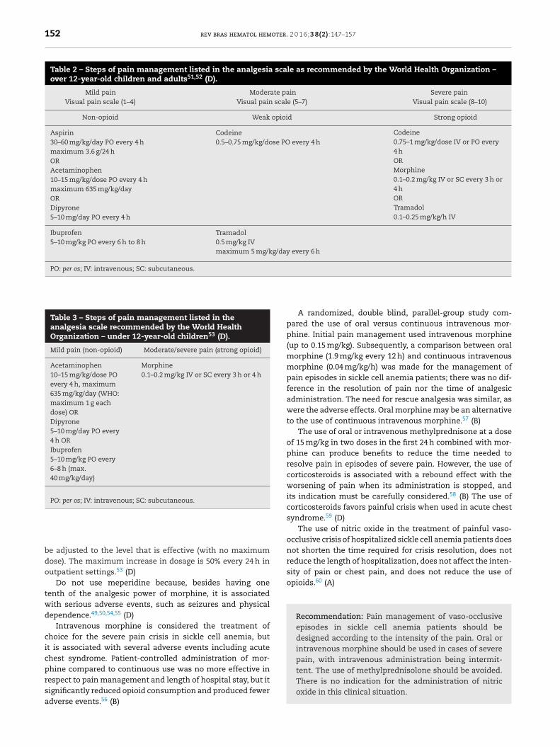
152 rev bras hematol hemoter. 2 0 1 6;3 8(2):147–157
Table 2 – Steps of pain management listed in the analgesia scale as recommended by the World Health Organization –over 12-year-old children and adults51,52 (D).
Mild painVisual pain scale (1–4)
Moderate painVisual pain scale (5–7)
Severe painVisual pain scale (8–10)
Non-opioid Weak opioid Strong opioid
Aspirin30–60 mg/kg/day PO every 4 hmaximum 3.6 g/24 hORAcetaminophen10–15 mg/kg/dose PO every 4 hmaximum 635 mg/kg/dayORDipyrone5–10 mg/day PO every 4 h
Codeine0.5–0.75 mg/kg/dose PO every 4 h
Codeine0.75–1 mg/kg/dose IV or PO every4 hORMorphine0.1–0.2 mg/kg IV or SC every 3 h or4 hORTramadol0.1–0.25 mg/kg/h IV
Ibuprofen5–10 mg/kg PO every 6 h to 8 h
Tramadol0.5 mg/kg IVmaximum 5 mg/kg/day every 6 h
PO: per os; IV: intravenous; SC: subcutaneous.
Table 3 – Steps of pain management listed in theanalgesia scale recommended by the World HealthOrganization – under 12-year-old children53 (D).
Mild pain (non-opioid) Moderate/severe pain (strong opioid)
Acetaminophen10–15 mg/kg/dose POevery 4 h, maximum635 mg/kg/day (WHO:maximum 1 g eachdose) ORDipyrone5–10 mg/day PO every4 h ORIbuprofen5–10 mg/kg PO every6–8 h (max.40 mg/kg/day)
Morphine0.1–0.2 mg/kg IV or SC every 3 h or 4 h
PO: per os; IV: intravenous; SC: subcutaneous.
tent. The use of methylprednisolone should be avoided.There is no indication for the administration of nitric
be adjusted to the level that is effective (with no maximumdose). The maximum increase in dosage is 50% every 24 h inoutpatient settings.53 (D)
Do not use meperidine because, besides having onetenth of the analgesic power of morphine, it is associatedwith serious adverse events, such as seizures and physicaldependence.49,50,54,55 (D)
Intravenous morphine is considered the treatment ofchoice for the severe pain crisis in sickle cell anemia, butit is associated with several adverse events including acutechest syndrome. Patient-controlled administration of mor-phine compared to continuous use was no more effective inrespect to pain management and length of hospital stay, but it
significantly reduced opioid consumption and produced feweradverse events.56 (B)A randomized, double blind, parallel-group study com-pared the use of oral versus continuous intravenous mor-phine. Initial pain management used intravenous morphine(up to 0.15 mg/kg). Subsequently, a comparison between oralmorphine (1.9 mg/kg every 12 h) and continuous intravenousmorphine (0.04 mg/kg/h) was made for the management ofpain episodes in sickle cell anemia patients; there was no dif-ference in the resolution of pain nor the time of analgesicadministration. The need for rescue analgesia was similar, aswere the adverse effects. Oral morphine may be an alternativeto the use of continuous intravenous morphine.57 (B)
The use of oral or intravenous methylprednisone at a doseof 15 mg/kg in two doses in the first 24 h combined with mor-phine can produce benefits to reduce the time needed toresolve pain in episodes of severe pain. However, the use ofcorticosteroids is associated with a rebound effect with theworsening of pain when its administration is stopped, andits indication must be carefully considered.58 (B) The use ofcorticosteroids favors painful crisis when used in acute chestsyndrome.59 (D)
The use of nitric oxide in the treatment of painful vaso-occlusive crisis of hospitalized sickle cell anemia patients doesnot shorten the time required for crisis resolution, does notreduce the length of hospitalization, does not affect the inten-sity of pain or chest pain, and does not reduce the use ofopioids.60 (A)
Recommendation: Pain management of vaso-occlusiveepisodes in sickle cell anemia patients should bedesigned according to the intensity of the pain. Oral orintravenous morphine should be used in cases of severepain, with intravenous administration being intermit-
oxide in this clinical situation.
er. 2 0
Imaaam
oms
(wTwettupspTll
Bdvitwiip
grp
riwpoo
gt
rev bras hematol hemot
s there evidence for the use of adjuvantedications such as non-steroidal
nti-inflammatory drugs, antihistamines,ntidepressants, benzodiazepines,nticonvulsants or corticosteroids in theanagement of painful vaso-occlusive crisis?
P: Patients between 0 and 18 years old with sickle cell anemiaand painful crisisI: Treatment with non-steroidal anti-inflammatory drugs(diclofenac, nimesulide, aspirin, COX 1 inhibitors andCOX 2 inhibitors), antihistamines, antidepressants, benzo-diazepines and anticonvulsantsC: Treatment with paracetamol or dipyroneO: Adequate pain control
There are few randomized clinical trials on the treatmentf the painful crisis in sickle cell anemia45 (A),46 (B), so thatost of the treatment is based on data from non-randomized
tudies.49,55,61,62 (D)Five studies with non-steroidal anti-inflammatory drugs
NSAIDs) as adjuvant pain medications for children and adultsith SCD were reviewed. Oral diflunisal was similar to placebo.
wo studies with intravenous ketorolac in hospitalized adultsere favorable to ketorolac for pain control, while two oth-
rs were not. The small sample sizes and the heterogeneity ofhe methods preclude any conclusion in the use of NSAID toreat pain in SCD and make any meta-analysis on this subjectnrealistic.45 (A) A phase III trial, double blind, randomized,lacebo-controlled of 66 episodes of painful vaso-occlusive cri-is concluded that ketorolac was not effective to control theain and the use of morphine when compared to a placebo.he authors do not recommend the systematic use of ketoro-
ac in these patients.63 (A) No studies were found in theiterature on other NSAIDs, such as acetaminophen.45 (A)
A double-blind randomized placebo-controlled study inrazil showed no benefit with the use of piracetam in chil-ren and young adults with sickle cell anemia and painfulaso-occlusive crisis in relation to the use of opioids, painntensity and hospitalization.64 (A) Piracetam is ineffective inhe prevention of vaso-occlusive pain crisis.64 (A) These dataere confirmed in a meta-analysis with three studies, includ-
ng the Brazilian study, where it was concluded that there arensufficient data to indicate piracetam for painful crisis in SCDatients.65 (A)
Methylprednisolone or intravenous dexamethasone sug-est benefits in relation to pain management, but patients whoeceived a single dose of corticosteroids had a higher risk ofain reactivation.45 (A)
Magnesium is a vasodilator that enhances the hydration ofed blood cells. For this reason it was investigated in a random-zed, double-blind, placebo-controlled study of 104 children
ith SCD hospitalized to receive intravenous analgesia forainful crisis. Intravenous magnesium sulfate had no effectn reducing the length of hospital stay, improvements in pain
66
r reductions in the cumulative dose of analgesics. (A)Adjuvant medications have been used to enhance the anal-esic effect of opioids in patients with cancer and to decreaseheir adverse events. Antihistamines (chlorpheniramine
1 6;3 8(2):147–157 153
0.15 mg/kg/day orally every 6 h with a maximum dose of12 mg/day), antidepressants (amitriptyline 10–20 mg orally atnight), benzodiazepines (the dose depends on the benzodi-azepine) and anticonvulsants (carbamazepine 5–10 mg/kg/dayonce or twice a day) have mild analgesic effects. When indi-cated, these medications should be used carefully.49,51,52 (D)
Recommendations: There is no consistent evidence sup-porting the use of antidepressants, benzodiazepines andanticonvulsants in the management of painful vaso-occlusive crisis in sickle cell anemia. There are nobenefits with piracetam or magnesium sulfate use inpainful vaso-occlusive crisis.
Is there evidence for the use of oxygen therapyor hyperhydration in the management ofpainful vaso-occlusive crisis?
P: Patients between 0 and 18 years old with sickle cell anemiaand painful crisisI: Treatment with oxygen therapy or hyperhydrationC: Treatment without oxygen therapy or hyperhydrationO: Adequate pain control
Although inhaled oxygen therapy (50% oxygen) in patientswith SCD reverses the sickling of red blood cells, there is noevidence in the literature that its use in vaso-occlusive paincrisis provides benefits by reducing the pain, the use of opioidsor hospitalization.67,68 (B)
Dehydration is an important factor in the sickling process.The decreased ability to concentrate urine, a characteristicof SCD patients, results in inadequate control of hydration.Thus, during painful crisis, patients receive intravenous ororal hydration, regardless of their state of hydration. However,care is necessary not to cause cardiac or pulmonary volumeoverload. No randomized controlled trials report benefits inrespect to improving or resolving pain with the infusion ofintravenous fluids as adjuvant treatment in sickle cell anemiapatients with painful vaso-occlusive crisis, regardless of thestate of hydration.47 (A)
Hydration in patients with sickle cell anemia should be car-ried out with caution. Hypotonic solutions may be used toestablish baseline hydration, maximum 1–1.5 times the main-tenance volume including the medications volume.49,55,61,69,70
(D) Care should be taken to avoid excessive hydration becauseit decreases the plasma oncotic pressure and increases thehydrostatic pressure, with risk of pulmonary edema (espe-cially in patients with kidney disease, heart disease orprevious lung disease).70 (D)
Recommendations: There is no published evidence on
the benefits of oxygen therapy or excessive hydration insickle cell anemia patients with painful vaso-occlusivecrisis.
oter.
154 rev bras hematol hemIs there evidence that bone scintigraphy is agood test to differentiate the painfulvaso-occlusive crisis from osteomyelitis? Isthere any test that allows this differentiation?
P: Patients between 0 and 18 years old with sickle cell anemiaand doubts between painful crisis and osteomyelitisI: Bone scintigraphyC: Clinical diagnosisO: Differential diagnosis between painful vaso-occlusive cri-sis and osteomyelitis
In sickle cell anemia, bone infarction is about 50 times morecommon than osteomyelitis.71 (B) However, the differentia-tion between osteomyelitis and bone infarction is difficult. Inboth situations the patient can have pain, fever, edema, ery-thema, and leukocytosis and imaging tests (X-ray, computedtomography) are nonspecific.72,73 (B)
A prospective study of 42 episodes of bone pain in childrenwith SCD showed that in most cases the diagnosis of boneinfarction is clinical. However, scintigraphy with technetiumcan help in cases where there is doubt about osteomyelitis.When bone marrow scintigraphy with technetium is madewithin ten days of onset, the uptake is normal at the injurysite in the case of osteomyelitis (100% of cases) while inbone infarction the uptake is diminished (93% of cases). Bonescintigraphy with technetium has proven effective to identifyosteomyelitis in 100% of cases due to the higher uptake, but itis not specific for bone infarction.74 (B)
Combination of sequential bone and bone marrow scintig-raphy with technetium showed benefits in the differentialdiagnosis of bone infarction and osteomyelitis in 39 childrenwith SCD and bone pain. These exams should be performedwithin 72 h of the onset of pain. Decreased bone marrowuptake associated with normal or reduced bone uptake inscintigraphy suggests a diagnosis of bone infarction, whereasincreased bone uptake suggests infection.75 (B)
Scintigraphy images of 79 children with SCD performedwithin 24 h of the onset of pain were retrospectively analysed.Bone marrow scintigraphy with technetium was performedfollowed by bone scintigraphy. Reduced uptake by the bonemarrow and abnormal bone uptake suggested bone infarc-tion and normal uptake by the bone marrow with increasedbone uptake suggested osteomyelitis. Seventy cases of boneinfarction were diagnosed with 66 being successfully treatedwithout antibiotics. Four cases were diagnosed as osteomyeli-tis, in which three cultures were positive; no false negativeresults were observed.73 (B)
A retrospective study of 22 episodes of suspectedosteomyelitis in SCD children showed that the combinationof bone scintigraphy with gallium and technetium can help inthe differential diagnosis of bone infarction and osteomyeli-tis. The best data for diagnosis are obtained within 24 h afterthe onset of symptoms.72 (B) The use of bone marrow scintig-raphy with gallium and technetium was also effective for this
76
differential diagnosis in a study of 18 episodes of pain. (C)A prospective study of 53 SCD children with the need todifferentiate between vaso-occlusive crisis and osteomyeli-tis found ultrasound changes of the soft tissue suggestive of
2 0 1 6;3 8(2):147–157
osteomyelitis in 17 patients; pus was drained from all patients.The authors suggest that ultrasound should be considered asthe first option to investigate osteomyelitis.77 (B) The over-all sensitivity of soft tissue ultrasound in the diagnosis ofosteomyelitis was 74% with a specificity of 63%.78 (C) The ultra-sound was normal in cases of bone infarction, and periostealelevation, subperiosteal or intramedullary abscesses and cor-tical bone erosion can be observed in osteomyelitis.77 (B)78 (C)The diagnosis of osteomyelitis by ultrasound can be enhancedby investigating reactive protein C serum levels and leuko-cyte counts, as these values are significantly increased inosteomyelitis.79 (B)
Magnetic resonance imaging (MRI) is a good imaging examto evaluate the bone marrow and to detect bone infarction.However, in SCD, it is difficult to differentiate between infarc-tion and osteomyelitis with MRI.80 (D)
Although imaging tests can help to differentiate betweenbone infarction and osteomyelitis, a clinical diagnosis isalways better.73,74,81 (B)
Recommendation: Differentiating bone infarction andosteomyelitis remains a challenge because the clinicalpresentation is similar in both cases and laboratory testsare still of limited use. Bone scintigraphy with tech-netium does not help to differentiate bone infarction insickle cell anemia patients with bone pain. Thus, thecombination of bone and bone marrow scintigraphy withtechnetium can assist in the differentiation betweenosteomyelitis and bone infarction, as can scintigraphyusing gallium and ultrasound of the soft tissues. MRI mayplay a role and be sought wherever possible in cases offever and prolonged pain.
Conflicts if interest
The authors declare no conflicts of interest.
Appendix A.
PICO 1What is the prevalence of sickle cell disease and how
are the results of neonatal screening for hemoglobinopathiesinterpreted?
((Anemia, Hemolytic, Congenital AND screeningAND neonatal)) OR (((screening AND neonatal)) AND(Hemoglobinopathies))
PICO 2Is there evidence for the need to perform a confirmatory
electrophoresis exam after the sixth month of life?(Anemia, Sickle Cell OR Hemoglobinopathies OR
Hemoglobins) AND (Electrophoresis) AND (Neonatal OR
Newborn)PICO 3Is there evidence on the factors that cause vaso-occlusive
crisis?

er. 2 0
odOo
ic
ods
v
ov
aatc
ASod
d
odTD
dI
r
1
1
1
1
1
1
1
1
1
1
2
2
rev bras hematol hemot
Sickle cell anemia AND (veno-occlusive disease OR veno-cclusive syndrome OR pain OR vaso-occlusive OR vascularisease OR crisis) AND (fever OR dehydration OR infectionR metabolic syndrome OR cold OR heat OR alcohol ORsteomyelitis OR cocaine OR marihuana OR etiology OR risk)
PICO 4Is there evidence that the use of pain assessment scales
s a good method to monitor pain related to vaso-occlusiverisis?
Sickle cell anemia AND (veno-occlusive disease OR veno-cclusive syndrome OR pain OR vaso-occlusive OR vascularisease OR crisis) AND (Severity of Illness Index OR scale ORcore OR VAS OR measurement)
PICO 5What is the best sequence of medications to control painful
aso-occlusive crisis?(Therapy/broad[filter] AND Sickle cell anemia AND (veno-
cclusive disease OR veno-occlusive syndrome OR pain ORaso-occlusive OR vascular disease OR crisis))
PICO 6Is there evidence for the use of adjuvant medications such
s non-steroidal anti-inflammatory drugs, antihistamines,ntidepressants, benzodiazepines, anticonvulsants or cor-icosteroids in the management of painful vaso-occlusiverisis?
(Therapy/broad[filter] OR anti-inflammatory OR Histaminentagonists OR anticonvulsant OR Benzodiazepines) ANDickle cell anemia AND (veno-occlusive disease OR veno-cclusive syndrome OR pain OR vaso-occlusive OR vascularisease OR crisis)
PICO 7Is there evidence for the use of oxygen therapy or hyperhy-
ration in the management of painful vaso-occlusive crisis?Sickle cell anemia AND (veno-occlusive disease OR veno-
cclusive syndrome OR pain OR vaso-occlusive OR vascularisease OR crisis) AND (oxygen therapy OR OXYGEN OR Fluidherapy OR Infusions, Parenteral OR HYDRATION OR DEHY-RATION)
PICO 8Is there evidence that bone scintigraphy is a good test to
ifferentiate painful vaso-occlusive crisis from osteomyelitis?s there any test that allows this differentiation?
((Radionuclide Imaging)) AND (sickle cell anemia)
e f e r e n c e s
1. National Institute of Health, National Heart, Lung, and BloodInstitute, Division of Blood Diseases and Resources. Themanagement of sickle cell disease; 2002.
2. Agência Nacional de Vigilância Sanitária (Anvisa). Manual deDiagnóstico e Tratamento de Doencas Falciformes; 2002. p.142. Available from: http://bvsms.saude.gov.br/bvs/publicacoes/anvisa/diagnostico.pdf [cited 10.01.15] [Internet].
3. Braga JA, Loggetto SR, Campanaro CM, Lyra IM, Viana MB,Anjos AC, et al. Doenca falciforme. In: Loggetto SR, Braga JA,
Tone LG, editors. Hematologia e Hemoterapia Pediátrica. SãoPaulo: Atheneu; 2014. p. 139–62.4. Shafer FE, Lorey F, Cunningham GC, Klumpp C, Vichinsky E,Lubin B. Newborn screening for sickle cell disease: 4 years of
2
1 6;3 8(2):147–157 155
experience from California’s newborn screening program. JPediatr Hematol Oncol. 1996;18(1):36–41.
5. Brandelise S, Pinheiro V, Gabetta CS, Hambleton I, Serjeant B,Serjeant G. Newborn screening for sickle cell disease in Brazil:the Campinas experience. Clin Lab Haematol. 2004;26(1):15–9.
6. Wagner SC, de Castro SM, Gonzalez TP, Santin AP, Zaleski CF,Azevedo LA, et al. Neonatal screening forhemoglobinopathies: results of a public health system inSouth Brazil. Genet Test Mol Biomarkers. 2010;14(4):565–9.
7. Magalhães PK, Turcato M de F, Angulo Ide L, Maciel LM.Neonatal screening program at the university hospital of theRibeirão Preto School of Medicine, São Paulo University,Brazil. Cad Saude Publica. 2009;25(2):445–54.
8. Streetly A, Latinovic R, Hall K, Henthorn J. Implementation ofuniversal newborn bloodspot screening for sickle cell diseaseand other clinically significant haemoglobinopathies inEngland: screening results for 2005-7. J Clin Pathol.2009;62(1):26–30.
9. Ducrocq R, Pascaud O, Bévier A, Finet C, Benkerrou M, Elion J.Strategy linking several analytical methods of neonatalscreening for sickle cell disease. J Med Screen. 2001;8(1):8–14.
0. Almeida AM, Henthorn JS, Davies SC. Neonatal screening forhaemoglobinopathies: the results of a 10-year programme inan English Health Region. Br J Haematol. 2001;112(1):32–5.
1. Lê PQ, Ferster A, Cotton F, Vertongen F, Vermylen C,Vanderfaeillie A, et al. Sickle cell disease from Africa toBelgium, from neonatal screening to clinical management.Med Trop (Mars). 2010;70(5–6):467–70.
2. Bardakdjian-Michau J, Bahuau M, Hurtrel D, Godart C, Riou J,Mathis M, et al. Neonatal screening for sickle cell disease inFrance. J Clin Pathol. 2009;62(1):31–3.
3. Diniz D, Guedes C, Barbosa L, Tauil PL, Magalhães I.Prevalence of sickle cell trait and sickle cell anemia amongnewborns in the Federal District, Brazil, 2004 to 2006. CadSaude Publica. 2009;25(1):188–94.
4. Paixão MC, Cunha Ferraz MH, Januário JN, Viana MB, Lima JM.Reliability of isoelectrofocusing for the detection of Hb S HbC, and HB D in a pioneering population-based program ofnewborn screening in Brazil. Hemoglobin. 2001;25(3):297–303.
5. Lobo CL, Bueno LM, Moura P, Ogeda LL, Castilho S, de CarvalhoSM. Neonatal screening for hemoglobinopathies in Rio deJaneiro, Brazil. Rev Panam Salud Publica. 2003;13(2–3):154–9.
6. Watanabe AM, Pianovski MA, Zanis Neto J, Lichtvan LC,Chautard-Freire-Maia EA, Domingos MT, et al. Prevalence ofhemoglobin S in the State of Parana, Brazil, based on neonatalscreening. Cad Saude Publica. 2008;24(5):993–1000.
7. Adorno EV, Couto FD, Moura Neto JP, Menezes JF, Rêgo M, ReisMG, et al. Hemoglobinopathies in newborns from Salvador,Bahia, Northeast Brazil. Cad Saude Publica. 2005;21(1):292–8.
8. Bandeira FM, Leal MC, Souza RR, Furtado VC, Gomes YM,Marques NM. Hemoglobin “S” positive newborn detected bycord blood and its characteristics. J Pediatr (Rio J).1999;75(3):167–71.
9. de Araújo MC, Serafim ES, de Castro WA Jr, de Medeiros TM.Prevalence of abnormal hemoglobins in newborns in Natal,Rio Grande do Norte, Brazil. Cad Saude Publica.2004;20(1):123–8.
0. Thuret I, Sarles J, Merono F, Suzineau E, Collomb J, Lena-RussoD, et al. Neonatal screening for sickle cell disease in France:evaluation of the selective process. J Clin Pathol.2010;63(6):548–51.
1. Miller FA, Hayeems RZ, Bombard Y, Little J, Carroll JC, WilsonB, et al. Clinical obligations and public health programmes:healthcare provider reasoning about managing the incidentalresults of newborn screening. J Med Ethics. 2009;35(10):626–34.
2. Kladny B, Gettig EA, Krishnamurti L. Systematic follow-up
and case management of the abnormal newborn screen canimprove acceptance of genetic counseling for sickle cell orother hemoglobinopathy trait. Genet Med. 2005;7(2):139–42.
oter.
2
2
2
2
2
2
2
3
3
3
3
3
3
3
3
3
3
4
4
4
4
4
4
4
4
4
4
5
5
5
5
5
5
5
5
5
5
6
156 rev bras hematol hem
3. Bain BJ. Haemoglobinopathy diagnosis: algorithms, lessonsand pitfalls. Blood Rev. 2011;25(5):205–13.
4. Boemer F, Vanbellinghen JF, Bours V, Schoos R. Screening forsickle cell disease on dried blood: a new approach evaluatedon 27,000 Belgian newborns. J Med Screen. 2006;13(3):132–6.
5. Ducrocq R, Bévier A, Leneveu A, Maier-Redelsperger M,Bardakdjian-Michau J, Badens C, et al. Compoundheterozygosity Hb S/Hb Hope (beta 136 Gly→Asp): a pitfall inthe newborn screening for sickle cell disease. J Med Screen.1998;5(1):27–30.
6. Shapiro BS. The management of pain in sickle cell disease.Pediatr Clin North Am. 1989;36(4):1029–45.
7. Smith WR, Bauserman RL, Ballas SK, McCarthy WF, SteinbergMH, Swerdlow PS, et al. Climatic and geographic temporalpatterns of pain in the Multicenter Study of Hydroxyurea.Pain. 2009;146(1–2):91–8.
8. Slovis CM, Talley JD, Pitts RB. Non relationship of climatologicfactors and painful sickle cell anemia crisis. J Chronic Dis.1986;39(2):121–6.
9. Jones S, Duncan ER, Thomas N, Walters J, Dick MC, Height SE,et al. Windy weather and low humidity are associated withan increased number of hospital admissions for acute painand sickle cell disease in an urban environment with amaritime temperate climate. Br J Haematol. 2005;131(4):530–3.
0. Nolan VG, Zhang Y, Lash T, Sebastiani P, Steinberg MH.Association between wind speed and the occurrence of sicklecell acute painful episodes: results of a case-crossover study.Br J Haematol. 2008;143(3):433–8.
1. West DC, Romano PS, Azari R, Rudominer A, Holman M,Sandhu S. Impact of environmental tobacco smoke onchildren with sickle cell disease. Arch Pediatr Adolesc Med.2003;157(12):1197–201.
2. Glassberg J, Spivey JF, Strunk R, Boslaugh S, DeBaun MR.Painful episodes in children with sickle cell disease andasthma are temporally associated with respiratorysymptoms. J Pediatr Hematol Oncol. 2006;28(8):481–5.
3. Hargrave DR, Wade A, Evans JP, Hewes DK, Kirkham FJ.Nocturnal oxygen saturation and painful sickle cell crises inchildren. Blood. 2003;101(3):846–8.
4. Nebor D, Bowers A, Hardy-Dessources MD, Knight-MaddenJM, Romana M, Reid H, et al. Frequency of painful crises insickle cell anemia and its relationships with thesympatho-vagal balance, blood viscosity and inflammation.Haematologica. 2011;96(11):1589–94.
5. Yoong WC, Tuck SM. Menstrual pattern in women with sicklecell anaemia and its association with sickling crises. J ObstetGynaecol. 2002;22(4):399–401.
6. Zempsky WT. Evaluation and treatment of sickle cell pain inthe emergency department: paths to a better future. ClinPediatr Emerg Med. 2010;11(4):265–73.
7. Luffy R, Grove SK. Examining the validity, reliability, andpreference of three pediatric pain measurement tools inAfrican-American children. Pediatr Nurs. 2003;29(1):54–9.
8. Zempsky WT, Corsi JM, McKay K. Pain scores: are they used insickle cell pain? Pediatr Emerg Care. 2011;27(1):27–8.
9. Frei-Jones MJ, Baxter AL, Rogers ZR, Buchanan GR.Vaso-occlusive episodes in older children with sickle celldisease: emergency department management and painassessment. J Pediatr. 2008;152(2):281–5.
0. Smith WR, Penberthy LT, Bovbjerg VE, McClish DK, Roberts JD,Dahman B, et al. Daily assessment of pain in adults withsickle cell disease. Ann Intern Med. 2008;148:94–101.
1. Jacob E, Miaskowski C, Savedra M, Beyer JE, Treadwell M,Styles L. Quantification of analgesic use in children with
sickle cell disease. Clin J Pain. 2007;23(1):8–14.2. Lopez BL, Flenders P, Davis-Moon L, Corbin T, Ballas SK.Clinically significant differences in the visual analog pain
6
2 0 1 6;3 8(2):147–157
scale in acute vasoocclusive sickle cell crisis. Hemoglobin.2007;31(4):427–32.
3. Doyle Portugal R, Morgado Loureiro M. Evaluation of painscale to predict hospital admission in patients with sickle celldisease. Haematologica. 2003;88(4):ELT11.
4. Sporrer KA, Jackson SM, Agner S, Laver J, Abboud MR. Pain inchildren and adolescents with sickle cell anemia: aprospective study utilizing self-reporting. Am J PediatrHematol Oncol. 1994;16(3):219–24.
5. Dunlop RJ, Bennett KC. Pain management for sickle celldisease. Cochrane Database Syst Rev. 2006;(2):CD003350.
6. Kavanagh PL, Sprinz PG, Vinci SR, Bauchnr H, Wang CJ.Management of children with sickle cell disease: acomprehensive review of the literature. Pediatrics.2011;128(6):e1552–74.
7. Okomo U, Meremikwu MM. Fluid replacement therapy foracute episodes of pain in people with sickle cell disease.Cochrane Database Syst Rev. 2012;6:CD005406.
8. Lottenberg R, Hassell KL. An evidence-based approach to thetreatment of adults with sickle cell disease. Hematol Am SocHematol Educ Program. 2005:58–65.
9. U.S. Department of Health and Human Services, Public HealthService, National Institutes of Health, National Heart, Lung,and Blood Institute. The management of sickle cell disease.NIH Publication No. 02-2117; 2002. Available from:https://www.nhlbi.nih.gov/files/docs/guidelines/sc mngt.pdf[cited 10.01.15] [Internet].
0. Brunetta DM, Clé DV, Haes TM, Roriz-Filho JS, Moriguti JC.Manejo das complicacões agudas da doenca falciforme.Medicina (Ribeirão Preto). 2010;43(3):231–7.
1. Ministério da Saúde, Instituto Nacional de Câncer. Cuidadospaliativos oncológicos: controle da dor. INCA: Rio de Janeiro;2001. p. 124.
2. World Health Organization. Cancer pain relief and palliativecare. World Health Organization Techinical Report Series 804.Geneva, Switzerland: World Health Organization; 1990. p.1–75.
3. WHO guidelines on the pharmacological treatment ofpersisting pain in children with medical illnesses. WorldHealth Organization; 2012. p. 36–53. ISBN 978 92 4 154812 0.
4. SCAC (the Sickle Cell Advisory Committee) of GENES (TheGenetic Network of New York, Puerto Rico and the VirginIslands). Guidelines for the treatment of people with sicklecell disease; March 2002.
5. Ministério da Saúde, Secretaria de Atencão à Saúde,Departamento de Atencão Especializada. Manual de eventosagudos em doenca. Brasília: Editora do Ministério da Saúde;2009, 50 p.: il. [Série A. Normas e Manuais Técnicos].
6. van Beers EJ, van Tuijn CF, Nieuwkerk PT, Friederich PW,Vranken JH, Biemond BJ. Patient controlled analgesia versuscontinuous infusion of morphine during vaso-occlusive crisisin sickle cell disease, a randomized controlled trial. Am JHematol. 2007;82(11):955–60.
7. Jacobson SJ, Kopecky EA, Joshi P, Babul N. Randomised trial oforal morphine for painful episodes of sickle-cell disease inchildren. Lancet. 1997;350(9088):1358–61.
8. Griffin TC, McIntire D, Buchanan GR. High-dose intravenousmethylprednisolone therapy for pain in children andadolescents with sickle cell disease. N Engl J Med.1994;330(11):733–7.
9. Caboot JB, Allen JL. Pulmonary complications of sickle celldisease in children. Curr Opin Pediatr. 2008;20(3):279–87.
0. Gladwin MT, Kato GJ, Weiner D, Onyekwere OC, Dampier C,Hsu L, et al. Nitric oxide for inhalation in the acute treatmentof sickle cell pain crisis: a randomized controlled trial. JAMA.
2011;305(9):893–902.1. Ministério da Saúde, Secretaria de Atencão à Saúde,Departamento de Atencão Especializada. Manual de condutas

er. 2 0
6
6
6
6
6
6
6
6
7
7
7
7
7
7
7
7
7
7
8
rev bras hematol hemot
básicas na doenca falciforme. Brasília: Editora do Ministérioda Saúde; 2006. p. 56 [Série A. Normas e Manuais Técnicos].
2. National Institute for Health and Clinical Excellence (NICE).Clinical Guideline 143. Sickle cell acute painful episode:management of an acute painful sickle cell episode inhospital; 2012. Available from: https://www.nice.org.uk/guidance/cg143/resources/sickle-cell-disease-managing-acute-painful-episodes-in-hospital-35109569155525 [cited10.01.15] [Internet].
3. Bartolucci P, El Murr T, Roudot-Thoraval F, Habibi A, Santin A,Renaud B, et al. A randomized, controlled clinical trial ofketoprofen for sickle-cell disease vaso-occlusive crises inadults. Blood. 2009;114(18):3742–7.
4. Alvim RC, Viana MB, Pires MA, Franklin HM, Paula MJ, BritoAC, et al. Inefficacy of piracetam in the prevention of painfulcrises in children and adolescents with sickle cell disease.Acta Haematol. 2005;113(4):228–33.
5. Al Hajeri AA, Fedorowicz Z, Omran A, Tadmouri GO. Piracetamfor reducing the incidence of painful sickle cell disease crises.Cochrane Database Syst Rev. 2007;(2):CD006111.
6. Goldman RD, Mounstephen W, Kirby-Allen M, Friedman JN.Intravenous magnesium sulfate for vaso-occlusiveepisodes in sickle cell disease. Pediatrics. 2013;132(6):e1634–1641.
7. Zipursky A, Robieux IC, Brown EJ, Shaw D, O’Brodovich H,Kellner JD, et al. Oxygen therapy in sickle cell disease. Am JPediatr Hematol Oncol. 1992;14(3):222–8.
8. Robieux IC, Kellner JD, Coppes MJ, Shaw D, Brown E, Good C,et al. Analgesia in children with sickle cell crisis: comparisonof intermittent opioids vs. continuous intravenous infusion ofmorphine and placebo-controlled study of oxygen inhalation.Pediatr Hematol Oncol. 1992;9(4):317–26.
9. Sickle Cell Society, Department of Health, UK Forum onHaemoglobin Disorders. Standards for the clinical care of
adults with sickle cell disease in the UK; 2008.0. Tostes MA, Braga JA, Len CA. Abordagem da crise dolorosa emcriancas portadoras de doenca falciforme. Rev Ciênc MédCampinas. 2009;18(1):47–55.
8
1 6;3 8(2):147–157 157
1. Keeley K, Buchanan GR. Acute infarction of long bones inchildren with sickle cell anemia. J Pediatr. 1982;101(2):170–5.
2. Amundsen TR, Siegel MJ, Siegel BA. Osteomyelitis andinfarction in sickle cell hemoglobinopathies: differentiationby combined technetium and gallium scintigraphy. Radiology.1984;153(3):807–12.
3. Skaggs DL, Kim SK, Greene NW, Harris D, Miller JH.Differentiation between bone infarction and acuteosteomyelitis in children with sickle-cell disease with use ofsequential radionuclide bone-marrow and bone scans. J BoneJoint Surg Am. 2001;83A(12):1810–3.
4. Rao S, Solomon N, Miller S, Dunn E. Scintigraphicdifferentiation of bone infarction from osteomyelitis inchildren with sickle cell disease. J Pediatr. 1985;107(5):685–8.
5. Kim HC, Alavi A, Russell MO, Schwartz E. Differentiation ofbone and bone marrow infarcts from osteomyelitis in sicklecell disorders. Clin Nucl Med. 1989;14(4):249–54.
6. Kahn CE Jr, Ryan JW, Hatfield MK, Martin WB. Combined bonemarrow and gallium imaging. Differentiation of osteomyelitisand infarction in sickle hemoglobinopathy. Clin Nucl Med.1988;13(6):443–9.
7. Sadat-Ali M, al-Umran K, al-Habdan I, al-Mulhim F.Ultrasonography: can it differentiate between vasoocclusivecrisis and acute osteomyelitis in sickle cell disease? J PediatrOrthop. 1998;18(4):552–4.
8. William RR, Hussein SS, Jeans WD, Wali YA, Lamki ZA. Aprospective study of soft-tissue ultrasonography in sickle celldisease patients with suspected osteomyelitis. Clin Radiol.2000;55(4):307–10.
9. Inusa BP, Oyewo A, Brokke F, Santhikumaran G, JogeesvaranKH. Dilemma in differentiating between acute osteomyelitisand bone infarction in children with sickle cell disease: therole of ultrasound. PLOS ONE. 2013;8(6):e65001.
0. Lonergan GJ, Cline DB, Abbondanzo SL. Sickle cell anemia.
Radiographics. 2001;21(4):971–94.1. Berger E, Saunders N, Wang L, Friedman JN. Sickle cell diseasein children: differentiating osteomyelitis from vaso-occlusivecrisis. Arch Pediatr Adolesc Med. 2009;163(3):251–5.