Embed Size (px)
Citation preview

R E V B R A S R E U M A T O L . 2 0 1 4 ; 5 4 ( 4 ) : 3 2 6 – 3 2 9
www.reumatologia.com.br
REVISTA BRASILEIRA DE REUMATOLOGIA
Case report
Multicentric Castleman disease not associated with HHV-8 and HIV viruses
Denise de Fatima Forteskia, Fernanda Calil Machado Nettoa, Andrea Barranjard Vannucci Lomontea, Bruno César Cavalcanti dos Anjosa, Maria Claudia Nogueira Zerbinib, Cristiano Augusto de Freitas Zerbinia,*a Rheumatology Service, Hospital Heliópolis, São Paulo, SP, Brazilb Medicine School, Universidade de São Paulo, São Paulo, SP, Brazil
a r t i c l e i n f o
Article history:
Received on 29 May 2012
Accepted on 18 May 2013
Keywords:
Castleman’s disease
B lymphocytes
Anaemia
Interleukin-6
Inflammation
Autoimmune
a b s t r a c t
Castleman’s disease (CD) is a polyclonal lymphoproliferative disorder also known as giant
nodular hyperplasia or angiofollicular lymph node hyperplasia. It is a rare disease often
associated to human immunodeficiency virus (HIV) and human herpes virus 8 (HHV-8).
Histopathological findings in Castleman’s disease suggest an exaggerated response to an-
tigenic stimuli seen in other diseases associated with immune activation, such as rheu-
matoid arthritis. An important aspect of its pathogenesis is the autonomous production of
interleukin-6 (IL-6). In this disease, the clinical manifestations are associated to IL-6 serum
levels, and surgical removal of the compromised lymph nodes or use of anti-IL-6 antibodies
can slow down the symptoms. We describe a multicentric Castleman’s disease in a young
woman not associated to HHV-8 virus infection or immunosuppression. A short review of
the literature follows the description of this clinical case.
© 2014 Sociedade Brasileira de Reumatologia. Published by Elsevier Editora Ltda.
All rights reserved.
* Corresponding author.
E-mail: [email protected] (C.A.F. Zerbini).
0482-5004/$ - see front matter. © 2014 Sociedade Brasileira de Reumatologia. Published by Elsevier Editora Ltda. All rights reserved.
http://dx.doi.org/10.1016/j.rbr.2013.05.004
Doença de Castleman multicêntrica não associada aos vírus HHV-8 e HIV
Palavras-chave:
Doença de Castleman
Linfócitos B
Anemia
Interleucina-6
Inflamação
Autoimune
r e s u m o
A doença de Castleman (DC) é uma desordem linfoproliferativa policlonal, também co-
nhecida como hiperplasia nodular gigante ou hiperplasia angiofolicular linfoide. Esta é
uma doença rara que está frequentemente associada ao vírus da imunodeficiência hu-
mana (HIV) e ao herpes vírus 8 (HHV-8). Os achados histopatológicos encontrados na DC
sugerem uma intensa resposta aos estímulos antigênicos observada em várias doenças
associadas com ativação imune, como a artrite reumatoide. Um fator importante impli-
cado na patogênese da DC é a produção autônoma da interleucina-6 (IL-6). Nessa doença,
as manifestações clínicas estão relacionadas aos níveis de IL-6, e a remoção cirúrgica dos
linfonodos acometidos ou a utilização de anticorpos anti-IL-6 fazem regredir os sintomas.
Descrevemos um caso da DC multicêntrica em uma mulher jovem, não associada à in-
327R E V B R A S R E U M A T O L . 2 0 1 4 ; 5 4 ( 4 ) : 3 2 6 – 3 2 9
fecção pelo vírus HHV-8 ou à imunossupressão. Uma breve revisão da literatura se segue
à descrição do caso clínico.
© 2014 Sociedade Brasileira de Reumatologia. Publicado por Elsevier Editora Ltda.
Todos os direitos reservados.
Introduction
The lymphoid angiofollicular hyperplasia or Castleman’s dis-ease (CD) was described in 1956 by Benjamin Castleman, who identified a number of patients with solitary and hyperplasic mediastinal lymph nodes containing follicles with interfollic-ular vascular proliferation.1 After the discovery of herpes vi-rus associated with Kaposi’s sarcoma (HHV-8) in HIV-positive patients and its identification in patients with CD, there was a real breakthrough in the understanding of disease patho-genesis.2
CD is a rare disease and there are no reliable estimates of its incidence in the population. There are two clinical syn-dromes: unicentric and multicentric. In the unicentric form, only one lymph node is affected, usually in the mediastinum, and the patient is clinically asymptomatic. In the multicentric form, the patient can present various clinical symptoms in-cluding anaemia, fatigue, anorexia, night sweats, weight loss, fever and hepatosplenomegaly.3
The multicentric form is often associated with HHV-8 and HIV-1 viruses. In HIV-positive patients, the disease tends to be more aggressive, with intense constitutional symptoms, splenomegaly, generalized lymphadenopathy, pancytopenia, interstitial pneumonitis and increased incidence of Kaposi sarcoma.4 In the multicentric form, associated or not with HHV-8, there is an overproduction of IL-6 and a polyclonal proliferation of B lymphocytes.
These changes stimulate the onset of autoimmune mani-festations, including POEMS (polyneuropathy, organomegaly, endocrinopathy, monoclonal gammopathy and skin changes) syndrome, and this complicates the diagnosis during the clin-ical investigation of a connective tissue disease. By histologi-cal analysis of lymph nodes, DC is classified in three forms: hyaline-vascular variant, plasmacytic variant, and a variant associated with HHV-8.
The hyaline-vascular form is characterized by separate follicles, with expansion of the mantle zone and presence of small lymphocytes forming concentric rings that surround the germinal center, which presents intense vascularization. The vascular proliferation also occurs among the follicles and there is often perivascular hyalinization.
In the plasmacytic form, a disruption of lymph node ar-chitecture can be observed, with variable germinal center hy-perplasia, expansion of the mantle zone and marked plasma-cytosis. In the unicentric form, the hyaline-vascular variant constitutes the most common histologic pattern, and in the multicentric form the most frequent histologic pattern is the plasmacytic variant, which can be associated or not to HHV-8 and to HIV-1.5
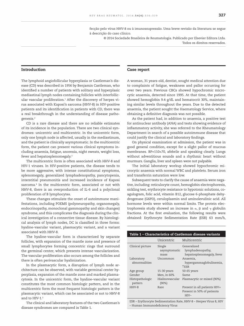
The clinical and laboratory features of the two Castleman’s disease syndromes are compared in Table 1.
Case report
A woman, 31 years-old, dentist, sought medical attention due to complaints of fatigue, weakness and pallor occurring for over two years. Previous CBCs showed hypochromic micro-cytic anaemia, detected since 1995. At that time, the patient showed hemoglobin 9.4 g/dL and hematocrit 30%, maintain-ing similar levels throughout the years. Due to the detected anaemia, the patient sought the Haematology Service, where obtaining a definitive diagnosis was not possible.
As the patient had, in addition to anaemia, a positive test for antinuclear antibody (ANA) and tests showing evidence of inflammatory activity, she was referred to the Rheumatology Department in search of a possible autoimmune disease that could justify the clinical and laboratory findings.
On physical examination at admission, the patient was in good general condition, except for a slight pallor of mucous membranes. BP=110×70, afebrile and speaking clearly. Lungs without adventitious sounds and a rhythmic heart without murmurs. Ganglia, liver and spleen were not palpable.
The initial laboratory workup showed hypochromic mi-crocytic anaemia with normal WBC and platelets. Serum iron and transferrin saturation were low.
Subsequent tests to clarify the cause of anaemia were nega-tive, including: reticulocyte count, hemoglobin electrophoresis, sickling test, erythrocyte resistance to hypotonic solutions, co-agulogram, folic acid, vitamin B12, glucose 6-phosphate dehy-drogenase (G6PD), ceruloplasmin and aminolevulinic acid. All hormone levels were within normal limits. The protein elec-trophoresis study showed an increase in α, β and γ globulin fractions. At the first evaluation, the following results were obtained: Erythrocyte Sedimentation Rate (ESR) 63 mm/h,
Table 1 – Characteristics of Castleman disease variants
Unicentric Multicentric
Clinical picture Single asymptomatic mass
Generalized lymphadenopathy, hepatosplenomegaly, fever
Laboratory abnormalities
Uncommon Anaemia, hypergammaglobulinemia,
↑ESR Age group 15-30 years 50-65 yearsGender Men, in 60% SameHistopathologic
patternHialinovascular
(90%)Plasmacytic or mixed (90%)
HHV-8 Rare Present in all patients HIV+Present in 50% of patients
HIV–
ESR – Erythrocyte Sedimentation Rate, HHV-8 – Herpes Virus 8, HIV – Human Immunodeficiency Virus

328 R E V B R A S R E U M A T O L . 2 0 1 4 ; 5 4 ( 4 ) : 3 2 6 – 3 2 9
C Reactive Protein (CRP) 19.2 mg/dL (Reference Value [RV] up to 0.5 mg/dL), Complement fraction 3 (C3) 186 mg/dL (RV 90-189 mg/dL) and total complement 426 mg/dL (RV 170-330 mg/dL).
Autoantibodies tests, including anti-Ro/SSA, anti-La/SSB, anti-native DNA, anti-Sm, anti-RNP, antineutrophil cytoplas-mic antibodies (ANCA) and rheumatoid factor, were negative, with ANA 1/80 positive with a fine speckled pattern. The se-rology for HIV, toxoplasmosis, Epstein-Barr virus and HHV-8 was negative. Tests for anti-HBs and anti-herpes simplex virus (HSV) were positive and our patient reported an episode of oral herpes involvement and a previous vaccination against hepati-tis B. The study of tumour markers was negative. Examinations of urine, faeces and cervical secretion showed no changes.
Thoracic radiographs and pelvic ultrasound showed no changes. An ultrasonography study of the abdomen revealed solid nodules: the smaller in the splenic hilum, measuring 2.8 × 1.6cm, and the larger anterior to the left kidney and lateral to the tail of the pancreas, measuring 7.0 × 4.1 × 4.0cm.
The myelogram showed normocellular granulocytic, erythrocytic and megakaryocytic series, medullary iron ++ (RV ++/+++), sideroblasts 5% (RV 30%), and ring sideroblasts ab-sent – findings consistent with microcytic anaemia of chronic inflammation.
A magnetic nuclear resonance study of the abdomen showed multiple oval nodular images in the splenic hilum with 1-3cm in diameter, not impregnated by the ferric contrast, and a solid mass of well-defined contours located medially to the inferior pole of the spleen, measuring approximately 6.0 × 4.5 × 4.5cm. The gallium-67 scintigraphy showed anomalous and in-tense hyperconcentration of the radiotracer in the topography of a solid mass at the lower pole of the spleen.
An ultrasound-guided fine needle aspiration (FNA) biopsy of an abdominal lymph node revealed cytological findings consistent with typical mature lymphoid and myeloid cells, which could correspond to an accessory spleen or to myeloid metaplasia. A bone marrow biopsy revealed hypocellular granulocytic, erythrocytic and megakaryocytic series and no granulomas, necrosis, amyloid or abnormal cells.
A videolaparoscopic surgery was performed with a biopsy of enlarged retrogastric lymph nodes and spleen chain. The pathologic examination of the specimens was consistent with the diagnosis of Castleman’s disease (plasmacytic vari-ant) (Fig. 1).
In the case described, the patient was initially treated with corticosteroids, with no clinical response. On that occasion, tocilizumab (an anti-IL-6 receptor antibody) was not avail-able for use in our country. Due to the chronic and persis-tent inflammation, the patient developed renal amyloidosis, progressing to chronic renal failure. After a prolonged period on dialysis, the patient underwent renal transplantation and currently remains clinically well.
Discussion
In this paper, we describe the case of a young female patient with symptoms of fatigue on exertion, hypochromic micro-cytic anaemia, tests of inflammatory activity persistently high and a positive ANA test. In principle, these findings could be consequent to a number of diseases, including SLE, and
this was the reason for the referral of our patient to the De-partment of Rheumatology.
Only after the completion of imaging studies (ultrasonog-raphy, MRI and scintigraphy), our diagnostic reasoning sug-gested lymphoproliferative or infectious disorders that could justify the laboratory changes and also the presence of mul-tiple intra-abdominal nodules. Despite all clinical investiga-tion, the diagnosis of CD was obtained only after the biopsy of abdominal nodes by videolaparoscopy, followed by histo-pathological and immunohistochemical analysis.
The clinical and laboratory data of this patient can be ex-plained by the biological actions of IL-6 overproduction in-cluding anaemia and an increase of immunoglobulins and high inflammatory activity tests. Furthermore, IL-6 can in-duce the formation of autoantibodies, thus justifying the pos-itive ANA test.6 During the evolution of the clinical case, we could not obtain a value for serum IL-6 because this test was not yet standardized in our country.
The systemic involvement and the plasmacytic histologi-cal presentation allowed us to classify our patient in CD’s multicentric form. The connective tissue diseases and the multicentric form of CD share many pathophysiological char-acteristics, and this may cause diagnostic difficulties. A re-view of the presence of autoimmune diseases concomitant to DC revealed an association with RA, Sjogren’s syndrome, my-asthenia gravis, SLE/polymyositis overlap syndrome, mixed connective tissue disease and SLE.7
The rheumatologist should consider the investigation of a solitary DC or of DC in association with a connective tissue disease when his/her patient has additional clinical features not expected in his/her disease development, a persistence of symptoms and of constitutional signs unusual for a connec-tive tissue disease, lymphadenomegaly on physical examina-tion or on imaging studies, or if unexpected difficulties arise during his/her treatment.
Fig. 1 – Abdominal lymph node biopsy showed histological findings consistent with Castleman’s disease, plasmacytic variant. A) Lymphoid follicular hyperplasia with prominent germinal centers, B) Paracortical proliferated blood vessels with plump endotelial cells, C) Numerous expressing kappa and lambda immunoglobulin light chains.
329R E V B R A S R E U M A T O L . 2 0 1 4 ; 5 4 ( 4 ) : 3 2 6 – 3 2 9
Conflicts of interest
The authors declare no conflicts of interest.
R E F E R E N C E S
1. Castleman B, Iverson L, Menendez VP. Localized mediastinal lymphnode hyperplasia resembling thymoma. Cancer. 1956 Jul-Aug;9:822-30.
2. Chang Y, Cesarman E, Pessin MS, Lee F, Culpepper J, Knowles DM, Moore PS. Identification of herpesvirus-like DNA sequences in AIDS-associated Kaposi’s sarcoma. Science. 1994 Dec 16;266:1865-9.
3. Weisenburger DD, Nathwani BN, Winberg CD, Rappaport H. Multicentric angiofollicular lymph node hyperplasia: a clinicopathologic study of 16 cases. Hum Pathol. 1985 Feb;16:162-72.
4. Reddy D, Mitsuyasu R. HIV-associated multicentric Castleman disease. Curr Opin Oncol. 2011 Sep;23:475-81.
5. Van Rhee F, Stone K, Szmania S, Barlogie B, Singh Z. Castleman disease in the 21st century: an update on diagnosis, assessment, and therapy. Clin Adv Hematol Oncol. 2010 Jul;8:486-98.
6. Yoshizaki K, Matsuda T, Nishimoto N, Kuritani T, Taeho L, Aozasa K, Nakahata T, Kawai H et al. Pathogenic significance of interleukin-6 (IL-6/BSF-2) in Castleman’s disease. Blood. 1989 Sep;74:1360-7.
7. Muskardin TW, Peterson BA, Molitor JA. Castleman disease and associated autoimmune disease. Curr Opin Rheumatol. 2012 Jan;24:76-83.