Embed Size (px)
Citation preview
FACULDADE DE MEDICINA DA UNIVERSIDADE DE COIMBRA
TRABALHO FINAL DO 2º ANO DO MESTRADO EM INVESTIGAÇÃO BIOMÉDICA
MARTA DE JESUS RIBEIRO
TERAPIA GÉNICA – AVALIAÇÃO DO POTENCIAL
TERAPÊUTICO EM CÉLULAS DE CARCINOMA
HEPATOCELULAR EM CULTURA.
ÁREA CIENTÍFICA DE BIOLOGIA MOLECULAR/ONCOLOGIA
TRABALHO REALIZADO SOB A ORIENTAÇÃO DE:
PROFESSOR DOUTOR JOSÉ MANUEL NASCIMENTO COSTA
DOUTORA SILVIA NEVES
SETEMBRO 2012
GENE THERAPY - EVALUATION OF THE THERAPEUTIC POTENTIAL IN
HEPATOCELLULAR CARCINOMA CELL LINES
Ribeiro M.(1)
, Neves S.(1,2,3)
, Sarmento Ribeiro A. B. (1,2,3)
, Nascimento Costa J.M. (1,2,3,4)
1Faculty of Medicine, University of Coimbra (FMUC), Portugal;
2 Center of Investigation
in Environment, Genetics and Oncobiology (CIMAGO), FMUC, Portugal; 3
Center for
Neuroscience and Cell Biology (CNC), University of Coimbra, Portugal; 4
University
Hospital of Coimbra, Portugal;
Correspondence: Ana Bela Sarmento Ribeiro, Applied Molecular Biology/Biochemistry
Institute, Faculty of Medicine - University of Coimbra – Azinhaga de Santa Comba – Celas -
3000-548, Coimbra, Portugal. Email: [email protected].
This work was supported by a grant from GAPI – Office for Support of Investigational
Projects, Faculty of Medicine of the University of Coimbra and Calouste Gulbenkian
Foundation, Portugal.
Ribeiro M. et al./ Gene Therapy in Hepatocellular carcinoma
i
INDEX
Resumo ............................................................................................................................... 1
Palavras-chave .................................................................................................................... 2
Abstract ............................................................................................................................... 3
Keywords ............................................................................................................................ 4
Abbreviations list ................................................................................................................ 5
Introduction ......................................................................................................................... 6
1- Hepatocellular carcinoma (HCC)................................................................................ 6
2- Gene therapy ............................................................................................................. 12
3- Cancer gene therapy .................................................................................................. 12
3.1 Tumor suppressor gene replacement ................................................................... 13
3.2 Oncogene inactivation ......................................................................................... 14
3.3 Immunotherapy .................................................................................................... 15
3.4 Inhibition of angiogenesis.................................................................................... 16
3.5 Suicide gene therapy ............................................................................................ 17
3.5.1 Thymidine kinase and ganciclovir ............................................................... 19
3.5.2 Cytosine deaminase and 5-fluorocytosine ................................................... 20
4- Vectors ...................................................................................................................... 21
4.1 Viral vectors......................................................................................................... 22
4.2 Non-viral vectors ................................................................................................. 24
4.2.1 Cationic liposomes ....................................................................................... 25
Materials and Methods ...................................................................................................... 28
1- Materials ................................................................................................................... 28
2- Cell line culture conditions ....................................................................................... 29
3- Cationic liposomes and lipoplexes preparation ........................................................ 29
4- In vitro gene transfer ................................................................................................. 30
Ribeiro M. et al./ Gene Therapy in Hepatocellular carcinoma
ii
5- Transfection activity ................................................................................................. 30
6- Cytotoxicity studies, evaluation of cell viability ...................................................... 31
7- Morphological analysis ............................................................................................ 32
8- Flow cytometry assays ............................................................................................. 33
8.1 Cell death analysis ............................................................................................... 33
8.2 Cell cycle analysis ............................................................................................... 33
9- Statistical analysis .................................................................................................... 34
Results .............................................................................................................................. 35
1- Transfection efficiency ............................................................................................. 35
2- Analysis of cell viability ........................................................................................... 36
2.1 Effect of HSV-TK/GCV or CD/5-FC treatment on the viability of HUH-7 and
Hep-G2 cells ..................................................................................................................... 36
2.2 Effect of HSV-TK/GCV or CD/5-FC treatment in association with Doxorubicin
and MG262 on the viability of HUH-7 and Hep-G2 cells ................................................ 39
3- Morphological analysis ............................................................................................ 42
4- Flow cytometry studies............................................................................................. 45
4.1 Analysis of cell death .......................................................................................... 45
4.2 Analysis of cell cycle .......................................................................................... 46
Discussion and Conclusion............................................................................................... 48
References ........................................................................................................................ 54
Ribeiro M. et al./ Gene Therapy in Hepatocellular carcinoma
iii
FIGURE ÍNDEX
Figure 1: Schematic representation of the multistep process of hepetocarcinogenesis ...... 9
Figure 2: Schematic representation of suicide gene therapy. ............................................ 18
Figure 3: Schematic representation of suicide therapy HSV-TK/GCV. ........................... 19
Figure 4: Conversion of 5-fluorocytosine to 5-fluorouracil by cytosine deaminase. ....... 20
Figure 5: Esquematic lipoplex-mediated transfection and endocytosis. ........................... 25
Figure 6: Representative structure of: A – Cationic lipid DOTAP; B – Neutral helper
lipid cholesterol. ....................................................................................................................... 26
Figure 7: Alamar blue. Convertion of resazurin to resorufin. ........................................... 32
Figure 8: Transfection efficiency in HUH-7 and Hep-G2 using different lipoplex cationic
lipid / DNA (+/-) charge ratios. ................................................................................................ 36
Figure 9: Dose response curve in HUH-7 cells treated with CD/5-FC. ............................ 37
Figure 10: Dose response curve in Hep-G2 cells treated with CD/5-FC. ......................... 37
Figure 11: Dose response curve in HUH-7 cells treated with HSV-TK/GCV. ................ 38
Figure 12: Dose response curve in Hep-G2 cells treated with HSV-TK/GCV. ............... 38
Figure 13: Dose response curve of CD/5-Fc in association with Doxorubicin (up) and
MG262 (below) in HUH-7 cells. .............................................................................................. 40
Figure 14: Dose response curve of CD/5-Fc in association with Doxorubicin (up) and
MG262 (below) in Hep-G2 cells. ............................................................................................. 40
Figure 15: Dose response curve of HSV-TK/GCV in association with Doxorubicin (up)
and MG262 (below) in HUH-7 cells. ....................................................................................... 41
Figure 16: Dose response curve of HSV-TK/GCV in association with Doxorubicin (up)
and MG262 (below) in Hep-G2 cells. ...................................................................................... 41
Figure 17: Morphological analysis of HUH-7 cells by optical microscopy. .................... 43
Figure 18: Morphological analysis of Hep-G2 cells by optical microscopy. ................... 44
Ribeiro M. et al./ Gene Therapy in Hepatocellular carcinoma
iv
Figure 19: Evaluation of cell death in HUH-7 by FC using AV and PI incorporation. ... 45
Figure 20: Evaluation of cell death in Hep-G2 by FC using AV and PI incorporation. .. 45
Figure 21: Evaluation of cell cycle in HUH-7 cell line. A – CTL. B – Lipoplexes. C –
TK/GCV. D – TK/GCV + Dox. E – TK/GCV + MG262. ....................................................... 47
Figure 22: Evaluation of cell cycle in HUH-7 cell line. A – CTL. B – Lipoplexes. C –
CD/5-FC. D – CD/5-FC + Dox. E – CD/5-FC + MG262. ....................................................... 47
Figure 23: Evaluation of cell cycle in Hep-G2 cell line. A – CTL. B – Lipoplexes. C –
TK/GCV. D – TK/GCV + Dox. E – TK/GCV + MG262. ....................................................... 47
Figure 24: Evaluation of cell cycle in Hep-G2 cell line. A – CTL. B – Lipoplexes. C –
CD/5-FC. D – CD/5-FC + Dox. E – CD/5-FC + MG262. ...................................................... 47
Ribeiro M. et al./ Gene Therapy in Hepatocellular carcinoma
1
RESUMO
O carcinoma hepatocelular (CHC) é uma doença com mau prognóstico cuja incidência
tem aumentado dramaticamente nas últimas décadas. Muitos estudos têm vindo a ser
realizados e variados protocolos investigados para o seu tratamento. Apesar disso, a taxa de
mortalidade dos doentes com CHC avançado ainda é muito elevada, havendo assim a
necessidade de desenvolvimento de novos fármacos, e de novas opções de tratamento, que
poderão ser utilizados como agentes únicos ou em combinação. Esta falta de opções
terapêuticas seguras e eficientes contra muitos tipos de cancro tem vindo a incentivar o
desenvolvimento de novas aplicações para a terapia génica.
A terapia génica suicida envolve a entrega de um gene suicida às células-alvo, tornando-
as sensíveis a determinado pró-fármaco sendo os sistemas mais utilizados a timidina cinase do
Vírus Herpes Simplex (HSV-TK)/ ganciclovir (GCV) e a citosina desaminase bacteriana
(CD)/ 5-fluorocitosina (5-FC). A entrega do material genético pode ser feita por vectores
virais ou não virais. Devido aos problemas que têm surgido com a utilização de vectores
virais, principalmente relacionados com questões de segurança, os lipossomas catiónicos
surgiram como sistemas promissores, devido à sua baixa toxicidade e imunogenicidade, à
falta de patogenicidade e versatilidade.
O objectivo do presente estudo consiste na avaliação do potencial terapêutico in vitro, em
linhas celulares de carcinoma hepatocelular (HUH-7 e Hep-G2), das estratégias de terapia
génica suicida (HSV-TK / GCV ou CD / 5-FC), em monoterapia ou em terapia combinada
com agentes de quimioterapia convencional (doxorrubicina) e novos fármacos dirigidos a
alvos moleculares (inibidor proteasoma MG-262), utilizando para a transfecção lipossomas
associados a transferrina. A viabilidade foi avaliada pela técnica do Alarmar Blue e os
mecanismos de morte verificados por citometria de fluxo com AV/IP, morfologia em
esfregaços corados por May-Grünwald-Giemsa e análise do ciclo celular.

Ribeiro M. et al./ Gene Therapy in Hepatocellular carcinoma
2
Os resultados obtidos indicam que a aplicação da terapia génica suicida utilizando os
sistemas HSV-TK/GCV ou CD/5-FC, tem um efeito antiproliferativo e citotóxico dependente
da dose de pro-fármaco utilizada e do tempo de incubação. A combinação destas abordagens
terapêuticas com doses inferiores ao IC50 de MG-262 ou doxorrubicina apresenta aumento do
efeito antiproliferativo e citotóxico, indicando que a aplicação combinada de doses mais
baixas de outros fármacos poderá levar a uma potenciação dos resultados com níveis de
toxicidade e efeitos secundários reduzidos. Este efeito antiproliferativo foi verificado através
das alterações observadas no ciclo celular das células (acumulação na fase G2 / M). Por
citometria de fluxo e por estudos de morfologia, verificou-se que o mecanismo de morte
celular predominantemente observado após aplicação dos diferentes tratamentos foi a
apoptose.
Estes resultados sugerem que a terapia de génica suicida com HSV-TK/GCV ou CD/5-
FC pode constituir uma nova abordagem com potencial terapêutico no CHC não só em
monoterapia, mas também, em associação com as terapias convencionais ou novos fármacos
dirigidos a alvos moleculares.
PALAVRAS-CHAVE
Carcinoma hepatocelular, linha celular HUH-7, linha celular Hep-G2, terapia génica
suicida, TK/GCV, CD/5-FC, inibidor do proteasoma MG262, lipossomas catiónicos,
transferrina, apoptose.

Ribeiro M. et al./ Gene Therapy in Hepatocellular carcinoma
3
ABSTRACT
HCC is a deadly cancer whose incidence has increased dramatically over the past decades.
Although many studies have been made and protocols investigated to treat HCC, the
mortalilty rate of patients with advanced HCC is still high, so, there is an unmet need for new
drugs, or new treatment options either as a single agents or in combination. This lack of safe
and efficient therapeutic options against many types of cancer is encouraging the development
of new gene therapy applications.
Suicide gene therapy involves the delivery of a suicide gene into target cells, making
them sensitive to an appropriate prodrug being the most commonly used Herpes Simplex
Virus thymidine kinase (HSV-TK)/ ganciclovir (GCV) and the bacterial cytosine deaminase
(CD)/ 5-fluorocytosine (5-FC) systems. This delivery can be made by viral or non-viral
vectors. However, due to the problems involving the use of viral vectors, namely regarding
safety issues, cationic liposomes emerged as promising systems due to their low toxicity and
immunogenicity, lack of pathogenicity and versatility.
The aim of the present study is to test the in vitro therapeutic potential in HCC cell lines
(HUH-7 and Hep-G2), of suicide gene approaches (HSV-TK/ GCV or CD/ 5-FC), in
monotherapy and in combination with conventional anticarcinogenic agents (doxorrubicin)
and new targeted drugs (proteassome inhibitor MG-262), using liposomes coupled with
transferrin. Viability was assessed by Alamar Blue assay and death mechanisms were verified
by flow cytometry with AV/PI, morphology in smears stained with May-Grünwald-Giemsa
and cell cycle analyses.
Our results showed that suicide gene therapy using HSV-TK/GCV or CD/5-FC
systems, resulted in a decreased viability, depending on the respective pro-drug dose and
incubation time in HCC cell lines in culture. Combinations of these systems with lower doses
than the IC50 of MG-262 or Doxorubicin enhanced the antiproliferative and cytotoxic effect
Ribeiro M. et al./ Gene Therapy in Hepatocellular carcinoma
4
meaning lower doses of other drugs with potentiation of the results and reduced toxicity
levels and side effects. This antiproliferative effect was in agreement with the observed cell
cycle alterations (accumulation in G2/M phase) upon applications of the suicide gene systems
alone or in combination with doxorubicin and proteasome inhibitor. By flow cytometry and
morphology studies, we observed that the mechanism of cell death was apoptosis upon
application of the different treatments.
These results suggest that suicide gene therapy with HSV-TK/GCV or CD/5-FC may
constitute a new potential therapeutic approach in HCC not only in monotherapy, but also in
association with conventional therapies or new targeted drugs.
KEYWORDS
Hepatocellular carcinoma, HUH-7 cell line, Hep-G2 cell line, suicide gene therapy,
TK/GCV, CD/5-FC, proteassome inhibitor, cationic liposomes, transferrin, apoptosis.
Ribeiro M. et al./ Gene Therapy in Hepatocellular carcinoma
5
ABBREVIATIONS LIST
5FC: 5-fluorocytosine
AV: annexin V
CD: cytosime desaminase
DMEM: Dulbecco’s Modified Eagle’s medium
DOX: doxorubicin
FBS: fetal bovine serum
FC: flow cytometry
FICT: fluorescein isothiocyanate
GCV: ganciclovir
HCC: hepatocellular carcinoma
IC50: half-maximal inhibitory concentration
MIF: mean intensity of fluorescence
PBS: phosphate buffer solution
PI: propidium iodide
TK: tirosine kinase
Ribeiro M. et al./ Gene Therapy in Hepatocellular carcinoma
6
INTRODUCTION
1- Hepatocellular carcinoma (HCC)
Liver cancer is one of the most common malignant tumors worldwide. In men, liver
cancer is the fifth most common neoplasm in the world and the second most common cause of
cancer-related death. In women, it is the seventh most common cancer and the sixth leading
cause of cancer death. An estimated 748,300 new liver cancer cases and 695,900 deaths
occurred worldwide in 2008 (Jemal A, 2011).
Hepatocellular carcinoma (HCC) is a major health problem, representing the major
histological subtype among primary malignant liver tumors, accounting for 70% to 85% of
the total liver cancer burden worldwide, (Avila MA, 2006; Jemal A, 2011).
The highest liver cancer rates are found in East and South-East Asia and in Middle and
Western Africa, with an incidence of 50–150 cases per 100,000 population and year, whereas
in South-Central and Western Asia, as well as in Northern and Eastern Europe, the rates are
lower. A higher incidence, of 10 cases per 100,000 population year, is found in North
America and Western Europe (Jemal A, 2011; Spangenberg HC, 2008 ).
These geographical differences in incidence may reflect variations in the main causal
HCC factors.
In some parts of Asia and Africa the high HCC incidence rates reflect the elevated
prevalence of chronic hepatitis B (HBV) or C (HCV) virus and aflatoxin B1 intake from
contaminated food. Their interaction has also been noted to increase liver cancer (Jemal A,
2011; Severi T, 2010 ).
In the USA and other low-risk Western countries, HCV infection is, as well as other
causes of cirrhosis, such as alcohol and haemochromatosis and possibly nonalcoholic fatty
Ribeiro M. et al./ Gene Therapy in Hepatocellular carcinoma
7
liver disease, associated with obesity, account for the majority of liver cancer (Jemal A, 2011;
Llovet JM B. A., 2003). Being the rise in its incidence and mortality possibly due to epidemic
obesity and the rise in HCV infection (Severi T, 2010 ; Jemal A, 2011),. In contrast, in some
historically high-risk areas rates decreased, possibly due to the HBV vaccine (Jemal A, 2011).
In a study performed from 1993 to 2005, in Portugal, the conclusions of the
demographic data were that the rate of mortality by Chronic Liver Disease and Cirrhosis are
similar to that by HCC. Besides that, one quarter of patients admitted to hospitals in Portugal
for HCC died and the most common cause of cirrhosis in Portugal is alcoholic cirrhosis, with
two-thirds of the total number of cases (Marinho RT, 2007).
In more than 80% of cases, HCC is associated with cirrhosis or with advanced fibrosis.
In the absence of cirrhosis or advanced fibrosis HBV infection, aflatoxin B1, some genetic
disorders such as Tyrosinosis and drugs (e.g. anabolic steroids) are the main risk factors
(Severi T, 2010 ; Lachenmayer A, 2010). However in patients with liver cirrhosis, the risk to
develop HCC depends on the activity, duration, and the etiology of the underlying liver
disease.
Some clinical and biological variables as age, anti-HCV positivity, partial prothrombin
time value, and platelet count, may enable identification of a subset of patients with the
highest risk of HCC development. Coexistence of various etiologies, as HBV and HCV
infection, aflatoxin B1 exposure, alcohol and/ or tobacco use, diabetes mellitus, liver steatosis
and obesity, has also to be considered and further increases the relative risk of HCC
development (Spangenberg HC, 2008 ).
HCC is phenotypically (morphology, microscopy) and genetically very heterogeneous,
possibly in part due to the heterogeneity of etiologic factors implicated in HCC development,
the complex functions of the liver cell, and the advanced stage at which HCC usually are
becoming clinically symptomatic and diagnosed (Spangenberg HC, 2008 ).

Ribeiro M. et al./ Gene Therapy in Hepatocellular carcinoma
8
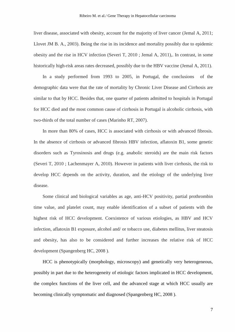
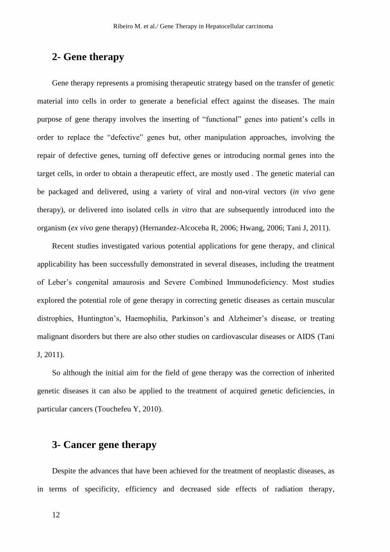
As for most types of cancer, hepatocarcinogenesis is a multifactorial multistep process
(Figure 1) involving different genetic and epigenetic alterations, chromosomal aberrations,
mutations, and altered molecular pathways that result in the deregulation of key oncogenes
and tumor-suppressor genes involved in several signaling pathways. These events accumulate
in a cell and ultimately lead to malignant transformation of the hepatocyte and to clinical liver
cancer. The molecular contribution of the different factors, their interaction and exact
sequence of hepatocarcinogenesis, including the development of preneoplasic lesions and
progression to HCC, are still poorly understood (Lachenmayer A, 2010; Llovet JM B. J.,
2008).
The previously mentioned risk factors for HCC can cause initial damage to the liver, but
several additional external or internal influences are needed to initiate the multistep process
leading to HCC development (Lachenmayer A, 2010).
In general, any condition leading to chronic inflammation in the liver causes genomic
and mitochondrial cells damage facilitating the development of neoplasms. Malignant
transformation of hepatocytes may occur, regardless of the etiologic agent, through a pathway
leading to increased liver cell turnover, induced by chronic liver injury and regeneration in a
context of inflammation, immune response and oxidative DNA damage. So, in populations of
both mature hepatocytes or liver stem cells this increased turnover renders liver cells more
sensitive to the adverse effects of other mutagenic agents and genetic and epigenetic changes
may occur, leading to the subsequent formation of dysplastic nodules and finally to HCC
(Spangenberg HC, 2008 ; Lachenmayer A, 2010; Severi T, 2010 ).
Chronic hepatitis B, C, and D, alcohol, metabolic liver diseases such as hemochromatosis
and α-1-antitrypsin deficiency may act predominantly through this pathway of chronic liver
injury, regeneration, and cirrhosis (Spangenberg HC, 2008 ).
Ribeiro M. et al./ Gene Therapy in Hepatocellular carcinoma
9
Figure 1: Schematic representation of the multistep process of hepetocarcinogenesis (Adapted from
Severi T, 2010).
From an epidemiological and clinical point of view HBV, HCV and alcoholic cirrhosis
are clearly the most important diseases for the development of HCC. From a
pathophysiological perspective, oxidative stress represents an important pathway by which
viruses or others risk factors exert their carcinogenic properties (Severi T, 2010 ).
The tumor environment also plays an important role in tumorigenesis and tumor
progression. Cancer cells are not as autonomous as once thought, they depend on
angiogenesis, inflammatory cells and fibroblasts. Most HCC cases occur after many years of
chronic liver disease that provides a mitogenic and mutagenic environment with abundance of
fibroblasts, some of them cancer associated fibroblasts (Severi T, 2010 ). While there is
evidence that HBV and HCV may, under certain circumstances, play an additional direct role
in the molecular hepatocarcinogenesis, aflatoxins have been shown to induce mutations of the
p53 tumor suppressor gene, thus pointing to the contribution of an environmental factor to
tumor development at the molecular level (Spangenberg HC, 2008 ).
Ribeiro M. et al./ Gene Therapy in Hepatocellular carcinoma
10
Current therapeutic strategies for HCC can be divided into established therapies as
surgical interventions (tumor resection and liver transplantation), percutaneous interventions
(ethanol injection, radiofrequency thermal ablation), transarterial interventions (embolization,
chemoperfusion, or chemoembolization), and experimental strategies such as radiation
therapy and drugs, including gene and immune therapy. Potentially curative therapies are
tumor resection, liver transplantation, and percutaneous interventions that can result in
complete responses and improved survival in a large proportion of patients. However, surgical
resection is limited by tumor size, the presence of multiple lesions, and impaired function in
the case of cirrhotic livers and liver transplantation is limited by a shortage of organ donations
and occurrence of transplant relapse (Spangenberg HC, 2008 ). In advanced disease, not
eligible to the use of potentially curative therapies, systemic treatment has been suggested as
beneficial to some patients, especially with the use of tamoxifen and doxorubicin (Alves RC,
2011).
Until recently, there wasn’t any available therapy that prolonged overall survival in
patients with advanced HCC, indicating the need for new therapies. In 2007, with the advent
of new targeted drugs hope has been offered to patients. One of this new targeted drug is
Sorafenib, a multikinase inhibitor, that block the VEGF and PDGF-dependent angiogenesis,
showed an increase on overall survival in patients with unresectable HCC. This drug is now
considered the standard of care in patients with advanced HCC and preserved liver function.
Unfortunately, all patients with advanced HCC still die from the disease and there is an unmet
need for other drugs, or treatment options either as a single agent or in combination (Severi T,
2010 ).
Since the inhibition of the ubiquitin-proteasome pathway in tumor cells results in
accumulation of tumor suppressor and pro-apoptotic proteins, the possibility of targeting this
pathway in cancer therapy is a viable option (Landis-Piwowar KR, 2006). Proteasome
Ribeiro M. et al./ Gene Therapy in Hepatocellular carcinoma
11
inhibition has already been established as a strategy for multiple myeloma and non-Hodgkin’s
lymphoma patients (Baiz D, 2009).
Treatment of patients with HCC is based on staging, which includes assessment of tumor
extent, liver function, portal pressure and clinical performance status.
Several systems that address the extent and prognosis of the disease have been proposed
for HCC staging, being the more used the Barcelona Clinic Liver Cancer (BCLC)
classification (Spangenberg HC, 2008 ). So far, HCC molecular classification has not yet
found its place in the staging systems and selection of therapy. However, the active search and
analysis for biomarkers in all current phase III studies may rapidly change the field (Severi T,
2010 ).
The widely used Barcelona-Clinic-Liver-Cancer (BCLC) staging system links staging of
HCC in cirrhosis with treatment modalities. The system identifies patients with early HCC
(stage 0 and A) who may benefit from curative therapies (resection, radiofrequency ablation,
liver transplantation), patients at intermediate or advanced stage (stage B, C) who may benefit
from palliative treatments (such as transarterial chemo- or radioembolisation), and the patients
with a very poor life expectancy (stage D) where supportive care is the only option (Severi T,
2010 ).
In 2010 less than 40% of patients in the western world fulfilled criteria for curative
treatment (resection, transplantation, local ablation) and only 20% were eligible for
chemoembolization. Alternative or palliative treatment options are very limited due to
resistance to conventional chemotherapy and radiotherapy (Lachenmayer A, 2010).
Therefore, there is an urgent need for new therapeutic strategies for HCC and the knowledge
of its molecular pathogenesis can provide the opportunity to indentify new biomarkers and
establish a molecular classification for HCC and for the development of new experimental
strategies, including target therapies, gene and immune therapy.
Ribeiro M. et al./ Gene Therapy in Hepatocellular carcinoma
12
2- Gene therapy
Gene therapy represents a promising therapeutic strategy based on the transfer of genetic
material into cells in order to generate a beneficial effect against the diseases. The main
purpose of gene therapy involves the inserting of “functional” genes into patient’s cells in
order to replace the “defective” genes but, other manipulation approaches, involving the
repair of defective genes, turning off defective genes or introducing normal genes into the
target cells, in order to obtain a therapeutic effect, are mostly used . The genetic material can
be packaged and delivered, using a variety of viral and non-viral vectors (in vivo gene
therapy), or delivered into isolated cells in vitro that are subsequently introduced into the
organism (ex vivo gene therapy) (Hernandez-Alcoceba R, 2006; Hwang, 2006; Tani J, 2011).
Recent studies investigated various potential applications for gene therapy, and clinical
applicability has been successfully demonstrated in several diseases, including the treatment
of Leber’s congenital amaurosis and Severe Combined Immunodeficiency. Most studies
explored the potential role of gene therapy in correcting genetic diseases as certain muscular
distrophies, Huntington’s, Haemophilia, Parkinson’s and Alzheimer’s disease, or treating
malignant disorders but there are also other studies on cardiovascular diseases or AIDS (Tani
J, 2011).
So although the initial aim for the field of gene therapy was the correction of inherited
genetic diseases it can also be applied to the treatment of acquired genetic deficiencies, in
particular cancers (Touchefeu Y, 2010).
3- Cancer gene therapy
Despite the advances that have been achieved for the treatment of neoplastic diseases, as
in terms of specificity, efficiency and decreased side effects of radiation therapy,
Ribeiro M. et al./ Gene Therapy in Hepatocellular carcinoma
13
chemotherapy, as in the development of drugs directed to new molecular targets, many tumors
remain resistant to existing therapies especially those who are diagnosed in advanced stage.
This lack of safe and efficient therapeutic options against many types of cancer, gives
rise to the necessity to develop alternative therapies that can achieve more effective results,
encouraging in this way, the development of new gene therapy applications for these diseases
(Sangro B, 2010).
Gene therapy for treatment of cancer is a rapidly growing field and the most frequent
application of experimental gene therapy approaches (Tani J, 2011). Besides the mentioned
lack of therapeutic options, some of the reasons why the applicability of gene therapy in
cancer is a rapidly growing field, are related to the genetic alterations that give rise or
contribute to the malignant transformation of cells being unraveled. These provide multiple
candidate targets for gene therapy intervention. (Hernandez-Alcoceba R, 2006).
Cancer gene therapy involves the manipulation of intracellular DNA in order to control
or destroy cancer cells. Like cytostatic chemotherapies, it is assumed that there are certain
biochemical, molecular or environmental characteristics of tumour cells which distinguishes
them from normal tissues, that are able to be exploited for gene therapy (Hughes, 2004; Dachs
GU, 2009). In this way gene therapy has the potential to provide highly selective, curative
cancer treatment without systemic toxicity.
Strategies for cancer gene therapy include inhibition of activated oncogenes, transfer or
activation of tumor suppressor and cytokine genes (immunotherapy), inhibition of
angiogenesis and selective prodrug activation by suicide genes (Neves S, 2009).
3.1 Tumor suppressor gene replacement
Tumor suppressor genes encode a variety of proteins that regulate the cell cycle and
mediate DNA repair pathways. These genes control cell proliferation and apoptosis in order to
Ribeiro M. et al./ Gene Therapy in Hepatocellular carcinoma
14
maintain an equilibrated cell turnover in each tissue. The progression from normal tissue to
tumor involves the presence of mutations or loss of function of some of these genes and by
inhibiting the expression of tumor suppressor genes. Cancer cells can continue to proliferate,
acquire new gene mutations, become less differentiated, and avoid apoptosis. One gene
therapy approach is, thus, to restore tumour-suppressor gene expression (Hughes, 2004),
(Hernandez-Alcoceba R, 2006).
In fact transfection of tumor suppression genes into cancer cells has been effective at
arresting growth and inducing apoptosis. Among the various tumor suppressor genes capable
of triggering apoptosis, the gene encoding the p53 protein has been the more studied, since the
functional inactivation of this protein occurs in over 50% of tumors (Sherr, 2004). The goal of
this approach is to restore the function of p53, which is involved in the regulation of DNA
repair processes and apoptosis in abnormal or damaged cells, and therefore, in inhibiting
tumor growth. Under experimental conditions, it has been demonstrated that the restoration of
tumour suppressor genes can revert the malignant phenotype of cells. The transfer of p53
tumour suppressor gene has shown effect in several cancer animal models, including HCC
(Hernandez-Alcoceba R, 2006).
About 11% of transferred genes in gene therapy clinical trials are tumour-suppressor
genes and many trials have been performed in cancer gene therapy using the p53 gene, mostly
including patients with lung or head and neck cancers (Touchefeu Y, 2010).
3.2 Oncogene inactivation
The progression from normal tissue to tumor also involves the presence of mutations in
proto-oncogenes that are silenced after fetal development to prevent abnormal tissue growth.
Cancer cells often propagate by activating and amplifying these proto-oncogenes (Hughes,
2004)
Ribeiro M. et al./ Gene Therapy in Hepatocellular carcinoma
15
One form of cancer gene therapy is targeted disruption of tumor oncogenes that are
active in the tumours like tumours like RAS, c-MYC, ERB-2 or BCL-2 genes. This can be
accomplished by several strategies : (1) inhibition of the oncogene transcription into mRNA,
using DNA oligonucleotides designed to bind to specific oncogene promoter regions; (2)
reduction of mRNA translation into protein, by the transfer of antisense nucleotides, artificial
sequences complementary to the mRNA corresponding to the gene whose inhibition is
attempted or, more recently, with RNA interference, a posttranscriptional gene silencing
mechanism based on the production of double-stranded stretches of RNA complementary to
the target mRNA or (3) interference with the oncoprotein transportation and function by
intracellular expression of single chain antibody-based molecules against the oncoprotein
(Hughes, 2004; Hernandez-Alcoceba R, 2006).
It is anticipated that the inhibition of oncogene expression will decrease cell proliferation,
but also restore sensitivity of cells to apoptotic stimuli. For instance it is known that the
inhibition of the RAS oncogene, blocks a cascade of mitotic signals, but also relieves the
repression exerted on the p53 pathway and predisposes cells to apoptosis In the case of HCC,
the inhibition of several genes had shown potential antitumor effect with in vitro studies
showing growth inhibition or induction of apoptosis, and in vivo with tumours growth
retardation (Hernandez-Alcoceba R, 2006).
3.3 Immunotherapy
Cancer immunotherapy or immunopotentiation consists in the enhancement of the
immune system’s ability to destroy cancer cells.
Passive immunopotentiation involves boosting the natural immune response to make it
more effective by delivering into tumor cells genes that encode immunomodulators such as
cytokines, TNF-α, interleukins (IL-2, IL-12), interferon (INF-γ) and growth factor
Ribeiro M. et al./ Gene Therapy in Hepatocellular carcinoma
16
granulocyte macrophage (GM-CSF), to enhance the antitumoral immune response (Touchefeu
Y, 2010; Cross D, 2006).
Active immunopotentiation requires the initiation of an immune response against a
previously unrecognized or poorly antigenic tumor, being a promising antitumor strategy, also
designated genetic vaccine. This strategy aims to immunize patients specifically against their
own tumors through genetic modification of target cells with a gene, that usually encodes
tumor-specific antigens, and which product increases the immune reactivity anti-tumor (Rice
J, 2008; Cross D, 2006). Currently a “dendritic cell vaccine” for recurrent prostate cancer has
been recommended for approval by US FDA (Tani J, 2011).
3.4 Inhibition of angiogenesis
The sustainable growth of a solid tumor depends on its ability to develop and maintain an
adequate blood supply that meets demands for nutrients and oxygen. Experimental and
clinical studies have shown that primary tumors as well as metastases can remain dormant due
to a balance between proliferation and apoptosis unless the angiogenesis is switched on. The
onset of tumor angiogenesis, required for the rapid growth of solid tumors and tumor
metastases, is likely to be triggered by an upregulation of angiogenic factors such as basic
fibroblast growth factor (bFGF) or vesicular endothelial growth factor (VEGF), or by a down-
regulation of antiangiogenic factors such as angiostatin, endostatin, or thrombospondin
(Hwang, 2006).
Realization that tumor growth requires intense neovascularization is the basis for a series
of approaches aimed to specifically block the cancer-induced angiogenesis and tumor
adaptation to hypoxia. Expression of anti-angiogenic factors such as endostatin have
demonstrated the ability to inhibit tumor growth in vivo and gene therapy may play an
important role in this field, because anti-angiogenic factors need to be delivered for long
Ribeiro M. et al./ Gene Therapy in Hepatocellular carcinoma
17
period of times to control the progression of tumors. Other anti-angiogenic approaches are
focused on blocking angiogenic factors as the VEGF receptor, an important mediator of
angiogenesis, or the endothelium-specific receptor Tie2, which affects direct tumor growth
and neovascularization (Hernandez-Alcoceba R, 2006; Hughes, 2004).
3.5 Suicide gene therapy
Suicide gene therapy is another gene therapy strategy to treat cancer by selective tumor
destruction without inducing significant systemic toxicity. Opposing to the conventional
drugs that usually have toxicity both in malignant and non-malignant cell the aim of this
approach is to improve conventional cancer chemotherapy by selectively activating the
prodrug at the tumor site. This allows higher active drug concentrations at the tumor,
minimizing tumor burden, by limiting the exposition of the patient’s normal tissues and
organs to potentially harmful quantities of drugs (Hwang, 2006; Silke Schepelmann, 2008).
Suicide-gene therapy, or prodrug activation therapy, is based in the introduction into
tumor cells of a viral, bacterial or fungal gene that encodes enzymes able to metabolize a
nontoxic prodrug into a cytotoxic drug. These strategies are based on the fact that some
viruses, bacteria and fungi use enzyme systems not found in humans and the prodrugs have
been developed in a way that for them to be converted into a toxic form, it is necessary the
digestion performed by an enzyme expressed from the exogenous gene added (Portsmouth D,
2007).
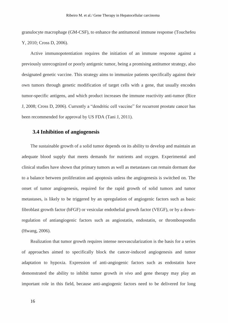
This approach involves two steps. In the first step, the gene for a foreign enzyme is
targeted to the tumor cells. After expression of the foreign gene at the tumor site, a relatively
nontoxic prodrug is administered, which is converted into an active, cytotoxic drug by the
foreign enzyme (Figure 2) (Silke Schepelmann, 2008).
Ribeiro M. et al./ Gene Therapy in Hepatocellular carcinoma
18
Figure 2: Schematic representation of suicide gene therapy. Suicide gene therapy consists in the
delivery of a suicide gene to the target cell that encodes an enzyme which will activate a subsequently
administered prodrug, into a toxic metabolite leading to cell death. (Adapted from Neves, 2009)
A critical problem to overcome in cancer gene therapy is the delivery of genes to a
sufficient number of tumour cells to cause tumour regression, which is especially important
since gene transfer efficiencies in the clinic, are unlikely to exceed 10% of the target tissue.
Suicide gene therapy is associated with bystander effects which cause the death of
nontransgenic cells, due to indirect effects of neighbouring transgenic cells treatment (Dachs
GU, 2009). The local bystander effect is known to induce tumor regression, although only a
fraction of tumor cells express the suicide gene and several hypotheses have been proposed to
explain killing of neighboring untransfected tumor cells, as the passive diffusion of the drug;
passage of the drug through gap junctions; endocytosis of apoptotic vesicles; release of
soluble factors; or stimulation of the immune system in vivo (Duarte S, 2012).
Among the different suicide systems the herpes simplex virus thymidine kinase gene
(HSV-tk) with ganciclovir (GCV) as prodrug, and the cytosine deaminase gene (CD) of
Escherichia coli, which converts the non-toxic antifungal agent 5- fluorocytosine (5-FC) into
5-fluorouracil (5-FU), are the most extensively studied (Duarte S, 2012).
Ribeiro M. et al./ Gene Therapy in Hepatocellular carcinoma
19
3.5.1 Thymidine kinase and ganciclovir
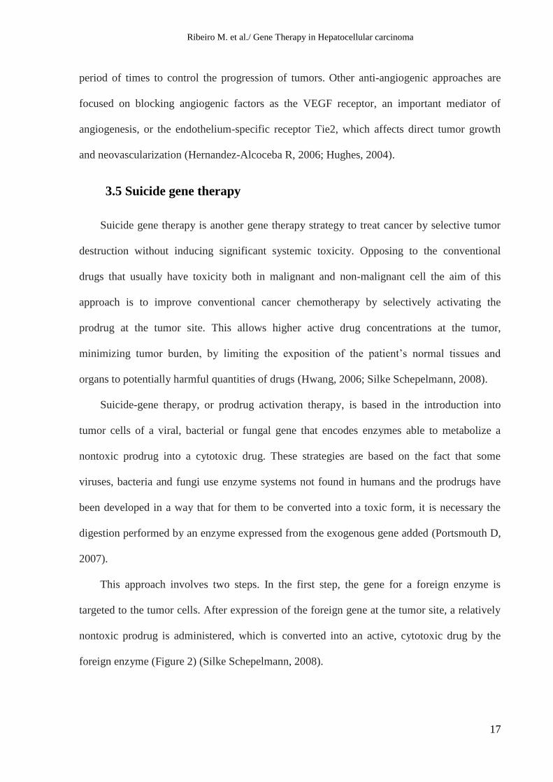
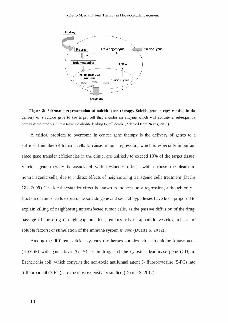
The thymidine kinase from Herpes Simplex Virus (HSV-TK) is the best characterized
suicide gene and in combination with the prodrug GCV constitutes the most used suicide gene
therapy (Figure 3).
Figure 3: Schematic representation of suicide therapy HSV-TK/GCV. HSV-TK gene is delivered to
cells by a vector. The thymidine kinase enzyme expressed converts ganciclovir, an analogue of deoxyguanine
subsequently administered, into his monophosphorylated form. The host cell endogenous kinases promote the
addition of two more phosphates to form ganciclovir triphosphate, highly toxic, which is incorporated into DNA
during replication, resulting in the inhibition of the DNA strand synthesis, causing cell death (Adapted from
Neves, 2009).
GCV is a synthetic analogue of 2'-deoxy-guanosine first synthesized in 1980 as an
antiviral agent. GCV is phosphorylated by the thymidine kinase from HSV-1 (HSV-TK) to a
monophosphate, and cellular kinases complete the conversion to the active triphosphate
(Dachs GU, 2009). Although human cells express cytosolic and mitochondrial TK enzymes,
these endogenous enzymes have much lower ability to convert GCV compared to HSV-TK,
so the GCV phosphorylation by the HSV-TK to GCV monophosphate is the limiting step of
the prodrug transformation in a toxic metabolite (Balfour, 1999). The triphosphate form is
structurally similar to 2’-deoxyguanosine triphosphate (dGTP) and is erroneously
incorporated into DNA during replication, causing inhibition of DNA polymerase and rapid
chain termination in both nuclear and mitochondrial DNA synthesis, mainly by the induction
of errors in the synthesis of a new DNA strand which subsequently leads to DNA
Ribeiro M. et al./ Gene Therapy in Hepatocellular carcinoma
20
fragmentation and activation of apoptosis mechanisms, leading to cell death (Dachs GU, 2009;
Duarte S, 2012).
Anti-tumor activity of the TK/GCV system has been demonstrated in vivo in several
carcinoma animal models, including leukemia, glioma, bladder cancer, intrahepatic metastasis
of liver cancer, colon adenocarcinoma, and oral cancer (Duarte S, 2012).
The promising results achieved in the pre-clinical studies led to the HSV-TK/GCV
system application in several clinical trials in different types of cancer as glioblastoma
(Rainov, 2000; Voges J, 2003), prostate cancer (Nasu Y, 2007), head and neck cancer (Xu F,
2009) and hepatocellular carcinoma (Li N, 2007; Sangro B M. G., 2010 ).
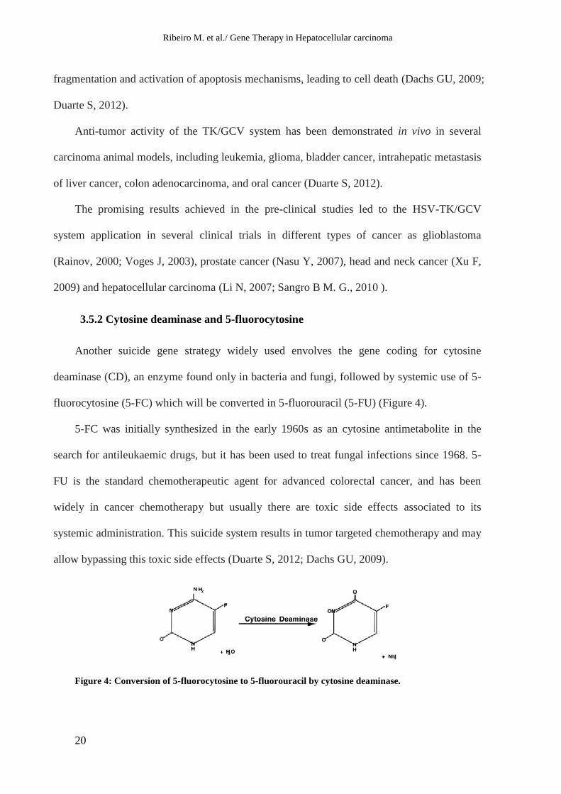
3.5.2 Cytosine deaminase and 5-fluorocytosine
Another suicide gene strategy widely used envolves the gene coding for cytosine
deaminase (CD), an enzyme found only in bacteria and fungi, followed by systemic use of 5-
fluorocytosine (5-FC) which will be converted in 5-fluorouracil (5-FU) (Figure 4).
5-FC was initially synthesized in the early 1960s as an cytosine antimetabolite in the
search for antileukaemic drugs, but it has been used to treat fungal infections since 1968. 5-
FU is the standard chemotherapeutic agent for advanced colorectal cancer, and has been
widely in cancer chemotherapy but usually there are toxic side effects associated to its
systemic administration. This suicide system results in tumor targeted chemotherapy and may
allow bypassing this toxic side effects (Duarte S, 2012; Dachs GU, 2009).
Figure 4: Conversion of 5-fluorocytosine to 5-fluorouracil by cytosine deaminase.
Ribeiro M. et al./ Gene Therapy in Hepatocellular carcinoma
21
CD enzyme catalyses the deamination of cytosine to uracil converting 5-FC into the toxic
anabolite 5-FU, which is subsequently processed by intracellular enzymes leading to the
production of either to 5-fluorouracil triphosphate, which is incorporated into the RNA and
interferes with RNA processing, or to 5-fluoro-2'-deoxyuridine 5'-monophosphate that
irreversibly inhibits thymidylate synthase, a key enzyme in pirimidine biosynthesis, and thus
interferes with the DNA synthesis (Kuriyama S, 1999).
In vivo anti-tumor activity of the CD/5-FC combination has been demonstrated in several
animal models, including fibrosarcomas, colo-rectal carcinomas, hepatocellular carcinomas
(Kanai F, 1997), gliomas and metastatic formations of different origin (Duarte S, 2012).
Several clinical trials have been reported using the CD/5-FC system in patients with
breast cancer (Pandha HS, 1999), prostate cancer (Freytag SO, 2003), liver cancer (Crystal
RG, 1997; Cunningham C, 2001), and lung cancer (Zarogoulidis P, 2012).
4- Vectors
In order to deliver therapeutic gene into target cells, the gene of interest must be inserted
into a vector adjacent to a promoter that induces transcription. Then, the construct must be
packaged and delivered to a specific target cell, transcribed and expressed in high enough
concentration to have an effect. The effectiveness of gene therapy is highly dependent on the
efficient and selective delivery of the therapeutic genes to tumor cells (Silke Schepelmann,
2008).
All of the current methods of gene delivery have some limitation. So, the choice of
vector will often be dictated by the need. If expression of the gene is required for only a short
time (for example, expression of a toxic gene-product in cancer cells) the adenoviral vectors
are ideal. But if sustained expression is needed (such as for most genetic diseases), then an
Ribeiro M. et al./ Gene Therapy in Hepatocellular carcinoma
22
integrating vector without attendant immunological problems is more desirable (Somia N,
2000).
An ideal vector may have to borrow properties from both viral and synthetic systems,
and should have: (1) capability to protect the genetic material from degradation in the
extracellular environment and unlimited packaging capacity; (2) a simple and reproducible
production at high concentration on a commercial scale allowing many cells to be infected; (3)
a long-term expression, once delivered, it should be able to express its genetic cargo over a
sustained period or expression should be regulable in a precise way; (4) the ability to repeat
delivery if needed, since none of its components elicit an immune response; (5) efficient gene
transfer to the desired type of cell to achieve specificity of action; (6) the ability to infect both
dividing and non-dividing cells so that is possible the transduction of post-mitotic cells as
neurons and hepatocytes; (7) the ability to allow integration of the exogenous gene in the host
cell at a specific location, to eliminate the uncertainty of random integration into the host
chromosome causing mutations or activating oncogenes (Al-Allaf FA, 2010; Somia N, 2000).
Various vector systems have been proposed for gene therapy. Which can be generally
grouped in viral or non-viral gene delivery vectors.
4.1 Viral vectors
Due to their higher efficiency of gene transfer, compared to non-viral techniques, viral
vectors have been used in the majority of gene therapy studies. Viral vectors may be RNA or
DNA virus-based. RNA viruses include the retroviruses, the lentiviruses and the
spumaviruses. The DNA viruses include adenovirus, adeno-associated viruses, vaccinia virus,
and HSV. These vectors are designed by replacing non-essential genes involved in viral
replication or pathogenic protein production with foreign therapeutic genes (Hughes, 2004;
Silke Schepelmann, 2008).
Ribeiro M. et al./ Gene Therapy in Hepatocellular carcinoma
23
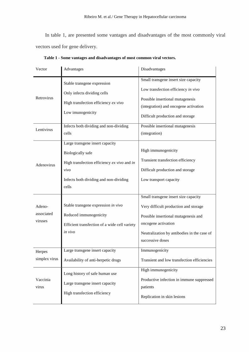
In table 1, are presented some vantages and disadvantages of the most commonly viral
vectors used for gene delivery.
Table 1 - Some vantages and disadvantages of most common viral vectors.
Vector Advantages Disadvantages
Retrovirus
Stable transgene expression
Only infects dividing cells
High transfection efficiency ex vivo
Low imunogenicity
Small transgene insert size capacity
Low transfection efficiency in vivo
Possible insertional mutagenesis
(integration) and oncogene activation
Difficult production and storage
Lentivirus Infects both dividing and non-dividing
cells
Possible insertional mutagenesis
(integration)
Adenovirus
Large transgene insert capacity
Biologically safe
High transfection efficiency ex vivo and in
vivo
Infects both dividing and non-dividing
cells
High immunogenicity
Transient transfection efficiency
Difficult production and storage
Low transport capacity
Adeno-
associated
viruses
Stable transgene expression in vivo
Reduced immunogenicity
Efficient transfection of a wide cell variety
in vivo
Small transgene insert size capacity
Very difficult production and storage
Possible insertional mutagenesis and
oncogene activation
Neutralization by antibodies in the case of
successive doses
Herpes
simplex virus
Large transgene insert capacity
Availability of anti-herpetic drugs
Immunogenicity
Transient and low transfection efficiencies
Vaccinia
virus
Long history of safe human use
Large transgene insert capacity
High transfection efficiency
High immunogenicity
Productive infection in immune suppressed
patients
Replication in skin lesions

Ribeiro M. et al./ Gene Therapy in Hepatocellular carcinoma
24
Despite their high potential for gene delivery, this vectors present some disadvantages
related to their safe application, as immune recognition, mutagenic integration (retroviral and
lentiviral vectors), and inflammatory toxicity (adenoviral vectors), which appear as limitations
for their use and led to a renewed interest in non-viral methods (Silke Schepelmann, 2008).
4.2 Non-viral vectors
Over the past decade, employing non-viral gene delivery systems in gene therapy has
attracted a lot of attention. Comparing with viral vectors these non-viral delivery systems
offer several advantages such as low immunogenicity and toxicity, easier and less expensive
large scale production; they are also relatively safer, and have the potential for more tissue
specificity. But, even with recent technological advances, the transfection efficiency of non-
viral vectors still needs to be improved so has their targeting ability. Nonetheless, recent
studies had shown that non-viral vectors indeed hold great promise for their future
development (Ding B, 2012; Al-Dosari MS, 2009).
The numerous non-viral methods for gene transfer that had been proposed can be divided
in physical methods and chemical methods.
In physical methods, researchers attempt to enhance gene delivery by exerting physical
forces. Methods such as needle and jet injection, hydrodynamic gene transfer, gene gun
delivery, electroporation and sonoporation had been described. Chemical methods mainly
involve the employment of chemical vectors with cationic components, currently in use
include are cationic lipids (forming lipoplexes upon mixing with DNA), cationic polymers as
PEI, peptides or dextrans (forming polyplexes upon mixing with DNA) and inorganic
nanoparticles (Al-Dosari MS, 2009).
Ribeiro M. et al./ Gene Therapy in Hepatocellular carcinoma
25
4.2.1 Cationic liposomes
Cationic liposomes are presented as the most promising vectors among the non-viral
systems and have been extensively used. This widespread application is due to a number of
important advantages, including their capacity to transport large amounts of genetic material;
their physico-chemical versatility, allowing innumerous modifications; their easy and
inexpensive large scale production; and their low immunogenic response. Since cationic
liposomes were first described for gene delivery, an increasing number of new cationic lipids
have been produced and used in transfection protocols of different cell lines, animal models
and patients submitted to gene therapy clinical trials. At present, 6.4% of the protocols
approved for gene therapy trials involve lipoplexes, these being mainly applied in the
treatment of cancer and cystic fibrosis (Duarte S, 2012).
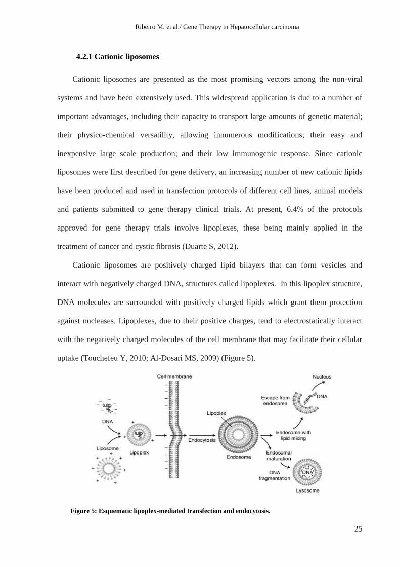
Cationic liposomes are positively charged lipid bilayers that can form vesicles and
interact with negatively charged DNA, structures called lipoplexes. In this lipoplex structure,
DNA molecules are surrounded with positively charged lipids which grant them protection
against nucleases. Lipoplexes, due to their positive charges, tend to electrostatically interact
with the negatively charged molecules of the cell membrane that may facilitate their cellular
uptake (Touchefeu Y, 2010; Al-Dosari MS, 2009) (Figure 5).
Figure 5: Esquematic lipoplex-mediated transfection and endocytosis.
Ribeiro M. et al./ Gene Therapy in Hepatocellular carcinoma
26
For the formation of liposomes for gene transfection a solution of cationic lipids with
neutral helper lipids is often used and liposome formulations used in genetic therapy seek to
find a compromise between stability and toxicity (Balazs DA, 2011; Al-Dosari MS, 2009).
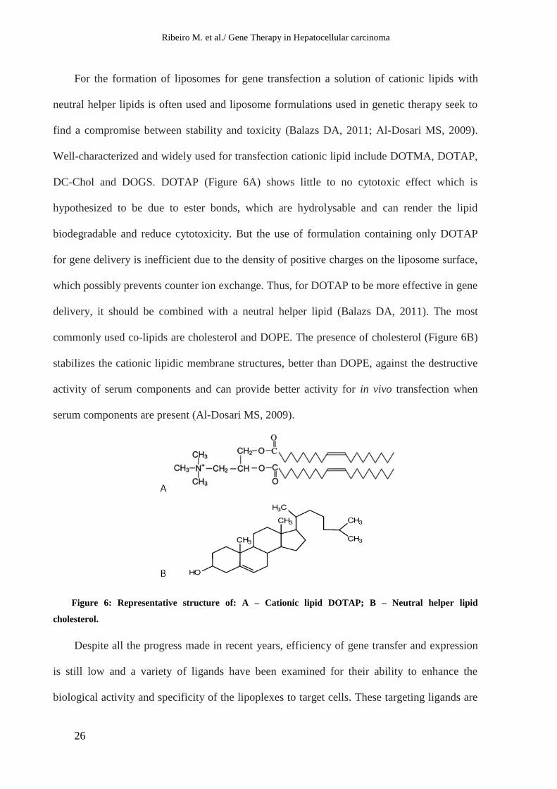
Well-characterized and widely used for transfection cationic lipid include DOTMA, DOTAP,
DC-Chol and DOGS. DOTAP (Figure 6A) shows little to no cytotoxic effect which is
hypothesized to be due to ester bonds, which are hydrolysable and can render the lipid
biodegradable and reduce cytotoxicity. But the use of formulation containing only DOTAP
for gene delivery is inefficient due to the density of positive charges on the liposome surface,
which possibly prevents counter ion exchange. Thus, for DOTAP to be more effective in gene
delivery, it should be combined with a neutral helper lipid (Balazs DA, 2011). The most
commonly used co-lipids are cholesterol and DOPE. The presence of cholesterol (Figure 6B)
stabilizes the cationic lipidic membrane structures, better than DOPE, against the destructive
activity of serum components and can provide better activity for in vivo transfection when
serum components are present (Al-Dosari MS, 2009).
Figure 6: Representative structure of: A – Cationic lipid DOTAP; B – Neutral helper lipid
cholesterol.
Despite all the progress made in recent years, efficiency of gene transfer and expression
is still low and a variety of ligands have been examined for their ability to enhance the
biological activity and specificity of the lipoplexes to target cells. These targeting ligands are
Ribeiro M. et al./ Gene Therapy in Hepatocellular carcinoma
27
selected based upon specific target cell receptors and include, folates, haloperidol and
transferrin (Balazs DA, 2011). Transferrin is a popular ligand for delivery of anticancer drugs
to solid tumors in vivo since cancer cells presents elevated levels of transferrin receptor (TfR)
expression, attributed to the vigorous proliferation and requirement of high iron level for their
growth. There is also indication of higher affinity of the TfR to transferrin (Tf) which can be
useful for a targeted therapy (Lu Q, 2008). Some studies showed that using Tf as ligand
increases gene transfection efficiency (Lu Q, 2008; Zhong ZR, 2007).
In this study we intend to test the therapeutic potential of gene therapy using suicide
genes (HSV-TK/ GCV or CD/ 5-FC) in monotherapy and in combination with conventional
anticarcinogenic agents (Doxorrubicin) and new targeted drugs (proteassome inhibitor MG-
262) in the hepatocellular carcinoma cell lines, HUH-7 and Hep-G2 in culture. The
mechanisms involved in the toxicity will be also studied, namely the mechanisms involved in
cell death and changes in cell cycle.
Ribeiro M. et al./ Gene Therapy in Hepatocellular carcinoma
28
MATERIALS AND METHODS
1- Materials
MG-262 was obtained from BiomoL (USA). Doxorubicin, Resazurin, May-Grünwald
solution and Giemsa solution were purchased from Sigma (St. Louis, MO, USA). 5-
Fluorocitosine and Ganciclovir were purchased from Invivogen (Toulouse, France).
FBS and DMEM medium were purchased from GIBCO (Barcelona, Spain).
The plasmid pCMVTK that encodes the therapeutic gene thymidine kinase, the
pCDβGEO plasmid that encodes the therapeutic gene cytosine deaminase gene under the
control of the CMV promoter and the plasmid pCMVGFP, that encodes the reporter gene
GFP, were kindly provided by Professora Doutora Maria da Conceição Pedroso de Lima from
Centre for Neuroscience and Cell Biology of Coimbra.
Plasmids were amplified according to the manufacturer’s instructions using the kit
GenElute™ Endotoxin-free Plasmid Midiprep Kit purchased from Sigma (St. Louis, MO,
USA).
The cationic lipid DOTAP, cholesterol (Chol), and the human holo-transferrin were
obtained from Sigma (St. Louis, MO, USA).
The kit FITC-labelled annexin V (AV) and propidium iodide (PI) were obtained from
Immunotec (Canada). PI/RNase kit used for cell cycle analyses for flow cytometry was
purchased from Immunostep (Salamanca, Spain).
Ribeiro M. et al./ Gene Therapy in Hepatocellular carcinoma
29
2- Cell line culture conditions
The human HUH-7 cell line, offered by Professora Doutora Maria Conceição Pedroso
Lima (Center for Neuroscience and Cell biology), was established by Nakabayashi et al.,
(1982) from hepatoma tissue of a 57-yr-old Japanese male with well differentiated
hepatocellular carcinoma which expressed high levels of mutated p53 (Carloni V, 2005).
The human Hep-G2 cell line, offered by Professora Doutora Filomena Botelho, from
Biophysics/Biomathematics of the Faculty of Medicine, University of Coimbra, was
established by Aden et al., (1979) from a liver tumor biopsy, with normal p53 expression,
obtained from a 15-yr-old Caucasian male. The morphological characteristics and epithelial
cell shape were compatible with that of liver parenchymal cells. Histology of the liver biopsy
revealed well differentiated hepatocellular carcinoma with a trabecular pattern.
The cell lines were maintained in DMEM medium supplemented with 10% FBS, L-
glutamine 2mM, NaHCO3, penicillin 100U/mL and streptomycin 100μg/mL at 37ºC in a
humidified incubator containing 5% CO2. Before the assays, both cells were seeded 24h
before. at a density of 50000 cells per cm2.
3- Cationic liposomes and lipoplexes preparation
Cationic liposomes composed of DOTAP and Chol (1:1 molar ratio) were prepared by
the ethanol injection method (Cardoso AL, 2007), (Penacho N, 2009). The lipids, from stock
solutions in chloroform, were mixed in a glass tube to a final concentration of 6mM and then
dried under nitrogen flow in order to obtain a thin lipid film. The dried lipid film was
dissolved in 100µL of pure ethanol and the resulting solution injected, using a Hamilton
syringe, under vortex, into 900µL of HBS buffer (10mM NaCl, 20mM HEPES, pH 7.4). The
Ribeiro M. et al./ Gene Therapy in Hepatocellular carcinoma
30
resulting MLVs (multilamellar vesicles) were then sonicated for 10 minutes to obtain SUVs
(small unilamellar vesicles). The suspension was stored at 4ºC until use.
Transferrin-associated lipoplexes (Tf-lipoplexes) were prepared by adding liposomes (the
amount required for the different charge ratio) to 100µl of a HBS solution containing 320
μg/ml of transferrin (to obtain a final concentration of 32µg of transferrin per µg DNA). This
mixture was incubated at room temperature for 15 minutes to allow interaction between
transferrin and liposomes. Then 100μl of HBS buffer containing 1μg plasmid DNA
(pCMVTK, pCDβGEO or pCMVGFP) was added and the solution was further incubated at
room temperature for 15 minutes (Neves S, 2009).
For complexes with different charge ratios (+/-), amount of DNA remained constant
(1mg DNA corresponds to 3.03nmol of negative charges) and only varying the amount of
liposomes. The complexes were used immediately after being prepared.
4- In vitro gene transfer
Cells, plated in 48 well plates 24h before assay, were rinsed with serum-free medium and
then covered with 0.3ml of DMEM (without serum) before lipoplexes were added. The
complexes were gently added to cells in a volume of 0.2ml per well. After incubation for 4 h
(in 5% CO2 at 37 °C), the medium was replaced with DMEM supplemented with 10% FBS,
L-glutamine 2mM, NaHCO3, penicillin 100U/mL and streptomycin 100μg/mL, and the cells
were further incubated under different experimental conditions.
5- Transfection activity
Transfection efficiency mediated by the complexes was evaluated using the reporter gene
green fluorescent protein (GFP). For this purpose, HUH-7 and Hep-G2 cells were plated in 6
Ribeiro M. et al./ Gene Therapy in Hepatocellular carcinoma
31
well plates and treated as previously described, using the appropriate volumes. 48h or 72h
after transfection the percentage of transfected cells was accessed by flow cytometry. For that,
cellswere trypsinized, centrifuged at 300xg for 5min, washed twice with PBS, ressuspended
in FACSflow buffer and analyzed in a FACScalibur cytometer (BD Biosciences, Heildelberg,
Germany) equipped with an argon-ion laser emitting at 488nm which recorded forward scatter
(FSC), side scatter (SSC) and green fluorescence (FL1). FSC and SSC data were used to
identify viable cells, gates were set to exclude cellular debris and results were evaluated to
determine the number of transfected cells (percentage of cells expressing GFP).
6- Cytotoxicity studies, evaluation of cell viability
To evaluate the cytotoxicity induced by suicide gene therapy in these cell lines, HUH-7
and Hep-G2 cells were transfected with transferrin associated lipoplexes in the absence or
presence of GCV, in a range of concentrations from 0,05mM to 0,5mM, or 5-FC, in a range of
concentrations from 5mM to 50mM, during 168h.
To check for possible synergistic effects, we performed combination treatments of the
suicide gene therapy (TK/GCV or CD/5-FC) with Doxorubicin or MG262, using doses below
the IC50 of both the drugs and pro-drugs during 168h.
Cell viability, under the different experimental conditions, was assessed by Alamar Blue
assay. In this assay the metabolic activity of cells is measured through the conversion of the
active compound of the Alamar Blue resazurin (oxidized form) to resorufin (reduced form) by
viable cells (O'Brien J, 2000). This results in colorimetric (absorbance) and fluorescence
changes. Resazurin is blue and non-fluorescent whereas resorufin is red and highly
fluorescent and the difference between the two compounds can be evaluated by spectroscopy.

Ribeiro M. et al./ Gene Therapy in Hepatocellular carcinoma
32
Figure 7: Alamar blue. Convertion of resazurin to resorufin.
The medium was removed and 300µl of 10% (v/v) resazurin in DMEM were added to
each well. After 2 h to 4 h incubation at 37 °C, 200μl of supernatant were collected from each
well and transferred to 96-well plates. The absorbance at 570 nm and 600 nm was measured
using a Mediators PhL luminometer (Mediators Diagnostika, Vienna, Austria). Cell viability
(as a percentage of control) was calculated according to the formula (A570−A600) of treated
cells×100/ (A570−A600) of control cells (Neves SS, 2006).
Readings were performed at 48h, 72h, 144h and 168h and after each reading the culture
medium was replaced by fresh one with the appropriate drugs concentration.
7- Morphological analysis
After incubation for 48h, under the different experimental conditions, the cells were
trypsined, centrifuged at 300xg for 5min and ressuspended in serum in order to obtain a
density of 50000cells/mL. Then, cells were placed on a slide and stained for 5min with May-
Grünwald solution (0.3% v/v in methanol) (Sigma, St. Louis, MO, USA), diluted in 1:1 ratio
with distilled water followed by staining with Giemsa solution (0.75% p/v in
glycerol/methanol 1:1) (Sigma, St. Louis, MO, USA) diluted 8x in distilled water for 20 min.
After rinsed with distilled water, cell morphology was analyzed by light microscopy using a
Motic AE31 microscope associated with a Moticam 2300 digital camera

Ribeiro M. et al./ Gene Therapy in Hepatocellular carcinoma
33
8- Flow cytometry assays
8.1 Cell death analysis
HUH-7 and Hep-G2 cells were added in triplicate to a 6-well culture plate, 24 h later they
were transfected and treated as described previously. After 48h incubation cells were
trypsinized, centrifuged at 300xg for 5min and incubated for 10min at 4ºC with 440μL
annexin buffer containing 5μL FITC-labelled annexin V (AV) and 2μL propidium iodide (PI).
Annexin V binds with high affinity to phospholipids negatively charged including
phosphatidylserine which is exposed in outer leaflet of the plasma membrane during apoptotic
process. PI is a non-specific DNA marker which is internalized by cells killed due to the
membrane integrity loss associated with necrosis (Darzynkiewicz Z, 1997). With this
technique, it is possible to distinguish non-apoptotic live cells (AV-FITC and PI negative),
early apoptotic cells (AV-FITC positive and PI negative), late apoptotic (positive for FITC-
AV and PI) and necrotic cells (positive for PI and AV-FITC negative).
Cells were then washed twice with PBS, ressuspended in the same buffer and analyzed in
a FACScalibur cytometer (BD Biosciences, Heildelberg, Germany) equipped with an argon
ion laser emitting at 488nm. FSC and SSC data were used to identify viable cells, and gates
were set to exclude cellular debris. The fluorescence of AV-FITC and PI was evaluated at 525
and 610nm, respectively.
The results were expressed as percentage of viable, early apoptotic, late
apoptotic/necrotic and necrotic cells (Darzynkiewicz Z, 1997; Neves SS, 2006).
8.2 Cell cycle analysis
The effect of the HSV-Tk/GCV or CD/5-FC suicide gene therapy and the described
combinations on the cell cycle phase distribution of the HCC cell lines was analyzed by flow
Ribeiro M. et al./ Gene Therapy in Hepatocellular carcinoma
34
cytometry using the PI/RNase kit (Immunostep, Salamanca, Spain) according to the
manufacturer’s instructions.
PI binds to DNA by intercalating into the double stranded macromolecule, it also binds
to RNA and is necessary to remove the RNA with a nucleases treatment (RNase) for optimal
DNA resolution. Mammalian cells are characterized for having three populations or definite
regions, cells in G2 and M phases of the cell cycle that have double DNA content, those in G0
and G1 phases, and a region correspond to cells in phase S. The quantity of binded PI is
proportional to the quantity of DNA and quantification of the DNA content permit us to know
the distribution of cells along the different phases of the cell cycle represented in
fluorescence intensity.
Briefly, cells were added in triplicate to a 6-well culture plate, 24 h later they were
transfected and treated as described previously. After 48h incubation, cells were detached
using trypsin/EDTA after previous wash with PBS. Cells were centrifuged at 300xg for 5min,
fixed with 200 µl of 70% ethanol by vortexing and incubating 30 minutes at – 4ºC and, then,
washed once with PBS + 2% BSA. Finally, 0,5 mL of propidium iodide solution (PI/RNase)
were added and after mixing well the cells were incubated for 15 min at room temperature
before analyzed by flow cytometry.
9- Statistical analysis
Statistical analyses were performed using GraphPad Prism software, version 5.0
(GraphPad Prism software, Inc., San Diego, CA). Statistically significant differences (p<0,05)
between the experimental groups were determined by Student’s t test.
Ribeiro M. et al./ Gene Therapy in Hepatocellular carcinoma
35
RESULTS
1- Transfection efficiency
Lipoplexes charge ratio is an important factor to take into account in the efficiency of this
type of transfection since it changes the characteristics of the lipoplexes, particularly in terms
size and surface charge. So, in order to choose the most effective formulation to transfect our
hepatocellular carcinoma cells, we examined the biological activity of complexes prepared at
different charge ratios cationic lipid / DNA (+ / -), from variable amounts of DOTAP: Chol
liposomes and 1 µg of pCMVGFP. Also, all formulations were made in the presence of
transferrin because it facilitates the internalization of the liposome by endocytosis with the aid
of the transferrin receptor increasing transfection efficiency.
Determination of the transfection efficiency using green fluorescence protein (GFP)
fluorescence can be noninvasively detected in living cells. Flow cytometric analysis of GFP
expression technique permits the measurement of transfection efficiency without the
requirement for cell fixation or sample preparation. For this purpose, cells suspended in PBS
were analyzed by flow cytometry 48 and 72h after transfection in order to determine the
percentage of positive cells (fluorescent cells that express GFP).
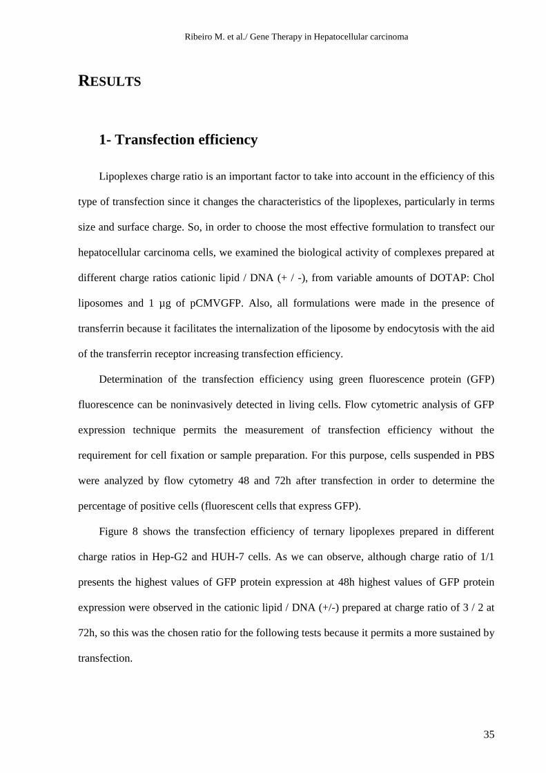
Figure 8 shows the transfection efficiency of ternary lipoplexes prepared in different
charge ratios in Hep-G2 and HUH-7 cells. As we can observe, although charge ratio of 1/1
presents the highest values of GFP protein expression at 48h highest values of GFP protein
expression were observed in the cationic lipid / DNA (+/-) prepared at charge ratio of 3 / 2 at
72h, so this was the chosen ratio for the following tests because it permits a more sustained by
transfection.
Ribeiro M. et al./ Gene Therapy in Hepatocellular carcinoma
36
CTL
Lipossom
es 1/1
2/1
3/2
0
3
6
9
12
15
72h
48h
HU
H-7
- T
ran
sfe
ctio
n e
ffic
ien
cy (
%)
CTL
Lipossom
es 1/1
2/1
3/2
0
3
6
9
12
1548h
72h
He
p-G
2 -
Tra
nsfe
ctio
n e
ffic
ien
cy (
%)
Figure 8: Transfection efficiency in HUH-7 and Hep-G2 using different lipoplex cationic lipid / DNA
(+/-) charge ratios. Cells were transfected with cationic liposomes containing DOTAP and cholesterol
complexed with 1 µg of pCMVGFP in the indicated charge ratios (+/-) in the presence of 32 µg of transferrin.
After incubation of cells with the complex for a period of 4 hours at 37 ° C, the culture medium was replaced
with medium enriched with 10% FBS and the cells were maintained in culture for another 48 or 72 hours. The
transfection efficiency was assessed in terms of percentage of cells expressing the GFP protein SD, measured
by flow cytometry in2 independent experiments.
2- Analysis of cell viability
2.1 Effect of HSV-TK/GCV or CD/5-FC treatment on the viability of
HUH-7 and Hep-G2 cells
In order to evalute the therapeutic effect of gene therapy using suicide genes (HSV-TK/
GCV or CD/ 5-FC) in hepatocellular carcinoma cell lines, transfected and non transfected
HUH-7 and Hep-G2 cells were cultured in the absence and in the presence of the appropriate
prodrug (GCV or 5-FC) for up to 168h. The antiproliferative effect was evaluated by the
Alamar Blue assay.
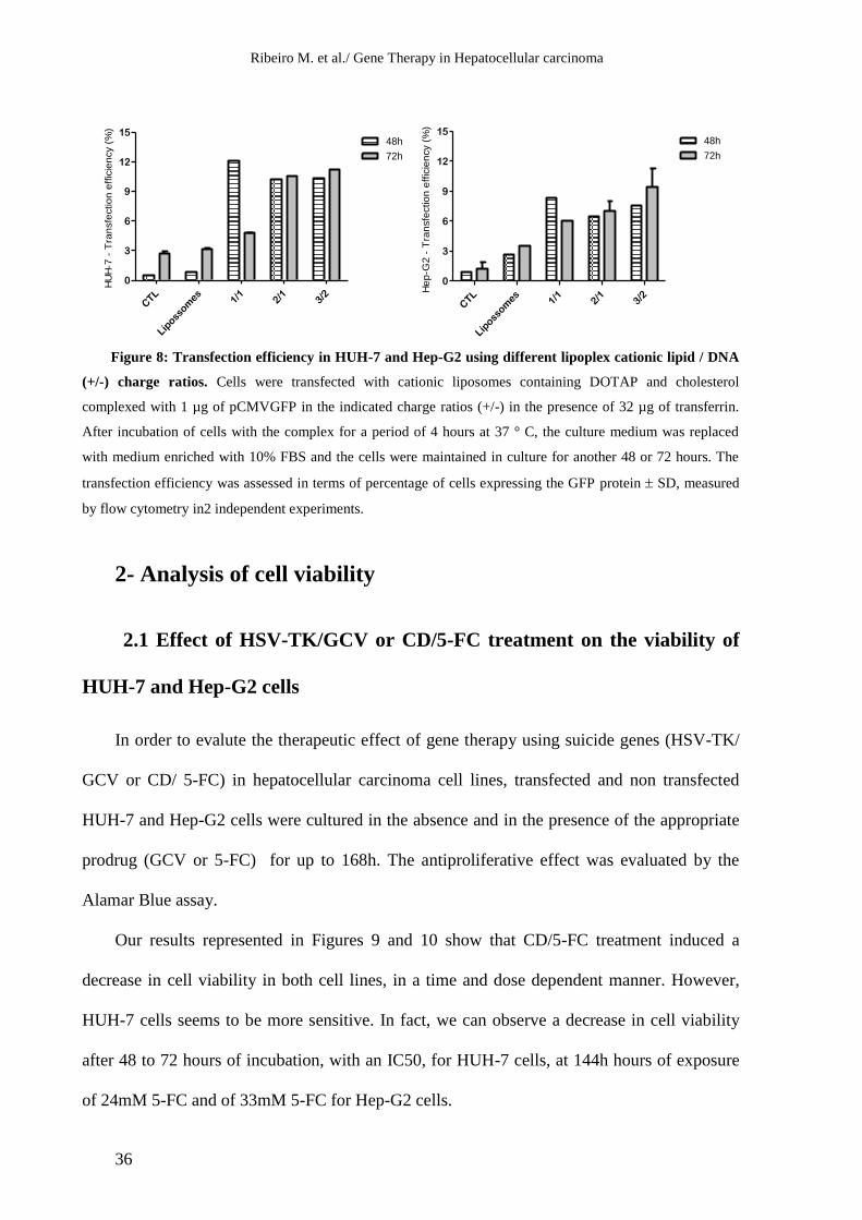
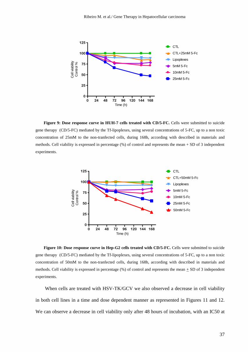
Our results represented in Figures 9 and 10 show that CD/5-FC treatment induced a
decrease in cell viability in both cell lines, in a time and dose dependent manner. However,
HUH-7 cells seems to be more sensitive. In fact, we can observe a decrease in cell viability
after 48 to 72 hours of incubation, with an IC50, for HUH-7 cells, at 144h hours of exposure
of 24mM 5-FC and of 33mM 5-FC for Hep-G2 cells.
Ribeiro M. et al./ Gene Therapy in Hepatocellular carcinoma
37
0 24 48 72 96 120 144 168
0
25
50
75
100
125
CTL
CTL+25mM 5-Fc
Lipoplexes
5mM 5-Fc
10mM 5-Fc
25mM 5-Fc
Time (h)
Cell
viabili
ty
Contr
ol %
Figure 9: Dose response curve in HUH-7 cells treated with CD/5-FC. Cells were submitted to suicide
gene therapy (CD/5-FC) mediated by the Tf-lipoplexes, using several concentrations of 5-FC, up to a non toxic
concentration of 25mM to the non-tranfected cells, during 168h, according with described in materials and
methods. Cell viability is expressed in percentage (%) of control and represents the mean + SD of 3 independent
experiments.
0 24 48 72 96 120 144 168
0
25
50
75
100
125 CTL
CTL+50mM 5-Fc
Lipoplexes
5mM 5-Fc
10mM 5-Fc
25mM 5-Fc
50mM 5-Fc
Time (h)
Cell
viabili
ty
Contr
ol %
Figure 10: Dose response curve in Hep-G2 cells treated with CD/5-FC. Cells were submitted to suicide
gene therapy (CD/5-FC) mediated by the Tf-lipoplexes, using several concentrations of 5-FC, up to a non toxic
concentration of 50mM to the non-tranfected cells, during 168h, according with described in materials and
methods. Cell viability is expressed in percentage (%) of control and represents the mean + SD of 3 independent
experiments.
When cells are treated with HSV-TK/GCV we also observed a decrease in cell viability
in both cell lines in a time and dose dependent manner as represented in Figures 11 and 12.
We can observe a decrease in cell viability only after 48 hours of incubation, with an IC50 at
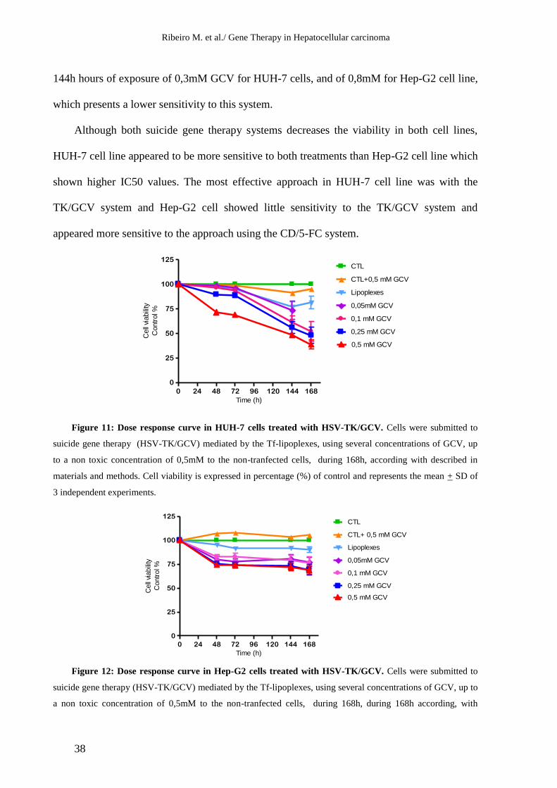
Ribeiro M. et al./ Gene Therapy in Hepatocellular carcinoma
38
0 24 48 72 96 120 144 168
0
25
50
75
100
125CTL
CTL+0,5 mM GCV
Lipoplexes
0,05mM GCV
0,1 mM GCV
0,25 mM GCV
0,5 mM GCV
Time (h)
Cell
viabili
ty
Contr
ol %
0 24 48 72 96 120 144 168
0
25
50
75
100
125CTL
CTL+ 0,5 mM GCV
Lipoplexes
0,05mM GCV
0,1 mM GCV
0,25 mM GCV
0,5 mM GCV
Time (h)
Cell
viabili
ty
Contr
ol %
144h hours of exposure of 0,3mM GCV for HUH-7 cells, and of 0,8mM for Hep-G2 cell line,
which presents a lower sensitivity to this system.
Although both suicide gene therapy systems decreases the viability in both cell lines,
HUH-7 cell line appeared to be more sensitive to both treatments than Hep-G2 cell line which
shown higher IC50 values. The most effective approach in HUH-7 cell line was with the
TK/GCV system and Hep-G2 cell showed little sensitivity to the TK/GCV system and
appeared more sensitive to the approach using the CD/5-FC system.
Figure 11: Dose response curve in HUH-7 cells treated with HSV-TK/GCV. Cells were submitted to
suicide gene therapy (HSV-TK/GCV) mediated by the Tf-lipoplexes, using several concentrations of GCV, up
to a non toxic concentration of 0,5mM to the non-tranfected cells, during 168h, according with described in
materials and methods. Cell viability is expressed in percentage (%) of control and represents the mean + SD of
3 independent experiments.
Figure 12: Dose response curve in Hep-G2 cells treated with HSV-TK/GCV. Cells were submitted to
suicide gene therapy (HSV-TK/GCV) mediated by the Tf-lipoplexes, using several concentrations of GCV, up to
a non toxic concentration of 0,5mM to the non-tranfected cells, during 168h, during 168h according, with

Ribeiro M. et al./ Gene Therapy in Hepatocellular carcinoma
39
described in materials and methods. Cell viability is expressed in percentage (%) of control and represents the
mean + SD of 3 independent experiments.
2.2 Effect of HSV-TK/GCV or CD/5-FC treatment in association with
Doxorubicin and MG262 on the viability of HUH-7 and Hep-G2 cells
In order to evaluate the cytotoxicity of suicide gene therapy associated with a
conventional chemotherapeutic agent and a drug directed to novel molecular targets HUH-7
and Hep-G2 cells were incubated with the pro-drugs in combination with Doxorubicin and
MG262. The IC50 for these drugs were previously determined (Data not shown) and in these
associations were used concentrations below the IC50.
As we can see in Figures 13 and 14, a greater reduction in cell viability in both cell lines,
was observed when associated the CD/5-FC system with Doxorubicin or MG262 showing an
addition synergism between the two therapies.
Using TK / GCV system in combination with Doxorubicin or MG262 (Figures 15 and
16), this effect was even more pronounced for Hep-G2 cell line especially in the combination
with Doxorubicin in which it can be observed an addition synergism.
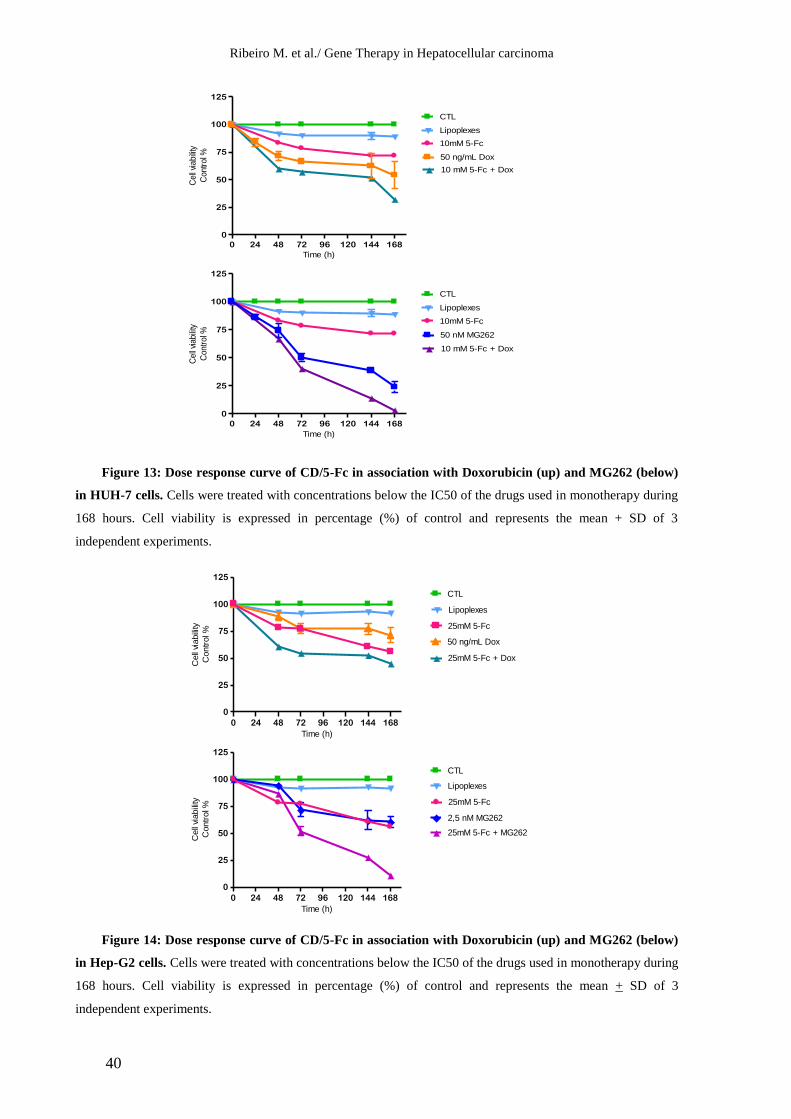
Ribeiro M. et al./ Gene Therapy in Hepatocellular carcinoma
40
0 24 48 72 96 120 144 168
0
25
50
75
100
125
CTL
Lipoplexes
10mM 5-Fc
10 mM 5-Fc + Dox
50 ng/mL Dox
Time (h)
Cell
viabili
ty
Contr
ol %
0 24 48 72 96 120 144 168
0
25
50
75
100
125
CTL
Lipoplexes
10mM 5-Fc
10 mM 5-Fc + Dox
50 nM MG262
Time (h)
Cell
viabili
ty
Contr
ol %
0 24 48 72 96 120 144 168
0
25
50
75
100
125
CTL
Lipoplexes
25mM 5-Fc
25mM 5-Fc + Dox
50 ng/mL Dox
Time (h)
Cell
viabili
ty
Contr
ol %
0 24 48 72 96 120 144 168
0
25
50
75
100
125
CTL
Lipoplexes
25mM 5-Fc
25mM 5-Fc + MG262
2,5 nM MG262
Time (h)
Cell
viabili
ty
Contr
ol %
Figure 13: Dose response curve of CD/5-Fc in association with Doxorubicin (up) and MG262 (below)
in HUH-7 cells. Cells were treated with concentrations below the IC50 of the drugs used in monotherapy during
168 hours. Cell viability is expressed in percentage (%) of control and represents the mean + SD of 3
independent experiments.
Figure 14: Dose response curve of CD/5-Fc in association with Doxorubicin (up) and MG262 (below)
in Hep-G2 cells. Cells were treated with concentrations below the IC50 of the drugs used in monotherapy during
168 hours. Cell viability is expressed in percentage (%) of control and represents the mean + SD of 3
independent experiments.
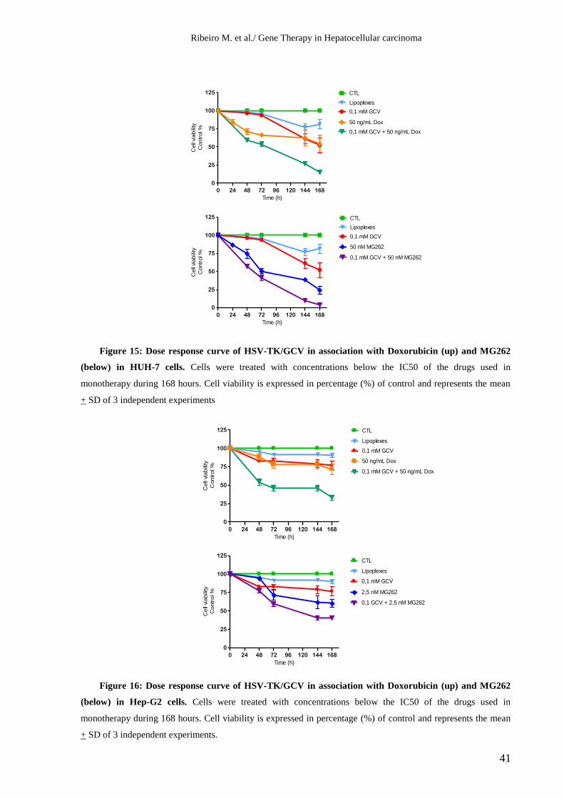
Ribeiro M. et al./ Gene Therapy in Hepatocellular carcinoma
41
0 24 48 72 96 120 144 168
0
25
50
75
100
125 CTL
Lipoplexes
0,1 mM GCV
0,1 mM GCV + 50 ng/mL Dox
50 ng/mL Dox
Time (h)
Cell
viabili
ty
Contr
ol %
0 24 48 72 96 120 144 168
0
25
50
75
100
125CTL
Lipoplexes
0,1 mM GCV
0,1 GCV + 2,5 nM MG262
2,5 nM MG262
Time (h)
Cell
viabili
ty
Contr
ol %
0 24 48 72 96 120 144 168
0
25
50
75
100
125 CTL
Lipoplexes
0,1 mM GCV
0,1 mM GCV + 50 ng/mL Dox
50 ng/mL Dox
Time (h)C
ell
viabili
ty
Contr
ol %
0 24 48 72 96 120 144 168
0
25
50
75
100
125 CTL
Lipoplexes
0,1 mM GCV
0,1 mM GCV + 50 nM MG262
50 nM MG262
Time (h)
Cell
viabili
ty
Contr
ol %
Figure 15: Dose response curve of HSV-TK/GCV in association with Doxorubicin (up) and MG262
(below) in HUH-7 cells. Cells were treated with concentrations below the IC50 of the drugs used in
monotherapy during 168 hours. Cell viability is expressed in percentage (%) of control and represents the mean
+ SD of 3 independent experiments
Figure 16: Dose response curve of HSV-TK/GCV in association with Doxorubicin (up) and MG262
(below) in Hep-G2 cells. Cells were treated with concentrations below the IC50 of the drugs used in
monotherapy during 168 hours. Cell viability is expressed in percentage (%) of control and represents the mean
+ SD of 3 independent experiments.
Ribeiro M. et al./ Gene Therapy in Hepatocellular carcinoma
42
3- Morphological analysis
Since tumour cell death mechanisms can interfere with the therapeutic strategy, we also
analysed the cytotoxic effect induced by the suicide gene therapy strategies associated with
MG262 and Doxorubicin in both cells lines by studying cell death process through
morphological analysis by optical microscopy and flow cytometry using the AV/PI staining.
Figures 17 and 18 show the morphology of cell smears stained with May-Grünwald-
Giemsa before (control) and after treatments during 48h in HUH-7 cell lines (Figure 17) and
Hep-G2 cell line (Figure 18).
As it can be seen, both in Figure 17 and Figure 18, cells treated with liposomes are
morphologically similar the control, the same happening with cells treated with prodrugs
(GCV or 5-FC) alone, which present only few morphologically apoptotic cells. Cells treated
with the suicide systems (TK/GCV or CD/5-FC) alone or in combination with doxorubicin or
MG262 have mainly typical morphological characteristics of apoptosis, such as cellular
retraction, nuclear fragmentation, blebbing and apoptotic bodies’ formation. The presence of
some necrotic cells with characteristics as the swelling of the cell, cell lysis or release of
cytoplasmic contents were also observed.
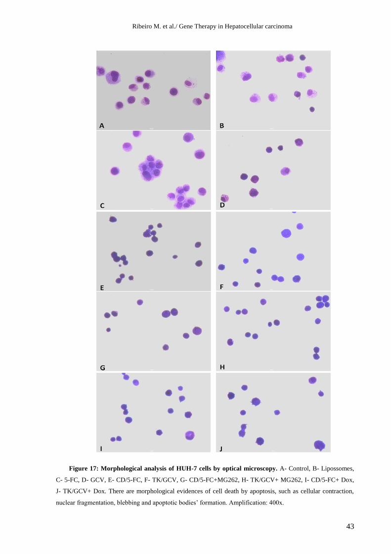
Ribeiro M. et al./ Gene Therapy in Hepatocellular carcinoma
43
Figure 17: Morphological analysis of HUH-7 cells by optical microscopy. A- Control, B- Lipossomes,
C- 5-FC, D- GCV, E- CD/5-FC, F- TK/GCV, G- CD/5-FC+MG262, H- TK/GCV+ MG262, I- CD/5-FC+ Dox,
J- TK/GCV+ Dox. There are morphological evidences of cell death by apoptosis, such as cellular contraction,
nuclear fragmentation, blebbing and apoptotic bodies’ formation. Amplification: 400x.
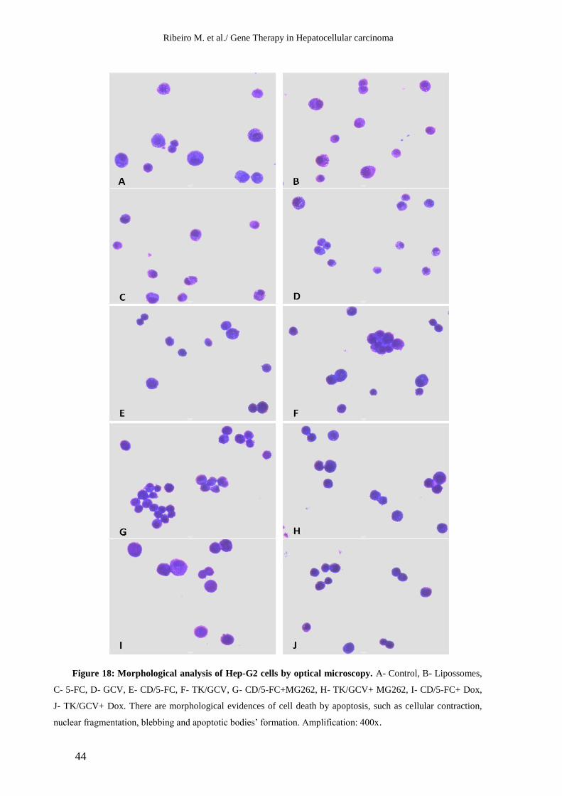
Ribeiro M. et al./ Gene Therapy in Hepatocellular carcinoma
44
Figure 18: Morphological analysis of Hep-G2 cells by optical microscopy. A- Control, B- Lipossomes,
C- 5-FC, D- GCV, E- CD/5-FC, F- TK/GCV, G- CD/5-FC+MG262, H- TK/GCV+ MG262, I- CD/5-FC+ Dox,
J- TK/GCV+ Dox. There are morphological evidences of cell death by apoptosis, such as cellular contraction,
nuclear fragmentation, blebbing and apoptotic bodies’ formation. Amplification: 400x.
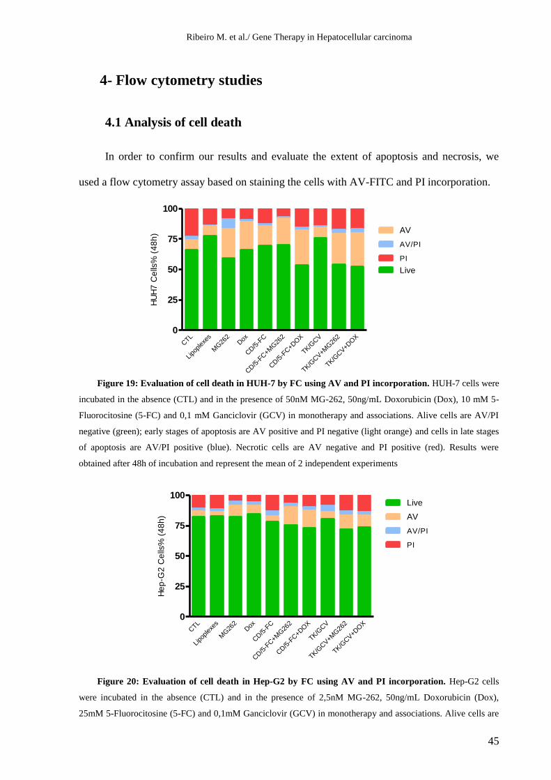
Ribeiro M. et al./ Gene Therapy in Hepatocellular carcinoma
45
CTL
Lipo
plex
es
MG26
2Dox
CD/5
-FC
CD/5
-FC+M
G26
2
CD/5
-FC+D
OX
TK/G
CV
TK/G
CV+M
G26
2
TK/G
CV+D
OX
0
25
50
75
100
AV/PI
PI
AV
Live
HU
H7
Ce
lls%
(4
8h
)
CTL
Lipo
plex
es
MG26
2Dox
CD/5
-FC
CD/5
-FC+M
G26
2
CD/5
-FC+D
OX
TK/G
CV
TK/G
CV+M
G26
2
TK/G
CV+D
OX
0
25
50
75
100
AV/PI
PI
Live
AV
He
p-G
2 C
ells
% (
48
h)
4- Flow cytometry studies
4.1 Analysis of cell death
In order to confirm our results and evaluate the extent of apoptosis and necrosis, we
used a flow cytometry assay based on staining the cells with AV-FITC and PI incorporation.
Figure 19: Evaluation of cell death in HUH-7 by FC using AV and PI incorporation. HUH-7 cells were
incubated in the absence (CTL) and in the presence of 50nM MG-262, 50ng/mL Doxorubicin (Dox), 10 mM 5-
Fluorocitosine (5-FC) and 0,1 mM Ganciclovir (GCV) in monotherapy and associations. Alive cells are AV/PI
negative (green); early stages of apoptosis are AV positive and PI negative (light orange) and cells in late stages
of apoptosis are AV/PI positive (blue). Necrotic cells are AV negative and PI positive (red). Results were
obtained after 48h of incubation and represent the mean of 2 independent experiments
Figure 20: Evaluation of cell death in Hep-G2 by FC using AV and PI incorporation. Hep-G2 cells
were incubated in the absence (CTL) and in the presence of 2,5nM MG-262, 50ng/mL Doxorubicin (Dox),
25mM 5-Fluorocitosine (5-FC) and 0,1mM Ganciclovir (GCV) in monotherapy and associations. Alive cells are
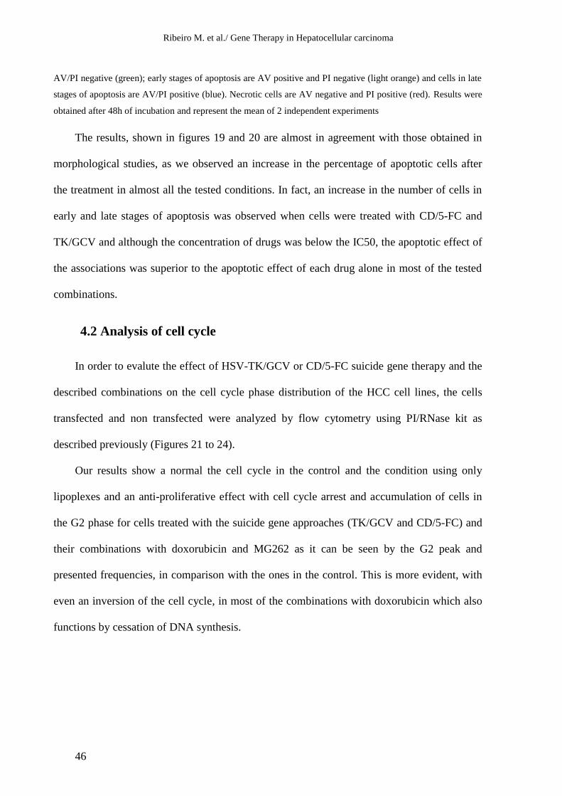
Ribeiro M. et al./ Gene Therapy in Hepatocellular carcinoma
46
AV/PI negative (green); early stages of apoptosis are AV positive and PI negative (light orange) and cells in late
stages of apoptosis are AV/PI positive (blue). Necrotic cells are AV negative and PI positive (red). Results were
obtained after 48h of incubation and represent the mean of 2 independent experiments
The results, shown in figures 19 and 20 are almost in agreement with those obtained in
morphological studies, as we observed an increase in the percentage of apoptotic cells after
the treatment in almost all the tested conditions. In fact, an increase in the number of cells in
early and late stages of apoptosis was observed when cells were treated with CD/5-FC and
TK/GCV and although the concentration of drugs was below the IC50, the apoptotic effect of
the associations was superior to the apoptotic effect of each drug alone in most of the tested
combinations.
4.2 Analysis of cell cycle
In order to evalute the effect of HSV-TK/GCV or CD/5-FC suicide gene therapy and the
described combinations on the cell cycle phase distribution of the HCC cell lines, the cells
transfected and non transfected were analyzed by flow cytometry using PI/RNase kit as
described previously (Figures 21 to 24).
Our results show a normal the cell cycle in the control and the condition using only
lipoplexes and an anti-proliferative effect with cell cycle arrest and accumulation of cells in
the G2 phase for cells treated with the suicide gene approaches (TK/GCV and CD/5-FC) and
their combinations with doxorubicin and MG262 as it can be seen by the G2 peak and
presented frequencies, in comparison with the ones in the control. This is more evident, with
even an inversion of the cell cycle, in most of the combinations with doxorubicin which also
functions by cessation of DNA synthesis.
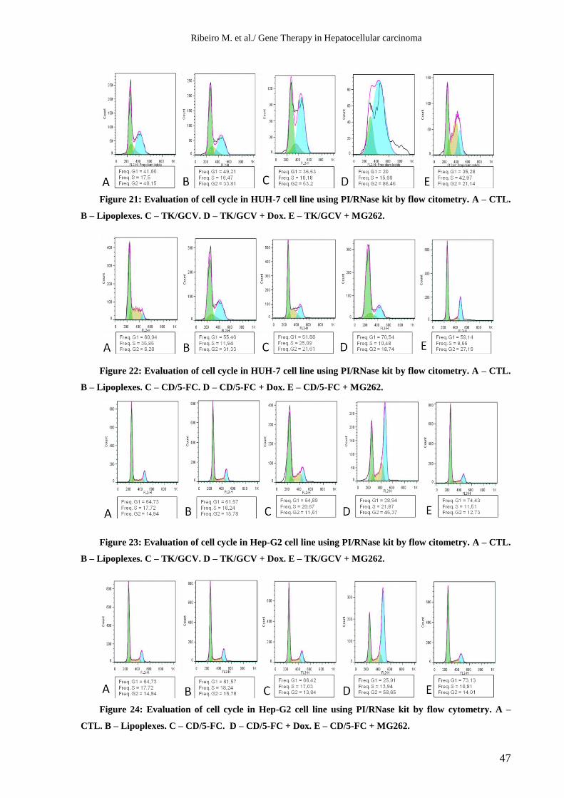
Ribeiro M. et al./ Gene Therapy in Hepatocellular carcinoma
47
Figure 21: Evaluation of cell cycle in HUH-7 cell line using PI/RNase kit by flow citometry. A – CTL.
B – Lipoplexes. C – TK/GCV. D – TK/GCV + Dox. E – TK/GCV + MG262.
Figure 22: Evaluation of cell cycle in HUH-7 cell line using PI/RNase kit by flow citometry. A – CTL.
B – Lipoplexes. C – CD/5-FC. D – CD/5-FC + Dox. E – CD/5-FC + MG262.
Figure 23: Evaluation of cell cycle in Hep-G2 cell line using PI/RNase kit by flow citometry. A – CTL.
B – Lipoplexes. C – TK/GCV. D – TK/GCV + Dox. E – TK/GCV + MG262.
Figure 24: Evaluation of cell cycle in Hep-G2 cell line using PI/RNase kit by flow cytometry. A –
CTL. B – Lipoplexes. C – CD/5-FC. D – CD/5-FC + Dox. E – CD/5-FC + MG262.
Ribeiro M. et al./ Gene Therapy in Hepatocellular carcinoma
48
DISCUSSION AND CONCLUSION
HCC is a deadly cancer whose incidence has increased dramatically over the past
decades. In Europe and the United States, this increasing incidence exists mainly due to
the epidemic obesity and rise in hepatitis C virus infection (Jemal A, 2011).
Although many studies have been made and protocols investigated to treat HCC, the
majority of patients with advanced HCC still die from the disease and there is an unmet
need for other drugs, or treatment options, either as a single agents or in combination
(Severi T, 2010 ). This lack of safe and efficient therapeutic options against many types
of cancer, as in liver cancer, gives rise to the necessity to develop alternative therapies
that can achieve more effective results and encouraging the development of new gene
therapy applications for these diseases (Sangro B, 2010).
Suicide gene therapy, opposing to the conventional antiproliferation drugs, that
usually have toxicity both in malignant and non-malignant cells, aims to improve
conventional cancer chemotherapy by selectively activating the prodrug at the tumor site,
thus permitting higher concentrations of active drug at the tumor and minimizing tumor
burden, by limiting the exposition of the patient’s normal tissues and organs to potentially
harmful quantities of drugs (Hwang, 2006), (Silke Schepelmann, 2008).
Insufficiency of gene transfection and target-orientated gene therapy are main
obstacles to the application of gene therapy. The selection of an appropriate vector system
for transferring the desired therapeutic gene into the target is an important issue in
developing a safe and efficient gene therapy.
Viral vectors generally facilitate highly efficient transfer but they lack desirable
targeting ability and can be immunogenic, cytopathic or recombinogenic (Silke
Schepelmann, 2008). This safety concerns led to the exploration of non-viral vectors, as
the cationic liposomes, which have been tested for safety with little or no toxicity

Ribeiro M. et al./ Gene Therapy in Hepatocellular carcinoma
49
reported (Lu Q, 2008) and are simple to prepare, can complex with large amounts of
DNA, and are reliable to transfect even into non-dividing cells, without the risk of
random integration (Al-Dosari MS, 2009), (Balazs DA, 2011). Though, cationic
liposomes lack tumor specificity and have relatively low transfection efficiencies a
variety of ligands as transferrin is used to enhance the biological activity and specificity
of the lipoplexes to target cells. (Balazs DA, 2011)
With this regards, we evaluated, using HCC cell lines (HUH-7 and Hep-G2 cells),
the in vitro therapeutic potential, using liposomes coupled with transferrin, of suicide
gene approaches (HSV-TK/ GCV or CD/ 5-FC), in monotherapy and in combination with
conventional anticarcinogenic agents (Doxorrubicin) and new targeted drugs
(proteassome inhibitor MG-262).
In fact there are already several clinical trials using HSV-TK/ GCV or CD/ 5-FC
systems, in various types of cancer, but not many are in HCC and either with the
proposed treatment combinations or formulation of vectors used for the gene therapy
(Rainov, 2000; Voges J, 2003; Nasu Y, 2007; Xu F, 2009; Li N, 2007; Sangro B M. G.,
2010 ; Pandha HS, 1999; Freytag SO, 2003; Crystal RG, 1997; Cunningham C, 2001;
Zarogoulidis P, 2012)
We first evaluated the transfection efficiency of several formulation DOTAP:Chol
associated with transferrin prepared at different (+/-) charge ratio. Our results showed
that, even in the chosen charge ratio of 3/2, the transfection efficiency levels were low
(approximately 10% of transfected cells). Similar results were obtained by Neves et al.
(2006) where the highest values of transgene expression in the Tf-lipoplexes was found
for the 3/2 lipid/DNA (+/−) charge ratio at which the zeta potential of the lipoplexes is
close to neutrality and large aggregates formed.
Ribeiro M. et al./ Gene Therapy in Hepatocellular carcinoma
50
Our results on the toxicity mediated by suicide gene therapy showed decreases in
viability in both CD/5-FC and HSV-TK/GCV treatment, with both cell lines, depending
on the applied prodrug dose and incubation period. HUH-7 cell line appeared to be more
sensitive than Hep-G2 cell line to both treatments. The most effective approach in HUH-7
cell line was with the TK/GCV system and Hep-G2 cell appeared more sensitive to the
approach using the CD/5-FC system although there were not remarkable differences.
Having into account the transfection efficiency levels the cytotoxicity results can be
explained by the bystander effect and probably the characteristics of the different cell
lines. The CD/5-FC system has a significant local bystander effect that does not require
direct cell contact, since 5-FU can move through the cell membrane by non-facilitated
diffusion. 5- FC also accumulates after multiple doses, unlike GCV (Dachs GU, 2009). In
vitro experiments showed that 1– 30% of CD-expressing cells exposed to 5-FC could
generate sufficient 5-FU to inhibit the growth of the untransfected neighboring cell (Al-
Dosari MS, 2009). In vivo studies on tumours containing only 2% CD transduced cells
were shown to produce significant tumour reductions (Huber BE, 1994). Since GCV
triphosphate cannot passively diffuse to neighboring cells, the bystander effect of the
TK/GCV system was shown to be mediated by different mechanisms. It was
demonstrated it requires cell-to-cell contact and the requirement of gap junctions was
evidenced by comparing the bystander effect in tumor cell lines transfected or not with
connexin genes. It can also be mediated via apoptotic bodies generated from dying TK-
expressing cells and phagocytosed by unmodified neighboring cells which was
demonstrated with flow cytometry and electron microscopic analysis and a gap junction-
independent local bystander effect mediated by soluble factors, likely corresponding to
phosphorylated GCV metabolites was also reported. (Duarte S, 2012). More recently, the
expression of E-cadherin, which is involved in the formation and function of gap
Ribeiro M. et al./ Gene Therapy in Hepatocellular carcinoma
51
junctions, was shown to correlate strongly with the TK/GCV bystander effect (Garcia-
Rodríguez L, 2011).
The combination of CD/5-FC system with doxorubicin or MG262 demonstrates a
greater reduction in cell viability even with the use of doses lower than the IC50 with a
more pronounced effect in the combinations using MG262. With the TK/GCV system in
combination with doxorubicin or MG262 this effect was even more pronounced
especially for Hep-G2 cells in the combination with MG262 in which can be observed an
addition synergism. In fact the same or higher antiproliferative effect is achieved at lower
concentrations than those obtained for all the tested conditions in monotherapy. And the
reduction of concentrations of new or conventional drugs and even the ones of the
prodrugs, may decrease their potential side effects.
In our work, we also concluded that apoptosis is the main mechanism involved in the
cytotoxicity induced by the tested drugs. In fact, there are morphological evidences
characteristic of apoptotic cell death, such as cellular retraction, nuclear fragmentation,
blebbing and apoptotic bodies’ formation. These morphological changes observed in
microscopic slides are in agreement with those observed by FC analysis using AV/PI
incorporation where there is an increase in percentage of apoptotic cells after treatment
with the tested conditions, although in this study the differences were not pronounced,
probably due to the short period of incubation of 48 hours.
Our results are in concordance with other studies. In fact, some authors found
activation of caspases (Tomicic MT, 2002), and the aggregation of receptors associated
with cell death FAS (FADD) in cells submitted to the action of HSV-TK/GCV system
(Beltinger C, 1999). Other studies showed that, upon application of this system GCV-
induced apoptosis occurred mainly by activating the mitochondrial damage pathway, with
cytochrome C release, accumulation of p53 protein and decline in BCL-2 levels (Tomicic
Ribeiro M. et al./ Gene Therapy in Hepatocellular carcinoma
52
MT, 2002; Beltinger C, 1999; Fischer U, 2005). And similarly to HSV-TK/GCV system,
apoptosis is also involved in the mechanism of cytotoxicity induced by the CD/5-FC
suicide system. It was reported that in glioma cells, the mitochondrial pathway is
involved in the process of cell death induced by both suicide gene systems, while p53 and
death receptors are not implicated in such process. But even though mitochondrial
pathway is involved, the mechanisms of modulation of BCL-2 proteins were found to be
different (Fischer U, 2005).
In the analyses of the effect of HSV-TK/GCV or CD/5-FC suicide gene therapy and
the described combinations on cell cycle, our results showed an anti-proliferative effect
with a cell cycle arrest and accumulation of cells in the G2 phase, which is consistent
with the cessation of DNA synthesis effect of suicide gene therapy. This results were
even more evident in most of the combination with doxorubicin.
Some studies demonstrated that erroneous GCV triphosphate incorporation into
DNA, results in S-phase delay, as well as G2-phase arrest by the activation of 3’
exonuclease and post-replicative endonuclease repair mechanisms (Halloran PJ, 1998 ).
As a result of GCV-induced cell cycle arrest, Wei et al.(1998) found that apoptosis rather
than a direct chemical effect, was involved in HSV-TK-transduced B16F10 melanoma
cell death. In a recent study, the relevance of the cell cycle control towards the sensitivity
of pancreatic tumor cells to the cytotoxicity induced by the HSV-TK/ GCV system was
demonstrated, since a Chk1 activation was associated with a greater HSV-TK/GCV
extent of cell death (Abate-Daga D, 2010). Results in combinations with doxorubicin are
justifiable through doxorubicin mechanisms of action because it acts as an intercalating
agent and wedges between the DNA bases, blocking DNA synthesis and transcription. It
also inhibits the activity of topoisomerase type II and this leads to breaks in the genomic

Ribeiro M. et al./ Gene Therapy in Hepatocellular carcinoma
53
DNA. Both of these mechanisms result in DNA disruption which also justifies the
observed effect in the cell cycle (Kim HS, 2009).
In overall in the present study suicide gene therapy using HSV-TK/GCV or CD/5-FC
resulted in decreased viability of HCC cell lines in culture, depending on the respective
pro-drug dose and incubation time, inducing cell death by apoptosis. Combinations of
these systems with lower doses than the IC50 of MG-262 or Doxorubicin enhanced the
antiproliferative and cytotoxic effect meaning that lower doses of other drugs with the
potentiation of the results and reduced toxicity levels and side effects. This
antiproliferative effect was in agreement with the observed cell cycle alterations
(accumulation in G2/M phase).
These results suggest that suicide gene therapy HSV-TK/GCV or CD/5-FC may
constitute a new potential therapeutic approach in HCC not only in monotherapy, but also
in association with conventional therapies or new targeted drugs.
In the future the systems and combinations used should be tested in pre-clinical trials
in an animal model of hepatocellular carcinoma.
Ribeiro M. et al./ Gene Therapy in Hepatocellular carcinoma
54
REFERENCES
Abate-Daga D, G.-R. L. (Oct de 2010). Cell cycle control pathways act as conditioning factors for
TK/GCV sensitivity in pancreatic cancer cells. Biochim Biophys Acta, 1175-85.
Aden DP, F. A. (Dec de 1979). Controlled synthesis of HBsAg in a differentiated human liver.
Nature, 615-6.
Al-Allaf FA, C. C. (Dec de 2010). LDLR-Gene therapy for familial hypercholesterolaemia:
problems, progress, and perspectives. Int Arch Med., 3:36.
Al-Dosari MS, G. X. (Dec de 2009). Nonviral gene delivery: principle, limitations, and recent
progress. AAPS J., 671-81.
Alves RC, A. D. (2011). Advanced hepatocellular carcinoma. Review of targeted molecular
drugs. Ann Hepatol., 21-7.
Avila MA, B. C. (Jun de 2006). New therapies for hepatocellular carcinoma. Oncogene., 3866-84.
Baiz D, P. G. ( Mar de 2009). Bortezomib arrests the proliferation of hepatocellular carcinoma
cells HepG2 and JHH6 by differentially affecting E2F1, p21 and p27 levels. Biochimie.,
373-82.
Balazs DA, G. W. (2011). Liposomes for use in gene delivery. J Drug Deliv.
Balfour, H. J. (Apr de 1999). Antiviral drugs. N Engl J Med., 1255-68.
Beltinger C, F. S. (Jul de 1999). Herpes simplex virus thymidine kinase/ganciclovir-induced
apoptosis involves ligand-independent death receptor aggregation and activation of
caspases. Proc Natl Acad Sci U S A, 8699-704.
Cardoso AL, S. S. (Mar de 2007). siRNA delivery by a transferrin-associated lipid-based vector: a
non-viral strategy to mediate gene silencing. J Gene Med, 170-83.
Carloni V, V. F. (Jun de 2005). Farnesyltransferase inhibitor, ABT-100, is a potent liver cancer
chemopreventive agent. Clin Cancer Res, 4266-74.
Cross D, B. J. (Sep de 2006). Gene therapy for cancer treatment: past, present and future. Clin
Med Res., 218-27.
Crystal RG, H. E. (May de 1997). Phase I study of direct administration of a replication deficient
adenovirus vector containing the E. coli cytosine deaminase gene to metastatic colon
carcinoma of the liver in association with the oral administration of the pro-drug 5-
fluorocytosine. Hum Gene Ther., 985-1001.
Ribeiro M. et al./ Gene Therapy in Hepatocellular carcinoma
55
Cunningham C, N. J. (Aug de 2001). A phase I trial of genetically modified Salmonella
typhimurium expressing cytosine deaminase (TAPET-CD, VNP20029) administered by
intratumoral injection in combination with 5-fluorocytosine for patients with advanced or
metastatic cancer. Hum Gene Ther., 1594-6.
Dachs GU, H. M. (Nov de 2009). Bystander or no bystander for gene directed enzyme prodrug
therapy. Molecules., 4517-45.
Darzynkiewicz Z, J. G. (Jan de 1997). Cytometry in cell necrobiology: analysis of apoptosis and
accidental cell death (necrosis). Cytometry, 1-20.
Ding B, L. T. (Apr de 2012). Advances in liver-directed gene therapy for hepatocellular
carcinoma by non-viral delivery systems. Curr Gene Ther., 92-102.
Duarte S, C. G.-C. (Nov de 2012). Suicide gene therapy in cancer: Where do we stand now?
Cancer Lett., 160-70.
Fischer U, S. S.-O. (Feb de 2005). Mechanisms of thymidine kinase/ganciclovir and cytosine
deaminase/ 5-fluorocytosine suicide gene therapy-induced cell death in glioma cells.
Oncogene, 1231-43.
Freytag SO, S. H.-V. (Nov de 2003). Phase I study of replication-competent adenovirus-mediated
double-suicide gene therapy in combination with conventional-dose three-dimensional
conformal radiation therapy for the treatment of newly diagnosed, intermediate- to high-
risk prostate cancer. Cancer Res, 7497-506.
Garcia-Rodríguez L, A.-D. D. (Jan de 2011). E-cadherin contributes to the bystander effect of
TK/GCV suicide therapy and enhances its antitumoral activity in pancreatic cancer
models. Gene Ther, 73-81.
Halloran PJ, F. R. (1998 ). Irreversible G2-M arrest and cytoskeletal reorganization induced by
cytotoxic nucleoside analogues. Cancer Res, 3855-65.
Hernandez-Alcoceba R, S. B. (Oct de 2006). Gene therapy of liver cancer. World J
Gastroenterol., 6085-97.
Huber BE, A. E. (Aug de 1994). Metabolism of 5-fluorocytosine to 5-fluorouracil in human
colorectal tumor cells transduced with the cytosine deaminase gene: significant antitumor
effects when only a small percentage of tumor cells express cytosine deaminase. Proc
Natl Acad Sci U S A, 8302-6.
Hughes, R. (Jan de 2004). Strategies for cancer gene therapy. J Surg Oncol., 28-35.
Hwang, L. (Jul de 2006). Gene therapy strategies for hepatocellular carcinoma. J Biomed Sci.,
453-68.
Ribeiro M. et al./ Gene Therapy in Hepatocellular carcinoma
56
Jemal A, B. F. (2011, Mar-Apr). Global cancer statistics. CA Cancer J Clin., 69-90.
Kanai F, L. K. (Feb de 1997). In vivo gene therapy for alpha-fetoprotein-producing hepatocellular
carcinoma by adenovirus-mediated transfer of cytosine deaminase gene. Cancer Res, 461-
5.
Kim HS, L. Y. (2009). Doxorubicin exerts cytotoxic effects through cell cycle arrest and Fas-
mediated cell death. Pharmacology., 300-9.
Kuriyama S, M. A. (Oct de 1999). Comparison of gene therapy with the herpes simplex virus
thymidine kinase gene and the bacterial cytosine deaminase gene for the treatment of
hepatocellular carcinoma. Scand J Gastroenterol., 1033-41.
Lachenmayer A, A. C. (Jul de 2010). Molecular approaches to treatment of hepatocellular
carcinoma. Dig Liver Dis. , S264-72.
Landis-Piwowar KR, M. V. (Dec de 2006). The proteasome as a potential target for novel
anticancer drugs and chemosensitizers. Drug Resist Updat., 263-73.
Li N, Z. J. (Oct de 2007). Adjuvant adenovirus-mediated delivery of herpes simplex virus
thymidine kinase administration improves outcome of liver transplantation in patients
with advanced hepatocellular carcinoma. Clin Cancer Res., 5847-54.
Llovet JM, B. A. (Dec de 2003). Hepatocellular carcinoma. Lancet., 1907-17.
Llovet JM, B. J. (Oct de 2008). Molecular targeted therapies in hepatocellular carcinoma.
Hepatology., 1312-27.
Lu Q, T. G. (Feb de 2008). Enhancement of p53 gene transfer efficiency in hepatic tumor
mediated by transferrin receptor through trans-arterial delivery. Cancer Biol Ther., 218-
24.
Marinho RT, G. J. (Mar de 2007). Rising costs and hospital admissions for hepatocellular
carcinoma in Portugal (1993-2005). World J Gastroenterol., 1522-7.
Nakabayashi H, T. K. (Sep de 1982). Growth of human hepatoma cells lines with differentiated
functions in. Cancer Res, 3858-63.
Nasu Y, S. T. (Apr de 2007). Suicide gene therapy with adenoviral delivery of HSV-tK gene for
patients with local recurrence of prostate cancer after hormonal therapy. Mol Ther, 834-
40.
Neves S, F. H.-C. (Jan de 2009). Transferrin lipoplex-mediated suicide gene therapy of oral
squamous cell carcinoma in an immunocompetent murine model and mechanisms
involved in the antitumoral response. Cancer Gene Ther., 91-101.
Ribeiro M. et al./ Gene Therapy in Hepatocellular carcinoma
57
Neves SS, S.-R. A. (Nov de 2006). Transfection of oral cancer cells mediated by transferrin-
associated lipoplexes: mechanisms of cell death induced by herpes simplex virus
thymidine kinase/ganciclovir therapy. Biochim Biophys Acta, 1703-12.
Neves, S. S. (2009). Terapia génica “suicida” para cancro. Coimbra.
O'Brien J, W. I. (Sep de 2000). Investigation of the Alamar Blue (resazurin) fluorescent dye for
the assessment of mammalian cell cytotoxicity. Eur J Biochem, 5421-6.
Pandha HS, M. L. (Jul de 1999). Genetic prodrug activation therapy for breast cancer: A phase I
clinical trial of erbB-2-directed suicide gene expression. J Clin Oncol, 2180-9.
Penacho N, S. S. (May de 2009). Polyethylenimine of various molecular weights as adjuvant for
transfection mediated by cationic liposomes. Mol Membr Biol, 249-63.
Portsmouth D, H. J. (Feb de 2007). Suicide genes for cancer therapy. Mol Aspects Med., 4-41.
Rainov, N. (Nov de 2000). A phase III clinical evaluation of herpes simplex virus type 1
thymidine kinase and ganciclovir gene therapy as an adjuvant to surgical resection and
radiation in adults with previously untreated glioblastoma multiforme. Hum Gene Ther,
2389-401.
Rice J, O. C. (Feb de 2008). DNA vaccines: precision tools for activating effective immunity
against cancer. Nat Rev Cancer., 108-20.
Sangro B, M. G. (Dec de 2010 ). A phase I clinical trial of thymidine kinase-based gene therapy
in advanced hepatocellular carcinoma. Cancer Gene Ther., 837-43.
Sangro B, P. J. (Oct de 2010). Gene therapy for liver cancer: clinical experience and future
prospects. Curr Opin Mol Ther., 561-9.
Severi T, v. M. (Nov de 2010 ). Tumor initiation and progression in hepatocellular carcinoma:
risk factors, classification, and therapeutic targets. Acta Pharmacol Sin., 1409-20.
Sherr, C. (Jan de 2004). Principles of tumor suppression. Cell., 235-46.
Silke Schepelmann, I. N.-D. (2008). Suicide Gene Therapy. In M. F. Miguel H. Bronchud,
Principles of Molecular Oncology (pp. 367-82). Totowa, New Jersey: Humana Press.
Somia N, V. I. (Nov de 2000). Gene therapy: trials and tribulations. Nat Rev Genet, 91-9.
Spangenberg HC, T. R. (Sep de 2008 ). Evolving therapies in the treatment of hepatocellular
carcinoma. Biologics., 453-62.
Tani J, F. S. (Mar de 2011). Updates on current advances in gene therapy. West Indian Med J.,
188-94.
Ribeiro M. et al./ Gene Therapy in Hepatocellular carcinoma
58
Tomicic MT, T. R. (Mar de 2002). Ganciclovir-induced apoptosis in HSV-1 thymidine kinase
expressing cells: critical role of DNA breaks, Bcl-2 decline and caspase-9 activation.
Oncogene, 2141-53.
Touchefeu Y, H. K. (Oct de 2010). Review article: gene therapy, recent developments and future
prospects in gastrointestinal oncology. Aliment Pharmacol Ther., 953-68.
Voges J, R. R. (Oct de 2003). Imaging-guided convection-enhanced delivery and gene therapy of
glioblastoma. Ann Neurol, 479-87.
Wei SJ, C. Y.-P. (May de 1998). S- and G2-phase cell cycle arrests and apoptosis induced by
ganciclovir in murine melanoma cells transduced with herpes simplex virus thymidine
kinase. Exp Cell Res, 66-75.
Xu F, L. S. (Sep de 2009). Phase I and biodistribution study of recombinant adenovirus vector-
mediated herpes simplex virus thymidine kinase gene and ganciclovir administration in
patients with head and neck cancer and other malignant tumors. Cancer Gene Ther, 723-
30.
Zarogoulidis P, C. E.-S. (Sep de 2012). Management of malignant pleural effusion by suicide
gene therapy in advanced stage lung cancer: a case series and literature review. Cancer
Gene Ther, 593-600.
Zhong ZR, Z. Z. (Jul de 2007). Preparation of a TK/GCV administration system mediated by
transferrin modified pro-cationic liposomes. Pharmazie., 522-7.