Embed Size (px)
Citation preview

1
UNIVERSIDADE DE UBERABA MESTRADO EM ODONTOLOGIA
NATYELLE FERNANDA SILVA BELLOCCHIO CORRÊA
INFLUÊNCIA DOS LASERS Er:YAG E Nd:YAG ASSOCIADOS OU NÃO AO FLUORETO DE SÓDIO NA PREVENÇÃO DA HIPERSENSIBILIDADE
DENTINÁRIA
UBERABA-MG
2015

2
NATYELLE FERNANDA SILVA BELLOCCHIO CORRÊA
INFLUÊNCIA DOS LASERS Er:YAG E Nd:YAG ASSOCIADOS OU NÃO AO FLUORETO DE SÓDIO NA PREVENÇÃO DA HIPERSENSIBILIDADE
DENTINÁRIA
Dissertação apresentada como parte dos requisitos para obtenção do título de Mestre em Odontologia, do Programa de Pós-Graduação em Mestrado Acadêmico em Odontologia da Universidade de Uberaba. Área de concentração: Biomateriais. Orientador: Prof. Dr. Cesar Penazzo Lepri
UBERABA-MG 2015

3
Catalogação elaborada pelo Setor de Referência da Biblioteca Central UNIUBE
Corrêa, Natyelle Fernanda Silva Bellocchio. C817i Influência dos lasers Er:yag e Nd:yag associados ou não ao fluoreto
de sódio na prevenção da hipersensibilidade dentinária / Natyelle Fernanda Silva Bellocchio Corrêa. – Uberaba, 2015. 67 f. : il. color.
Dissertação (mestrado) – Universidade de Uberaba. Programa de Mestrado em Odontologia. Área de Biomateriais, 2015.
Orientador: Prof. Dr. Cesar Penazzo Lepri. 1. Dentina - Sensibilidade. 2. Lasers em odontologia. 3. Flúor. I. Universidade de Uberaba. Programa de Mestrado em Odontologia. Área de Biomateriais. II. Título. 617.634

4
S
cann
ed b
y C
amS
cann
er
5
Dedico este trabalho aos meus amados pais, Ednaldo Marcos
da Silva e Odete Carvalho da Silva, que sempre fizeram o
possível e o impossível por mim, me corrigindo nos momentos
necessários, encorajando-me à arriscar mais, dando apoio nos
momentos mais difíceis. Vocês são meus pais, amigos,
confidentes, enfim meu porto seguro. Mãe, sеυ cuidado е
dedicação fоі que deram, еm alguns momentos, а esperança
pаrа seguir. Pai, sυа presença significou segurança е certeza
dе qυе não estou sozinha nessa caminhada.
Ao meu marido Carlos Eduardo Bellocchio Corrêa, vulgo Kadu,
que desde o início da Graduação sempre esteve ao meu lado,
e não foi diferente no Mestrado, fez papel de marido, de
professor, de pai exigente. Meu amor você sabe o quanto sou
grata à você pelo que fez e faz por mim, te amo. Agradeço à
você meu bem qυе dе forma especial е carinhosa mе dеυ força
е coragem, mе apoiando nоs momentos dе dificuldades
Ao meu irmão Danilo, que mesmo com o seu jeito desligado,
sempre mostrou preocupação comigo, obrigada pela amizade e
obrigada à Deus por ter me dado um irmão tão abençoado.
Obrigada à minha Avó Clarinda Fratta Carvalho, por ser essa
avó tão boazinha, a energia da senhora é contagiante, uma
alma muito boa, obrigada por me apoiar nas minhas escolhas e
torcer por mim.
Tia Alzira, agradeço pelo esforço em me arrumar os dentes
bovinos, sei que não foi fácil, obrigada de coração.
Enfim agradeço à todos aqueles qυе dе alguma forma
estiveram е estão próximos dе mim, fazendo esta vida valer
cada vеz mais а pena.
6
AGRADECIMENTO ESPECIAL
Ao professor Cesar Penazzo Lepri pela paciência nа orientação е incentivo qυе tornaram
possível а conclusão desta dissertação.
Cesar, sou grata aos seus ensinamentos, confiança ao longo das correções das minhas
atividades realizadas durante o projeto, foi um enorme prazer tê-lo como orientador, e além
de tudo obrigada pela paciência em me explicar mais de uma vez aquilo que não conseguia
compreender, com o seu jeito calmo e cauteloso.
Obrigada.

7
AGRADECIMENTOS,
Agradeço primeiramente à Deus um ser tão iluminado que me deu a grande oportunidade
de conviver com pessoas tão humanas, por permitir que eu desfrute momentos tão
especiais com as pessoas que amo.
À Universidade de Uberaba, representada pelo Digníssimo Reitor Dr. Marcelo Palmério.
À Pró-Reitoria de Pós-Graduação, Pesquisa e Extensão da Universidade de Uberaba, na
pessoa do Pró-Reitor Prof. Dr. André Luís Teixeira Fernandes.
Às Professoras Anita Carvalho Duarte e Maria Angélica Hueb de Menezes Oliveira, e aos
Professores Vinícius Rangel Geraldo Martins e Marcelo Rodrigues Pinto, membros da banca
do meu exame de qualificação do mestrado.
Aos professores Cesar Penazzo Lepri, Vinícius Rangel Geraldo Martins e Walter Raucci
Neto, membros da banca do meu exame de defesa do mestrado.
À CAPES, pela concessão do auxílio financeiro sob a forma de bolsa de estudo.
Aos colegas de pós-graduação com os quais convivi: Bárbara Bellocchio Bertoldo, Ana
Luiza Silvestre Abrahão, Lara Almeida Cyrillo Cerqueira de Oliveira, Guilherme Ortiz Pinto
da Cruz, Carlla Martins Guimarães, Fernanda Lúcia Lago de Camargo Modesto.
Ao Prof. Dr. Gilberto Antônio Borges, pela paciência e amizade, professor que tenho uma
enorme consideração, desde o curso de Graduação sempre disponível para esclarecer
qualquer dúvida, mesmo que essa não fosse pertinente à sua área.
Ao Prof. Dr. Vinícius Rangel Geraldo Martins, pela amizade desde a Clínica da Graduação,
pelos conselhos que me foram dados durante o atendimento clínico na graduação, e que
persistiu no mestrado, meu muito obrigada.
À Profa. Dra. Ruchele Dias Nogueira Geraldo Martins, pela solicitude em esclarecer dúvidas,
tenho muita consideração desde a época do TCC, onde foi minha orientadora, admiro sua
humildade.
A Todos os Professores e Técnicos da UNIUBE (Universidade de Uberaba) e USP-Ribeirão-
Preto que contribuíram durante o desenvolvimento do projeto e no uso dos lasers.
À Flávia, secretária do Curso de Pós-Graduação da Universidade de Uberaba, pela
dedicação ao trabalho e pontualidade quando precisei.
Ao Marcelo Hermeto, técnico de Laboratório de Materiais, por me ajudar quando precisei, e
pela disponibilidade de horário que me proporcionou.
À Karina, Camila, Aline e Rayane, técnicas do Laboratório de Biopatologia da Universidade
de Uberaba, obrigada pela amizade.
Ao Matadouro e Frigorífico Olhos D’agua Ltda, pelo fornecimento dos dentes bovinos.
A todos que, de alguma forma, contribuíram para a realização deste trabalho.

8
Corrêa, NFSB. Influência dos lasers Er:YAG e Nd:YAG associados ou não ao fluoreto de sódio na prevenção da hipersensibilidade dentinária. [Dissertação de Mestrado]. Uberaba: Universidade de Uberaba- UNIUBE; 2015.
Resumo
Hipersensibilidade dentinária (HD) é uma dor aguda, de curta duração, manifestando-se de maneira desconfortável ao paciente. Essa dor ocorre devido a presença de túbulos dentinários abertos em uma superfície dentinária exposta. O objetivo deste estudo foi avaliar a eficácia dos lasers Er:YAG e Nd:YAG na prevenção da hipersensibilidade dentinária associado ou não ao fluoreto de sódio 1,23%, após desafio ácido com Coca-Cola®. Foram obtidos 104 espécimes a partir de dentina radicular bovina (4,25mm x 4,25mm x 3,00mm de altura), os quais foram polidos e divididos aleatoriamente em 8 grupos de acordo com os tratamentos preventivos realizados: G1 irradiação do laser Er:YAG; G2 irradiação laser Er:YAG seguido da aplicação tópica de Flúor Fosfato Acidulado (FFA); G3 aplicação do FFA seguido da irradiação do laser Er:YAG simultaneamente, G4 irradiação laser Nd:YAG; G5 irradiação laser Nd:YAG seguido da aplicação tópica de Flúor Fosfato Acidulado (FFA); G6 aplicação do FFA seguido da irradiação do laser Nd:YAG simultaneamente; G7 aplicação do FFA; G8 sem tratamento. A metade da superfície da dentina de cada espécime foi isolada com esmalte cosmético e cera utilidade (área controle) e a outra metade exposta ao tratamento preventivo. Os parâmetros para irradiação com o laser Er:YAG foram: 10s de irradiação, 4mm de distância (pré-focado), refrigeração com fluxo de água a 2mL/min, taxa de repetição 2Hz e densidade de energia 3,92J/cm2. Para o laser Nd:YAG: 10s de irradiação, 1mm de distância (desfocado), sem refrigeração, taxa de repetição 10Hz e densidade de energia 70,7 J/cm2. Quando utilizado, o fluoreto foi aplicado por um tempo total de 4min. O desafio erosivo foi feito com Coca-Cola, em agitador magnético, à temperatura de 4oC (pH=2,42), durante 1 minuto, 3 vezes ao dia, por 5 dias consecutivos. Após, realizou a análise da rugosidade superficial e do desgaste em microscopia confocal a laser 3-D. Os dados de rugosidade superficial foram submetidos ao teste ANOVA (α=5%). Para o desgaste, os dados foram submetidos ao teste estatístico não-paramétrico Kruskal-Wallis seguido do teste de Dunn, ambos com nível de significância de 5%. Em relação à rugosidade superficial, não houve diferença estatisticamente significante entre os grupos (p>0,05). Os grupos irradiados com o laser Er:YAG tiveram uma perda de volume significantemente menor quando comparados aos demais grupos (p<0,05). O grupo G6 apresentou valores maiores que os grupos irradiados com o laser Er:YAG e valores menores que os demais grupos. Os outros grupos irradiados com o laser Nd:YAG mostraram resultados similares aos grupos controle (p>0,05). A rugosidade superficial dos grupos tratados e submetidos ao desafio erosivo foi similar aos grupos controle (tanto positivo quanto negativo) nas mesmas condições experimentais, demonstrando que a irradiação laser em dentina bovina é segura, uma vez que não alterou a propriedade analisada. O laser Er:YAG apresentou os menores valores percentuais de perda de volume na análise do desgaste, sugerindo que este laser aumentou a resistência ácida da dentina. Portanto, a irradiação de dentina radicular bovina com lasers de alta intensidade provou ser um método promissor para aumentar a resistência ácida.
Palavras-chave: Hipersensibilidade da dentina; laser Er:YAG; laser Nd:YAG; fluoreto de sódio.
9
Correa, NFSB. Influence of Er:YAG and Nd:YAG, associated or not with fluoride, on dentin hypersensitivity prevention. [Master’s thesis]. Uberaba: University of Uberaba- UNIUBE; 2015.
Abstract
Dentin hypersensitivity (DH) is an acute and short-term pain, uncomfortably to the patient. This pain occurs due to the presence of open dentinal tubules in an exposed dentin surface. The objective of this study was to evaluate the effectiveness of Er:YAG and Nd:YAG on dentin hypersensitivity prevention, associated or not to sodium fluoride 1.23%, after erosive challenge with Coca-Cola®. 104 specimens were obtained from bovine root dentine (4mm x 4mm x 3mm height), which were polished and randomly divided 8 groups according to the preventive treatment carried out G1 irradiation of Er:YAG; G2 irradiation laser Er:YAG followed by topical application of acidulated phosphate fluoride (APF); G3 application of APF followed by irradiation of Er:YAG laser simultaneously; G4 laser irradiation Nd:YAG; G5 laser irradiation Nd:YAG followed by topical acidulated phosphate fluoride (APF); G6 application of FFA followed by laser irradiation Nd:YAG simultaneously; G7 application of APF; G8 untreated. Half of the dentin surface of each specimen was isolated and utility wax nail varnish (control area) and the other half exposed to preventive treatment. The parameters for irradiation with the Er:YAG laser were: 10s irradiation, distance of 4mm (pre-focused), water cooling flow of 2mL/min, 2Hz repetition rate and energy density of 3.92J/cm2. For the Nd:YAG laser: 10s irradiation, distance of 1mm (unfocused), without cooling, 10Hz repetition rate and energy density of 70.7J/cm2 . When used, the fluoride was applied for a total time of 4 minutes. The erosive challenge was done in Coca-Cola, magnetic stirrer, at a temperature of 4°C (pH=2.42), 3 times a day for a period of 1 minute for 5 days. Afterwards, surface roughness and wear analysis were evaluated in 3-D confocal laser microscope. Surface roughness data were submitted to ANOVA test (α=5%). For wear analysis, data were submitted to non-parametric test of Kruskal-Wallis followed by Dunn test, both with α=5%. As regards surface roughness, there was no statistically significant difference among the groups (p>0.05). The groups irradiated with Er:YAG laser had a volume loss significantly lower when compared to other groups (p<0.05). G6 showed higher values than the groups irradiated with Er:YAG and lower values than the other groups. The other groups irradiated with Nd:YAG laser showed similar wear results to the control groups (p>0.05). Surface roughness of the groups, treated and submitted to erosive challenge, was similar to control groups (either positive or negative) in the same experimental conditions, demonstrating that laser irradiation in bovine dentin is safe, because did not alter the analyzed property. The Er:YAG laser showed the lowest percentage values of volume loss from wear analysis, suggesting that this laser has increased the acid resistance of dentin. Therefore, the irradiation of bovine root dentine with high intensity lasers proved to be a promising method for increase the acid resistance.
Key Words: Dentinal hypersensitivity; Er:YAG laser; Nd:YAG laser; sodium fluoride.

10
LISTA DE FIGURAS
Figura 1 Obtenção dos espécimes - A) Incisivo bovino B) Ilustração
dos cortes que foram realizados C) e D) Espécimes
obtidos após os cortes. 61
Figura 2 Máquina de corte 61
Figura 3 Fita isolante fixada no espécime 61
Figura 4 A) Proteção da área controle com esmalte cosmético- B)
Espécimes protegidos com esmalte cosmético. 62
Figura 5 A) Cera de escultura e gotejador elétrico- B) Impermeabilização
dos espécimes. C) Espécimes impermeabilizados. D) Remoção
da fita isolante com lâmina de bisturi. E) Exposição da área que
receberá os tratamentos preventivos e erosivos. 62
Figura 6 A) Fluoreto de sódio 1,23%. B) Espécime que receberá o
tratamento preventivo. C) Aplicação do fluoreto de sódio com
auxílio do microbrush. 63
Figura 7 Laser Er:YAG 63
Figura 8 Laser Nd:YAG 63
Figura 9 Refrigerante à base de Cola 64
Figura 10 Máquina de agitação 64
Figura 11 A) Espécimes inseridos em um Becker de 50mL. B) Desafio
erosivo em Coca-Cola. C) Espécimes sendo lavados com água
destilada. 64
Figura 12 Remoção da cera e esmalte, para as análises de rugosidade
superficial e desgaste. 65
Figura 13 Microscópio confocal a laser- 3D 65

11
LISTA DE TABELAS
Tabela 1 - Treatment used in the different groups 34
Tabela 2 - Lasers parameters of the experimental groups 34
Tabela 3 - Means (µm) ±standard deviations of the surface roughness of the dentin
surface after different preventive pretreatments followed by erosive
challenge 35
Tabela 4 - Lost volume (%) and stardad deviations of the wear of the dentin surface
after different preventive pretreatments followed by erosive challenge,
comparing the treated area to the reference area. 35

12
LISTA DE ABREVIATURAS, SIGLAS E SÍMBOLOS
µm micrômetro
CO2 dióxido de carbono
Er:YAG laser de érbio dopado com ítrio, alumínio, granada
Er,Cr:YSGG laser de érbio-cromo dopado com ítrio, scandium, gálio, granada
Nd:YAG laser de neodímio dopado com ítrio, alumínio, granada
He-Ne laser de hélio-neônio
et al. e colaboradores
F flúor
FFA flúor fosfato acidulado
G grupo
g/f grama força
HD hipersensibilidade dentinária
Hz hertz
J/cm2 joule por centímetro quadrado
KHN Knoop Hardness Number
kV quilovolt(s)
mL mililitro(s)
mm milímetro(s)
NaF fluoreto de sódio
oC grau Celsius
pH logaritmo negativo de concentração hidrogeniônica (-log[H+])
W watts

13
SUMÁRIO
RESUMO 08
ABSTRACT 09
1 INTRODUÇÃO 15
2 PROPOSIÇÃO 21
3 CAPÍTULO 1 23
4 INTRODUCTION 25
5 OBJECTIVE 26
6 MATERIALS AND METHODS 26
6.1. Preparation of the Samples 26
6.2. Experimental Groups 26
6.3. Erosive Challenge 27
6.4. Surface roughness measurement and Wear analysis 27
6.5. Statistical Analysis 28
7 RESULTS 28
8 DISCUSSION 28
9 CONCLUSION 29
10 ACKNOWLEDGMENTS 30
11 REFERENCES 30
12 CONCLUSÃO 37
13 AGRADECIMENTOS 39
14 REFERÊNCIAS BIBLIOGRÁFICAS 41
15 ANEXOS 47
15.1 Anexo I: Normas para publicação no periódico “Lasers in 48 Medical Science
15.2 Apêndice I: Figuras referentes aos Materiais e Métodos 61
15.3 Apêndice II: Figuras referentes aos Resultados 66

14
1 Introdução
15
A hipersensibilidade dentinária (HD) ou hiperalgesia é compreendida como sendo
uma dor aguda, de curta duração, manifestando-se de maneira desconfortável para o
paciente. Essa hiperalgesia ocorre devido à presença de túbulos dentinários abertos em
uma superfície dentinária exposta (RIMONDINI et al. 1995; REES & ADDY 2002;
TORWANE et al. 2013). A exposição da dentina ao meio bucal surge em decorrência da
perda do esmalte e do cemento (RIMONDINI et al. 1995). Essa perda é resultado de vários
fatores, como: raspagem sub-gengival, apinhamento dental, recessão gengival ou pela
associação de dois ou mais fatores. A associação destes fatores, como abrasão, abfração e
erosão ácida também acarretam HD e a erosão ácida pode surgir através dos fatores
extrínsecos (alimentos e bebidas ácidas, como frutas cítricas, café, refrigerantes, vinho e as
demais bebidas alcoólicas) e os intrínsecos (anorexia, xerostomia, bulimia e refluxo
gástrico), e até mesmo a força aplicada na escova dental pode ser um fator agravante da
erosão (GANDARA & TRUELOVE 1999; EHLEN et al. 2009; MAGALHÃES et al. 2009;
NAIDU et al. 2014).
A erosão ácida tem sido apontada como um dos principais fatores desencadeadores
da HD, podendo atuar isoladamente ou em associação com uma ou mais situações clínicas
citadas acima (SCHEUTZEL 1996; DABABNEH et al. 1999; KELLEHER & BISHOP 1999;
HE et al. 2011). A HD é definida como uma dor derivada da dentina exposta em resposta a
estímulos químicos, térmicos, tácteis, ou osmóticos que não pode ser explicada como
surgimento a partir de qualquer outro defeito dental ou doença (KO et al. 2014). Diversas
teorias foram propostas para explicar a etiologia da hipersensibilidade dentinária, mas a
teoria mais comumente aceita para explicar o mecanismo da transmissão da dor é a “Teoria
Hidrodinâmica”, proposta por Brännström. Conforme essa teoria, a exposição dos túbulos
dentinários ao meio bucal permitiria a movimentação dos fluidos dentinários, estimulando
assim as fibras nervosas, ocasionando desta forma a sensação de dor (BRANNSTROM
1966; BRANNSTROM et al. 1979).
A exposição da dentina cervical é mais trivial na face vestibular de caninos e pré-
molares devido ao posicionamento destes dentes na arcada dentária. A prevalência
aumenta com a idade (ADDY & WEST 1994; SOBRAL 1995; Y ZHANG et al. 2014). Além
disso, acomete mais mulheres do que homens de acordo com FLYNN et al. 1985; OYAMA &
MATSUMOTO, 1991; FISCHER et al. 1992; WALTERS 2005. Em contrapartida, em
pesquisa recente, RANE et al. 2013 avalariam 960 pacientes, 528 homens e 432 mulheres.
1 Introdução

16
Estes foram classificados de acordo com a faixa etária e sexo, onde 288 pessoas tinham
entre 20 e 29 anos, outros 432 indivíduos entre 30 e 39 e os demais variavam de 40 a 50
anos de idade. Os resultados mostraram que a hipersensibilidade dentinária foi mais comum
nos indivíduos do sexo masculino (60,8%) quando comparado ao sexo feminino (39,2%),
acometendo indivíduos da faixa etária dos 30-39 anos (39,2%), seguido de 40-50 (37,3%) e
por último o grupo de 20-29 anos (23,5%).
Esta prevalência pode variar de um país para o outro e em territórios diferentes
dentro do mesmo país, devido à diversidade de hábitos alimentares, sociais e culturais
(PEREIRA, 1995). Na América do Norte, segundo GAFFAR 1998, calcula-se que quarenta
milhões de adultos relataram ter apresentado hipersensibilidade dentinária e a cada seis
pacientes que procuram atendimento clínico, um apresenta algum grau de
hipersensibilidade dentinária em pelo menos um dente (SOBRAL et al. 1995).
A literatura (LEE & EAKLE 1996; BURKE et al. 2000; PRADEEP & SHARMA 2010)
afirma que uma extensa variedade de agentes dessensibilizantes são eficazes para a cura
da hipersensibilidade dentinária, entretanto outras pesquisas (ARANHA et al. 2009; DOS
REIS DERCELI et al. 2013) mostram que o uso de agentes dessensibilizantes produz uma
resposta de curta duração, ou seja, o efeito do tratamento não é duradouro.
Existem vários métodos disponíveis (ADDY & WEST 2013; MALEKI et al. 2015;
TAHA et al. 2015) para o tratamento da hipersensibilidade dentinária, todos com o mesmo
intuito: vedar os túbulos dentinários. Dentre esses métodos, pode-se citar: uso de vernizes
fluoretados, oxalato de potássio, sistema adesivo autocondicionante, dentifrícios especiais.
Outro método também utilizado para tratar a hipersensibilidade dentinária é a iontoforese.
Os compostos fluoretados são os mais utilizados para a redução da hipersensibilidade
dentinária (VAN DEN BERGHE et al.1984; CAMILOTTI et al. 2012).
GAFFAR (1998) em sua pesquisa com o verniz fluoretado Duraphat observou a
formação de cristais de fluoreto de cálcio que impediam a abertura dos túbulos dentinários,
promovendo a remineralização e consequentemente um alívio duradouro da
hipersensibilidade dentinária. O oxalato de potássio é um agente dessensibilizante que age
na obliteração dos túbulos e despolarização de termininações nervosas; é apresentado tanto
na forma de dentifrícios quanto em aplicações tópicas (ASSIS et al. 2011). STEAD et al.
(1996) notaram redução da permeabilidade dentinária devido à obliteração dos túbulos
dentinários, porém esse resultado era temporário pois os cristais eram dissolvidos
parcialmente na saliva.
SANTIAGO et al. (2006) observaram que várias formulações de oxalato de potássio
diminuíram a permeabilidade dentinária em cerca de 75%, atestando a eficácia destes
produtos. OSMARI et al. (2013) verificaram a ação do verniz fluoretado Duraphat Colgate-
Palmolive Company (New York, EUA), oxalato de potássio monohidratado (Oxa-gel Kota

17
Indústria e Comércio LTda (São Paulo, Brasil), sistema adesivo autocondicionante de 2
passos (SA) Clearfil TM SE Bond Kuraray (Osaka, Japão) e laser diodo (Thera Lase Surgery
DMC Equipamentos Ltda São Carlos SP, Brasil), para uma maior compreensão dos
mecanismos de ação quando da sua aplicação clínica.
Avaliando as modificações morfológicas da dentina após a aplicação desses quatro
agentes dessensibilizantes usados no tratamento da hipersensibilidade dentinária, os
autores concluíram que os quatro agentes dessensibilizantes mostraram ser eficazes na
oclusão dos túbulos dentinários, com os diferentes mecanismos de ação, sendo que quando
aplicado o sistema adesivo autocondiconante, visualizou-se uma película contínua e
uniforme sobre a superfície dentinária, não sendo possível visualizar os túbulos. Dessa
forma, os autores sugerem a realização de estudos clínicos para verificar a efetividade dos
achados (OSMARI et al. 2013)
A utilização de dentifrícios especiais tem sido uma das primeiras opções no
tratamento da hipersensibilidade dentinária devido ao fácil acesso, entretanto possui um
baixo custo-benefício (PRATI et al. 2002; WANG et al. 2010). PINTO et al. (2012)
compararam os efeitos de diferentes marcas comerciais de dentifrícios dessensibilizantes
em combinação com a escovação dental e concluíram que estes foram capazes de diminuir
a permeabilidade da dentina, embora tenham causado a obliteração parcial dos túbulos
dentinários. O dentifrício à base de nitrato de potássio reestabelece o fluxo de potássio no
interior do odontoblasto, onde esse fluxo é perdido devido a estímulos externos. Dessa
forma, estabiliza-se a polaridade das terminações nervosas (PURRA et al. 2014). Já os
dentifrícios à base de cloreto de estrôncio atuam na obliteração dos túbulos dentinários,
criando uma barreira impermeável, estimulando a formação de dentina reparativa,
diminuindo a hipersensibilidade dentinária (RICO, 1992). A iontoforese usa um potencial
elétrico que é capaz de transferir íons dentro do corpo humano. Na hipersensibilidade
dentinária o objetivo é levar íons flúor mais profundamente aos túbulos dentinários
(BRAHMBHATT et al. 2012).
De acordo com PETERSSON (2013) o flúor e diferentes combinações de agentes
apresentam propriedades que são capazes de ocluir os túbulos dentinários, tais como íons,
sílica, nitrato e oxalatos, podendo amenizar os efeitos adversos. Entretanto, a pasta dental
com fluoreto de estanho apresenta um resultado mais satisfatório em relação aos outros
componentes, porém com uma desvantagem: seu uso acarreta na descoloração dos dentes.
Para PETERSSON (2013) os tratamentos dessensibilizantes devem ser empregados
sistematicamente, a começar com a prevenção e tratamentos realizados em casa com o uso
de creme dental com flúor e complementado com as modalidades realizadas em consultório
pelo cirurgião-dentista, com a sua supervisão conforme necessário.
18
Outra forma de tratamento da hipersensibilidade dentinária pode ser obtida através
da utilização de lasers. A utilização de terapias com laser, associado ou não ao flúor, nos
casos de hipersensibilidade dentinária, têm promovido resultados satisfatórios (LOPES &
ARANHA. 2013). O primeiro laser foi descoberto por MAIMAN (1960) criando o primeiro
laser sólido, utilizando o rubi como meio. Este laser é localizado na faixa visível do espectro
eletromagnético. Em 1961 houve a primeira intervenção cirúrgica com laser em um tumor de
retina (BRUGNERA et al. 1991). PATEL em 1964 criou o laser cirúrgico de dióxido de
carbono (CO2), e na mesma época Sinclair & Knoll desenvolveram outro tipo de laser,
conhecido como soft laser (BRUGNERA 2003). Em 1968 destacava-se o laser argônio, por
permitir maior controle do operador. TAYLOR et al. (1965) observaram o efeito do laser de
rubi nos dentes e mucosa de hamster sírio. No ano de 1971 o pesquisador Hall comparou a
ação do laser de CO2, eletrocautério e bisturi em cirurgia de tecido mole e constatou que as
incisões realizadas com este laser curavam mais lentamente do que as realizadas com
bisturi. BRUGNERA & PINHEIRO (1998) demonstraram que se obtém um grande sucesso
nas cirurgias realizadas com o laser de CO2, motivo pelo qual é largamente utilizado na
Odontologia.
O primeiro trabalho publicado com a utilização de laser na Odontologia foi em 1964
(STERN & SOGNNAES). Eles utilizaram o laser de rubi para irradiar esmalte e dentina e
observaram redução da permeabilidade dentinária e consequentemente redução da
desmineralização do esmalte dental. ADRIAN et al (1971) demonstraram por meio de
pesquisas que o laser de rubi é nocivo no que se diz respeito à vitalidade pulpar, devido a
grande quantidade de energia que é gerada, resultando em um calor excessivo e causando
danos pulpares irreversíveis.
De acordo com HE et al. (2011) uma revisão sistemática da literatura indicou que a
terapia a laser tem uma leve vantagem clínica em relação aos medicamentos tópicos
utilizados no tratamento da hipersensibilidade dentinária (CUNHA-CRUZ, 2011). Muitos
estudos avaliaram apenas a aplicação isolada dos lasers, sem a associação do flúor tópico,
porém poucos estudos elucidam a combinação do laser juntamente com a aplicação tópica
de fluoreto, além de não apresentarem um resultado duradouro (BELA & YASSIN, 2014;
MALEKI et al. 2015).
Tratamentos da HD nem sempre produzem os efeitos esperados pelos pacientes,
pois seus efeitos muitas vezes não são permanentes, levando o paciente a sofrer
novamente com as dores incômodas devido a estímulos externos (YAZICI et al. 2010).
Pesquisas recentes estão demonstrando resultados promissores no que diz respeito ao
tratamento da HD com o uso de laser. Desde os experimentos realizados com o laser de
rubi, outros lasers foram testados e utilizados no tratamento da hipersensibilidade dentinária,
19
tais como: CO2, diodo (GaAlAs), He-Ne, Nd:YAG, Er:YAG, Er,Cr:YSGG (KUMAR & MEHTA
2005; YILMAZ et al. 2011; ARANHA & EDUARDO 2012).
PALAZON et al. (2013) avaliaram o efeito do laser Nd:YAG e dessensibilizante
(pasta Colgate Sensitive Pró- Alívio) na vedação dos túbulos dentinários. Após o tratamento
as amostras foram submetidas a uma sequência de desafios erosivos e abrasivos.
Observou-se que apenas o tratamento com a irradiação com laser Nd:YAG foi capaz de
vedar imediatamente os túbulos dentinários, contudo nenhum dos tratamentos realizados
mostrou eficácia na manutenção de vedação desses túbulos dentinários após estes serem
submetidos aos desafios erosivos e abrasivos. ARANHA & EDUARDO (2012) seguiram a
mesma linha de pesquisa e obtiveram resultados semelhantes, avaliando 2 lasers:
Er,Cr:YSGG com duas potências diferentes (0,25W e 0,50W) e Er:YAG. Baseado nos
resultados e dentro dos limites do estudo, concluíram que nenhum dos tratamentos a laser
foi capaz de eliminar completamente a dor, porém o laser Er,Cr:YSGG a uma potência de
0,25 W exibiu o melhor desempenho nas avaliações.
O uso do laser Er:YAG associado ao flúor tópico (gel de flúor fosfato acidulado
1,23%) na prevenção de lesões erosivas no esmalte também foi estudado em trabalho
recente. Os tratamentos feitos não preveniram o desgaste dental e, de acordo com os
autores, é necessário a realização de outros estudos para determinar comprimento de onda,
protocolo de aplicação e sua ação com flúor para ser utilizado como um método de
prevenção de processos erosivos, visto que existem poucos estudos que abordam o uso do
laser associado com o flúor na prevenção da erosão dental. (DOS REIS DERCELI et al.
2013).Portanto, tanto o laser Er:YAG quanto o Nd: YAG podem ser usados para reduzir a
hipersensibilidade dentinária.
De acordo com DILSIZ et al. 2009, o Nd:YAG é mais eficaz no tratamento da HD
em relação ao Er:YAG e diodo, em três meses de estudos obtiveram resultados promissores
em relação ao tratamento proposto. A hipersensibilidade dentinária representa um grande
problema para pacientes que possuem doença periodontal que constantemente apresentam
recessão gengival e superfícies da raiz exposta. O fato mais importante do uso da
laserterapia, e que deve ser sempre considerado, é alcançar resultados satisfatórios, sem
provocar danos pulpares prejudiciais, fraturas e carbonização (MOHAMMAD & MASOUMEH
2013).
Devido a uma grande variedade nos métodos e tipos de lasers, ainda não foi
possível propor um método definitivo para tratar a HD. Desta forma, seria interessante a
obtenção de parâmetros seguros e ideais, utilizando lasers de alta potência, no intuito de se
obter alterações morfológicas nos tecidos dentais, como selamento e oclusão dos túbulos
dentinários pelo derretimento e recristalização da dentina.

20
2 Proposição

21
O objetivo desse estudo in vitro foi avaliar a efetividade da irradiação de lasers na
prevenção da hipersensibilidade dentinária, após desafio erosivo (imersão em Coca-Cola®),
analisando a influência do tipo de laser (Er:YAG, Nd:YAG) associado ou não ao flúor, por
meio das análises de:
-rugosidade superficial dos espécimes, através da microscopia confocaa laser;
-avaliação do desgaste, através de microscopia confocal a laser
2 Proposição

22
3 Capítulo 1

23
Influence of Er:YAG and Nd:YAG laser irradiation, associated or not with fluoride, on
dentin hypersensitivity prevention
Natyelle Fernanda Silva Bellocchio Corrêa - DDS
MSc Student, School of Dentistry, University of Uberaba, Uberaba-MG, Brazil
Letícia de Freitas Queiroz - Undergraduate
DDS student, School of Dentistry, University of Uberaba, Uberaba-MG, Brazil
Samanta Rodrigues Carvalho - Undergraduate
DDS student, School of Dentistry, University of Uberaba, Uberaba-MG, Brazil
Vinícius Rangel Geraldo-Martins - DDS, MSc, PhD
Adjunct Professor, School of Dentistry, University de Uberaba, Uberaba-MG, Brazil
Juliana Jendiroba Faraoni-Romano - DDS, MSc, PhD
Research Associate, Ribeirao Preto School of Dentistry, University of Sao Paulo, Ribeirao
Preto-SP, Brazil
Regina Guenka Palma-Dibb - DDS, MSc, PhD
Associate Professor, Ribeirao Preto School of Dentistry, University of Sao Paulo, Ribeirao
Preto-SP, Brazil
Cesar Penazzo Lepri - DDS, MSc, PhD
Doctor Professor, School of Dentistry, University of Uberaba, Uberaba-MG, Brazil
Concise title: Influence of lasers on dentin hypersensitivity prevention
Corresponding Author
Cesar Penazzo Lepri
Faculdade de Odontologia de Uberaba
Universidade de Uberaba - UNIUBE
Av. Nenê Sabino, 1801 Universitário
38055-500 Uberaba – MG – Brazil
Phone +55 34 3319-8913
Fax +55 34 3319-8800
e-mail: [email protected]

24
Abstract
Dentin hypersensitivity (DH) is an acute and short-term pain, uncomfortably to the patient. This pain occurs due to the presence of open dentinal tubules in an exposed dentin surface. The objective of this study was to evaluate the effectiveness of Er:YAG and Nd:YAG on dentin hypersensitivity prevention, associated or not to sodium fluoride 1.23%, after erosive challenge with Coca-Cola®. 104 specimens were obtained from bovine root dentine (4mm x 4mm x 3mm height), which were polished and randomly divided into 8 groups according to the preventive treatment carried out G1 irradiation of Er:YAG; G2 irradiation laser Er:YAG followed by topical application of acidulated phosphate fluoride (APF); G3 application of APF followed by irradiation Er:YAG laser simultaneously; G4 laser irradiation Nd:YAG; G5 laser irradiation Nd:YAG followed by topical acidulated phosphate fluoride (APF); G6 application of APF followed by laser irradiation Nd:YAG simultaneously; G7 application of APF; G8 untreated). Half of the dentin surface of each specimen was isolated and utility wax nail varnish (control area) and the other half exposed to preventive treatment. The parameters for irradiation with the Er:YAG laser were: 10s irradiation, distance of 4mm (pre-focused), water cooling flow of 2mL/min, 2Hz repetition rate and energy density 3.92J/cm2. For the Nd:YAG laser: 10s irradiation, distance of 1mm (unfocused), without cooling, 10Hz repetition rate and energy density 70.7J/cm2. When used, the fluoride was applied for a total time of 4 minutes. The erosive challenge was done in Coca-Cola, magnetic stirrer, at a temperature of 4°C, 3 times a day for a period of 1 minute for 5 days. Afterwards, surface roughness and wear analysis were done in 3-D confocal laser microscope. Surface roughness data were submitted to ANOVA test (α=5%). For wear analysis, data were submitted to non-parametric test of Kruskal-Wallis followed by Dunn test, both with α=5%. As regards surface roughness, there was no statistically significant difference among the groups (p>0.05). The groups irradiated with Er:YAG laser had a volume loss significantly lower when compared to other groups (p<0.05). G6 showed higher values than the groups irradiated with Er:YAG and lower values than the other groups. The other groups irradiated with Nd:YAG laser showed similar wear results to the control groups (p>0.05). Surface roughness of the groups, treated and submitted to erosive challenge, was similar to control groups (either positive or negative) in the same experimental conditions, demonstrating that laser irradiation in bovine dentin is safe, because did not alter the analyzed property. The Er:YAG laser showed the lowest percentage values of volume loss from wear analysis, suggesting that this laser has increased the acid resistance of dentin.
Key Words: Dentinal hypersensitivity; Er:YAG laser; Nd:YAG laser; sodium fluoride.

25
4. Introduction
The dentin hypersensitivity (DH) or hyperalgesia is understood to be a sharp pain,
short, manifesting itself uncomfortably to the patient. This pain occurs as a result of exposed
dentine in response to chemical, thermal, tactile or osmotic stimulus, which can not be
explained as arising from any other dental defect or disease [1]. It occurs due to the
presence of open dentinal tubules on an exposed dentin surface [2-4]. Enamel and
cementum loss causes dentine exposure to the oral environment. [2].
This loss is derived from several factors, such as sub-gingival scaling, dental
crowding, or the combination of two or more factors. The combination of these factors, such
as abrasion, abfraction and acid erosion also cause DH and acid erosion can arise due to
extrinsic factors (acidic foods and drinks such as citrus fruits, coffee, soft drinks, wine and
other alcoholic drinks) and intrinsic, caused by eating disorders and gastroesophageal
disorders (anorexia, xerostomia, bulimia and acid reflux), and even the force applied during
dental hygiene can be an aggravating factor of erosion [5-8].
The most commonly accepted theory to explain the pain transmission mechanism is
the hydrodynamic theory, proposed by Brännström. Under this theory, exposure of dentinal
tubules to the oral environment would allow the movement of dentinal fluid, thereby
stimulating the nerve fibers, thus causing the pain sensation [9, 10].
Several methods [11-13] are available for the treatment of dentin hypersensitivity, all
with the same purpose: seal the dentinal tubules. Among these methods, it can be cited the
use of fluoride varnishes, potassium oxalate, self-etching adhesive system, special
toothpastes. Another method also used to treat tooth sensitivity is iontophoresis [14].
Fluoride compounds are the most commonly used for the reduction of dentin hypersensitivity
[15, 16]. These desensitizing treatments should be used systematically, beginning with
prevention and treatments performed at home with the use of fluoride dental toothpaste and
complemented by dentists, with their supervision with the procedures performed at dental
office [17].
The fluoride topical application prevents the dissolution of the dental substrate [18,
19], consequently increasing the acid resistance of enamel, but its mechanism will depend
on its ability to interfere with the demineralization and remineralization process.
Another way to treat dentinal hypersensitivity may be obtained by using lasers.
Currently, the laser therapy is used, with or without fluoride, with satisfactory results [20]. The
first laser was discovered in 1960 by Maiman [21], creating the first solid-state laser and
using ruby as the medium. This laser is situated in the visible range of the electromagnetic
spectrum. From the experiments carried out with the ruby laser, other lasers have been

26
developed and used in the treatment of dentinal hypersensitivity, such as CO2, diode
(GaAlAs), He-Ne, Nd:YAG, Er:YAG and Er,Cr:YSGG [22-24].
Due to the variety methods and types of lasers, it was not possible to propose a
definite method for treating DH. This way, it would be interesting to obtain safe and ideal
parameters using high power lasers, in order to get morphological changes in dental tissues,
such as sealing and occlusion of dentinal tubules by melting and recrystallization of dentin.
5. Objective
The aim of the present study was to analyse the effects of Er:YAG and Nd:YAG
laser irradiation, associated or not with 1,23% sodium fluoride (NaF) application on dentin
hypersensitivity prevention, after erosive challenge, assessed by surface roughness and
wear analysis (confocal laser microscopy).
6. Materials and Methods
6.1. Preparation of the Samples
Fifty two bovine incisive teeth were collected and immediately stored in distilled
water. The teeth that had microcracks, stains due hypoplasia or wear were discarded. After
cleansing and root planning using a curette until the dentin exposition, the teeth were stored
in distilled water under refrigeration at 4°C. The crowns were separated from the roots at the
cement-enamel junction using a section machine (Iso Met® 1000, BUEHLER-Lake Bluff, IL
60044/USA) with a water-cooled diamond disk (Isomet; 10.2cm×0.3mm, arbour size 1/2 in.,
series 15HC diamond; Buehler Ltd., Lake Bluff, IL, the USA) in low speed.
Then, the roots were sectioned and divided in half to obtain 104 fragments of
4.25×4.25×3.00mm. The specimens were delineated and polished under water cooling and
sandpaper (granulation #600 and #1200).
After polishing, all fragments were coated with two layers of nail varnish and wax
(reference area), leaving half of the dentin surface without protection (9mm2) to apply the
preventive treatments and induce erosive challenge. Afterwards, the specimens were
randomly divided eight groups according to the treatments performed.
6.2. Experimental Groups
One hundred and four root dentin samples were randomly divided into 8 groups
(n=13). In each sample, the delimitated area was treated according to Table 1.
Group 1 was only irradiated with Er:YAG laser; G4 received only Nd:YAG laser. In
groups 2 and 5, the NaF (1,23% fluoride gel - DFL Industria e Comercio SA - RJ/Brazil) was

27
applied after irradiation during 4 minutes. The samples of the groups 3 and 6 received NaF
during 1 minute, simultaneously irradiated (10 seconds) and NaF was left in the specimen
until completing 4 minutes. In group 7, a NaF gel was applied on the samples for 4 minutes
(positive control group). For all groups that received NaF, the excess gel was removed with
gauze immediately after completing the fourth minute and then the specimens were stored in
distilled water at 37°C until the next step of the experiment. Finally, group 8 received no
treatment (negative control group).
To ensure consistent spot size with the hand irradiation, an endodontic file was fixed
on the handpiece, and kept a determined distance from the surface during the irradiation
procedures. The laser parameters used for laser irradiation in each group are shown in Table
2. The handpiece was positioned perpendicularly to the root dentin surface, and the samples
were irradiated once in each direction, moving the handpiece slowly horizontally and
vertically, in order to promote homogeneous irradiation and to cover the entire sample area.
The irradiation was performed by hand (simulating a clinical situation) and scanning the
dentin surface during 10 seconds. The output power was measured with a power meter (TM-
744D,Tenmars Electronics Co. Ltd., Taipei, Taiwan). At the end of these treatments, all
samples were kept in distilled water at 37°C until the next step. Afterwards, the samples of all
groups were submitted to an erosive challenge.
6.3. Erosive Challenge
For the erosive challenge, samples were submitted to daily immersion in 50mL of
Coca-Cola at 4oC (pH=2.42), under stirring, during one minute, three times a day. This cycle
was carried out for 5 days. The specimens were storage in distilled water between the
cycles. At the end of each day, these also remained in distilled water, which was daily
changed.
6.4. Surface roughness measurement and Wear analysis
The specimens were washed with distilled water and dried with paper tissue. The
wax and nail varnish were carefully removed, exposing the control area. The surface
roughness and dentin wear were evaluated with a laser confocal microscope (LEXT-
Olympus) connected to a computer with specific software (OLS4000).
As regards surface roughness, each specimen was measured seven times in each
area (reference or treated). This variable was evaluated in Ra parameter, measured in
micrometers (ISO 25178).

28
The wear measurements of the treated/eroded surface were performed in relation to
the untreated area (reference area). After profile determination, the wear measurement was
calculated in volume (µm3), considering the medium line of the graphic (referring to the
protected area = reference area) and the erosion line (treated/eroded area). Each specimen
was measured in a central area of 1mm2. Finally, we considered the percentage of lost
volume, comparing the treated area to the reference area.
6.5. Statistical Analysis
For the surface roughness analysis, firstly, the assumptions of equality of variances
(modified Levene equal-variance test) and the normality of the error distributions (Shapiro-
Wilk test) were checked for the response variables tested. Since the assumptions were
satisfied, the ANOVA test (α=5%) was applied using SPSS Statistics Version 17.0 software
(Chicago: SPSS Inc.). For wear analysis, data were submitted to non-parametric test of
Kruskal-Wallis followed by Dunn test, both with α=5%.
7. Results
There results, expressed in Ra (µm), are described in Table 3. There was no
statistically significant difference among all groups (p>0.05).
The groups irradiated with Er:YAG laser had a volume loss significantly lower when
compared to other groups (p<0.05). G6 group (NaF application followed by Nd:YAG laser
irradiation, simultaneously) showed higher values than the groups irradiated with Er:YAG and
lower values than the other groups. The other groups irradiated with Nd:YAG laser showed
similar wear results to the control groups (p>0.05). The percentages of lost volume are
shown in Table 4.
8. Discussion
The use of laser therapy for dentin hypersensitivity prevention has been shown to
be a promising method. Our study confirmed this hypothesis.
Although exists evidences on the effects of fluoride on dental tissue, it is also known
that such methods have limited actions in an acid environment [25, 26]. Fluoride application
leads to the formation of a calcium fluoride-like compound that is more instable and easily
dissolved by most acidic beverages and acids from the cariogenic challenge.
Thus, new technologies, including laser therapy, have been developed to allow the
enamel to obtain greater resistance to acid attack [27, 28].
29
The parameters of the Er:YAG laser used to treat HD, according to Mohammad &
Masoumeh [29] are 1W and 10-12 Hz, with irradiation duration of less than 60 seconds, in
order to prevent damage to dental surface and soft tissues. According to Aranha et al. the
Er:YAG laser is highly effective in reducing the diameter of dentinal tubules under specific
conditions, with partial obliteration of the tubules [30].
In the present study, Er:YAG and Nd:YAG lasers with sub-ablative parameters were
used to obtain an adequate energy density for the prevention of dental demineralization,
without damaging the surface through the ablative process. We proposed to study surface
roughness because the presence of irregularities can lead bacterial biofilm retention and
gingival irritation, increasing the risk of caries and periodontal inflammation [31].
Dilber et al. used three types of lasers: Er:YAG, Nd:YAG and KTP. They concluded
that irradiation with these lasers did not affect the structure and the composition of the dentin
surface. The average percentage of minerals weight, such as Ca, K, Mg, Na and P were not
affected [32]. Previously, in other research with Er:YAG and Nd:YAG lasers, Rohanizadeh et
al, they noted that the proportion of minerals Ca and P was decreased in Er:YAG irradiated
tissue, and increased in the Nd:YAG irradiated tissue [33]. This might be explained by the
Nd:YAG action mechanism: the hydroxyapatite crystals melt in the presence of energy,
immediately occluding the tubules [34].
The Nd:YAG laser was effective only when it was previously performed the
application of fluoride. This finding is different to that found by Raucci-Neto et al. [35],
probably because the substrate evaluated in that study was the enamel, witch has significant
differences from the dentin studied in this study.
The findings in the present study suggest that the laser irradiation with both devices
are effective when the roughness parameter was analyzed, however, more studies are
needed to assess whether there is change in the percentage of dentin minerals.
Lastly, the Er:YAG laser has been shown to be safe in dental irradiation, since it
promoted acceptable temperature increases [36, 37]. Furthermore, it also presented in the
present study the advantage of significantly reduce the mineral volume loss after erosive
challenge. Therefore, further studies are needed in human teeth to validate these findings
and determine the optimal parameters of irradiation.
9. Conclusion
Surface roughness of the groups, treated and submitted to erosive challenge, was
similar to control group (either positive or negative) in the same experimental conditions,
demonstrating that laser irradiation in dentin is safe, because did not alter the analyzed
property.
30
The Er:YAG laser showed the lowest percentage values of volume loss from wear
analysis, suggesting that this laser has increased the acid resistance of dentin.
Therefore, the irradiation of bovine root dentine with high intensity lasers proved to
be a promising method for dentin hypersensitivity prevention.
10. Acknowledgments
The authors would like to thank the financial support (scholarship) of the following
funding agencies: CAPES (PROSUP), CNPq (PIBIC) and FAPEMIG (PIBIC). We also thank
CAPES (AEX) for the support to participate in international scientific event.
11. References
1. Ko Y, Park J, Kim C, Park J, Baek SH, Kook YA (2014) Treatment of dentin
hypersensitivity with a low-level laser-emitting toothbrush: double-blind randomised
clinical trial of efficacy and safety. J Oral Rehabil 41(7): 523-531
2. Rimondini L, Baroni C, Carrassi A (1995) Ultrastructure of hypersensitive and non-
sensitive dentine: a study on replica models. J Clin Periodontol Copenhagen 22(12):
899-902
3. Rees JS, Addy M (2002) A cross-sectional study of dentine hypersensitivity. J Clin
Periodontol 29:997–1003
4. Torwane NA, Hongal S, Goel P, BRC, Jain M, Saxena E, Gouraha A, Yadav S (2013)
Effect of Two Desensitizing Agents in Reducing Dentin Hypersensitivity: An in-vivo
Comparative Clinical Trial. J Clin Diagn Res 7(9): 2042-2046
5. Gandara BK, Truelove EL (1999) Diagnosis and management of dental erosion. J
Contemp Dent Pract 15(1): 16-23
6. Magalhães AC, Wiegand A, Rios D, Honório HM, Buzalaf MA (2009) Insights into
preventive measures for dental erosion. J Appl Oral Sci 17:75–86 7. Ehlen LA, Marshall TA, Qian F, Wefel JS, Warren JJ (2008) Acidic beverages
increase the risk of in vitro tooth erosion. Nutr Res 28(5): 299-303 8. Naidu GM, Ram KC, Sirisha NR, Sree YS, Kopuri RK, Satti NR, Thatimatla C (2014)
Prevalence of dentin hypersensitivity and related factors among adult patients visiting
a dental school in andhra pradesh, southern India. J Clin Diagn Res 8(9): 48-51
9. Brännström (1966) Sensitivity of dentine. Oral Surg Oral Med Oral Pathol 21: 517-
527.

31
10. Brannstrom M, Johnson G, Nordenvall KJ (1979) Transmission and control of
dentinal pain: resin impregnation for the desensitization of dentin. J Am Dent Assoc
99(4): 612-618
11. Addy M, West NX (2013) The role of toothpaste in the a etiology and treatment of
dentine hypersensitivity. Monogr Oral Sci 23:75-87.
12. Maleki-Pour MR, Birang R, Khoshayand M, Naghsh N (2015) Effect of Nd:YAG Laser
Irradiation on the Number of Open Dentinal Tubules and Their Diameter with and
without Smear of Graphite: An in Vitro Study. J Lasers Med Sci 6 (1): 32-39.
13. Taha ST, Han H, Chang SR, Sovadinova I, Kuroda K, Langford RM, Clarkson BH
(2015) Nano/micro fluorhydroxyapatite crystal pastes in the treatment of dentin
hypersensitivity: an in vitro study. Clin Oral Investig.
14. Brahmbhatt N, Bhavsar N, Sahayata V, Acharya A, Kshatriya P (2012) A double blind
controlled trial comparing three treatment modalities for dentin hypersensitivity. Med
Oral Patol Oral Cir Bucal 17(3): 483-490.
15. Van Den Berghe L, De Boever J, Adriaens PA (1984) Hyperesthésie du collet:
ontogenèse etthérapie. Un status questionis. Rev. Belge. Med. Dent, Bruxelles 39(1)
2-6.
16. Camilotti V, Zilly J, Busato Pdo M, Nassar CA, Nassar PO (2012) Desensitizing
treatments for dentin hypersensitivity: a randomized, split-mouth clinical trial. Braz
Oral Res 26(3): 263-268.
17. Petersson LG (2013) The role of fluoride in the preventive management of dentin
hypersensitivity and root caries. Clin Oral Investig 17(1): 63-71
18. Ganss C, Klimek J, Brune V, Schürmann A (2004) Effects of two fluoridation
measures on erosion progression in human enamel and dentine in situ. Caries Res
38(6): 561-566. 19. Arnold WH, Dorow A, Langenhorst S, Gintner Z, Bánóczy J, Gaengler P (2006) Effect
of fluoride toothpastes on enamel demineralization. BMC Oral Health. 6(8): 1-6.
20. Lopes AO, Aranha AC (2013) Comparative evaluation of the effects of Nd:YAG laser
and a desensitizer agent on the treatment of dentin hypersensitivity: a clinical study.
Photomed Laser Surg 31(3): 132-138.
21. Maiman TH (1960) Stimulated optical radiation in ruby. Nature, 187: 493-494.
22. Kumar NG, Mehta DS (2005) Short-term assessment of the Nd:YAG laser with and
without sodium fluoride varnish in the treatment of dentin hypersensitivity - a clinical
and scanning electron microscopy study. Journal of Periodontology 76(7): 1140-1147.
23. Yilmaz HG, Cengiz E, Kurtulmus-Yilmaz S, Leblebicioglu B (2011) Effectiveness of
Er,Cr:YSGG laser on dentine hypersensitivity: a controlled clinical trial. J Clin
Periodontol 38(4): 341-346.
32
24. Aranha A, Eduardo C (2012) Effects of Er:YAG and Er,Cr:YSGG lasers on dentine
hypersensitivity. Short-term clinical evaluation. Lasers Med Sci 27(4): 813–818.
25. Hove L, Holme B, Øgaard B, Willumsen T, Tveit AB (2006) The protective effect of
TIF4, SnF2 and NAF on erosion of enamel by hydrochloric acid in vitro measured by
white light interferometry. Caries Res;40:440-443.
26. Magalhães AC, Romanelli AC, Rios D, Comar LP, Navarro RS, Grizzo LT, Aranha
ANC, Buzalaf MAR (2011) Effect of a single application of TiF4 and NAF varnishes
and solutions combined with Nd:YAG laser irradiation on enamel erosion in vitro.
Photomed Laser Surg 29:537-544.
27. Ana PA, Bachmann L, Zezell DM (2006) Lasers effects on enamel for caries
prevention. Laser Physics 16:865-875.
28. Freitas PM, Rapozo-Hilo M, Eduardo CP (2008) Featherstone JDB. In vitro evaluation
of erbium, chromium: yttrium-scandium-gallium-garnet laser-treated enamel
demineralization. Lasers Med Sci 25:165-170.
29. Mohammad Asnaashari and Masoumeh Moeini (2013) Effectiveness of Lasers in the
Treatment of Dentin Hypersensitivity. J Lasers Med Sci 4(1): 1-7.
30. Aranha AC, Domingues FB, Franco VO, Gutknecht N, Eduardo CP (2005) Effects of
Er:YAG and Nd:YAG lasers on dentin permeability in root surfaces: a preliminary in
vitro study. Photomed Laser Surg 23(5): 504-508.
31. Lepri CP, Palma-Dibb RG (2012) Surface roughness and color change of a
composite: influence of beverages and brushing. Dent Mater J (4): 689-96.
32. Dilber E, Malkoc MA, Ozturk AN, Ozturk F (2013) Effect of various laser irradiations
on the mineral content of dentin. European Journal of Dentistry 7(1): 74-80.
33. Rohanizadeh R, LeGeros RZ, Fan D, Jean A, Daculsi G (1999) Ultrastructural
properties of laser-irradiated and heat-treated dentin. J Dent Res 78(12): 1829-1835.
34. Lan WH & Liu HC (1996) Treatment of dentin hypersensitivity by Nd:YAG Laser.
Journal of Clinical Laser Medicine & surgery 14: 89-92.
35. Raucci-Neto W, de Castro-Raucci LM, Lepri CP, Faraoni-Romano JJ, da Silva JM,
Palma-Dibb RG (2015) Nd:YAG laser in occlusal caries prevention of primary teeth: A
randomized clinical trial; Lasers Med Sci 30: 761-68.
36. Geraldo-Martins VR, Tanji EY, Wetter NU, Nogueira RD, Eduardo CP (2005)
Intrapulpal temperature during preparation with the Er:YAG laser: an in vitro study.
Photomed Laser Surg 23(2): 182-186.
37. Raucci-Neto W, De Castro LM, Corrêa-Afonso AM, Da Silva RS, Pécora JD, Palma-
Dibb RG (2007) Assessment of thermal alteration during class V cavity preparation
using the Er:YAG laser. Photomed Laser Surg 25(4): 281-28

33
Legends
Table 1. Treatment employed in the different groups
Table 2. Lasers parameters of the experimental groups
Table 3. Means (µm) ± standard deviations of the surface roughness of the dentin
surface after different preventive pretreatments followed by erosive
challenge
Table 4. Lost volume (%) and standard deviations of the wear of the dentin surface
after different preventive pretreatments followed by erosive challenge,
comparing the treated area to the reference area.

34
Table 1. Treatment used in the different groups
Group Treatment
G1 Er:YAG laser irradiation
G2 Er:YAG laser irradiation followed by NaF application
G3 NaF application followed by Er:YAG laser irradiation,
simultaneously
G4 Nd:YAG laser irradiation
G5 Nd:YAG laser irradiation followed by NaF application
G6 NaF application followed by Nd:YAG laser irradiation,
simultaneously
G7 NaF application (positive control group)
G8 No treatment (negative control group)
Table 2. Lasers parameters of the experimental group
Parameters Lasers
Er:YAG Nd:YAG
Manufacturer Kavo Co., Germany Deka, Italy
Equipament
Template
Kavo Key Laser II Smartfile
Wavelength (nm) 2,940 1,064
Repetition Rate
(Hz)
2 10
Pulse Length (µs) 250 (short-pulsed) 350 (short-pulsed)
Beam Diameter
(mm)
0.63 0.30
Irradiation distance
(mm)
4 (prefocused) 1 (unfocused)
Output Power (W) 0.6 0.5
Energy Density
(J/cm2)
3.92 70.7
Water Flow 2.0mL/min No cooling
Irradiation time (s) 10 10

35
Table 3. Means (µm) ± standard deviations of the surface roughness of the dentin surface
after different preventive pretreatments followed by erosive challenge
Group Reference
Area (1)
Pretreated + Eroded
Area (2)
Surface
Roughness
Difference (2-1)
G1 – Er:YAG 1.845 ±
0.278
2.258
± 0.537
0.413a
G2 – Er:YAG followed by NaF 1.901 ±
0.198
2.145
± 0.449
0.244a
G3 – NaF followed by Er:YAG 1.881 ±
0.097
2.189
± 0.522
0.308 a
G4 – Nd:YAG 1.756 ±
0.277
2.204
± 0.477
0.448 a
G5 – Nd:YAG followed by NaF 1.823 ±
0.117
2.263
± 0.501
0.440 a
G6 – NaF followed by Nd:YAG 1.940 ±
0,273
2.208
± 0.560
0.268 a
G7 – NaF (positive control) 1.934 ±
0.129
2.155
± 0.432
0.221 a
G8 – no treatment
(negative control)
1.850 ±
0.207
2.205
± 0.382
0.355 a
*Same letter represents statistical similarity.
Table 4. Lost volume (%) and standard deviations of the wear of the dentin surface after
different preventive pretreatments followed by erosive challenge, comparing the treated area
to the reference area.
Group Lost Volume (%) Standard Deviation
G1 – Er:YAG 17.9 1.8 a
G2 – Er:YAG followed by NaF 18.2 1.1 a
G3 – NaF followed by Er:YAG 15.5 1.9 a
G4 – Nd:YAG 30.8 2.7 c
G5 – Nd:YAG followed by NaF 29.5 3.9 c
G6 – NaF followed by Nd:YAG 22.7 2.3 b
G7 – NaF (positive control) 32.1 4.1 c
G8 – no treatment (negative control) 35.7 3.3 c
*Same letter represents statistical similarity.

36
12 Conclusão

37
A rugosidade superficial dos grupos, tratados e submetidos a desafio erosivo, foi
similar aos grupos controle (tanto positivo quanto negativo) nas mesmas condições
experimentais, demonstrando que a irradiação laser em dentina bovina é segura, uma vez
que não alterou a propriedade analisada.
O laser Er:YAG mostrou os menores valores percentuais de perda de volume
mineral na análise de desgaste, sugerindo que este laser aumentou a resistência ácida de
dentina.
Portanto, a irradiação de dentina radicular bovina com lasers de alta intensidade
provou ser um método promissor na prevenção da hipersensibilidade dentinária.
Conclusão

38
13 Agradecimentos

39
- Às agências de fomento: CAPES (PROSUP), CNPq (PIBIC) e FAPEMIG (PIBIC).
Agradecemos também a CAPES (AEX) pelo apoio para participar de evento científico
internacional.
- Ao laboratório de Laser em Odontologia do Departamento de Odontologia
Restauradora da Faculdade de Odontologia de Ribeirão Preto da Universidade de São
Paulo, pela disponibilização dos lasers utilizados neste estudo. Especialmente às
Professoras Regina Guenka Palma Dibb e Juliana Jendiroba Faraoni Romano.
- Ao laboratório de Biomateriais de Universidade de Uberaba, aos técnicos
Natanael e Marcelo, pela ajuda incessante durante todas as fases de execução do
experimento.
Agradecimentos

40
14 Referências Bibliográficas

41
REFERÊNCIAS BIBLIOGRÁFICAS*
1. Addy M, West N. Etiology, mechanisms, and management of dentine hypersensitivity.
Curr.Opin. Periodontol 1994; 71-77.
2. Addy M, West NX The role of toothpaste in the aetiology and treatment of dentine
hypersensitivity. Monogr Oral Sci 2013; 23:75-87.
3. Adrian JC, Bernier JL, Sprague WG. Laser and the dental pulp. J Am Dent Assoc
1971; 83-113.
4. Ana PA, Bachmann L, Zezell DM. Lasers effects on enamel for caries prevention.
Laser Physics 2006; 16:865–75.
5. Aranha AC, Domingues FB, Franco VO, Gutknecht N, Eduardo CP. Effects of
Er:YAG and Nd:YAG lasers on dentin permeability in root surfaces: a preliminary in
vitro study. Photomed Laser Surg 2005; 23(5): 504-8.
6. Aranha AC, Pimenta LA, Marchi GM. Clinical evaluation of de- sensitizing treatments
for cervical dentin hypersensitivity. Braz Oral Res 2009; 23(3): 333-9.
7. Aranha AC, Eduardo Cde P. Effects of Er:YAG and Er,Cr:YSGG lasers on dentine
hypersensitivity. Short-term clinical evaluation. Lasers Med Sci 2012; 27(4): 813-8.
8. Arnold WH, Dorow A, Langenhorst S, Gintner Z, Bánóczy J, Gaengler P. Effect of
fluoride toothpastes on enamel demineralization. BMC Oral Health 2006; 6(8):1-6.
9. Assis JS, Azevedo Rodrigues LK, Fonteles CSR, Colares RCR, Souza AMB,
Santiago SL. Dentin hypersensitivity after treatment with desensitizing agents: a
randomized, double-blind, split-mouth clinical trial. Braz Dent J 2011; 22(2).
10. Bella MH, Yassin A. A comparative evaluation of CO2 and erbium-doped yttrium
aluminium garnet laser therapy in the management of dentin hypersensitivity and
assessment of mineral content. J Periodontal Implant Sci. 2014; 44(5):227-34.
11. Burke FJT, Malik R, McHugh, Crisp RJ, Lamb JJ. Treatment of dentinal
hypersensitivity using a dentine bonding system. Int Dent J 2000; 50:283-8.
12. Brahmbhatt N, Bhavsar N, Sahayata V, Acharya A, Kshatriya P. A double blind
controlled trial comparing three treatment modalities for dentin hypersensitivity. Med
Oral Patol Oral Cir Bucal 2012; 17(3): 483-90.
13. Brännström. Sensitivity of dentine. Oral Surg Oral Med Oral Pathol 1966; 21: 517-27.
14. Brannstrom M, Johnson G, Nordenvall KJ. Transmission and control of dentinal pain:
resin impregnation for the desensitization of dentin. J Am Dent Assoc Oct 1979;
99(4): 612-8.
__________________________________________________________________ *De acordo com o estilo Vancouver. Disponível em http://www.nlm.nih.gov/bsd/uniform _requiriments.html. Abreviaturas dos periódicos em conformidade com Baseline.

42
15. Brugnera AJ, Villa RG, Genovese WJ. Laser na Odontologia. São Paulo: Pancast;
1991.
16. Brugnera Junior A, Pinheiro ALB. Lasers na Odontologia Moderna, São Paulo :
Pancast 1998; 356.
17. Brugnera Junior A, Santos AECG, Bologna ED et al. Atlas de laserterapia aplicada à
clínica odontológica. São Paulo: Santos 2003.
18. Camilotti V, ZILLY J, Busato Pdo M, Nassar CA, Nassar PO. Desensitizing
treatments for dentin hypersensitivity: a randomized, split-mouth clinical trial. Braz
Oral Res 2012; 26(3): 263-8.
19. Cunha-Cruz J. Laser therapy for dentine hypersensitivity. Evid Based Dent
2011;12(3):74-5.
20. Dababneh RH, Khouri AT, Addy M. Dentine hypersensitivity an enigma? A review of
terminology, mechanisms, a etiology and management. Br Dent J. 1999; 187(11):
606-11; discussion 603.
21. Dilber E, Malkoc MA, Ozturk AN, Ozturk F. Effect of various laser irradiations on the
mineral content of dentin. European Journal of Dentistry 2013; 7(1): 74-80.
22. Dilsiz A, Canakci V, Ozdemir A,Kaya Y. Clinical Evaluation of Nd:YAG and 685-nm
Diode Laser Therapy for Desensitization of Teeth with Gingival Recession. Photomed
Laser Surg 2009; 27(6): 843-8.
23. Dos Reis Derceli J, Faraoni-Romano JJ, Azevedo DT, Wang L, Bataglion C, Palma-
Dibb RG. Effect of pretreatment with an Er:YAG laser and fluoride on the prevention
of dental enamel erosion. Lasers Med Sci 2013; 23.
24. Ehlen LA, Marshall TA, Qian F, Wefel JS, Warren JJ. Acidic beverages increase the
risk of in vitro tooth erosion. Nutr Res 2008; 28(5): 299-303.
25. Fischer C, Fischer RG, Wennberg A. Prevalence and distribution of cervical dentine
hypersensitivity in a population in Rio de Janeiro, Brazil. J Dent 1992; 20:272-76.
26. Flynn J, Galloway R, Orchardson R. The incidence of hypersensitive teeth in the west
of Scotland. J Dent 1985; 13:230-36.
27. Freitas PM, Rapozo-Hilo M, Eduardo CP, Featherstone JDB. In vitro evaluation of
erbium, chromium: yttrium-scandium-gallium-garnet laser-treated enamel
demineralization. Lasers Med Sci 2008; 25:165-70.
28. Gaffar A. Treating hypersensitivity with fluoride varnishes. Compend. Contin. Educ.
Dent Jamesburg 1998; 19(11): 1088-97.
29. Gandara BK, Truelove EL. Diagnosis and management of dental erosion. J Contemp
Dent Pract 1999; 15(1): 16-23.

43
30. Ganss C, Klimek J, Brune V, Schürmann A. Effects of two fluoridation measures on
erosion progression in human enamel and dentine in situ. Caries Res 2004; 38(6):
561-6.
31. Geraldo-Martins VR1, Tanji EY, Wetter NU, Nogueira RD, Eduardo CP. Intrapulpal
temperature during preparation with the Er:YAG laser: an in vitro study. Photomed
Laser Surg 2005; 23(2): 182-6.
32. Hall RR. The healing of tissues incised by a carbon-dioxide laser. Br J Surg 1971a;
58(3): 222-5.
33. He S, Wang Y, Li X, Hu D. Effectiveness of laser therapy and topical desensitizing
agents in treating dentine hypersensitivity: a systematic review. J Oral Rehabil 2011;
38(5): 348-58.
34. Hove L, Holme B, Øgaard B, Willumsen T, Tveit AB. The protective effect of TIF4,
SnF2 and NAF on erosion of enamel by hydrochloric acid in vitro measured by white
light interferometry. Caries Res 2006; 40: 440-3.
35. Kelleher M, Bishop K. Tooth surface loss: an overview. Br Dent J. 1999; 186(2):61-6
36. Ko Y, Park J, Kim C, Park J, Baek SH, Kook YA. Treatment of dentin hypersensitivity
with a low-level laser-emitting toothbrush: double-blind randomized clinical trial of
efficacy and safety. J Oral Rehabil 2014; 41(7): 523-31.
37. Kumar NG, Mehta DS. Short-term assessment of the Nd:YAG laser with and without
sodium fluoride varnish in the treatment of dentin hypersensitivity - a clinical and
scanning electron microscopy study. Journal of Periodontology 2005; 76(7): 1140-47.
38. Lan WH & Liu HC. Treatment of dentin hypersensitivity by Nd:YAG Laser. Journal of
Clinical Laser Medicine & surgery 1996; 14: 89-92
39. Lee WC, Eakle WS. Stress-induced cervical lesions: review of advances in the past
10years. J Prosthet Dent 1996; 75: 487-94
40. Lopes AO, Aranha AC. Comparative evaluation of the effects of Nd:YAG laser and a
desensitizer agent on the treatment of dentin hypersensitivity: a clinical study.
Photomed Laser Surg 2013; 31(3): 132-8.
41. Lepri CP, Palma-Dibb RG. Surface roughness and color change of a composite:
influence of beverages and brushing. Dent Mater J. 2012; 31(4): 689-96.
42. Magalhães AC, Wiegand A, Rios D, Honório HM, Buzalaf MA. Insights into preventive
measures for dental erosion. J Appl Oral Sci 2009; 17: 75-86.
43. Magalhães AC, Romanelli AC, Rios D, Comar LP, Navarro RS, Grizzo LT, Aranha
ANC, Buzalaf MAR (2011) Effect of a single application of TiF4 and NAF varnishes
and solutions combined with Nd:YAG laser irradiation on enamel erosion in vitro.
Photomed Laser Surg 29:537-544.

44
44. Maleki-Pour MR, Birang R, Khoshayand M, Naghsh N. Effect of Nd:YAG Laser
Irradiation on the Number of Open Dentinal Tubules and Their Diameter with and
without Smear of Graphite: An in Vitro Study. J Lasers Med Sci 2015; 6 (1): 32-9.
45. Maiman TH Stimulated optical radiation in ruby. Nature 1960; 187: 493-4.
46. Mohammad Asnaashari and Masoumeh Moeini. Effectiveness of Lasers in the
Treatment of Dentin Hypersensitivity. J Lasers Med Sci 2013; 4(1): 1-7.
47. Naidu GM, Ram KC, Sirisha NR, Sree YS, Kopuri RK, Satti NR, Thatimatla C.
Prevalence of dentin hypersensitivity and related factors among adult patients visiting
a dental school in andhra pradesh, southern India. J Clin Diagn Res 2014; 8(9): 48-
51.
48. Oyama T, Matsumoto K. A clinical and morphological study of cervical
hypersensitivity. J Endod 1991; 17: 500–2.
49. Osmari D, de Oliveira Ferreira AC, de Carlo Bello M, Henrique Susin A, Cecília
Correa Aranha A, Marquezan M, Lopes da Silveira B. Micromorphological evaluation
of dentin treated with different desensitizing agents. J Lasers Med Sci 2013; 4(3):
140-6.
50. Palazon MT, Scaramucci T, Aranha AC, Prates RA, Lachowski KM, Hanashiro FS,
Youssef MN. Immediate and short-term effects of in-office desensitizing treatments
for dentinal tubule occlusion. Photomed Laser Surg 2013; 31(6): 274-82.
51. Patel CK, Macfarlane RA, Faust WL. Selective excitation transfer and optical maser
action in N2CO2. Physiol Rev,1964; 13: 617-9.
52. Pereira JC. Hiperestesia dentinária: aspectos clínicos e formas de tratamento. Maxi-
Odonto: Dentística 1995; Bauru, 1(2): 1-24.
53. Petersson LG. The role of fluoride in the preventive management of dentin
hypersensitivity and root caries. Clin Oral Investig 2013; 17(1): S63-71.
54. Pinto SCS, Camila Maggi Maia Silveira, Márcia Thaís Pochapski, Gibson Luiz Pilatt,
Fábio André Santos. Effect of desensitizing toothpastes on dentin. Braz. oral res
2012; 26(5).
55. Purra AR, Mushtag M, Acharya SR, Saraswati V. A comparative evaluation of
propolis and 5.0% potassium nitrate as a dentine desensitizer: A clinical study. J
Indian Soc Periodontol 2014; 18(4): 466-71
56. Pradeep AR, Sharma A. Comparison of clinical efficacy of a dentifrice containing
calcium sodium phosphosilicate to a dentifrice containing potassium nitrate and to a
placebo on dentinal hypersensitivity: a randomized clinical trial. J Periodontol 2010;
81: 1167–1173.

45
57. Prati C, Venturi L, Valdre G, Mongiorgi R. Dentin morphology and permeability after
brushing with different toothpastes in the presence and absence of smear layer. J
Periodontol 2002; 73(2): 183-90.
58. Rane P, Pujari S, Patel P. et al. Epidemiological study to evaluate the prevalence of
dentine hypersensitivity among patients. J Int Oral Health 2013; 5(5): 15-9.
59. Raucci-Neto W, De Castro LM, Corrêa-Afonso AM, Da Silva RS, Pécora JD, Palma-
Dibb RG. Assessment of thermal alteration during class V cavity preparation using
the Er:YAG laser. Photomed Laser Surg 2007; 25(4): 281-6.
60. Raucci-Neto W, de Castro-Raucci LM, Lepri CP, Faraoni-Romano JJ, da Silva JM,
Palma-Dibb RG. Nd:YAG laser in occlusal caries prevention of primary teeth: A
randomized clinical trial; Lasers Med Sci 2015; 30(2): 761-8.
61. Rees JS, Addy M. A cross-sectional study of dentine hypersensitivity. J Clin
Periodontol 2002; 29: 997–1003.
62. Rico AJ. Hipersensibilidad dentinal. Acta Clin Odontol 1992; 15(28):17-29.
63. Rimondini L, Baroni C, Carrassi A. Ultrastructure of hypersensitive and non-sensitive
dentine: a study on replica models. J Clin Periodontol 1995; 22(12): 899-902.
64. Rohanizadeh R, LeGeros RZ, Fan D, Jean A, Daculsi G. Ultrastructural properties of
laser-irradiated and heat-treated dentin. J Dent Res 1999; 78(12): 1829-1835.
65. Santiago SL, Pereira JC, Martineli ACBF. Effect of commercially available and
experimental potassium oxalate-based dentin desensitizing agents in dentin
permeability: influence of time and filtration system. Braz Dent J 2006; 17(4): 300-5.
66. Scheutzel P. Etiology of dental erosion intrinsic factors. Eur J Oral Sci. 1996; 104 (2):
178-90.
67. Sobral MAP, Carvalho RCR, Garone Netto N. Prevalência da hipersensibilidade
dentinária cervical. Rev. Odontol 1995; 9(3):177-181.
68. Stead WJ, Orchardson R, Warren PB. A mathematical model of potassium ion
diffusion in dentinal tubules. Arch oral Biol 1996; 41(7): 679-87.
69. Stern RH, Sognnaes RF. Laser beam effect on dental hard tissues. J Dent Res 1964;
43:873.
70. Taha ST, Han H, Chang SR, Sovadinova I, Kuroda K, Langford RM, Clarkson BH.
Nano/micro fluorhydroxyapatite crystal pastes in the treatment of dentin
hypersensitivity: an in vitro study. Clin Oral Investig; 2015.
71. Taylor R, Shklar G, Roeber F. The effects of laser radiation on teeth, dental pulp, and
oral mucosa of animals. Oral Surg Oral Med Oral Pathol 1965; 19: 786.
72. Torwane NA, Hongal S, Goel P, BRC, Jain M, Saxena E, Gouraha A, Yadav S. Effect
of Two Desensitizing Agents in Reducing Dentin Hypersensitivity: An in-vivo
Comparative Clinical Trial. J Clin Diagn Res 2013; 7(9): 2042-6.
46
73. Van Den Berghe L, De Boever J, Adriaens PA. Hyperesthésie du collet: ontogenèse
etthérapie. Un status questionis. Rev. Belge. Med. Dent 1984, Bruxelles, 39(1): 2-6.
74. Walters PA. Dentinal hypersensitivity: A review. J Contemp Dent Pract 2005; 6(2):
107-17.
75. Wang Z, Sa Y, Sauro S, Chen H, Xing W, Ma X, et al. Effect of desensitising
toothpastes on dentinal tubule occlusion: a dentine permeability measurement and
SEM in vitro study. J Dent 2010; 38(5): 400-10.
76. Y Zhang, R Cheng, G Cheng & X. Zhang. Prevalence of dentine hypersensitivity in
Chinese rural adults with dental fluorosis. Journal of Oral Rehabilitation 2014; 41:289-
95.
77. Yazici E, Gurgan S, Gutknecht N, Imazato S. Effects of erbium:yttrium-aluminum-
garnet and neodymium:yttrium-aluminum-garnet laser hypersensitivity treatment
parameters on the bond strength of self-etch adhesives. Lasers Med Sci 2010; 25(4):
511-6.
78. Yilmaz HG, Cengiz E, Kurtulmus-Yilmaz S, Leblebicioglu B. Effectiveness of
Er,Cr:YSGG laser on dentine hypersensitivity: a controlled clinical trial. J Clin
Periodontol 2011; 38(4): 341-346.

47
15 Anexos
48
Anexo I: Normas para publicação no periódico “Lasers in Medical Science”

49
05/02/2015 Lasers in Medical Science – incl. option to publish open access
http://www.springer.com/medicine/journal/10103?print_view=true&detailsPage=pltci_1060555 2/13
LaTeX macro package (zip, 182 kB)
– at the institute where the work has been carried out. The publisher will not be held legallyresponsible should there be any claims for compensation.
Permissions
Authors wishing to include figures, tables, or text passages that have already been publishedelsewhere are required to obtain permission from the copyright owner(s) for both the print andonline format and to include evidence that such permission has been granted when submittingtheir papers. Any material received without such evidence will be assumed to originate fromthe authors.
Online Submission
Authors should submit their manuscripts online. Electronic submission substantially reducesthe editorial processing and reviewing times and shortens overall publication times. Pleasefollow the hyperlink “Submit online” on the right and upload all of your manuscript filesfollowing the instructions given on the screen.
TITLE PAGE
Title Page
The title page should include:
The name(s) of the author(s)A concise and informative titleThe affiliation(s) and address(es) of the author(s)The e-mail address, telephone and fax numbers of the corresponding author
Abstract
Please provide an abstract of 150 to 250 words. The abstract should not contain anyundefined abbreviations or unspecified references.
Keywords
Please provide 4 to 6 keywords which can be used for indexing purposes.
TEXT
Text Formatting
Manuscripts should be submitted in Word.
Use a normal, plain font (e.g., 10-point Times Roman) for text.
Use italics for emphasis.
Use the automatic page numbering function to number the pages.
Do not use field functions.
Use tab stops or other commands for indents, not the space bar.
Use the table function, not spreadsheets, to make tables.
Use the equation editor or MathType for equations.
Save your file in docx format (Word 2007 or higher) or doc format (older Wordversions).
Manuscripts with mathematical content can also be submitted in LaTeX.

50
05/02/2015 Lasers in Medical Science – incl. option to publish open access
http://www.springer.com/medicine/journal/10103?print_view=true&detailsPage=pltci_1060555 3/13
Headings
Please use no more than three levels of displayed headings.
Abbreviations
Abbreviations should be defined at first mention and used consistently thereafter.
Footnotes
Footnotes can be used to give additional information, which may include the citation of areference included in the reference list. They should not consist solely of a reference citation,and they should never include the bibliographic details of a reference. They should also notcontain any figures or tables.
Footnotes to the text are numbered consecutively;; those to tables should be indicated bysuperscript lower-case letters (or asterisks for significance values and other statistical data).Footnotes to the title or the authors of the article are not given reference symbols.
Always use footnotes instead of endnotes.
Acknowledgments
Acknowledgments of people, grants, funds, etc. should be placed in a separate section beforethe reference list. The names of funding organizations should be written in full.
SCIENTIFIC STYLE
Generic names of drugs and pesticides are preferred;; if trade names are used, the genericname should be given at first mention.
Units and abbreviations
Please adhere to internationally agreed standards such as those adopted by thecommission of the International Union of Pure and Applied Physics (IUPAP) ordefined by the International Organization of Standardization (ISO). Metric SI unitsshould be used throughout except where non-SI units are more common [e.g. litre(l) for volume].Abbreviations (not standardized) should be defined at first mention in the abstractand again in the main body of the text and used consistently thereafter.
Drugs
When drugs are mentioned, the international (generic) name should be used. Theproprietary name, chemical composition, and manufacturer should be stated in fullin Materials and methods.
REFERENCES
Citation
Reference citations in the text should be identified by numbers in square brackets. Someexamples:
1. Negotiation research spans many disciplines [3].
2. This result was later contradicted by Becker and Seligman [5].
3. This effect has been widely studied [1-3, 7].
Reference list
The list of references should only include works that are cited in the text and that have beenpublished or accepted for publication. Personal communications and unpublished works

51
05/02/2015 Lasers in Medical Science – incl. option to publish open access
http://www.springer.com/medicine/journal/10103?print_view=true&detailsPage=pltci_1060555 4/13
EndNote style (zip, 2 kB)
should only be mentioned in the text. Do not use footnotes or endnotes as a substitute for areference list.
The entries in the list should be numbered consecutively.
Journal article
Gamelin FX, Baquet G, Berthoin S, Thevenet D, Nourry C, Nottin S, Bosquet L(2009) Effect of high intensity intermittent training on heart rate variability inprepubescent children. Eur J Appl Physiol 105:731-738. doi: 10.1007/s00421-008-0955-8
Ideally, the names of all authors should be provided, but the usage of “et al” in longauthor lists will also be accepted:
Smith J, Jones M Jr, Houghton L et al (1999) Future of health insurance. N Engl JMed 965:325–329
Article by DOI
Slifka MK, Whitton JL (2000) Clinical implications of dysregulated cytokineproduction. J Mol Med. doi:10.1007/s001090000086
Book
South J, Blass B (2001) The future of modern genomics. Blackwell, London
Book chapter
Brown B, Aaron M (2001) The politics of nature. In: Smith J (ed) The rise of moderngenomics, 3rd edn. Wiley, New York, pp 230-257
Online document
Cartwright J (2007) Big stars have weather too. IOP Publishing PhysicsWeb.http://physicsweb.org/articles/news/11/6/16/1. Accessed 26 June 2007
Dissertation
Trent JW (1975) Experimental acute renal failure. Dissertation, University ofCalifornia
Always use the standard abbreviation of a journal’s name according to the ISSN List of TitleWord Abbreviations, see
ISSN.org LTWA
If you are unsure, please use the full journal title.
For authors using EndNote, Springer provides an output style that supports the formatting of in-text citations and reference list.
Authors preparing their manuscript in LaTeX can use the bibtex file spbasic.bst which isincluded in Springer’s LaTeX macro package.
TABLES
All tables are to be numbered using Arabic numerals.
Tables should always be cited in text in consecutive numerical order.
For each table, please supply a table caption (title) explaining the components ofthe table.
Identify any previously published material by giving the original source in the formof a reference at the end of the table caption.
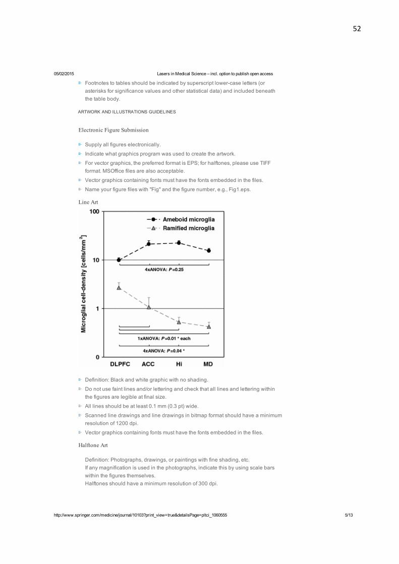
52
05/02/2015 Lasers in Medical Science – incl. option to publish open access
http://www.springer.com/medicine/journal/10103?print_view=true&detailsPage=pltci_1060555 5/13
Footnotes to tables should be indicated by superscript lower-case letters (orasterisks for significance values and other statistical data) and included beneaththe table body.
ARTWORK AND ILLUSTRATIONS GUIDELINES
Electronic Figure Submission
Supply all figures electronically.
Indicate what graphics program was used to create the artwork.
For vector graphics, the preferred format is EPS;; for halftones, please use TIFFformat. MSOffice files are also acceptable.
Vector graphics containing fonts must have the fonts embedded in the files.
Name your figure files with "Fig" and the figure number, e.g., Fig1.eps.
Line Art
Definition: Black and white graphic with no shading.
Do not use faint lines and/or lettering and check that all lines and lettering withinthe figures are legible at final size.
All lines should be at least 0.1 mm (0.3 pt) wide.
Scanned line drawings and line drawings in bitmap format should have a minimumresolution of 1200 dpi.
Vector graphics containing fonts must have the fonts embedded in the files.
Halftone Art
Definition: Photographs, drawings, or paintings with fine shading, etc.If any magnification is used in the photographs, indicate this by using scale barswithin the figures themselves.Halftones should have a minimum resolution of 300 dpi.

53
05/02/2015 Lasers in Medical Science – incl. option to publish open access
http://www.springer.com/medicine/journal/10103?print_view=true&detailsPage=pltci_1060555 6/13
Combination Art
Definition: a combination of halftone and line art, e.g., halftones containing linedrawing, extensive lettering, color diagrams, etc.Combination artwork should have a minimum resolution of 600 dpi.
Color Art
Color art is free of charge for online publication.If black and white will be shown in the print version, make sure that the maininformation will still be visible. Many colors are not distinguishable from oneanother when converted to black and white. A simple way to check this is to make axerographic copy to see if the necessary distinctions between the different colorsare still apparent.If the figures will be printed in black and white, do not refer to color in the captions.

54
05/02/2015 Lasers in Medical Science – incl. option to publish open access
http://www.springer.com/medicine/journal/10103?print_view=true&detailsPage=pltci_1060555 7/13
Color illustrations should be submitted as RGB (8 bits per channel).
Figure Lettering
To add lettering, it is best to use Helvetica or Arial (sans serif fonts).
Keep lettering consistently sized throughout your final-sized artwork, usually about2–3 mm (8–12 pt).
Variance of type size within an illustration should be minimal, e.g., do not use 8-pttype on an axis and 20-pt type for the axis label.
Avoid effects such as shading, outline letters, etc.
Do not include titles or captions within your illustrations.
Figure Numbering
All figures are to be numbered using Arabic numerals.Figures should always be cited in text in consecutive numerical order.Figure parts should be denoted by lowercase letters (a, b, c, etc.).If an appendix appears in your article and it contains one or more figures, continuethe consecutive numbering of the main text. Do not number the appendix figures,
"A1, A2, A3, etc." Figures in online appendices (Electronic Supplementary Material)should, however, be numbered separately.
Figure Captions
Each figure should have a concise caption describing accurately what the figuredepicts. Include the captions in the text file of the manuscript, not in the figure file.
Figure captions begin with the term Fig. in bold type, followed by the figure number,also in bold type.
No punctuation is to be included after the number, nor is any punctuation to beplaced at the end of the caption.
Identify all elements found in the figure in the figure caption;; and use boxes, circles,etc., as coordinate points in graphs.
Identify previously published material by giving the original source in the form of areference citation at the end of the figure caption.
Figure Placement and Size
When preparing your figures, size figures to fit in the column width.For most journals the figures should be 39 mm, 84 mm, 129 mm, or 174 mm wideand not higher than 234 mm.For books and book-sized journals, the figures should be 80 mm or 122 mm wideand not higher than 198 mm.
Permissions
If you include figures that have already been published elsewhere, you must obtainpermission from the copyright owner(s) for both the print and online format. Please be awarethat some publishers do not grant electronic rights for free and that Springer will not be able torefund any costs that may have occurred to receive these permissions. In such cases, materialfrom other sources should be used.
Accessibility
In order to give people of all abilities and disabilities access to the content of your figures,please make sure that

55
05/02/2015 Lasers in Medical Science – incl. option to publish open access
http://www.springer.com/medicine/journal/10103?print_view=true&detailsPage=pltci_1060555 8/13
All figures have descriptive captions (blind users could then use a text-to-speechsoftware or a text-to-Braille hardware)Patterns are used instead of or in addition to colors for conveying information(colorblind users would then be able to distinguish the visual elements)Any figure lettering has a contrast ratio of at least 4.5:1
ELECTRONIC SUPPLEMENTARY MATERIAL
Springer accepts electronic multimedia files (animations, movies, audio, etc.) and othersupplementary files to be published online along with an article or a book chapter. This featurecan add dimension to the author's article, as certain information cannot be printed or is moreconvenient in electronic form.
Submission
Supply all supplementary material in standard file formats.Please include in each file the following information: article title, journal name,author names;; affiliation and e-mail address of the corresponding author.To accommodate user downloads, please keep in mind that larger-sized files mayrequire very long download times and that some users may experience otherproblems during downloading.
Audio, Video, and Animations
Always use MPEG-1 (.mpg) format.
Text and Presentations
Submit your material in PDF format;; .doc or .ppt files are not suitable for long-termviability.A collection of figures may also be combined in a PDF file.
Spreadsheets
Spreadsheets should be converted to PDF if no interaction with the data isintended.If the readers should be encouraged to make their own calculations, spreadsheetsshould be submitted as .xls files (MS Excel).
Specialized Formats
Specialized format such as .pdb (chemical), .wrl (VRML), .nb (Mathematicanotebook), and .tex can also be supplied.
Collecting Multiple Files
It is possible to collect multiple files in a .zip or .gz file.
Numbering
If supplying any supplementary material, the text must make specific mention of thematerial as a citation, similar to that of figures and tables.Refer to the supplementary files as “Online Resource”, e.g., "... as shown in theanimation (Online Resource 3)", “... additional data are given in Online Resource4”.Name the files consecutively, e.g. “ESM_3.mpg”, “ESM_4.pdf”.
Captions

56
05/02/2015 Lasers in Medical Science – incl. option to publish open access
http://www.springer.com/medicine/journal/10103?print_view=true&detailsPage=pltci_1060555 9/13
For each supplementary material, please supply a concise caption describing thecontent of the file.
Processing of supplementary files
Electronic supplementary material will be published as received from the authorwithout any conversion, editing, or reformatting.
Accessibility
In order to give people of all abilities and disabilities access to the content of yoursupplementary files, please make sure that
The manuscript contains a descriptive caption for each supplementary materialVideo files do not contain anything that flashes more than three times per second(so that users prone to seizures caused by such effects are not put at risk)
INTEGRITY OF RESEARCH AND REPORTING
Ethical standards
Manuscripts submitted for publication must contain a statement to the effect that all human andanimal studies have been approved by the appropriate ethics committee and have thereforebeen performed in accordance with the ethical standards laid down in the 1964 Declaration ofHelsinki and its later amendments.
It should also be stated clearly in the text that all persons gave their informed consent prior totheir inclusion in the study. Details that might disclose the identity of the subjects under studyshould be omitted.
These statements should be added in a separate section before the reference list. If thesestatements are not applicable, authors should state: The manuscript does not contain clinicalstudies or patient data.
The editors reserve the right to reject manuscripts that do not comply with the above-mentioned requirements. The author will be held responsible for false statements or failure tofulfill the above-mentioned requirements
Conflict of interest
All benefits in any form from a commercial party related directly or indirectly to the subject ofthis manuscript or any of the authors must be acknowledged. For each source of funds, boththe research funder and the grant number should be given. This note should be added in aseparate section before the reference list.
If no conflict exists, authors should state: The authors declare that they have no conflict ofinterest.
ETHICAL RESPONSIBILITIES OF AUTHORS
This journal is committed to upholding the integrity of the scientific record. As a member of theCommittee on Publication Ethics (COPE) the journal will follow the COPE guidelines on how todeal with potential acts of misconduct.
Authors should refrain from misrepresenting research results which could damage the trust inthe journal, the professionalism of scientific authorship, and ultimately the entire scientificendeavour. Maintaining integrity of the research and its presentation can be achieved byfollowing the rules of good scientific practice, which include:
The manuscript has not been submitted to more than one journal for simultaneous

57
05/02/2015 Lasers in Medical Science – incl. option to publish open access
http://www.springer.com/medicine/journal/10103?print_view=true&detailsPage=pltci_1060555 10/13
consideration.
The manuscript has not been published previously (partly or in full), unless the newwork concerns an expansion of previous work (please provide transparency on there-use of material to avoid the hint of text-recycling (“self-plagiarism”)).
A single study is not split up into several parts to increase the quantity ofsubmissions and submitted to various journals or to one journal over time (e.g.“salami-publishing”).
No data have been fabricated or manipulated (including images) to support yourconclusions
No data, text, or theories by others are presented as if they were the author’s own(“plagiarism”). Proper acknowledgements to other works must be given (thisincludes material that is closely copied (near verbatim), summarized and/orparaphrased), quotation marks are used for verbatim copying of material, andpermissions are secured for material that is copyrighted.
Important note: the journal may use software to screen for plagiarism.
Consent to submit has been received explicitly from all co-authors, as well as fromthe responsible authorities - tacitly or explicitly - at the institute/organization wherethe work has been carried out, before the work is submitted.
Authors whose names appear on the submission have contributed sufficiently tothe scientific work and therefore share collective responsibility and accountabilityfor the results.
In addition:
Changes of authorship or in the order of authors are not accepted after acceptanceof a manuscript.Requesting to add or delete authors at revision stage, proof stage, or afterpublication is a serious matter and may be considered when justifiably warranted.Justification for changes in authorship must be compelling and may be consideredonly after receipt of written approval from all authors and a convincing, detailedexplanation about the role/deletion of the new/deleted author. In case of changes atrevision stage, a letter must accompany the revised manuscript. In case of changesafter acceptance or publication, the request and documentation must be sent viathe Publisher to the Editor-in-Chief. In all cases, further documentation may berequired to support your request. The decision on accepting the change rests withthe Editor-in-Chief of the journal and may be turned down. Therefore authors arestrongly advised to ensure the correct author group, corresponding author, andorder of authors at submission.Upon request authors should be prepared to send relevant documentation or datain order to verify the validity of the results. This could be in the form of raw data,samples, records, etc.
If there is a suspicion of misconduct, the journal will carry out an investigation following theCOPE guidelines. If, after investigation, the allegation seems to raise valid concerns, theaccused author will be contacted and given an opportunity to address the issue. If misconducthas been established beyond reasonable doubt, this may result in the Editor-in-Chief’simplementation of the following measures, including, but not limited to:
If the article is still under consideration, it may be rejected and returned to theauthor.If the article has already been published online, depending on the nature andseverity of the infraction, either an erratum will be placed with the article or insevere cases complete retraction of the article will occur. The reason must be givenin the published erratum or retraction note.The author’s institution may be informed.

58
05/02/2015 Lasers in Medical Science – incl. option to publish open access
http://www.springer.com/medicine/journal/10103?print_view=true&detailsPage=pltci_1060555 11/13
COMPLIANCE WITH ETHICAL STANDARDS
To ensure objectivity and transparency in research and to ensure that accepted principles ofethical and professional conduct have been followed, authors should include informationregarding sources of funding, potential conflicts of interest (financial or non-financial), informedconsent if the research involved human participants, and a statement on welfare of animals ifthe research involved animals.
Authors should include the following statements (if applicable) in a separate section entitled“Compliance with Ethical Standards” before the References when submitting a paper:
Disclosure of potential conflicts of interestResearch involving Human Participants and/or AnimalsInformed consent
Please note that standards could vary slightly per journal dependent on their peer reviewpolicies (i.e. double blind peer review) as well as per journal subject discipline. Beforesubmitting your article check the Instructions for Authors carefully.
The corresponding author should be prepared to collect documentation of compliance withethical standards and send if requested during peer review or after publication.
The Editors reserve the right to reject manuscripts that do not comply with the above-mentioned guidelines. The author will be held responsible for false statements or failure tofulfill the above-mentioned guidelines.
DISCLOSURE OF POTENTIAL CONFLICTS OF INTEREST
Authors must disclose all relationships or interests that could have direct or potential influenceor impart bias on the work. Although an author may not feel there is any conflict, disclosure ofrelationships and interests provides a more complete and transparent process, leading to anaccurate and objective assessment of the work. Awareness of a real or perceived conflicts ofinterest is a perspective to which the readers are entitled. This is not meant to imply that afinancial relationship with an organization that sponsored the research or compensationreceived for consultancy work is inappropriate. Examples of potential conflicts of interests thatare directly or indirectly related to the research may include but are not limited to thefollowing:
Research grants from funding agencies (please give the research funder and thegrant number)
Honoraria for speaking at symposia
Financial support for attending symposia
Financial support for educational programs
Employment or consultation
Support from a project sponsor
Position on advisory board or board of directors or other type of managementrelationships
Multiple affiliations
Financial relationships, for example equity ownership or investment interest
Intellectual property rights (e.g. patents, copyrights and royalties from such rights)
Holdings of spouse and/or children that may have financial interest in the work
In addition, interests that go beyond financial interests and compensation (non-financialinterests) that may be important to readers should be disclosed. These may include but are notlimited to personal relationships or competing interests directly or indirectly tied to thisresearch, or professional interests or personal beliefs that may influence your research.

59
05/02/2015 Lasers in Medical Science – incl. option to publish open access
http://www.springer.com/medicine/journal/10103?print_view=true&detailsPage=pltci_1060555 12/13
The corresponding author collects the conflict of interest disclosure forms from all authors. Inauthor collaborations where formal agreements for representation allow it, it is sufficient for thecorresponding author to sign the disclosure form on behalf of all authors. Examples of formscan be found
here:
The corresponding author will include a summary statement in the text of the manuscript in aseparate section before the reference list, that reflects what is recorded in the potential conflictof interest disclosure form(s).
See below examples of disclosures:
Funding: This study was funded by X (grant number X).
Conflict of Interest: Author A has received research grants from Company A. Author B hasreceived a speaker honorarium from Company X and owns stock in Company Y. Author C is amember of committee Z.
If no conflict exists, the authors should state:
Conflict of Interest: The authors declare that they have no conflict of interest.
RESEARCH INVOLVING HUMAN PARTICIPANTS AND/OR ANIMALS
INFORMED CONSENT
AFTER ACCEPTANCE
DOES SPRINGER PROVIDE ENGLISH LANGUAGE SUPPORT?
1) Statement of human rights
When reporting studies that involve human participants, authors should include a statementthat the studies have been approved by the appropriate institutional and/or national researchethics committee and have been performed in accordance with the ethical standards as laiddown in the 1964 Declaration of Helsinki and its later amendments or comparable ethicalstandards.
If doubt exists whether the research was conducted in accordance with the 1964 HelsinkiDeclaration or comparable standards, the authors must explain the reasons for their approach,and demonstrate that the independent ethics committee or institutional review board explicitlyapproved the doubtful aspects of the study.
The following statements should be included in the text before the References section:
Ethical approval: “All procedures performed in studies involving human participants were inaccordance with the ethical standards of the institutional and/or national research committeeand with the 1964 Helsinki declaration and its later amendments or comparable ethicalstandards.”
For retrospective studies, please add the following sentence:
“For this type of study formal consent is not required.”
READ THIS JOURNAL ON SPRINGERLINK
Online First Articles
All volumes & issues

60
05/02/2015 Lasers in Medical Science – incl. option to publish open access
http://www.springer.com/medicine/journal/10103?print_view=true&detailsPage=pltci_1060555 13/13
2013 Impact Factor 2.419
FOR AUTHORS AND EDITORS
SERVICES FOR THE JOURNAL
ALERTS FOR THIS JOURNAL
Get the table of contents of every new issue published inLasers in Medical Science.
Your E-Mail Address
Please send me information on new Springerpublications in Medicine (general).
Aims and Scope
Submit Online
Open Choice - Your Way to Open Access
Instructions for Authors
Author Academy: Training for Authors
Contacts
Download Product Flyer
Shipping dates
Order back issues
Article Reprints
Bulk Orders
SUBMIT

61
Apêndice I: Figuras referentes aos Materiais e Métodos
Figura 1: Obtenção dos espécimes – A) Incisivo bovino. B) Ilustração dos cortes que foram realizados. C) e D) Espécimes obtidos após os cortes.
Figura 2: Máquina de corte. Figura 3: Fita isolante fixada no espécime.
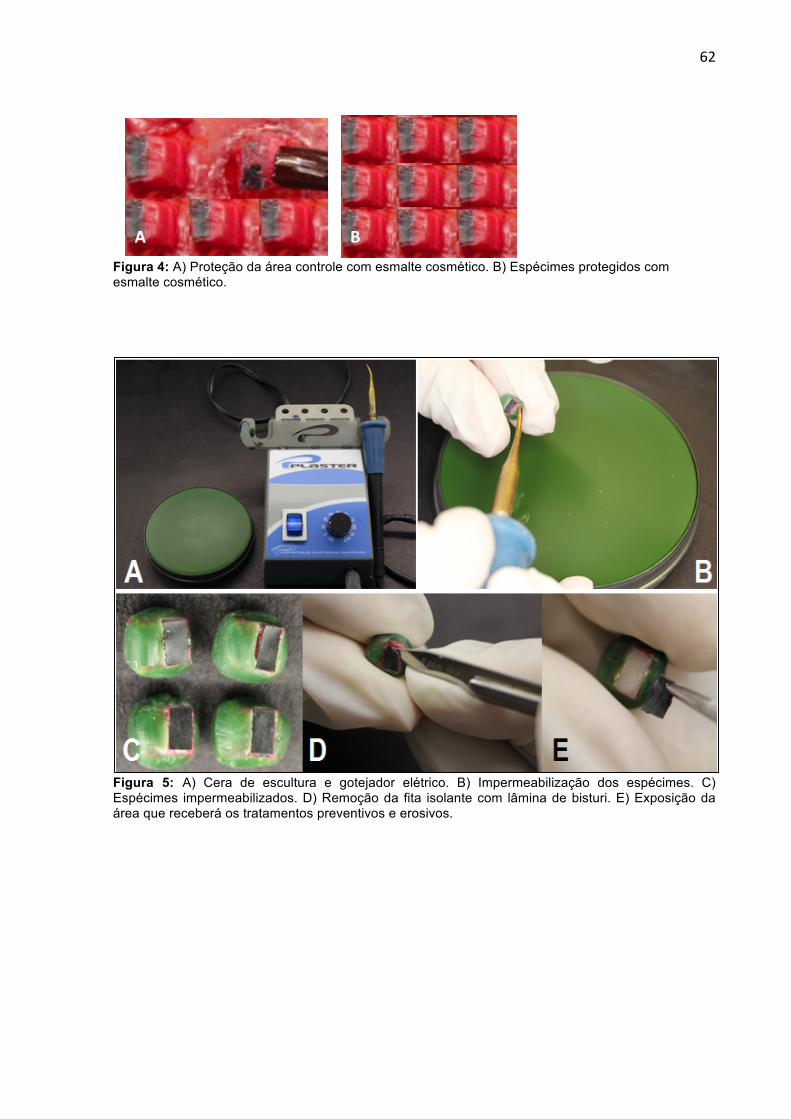
62
Figura 4: A) Proteção da área controle com esmalte cosmético. B) Espécimes protegidos com esmalte cosmético.
Figura 5: A) Cera de escultura e gotejador elétrico. B) Impermeabilização dos espécimes. C) Espécimes impermeabilizados. D) Remoção da fita isolante com lâmina de bisturi. E) Exposição da área que receberá os tratamentos preventivos e erosivos.
A B

63
Figura 6: A) Fluoreto de sódio 1,23%. B) Espécime que receberá os tratamentos preventivos. C) Aplicação do fluoreto de sódio com auxílio do microbrush.
Figura 7: Laser Er:YAG Figura 8: Laser Nd:YAG

64
Figura 11: A) Espécimes inseridos em um Becker de 50 mL. B) Desafio erosivo em Coca-Cola. C) Espécimes sendo lavados com água destilada.
Figura 9: Refrigerante à base de cola.
Figura 10: Máquina de agitação

65
Figura 12: Remoção da cera e esmalte, para as Figura 13: Microscópio Confocal a laser 3D. análises de rugosidade superficial e desgaste.

66
Apêndice II: Figuras referentes aos Resultados
Figura 14: Fotos referentes à análise de rugosidade superficial

67
Figura 15: Imagens demonstrativas da avaliação do desgaste no Software OLS4000


![Tese Odete Alves 16Dez11[1]](https://img.document.onl/doc/110x75/55cf94c4550346f57ba44166/tese-odete-alves-16dez111.jpg)