Embed Size (px)
Citation preview
UNIVERSIDADE FEDERAL DE PELOTAS
Faculdade de Odontologia
Programa de Pós-Graduação em Odontologia
Tese de doutorado
Avaliação da biocompatibilidade e análise da alteração de cor dentária
induzida por diferentes materiais obturadores de canais radiculares de dentes
decíduos
Katerine Jahnecke Pilownic
Pelotas, 2017
Katerine Jahnecke Pilownic
Avaliação da biocompatibilidade e análise da alteração de cor dentária
induzida por diferentes materiais obturadores de canais radiculares de dentes
decíduos
Tese apresentada ao Programa de Pós-
Graduação em Odontologia, da Faculdade
de Odontologia, da Universidade Federal de
Pelotas, como requisito parcial à obtenção
do título de Doutor em Odontologia, Área de
Concentração Odontopediatria.
Orientadora: Profa. Drª. Fernanda Geraldo Pappen
Co-orientadora: Profa. Drª. Ana Regina Romano
Pelotas, 2017
8
9
Katerine Jahnecke Pilownic
Avaliação da biocompatibilidade e análise da alteração de cor dentária
induzida por diferentes materiais obturadores de canais radiculares de dentes
decíduos
Tese apresentada, como requisito parcial, para obtenção do grau de Doutor em
Odontologia, Programa de Pós-Graduação em Odontologia, área de concentração
Odontopediatria, Faculdade de Odontologia, Universidade Federal de Pelotas.
Data da defesa: 02 de maio de 2017.
Banca examinadora:
Profa. Dra.Fernanda Geraldo Pappen (Presidente)
Doutora em Endodontia, pela Universidade Estadual Paulista, Araraquara, SP.
Profa. Dra. Marília Leão Goettems
Doutora em Odontologia, área Odontopediatria, pela Universidade Federal de
Pelotas, Pelotas, RS.
Dra. Aline Oliveira Ogliari,
Doutora em Odontologia, área Materiais dentários, pela Universidade Federal de
Pelotas, Pelotas, RS.
Profa. Dra. Nádia de Souza Ferreira
Doutora em Odontologia, área Endodontia, pela Universidade Estadual Paulista, São
José do Campos, SP.
Profa. Dra. Luiza Helena Silva de Almeida
Doutora em Odontologia, área Odontopediatria, pela Universidade Federal de
Pelotas, Pelotas, RS.
Profa. Dra. Gabriela dos Santos Pinto
Doutora em Odontologia, área Odontopediatria, pela Universidade Federal de
Pelotas, Pelotas, RS (Suplente).
Profª.Drª. Maria Laura de Menezes Bonow
Doutora em Odontologia, área Odontopediatria, pela Universidade de São Paulo,
São Paulo, SP (Suplente).
Notas Preliminares
A presente dissertação foi redigida segundo o Manual de Normas para
Dissertações, Teses e Trabalhos Científicos da Universidade Federal de Pelotas de
2013, adotando o Nível de Descrição 4 – estrutura em Artigos, descrita no Apêndice
D do referido manual. <http://sisbi.ufpel.edu.br/?p=manual> Acesso em: 20 de março
de 2017.
Resumo
PILOWNIC, Katerine Jahnecke. Avaliação da biocompatibilidade e análise da
alteração de cor dentária induzida por diferentes materiais obturadores de
canais radiculares de dentes decíduos. 2017.67f.Projeto de tese (Doutorado em
Odontologia) – Programa de Pós-graduação em Odontologia. Universidade Federal
de Pelotas, Pelotas, 2017.
Este estudo avaliou a biocompatibilidade de diferentes materiais obturadores de canais radiculares de dentes decíduos e o seu potencial de alteração de cor. Foram avaliados os seguintes materiais: um material à base de MTA, um material à base de hidróxido de cálcio e iodofórmio (Vitapex), Óxido de zinco e eugenol (OZE), e um material à base de hidróxio de cálcio (Calen) espessado com óxido de zinco (ZO). Para a avaliação da biocompatibilidade, os materiais foram inseridos em tubos de polietileno e implantados no tecido subcutâneo dorsal de quinze ratos Wistar (Rattus norvegicus). Após 15, 30 e 60 dias, os animais foram eutanasiados, os tubos e tecidos circundantes foram removidos e histologicamente processados. As amostras foram avaliadas quanto à intensidade da resposta inflamatória, a espessura da cápsula fibrosa, presença de calcificação e células gigantes. Para a análise do potencial de discoloração, setenta e cinco blocos cubóides de esmalte-dentina foram preenchidos com os diferentes materiais obturadores e selados com ionômero de vidro fotoativado. A alteração de cor (∆E00) foi calculada baseada nas coordenadas de cor medido com o espectrofotômetro (VITA Easyshadecompact) antes e depois da inserção dos materiais e após 1 semana, 1, 3, 6 e 9 meses. A alteração de cor ∆E00 foi calculada pelo CIEDE 2000 para cada espécime a partir do baseline. Os resultados do teste de biocompatibilidade mostraram que os materiais apresentaram decréscimo na severidade da inflamação a partir de 30 dias. Da mesma forma, em todos os grupos, a cápsula fibrosa, inicialmente espessa, se tornou mais delgada ao longo do período experimental. Células gigantes foram observadas em todos os grupos experimentais principalmente no período de 15 dias, enquanto a ocorrência de calcificação foi observada com mais frequência no grupo Calen+ZO. O teste de avaliação da capacidade de indução de alteração de cor revelou que o tempo teve efeito nos valores de ∆E00 (p <0,0001), assim como os materiais (p = 0,004). As interações entre tempo e os materiais provocaram alterações de (∆E00) (p < 0,0001). O material à base de MTA apresentou menores valores de alteração de cor (∆E= 3,02) ao longo do tempo enquanto o óxido de zinco e eugenol demonstrou os maiores valores (∆E= 5,42) (p= 0,018). O material à base de MTA apresenta propriedades satisfatórias podendo ser uma alternativa aos materiais obturadores convencionais, para utilização em canais radiculares de dentes decíduos.
Palavras-chave:dentes decíduos; escurecimento; biocompatibilidade; endodontia;
obturação.
Abstract
PILOWNIC, Katerine Jahnecke. Biocompatibility evaluation and analysis of color alteration induced by endodontic materials used in primary teeth. 2017. 67 f.Doctoral thesis (PhD in Dentistry). Postgraduate Program in Dentistry. Federal University of Pelotas, Pelotas, 2017.
The aim of this study was to evaluate the biocompatibility and discoloration potential of experimental MTA-based endodontic filling material in comparison to zinc oxide eugenol (ZOE) cement, Calen paste thickened with zinc oxide, and Vitapex. In the biocompatibility assay, the materials were introduced into polyethylene tubes and implanted into the dorsal subcutaneous tissue of fifteen Wistar rats (Rattus norvegicus). After 15, 30 and 60 days, the animals were euthanized, the surrounding tubes and tissues were removed and processed for histopathological evaluation. The samples were analyzed for the intensity of the inflammatory response, the thickness of the fibrous capsule, presence of calcification and giant cells. For the analysis of discoloration potential, seventy-five cuboidal enamel-dentin blocks were filled with different endodontic filling materials and sealed with photoactivated glass ionomer cement. Standardized color measurements were performed using a spectrophotometer (Vita Easyshade, Vita-Zahnfabrik, Oberding, Germany) and expressed in ΔE*00 units at the following intervals: prior to and after placement of the filling and after 1 week,1 month, 3 months, 6 months, and 9 months. The results of biocompatibility showed the materials tested, as well as the control, had a lower inflammation severity in 30 and 60 days in relation to 15 days. Likewise, the thickness of fibrous capsule in all groups of materials in 15 days was thick and decreased over time. It was possible to observe the presence of giant cells in all groups, mainly in 15 days. The presence of calcification was observed around implantation sites in all groups, but more frequently in Calen + ZO samples. The findings of discoloration potential exhibited the time had a significant effect on color variation values (ΔE*00) (p <0.0001).The effect of the materials on color variation (ΔE*00) was also statistically significant (p = 0.004). Interactions between time and materials demonstrated a significant effect on the values (ΔE*00) (p <0.0001). The MTA-based experimental material showed the smallest color variation throughout the experimental time (ΔE*00= 3.02), where as the zinc oxide and eugenol cement (ZOE) showed the highest darkening (ΔE*00= 5.42) (p = 0.018). ).In conclusion, experimental MTA-based material has satisfactory properties for use as endodontic filling material.
Key words: primary teeth; discoloration; biocompatibility; endodontics; root canal
filling.
Lista de Figuras
Projeto de Pesquisa
Figura 1 Bloco cubóide de esmalte-dentina (10x10x3,5mm) preparado a
partir do terço médio da coroa dente
bovino………………………………….……………...……………....... 22
Figura 2 Após preenchimento da cavidade com os materiais a serem
avaliados, selamento com cimento de ionômero de vidro
fotoativado....................................................................................... 23
Figura 3 Dispositivo confeccionado para fazer a mensuração de cor do terço
cervical da coroa do dente bovino................................................... 24
Artigo1
Figura1 Subcutaneous tissue reactions within the different experimental
groups. Hematoxylin and eosin staining,10X………………………..46
Artigo 2
Figura1 Schematic presentation of performed procedures prior to color
measurements…………………….....................................................58
Lista de Tabelas
Projeto de Pesquisa
Tabela 1 Composição dos materiais obturadores testados.........................19
Tabela 2 Distribuição dos implantes em relação aos materiais e períodos
empregados no experimento....................................................... 20
Tabela 3 Distribuição dos materiais experimentais.................................... 23
Artigo 1
Tabela 1 Percentage of tissue reaction according to different groups and
different times of evaluation.……………………………………….. 48
Artigo 2
Tabela 1 Distribution of experimental materials.......................................... 59
Tabela 2 Color variation results (ΔE*00) byANOVA with repeated measures
with regard to the effects of weather and their interaction
groups……………………………………………………………………..60
Tabela 3 Mean and standard deviation of variation in color values (ΔE*00) in
the different experimental groups…………………………………….. 61
Lista de Abreviaturas e Siglas
CIV Cimento de ionômero de vidro
MTA Agregado de trióxido mineral
OZE Óxido de Zinco e Eugenol
UFPel Universidade Federal de Pelotas
ZO Zinc oxide
L* luminosidade
a* croma
B* croma
Sumário
1 Introdução........................................................................................... 10
2 Projeto de Pesquisa........................................................................... 14
2.1 Antecedentes e Justificativas........................................................ 14
2.2 Objetivos..........................................................................................
2.2.1 Objetivo Geral..............................................................................
2.2.2 Objetivos Específicos................................................................
18
18
18
2.3 Metodologia.................................................................................... 18
2.3.1 Material testados......................................................................... 18
2.3 Teste de biocompatibilidade........................................................ 19
2.3 Teste de alteração de cor............................................................ 21
2.4 Orçamento........................................................................................ 26
2.5 Cronograma de atividades............................................................ 27
3. Relatório do trabalho de campo....................................................... 28
4 Artigos................................................................................................. 32
4.1 Artigo 1............................................................................................ 32
4.2 Artigo 2........................................................................................... 49
5 Considerações finais......................................................................... 62
Referências Bibliográficas................................................................ 64
Anexos .............................................................................................. 67
1 Introdução
Algumas propriedades dos materiais obturadores de dentes decíduos devem
ser ponderadas durante a escolha para uso. Idealmente este material deve ser
reabsorvível, radiopaco, bactericida, promover adequado preenchimento e aderência
às paredes dos canais radiculares, facilmente removido quando necessário; além de
não provocar danos aos tecidos periapicais e ao germe do dente permanente, e
tampouco alteração da coloração das estruturas dentárias. No entanto, não existe
um material único que preencha todos os requisitos desejáveis para um material
obturador, além de não haver consenso na literatura sobre o melhor material a ser
utilizado na endodontia de dentes decíduos (FUKS, 2000, KUBOTA; GOLDEN;
PENUGONDA, 1992; MASS; ZILBERMAN, 1989; MORTAZAVI; MESBAHI, 2004,
PINTO et al., 2011). É importante destacar que o uso de um material obturador com
todos os requisitos desejáveis melhoraria o prognóstico do tratamento endodôntico
de dentes decíduos (SEGATO et al., 2016).
A capacidade do material de ser reabsorvível é um dos principais requisitos
para a sua indicação como material obturador de dentes decíduos (COULD, 1972,
KUBOTA; GOLDEN; PENUGONDA, 1992). A reabsorção do material obturador deve
ocorrer simultaneamente à reabsorção radicular durante a esfoliação, permitindo a
natural erupção do dente permanente (RANLY; GARCIA-GODOY, 1991; QUEIROZ
et al., 2009; SILVA et al., 2010). Além disso, em casos de sobre-obturação, o
material deve ser reabsorvível e não tóxico aos tecidos periapicais bem como ao
germe do permanente (KUBOTA; GOLDEN; PENUGONDA, 1992).
Diversos materiais têm sido utilizados na endodontia de dentes decíduos. O
cimento de óxido de zinco e eugenol (OZE) foi o primeiro material a ser utilizado
como obturador de canais radiculares de dentes decíduos. Desde então, vários
autores têm relatado índices de sucesso moderados a altos na proservação de
11
dentes decíduos tratados endodonticamente com esse material (MASS,
ZILBERMAN, 1989). O OZE apresenta características indesejáveis como ser
irritante aos tecidos periapicais desencadeando reações inflamatórias de corpo
estranho, nos casos de extravasamento. Além disso, apresenta baixa capacidade de
reabsorção, deixando partículas de óxido de zinco e de eugenol nos tecidos
periapicais quando ocorre a reabsorção fisiológica (BARJA-FIDALGO et al., 2011;
FUKS; EIDELMAN, 1991; MORTAZAVI; MESBAHI, 2004; PINTO et al., 2011).
Quando extravasado pelo ápice radicular, existe o risco de deflexão da erupção dos
dentes permanentes devido a sua dureza (COLL; SADRIAN, 1996). Alguns autores,
afirmam ainda que o OZE tem limitada ação antimicrobiana (COX; HEMBREE;
MCKNIGHT, 1978; TCHAOU et al., 1996).
As pastas à base de hidróxido de cálcio também são indicadas como material
obturador de canais radiculares de dentes decíduos devido a sua natureza
hidrossolúvel, baixa solubilidade e biocompatibilidade (CERQUEIRA et al. 2008;
CHAWLA et al. 2008). São também propriedades benéficas do hidróxido de cálcio
sua atividade antimicrobiana, devido à dissociação iônica dos íons cálcio e hidroxila
(FAVA; SAUNDERS, 1999; QUEIROZ et al., 2009; RANLY; GARCIA-GODOY,2000),
capacidade de produzir a hidrólise da endotoxina bacteriana, indução de formação
de tecido mineralizado, além da ativação da fosfatase alcalina e síntese de colágeno
(DE SOUZA, 2009, MIZUNO; BANZAI, 2008, SILVA et al., 2008, SCHRODER, 1985,
QUEIROZ et al., 2009).
Um material à base de hidróxido de cálcio disponível no mercado é a pasta
Calen (S.S. White, Artigos Dentários, Rio de Janeiro, RJ, Brasil), o qual apresenta o
polietilenoglicol 400 como veículo. Este veículo viscoso permite a dissociação mais
lenta dos íons hidroxila, diminuindo significativamente a velocidade de solubilização
da pasta. Em dentes decíduos, a pasta Calen é indicada como material obturador
devido à sua boa tolerância tecidual, natureza hidrossolúvel (viscoso) e baixa
solubilidade (RANLY; GARCIA-GODOY, 2000). Alguns autores têm sugerido a
adição de óxido de zinco à pasta Calen, o qual melhora ainda mais a consistência do
material, e auxilia na redução da velocidade de fagocitose do material (FAVA;
SAUNDERS, 1999; QUEIROZ et al., 2009).
As pastas de hidróxido de cálcio vêm sendo utilizadas ainda em associação
com iodofórmio. Diversos estudos têm demonstrado que estes materiais são
12
facilmente reabsorvidos na região periapical em caso de extravasamento, e não
causam reação de corpo estranho ou têm qualquer efeito indesejável aos dentes
permanentes (CERQUEIRA et al. 2008; MATHEWSON; PRIMOSCH, 1995;
MCDONALD; AVERY; DEAN, 2000). Além disso, os materiais contendo iodofórmio
em sua composição apresentam uma atividade antibacteriana satisfatória
(CERQUEIRA et al. 2008; MATHEWSON; PRIMOSCH, 1995; MCDONALD; AVERY;
DEAN, 2000).
Dentre esses, o Vitapex (Neo Dental International Inc., Federal Way, WA,
Estados Unidos), material na forma de pré-mistura de hidróxido de cálcio e
iodofórmio, é tido como quase ideal para obturação de dentes decíduos (KUBOTA;
GOLDEN; PENUGONDA, 1992). No entanto, como desvantagem, apresenta a
rápida eliminação do iodofórmio pelo organismo, deixando espaços vazios no interior
do canal radicular, o que pode comprometer o sucesso do tratamento endodôntico
(KUBOTA; GOLDEN; PENUGONDA, 1992). Além disso, o uso de pastas
iodoformadas pode provocar discoloração marrom-amarelada na coroa dentária,
comprometendo a estética (GARCÍA-GODOY 1987). O potencial tóxico do
iodofórmio, em contato com os tecidos vivos é considerado outra limitação do uso
desse material (BARJA-FIDALGO et al., 2011; SILVA et al., 2010).
Um novo material à base de agregado mineral trióxido (MTA) destinado à
obturação de canais radiculares de dentes decíduos está sendo desenvolvido.
Segundo o fabricante, de forma semelhante ao MTA-Fillapex, o material
experimental trata-se de uma pasta única e é composto por ester salicilato de glicol,
dióxido de titânio, tungstato de cálcio, dióxido de silicone, tolueno sulfonamida e
silicato de cálcio. Contudo, este material experimental contém menor proporção de
salicilato, cerca de 30% de MTA, em comparação com MTA-Fillapex. O material
experimental à base de MTA apresenta requisitos desejáveis para um material
obturador de dentes decíduos como ação antibacteriana, radiopacidade satisfatória,
baixa citotoxicidade e pH adequado (PILOWNIC, 2015). O uso de material
endodôntico pronto para usar é desejável em odontpediatria, uma vez que pode
reduzir o tempo de consulta. Adicionalmente, os materiais que necessitam ser
manipulados podem ter as suas proporções alteradas modificando o desempenho
clínico do material.
Os testes in vitro e in vivo são ensaios preliminares indispensáveis para o
desenvolvimento de novos materiais odontológicos, já que buscam simular em
13
laboratório, condições biológicas mais próximas das reais. A biocompatibilidade de
materiais odontológicos que possam entrar em contato com os tecidos vivos deve
ser avaliada devido ao contato de substâncias que podem provocar reações no
tecido periapical (KAPLAN et al. 2003; HO et al. 2006). A implantação do material no
tecido conjuntivo subcutâneo é considerado um teste adequado para a avaliação da
biocompatibilidade (OLSSON 1981; TORNECK 1966; PINTO et al. 2011; SILVA et
al. 2010,).
No entanto, a terapia endodôntica não deve focar apenas nos aspectos
biológicos e funcionais; a estética também deve ser levada em consideração durante
o tratamento de canais radiculares inclusive de dentes decíduos (LENHERR et
al.,2012; KRASTL et al.,2013). As crianças são conscientes sobre a sua estética
dental e sobre a aparência de outras crianças (VALE et al., 2009) sendo o
escurecimento dental, a principal percepção negativa delas em relação à boca
(NEWTON; MINHAS, 2005; KERSHAW; NEWTON; WILLIAMS, 2008). O
escurecimento da coroa dentária associado aos materiais obturadores está
relacionado com o tempo de contato do material com a estrutura dentária, e com o
pontencial cromogênico dos materiais utilizados no tratamento (PARSONS, 2001;
PARTOVI, 2006; VAN DER BURGDT, 1985).
Assim, considerando a composição do material experimental à base de MTA,
com a hipótese de que ele apresenta propriedades biológicas satisfatórias, será
avaliada a reação tecidual induzida por esse material obturador experimental quando
implantado no tecido conjuntivo subcutâneo comparando com os outros materiais
obturadores de canais radiculares usados em dentes decíduos. A alteração de cor
da estrutura dentária associada ao uso do material experimental à base de MTA
também será testada.
2 Projeto de pesquisa
2.1 Antecedentes e justificativas
Apesar dos avanços na prevenção da cárie dentária e da melhor
compreensão da importância de se manter a dentição decídua hígida,
frequentemente, ocorre grande número de lesões cariosas profundas nos dentes
decíduos, com comprometimento pulpar. Além disso, lesões traumáticas,
principalmente nos dentes anteriores, apresentam uma prevalência elevada. A
conservação dos dentes decíduos com alterações pulpares provocadas por lesão de
cárie ou por traumatismo é um grande desafio terapêutico (CORRÊA et al., 2011).
A terapia pulpar em dentes decíduos visa à manutenção do elemento dentário
assim como a preservação da saúde da criança (GARCIA-GODOY, 1987). Sempre
que o tecido pulpar torna-se irreversivelmente infectado, ou quando ocorre necrose,
em decorrência de cárie ou traumatismo dental, a terapia pulpar dos dentes
decíduos é indicada (AMERICAN ACADEMY OF PEDIATRIC DENTISTRY, 2009). O
tratamento endodôntico de dentes decíduos visa o reparo dos tecidos apicais e
periapicais, levando a manutenção da integridade do germe dentário permanente
(PINTO et al., 2011).
Algumas propriedades dos materiais obturadores de dentes decíduos devem
ser ponderadas durante a escolha para uso. Idealmente este material deve ser
reabsorvível, radiopaco, bactericida, promover adequado preenchimento e aderência
às paredes dos canais radiculares, facilmente removido quando necessário; além de
não provocar dano aos tecidos periapicais e ao germe do dente permanente, e
tampouco alteração da coloração das estruturas dentárias. No entanto, não existe
um material único que preencha todos os requisitos desejáveis para um material
obturador, além de não haver consenso na literatura sobre o melhor material a ser
utilizado na endodontia de dentes decíduos (FUKS, 2000; KUBOTA; GOLDEN;
15
PENUGONDA, 1992; MASS; ZILBERMAN, 1989; MORTAZAVI; MESBAHI, 2004;
PINTO et al., 2011).
A capacidade do material de ser reabsorvível é um dos principais requisitos
para a sua indicação como material obturador de dentes decíduos (COULD, 1972;
KUBOTA; GOLDEN; PENUGONDA, 1992). A reabsorção do material obturador deve
ocorrer simultaneamente à reabsorção radicular durante a esfoliação, permitindo a
normal erupção do dente permanente (RANLY, GARCIA-GODOY, 1991; QUEIROZ
et al., 2009; SILVA et al., 2010). Além disso, em casos de sobre-obturação, o
material deve ser reabsorvível e não tóxico aos tecidos periapicais e germe do
permanente (KUBOTA; GOLDEN; PENUGONDA, 1992).
Diversos materiais têm sido utilizados na endodontia de dentes decíduos. O
cimento de óxido de zinco e eugenol (OZE) foi o primeiro material a ser utilizado
como obturador de dentes decíduos. Desde então, vários autores têm relatado
índices de sucesso moderados a altos na proservação de dentes decíduos tratados
endodonticamente com esse material (MASS, ZILBERMAN, 1989). O OZE
apresenta características indesejáveis como ser irritante aos tecidos periapicais,
desencadeando reações inflamatórias de corpo estranho, nos casos de
extravasamento. Além disso, apresenta baixa capacidade de reabsorção, deixando
partículas de óxido de zinco e de eugenol nos tecidos periapicais quando ocorre a
reabsorção fisiológica (BARJA-FIDALGO et al., 2011; FUKS; EIDELMAN, 1991,
MORTAZAVI; MESBAHI, 2004; PINTO et al., 2011). Quando extravasado pelo ápice
radicular, existe o risco de deflexão da erupção dos dentes permanentes devido a
sua dureza (COLL; SADRIAN, 1996). Alguns autores, afirmam ainda que o OZE tem
limitada ação antimicrobiana (COX; HEMBREE; MCKNIGHT, 1978; TCHAOU et al.
1996).
As pastas à base de hidróxido de cálcio também são indicadas como material
obturador de canais radiculares de dentes decíduos devido a sua natureza
hidrossolúvel, baixa solubilidade e biocompatibilidade (CERQUEIRA et al. 2008;
CHAWLA et al. 2008). São também propriedades benéficas do hidróxido de cálcio
sua atividade antimicrobiana, devido à dissociação iônica dos íons cálcio e hidroxila
(FAVA; SAUNDERS, 1999; QUEIROZ et al., 2009; RANLY; GARCIA-GODOY, 2000)
capacidade de produzir a hidrólise da endotoxina bacteriana, indução de formação
16
de tecido mineralizado, além da ativação da fosfatase alcalina e síntese de colágeno
(DE SOUZA, 2009; MIZUNO; BANZAI, 2008; SILVA et al., 2008; SCHRODER, 1985;
QUEIROZ et al., 2009).
Um material à base de hidróxido de cálcio disponível no mercado é a pasta
Calen (S.S. White, Artigos Dentários, Rio de Janeiro, RJ, Brasil), o qual apresenta o
polietilenoglicol 400 como veículo. Este veículo viscoso permite a dissociação mais
lenta dos íons hidroxila, diminuindo significativamente a velocidade de solubilização
da pasta. Em dentes decíduos, a pasta Calen é indicada como material obturador
devido à sua boa tolerância tecidual, natureza hidrossolúvel (viscoso) e baixa
solubilidade (RANLY; GARCIA-GODOY, 2000). Alguns autores têm sugerido a
adição de óxido de zinco à pasta Calen, o qual melhora ainda mais a consistência do
material, e auxilia na redução da velocidade de fagocitose do material (FAVA;
SAUNDERS, 1999; QUEIROZ et al., 2009).
As pastas de hidróxido de cálcio vêm sendo utilizadas ainda em associação
com iodofórmio. Diversos estudos têm demonstrado que estes materiais são
facilmente reabsorvidos na região periapical em caso de extravasamento, e não
causam reação de corpo estranho ou têm qualquer efeito indesejável aos dentes
permanentes (CERQUEIRA et al. 2008; MATHEWSON; PRIMOSCH, 1995;
MCDONALD; AVERY; DEAN, 2000). Além disso, os materiais contendo iodofórmio
em sua composição apresentam uma atividade antibacteriana satisfatória
(CERQUEIRA et al. 2008; MATHEWSON; PRIMOSCH, 1995; MCDONALD; AVERY;
DEAN, 2000).
Dentre esses, o Vitapex (Neo Dental International Inc., Federal Way, WA,
Estados Unidos), material na forma de pré-mistura de hidróxido de cálcio e
iodofórmio, é tido como quase ideal para obturação de dentes decíduos (KUBOTA;
GOLDEN; PENUGONDA, 1992). No entanto, como desvantagem, apresenta a
rápida eliminação de iodofórmio pelo organismo, deixando espaços vazios no interior
do canal radicular, o que pode comprometer o sucesso do tratamento endodôntico
(KUBOTA; GOLDEN; PENUGONDA, 1992). Além disso, o uso de pastas
iodoformadas pode provocar discoloração marrom-amarelada na coroa dentária,
comprometendo a estética (GARCÍA-GODOY 1987). O potencial tóxico do
iodofórmio, em contato com os tecidos vivos é considerado outra limitação do uso
desse material (BARJA-FIDALGO et al., 2011; SILVA et al., 2010).
17
Atualmente, está sendo desenvolvido um novo material destinado à obturação
de canais radiculares de dentes decíduos. O material experimental à base de MTA
(Mineral Trioxide Aggregate) apresenta requisitos desejáveis para um material
obturador de dentes decíduos como ação antibacteriana, radiopacidade satisfatória,
baixa citotoxicidade e pH adequado (PILOWNIC, 2015).
Os testes in vitro e in vivo são ensaios preliminares indispensáveis para o
desenvolvimento de novos materiais odontológicos, já que buscam simular, em
laboratório, condições biológicas mais próximas das reais. A biocompatibilidade de
materiais odontológicos que possam entrar em contato com os tecidos vivos deve
ser avaliada. É importante analisar as propriedades biológicas devido ao contato de
substâncias que podem provocar reações no tecido periapical (KAPLAN et al. 2003,
HO et al. 2006). A implantação do material no tecido conjuntivo subcutâneo é
considerado um teste adequado para a avaliação da biocompatibilidade (OLSSON
1981; PINTO et al. 2011; SILVA et al. 2010;TORNECK 1966).
No entanto, a terapia endodôntica não deve focar apenas nos aspectos
biológicos e funcionais; a estética também deve ser levada em consideração durante
o tratamento de canais radiculares inclusive de dentes decíduos (KRASTL et
al.,2013; ENHERR et al.,2012). As crianças são conscientes sobre a sua estética
dental e sobre a aparência de outras crianças (VALE et al., 2009), sendo o
escurecimento dental, a principal percepção negativa delas em relação à boca
(NEWTON; MINHAS, 2005; KERSHAW; NEWTON; WILLIAMS, 2008). O
escurecimento da coroa dentária associado aos materiais obturadores (VAN DER
BURGDT, 1985; PARTOVI, 2006) está relacionado com o tempo de contato do
material com a estrutura dentária, e com o pontencial cromogênico dos materiais
utilizados no tratamento (PARSONS, 2001).
18
2.2 Objetivos
2.2.1 Objetivo geral
Avaliar propriedades físicas e biológicas de um material obturador de canais
radiculares de dentes decíduos à base de MTA comparativamente a outros materiais
obturadores de dentes decíduos.
2.2.2 Objetivos específicos
2.2.2.1 Avaliar a reação tecidual induzida por um material obturador experimental à
base de MTA quando implantado no tecido conjuntivo subcutâneo.
2.2.2.2 Avaliar a capacidade de reabsorção do material experimental à base de MTA
quando em contato com o tecido conjuntivo subcutâneo.
2.2.2.3 Avaliar a alteração de cor dentária induzida por materiais obturadores de
canais radiculares de dentes decíduos.
2.2.2.4 Avaliara influência do fator tempo na progressão da discoloração dentária.
2.3 Metodologia 2.3.1 Materiais testados 2.3.1.1Os materiais obturadores utilizados nesse estudo serão: cimento de óxido de
zinco e eugenol, pasta à base de hidróxido de cálcio (Calen) espessada com óxido
de zinco, pasta à base de hidróxido de cácio e iodofórmio (Vitapex), e material
experimental à base de MTA. Os materiais serão manipulados de acordo com as
instruções dos fabricantes. A composição dos materiais está descrita na (Tabela 1).

19
Tabela 1- Composição dos materiais obturadores testados
Material Fabricante Composição
Cimento de
óxido de
zinco e
eugenol
Biodinâmica, Ibiporã, PR,
Brasil
Pó: Óxido de Zinco
Líquido: Eugenol, ácido acético glacial
Pasta Calen
espessada
com óxido de
zinco
S.S. White, Artigos
Dentários, Rio de Janeiro,
RJ, Brasil
2.5 g Hidróxido de cálcio, 0.5 g Óxido de
zinco, 0.05 g Colofônia, e 1.75 ml
Polietilenoglicol 400 (veículo).A pasta Calen
será espessada misturando 1.0 g da pasta
com 1.0 g de óxido de zinco sobre uma placa
de vidro.
Vitapex
Neo Dental International
Inc,Federal Way, WA,
Estados Unidos
30% Hidróxido de cálcio, 40.4% Iodofórmio,
22.4% Óleo de silicone, 6.9% inerte.
Material
experimental
à base de
MTA
Angelus, Londrina, PR,
Brasil
Trióxido Mineral Agregado (MTA), Tungstato
de Cálcio, Polietilenoglicol, Glicerina, Óxido
de Silício e Silicone.
2.3.2 Teste de biocompatibilidade
Para realização do estudo, serão utilizados 18 ratos da espécie Wistar, com
média de 4 meses de idade, pesando entre 250 e 300g, obtidos do Biotério da
Universidade Federal de Pelotas. Previamente ao início do experimento, o projeto de
pesquisa será submetido ao Comitê de Ética em experimentos com animais da
Universidade Federal de Pelotas, RS, Brasil.
Os animais serão identificados individualmente pela cauda, e acomodados em
uma casa convencional própria para o seu desenvolvimento. A alimentação dos
animais, bem como os demais cuidados, seguirá os protocolos do Biotério da
Universidade Federal de Pelotas.
Para implantação dos materiais no tecido subcutâneo dos animais, serão
confeccionados tubos de polietileno (Abbott Laboratórios do Brasil, São Paulo,
Brasil) de 1,0mm de diâmetro e 10mm de comprimento. Para evitar a extrusão dos
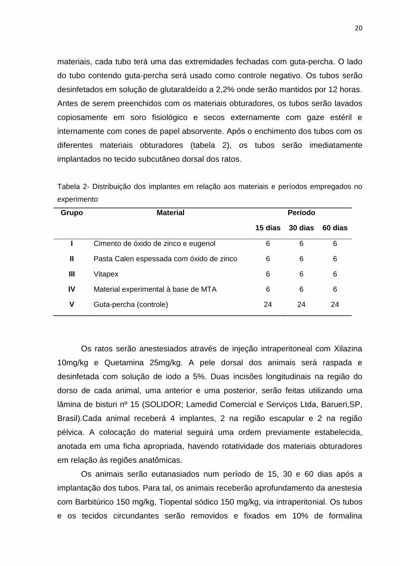
20
materiais, cada tubo terá uma das extremidades fechadas com guta-percha. O lado
do tubo contendo guta-percha será usado como controle negativo. Os tubos serão
desinfetados em solução de glutaraldeído a 2,2% onde serão mantidos por 12 horas.
Antes de serem preenchidos com os materiais obturadores, os tubos serão lavados
copiosamente em soro fisiológico e secos externamente com gaze estéril e
internamente com cones de papel absorvente. Após o enchimento dos tubos com os
diferentes materiais obturadores (tabela 2), os tubos serão imediatamente
implantados no tecido subcutâneo dorsal dos ratos.
Tabela 2- Distribuição dos implantes em relação aos materiais e períodos empregados no
experimento
Grupo Material Período
15 dias 30 dias 60 dias
I Cimento de óxido de zinco e eugenol 6 6 6
II Pasta Calen espessada com óxido de zinco 6 6 6
III Vitapex 6 6 6
IV Material experimental à base de MTA 6 6 6
V Guta-percha (controle) 24 24 24
Os ratos serão anestesiados através de injeção intraperitoneal com Xilazina
10mg/kg e Quetamina 25mg/kg. A pele dorsal dos animais será raspada e
desinfetada com solução de iodo a 5%. Duas incisões longitudinais na região do
dorso de cada animal, uma anterior e uma posterior, serão feitas utilizando uma
lâmina de bisturi nº 15 (SOLIDOR; Lamedid Comercial e Serviços Ltda, Barueri,SP,
Brasil).Cada animal receberá 4 implantes, 2 na região escapular e 2 na região
pélvica. A colocação do material seguirá uma ordem previamente estabelecida,
anotada em uma ficha apropriada, havendo rotatividade dos materiais obturadores
em relação às regiões anatômicas.
Os animais serão eutanasiados num período de 15, 30 e 60 dias após a
implantação dos tubos. Para tal, os animais receberão aprofundamento da anestesia
com Barbitúrico 150 mg/kg, Tiopental sódico 150 mg/kg, via intraperitonial. Os tubos
e os tecidos circundantes serão removidos e fixados em 10% de formalina
21
tamponada a um ph 7,0, em frascos unitários, com identificação do rato, grupo e
localização do tubo (região anterior ou posterior). Os tubos serão removidos
utilizando bisturi nº 11 (SOLIDOR; Lamedid Comercial e Serviços Ltda, Barueri, SP,
Brasil) e processados para avaliação histológica por coloração com HE. As amostras
serão avaliadas quanto à intensidade da resposta inflamatória, a espessura da
cápsula fibrosa, presença de calcificação e células gigantes. Reações nos tecidos
que estavam em contato com o material na abertura do tubo serão pontuadas como
se segue: (0) nenhuma ou poucas células inflamatórias e nenhuma reação; (1)
menos de 25 células e reação moderada, (2) entre 25 e 125 células e reação
moderada, e (3) 125 ou mais células e severa reação. As cápsulas fibrosas foram
classificadas como ''fina'' se espessura <150 mm ou ―grossa‖ se > 150 mm. A
presença de células gigante e a calcificação serão registradas como presente ou
ausente.
Uma média do número de células em cada grupo será obtida a partir da
pontuação de 10 áreas separadas. Os resultados serão analisados pelo teste de
Kruskal-Wallis ao nível de significância de 5%.
2.3.3 Teste de alteração de cor
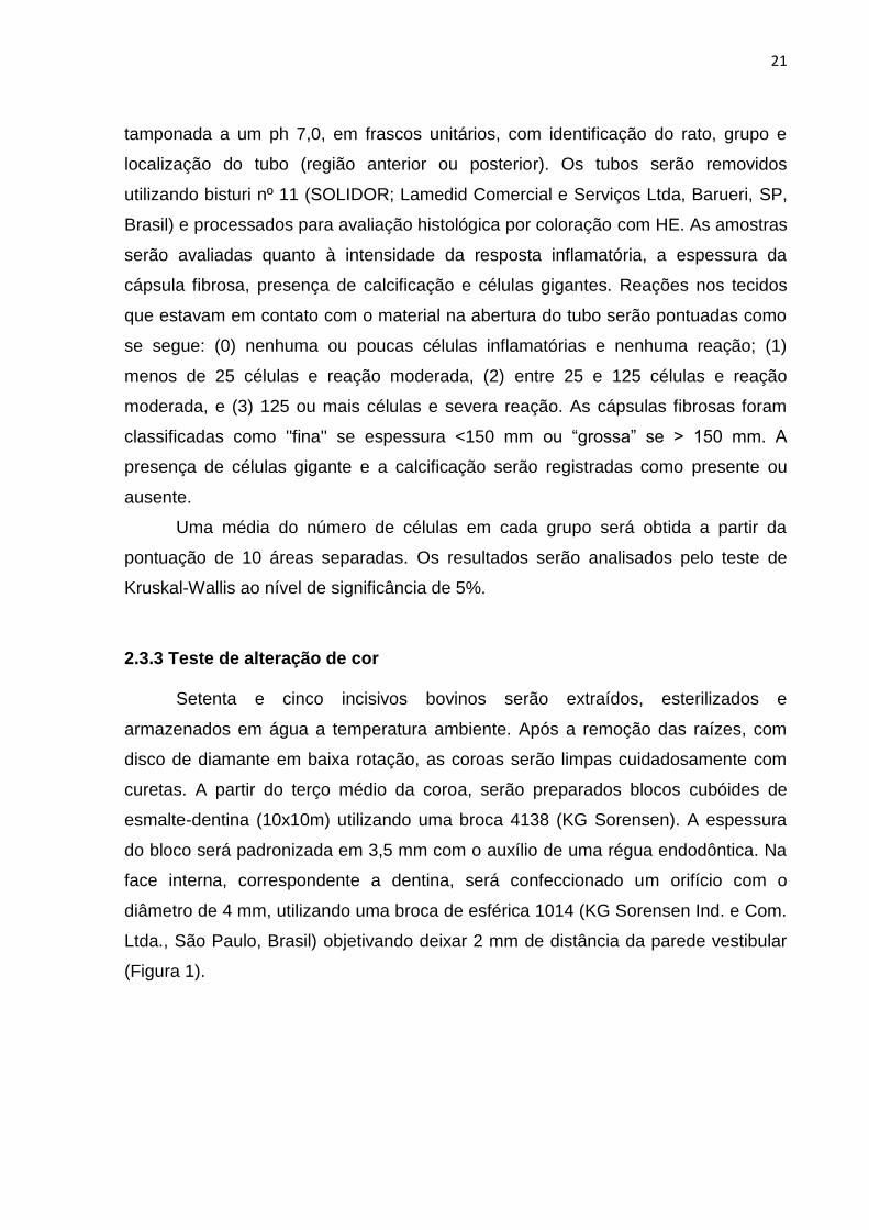
Setenta e cinco incisivos bovinos serão extraídos, esterilizados e
armazenados em água a temperatura ambiente. Após a remoção das raízes, com
disco de diamante em baixa rotação, as coroas serão limpas cuidadosamente com
curetas. A partir do terço médio da coroa, serão preparados blocos cubóides de
esmalte-dentina (10x10m) utilizando uma broca 4138 (KG Sorensen). A espessura
do bloco será padronizada em 3,5 mm com o auxílio de uma régua endodôntica. Na
face interna, correspondente a dentina, será confeccionado um orifício com o
diâmetro de 4 mm, utilizando uma broca de esférica 1014 (KG Sorensen Ind. e Com.
Ltda., São Paulo, Brasil) objetivando deixar 2 mm de distância da parede vestibular
(Figura 1).
22
Figura 1. Bloco cubóide de esmalte-dentina (10x10x3,5mm) preparado a partir do terço médio da coroa dente bovino.
Os espécimes serão imersos em hipoclorito de sódio 1% (Asfer Indústria e
Comércio Ltda) durante 30 min e após secagem, serão imersos em EDTA 17%
(Biodinâmica, Ibiporã, Pr, Brasil) por 3 minutos. Em seguida, os espécimes serão
colocados por 3 minutos em hipoclorito de sódio e posteriormente, armazenados em
água.
Os espécimes serão divididos aleatoriamente em quatro grupos
experimentais, de acordo com o material obturador a ser avaliado (n = 15). As
cavidades serão preenchidas com os seguintes materiais: Grupo 1, material
experimental à base de MTA (Angelus, Londrina, PR, Brazil); Grupo 2, Vitapex (Neo
dental Internacional, Federal Way / WA); Grupo 3, pasta Calen (S.S. White, Artigos
Dentários, Rio de Janeiro, RJ, Brasil) espessada com óxido de zinco (Biodinâmica
Química e Farmacêutica Ltda., Ibiporã, PR, Brazil) na proporção 1:1; Grupo 4, óxido
de zinco-eugenol (OZE) (Biodinâmica, Ibiporã, PR, Brasil). No grupo controle (Grupo
5), as cavidades serão mantidas sem material obturador (Tabela 3).
23
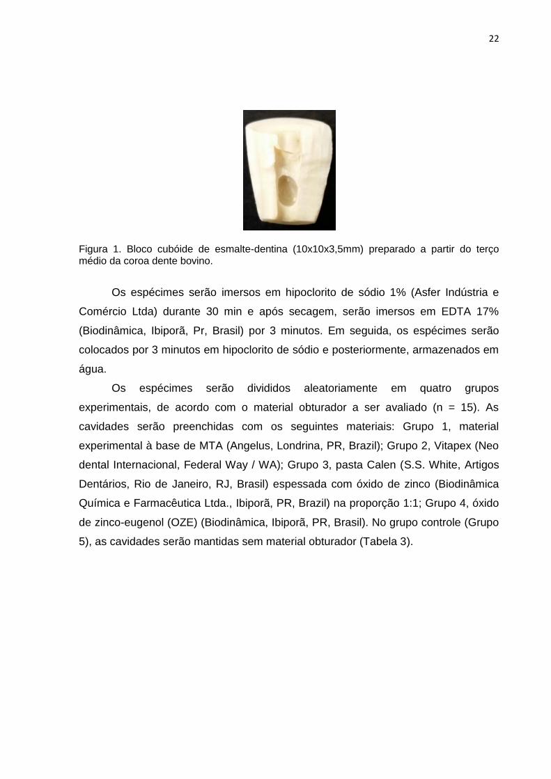
Todas as cavidades, inclusive as do grupo controle, serão seladas com Vitro
Fil LC (DFL Ind e Comércio S.A, Rio de Janeiro, Brasil) (Figura 2). A polimerização
será realizada utilizando um fotopolimerizador LED (Emitter A Fotopolimerizador/
Schuster Comércio de Equipamentos Odontológicos Ltda, Santa Maria, RS, Brasil)
por 20 s. Cada espécime será colocado em um tubo Falcon contendo 15 ml com
água. Os tubos serão armazenados a temperatura ambiente e mantidos no escuro
durante os 3 primeiros meses. Durante o período seguinte e até o final dos 9 meses,
as amostras serão expostas a luz solar de forma indireta (LENHERR,2012).
Figura 2 - Após preenchimento da cavidade com os materiais a serem avaliados, selamento com cimento de ionômero de vidro fotoativado.
As medições de cor serão realizadas em uma sala escura usando um
espectrofotômetro (Vita Easyshade, Vita- Zahnfabrik, Alemanha) sob condições
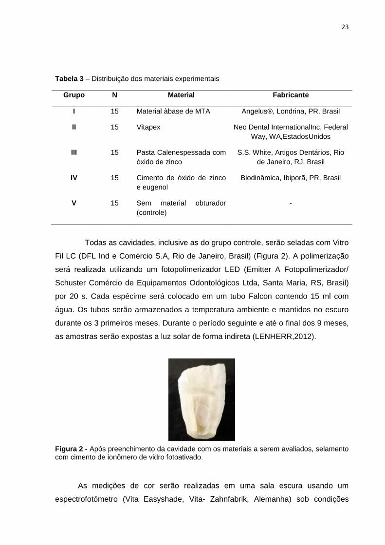
Tabela 3 – Distribuição dos materiais experimentais
Grupo N Material Fabricante
I 15 Material àbase de MTA Angelus®, Londrina, PR, Brasil
II 15 Vitapex Neo Dental InternationalInc, Federal
Way, WA,EstadosUnidos
III 15 Pasta Calenespessada com
óxido de zinco
S.S. White, Artigos Dentários, Rio
de Janeiro, RJ, Brasil
IV 15 Cimento de óxido de zinco
e eugenol
Biodinâmica, Ibiporã, PR, Brasil
V 15 Sem material obturador
(controle)
-
24
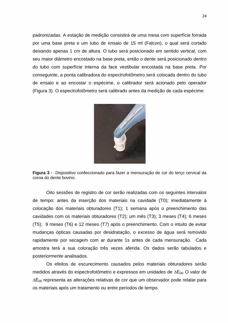
padronizadas. A estação de medição consistirá de uma mesa com superfície forrada
por uma base preta e um tubo de ensaio de 15 ml (Falcon), o qual será cortado
deixando apenas 1 cm de altura. O tubo será posicionado em sentido vertical, com
seu maior diâmetro encostado na base preta, então o dente será posicionado dentro
do tubo com superfície interna da face vestibular encostada na base preta. Por
conseguinte, a ponta calibradora do espectrofotômetro será colocada dentro do tubo
de ensaio e ao encostar o espécime, o calibrador será acionado pelo operador
(Figura 3). O espectrofotômetro será calibrado antes da medição de cada espécime.
Figura 3 - Dispositivo confeccionado para fazer a mensuração de cor do terço cervical da coroa do dente bovino.
Oito sessões de registro de cor serão realizadas com os seguintes intervalos
de tempo: antes da inserção dos materiais na cavidade (T0); imediatamente à
colocação dos materiais obturadores (T1); 1 semana após o preenchimento das
cavidades com os materiais obturadores (T2); um mês (T3); 3 meses (T4); 6 meses
(T5); 9 meses (T6) e 12 meses (T7) após o preenchimento. Com o intuito de evitar
mudanças ópticas causadas por desidratação, o excesso de água será removido
rapidamente por secagem com ar durante 1s antes de cada mensuração. Cada
amostra terá a sua coloração três vezes aferida. Os dados serão tabulados e
posteriormente analisados.
Os efeitos de escurecimento causados pelos materiais obturadores serão
medidos através do espectrofotômetro e expressos em unidades de ∆E00. O valor de
∆E00 representa as alterações relativas de cor que um observador pode relatar para
os materiais após um tratamento ou entre períodos de tempo.
25
O cálculo da diferença de cor será realizado a partir dos valores de L*, a* e b*
obtidos das leituras de cor nos diferentes tempos experimentais em comparação ao
T0 (Baseline). Dessa maneira será possível comparar a diferença de cor dos
espécimes através da fórmula CIEDE 2000.
Onde o valor de L descreve a luminosidade que varia de preto (0) a branco
(100), enquanto a* e b* indicam croma vermelho/verde e azul/amarelo.
Quanto menor o valor do ∆E 00, menor a diferença de cor entre a cor inicial do
dente e cor final ao longo do tempo.
Os dados serão submetidos ao teste de Anova com Repeated Measurese
comparação múltipla de Tukey para avaliar o efeito dos fatores tempo e material no
escurecimento dentário (p = 0,05).
26
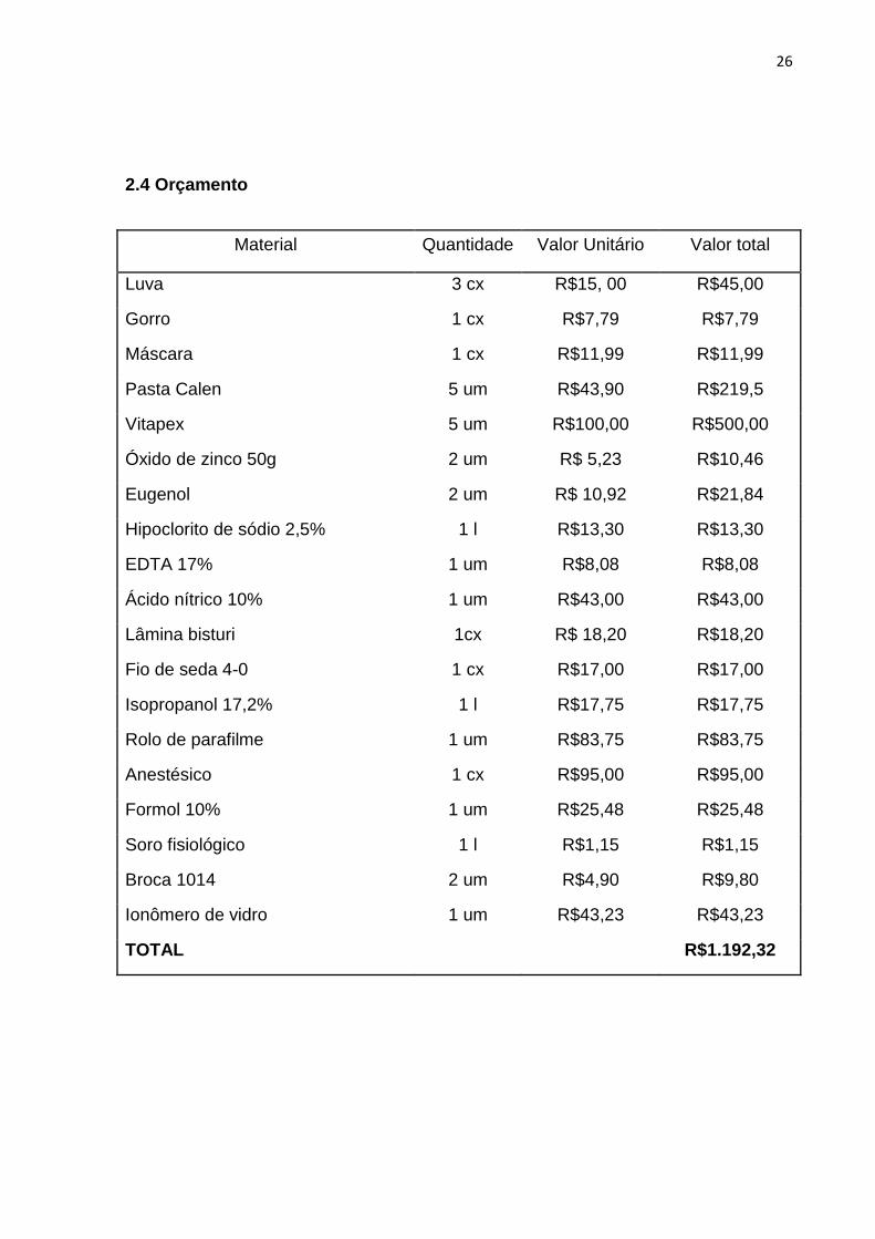
2.4 Orçamento
Material Quantidade Valor Unitário Valor total
Luva 3 cx R$15, 00 R$45,00
Gorro 1 cx R$7,79 R$7,79
Máscara 1 cx R$11,99 R$11,99
Pasta Calen 5 um R$43,90 R$219,5
Vitapex 5 um R$100,00 R$500,00
Óxido de zinco 50g 2 um R$ 5,23 R$10,46
Eugenol 2 um R$ 10,92 R$21,84
Hipoclorito de sódio 2,5% 1 l R$13,30 R$13,30
EDTA 17% 1 um R$8,08 R$8,08
Ácido nítrico 10% 1 um R$43,00 R$43,00
Lâmina bisturi 1cx R$ 18,20 R$18,20
Fio de seda 4-0 1 cx R$17,00 R$17,00
Isopropanol 17,2% 1 l R$17,75 R$17,75
Rolo de parafilme 1 um R$83,75 R$83,75
Anestésico 1 cx R$95,00 R$95,00
Formol 10% 1 um R$25,48 R$25,48
Soro fisiológico 1 l R$1,15 R$1,15
Broca 1014 2 um R$4,90 R$9,80
Ionômero de vidro 1 um R$43,23 R$43,23
TOTAL R$1.192,32

2.5 Cronograma de atividades
Ano 2015 2016
Mês J J A S O N D J F M A M J J A S O N D
Revisão da
literatura
X x x X X x x
Qualificação
do projeto
x
Piloto X
Experimento 1 X X x x x X
Experimento 2 x x x X x x X X x x X
Digitação dos
dados
x x X x
Análise dos
dados
x X x
Redação da
dissertação
x x
Redação dos
artigos
x x
Defesa x

3 Relatório do Trabalho de campo
Neste capítulo estão relatadas as complementações e as mudanças
ocorridas no planejamento e execução dos experimentos desta pesquisa.
3.1 O projeto de qualificação de tese por mim apresentado a banca, se
intitulava ―Revascularização de dentes com polpa necrosada e rizogênese
incompleta: estudo clínico randomizado‖. No entanto, não estou apresentando
esse trabalho como tese de doutorado devido a dificuldade de conseguir
pacientes que se enquadrassem nos critérios de inclusão do estudo.
3.2 O trabalho apresentado como tese de doutorado foi qualificado
durante o meu mestrado.
3.3 No teste de biocompatibilidade houve uma perda da amostra durante
o processamento histológico, o que reduziu o nosso n de 18 animais para 15.
3.4 As mensurações de cor seriam prorrogadas por mais três meses, no
entanto, o espectrofotômetro (Vita Easyshade) da Faculdade de Odontologia
de Pelotas encontrava-se danificado. As mensurações foram realizadas,
portanto, pelo tempo máximo de 9 meses, sendo que esse tempo foi suficiente
para verificarmos a discoloração dentária ocorrida nos espécimes. É importante
salientar que o uso de outro equipamento poderia trazer desvio de resultados.
4 Artigos
4.1 Artigo 1*
Tissue reaction to a new MTA-based root canal filling material for primary
teeth
K. J. PILOWNIC1; A. P. N. GOMES1; A. C. FELIX2; A. R. ROMANO1& F. G.
PAPPEN1
1 Graduate Program in Dentistry, Federal University of Pelotas, Pelotas, RS,
Brazil.
2Central Vivarium, Faculty of Veterinary, Federal University of Pelotas, Pelotas,
Brazil
Correspondence address: Fernanda Geraldo Pappen
Graduate Program, Dentistry, Federal University of Pelotas
R. Gonçalves Chaves, 457, sala 507, Pelotas, RS, Brasil. CEP: 96015-560
E-mail: [email protected]
* Artigo estruturado segundo as normas do periódico Pediatric Dentistry
30
Abstract
Purpose: This study evaluated the tissue reactions to the experimental MTA-
based material in comparison to Zinc Oxide and Eugenol cement (ZOE);
Vitapex; and Calen paste thickened with zinc oxide (Calen+ZO). Methods:
Fifteen Wistar rats had received four implants containing the tested filling
materials. The tubes had one of the ends closed with gutta-percha used as
negative control. After 15, 30 and 60 days, the animals were euthanized and the
tubes and surrounding tissue were removed and processed for histopathologic
evaluation. The specimens were evaluated for intensity of inflammatory
response, fibrous capsule thickness, and for the presence of calcification and
giant cells. The results were analyzed by the Kruskal-Wallis test. Results: On
day 15, thick fibrous capsule and moderate inflammatory response were
observed in Vitapex and experimental MTA-based material, while in ZOE and
Calen+ZO groups, the intensity of inflammatory reaction was moderate to
severe. On day 30, most of experimental MTA-based material specimens
showed mild inflammatory reaction and a thin fibrous capsule. After 60 days,
Calen+ZO group still showed moderate inflammation, while the others showed
predominantly mild inflammatory reaction and a thin fibrous capsule.
Conclusion: Experimental MTA-based material had showed to be
biocompatible, representing an alternative for endodontic filling of primary teeth.
Key Words: primary teeth; root canal filling materials; biocompatibility
31
Introduction
An ideal endodontic filling material for primary teeth must have several
properties that make it suitable for use, such as resorption at a rate similar to
that of the primary root, be harmless to the periapical tissues and permanent
tooth germ and readily resorb if pressed beyond the apex.1,2
Several materials have been used for endodontic filling in primary teeth.
Zinc oxide (ZO) and zinc oxide eugenol (ZOE) have been widely used in the
primary dentition. 1,2,3 Nevertheless, due to their irritating potential 1,2 and low
resorption capacity3 the use of these materials have been substituted by
iodoform containing materials, or calcium hydroxide pastes, because of their
antibacterial activity, biocompatibility and easy resorption.2,4,5 Other studies
have also suggested the use of association of calcium hydroxide and zinc oxide
6,7 as well as calcium hydroxide and iodoform. 4,6,7
Recently, an experimental MTA-based material, ready to use, endodontic
filling material for primary teeth had been developed (Angelus, Londrina, PR,
Brazil). According to the manufacturer, the experimental MTA-based material
has in its composition mineral trioxide aggregate (MTA), calcium tungstate,
polyethylene glycol, glycerin, silicate oxide e silicon. Unpublished data
demonstrated that this material has acceptable radiopacity, low cytotoxity,
antimicrobial efficacy and proper pH.
The biocompatibility of all experimental dental materials that might come
in contact with tissues should be examined. Biological properties are amongst
the most important aspects of endodontic filling materials because the release
of substances from the materials may generate reactions in the periapical
32
tissue.8-9The subcutaneous connective tissue implantation method is one of the
main methods used to evaluate the biocompatibility of a material.10-13
Therefore, the purpose of this study was to evaluate the tissue reaction
induced by an experimental MTA-based material when implanted in
subcutaneous connective tissues, in comparison with others endodontic filling
materials used for primary teeth.
Materials and Methods
The materials tested were the experimental MTA-based material
(Angelus, Londrina, PR, Brazil); Zinc Oxide and Eugenol cement (ZOE)
(S.S.White Artigos Dentários Ltda., Rio de Janeiro, RJ, Brazil); Vitapex, a
premixed calcium hydroxide and iodoform paste (Neo Dental International Inc,
Federal Way, WA, USA); Calen, a premixed calcium hydroxide and
polyethylene glycol-based paste (S.S.White Artigos Dentários Ltda.), thickened
with zinc oxide (ZO) (1:1)(Biodinâmica Química e Farmacêutica Ltda.,Ibiporã,
PR, Brazil).
The materials were inserted into sterile polyethylene tubes measuring 10
mm long and 1.0 mm internal diameter (Abbott Laboratorios do Brasil, São
Paulo, Brazil). To avoid the materials extrusion, each tube had one of the ends
closed with gutta-percha. The side of the tube containing gutta-percha was
used as negative control. After complete filling, the tubes were immediately
implanted on animal‘s dorsal subcutaneous tissue.
The Research Ethics Committee for Animal Use of the Federal University
of Pelotas approved this study (Protocol #8977; 02/04/14). All procedures were
carried out in accordance with institutional guidelines for animal care and use.
Fifteen Wistar rats (Rattusnorvegicus; age, 6 months; weight, ~250 g) were
33
used in this study. The animals‘ tails were marked for individual identification.
The rats were housed in plastic cages (twoper cage) placed in ventilated racks
(Alesco, Monte Mor, SP, Brazil) at 22°C with a 12 hours light⁄dark cycle (lights
on at 7:00 am, off at 7:00 pm). During the experiments, the rats were provided
with a standard diet of rat chow (Nuvilab, Colombo, PR, Brazil) and filtered
water ad libitum.
The animals were anesthetized by intraperitoneal injection of ketamine
(80mg kg-1 by weight) combined with xylazine (4mg kg-1 by weight; both from
SDC ECommerce for Agricultural Products LTDA, Marilia, SP, Brazil). Dorsal
trichotomy and antisepsis was performed using iodinated alcohol and sterile
distilled water. 14
Four 2-cm incisions were made on the shaved backs in a head-to-tail
orientation using a #15 Bard Parker blade (LamedidSolidor, Osasco, SP,
Brazil). Using blunt-tipped scissors, the lateral tearing of the subcutaneous
tissue provided four surgical cavities displayed in quadrants equidistant from the
center of the animals' backs. Each animal received four implants, two in the
scapular region and two in the caudal region. The tubes filled with materials
were implanted into the spaces created by blunt dissection, according to a
previously established placement order and site rotation, and the skin was
closed using a 4-0 silk suture (Johnson & Johnson Produtos Profissionais Ltda,
São José dos Campos, SP, Brazil). Sterile instruments and aseptic techniques
were used throughout the experiments. The animals were placed in individual
cages until they recovered from anesthesia. To aid recovery, paracetamol (0.06
mg g-1day-1) was added to their drinking water for 72 h.15
34
The animals were euthanized at 15, 30 and 60 days after the dorsal
implantation (n=5/group at each time point). They were anesthetized with an
intraperitoneal injection of chloral hydrate (350 mg/kg) (Sigma Aldrich, St. Louis,
MI, USA), and physiological saline (Sigma Aldrich, St. Louis, MI, USA) followed
by 10% paraformaldehyde in 0.1 M phosphate buffer (pH 7.4) (Sigma Aldrich,
St. Louis, MI, USA) was perfused transcardially. The tubes and the surrounding
tissues were removed and fixed in 10% buffered formalin at a pH of 7.0. The
tubes were bisected transversely, and both halves were subsequently cut
longitudinally with a sharp blade to allow the surfaces to maintain contact with
the processing solutions. The specimens were paraffin embedded, sectioned
serially to 3-mm slices, and stained with hematoxylin and eosin.
Tissue reactions in contact with the material at the opening of the tube
were scored as follow: 0, no or few inflammatory cells and no reaction; 1, less
than 25 cells and mild reaction; 2, between 25 and 125 cells and moderate
reaction; and 3, 125 or more cells and severe reaction. Fibrous capsules were
classed as ‗‗thin‘‘ or ‗‗thick‘‘ if the thickness was <150 mm or >150 mm,
respectively. Calcification was recorded as present or absent. Also the
presence of giant cells was recorded. In a random order, only one control in
each animal was evaluated. The observer, who was a pathologist, was blinded
to the treatment. The results were analyzed by the Kruskal-Wallis test at a 5%
significance level.
Results
On day 15, thick fibrous capsule formation and moderate cell
inflammatory infiltration by lymphocytes and macrophages were observed with
35
all the Vitapex and experimental MTA-based material specimens, while in ZOE
and Calen+ZO groups, the intensity of inflammatory reaction was moderate to
severe (p = 0.001). The capsule thickness surrounding the tubes was
significantly thinner in the control specimens when compared with the
experimental materials (p < 0.001) and the presence of giant cells in this period
varied from 20 to 60% in the experimental groups (p > 0.005). The presence of
calcificated precipitates was noticed in 40% of specimens from Calen+ZO and
Vitapex on day 15, and it was not observed in ZOE and experimental MTA-
based material groups (Table 1).
The Vitapex group showed a large capsule extension, with evident
presence of macrophage foam cells on day 15 and 30. Mild to moderate
inflammatory cell infiltration and a reduction in the thickness of the fibrous
capsule was evident from day 30 onward (Fig.1C;H).
On day 30, moderate inflammatory cell infiltration was observed in 100%
of Vitapex and Calen+ZO, and 80% of ZOE specimens, while most
experimental MTA-based material specimens showed mild inflammatory
reaction only (p = 0.001). The fibrous capsule thickness was also significantly
thinner in the experimental MTA-based material group (p = 0.001). On this
period, the presence of giant cells was mostly observed in Vitapex specimens,
but was still noticed in the other groups (p > 0.005). Vitapex and Calen+ZO
induced the deposition of calcified tissue more frequently than the other
materials (p = 0.02).
In the control group, mild inflammatory reaction with lymphocytes and
macrophages was observed at 15 and 30 days. After 60 days, inflammatory
36
response was absent in the areas in contact with gutta-percha. There was no
giant cells or calcificated precipitated in the specimens from control group.
At 60 days, the inflammatory reaction was mild in most specimens of
ZOE, experimental MTA and Vitapex, while 80% of specimens from Calen+ZO
group still showed moderate inflammatory infiltrate (p = 0.001). The capsule
extension was predominantly thin for all the materials (p > 0.005), and the
presence of giant cells were recorded only in few ZOE specimens (p > 0.005).
The occurrence of calcification, was more common in specimens from ZOE and
Calen+ZO (p = 0.003).
Discussion
The biomechanical endodontic treatment of primary teeth remains far
from ideal, especially due to their anatomical features and difficulties in
obtaining a good radiographic view of the apex for determining the working
length.16Due to the physiological root resorption process, materials used in
endodontic treatment of primary teeth should be harmless to the periapical
tissues and permanent tooth germs.17The experimental MTA-based material
has been developed as an alternative to be used in the endodontic filling for
primary teeth, and in the present study demonstrated promising results for the
material in terms of subcutaneous implantation.
The method of implantation of endodontic filling materials in the
subcutaneous tissue of rats, in part simulates the situation of the root canal, and
is one of the most appropriate tests to evaluate materials biocompatibility.12-
13Thus, our results consist in a preliminary source of information regarding the
37
biocompatibility of experimental MTA-based material. Besides, the inert nature
of polyethylene tubes and their ability to expose a test material to living tissue in
a controlled and effective manner justify its choice.18In the present
methodology, one end of the tubes were sealed with gutta-percha in order to
avoid material extrusion. Also, since gutta-percha is well tolerated by connective
tissues, this tube extremity was used as negative control.9,13
Both, the control and the tested material specimens presented
significantly less inflammation severity at longer time intervals (30 and 60 days)
in comparison to 15 days specimens. Similarly, the capsule extension was
mostly thick in all groups on day 15 and it declined by over the time,
demonstrating that every tested material was well tolerated by the
subcutaneous tissues. It was possible to observe collagen fiber formation at the
area in contact with the all the tested materials. Studies had described that
there is a meaningful relationship between inflammation and fibrous capsule
thickness.19, 20, 21 Makkes et al.19 concluded that decreasing capsule thickness is
a sign of biocompatibility, and the deferred harmful effects of a material are
considered to be more important than its initial effects in biocompatibility tests.22
On day 60, the inflammatory reaction was absent in control group; mild in
most specimens of ZOE, experimental MTA-based material and Vitapex, while
most specimens from Calen+ZO group still showed moderate inflammatory
infiltrate. Contrary to our findings, Nelson-Filho et al.23 and Queiroz et al.24 had
reported that zinc oxide does not interfere the biocompatibility of calcium
hydroxide. When used alone, Calen paste present recognized
biocompatibility23,25, thus the persistent inflammation observed in Calen+ZO
group may be attributed to the higher concentration of zinc in the medicament,
38
since zinc has the capacity to influence in the inflammatory process for reducing
phagocytic capacity of macrophages and to interfere in the membrane of the
lysosomes.10, 11
The tissue reaction demonstrated by ZOE in this study was similar to
those described by other authors for ZOE and other ZOE-based sealers.26, 27 In
the initial periods, ZOE demonstrated severe inflammation which had decline by
over the time. ZOE cement is initially highly toxic because of its traces of
unreacted eugenol, exerting a toxic reaction to cells.28,29 This profile tends to
change significantly along the time, when the material becomes more
biocompatible, due to the reduced availability of unreacted eugenol.
Although the conventional MTA is well known by its excellent
biocompatibility,30,31 The composition of experimental MTA-based material
differs from the traditional MTA: besides a portion of MTA, the experimental
filling material also has in its composition calcium tungstate, polyethylene glycol,
glycerin, silicate oxide and silicon. Our results demonstrated an optimal tissue
reaction to the experimental MTA-based material, what had occurred probably
due to the presence of MTA in its composition and the absence of toxic
components. Also, despite the optimal clinical and radiographic success rates of
Vitapex as a endodontic filling material of primary teeth,2,32 there are few
information regarding its biocompatibility. Similarly to our results, Huang et al.4
also showed Vitapex to be biocompatible, since it resulted in high survival rate
of cells after the experimental period.
In all the experimental groups, it was possible to observe the presence of
giant cells, mainly within 15 days. The presence of giant cells may be
39
associated with the presence of material that the organism hardly breaks
down31. However, the number of particles of materials present in the
subcutaneous tissue had decreased by over the time, suggesting the capacity
of these filling materials to be reabsorbed when in contact with connective
tissue. The ability of the material to be resorbable is considered one of the main
requirements for the indication of a endodontic filling material for primary teeth.
The material resorption must occur simultaneously to root resorption during
exfoliation, allowing normal eruption of permanent teeth. Also in cases of over-
filling, the material should be resorbable and non-toxic to the periapical tissues
and germ of the permanent.1,2
Calcific precipitation was observed around implantation sites in all
experimental groups, but more frequently in Calen+ZO specimens. The
production of calcific structures in subcutaneous investigations is a sign of
osteoinductivity of the material.33The osteoinductivity and conductivity the
materials may be attributed to the release of calcium and phosphorous as well
as the formation of hydroxyapatite crystals over the material.25,34 Similar to our
study, other authors had also reported the presence of calcification in
conventional MTA and Calcium hydroxide based materials.30MTA does not
have calcium hydroxide in its composition, but it has calcium oxide that could
react with tissue fluids to form calcium hydroxide, what can explain the similar
behavior of experimental-MTA, Calen+ZO and Vitapex regarding the induction
of calcification. However, no prior study demonstrated the osteoinductivity of
ZOE.
Within the limits of this study, our results showed that all endodontic
filling materials were well tolerated by the subcutaneous tissues. The
40
experimental-MTA based material showed to be biocompatible, representing an
alternative for the endodontic filling of primary teeth.
REFERENCES
1. Kubota K, Golden BE, Penugonda B. Root canal filling materials for
primary teeth: A review of the literature. J Dent Child.1992;59(3):225–7.
2. Mortazavi M, Mesbahi M. Comparison of zinc oxide and eugenol, and
Vitapex for root canal treatment of necrotic primary teeth. Int J Paediatr
Dent 2004;14(6):417– 24.
3. Fuks AB, Eidelman E. Pulp therapy in the primary dentition. CurrOpin
Dent 1991;1(5):556 –63.
4. Huang TH, Ding SJ, Kao CT. Biocompatibility of various formula root
filling materials for primary teeth. J Biomed Mater Res B Appl
Biomater.2007; 80b(2):486-90.
5. Ranly DM, Garcia-Godoy F. Current and potential pulp therapies for
primary and young permanent teeth. J Dent. 2000;28(3):153–61
6. Chawla HS, Mathur VP, Gauba K, Goyal A. A mixture of Ca(OH)2 paste
and ZnO powder as a root canal filling material for primary teeth: a
preliminary study. J IndianSocPedodPrevDent.2001;19(3):107-9.
7. Queiroz AM, Nelson-Filho P, Silva LA, Assed S, Silva RA, Ito
IY.Antibacterial activity of root canal filling materials for primary teeth: zinc
oxide and eugenolcement, Calen paste thickened with zinc oxide,
Sealapex and EndoREZ. Braz Dent J. 2009; 20(4):290-6.
41
8. Kaplan AE, Ormaechea MF, Picca M, Canzobre MC, Ubios AM.
Rheological properties and biocompatibility of endodontic sealers.
IntEndod J. 2003;36(8):527–32.Ho YC, Huang FM, Chang YC.
Mechanisms of cytotoxicity of eugenol in human osteoblast cells in vitro.
IntEndod J.2006;39 (5):389–93.
9. Pinto DN, Sousa DL, Rocha RB, Moreira JJ. Eighteen-month clinical and
radiographic evaluation of two root canal-filling materials in primary teeth
with pulp necrosis secondary to trauma. Dent Traumatol.2011;27(3):221–
4.
10. Silva, LAB, Leonardo MR, Oliveira DS, Silva RA, Queiroz AM, Hernández
PG, Nelson-Filho P. Histopathological evaluation of root canal filling
materials for primary teeth. Braz Dent J.2010; 21(1):38–45.
11. Olsson B, Sliwkowsky A, Langeland K. Subcutaneous implantation for the
biological evaluation of endodontic materials. J Endod. 1981;7(8):355–67.
12. Torneck CD. Reaction of rat conective tissue to polyethylene tube
implants.Oral Surg.1966;21(3):379-87.
13. Sobrinho AP, Barros MH, Nicoli JR,Carvalho MA, Farias LM, Bambirra
EA.et al. Experimental root canal infections in conventional and germ-free
mice. J Endod.1998; 24(6), 405-8.
14. Simon S, Cooper P, Smith A, Picard B, Ifi CN, Berdal A. Evaluation of a
new laboratory model for pulp healing: preliminary study. Int Endod
J.2008; 41(9): 781-90.
15. Özalp N, Saroglu I, Sönmez H. Evaluation of various root canal filling
materials in primary molar pulpectomies: an in vivo study. Amer J Dent
2005;18(6):347-50.
42
16. Mortazavi M, Mesbahi M. Comparison of zinc oxide and eugenol, and
Vitapex for root canal treatment of necrotic primary teeth. Int J Paediatr
Dent. 2004;14:417-24.
17. Langeland K, Olsson B, Pascon EA. Biological evaluation of hydron. J
Endo.1981;7(5):196-204.
18. Makkes PC, van Velzen SK, Wesselink PR, deGreeve PC. Polyethylene
tubes as a model for the root canal. Oral Surg, Oral Med, Oral Pathol.
1977; 44(2): 293–300.
19. Quinalan CA, ZistererDM, Tipton KF, O‘Sullivan MJ. In vitro cytotoxicity
of a composite resin and compomer. Int Endod J. 2002; 35(1):47–55.
20. Sanders JE, Rochefort JR. Fibrous encapsulation of single polymer
depends on their vertical dimension in subcutaneous tissue. 2003; J
Biomed Mat Res;67(4) 1181–7.
21. Stanford JW. Recommended standard practices for biological evaluation
of dental materials Federation Dentaire International, Commission of
Dental Materials, Instruments, Equipment and Therapeutics. Int Dent J
1980; 30(2):140–88.
22. Nelson Filho P, Silva LA, Leonardo MR, Utrilla LA, Figueiredo F.
Connective tissue responses to calcium hydroxide-based root canal
medicaments. Int Endod J.1999; 32(4): 303–11.
23. Queiroz AM, Assed S, Consolaro A, Nelson-Filho P, Leonardo MR, Silva
RA, Silva LA. Subcutaneous connective tissue response to primary root
canal filling materials. Braz Dent J.2011;22(3):203-11.
24. Leonardo MR, Hernandez ME, Silva LA, Tanomaru-FilhoM. Effect of a
calcium hydroxide-based root canal dressing on periapical repair in dogs:

43
a histological study. Oral Surg Oral Med Oral Pathol Oral RadiolEndod.
2006;102(5):680-5.
25. Tagger M, Tagger E. Subcutaneous reactions to implantation of tubes
with AH26 and Grossman‘s sealers. Oral Surg Oral Med Oral Pathol
1986;62 (4):434–40.
26. Yesilsoy C, Koren LZ, Morse DR, Kobayashi C. A comparative tissue
toxicity evaluation of established and newer root canal sealers. Oral Surg
Oral Med Oral Pathol1988;65 (4):459–67.
27. Rodrigues H, Spangberg L, Langeland K. Biologic effects of dental
materials. 9. Effect of zinc oxide-eugenol cements on hella cells in vitro.
Estomat Cult 1975;9(2):191–4.
28. Zmener O, Goldberg F, Cabrini RL. Effects of two gutta-percha
formulations and one zinc oxide-eugenol and Canada balsam mixture on
human blood monocytes and lymphocytes. Endod Dent Traumatol
1989;5(2):73–7.
29. Holland R, de Souza V, Nery MJ, Otobon iFilho JA, Bernabé PF, Dezan
Júnior E .Reaction of rat connective tissue to implanted dentin tubes filled
with mineral trioxide aggregate or calcium hydroxide.J Endod.
1999;25(3):161-6.
30. Khashaba RM, Moussa MM, Chutkan NB, Borke JL. The response of
subcutaneous connective tissue to newly developed calcium phosphate-
based root canal sealers. Int Endod J.2011;44(4):342-52.
31. Trairatvorakul C, Chunlasikaiwan S. Success of pulpectomy with zinc
oxide–eugenol vs calcium hydroxide/iodoform paste in primary molars: a
clinical study. Ped Dent.2008;30(4):303–8.
44
32. Moretton TR, Brown CE Jr, Legan JJ, Kafrawy AH. Tissue reactions after
subcutaneous and intra osseous implantation of mineral trioxide
aggregate and ethoxy benzoic acid cement. J Biomed Mater Res.2000;
52(3):528–33.
33. Mizuno M, Banzai Y. Calcium ion release from calcium hydroxide
stimulated fibronectin gene expression in dental pulp cells and the
differentiation of dental pulp cells to mineralized tissue forming cells by
fibronectin. Int Endod J. 2008;41(11):933-8.
45
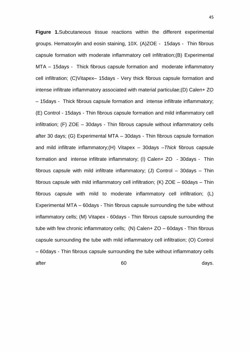
Figure 1.Subcutaneous tissue reactions within the different experimental
groups. Hematoxylin and eosin staining, 10X. (A)ZOE - 15days - Thin fibrous
capsule formation with moderate inflammatory cell infiltration;(B) Experimental
MTA – 15days - Thick fibrous capsule formation and moderate inflammatory
cell infiltration; (C)Vitapex– 15days - Very thick fibrous capsule formation and
intense infiltrate inflammatory associated with material particulae;(D) Calen+ ZO
– 15days - Thick fibrous capsule formation and intense infiltrate inflammatory;
(E) Control - 15days - Thin fibrous capsule formation and mild inflammatory cell
infiltration; (F) ZOE – 30days - Thin fibrous capsule without inflammatory cells
after 30 days; (G) Experimental MTA – 30days - Thin fibrous capsule formation
and mild infiltrate inflammatory;(H) Vitapex – 30days –Thick fibrous capsule
formation and intense infiltrate inflammatory; (I) Calen+ ZO - 30days - Thin
fibrous capsule with mild infiltrate inflammatory; (J) Control – 30days – Thin
fibrous capsule with mild inflammatory cell infiltration; (K) ZOE – 60days – Thin
fibrous capsule with mild to moderate inflammatory cell infiltration; (L)
Experimental MTA – 60days - Thin fibrous capsule surrounding the tube without
inflammatory cells; (M) Vitapex - 60days - Thin fibrous capsule surrounding the
tube with few chronic inflammatory cells; (N) Calen+ ZO – 60days - Thin fibrous
capsule surrounding the tube with mild inflammatory cell infiltration; (O) Control
– 60days - Thin fibrous capsule surrounding the tube without inflammatory cells
after 60 days.
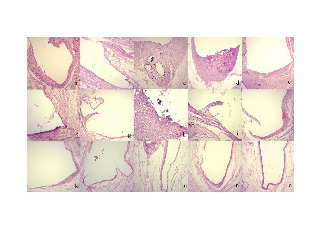
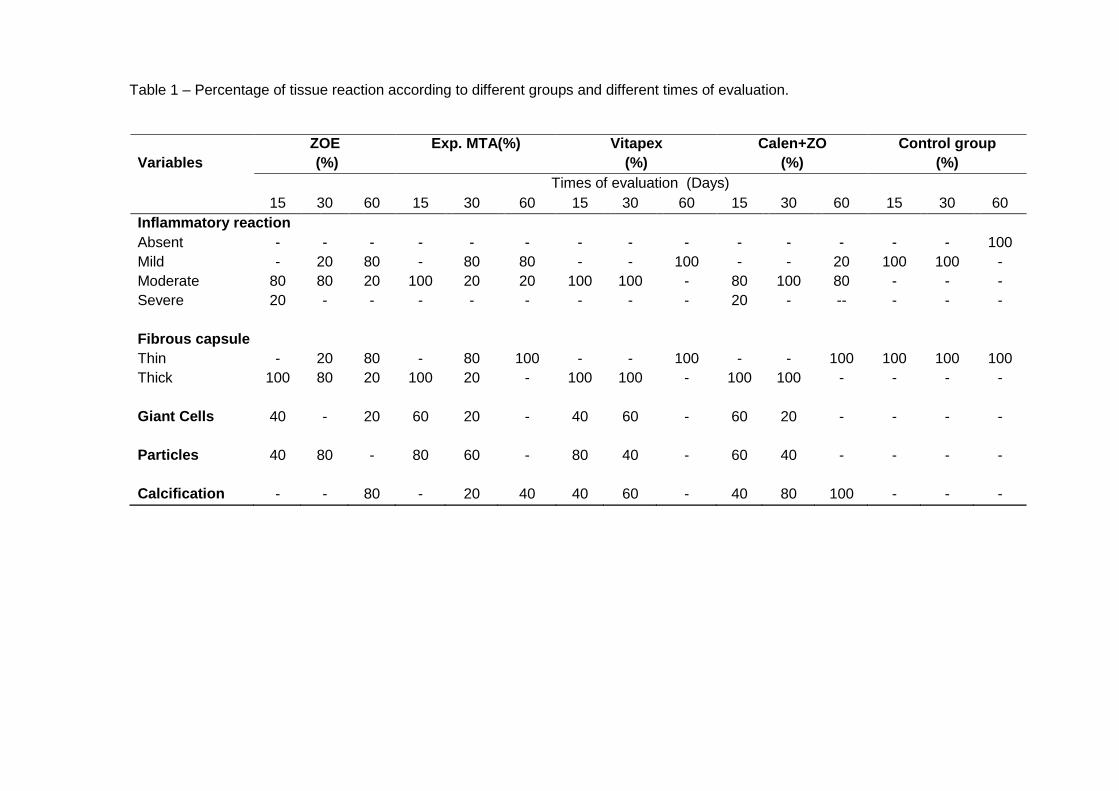
Table 1 – Percentage of tissue reaction according to different groups and different times of evaluation.
Variables
ZOE
(%)
Exp. MTA(%) Vitapex
(%)
Calen+ZO
(%)
Control group
(%)
Times of evaluation (Days)
15 30 60 15 30 60 15 30 60 15 30 60 15 30 60
Inflammatory reaction
Absent - - - - - - - - - - - - - - 100
Mild - 20 80 - 80 80 - - 100 - - 20 100 100 -
Moderate 80 80 20 100 20 20 100 100 - 80 100 80 - - -
Severe 20 - - - - - - - - 20 - -- - - -
Fibrous capsule
Thin - 20 80 - 80 100 - - 100 - - 100 100 100 100
Thick 100 80 20 100 20 - 100 100 - 100 100 - - - -
Giant Cells 40 - 20 60 20 - 40 60 - 60 20 - - - -
Particles 40 80 - 80 60 - 80 40 - 60 40 - - - -
Calcification - - 80 - 20 40 40 60 - 40 80 100 - - -
4.2 Artigo 2*
BOVINE TOOTH DISCOLORATION INDUCED BY ENDODONTIC FILLING
MATERIALS FOR PRIMARY TEETH
Samantha Rodrigues XAVIER*; Katerine Jahnecke PILOWNIC*; Andressa Heberle
GASTMANN*, Mariana Silveira ECHEVERRIA*; Ana Regina ROMANO*, Fernanda
Geraldo PAPPEN*l
*Graduate Program in Dentistry, Federal University of Pelotas, Pelotas, RS, Brazil.
Short title: PRIMARY TEETH DISCOLORATION
Corresponding author:
Fernanda Geraldo Pappen
Address: Graduate Program in Dentistry – Federal University of Pelotas
– R. Gonçalves Chaves, 457, 507, Pelotas, RS, Brasil. CEP: 96015-560.
Phone./Fax:+55-53-3222-6690 r. 135
Email: [email protected]
* Artigo aceito para publicação no periódico International Journal of Dentistry
49
Abstract
Objective: The present study aimed to evaluate the discoloration potential of
endodontic materials used in primary teeth. Material and Methods: Dentine-enamel
blocks were prepared from the middle coronary thirds of 75 bovine teeth, assorted in
five experimental groups (n = 15). The tested materials included an MTA-based
material; zinc oxide and eugenol cement (ZOE); Vitapex, a calcium hydroxide and
iodoform paste; and calcium hydroxide thickened with zinc oxide (Calen+ZO). The
materials were placed in the cavities and standardized color measurements were
performed using a spectrophotometer (Vita Easyshade, Vita-Zahnfabrik, Oberding,
Germany) and expressed in ΔE*00 units at the following intervals: prior to (T0) and
after placement of the filling (T1); after 1 week (T2),1 month (T3), 3months (T4), 6
months (T5), and 9 months (T6). Data were submitted to ANOVA with repeated
measures and the Tukey‘s test. Results: The time had a significant effect on the color
variation values (ΔE*00) (p <0.0001).The effect of the materials on the color variation
(ΔE*00) was also statistically significant (p = 0.004). Interactions between time and
materials demonstrated a significant effect on the values (ΔE*00) (p <0.0001). The
ZOE cement showed the highest darkening effect (ΔE*00= 5.42) (p = 0.018).In
conclusion, the MTA-based material showed the smallest discoloration during the
experimental time, however, it was similar to the other materials and with the control
group. Zinc oxide and eugenol showed higher discoloration.
Keywords: tooth discoloration, root canal filling materials, primary teeth, bovine teeth.
50
INTRODUCTION
Endodontic therapy should not focus solely on biological and functional
aspects; aesthetic considerations must also be seen in primary teeth [1,2].Children
are conscious about their dental aesthetic appearance and that of others [3].Dental
discoloration is the main negative perception of children in relation to their mouth
[4].The main causes of intrinsic tooth discoloration related to endodontic treatment
are decomposition of the necrotic pulp tissue, hemorrhage into the pulp chamber,
endodontic medications, and filling materials[5,6].
Crown discoloration related to endodontic filling materials [6,7] is associated
with the material time to contact the tooth structure, as well as the potential
chromogenic materials used in the treatment (8). The materials commonly used in
endodontics for primary teeth are zinc oxide eugenol (ZOE), iodoform pastes, and
calcium hydroxide pastes [9,10]. Recently, an ready-to-use MTA-based (Mineral
Trioxide Aggregate) root filling material for primary teeth has been developed
(Angelus, Londrina, PR, Brazil). Unpublished data have shown satisfactory
cytotoxicity, radiopacity, pH, and antimicrobial capacity.
The aim of the present study was to investigate the discoloration potential of
some endodontic filling materials for primary teeth using bovine tooth model. The
tested hypotheses were that there would be no difference in discoloration among the
tested materials after 9 months and that all materials would show a similar
progression of discoloration over time.
MATERIAL AND METHODS
Specimen preparation
The present study followed the method described by Lenherr et al. [1].
Seventy-five bovine incisors were extracted, cleaned, and stored in water at room
temperature. Next, the roots were removed and the labial surface of each tooth was
cleaned with scalers. A cuboid enamel-dentine block (10 x 10 mm) was prepared
from the middle third of each crown using a bur 4138 (KG Sorensen, São Paulo, SP,
Brazil).
51
The height of each block was standardized at 3.5 ± 0.1 mm, measured with an
endodontic scale. A cylindrical-shaped hole with a diameter of 4 mm was drilled with
a bur 1014 (KG Sorensen) in the center of each specimen to leave 2 mm of the labial
tooth structure (Figure 1). The specimens were immersed in 1% sodium hypochlorite
(Asfer, São Caetano do Sul, SP, Brazil) for 30 minutes followed by 17% EDTA
(Biodinâmica, Ibiporã, PR, Brazil) for 3 minutes to remove the smear layer. After a
final three minutes in sodium hypochlorite, the specimens were stored insterilized
distilled water.
The tested materials included a MTA-based material (Angelus, Londrina, PR,
Brazil), zinc oxide and eugenol cement (ZOE) (Biodinâmica Química e Farmacêutica
Ltda., Ibiporã, PR, Brazil), a premixed calcium hydroxide and iodoform paste
(Vitapex, Neo Dental International Inc., Federal Way, OH, USA), and 1.0g of a
premixed calcium hydroxide and polyethylene glycol-based paste (Calen, S.S.White,
Rio de Janeiro, RJ, Brazil) thickened with 1.0 g zinc oxide (Biodinâmica Química e
Farmacêutica Ltda.,Ibiporã,PR, Brazil) (Calen+ZO).
The specimens were randomly divided into 5 groups (n = 15), and the
endodontic filling materials were placed into the cavities (Table 1). All cavities,
including those in the control group, were sealed with glass ionomer cement (Vitro Fill
LC, DFL, Rio de Janeiro, RJ, Brazil). The polymerization was carried out with an LED
light curing (800 mW/c2 irradiance) (Emitter, Schuster Ltda., Santa Maria, RS, Brazil)
for 20 s.
Every specimen was placed into a single15-mL Falcon tube (Kasvi, Curitiba,
PR, Brazil) with water. The tubes were stored at 370 C, in the dark, for the following
period up to 9 months.
Color determination
In a dark room, color measurements were recorded using a
spectrophotometer (Vita Easyshade, Vita-Zahnfabrik, Oberding, BV, Germany),
under standardized conditions, using a measuring station, which permits that all
spectrophotometer readings to be done in the same tooth area. The instrument was
calibrated before the measurement in each group.
Seven sessions of color measurements were performed at the following
intervals: prior to (T0 = baseline); after placement of the endodontic filling material
(T1); after 1 week (T2); 1 month (T3); 3 months (T4); 6 months (T5); and9 months

52
(T6). In order to avoid optical changes caused by dehydration, the excess water was
removed briefly by air-drying for 1 s before each measurement. The CIE L*a*b* data
were collected and further analyzed. Color change (∆E*00) values were calculated
using the following formula CIEDE2000 for each specimen [11, 12]:
The L* values describe the luminosity, which varies from black (0) to white (100),
while the a* and b* values indicate the chromatic direction red/green and blue/yellow,
respectively [11, 12]. The smaller the ΔE00 value, the lower the color difference
between the initial color and the final color of the tooth over time.
Statistical analysis
Considering the statistical analysis, data that violated the assumptions of
equality of variances and normal distribution of errors were ranked and analyzed by
repeated measures ANOVA. Additionally, the mean value of all groups were
compared using the Tukey multiple comparison test to evaluate the effect of the
factors of time and material on the dental discoloration (p = 0.05) by using SPSS 16.0
software (SPSS Inc, Chicago, IL).
RESULTS
Table 2 shows the results for color variation (ΔE*00) with respect to the effects
of time and their interaction with the experimental groups. The time had a significant
effect on color change values (ΔE*00) (p <0.0001). The effect of the materials on color
change (ΔE*00) was also statistically significant (p = 0.004). The interactions between
time and materials demonstrated a significant effect on ΔE*00 values (p <0.0001). The
average color change at different times for all experimental groups is described in
Table 3.
The MTA-based material showed the smallest ΔE*00values during the
experimental time, however, it was similar to the other materials and with the control
group (p > 0.05). Zinc oxide and eugenol showed higher discoloration (p = 0.018).

53
DISCUSSION
In the present study, all materials caused color changes, even in the first
experimental time. The evaluated endodontic filling materials provided ΔE*00values
higher than 1.8, which is the clinical threshold for acceptability of color differences
[12]. All materials except the ZOE cement had similar mean discoloration to the
control group. Children are able to realize aesthetic changes, and tooth discoloration
is the main negative perception [3,4]. For this reason, it is important to assess the
staining ability of endodontic filling materials for primary teeth when there are no
studies available in the literature [10], since endodontic filling materials may induce
tooth discoloration [6,7], particularly if applied in the pulp chamber and above the
gingival margin.
A bovine tooth model was used to avoid variability in human tooth morphology
[1].Due to the number of dentinal tubules, bovine teeth have no significant difference
compared to human teeth [13]. For the assessment of discoloration, enamel-dentine
blocks were used in order to standardize the thickness of the dentine in contact with
the material, as well as the size of the cavity and the amount of material employed
[1,14].
Color determination can be performed through visual techniques or the use of
instruments such as a spectrophotometer or a colorimeter. Despite visual
assessment being the most used in clinical practice, it is based on subjective
measurements using a visual color scale to compare shades [15]. The ideal method
for color measurement should be reliable, reproducible, and easy to perform in order
to allow comparison of the color measurements at different times [16]. In the present
study, the Vita Easyshade Device was chosen due to its high data stability and
excellent repeatability [1,17].
There are two main limitations to evaluating color differences: perceptibility
threshold and acceptability threshold [18]. The use of a suitable color difference
formula is important for obtaining a correct correlation of awareness and acceptance
by the color difference values obtained by a spectrophotometer. Thus, the use of the
CIEDE2000 system, recommended by CIE using the color difference formula (E00)
[19], is considered more appropriate than the CIE L*a*b* formula [20].In the
CIEDE2000 formula, specific corrections are included for the non-uniformity of the
CIE L*a*b* space (weighing functions SL, SC, and SH)) and parameters that account
54
for the influence of illuminating and viewing conditions in the evaluation of color
difference (parametric factors KL, KC and KH)[21].
The results of the present study showed that even in the control group the
color difference could be perceived by the human eye. This result occurred because
the baseline for the color variation measurements was the bovine teeth, in the
moment that the cavities were not restored. Consequently, the color alteration was
possibly influenced by the post endodontic treatment restoration [22, 23]. Among the
tested materials, the ZOE cement showed the highest color change over time,
reaching ΔE*00= 9.59 after 270 days. This result is similar to previous studies [6, 7]
and probably occurred due to the chromogenic potential of the ZOE cement,
attributed to the unstable chemical bond between ZnO and eugenol. Even after the
end of the setting reaction, eugenol release leads to self-oxidation and becomes
darker with time[6,7,24].
Calcium hydroxide has its discoloration capacity changed according to the
components added to its formula [1]. Previous studies have demonstrated that pure
calcium hydroxide caused no visible discoloration in any experimental time [1,25]. In
the present study, the Vitapex, composed of calcium hydroxide and iodoform, and
calcium hydroxide paste thickened with zinc oxide showed ΔE*00 values above the
limit capable of viewing by the human eye [14]. This color change caused by Calen +
ZO may be attributed to the zinc oxide present in its formula, while in the Vitapex
group,it may be related to the production of a yellowish-brown discoloration
generated by iodoform, compromising the aesthetics [26].
Other studies have shown that conventional MTA, both white and grey MTA
formulas, which has bismuth oxide as an opacifier, are able to induce dental
discoloration [1,24]. This is due to crystals of bismuth atoms under light conditions
[27].The MTA-based material had tungstate as an opacifier, and previous studies
with Portland cement with 20% calcium tungstate showed no discoloration over time
[28]. Maybe this is the reason for the values found over time with the MTA-based
material (ΔE*00= 3.91), which were lower compared to the ZOE (ΔE*00= 9.59).
Therefore, in terms of aesthetics, the use of this material appears to be favorable.
Up to now, there is limited information about the properties of root canal filling
materials used in primary teeth. Additional in vitro, ex vivo, and in vivo researches
55
must be conducted to evaluate the performance of the MTA-based material in order
to confirm its use in endodontic therapy.
CONCLUSIONS
The MTA-based material showed the smallest discoloration during the
experimental time, however, it was similar to the other materials and with the control
group. Zinc oxide and eugenol showed higher discoloration.
REFERENCES
1. Lenherr P, Allgayer N, Weiger R, Filippi A, Attin T, Krastl, G.―Tooth discoloration
induced by endodontic materials: a laboratory study‖. International Endodontic
Journal, vol. 45, no. 10, pp. 942-949, 2012.
2. Krastl G, Allgayer N, Lenherr P,Filippi A, Taneja P, Weiger R. ―Tooth
discoloration induced by endodontic materials: a literature review‖. Dental
Traumatology, vol. 29, no. 1, pp. 2-7, 2013.
3. Vale T, Santos P, Moreira J,Manzanares MC, Ustrell JM. ―Perception of dental
aesthetics in paediatric dentistry‖. European Journal of Paediatric Dentistry, vol.
10, no. 3, pp. 110-114, 2009.
4. Kershaw S, Newton JT, Williams D. ―Summary of: The influence of tooth color on
the perceptions of personal characteristics among female dental patients:
comparisons of unmodified, decayed and ‗whitened‘ teeth‖. British Dental
Journal, vol. 204, no. 5, pp. 256-257, 2008.
5. Sheets CG, Paquette JM, Wright RS. ―Tooth whitening modalities for pulpless
and discolored teeth‖. In: Pathways of the Pulp. Cohen S, Burns RC, Eds., pp.
755, Mosby, London, UK, 2002.
6. Van der Burgt TP, Mullaney TP, Plasschaert AJ. ―Tooth discoloration induced by
endodontic sealers‖. Oral Surgery Oral Medicine and Oral Pathology, vol. 61, no.
1, pp. 84-89, 1986.
7. Partovi M,Al-HavvazAH, Soleimani B. ―In vitro computer analysis of crown
discoloration from commonly used endodontic sealers‖. Australian Endodontic
Journal, vol. 32, no. 3, pp. 116-119, 2006.
56
8. Parsons JR, Walton RE,Ricks-Williamson L.―In vitro longitudinal assessment of
coronal discoloration from endodontic sealers‖. Journal of Endodontics, vol. 27,
no. 11, pp. 699-702, 2001.
9. Barcelos R, Santos MP, Primo LG, Luiz RR, Maia LC. ―ZOE paste pulpectomies
outcome in primary teeth: a systematic review‖. Journal of Clinical Pediatric
Dentistry, vol. 35, no. 3, p. 241-248, 2011.
10. Barja-Fidalgo F, Moutinho-Ribeiro M, Oliveira MA, de Oliveira BH. ―A systematic
review of root canal filling materials for deciduous teeth: is there an alternative for
zinc oxide-eugenol?‖.ISRN Dentistry, vol. 2011, pp. 1-7, 2011.
11. Sharma G, Wu W, Dalal E N. ―The CIEDE2000 color-difference formula:
Implementation notes, supplementary test data, and mathematical observations‖.
Color Research & Application, vol. 30, no. 1, pp. 21-30, 2005.
12. Paravina RD, Ghinea R, Herrera LJ, Bona AD, Igiel C, Linninger M, et al. ―Color
difference thresholds in dentistry‖. International Journal of Esthetic Dentistry, vol.
27, no. 1, pp. s1-9, 2015.
13. Schilke R, Lisson JA, Bauss O, Geurtsen W. ―Comparison of the number and
diameter of dentinal tubules in human and bovine dentine by scanning electron
microscopic investigation‖. Archives of Oral Biology, vol. 45, no. 5, pp. 355-361,
2000.
14. Dettwiler CA, Walter M, Zaugg LK, Lenherr P, Weiger R, Krastl G. ―In vitro
assessment of the tooth staining potencial of endodontic materials in a bovine
tooth model‖. Dental Traumatology, vol. 32, no. 6, pp. 480-487, 2016.
15. Horn DJ, Bulan-Brady J, Hicks ML. ―Spherespectrophotometer versus human
evaluation of tooth shade‖. Journal of Endodontics, vol. 24, no. 12, pp. 786-790,
1998.
16. Joiner A. ―Tooth color: a review of the literature‖. Journal of Dentistry, vol. 32, no.
1, pp. 3-12, 2004.
17. Lehmann KM, Igiel C, Schmidtmann I, Scheller H. ―Four color-measuring devices
compared with a spectrophotometric reference system‖. Journal of Dentistry, vol.
38, no. 2, pp. e65-70, 2010.
18. Perez MM, Ghinea R, Herrera LJ, Ionescu AM, Pomares H, Pulgar R et al.
―Dental ceramics: a CIEDE2000 acceptability thresholds for lightness, chroma
and hue differences‖. Journal of Dentistry, vol. 39, no. 3, pp. e37-44, 2011.
57
19. Commission Internationale de l´Eclairage. ―CIE technical report: improvement to
industrial color-difference evaluation‖. CIE Central Bureau, Austria 2001.
20. Ghinea R, Peres MM, Herrera LJ, Rivas MJ, Yebra A, Paravina RD. ―Color
difference thresholds in dental ceramic‖. Journal of Dentistry, vol. 38, no. 2, pp.
e57-64, 2010.
21. Perez Mdel M, Saleh A, Yebra A, Pulgar R. ―Study of the variation between
CIELAB delta E* and CIEDE2000 color-differences of resin composites‖. Dental
Materials Journal, vol. 26, no. 1, pp. 21-28, 2007.
22. Watts A, Addy M. ―Tooth discolouration and staining: a review of the literature‖.
British Dental Journal, vol. 190, no. 6, pp. 309–16, 2001.
23. Sulieman M. ―An overview of tooth discoloration: extrinsic, intrinsic and
internalized stains‖. Dental Update, vol. 32, no. 8 , pp. 463–4, 466-8, 471, 2005.
24. Ioannidis K, Beltes P, Lambrianidis T, Kapagiannidis D, Karagiannis V. ―Crown
discoloration inuced by endodontic sealers: spectrophotometric measurement of
Commission international de I‘Eclairage's L*, a*, b* chromatic parameters‖.
Operative Dentistry, vol. 38, no. 3, pp. 1-12, 2013.
25. Esmaeili B, Alaghehmand H, Kordafshari T, Daryakenari G, Ehsani M, Bijani A.
―Coronal Discoloration Induced by calcium-enriched mixture, Mineral Trioxide
Aggregate and Calcium Hydroxide: A Spectrophotometric Analysis‖. Iranian
Endodontic Journal, vol. 11, no. 1, pp. 23-28, 2016.
26. García-Godoy F. ―Evaluation of an iodoform paste in root canal therapy for
infected primary teeth‖. ASDC Journal of Dentistry for Children, vol. 54, no. 1, pp.
30-34, 1987.
27. Vallés M, Mercadé M, Duran-Sindreu F, Bourdelande JL, Roig M. ―Color stability
of white mineral trioxide aggregate‖. Clinical Oral Investigation, vol. 17, no. 4, pp.
1155-1159, 2013.
28. Marciano MA,Costa RM, Camilleri J, Mondelli RFL, Guimarães BM, Duarte
MAH. ―Assessment of Color Stability of White Mineral Trioxide aggregate angelus
and bismuth oxide in contact with tooth structure‖. Journal of Endodontics, vol.
40, no. 8, pp. 1235-1240, 2014.
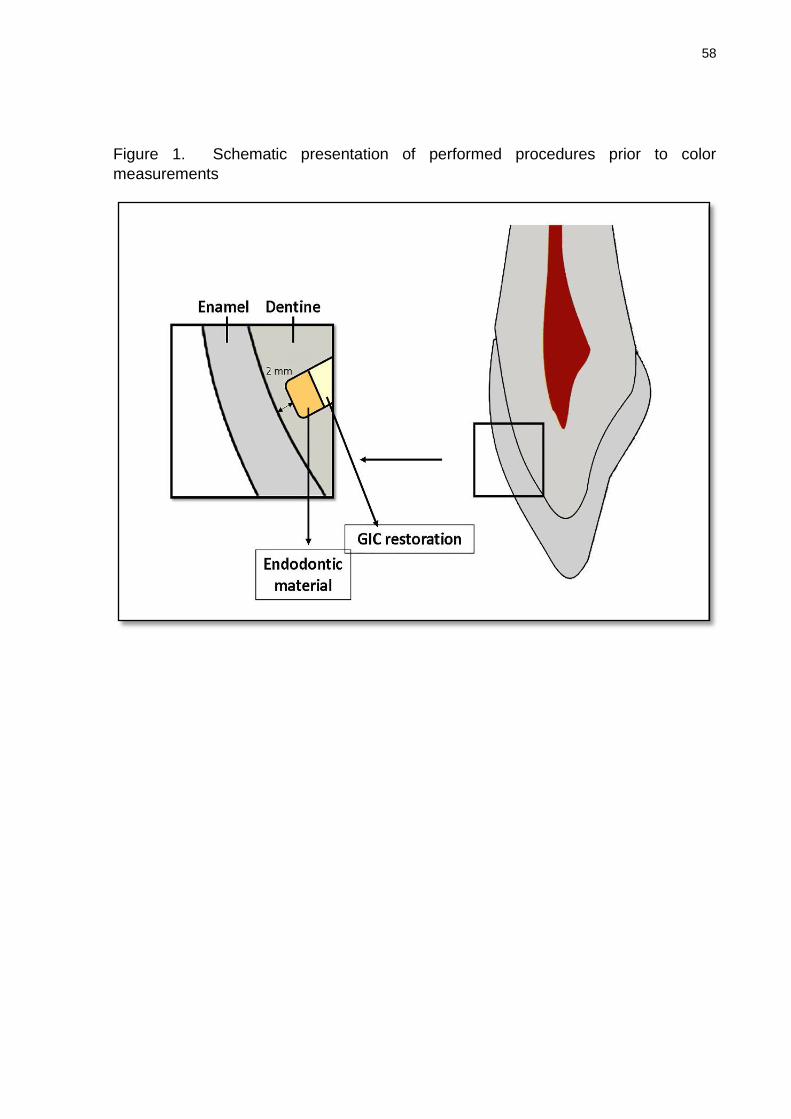
58
Figure 1. Schematic presentation of performed procedures prior to color
measurements
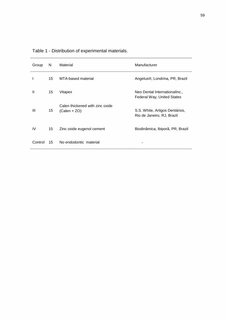
59
Table 1 - Distribution of experimental materials.
Group N Material Manufacturer
I 15 MTA-based material Angelus®, Londrina, PR, Brazil
II 15 Vitapex Neo Dental InternationalInc.,
Federal Way, United States
III 15
Calen thickened with zinc oxide
(Calen + ZO) S.S. White, Artigos Dentários,
Rio de Janeiro, RJ, Brazil
IV 15 Zinc oxide eugenol cement Biodinâmica, Ibiporã, PR, Brazil
Control 15 No endodontic material -
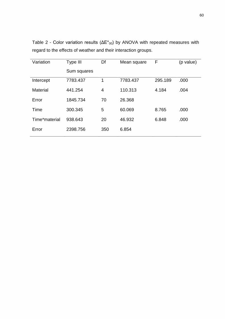
60
Table 2 - Color variation results (ΔE*00) by ANOVA with repeated measures with
regard to the effects of weather and their interaction groups.
Variation Type III
Sum squares
Df Mean square F (p value)
Intercept 7783.437 1 7783.437 295.189 .000
Material 441.254 4 110.313 4.184 .004
Error 1845.734 70 26.368
Time 300.345 5 60.069 8.765 .000
Time*material 938.643 20 46.932 6.848 .000
Error 2398.756 350 6.854
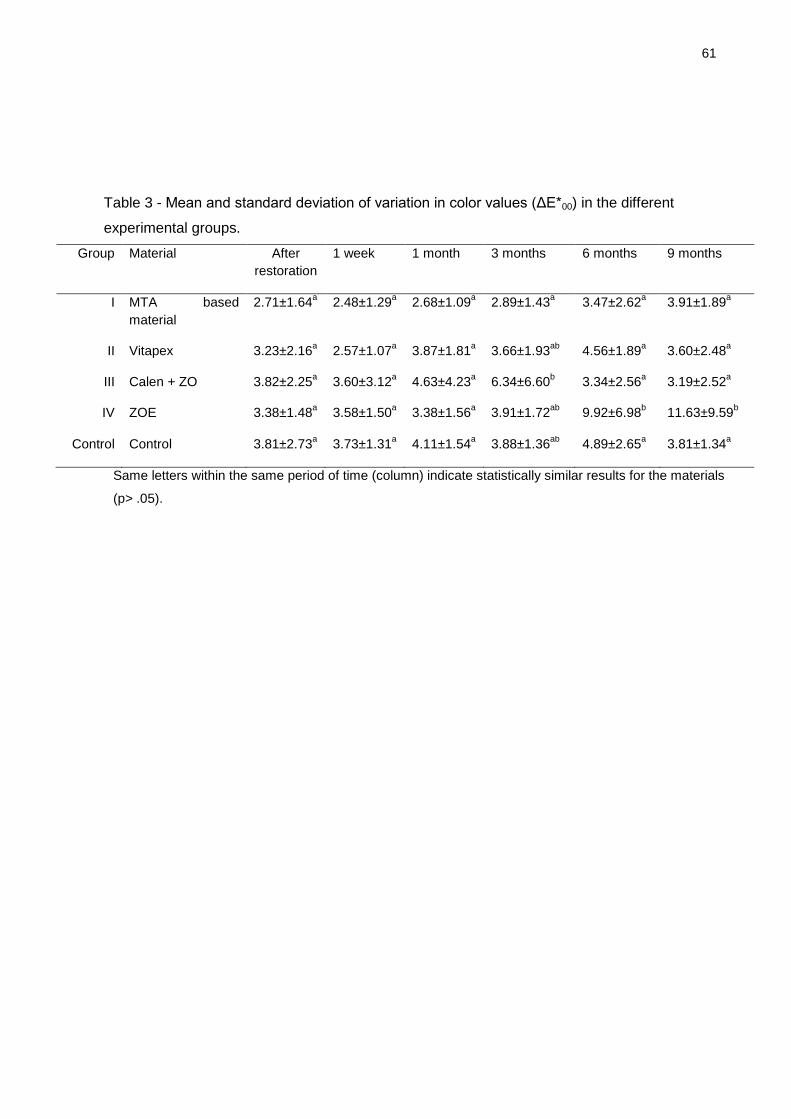
61
Table 3 - Mean and standard deviation of variation in color values (ΔE*00) in the different
experimental groups.
Group Material After
restoration
1 week 1 month 3 months 6 months 9 months
I MTA based
material
2.71±1.64a 2.48±1.29
a 2.68±1.09
a 2.89±1.43
a 3.47±2.62
a 3.91±1.89
a
II Vitapex 3.23±2.16a 2.57±1.07
a 3.87±1.81
a 3.66±1.93
ab 4.56±1.89
a 3.60±2.48
a
III Calen + ZO 3.82±2.25a 3.60±3.12
a 4.63±4.23
a 6.34±6.60
b 3.34±2.56
a 3.19±2.52
a
IV ZOE 3.38±1.48a 3.58±1.50
a 3.38±1.56
a 3.91±1.72
ab 9.92±6.98
b 11.63±9.59
b
Control Control 3.81±2.73a 3.73±1.31
a 4.11±1.54
a 3.88±1.36
ab 4.89±2.65
a 3.81±1.34
a
Same letters within the same period of time (column) indicate statistically similar results for the materials
(p> .05).
5 Considerações Finais
Com base nas metodologias utilizadas neste estudo experimental conclui-se:
5.1 Todos os materiais obturadores de canais radiculares de dentes decíduos
testados foram bem tolerados pelos tecidos subcutâneos. O material experimental à
base de MTA mostrou-se biocompatível.
5.2 Todos os materiais causaram alteração de cor na estrutura dentária, sendo
óxido de zinco e eugenol, o que ocasionou maior efeito de escurecimento. O
material experimental à base de MTA apresentou a menor variação de cor ao longo
do tempo experimental.
5.3 O material experimental à base de MTA apresenta propriedades físicas e
biológicas satisfatórias, podendo ser uma alternativa aos materiais obturadores
convencionais utilizados em canais radiculares de dentes decíduos.

Referências Bibliográficas
AMERICAN ACADEMY OF PEDIATRIC DENTISTRY, Guideline on pulp therapy for
primary and young permanent teeth. Pediatric Dentistry, vol. 31, n. 6, p.179–186,
2009.
BARJA-FIDALGO, F. ;MOUTINHO-RIBEIRO, M.; OLIVEIRA, M.A. et al. Systematic
review of root canal filling materials for deciduous teeth: is there an alternative for
zinc oxide-eugenol? ISRN Dentistry, v.2011, p.1-7, 2011.
CERQUEIRA, D.F.; MELLO-MOURA, A.C.; SANTOS, E.M. et al. Cytotoxicity,
histopathological, microbiological and clinical aspects of an endodontic iodoform-
based paste used in pediatric dentistry: a review. Journal of Clinical Pediatric
Dentistry, v.32,n.2, p.105-110, 2008.
CHAWLA, H.S.; SETIA, S.; GUPTA, N. et al. Evaluation of a mixture of zinc oxide,
calcium hydroxide, and sodium fluoride as a new root canal filling material for primary
teeth, Journal of Indian Society of Pedodontics and Preventive Dentistry , v.26,
n.2, p.53-58, 2008.
COLL, J.A.; SADRIAN, R. Predicting pulpectomy success and its relationship to
exfoliation and succedaneous dentition. Pediatric Dentistry, v.18, n.1, p.57–63,
1996.
CORREA, M.S.P.et al. Odontopediatria na primeira infância. 3.ed.,1.reimp.São
Paulo: Santos, 2011.611p.
COULD, J.M. Root canal therapy for infected primary molar teeth—Preliminary
report. ASDC Journal of Dentistry for Children, v.39, n.4, p.269– 273, 1972.
COX, S.T.; HEMBREE, J.H.; MCKNIGHT, J.P. The bactericidal potential of various
endodontic materials for primary teeth. Oral Surgery, Oral Medicine, Oral
Pathology, v.45, n.6, p.947–954, 1978.
64
DARTE, M. A.H.; DE OLIVEIRA, E. K.; GUÂNIARA, et al. Radiopacity of portland
cement associated with different radiopacifying agents. Journal of Endodontics,
v.35, n.5, p.737-740, 2009.
DE SOUZA, R.S.; DE SOUZA, V.; HOLLAND, R. et al Effect of calcium hydroxide-
based materials on periapical tissue healing and orthodontic root resorption of
endodontically treated teeth in dogs. Dental Traumatology, v.25, n.2, p.213- 218,
2009.
FAVA, L.R.G.; SAUNDERS, W.P. Calcium hydroxide pastes: classification and
clinical indications: review. International Endodontic Journal, v.32, n.4, p.257–82,
1999.
FUKS, A.B. Pulp therapy for the primary and young permanent dentitions. Dental
Clinics of North America v.44, n.3, p.571-96, 2000.
FUKS, A.B.; EIDELMAN, E. Pulp therapy in the primary dentition. Current Opinion
in Dentistry, v.1,n.5, p.556–563, 1991.
GARCIA-GODOY F. Evaluation of an iodoform paste in root canal therapy in infected
primary teeth. Journal of Dentistry for Children, v.54, n.1, p.30 –34, 1987.
HO, Y.C.; HUANG, F.M.; CHANG, Y.C. Mechanisms of cytotoxicity of eugenol in
human osteoblast cells in vitro. International Endodontic Journal , v.39,n.5,p.389–
93, 2006.
KAPLAN, A.E; ORMAECHEA, M.F.; PICCA, M.et al.. Rheological properties and
biocompatibility of endodontic sealers. International Endodontic Journal,
v.36,n.8,p.527–32,2003.
KRASTL, G; ALLGAYER, N; LENHERR, P. et al. Tooth discoloration induced by
endodontic materials: a literature review. Dental Traumatology, v. 29,n.1, p. 2–7,
2013.
KUBOTA, K.; GOLDEN, B.E.; PENUGONDA, B. Root canal filling materials for
primary teeth: a review of the literature. ASDC Journal of Dentistry for Children,
v.59, n.3, p.225-7, 1992.
LENHERR, P; ALLGAYER, N; WEIGER, R. Tooth discoloration induced by
endodontic materials: a laboratory study. International Endodontic
Journal,v.45,n.10, p. 942–9, 2012.
MASS, E.; ZILBERMAN, U.L. Endodontic treatment of infected primary teeth, using
Maisto‘s paste. ASDC Journal of Dentistry for Children, v.56, n.2, p.117-20, 1989.

65
MATHEWSON, R.J.; PRIMOSCH, R.E. Fundamentals of Paediatric Dentistry, 3rd
edn. Chicago, IL: Quintessence Publishing, p.257–280, 1995.
MCDONALD, R.E.; AVERY, D.R.; DEAN, J.A. Treatment of deep caries, vital pulp
exposure, and pulpless teeth. In: McDonald RE, Avery DR (eds). Dentistry for the
Child and Adolescent, 7th ed. St Louis, MO: Mosby, p.413–437, 2000.
MIZUNO, M.; BANZAI, Y. Calcium ion release from calcium hydroxide stimulated
fibronectin gene expression in dental pulp cells and the differentiation of dental pulp
cells to mineralized tissue forming cells by fibronectin. International Endodontic
Journal, v.41, n.11, p.933-938, 2008.
MORTAZAVI, M.; MESBAHI, M. Comparison of zinc oxide and eugenol, and Vitapex
for root canal treatment of necrotic primary teeth. International Journal of
Paediatric Dentistry, v.14, n.6, p.417–424, 2004.
NEWTON, J.T; MINHAS, G. Exposureto ideal facial images reduces facial
satisfaction: an experimental study. Community Dentistry and Oral Epidemiology,
v. 33, n.6, p. 410–418, 2005.
OLSSON,B.; SLIWKOWSKY, A.; LANGELAND, K. Subcutaneous implantation for
the biological evaluation of endodontic materials. Journal of Endodontics,
v.7,n.8,p.355–67, 1981.
PARSONS, J.R; WALTON, R.E; RICKS-WILLIAMSON, L. In vitro longitudinal
assessment of coronal discoloration from endodontic sealers .Journal of
Endodontics, v. 27, n.11, p. 699-702, 2001.
PARTOVI, M; AL-HAVVAZ, AH, SOLEIMANI, B. In vitro computer analysis of crown
discoloration from commonly used endodontic sealers Australian. International
Endodontic Journal, v. 32, n. 3, p. 116-119, 2006.
PILOWNIC, KaterineJahnecke. Avaliação físico-química e biológica de materiais
obturadores de canais radiculares de dentes decíduos. 2015. 75f. Dissertação
(Mestrado em Odontologia) - Faculdade de Odontologia, Universidade Federal de
Pelotas, Pelotas, 2015.
PINTO,D.N. SOUSA D.L; ROCHA, R.B et al. Eighteen-month clinical and
radiographic evaluation of two root canal-filling materials in primary teeth with pulp
necrosis secondary to trauma. Dental Traumatology, v.27, n.3, p.221–224, 2011.
QUEIROZ, A.M.; NELSON-FILHO P.; SILVA L.A. et al. Antibacterial activity of root
canal filling materials for primary teeth: zinc oxide and eugenol cement, Calen paste
66
thickened with zinc oxide, Sealapex and EndoREZ. Brazilian Dental Journal, v.20,
n.4, p.290–296, 2009.
RANLY, D.M.; GARCIA-GODOY F. Reviewing pulp treatment for primary
teeth.Journal of the American Dental Association, v.122, n.9, p.83–85, 1991.
RANLY, D.M.; GARCIA-GODOY, F. Current and potential pulp therapies for primary
and young permanent teeth. Journal of Dentistry, v. 28, n.3, p.153–161, 2000.
SCHRODER, V. Effects of calcium hydroxide containing pulp capping agents on pulp
cell migration, proliferation and differentiation. Journal of Dental Research, v.64,
p.541-548, 1985.
SEGATO,R.A.B;PUCINELLI, C.M;FERREIRA, D.C.A et al. Physicochemical
properties of root canal filling materials for primary teeth. Brazilian Dental Journal,
v.27, n.2, p.196-201, 2016.
SILVA, L.A.B.; LEONARDO,M.R.; OLIVEIRA, D.S.B. et al. Histopathological
evaluation of root canal filling materials for primary teeth. Brazilian Dental Journal,
v.21, n.1, p.38–45, 2010.
SILVA, L.A.B.; SILVA, R.A.B.; BRANCO, L.G.et al.Quantitative radiographic
evaluation of periapical bone resorption in dog‘s teeth contaminated with bacterial
endotoxin (LPS) associated or not with calcium hydroxide. Brazilian Dental Journal,
v.19, n.4, p.296-300, 2008.
TCHAOU, W.S.; TURNG, B.F.; MINAH, G.E.et al. Inhibition of pure cultures of oral
bacteria by root canal filling materials. Pediatric Dentistry, v.18, n.7, p.444–449,
1996.
TORNECK CD. Reaction of rat conective tissue to polyethylene tube implants.Oral
Surgery, v.21,n.3, p.379-87,1996.
VALE, T.; SANTOS, P.; MOREIRA, J. et al. Perception of dental aesthetics in
paediatric dentistry. European Journal of Paediatric Dentistry, v. 10,n.3, p. 110-
114, 2009.

67
Anexos
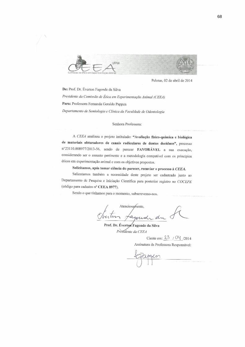
68

![TOWARDS CULTURE- CENTRED DESIGN SHEN; WOOLEY; PRIOR [2005] Mestrado em Design | UFPR 2009 HCI | Stephania Padovani Rosana Vasques](https://img.document.onl/doc/110x75/552fc14a497959413d8e1f1d/towards-culture-centred-design-shen-wooley-prior-2005-mestrado-em-design-ufpr-2009-hci-stephania-padovani-rosana-vasques.jpg)