Embed Size (px)
Citation preview
1
UNIVERSIDADE FEDERAL DE SERGIPE
PROGRAMA DE PÓS-GRADUAÇÃO EM ODONTOLOGIA
ANÁLISE DA INFLUÊNCIA DE DIFERENTES MEIOS DE
ARMAZENAGEM NA VIABILIDADE DE FIBROBLASTOS E NA
COMPOSIÇÃO IÔNICA DA DENTINA RADICULAR: ESTUDO in vitro.
Aracaju
2015
2
IVANILTON ALAN DE SOUZA SILVA
ANÁLISE DA INFLUÊNCIA DE DIFERENTES MEIOS DE
ARMAZENAGEM NA VIABILIDADE DE FIBROBLASTOS E NA
COMPOSIÇÃO IÔNICA DA DENTINA RADICULAR: ESTUDO in vitro.
Dissertação apresentada ao Programa de Pós-
Graduação em Odontologia da Universidade
Federal de Sergipe, para obtenção do título de
Mestre em Odontologia.
Orientador: Prof. Dr. Luiz Carlos Ferreira da Silva
Aracaju
2015
3
FICHA CATALOGRÁFICA ELABORADA PELA BIBLIOTECA BISAU UNIVERSIDADE FEDERAL DE SERGIPE
S586
Silva, Ivanilton Alan de Souza Análise da influência de diferentes meios de armazenagem na
viabilidade de fibroblastos e na composição iônica da dentina radicular: estudo in vitro / Ivanilton Alan de Souza Silva ; orientador Luiz Carlos Ferreira da Silva. – Aracaju, 2015.
42 f.
Dissertação (mestrado em Odontologia)– Universidade Federal de Sergipe, 2015.
1. Odontologia. 2. Ligamento periodontal. 3. Avulsão dentária. 4. Meios de cultura. 5. Fibroblastos. I. Silva, Luiz Carlos Ferreira da, orient. II. Título.
CDU 616.314
4
5
AGRADECIMENTOS
Primeiramente a Deus, pois sem ele esse sonho não seria possível. Agradeço muito por
iluminar o meu caminho e por ter me dado forças nos momentos de maiores dificuldade para
continuar lutando.
Aos meus pais e irmão, pela compreensão, incentivo e por acreditar em mim, me
apoiado em todos os momentos. Por estar sempre ao meu lado não importando a situação. Muito
Obrigado!
Ao meu orientador Professor Luiz Carlos Ferreira da Silva, por nunca ter desistido de
mim mesmo nos meus momentos de fraqueza. Sem ele este trabalho nunca seria concluído.
Ao Programa de Pós-Gaduação em Odontologia da Faculdade de Odontologia da
Universidade Federal de Uberlândia, no qual esta parceria de programas possibilitou a
realização integral do trabalho. Em especial, ao professor Carlos José Soares, a professora
Priscilla Barbosa Ferreira Soares, professora Manuella Verdinelli de Paula Reis, que me
guiaram em cada etapa além de contribuírem para o meu crescimento profissional.
A todos do Programa de Pós-Gaduação em Odontologia da Faculdade de Odontologia
da Universidade Federal de Uberlândia que me acolheram num momento difícil e corrido e
mesmo não atuando diretamente, me auxiliaram em momentos de necessidades.
A todos os professores do Programa de Pós-Graduação em Odontologia da UFS que
fizeram parte dessa conquista.
Aos meus amigos de curso pela paciência, companheirismo, sinceridade e assistência
no decorrer deste trabalho e de todo mestrado.
Por fim, agradeço a todos aqueles que de forma direta ou indireta fizeram parte dessa
etapa da minha vida.
6
Determinação, coragem e autoconfiança são fatores decisivos
para o sucesso. Se estamos Possuídos por uma inabalável
determinação conseguiremos superá-los. Independentemente das
circunstâncias, devemos ser sempre humildes, recatados e
despidos de orgulho
(Dalai Lama)
7
RESUMO
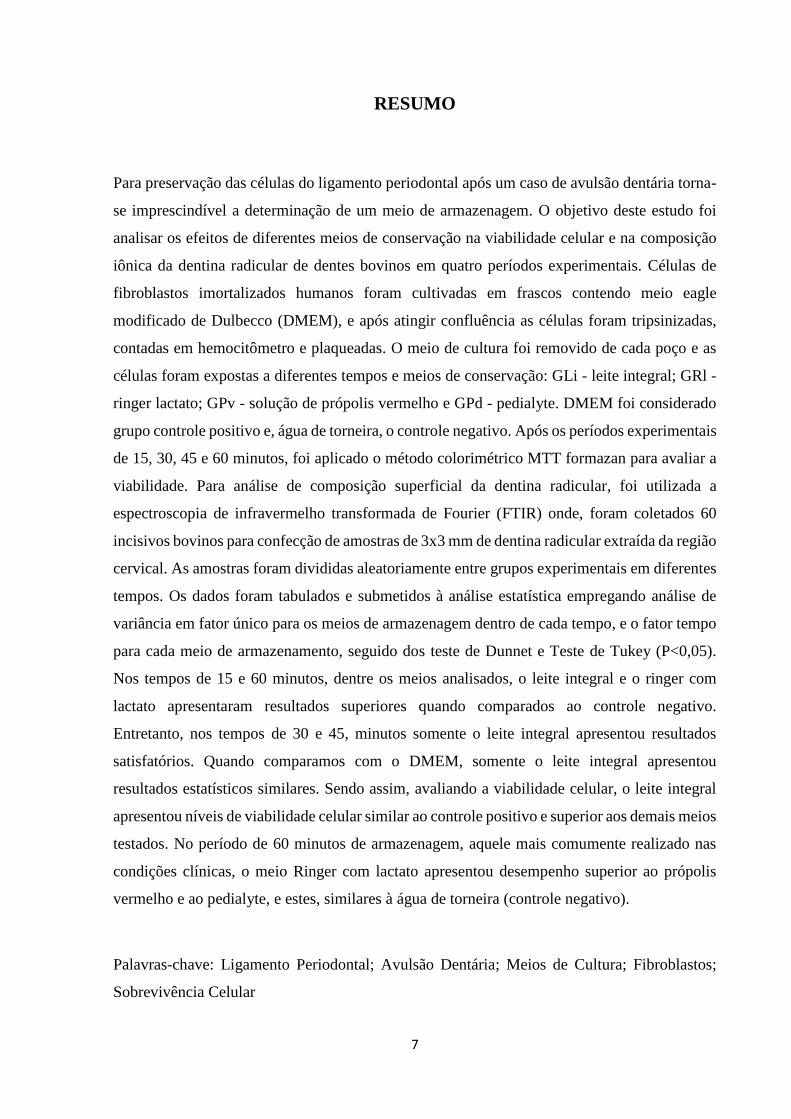
Para preservação das células do ligamento periodontal após um caso de avulsão dentária torna-
se imprescindível a determinação de um meio de armazenagem. O objetivo deste estudo foi
analisar os efeitos de diferentes meios de conservação na viabilidade celular e na composição
iônica da dentina radicular de dentes bovinos em quatro períodos experimentais. Células de
fibroblastos imortalizados humanos foram cultivadas em frascos contendo meio eagle
modificado de Dulbecco (DMEM), e após atingir confluência as células foram tripsinizadas,
contadas em hemocitômetro e plaqueadas. O meio de cultura foi removido de cada poço e as
células foram expostas a diferentes tempos e meios de conservação: GLi - leite integral; GRl -
ringer lactato; GPv - solução de própolis vermelho e GPd - pedialyte. DMEM foi considerado
grupo controle positivo e, água de torneira, o controle negativo. Após os períodos experimentais
de 15, 30, 45 e 60 minutos, foi aplicado o método colorimétrico MTT formazan para avaliar a
viabilidade. Para análise de composição superficial da dentina radicular, foi utilizada a
espectroscopia de infravermelho transformada de Fourier (FTIR) onde, foram coletados 60
incisivos bovinos para confecção de amostras de 3x3 mm de dentina radicular extraída da região
cervical. As amostras foram divididas aleatoriamente entre grupos experimentais em diferentes
tempos. Os dados foram tabulados e submetidos à análise estatística empregando análise de
variância em fator único para os meios de armazenagem dentro de cada tempo, e o fator tempo
para cada meio de armazenamento, seguido dos teste de Dunnet e Teste de Tukey (P<0,05).
Nos tempos de 15 e 60 minutos, dentre os meios analisados, o leite integral e o ringer com
lactato apresentaram resultados superiores quando comparados ao controle negativo.
Entretanto, nos tempos de 30 e 45, minutos somente o leite integral apresentou resultados
satisfatórios. Quando comparamos com o DMEM, somente o leite integral apresentou
resultados estatísticos similares. Sendo assim, avaliando a viabilidade celular, o leite integral
apresentou níveis de viabilidade celular similar ao controle positivo e superior aos demais meios
testados. No período de 60 minutos de armazenagem, aquele mais comumente realizado nas
condições clínicas, o meio Ringer com lactato apresentou desempenho superior ao própolis
vermelho e ao pedialyte, e estes, similares à água de torneira (controle negativo).
Palavras-chave: Ligamento Periodontal; Avulsão Dentária; Meios de Cultura; Fibroblastos;
Sobrevivência Celular

8
ABSTRACT
For preservation of periodontal ligament cells after one tooth avulsion becomes necessary to
determine a storage medium. The aim of this study was to analyze the preservation of human
fibroblast cells and analyze the composition of root dentin of bovine teeth in different storage
media. Immortalized human fibroblasts cells were grown in flasks containing Dulbecco's
modified eagle medium (DMEM), and ffter reaching confluence the cells were trypsinized,
counted by hemocytometer and plated. The culture medium was removed from each well and
the cells were exposed at different times in the solutions: GLi - milk; GRl - ringer’s lactate;
GPv - red propolis solution and GPd - Pedialyte. DMEM was considered positive control group
and tap water was the negative control. After the experimental period of 15, 30, 45 and 60
minutes, we used the colorimetric method MTT formazan. For analysis of surface composition
of root dentin, were using Fourier transform infrared (FTIR) where, were collected 60 bovine
incisors. Samples were extracted from the cervical region of the root dentin in 3x3mm. The
samples were randomly divided among experimental groups in differents times. Data were
tabulated and statistically analyzed using analysis of variance for the storage media within each
time and the time factor for each storage medium, followed by Dunnet test and Tukey's test (P
<0.05 ). In 15 and 60 minutes, from the media types, the milk and Ringer's lactate showed better
results when compared to the negative control. However, in 30 and 45 minutes only the milk
showed satisfactory results. A comparison with DMEM only the milk showed similar statistical
results. In assessing the viability of human fibroblasts, milk showed cell viability levels similar
to the positive control and superior to other media tested. Within 60 minutes storage, the one
most commonly used in clinical conditions, the ringer's lactate has higher performance than the
red propolis and the pedialyte and these similar to tap water (negative control).
Keywords: Periodontal Ligament; Dental avulsion; Culture Media; Fibroblasts; Cell Survival
9
SUMÁRIO
1. Introdução ................................................................................................................... 10
2. Objetivos ...................................................................................................................... 13
2.1.Objetivo Geral ........................................................................................................ 13
2.2.Objetivos Específicos ............................................................................................. 13
3. Metodologia ................................................................................................................. 14
3.1. Tipo de Estudo ....................................................................................................... 14
3.2. Local do Experimento ............................................................................................ 14
3.3.Cultura de Células ................................................................................................... 14
3.4. Avaliação da viabilidade celular ............................................................................ 14
3.5. Análise da composição da dentina radicular ......................................................... 18
3.5.1. Obtenção dos fragmentos da dentina .......................................................... 18
3.5.2. Análise dos fragmentos no FTIR ................................................................ 20
3.5.3. Análise dos dados obtidos ........................................................................... 21
3.6.Análise estatística ................................................................................................... 22
4. Resultados ................................................................................................................... 23
5. Considerações Finais .................................................................................................. 39
6. Comunicado à Imprensa ............................................................................................ 40
Referências ...................................................................................................................... 41

10
1 INTRODUÇÃO
O termo avulsão, é usado para descrever uma situação em que, um dente é deslocado
por completo do alveolo, resultando em trauma dento alveolar grave¹. Devido à complexidade
desta lesão, o suprimento neurovascular é severamente comprometido, na maioria dos casos,
causando perda de vitalidade pulpar. Os fatores etiológicos mais prevalentes são, os traumas
decorridos de luta e esportes, bem como quedas e colisões contra objetos duros2. O reimplante
imediato é considerado como o tratamento ideal para esse tipo de trauma, mas nem sempre é
possível sua realização,3 e consequentemente, o reimplante tardio é comumente presente. O
tempo e o meio de armazenamento extraoral, são dois dos fatores mais importantes na
determinação da viabilidade de células restantes do ligamento periodontal e, portanto,
influenciam nas capacidades curativas do ligamento periodontal em um dente avulsionado.
Imediatamente, após a avulsão, o número de células viáveis do ligamento periodontal tende a
decair com o decorrer do tempo extraoral, e no decorrer de 2 horas torna-se impossível detectar
célular viáveis. Na prática clínica, o reimplante tardio de dentes sem nenhum meio de
conservação, não apresentam o mesmo manejo clínico e prognóstico, como o reimplante tardio
de dentes mantidos em soluções de armazenamento4,5,6.
O meio de armazenamento ideal, deve apresentar o valor de PH e osmolaridade
fisiológica adequados, devendo incluir a presença de substâncias nutritivas que permitem a
sobrevivência das células do ligamento periodontal. Além disso, o meio de armazenamento
deve estar prontamente disponível e ser simples de manusear7. Atualmente, vários meios de
armazenamento, como a Saliva, Soro Fisiológico, Leite, meios de cultura, solução salina
balanceada de Hank’s (HBSS) e viaspan, foram examinados e alguns outros meios de
conservação foram testados recentemente, sendo eles, a água de coco, extrato de própolis,
albumina de ovo, gatorade, chá verde3,7.
O leite integral, apesar da sua consagração, ainda continua sendo bastante estudado.
Possuindo PH e osmolaridade fisiológica, o leite possui a capacidade de manter as células
viáveis fornecendo nutrientes14 sendo que, o tempo pode influenciar no PH do leite, reduzindo-
o e comprometendo sua qualidade. Na sua composição também há presença de gordura que,
por sua vez, afeta a viabilidade celular. Devido a fácil disponibilidade e praticidade, o leite
integra, é o meio mais adotado nos casos de avulsão dentária sendo eleito pela Associação
11
Americana de Endodontistas como solução de armazenagem para transporte do dente
avulsionado8,9,10,11.
A água de torneira, é descrita como um meio inadequado para conservar as células do
ligamento periodontal, devido a sua baixa osmolalidade e presença de cloro9. Em um estudo,
Lindskog e Blomlöf (1981), concluiram que, após 3 horas de armazenamento neste meio, todas
as células do ligamento periodontal foram perdidas. A partir dos resultados da água, sua
aplicabilidade em trabalhos é únicamente de comparação como controle negativo.
O meio eagle modificado Dulbecco (DMEM), também usado para o cultivo de células
suplementado com soro fetal bovino, possui sal, glicose, vitaminas, aminoácidos, antibióticos
além de fatores de crescimento e alto teor de gama-globulina. Este meio de Eagle mostrou-se
superior a outros meios testados além de ser um excelente meio para conservação de células, se
fosse facilmente obtido. Fibroblastos viáveis e com capacidade de divisão foram observados
após 1 ano de armazenamento em meio de Eagle suplementado com nutrientes, soluções tampão
e antimicrobiana. Pesquisadores relataram que após um período de secagem de 60 min, a pré-
imersão de dentes de macacos em meio de Eagle, por 5, 7 ou 14 dias, melhorou a condição
periodontal, diminuindo o percentual de reabsorção inflamatória após o reimplante12.
O ringer com lactato apresenta uma concentração de sódio de 130 mmol / L, a
concentração de cloreto de 109 mmol / L, o nível de cálcio de 2,7 mmol / L, o nível de HCO3
de 2,7 mmol / L a partir do metabolismo do lactato e osmolaridade de 265 mOsm / L ; tanto
[Na +] e osmolaridade são ligeiramente abaixo dos valores de plasma,13 composição que se
assemelha com a concentração ideal para a conservação das células do ligamento periodontal.
O própolis é uma substância resinosa natural, não-tóxica que foi coletada a partir de
vários tipos de plantas por abelhas para cobrir e proteger a colméia. Atualmente, o própolis tem
sido empregado na Medicina e Odontologia por causa de seus efeitos anti-inflamatórios, anti-
sépticos, de cura e propriedades antimicrobianas. Além dessas características, o própolis
contém elementos que têm sido essenciais durante a síntese de colagéno14,15,16. Por mais de 30
anos o própolis brasileiro é estudado no setor da farmacologia e da quimica. Dentre os tipos de
própolis encontramos o própolis vermelho que é extraído no nordeste do Brasil e apresenta
capacidade antibacteriana, antifúngica, antiinflamatória, antioxidante, antitumoral entre
outros.17 Em 2014, Frozza e colaboradores18, analisaram 3 proporções de própolis vermelhos
(6µg/ml, 60µg/ml e 120µg/ml) com intuito de avaliar suas propriedades frente a células
tumorais chegando a conclusão de que, somente o própolis vermelho 6µg/ml não apresenta
citotoxidade mostrando-se compatível com o grupo controle.

12
Pedialyte® é uma solução de manutenção do eletrólito via oral, que repõe os líquidos e
nutrientes perdidos essenciais para as células. Pedialyte tem sua composição semelhante à
Ricentral. Pedialyte contém citrato de Potássio monoidratado (1,080g), cloreto de sodio
(1,038g), citrato de sodio diidratado (470mg), ácido cítrico anidro, gliconato de Zinco (30,5mg)
e glicose monoidratada (12,5g) apresentando na sua composição o arroz cozido em adição aos
eletrólitos. Sua disponibilidade em farmácias também o torna prático em casos de acidentes12.
Sendo assim, o propósito desse estudo é de avaliar a influência de diferentes meios de
armazenagem na viabilidade de fibroblastos humanos além de analisar a composição iônica da
dentina radicular por meio de espectrômetro de infravermelho.

13
2 OBJETIVO
2.1 Objetivo Geral
Avaliar a viabilidade de fibroblastos humanos e analisar a composição iônica da dentina
radicular de dentes bovinos em diferentes meios de conservação em quatro períodos
experimentais.
2.2 Objetivos Específicos
Analisar o potencial do ringer com lactato, do própolis e do pedialyte em manter a
viabilidade de fibroblastos de boca imortalizados comparando-os ao leite integral bovino e
ao meio eagle modificado Dulbecco (DMEM) e a água de torneira em quatro períodos
experimentais, usando o método de MTT formazan.
Analisar a composição iônica da dentina radicular por meio do espectrofotômetro de
infravermelho por transformada de Fourier (FTIR) antes e após a armazenagem nas
soluções experimentais, em quatro períodos experimentais.
14
3 METODOLOGIA
3.1 Tipo de estudo
Neste trabalho foi realizada uma pesquisa laboratorial sendo um estudo in vitro, com
caráter exploratório e comparativo utilizando-se uma abordagem indutiva.
3.2 Local do Experimento
O experimento foi realizado no Centro de Pesquisa Odontológico Biomecânica,
Biomateriais e Biologia Celular – CPbio, na Faculdade de Odontologia da Universidade Federal
de Uberlândia (FOUFU), no município de Uberlandia, Minas Gerais, Brasil.
3.3 Cultura das células
Células de fibroblastos imortalizados humanos (banco de células do Rio de Janeiro, Rio
de Janeiro, RJ, Brasil) foram cultivadas em frascos T-25 de cultura de células contendo meio
eagle modificado de Dulbecco (DMEM) (Sigma Chemical Co., St. Louis, MO, EUA),
suplementado com 20% de soro fetal bovino (Invitrogen, Branchburg, NJ, EUA), 100 unidades
de mL⁻¹ de penicilina/estreptomicina (Sigma Chemical Co.) em incubadora de humidificador
com 5% de CO2 e 95% de ar a 37 ° C. O crescimento foi permitido até as células atingirem
confluência de 80%. Estas células foram tripsinizadas (1 ml), contadas em hemocitômetro, e
plaqueadas em placas 96 poços (Coastar Corp, Cambridge, MA, EUA,) em densidade de 2x10⁴
células por poço em 100 µL de meio de cultura. As placas foram devolvidas à estufa de CO2
por 48 horas.
3.4 Avaliação da viabilidade celular
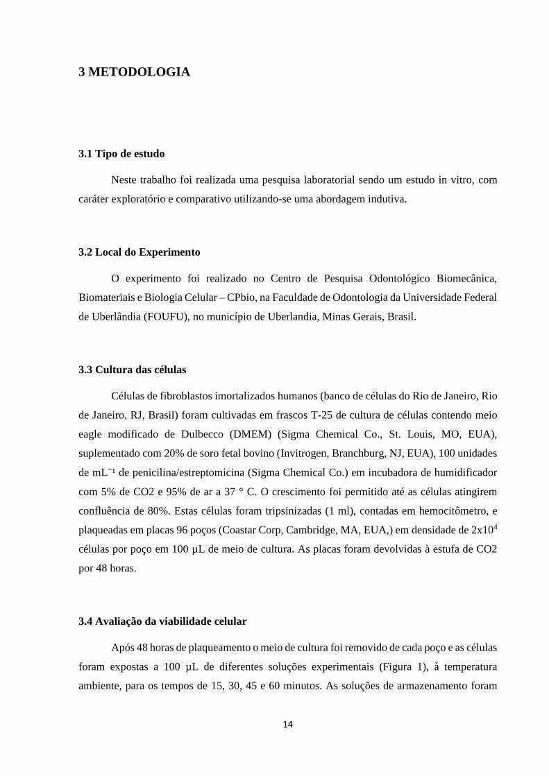
Após 48 horas de plaqueamento o meio de cultura foi removido de cada poço e as células
foram expostas a 100 µL de diferentes soluções experimentais (Figura 1), à temperatura
ambiente, para os tempos de 15, 30, 45 e 60 minutos. As soluções de armazenamento foram
15
leite integral, ringer com lactato, extrato de própolis vermelho, pedialyte, água de torneira e
meio eagle modificado Dulbecco (Quadro 1).
Quadro 1: Disposição dos meios experimentais com PHs e Osmolaridades.
GRUPO PH Osmolaridade
GLi Leite integral (CALU, Uberlândia, MG, Brasil) 6.65 280 mOsm/l
GRl Ringer com lactato (HalexIstar, Hospitalar, Goiânia, Brasil) 6.0 274,4 mOsm/l
GPv Extrato de própolis vermelho 6 µg/ml (Manipulada em álcool
e água 50/50, de acordo com o trabalho de Frozza, 2014)
6.38 70 mOsm/l
GPd Pedialyte (ABBOTT, São Paulo, SP, Brasil) 3.98 250 mOsm/l
GAt Água de torneira – Controle Negativo 6.18 24 mOsm/l
GDm Meio eagle modificado Dulbecco (Sigma Chemical Co., St.
Louis, MO, EUA) – Controle Positivo
8.56 310 mOsm/l
Figura 1: Meios experimentais – (a)meio eagle modificado Dulbecco (DMEM), (b)água de
torneira, (c) extrato de própolis vermelho, (d)pedialyte, (e)ringer com lactato e (f)leite integral.
16
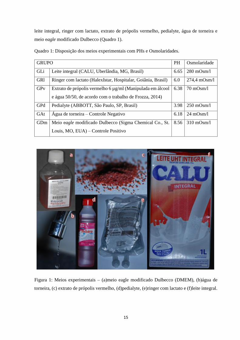
Após os tempos experimentais, as soluções foram descartadas e acrescido 20% de
DMEM em solução de MTT (3-[4, 5-dimethylthiazolyl-2]-2, 5-diphenyltetrazolium bromide)
(5 mg. mL⁻¹) para avaliação da viabilidade celular (Figura 2). O mesmo protocolo foi utilizado
para o grupo de controle positivo (DMEM).
Figura 2: Solução de MTT para avaliação da viabilidade celular.
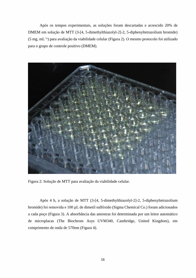
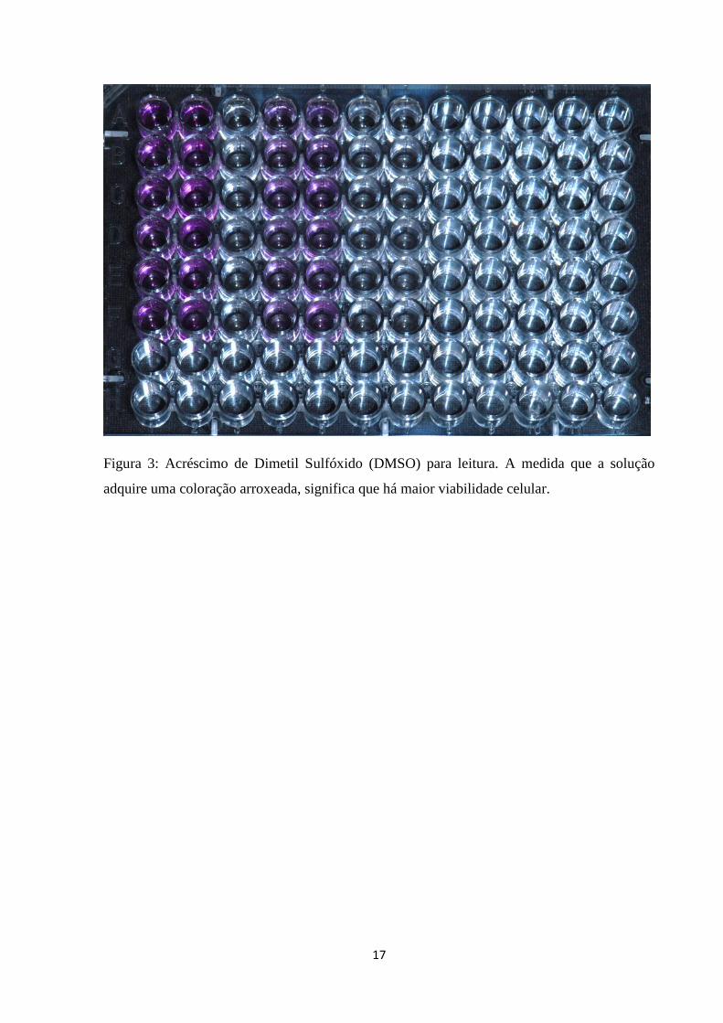
Após 4 h, a solução de MTT (3-[4, 5-dimethylthiazolyl-2]-2, 5-diphenyltetrazolium
bromide) foi removida e 100 µL de dimetil sulfóxido (Sigma Chemical Co.) foram adicionados
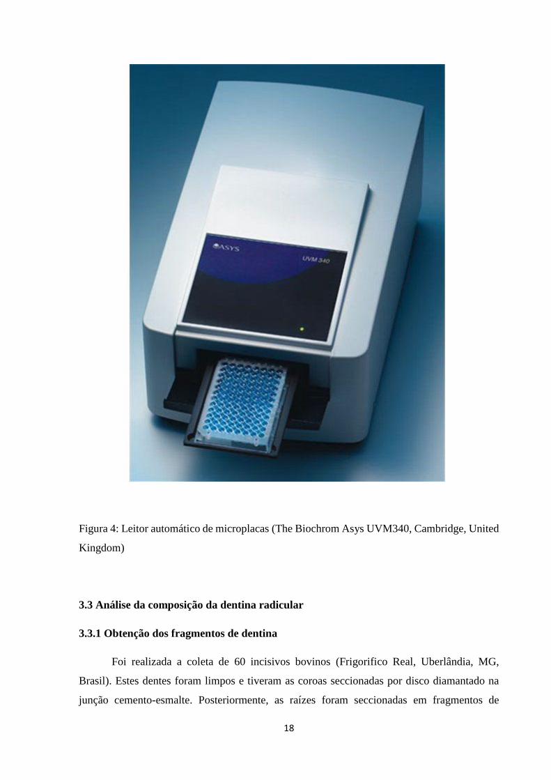
a cada poço (Figura 3). A absorbância das amostras foi determinada por um leitor automático
de microplacas (The Biochrom Asys UVM340, Cambridge, United Kingdom), em
comprimento de onda de 570nm (Figura 4).
17
Figura 3: Acréscimo de Dimetil Sulfóxido (DMSO) para leitura. A medida que a solução
adquire uma coloração arroxeada, significa que há maior viabilidade celular.
18
Figura 4: Leitor automático de microplacas (The Biochrom Asys UVM340, Cambridge, United
Kingdom)
3.3 Análise da composição da dentina radicular
3.3.1 Obtenção dos fragmentos de dentina
Foi realizada a coleta de 60 incisivos bovinos (Frigorifico Real, Uberlândia, MG,
Brasil). Estes dentes foram limpos e tiveram as coroas seccionadas por disco diamantado na
junção cemento-esmalte. Posteriormente, as raízes foram seccionadas em fragmentos de

19
aproximadamente 3 mm de largura por 3 mm de altura e com espessura determinada na metade
do diâmetro do canal (Figura 5). Os fragmentos foram levados em grupos de 5, em beckers
contendo água destilada, para sua limpeza em uma cuba ultrassônica por um período de 20
minutos (10 minutos com a superfície pulpar para baixo e 10 minutos com a superfície pulpar
voltado para o lado).
(a) (b) (c)
(d)
Figura 5: Confecção das amostras. (a)Secção na linha amelodentinária com auxílio de disco
diamantado; (b)Delimitando a altura em 3 mm; (c)Delimitando a largura em 3 mm;
(d)Fragmentos em grupos de 5 dentro de beckers para limpeza em cuba ultrassônica.
Estas fatias foram divididas em grupos experimentais nos tempos de 15, 30, 45 e 60
minutos de acordo com a tabela 1.

20
Tabela 1: Disposição dos fragmentos para cada meio para análise em FTIR
GRUPO 15 min 30 min 45 min 60 min TOTAL
GLi Leite Integral n = 5 n = 5 n = 5 n = 5 n = 20
GRl Ringer com
Lactato
n = 5 n = 5 n = 5 n = 5 n = 20
GPv Extrato de
Própolis
Vermelho 6
µg/ml
n = 5 n = 5 n = 5 n = 5 n = 20
GPd Pedialyte n = 5 n = 5 n = 5 n = 5 n = 20
GAt Água de
Torneira
n = 5 n = 5 n = 5 n = 5 n = 20
GDm Meio Eagle
Modificado
Dulbecco
n = 5 n = 5 n = 5 n = 5 n = 20
TOTAL n = 120
3.3.2 Análise dos fragmentos no espectrofotómetro de infravermelho por transformada
de Fourier FTIR
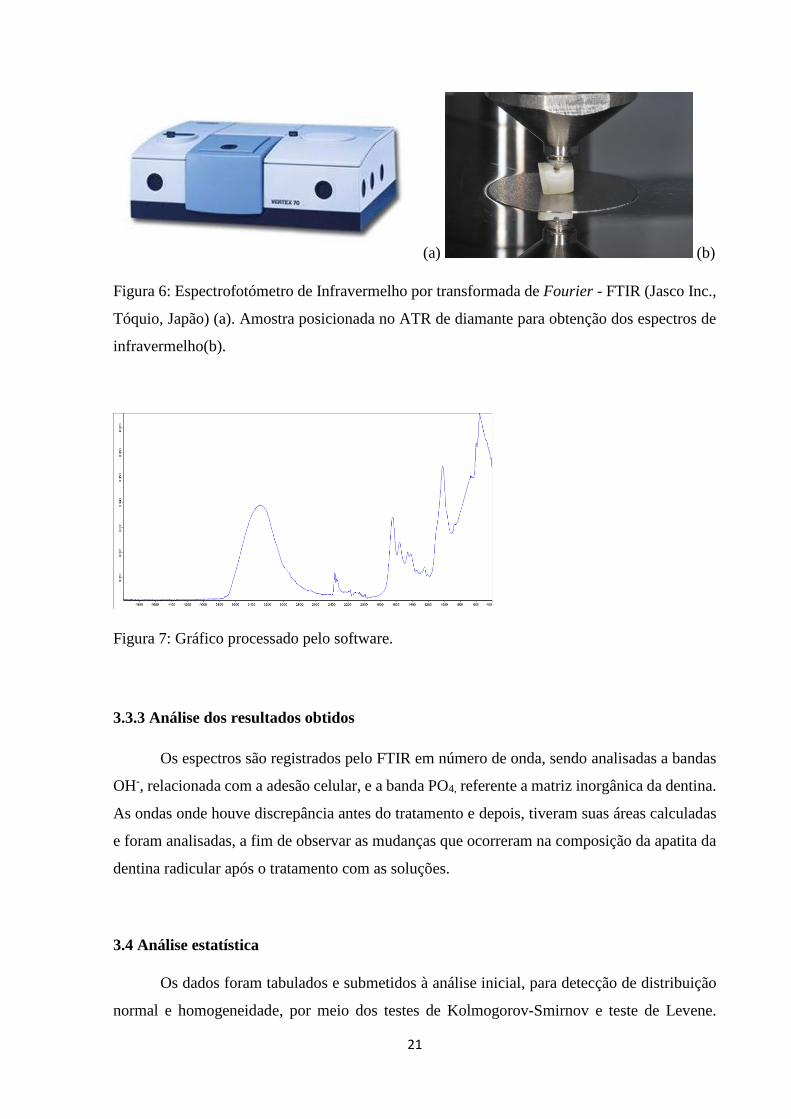
Antes e depois do tratamento com as soluções, as amostras foram analisadas em um
espectrofotómetro de infravermelho por transformada de Fourier - FTIR (Jasco Inc., Tóquio,
Japão). As amostras foram secas e posicionadas com a parede pulpar voltada para cima, de
forma que a superfície radicular seja mantida sobre o suporte de amostras no equipamento, onde
a técnica de Reflectância Total Atenuada (ATR) foi empregada (Figura 6) obtendo os espectros
de infravermelho que correspondem à análise da dentina. Cada espectro foi processado pelo
software e o resultado da análise é obtido sob a forma de gráfico (Figura 7).
21
(a) (b)
Figura 6: Espectrofotómetro de Infravermelho por transformada de Fourier - FTIR (Jasco Inc.,
Tóquio, Japão) (a). Amostra posicionada no ATR de diamante para obtenção dos espectros de
infravermelho(b).
Figura 7: Gráfico processado pelo software.
3.3.3 Análise dos resultados obtidos
Os espectros são registrados pelo FTIR em número de onda, sendo analisadas a bandas
OH-, relacionada com a adesão celular, e a banda PO4, referente a matriz inorgânica da dentina.
As ondas onde houve discrepância antes do tratamento e depois, tiveram suas áreas calculadas
e foram analisadas, a fim de observar as mudanças que ocorreram na composição da apatita da
dentina radicular após o tratamento com as soluções.
3.4 Análise estatística
Os dados foram tabulados e submetidos à análise inicial, para detecção de distribuição
normal e homogeneidade, por meio dos testes de Kolmogorov-Smirnov e teste de Levene.

22
Como os valores apresentarem requisitos que possibilite o emprego de análise paramétrica foi
empregada análise estatística usando análise de variância, e teste de Tukey e teste de Dunnet
para efetivar a comparação aos grupos controles. Todos os testes estatísticos foram realizados
com nível de significância de α=0.05.
23
4 RESULTADOS
PERIÓDICO: DENTAL TRAUMATOLOGY
IN VITRO ANALYSIS OF FIBROBLAST VIABILITY AND DENTIN’S IONIC
COMPOSITION IN DIFFERENT STORAGE MEDIA
Ivanilton Alan de Souza Silva1. Priscilla Barbosa Ferreira Soares2. Manuella Verdinelli de Paula
Reis3. Carlos José Soares4. Juliana Cardoso5. Luiz Carlos Ferreira da Silva6.
1. DDS, MS, Student of Dental School, UFS - Federal University of Sergipe, Aracaju, Sergipe,
Brazil. R. Cláudio Batista s/n, Sanatório, - CEP 49060-100, Aracaju, Sergipe, Brasil. Phone:
(79) 2105-1824.
2. DDS, MS, PhD Student of Dental School, School of Dentistry, Federal University of
Uberlandia, Uberlandia, Minas Gerais, Brazil. Av. Pará, nº 1720 - Bloco 4L Anexo B – Sala
4LA – CEP 38400-902, Uberlândia, Minas Gerais, Brasil. Phone: 55-34-3218 22 55 Fax: 55-
34- 3218 2279.
3. DDS, MS, Student of Dental School, School of Dentistry, Federal University of Uberlandia,
Uberlandia, Minas Gerais, Brazil. Av. Pará, nº 1720 - Bloco 4L Anexo A – Sala 4LA – CEP
38400-902, Uberlândia, Minas Gerais, Brasil. Phone: 55-34-3218 22 55 Fax: 55-34- 3218 2279.
4. DDS, MS, PhD, Department of Operative Dentistry and Dental Materials, School of
Dentistry, Federal University of Uberlandia, Uberlandia, Minas Gerais, Brazil. Av. Pará, nº
1720 - Bloco 4L Anexo A – Sala 4LA – CEP 38400-902, Uberlândia, Minas Gerais, Brasil.
Phone: 55-34-3218 22 55 Fax: 55-34- 3218 2279.
5. DDS, MS, PhD, Department of Farmacology, UNIT – Tiradentes University, Aracaju,
Sergipe, Brazil. Av. Murilo Dantas, 300, prédio do ITP, Farolândia CEP: 49032-490 – Aracaju,
Sergipe, Brasil. Tel/Fax.: (79) 3218-2190
24
6. DDS, MS, PhD, Department of Oral and Maxillofacial Surgery, UFS - Federal University of
Sergipe, Aracaju, SE, Brazil. R. Cláudio Batista s/n, Sanatório, - CEP 49060-100, Aracaju,
Sergipe, Brasil. Phone: (79) 2105-1824.
Corresponding Author:
Luiz Carlos Ferreira da Silva, School of dentistry, Federal University of Sergipe, Aracaju, Sergipe,
Brazil. R. Cláudio Batista s/n, Sanatório, - CEP 49060-100, Aracaju, Sergipe, Brasil. Phone: (79) 2105-
1824. E-mail: [email protected]
Abstract
The preservation of periodontal ligament cells after one tooth avulsion becomes necessary to
determine a storage medium. The aim of this study was to analyze the preservation of human
fibroblast cells and analyze the composition of root dentin of bovine teeth in different storage
media. Immortalized human fibroblasts cells were grown in flasks containing Dulbecco's
Modified Eagle Medium (DMEM). After reaching confluence the cells were trypsinized,
counted by hemocytometer and plated. The culture medium was removed from each well and
the cells were exposed at different times in the solutions: GLi - milk; GRl - ringer’s lactate;
GPv - red propolis solution and GPd - pedialyte. DMEM was considered positive control group
and tap water negative control. After the experimental period, we used the colorimetric method
MTT formazan. For analysis of surface composition of root dentin by FTIR were collected 60
bovine incisors. Samples were extracted from the cervical region of the root dentin in 3x4mm.
The samples were randomly divided among experimental groups in differents times. Data were
tabulated and statistically analyzed using analysis of variance for one factor for the storage
media within each time and the time factor for each storage medium, followed by Dunnet test
and Tukey's test (P <0.05 ). In 15 and 60 minutes, from the media types, the milk and Ringer's
lactate showed better results when compared to the negative control. However, in 30 and 45
minutes only the milk showed satisfactory results. A comparison with DMEM only the milk
showed similar statistical results. In assessing the viability of human fibroblasts, milk showed
cell viability levels similar to the positive control and superior to other media tested. Within 60
minutes storage, the one most commonly used in clinical conditions, the Ringer's lactate has
higher performance than the red propolis and the pedialyte and all these better than tap water
(negative control).
25
Keywords:
Keywords: Periodontal Ligament; Dental avulsion; Culture Media; Fibroblasts; Cell Survival
INTRODUCTION
Traumatic dental injuries represent one of the most common reasons for emergency
appointments, and ensuring the survival of traumatized teeth is one of the main responsibilities
of dentists and also physicians.1 During the extra-alveolar period, adherent cells on to the root
are subject to contamination and dehydration and might become necrotic. Ideally, the tooth
should be replanted immediately after the injury.2,3 Teeth that are replanted immediately after
avulsion usually show excellent healing and have been found to have a good prognosis.
However, this is not always possible, and the problem of tooth preservation is very important
for ensuring a successful replantation. Therefore, it is essential to develop storage solutions that
can maintain the viability and function of the periodontal ligament (PDL) for longer periods.3,4
An ideal storage medium should be one that is capable of preserving the viability, mitogenicity
and clonogenic capacity of the damaged PDL cells to facilitate proliferation of these cells over
the denuded root surface, thereby preventing further root resorption.5 Different storage media
such as Hank’s balanced salt solution, milk, and saline solution have been used with some
success, but an ideal storage medium has not been found.6 Likewise, an ideal storage medium
should be readily available or easily accessible at the site of an accident. There are many storage
media available for avulsed teeth, including milk, physiologic saline solution, egg white,
pedialyte6, própolis7, and Hanks’ balanced salt solution (HBSS)8. The purpose of this study is
to evaluate the influence of different storage media in maintaining human fibroblasts in addition
to analyzing the composition of root dentin through infrared spectrometer.
MATERIALS AND METHODS
Cell Culture
Immortalized human mouth fibroblasts (Cell Bank of Rio de Janeiro, Rio de Janeiro,
RJ, Brazil) were cultured in T-25 cell culture flasks containing Dulbecco’s Modified Eagle
26
Medium (DMEM) (Sigma Chemical Co., St. Louis, MO, USA) supplemented with 10% fetal
bovine serum (FBS; Gibco, Grand Island, NY, USA), with 100 IU/mL penicillin, 100 μg/mL
streptomycin and 2 mM/L glutamine (Gibco, Grand Island, NY, USA) in a humidified incubator
with 5% CO2 and 95% air at 37oC (Isotemp; Fisher Scientific, Pittsburgh, PA, USA). Growth
was permitted until the cells achieved confluence.
Cell Metabolism (MTT Assay)
These cells were detached, counted using a hemocytometer and plated in 96-well plates
(Coastar Corp., Cambridge, MA, USA) at an initial density of 2 x 10⁴ cells.well-1 in 100 µL of
culture medium. The plates were returned to the incubator for 24 hours. Subsequently, the
culture medium was drained from each well and the cells were exposed to 100 µL of the
different experimental solutions at room temperature for 15, 30, 45 and 60 minutes. The storage
solutions used in the experiments were cow milk (CALU Integral; CALU, Uberlândia, MG,
Brazil), Ringer Lactate (HalexIstar, Hospitalar, Goiânia, GO, Brazil), Pedialyte (ABBOTT, São
Paulo, SP, Brasil), Red Própolis 6µg/ml and tap water as negative control. The positive control
corresponded to cells maintained in 20% DMEM, without additional treatment. After the
experimental times ran out, the storage solutions were replaced by 20% DMEM and incubated
with MTT solution (5 mg. mL-1) prior to cell viability evaluation. The same protocol was used
for the positive control group (DMEM). After 4 h, the MTT solution was removed and 100 µL
of dimethyl sulfoxide (Sigma Chemical Co.) were added to each well. Cell viability was
determined by measuring the optical density at 540 nm on a microplate reader.
Preparation of the dentin fragments
The collection of 60 bovine incisors was performed (Fridge Real, Uberlandia, MG,
Brazil). These teeth were clean and had crowns sectioned with a diamond disc in the
cementoenamel junction. Subsequently, the roots were cut into fragments of approximately 3
mm wide by 3 mm high and with a certain thickness in the middle of the channel diameter. The
fragments were taken in groups of 15 and the pulp surface positioned downward in distilled
water for cleaning with ultrasson for a period of 20 minutes. These slices were divided into
experimental groups in the time of 15, 30, 45 and 60 minutes.
27
Analysis of the fragments in the Fourier transform infrared spectroscopy
Before and after the treatment with the solutions, samples were analyzed on a Fourier
transform infrared spectroscopy (Jasco Inc., Tokyo, Japan). The samples were dried and placed
in the pulp wall facing upwards so that the root surface is maintained on the sample holder in
the machine, which get a diamond ATR (attenued total reflectance) infrared spectra that
correspond to the analysis of dentin. Each spectrum was processed by the software and the test
result is obtained in the form of a graph.
Analysis of results
The spectra are recorded by FTIR in wave number, and analyzed the OH- bands, related
to cell adhesion, and the PO4 band, referring to inorganic matrix of dentin. The waves where
there was discrepancy before treatment and then had their areas calculated and analyzed in order
to observe the changes that occurred in the composition of apatite root dentin after treatment
with the solutions.
Statistical analysis
Data were tabulated and submitted to initial analysis for detection of normal distribution
and homogeneity through the tests Kolmogorov-Smirnov and Levene. Since the values present
requirements that allows the use of parametric analysis, was used statistical analysis using
analysis of variance and Tukey test and Dunnett test to effect compared to control groups. All
statistical tests were performed with a significance level of α = 0,05
RESULTS
MTT Assay
In the graphs 1-5, comparative analysis with all experimental groups are described. In
Graph 1, relating to the experimental time of 15 minutes it appears that the DMEM medium
resulted in significantly higher cell viability than those values obtained with ringer's lactate
solution, pedialyte and red propolis. The milk had similar values to DMEM, positive control.
The Ringer's lactate showed significantly higher values than the red propolis. DMEM, milk and

28
ringer's lactate resulted in cell viability values greater than those obtained with tap water
(negative control).
Graph 1: Statistical Analysis of the experimental period of 15 minutes.
Different letters represent statistical differences between the experimental groups. * represents
the difference of the experimental groups with DMEM (positive control); θ represents difference
between the experimental groups with tap water (negative control).
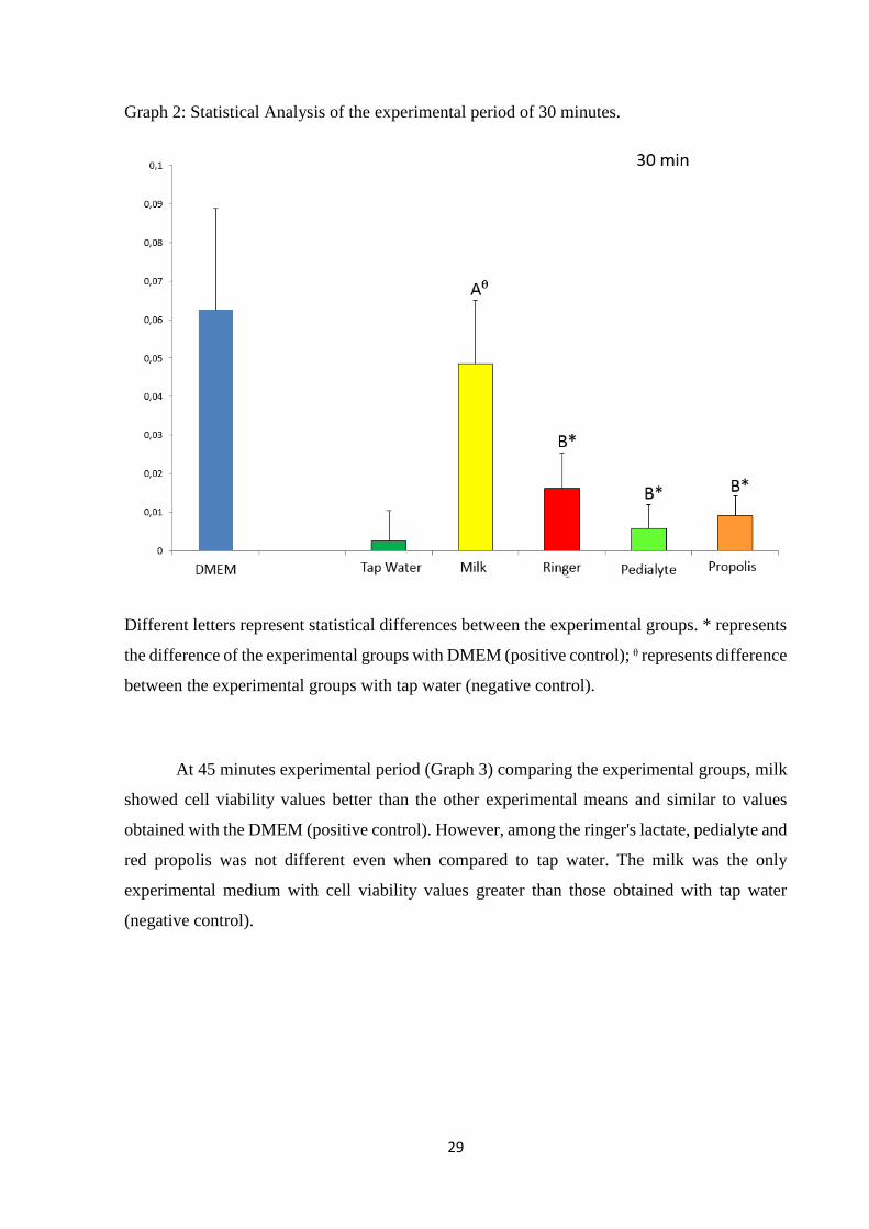
At 30 minutes (Graph 2) comparing the experimental groups, milk showed cell viability
values better than the other experimental means and similar to values obtained with the DMEM
(positive control). Among the ringer's lactate, pedialyte and the red propolis no statistical
difference was not compared to tap water. The milk was the only experimental medium with
cell viability values greater than those obtained with tap water (negative control).
29
Graph 2: Statistical Analysis of the experimental period of 30 minutes.
Different letters represent statistical differences between the experimental groups. * represents
the difference of the experimental groups with DMEM (positive control); θ represents difference
between the experimental groups with tap water (negative control).
At 45 minutes experimental period (Graph 3) comparing the experimental groups, milk
showed cell viability values better than the other experimental means and similar to values
obtained with the DMEM (positive control). However, among the ringer's lactate, pedialyte and
red propolis was not different even when compared to tap water. The milk was the only
experimental medium with cell viability values greater than those obtained with tap water
(negative control).

30
Graph 3: Statistical Analysis of the experimental period of 45 minutes.
Different letters represent statistical differences between the experimental groups. * represents
the difference of the experimental groups with DMEM (positive control); θ represents difference
between the experimental groups with tap water (negative control).
At 60 minutes experimental period (Graph 4) comparing the experimental groups, milk
showed cell viability values better than the other experimental means and similar to values
obtained with the DMEM (positive control). The ringer's lactate means, pedialyte and the red
propolis showed similar results to each other and to those obtained with tap water. The milk
and Lactated Ringer, cell viability showed values higher than those obtained with tap water
(negative control).

31
Graph 4: Statistical Analysis of the experimental period of 60 minutes.
Different letters represent statistical differences between the experimental groups. * represents
the difference of the experimental groups with DMEM (positive control); θ represents difference
between the experimental groups with tap water (negative control).
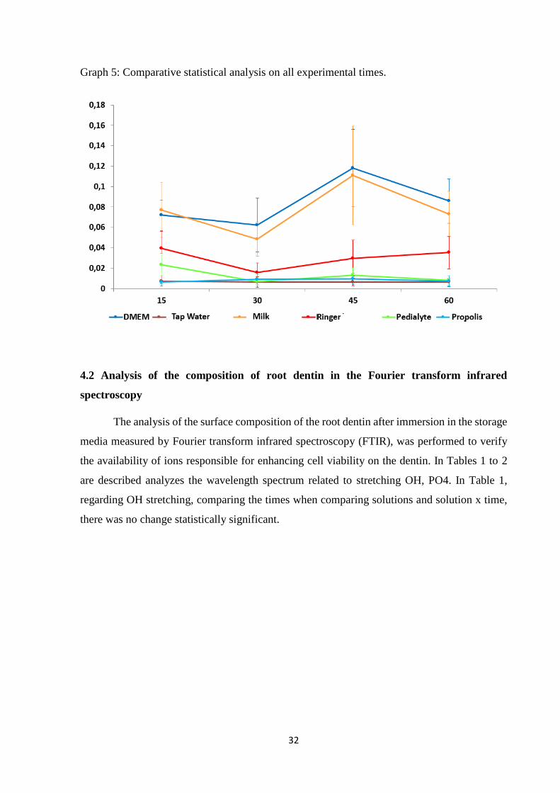
In Graph 5, the performance is verified of the storage means over the evaluation period.
The performance of red propolis and pedialyte were very low regardless of the evaluation
period. The ringer's lactate has intermediaries’ values of cell viability and kept evenly over the
periods. For milk means and DMEM increased slightly viability levels in periods of 45 minutes,
with no significant difference among the remaining periods.
32
Graph 5: Comparative statistical analysis on all experimental times.
4.2 Analysis of the composition of root dentin in the Fourier transform infrared
spectroscopy
The analysis of the surface composition of the root dentin after immersion in the storage
media measured by Fourier transform infrared spectroscopy (FTIR), was performed to verify
the availability of ions responsible for enhancing cell viability on the dentin. In Tables 1 to 2
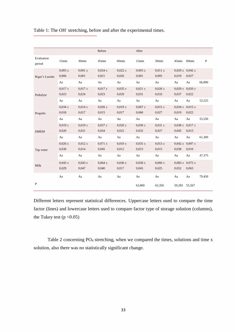
are described analyzes the wavelength spectrum related to stretching OH, PO4. In Table 1,
regarding OH stretching, comparing the times when comparing solutions and solution x time,
there was no change statistically significant.
33
Table 1: The OH- stretching, before and after the experimental times.
Different letters represent statistical differences. Uppercase letters used to compare the time
factor (lines) and lowercase letters used to compare factor type of storage solution (columns),
the Tukey test (p <0.05)
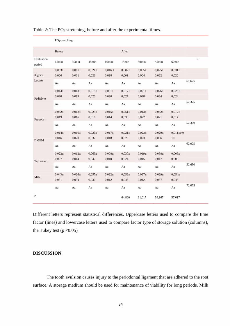
Table 2 concerning PO4 stretching, when we compared the times, solutions and time x
solution, also there was no statistically significant change.
Before After
Evaluation
period 15min 30min
45min 60min 15min 30min 45min 60min P
Riger’s Lactate
0,003 ±
0,006
0,001 ±
0,001
0,024 ±
0,021
0,022 ±
0,026
0,003 ±
0,001
0,011 ±
0,005
0,029 ±
0,019
0,042 ±
0,027
Aa Aa Aa Aa Aa Aa Aa Aa 66,800
Pedialyte
0,017 ±
0,023
0,017 ±
0,024
0,017 ±
0,023
0,035 ±
0,029
0,021 ±
0,031
0,026 ±
0,033
0,029 ±
0,037
0,020 ±
0,022
Aa Aa Aa Aa Aa Aa Aa Aa 52,525
Propolis
0,034 ±
0,018
0,014 ±
0,017
0,026 ±
0,015
0,019 ±
0,017
0,067 ±
0,060
0,015 ±
0,027
0,034 ±
0,019
0,015 ±
0,022
Aa Aa Aa Aa Aa Aa Aa Aa 55,550
DMEM
0,019 ±
0,020
0,019 ±
0,021
0,027 ±
0,034
0,020 ±
0,022
0,030 ±
0,032
0,031 ±
0,027
0,038 ±
0,045
0,017 ±
0,015
Aa Aa Aa Aa Aa Aa Aa Aa 61,300
Tap water
0,026 ±
0,030
0,012 ±
0,014
0,071 ±
0,045
0,010 ±
0,012
0,035 ±
0,023
0,013 ±
0,015
0,042 ±
0,038
0,007 ±
0,010
Aa Aa Aa Aa Aa Aa Aa Aa 47,375
Milk 0,043 ±
0,029
0,043 ±
0,047
0,064 ±
0,040
0,036 ±
0,017
0,058 ±
0,045
0,060 ±
0,025
0,083 ±
0,052
0,075 ±
0,063
Aa Aa
Aa Aa Aa Aa Aa Aa 79,450
P
63,800 63,350 59,283 55,567
34
Table 2: The PO4 stretching, before and after the experimental times.
PO4 stretching
Before After
Evaluation
period 15min 30min 45min 60min 15min 30min 45min 60min
P
Riger’s
Lactate
0,003±
0,006
0,001±
0,001
0,024±
0,026
0,016 ±
0,018
0,002±
0,001
0,005±
0,004
0,025±
0,022
0,031±
0,020
Aa Aa Aa Aa Aa Aa Aa Aa 61,625
Pedialyte
0,014±
0,020
0,013±
0,019
0,015±
0,020
0,031±
0,020
0,017±
0,027
0,021±
0,028
0,026±
0,034
0,020±
0,024
Aa Aa Aa Aa Aa Aa Aa Aa 57,325
Propolis
0,032±
0,019
0,012±
0,016
0,025±
0,016
0,015±
0,014
0,051±
0,038
0,013±
0,022
0,032±
0,021
0,012±
0,017
Aa Aa Aa Aa Aa Aa Aa Aa 57,300
DMEM
0,014±
0,016
0,016±
0,020
0,025±
0,032
0,017±
0,018
0,021±
0,026
0,023±
0,023
0,029±
0,036
0,011±0,0
10
Aa Aa Aa Aa Aa Aa Aa Aa 62,025
Tap water
0,022±
0,027
0,012±
0,014
0,065±
0,042
0,008±
0,010
0,030±
0,024
0,019±
0,015
0,038±
0,047
0,006±
0,009
Aa Aa Aa Aa Aa Aa Aa Aa 52,650
Milk 0,043±
0,031
0,036±
0,034
0,057±
0,030
0,032±
0,012
0,052±
0,044
0,037±
0,012
0,069±
0,037
0,054±
0,043
Aa Aa Aa Aa Aa Aa Aa Aa
72,075
P 64,800 61,017 59,167 57,017
Different letters represent statistical differences. Uppercase letters used to compare the time
factor (lines) and lowercase letters used to compare factor type of storage solution (columns),
the Tukey test (p <0.05)
DISCUSSION
The tooth avulsion causes injury to the periodontal ligament that are adhered to the root
surface. A storage medium should be used for maintenance of viability for long periods. Milk
35
was one of the first storage media studied which showed satisfactory effectiveness, either for
the sake of preserving the cells as at cost9,10.
The hypothesis of this study was that, Ringer's lactate solution, Propolis solution and
Pedialyte solution have the same capacity to preserve periodontal ligament cells as milk has.
However, the results showed that Milk has statistically preserved more cells than the other three
solutions, and the hypothesis was rejected. Milk´s better performance in this study was probably
due to its higher concentration of carbohydrates, proteins and fat, once other caracteristics – ph,
osmolarity and electrolytes concentration – were similar between all solutions.
Recent studies have demonstrated red propolis’s potential to preserve cells, and one
reported that it had a better performance preserving periodontal ligament cells than Hank’s
balanced salt solution and milk13 but this potential was not observed in this study. Some studies
have evaluated the properties of red propolis11,12. The red propolis extract shows high
concentration of phenolic acids and flavonoids such as formononetin, isoliquiritigenin,
liquiritigenin, medicarpin and biochanin that are often associated with a variety of health
benefits12. In fact red propolis showed similar statistical values to the negative control.
However, this study used different methods than other studies, and this may have been the cause
of the conflicting results.
As red propolis, pedyalite also showed no better performance than the negative control
and worst results than Ringers solution and Milk. Pedialyte was presented as a promising
medium for the maintenance of cells6 but, in this study, pedialyte showed the lower overall ph
values. It is known that an acid medium has a negative influence on cell preservation, therefore,
this was the probable reason for its negative results
Regarding cell adhesion to dentin surface, we found no statistical difference between
the solutions, for all experimental times. All solutions were to adhesion of fibroblasts to root
dentin. Some studies indicated that the solution could have an influence on cell adhesion by
changing the dentin layer´s composition and therefore, promoting an unfavorable surface15, but
this was not observed in this study.
While analyzing means to preserve human fibroblasts, we sought to identify whether
this would imply the dentin surface. Many solutions can influence the inorganic dentin’s matrix,
particularly those with a pH more acidic than neutral15. The PO4 connection determines the
mineral formation of dentin, and when we compare the solutions with each other, although
some submit an improper pH for cell preservation, statistically there was no change to the root
surface.

36
Chemically, the hydroxyl radical OH is closely linked to cell adhesion. It is known that
at higher ratios there is an enhancement in the binding of fibroblasts to dentin surface. Therefore
we seek to investigate whether some of the solutions would influence adherence and in this
comparison, all means presented compatible.
This research showed that milk should remain the medium of choice for storage of
avulsed teeth. Ringer's lactate solution also showed homogeneous effectiveness in all
experimental times becoming an alternative to transport the tooth to the dental office. Ringer's
lactate is inexpensive and is advantageous for its use.
CONCLUSION
Milk showed a better potential to preserve periodontal ligament cells, however no
difference in ionic composition of dentin methods. Since the milk also has good availability
and low cost, there it should still remain the first choice of storage medium for avulsed teeth.
CONFLICT OF INTEREST
No potential conflict of interest relevant to this article was reported.
ACKNOWLEDGMENTS
This study was financially supported by FAPITEC (Fundação de Apoio à Pesquisa e à Inovação
Tecnológica no Estado de Sergipe, SE, Brazil). This study was carried out in the CPBIO-
FOUFU (Research Center at the School of Dentistry - Federal University of Uberlândia).
REFERENCES
1. Karaylmaz H, Kirzioglu Z, Gungor OE. Aetiology, treatment patterns and long-term
outcomes of tooth avulsion in children and adolescents. Pak J Med Sci 2013;29:464-
468;
2. Souza BDM, Luckemeyer DD, Reyes-Carmona JF, Felippe WT, Simões CMO, Felippe
MCS. Viability of human periodontal ligament fibroblasts in milk, Hank’s balanced salt
solution and coconut water as storage media. Inter Endo J 2011;44:111–115;
37
3. Moura CCG, Soares PBF, Reis MVP, Neto AJF, Barbosa DZ, Soares CJ. Potential of
coconut water and soy milk for use as storage media to preserve the viability of
periodontal ligament cells: an in vitro study. Dent Trauma 2014;30: 22–26;
4. Huangqin C, Huang B. (-)-Epigallocatechin-3-gallate: a novel storage medium for
avulsed teeth. Dent Trauma 2012;28:158–160;
5. Mahal NK, Singh N, Thomas & N, Kakkar M. Effect of three different storage media
on survival of periodontal ligament cells using collagenase–dispase assay. International
Endo J 2013;46:365–370;
6. Macway-Gomez S, Lallier TE. Pedyalite promotes periodontal ligament cells survival
and motility. JOE 2013 Feb:39;
7. Mori GG, Nunes DC, Castilho LR, Moraes IG, Poi WR. Propolis as storage media for
avulsed teeth: microscopic and morphometric analysis in rats. Dent Trauma
2010;26:80–85;
8. Jamalpour MR, Soltanian AR, Tootunchi AS, Rosphanipaian M. Temporary
preservation of avulsed tooth in oral submucosal tissue: an experimental study in cat.
Dent Trauma 2014;30:265–269;
9. Blomlof L. Storage of Human Periodontal Ligament Cells in a Combination of Different
Media, J Dent Res 1980 Nov:60:1904-1906;
10. Blomlof L, Lindskog S, Hammarslrom L. Periodontal healing of exarticulaled monkey
teeth stored in milk or saliva. Scand J Dent Res 1981:89:251-259;
11. Frozza COS, Garcia CSC, Gambato G, Souza MDO, Salvador M, Moura S, and others.
Chemical characterization, antioxidant and cytotoxic activities of Brazilian red propolis.
Food and chem Toxic 2013;52:137-142;
12. Frozza COS, Ribeiro TS, Gambato G, Menti C, Moura S, Pinto PM, and others.
Proteomic analysis identifies differentially expressed proteins after red propolis
treatment in Hep-2 cells. Food and Chem Toxic 2014;63:195-204;
13. Martin MP, Pileggi R, A quantitative analysis of própolis: A promising new storage
media following avulsion, Dent Trauma 2004;20:85-89;
14. Zhang X, Neoh KG, Lin CC, Kishen A. Remineralization of partially demineralized
dentine substrate based on a biomimetic strategy. J Mater Sci Mater Med 2012;23:733–
742;

38
15. Eliadesa G, Mantzouranib M, Labelliah R, Muttic B, Sharmad D. Interactions of dentine
desensitisers with human dentine: Morphology and composition. J of Dent 2013;41:28–
39.
39
5 CONSIDERAÇÕES FINAIS
Na avaliação da viabilidade dos fibroblastos humanos, o leite integral resultou em níveis
de viabilidade celular similares ao grupo controle positivo e em geral superior aos
demais meios de armazenagem testados. Porém, no período de 60 minutos, período
normalmente decorrido até o reimplante dental, o meio ringer com lactato apresentou
valores superiores ao pedialyte, própolis vermelho e água de torneira. A água de torneira
apresentou valores de viabilidade celular muito baixos independente do período
avaliado.
Analisando a composição iônica da superfície da dentina radicular, por não apresentar
nenhuma discrepância estatística significativa, tanto comparando tempos e meios de
armazenagem, podemos concluir que, as soluções não influenciam na adesão celular
bem como não alteram a matriz inorgânica da dentina. Porém mais estudos são
necessários para analisar em maiores períodos de armazenagem.
40
6 – COMUNICADO DE IMPRENSA (PRESS RELEASE)
ANÁLISE DA INFLUÊNCIA DE DIFERENTES MEIOS DE ARMAZENAGEM NA
VIABILIDADE DE FIBROBLASTOS E NA COMPOSIÇÃO IÔNICA DA DENTINA
RADICULAR: ESTUDO in vitro
Aracaju, 04 de Fevereiro de 2015 – Pesquisadores da área de odontologia da Universidade
Federal de Sergipe – UFS – divulgaram resultado de pesquisa na qual o leite integral comparado com a
solução de ringer com lactato, o própolis vermelho e o pedialyte, deve ser adotado como solução para
armazenagem de dentes avulsionados (Dentes que caíram acidentalmente) até o período de 60 minutos
com grande chance de evitar a perda do dente.
As células que ficam unidas a superfície da raiz dentária, foram cultivadas e avaliadas em
diferentes soluções nos tempos de 15, 30, 45 e 60 minutos. Após os tempos experimentais, as células de
cada grupo foram contadas e calculadas estatisticamente. Esta pesquisa também buscou analisar a
influência destas soluções ao dente. Comparando também nos mesmos tempos experimentais todas as
soluções não demonstraram efeito estatístico significativo sobre o dente.
Para realização desta pesquisa, os pesquisadores tiveram a parceria com a Faculdade de
Odontologia da Universidade Federal de Uberlândia com apoio financeiro da Fundação de Apoio à
Pesquisa e à Inovação Tecnológica no Estado de Sergipe (FAPITEC) e da Coordenação de
Aperfeiçoamento de Pessoal de Ensino Superior (CAPES).
41
REFERÊNCIAS
1. Karayilmaz H, Kirzioglu Z, Gungor OE. Aetiology treatment patterns and long-term
outcomes of tooth avulsion in children and adolescents. Pak J Med Sci 2013;29:464-
468;
2. Silva EJNL, Rollemberg CB, Coutinho-Filho TS, Zaia AA. A multiparametric assay to
compare the cytotoxicity of soy milk with diferente storage media. Dent Trauma
2013;29:319–322;
3. Eskandarian T, Badakhsh S, Esmaeilpour T. The Effectiveness of Oral Rehydration
Solution at Various Concentrations as a Storage Media for Avulsed Teeth. Iran Endo J
2013;8:22-24;
4. Mahal NK, Singh N, Thomaz N, Kakkar M. Effect of three different storage media on
survival of periodontal ligament cells using collagenase–dispase assay. Inter Endo J
2013;46:365–370;
5. Moura CCG, Soares PBF, Reis MVP, Neto AJF, Barbosa DZ, Soares CJ. Potential of
coconut water and soy milk for use as storage media to preserve the viability of
periodontal ligament cells: an in vitro study. Dent Trauma 2014;30: 22–26;
6. Jamalpour MR, Soltanian AR, Tootunchi AS, Rosphanipaian M. Temporary
preservation of avulsed tooth in oral submucosal tissue: an experimental study in cat
Dental Traumatology 2014; 30:265–269;
7. Reis MVP, et al. Histologic and Micro–Computed Tomographic Analyses of Replanted
Teeth Stored in Different Kind of Media. JOE 2012 may;40;
8. Blomlof L, Lindskog S, Hammarslrom L. Periodontal liealing of exarticulaled monkey
teeth stored in milk or saliva. Scand J Dent Res 1981;89:251-259;
9. Blomlof L, Storage of Human Periodontal Ligament Cells in a Combination of Different
Media. J Dent Res 1981 nov;60:1904-1906;
10. Lekic PC, Kenny DJ, Barret EJ. The influence of storage conditions on the clonogenic
capacity of periodontal ligament cells: implications for tooth replantation. Inter Endo J
1998;31:137–140;
11. Pearson RM. Human Periodontal Ligament Cell Viability in Milk and Milk Substitutes.
J of Endo 2003 mar;29;
12. Macway-Gomez S, Lallier TE. Pedyalite promotes periodontal ligament cells survival
and motility. JOE 2013 feb:39;
42
13. Zunini GS, Rando KAE, Cox RG. Fluid Replacement in Craniofacial Pediatric Surgery:
Normal Saline or Ringer’s Lactate?. The J of Cranio Sur 2011; 22;
14. Mori GG, Nunes DC, Castilho LR, Moraes IG, Poi WR. Propolis as storage media for
avulsed teeth: microscopic and morphometric analysis in rats. Dent Trauma
2010;26:80–85;
15. Martin MP, Pileggi R. A quantitative analysis of própolis: A promising new storage
media following avulsion. Dent Trauma 2004:20:85-89;
16. Ozan, F. et al. Effect of propolis on survival of periodontal ligament cells: new storage
media for avulsed teeth. J Endo 2007;33:570-573;
17. Frozza COS, Garcia CSC, Gambato G, Souza MDO, Salvador M, Moura S, and others.
Chemical characterization, antioxidant and cytotoxic activities of Brazilian red propolis.
Food and chem Toxic 2013;52:137-142;
18. Frozza COS, Ribeiro TS, Gambato G, Menti C, Moura S, Pinto PM, and others.
Proteomic analysis identifies differentially expressed proteins after red propolis
treatment in Hep-2 cells. Food and Chem Toxic 2014;63:195-204.