FACULDADE DE ODONTOLOGIA
-
MESTRADO EM ODONTOLOGIA
DAIANA FLORES GONÇALVES GIANNASTASIO
ÁREAS DE DESGASTE EM PRÉ-MOLARES SUPERIORES COM TRÊS RAÍZES MEDIANTE O EMPREGO DAS TÉCNICAS MANUAL, ROTATÓRIA
E RECIPROCANTE: AVALIAÇÃO DO PREPARO APICAL POR MICROTOMOGRAFIA COMPUTADORIZADA
Porto Alegre 2014

DAIANA FLORES GONÇALVES GIANNASTASIO
ÁREAS DE DESGASTE EM PRÉ-MOLARES SUPERIORES COM TRÊS RAÍZES MEDIANTE O EMPREGO DAS TÉCNICAS MANUAL, ROTATÓRIA
E RECIPROCANTE: AVALIAÇÃO DO PREPARO APICAL POR MICROTOMOGRAFIA COMPUTADORIZADA
Dissertação - .
Orientadora: Profa. Dra. Fabiana Vieira Vier-Pelisser
Porto Alegre 2014

Dedico esta dissertação ao meu filho Guilherme, que é minha razão de viver, minha fonte de forças e motivação...
AGRADECIMENTOS
Agradeço primeiramente a Deus, que me deu a vida... exatamente
como sempre pedi a Ele...
Aos meus pais, os escolhidos por Deus para me receberem e por me
encaminharem na vida!!! Me deram amor, dedicação, por passarem os mais
importantes valores que uma pessoa precisa para ter uma vida digna.
Ao meu marido, Sandro, pela paciência!!! Sei que algumas vezes
abusei dela... mas obrigada mesmo assim!!! Obrigada por ser meu fiel
parceiro, o queijo da minha goiabada!!! É muito bom viver ao teu lado...
Aos meus irmãos, Juliano, Rafael e Lucas... meus amigos de fé, meus
irmãos camaradas... é muito bom ter vocês na minha vida... Triste aqueles
que não tem um irmão!
Aos meus familiares e amigos, pela compreensão nos momentos de
ausência e impaciência...
A minha querida orientadora, pela paciência, dedicação, entusiasmo e
empenho. Para mim um grande exemplo de mestre! Sempre pronta a
ajudar... uma pessoa de coração gigante, com seu super coração maternal.
Abraça a causa do aluno e faz tudo o que está ao seu alcance para ajudá-lo.
É realmente uma orientadora, pois está ao lado do aluno em todas as etapas,
do início ao fim.
A Profa Dra Roberta Scarparo, pela incansável e fundamental
colaboração e comprometimento com o desenvolvimento desse trabalho. É
difícil achar palavras para agradecer o incentivo dado por ela na decisão por
fazer o mestrado!!! É impossível mensurar minha gratidão por ela.
A Profa Dra Vânia Fontanella, pela presteza e disponibilidade. Pela
colaboração indispensável! Pelos acréscimos que seu conhecimento trouxe
ao estudo.
Ao professor José Antonio Poli de Figueiredo, por nos passar
h x (“- Quero que saiam melhores do que
!”). h ,
cultura. Pra mim, uma mente brilhante!!!
Aos meus colegas do mestrado, pela convivência agradável, pelas
conversas, pelos estudos e pela parceria. Pelo incentivo para que fosse dada
continuidade à minha vida acadêmica. Sem eles eu estaria apenas
encerrando um ciclo, com vontade, mas sem coragem para continuar...
Grandes amizades ficaram!!!
A todos os professores e professoras do curso, pela dedicação e
interesse em passar conhecimento atualizado e de qualidade.
A equipe da secretaria de graduação e Pós-Graduação pelo suporte
incansável em muitos momentos.
rio da Educaç , , pelo incentivo financeiro
oportunizando a realização do curso.

RESUMO
Introdução: Este estudo avaliou, in vitro, através da microtomografia
z (μT ), áreas de desgaste em canais radiculares de pré-
molares superiores com três raízes, causadas por três diferentes sistemas de
preparo. Métodos: Após a abertura coronária, esvaziamento dos canais e
estabelecimento da odontometria a 1,5 mm do vértice radiográfico, dezoito
espécimes foram divididos em três grupos, de acordo com a técnica de
preparo empregada para o preparo do canal radicular: Grupo manual (Hand)
(n=6) - os dentes foram preparados manualmente pela técnica coroa-ápice,
com limas de aço-inoxidável flexíveis; Grupo ProTaper (GPt) (n=6) – dentes
preparados pelo sistema rotatório ProTaper e Grupo WaveOne (GWO) (n=6)
- dentes preparados pelo sistema reciprocante WaveOne. Os alargamentos
apicais finais nos canais mésio-vestibular (MV), disto-vestibular (DV) e
palatino (P) corresponderam a 0,25 mm, 0,25 mm e 0,40 mm,
respectivamente. Os dentes foram microtomografados antes e após o
preparo dos canais. As imagens pré e pós-operatórias, correspondentes a 0,
2, 4, 6 mm do batente apical, foram sobrepostas e a diferença entre as áreas
dos canais cirúrgico e anatômico foram mensuradas, com o auxílio do
programa PhotoShop. Os grupos foram comparados estatisticamente,
empregando-se Anova de Dois Critérios e Post-hoc de Tukey (P<0,05).
Resultados: Não houve diferença entre as técnicas de preparo (P<0,05).
Independente da técnica empregada, percebeu-se um maior alargamento em
direção a porção mais cervical do canal. Alguns canais apresentaram
alargamento excessivo porém, não houve perfuração. Por outro lado, em
algumas raízes observou-se áreas não tocadas. Conclusão: Não houve

diferença entre as técnicas manual, rotatória e reciprocante, empregadas
para o preparo apical de pré-molares com três raízes. Todas são seguras
para o preparo do terço apical desses dentes.
Palavras-chave: pré-molares superiores com três raízes, microtomografia
comptadorizada, preparo de canal radicular, sistema rotatório, sistema
reciprocante.

ABSTRACT
Introduction: The aim of the present study was to evaluate the apical
enlargement areas of three-rooted human upper premolars by means of
micro-computed tomography-(CT). Métodos: After the cavity access, canal
exploration and odontometry, eighteen samples were divided into three
groups according to the preparation technique: manual group (Hand) (n=6) –
teeth were instrumented by crown-down technique and stainless steel files;
ProTaper group (GPt) (n=6) – teeth were instrumented by using the Rotary
movement and ProTaper system; and WaveOne group (GWO) (n=6) – teeth
were instrumented by WaveOne instrument operated in reciprocating
movement. The final canal enlargement at mesiobuccal (MB), distobucal (DB)
and palatal (P) roots match 0,25 mm, 0,25 mm and 0,40 mm, respectively.
Micro-computed tomography-(CT) was performed before and after
preparation. The pre and post images corresponding to 0, 2, 4 and 6 mm from
the apical stop were overlaid and the difference between the areas of surgical
and anatomical canals were measured with the aid of PhotoShop program.
The groups were statistically compared using two-way Analysis of Variance
(ANOVA) and Tukey post-hoc test at 5% significance level. Results: There
was no difference between the preparation techniques. Regardless of the
technique, we found further enlargement toward the cervical portion of the
canal. Some canals had excessive enlargement, but no perforation.
Moreover, untouched areas were observed in some roots. Conclusion: There
were no differences in all roots when comparing hand, rotary and
reciprocating techniques. All of them are safe for the instrumentation of the
apical third of three-rooted premolars.

Key-words: three-rooted premolars, micro-computed tomography, root canal
preparation, rotary systems, reciprocating systems.

SUMÁRIO
1 INTRODUÇÃO ............................................................................................ 11
2 ARTIGO ...................................................................................................... 16
3 DISCUSSÃO............................................................................................... 33
4 REFERÊNCIAS .......................................................................................... 37
ANEXOS ........................................................................................................ 40

11
1 INTRODUÇÃO
O tratamento endodôntico visa o correto debridamento do sistema de
canais radiculares (SCR), respeitando-se as características anatômicas dos
dentes (SHILDER, 1974; SATHORN et al., 2005; FORNARI et al., 2010).
Sendo assim, o conhecimento da anatomia dental interna é de extrema
importância para o sucesso da terapia (Peters et al., 2001).
Os pré-molares superiores apresentam grande variabilidade em sua
morfologia e, normalmente apresentam duas raízes distintas (Vertucci e
Gegauff, 1979; Mattuella, 2005). Entretanto, estudos demonstram que a
ocorrência de três raízes pode variar em até 6% das amostras (Pécora, 1992;
Loh, 1998; Kartal et al., 1998; Chaparro et al., 1999; Atieh, 2008; Neelakatan
et al., 2011; Tian et al., 2012) sendo relatada fragilidade das paredes
dentinárias, especialmente, em relação às raízes vestibulares (Hartmann et
al., 2013). Por ser semelhante ao primeiro molar superior, em número e
disposição das raízes e secção transversal, estes dentes têm sido chamados
de minimolares (Malibaum, 1989; Goon, 1993; Vier-Pelisser et al., 2010).
Alguns importantes estudos vem sendo realizados com pré-molares
superiores com três raízes, tendo como método de avaliação a
microtomografia computadorizada (µCT) (Vier-Pelisser et al., 2010; Marca et
al., 2013; Hartmann et al., 2013).
A µCT apresenta-se como um método de grande valia para a pesquisa
odontológica. Com esta tecnologia pode-se obter uma reconstrução em 3D
da morfologia dental com voxels cúbicos e resolução isotrópica, sendo mais
12
precisa do que a tomografia cone beam (CBCT) (Marca et al., 2013). Além
disso, é um método não invasivo que oportuniza a integridade da amostra.
Dessa forma, o espécime pode ser utilizado em pesquisas futuras. Outra
vantagem é a quantidade de dados que podem ser obtidos das imagens,
permitindo análise qualitativa e quantitativa das amostras (Rhodes et al.,
1999)
Marca et al. (2013) utilizaram a tomografia computadorizada de feixe
cônico (CBCT) e a µCT para avaliar a anatomia radicular de pré-molares com
três raízes. Os autores concluíram que, apesar das variações anatômicas
serem similares, a µCT fornece imagens com maior resolução e maior
riqueza de detalhes e isso pode ser explicado pela variação no tamanho do
voxel. As imagens microtomográficas revelaram a possibilidade de presença
de mais de um canal no terço médio dos canais vestibulares de um
espécime, presença de canais laterais, trifurcação do canal no terço apical,
assim como diferentes formas de secções de canais e raízes, enquanto as
imagens da CBCT não apresentaram o mesmo grau de detalhamento e
peculiaridades anatômicas.
Vier-Pelisser et al. (2010) avaliaram, através de microtomografia
computadorizada (µCT), a secção transversal, a distância entre o teto da
câmara pulpar e a bi ou trifurcação dos canais e as variações anatômicas de
pré-molares superiores com três raízes. Os autores relataram grande
variação em todos os aspectos analisados nesse grupo de dentes, revelando
uma anatomia interna rica, com a presença de canais laterais, delta apical e
canal acessório na região da furca. Há inclusive, mudanças graduais da
secção transversal do canal ao longo da raiz.
13
Complementando estudos anteriores, Hartmann et al. (2013) avaliaram,
no mesmo grupo dental, características como: diâmetro dos canais
radiculares, espessura das paredes dentinárias, áreas de segurança ou de
perigo para a instrumentação e os limites aconselháveis para o alargamento
do canal radicular. As avaliações através de µCT confirmaram a fragilidade
das paredes vestibulares. Além disso, a partir das medidas do diâmetro do
canal e das paredes dentinárias, pôde-se concluir que o uso de instrumento
de calibre superior a 0,25 mm nas raízes vestibulares e 0,40 na raiz palatina,
em apical, poderia ser responsável por causar iatrogenias durante a fase do
preparo químico-mecânico.
Com os avanços da ciência e da tecnologia e com o objetivo de realizar
o tratamento endodôntico dentro dos parâmetros ideais, com maior facilidade
e segurança, novos sistemas vêm sendo constantemente lançados no
mercado. Conforme López et al. (2008), por serem menos flexíveis, os
instrumentos de aço inoxidável causam mais alterações na anatomia do
canal quando comparados a instrumentos de níquel titânio com o mesmo
calibre. Sendo assim, atualmente, as limas em níquel-titânio vêm sendo as
mais estudadas. Entretanto, em virtude do baixo custo e acessibilidade, o uso
das limas de aço inoxidável ainda é muito presente e está normalmente
associado a técnica de instrumentação coroa-ápice. Esta, preconiza o
preparo do terço cervical e médio antes do terço apical. Dessa maneira,
elimina interferências dentinárias e reduz a possibilidade de causar extrusão
de conteúdo necrótico para região periapical (Goerig, Michelich e Schultz,
1982). Por possibilitar acesso mais livre ao terço apical, reduz o desvio nesta
região. A técnica coroa-ápice também é aplicável na instrumentação com

14
limas de níquel-titanio, trazendo para o preparo os mesmos benefícios
citados anteriormente.
Os instrumentos de níquel-titânio apresentam superelasticidade e
mostram-se eficientes na modelagem do canal radicular, causando menores
alterações no ângulo de curvatura, bem como mantendo os preparos mais
centralizados quando comparados aos instrumentos de aço-inoxidável
(Gambil et al., 1996; Peters, 2004; Hulsmann et al., 2005; Peters e Paqué,
2010).
Os instrumentos de níquel-titânio ProTaper (Dentisply Maillefer,
Balaigues, Switzerland) representam uma nova geração de instrumentos
endodônticos. Sua conicidade varia ao longo do eixo cortante de cada
instrumento. Possuem secção transversal triangular e ponta inativa. Esse
sistema é comporto por instrumentos de corte e de acabamento que
trabalham em movimento contínuo. Estes últimos são responsáveis pelo
preparo do terço apical e denominados F1, F2, F3 e F4 e F5 com diâmetro da
ponta e taper 20/07, 25/08, 30/09, 40/06 e 50/05, respectivamente. O uso
correto da sequência de instrumentos proporciona eficiência, segurança e
flexibilidade (Ruddle, 2005).
Recentemente, a Dentsply Maillefer lançou no mercado o sistema
WaveOne, que é composto por três instrumentos, denominados de pequeno,
médio e grande, com diâmetro da ponta e taper 21/06, 25/08 e 40/08
respectivamente, desenvolvidos para preparar completamente o canal
radicular do início ao fim, com um único instrumento. Esse sistema faz uso de
um motor específico que trabalha com o movimento recíproco, ou seja, faz

15
movimentos no sentido horário (menor ângulo) e anti-horário (maior ângulo).
A cada três ciclos de movimentos recíprocos completa-se uma rotação
reversa completa e, gradualmente, o instrumento avança no canal
necessitando mínima pressão apical (Webber et al. 2011; Plotino et al.,
2012).
Com o objetivo de dar continuidade e ampliar os conhecimento já
existentes a respeito dos pré-molares superiores com três raízes, e de
discutir a influência da anatomia no resultado final do preparo, este trabalho
tem o objetivo de avaliar, através da microtomografia computadorizada, as
áreas de desgastes no terço apical dos canais de pré-molares superiores
com três raízes, quando os mesmos são preparados pelas técnicas manual,
sistema rotatório (ProTaper, Ballaigues, Switzerland) e reciprocante
(WaveOne, Ballaigues, Switzerland). Sendo assim, será possível que o
profissional avalie se a técnica de preparo exerce alguma influência
expressiva no tratamento de dentes com dimensões reduzidas e paredes
frágeis como os mini-molares.
16
2 ARTIGO
Three-rooted premolars canal enlargement through hand, rotary and
reciprocating techniques: Micro-CT evaluation of apical preparation
Formatado conforme diretrizes do periódico Journal of Endodontics, Qualis
A1 e Fator de impacto 2.880
Daiana Flores Gonçalves Giannastasio DDS1, Roberta Kochenborger
Scarparo DDS, MsC, PhD1, Cauana Oliva Tavares DDS1, Vânia Fontanella
DDS, MsC, PhD2, Fabiana Vieira Vier-Pelisser DDS, MsC, PhD1
1 Pontifical Catholic University of Rio Grande do Sul, Porto Alegre, Brazil
2 Federal University of Rio Grande do Sul, Porto Alegre, Brazil
Corresponding Author
Roberta Kochenborger Scarparo
Av. Ipiranga 6681 – Prédio 6
Porto Alegre – RS, Brazil
CEP 90619-900

17
Three-rooted premolars canal enlargement through hand, rotary and
reciprocating techniques: Micro- CT evaluation of apical preparation
ABSTRACT
Introduction: This study aimed to evaluate, by means of micro-computed
tomography-(CT), the apical enlargement areas produced by hand, rotary
and reciprocating instrumentation in three-rooted upper premolars. Methods:
Eighteen (18) teeth (n= 6 per group) were divided into groups according to the
preparation technique: crown-down hand instrumentation, ProTaper and
WaveOne. Instruments with similar apical diameters were used (.25 and .40
mm for the buccal and palatal canals, respectively. Canal enlargement was
evaluated thr h μ T
instrumentation using Adobe Photoshop CS5 software (version CS3, Adobe
Systems Inc, San Jose, USA). Distances of 0, 2, 4 and 6 mm from the apical
stop were considered. Differences between pre and post-instrumented canal
areas were compared using two-way ANOVA and Tukey post-hoc (P<0.05).
Results: No differences were observed among instrumentation techniques.
Regardless of instrumentation technique, there was a trend to greater
enlargement toward the most cervical portion of the canal. Exceeded
enlargement was observed only in some samples, and root perforation did not
occur. On the other hand, some samples presented untreated canal areas.
Conclusion: There were no differences in all roots when comparing hand,
rotary and reciprocating techniques. All of them are safe for the
instrumentation of the apical third of three-rooted premolars.
Key words: three-rooted premolars, rotary systems, reciprocating systems,
µCT, root canal preparation
18
Introduction
Three-rooted premolars are described as rare occurrence, being limited
to about 6% of cases (1-7). Concerning this, measures of clinical interest for
root canal treatment, such as root wall thickness and canal diameters has
been explored in a recent investigation (8). Dental fragility, especially related
to buccal roots is a matter of concern, being critical when determining the
amount of canal enlargement during instrumentation and post space
preparation.
In this context, it is yet to be determined whether the available
preparation techniques provide safe and efficient canal enlargement. Hand
instrumentation is often related to greater canal displacement, particularly with
stainless steel files (9). Considering the new fashion for wider apical
preparation (10), rotary and reciprocating systems appear as an alternative
(11,12). The use of Ni-Ti rotary instruments with larger tapers, such as
Protaper, was shown to be more efficient than hand files (13). More recently,
systems as the WaveOne has been claimed to produce faster preparation
using a single instrument (14). The reciprocating movement relieves stress on
the file during cutting action movement (counterclockwise) and instrument
release (clockwise), aiming at reducing the risk of cyclic fatigue (15-17), while
keeping canal cleanliness and centralization ability (11,18).
Taken into account the aforementioned, the present study aimed to
investigate, by means of µ-CT, the enlargement areas produced by hand,
rotary (Protaper) and reciprocating (WaveOne) instrumentation in three-rooted
upper premolars.
19
Material and methods
This study was approved by the Ethics and Research Committee of
Pontifical Catholic University of Rio Grande do Sul (PUCRS - protocol no.
13/409.905).
Sample selection and specimen preparation
This study includes eighteen (18) three-rooted human upper premolars
with completely formed apices extracted for therapeutic reasons. Most of
them were evaluated in earlier studies using other approaches (19,20)
Crowns were sectioned at cementum-enamel junction, using a high-
speed diamond bur (KG Sorensen, Cotia, Brazil) under water-cooling. Canal
patency was verified by inserting # 10 and # 15 K-files (Dentisply Maillefer,
Balaigues, Switzerland) until the apical foramen. The working length was
visually established 1.5 mm shorter to this measure. Teeth were fitted into
special designed devices in such a way to enable similar scanning pre and
post canal preparation. Three specimens were mounted in each apparatus,
which were adapted in the micro-tomograph.
High-resolution computed tomography
Canal enlargement areas were y μ T
images obtained before and after instrumentation. Teeth were scanned cross-
sectionally using a high- k μ T y 100 KV 100 mA
(Skyscan 1172, Kontich, Belgium). For each tooth, about 1300 slices were
obtained at a voxel size of 13 × 13 × 13 μ . Th w
the Skyscan software and converted into a bpm format.
20
Root canal preparation
Teeth were randomly divided into three groups (n=6 per group),
according to the instrumentation technique employed: crown-down hand
preparation, Protaper rotary system (Dentsply Maillefer, Ballaigues,
Switzerland) and WaveOne reciprocating system (Dentsply Maillefer,
Ballaigues, Switzerland). All teeth were prepared by a single trained operator.
For all groups, canals were explored with size 15 Flexofiles at the previous
known working length (WL) and irrigated with 1 mL of 1% NaOCl at each
change of file. The working length was checked when the instrument had
reached the limit between the middle and apical third; after shapping was
accomplished.
Hand Preparation
Hand preparation was performed with Flexofile (Dentsply Maillefer,
Ballaigues, Switzerland). It was initiated with the instrument which adapted in
the cervical 2 mm of the canal by means of balanced force movements (21).
This procedure was repeated with thinner instruments toward apex direction
until the WL was reached. The apical stop was prepared until #25 and #40
files in the buccal and palatal roots, respectively. After, in order to ensure
adequate canal tapers, step back preparation was established using four
instruments.

21
Protaper Preparation
Rotary instrumentation was performed using EndoPro Torque motor
(VK Driller Equipamentos Elétricos Ltda, Jaguaré, Brazil) with a torque of
2N.cm and speed of 250 rpm. S1 and Sx instruments were used for cervical
preparation, followed by S2 instrument in the medium third of the canal, by
using paintbrush movements. After, F1 and F2 files (for buccal canals) or F1,
F2, F3 and F4 instruments (for palatal canals) were employed in the WL with
packing movements.
WaveOne Preparation
Reciprocating preparation was carried out using WaveOne motor
(Dentsply Maillefer, Ballaigues, Switzerland). Packing motion with slight apical
pressure were employed. Buccal canals were instrumented with primary size
file, while the palatal canals were prepared with the large size file. When the
instrument did not advanced, the instrument size 15 Flexofiles was used and
the canal was irrigated with 1 mL of 1% NaOCl.
Image analysis
Measurements of the original and prepared canal areas were
performed in cross-sectional images by a blinded and pre-calibrated observer
(ICC = 0.97), using Adobe Photoshop software (version CS3, Adobe Systems
Inc, San Jose, USA) (Figure 1). Images were selected starting from the apical
stop until 6 mm from it, with a distance of 2 mm between each section. Each
image had a Z value (in mm) on its left upper corner, indicating the distance
from one slice to the previous one, which facilitated measurements.
22
Statistical analysis
Differences between pre and post-instrumented canal areas were
compared among groups, and among pre-determined distances from the
apical stop. Data was analyzed by means of two-way ANOVA and Tukey
post-hoc (P<0.05) using GraphPad Prism Software (GraphPad Software, Inc.
La Jolla, CA 92037 USA ).
Results
The results of enlargement areas using the three preparation
techniques are presented in figure 2.
There were no differences in all roots comparing hand, rotary and
reciprocating techniques. These results were observed both when analyzing
the overall data and when comparing the techniques in each one of the
distances from the apical stop.
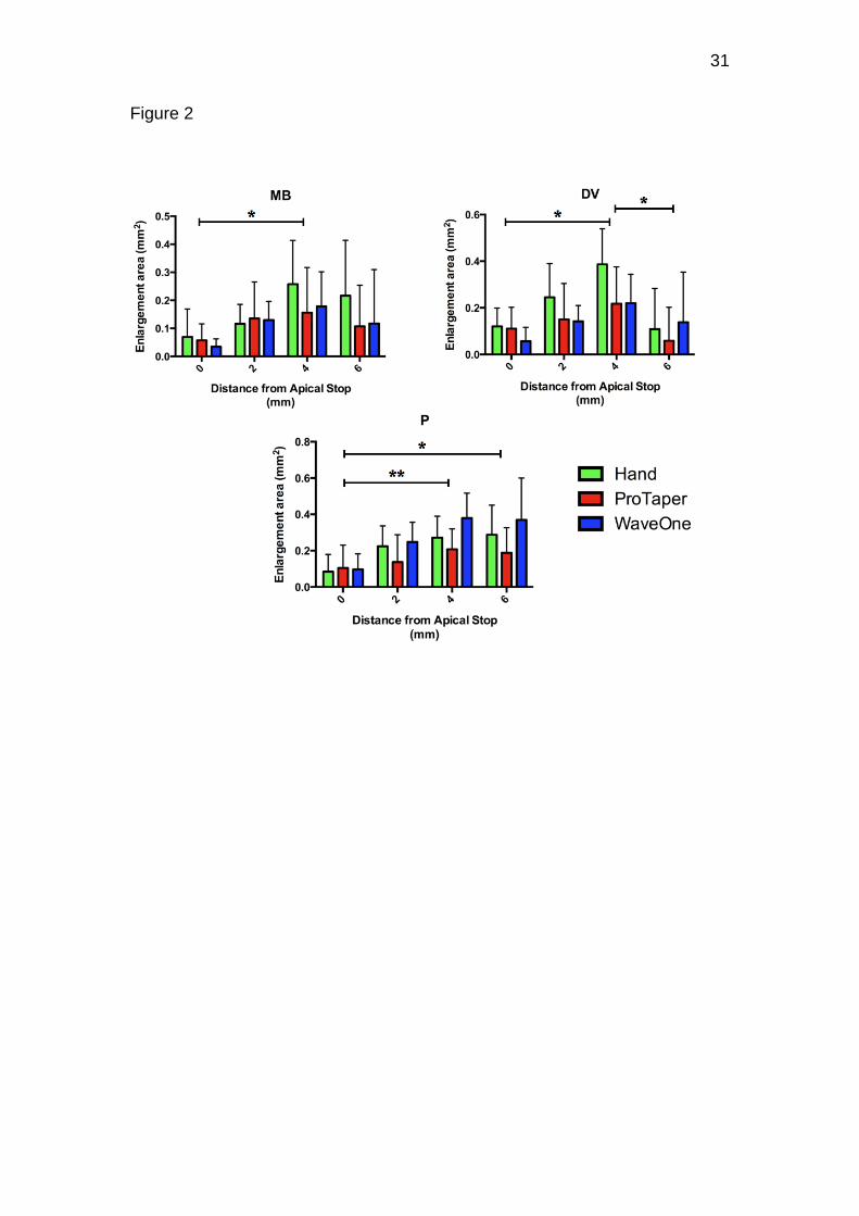
When specific points were evaluated, it was noted that enlargement
areas were more pronounced at 4 mm compared to the apical stop (0 mm) -
P<0.01 for the mesio-buccal and disto-buccal canals and P<0.001 for the
palatal canal. At the disto-buccal root this difference was also noted between
4 and 6 mm (P<0.01), while in the palatal root, between 0 and 6 mm (P<0.01).
Exceeded enlargement (where there is perforation risk) was observed
only in some samples, and root perforation did not occur, regardless of the
preparation technique. In contrast, some samples presented untreated canal
areas (Figure 3 a-b).

23
Discussion
Given these results, we set out to discuss the amount of enlargement
recommended when endodontic treatment of three rooted upper premolars is
required. Considering that this morphological occurrence is rare (22-24), little
is known about the safe and efficient limits for preparation.
The limits of apical preparation established herein were based on the
previously described means of three-rooted premolars canals anatomical
diameters (8). Thus, for buccal and palatal canals, instruments with an apical
diameter of .25 and .40 mm were selected, respectively. On the other hand,
variations on the anatomical configuration of apical cross-sections, also
pointed previously (8), lead to a safe, but not always efficient apical
preparation, regardless the technique used. Exaggerated enlargement was
observed in few samples, and root perforation did not occur. However, some
samples showed deficient enlargement, and untreated canal areas were
detected.
Taken into account the aforementioned, it is important to point out that
the use of mean canal diameters was not accurate in determining the ideal
dimensions of the apical instrument. Canal diameters standard deviations can
vary up to .15 mm (8), which brings out insights on the need of adopting
further technical strategies to obtain proper and safe enlargements, especially
in infected teeth (10). In the present study, the individual tactile determination
of the anatomical file was not performed, which certainly had an influence in
the observation of untreated canal areas in some samples. Nevertheless, the
ideal procedures for determining apical diameter have yet to be tested in

24
three-rooted premolars. Thus, root wall thickness in the cervical portion of
these teeth may affect the safety of pre-flaring procedures (8).
Considering the development of new instrumentation techniques, it is
of paramount importance to evaluate differences regarding the resulting
enlargement of new approaches compared to commonly employed
preparation techniques that intend to similar apical dilation. WaveOne is a
novel reciprocating system which suggests the employment of a single
instrument with continuous taper (25). Up to date, little is known about the
resulting canal shape promoted by instruments that act by means of this
kinematics. This issue is especially important if considering the safe use of
this technique associated to the fragility of three-rooted upper premolars (8).
For this reason, the present study compared canal enlargement of WaveOne
to the obtained after using Rotary (ProTaper) and Hand instrumentation.
However, there is no difference among groups.
Taken into account the greater taper of WaveOne and ProTaper
instruments compared to hand files, it was expected that a superior
enlargement would occur. In this regard, the absence of differences may be
related to the instruments dimensions in their apical portion and technical
sequence. As a matter of fact, in the three apical milimeters, both PT and WO
present the same taper (25,26). Moreover, although hand files are
characterized by continuous and lower tapers (.02), the crown-down
preparation enables a tapered canal final shape (27). This fact could be
proved when analyzing differences in the amount of canal enlargement
amongst specific distances from the apical stop. Regardless the

25
instrumentation technique, there was a trend to greater enlargement toward
the most cervical portion of the canal.
Another explanation for the absence of differences amongst groups
could be the number of samples evaluated. Due to the low frequency of three-
rooted premolars, the data presented herein was based on the analysis of
eighteen specimens, which have been previously evaluated on their
anatomical characteristics (19,20). On the other hand, in order to increase the
data reliability, high resolution tomography was employed to evaluate canal
enlargement. Previous investigations have demonstrated that this method is
more accurate than cone beam computed tomography (20, 28) enabling
precise measures of root canal pre and post-preparation (29,30).
Taken together, the current results established that hand, rotary and
reciprocating techniques are safe for the instrumentation of the apical third of
three-rooted premolars. Improvements regarding the determination of efficient
enlargement are still required. Further studies are needed to evaluate these
aspects in the cervical portion of canals, as well as to determine the centering
ability promoted by different instrumentation techniques.
Reference
1. Tian YY, Guo B, Zhang R, et al. Root canal morphology of maxilary
first pré molars in a Chinese Subpopulation evaluetade using cone-
beam computed tomography. Int Endod J 2012;45:996-1003.
2. Vertucci F, Seelig A, Gillis R. Root canal morphology of the human
maxillary second premolar. Oral Surg Oral Med Oral Pathol
1974;38:456–64.

26
3. Vertucci FJ, Gegauff A. Root canal morphology of the maxillary first
premolar. J Am Dent Assoc 1979;99:194–8.
4. Loh HS. Root morphology of the maxillary first premolar in
Singaporeans. Aust Dent J 1998;43:399-402.
5. Kartal N, Ozcelik B, Cimilli H. Root canal morphology of maxillary
premolars. J Endod 1998;24:417–9.
6. Chaparro AJ, Segura JJ, Guerrero E, et al. Number of roots and canals
in maxillary first premolars: study of an Andalusian population. Endod
Dent Traumatol 1999;15: 65–7.
7. Neelakantan P, Subbarao C, Ahuja R, et al. Root and canal
morphology of Indian maxillary premolars by a modified root canal
staining technique. Odontology 2011;99:18–21.
8. Hartmann RC, Baldasso FE, Stürmer CP, et al. Clinically relevant
dimensions of 3-rooted maxilary premolars obtained via high-resolution
computed tomography. J Endod 2013, 39:1639-1645.
9. López FU, Fachin EV, Fontanella VR, et al. Apical transportation: A
comparative evaluation of three root canal instrumentation techiniques
with three different apical diameters. J Endod 2008;34:1545-1548.
10. Wu MK,Barkis D,Roris A, etal. Does the first file to bind correspond to
the diameter of the canal in the apical region? Int Endod J
2002;35:264–7.
11. Bürklein S, Hinschitza K, Dammaschke T, et al. Shaping ability and
cleaning effectiveness of two single-file system in severely curved root
canals of extracted teeth: Reciproc and WaveOne versus Mtwo and
Protaper. Int Endod J 2012; 45:449-461.
27
12. Liu R, Hou BX, Wesselink PR, et al. The incidence of root microcracks
caused by 3 different single-file systems versus the ProTaper system. J
Endod 2013; 39:1054-1056.
13. Elayouti A, Chu AL, KimionisI, et al. Efficacy of rotary instruments with
greater taper in preparing oval root canals. I Endod J 2008; 41:1088-
1092.
14. You SY, Kim HC, Bae KS, et al. Shaping ability of reciprocating motion
in curved root canals: a comparative study with micro-computed
tomography. J Endod 2011;37: 1296–300.
15. De-Deus G, Moreira EJ, Lopes HP, et al. Extended cyclic fatigue life of
F2 proTaper instruments used in reciprocating movement. I Endod J
2010; 43:1063-1068.
16. Varela-Patino P, Ibanez-Parraga A,Rivas-Mundina B,etal. Alternating
versus contin- uous rotation: a comparative study of the effect on
instrument life. J Endod 2010;36: 157–9.
17. Plotino G, Grande NM, Testarelli L, et al. Cyclic fatigue of reciproc and
WaveOne reciprocating instruments. I Endod J 2012; 45:614-618.
18. Junaid A, Freire LG, Bueno CE, et al. Influence of single-file
endodontics on apical transportation in curved root canals: An ex-vivo
micro-computed tomographic study. J Endod 2013; ARTICLE IN
PRESS.
19. Vier FV, Tochetto FF, Orlandin LI, et al. In vitro study of the anatomic
diameter of root canals in human molars regarding tooth age. JBE J
Bras Endodontia 2004;5: 52–60.

28
20. Marca C, Dummer PM, Bryant S, et al. Three-rooted premolar
analyzed by high- resolution and cone beam CT. Clin Oral Investig
2013;17:1535–40.
21. Roane JB, Sabala CL, Duncanson MG. The balanced force concept for
instrumentation of curved canals. J Endod 1985; 11:203-11.
22. Schilder H. Cleaning and shaping the root canal. Dent Clin North Am
1974;18: 269–96.
23. Sathorn C, Palamara JE, Messer HH. A comparison of the effects of
two canal prep- aration techniques on root fracture susceptibility and
fracture pattern. J Endod 2005;31:283–7.
24. Fornari VJ, Silva-Sousa YT, Vanni JR, et al. Histological evaluation of
the effectiveness of increased apical enlargement for cleaning the
apical third of curved canals. Int Endod J 2010;43:988–94.
25. Webber J, Machtou P, Perlot W, et al. The WaveOne single-file
reciprocating system. Roots 2011, 1:28-33.
26. Ruddle CJ. The ProTaper technique. Endodontic Topics 2005, 10:187-
190
27. Goerig AC, Michelich RJ, Schultz HH. Instrumentation of root canals in
molar using the step-down technique. J Endod 1982, 8:550-554.
28. , V . T z
na Odontologia. Pesq Bras Odontoped Clin Integr. 2007 Set-Dez; 7(3):
317-24.
29. Paqué F, Ganahl D, Peters OA. Effects os root canal preparation on
apical geometry assessed by micro-computed tomography. J Endod
2009, 35:1056-1059.
29
30. Stern S, Patel S, Foschi F, et al. Changes in centering and shaping
abilit using three nickel-titanium instrumentation techniques analysed
by micro-computed tomography. I Endod J 2012, 45: 514-523.
Figure Legends
Figure 1. Images of the axial sections corresponding to the pre-determined
distances from the apical stop were transferred to the Adobe Photoshop
program (version CS3, Adobe Systems Inc, San Jose, USA). The original (A)
and the resulting canal area (B) after preparation were measured, being the
superposition between them (C) compared among the groups.
Figure 2. Enlargement areas at the mesio-buccal (MB), disto-buccal (DV) and
palatal (P) canals of three-rooted upper premolars. Although no differences
were observed among the techniques, increased diameters were observed
comparing specific distances from the apical stop (0 mm)- * P<0.01;
**P<0.001.
Figure 3. Axial cross section of prepared canals of three-rooted upper
premolars showing untouched areas (red arrows) and exaggerated
enlargement (white arrows). PT (ProTaper). WO (WaveOne)

30
Figure 1
31
Figure 2

32
Figure 3

33
3 DISCUSSÃO
O presente estudo avaliou o alargamento do canal radicular de pré-
molares superiores após o preparo químico mecânico. Considerando-se a
rara ocorrência desses dentes (Schilder H, 1974; Sathorn C et al., 2005 e
Fornari VJ et al., 2010), pouco sabe-se sobre o limite seguro e eficiente para
o preparo.
O limite do alargamento apical estabelecidos foram baseados nos
diâmetros anatômicos dos canais descritos por Hartmann et al (2014). Sendo
assim, para os canais vestibulares e palatinos, foram selecionados
instrumentos com diâmetro apical .25 e .40 mm, respectivamente. Por outro
lado, as variações na configuração anatômica das secções transversais
apicais (Hartmann et al, 2013) garantem um preparo seguro, mas nem
sempre eficiente, independentemente da técnica utilizada. Ampliação
exagerada foi observada em algumas amostras. Considerou-se alargamento
exagerado quando a espessura da parede dentinária fica reduzida a ponto de
existir risco de perfuração. Porém, não ocorreu perfuração radicular. Assim,
algumas amostras apresentaram alargamento deficiente e áreas não
tratadas. No grupo 1 (Hand), foram avaliadas 98 imagens; em 7 houve
desgaste excessivo e em 20, áreas não tocadas. No grupo 2 (ProTaper)
foram avaliadas 90 imagens; em 1 houve desgaste excessivo e em 16, áreas
não tocadas. No grupo 3 (WaveOne), 92 imagens foram avaliadas, em 2
houve desgaste excessivo e em 9, áreas não tocadas.
Considerando-se o mencionado acima, a determinação das dimensões
ideais do instrumento apical inicial, baseada nos diâmetros médios dos
34
canais, pode não ser precisa. O diâmetro dos canais pode variar em até .15
mm (Hartmann et al. 2013), sinalizando a necessidade de que novas
estratégias técnicas devam ser empregadas, para se obter ampliações
adequadas e seguras, especialmente em dentes infectados (wu et al., 2002).
No presente estudo, a determinação tátil do instrumento anatômico não foi
realizada, influenciando nas áreas de canal não tratadas em algumas
amostras. No entanto, os procedimentos ideais para determinar diâmetro
apical de pré-molares superiores com três raízes ainda devem ser testados.
Deve-se considerar que as características da porção dentinária cervical
podem ser relevantes, quando do emprego de procedimentos prévios de
alargamento nessa região (Hartmann et al., 2013).
Considerando o desenvolvimento de novas técnicas de
instrumentação, é de suma importância avaliar as diferenças em relação ao
alargamento resultante do emprego dessas novas técnicas de preparo
empregadas para a dilatação apical. O WaveOne é um sistema novo, que
associa o uso de um único instrumento para o preparo completo do canal, ao
movimento recíproco. Até o momento, pouco se sabe sobre a modelagem
resultante do canal, promovida por instrumentos que agem por meio desta
cinemática. Esta questão é especialmente importante ao considerar a
utilização desta técnica associada à fragilidade dos pré-molares superiores
de três raízes (Hartmann et al., 2013). Por esta razão , o presente estudo
comparou o alargamento do canal promovido pelo WaveOne, aquele obtido
após o uso do sistema rotatório ( ProTaper ) e da instrumentação manual. No
entanto, não nenhuma diferença entre os grupos foi verificada.
35
Levando-se em conta a maior conicidade dos instrumentos WaveOne
e ProTaper em relação ao manuais, esperava-se que um aumento maior
ocorresse associados a esses primeiros. Entretanto, a ausência de
diferenças pode estar relacionada às dimensões dos instrumentos na sua
porção apical e à sequência técnica. Nos três milímetros apicais, o PT e WO
apresentam a mesma conicidade. Além disso, embora os instrumentos de
aço inoxidável manuais sejam caracterizados por taper contínuo e inferior
(0,02 ), a técnica coroa-ápice produz um formato final cônico do canal (Goerig
et al., 1982). Este fato pode ser comprovado quando se analisa as diferenças
na quantidade de alargamento do canal, entre distâncias específicas do
batente apical, independentemente da técnica de instrumentação. No
presente estudo, houve uma tendência a um maior alargamento em direção à
porção mais cervical do canal.
Outra explicação para a ausência de diferenças entre os grupos pode
ser o número de amostras avaliadas. Devido à baixa frequência dos pré-
molares com três raízes, os dados aqui apresentados foram baseados na
análise de dezoito amostras, as quais tiveram suas características
anatômicas previamente avaliadas (Vier Pelisser et al., 2004; Marca et al.
2013). Por outro lado, a fim de aumentar a confiabilidade dos dados, a
tomografia computadorizada de alta resolução foi empregada para avaliar o
alargamento do canal. Investigações anteriores demonstraram que este
método é mais preciso do que a tomografia computadorizada de feixe cônico
(Marca et al. 2013; Rogrigues e Vitral, 2007), fornecendo medidas precisas
do canal radicular pré e pós- preparação (Paqué at al., 2009; stern et al.,
2012).

36
Considerando os resultados obtidos, observa-se que, tanto a técnica
manual, como a rotatória e a reciprocante são alternativas seguras para a
instrumentação do terço apical de pré-molares de três raízes. Estudos em
relação à determinação do alargamento eficiente ainda são necessários,
considerando a porção cervical desses canais, bem como para determinar a
capacidade de centralização promovida pelas diferentes técnicas de
instrumentação.

37
4 REFERÊNCIAS
Atieh MA. Root and canal morphology of maxillary first premolars in a Saudi population. J Contemp Dent Prac., New Deli, v.9, n.1, p.46-53, Jan 2008.
Chaparro AJ; Segura JJ; Guerreiro E; Jiménez-Rubio A; Murillo C, Feito JJ. Number of roots and canals in maxillary first premolars: study of an Andalusian population. Endod Dent Traumatol., Copenhagen, v.15, n.2, p.65-67, Apr 1999.
Fornari VJ; Silva-Souza YT, Vanni JR; Pécora JD; Versiani MA; Souza-Neto MD. Histological evaluation of the effectiveness of increased apical enlargement for cleaning the apical third of curved canals. I Endod J., Oxford, v.43, p.988-994, Mar 2010.
Gambill JM; Alder M; del Rio CE. Comparison of nickel-titanium and stainless steel hand-file instrumentation using computed tomography. J Endod., Baltimore, v.22, n.7, p.369-75, July 1996.
Goerig AC, Michelich RJ, Schultz HH. Instrumentation of root canals in molar using the step-down technique. J Endod., Baltimore, v.8, n.12, p. 550-554, Dec1982.
Goon WW. The "radiculous" maxillary premolar: recognition, diagnosis, and case report of surgical intervention. Northwest Dent., Minneapolis, v.72, n.2, p. 31-33, Mar-Apr 1993. Hartmann RC; Baldasso FER, Stürmer C; Acauan MD; Scarparo RK; Morgental RD; et al. Clinically Relevant Dimensions of 3-rooted Maxillary Premolars Obtained Via High-resolution Computed Tomography. J Endod., Baltimore, v.39, n.12, p.1639-1645, Dec 2013. Hulsmann M; Peters OA; Dummer PMH. Mechanical preparation of root canals: shaping goals, techniques and means. Endod Top., Vancouver, v.10, p.30-76, Mar 2005. Kartal N; Özçelik B; Cimilli H. Root canal morphology of maxillary premolars. J Endod., Baltimore, v.24, n.6, p.417-9, Jun 1998. Loh HS. Root morphology of the maxillary first premolar in Singaporeans. Aust Dent j. , Australia, v.43, n.6, p.399-402, Dec 1998. López FU; Fachim EV; Fontanella VRC; Barletta FB; Só MVR; Grecca FS. Apical transportation: A comparative evaluation of three root canal instrumentation techiniques with three different apical diameters. J Endod., Baltimore , v.34, n.12, p.1545-1548, Dec 2008.

38
Javidi M; Zarei M; Vantapour M. Endodontic treatment „ ‟ maxillary premolar: a case report. journal of Oral Science, Matsudo-shi, v.50, n.1, p.99-102, Mar 2008.
Marca C; Dummer PM, Bryant S; Vier-Pelisser FV; Só MV; Fontanella V; et al. Three-rooted premolar analysed by high-resolution and cone beam CT. Clin Oral Invest, Germany, v.17, n.6, p.1535-1540, Sep 2012.
Mattuella LG; Mazzoccato G; Vier FV; Só MVR. Root canals and apical foramina of the buccal root of maxillary first premolars with longitudinal sulcus. Braz Dent J., São Paulo, v.16, n.1, p.23-29, 2005.
Neelakatan P; Subbarao C; Ahuja R; Subbarao CV. Root and canal morphology of Indian maxillary premolars by a modified root canal staining technique. Odontology, Tokyo, v.99, n.1, p.18-21, Jan 2011. Pecora JD; Saquy PC; Souza-Neto MD; Woelfel JB. Root form and canal anatomy of maxillary first premolars. Braz Dent J., São Paulo, v.2, n.2, p.87-94, 1992. Paqué F, Ganahl D, Peters OA. Effects os root canal preparation on apical geometry assessed by micro-computed tomography. J Endod., Baltimore v. 35, n.7, p.1056-1059, Jul 2009. Paqué F; Balmer M; Attin T; Peters OA. Preparation of oval-shaped root canals in mandibular molars using Nickel-Titanium rotatory instruments: A micro-computed tomography study. J Endod., Baltimore v.36, n.4, p.703-7, Apr 2010.
Peters OA; Laib A; Gohring Tn; Barbakow F. Changes in root canal geometry after preparation assessed by high-resolution computed tomography. J Endod., Baltimore v.27, n.1, p.1-6, Jan 2001.
Peters OA. Current challenges and concepts in the preparation of root canal systems: a rewiew. J Endod., Baltimore v.30, n 8, p.559-67, Aug 2004.
Plotino G; Grand NM; Testarelli L; Gambarini G. Cyclic fatigue of Reciproc and WaveOne reciprocating instruments. Int Endod J., Oxford, v.45, n.7, p.614-618, Jan 2012.
Rhodes J; Pitt Ford TR; Lynch JA; Lepins PJ; Curtis RV. Micro-computed tomography: a new tool for experimental endodontology. Int Endod J., Oxford v.32, p.165-70, May 1999.
Rodrigues AF, Vitral RWF. T z Odontologia. Pesq Bras Odontoped Clin Integr., João Pessoa, v..7, n.3, p.317-324, Set-Dez 2007.
Ruddle CJ. The ProTaper technique. Endodontics Topics, Vancouver, v.10, p.187-190, mes 2005.
39
Schilder H. Cleaning and shaping the root canal. Dent Clin North Am, v.18, n.2, p. 269-96, Apr1974.
Sathorn C; Palamara JEA, Palamara D; Messer HIH. Effect of root canal size and external root surface morphology on fracture susceptibility and pattern: A finite element analysis. J Endod., Baltimore ,v.31, n.4, p.288-292, Apr, 2005. Stern S; Patel S; Foschi F; Sherriff M; Mannocci F. Changes in centering and shaping abilit using three nickel-titanium instrumentation techniques analysed by micro-computed tomography. I Endod J., Oxford, v.45, n.6, p.514-523 Jun 2012. Tian YY; Guo B; Zhang R; Yu X; Wang H; Hu T; Dummer PM. Root and canal morphology of maxillary first premolars in a Chinese subpopulation evaluated using cone-beam computed tomography. Int Endod J., Oxford, v.45, n.11, p.996-1003, Nov 2012. Vertucci FJ, Gegauff A. Root canal morphology of the maxillary first premolar. J Am Dent Assoc., Porto Alegre, v.99, n.2, p.194-198, Aug 1979
Vier-Pelisser FV; Dummer PMH; Bryant S; Marca C; So MVRS; Figueiredo JAP. The anatomy of the root canal system of three-rooted maxillary premolars analysed using high-resolution computed tomography. Int Endod J., Oxford, v.43, p.1122-1131, Jul 2010.
Webber J, Machtou P; Perlot W; Kuttler S; Ruddle C; West J. The WaveONe single-file reciprocating system. Roots, v.1, p.28-33, 2011.
Wu MK, Barkis D, Roris A, Wesselink PR. Does the first file to bind correspond to the diameter of the canal in the apical region? Int Endod J., Oxford v.;35, n.3, p.264–7mês, Mar 2002.

40
ANEXOS
41

42

43
Recommended