Embed Size (px)
Citation preview

0
UNIVERSIDADE FEDERAL DO CEARÁ
FACULDADE DE FARMÁCIA, ODONTOLOGIA E ENFERMAGEM
PROGRAMA DE PÓS-GRADUAÇÃO EM ODONTOLOGIA
MARIO ROBERTO PONTES LISBOA
AVALIAÇÃO DOS EFEITOS DA ELETROACUPUNTURA NA PERIODONTITE
INDUZIDA POR LIGADURA EM RATOS
FORTALEZA
2014

1
MARIO ROBERTO PONTES LISBOA
AVALIAÇÃO DOS EFEITOS DA ELETROACUPUNTURA NA PERIODONTITE
INDUZIDA POR LIGADURA EM RATOS
FORTALEZA
2014
Dissertação de Mestrado apresentada à
coordenação do Programa de Pós-Graduação em
Odontologia da Faculdade de Farmácia,
Odontologia e Enfermagem da Universidade
Federal do Ceará, como requisito parcial para
obtenção do título de Mestre em Odontologia
Área de concentração: Clínica Odontológica
Orientadora: Prof.ª Dr.ª Flávia Aparecida Chaves
Furlaneto Messora
2
Dados Internacionais de Catalogação na Publicação
Universidade Federal do Ceará
Biblioteca de Ciências da Saúde
L749a Lisboa, Mario Roberto Pontes.
Avaliação dos efeitos da eletroacupuntura na periodontite induzida por ligadura em ratos. /
Mario Roberto Pontes Lisboa. – 2014.
55 f. : il. color., enc.; 30 cm.
Dissertação (mestrado) – Universidade Federal do Ceará; Faculdade de Farmácia, Odontologia
e Enfermagem; Departamento de Odontologia; Programa de Pós-Graduação em Odontologia;
Mestrado em Odontologia, Fortaleza, 2014.
Área de Concentração: Clínica Odontológica.
Orientação: Profa. Dra. Flávia Aparecida Chaves Furlaneto Messora.
1. Eletroacupuntura. 2. Periodontite. 3. Reabsorção Óssea. I. Título.
CDD 617.632

3

4
Dedico este trabalho ao meu porto-seguro, meus pais, por terem sido minha principal fonte de
força e apoio em toda essa caminhada.

5
AGRADECIMENTOS
Profiro os mais profundos agradecimentos à minha orientadora, professora Flávia Furlaneto,
por todo o empenho e a paciência de propagar seu conhecimento e de me ajudar, não somente
nesta pesquisa, mas em tudo que fosse necessário. Óbvio que vieram momentos complicados
e de correria, mas o engrandecimento advindo dessas situações foi ímpar, insubstituível.
Obrigado por proporcionar essa incrível experiência no Mestrado e por ser uma das principais
responsáveis pelo processo de amadurecimento (científico, profissional e pessoal) vivido por
mim nesses últimos dois anos.
À professora Delane Gondim, pela participação laboratorial na aplicação da eletroacupuntura,
pelas contribuições imensuráveis para esse trabalho e pelo apoio e motivação dados em todos
os momentos em que precisei.
À professora Mônica Studart, pelo suporte emocional, clínico e científico, mas principalmente
pelo incentivo e estímulo que sempre me foi dado desde a graduação até hoje.
À professora Mariana Vale, pela calorosa recepção para trabalhar no Laboratório de
Farmacologia da Inflamação e do Câncer, bem como pela colaboração científica dada neste
trabalho.
À professora Nádia Accioly, pelas valorosas contribuições sugeridas.
Aos professores Michel Messora e Mario Taba Jr pelo dedicado acolhimento para o estágio de
pesquisa na Faculdade de Odontologia de Ribeirão Preto e pelo engrandecimento científico
ímpar lá adquirido.
Às colegas de Mestrado Nicolly Frota e Nara Nunes, pelas inúmeras e imprescindíveis
contribuições experimentais e intelectuais cedidas durante esses dois anos.
Aos alunos de Iniciação Científica Kely Yamamoto, Ivan Oliveira e Gisele Alcântara por
participarem de maneira extremamente competente das incontáveis horas de laboratório e de
todas as atividades de pesquisa nesse período.
Aos alunos da Pós-Graduação de Odontologia da Faculdade de Odontologia de Ribeirão Preto
Patrícia Garani, Luiz Fernando Ferreira e Viviane Mariguela pelo apoio laboratorial exímio e
singular dado em vários momentos.

6
Aos técnicos de laboratório Adriana de Almeida (Lab 3D Bio – FORP/USP), Milla Ricolde
(Laboratório de Biologia Molecular – FORP/USP), Adalberto Júnior (Laboratório de
Histologia – Faculdade de Medicina de Sobral/UFC), David Queiroz (Laboratório de Pesquisa
Odontológica – FFOE/UFC) e Maria Silvandira Pinheiro (Lafica – FAMED/UFC) pelo tempo
cedido e pela valorosa ajuda.
A Pietro Danziato, Pedro Almir, Carolina Figueiredo, Rafael Prado e todos que, apesar de
terem se envolvido de maneira pontual nesses estudos, contribuíram de maneira essencial para
o resultado final.
À Universidade Federal do Ceará, na pessoa do Magnífico Reitor Jesualdo Pereira Farias, por
ter me acolhido e proporcionado maravilhosas experiências desde a graduação.
Ao Programa de Pós-Graduação em Odontologia, na pessoa da Coordenadora Professora
Lidianny Rodrigues, por me oferecer suporte técnico e científico durante todo o curso de
Mestrado.
Às secretárias do Programa de Pós-Graduação em Odontologia, Lúcia e Janaíne, pela
disponibilidade de ajudar em todas as funções burocráticas.
A todos os colegas de Mestrado, pelo companheirismo e pela amizade formada nesse período.
A todos os alunos e funcionários da Universidade Federal do Ceará e da Faculdade de
Odontologia de Ribeirão Preto da Universidade de São Paulo, por estarem sempre dispostos a
contribuir.
Ao Conselho Nacional de Desenvolvimento Científico e Tecnológico (CNPq – Brasília, DF;
Processo nº 150128/2013-4) pelo fomento para a realização do projeto de pesquisa que
originou essa dissertação de Mestrado.

7
AGRADECIMENTOS ESPECIAIS
Aos meus pais, por prestarem todo o apoio e toda a assistência, por terem se empenhado em
me proporcionar tudo que teve aos seus alcances. Sou e sempre serei completa e eternamente
grato a vocês.
Às minhas irmãs, Lia e Roberta, por estarem constantemente me encorajando em todos os
aspectos.
Aos meus amigos, por sempre se fazerem presentes, até nos momentos de extrema correria
em que eu não pude estar presente para eles.
A todos que tenham participado positivamente em qualquer instância da minha vida. Que
somente bons frutos tenham surgido desse convívio.

8
RESUMO
Tem sido relatado que a acupuntura é capaz de modular a resposta imunoinflamatória do
hospedeiro. O objetivo deste estudo foi a avaliar os efeitos da eletroacupuntura (EA) na
periodontite induzida por ligadura em ratos. Trinta e dois animais foram divididos nos grupos
C (controle), PE (periodontite experimental), PE/EA-sham e PE/EA. Nos grupos PE, uma
ligadura foi posicionada ao redor dos 1os
molares inferiores direitos. Cinco sessões de EA ou
EA-sham foram realizadas a cada dois dias, iniciando-se no dia seguinte à colocação da
ligadura. Para o tratamento com EA, os acupontos IG4, IG11, E36 e E44 foram utilizados. A
EA-sham foi realizada em pontos localizados fora de meridianos. Os animais foram
submetidos à eutanásia 11 dias após a indução da periodontite. Análises histomorfométrica e
microtomográfica foram realizadas. Expressões dos RNAm de interleucina (IL)-1β,
metaloproteinase de matriz (MMP)-8, IL-6, fator de necrose tumoral (TNF)-α e ciclo-
oxigenase (COX)-2 foram avaliadas por meio da reação em cadeia da polimerase da
transcrição reversa em tempo real (qRT-PCR). Os dados foram estatisticamente analisados
(ANOVA, p<0,05). As análises histomorfométrica e microtomográfica demonstraram que o
grupo PE/EA apresentou perda óssea alveolar reduzida quando comparado ao grupo PE
(p<0,05). O tratamento com EA diminuiu a expressão gênica de IL-1β e MMP-8 (p<0,05),
aumentou a expressão do RNAm de IL-6 (p<0,05) e não modificou a expressão gênica de
TNF-α e COX-2 em animais com PE (p>0,05). Dentro dos limites do presente estudo, pode
ser concluído que a EA reduz a destruição tecidual periodontal e a expressão de alguns
mediadores pró-inflamatórios na PE em ratos.
Palavras-chave: Eletroacupuntura; Periodontite; Reabsorção Óssea; Mediadores
Inflamatórios.
9
ABSTRACT
Acupuncture has been reported as capable of modulating the host’s immuno-inflammatory
response. The purpose of this study was to evaluate the effects of electroacupuncture (EA) on
ligature-induced periodontitis in rats. Thirty-two animals were divided into groups C
(control), EP (experimental periodontitis), EP/EA-sham and EP/EA. On EP groups, a ligature
was placed around right mandibular 1st molars. Five sessions of EA or EA/sham were
assigned every other day, starting one day after ligature placement. For EA treatment,
acupoints LI4, LI11, ST36 and ST44 were used. EA-sham was performed in off-meridian
points. Animals were euthanized 11 days after the induction of periodontitis.
Histomorphometric and microtomographic analyses were performed. Expressions of
interleukin (IL)-1β, matrix metalloproteinase (MMP)-8, IL-6, tumor necrosis factor (TNF)-α
and cyclooxygenase (COX)-2 mRNA were evaluated by quantitative reverse transcription
polymerase chain reaction (qRT-PCR). Data were statistically analyzed (ANOVA, p<0.05).
Histomorphometric and microtomographic analyses demonstrated that group EP/EA
presented reduced alveolar bone loss when compared with group EP (p<0.05). EA treatment
decreased the genic expression of IL-1β and MMP-8 (p<0.05), increased the mRNA
expression of IL-6 (p<0.05) and did not modify the genic expression of TNF-α and COX-2 in
animals with EP (p>0.05). Within the limits of the present study, it can be concluded that EA
reduces periodontal tissue destruction and the expression of some pro-inflammatory mediators
in EP in rats.
Key Words: Electroacupuncture; Periodontitis; Bone Resorption; Inflammation Mediators.

10
SUMÁRIO
RESUMO................................................................................................................... 7
ABSTRACT............................................................................................................... 8
1. INTRODUÇÃO GERAL....................................................................................... 10
2. PROPOSIÇÃO....................................................................................................... 15
3. DESENVOLVIMENTO........................................................................................ 16
4. CONCLUSÕES GERAIS...................................................................................... 48
REFERÊNCIAS......................................................................................................... 49
ANEXO A.................................................................................................................. 55

11
1. INTRODUÇÃO GERAL
A doença periodontal é uma doença multifatorial que envolve biofilmes
bacterianos e a geração de respostas inflamatórias.1, 2
Sob certas condições, como
predisposição genética, fumo e diabetes melito, a flora bacteriana patogênica pode exceder a
capacidade de defesa do sistema imunológico do hospedeiro, o que pode levar à periodontite.
Esta é caracterizada principalmente pela reabsorção óssea alveolar, perda de inserção e
formação de bolsas periodontais.3
O conhecimento sobre a patogênese das doenças periodontais evoluiu
consideravelmente nos últimos 50 anos, desde que foi relatado, pela primeira vez, que o
biofilme bacteriano exerce uma função importante no estabelecimento e na progressão das
doenças periodontais.1 Sabe-se que a presença de uma flora oral patogênica (por
exemplo, Porphyromonas gingivalis, Treponema denticola, Tannerella forsythia,
Aggregatibacter actinomycetemcomitans)4 pode induzir uma reação inflamatória levando à
secreção de mediadores pró-inflamatórios, tais como Interleucina (IL) -1β, Fator de Necrose
Tumoral-α (TNF-α), Prostaglandina E2 (PGE2) e Metaloproteinases de Matriz (MMPs) por
células imunes (leucócitos e macrófagos) e também por fibroblastos gengivais.5 Os
mediadores pró-inflamatórios estimulam a reabsorção óssea alveolar mediada por osteoclastos
e também a migração apical do epitélio juncional. A severidade e a progressão da doença são
modificadas em indivíduos geneticamente suscetíveis e/ou na presença de fatores de risco
imunorreguladores.6 Para os pacientes que são suscetíveis, a maior parte da destruição
periodontal pode ocorrer devido à resposta inflamatória do hospedeiro.7
Os pré-requisitos para um tratamento periodontal convencional bem-sucedido são
a cooperação do paciente, uma adequada higiene oral8 e o debridamento mecânico de todas as
superfícies dentárias (Raspagem e Alisamento Radicular).9, 10
Entretanto, recentemente tem
sido estudada uma nova abordagem para o tratamento periodontal, envolvendo o controle das
respostas do hospedeiro à agressão bacteriana.7 Com base na premissa de que
a resposta imunoinflamatória é um fator primordial na determinação da severidade da doença
periodontal, alguns autores propuseram o conceito de “modulação da resposta do
hospedeiro”, com intervenções que visam modular essa resposta.11-13
A acupuntura é uma modalidade de terapia da medicina tradicional chinesa que
tem seu fundamento na associação entre os sistemas nervoso e imunológico, tendo sido

12
primeiramente relatada em meados do século II a.C..14
O método baseia-se na colocação e
estimulação de agulhas na pele de determinadas regiões corpóreas, objetivando o ajuste da
energia vital (Qi), que circula por meridianos situados ao longo do organismo.14
As agulhas
podem ser estimuladas manualmente, com movimentos rotacionais ou oscilatórios, e também
eletricamente, com a ligação de uma fonte elétrica de baixa amperagem às agulhas,
estimulando-as em frequências da ordem de 10 Hz.15, 16
Embora em menor escala, outros
métodos de acupuntura também foram utilizados, como a apipuntura, na qual veneno de
abelha em pequenas concentrações é injetado nos pontos acupunturais, e a acupuntura a laser,
na qual os pontos são estimulados por radiação a laser de baixa intensidade.17, 18
Alguns
estudos mostraram uma superioridade da eletroacupuntura (EA) em relação à estimulação
manual, quando foram consideradas a fadiga muscular e a resposta cerebral à estimulação.19,
20
Em 1997, o NIH publicou um relatório de consenso (National Institutes of Health
Consensus Statement, EUA) reconhecendo a eficácia da acupuntura para o tratamento de
náuseas e vômitos, consequentes de quimioterapia ou intervenções cirúrgicas, e da dor
dentária pós-operatória. O valor da acupuntura também foi reconhecido como um tratamento
adjunto ou como uma alternativa de tratamento para a dor e/ou inflamação em uma variedade
de condições, como: enxaquecas, cólicas menstruais, dores de cabeça, epicondilite,
fibromialgia, dor miofascial, osteoartrite, dores lombares, síndrome do túnel carpal, asma e
obesidade.21, 22
O método ainda foi considerado promissor na reabilitação de pacientes
viciados em álcool, tabaco e outras drogas e de pacientes que sofreram acidentes vasculares
cerebrais.21
Uma evidência da relevância da acupuntura para as Ciências da Saúde foi o
estabelecimento de um conjunto de regras-guia para o delineamento de ensaios clínicos com o
método, chamado “Standards for Reporting Interventions in Clinical Trials of Acupuncture”
(STRICTA), que teve como base o CONSORT (“Consolidated Standards of Reporting
Trials”).23
Publicado pela primeira vez em 2001 e com várias atualizações a partir desta data,
o STRICTA tem como objetivo melhorar a integridade e a transparência do relato dos ensaios
clínicos de acupuntura, para que esses sejam mais apuradamente interpretados e prontamente
replicados. Desta maneira, os viéses e a tendenciosidade são evitados, oferecendo aos leitores
uma visão clara da metodologia e dos resultados do tratamento.24
Experimentalmente, a acupuntura tem apresentado ação moduladora da resposta
inflamatória em diversas condições induzidas em ensaios, como asma, lesão de medula
13
espinal, peritonite, doença pulmonar obstrutiva crônica, artrite da articulação
temporomandibular, dor neuropática e lesões cerebrais por isquemia. 25-33
Após a aplicação da
acupuntura, observa-se diminuição da dor, do edema, do infiltrado inflamatório e/ou da
produção de citocinas, ocorrendo uma tendência de resolução do quadro inflamatório.25-33
A acupuntura pode agir no eixo neuroimunológico por meio de diferentes
mecanismos. A inflamação aguda causada pela colocação e estimulação da agulha leva à
secreção local de peptídeos, como substância P, histamina, bradicinina e enzimas
proteolíticas, que ativam o sistema opioide, reduzindo a dor inflamatória.34
Essas substâncias
também ativam o sistema nervoso autônomo e o sistema endocanabinoide, regulando a
expressão e a secreção de citocinas inflamatórias.14, 35-37
O estresse local causado pela
acupuntura induz à secreção central de catecolaminas, ativando o sistema nervoso simpático.
A epinefrina, ligando-se ao receptor adrenérgico β2 nas células imunes, culmina no
decréscimo da produção de moléculas inflamatórias, como TNF-α, IL-1β, IL-6 e IL-18.38
Todavia, alguns autores apontam que o sistema nervoso autônomo parassimpático apresenta
um papel anti-inflamatório ainda mais relevante do que o do sistema nervoso simpático.14, 38,
39 Com a ativação do sistema nervoso parassimpático, induz-se à secreção vagal de
acetilcolina, que se liga a receptores nicotínicos α7 no sistema monócito-macrófago, inibindo
a síntese de citocinas pró-inflamatórias, como TNF-α e IL-6, e também a ativação do sistema
do Fator Nuclear κB (NF-κB).39
Ademais, observou-se que receptores muscarínicos
periféricos também mediam os efeitos anti-inflamatórios da acupuntura.40
Mais recentemente, evidenciou-se que a acupuntura apresenta ação
antinociceptiva e anti-inflamatória também por meio do sistema endocanabinoide (SEC),
aumentando a produção de agonistas canabinoides endógenos.41, 42
O SEC é um sistema de
sinalização endógena lipídica, formado pelos receptores canabinoides CB1 e CB2, dois
agonistas principais, a anandamida e o 2-araquidonoil glicerol, e um aparato bioquímico
responsável pela degradação desses agonistas.43
Os receptores CB1 são encontrados
principalmente em células do sistema nervoso, sendo responsáveis pela maioria das ações
centrais dos agonistas canabinoides. Já os receptores CB2 são normalmente encontrados em
células do sistema imunológico, como monócitos e macrófagos, embora também sejam
expressos em células do sistema nervoso.44
A antagonização dos receptores CB2 mostrou-se
capaz de reverter os efeitos antinociceptivos e anti-inflamatórios da EA em modelos
experimentais de edema de pata e artrite da articulação têmporo-mandibular em ratos,

14
aumentando a dor inflamatória, o edema e a secreção de citocinas pró-inflamatórias, como IL-
1β, IL-6 e TNF-α.29, 36, 41
O SEC também parece exercer um importante efeito na modulação da resposta
imunoinflamatória nos tecidos periodontais. Diversas células presentes no periodonto, como
fibroblastos gengivais, células do ligamento periodontal, macrófagos e células endoteliais,
expressam tanto receptores CB1 como CB2.45-47
De fato, a ativação do SEC em modelos
experimentais de periodontite foi capaz de diminuir a perda óssea alveolar,48, 49
provavelmente agindo via modulação do sistema NF-κB.45, 48
Além disso, a ativação de
receptores CB1 mostrou-se capaz de diminuir a secreção de TNF-α e PGE2 por fibroblastos
gengivais.49
Em modelos experimentais de periodontite em ratos, a administração de
anandamida diminuiu a produção de IL-1β e TNF-α.48, 50
Somado aos efeitos anti-
inflamatórios, o SEC também pode diminuir o colapso do tecido conjuntivo via células do
ligamento periodontal, aumentando a produção de fibronectina e de fator de transformação do
crescimento-β (moléculas importantes para a produção de matriz extracelular) e diminuindo a
produção e a atividade de MMPs -1 e -2.51
Ademais, o SEC também parece exercer um efeito
importante no reparo dos tecidos periodontais. Após cirurgia periodontal em humanos, os
níveis de anandamida no fluido crevicular gengival encontram-se aumentados.52
In vitro, a
anandamida aumentou a proliferação de fibroblastos gengivais.52
É importante considerar que
os receptores canabinoides também são expressos no tecido ósseo. Os receptores CB1 são
encontrados prioritariamente nas porções terminais de ramificações nervosas simpáticas no
tecido ósseo, próximos a osteoblastos, e os receptores CB2 são encontrados nos osteoblastos,
pré-osteoblastos e osteócitos.53
Dessa forma, o SEC tem um papel importante na regulação do
metabolismo ósseo, com uma ação pró-osteogênica por meio da sinalização cérebro-óssea
intermediada por receptores CB1 e por estimulação mitótica celular dos osteoblastos
intermediada por receptores CB2.53
Na Odontologia, a acupuntura ainda é pouco utilizada, mas tem sido aplicada
como método adjunto no tratamento de disfunções têmporo-mandibulares, dores faciais e dor
pós-operatória.21, 54-57
A literatura é escassa no que diz respeito aos efeitos da acupuntura nos
tecidos periodontais. Schoor e colaboradores58
relataram o caso de uma paciente que
apresentava um quadro de desconforto crônico na gengiva, diagnosticado como doença
periodontal, e que desapareceu após tratamento com acupuntura. Outros autores afirmaram
que a acupuntura, associada à raspagem e à moxabustão, é um tratamento rápido e confiável

15
para a periodontite.59
Contudo, não há estudos clínicos ou experimentais que analisaram a
influência da acupuntura na doença periodontal.

16
2. PROPOSIÇÃO
O objetivo deste estudo foi avaliar os efeitos eletroacupuntura nos tecidos
periodontais, por meio de análises histomorfométrica e microtomográfica, e na expressão
gênica dos mediadores inflamatórios IL-1β, metaloproteinase (MMP)-8, IL-6, TNF-α e ciclo-
oxigenase-2 (COX-2), por meio da reação em cadeia da polimerase da transcrição reversa em
tempo real, na periodontite induzida por ligadura em ratos.

17
3. DESENVOLVIMENTO
Esta dissertação de Mestrado baseia-se no Artigo 46º do Regimento Interno do
Programa de Pós-Graduação em Odontologia da Universidade Federal do Ceará, que
regulamenta o formato alternativo para dissertações de Mestrado e teses de Doutorado. Este
capítulo consta de uma cópia do artigo científico de autoria do candidato, redigido de acordo
com as normas da revista científica escolhida para publicação (“Journal of Periodontology”).
Por se tratar de pesquisa envolvendo animais, o projeto de pesquisa referente a
esta dissertação foi submetido à apreciação da Comissão de Ética em Pesquisa Animal
(CEPA) da Universidade Federal do Ceará, tendo sido aprovado sob número de protocolo
56/2012 (Anexo A).

18
Artigo Científico:
Effects of Electroacupuncture on Experimental Periodontitis in Rats.
Mario R. P. Lisboa, DDS*
Flávia A. C. Furlaneto, DDS, PhD†
Corresponding author:
Flávia Aparecida Chaves Furlaneto
E-mail address: [email protected] (e-mail can be published)
Division of Periodontics, Department of Surgery and Integrated Clinic, Dental School of
Aracatuba, Univ. Estadual Paulista - UNESP
Rua José Bonifácio, 1193 16015-900
Vila Mendonça
Araçatuba/SP, Brazil
Fax: +55 3636 3332 (fax number can be published)
Source of support: National Council for Research and Technological Development (CNPq –
Brasília, DF, Brazil; Process 150128/2013-4).
There is no relationship between any author and commercial firms that may pose a conflict of
interest.
Word count: 4654
Number of figures: 5 Number of tables: 1
Running title: Effects of electroacupuncture on experimental periodontitis.
Summary sentence: Electroacupuncture reduces periodontal tissue destruction in
experimental periodontitis in rats.

19
ABSTRACT
Background: Acupuncture has been reported as capable of modulating the host’s immuno-
inflammatory response. The purpose of this study was to evaluate the effects of
electroacupuncture (EA) on ligature-induced periodontitis in rats.
Methods: Thirty-two animals were divided into groups C (control), EP (experimental
periodontitis), EP/EA-sham and EP/EA. On EP groups, a ligature was placed around right
mandibular 1st molars. Five sessions of EA or EA/sham were assigned every other day,
starting one day after ligature placement. For EA treatment, acupoints LI4, LI11, ST36 and
ST44 were used. EA-sham was performed in off-meridian points. Animals were euthanized
11 days after the induction of periodontitis. Histomorphometric and microtomographic
analyses were performed. Expressions of interleukin (IL)-1β, matrix metalloproteinase
(MMP)-8, IL-6, tumor necrosis factor (TNF)-α and cyclooxygenase (COX)-2 mRNA were
evaluated by quantitative reverse transcription polymerase chain reaction (qRT-PCR). Data
were statistically analyzed (ANOVA, p<0.05).
Results: Histomorphometric and microtomographic analyses demonstrated that group EP/EA
presented reduced alveolar bone loss when compared with group EP (p<0.05). EA treatment
decreased the genic expression of IL-1β and MMP-8 (p<0.05), increased the mRNA
expression of IL-6 (p<0.05) and did not modify the genic expression of TNF-α and COX-2 in
animals with EP (p>0.05).
Conclusion: Within the limits of the present study, it can be concluded that EA reduces
periodontal tissue destruction and the expression of some pro-inflammatory mediators in EP
in rats.
Key Words: Electroacupuncture; Periodontitis; Bone Resorption; Inflammation Mediators.

20
INTRODUCTION
Periodontitis (PD) is a worldwide health problem1-3
and affects almost half of the
population aged 30 years or older in the United States.2 It is estimated that 5 to 15% of the
global population presents the severe forms of PD.4 Its development is related to the
formation of a periodontopathogenic biofilm, which induces a periodontal inflammatory
response.5, 6
Although it is known that the microbial challenge is necessary for the
development of PD, the host’s inflammatory response is the ultimate responsible for the
appearance of its main clinical features, such as bone loss and periodontal tissue collapse.7, 8
Although the main prerequisites for a successful conventional periodontal treatment
are patient’s cooperation, an adequate oral hygiene regimen9 and mechanical removal of
dental plaque and plaque retentive factors,10
researchers have been focusing on the
modulation of the host’s inflammatory response, a new periodontal approach.11-13
Acupuncture is a modality of the traditional Chinese medicine that relies on skin
stimuli of specific points called acupoints by needles. Nowadays, acupuncture is considered
an adjunct treatment or an acceptable alternative for a number of clinical conditions, such as
addiction, stroke rehabilitation, headache, menstrual cramps, tennis elbow, fibromyalgia, low
back pain, carpal tunnel syndrome and asthma.14
With the rising of scientific evidence
proving acupuncture to be an effective therapy, a set of guidelines called “Standards for
Reporting Interventions in Clinical Trials of Acupuncture” (STRICTA), an official extension
of CONSORT (Consolidated Standards of Reporting Trials),15
started to be used, aiming to
improve the integrity and transparence of clinical trials regarding acupuncture.16
The stimulation of acupoints leads to neuroendocrine inflammatory responses through
many pathways, activating different systems, such as opioid, endocannabinoid and
autonomous nervous systems.17-19
Acupuncture has been proved to reduce the genic
expression and the protein levels of certain inflammatory cytokines, such as tumor necrosis
factor-α (TNF-α), interleukin (IL)-1β and IL-6, impairing pro-inflammatory reactions in
arthritis and skin inflammation.20, 21
It has been used as a treatment for the inflammatory
aspects of many other experimental diseases, such as colorectal distention, neuropathic pain
and asthma.22-24
In addition, it was demonstrated that electroacupuncture (EA) was capable of
influencing bone metabolism experimentally, enhancing cellular proliferation and BMD.25-27
To the best of our knowledge, very little data has been published linking acupuncture
therapy to periodontal inflammation. Schoor et al.28
reported a case of a patient that presented
a chronic low-grade discomfort in the gingiva, diagnosed as PD, which disappeared after

21
acupuncture treatment. Other researchers stated that acupuncture, moxibustion therapy and
scaling are a rapid and reliable treatment for PD.29
However, there are no clinical trials or
experimental studies analyzing the influence of acupuncture treatment on PD.
Based on the potentiality of acupuncture to modulate the inflammatory response, the
purpose of this study was to evaluate the effects of EA on ligature-induced PD in rats.
METHODS
Sample
This study was conducted in compliance with the ethical principles of animal
experimentation, as well as standards for the didactic-scientific practice of vivisection and the
Universal Declaration of Animal Rights by United Nations Educational, Scientific and
Cultural Organization (UNESCO). The present study was conducted only after review and
approval by the Ethics Committee on Animal Research at Federal University of Ceara - UFC
(protocol 56/2012).
Experimental Model
Thirty-two adult male rats (Rattus norvegicus, albinus, Wistar), weighing between 200
and 250 g, were used (Central Animal Facility, UFC). The rats were kept in a room with a 12-
hour light/dark cycle and temperature between 22 and 24°C. Throughout the experiment, the
animals were housed in plastic cages and fed with selected solid diet and water ad libitum.
They were randomly assigned to one of 4 experimental groups (n = 8), according to the
following protocol:
Group C (control): Experimental Periodontitis (EP) was not induced and EA or
EA-sham were not performed;
Group EP: EP was induced with ligature. EA or EA-sham were not performed;
Group EP/EA-sham: EP was induced with ligature and EA-sham was
performed;
Group EP/EA: EP was induced with ligature and EA was performed.
Induction of Experimental Periodontitis
All animals were anesthetized by an intra-muscular injection of ketamine (70 mg/kg
body weight) and xylazine (6 mg/kg bodyweight). They were positioned on an operating

22
table, allowing the maintenance of the rats mouth opened, facilitating the access to the
posterior mandibular teeth. A cotton ligature was placed around the cervical area of the right
mandibular 1st molar of each animal, except for the ones of group C. The ligatures were
knotted at the buccal surface of the tooth and remained in place for 11 days.
EA and EA-sham Procedures
The animals were not anesthetized nor sedated for these procedures. To reduce
animals’ stress during these events, they were adapted to a specially manufactured bed that
allows the exposure of their tails and front and back paws. During the 5 days that preceded the
EP induction, the rats remained in these beds for 10 minutes a day.
EA and EA-sham were performed with 0.18 mm in diameter and 8 mm in length
stainless steel needles‡ inserted to a depth of 3 mm under the skin in predetermined points.
The acupuncture point selection was based on Traditional Chinese Medicine meridian theory.
In group EP/EA, the large intestine meridian points 4 and 11 (LI4 and LI11, respectively) and
the stomach meridian points 36 and 44 (ST36 and ST44, respectively) were stimulated.19, 30
The stimulation of these acupoints causes a local acute inflammation that targets the
activation of opioid, sympathetic, parasympathetic and endocannabinoid systems.17-19
In
group EP/EA-sham, two sham-points located 5 mm laterally and 5 mm above the gallbladder
meridian point 30 (GB30) were stimulated instead.19, 30
When sham-points are stimulated,
even though a local inflammatory response takes place, the activation of the anti-
inflammatory pathways aforementioned is not observed.31
The LI4 point is located in the front
paw, between the 1st and the 2
nd metacarpal bone. The LI11 point is the depression formed
when the elbow is flexed at the lateral end of the transverse cubital crease near the lateral
epicondyle of the humerus. The ST36 point is at the proximal 1/5 site of the craniolateral
surface of the leg distal to the head of the tibia in a depression between the muscles of the
cranial tibia and long digital extensor. The ST44 point is at the dorsum of the hind leg,
proximal to the web margin between the 2nd
and the 3rd
metatarsal.19, 30
The GB30 point is
located at the junction of the lateral 1/3 and medial 2/3 distance between the prominence of
the greater trochanter and the hiatus of the sacrum.21
The sessions of EA and EA-sham were performed every other day, starting from the
day after the ligature placement. All acupoints or sham-points were stimulated bilaterally and
simultaneously with low frequency and rectangular pulses (f=10 Hz, recurrence time=1 s,
intensity=3 mA)19, 30, 32, 33
during 20 minutes, using an electric stimulation device§. EA and
EA-sham procedures were conducted by an experienced practitioner (D.V.G).19, 30, 34

23
The animals were euthanized under anesthesia with a final solution of xylazine (30
mg/kg body weight) and ketamine (240 mg/kg body weight) 11 days after the placement of
the ligatures. Samples of gingival tissues around right mandibular 1st molars of each animal
were collected and stored at -80 ºC. The right mandibles were excised, fixed in 4%
paraformaldehyde for 24 hours and rinsed with water.
Microcomputed Tomography Analysis
Non-demineralized specimens were scanned by a cone beam micro-computed
tomography (CT) system||. The x-ray generator was operated at an accelerated potential of 50
kV with a beam current of 200 µA and an exposure time of 650 ms per projection. Images
were produced with a voxel size of 6 x 6 x 6 µm.
Using an appropriated software¶, the generated 3 dimensional models were rotated into
a standard position as the following criteria: (1) in transaxial plane, the mandibular 1st molar
(M1) had its axis vertically positioned and (2) in coronal plane, the mandibular bone was
vertically orientated, with the mesial root of the M1 in the upper position of the image. Linear
measurements on alveolar bone level (ABL) were performed at 3 different sites: buccal,
lingual and interproximal. For buccal and lingual sites, on the transaxial image passing
through the distal root of the M1, the linear distances from cementoenamel junction (CEJ) to
buccal/lingual alveolar bone crest (ABC) were measured (Figure 1A). For the interproximal
site, coronal dataset was analyzed using appropriated software#. The distance between the last
image showing the ABC, between mandibular 2nd
molar (M2) and M1, and the first image
showing the CEJ of M1, was measured (Figures 1B,1C).
Bone mineral density (BMD) was also analyzed. A volume of interest (prismatic
section) was outlined from the apexes of all roots of M1 up to the roof of the furcation of M1,
touching the roots surfaces, in all images of the coronal dataset. BMD was determined by
comparing the volume of interest of the samples with a pattern that presented a known
mineral density, using the same software applied for the analysis of the interproximal site
(Figure 1D).
All micro-CT analyses were performed by one calibrated examiner (M.R.P.L.) who
was blinded to the experimental groups and treatments rendered.
Histopathological and Histometric Analysis of Periodontal Tissues
The mandibles were decalcified in 4% Ethylenediamine tetraacetic acid (EDTA)
solution. After complete decalcification, the specimens were processed and embedded in

24
paraffin. Serial sections, 4 µm thick, were obtained in a mesiodistal direction. The sections
were stained with hematoxylin and eosin (H&E) for analysis by light microscopy.
The histopathological analysis was performed by a certified histologist (E.E.) using a
light microscope**. The following parameters were evaluated: nature and degree of
inflammation of periodontal tissues, influence of the inflammatory process on surrounding
tissues, presence and extension of tissue necrosis, presence and extension of osseous
sequestrum, presence and extension of root resorption, vascular status and cellularity pattern
of epithelial, connective, bone and hematopoietic tissues.
For histometric analysis, sections representing the most central buccal-lingual portion of right
mandibular 1st molars were selected. Microphotographies were captured using a digital
camera††
connected to a light microscope‡‡
with an original magnification of x40. The
generated images were analyzed with an adequate software§§
. The furcation area not filled
with bone or periodontal ligament (Area of No Bone or Periodontal Ligament-ANBL) was
measured by outlining the region surrounded by the roof of the furcation, the most coronal
portion of the ABC in furcation, the mesial and the distal roots of the 1st molar. Histometric
analysis was performed by one calibrated examiner (M.R.P.L.) who was blinded to the
experimental groups and treatments rendered.
Quantitative Reverse Transcription Polymerase Chain Reaction (qRT-PCR)
The gingival samples were manually macerated under freezing with liquid nitrogen.
The tissue was homogenized with TRIzol|| ||
(1 mL/0,1 mg of gingival tissue) as recommended
by the manufacturer’s protocol. Total RNA extraction was performed by an extraction kit¶¶
following manufacturer’s recommendations and spectrophotometrically quantified##
. Target
genes, manufacturer’s*** reference of the probes used and their predicted amplicon sizes are
shown in Table 1.10 μM of each probe for detection of IL-1β, matrix metalloproteinase
(MMP)-8, IL-6, TNF-α and cyclooxygenase (COX)-2 and 5 μL of complementary DNA
(cDNA) were used in every reaction. The amplification was performed in a thermocycler†††
for 40 cycles and according to the manufacturer’s protocol. For mRNA analysis, the genic
expression levels of IL-1β, MMP-8, IL-6, TNF-α and COX-2 were calculated by comparison
with levels of β-actin mRNA expression in the same sample, using the cycle threshold
method. The cycle threshold of the target genes was normalized to an endogenous reference
(β-actin), relative to a calibrator group (group C), and was given by the ΔΔCT method using
the formula 2−ΔΔCT
.35

25
Examiner Calibration
To estimate the intra and inter-examiner error, histometric and microtomographic analyses
were performed by two examiners who were blinded to the experimental groups and treatments
rendered. A second sample was measured again 48 hours after the first measurement. The paired t test
was used to calculate the intraexaminer error. A Pearson correlation analysis between the data
obtained by the two examiners was also performed. P values > 0.05 in paired t test and r > 0.90 values
in the Pearson correlation test were considered to estimate the feasibility of the proposed method.
Statistical Analysis of the Data
The data obtained were grouped and presented as means and standard deviations. The
significance of differences among groups were verified by analysis of variance (ANOVA)
followed by post-hoc Tukey test. The significance level was set at 5% in all tests.
RESULTS
All animals tolerated the experimental procedures well and remained healthy
throughout the experimental period. No significant differences regarding body weight
variation were observed among groups (ANOVA, p>0.05).
Examiner Calibration
There were no significant differences between the measurements performed by the
same examiner in all analyses performed when the first and the second evaluations were
compared (p>0.05). There was also a significant correlation between the measurements
obtained by the two examiners (r>0.90).
Micro-CT and Statistical Analyses
At lingual site, group EP/EA presented significant less alveolar bone resorption than
groups EP and EP/EA-sham (p<0.05) and no significant difference when compared with
group C (p>0.05; Figure 2A). At both buccal and interproximal sites, however, group EP/EA
demonstrated ABL not statistically different from the other groups (p>0.05) (Figures 2B, 2C).
BMD assessment revealed that group EP/EA presented a reduced BMD when
compared with group C, but a greater BMD in relation to groups EP and EP/EA-sham,
although no significant differences were found when group EP/EA was compared with any of
the other groups (p>0.05; Figure 2D).
26
Histopathological Analysis of Periodontal Tissues
In group C, periodontal ligament was comprised of a great amount of collagen fibers,
fibroblasts and blood vessels. Collagen fibers were inserted both in cementum and in alveolar
bone. Cementum surface was totally sound and covered with cementoblasts. The bone tissue
of the interradicular septum presented a few irregularities on its surface and it was coated with
osteoblasts or bone lining cells. At this site, bone trabeculae were considerably thick, limiting
little medullary spaces (Figures 3A, 3B). Groups EP and EP/EA-sham presented a moderate
inflammatory infiltrate predominantly composed of mononuclear cells and a small amount of
neutrophils in the periodontal ligament. This ligament presented interstitial edema and a
reduced amount of collagen fibers when compared with the periodontal ligament observed in
group C. In the majority of the specimens, the cementum presented small areas of active
resorption. The bone tissue in the interradicular septum presented thin trabeculae, with a very
irregular contour and many active osteoclasts (Figures 3C, 3D, 3E, 3F). In group EP/EA, the
mononuclear inflammatory infiltrate in the periodontal ligament was very slight. This tissue
was much more fibrous and less edematous than the periodontal ligament present in groups
EP and EP/EA-sham, which demonstrates minor alterations in collagen fibrilogenesis. The
cementum was sound in the majority of the specimens, although some samples presented
areas with active resorption. Bone tissue in the interradicular septum was comprised of bone
trabeculae with an irregular external contour and it was covered with osteoblasts or bone
lining cells. Few active osteoclasts were observed (Figures 3G, 3H).
Histometric and Statistical Analyses
Group EP/EA (0.158 ± 0.056 mm²) presented lower ANBL than groups EP (0.312 ±
0.0844 mm², p<0.05) and EP/EA-sham (0.324 ± 0.0933 mm², p<0.05). There were no
differences in ANBL when groups C (0.096 ± 0.021 mm²) and EP/EA were compared
(p>0.05; Figure 4).
qRT-PCR and Statistical Analyses
Regarding the expression of IL-1β mRNA, groups EP/EA and EP/EA-sham presented
lower levels when compared with group EP (5.4-fold higher when compared with group C;
p<0.05) and similar levels to the ones presented by group C (p>0.05; Figure 5A).
Group EP/EA presented a decreased expression of MMP-8 mRNA when compared
with groups EP (3.47-fold higher in relation to group C; p<0.05) and EP/EA-sham (2.08-fold

27
higher when compared with group C; p<0.05). On the other hand, the expression of MMP-8
mRNA in group EP/EA was similar to the one found in animals not submitted to the induction
of EP (group C; p>0.05; Figure 5B).
Group C presented higher expression of IL-6 mRNA than group EP (p<0.05). The
animals treated with EA (group EP/EA) exhibited expression of IL-6 mRNA even greater
than the presented by group C (1.30-fold higher; p<0.05; Figure 5C).
No significant differences were found among groups regarding the levels of TNF-α
mRNA (p>0.05; Figure 5D). Group EP/EA-sham presented increased expression of COX-2
mRNA when compared with the other groups (2.11-fold higher in comparison to group C;
p<0.05; Figure 5E).
DISCUSSION
Acupuncture has been reported as a therapy capable of modulating the inflammatory
response.17, 30
To the best of our knowledge, this is the first experimental study analyzing the
influence of acupuncture treatment on PD. The aim of this study was to evaluate the effects of
EA on ligature-induced PD in rats.
Notably, the present model of PD induction was effective. Significant bone loss,
decrease in BMD and moderate inflammatory infiltrate in the periodontal ligament were
observed in group EP, but not in group C. Moreover, some pro-inflammatory molecules, such
as IL-1β and MMP-8, presented their genic expression up-regulated in group EP, when
compared with group C. These mediators are commonly associated with PD.36, 37
In fact,
some studies have shown that the ligature model is one of the most representative
experimental models of PD.38-40
Overall, the treatment with EA was able to decrease the amount of bone resorption in
EP in the present study. It was noticeably observed when the bone tissue was analyzed at
furcation and lingual sites (histologically and through micro-CT analysis, respectively).
Although group EP/EA did not present significant differences in ABL when
microtomographies of interproximal and buccal sites were analyzed, it can be observed a clear
trend towards a reduction in bone resorption in this group when compared with group EP. The
same was noticed in relation to the BMD measured at the furcation area of the specimens. In
fact, radiographic analysis of ovariectomized rabbits’ femurs demonstrated that EA treatment
was capable of restoring their BMD towards what was observed in naive rabbits.41
It has also
been shown that EA positively influenced bone metabolism.25
In ovariectomized rats, EA

28
prevented osteoporosis, enhancing the number of bone trabeculae as well as the trabecular
area.26
In addition, EA induced cellular proliferation in experimental bone fracture in rats,
leading to an enhanced bone repair.27
In the present study, it was observed that animals treated with EA presented reduced
mRNA expression of some inflammatory mediators, such as IL-1β and MMP-8, when
compared with animals not treated with EA (group EP). These results are in accordance with
other studies, which found a significant decrease in the mRNA expression and/or protein
levels of IL-1β in experimental wound healing and paw inflammation in rats, when they were
treated with acupuncture or EA.21, 42
Acupuncture was capable of reducing mRNA expression
levels of MMP-9 and MMP-13.43, 44
However, to the best of our knowledge, this is the first
assessment of the effects of EA on MMP-8, and a decrease in the genic expression of MMP-8
was demonstrated in the animals treated.
In this research, there was a decreased genic expression of IL-6 in group EP and an
increased genic expression of IL-6 in group EP/EA, both in comparison with group C. While
some reported the expression of IL-6 as an important biomarker of PD progression,45, 46
others47, 48
hypothesized that IL-6 might have an anti-inflammatory role in bone destruction.
Knock-out mice for the IL-6 gene presented more severe bone destruction than wild type mice
in experimental periapical lesion.47, 48
In fact, Darowish et al.49
reported that IL-6 plays an
important role in bone protection, blocking the differentiation of early osteoclast cells.
Therefore, the elevated genic expression of IL-6 observed in group EP/EA in this study might
have favored a decrease in bone resorption.
The mRNA expression of TNF-α was not reduced in the animals treated with EA in
the present study. Other studies found that acupuncture significantly decreased the mRNA
expression and protein levels of TNF-α in inflammatory conditions.21, 42, 50, 51
However, a lack
in the reduction of TNF-α protein levels after acupuncture was demonstrated in peritonitis in
mice and in rats.52, 53
It is also important to consider that, in the present study, the levels of
TNF-α were similar in all groups, including no differences between groups C and EP. It may
point out for a lack of participation of TNF-α in the inflammatory reactions at this time of
evaluation of the EP (after 11 days of the placement of the ligature) or even indicate that
TNF-α might not be a powerful biomarker of the inflammatory process in PD.54-56
The levels of COX-2 mRNA expression were not statistically different between
groups C and EP, though these levels were greater in group EP in this study. The expression
of COX-2 in periodontal tissues is closely correlated to the amount of prostaglandin E2
(PGE2) produced by the oxidation of the arachidonic acid, catalyzed by the aforementioned

29
enzyme.57
Although PGE2 was initially recognized for its effects on bone resorption, it
became evident that it also stimulates bone formation.58
Understanding the role of
prostaglandins in skeletal metabolism has been complicated because they act locally and
transiently, are regulated at several levels, have multiple receptors, and can have opposing
effects depending on the test system.58
The treatment with EA did not reduce the COX-2
genic expression when compared with the animals not treated (group EP) in the present study.
In fact, while it was found that acupuncture may decrease COX-2 and PGE2 levels,59, 60
it was
also demonstrated that acupuncture effects might not be mediated by changes in
prostaglandins levels.61
Although acupuncture is currently used worldwide to treat several conditions, its
mechanisms of action are not totally elucidated.60
Acupuncture may activate sympathetic,
parasympathetic or endocannabinoid systems, leading to anti-inflammatory effects.17, 19, 21, 62-
64 The needle stimulation causes a secretion of catecholamines, which can lead to anti-
inflammatory reactions through activation of the adrenergic receptors α2 and β.18, 62
Furthermore, systemic injection of atropine, a cholinergic antagonist, totally impaired the
anti-inflammatory effects of acupuncture in a carrageenan-induced paw inflammation in
rats.64
In fact, vagal stimulation leads to an increase in acetylcholine production, which binds
to both nicotinic and muscarinic receptors, inducing anti-inflammatory effects.65
The anti-
inflammatory effects of acupuncture through the endocannabinoid system were recently
described.19, 21, 63
Cannabinoid receptors CB1 and CB2 can be found both in gingival
fibroblasts66, 67
and in periodontal ligament cells.68, 69
Moreover, the treatment with
cannabinoid agonists, such as anandamide, cannabidiol, HU-308 and methanandamide,
reduced the alveolar bone resorption in EP in rats,70, 71
the activation of nuclear factor (NF)-
κB pathway in periodontal cells66, 68
and the expression of TNF-α, IL-1β, IL-6 and PGE2 in
periodontal tissues in vitro or in vivo.66, 70-72
In addition, cannabidiol led to a dose-dependent
reduction of the production of MMP-1 and MMP-2 and of the activity of MMP-2 in vitro.73
It
was reported that anandamide induces an up-regulation of COX-2 mRNA mainly via CB1,
leading to an increased production of PGE2.74
Hypothesizing that EA treatment acted through
the endocannabinoid system in this study, it is possible that the increased mRNA expression
of COX-2 observed in group EP/EA was mainly due to the activation of the endocannabinoid
system. Also, endocannabinoid system plays an important role on bone metabolism
regulation, presenting a pro-osteogenic function mediated by brain-bone signaling through
CB1 receptors, and a mitotic stimulatory function on osteoblasts through CB2 receptors.75

30
In studies evaluating the effects of the acupuncture, it is essential to carefully select
the sites of sham acupuncture. Based on Chinese medicine theory, it is possible that the
acupoints for other unrelated conditions or non-acupoints on the meridian can also exert a
certain degree of therapeutic effects.76
Therefore, in the present study, non-acupoints outside
the channel of meridian were used for sham acupuncture, as recommended.76
Although group
EP/EA-sham presented worse results than the presented by group EP/EA in most of the
analyses, it is intriguing that both groups presented similar expressions of IL-1β mRNA. In
addition, the expression of MMP-8 mRNA was decreased in group EP/EA-sham when
compared with group EP. A possible explanation for the down-regulation of those genes in
the sham-treated group is the fact that the electrical stimulation itself may conduct to an anti-
inflammatory reaction.77-79
Transcutaneous electrical nerve stimulation is a non-acupoint
electrical stimulation management that has been shown to reduce the production of IL-1β in
experimental wound healing in rats.78
In spite of the lack of evidence regarding the effects of
electrical stimulation in genic expression or production of MMP-8, other MMPs, as well as
the tissue inhibitor of metalloproteinase-1 (TIMP-1) may be influenced by electrical
stimulus.77, 79
Uemura et al.77
reported that vagal electrical stimulation led to a decrease in
MMP-9 activity and to an increase of TIMP-1 in a cardiac ischemia-reperfusion model in
rabbits. In addition, annulus fibrosus cells exposed to an electrical field presented a reduction
in the level of MMP-1.79
However, it is important to emphasize that the inflammatory process
is complex and involve many other aspects besides the roles played by IL-1β and MMP-8.
This may explain why the decrease in their genic expression did not result in a reduced
alveolar bone loss in sham-treated animals in this study.
Besides the fact that this study presents the inherent limitations of an experimental
research in rats, many other investigations should be performed to understand the actual
influence of the EA treatment on PD before a clinical application could be projected. The
mRNA expressions of some inflammatory mediators were evaluated in this study, but since
these assays are not necessarily correlated to the tissue expression of the target molecules,80, 81
other analysis should be performed. Further studies are encouraged in order to clarify the
possible participation of other inflammatory mediators. It is also necessary to elucidate the
pathways by which EA influenced some inflammatory aspects in EP, for instance evaluating
the expression of endocannabinoid receptors as well as the production of endocannabinoid
agonists. In addition, even though the acupoints used in this study have been proved to
present anti-inflammatory effects,19, 30, 50, 82
many other acupoints that succeeded in
modulating the immuno-inflammatory response should be investigated, such as the ones

31
located at the stomach, bladder, gallbladder and governing vessel meridians.21, 24, 63, 83-85
Since
some studies demonstrated that acupuncture may produce different effects when applied as a
treatment or a pre-treatment for some inflammatory conditions,30, 86
it might support
additional studies with different treatment protocols for EP.64, 87
There are potential interesting implications regarding the study of the acupuncture in
PD, since it may influence some systemic factors possibly associated with PD. Acupuncture is
considered a valid treatment for psychological stress88, 89
and there are evidences that stress is
capable of influencing the development and progression of PD90-92
. In addition, acupuncture is
associated with changes in eating habits and with the resolution of metabolic syndrome in
obese patients,93
a condition that might also be associated with PD.94, 95
CONCLUSION
Within the limits of the present study, it can be concluded that EA reduces periodontal
tissue destruction and the expression of some pro-inflammatory mediators in EP in rats.
FOOTNOTES
*Division of Periodontics, Department of Surgery and Integrated Clinic, Dental School
of Aracatuba, Univ. Estadual Paulista - UNESP, Aracatuba/SP, Brazil.
†Federal University of Ceara - UFC, Brazil.
‡Dongbang, Dongbang Acupuncture Inc., Ungcheon, Korea.
§NKL EL 530, NKL Produtos Eletronicos Ltda., Brusque, SC, Brazil.
||Skyscan 1172, Bruker, Kontich, Belgium.
¶Data Viewer®, version 1.5.0, Bruker, Kontich, Belgium.
#CT-Analyser®, version 1.13.5.1+, Bruker, Kontich, Belgium.
**Axiovision 4.8.2, Carl Zeiss MicroImaging GmbH, Jena, Germany.
††DC300F, Leica Microsystems, Wetzlar, Germany.
‡‡DMLB, Leica Microsystems, Wetzlar, Germany.
§§ImageJ®, National Institutes of Health, Washington, DC, USA.
|| ||Invitrogen™, Life Tecnhologies, Carlsbad, CA, USA.
¶¶SV Total RNA Isolation System, PROMEGA, Fitchburg, WI, USA.
##NanoVue Plus, GE Healthcare Life Sciences, Fairfield, CT, USA.
***Life Technologies, Carlsbad, CA, USA.

32
†††StepOnePlus, Applied Biosystems, Foster City, CA, USA.
ACKNOWLEDGEMENTS
The authors thank the National Council for Research and Technological Development
(CNPq – Brasília, DF, Brazil; Process 150128/2013-4).
All authors declare that there is no conflict of interest.
REFERENCES
1. Ababneh KT, Abu Hwaij ZM, Khader YS. Prevalence and risk indicators of gingivitis
and periodontitis in a multi-centre study in North Jordan: a cross sectional study. BMC
Oral Health 2012;12:1.
2. Eke PI, Dye BA, Wei L, Thornton-Evans GO, Genco RJ, Cdc Periodontal disease
surveillance workgroup: James Beck GDRP. Prevalence of periodontitis in adults in
the United States: 2009 and 2010. J Dent Res 2012;91(10):914-920.
3. Haisman-Welsh RJ, Thomson WM. Changes in periodontitis prevalence over two
decades in New Zealand: evidence from the 1988 and 2009 national surveys. N Z Dent
J 2012;108(4):134-138.
4. Dye BA. Global periodontal disease epidemiology. Periodontol 2000 2012;58(1):10-
25.
5. Loe H, Theilade E, Jensen SB. Experimental Gingivitis in Man. J Periodontol
1965;36:177-187.
6. Lindhe J, Nyman S. The effect of plaque control and surgical pocket elimination on
the establishment and maintenance of periodontal health. A longitudinal study of
periodontal therapy in cases of advanced disease. J Clin Periodontol 1975;2(2):67-79.
7. Darveau RP. Periodontitis: a polymicrobial disruption of host homeostasis. Nat Rev
Microbiol 2010;8(7):481-490.
8. Ertugrul AS, Sahin H, Dikilitas A, Alpaslan N, Bozoglan A. Comparison of CCL28,
interleukin-8, interleukin-1beta and tumor necrosis factor-alpha in subjects with
gingivitis, chronic periodontitis and generalized aggressive periodontitis. J
Periodontal Res 2013;48(1):44-51.

33
9. Apatzidou DA, Zygogianni P, Sakellari D, Konstantinidis A. Oral hygiene
reinforcement in the simplified periodontal treatment of 1 hour. J Clin Periodontol
2014;41(2):149-156.
10. Matthews D. Local antimicrobials in addition to scaling and root planing provide
statistically significant but not clinically important benefit. Evid Based Dent
2013;14(3):87-88.
11. Kantarci A, Hasturk H, Van Dyke TE. Host-mediated resolution of inflammation in
periodontal diseases. Periodontol 2000 2006;40:144-163.
12. Foureaux RD, Messora MR, de Oliveira LF, et al. Effects of probiotic therapy on
metabolic and inflammatory parameters of rats with ligature-induced periodontitis
associated with restraint stress. J Periodontol 2013.
13. Furlaneto FA, Nunes NL, Oliveira Filho IL, et al. Effects of locally-administered
tiludronic acid on experimental periodontitis in rats. J Periodontol 2014.
14. National Institute od Health. Acupuncture. NIH Consensus Statement 1997;15(5):34.
15. Schulz KF, Altman DG, Moher D, Group C. CONSORT 2010 statement: updated
guidelines for reporting parallel group randomised trials. PLoS Med
2010;7(3):e1000251.
16. MacPherson H, Altman DG, Hammerschlag R, et al. Revised STandards for Reporting
Interventions in Clinical Trials of Acupuncture (STRICTA): extending the CONSORT
statement. PLoS Med 2010;7(6):e1000261.
17. Kavoussi B, Ross BE. The neuroimmune basis of anti-inflammatory acupuncture.
Integr Cancer Ther 2007;6(3):251-257.
18. Kim HW, Uh DK, Yoon SY, et al. Low-frequency electroacupuncture suppresses
carrageenan-induced paw inflammation in mice via sympathetic post-ganglionic
neurons, while high-frequency EA suppression is mediated by the sympathoadrenal
medullary axis. Brain Res Bull 2008;75(5):698-705.
19. Gondim DV, Araujo JC, Cavalcante AL, et al. CB1 and CB2 contribute to
antinociceptive and anti-inflammatory effects of electroacupuncture on experimental
arthritis of the rat temporomandibular joint. Can J Physiol Pharmacol
2012;90(11):1479-1489.
20. Mi WL, Mao-Ying QL, Wang XW, et al. Involvement of spinal neurotrophin-3 in
electroacupuncture analgesia and inhibition of spinal glial activation in rat model of
monoarthritis. J Pain 2011;12(9):974-984.

34
21. Su TF, Zhao YQ, Zhang LH, et al. Electroacupuncture reduces the expression of
proinflammatory cytokines in inflamed skin tissues through activation of cannabinoid
CB2 receptors. Eur J Pain 2012;16(5):624-635.
22. Cui KM, Li WM, Gao X, Chung K, Chung JM, Wu GC. Electro-acupuncture relieves
chronic visceral hyperalgesia in rats. Neurosci Lett 2005;376(1):20-23.
23. Lorenzini L, Giuliani A, Giardino L, Calza L. Laser acupuncture for acute
inflammatory, visceral and neuropathic pain relief: An experimental study in the
laboratory rat. Res Vet Sci 2010;88(1):159-165.
24. Xu YD, Cui JM, Wang Y, et al. Proteomic analysis reveals the deregulation of
inflammation-related proteins in acupuncture-treated rats with asthma onset. Evid
Based Complement Alternat Med 2012;2012:850512.
25. Nakajima M, Inoue M, Hojo T, et al. Effect of electroacupuncture on the healing
process of tibia fracture in a rat model: a randomised controlled trial. Acupunct Med
2010;28(3):140-143.
26. Zhou J, Chen S, Guo H, et al. Electroacupuncture prevents ovariectomy-induced
osteoporosis in rats: a randomised controlled trial. Acupunct Med 2012;30(1):37-43.
27. Inoue M, Nakajima M, Hojo T, Itoi M, Kitakoji H. The effect of electroacupuncture
on osteotomy gap healing in a rat fibula model. Acupunct Med 2013;31(2):222-227.
28. Schoor RS, Sussman HI, Kazandjian GK. Acupuncture: a unique effort to treat
periodontal disease. J Am Dent Assoc 2001;132(12):1705-1706.
29. Wu YT, Liu LA. Advances of clinical studies on acupuncture and moxibustion for
treatment of periodontitis. Zhongguo Zhen Jiu 2007;27(8):620-622.
30. Gondim DV, Costa JL, Rocha SS, Brito GA, Ribeiro Rde A, Vale ML.
Antinociceptive and anti-inflammatory effects of electroacupuncture on experimental
arthritis of the rat temporomandibular joint. Can J Physiol Pharmacol
2012;90(4):395-405.
31. Ceniceros S, Brown GR. Acupuncture: a review of its history, theories, and
indications. South Med J 1998;91(12):1121-1125.
32. Garrido-Suarez BB, Garrido G, Marquez L, et al. Pre-emptive anti-hyperalgesic effect
of electroacupuncture in carrageenan-induced inflammation: role of nitric oxide. Brain
Res Bull 2009;79(6):339-344.
33. Li H, Li XH, Zhang LF. Influence of electroacupuncture of "Dazhui" (GV 14)
"Mingmen" (GV 4) and non-acupoint on the inflammation and immune reactions in
adjuvant arthritis rats. Zhen Ci Yan Jiu 2009;34(4):225-229.

35
34. Gondim DV, Carvalho KM, Vale ML. Pain behavior to electroacupuncture in rabbit
tooth pulp. Braz J Oral Sci 2010;9(7):415.
35. Livak KJ, Schmittgen TD. Analysis of relative gene expression data using real-time
quantitative PCR and the 2(-Delta Delta C(T)) Method. Methods 2001;25(4):402-408.
36. Page RC, Offenbacher S, Schroeder HE, Seymour GJ, Kornman KS. Advances in the
pathogenesis of periodontitis: summary of developments, clinical implications and
future directions. Periodontol 2000 1997;14:216-248.
37. Salminen A, Gursoy UK, Paju S, et al. Salivary biomarkers of bacterial burden,
inflammatory response, and tissue destruction in periodontitis. J Clin Periodontol
2014.
38. Holzhausen M, Rossa Junior C, Marcantonio Junior E, Nassar PO, Spolidorio DM,
Spolidorio LC. Effect of selective cyclooxygenase-2 inhibition on the development of
ligature-induced periodontitis in rats. J Periodontol 2002;73(9):1030-1036.
39. Cetinkaya BO, Keles GC, Ayas B, Sakallioglu EE, Acikgoz G. The expression of
vascular endothelial growth factor in a rat model at destruction and healing stages of
periodontal disease. J Periodontol 2007;78(6):1129-1135.
40. de Molon RS, de Avila ED, Boas Nogueira AV, et al. Evaluation of the host response
in various models of induced periodontal disease in mice. J Periodontol
2014;85(3):465-477.
41. He J, Yang L, Qing Y, He C. Effects of electroacupuncture on bone mineral density,
oestradiol level and osteoprotegerin ligand expression in ovariectomised rabbits.
Acupunct Med 2014;32(1):37-42.
42. Park SI, Sunwoo YY, Jung YJ, et al. Therapeutic Effects of Acupuncture through
Enhancement of Functional Angiogenesis and Granulogenesis in Rat Wound Healing.
Evid Based Complement Alternat Med 2012;2012:464586.
43. Lu T, Luo Y, Sun H, Qin W, Li Y. Electroacupuncture improves behavioral recovery
and increases SCF/c-kit expression in a rat model of focal cerebral
ischemia/reperfusion. Neurol Sci 2013;34(4):487-495.
44. Qin Y, He J, Xia L, Guo H, He C. Effects of electro-acupuncture on oestrogen levels,
body weight, articular cartilage histology and MMP-13 expression in ovariectomised
rabbits. Acupunct Med 2013;31(2):214-221.
45. Takahashi K, Takigawa M, Takashiba S, et al. Role of cytokine in the induction of
adhesion molecules on cultured human gingival fibroblasts. J Periodontol
1994;65(3):230-235.

36
46. Lee HJ, Kang IK, Chung CP, Choi SM. The subgingival microflora and gingival
crevicular fluid cytokines in refractory periodontitis. J Clin Periodontol
1995;22(11):885-890.
47. Balto K, Sasaki H, Stashenko P. Interleukin-6 deficiency increases inflammatory bone
destruction. Infect Immun 2001;69(2):744-750.
48. Huang GT, Do M, Wingard M, Park JS, Chugal N. Effect of interleukin-6 deficiency
on the formation of periapical lesions after pulp exposure in mice. Oral Surg Oral
Med Oral Pathol Oral Radiol Endod 2001;92(1):83-88.
49. Darowish M, Rahman R, Li P, et al. Reduction of particle-induced osteolysis by
interleukin-6 involves anti-inflammatory effect and inhibition of early osteoclast
precursor differentiation. Bone 2009;45(4):661-668.
50. Geng WY, Liu ZB, Song NN, et al. Effects of electroacupuncture at Zusanli (ST36) on
inflammatory cytokines in a rat model of smoke-induced chronic obstructive
pulmonary disease. J Integr Med 2013;11(3):213-219.
51. Park JY, Park HJ, Choi YY, Kim MH, Kim SN, Yang WM. Effects of acupuncture on
1-chloro-2,4-dinitrochlorobenzene-induced atopic dermatitis. Evid Based Complement
Alternat Med 2013;2013:982095.
52. Scognamillo-Szabó MVR, Bechara GH, Cunha FQ. Effect of acupuncture on TNF-α,
IL-1β and IL-10 concentrations in the peritoneal exudates of carrageenan-induced
peritonitis in rats. Ciência Rural 2005;35(1):6.
53. da Silva MD, Guginski G, Werner MF, Baggio CH, Marcon R, Santos AR.
Involvement of Interleukin-10 in the anti-inflammatory effect of Sanyinjiao (sp6)
acupuncture in a mouse model of peritonitis. Evid Based Complement Alternat Med
2011;2011:217946.
54. Galbraith GM, Hagan C, Steed RB, Sanders JJ, Javed T. Cytokine production by oral
and peripheral blood neutrophils in adult periodontitis. J Periodontol 1997;68(9):832-
838.
55. Teles RP, Likhari V, Socransky SS, Haffajee AD. Salivary cytokine levels in subjects
with chronic periodontitis and in periodontally healthy individuals: a cross-sectional
study. J Periodontal Res 2009;44(3):411-417.
56. Yousefimanesh H, Maryam R, Mahmoud J, Mehri GB, Mohsen T. Evaluation of
salivary tumor necrosis factor-alpha in patients with the chronic periodontitis: A case-
control study. J Indian Soc Periodontol 2013;17(6):737-740.

37
57. Bage T, Kats A, Lopez BS, et al. Expression of prostaglandin E synthases in
periodontitis immunolocalization and cellular regulation. Am J Pathol
2011;178(4):1676-1688.
58. Blackwell KA, Raisz LG, Pilbeam CC. Prostaglandins in bone: bad cop, good cop?
Trends Endocrinol Metab 2010;21(5):294-301.
59. Lee SH, Lee BC. Electroacupuncture relieves pain in men with chronic
prostatitis/chronic pelvic pain syndrome: three-arm randomized trial. Urology
2009;73(5):1036-1041.
60. Choi DC, Lee JY, Lim EJ, Baik HH, Oh TH, Yune TY. Inhibition of ROS-induced
p38MAPK and ERK activation in microglia by acupuncture relieves neuropathic pain
after spinal cord injury in rats. Exp Neurol 2012;236(2):268-282.
61. Shi GX, Liu CZ, Zhu J, Guan LP, Wang DJ, Wu MM. Effects of acupuncture at
Sanyinjiao (SP6) on prostaglandin levels in primary dysmenorrhea patients. Clin J
Pain 2011;27(3):4.
62. Baek YH, Huh JE, Lee JD, Choi do Y, Park DS. Antinociceptive effect and the
mechanism of bee venom acupuncture (Apipuncture) on inflammatory pain in the rat
model of collagen-induced arthritis: Mediation by alpha2-Adrenoceptors. Brain Res
2006;1073-1074:305-310.
63. Chen L, Zhang J, Li F, et al. Endogenous anandamide and cannabinoid receptor-2
contribute to electroacupuncture analgesia in rats. J Pain 2009;10(7):732-739.
64. Chung WY, Zhang HQ, Zhang SP. Peripheral muscarinic receptors mediate the anti-
inflammatory effects of auricular acupuncture. Chin Med 2011;6(1):3-10.
65. Tracey KJ. Physiology and immunology of the cholinergic antiinflammatory pathway.
J Clin Invest 2007;117(2):289-296.
66. Nakajima Y, Furuichi Y, Biswas KK, et al. Endocannabinoid, anandamide in gingival
tissue regulates the periodontal inflammation through NF-kappaB pathway inhibition.
FEBS Lett 2006;580(2):613-619.
67. Kozono S, Matsuyama T, Biwasa KK, et al. Involvement of the endocannabinoid
system in periodontal healing. Biochem Biophys Res Commun 2010;394(4):928-933.
68. Qian H, Zhao Y, Peng Y, et al. Activation of cannabinoid receptor CB2 regulates
osteogenic and osteoclastogenic gene expression in human periodontal ligament cells.
J Periodontal Res 2010;45(4):504-511.
69. Haruta C. Effects of anandamide on IL-11 production through the TRPV1 of human
periodontal ligament cells. Kokubyo Gakkai Zasshi 2012;79(1):7-14.

38
70. Napimoga MH, Benatti BB, Lima FO, et al. Cannabidiol decreases bone resorption by
inhibiting RANK/RANKL expression and pro-inflammatory cytokines during
experimental periodontitis in rats. Int Immunopharmacol 2009;9(2):216-222.
71. Ossola CA, Surkin PN, Pugnaloni A, Mohn CE, Elverdin JC, Fernandez-Solari J.
Long-term treatment with methanandamide attenuates LPS-induced periodontitis in
rats. Inflamm Res 2012;61(9):941-948.
72. Rettori E, De Laurentiis A, Zorrilla Zubilete M, Rettori V, Elverdin JC. Anti-
inflammatory effect of the endocannabinoid anandamide in experimental periodontitis
and stress in the rat. Neuroimmunomodulation 2012;19(5):293-303.
73. Rawal SY, Dabbous M, Tipton DA. Effect of cannabidiol on human gingival
fibroblast extracellular matrix metabolism: MMP production and activity, and
production of fibronectin and transforming growth factor beta. J Periodontal Res
2012;47(3):320-329.
74. Chen P, Hu S, Yao J, Moore SA, Spector AA, Fang X. Induction of cyclooxygenase-2
by anandamide in cerebral microvascular endothelium. Microvasc Res 2005;69(1-
2):28-35.
75. Bab I, Ofek O, Tam J, Rehnelt J, Zimmer A. Endocannabinoids and the regulation of
bone metabolism. J Neuroendocrinol 2008;20 Suppl 1:69-74.
76. Zhang H, Bian Z, Lin Z. Are acupoints specific for diseases? A systematic review of
the randomized controlled trials with sham acupuncture controls. Chin Med 2010;5:1.
77. Uemura K, Li M, Tsutsumi T, et al. Efferent vagal nerve stimulation induces tissue
inhibitor of metalloproteinase-1 in myocardial ischemia-reperfusion injury in rabbit.
Am J Physiol Heart Circ Physiol 2007;293(4):H2254-2261.
78. Gurgen SG, Sayin O, Cetin F, Tuc Yucel A. Transcutaneous Electrical Nerve
Stimulation (TENS) Accelerates Cutaneous Wound Healing and Inhibits Pro-
inflammatory Cytokines. Inflammation 2013.
79. Kim JH, Choi H, Suh MJ, Shin JH, Hwang MH, Lee HM. Effect of biphasic electrical
current stimulation on IL-1beta-stimulated annulus fibrosus cells using in vitro
microcurrent generating chamber system. Spine (Phila Pa 1976) 2013;38(22):E1368-
1376.
80. Oda M, Arihiro K, Kataoka T, Osaki A, Asahara T, Ohdan H. Comparison of
immunohistochemistry assays and real-time reverse transcription-polymerase chain
reaction for analyzing hormone receptor status in human breast carcinoma. Pathol Int
2010;60(4):305-315.

39
81. Abdul Murad NA, Razak ZA, Hussain RM, et al. Quantification of Her-2/Neu gene in
breast cancer patients using real time-polymerase chain reaction (Q-PCR) and
correlation with immunohistochemistry findings. Asian Pac J Cancer Prev
2013;14(3):1655-1659.
82. Lan L, Tao J, Chen A, et al. Electroacupuncture exerts anti-inflammatory effects in
cerebral ischemia-reperfusion injured rats via suppression of the TLR4/NF-kappaB
pathway. Int J Mol Med 2013;31(1):75-80.
83. Carneiro ER, Xavier RA, De Castro MA, Do Nascimento CM, Silveira VL.
Electroacupuncture promotes a decrease in inflammatory response associated with
Th1/Th2 cytokines, nitric oxide and leukotriene B4 modulation in experimental
asthma. Cytokine 2010;50(3):335-340.
84. Lau WK, Lau YM, Zhang HQ, Wong SC, Bian ZX. Electroacupuncture versus
celecoxib for neuropathic pain in rat SNL model. Neuroscience 2010;170(2):655-661.
85. Liang Y, Fang JQ, Du JY, Fang JF. Effect of electroacupuncture on activation of
p38mapk in spinal dorsal horn in rats with complete Freund's adjuvant-induced
inflammatory pain. Evid Based Complement Alternat Med 2012;2012:568273.
86. Huang CL, Tsai PS, Wang TY, Yan LP, Xu HZ, Huang CJ. Acupuncture stimulation
of ST36 (Zusanli) attenuates acute renal but not hepatic injury in lipopolysaccharide-
stimulated rats. Anesth Analg 2007;104(3):646-654.
87. Xiang XH, Chen YM, Zhang JM, Tian JH, Han JS, Cui CL. Low- and high-frequency
transcutaneous electrical acupoint stimulation induces different effects on cerebral mu-
opioid receptor availability in rhesus monkeys. J Neurosci Res 2014;92(5):555-563.
88. Yun SJ, Park HJ, Yeom MJ, Hahm DH, Lee HJ, Lee EH. Effect of electroacupuncture
on the stress-induced changes in brain-derived neurotrophic factor expression in rat
hippocampus. Neurosci Lett 2002;318(2):85-88.
89. Park HJ, Chae Y, Jang J, Shim I, Lee H, Lim S. The effect of acupuncture on anxiety
and neuropeptide Y expression in the basolateral amygdala of maternally separated
rats. Neurosci Lett 2005;377(3):179-184.
90. Deinzer R, Kottmann W, Forster P, Herforth A, Stiller-Winkler R, Idel H. After-
effects of stress on crevicular interleukin-1beta. J Clin Periodontol 2000;27(1):74-77.
91. Breivik T, Gundersen Y, Myhrer T, et al. Enhanced susceptibility to periodontitis in
an animal model of depression: reversed by chronic treatment with the anti-depressant
tianeptine. J Clin Periodontol 2006;33(7):469-477.

40
92. Reners M, Brecx M. Stress and periodontal disease. Int J Dent Hyg 2007;5(4):199-
204.
93. Sui Y, Zhao HL, Wong VC, et al. A systematic review on use of Chinese medicine
and acupuncture for treatment of obesity. Obes Rev 2012;13(5):409-430.
94. Saito T, Shimazaki Y, Koga T, Tsuzuki M, Ohshima A. Relationship between upper
body obesity and periodontitis. J Dent Res 2001;80(7):1631-1636.
95. Genco RJ, Grossi SG, Ho A, Nishimura F, Murayama Y. A proposed model linking
inflammation to obesity, diabetes, and periodontal infections. J Periodontol
2005;76(11 Suppl):2075-2084.

41
FIGURE LEGENDS
Figure 1. Micro-CT images for linear measurements and BMD evaluation in the area of
mandibular 1st molar. In transaxial plane, buccal and lingual alveolar bone levels (blue and
red lines, respectively; A). In coronal dataset, the last image showing ABC (red arrowhead;
B), the first image showing CEJ of M1 (red arrowhead; C) and a bidimensional representation
of the prismatic area used for determining the BMD.
Figure 2. Micro-CT analysis. Means and standard deviations of the ABL, with comparisons
among groups, at lingual (A), buccal (B) and interproximal (C) sites, and of the BMD
assessment (D). Same letters indicate no significant differences among groups (ANOVA,
Tukey, p<0.05). Abbreviations: C, control; EA, electroacupuncture; EP, experimental
periodontitis.
Figure 3. Photomicrographs of the periodontal tissues in the furcation areas of mandibular 1st
molars. Abbreviations and symbols: ab, alveolar bone; C, control; EA, electroacupuncture;
EP, experimental periodontitis; pdl, periodontal ligament; Hematoxylin and Eosin staining.
Figure 4. Histometric analyses. Means and standard deviations of the ANBL of mandibular
1st molars, with comparisons among groups. Same letters indicate no significant differences
among groups (ANOVA, Tukey, p<0.05). Abbreviations: C, control; EA, electroacupuncture;
EP, experimental periodontitis.
Figure 5. qRT-PCR analyses. Means and standard deviations of the relative genic expression
of IL-1β (A), MMP-8 (B), IL-6 (C), TNF-α (D) and COX-2 (E), with comparison among
groups. Same letters indicate no significant differences among groups (ANOVA, Tukey,

42
p<0.05). Abbreviations: C, control; COX, cyclooxygenase; EA, electroacupuncture; EP,
experimental periodontitis; IL, interleukin; MMP, matrix metalloproteinase; TNF, tumor
necrosis factor.

43
Table 1. Target genes analyzed, probe references and their amplicon
sizes
Target
gene
Manufacturer’s***
Reference
Amplicon
size
IL-1β Rn00580432_m1 74 bp
MMP-8 Rn00573646_m1 92 bp
IL-6 Rn01410330_m1 121 bp
TNF-α Rn01525859_g1 92 bp
COX-2 Rn01483828_m1 112 bp
bp, base pairs of amplicon sizes; COX, cyclooxigenase; IL, interleukin;
MMP, matrix metalloproteinase; TNF, tumor necrosis factor.
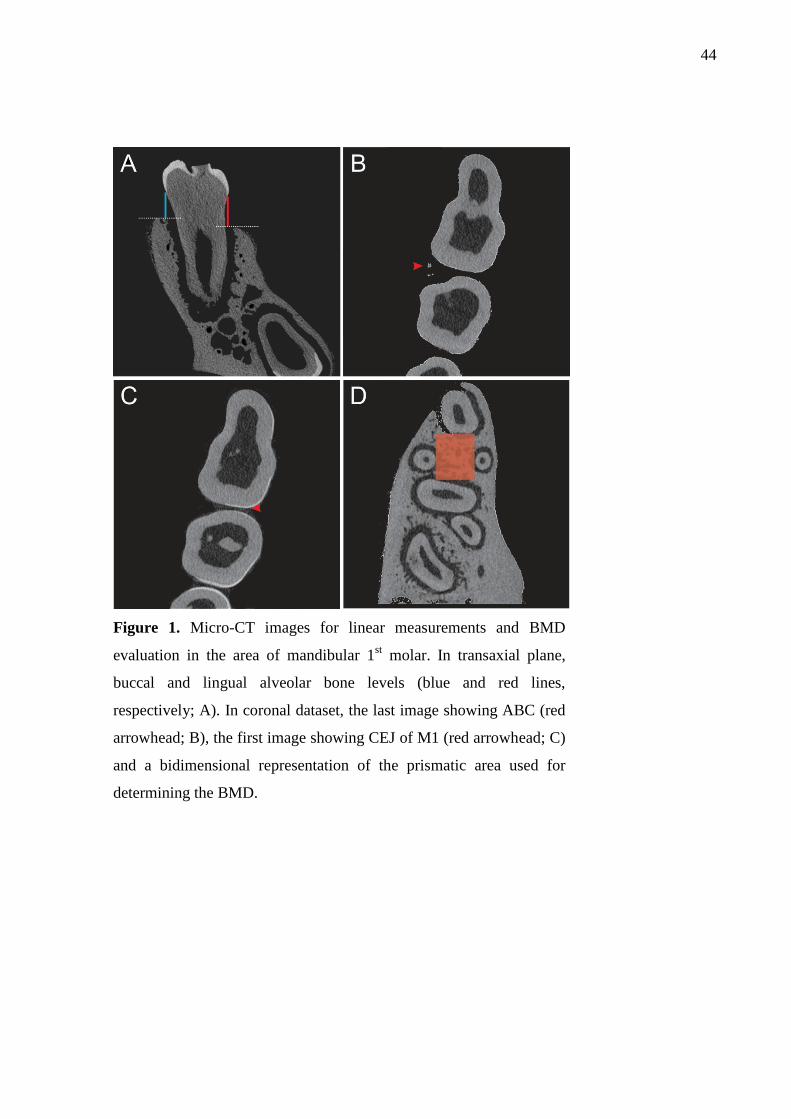
44
Figure 1. Micro-CT images for linear measurements and BMD
evaluation in the area of mandibular 1st molar. In transaxial plane,
buccal and lingual alveolar bone levels (blue and red lines,
respectively; A). In coronal dataset, the last image showing ABC (red
arrowhead; B), the first image showing CEJ of M1 (red arrowhead; C)
and a bidimensional representation of the prismatic area used for
determining the BMD.

45
Figure 2. Micro-CT analyses. Means and standard deviations of the
ABL, with comparisons among groups, at lingual (A), buccal (B) and
interproximal (C) sites, and of the BMD assessment (D). Same letters
indicate no significant differences among groups (ANOVA, Tukey,
p<0.05). Abbreviations: C, control; EA, electroacupuncture; EP,
experimental periodontitis.

46
Figure 3. Photomicrographs of the periodontal tissues in the furcation areas of mandibular 1st molars.
Abbreviations and symbols: ab, alveolar bone; C, control; EA, electroacupuncture; EP, experimental
periodontitis; pdl, periodontal ligament; Hematoxylin and Eosin staining.

47
Figure 4. Histometric analysis. Means and
standard deviations of the ANBL of mandibular
1st molars, with comparisons among groups.
Same letters indicate no significant differences
among groups (ANOVA, Tukey, p<0.05).
Abbreviations: C, control; EA,
electroacupuncture; EP, experimental
periodontitis.

48
Figure 5. qRT-PCR analyses. Means and standard deviations of the
relative genic expression of IL-1β (A), MMP-8 (B), IL-6 (C), TNF-α
(D) and COX-2 (E), with comparison among groups. Same letters
indicate no significant differences among groups (ANOVA, Tukey,
p<0.05). Abbreviations: C, control; COX, cyclooxygenase; EA,
electroacupuncture; EP, experimental periodontitis; IL, interleukin;
MMP, matrix metalloproteinase; TNF, tumor necrosis factor.
49
4. CONCLUSÕES GERAIS
Dentro dos limites deste estudo, pode ser concluído que:
1. A aplicação de eletroacupuntura reduz a perda óssea alveolar na periodontite induzida
por ligadura em ratos;
2. O tratamento com eletroacupuntura diminui a magnitude do infiltrado inflamatório e a
destruição dos tecidos periodontais na periodontite experimental em ratos;
3. A eletroacupuntura reduz a expressão gênica de alguns mediadores pró-inflamatórios
na periodontite induzida por ligadura em ratos.

50
REFERÊNCIAS
1. Loe H, Theilade E, Jensen SB. Experimental Gingivitis in Man. The Journal of
Periodontology, v. 36, n., p. 177-187, 1965.
2. Lindhe J, Nyman S. The effect of plaque control and surgical pocket elimination on
the establishment and maintenance of periodontal health. A longitudinal study of
periodontal therapy in cases of advanced disease. Journal of Clinical
Periodontology, v. 2, n. 2, p. 67-79, 1975.
3. Socransky SS, Haffajee AD. The bacterial etiology of destructive periodontal disease:
current concepts. The Journal of Periodontology, v. 63, n. 4 Suppl, p. 322-331,
1992.
4. Socransky SS, Haffajee AD, Cugini MA, Smith C, Kent RL, Jr. Microbial complexes
in subgingival plaque. Journal of Clinical Periodontology, v. 25, n. 2, p. 134-144,
1998.
5. Page RC, Offenbacher S, Schroeder HE, Seymour GJ, Kornman KS. Advances in the
pathogenesis of periodontitis: summary of developments, clinical implications and
future directions. Periodontology 2000, v. 14, n., p. 216-248, 1997.
6. Seymour GJ, Gemmell E, Westerman B, Cullinan M. Periodontics into the 21st
century. Annals of the Royal Australasian College of Dental Surgeons, v. 13, n., p.
71-78, 1996.
7. Badran Z, Kraehenmann MA, Guicheux J, Soueidan A. Bisphosphonates in
periodontal treatment: a review. Oral Health & Preventive Dentistry, v. 7, n. 1, p. 3-
12, 2009.
8. Axelsson P, Lindhe J. Effect of controlled oral hygiene procedures on caries and
periodontal disease in adults. Results after 6 years. Journal of Clinical
Periodontology, v. 8, n. 3, p. 239-248, 1981.
9. Lindhe J, Nyman S. Scaling and granulation tissue removal in periodontal therapy.
Journal of Clinical Periodontology, v. 12, n. 5, p. 374-388, 1985.
10. Kalpidis CD, Ruben MP. Treatment of intrabony periodontal defects with enamel
matrix derivative: a literature review. The Journal of Periodontology, v. 73, n. 11, p.
1360-1376, 2002.

51
11. Reddy MS, Geurs NC, Gunsolley JC. Periodontal host modulation with antiproteinase,
anti-inflammatory, and bone-sparing agents. A systematic review. Annals of
Periodontology, v. 8, n. 1, p. 12-37, 2003.
12. Serhan CN. Clues for new therapeutics in osteoporosis and periodontal disease: new
roles for lipoxygenases? Expert Opinion on Therapeutic Targets, v. 8, n. 6, p. 643-
652, 2004.
13. Kantarci A, Hasturk H, Van Dyke TE. Host-mediated resolution of inflammation in
periodontal diseases. Periodontology 2000, v. 40, n., p. 144-163, 2006.
14. Kavoussi B, Ross BE. The neuroimmune basis of anti-inflammatory acupuncture.
Integrative Cancer Therapies, v. 6, n. 3, p. 251-257, 2007.
15. Park HJ, Chae Y, Kim JW, Lee H, Chung JH. Effect of acupuncture on hypothalamic-
pituitary-adrenal system in maternal separation rats. Cellular and molecular
neurobiology, v. 31, n. 8, p. 1123-1127, 2011.
16. Vixner L, Martensson LB, Stener-Victorin E, Schytt E. Manual and
electroacupuncture for labour pain: study design of a longitudinal randomized
controlled trial. Evidence-based Complementary and Alternative Medicine, v.
2012, id. 943198, 2012.
17. Baek YH, Huh JE, Lee JD, Choi do Y, Park DS. Antinociceptive effect and the
mechanism of bee venom acupuncture (Apipuncture) on inflammatory pain in the rat
model of collagen-induced arthritis: Mediation by alpha2-Adrenoceptors. Brain
Research, v. 1073-1074, n., p. 305-310, 2006.
18. Lorenzini L, Giuliani A, Giardino L, Calza L. Laser acupuncture for acute
inflammatory, visceral and neuropathic pain relief: An experimental study in the
laboratory rat. Research in Veterinary Science, v. 88, n. 1, p. 159-165, 2010.
19. Napadow V, Makris N, Liu J, Kettner NW, Kwong KK, Hui KK. Effects of
electroacupuncture versus manual acupuncture on the human brain as measured by
fMRI. Human Brain Mapping, v. 24, n. 3, p. 193-205, 2005.
20. Toda S. Investigation of electroacupuncture and manual acupuncture on carnitine and
glutathione in muscle. Evidence-based Complementary and Alternative Medicine,
v. 2011, id. 297130, 2011.
21. National Institute of Health. Acupuncture. NIH Consensus Statement, v. 15, n. 5, p.
34, 1997.

52
22. Sui Y, Zhao HL, Wong VC, et al. A systematic review on use of Chinese medicine
and acupuncture for treatment of obesity. Obesity Reviews, v. 13, n. 5, p. 409-430,
2012.
23. Schulz KF, Altman DG, Moher D, Group C. CONSORT 2010 statement: updated
guidelines for reporting parallel group randomised trials. PLoS Medicine, v. 7, n. 3,
id. e1000251, 2010.
24. MacPherson H, Altman DG, Hammerschlag R, et al. Revised STandards for Reporting
Interventions in Clinical Trials of Acupuncture (STRICTA): extending the CONSORT
statement. PLoS Medicine, v. 7, n. 6, id. e1000261, 2010.
25. Carneiro ER, Xavier RA, De Castro MA, Do Nascimento CM, Silveira VL.
Electroacupuncture promotes a decrease in inflammatory response associated with
Th1/Th2 cytokines, nitric oxide and leukotriene B4 modulation in experimental
asthma. Cytokine, v. 50, n. 3, p. 335-340, 2010.
26. Lau WK, Lau YM, Zhang HQ, Wong SC, Bian ZX. Electroacupuncture versus
celecoxib for neuropathic pain in rat SNL model. Neuroscience, v. 170, n. 2, p. 655-
661, 2010.
27. da Silva MD, Guginski G, Werner MF, Baggio CH, Marcon R, Santos AR.
Involvement of Interleukin-10 in the Anti-Inflammatory Effect of Sanyinjiao (SP6)
Acupuncture in a Mouse Model of Peritonitis. Evidence-based Complementary and
Alternative Medicine, v. 2011, doi:10.1093/ecam/neq036, 2011.
28. Choi DC, Lee JY, Lim EJ, Baik HH, Oh TH, Yune TY. Inhibition of ROS-induced
p38MAPK and ERK activation in microglia by acupuncture relieves neuropathic pain
after spinal cord injury in rats. Experimental Neurology, v. 236, n. 2, p. 268-282,
2012.
29. Gondim DV, Araujo JC, Cavalcante AL, et al. CB1 and CB2 contribute to
antinociceptive and anti-inflammatory effects of electroacupuncture on experimental
arthritis of the rat temporomandibular joint. Canadian Journal of Physiology and
Pharmacology, v. 90, n. 11, p. 1479-1489, 2012.
30. Gondim DV, Costa JL, Rocha SS, Brito GA, Ribeiro Rde A, Vale ML.
Antinociceptive and anti-inflammatory effects of electroacupuncture on experimental
arthritis of the rat temporomandibular joint. Canadian Journal of Physiology and
Pharmacology, v. 90, n. 4, p. 395-405, 2012.

53
31. Geng WY, Liu ZB, Song NN, et al. Effects of electroacupuncture at Zusanli (ST36) on
inflammatory cytokines in a rat model of smoke-induced chronic obstructive
pulmonary disease. Journal of Integrative Medicine, v. 11, n. 3, p. 213-219, 2013.
32. Lan L, Tao J, Chen A, et al. Electroacupuncture exerts anti-inflammatory effects in
cerebral ischemia-reperfusion injured rats via suppression of the TLR4/NF-kappaB
pathway. International Journal of Molecular Medicine, v. 31, n. 1, p. 75-80, 2013.
33. Lu T, Luo Y, Sun H, Qin W, Li Y. Electroacupuncture improves behavioral recovery
and increases SCF/c-kit expression in a rat model of focal cerebral
ischemia/reperfusion. Neurological Sciences, v. 34, n. 4, p. 487-495, 2013.
34. Zhang RX, Lao L, Wang L, et al. Involvement of opioid receptors in
electroacupuncture-produced anti-hyperalgesia in rats with peripheral inflammation.
Brain Research, v. 1020, n. 1-2, p. 12-17, 2004.
35. Son YS, Park HJ, Kwon OB, Jung SC, Shin HC, Lim S. Antipyretic effects of
acupuncture on the lipopolysaccharide-induced fever and expression of interleukin-6
and interleukin-1 beta mRNAs in the hypothalamus of rats. Neuroscience Letters, v.
319, n. 1, p. 45-48, 2002.
36. Su TF, Zhao YQ, Zhang LH, et al. Electroacupuncture reduces the expression of
proinflammatory cytokines in inflamed skin tissues through activation of cannabinoid
CB2 receptors. European Journal of Pain, v. 16, n. 5, p. 624-635, 2012.
37. Kim HW, Uh DK, Yoon SY, et al. Low-frequency electroacupuncture suppresses
carrageenan-induced paw inflammation in mice via sympathetic post-ganglionic
neurons, while high-frequency EA suppression is mediated by the sympathoadrenal
medullary axis. Brain Research Bulletin, v. 75, n. 5, p. 698-705, 2008.
38. Matthay MA, Ware LB. Can nicotine treat sepsis? Nature Medicine, v. 10, n. 11, p.
1161-1162, 2004.
39. Gallowitsch-Puerta M, Tracey KJ. Immunologic role of the cholinergic anti-
inflammatory pathway and the nicotinic acetylcholine alpha 7 receptor. Annals of the
New York Academy of Sciences, v. 1062, n., p. 209-219, 2005.
40. Chung WY, Zhang HQ, Zhang SP. Peripheral muscarinic receptors mediate the anti-
inflammatory effects of auricular acupuncture. Chinese Medicine, v. 6, n. 1, p. 3-10,
2011.
41. Chen L, Zhang J, Li F, et al. Endogenous anandamide and cannabinoid receptor-2
contribute to electroacupuncture analgesia in rats. The Journal of Pain, v. 10, n. 7, p.
732-739, 2009.

54
42. Wang Q, Peng Y, Chen S, et al. Pretreatment with electroacupuncture induces rapid
tolerance to focal cerebral ischemia through regulation of endocannabinoid system.
Stroke, v. 40, n. 6, p. 2157-2164, 2009.
43. Pagotto U, Marsicano G, Cota D, Lutz B, Pasquali R. The emerging role of the
endocannabinoid system in endocrine regulation and energy balance. Endocrine
Reviews, v. 27, n. 1, p. 73-100, 2006.
44. Jhaveri MD, Richardson D, Chapman V. Endocannabinoid metabolism and uptake:
novel targets for neuropathic and inflammatory pain. British Journal of
Pharmacology, v. 152, n. 5, p. 624-632, 2007.
45. Nakajima Y, Furuichi Y, Biswas KK, et al. Endocannabinoid, anandamide in gingival
tissue regulates the periodontal inflammation through NF-kappaB pathway inhibition.
FEBS Letters, v. 580, n. 2, p. 613-619, 2006.
46. Qian H, Zhao Y, Peng Y, et al. Activation of cannabinoid receptor CB2 regulates
osteogenic and osteoclastogenic gene expression in human periodontal ligament cells.
Journal of Periodontal Research, v. 45, n. 4, p. 504-511, 2010.
47. Haruta C. Effects of anandamide on IL-11 production through the TRPV1 of human
periodontal ligament cells. Kokubyo Gakkai Zasshi, v. 79, n. 1, p. 7-14, 2012.
48. Napimoga MH, Benatti BB, Lima FO, et al. Cannabidiol decreases bone resorption by
inhibiting RANK/RANKL expression and pro-inflammatory cytokines during
experimental periodontitis in rats. International Immunopharmacology, v. 9, n. 2, p.
216-222, 2009.
49. Ossola CA, Surkin PN, Pugnaloni A, Mohn CE, Elverdin JC, Fernandez-Solari J.
Long-term treatment with methanandamide attenuates LPS-induced periodontitis in
rats. Inflammation Research, v. 61, n. 9, p. 941-948, 2012.
50. Rettori E, De Laurentiis A, Zorrilla Zubilete M, Rettori V, Elverdin JC. Anti-
inflammatory effect of the endocannabinoid anandamide in experimental periodontitis
and stress in the rat. Neuroimmunomodulation, v. 19, n. 5, p. 293-303, 2012.
51. Rawal SY, Dabbous M, Tipton DA. Effect of cannabidiol on human gingival
fibroblast extracellular matrix metabolism: MMP production and activity, and
production of fibronectin and transforming growth factor beta. Journal of
Periodontal Research, v. 47, n. 3, p. 320-329, 2012.
52. Kozono S, Matsuyama T, Biwasa KK, et al. Involvement of the endocannabinoid
system in periodontal healing. Biochemical and Biophysical Research
Communications, v. 394, n. 4, p. 928-933, 2010.

55
53. Bab I, Ofek O, Tam J, Rehnelt J, Zimmer A. Endocannabinoids and the regulation of
bone metabolism. Journal of Neuroendocrinology, v. 20 Suppl 1, n., p. 69-74, 2008.
54. Johansson A, Wenneberg B, Wagersten C, Haraldson T. Acupuncture in treatment of
facial muscular pain. Acta Odontologica Scandinavica, v. 49, n. 3, p. 153-158, 1991.
55. Rosted P. Introduction to acupuncture in dentistry. British Dental Journal, v. 189, n.
3, p. 136-140, 2000.
56. Lao L, Bergman S, Langenberg P, Wong RH, Berman B. Efficacy of Chinese
acupuncture on postoperative oral surgery pain. Oral Surgery, Oral Medicine, Oral
Pathology, Oral Radiology, and Endodontics, v. 79, n. 4, p. 423-428, 1995.
57. Thayer ML. The use of acupuncture in dentistry. Dental Update, v. 34, n. 4, p. 244-
246, 249-250, 2007.
58. Schoor RS, Sussman HI, Kazandjian GK. Acupuncture: a unique effort to treat
periodontal disease. Journal of the American Dental Association, v. 132, n. 12, p.
1705-1706, 2001.
59. Wu YT, Liu LA. Advances of clinical studies on acupuncture and moxibustion for
treatment of periodontitis. Zhongguo Zhen Jiu, v. 27, n. 8, p. 620-622, 2007.

56
ANEXO A