Embed Size (px)
Citation preview

i
UNIVERSIDADE ESTADUAL DE CAMPINAS
FACULDADE DE ODONTOLOGIA DE PIRACICABA
GUSTAVO HAUBER GAMEIRO
A INFLUÊNCIA DO ESTRESSE SOBRE A NOCICEPÇÃO
INDUZIDA NA ARTICULAÇÃO TEMPOROMANDIBULAR
Tese apresentada à Faculdade de
Odontologia de Piracicaba, da
Universidade Estadual de Campinas,
para obtenção do título de Doutor em
Odontologia, Área de Concentração em
Fisiologia Oral.
PIRACICABA -2006-

ii
UNIVERSIDADE ESTADUAL DE CAMPINAS FACULDADE DE ODONTOLOGIA DE PIRACICABA
GUSTAVO HAUBER GAMEIRO
A INFLUÊNCIA DO ESTRESSE SOBRE A NOCICEPÇÃO
INDUZIDA NA ARTICULAÇÃO TEMPOROMANDIBULAR
Tese apresentada à Faculdade de Odontologia de Piracicaba, da Universidade Estadual de Campinas, para obtenção do título de Doutor em Odontologia, Área de Concentração em Fisiologia Oral.
Orientadora:
Profª Dra. Maria Cecília Ferraz de Arruda Veiga
Banca Examinadora:
Prof. Dr. Carlos Alberto da Silva
Prof. Dr. Eduardo Dias de Andrade
Profª Dra. Maria José Alves da Rocha
Prof. Dr. Paulo Henrique Ferreira Caria
PIRACICABA
-2006-

iii
FICHA CATALOGRÁFICA ELABORADA PELA BIBLIOTECA DA FACULDADE DE ODONTOLOGIA DE PIRACICABA
Bibliotecário:Sueli Ferreira Julio de Oliveira– CRB-8a. / 2380
G145i
Gameiro, Gustavo Hauber. A influência do estresse sobre a nocicepção induzida na articulação temporomandibular. / Gustavo Hauber Gameiro. -- Piracicaba, SP : [s.n.], 2006. Orientador: Maria Cecília Ferraz de Arruda Veiga. Tese (Doutorado) – Universidade Estadual de Campinas, Faculdade de Odontologia de Piracicaba.
1. Estresse. 2. Articulação temporomandibular. 3. Dor facial.
I. Veiga, Maria Cecília Ferraz de Arruda. II. Universidade
Estadual de Campinas. Faculdade de Odontologia de
Piracicaba. III. Título.
(sfjo/fop)
Título em inglês: The influence of stress on nociception induced in temporomandibular joint. Palavras-chave em inglês (Keywords): 1. Stress. 2. Temporomandibular joint. 3. Facial pain. Área de concentração: Fisiologia Oral Titulação: Doutor em Odontologia Banca examinadora: Carlos Alberto da Silva, Eduardo Dias de Andrade, Maria José Alves da Rocha, Paulo Henrique Ferreira Caria. Data da defesa: 16/02/2006.


v
DEDICO ESTE TRABALHO...
À minha orientadora Professora Maria Cecília Ferraz de Arruda Veiga,
por me mostrar com seu amor e dedicação ao trabalho, o rumo que pretendo
seguir como professor.
“os professores ideais, são os que se fazem de pontes, que convidam os alunos a
atravessarem e depois, tendo facilitado a travessia, desmoronam-se com prazer,
encorajando-os a criarem suas próprias pontes.”
Nikos Kazantzakis
vi
AGRADECIMENTOS ESPECIAIS
A meus pais João Luis Gameiro e Mara Hauber Gameiro,
agradeço inicialmente pela vida.
Agradeço pela educação,
...pelo carinho
...pela confiança
...pelo apoio
...pela compreensão
...pelos exemplos de dignidade e honradez
...enfim, agradeço por esse amor incondicional que me
fizeram vencer mais um desafio.

vii
AGRADECIMENTOS ESPECIAIS
A Deus, que sempre ilumina e direciona os meus caminhos.
A Annicele Andrade, pela ajuda com os experimentos, e principalmente por estar junto a
mim em todos os momentos.
A minha irmã, a bióloga Paula Hauber Gameiro, pelo auxílio na parte mais difícil dos
experimentos (morte dos animais) e pela parceria e amizade de sempre.
Ao meu irmão Augusto Hauber Gameiro, minha cunhada Mariana Perozzi Gameiro e
minha querida afilhada Manoela Perozzi Gameiro, por serem minha família e meus
melhores amigos em Piracicaba.
A minha avó Rosália Hauber e a minha segunda mãe Maria Luiza, que apesar da distância,
moram no meu coração.
viii
AGRADECIMENTOS ESPECIAIS
Às agências de fomento brasileiras:
CNPq
pelo apoio financeiro para o desenvolvimento desta pesquisa, na concessão da
Bolsa de Doutorado.
FAPESP
pelo apoio financeiro para o desenvolvimento desta pesquisa, na concessão da Bolsa
Auxílio.

ix
AGRADECIMENTOS ESPECIAIS
Aos animais de laboratório, fundamentais para a realização deste trabalho.
"O respeito aos animais se dá a partir de atitudes éticas e tratamento digno de seres vivos
e não somente mencionando-os nos resultados das pesquisas."
COBEA - Colégio Brasileiro de Experimentação Animal

x
AGRADECIMENTOS
À Universidade Estadual de Campinas, na pessoa do seu Magnífico Reitor Prof. Dr. José
Tadeu Jorge; à Faculdade de Odontologia de Piracicaba, na pessoa do seu diretor Prof. Dr.
Thales Rocha de Mattos Filho, do Coordenador Geral da Pós-Graduação da FOP –
UNICAMP Prof. Dr. Pedro Luiz Rosalen, do Coordenador do programa de Pós-Graduação
em Odontologia da FOP-UNICAMP Prof. Dr. Francisco Carlos Groppo, pela oportunidade
de um crescimento científico e profissional nesta conceituada instituição.
Aos professores integrantes da banca examinadora desta tese: Prof. Dr. Carlos Alberto da
Silva, Profa. Dra. Maria José Alves da Rocha, Prof. Dr. Eduardo Dias de Andrade, Prof. Dr.
Paulo Henrique Ferreira Caria, Profa. Dra. Vânia Célia Vieira de Siqueira, Prof. Dr. Franco
Arsati e Profa. Dra. Ynara Bosco de Oliveira Lima Arsati, pela avaliação e colaboração em
nosso trabalho.
Ao Laboratório de Endocrinologia da Faculdade de Medicina de Ribeirão Preto-USP, na
pessoa da Profª. Drª. Margaret de Castro e Adriana Rossi, pela realização das dosagens
hormonais e colaboração no nosso trabalho.
À Prof ª Dr ª Gláucia Maria Bovi Ambrosano, por toda a atenção e ajuda dispensada na
execução das análises estatísticas.
À Profª. Drª. Cínthia Pereira. Machado Tabchoury, pela boa vontade, atenção e pronta
ajuda em todos os momentos necessários. Á Profª. Drª. Maria Beatriz Duarte Gavião, pela
participação neste trabalho, pela orientação no estágio voluntário de atendimento às
crianças e pela freqüente assistência na elaboração de meus trabalhos de pesquisa.
Aos professores Dr. Darcy Flávio Nouer, Drª. Maria Beatriz Magnani Araújo, Drª. Vânia
Célia Vieira de Siqueira e Prof. Dr. João Sarmento, pela atenção e recepção durante minha
estada na área de Ortodontia.

xi
Á Profª. Drª. Fernanda Klein Marcondes e Profª. Drª. Cláudia Herrera Tambeli, professoras
da disciplina de Fisiologia da FOP, UNICAMP.
Aos meus “bruxos” Luciano Pereira, Leonardo Bonjardim e Maximiliano Cenci, pelo
apoio, companheirismo e sincera amizade durante toda a caminhada.
Às amigas Ana Paula Tanno, Tatiana Cunha, Mariana Arthuri, Cristiana Tengan e Paula
Castelo, pelos bons momentos de descontração, divertimento e, principalmente, pela
carinhosa amizade, cujo valor não tem preço.
Aos meus novos colegas e amigos Ricardo, Ana Zilda, Viviane e Vanessa. Espero que
nossa união perdure e favoreça o crescimento de todos.
À aluna de iniciação científica Lígia Ferrinho Pereira, pelo empenho, dedicação e ajuda. Ao técnico Carlos Alberto Feliciano, pela colaboração, paciência e disposição durante a
utilização dos laboratórios da fisiologia.
Às secretárias Eliete, Elisa, Elisabete e Nilmes, sempre prestativas e atenciosas.
Aos Funcionários da Biblioteca da FOP – UNICAMP, pela orientação e ajuda.
Aos companheiros na Fisiologia Fabrício, Gérson, Vander, Marília, Kátia, Rose, Luciane,
Maria Cláudia, Juliana, Caroline, Luana e Vanessa.
A todos meus amigos e familiares, avós e avôs, tios e tias, primos e primas, sogro e sogra,
cunhados, enfim, a todos vocês que são fundamentais na minha formação. Obrigado
pelas orações, pelo carinho e pela força. A todos que direta ou indiretamente contribuíram
para a realização deste trabalho.
Meus sinceros agradecimentos.

xii
SUMÁRIO RESUMO 1 ABSTRACT 2 I – INTRODUÇÃO 3 II – PROPOSIÇÃO 6 III – CAPÍTULOS 7
Artigo 1: How do stressful experiences contribute to the development of orofacial pain? 8 Artigo 2: The effects of acute and chronic restraint stress on nociceptive responses induced by formalin injected in rat’s TMJ 31 Artigo 3: Nociception- and anxiety-like behavior in rats submitted to different periods of restraint stress 50
IV- CONCLUSÕES 70 V- REFERÊNCIAS BIBLIOGRÁFICAS 71 ANEXOS 75 APÊNDICE 79

1
RESUMO
Estudos recentes têm investigado o papel dos fatores psicológicos nas desordens
temporomandibulares (DTM). Entretanto, os mecanismos responsáveis pelas alterações
nociceptivas induzidas pelo estresse não estão bem estabelecidos. Desta maneira, o objetivo
deste estudo foi avaliar os efeitos do estresse agudo, sub-crônico e crônico sobre a
nocicepção induzida pela injeção de formalina na articulação temporomandibular (ATM)
de ratos. Foi avaliada a relação entre os níveis sangüíneos de adrenocorticotropina (ACTH),
corticosterona, os níveis de ansiedade e as respostas nociceptivas registradas após os
diversos protocolos de estresse. Os animais foram inicialmente submetidos a uma sessão de
estresse agudo por contenção (15 min; 30min e 1h), ou expostos a um estresse sub-crônico
(3 dias–1h/dia) ou crônico (40 dias–1h/dia). Logo depois, os animais foram (1) mortos
imediatamente para coleta de sangue e mensuração hormonal por radioimunoensaio; ou (2)
submetidos ao teste do labirinto em cruz elevado para avaliação da ansiedade; ou (3)
submetidos ao teste da formalina na ATM para avaliação da nocicepção. Finalmente, foi
avaliado o papel do sistema serotoninérgico e opióide nas alterações nociceptivas induzidas
pelo estresse. Para isso, um inibidor seletivo da recaptação de serotonina (fluoxetina 10
mg/Kg) e um agonista opióide (morfina 1-5 mg/Kg) foram administrados antes da
realização dos ensaios de nocicepção. Os resultados mostraram que todos protocolos de
estresse aumentaram significativamente os níveis de ACTH ou corticosterona, bem como o
comportamento de ansiedade. Em relação à nocicepção, os animais cronicamente
estressados apresentaram aumento nas respostas nociceptivas (hiperalgesia). Nesse grupo
ocorreu redução do efeito analgésico da morfina, indicando disfunção do sistema opióide
endógeno. A fluoxetina teve efeito analgésico tanto no grupo estressado (hiperalgésico)
quanto no grupo controle (não-estressado), porém o efeito foi maior no grupo estressado.
Concluiu-se que a hiperalgesia induzida pelo estresse resultou das alterações nos sistemas
serotoninérgicos e opióides, as quais podem explicar, pelo menos em parte, a importante
ligação entre estresse e dor orofacial.
Palavras-chave: Estresse, Articulação temporomandibular, Dor facial

2
ABSTRACT
Recent studies have investigated he role of psychological factor in temporomandibular
disorders (TMD). However, the mechanisms responsible for nociceptive changes induced
by stress are not established. Thus, the aim of this study was to evaluate the effect of acute,
sub-chronic and chronic stress on nociception induced by formalin injection in rats’
temporomandibular joint (TMJ). The relation beetwen blood levels of adrenocorticotropin
(ACTH), corticosterone, the levels of anxiety and nociceptive responses recorded after the
various stress protocols was evaluated. Animals were initially submitted to one session of
acute restraint stress (15 min; 30 min and 1 h), or exposed to sub-chronic stress (3 days-
1h/day) or chronic stress (40 days-1h/day). After, animals were (1) killed immediately to
collect blood for hormonal determinations by radioimmunoassay; or (2) submitted to the
elevated plus-maze to evaluate anxiety; or (3) submitted to the TMJ formalin test to
evaluate nociception. Finally, the role of serotoninergic and opioid systems in nociceptive
changes induced by stress was evaluated. For this, the serotonin-selective reuptake inhibitor
(fluoxetine 10 mg/Kg) and the opioid agonist (morphine 1-5 mg/Kg) were administered
before the nociception tests. The results showed that all stress protocols increased
significantly the levels of ACTH or corticosterone, as well as the anxiety behavior. In
relation to nociception, the chronic stressed animals showed an increase in nociceptive
responses (hyperalgesia). In this group, there was a reduction in the morphine analgesic
effects, suggesting dysfunction in the endogenous opioid system. Fluoxetine had an
analgesic effect in both stressed (hyperalgesic) and control groups (non-stressed), although
the effect was more significant in the stressed-group. It was concluded that stress-induced
hyperalgesia may result from changes in the serotoninergic and opioid systems, which can
explain, at least in part, the important link between stress and orofacial pain.
Keywords: Stress, Temporomandibular joint, Facial pain

3
I. INTRODUÇÃO
Inúmeras investigações têm examinado a relação entre estresse psicológico e
desordens temporomandibulares (DTM) (Grzesiak, 1991; Vanderas, 1994; Wexler & Steed,
1998). Foi observado que pessoas expostas a situações estressantes estão sob maior risco de
ocorrência e progressão de DTM (Speculand et al., 1984), e pacientes com disfunção
relatam que seus sintomas aumentam durante eventos estressantes (Suvinen et al., 1997). O
efeito do estresse nas funções do sistema estomatognático ocorre por meio de complexas
inter-relações no sistema nervoso central. Interação entre o sistema límbico e o centro de
atividade motora permite a transformação de um processo emotivo e cognitivo em resposta
motora (Bullock & Rosedahl, 1992), que na área do sistema estomatognático manifesta-se
como aumento do tônus muscular. A tensão muscular que acompanha condições
emocionais estressantes é um importante fator etiológico para muitos problemas
disfuncionais e dolorosos (Parker, 1990). Além disso, a disfunção muscular induzida por
estresse pode secundariamente produzir alterações na articulação temporomandibular
(ATM), resultando em mudanças na biomecânica articular, microtraumas às cápsulas
articulares e meniscos e alterações na percepção de dor (Uhac et al., 2003).
A analgesia induzida por estresse tem sido demonstrada tanto em humanos
(Bandura et al., 1988; Droste et al., 1991) como em animais (Mogil et al., 1996;
Wiedenmayer & Barr, 2000; Lapo et al., 2002). Em 1977, Chesher e Chan demonstraram
que o choque nas patas (footshock) de camundongos produzia um efeito analgésico, o qual
era antagonizado pela naloxona, um antagonista de receptor opióide. O footshock mostrou
ser capaz de aumentar os níveis de peptídeos opióides endógenos (Akil et al., 1976).
Subseqüentemente, diversos estressores incluindo o footshock, natação, imobilização,
isolamento e restrição têm sido utilizados para o estudo da analgesia induzida por estresse.
Os efeitos analgésicos induzidos por estes estressores são comparados àqueles causados
pela morfina em doses de 5-10 mg/Kg, porém a duração desses efeitos é relativamente
menor, desaparecendo aproximadamente dentro de 30 minutos (Snow e Dewey, 1983;
Giradot & Holloway, 1984).

4
Embora os estudos anteriores tenham demonstrado os clássicos efeitos
analgésicos do estresse, muitas pesquisas relatam que determinadas condições
experimentais (estresse agudo e crônico) podem provocar hiperalgesia ao invés de
analgesia (Vidal & Jacob, 1982; Satoh et al., 1992; Quintero et al., 2000; Imbe et al.,
2004). Por exemplo, uma breve exposição a um estresse emocional, como a exposição a
novos ambientes, produz uma hiperalgesia imediata e transitória (Vidal & Jacob, 1982),
enquanto o estresse prolongado por contenção (40 dias) induz hiperalgesia que persiste por
até 28 dias após a suspensão do estresse crônico (Torres et al., 2003). Os mecanismos
relacionados à hiperalgesia de longa duração ainda não estão esclarecidos. É possível que
esse aumento de percepção aos estímulos dolorosos estejam relacionados a alterações no
eixo hipotálamo-hipófise-adrenal, nos receptores opióides ou em qualquer outro sistema
responsável pela resposta de estresse. A deficiência na transmissão serotoninérgica central
pode produzir sensibilização das vias de transmissão da dor, por isso o estresse crônico
pode estar associado a aumentos na sensibilidade dolorosa (Quintero et al., 2000).
As divergências em relação aos efeitos do estresse sobre a nocicepção ocorrem,
pelo menos em parte, devido ao fato de que a resposta de estresse depende de fatores como
a natureza, a intensidade e a duração do estímulo estressor (Terman et al., 1986). Além
disso, o estresse geralmente é acompanhado por estados emocionais, como a ansiedade e o
medo (Mechiel Korte & DeBoer, 2003). Muitos trabalhos têm demonstrado que as
alterações nos estados emocionais tanto de humanos (Barlow et al., 1996), como de animais
(King et al., 1996) podem alterar fortemente a reatividade à sensação dolorosa. Por isso, um
modelo experimental destinado ao estudo da relação entre dor e estresse precisa considerar
as diversas variáveis, fisiológicas, psicológicas e comportamentais envolvidas em uma
situação de estresse.
Nos estudos citados anteriormente, os testes utilizados para medir a nocicepção
consistiam na aplicação de estímulos nocivos fásicos a tecidos superficiais, como por
exemplo o tail-flick, no qual é determinado o tempo de latência para mover a cauda após a
aplicação do estímulo. Não existem modelos experimentais em animais sobre o efeito do
estresse em condições dolorosas profundas, as quais possuem características diferentes em
relação às dores provenientes de tecidos cutâneos (Sessle & Hu, 1990).

5
Considerando a relação existente entre estresse e crises de dor facial (Suvinen et
al., 1997) e também a capacidade do estresse em alterar a percepção e resposta à dor,
estudos sobre os mecanismos das alterações nociceptivas induzidas pelo estresse nas dores
profundas são relevantes para a pesquisa sobre a etiologia das desordens
temporomandibulares.

6
II-PROPOSIÇÃO
Os objetivos do presente trabalho foram:
Verificar o efeito do estresse agudo, sub-crônico e crônico sobre as
respostas comportamentais nociceptivas induzidas pelo teste da formalina na ATM
de ratos.
Avaliar a relação entre os diversos protocolos de estresse, os níveis
de ansiedade, os níveis sangüíneos de ACTH e corticosterona e as respostas
comportamentais nociceptivas induzidas pelo teste da formalina na ATM.
Avaliar a participação do sistema opióide e serotoninérgico nas
alterações nociceptivas induzidas por situações estressantes.
O presente estudo foi realizado em formato alternativo, conforme deliberação
da Comissão Central de Pós-graduação (CCPG) da Universidade Estadual de Campinas
(UNICAMP) nº 001/98.

7
III- CAPÍTULOS
Artigo 1
“How do stressful experiences contribute to the development of orofacial
pain?”. Este artigo foi submetido à publicação no periódico Clinical Oral Investigations.
Artigo 2
“The effects of acute and chronic restraint stress on nociceptive responses
induced by formalin injected in rat’s TMJ”. Este artigo foi publicado no periódico
Pharmacology Biochemistry and Behavior (Pharmacol Biochem Behav. 2005
Oct;82(2):338-44).
Artigo 3
“Nociception- and anxiety-like behavior in rats submitted to different periods of
restraint stress ”. Este artigo foi aceito para publicação no periódico Physiology &
Behavior.

8
Review article
How do stressful experiences contribute to the development of
orofacial pain? Gustavo Hauber Gameiro, Annicele da Silva Andrade and Maria Cecília Ferraz de Arruda
Veiga
Laboratory of Orofacial Pain, Department of Physiology, Faculty of Dentistry of Piracicaba, University of Campinas – Unicamp, Piracicaba, Brazil.
Corresponding Author: Gustavo Hauber Gameiro, Laboratory of Orofacial Pain, Department of Physiology, Faculty of Dentistry of Piracicaba, University of Campinas - Unicamp, Av. Limeira 901 C.P. 52, CEP 13414-900, Piracicaba, São Paulo, Brazil. Tel.: +55-19-34125212; fax.: +55-19-34125218. E-mail address: [email protected] (Gustavo H Gameiro) Contribution of each author: Gustavo Hauber Gameiro1 – literature review, data organization, technical assistance Annicele da Silva Andrade2-literature review, manuscript preparation Maria Cecília Ferraz de Arruda Veiga3- literature review, data organization, statistical analysis 1 Doctor in Physiology/Faculty of Dentistry of Piracicaba, University of Campinas 2 Post-Graduation Student/Faculty of Dentistry of Piracicaba, University of Campinas 3Doctor in Physiology/Faculty of Dentistry of Piracicaba, University of Campinas

9
Abstract: Temporomandibular disorders (TMD) comprise the most common cause of
chronic facial pain conditions, and they are often associated with somatic and psychological
complaints including fatigue, sleep disturbances, anxiety and depression. For many health
professionals, the subjectivity of pain experience is frequently neglected, even when the
clinic does not find any plausible biologic explanation for the pain. This strictly biomedical
vision of pain cannot be justified scientifically. The purpose of this study is to demonstrate,
by original articles from the literature and recent studies conducted in our own laboratory,
the biological processes by which psychological stress can be translated into the sensation
of pain and contribute to the development of TMD. The role of the hypothalamic-pituitary-
adrenal axis, the serotoninergic and opioid systems in the pathogenesis of facial pain is
exposed, including possible future therapeutic approaches. It is hoped that knowledge from
apparently disparate fields of dentistry, integrated into a multidisciplinary clinical approach
to TMD will improve diagnosis and treatment for this condition, through a clinical practice
supported by scientific knowledge.
Descriptors: stress, temporomandibular disorders, facial pain.
Running head: Oral Physiology- Orofacial Pain

10
Introduction
Temporomandibular disorders are musculoskeletal pain conditions characterized by
pain in the temporomandibular joint and/or the masticatory muscles [1]. The clinical
condition of TMD can also involve sounds during mandibular movement and limited
mandibular movement [2]. TMD pain is the commonest symptom that compels patients to
seek therapy. In the USA and Europe, chronic facial pain accounts for 40% of all chronic
pain problems [3, 4]. In Brazil, the prevalence of TMD symptoms is between 40 -60% [5,
6]. Although the underlying cause of TMD remains poorly understood, it is widely
recognized to be multifactorial, involving physiological, behavioural, and environmental
factors. In dental research, dental occlusion and Para functional activities were the two
etiologic factors that have received the most attention in epidemiological studies [7, 8]. The
etiologic role of malocclusion, jaw position and biomechanical factors has been questioned.
For example, various studies did not find association between occlusion and TMD (for
review, see [9, 10, 11]). When such association was present, some studies revealed that
occlusal factors were only weakly associated with TMD signs and symptoms [12, 13]. A
prospective investigation over two decades into signs and symptoms of temporomandibular
disorders indicates that a lateral forced bite between the retruded contact position and the
intercuspal contact position and a unilateral crossbite deserve further consideration as
possible local risk factors for development of TMD [13]. In relation to oral parafunctions,
some experimentally induced habits can cause pain, similar to that related by patients with
TMD [14, 15]. Although parafunctional clenching involves increased masticatory muscle
activation [16], which can sometimes evoke pain [17], bruxism activity was not always
11
correlated with TMD pain [18]. Moreover, there are people classified as bruxers, who did
not present history of pain in masticatory muscles [19, 20]. Therefore, it is difficult to
establish any direct relation to prove that parafunctional activities can really cause TMD.
On the other hand, Laskin was the first to suggest that the main factors responsible
for TMD are emotional instead of physical [21]. During the last decade, numerous
investigations have been devoted to understanding the relationship between psychological
stress and TMD [22, 23, 24]. Patients suffering from this condition report that their
symptoms increase during stressful situations [25]. De Leeuw et al. (1994) consider that
muscle dysfunction and accompanying pain are very often the result of stress induced
muscular hyperactivity [26]. Stress induced muscular dysfunction may induce secondary
changes in the temporomandibular joint (TMJ). Raised elevator tonus leads to increased
intra-articular pressure in TMJ and alteration in the normal biomechanics, resulting in
microtraumatic damage to the joint capsules and disk attachment. However, the studies that
investigate psychological factors present mixed results. Some investigators related
electromyographic changes in masticatory muscle baseline values between patients with
TMD and control individuals [27, 28, 29] while others did not find significant differences
in electromyographic activity baseline values between patients and controls [30, 31]. These
inconsistencies may be probably due to different methodologies used.
The authors believe that both physical and psychological factors contribute to the
onset and maintenance of TMD. The balance of these factors produces many individual
differences in the perception of pain. More important than to argue in support of the
supremacy of some etiologic factor (physical or psychological), is to understand to what

12
extent some factor is responsible, how it is involved and what can be done to alleviate the
suffering of TMD patients.
The purpose of this article is to demonstrate the biologic process by which stressful
experiences can influence pain perception, and thus, the development of TMD. The notion
of the physiologic and pathophysiologic manifestations of stress system is described,
including possible future therapeutic approaches.
Stress System - Physiology
Life, as a high-order dynamic equilibrium, is constantly in a state of threatened
homeostasis, or stress. Thus, the forces that disturb homeostasis, the stressors, are
counterbalanced by adaptive forces generated by the organism [32]. Both physical and
emotional stressors set into motion central and peripheral responses, designed to preserve
homeostasis [33]. Centrally, neural pathways are facilitated, which among other functions,
mediate arousal, vigilance, cognition, as well as appropriate aggression, with concurrent
inhibition of pathways that subserve vegetative functions, such as feeding and reproduction.
Peripheral changes occur principally to promote an adaptive redirection of energy. Thus,
oxygen and nutrients are directed to the central nervous system and the stressed body site
[34].
It has to be borne in mind that not all states of stress are noxious. Selye made it clear
when he coined the terms "eustress" and "distress". Hence, he believed that mild, brief, and
controllable states of challenged homeostasis could actually be perceived as pleasant or
exciting and could be positive stimuli to emotional and intellectual growth and
development - it is notable that stress system activation occurs during both feeding and

13
sexual activity, for example. Selye believed that it was the more severe and uncontrollable
situations of psychological and physical distress that led to frank disease states [35].
The central components of the stress system are located in the hypothalamus and the
brainstem and include the corticotropin-releasing hormone (CRH) and the locus ceruleus-
norepinephrine/autonomic sympathetic nervous systems [36]. The peripheral limbs of the
stress system are the hypothalamic-pituitary-adrenal (HPA) axis, together with the efferent
sympathetic/adrenomedullary system, and components of the parasympathetic system [32].
Central CRH and norepinephrine systems, together with peripheral secretion of large
amounts of glucocorticoids and catecholamines, affect virtually every cell in the body [35].
Moreover, the stress system also interacts with other major central nervous system (CNS)
elements, including the mesocorticolimbic dopaminergic system, the amygdala, the
hippocampus, and the arcuate nucleus proopiomelanocortin (POMC) neuronal system [35].
The orchestrated interplay of several neurotransmitter systems in the brain underlies the
characteristic phenomenology of behavioural, endocrine, visceral, autonomic, and immune
responses to stress. These neurotransmitters include CRH, arginine vasopressine (AVP),
opioid peptides, substance P, dopamine, serotonin, and norepinephrine. Therefore, an
explanation about the functions of the neurotransmitters and hormones involved in the
stress response is outside the scope of this article (for review, see Herman and Cullinan
(1997) [37]). It is important to emphasize that most of the molecules mediating stress
effects are the same as those associated with pain modulation (for review see Millan (2002)
[38]), so the ability of stressful experiences to alter pain transmission and perception is
obvious. Melzack postulated the existence of a pain neuromatrix [39] in which the
experience of pain is produced by multiple influences and comprises a widely distributed

14
neural network with input from the body's stress regulation systems, including the
hypothalamic-pituitary-adrenal (HPA) axis.
HPA axis - pathology
Dysregulation of the HPA has been demonstrated in several psychiatric stress-
related disorders, such as depression [40] and post-traumatic stress disorder [41], which
have a significantly higher prevalence among patients with TMD [42]. Stress system
dysregulation can be expressed either as hyperfunction or as hypofunction. HPA axis
hyperactivity occurs, for example, in melancholic depression [43], anorexia nervosa [44],
obsessive-compulsive disorder [45], panic anxiety [33], and chronic active alcoholism [46].
On the other hand, stress system hypoactivation, rather than sustained activation, in which
chronically reduced CRH secretion may result in pathologic hypoarousal, characterizes
conditions such as fibromyalgia [47], seasonal depression [48], atypical depression [49],
some forms of obesity [43] and the chronic fatigue syndrome [50]. In relation to TMD, it
would appear that most TMD patients show HPA axis hyperactivity. Geissler [51] used
biochemical evidence (urinary cortisol: creatinine ratios) to show that patients with TMD
have higher urinary cortisol than normal individuals and therefore are under greater
emotional stress. This study was carried out in patients who had been rendered free of pain
or had only residual discomfort, so the stress factor would thus be emotional rather than
pain-induced. Another recent study [52] indicated very high daytime cortisol levels in
patients with facial pain, surprisingly much higher than those seen in depression or in
fybromyalgia patients with generalized muscle pain [53]. It remains possible that facial

15
region pain represents a greater stimulus to HPA axis activation than pain elsewhere in the
body.
Considering that pain itself acts as a strong activation of the HPA axis [54], it is
possible that high levels of cortisol in TMD patients represent a physiologic response to
chronic stress, with pain as a potential stressor, associated with chronically increased CRH
or other HPA axis central mediators. Increased activation of the stress axis central
components may result in hyperalgesia [55].
The study of the mechanisms involved in the relationship between stress and pain
modulation in humans becomes more difficult, because of methodological, psychological,
and ethical problems. On the other hand, animal models of nociception are very useful to
understand the neural basis of the mechanisms involved in pain perception. The authors’
laboratory is using an animal model of nociception, the TMJ formalin test [56], to evaluate
the influence of stress on nociception induced by TMJ injury. The authors observed that
rats submitted to chronic restraint stress (2 months) showed an increase in nociceptive
responses, indicating that chronic stress could induce hyperalgesia [57]. The mechanism by
which chronic stress produces hyperalgesia is not clear. In fact, more than one mechanism
could be involved. The HPA axis is just one of the stress system biologic mediators. Next,
the role of the serotoninergic and opioid systems in stress-induced hyperalgesia will be
emphasized.
The role of serotoninergic system
Neurons that contribute to ascending nociceptive pathways involved in pain
sensation are inhibited by descending serotoninergic and noradrenergic fibres, respectively

16
[58, 59]. Changes in the central serotoninergic system activities might, at least partly,
explain the bidirectional changes in nociception (analgesia and hyperalgesia) seen after
different stress conditions. For example, after acute exposure to different types of adverse
psychological or physical stimuli, there is an increase in the extracellular concentrations of
serotonin in several brain regions, especially in the raphe magnus [60]. Conversely,
prolonged stress diminishes the efflux of serotonin in some brain structures known to be
activated by stress, such as the amygdala and the lateral septum [61]. The magnitude of
tonic inhibition of pain transmission within the spinal cord horn appears to be dependent on
the behavioural state of the organism (depressed mood, anxiety, fear) [62]. The authors
suggested that anxiety and stress can cause a deficit in the central serotoninergic
transmission, which produces a sensitization of central pain relay pathways. First, stress
was induced in rats by immobilization for 1 h (acute stress) or 2 months (chronic stress).
This method is efficient to increase hormonal levels, as was detected by plasma
corticosterone and ACTH determination by radioimmunoassay [57]. Next, the authors’ test
to evaluate nociception in the TMJ was used, as previously described [63]. Briefly, the rats
received a 50 µl injection of diluted formalin (1.5 %) into the left TMJ region. The
injections were given via a 30-gauge needle introduced into the TMJ capsule. After the
TMJ injection, the rat was placed in the test chamber and nociceptive behavioural
responses, characterized by rubbing the orofacial region (seconds) and flinching the head
(number of times), were quantified for 30 min. A selective reuptake inhibitor, fluoxetine,
was used to block the stress-induced hyperalgesia. Actually, fluoxetine administered 30
min before formalin had an analgesic effect analogous to that of morphine, observed in one
17
of the authors’ studies [64]. These results are also consistent with correlational studies
indicating that anxiety is related to increased pain reports in clinical settings [65,66].
Schreiber [67] found that fluoxetine relieved low back pain with efficacy similar to
that of amitriptyline, and they suggested that fluoxetine could be an alternative for patients
unable to tolerate tricyclic antidepressant side effects. The authors question the possibility
of generalizing experimental findings to clinical settings, that is to say, it is too early to
affirm that fluoxetine could be effective for treating TMD patients, even though some
studies related that 5-HT re-uptake inhibitors have been associated with tooth-clenching or
tooth-grinding [68]. Future studies should evaluate the possibility of dentists using
fluoxetine to treat TMD patients.
Opioid Modulation
A major advance in the conception of the neural pain processing has occurred in the
past decade. It has become clear that pain is not passively received by the nervous system,
but is filtered and controlled (modulated) even at the first sensory synapse, by complex
modulatory systems [38]. The existence of multiple pain-modulatory systems is used to
clarify the bewildering profile of clinical observations resulting from various pain
treatments. A major component of these systems is the intrinsic opioid systems, which are
activated in stress situations and can diminish pain sensation [69]. For example, Maixner et
al. (1990) [70] have shown that ischemic pain induced in the left arm was able to reduce
pain sensation in patients suffering from acute dental pain. One important question is
whether these endogenous inhibitory systems are functional in patients suffering from
chronic facial pain. It is possible that chronic orofacial pain associated with TMD results
18
from diminished inhibitory systems in the central nervous systems. There is also evidence
to support this idea. For example, 70 to 80% of TMD patients suffer from psychosomatic
diseases, such as ulcers, headache, low back pain, asthma and dermatitis [21, 71]. The
biochemical' contents of psychological and physiological stress are elevated in TMD
patients when compared with controls [51, 52], suggesting that individuals with TMD are
really under greater emotional stress than control individuals.
The authors’ data from an experimental TMJ pain model indicate that endogenous
inhibitory systems may be less effective under chronic stress conditions. The authors results
demonstrate that repeatedly stressed rats display decreased morphine effects on nociception
compared with non-stressed controls in the TMJ formalin test [57]. The tolerance of
response to morphine observed in the authors study agrees with the hypothesis suggested
by previous studies that chronic stress could modify opioid system activities (for review,
see Drolet et al. (2001) [72]).
Conclusions and Future Therapeutic directions
Many patients with chronic facial pain improve with antidepressants, whether or not
they have a comorbid depressive disorder [73, 3]. Antidepressants have the ability to
modulate HPA axis activity and increase glucocorticoid receptors, though the mechanism
by which this occurs is still unknown [74]. In view of the involvement of the HPA axis in
depression and the deleterious effects of prolonged high cortisol levels, research into
potential treatments of mood and pain disorders has focused on modulating the effects of

19
hypercortisolemia. A promising approach is the use of corticotropin-releasing hormone
antagonists and there are several trials under way to test these agents in a variety of
psychiatric disorders including depression. Another possibility is the use of glucocorticoid
receptor antagonists to block any detrimental effects of the raised levels of circulating
cortisol and also cause a compensatory up-regulation of glucocorticoid receptor number
[75].
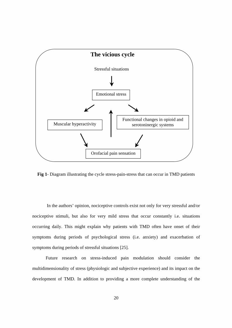
The authors concluded that the influence of stress on TMD is not as simple as
suggested according to Laskin's theory, in which the stress evokes chronic recurrent
muscular hyperactivity that progressively damages the joint, which in time becomes
symptomatic [21]. The authors propose that stress can profoundly affect the biological
processes of pain transmission and perception. Thus, inappropriate adaptational responses
could be maladaptive and act as stressors themselves (orofacial pain is a strong stressor),
feeding into a sustained vicious cycle. (fig.1).
20
Fig 1- Diagram illustrating the cycle stress-pain-stress that can occur in TMD patients
In the authors’ opinion, nociceptive controls exist not only for very stressful and/or
nociceptive stimuli, but also for very mild stress that occur constantly i.e. situations
occurring daily. This might explain why patients with TMD often have onset of their
symptoms during periods of psychological stress (i.e. anxiety) and exacerbation of
symptoms during periods of stressful situations [25].
Future research on stress-induced pain modulation should consider the
multidimensionality of stress (physiologic and subjective experience) and its impact on the
development of TMD. In addition to providing a more complete understanding of the
Functional changes in opioid and serotoninergic systems
Emotional stress
Muscular hyperactivity
Orofacial pain sensation
The vicious cycle
Stressful situations

21
centrifugal control of pain, it is hoped that such information might suggest ways of
relieving pain by less invasive means. The theoretical framework for testing the hypothesis
that a dysregulation in the stress system can lead to TMD has been set in place, with the
potential for improved understanding, diagnosis, and treatment of these disorders.
References
[1] LeResche L, Mancl L, Sherman JJ, Gandara B, Dworkin SF. Changes in
temporomandibular pain and other symptoms across the menstrual cycle. Pain. 2003
Dec;106(3):253-61.
[2] Miller VJ, Karic VV, Myers SL, Bodner L. Following treatment of myogenous TMD
patients with the temporomandibular opening index: an initial report. J Oral Rehabil.
2003 Jun;30(6):668-70.
[3] Feinmann C. The Mouth, the Face and the Mind. Oxford: Oxford University Press,
1999.
[4] LeResche L. Epidemiology of orofacial pain. In: Lund JP, Lavigne GJ, Dubner R,
Sessle BJ, eds. Orofacial Pain, Chapter 2. Chicago: Quintessence Publications, 2001.
[5] Conti PC, Ferreira PM, Pegoraro LF, Conti JV, Salvador MC. A cross-sectional study
of prevalence and etiology of signs and symptoms of temporomandibular disorders in
high school and university students. J Orofac Pain. 1996 Summer;10(3):254-62.
[6] Pedroni CR, De Oliveira AS, Guaratini MI. Prevalence study of signs and symptoms of
temporomandibular disorders in university students. J Oral Rehabil. 2003
Mar;30(3):283-9.

22
[7] Magnusson T, Egermark I, Carlsson GE. A longitudinal epidemiologic study of signs
and symptoms of temporomandibular disorders from 15 to 35 years of age. J Orofac
Pain. 2000 Fall;14(4):310-9.
[8] Thilander B, Rubio G, Pena L, de Mayorga C. Prevalence of temporomandibular
dysfunction and its association with malocclusion in children and adolescents: an
epidemiologic study related to specified stages of dental development. Angle Orthod.
2002 Apr;72(2):146-54.
[9] Clark GT, Adler RC. A critical evaluation of occlusal therapy: occlusal adjustment
procedures. J Am Dent Assoc. 1985 May;110(5):743-50. Review.
[10] Seligman DA, Pullinger AG. The role of functional occlusal relationships in
temporomandibular disorders: a review. J Craniomandib Disord. 1991 Fall;5(4):265-79.
Review.
[11] Koh H, Robinson PG. Occlusal adjustment for treating and preventing
temporomandibular joint disorders. J Oral Rehabil. 2004 Apr;31(4):287-92. Review.
[12] Mohlin BO, Derweduwen K, Pilley R, Kingdon A, Shaw WC, Kenealy P.
Malocclusion and temporomandibular disorder: a comparison of adolescents with
moderate to severe dysfunction with those without signs and symptoms of
temporomandibular disorder and their further development to 30 years of age. Angle
Orthod. 2004 Jun;74(3):319-27.
[13] Magnusson T, Egermarki I, Carlsson GE. A prospective investigation over two
decades on signs and symptoms of temporomandibular disorders and associated
variables. A final summary. Acta Odontol Scand. 2005 Apr;63(2):99-109.

23
[14] Christensen L. Some effects of experimental hyperactivity of the mandibular
locomotor system in man. J Oral Rehabil 1975;2:169-178.
[15] Moss RA, Ruff MH, Sturgis ET. Oral behavioural patterns in facial pain, headache and
non-headache populations. Behav Res Ther. 1984;22(6):683-7.
[16] Glaros AG, Burton E. Parafunctional clenching, pain, and effort in
temporomandibular disorders. J Behav Med. 2004 Feb;27(1):91-100.
[17] Ahlberg K, Ahlberg J, Kononen M, Alakuijala A, Partinen M, Savolainen A.
Perceived orofacial pain and its associations with reported bruxism and insomnia
symptoms in media personnel with or without irregular shift work. Acta Odontol
Scand. 2005 Aug;63(4):213-7.
[18] Pergamalian A, Rudy TE, Zaki HS, Greco CM. The association between wear facets,
bruxism, and severity of facial pain in patients with temporomandibular disorders. J
Prosthet Dent. 2003 Aug;90(2):194-200.
[19] Lavigne GJ, Kato T, Kolta A, Sessle BJ. Neurobiological mechanisms involved in
sleep bruxism. Crit Rev Oral Biol Med. 2003;14(1):30-46. Review.
[20] Fujii T, Torisu T, Nakamura S. A change of occlusal conditions after splint therapy for
bruxers with and without pain in the masticatory muscles. Cranio. 2005 Apr;23(2):113-
8.
[21] Laskin DM. Etiology of the pain-dysfunction syndrome. J Am Dent Assoc. 1969
Jul;79(1):147-53
[22] Grzesiak R.C., 1991. Psychologic consideration in temporomandibular dysfunction.
Dental Clinics of North America 35, 339.

24
[23] Vanderas A.P., 1994. Relationship between craniomandibular dysfunction and
malocclusion in white children with and without unpleasant life events. Journal of Oral
Rehabilitation 21, 177.
[24] Wexler G.B.; Steed P.A., 1998. Psychological factors and temporomandibular
outcomes. Cranio:The Journal of Craniomandibular Practice 16, 72.
[25] Suvinen TI, Hanes KR, Gerschman JA, Reade PC. Psychophysical subtypes of
temporomandibular disorders. J Orofac Pain. 1997 Summer;11(3):200-5.
[26] De Leeuw JR, Steenks MH, Ros WJ, Lobbezoo-Scholte AM, Bosman F, Winnubst JA.
Multidimensional evaluation of craniomandibular dysfunction. I: Symptoms and
correlates. J Oral Rehabil. 1994 Sep;21(5):501-14.
[27] Kapel L, Glaros AG, McGlynn FD. Psychophysiological responses to stress in patients
with myofascial pain-dysfunction syndrome. J Behav Med. 1989 Aug;12(4):397-406.
[28] Mercuri LG, Olson RE, Laskin DM. The specificity of response to experimental stress
in patients with myofascial pain dysfunction syndrome. J Dent Res. 1979
Sep;58(9):1866-71.
[29] Rugh JD, Montgomery GT. Physiological reactions of patients with TM disorders vs
symptom-free controls on a physical stress task. J Craniomandib Disord. 1987
Winter;1(4):243-50.
[30] Yemm R. Temporomandibular dysfunction and masseter muscle response to
experimental stress. Br Dent J. 1969 Dec 2;127(11):508-10.
[31] Moss RA, Adams HE. Physiological reactions to stress in subjects with and without
myofascial pain dysfunction symptoms. J Oral Rehabil. 1984 May;11(3):219-32.

25
[32] Chrousos GP. Stressors, stress, and neuroendocrine integration of the adaptive
response. The 1997 Hans Selye Memorial Lecture. Ann N Y Acad Sci. 1998 Jun
30;851:311-35.
[33] Gold PW, Pigott TA, Kling MK, Kalogeras K, Chrousos GP: Basic and clinical studies
with corticotropin releasing hormone: implications for a possible role in panic disorder.
Psychiatr Clin North Am 11:327, 1988.
[34] Habib KE, Gold PW, Chrousos GP. Neuroendocrinology of stress. Endocrinol Metab
Clin North Am. 2001 Sep;30(3):695-728; vii-viii. Review.
[35] Chrousos GP, Gold PW. The concepts of stress and stress system disorders. Overview
of physical and behavioural homeostasis. JAMA. 1992 Mar 4;267(9):1244-52. Review.
Erratum in: JAMA 1992 Jul 8;268(2):200.
[36] Chrousos GP, Loriaux DL, Gold PW, eds. Mechanisms of Physical and Emotional
Stress. New York, NY: Plenum Press; 1988. Advances in Experimental Medicine and
Biology, vol 245.
[37] Herman JP, Cullinan WE. Neurocircuitry of stress: central control of the hypothalamo-
pituitary-adrenocortical axis. Trends Neurosci. 1997 Feb;20(2):78-84. Review.
[38] Millan MJ. Descending control of pain. Prog Neurobiol. 2002 Apr;66(6):355-474.
Review.
[39] Melzack R. From the gate to the neuromatrix. Pain. 1999 Aug; Suppl 6:S121-6.
Review.
[40] Ferrier IN. Disturbed hypothalamic-pituitary-adrenal axis regulation in depression:
causes and consequences. In: Montgomery SA, Corn TH, editors.

26
Psychopharmacology of depression. New York: Oxford University Press; 1994. p.47-
56.
[41] Yehuda R, Southwick SM, Nussbaum G, Wahby V, Giller EL Jr, Mason JW. Low
urinary cortisol excretion in patients with posttraumatic stress disorder. J Nerv Ment
Dis. 1990 Jun;178(6):366-9.
[42] Korszun A, Hinderstein B, Wong M. Comorbidity of depression with chronic facial
pain and temporomandibular disorders. Oral Surg Oral Med Oral Pathol Oral Radiol
Endod. 1996 Nov;82(5):496-500.
[43] Bernini GP, Argenio GF, Vivaldi MS, Del Corso C, Sgro M, Franchi F, Luisi M.
Effects of fenfluramine and ritanserin on prolactin response to insulin-induced
hypoglycemia in obese patients: evidence for failure of the serotoninergic system.
Horm Res. 1989;31(3):133-7.
[44] Kaye WH, Gwirtsman HE, George DT, Ebert MH, Jimerson DC, Tomai TP, Chrousos
GP, Gold PW. Elevated cerebrospinal fluid levels of immunoreactive corticotropin-
releasing hormone in anorexia nervosa: relation to state of nutrition, adrenal function,
and intensity of depression. J Clin Endocrinol Metab. 1987 Feb;64(2):203-8.
[45] Insel TR, Kalin NH, Guttmacher LB, Cohen RM, Murphy DL. The dexamethasone
suppression test in patients with primary obsessive-compulsive disorder. Psychiatry
Res. 1982 Apr;6(2):153-60.
[46] Wand GS, Dobs AS. Alterations in the hypothalamic-pituitary-adrenal axis in actively
drinking alcoholics. J Clin Endocrinol Metab. 1991 Jun;72(6):1290-5.

27
[47] Griep EN, Boersma JW, de Kloet ER. Altered reactivity of the hypothalamic-pituitary-
adrenal axis in the primary fibromyalgia syndrome. J Rheumatol. 1993 Mar;20(3):469-
74.
[48] Vanderpool J, Rosenthal N, Chrousos GP, et al: Evidence for hypothalamic CRH
deficiency in patients with seasonal affective disorder. J Clin Endocrionol Metab
72:1382, 1991.
[49] Gold PW, Chrousos GP: The endocrinology of melancholic and atypical depression:
Relation to neurocircuitry and somatic consequences. Proc Assoc Am Physicians
111:22-34, 1999.
[50] Demitrack MA, Dale JK, Straus SE, Laue L, Listwak SJ, Kruesi MJ, Chrousos GP,
Gold PW. Evidence for impaired activation of the hypothalamic-pituitary-adrenal axis
in patients with chronic fatigue syndrome. J Clin Endocrinol Metab. 1991
Dec;73(6):1224-34.
[51] Geissler PR. An investigation of the stress factor in the mandibular dysfunction
syndrome. J Dent. 1985 Dec;13(4):283-7.
[52] Korszun A, Young EA, Singer K, Carlson NE, Brown MB, Crofford L. Basal
circadian cortisol secretion in women with temporomandibular disorders. J Dent Res.
2002 Apr;81(4):279-83.
[53] Klerman EB, Goldenberg DL, Brown EN, Maliszewski AM, Adler GK. Circadian
rhythms of women with fibromyalgia. J Clin Endocrinol Metab. 2001 Mar;86(3):1034-
9.
[54] Pacak K, Palkovits M. Stressor specificity of central neuroendocrine responses:
implications for stress-related disorders. Endocr Rev. 2001 Aug;22(4):502-48. Review.

28
[55] Lariviere WR, Melzack R. The role of corticotropin-releasing factor in pain and
analgesia. Pain. 2000 Jan;84(1):1-12. Review.
[56] Roveroni RC, Parada CA, Cecilia M, Veiga FA, Tambeli CH. Development of a
behavioural model of TMJ pain in rats: the TMJ formalin test. Pain. 2001
Nov;94(2):185-91.
[57] Gameiro GH, Andrade Ada S, de Castro M, Pereira LF, Tambeli CH, Veiga MC. The
effects of restraint stress on nociceptive responses induced by formalin injected in rat's
TMJ. Pharmacol Biochem Behav. 2005 Oct;82(2):338-44. Epub 2005 Oct 6.
[58] Wei F, Dubner R, Ren K. Nucleus reticularis gigantocellularis and nucleus raphe
magnus in the brain stem exert opposite effects on behavioural hyperalgesia and spinal
Fos protein expression after peripheral inflammation. Pain. 1999 Mar;80(1-2):127-41.
Erratum in: Pain 1999 May;81(1-2):215-9.
[59] Wei F, Dubner R, Ren K. Laminar-selective noradrenergic and serotoninergic
modulation includes spinoparabrachial cells after inflammation. Neuroreport. 1999 Jun
3;10(8):1757-61.
[60] Adell A, Casanovas JM, Artigas F. Comparative study in the rat of the actions of
different types of stress on the release of 5-HT in raphe nuclei and forebrain areas.
Neuropharmacology. 1997 Apr-May;36(4-5):735-41.
[61] Kirby LG, Allen AR, Lucki I. Regional differences in the effects of forced swimming
on extracellular levels of 5-hydroxytryptamine and 5-hydroxyindole acetic acid. Brain
Res. 1995 Jun 5;682(1-2):189-96.
[62] Mason P, Gao K. Raphe magnus serotoninergic neurons tonically modulate
nociceptive transmission. Pain Forum 1998;7:143-150.

29
[63] Gameiro GH Arthuri MT, Tambeli CH, Veiga MCFA. Effects of ethanol on deep pain
evoked by formalin injected in TMJ of rat. Life Sci. 2003 Nov 14;73(26):3351-61.
[64] Gameiro GH, Gameiro PH, Andrade AS, Arthuri MT, Pereira LF, Marcondes FK,
Veiga MCFA. “Nociception- and anxiety-like behavior in rats submitted to different
periods of restraint stress”. Physiology & Behavior (in press).
[65] Passchier J, Verheij R, Tulen JH, Timmerman L, Pepplinkhuizen L. Positive
associations between anticipatory anxiety and needle pain for subjective but not for
physiological measures of anxiety. Psychol Rep. 1992 Jun;70(3 Pt 2):1059-62.
[66] Palermo TM, Drotar D. Prediction of children's postoperative pain: the role of pre-
surgical expectations and anticipatory emotions. J Pediatr Psychol. 1996
Oct;21(5):683-98.
[67] Schreiber S, Vinokur S, Shavelzon V, Pick CG, Zahavi E, Shir Y. A randomized trial
of fluoxetine versus amitriptyline in musculo-skeletal pain. Isr J Psychiatry Relat Sci.
2001;38(2):88-94.
[68] Gerber PE, Lynd LD. Selective serotonin-reuptake inhibitor-induced movement
disorders. Ann Pharmacother. 1998 Jun;32(6):692-8. Review.
[69] Bodnar RJ, Klein GE. Endogenous opiates and behavior: 2003. Peptides. 2004
Dec;25(12):2205-56.
[70] Maixner W, Gracely RH, Zuniga JR, Humphrey CB, Bloodworth GR. Cardiovascular
and sensory responses to forearm ischemia and dynamic hand exercise. Am J Physiol.
1990 Dec;259(6 Pt 2):R1156-63.
[71] Rugh JD, Solberg WK. Psychological implications in temporomandibular pain and
dysfunction. Oral Sci Rev. 1976;7:3-30. Review.
30
[72] Drolet G, Dumont EC, Gosselin I, Kinkead R, Laforest S, Trottier JF. Role of
endogenous opioid system in the regulation of the stress response. Prog
Neuropsychopharmacol Biol Psychiatry. 2001 May;25(4):729-41. Review.
[73] Feinmann C. Psychogenic facial pain: presentation and treatment. J Psychosom Res.
1983;27(5):403-10.
[74] McQuade R, Young AH. Future therapeutic targets in mood disorders: the
glucocorticoid receptor. Br J Psychiatry. 2000 Nov;177:390-5. Review.
[75] Contoreggi C, Rice KC, Chrousos G. Nonpeptide corticotropin-releasing hormone
receptor type 1 antagonists and their applications in psychosomatic disorders.
Neuroendocrinology. 2004;80(2):111-23. Epub 2004 Oct 27. Review.

Gameiro et al. / Pharmacology Biochemistry and Behavior (2005)
31
Capítulo 2
The effects of restraint stress on nociceptive responses induced by formalin injected in rat’s TMJ
Gustavo Hauber Gameiroa, Annicele da Silva Andradea, Margaret de Castrob , Lígia
Ferrinho Pereiraa, Cláudia Herrera Tambelia, and Maria Cecília Ferraz de Arruda Veigaa
aLaboratory of Orofacial Pain, Department of Physiology, Faculty of Dentistry of Piracicaba, University of Campinas – Unicamp, Piracicaba, Brazil. bDivision of Endocrinology, Department of Internal Medicine, School of Medicine of Ribeirão Preto, University of São Paulo-USP, Ribeirão
Preto, Brazil. Corresponding Author: Gustavo Hauber Gameiro, Laboratory of Orofacial Pain, Department of Physiology, Faculty of Dentistry of Piracicaba, University of Campinas - Unicamp, Av. Limeira 901 C.P. 52, CEP 13414-900, Piracicaba, São Paulo, Brazil. Tel.: +55-19-34125212; fax.: +55-19-34125218. E-mail address: [email protected] (G.H. Gameiro)
Abstract
It has been reported that stress can alter nociception from superficial tissues, such as
skin and subcutaneous region. However, the influence of stress on an experimental deep
nociception model is not understood. In this study, the temporomandibular joint (TMJ)
formalin test was used to evaluate the effects of acute and chronic restraint stress on
nociceptive responses in rats. Animals were initially submitted to one session of acute restraint
stress (1 h) or exposed to chronic stress (40 days-1h/day). Then, animals were killed
immediately to collect blood for hormonal determinations by radioimmunoassay, or
submitted to the TMJ formalin test to evaluate nociception. Rats submitted to acute restraint
presented a performance similar to unstressed controls in the TMJ formalin test, whereas
chronically stressed rats showed an increase in nociceptive responses. After 40 days of
restraint, morphine was injected i.p. (1, 5 mg/Kg or saline). The stressed rats displayed
decreased morphine effects on nociception compared to unstressed controls. These findings
suggest that repeated stress can produce hyperalgesia, which is, at least in part, due to
alterations in the activity of opioid systems. This model may help elucidate the underlying
neural mechanisms that mediate the effects of repeated stress on orofacial pain.
Keywords: Stress; Hyperalgesia; Formalin test; Temporomandibular joint; nociception

Gameiro et al. / Pharmacology Biochemistry and Behavior (2005)
32
Introduction
Different effects upon the nociceptive response have been observed with exposure
to acute and chronic stress in rats (Vidal and Jacob, 1982; Watkins et al., 1982; Bodnar,
1986; Kavaliers and Innes, 1992; Quintero et al., 2000). Acute exposure to a variety of
stressors produces an immediate analgesia in several pain tests (Lewis et al., 1980; Urca et
al., 1985; Terman et al., 1986; Vacarino and Kastin, 2001). Some studies, although, have
reported that under some experimental conditions both acute and chronic stress can elicit
hyperalgesia instead of analgesia (Satoh et al., 1992; Quintero et al., 2000, Quintero et al.,
2003, Imbe et al., 2004). Repeated exposure to a cold environment (4oC for 30 min every
hour for 1 day) induces 3-day long mechanical hyperalgesia (Satoh et al., 1992). One hour
restraint a day for 40 days produces thermal hyperalgesia, which persists for at least 28
days after suspension of the chronic treatment (Torres et al., 2003a). Finally, repeated non-
noxious swim-stress (10-20 min a day for 3 days) elicits a delayed (after 24-48 h) and long-
lasting (8-9 days) thermal and chemical cutaneous hyperalgesia (Quintero et al., 2000).
Mechanisms regulating stress-induced changes in nociception include alterations in:
endogenous opioid (Lewis et al., 1980; Przewlocki et al., 1987; Amit and Galina, 1988;
Yamada and Nabeshima, 1995), serotoninergic (Quintero et al., 2000), adenosinergic
(Torres et al., 2003b) and noradrenergic systems (Watkins and Mayer, 1982), as well as the
hypothalamic-pituitary-adrenal (HPA) axis (Bodnar et al., 1979).
Although the precise mechanisms involved in the development of hyperalgesia
observed after repeated stress are not well known, there are strong evidences that they
could be related, at least in part, to alterations in the central or peripheral opioid activity
(Gamaro et al., 1998; Torres et al., 2001a). The absence of novelty-induced antinociception,
which has been attributed to opioid activation (Netto et al., 1987; Siegfried et al., 1987), in
chronic stressed animals supports this theory. Therefore, one of the aims of the present
work is to verify the effect of chronic restraint stress on morphine-induced antinociception,
as measured by the TMJ formalin test.
The formalin test has been used to evaluate the effect of stressful stimuli in
numerous experimental animal models, such as swim stress in mice (Carmody and Cooper,
Gameiro et al. / Pharmacology Biochemistry and Behavior (2005)
33
1987; Vaccarino et al., 1992) and the exposure to a cat odour in rats (Lester and Fanselow,
1985). Our understandings of the influence of stress on nociception are largely based on
experimental models of nociception in animals (Le Bars et al., 2001). Most of these models
of nociception measure the output responses induced by superficial stimuli, for example
tail-flick (Gamaro et al., 1998), hot-plate (King et al., 2003) and formalin injected in the
paw (Aloisi et al., 1998). It is important to point out that deep pain conditions differ from
the one evoked by superficial stimuli. There are different sensory disturbances in pain
conditions involving deep tissues rather than cutaneous tissues (Sessle and Hu, 1990).
Many deep craniofacial pain conditions, such as TMJ pain, are associated with
manifestations of pain spread and referral (Sessle, 2002). Indeed, TMJ inflammation results
in more robust changes in central nervous system when compared to perioral inflammation
(Iwata et al., 1999). It is, nevertheless, poorly understood due in part to the limited options
of experimental models available for the investigation of this condition.
Thus, considering that the nociceptive behavioral responses elicited by the injection
of formalin into the TMJ represent a valid and reliable model of orofacial deep pain
(Roveroni et al., 2001), the aim of this study was to evaluate the effects of acute and
chronic restraint stress on the nociceptive responses induced by TMJ formalin test.
Methods
Animals:
Male Wistar rats (weighing 200-230 g at the beginning of experiment) obtained
from Centro Multi-disciplinar de Bioterismo-Cemib, UNICAMP, Campinas, Brazil were
used in this study. The rats were housed in groups of five and maintained in a temperature-
controlled room (23 ± 1oC) with a 12/12 light-dark cycle (lights on at 7:00 AM) and food
and water were available ad libitum. Rats were adapted to the testing apparatus and handled
prior to behavioral testing. Procedures were performed between 08:00 and 15:00 h. The
study was conducted in accordance with the ethical guidelines for investigations of
experimental pain in conscious animals (Zimmermann, 1983).
Gameiro et al. / Pharmacology Biochemistry and Behavior (2005)
34
Stress exposure:
The animals were stressed by restraint 1 h daily, 5 days per week for 40 days in the
chronic model (Ely et al., 1997). In the acute model, there was a single exposure (Gamaro
et al., 1998). Restraint was carried out by placing the animal in a plastic restraint device
(adjustable in size depending on the animal's weight) for 1 h. The area of the tube could be
adjusted individually to each rat with a mobile inside wall and the tube was held firmly in
place with Velcro straps. There was a 1 cm hole in the far end for breathing. The control
group was not submitted to restraint. The immobilization procedure was carried out in a
separate quiet room between 10:00 and 12:00 h.
Hormonal assays:
Plasma corticosterone and ACTH levels were determined by radioimmunoassay
(RIA) after plasma extraction using ethanol or silic acid (Castro et al., 1995), respectively.
The rats were decapitated immediately after the last stress session and the whole blood was
collected. The time interval between the stress procedure and manipulations until sacrifice
were strictly maintained similar (30 sec.) among the different groups (acute restraint group
n=8; chronic restraint group n=8; acute control group n=8; chronic control group n=8).
Testing procedure for TMJ pain:
The design of this study follows that used by Roveroni et al. (2001). Testing
sessions took place between 08:00 and 15:00 h in a quiet room maintained at 23 ± 1oC.
Immediately after the period of stress procedures, each animal was lightly anesthetized by
inhalation of halothane to allow the TMJ injection.
Rats received a 50-µl injection of formalin diluted in saline (1.5 %) into the left
TMJ region. The injections were performed via a 30-gauge needle introduced into the TMJ
capsule. A cannula consisting of a polyethylene tube was connected to the needle and also
to a Hamilton syringe (50 µl) previously filled with formalin 1.5%.
Following the TMJ injection, the rat was placed in the test chamber (30 X 30 X 30
cm mirrored-wood chamber with glass at the front side) and nociceptive behavioral
responses characterized by rubbing the orofacial region (amount of time-seconds) and
Gameiro et al. / Pharmacology Biochemistry and Behavior (2005)
35
flinching the head (number of head flinches) were quantified for 30 min (10 blocks of 3
min). Considering that the flinching of the head behavior followed a uniform pattern of 1 s
in duration, each flinching was expressed as 1 s. The combination (sum) of both behaviors
provides a better measure of pain intensity than any single behavior (Roveroni et al., 2001;
Gameiro et al., 2003). An investigator, who was blind to the rat’s group assignment, made
the analysis of the behaviors.
At the end of each experiment, Evans blue dye (0.1%, 5 mg/Kg) was injected systemically
(via penile vein) in order to confirm the TMJ injection site at post-mortem, as previously
described (Hass, 1992) by the visual examination of formalin-induced plasma extravasation
of Evans blue dye bond to plasma protein.
Drug treatments:
In order to evaluate the role of endogenous opioids in nociceptive changes induced
by stress, one opioid antagonist (naloxone) and one agonist (morphine) were used. In
experiment 1, naloxone 10 mg/Kg (Vissers et al., 2004) was administered i.p. immediately
after the acute restraint stress (1h) and before the TMJ formalin test. In experiment 2, the
animals were submitted to chronic stress as described above. After 40 days of treatment
(control group was left undisturbed in their home cage), the rats were injected i.p. with
morphine 1.0 mg/Kg (Torres et al., 2003a), 5.0 mg/Kg (D’amato et al., 1999) or saline (n=
6/group) 30 min before the administration of formalin 1.5% into the TMJ. Morphine sulfate
was dissolved in 0.9% saline and administered i.p. immediately after the last stress session
in a volume of 1.0 ml/Kg.
Statistical analysis:
Statistical analysis of plasma corticosterone and ACTH data were performed using
Student’s t-test. Data were previously transformed to square-root or log, as indicated by the
program SAS (version 8.2 for windows). The sum of rubbing and flinching responses
exhibited by each animal was computed. The comparison between two groups was made by
Student’s t-test. The comparison of more than two groups (morphine effect analysis) was
made by two-way analysis of variance (ANOVA). All values are given as mean +/-

Gameiro et al. / Pharmacology Biochemistry and Behavior (2005)
36
standard error of the mean (SEM). A level of 5% was taken as evidence of statistical
significance. Data were analyzed using SAS (version 8.2 for windows) by Institute Inc.,
Cary, NC, USA-licensed to Universidade Estadual de Campinas.
Results
Effects of stress procedures on plasma corticosterone and ACTH levels:
This experiment was carried out to define the efficacy of restraint in inducing stress-
like hormonal modifications in the acute and chronic groups. There was a significant
increase in plasma corticosterone (p<0.0001, t-test, Fig. 1A) and ACTH levels (p=0.0011,
t-test, Fig. 1B) after a single restraint session for 1. The chronically stressed rats showed
higher levels of corticosterone than control animals (p=0.0261, t-test, Fig. 2A). However,
there was no difference in plasma ACTH levels between chronically stressed vs. control
rats (p=0.4134, t-test, Fig.2B).
Fig 1A. Plasma corticosterone level after a single restraint session (1 h). Each data point represents mean ± SEM from 8 rats. The vertical bars indicate the standard error of the means. Data were
analyzed using Student`s t-test. (*) Indicates significant difference compared with the control rats at p<0.0001. Fig 1B. Plasma ACTH level after a single restraint session (1 h). Each data point
represents mean ± SEM from 8 rats. The vertical bars indicate the standard error of the means. Data were analyzed using Student`s t-test. (*) Indicates significant difference compared with the control
rats at p=0.0011.
05
101520253035
1 2
Plas
ma
cort
icos
tero
ne (
g/dl
)
control acute stress
*A
0
50
100
150
200
1 2
Plas
ma
AC
TH (p
g/m
l)
control acute stress
*B

Gameiro et al. / Pharmacology Biochemistry and Behavior (2005)
37
Fig 2A. Plasma corticosterone level after the last session of chronic stress (8-week). Each data point represents mean ± SEM from 8 rats. The vertical bars indicate the standard error of the means. Data were analyzed using Student`s t-test. (*) Indicates significant difference compared with the control rats at p=0.0261. Fig 2B. Plasma ACTH level after the last session of chronic stress (8-week). Each data point represents mean ± SEM from 8 rats. The vertical bars indicate the standard error of the
means. Data were analyzed using Student`s t-test. There was no statistical difference between control and stressed groups (p=0.4134).
Effect of acute stress on nociceptive behavioral responses:
The exposure to a single restraint session for 1 h did not affect the nociceptive
responses evoked by formalin 1.5% injected in TMJ of rats (Fig.3). There was no statistical
difference (p=0.125) between the control group (non-stressed) and the stressed group.
Fig 3. Sum of flinching and rubbing behaviors recorded in formalin-treated animals (50 µl, 1.5%) previously submitted to 1 h of restraint (n=6) or left undisturbed in their home cage (n=6). Each
column represents the mean. Error bars indicate the SEM. No significant differences were found in nociceptive responses for control vs. stressed group (p=0.125, t-test).
0
5
10
15
1 2
Plas
ma
cort
icos
tero
ne (
g/dl
)
control Chronic stress
*
A
05
101520253035
1 2
Plas
ma
AC
TH (p
g/m
l)
control Chronic stress
B
050
100150200250300
1 2Sum
(Flin
chin
g +
Rub
bing
)
control acute stress

Gameiro et al. / Pharmacology Biochemistry and Behavior (2005)
38
0
50
100
150
200
250
300
1 2
Sum
(Flin
chin
g +
Rub
bing
)
control Chronic stress
*
Effect of chronic stress on nociceptive behavioral responses:
Results are shown in Fig. 4. Immediately after the last restraint session (1 h /40
days), the chronically-stressed animals were hyperalgesic. A statistically significant
increase in the nociceptive behavioral responses was observed in the stressed group when
compared with the control group (p<0.05, t-test).
Fig 4. Sum of flinching and rubbing behaviors recorded in formalin-treated animals (50 µl, 1.5%) previously submitted to chronic stress (n=6) or left undisturbed in their home cage (n=6).
Each column represents the mean. Error bars indicate the SEM. (*) Significant difference between the control and stressed group (p<0.05, t-test).
Effect of chronic restraint stress on rubbing spontaneous behaviors:
We also evaluated the spontaneous rubbing in order to exclude the possibility of an
increased motor behavior induced by the chronic stress procedure. The chronic stressed rats
exhibited a similar behavior than those of the control group (non-stressed) when saline was
administered in the rat’s TMJ (p=0.7488, Mann-Whitnet test, Fig.5).

Gameiro et al. / Pharmacology Biochemistry and Behavior (2005)
39
0
20
40
60
80
1 2Oro
faci
al ru
bbin
g (s
econ
ds)
control after chronic stress
Fig 5. Duration of the orofacial rubbing behavior in rats previously submitted to chronic stress (n=6) or left undisturbed in their home cage (n=6). Each column represents the mean. Error bars
indicate the SEM. There was no statistical difference between control and stressed groups (p=0.7488, Mann-Whitney test).
Effect of naloxone on nociception in rats submitted to acute restraint stress:
After one hour of immobilization, the injection of naloxone evoked an increase in
nociceptive behaviors (180,69 ± 45,29), when compared with saline (123,14 ± 16,53). The
increase in the sum of nociceptive behaviors (flinching + rubbing) was statistically
significant (p=0.0489, t-test, Fig.6).
Fig 6. Effects of naloxone or saline on formalin-treated animals (50 µl, 1.5%) previously submitted to acute restraint stress (n=6/group). Each column represents the mean. Error bars
indicate the SEM. (*) Indicates significant difference compared with the saline group (p=0.0011, t-test).
0
100
200
300
saline naloxone
After acute stress exposure
*

Gameiro et al. / Pharmacology Biochemistry and Behavior (2005)
40
Effect of morphine on nociception in repeatedly-stressed and control rats:
Results referring to the analgesic effect of morphine are shown in Fig. 7. ANOVA
revealed significant interaction between stress and morphine (p=0.003). Pos-hoc tests
(Tukey) revealed that morphine administration produced a significant reduction of
nociceptive behavioral responses in the control group (non-stressed). Morphine 1 mg/Kg
reduced the nociceptive responses 30 min after the administration (p<0.05), and morphine 5
mg/Kg also had this effect (p<0.05). In the stressed group, morphine had an effect only at
the dose of 5 mg/Kg (p<0.05) when compared to the saline group.
Fig 7. Sum of nociceptive responses to morphine (1 or 5 mg/Kg, i.p.) or saline after 40 days chronic restraint stress. Panel A: control groups (n=6/group); Panel B: stressed groups (n=6/group). Each column represents the mean. Error bars indicate the SEM. (*) Significant difference compared to
saline group (p<0.05, ANOVA + Tukey).
Discussion
A variety of environmental and/or stressful stimuli have been shown to elicit
analgesia, a phenomenon often referred to as stress-induced analgesia (SIA) (Amir and
Amit, 1978; Watkins et al., 1982; Furuta et al., 2003). In the present study, a single
exposure (1 h) to restraint stress did not reduce the nociceptive behavioral responses
evoked by nociceptive chemical stimulation (formalin 1.5%) of the rat’s TMJ. The ability
050
100150200250300
1 2 3Sum
(Flin
chin
g+R
ubbi
ng
saline morphine 1 morphine 5
control
A
* *
B
050
100150
200250300
1 2 3Sum
(Flin
chin
g+R
ubbi
ng)
stressed
saline morphine 1 morphine 5
*

Gameiro et al. / Pharmacology Biochemistry and Behavior (2005)
41
of the procedure to induce stress was confirmed by higher corticosterone and ACTH levels
in restraint rats than those of control rats. One effect of acute stress exposure is a reduction
of reflex responses that include tail or hinpaw withdrawal and licking in rats (Bodnar et al.,
1980; Lewis et al., 1980; Gamaro et al., 1998). Although most of these responses involve a
spinal-brain stem-spinal loop and appear to be purposeful, they do not depend upon cortical
processing of nociceptive signals that result in pain perception (Mauderli et al., 2000;
Vierck et al., 2002). King et al. 2003 showed that acute stress diminishes reflex responses
to nociceptive input while enhancing operant responding to the same stimuli (nociceptive
thermal stimuli), suggesting that stress induced hyporeflexia can coexist with stress induced
hyperalgesia. According to these findings, we speculate that a single restraint session did
not induce an analgesic effect on rats submitted to the TMJ formalin test, which evokes
nociceptive responses that have an organization different from those related to innate
reflexes, for example tail flick response that can be modulated directly at spinal levels
(King et al., 2003). Moreover, the absence of stress-induced analgesia in our model may be
related to the different site of formalin injection. As described in the introduction,
nociceptive response evoked by cutaneous stimuli differs from the one evoked by deep
stimuli. The discrepancy between nociception models in their susceptibility to modulation
by stress is evident not only in the present results, but also in the partial and transient
analgesic effects found in other studies employing the formalin test (Amir and Amit, 1979;
Fuchs and Melzack, 1996; Aloisi et al., 1998).
The increase in nociceptive behavioral responses produced by chronic restraint
stress has important implications in relation to other studies that have reported a
hyperalgesic effect after exposure to a variety of stressors (Satoh et al., 1992; Quintero et
al., 2000; Torres et al., 2003a,b). The present study confirmed the previously reported
results for nociceptive responses, using an experimental model for the study of nociception
from deep tissue injury: the TMJ pain. Although an extensive literature has reported the
relationship between stress and chronic facial pain (Grzesiak, 1991; Vanderas, 1994;
Korszun, 2002), little is known about the physiopathology of neural mechanisms that
mediates the effects of repeated stress on pain sensitivity and affective states. The
development of experimental models such as the present one may provide further

Gameiro et al. / Pharmacology Biochemistry and Behavior (2005)
42
information about the mechanisms involved in these painful conditions and may be used to
test the efficacy of drugs. In the current study, we were able to induce an increase in
nociceptive behaviors following a repeated restraint stress procedure. In agreement with our
results, previous studies have also found that chronic stress can elicit hyperalgesia rather
than hypoalgesia (Lewis et al., 1980; Quintero et al., 2000; Torres et al., 2003a,b). Previous
works have suggested that, when animals are repeatedly submitted to the same stressor,
some behavioral and physiological consequences of stress exposure are reduced
(habituation). For example, ACTH or corticosterone levels are reduced after repeated
exposure to the same stressor (Marti and Amario, 1998; Torres et al., 2001b), although
negative results have been reported (Dal-Zotto et al., 2000). In our model, corticosterone
and ACTH levels were reduced after the end of stress session in 8-week restraint rats.
However, the ability of the procedure to induce stress was confirmed by higher
corticosterone levels in 8-week restraint rats than those of control rats. We also evaluated
the spontaneous rubbing in order to exclude the possibility of an increased motor behavior
induced by the chronic stress procedure. The chronic stressed rats exhibited a similar
behavior than those of the control group (non-stressed) when saline was administered in the
rat’s TMJ. This result suggests the increase of flinching and rubbing behaviors is a
hyperalgesic effect induced by chronic stress. The mechanism trough which repeated stress
produces hyperalgesia is not clear; in fact, more than one mechanism could be involved.
Satoh et al., 1992 suggested that mechanical hyperalgesia induced by prolonged cold stress
involves peptide-containing primary afferents (substance-P and calcitonin-gene-related
peptide). Quintero et al., 2000 showed that the increased thermal and chemical nociception
observed after sub-chronic swimming stress might be mediated by changes in the activity
of the central serotoninergic system. Torres et al. 2003b suggested that repeated restraint
stress could induce an adaptative response in chronically stressed rats, which can lead to a
desensitization of adenosine receptors. In other study, Torres et al. 2003a also showed that
chronically stressed rats displayed decreased morphine effects on nociception.
In the last experiment, we tested control and repeatedly restrained rats injected with
morphine (1 and 5 mg/Kg) in the TMJ formalin test. Our results demonstrate that
repeatedly stressed rats display decreased morphine effects on nociception compared to

Gameiro et al. / Pharmacology Biochemistry and Behavior (2005)
43
non-stressed controls. Although it has been described that morphine induces analgesia in a
dose-related manner, in the present work it was not observed any difference between the
two doses of morphine administrated in the control group (non-stressed). This discrepancy
may be due to the different nociception assay used. We know that nociceptive transmission
and modulation are different even when distinct superficial nociceptive essays are used
(Fang and Proudfit, 1998). The stressed group needed an increased dose to show the classic
analgesic effect of morphine. This change in sensitivity to morphine may be result of
alterations in treatment-induced peptides release, i.e., persistent activation of opiate peptide
receptors by endogenous opioids released during restraint stress could lead to receptor
down-regulation, but it is possible that interactions with other released neurotransmitter
could induce these effects, for example, serotonin, glutamate, adenosine and other opioid
receptor systems have also been involved (Fitzgerald et al., 1996; Torres et al., 2003b). The
tolerance of response to morphine observed in the present study agrees with the hypothesis
suggested by previous studies that chronic restraint stress could modify the activity of
opioid systems (for review, see Drolet et al., 2001). Changes in the analgesic effect of
morphine observed in stressed rats might be due to alterations in central or peripheral
opioid receptors, both in their affinity or number, or these changes might be due to
alterations in other neuro-transmitter or hormonal systems able to interact with these
receptors. Omiya et al., 2000 showed that hypofunction of the supraspinal mu-opioid
receptor may explain the hyperalgesic effect of repeated cold stress loading in mice. Since
morphine exerts its antinociceptive effects primarily through mu-opiate receptor subtype,
the altered responses observed in animals submitted to TMJ formalin test after chronic
stress might be due to changes at the level of these receptors. Future studies should evaluate
the activity of the opioid receptors in this model. We suggest the influence of endogenous
opioids released during chronic stress on the development of tolerance to morphine
antinociceptive effects. This conclusion was based in the fact that restraint stress can
release endogenous opioids, as was observed by the effect of naloxone on the augment of
nociceptive responses in rats submitted to acute stress. In this case, it was expected that
acute stress would reduce formalin-induced nociception, a finding not observed in our
study. We believe that, in our model, the effects of endogenous opioids were

Gameiro et al. / Pharmacology Biochemistry and Behavior (2005)
44
counterbalanced by the enhance in pain perception evoked by stress-induced-anxiety.
Studies have shown that hyperalgesia is elicited by some experimental conditions
(Cornwall and Donderi, 1988; Al Absi and Rokke, 1991; Meagher et al., 1998). In our
laboratory, we have demonstrated that a single exposure to restraint stress (1 h) induced a
high level of anxiety in the elevated-plus-maze (data not shown). This factor could also be
determinant in the absence of stress-induced-analgesia. Continued research concerning the
mechanisms of stress-induced hyperalgesia may be relevant to the study of the etiology of
chronic pain disorders, like the temporomandibular disorder.
Acknowledgments
The authors thank Gláucia M. B. Ambrosano for statistical analysis. Thanks are due
to Adriana Rossi and José Roberto da Silva for technical assistance. This work was
supported by CNPq and FAPESP, Brazil.
References Al Absi M, Rokke PD. Can anxiety help us tolerate pain? Pain 1991;46:43-51. Aloisi, AM, Ceccarelli, I, Lupo, C. Behavioural and hormonal effects of restraint stress and formalin test in male and female rats. Brain Res Bull. 1998;47:57-62. Amir, S, Amit, Z. Endogenous opioid ligands may mediate stress-induced changes in the affective properties of pain related behavior in rats. Life Sci. 1978;23:1143-51. Amir, S, Amit, Z. The pituitary gland mediates acute and chronic pain responsiveness in stressed and non-stressed rats. Life Sci. 1979;24:439-48. Amit, Z, Galina, ZH. Stress induced analgesia plays an adaptive role in the organization of behavioral responding. Brain Res Bull. 1988;21:955-8. Bodnar, RJ. Neuropharmacological and neuroendocrine substrates of stress-induced analgesia. Ann N Y Acad Sci. 1986;467:345-60.

Gameiro et al. / Pharmacology Biochemistry and Behavior (2005)
45
Bodnar, RJ, Glusman, M, Brutus, M, Spiaggia, A, Kelly, DD. Analgesia induced by cold-water stress: attenuation following hypophysectomy. Physiol Behav. 1979;23:53-62. Bodnar, RJ, Kelly, DD, Brutus, M, Glusman, M. Stress-induced analgesia: neural and hormonal determinants. Neurosci Biobehav Rev. 1980;4:87-100. Carmody, J, Cooper, K. Swim stress reduces chronic pain in mice through an opioid mechanism. Neurosci Lett. 1987;74:358-63. Castro M, Figueiredo F, Moreira AC. Time-course of hypothalamic CRH and pituitary ACTH contents, and pituitary responsiveness to CRH stimulation after bilateral adrenalectomy. Horm Metab Res. 1995 Jan;27(1):10-5. Cornwall A, Donderi DC. The effect of experimentally induced anxiety on the experience of pressure pain. Pain 1988;35:105-113. Dal-Zotto, S, Marti, O, Armario, A.Influence of single or repeated experience of rats with forced swimming on behavioural and physiological responses to the stressor. Behav Brain Res. 2000;114:175-81. D'Amato, FR, Mazzacane, E, Capone, F, Pavone, F. Effects of postnatal manipulation on nociception and morphine sensitivity in adult mice. Brain Res Dev Brain Res. 1999;117:15-20. Drolet, G, Dumont, EC, Gosselin, I, Kinkead, R, Laforest, S, Trottier, JF.Role of endogenous opioid system in the regulation of the stress response. Prog Neuropsychopharmacol Biol Psychiatry. 2001;25:729-41. Ely, DR, Dapper, V, Marasca, J, Correa, JB, Gamaro, GD, Xavier, MH, Michalowski, MB, Catelli, D, Rosat, R, Ferreira, MB, Dalmaz, C. Effect of restraint stress on feeding behavior of rats. Physiol Behav. 1997;61:395-8. Fang, F. Proudfit, HK.. Antinociception produced by microinjection of morphine in the rat periaqueductal gray is enhanced in the foot, but not the tail, by intrathecal injection of α1-adrenoceptor antagonists. Brain Res. 1998;790(1-2), 14-24. Fitzgerald, LW, Ortiz, J, Hamedani, AG, Nestler, EJ. Drugs of abuse and stress increase the expression of GluR1 and NMDAR1 glutamate receptor subunits in the rat ventral tegmental area: common adaptations among cross-sensitizing agents. J Neurosci. 1996;16:274-82. Fuchs, PN, Melzack, R.Restraint reduces formalin-test pain but the effect is not influenced by lesions of the hypothalamic paraventricular nucleus. Exp Neurol. 1996;139:299-305.
Gameiro et al. / Pharmacology Biochemistry and Behavior (2005)
46
Furuta, S, Onodera, K, Kumagai, M, Honma, I, Miyazaki, S, Sato, T, Sakurada, S. Involvement of adenosine A1 receptors in forced walking stress-induced analgesia in mice. Methods Find Exp Clin Pharmacol. 2003;25:793-6. Gamaro, GD, Xavier, MH, Denardin, JD, Pilger, JA, Ely, DR, Ferreira, MB, Dalmaz C. The effects of acute and repeated restraint stress on the nociceptive response in rats. Physiol Behav. 1998;63:693-7. Gameiro, GH, Arthuri, MT, Tambeli, CH, Veiga, MCFA. Effects of ethanol on deep pain evoked by formalin injected in TMJ of rat. Life Sci. 2003;73:3351-61. Grzesiak, RC. Psychologic considerations in temporomandibular dysfunction. A biopsychosocial view of symptom formation. Dent Clin North Am. 1991;35:209-26. Haas, DA, Nakanishi, O, MacMillan, RE, Jordan, RC, Hu, JW. Development of an orofacial modelofacuteinflammationintherat. Arch Oral Biol. 1992;37:417-422. Imbe H, Murakami S, Okamoto K, Iwai-Liao Y, Senba E. The effects of acute and chronic restraint stress on activation of ERK in the rostral ventromedial medulla and locus coeruleus. Pain. 2004 Dec;112(3):361-71. Iwata, K, Tashiro, A, Tsuboi, Y, Imai, T, Sumino, R, Morimoto, T, Dubner, R, Ren, K. Medullary dorsal horn neuronal activity in rats with persistent temporomandibular joint and perioral inflammation. J Neurophysiol. 1999;82:1244-1253. Kavaliers, M, Innes, DG. Sex differences in the effects of Tyr-MIF-1 on morphine- and stress-induced analgesia. Peptides. 1992;13:1295-7. King, CD, Devine, DP, Vierck, CJ, Rodgers, J, Yezierski, RP. Differential effects of stress on escape and reflex responses to nociceptive thermal stimuli in the rat. Brain Res. 2003;987:214-22. Korszun A.Facial pain, depression and stress - connections and directions.J Oral Pathol Med. 2002;3:615-9. Le Bars, D, Gozariu, M, Cadden, SW. Animal models of nociception. Pharmacol Rev. 2001;53:597-652. Lester, LS, Fanselow, MS. Exposure to a cat produces opioid analgesia in rats. Behav Neurosci. 1985;99:756-9. Lewis, JW, Cannon, JT, Liebeskind, JC. Opioid and nonopioid mechanisms of stress analgesia. Science. 1980;208:623-5.

Gameiro et al. / Pharmacology Biochemistry and Behavior (2005)
47
Marti, O, Armario, A.Anterior pituitary response to stress: time-related changes and adaptation. Int J Dev Neurosci. 1998;16:241-60 Mauderli, AP, Acosta-Rua, A, Vierck, CJ.An operant assay of thermal pain in conscious, unrestrained rats. J Neurosci Methods. 2000;97:19-29. Meaguer MW, McLemore S, King TE, Grau JW. The generality of schock-induced hyperalgesia in rats. Soc Neurosci Abstracts 1998;24:1901. Netto, CA, Siegfried, B, Izquierdo, I. Analgesia induced by exposure to a novel environment in rats: effect of concurrent and post-training stressful stimulation. Behav Neural Biol. 1987;48:304-9. Omiya, Y, Goto, K, Ishige, A, Komatsu, Y. Changes in analgesia-producing mechanism of repeated cold stress loading in mice. Pharmacol Biochem Behav. 2000;65:261-6. Przewlocki, R, Lason, W, Hollt, V, Silberring, J, Herz, A. The influence of chronic stress on multiple opioid peptide systems in the rat: pronounced effects upon dynorphin in spinal cord. Brain Res. 1987;413:213-9. Quintero, L, Moreno, M, Avila, C, Arcaya, J, Maixner, W, Suarez-Roca, H. Long-lasting delayed hyperalgesia after subchronic swim stress. Pharmacol Biochem Behav. 2000;67(3):449-58. Quintero, L, Cuesta, MC, Silva, JA, Arcaya, JL, Pinerua-Suhaibar, L, Maixner, W, Suarez-Roca, H. Repeated swim stress increases pain-induced expression of c-Fos in the rat lumbar cord.Brain Res. 2003;965:259-68. Roveroni, RC, Parada, CA, Veiga MCFA, Tambeli, CH. Development of a behavioral model of TMJ pain in rats: the TMJ formalin test. Pain. 2001;94: 185-191. Satoh, M, Kuraishi, Y, Kawamura, M. Effects of intrathecal antibodies to substance P, calcitonin gene-related peptide and galanin on repeated cold stress-induced hyperalgesia: comparison with carrageenan-induced hyperalgesia. Pain. 1992;49:273-8. Sessle, BJ, Hu, JW. Mechanisms of pain arising from articular tissues. Can J Physiol Pharmacol.1990;69:617-626. Sessle BJ.Recent insights into brainstem mechanisms underlying craniofacial pain. Journal of Dental Education. 2002;66:108-112. Siegfried, B, Netto, CA, Izquierdo, I. Exposure to novelty induces naltrexone-reversible analgesia in rats. Behav Neurosci. 1987;101:436-8.

Gameiro et al. / Pharmacology Biochemistry and Behavior (2005)
48
Terman, GW, Morgan, MJ, Liebeskind, JC. Opioid and non-opioid stress analgesia from cold water swim: importance of stress severity.Brain Res. 1986;372:167-71. Torres, IL, Vasconcellos, AP, Silveira Cucco, SN, Dalmaz, C. Effect of repeated stress on novelty-induced antinociception in rats. Braz J Med Biol Res. 2001a;34:241-4. Torres, IL, Gamaro, GD, Silveira-Cucco, SN, Michalowski, MB, Correa, JB, Perry, ML, Dalmaz, C.Effect of acute and repeated restraint stress on glucose oxidation to CO2 in hippocampal and cerebral cortex slices. Braz J Med Biol Res. 2001b;34(1):111-6. Torres, ILS, Cucco, SN, Bassani, M, Duarte, MS, Silveira, PP, Vasconcellos, AP, Tabajara, AS, Dantas, G, Fontella, FU, Dalmaz, C, Ferreira, MB. Long-lasting delayed hyperalgesia after chronic restraint stress in rats-effect of morphine administration. Neurosci Res. 2003 a ;45:277-83. Torres, ILS, Bonan, CD, Crema, L, De Leon, Nunes, M, Battastini, AM, Sarkis, JJ, Dalmaz, C, Ferreira, MB. Effect of drugs active at adenosine receptors upon chronic stress-induced hyperalgesia in rats. Eur J Pharmacol. 2003b;481:197-201. Urca, G, Segev, S, Sarne, Y. Footshock-induced analgesia: its opioid nature depends on the strain of rat. Brain Res. 1985;329:109-16. Vaccarino AL, Kastin AJ. Endogenous opiates: 2000. Peptides. 2001 Dec;22(12):2257-328. Review. Vaccarino, AL, Marek, P, Liebeskind, JC. Stress-induced analgesia prevents the development of the tonic, late phase of pain produced by subcutaneous formalin. Brain Res. 1992;572:250-2. Vanderas AP. Relationship between craniomandibular dysfunction and malocclusion in white children with and without unpleasant life events. J Oral Rehabil. 1994;21:177-83. Vidal, C, Jacob, JJ. Stress hyperalgesia in rats: an experimental animal model of anxiogenic hyperalgesia in human. Life Sci. 1982;31:1241-4. Vierck, CJ, Acosta-Rua, A, Nelligan, R, Tester, N, Mauderli, A.Low dose systemic morphine attenuates operant escape but facilitates innate reflex responses to thermal stimulation. J Pain. 2002;3:309-19. Vissers KC, De Jongh RF, Crul BJ, Vinken P, Meert TF. Adrenalectomy affects pain behavior of rats after formalin injection. Life Sci. 2004 Jan 23;74(10):1243-51. Watkins, LR, Cobelli, DA, Faris, P, Aceto, MD, Mayer, DJ. Opiate vs non-opiate footshock-induced analgesia (FSIA): the body region shocked is a critical factor.Brain Res. 1982;242:299-308.

Gameiro et al. / Pharmacology Biochemistry and Behavior (2005)
49
Watkins, LR, Mayer, DJ. Organization of endogenous opiate and nonopiate pain control systems. Science. 1982;216:1185-92. Yamada, K, Nabeshima T. Stress-induced behavioral responses and multiple opioid systems in the brain. Behav Brain Res. 1995;67:133-45. Zimmermann M. Ethical guidelines for investigations of experimental pain in conscious animals. Pain 1983;16:109-110.

Nociception-and anxiety like behavior in rats
50
Capítulo 3 Nociception- and anxiety-like behavior in rats submitted to different
periods of restraint stress
Gustavo Hauber Gameiroa, Paula Hauber Gameirob, Annicele da Silva Andradea, Lígia Ferrinho Pereiraa, Mariana Trevisani Arthuria, Fernanda Klein Marcondesa and Maria
Cecília Ferraz de Arruda Veigaa
aDepartment of Physiological Sciences, Faculty of Dentistry of Piracicaba, State University of Campinas – Unicamp, Piracicaba, Brazil. bDepartment of Microbiology and Parasitology, Institute
of Biology-UFPEL, Pelotas, Brazil.
Corresponding Author: Maria Cecília Ferraz de Arruda Veiga, Laboratory of Orofacial
Pain, Departamento de Ciências Fisiológicas, Faculdade de Odontologia de Piracicaba,
Universidade Estadual de Campinas - Unicamp, Av. Limeira 901 C.P. 52, CEP 13414-900,
Piracicaba, São Paulo, Brasil.
Tel.: +55-19-34125212; fax.: +55-19-34125218.
E-mail address: [email protected] (Cecília Veiga);[email protected] (GH
Gameiro).
Abstract
The aim of this study was to evaluate the effect of acute, sub-chronic and chronic stress on
nociception induced by formalin injection in rats’ temporomandibular joint (TMJ). It was
evaluated the relation between blood levels of adrenocorticotropin, corticosterone, the
levels of anxiety and nociceptive responses recorded after different stress protocols.
Animals were initially submitted to acute restraint stress (15; 30 min and 1 h), or exposed
to sub-chronic (3 days-1h/day) or chronic stress (40 days-1h/day). Then, animals were (1)
killed immediately to collect blood for hormonal determinations; or (2) submitted to the
elevated plus-maze to evaluate anxiety; or (3) submitted to the TMJ formalin test to
evaluate nociception. It was also evaluated the role of serotoninergic and opioid systems in
nociceptive changes induced by stress. For this, the serotonin-selective reuptake inhibitor

Nociception-and anxiety like behavior in rats
51
(fluoxetine 10 mg/Kg) and the opioid agonist (morphine 1-5 mg/Kg) were administered
before the nociception test. All stress protocols significantly raised the levels of ACTH or
corticosterone, as well as the anxiety behavior. In relation to nociception, the chronic
stressed animals showed an increase in nociceptive responses (hyperalgesia). In this group,
there was a reduction in the morphine analgesic effects, suggesting dysfunction in the
endogenous opioid system. Fluoxetine had an analgesic effect in both stressed and control
groups, although this effect was more evident in the stressed group. It was concluded that
stress-induced hyperalgesia may result from changes in the serotoninergic and opioid
systems, which can explain, at least in part, the important link between stress and orofacial
pain.
Keywords: Stress, Anxiety, Temporomandibular disorders, Facial pain
Introduction
An extensive literature has shown that acute exposure to a variety of stressors produces an
immediate analgesia in several pain tests [1, 2, 3, 4]. Prolonged stress can also evoke
analgesia [5]. However, some studies have reported that under some experimental
conditions both acute and chronic stress can elicit hyperalgesia instead of analgesia. For
example, rats exposed to acute and chronic restraint stress exhibit elevation and reduction
of tail flick latencies, respectively [6]. Similarly, acute restraint stress reduced the duration
of lick/guard responses to nociceptive input (analgesic effect), while the same acute stress
for the same animals increased sensitivity to thermal stimulation, as assessed by learned
escape responses (hyperalgesic effect) [7]. Taken together, these results reveal that the
types of stressor, its intensity, duration, as well as the type of the nociceptive model used,
affect not only the potency of analgesic or hyperalgesic effect but also the neuronal
mechanisms responsible for them. The literature suggests that the stress-regulatory circuit
activated by a particular stressor is crucially dependent on stimulus attributes [for review,
see 8].
Nociception-and anxiety like behavior in rats
52
One factor that is particularly important is the emotional state induced by stress. For
example, anxiety can produce hypervigilance which should increase attention to pain in
human subjects, thereby amplifying its perceived intensity [9]. Recent work has shown that
temporomandibular disorders (TMD) patients show increased stress, depression, anxiety
and somatization compared with healthy controls [10, 11]. Many of the current treatments
for these diseases utilize drugs that increase the levels or activity of the biogenic amine
(e.g. serotonin, norepinephrine, dopamine) class of neurotransmitters. For example,
fluoxetine, a specific serotonin-reuptake inhibitor (SSRI) that blocks the activity of
serotonin transporter and increases the levels of 5-HT in the synaptic cleft, can be an
effective treatment for depression and anxiety [12]. Although recent studies have
investigated the role of psychological factors in TMD, the mechanisms responsible for
nociceptive changes induced by stress are not established. The existence of multiple pain-
modulatory systems is used to clarify the bewildering profile of clinical observation
resulting from various pain treatments. A major component of these systems is the intrinsic
opioid systems, which are activated in stress situations and can diminish pain sensation. For
example, Maixner et al. [13] have showed that ischemic pain induced in the left arm was
able to reduce pain sensation in patients suffering from acute dental pain. One important
question is if these endogenous inhibitory systems are functional in patients suffering from
chronic facial pain. It is possible that chronic orofacial pain associated with TMD result
from inhibitory systems diminished in the central nervous systems. The absence of novelty-
induced antinociception, which has been attributed to opioid activation [14, 15], in chronic
stressed animals supports this theory. Thus, considering that the nociceptive behavioral
responses elicited by the injection of formalin into the TMJ represent a valid and reliable
model of orofacial deep pain [16], one of the aims of the present work was to evaluate the
effects of different stress protocols on the nociceptive responses induced by TMJ formalin
test. The role of serotoninergic and opioid systems in nociceptive changes induced by stress
was also reported.

Nociception-and anxiety like behavior in rats
53
Methods
Animals:
Male Wistar rats (weighing 200-230 g at the beginning of experiment) obtained from
Centro Multidisciplinar de Investigação Biológica -Cemib, Unicamp, Campinas, Brazil
were used in this study. The rats were housed in groups of five and maintained in a
temperature-controlled room (23 ± 1oC) with a 12/12 light-dark cycle (lights on at 7:00 am)
and food and water were available ad libitum. Rats were adapted to the testing apparatus
and handled prior to behavioral testing. Procedures were performed between 08:00 am and
15:00 pm. The study was conducted in accordance with the ethical guidelines for
investigations of experimental pain in conscious animals [17]. This research was approved
by the institutional ethics committee in animal experimentation, according to the Brazilian
College of Experimentation Guidelines.
Stress exposure:
The animals were stressed by restraint during 15 min, 30 min or 1 h in the acute model. In
the sub-chronic model, animals were stressed by restraint 1 h daily, during 3 days. In the
chronic model, animals were stressed by restraint 1 h daily, 5 days per week for 40 days
[18]. The stress procedure in the chronic model consisted in 5 days of stress + 2 days of rest
until 60 days. Thus, the protocol finished when 40 stress days were summed up. This
protocol follows the design used by Gamaro et al., 1998 [19]. Restraint was carried out by
placing the animal in a plastic restraint device (adjustable in size depending on the animal's
weight) for 1 h. The area of the tube could be adjusted individually to each rat with a
mobile inside wall and the tube was held firmly in place with Velcro straps. There was a 1
cm hole in the far end for breathing. The control groups were not submitted to restraint and
were handled during the same time that their respective experimental groups. The control
rats were handled in a quiet room once every day (handling comprised picking up each rat
for a short period of time and then returning it to its home cage) during the days according
to experimental stress procedures: in the acute model (15;30;60 min), control rats was
handled just one time. In the sub-chronic model (3 days), control rats was handled (once a
day) for 3 days. In chronic model, control rats were handled (once a day) until 40 stress
Nociception-and anxiety like behavior in rats
54
days were summed up. The restraint procedure was carried out in a separate quiet room
between 10:00 and 12:00 am.
Hormonal assays:
Plasma corticosterone and ACTH levels were determined by radioimmunoassay (RIA) after
plasma extraction using ethanol or silic acid [20], respectively. The rats were decapitated
immediately after the last stress session and the whole blood was collected. The time
interval between the stress procedure and manipulations until sacrifice was strictly
maintained similar (30 sec.) among the different groups.
Evaluation of anxiety level:
The elevated plus-maze test was used to assess the anxiety level induced by different stress
protocols. The elevated plus-maze was made of wood, according to specifications described
in Morato and Brandão, 1997 [21]. The procedure was described elsewhere [22, 23].
Briefly, rats were placed in the central square facing a closed arm, and allowed to explore
the elevated plus-maze for 5 min. Before the next rat was introduced, the maze was cleaned
with a solution of 20% ethanol and dried. The conventional measures (percentage of open-
arm entries, and the time spent on open arms) were recorded. The experimental sessions
were recorded by a vertically mounted videocamera, linked to a monitor and VCR in an
adjacent room. Videotapes were analyzed by highly trained observers who remained blind
to treatment conditions. All tests were made immediately after the last stress session of the
various protocols (15, 30, 60 min., 3 days and 40 days-n=10/group).
Testing procedure for TMJ pain:
The design of this study follows that used by Roveroni et al. 2001 [16]. After the last stress
session, each animal was lightly anesthetized by inhalation of halothane to allow the TMJ
injection. Rats received a 50-µl injection of formalin diluted in saline (1.5 %) into the left
TMJ region. The injections were performed via a 30-gauge needle introduced into the TMJ
capsule. A cannula consisting of a polyethylene tube was connected to the needle and also
to a Hamilton syringe (50 µl) previously filled with formalin 1.5%.

Nociception-and anxiety like behavior in rats
55
Following the TMJ injection, the rat was placed in the test chamber (30 X 30 X 30 cm
mirrored-wood chamber with glass at the front side) and nociceptive behavioral responses
characterized by rubbing the orofacial region (amount of time-seconds) and flinching the
head (number of head flinches) were quantified for 30 min (10 blocks of 3 min).
Considering that the flinching of the head behavior followed a uniform pattern of 1 s in
duration, each flinching was expressed as 1 s in order to make simpler the quantification
and representation of nociceptive behaviors, as previously described [16]. Moreover, the
combination (sum) of both behaviors provides a better measure of pain intensity than any
single behavior [16, 24]. An investigator, who was blind to the rat’s group assignment,
made the analysis of the behaviors.
At the end of each experiment, Evans blue dye (0.1%, 5 mg/Kg) was injected systemically
(via penile vein) in order to confirm the TMJ injection site at post-mortem, as previously
described [25] by the visual examination of formalin-induced plasma extravasation of
Evans blue dye bond to plasma protein.
Drug treatments:
In order to evaluate the role of serotoninergic systems and endogenous opioids in
nociceptive changes induced by stress, the serotonin-selective reuptake inhibitor (fluoxetine
10 mg/Kg) and the opioid agonist (morphine 1-5 mg/Kg) were administered before the
nociception tests in some rats submitted to chronic stress as described above. Immediately
after the last stress session (control group was handled as described above), the rats were
injected i.p. with fluoxetine 10 mg/Kg [26], morphine 1.0 mg/Kg [27], 5.0 mg/Kg [28] or
saline (n= 6/group) 30 min before the administration of formalin 1.5% into the TMJ.
Morphine sulfate and fluoxetine hydrochloride (SIGMA) were dissolved in 0.9% saline and
administered i.p. immediately after the last stress session.
Statistical analyses:
Statistical analyses of plasma corticosterone and ACTH data were made using the Mann-
Whitney test (control vs. stressed) and Kruskal-Wallis (between stressed groups). The data
were previously transformed to square-root or log, as indicated by PROCLAB-program

Nociception-and anxiety like behavior in rats
56
SAS (version 8.2 for windows). Since collected data about anxiety behavior didn’t show
normal distribution, the percentage of open-arm entries (100 X open/total) and the time
spent in the open arms were calculated and analyzed by Mann-Whitney test. The sum of
rubbing and flinching responses exhibited by each animal was computed. The comparison
between two groups was made by Student’s t-test. The comparison of more than two
groups was made by two-way analysis of variance (ANOVA). The values for hormonal and
nociceptive assays are given as mean +/- standard error of the mean (SEM). The data of
anxiety behavior are expressed as median. A level of 5% was taken as evidence of
statistical significance. Data were analyzed using SAS (version 8.2 for windows) by
Institute Inc., Cary, NC, USA-licensed to Universidade Estadual de Campinas.
Results
Effects of stress procedures on plasma corticosterone and ACTH levels:
This experiment was carried out to define the efficacy of restraint in inducing stress-like
hormonal modifications. There was a significant increase in plasma corticosterone levels
after the various stress protocols used (Fig. 1; Mann-Whitney test, p<0.05). This increase
was lower after sub-chronic and chronic stress than after acute stress for 30 min (Fig.1;
Kruskal-Wallis, p<0.05).
Fig 1. Plasma corticosterone level after the various stress procedures. Each data point represents mean ± SEM from 8 rats. The vertical bars indicate the standard error of the means. Data were analyzed using Mann-Whitney test and Kruskal-Wallis. (*) p<0.05 compared to respective control groups. (⊗) p<0.05 compared to acute stress 30 min. (#) p<0.05 compared to acute stress 15 min.
0
10
20
30
40
50
1 2 3 4 5
Plas
ma
Corti
cost
eron
e (
g/dl
)
controlstressed
30 min15 min 1 h 3 days 40 days
**
** *
#⊗⊗

Nociception-and anxiety like behavior in rats
57
0
50100
150
200250
300
1 2 3 4 5
Plas
ma
AC
TH (p
g/m
l)
controlstressed
30 min15 min 1 h 3 days 40 days
* **
#♦⊗
#♦
0
10
20
30
40
50
1 2 3 4 5
% O
pen
Arm
Ent
ries
controlstressed
30 min15 min 1 h 3 days 40 days
* * * *
*
The increase in plasma ACTH levels was statistically significant for all acute groups tested
(Fig. 2; Mann-Whitney test, p<0.05). There was no statistical difference between sub-
chronic and chronic groups when compared with their respective control groups (Fig.2;
Mann-Whitney test, p<0.05).
Fig 2. Plasma ACTH level after the various stress procedures. Each data point represents mean ± SEM from 8 rats. The vertical bars indicate the standard error of the means. Data were analyzed
using Mann-Whitney test and Kruskal-Wallis. (*) p<0.05 compared to control groups. (#) p<0.05 compared to acute stress 15 min. (♦) p<0.05 compared to acute stress 30 min. (⊗) p<0.05 compared
to acute stress 60 min.
Effects of stress procedures on the anxiety levels:
Figure 3 and Figure 4 show the effect of stress procedures on conventional anxiety indexes
in the elevated plus-maze test. Stressed groups showed lower percentage of open arm
entries and also of time spent in open arms when compared with their respective control
groups (Fig 3 and Fig 4; Mann-Whitney test, p<0.05). No changes in the absolute number
of entries in the closed arms were observed (data not shown).
Fig 3. Effects of the various stress procedures on the percentage of entries in open arms. Bars represent the median. Number of subjects was set as N=10/group. (*) Indicates a significant
difference from the respective control (p<0.05).

Nociception-and anxiety like behavior in rats
58
05
101520253035
1 2 3 4 5
Tim
e in
Ope
n A
rms
(sec
)controlstressed
30 min15 min 1 h 3 days 40 days
* * *
*
*
0
50
100
150
200
250
300
1 2 3 4 5
Sum
(Flin
chin
g+R
ubbi
ng) control
stressed
30 min15 min 1 h 3 days 40 days
*
Fig 4. Effects of the various stress procedures on the time spent in open arms. Bars represent the median. Number of subjects was set as n=10/group. (*) Indicates a significant difference from the
respective control (p<0.05).
Effects of stress procedures on the nociceptive responses induced by the TMJ formalin test:
Results are shown in Figure 5. Immediately after the last restraint session (1 h /40 days),
the chronically-stressed animals were hyperalgesic. The increase in the nociceptive
behavioral responses was statistically significant (p<0.05, t-test) when the control group
was compared with the stressed group. There was no statistical difference between the
control groups (non-stressed) and the acute (15 min, 30 min or 60 min) and sub-chronic
restraint groups (Fig 5).
Fig 5. Sum of flinching and rubbing behaviors recorded in formalin-treated animals (50 µl, 1.5%)
previously submitted to stress procedures (n=6/group) or left undisturbed in their home cage (n=6/group). Each column represents the mean. Error bars indicate the SEM. Data were analyzed using Student`s t-test. No significant differences were found in nociceptive responses for control vs. acute stressed groups (15 min, p=0.1571), (30 min, p=0.0754) and (1 h, p=0.1247). There was
no statistical difference between sub-chronic and its respective control group (p=0.2149). (*) Indicates a significant between chronic and its respective control group (p<0.05).

Nociception-and anxiety like behavior in rats
59
Effect of fluoxetine on nociception in repeatedly-stressed and control rats:
Results are shown in Figure 6. ANOVA revealed difference between groups [F(1,20)=8.45;
p=0.0087), drugs [F(1,20)=90.07; p<0.0001] and a significant interaction between group
vs. Drug [F(1,20)=7.95; p=0.0106]. The administration of fluoxetine 10 mg/Kg 30 min
prior to the TMJ formalin test produced a significant reduction in nociceptive behavioral
responses both in control (p<0.001) and stressed rats (p<0.0001). The magnitude of the
reduction in nociceptive responses was higher in stressed group (79,3%) than in control
group (68%) (Fig 6).
Fig 6. Effects of fluoxetine or saline on formalin-treated animals (50 µl, 1.5%) previously submitted to chronic restraint stress (n=6/group) or left undisturbed in their home cage (n=6/group). Each column represents the mean. Error bars indicate the SEM. (*) Indicates significant difference
compared with the saline group (p=0.0001, t-test). (**) Indicates significant difference compared with the saline group (p<0.0001, t-test). (♦) Indicates significant difference between stressed and
control rats (p=0.0006, t-test).
Effect of morphine on nociception in repeatedly-stressed and control rats:
Results referring to the analgesic effect of morphine are shown in Fig. 7. ANOVA revealed
difference between groups [F(1,30)=53.54; p<0.0001), drugs [F(2,30)=35.94; p<0.0001]
and a significant interaction between stress and morphine [F(2,30)=10.88; p=0.003]. Pos-
hoc tests (Tukey) revealed that morphine administration produced a significant reduction of
nociceptive behavioral responses in the control group (non-stressed). Morphine 1 mg/Kg
reduced the nociceptive responses 30 min after the administration (p<0.05), and morphine 5
050
100150200250300
Control StressedSum
(flin
chin
g+R
ubbi
ng)
SalineFluoxetine*
* *
♦

Nociception-and anxiety like behavior in rats
60
mg/Kg also had this effect (p<0.05). In the stressed group, morphine had an effect only at
the dose of 5 mg/Kg (p<0.05) when compared to the saline group.
Fig 7. Sum of nociceptive responses to morphine (1 or 5 mg/Kg, i.p.) or saline after 40 days chronic restraint stress. White bars: control groups (n=6/group); Black bars: stressed groups (n=6/group). Each column represents the mean. Error bars indicate the SEM. (*) Significant difference between
saline vs. morphine (p<0.05, ANOVA + Tukey). (#) Significant difference between control vs. stressed rats (p<0.05, ANOVA + Tukey).
Discussion
In both clinical and experimental settings, anxiety and the experience of pain are sometimes
found to be positively related. It has been hypothesized that anxiety increases pain through
the release of catecholamines, peripherally sensitizing or even stimulating nociceptors [29].
This idea is supported by research on sympathetically maintained pain (SMP), a chronic
pain state that can be alleviated by sympathetic block or sympathectomy [30]. This is in
contrast with Bolles and Fanselow (1980) [31], who postulated the view that anxiety
inhibits pain through the release of endogenous opioids. Their view is also supported by
some studies with humans [32, 33]. Clearly, research on the relationship between anxiety
and pain so far has not led to unequivocal conclusions and underlying mechanisms are not
fully understood. These discrepancies are due to the fact that experimental manipulations
may radically alter the outcome of any behavioral model of nociception.
050
100150200250300
saline morphine 1 morphine 5
Sum
(Flin
chin
g +
Rubb
ing)
controlstressed* * *
#

Nociception-and anxiety like behavior in rats
61
Thus, it has become important to assess the effects of different manipulations on the
experimental animals during the stress procedures. In this context, we have measured the
plasma corticosterone and ACTH levels as well as the anxiety level after different stress
procedures. A significant increase in plasma corticosterone level was observed after acute
(15 min, 30 min, 60 min), sub-chronic (3 days) and chronic (40 days) restraint stress
sessions, although the level of ACTH was not statistically different between sub-chronic
and chronic groups when compared with their respective control groups. Moreover, as
expected, the increase in corticosterone levels was lower after chronic and sub-chronic
stress when compared to acute protocols. Considering the corticosterone level as an
indication of stress, all restraint procedures were able to induce stress. The various stress
protocols were also able to induce significant anxiety levels, as observed in the responses to
the elevated plus-maze test. Since the measurement of anxiety may be influenced by
locomotor activity, we examined this factor by recording the absolute number of closed-
arm entries, considered a clear index of general motor activity [22]. The lack of difference
in the number of closed-arm entries between the stressed (all protocols) and control rats
(data not shown) indicated that the locomotor activity was not influenced by stress
procedure. These findings indicated that the anxiogenic effect after stress protocols was
indeed related to anxiety and not to the locomotor activity of the rats.
Interestingly, the acute protocols (15 min, 30 min and 60 min) did not reduce the
nociceptive behavioral responses evoked by nociceptive chemical stimulation (formalin
1.5%) of the rats´ TMJ. King et al., 2003 [7] showed that acute stress diminishes reflex
responses to nociceptive input while enhancing operant responding to the same stimuli
(nociceptive thermal stimuli), suggesting that stress induced hyporeflexia can coexist with
stress induced hyperalgesia. According to these findings, we speculate that a single restraint
session did not induce an analgesic effect on rats submitted to the TMJ formalin test, which
evokes nociceptive responses that have an organization different from those related to
innate reflexes, for example tail flick response that can be modulated directly at spinal
levels [7]. Moreover, the absence of stress-induced analgesia in our model may be related
to the different site of formalin injection. The discrepancy between nociception models in
their susceptibility to modulation by stress is evident not only in the present results, but also

Nociception-and anxiety like behavior in rats
62
in the partial and transient analgesic effects found in other studies employing the formalin
test [34, 35, 36]. We have already showed that restraint stress can release endogenous
opioids [37]. In this case, it was expected that acute stress would reduce formalin-induced
nociception, a finding not observed in our study. We believe that, in our model, the effects
of endogenous opioids were counterbalanced by the enhance in pain perception evoked by
stress-induced-anxiety. Studies have shown that hyperalgesia is elicited by some
experimental conditions [38, 39, 40]. In the present work, we have demonstrated that a
single exposure to restraint stress (15 min, 30 min and 60 min) increased the level of
anxiety evaluated by the elevated-plus-maze test. This factor could also be determinant in
the absence of stress-induced-analgesia.
Also, the sub-chronic stress model was not able to induce nociceptive changes in the TMJ
formalin test. Quintero et al., 2000 [41] observed that rats showed an increased thermal and
chemical nociception after sub-chronic swimming stress. Again, we believe that the
different site of formalin injection and the stress procedure were responsible for these
different results. Indeed, TMJ inflammation results in more robust changes in central
nervous system when compared to perioral inflammation [42].
In contrast to acute and sub-chronic stress, we observed that the chronically stressed
animals showed an increase in nociceptive behavioral responses when compared with the
control group (non-stressed). In agreement with our results, previous studies have also
found that chronic stress can elicit hyperalgesia rather than hypoalgesia [2, 41, 27, 43].
Although many studies indicate that corticosterone [44,45] and ACTH [46] can reduce
nociceptive processing, we suggest that the stress-induced hyperalgesia on TMJ formalin
test was not due to the low levels of corticosterone and ACTH observed in the chronically
stressed rats. In light of our finding that rats submitted to sub-chronic stress also showed
low levels of corticosterone and ACTH with no alterations in nociceptive responses, it
appears that the hyperalgesia on chronic stress was the result of long-term effects evoked
by persistent stress and anxiety.
Changes in the activity of central serotoninergic systems might explain, at least in part, the
bidirectional changes in nociception (analgesia and hyperalgesia) seen after different stress
conditions. For example, after acute exposure to different types of adverse psychological or

Nociception-and anxiety like behavior in rats
63
physical stimuli, there is an increase in the extracellular concentrations of serotonin in
several brain regions, especially in the raphe magnus [47]. Conversely, prolonged stress
diminishes the efflux of serotonin in some brain structures known to be activated by stress,
such as the amygdala and the lateral septum [48]. We suggested that the anxiety and stress
can cause a deficit in the central serotoninergic transmission which produces a sensitization
of central pain relay pathways. In this study, we observed that chronically restraint rats
exhibited a significant increase in anxiety levels. Fluoxetine administrated 30 min before
formalin had an analgesic effect analog to that of morphine observed in one of our studies
[49]. We suggest that this effect was due to the analgesic properties of fluoxetine [50, 51].
First, although the reduction in nociceptive responses was more significant in the stressed
group, fluoxetine also reduced the nociceptive responses in the control group (non-
stressed). Second, previous studies have shown that an acute dose of fluoxetine had an
anxiogenic effect in the elevated plus-maze [52, 53]. These results indicate that the
reduction in nociceptive behavior observed in our study was due to fluoxetine-induced
antinociception, which involves both central opioid and the serotoninergic pathways [50].
Schreiber et al., 2001 [54] found that fluoxetine relieved low back pain with efficacy
similar to that of amitriptyline, and they suggested that fluoxetine could be an alternative
for patients unable to tolerate the tricyclic antidepressants' side effects.
We question the possibility to generalize experimental findings to clinical settings, that is to
say, it is early to affirm that fluoxetine could be effective to treat TMD patients, even
because some studies related that 5-HT re-uptake inhibitors have been associated with
tooth-clenching or tooth-grinding [55]. Future studies should evaluate the possibility of
dentist in using fluoxetine to treat TMD patients. We also tested control and repeatedly
restrained rats injected with morphine (1 and 5 mg/Kg) in the TMJ formalin test. Our
results demonstrate that repeatedly stressed rats display decreased morphine effects on
nociception compared to non-stressed controls. The tolerance of response to morphine
observed in our study agrees with the hypothesis suggested by previous studies that chronic
stress could modify the activity of opioid systems [for review, see 56].
Overall, these observations support the concept that several mechanisms may
simultaneously influence pain perception, some increasing and some inhibiting pain. The

Nociception-and anxiety like behavior in rats
64
development of experimental models such as the present one may provide further
information about the mechanisms involved in painful conditions and may be used to test
the efficacy of drugs. Stress induced hyperalgesia appears to result, at least in part, from
changes in serotoninergic and opioid systems. Continued research concerning the
mechanisms of stress-induced hyperalgesia may be relevant to the study of the etiology of
chronic pain disorders, like the temporomandibular disorder.
Acknowledgments
The authors thank Gláucia M. B. Ambrosano for statistical analyses. Thanks are due to
Adriana Rossi and José Roberto da Silva for technical assistance. This work was supported
by CNPq and FAPESP, Brazil.
References
[1] Akil H.; Madden J.; Patrick R.L.; Barchas J.D., 1976. Stress-induced increase in
endofenous opiate peptides: concurrent analgesia and its partial reversal by naloxone. In
H.W. Kosterlitz (Ed.), Opiate and Endogenous Opiate Peptides, Elsevier, Amsterdam,
63-70.
[2] Lewis, JW, Cannon, JT, Liebeskind, JC. Opioid and nonopioid mechanisms of
stress analgesia. Science. 1980;208:623-5.
[3] Calcagnetti DJ, Holtzman SG. Factors affecting restraint stress-induced potentiation
of morphine analgesia. Brain Res. 1990 Dec 24;537(1-2):157-62.
[4] Lapo I.B.; Konarzewski M.; Sadowski B., 2003. Effect of cold acclimation and
repeated swimming on opioid and nonopioid swim stress-induced analgesia in
selectively bred mice. Physiol Behav 78, 345-350.
[5] Pignatiello MF, Olson GA, Kastin AJ, Ehrensing RH, McLean JH, Olson RD. MIF-
1 is active in a chronic stress animal model of depression. Pharmacol Biochem Behav.
1989 Mar;32(3):737-42.
Nociception-and anxiety like behavior in rats
65
[6] Gamaro, GD, Xavier, MH, Denardin, JD, Pilger, JA, Ely, DR, Ferreira, MB,
Dalmaz C. The effects of acute and repeated restraint stress on the nociceptive response
in rats. Physiol Behav. 1998;63:693-7.
[7] King, CD, Devine, DP, Vierck, CJ, Rodgers, J, Yezierski, RP. Differential effects of
stress on escape and reflex responses to nociceptive thermal stimuli in the rat. Brain
Res. 2003;987:214-22.
[8] Herman, JP, Cullinan,WE. Neurocircuitry of stress: central control of the
hypothalamo-pituitary-adrenocortical axis. Trends Neurosci. 1997;20:78-84.
[9] Rhudy JL, Meagher MW. Fear and anxiety: divergent effects on human pain
thresholds. Pain. 2000 Jan;84(1):65-75.
[10] Gatchel RJ, Garofalo JP, Ellis E, Holt H. Major psychological disorders in acute
and chronic TMD: an initial examination. J Am Dent Ass. 1996;127: 1365 1374.
[11] Jones DA, Rollman GB, Brooke RL. The cortisol response to psychological stress
in temporomandibular dysfuction. Pain. 1997;72: 171 182.
[12] Stokes PE, Holtz A. Fluoxetine tenth anniversary update: the progress continues.
Clin Ther. 1997 Sep-Oct;19(5):1135-250. Review.
[13] Maixner W, Gracely RH, Zuniga JR, Humphrey CB, Bloodworth GR.
Cardiovascular and sensory responses to forearm ischemia and dynamic hand exercise.
Am J Physiol. 1990 Dec;259(6 Pt 2):R1156-63.
[14] Netto CA, Siegfried B, Izquierdo I. Analgesia induced by exposure to a novel
environment in rats: effect of concurrent and post-training stressful stimulation. Behav
Neural Biol. 1987 Sep;48(2):304-9.
[15] Siegfried B, Netto CA, Izquierdo I. Exposure to novelty induces naltrexone-
reversible analgesia in rats. Behav Neurosci. 1987 Jun;101(3):436-8.
[16] Roveroni, RC, Parada, CA, Veiga MCFA, Tambeli, CH. Development of a
behavioral model of TMJ pain in rats: the TMJ formalin test. Pain. 2001;94: 185-191.
[17] Zimmermann M. Ethical guidelines for investigations of experimental pain in
conscious animals. Pain 1983;16:109-110.

Nociception-and anxiety like behavior in rats
66
[18] Ely, DR, Dapper, V, Marasca, J, Correa, JB, Gamaro, GD, Xavier, MH,
Michalowski, MB, Catelli, D, Rosat, R, Ferreira, MB, Dalmaz, C. Effect of restraint
stress on feeding behavior of rats. Physiol Behav. 1997;61:395-8.
[19] Gamaro GD, Xavier MH, Denardin JD, Pilger JA, Ely DR, Ferreira MB, Dalmaz
C. The effects of acute and repeated restraint stress on the nociceptive response in rats.
Physiol Behav. 1998 Feb 15;63(4):693-7.
[20] Castro M, Figueiredo F, Moreira AC. Time-course of hypothalamic CRH and
pituitary ACTH contents, and pituitary responsiveness to CRH stimulation after
bilateral adrenalectomy. Horm Metab Res. 1995 Jan;27(1):10-5.
[21] Morato S, Brandao ML.Paradoxical increase of exploratory behavior in the
elevated plus-maze by rats exposed to two kinds of aversive stimuli. Braz J Med Biol
Res. 1997 Sep;30(9):1113-20.
[22] Cruz AP, Frei F, Graeff FG. Ethopharmacological analysis of rat behavior on the
elevated plus-maze. Pharmacol Biochem Behav. 1994 Sep;49(1):171-6.
[23] Marcondes FK, Miguel KJ, Melo LL, Spadari-Bratfisch RC. Estrous cycle
influences the response of female rats in the elevated plus-maze test. Physiol Behav.
2001 Nov-Dec;74(4-5):435-40.
[24] Gameiro, GH, Arthuri, MT, Tambeli, CH, Veiga, MCFA. Effects of ethanol on
deep pain evoked by formalin injected in TMJ of rat. Life Sci. 2003;73:3351-61.
[25] Haas, DA, Nakanishi, O, MacMillan, RE, Jordan, RC, Hu, JW. Development of an
orofacial modelofacuteinflammationintherat. Arch Oral Biol. 1992;37:417-422.
[26] Rocher C, Spedding M, Munoz C, Jay TM. Acute stress-induced changes in
hippocampal/prefrontal circuits in rats: effects of antidepressants. Cereb Cortex. 2004
Feb;14(2):224-9. Erratum in: Cereb Cortex. 2004 Mar;14(3):352.
[27] Torres, ILS, Cucco, SN, Bassani, M, Duarte, MS, Silveira, PP, Vasconcellos, AP,
Tabajara, AS, Dantas, G, Fontella, FU, Dalmaz, C, Ferreira, MB. Long-lasting delayed
hyperalgesia after chronic restraint stress in rats-effect of morphine administration.
Neurosci Res. 2003 a ;45:277-83.

Nociception-and anxiety like behavior in rats
67
[28] D'Amato, FR, Mazzacane, E, Capone, F, Pavone, F. Effects of postnatal
manipulation on nociception and morphine sensitivity in adult mice. Brain Res Dev
Brain Res. 1999;117:15-20.
[29] Chapman CR, Turner JA. Psychological control of acute pain in medical settings.
J Pain Symptom Manage. 1986 Winter;1(1):9-20.
[30] Wiesenfeld-Hallin Z, Hallin RG. The influence of the sympathetic system on
mechanoreception and nociception. A review. Hum Neurobiol. 1984;3(1):41-6.
[31] Bolles RC, Fanselow MS. A perceptual-defensive-recuperative model of fear and
pain. Behav Brain Sci. 1980;3:291–301
[32] Willer JC, Ernst M. Diazepam reduces stress-induced analgesia in humans. Brain
Res. 1986 Jan 8;362(2):398-402.
[33] Pitman RK, van der Kolk BA, Orr SP, Greenberg MS. Naloxone-reversible
analgesic response to combat-related stimuli in posttraumatic stress disorder. A pilot
study. Arch Gen Psychiatry. 1990 Jun;47(6):541-4.
[34] Amir, S, Amit, Z. The pituitary gland mediates acute and chronic pain
responsiveness in stressed and non-stressed rats. Life Sci. 1979;24:439-48.
[35] Fuchs, PN, Melzack, R.Restraint reduces formalin-test pain but the effect is not
influenced by lesions of the hypothalamic paraventricular nucleus. Exp Neurol.
1996;139:299-305.
[36] Aloisi, AM, Ceccarelli, I, Lupo, C. Behavioural and hormonal effects of restraint
stress and formalin test in male and female rats. Brain Res Bull. 1998;47:57-62.
[37] Gameiro GH, Andrade AS, Castro M, Pereira LF, Tambeli, CH, Veiga MCFA.
The effects of acute and chronic restraint stress on nociceptive responses induced by
formalin injected in rat’s TMJ. Pharmacology Biochemistry and Behavior 2005 (in
press).
[38] Cornwall A, Donderi DC. The effect of experimentally induced anxiety on the
experience of pressure pain. Pain 1988;35:105-113.
[39] Al Absi M, Rokke PD. Can anxiety help us tolerate pain? Pain 1991;46:43-51.
[40] Meaguer MW, McLemore S, King TE, Grau JW. The generality of schock-induced
hyperalgesia in rats. Soc Neurosci Abstracts 1998;24:1901.

Nociception-and anxiety like behavior in rats
68
[41] Quintero, L, Moreno, M, Avila, C, Arcaya, J, Maixner, W, Suarez-Roca, H. Long-
lasting delayed hyperalgesia after subchronic swim stress. Pharmacol Biochem Behav.
2000;67(3):449-58.
[42] Iwata, K, Tashiro, A, Tsuboi, Y, Imai, T, Sumino, R, Morimoto, T, Dubner, R,
Ren, K. Medullary dorsal horn neuronal activity in rats with persistent
temporomandibular joint and perioral inflammation. J Neurophysiol. 1999;82:1244-
1253.
[43] Torres, ILS, Bonan, CD, Crema, L, De Leon, Nunes, M, Battastini, AM, Sarkis, JJ,
Dalmaz, C, Ferreira, MB. Effect of drugs active at adenosine receptors upon chronic
stress-induced hyperalgesia in rats. Eur J Pharmacol. 2003b;481:197-201.
[44] MacLennan AJ, Drugan RC, Hyson RL, Maier SF, Madden J 4th, Barchas JD.
Dissociation of long-term analgesia and the shuttle box escape deficit caused by
inescapable shock. J Comp Physiol Psychol. 1982 Dec;96(6):904-12.
[45] Kelly DD, Silverman AJ, Glusman M, Bodnar RJ. Characterization of pituitary
mediation of stress-induced antinociception in rats. Physiol Behav. 1993
Apr;53(4):769-75.
[46] Bogdanov AI, Yarushkina NI. Mechanisms of the effects of adrenocorticotropic
hormone on pain sensitivity in rats. Neurosci Behav Physiol. 2003 Oct;33(8):795-8.
[47] Adell A, Casanovas JM, Artigas F. Comparative study in the rat of the actions of
different types of stress on the release of 5-HT in raphe nuclei and forebrain areas.
Neuropharmacology. 1997 Apr-May;36(4-5):735-41.
[48] Kirby LG, Allen AR, Lucki I. Regional differences in the effects of forced
swimming on extracellular levels of 5-hydroxytryptamine and 5-hydroxyindoleacetic
acid. Brain Res. 1995 Jun 5;682(1-2):189-96.
[49] Gameiro GH, Arthuri MT, Tambeli CH, Veiga MCFA. Influence of ethanol and
morphine on pain perception evoked by deep tissue injury. Brazilian Journal of
Pharmaceutical Sciences. 2004 v.40 (3), p.317-325.
[50] Singh VP, Jain NK, Kulkarni SK. On the antinociceptive effect of fluoxetine, a
selective serotonin reuptake inhibitor. Brain Res. 2001 Oct 12;915(2):218-26.

Nociception-and anxiety like behavior in rats
69
[51] Abdel-Salam OM, Nofal SM, El-Shenawy SM. Evaluation of the anti-
inflammatory and anti-nociceptive effects of different antidepressants in the rat.
Pharmacol Res. 2003 Aug;48(2):157-65.
[52] Silva RC, Brandao ML. Acute and chronic effects of gepirone and fluoxetine in
rats tested in the elevated plus-maze: an ethological analysis. Pharmacol Biochem
Behav. 2000 Feb;65(2):209-16.
[53] Uz T, Dimitrijevic N, Akhisaroglu M, Imbesi M, Kurtuncu M, Manev H. The
pineal gland and anxiogenic-like action of fluoxetine in mice. Neuroreport. 2004 Mar
22;15(4):691-4.
[54] Schreiber S, Vinokur S, Shavelzon V, Pick CG, Zahavi E, Shir Y. A randomized
trial of fluoxetine versus amitriptyline in musculo-skeletal pain. Isr J Psychiatry Relat
Sci. 2001;38(2):88-94.
[55] Gerber PE, Lynd LD. Selective serotonin-reuptake inhibitor-induced movement
disorders. Ann Pharmacother. 1998 Jun;32(6):692-8. Review.
[56] Drolet, G, Dumont, EC, Gosselin, I, Kinkead, R, Laforest, S, Trottier, JF.Role of
endogenous opioid system in the regulation of the stress response. Prog
Neuropsychopharmacol Biol Psychiatry. 2001;25:729-41.

70
IV-CONCLUSÕES
De acordo com os resultados do presente trabalho, concluiu-se que:
Apesar dos diversos protocolos de estresse utilizados em nosso
estudo terem sido capazes de alterar significativamente os níveis hormonais, bem
como o comportamento de ansiedade, apenas os animais cronicamente estressados
apresentaram aumento nas respostas nociceptivas (hiperalgesia) quando submetidos
ao teste da formalina na ATM.
No grupo de estresse crônico, ocorreu redução do efeito analgésico
da morfina, indicando disfunção do sistema opióide em animais cronicamente
estressados.
A fluoxetina teve efeito analgésico tanto no grupo estressado
(hiperalgésico) quanto no grupo controle (não-estressado), porém seu efeito foi
maior no grupo estressado, indicando o envolvimento dos sistemas serotoninérgicos
na hiperalgesia induzida pelo estresse.
A hiperalgesia induzida pelo estresse pode resultar de alterações nos
sistemas opióides e serotoninérgicos, as quais representam uma importante
comprovação para a relação existente entre estresse e dor orofacial.

71
V-REFERÊNCIAS BIBLIOGRÁFICAS*
Akil H.; Madden J.; Patrick R.L.; Barchas J.D., 1976. Stress-induced increase in
endofenous opiate peptides: concurrent analgesia and its partial reversal by naloxone. In
H.W. Kosterlitz (Ed.), Opiate and Endogenous Opiate Peptides, Elsevier, Amsterdam, 63-
70.
Bandura A.; O’Leary A.; Taylor C.B.; Gauthier J.; Gossard D., 1988. Perceived self-
efficacy and pain control: opioid and nonopioid mechanisms. J Pers Soc Psychol 55, 479-
488.
Barlow DH, Chorpita BF, Turovsky J. Fear, panic, anxiety, and disorders of emotion.
Nebr Symp Motiv. 1996;43:251-328. Review.
Bullock R.N.; Rosendahl P.P., 1992. Pathophysiology, Adaptations and Alterations in
Function, 3rd edn, p.143. J.B. Lippincott Company, Philadelphia.
Chesher G.B.; Chan B., 1977. Footshock induced analgesia in mice: its reversal by
naloxone and cross tolerance with morphine. Life Sciences 21, 1569-1574.
Droste C.; Greenleeve M.W.; Schrek M.; Roskamm H., 1991. Experimental pain thresholds
and plasma beta-endorphin levels during exercise. Med Sci Sports Exerc 23, 334-342.
Giradot M.N.; Holloway F.A., 1984. Intermittent cold water stress-analgesia in rats: cross-
tolerance to morphine. Pharmacol. Biochem. Behav. 20, 631-633.
* De acordo com a norma da UNICAMP/FOP, baseada no modelo Vancouver. Abreviatura dos periódicos em conformidade com o Medline.
72
Grzesiak R.C., 1991. Psychologic consideration in temporomandibular dysfunction. Dental
Clinics of North America 35, 339.
Imbe H, Murakami S, Okamoto K, Iwai-Liao Y, Senba E. The effects of acute and chronic
restraint stress on activation of ERK in the rostral ventromedial medulla and locus
coeruleus. Pain. 2004 Dec;112(3):361-71.
King TE, Joynes RL, Meagher MW, Grau JW. Impact of shock on pain reactivity: II.
Evidence for enhanced pain. J Exp Psychol Anim Behav Process. 1996 Jul;22(3):265-78.
Lapo I.B.; Konarzewski M.; Sadowski B., 2003. Effect of cold acclimation and repeated
swimming on opioid and nonopioid swim stress-induced analgesia in selectively bred mice.
Physiol Behav 78, 345-350.
Mechiel Korte S, De Boer SF. A robust animal model of state anxiety: fear-potentiated
behaviour in the elevated plus-maze. Eur J Pharmacol. 2003 Feb 28;463(1-3):163-75.
Review.
Mogil J.S.; Sternberg W.F.; Balain H.; Liebeskind J.C.; Sadowski B., 1996. Opioid and
nonopioid swim stress-induced analgesia: a parametric analysis in mice. Physiol Behav 59,
123-132.
Parker M.V., 1990. A dynamic model of etiology in temporomandibular disorders. Journal
of American Dental Association 120, 283.
Quintero L.; Moreno M.; Ávila C.; Arcaya J.; Maixner W.; Suarez-Roca H., 2000. Long-
lasting delayed hyperalgesia after subchronic swim stress. Pharmacol. Biochem. Behav. 67,
449-458.

73
Satoh M.; Kuraishi Y.; Kawamura M., 1992. Effects in intrathecal antibodies to substance
P, calcitonina gene-related peptide and galanin on repeated cold stress-induced
hyperalgesia: comparison with carrageenan-induced hyperalgesia. Pain 49, 257-271.
Sessle B.J., Hu J.W., 1990. Mechanisms of pain arising from articular tissues. Canadian
Journal of Physiology and Pharmacology 69(5), 617-626.
Speculand B., Hughes A.O.; Gross A.N., 1984. The role of recent stressful life events
experience in the onset of TMJ dysfunction pain. Community Dentistry and Oral
Epidemiology 12, 197.
Suvinen T.I.; Hanes K.R.; Gerscham J.A.; Reade P.C., 1997. Psychophisical subtypes of
temporomandibular disorders. Journal of Orofacial Pain 11, 200.
Terman G.W.; Morgan M.J.; Liebeskind J.C., 1986. Opioid and nonopioid stress analgesia
from cold water swim: importance of stress severity. Brain Research 372, 167-171.
Torres I.L.S.; Cucco S.N.S.; Bassani M.; Duarte M.S.; Silveira P.P.; Vasconcellos A.P.;
Tabajara A.S.; Dantas G.; Fontella F.U.; Dalmaz C.; Ferreira M.B.C., 2003. Long-lasting
delayed hyperalgesia after chronic restraint stress in rats- effect of morphine administration.
Neuroscience Research 45, 277-283.
Uhac I.; Kovac Z.; Valentic-Peruzovic M.; Juretic M.; Moro L.J., 2003. The influence of
war stress on the prevalence of signs and symptoms of temporomandibular disorders.
Journal of Oral Rehabilitation 30, 211-217.
Vanderas A.P., 1994. Relationship between craniomandibular dysfunction and
malocclusion in white children with and without unpleasant life events. Journal of Oral
Rehabilitation 21, 177.

74
Vidal C.; Jacob J.J.C., 1982. Stress hyperalgesia in rats: an experimental animal model of
anxiogenic hyperalgesia in humans. Life Sciences 31, 1241-1244.
Wexler G.B.; Steed P.A., 1998. Psychological factors and temporomandibular outcomes.
Cranio:The Journal of Craniomandibular Practice 16, 72.
Wiedenmayer C.P.; Barr G.A., 2000. Mu opioid receptors in the ventrolateral
periaqueductal gray mediate stress-induced analgesia. Behav Neurosci 114, 125-136.
75
ANEXOS
Certificado do Comitê de ética

76
Confirmação de Envio do Artigo para Publicação (Capítulo 1)
14-Feb-2006
Dear Dr. Gameiro:
Your manuscript entitled "How do stressful experiences contribute to the development of
orofacial pain?" has been successfully submitted online and is presently being given full
consideration for publication in the 'Clinical Oral Investigations'.
Your manuscript ID is COI-02-06-0026.
Please mention the above manuscript ID in all future correspondence or when calling the
office for questions. If there are any changes in your street address or e-mail address, please
log in to Manuscript Central at http://mc.manuscriptcentral.com/coi and edit your user
information as appropriate.
You can also view the status of your manuscript at any time by checking your Author
Center after logging in to http://mc.manuscriptcentral.com/coi .
Springer offers authors the option of making their articles available with open access via
our Open Choice programme. We advise you to familiarise yourself with the details of
Springer Open Choice in advance, to be able to decide quickly should your paper be
accepted for publication. Further information can be found at
www.springer.com/openchoice.
Thank you for submitting your manuscript to the 'Clinical Oral Investigations'.
Sincerely,
Gottfried Schmalz
Editor-in-Chief

77
Confirmação de Publicação do Artigo (Capítulo 2)
Pharmacol Biochem Behav. 2005 Oct;82(2):338-44. Epub 2005 Oct 6.
The effects of restraint stress on nociceptive responses induced by formalin
injected in rat's TMJ.
Gameiro GH, da Silva Andrade A, de Castro M, Pereira LF, Tambeli CH, Ferraz de
Arruda Veiga MC.
Laboratory of Orofacial Pain, Department of Physiology, Faculty of Dentistry of
Piracicaba, University of Campinas-Unicamp, Av. Limeira 901 C.P. 52, CEP 13414-900,
Piracicaba, Sao Paulo, Brazil.
It has been reported that stress can alter nociception from superficial tissues, such as skin
and subcutaneous region. However, the influence of stress on an experimental deep
nociception model is not understood. In this study, the temporomandibular joint (TMJ)
formalin test was used to evaluate the effects of acute and chronic restraint stress on
nociceptive responses in rats. Animals were initially submitted to one session of acute
restraint stress (1 h) or exposed to chronic stress (40 days-1 h/day). Then, animals were
killed immediately to collect blood for hormonal determinations by radioimmunoassay, or
submitted to the TMJ formalin test to evaluate nociception. Rats submitted to acute restraint
presented a performance similar to unstressed controls in the TMJ formalin test, whereas
chronically stressed rats showed an increase in nociceptive responses. After 40 days of
restraint, morphine was injected i.p. (1, 5 mg/kg or saline). The stressed rats displayed
decreased morphine effects on nociception compared to unstressed controls. These findings
suggest that repeated stress can produce hyperalgesia, which is, at least in part, due to
alterations in the activity of opioid systems. This model may help elucidate the underlying
neural mechanisms that mediate the effects of repeated stress on orofacial pain.
PMID: 16213578 [PubMed - in process]

78
Confirmação de Aceite do Artigo (Capítulo 3)
Data: Fri, 02 Dec 2005 16:37:41 -0000 De: [email protected]
Para: [email protected] Assunto: Your Submission Ms. Ref. No.: PHB-D-05-00114R1
Title: Nociception- and anxiety-like behavior in rats submitted to different periods of
restraint stress from, Gustavo Hauber Gameiro, Paula Hauber Gameiro, Annicele da Silva
Andrade, Lígia Ferrinho Pereira, Mariana Trevisani Arthuri, Fernanda Klein Marcondes
and Maria Cecília Ferraz de Arruda Veiga
Physiology & Behavior
Dear Gameiro,
I am pleased to inform you that your manuscript referenced above has been accepted for
publication in Physiology & Behavior.
Many thanks for submitting your fine paper to Physiology & Behavior. I look forward to
receiving additional papers from you in the future.
With kind regards,
Stephen C. Woods
Editor-in-Chief
Physiology & Behavior
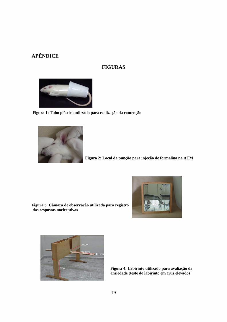
79
APÊNDICE
FIGURAS
Figura 1: Tubo plástico utilizado para realização da contenção
Figura 2: Local da punção para injeção de formalina na ATM
Figura 3: Câmara de observação utilizada para registro das respostas nociceptivas
Figura 4: Labirinto utilizado para avaliação da ansiedade (teste do labirinto em cruz elevado)

80
TABELAS REFERENTES AOS VALORES INDIVIDUAIS DA AMOSTRA
Tabela 1 – Valores individuais do nível de corticosterona plasmática em animais submetidos a uma
sessão de estresse agudo por 15 minutos.
Corticosterona Plasmática (µg/dl)
Animal Grupo Controle (não-
estressado) Grupo Estressado
1 2 3 4 5 6 7 8
Média ± EPM
0,7 5,5 1,0 3,2 2,3 4,8 4,3 1,3
2,89 ± 1,56
18,9 20,8 15,6 22,8 25,9 34,6 26,6 21,3
23,31± 10,15
Tabela 2– Valores individuais do nível de corticosterona plasmática em animais submetidos a uma sessão de estresse agudo por 30 minutos.
Corticosterona Plasmática (µg/dl)
Animal Grupo Controle (não-
estressado) Grupo Estressado
1 2 3 4 5 6 7 8
Média ± EPM
0,7 5,5 1,0 3,2 2,3 4,8 4,3 8,6
3,80 ± 2,00
31,6 35,0 14,8 40,0 24,3 37,8 27,7 40,0
31,40 ± 13,85
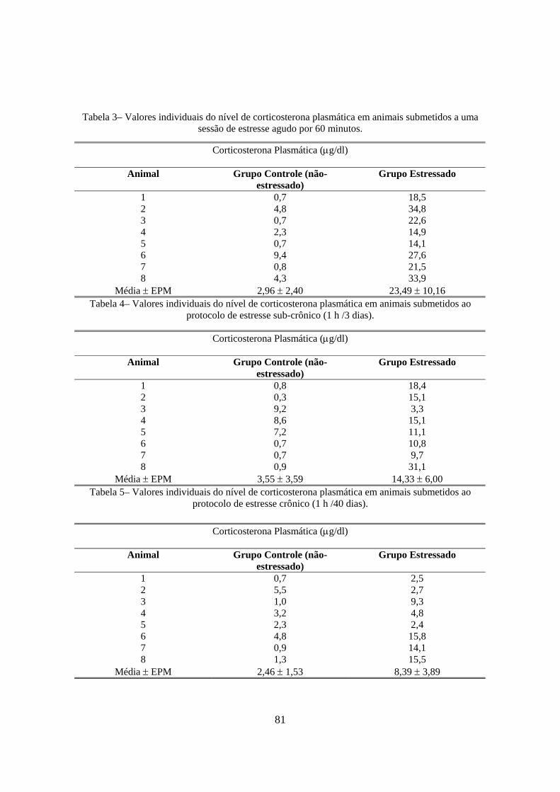
81
Tabela 3– Valores individuais do nível de corticosterona plasmática em animais submetidos a uma sessão de estresse agudo por 60 minutos.
Tabela 4– Valores individuais do nível de corticosterona plasmática em animais submetidos ao protocolo de estresse sub-crônico (1 h /3 dias).
Tabela 5– Valores individuais do nível de corticosterona plasmática em animais submetidos ao protocolo de estresse crônico (1 h /40 dias).
Corticosterona Plasmática (µg/dl)
Animal Grupo Controle (não-estressado)
Grupo Estressado
1 2 3 4 5 6 7 8
Média ± EPM
0,7 4,8 0,7 2,3 0,7 9,4 0,8 4,3
2,96 ± 2,40
18,5 34,8 22,6 14,9 14,1 27,6 21,5 33,9
23,49 ± 10,16
Corticosterona Plasmática (µg/dl)
Animal Grupo Controle (não-estressado)
Grupo Estressado
1 2 3 4 5 6 7 8
Média ± EPM
0,8 0,3 9,2 8,6 7,2 0,7 0,7 0,9
3,55 ± 3,59
18,4 15,1 3,3 15,1 11,1 10,8 9,7 31,1
14,33 ± 6,00
Corticosterona Plasmática (µg/dl)
Animal Grupo Controle (não-estressado)
Grupo Estressado
1 2 3 4 5 6 7 8
Média ± EPM
0,7 5,5 1,0 3,2 2,3 4,8 0,9 1,3
2,46 ± 1,53
2,5 2,7 9,3 4,8 2,4 15,8 14,1 15,5
8,39 ± 3,89

82
Tabela 6– Valores individuais do nível de ACTH plasmática em animais submetidos a uma sessão de estresse agudo por 15 minutos.
Tabela 7– Valores individuais do nível de ACTH plasmática em animais submetidos a uma sessão de estresse agudo por 30 minutos.
Tabela 8– Valores individuais do nível de ACTH plasmática em animais submetidos a uma sessão de estresse agudo por 60 minutos.
ACTH Plasmática (pg/ml)
Animal Grupo Controle (não-estressado)
Grupo Estressado
1 2 3 4 5 6 7 8
Média ± EPM
10,0 27,7 10,0 29,7 10,0 41,4 14,0 10,0
19,10 ± 10,38
77,6 132 83,6 261 706 410 40,6 200
238,85 ± 125,29
ACTH Plasmática (pg/ml)
Animal Grupo Controle (não-estressado)
Grupo Estressado
1 2 3 4 5 6 7 8
Média ± EPM
10,0 27,7 10,0 29,7 10,0 41,4 14,0 15,5
19,79 ± 9,86
246 185 42
82,8 55,5 172 66,6 104
119,24 ± 52,86
ACTH Plasmática (pg/ml)
Animal Grupo Controle (não-estressado)
Grupo Estressado
1 2 3 4 5 6 7 8
Média ± EPM
38,6 41,4 10,0 10,0 10,0 19,4 17,7 14,0
20,14 ± 9,93
138 90,8 33,0 35,8 38,0 172 41,7 309
107,29 ± 52,95

83
Tabela 9– Valores individuais do nível de ACTH plasmática em animais submetidos ao protocolo de estresse sub-crônico (1 h/3 dias).
ACTH Plasmática (pg/ml)
Animal Grupo Controle (não-estressado)
Grupo Estressado
1 2 3 4 5 6 7 8
Média ± EPM
17,7 27,0 19,4 15,5 17,6 38,6 10,0 10,6
19,55 ± 6,63
15,0 29,4 10,0 13,7 10,2 10,0 14,2 25,0
15,94 ± 6,27
Tabela 10– Valores individuais do nível de ACTH plasmática em animais submetidos ao protocolo de estresse crônico (1 h/40 dias).
ACTH Plasmática (pg/ml)
Animal Grupo Controle (não-estressado)
Grupo Estressado
1 2 3 4 5 6 7 8
Média ± EPM
10,0 27,7 10,0 29,7 10,0 41,4 10,6 10,0
18,68 ± 10,69
21,6 16,8 22,2 14,7 20,0 18,4 18,9 25,4
19,75 ± 6,36

84
Tabela 11 – Valores individuais do efeito desencadeado pelos diversos protocolos de estresse sobre a porcentagem de entrada nos braços abertos durante o teste do labirinto em cruz elevado.
Avaliação da ansiedade: % entrada nos braços abertos
Animais (N=10/grupo)
Grupo controle
Estresse 15 min.
Estresse 30 min.
Estresse 60 min.
Estresse 3 dias
(1h/dia)
Estresse 40 dias (1h/dia)
1 2 3 4 5 6 7 8 9
10 Mediana
20 33,3 44,4 30,0 44,4 42,9 45,5 33,3 50,0 37,5
40,20
16,7 0,00 22,2 12,5 11,1 42,9 0,00 11,1 0,00 21,4 11,8
0,00 33,3 0,00 5,90 0,00 20,0 0,00 0,00 25,0 33,3 2,95
10,0 50,0 50,0 0,00 20
14,3 16,7 0,00 0,00 0,00
12,15
0,00 0,00 0,00 11,1 42,9 12,5 0,00 0,00 0,00 8,30 0,00
0,00 0,00 0,00 13,3 26,7 40,0 0,00 33,3 7,70 0,00 3,85
Tabela 12 – Valores individuais do efeito desencadeado pelos diversos protocolos de estresse sobre
o tempo de permanência nos braços abertos durante o teste do labirinto em cruz elevado.
Avaliação da ansiedade: tempo permanência nos braços abertos (segundos)
Animais (N=10/grupo)
Grupo controle
Estresse 15 min.
Estresse 30 min.
Estresse 60 min.
Estresse 3 dias
(1h/dia)
Estresse 40 dias (1h/dia)
1 2 3 4 5 6 7 8 9
10 Mediana
10,0 10,03 30,0
19,72 88,2
31,57 71,84 59,75 82,34 30,19 30,88
7,40 0,00 25,0 13,05 18,72 29,7 0,00 2,06 0,00 33,5 10,23
0,00 22,74 0,00
10,22 0,00 9,94 0,00 0,00 5,12
27,31 2,56
9,53 19,78 29,84 0,00 15,16 1,84 32,13 0,00 0,00 0,00 5,69
0,00 0,00 0,00 26,23 55,08 23,40 0,00 0,00 0,00 11,78 0,00
0,00 0,00 0,00
33,29 40,54 12,41 0,00
51,97 14,09 0,00 6,21
85
Tabela 13 – Valores individuais da soma dos comportamentos nociceptivos [coçar (CO)+levantar rapidamente a cabeça (LC)] desencadeados pela injeção de formalina na ATM após estresse por
contenção durante 15 minutos.
Soma dos comportamentos (CO + LC)
Animal Grupo Controle Grupo Estressado 1 2 3 4 5 6
Média ± EPM
148,45 128,42 171,71 134,36 116,74 123,16
137,14 ± 15,29
124,07 49,34
138,54 129,55
144 73
109,75 ± 32,39
Tabela 14 – Valores individuais da soma dos comportamentos nociceptivos [coçar (CO)+levantar rapidamente a cabeça (LC)] desencadeados pela injeção de formalina na ATM após estresse por
contenção durante 30 minutos.
Tabela 15 – Valores individuais da soma dos comportamentos nociceptivos [coçar (CO)+levantar rapidamente a cabeça (LC)] desencadeados pela injeção de formalina na ATM após estresse por
contenção durante 60 minutos.
Soma dos comportamentos (CO + LC) Animal Grupo Controle Grupo Estressado
1 2 3 4 5 6
Média ± EPM
148,45 128,42 171,71 134,36 116,74 123,16
137,14 + 15,29
124 130,67 120,32
112 117,14 59,05
110,53 + 17,16
Soma dos comportamentos (CO + LC) Animal Grupo Controle Grupo Estressado
1 2 3 4 5 6
Média ± EPM
128,42 134,36 116,74 150,22 148,45 158,23
139,40 ± 12,90
116,74 124,69 148,79
108 122
144,05 127,38 ± 12,69

86
Tabela 16 – Valores individuais da soma dos comportamentos nociceptivos [coçar (CO)+levantar rapidamente a cabeça (LC)] desencadeados pela injeção de formalina na ATM após estresse por
contenção durante 3 dias (1h/dia).
Tabela 17 – Valores individuais da soma dos comportamentos nociceptivos [coçar (CO)+levantar rapidamente a cabeça (LC)] desencadeados pela injeção de formalina na ATM após estresse por
contenção durante 40 dias (1h/dia).
Tabela 18 – Valores individuais da soma dos comportamentos nociceptivos [coçar (CO)+levantar rapidamente a cabeça (LC)] desencadeados pela injeção de formalina na ATM após estresse por
contenção durante 60 minutos e administração de salina ou naloxona (10 mg/Kg).
Soma dos comportamentos (CO + LC) Animal Grupo Controle Grupo Estressado
1 2 3 4 5 6
Média ± EPM
158,23 128,42 171,71 134,36 116,74 123,16
138,77 ± 17,47
137 149,64 148,18 177,62 158,63 168,92
156,67 ± 11,73
Soma dos comportamentos (CO + LC) Animal Grupo Controle Grupo Estressado
1 2 3 4 5 6
Média ± EPM
128,42 134,36 116,74 150,22 148,45 158,23
139,40 ± 12,90
241,58 331,2
186,63 291,03 150,71
189 231,69 ± 56,25
Soma dos comportamentos (CO + LC) Animal Salina Naloxona
1 2 3 4 5 6
Média ± EPM
112,26 119,87
137 87,72 124 158
123,14 ± 16,53
209,17 120,7 268,1
200,69 114
171,5 180,69 ± 45,29

87
Tabela 19 – Valores individuais da soma dos comportamentos nociceptivos [coçar (CO)+levantar rapidamente a cabeça (LC)] desencadeados pela injeção de formalina na ATM após administração
de salina ou morfina (1 e 5 mg/Kg).
Tabela 20 – Valores individuais da soma dos comportamentos nociceptivos [coçar (CO)+levantar rapidamente a cabeça (LC)] desencadeados pela injeção de formalina na ATM após estresse por
contenção durante 40 dias (1h/dia) e administração de salina ou morfina (1 e 5 mg/Kg).
Soma dos comportamentos (CO + LC) Animal (Controle) Salina Morfina 1 mg/Kg Morfina 5 mg/Kg
1 2 3 4 5 6
Média ± EPM
145,41 131,90 208,80 188,09 132,85 219,40
171,07 ± 39,24
83 79
92,06 105 47,6 76,2
80,48 ± 12,88
72 106,63 55,68 53,88 89,31 48,53
71,01 ± 18,31
Soma dos comportamentos (CO + LC) Animal (Estressado) Salina Morfina 1 mg/Kg Morfina 5 mg/Kg
1 2 3 4 5 6
Média ± EPM
232,2 246,6
200,57 173,42 225,87
306 230,78 ± 30,82
208,5 221,23 232,86 296,31
240 187,2
231,02 ± 25,37
44 91,71 144,11 77,74 69,31 103,2
88,35 ± 24,66

88
Tabela 21 – Valores individuais da soma dos comportamentos nociceptivos [coçar (CO)+levantar rapidamente a cabeça (LC)] desencadeados pela injeção de formalina na ATM após estresse por
contenção durante 40 dias (1h/dia) e administração de salina ou fluoxetina (10 mg/Kg).
Tabela 22 – Valores individuais do efeito do estresse por contenção durante 40 dias (1h/dia) sobre o comportamento de coçar a região orofacial desencadeado pela injeção de salina na ATM.
Comportamento de coças a região orofacial (segundos)
Animal Grupo Controle Grupo Estressado 1 2 3 4 5 6
Média ± EPM
23,71 49,4 28,28 35,19 50,25 20,46
34,55 ± 10,40
43,05 0
11,02 71,02 18,9 52
32,67 ± 22,69
Soma dos comportamentos (CO + LC) Animal Grupo Controle (não estressado) Grupo Estressado
salina Fluoxetina Salina fluoxetina 1 2 3 4 5 6
Média ± EPM
171,15 165,14 158,07 123,81 165,16 90,34
145,61 ± 25,69
93 30,4 52 75 10 18
46,40 ± 26,93
232,2 246,6 200,57 173,42 225,87
306 230,78 ± 30,82
88,1 52 3 46 82 15
47,68 ± 26,35