Embed Size (px)
Citation preview

FABIANA DE OLIVEIRA VASCONCELOS AVALIAÇÃO DA PRESENÇA DE CITOMEGALOVÍRUS E VÍRUS EP STEIN-
BARR EM LESÕES PERIAPICAIS SINTOMÁTICAS E ASSINTOMÁ TICAS
BELO HORIZONTE
FACULDADE DE ODONTOLOGIA
UNIVERSIDADE FEDERAL DE MINAS GERAIS
2007
FABIANA DE OLIVEIRA VASCONCELOS
AVALIAÇÃO DA PRESENÇA DE CITOMEGALOVÍRUS E VÍRUS EP STEIN-
BARR EM LESÕES PERIAPICAIS SINTOMÁTICAS E ASSINTOMÁ TICAS
Dissertação apresentada ao Programa do Colegiado de Pós-Graduação da Faculdade de Odontologia da Universidade Federal de Minas Gerais, como requisito parcial para a obtenção do título de Mestre em Odontologia, Área de Concentração: Endodontia. Orientadora: Profª. Drª. Kátia Lucy de Melo Maltos Co-orientador: Prof. Dr. Ricardo Santiago Gomez
BELO HORIZONTE
FACULDADE DE ODONTOLOGIA
UNIVERSIDADE FEDERAL DE MINAS GERAIS
2007

Mamãe, Papai e Fernando,
dedico a vocês este trabalho.
Vocês foram fundamentais para que eu
chegasse até aqui. NÓS conseguimos!

AGRADECIMENTOS
A Deus, pela força e luz, tão necessárias durante esta caminhada.
Aos meus pais, Maria e Francisco, pelo carinho, apoio e compreensão
incondicionais.
Ao meu amor Fernando, pelo companheirismo, incentivo, paciência e pela enorme
dedicação ao me ajudar na elaboração deste trabalho.
À minha querida orientadora Kátia, pelos ensinamentos, amizade e confiança.
Ao professor Ricardo Gomez, pela orientação desde os primeiros passos durante a
graduação e pelo exemplo de competência profissional.
Aos amigos Flávio e Jeane, pela imensa ajuda na parte experimental deste trabalho.
Aos amigos do laboratório André, Carolina, Eliza, Guilherme, Luciano, pela
agradável convivência.
À Joyce e Michelli, pelo auxílio na execução dos experimentos.
Aos professores e funcionários do laboratório de Patologia Bucal, pela ajuda no
processamento e diagnóstico histopatológico dos casos.
Aos professores e alunos da Clínica de Cirurgia da FO-UFMG e à aluna Adriana,
pela essencial colaboração na coleta das amostras.
Aos pacientes, por compreenderem a importância deste estudo e por sua valiosa
participação.
Aos queridos Fernanda e João, pela amizade verdadeira de tantos anos e por
compreenderem minha ausência.
Ao Marcelo, pela grande amizade e contribuições na redação em Inglês.
Às colegas Alessandra e Carol, pela amizade e ajuda na Endodontia.
À tia Bete, pelo carinho e pela revisão de Português.
Aos amigos e familiares, pelos momentos de descontração e pelo apoio de sempre.

RESUMO
Recentes estudos sugeriram que alguns herpesvírus participariam da
etiopatogênese das periapicopatias. Assim, o objetivo deste trabalho foi avaliar a
presença do citomegalovírus humano (HCMV) e vírus Epstein-Barr (EBV) nas lesões
periapicais e verificar a possível associação desses vírus com a ocorrência de
grandes lesões sintomáticas e com o seu diagnóstico histopatológico. Incluíram-se
no estudo 27 lesões sintomáticas e 52 assintomáticas. A técnica da Reação em
Cadeia da Polimerase (PCR) foi empregada para investigar a presença do HCMV e
EBV. Os testes do Qui-quadrado e Exato de Fisher foram usados para análise
estatística. Observou-se que 64 lesões (81%) apresentaram EBV, 3 (4%) tinham
HCMV e nenhum vírus foi detectado em 15 amostras (19%). Notadamente, todas as
lesões positivas para HCMV mostraram também EBV. Nenhuma associação
significativa foi identificada entre as infecções por HCMV e EBV e a ocorrência de
sintomatologia ou o tamanho das lesões, enquanto a infecção por EBV estava
estatisticamente relacionada aos cistos periapicais. Concluindo, diferentemente do
HCMV, detectou-se uma alta freqüência do EBV nas lesões periapicais,
especialmente nos cistos. Entretanto, a presença desses vírus não estava
relacionada com a sintomatologia das lesões nem com o seu tamanho radiográfico.
Descritores: Citomegalovírus, vírus Epstein-Barr, lesões periapicais.
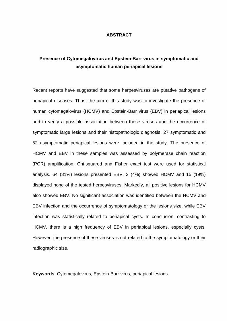
ABSTRACT
Presence of Cytomegalovirus and Epstein-Barr virus in symptomatic and
asymptomatic human periapical lesions
Recent reports have suggested that some herpesviruses are putative pathogens of
periapical diseases. Thus, the aim of this study was to investigate the presence of
human cytomegalovirus (HCMV) and Epstein-Barr virus (EBV) in periapical lesions
and to verify a possible association between these viruses and the occurrence of
symptomatic large lesions and their histopathologic diagnosis. 27 symptomatic and
52 asymptomatic periapical lesions were included in the study. The presence of
HCMV and EBV in these samples was assessed by polymerase chain reaction
(PCR) amplification. Chi-squared and Fisher exact test were used for statistical
analysis. 64 (81%) lesions presented EBV, 3 (4%) showed HCMV and 15 (19%)
displayed none of the tested herpesviruses. Markedly, all positive lesions for HCMV
also showed EBV. No significant association was identified between the HCMV and
EBV infection and the occurrence of symptomatology or the lesions size, while EBV
infection was statistically related to periapical cysts. In conclusion, contrasting to
HCMV, there is a high frequency of EBV in periapical lesions, especially cysts.
However, the presence of these viruses is not related to the symptomatology or their
radiographic size.
Keywords : Cytomegalovirus, Epstein-Barr virus, periapical lesions.

LISTA DE ABREVIATURAS, SIGLAS E NOTAÇÕES
- – menos % – porcentagem & – e < – menor que ® – marca registrada bp – base pairs – pares de base CD – cluster of differentiation – grupo de diferenciação DNA – Deoxyribonucleic acid – ácido desoxirribonucléico EBNA-1 – Epstein-Barr virus nuclear antigen-1 – antígeno nuclear do Epstein-Barr-1 EBV – Epstein-Barr virus – Vírus Epstein-Barr g – grama H&E – Hematoxylin and Eosin – Hematoxilina-Eosina HCMV – Human cytomegalovirus – Citomegalovírus Humano HHV – Human herpesvirus – Herpesvírus Humano HSV – Herpes Simplex Virus – Vírus do Herpes Simples IFN – Interferon – interferon IL – Interleukin – interleucina MHC – Major histocompatibility complex – Complexo de Histocompatibilidade Principal min – minuto ml – mililitro mm – milímetro ng – nanograma NK – Natural Killer ºC – grau centígrado p – probabilidade de significância PCR – Polymerase Chain Reaction – Reação em Cadeia da Polimerase pH – potencial de hidrogênio pmol – picomol RT-PCR – Reverse Transcription-PCR – PCR da transcrição reversa s – segundo Taq – Thermus aquaticus Th – linfócito T helper – linfócito T auxiliar TNF – Tumour necrosis factor – Fator de Necrose Tumoral V – Voltagem VZV – Varicella-zoster virus – Vírus Varicela-Zoster µl – microlitro µm – micrômetro

LISTA DE FIGURAS
FIGURA 01 - Gel de poliacrilamida a 6,5% corado pela prata mostrando
produtos de PCR referentes à detecção do HCMV em
lesões periapicais..................................................................
39
FIGURA 02 - Gel de poliacrilamida a 6,5% corado pela prata mostrando
produtos de PCR referentes à detecção do EBV em lesões
periapicais............................................................................. 40
FIGURA 03 - Imagens radiográficas de dentes associados a
radioluscências periapicais sugestivas de lesões de origem
endodôntica........................................................................... 60
FIGURA 04 - Lesões periapicais aderidas às raízes extraídas................... 60

LISTA DE TABELAS
TABELA 01 -
Primers e protocolos empregados na técnica de PCR..........
37
TABELA 02 - Relação entre herpesvírus e parâmetros das lesões
periapicais............................................................................. 38

SUMÁRIO
RESUMO
ABSTRACT
LISTA DE ABREVIATURAS, SIGLAS E NOTAÇÕES
LISTA DE FIGURAS
LISTA DE TABELAS
1 APRESENTAÇÃO................................................................................... 11
2 ARTIGO.................................................................................................. 16
Abstract................................................................................................... 17
Introduction............................................................................................. 18
Materials and methods............................................................................ 20
Results.................................................................................................... 24
Discussion............................................................................................... 24
Conclusions............................................................................................. 29
Acknowledgements................................................................................. 29
References.............................................................................................. 30
Tables..................................................................................................... 37
Figures.................................................................................................... 39
3 CONSIDERAÇÕES FINAIS.................................................................... 41
REFERÊNCIAS BIBLIOGRÁFICAS........................................................ 44
ANEXOS................................................................................................. 45
Anexo A – Termo de consentimento livre e esclarecido.............................................................................................. 45
Anexo B – Ficha clínica........................................................................... 46
Anexo C – Soluções e reagentes............................................................ 48
Anexo D – Dados clínicos coletados....................................................... 50
Anexo E – Normas para publicação – International Endodontic Journal....................................................................................................
55
Anexo F – Figuras................................................................................... 60

11
1 APRESENTAÇÃO
A inflamação periapical é conseqüência da extensão da inflamação pulpar,
capaz de se desenvolver em fase anterior à necrose total da polpa. Estabelecida a
infecção do sistema de canais radiculares, o egresso de bactérias e seus produtos
para os tecidos perirradiculares leva a respostas inflamatórias e imunológicas do
hospedeiro no local, resultando na formação de lesão periapical que visa a conter o
avanço da infecção endodôntica (TORABINEJAD, 1994; STASHENKO et al., 1994,
1998; TAKAHASHI, 1998).
Kakehashi et al. (1965) demonstraram o papel etiológico das bactérias
presentes no canal radicular na formação de lesões periapicais. Nesse estudo, a
exposição do tecido pulpar à cavidade bucal resultou em inflamação periapical
somente em ratos convencionais, e não em animais isentos de germes. Sundqvist
(1976) confirmou essa relação causal, relatando que, em dentes portadores de
lesões traumáticas com necrose pulpar e coroas íntegras, bactérias foram isoladas
somente daqueles que apresentavam lesões periapicais, ou seja, o tecido pulpar
não infectado não resultava em inflamação no periápice.
Embora já tenham sido realizadas numerosas investigações de possíveis
associações entre determinadas bactérias patogênicas que infectam o sistema de
canais radiculares e a ocorrência de sintomatologia ou outras características das
lesões (SUNDQVIST, 1976; BAUMGARTNER et al., 1999; SIQUEIRA Jr. et al.,
2001; SAKAMOTO et al., 2006), parece ser difícil obter uma forte evidência do papel
etiológico de uma espécie bacteriana, uma vez que existem outras variáveis
envolvidas na formação dessas lesões.

12
As lesões periapicais evoluem com períodos de exacerbação e remissão e
exibem uma variedade de manifestações clínicas e radiográficas. Ainda não foi
determinado se as variações clínicas e histopatológicas das periapicopatias têm
causas microbiológicas distintas ou são conseqüências de diferentes respostas
imunológicas do hospedeiro aos agentes infecciosos (SABETI et al., 2003a).
Recentemente, relatos da ocorrência de dois herpesvírus, o citomegalovírus
humano (HCMV) e o vírus Epstein-Barr (EBV), nos tecidos periapicais inflamados
sugeriram que eles estariam envolvidos na etiopatogênese das lesões periapicais
(SABETI et al., 2003a, b, c; SLOTS et al., 2003, 2004; SABETI; SLOTS, 2004;
YILDIRIM et al., 2006).
A família do herpesvírus humano (HHV) é conhecida como Herpesviridae. Os
seus membros apresentam uma dupla fita de DNA envolta por um capsídeo e um
tegumento amorfo. Circundando essas estruturas, encontra-se um envelope lipídico,
derivado das membranas celulares do hospedeiro, que contém um grande número
de glicoproteínas virais. A replicação dos herpesvírus acontece no núcleo das
células por eles infectadas. Dentre mais de 100 herpesvírus já identificados, apenas
oito são conhecidos por infectarem seres humanos: Vírus do Herpes Simples (HSV),
tipo 1 (HSV-1 ou HHV-1) e tipo 2 (HSV-2 ou HHV-2); Vírus Varicela-Zoster (VZV ou
HHV-3); Vírus Epstein-Barr (EBV ou HHV-4); Citomegalovírus (HCMV ou HHV-5);
HHV-6; HHV-7 e Vírus do Sarcoma de Kaposi (HHV-8) (SLOTS, 2004, 2005). Todos
esses vírus possuem a habilidade de habitar por toda a vida no interior do
hospedeiro infectado. Após a infecção inicial, são observados períodos variáveis de
latência e reativação com disseminação. Stress, mudanças hormonais, infecções,
medicamentos imunossupressores e outros eventos que provoquem uma diminuição
das defesas do hospedeiro podem levar à reativação viral (SLOTS, 2005).

13
Alguns herpesvírus, dentre eles o HCMV e o EBV, exibem tropismo por
células do sistema imune e, conseqüentemente, infecções por esses vírus podem
resultar em alterações nas funções de defesa, levando a quadros de
imunossupressão. Eles interferem com os mecanismos de respostas inata e
adaptativa, celular e humoral, através da inativação de células natural killer (NK),
supressão da apresentação de antígenos pelas moléculas de MHC classe I e II,
inibição da apoptose, além de afetar a produção de citocinas (MOGENSEN;
PALUDAN, 2001; SLOTS, 2005).
O HCMV, após infecção primária ou recorrente (reativação ou reinfecção),
pode ser detectado, por meses ou anos, na saliva, fluido crevicular, urina, leite
materno, lágrimas, secreção vaginal e sêmen, o que permite sua transmissão
horizontal ou vertical. Canto et al. (2000) encontraram uma alta prevalência do vírus
na saliva de crianças em creches brasileiras, variando de 39 a 46%. Por outro lado,
Noyola et al. (2005) detectaram a excreção de HCMV na saliva de em média 11,2%
crianças. Aproximadamente 10% das crianças são infectadas até os seis meses de
idade, através da placenta, durante o parto ou no decorrer da amamentação. O pico
seguinte de transmissão ocorre ao longo da adolescência, predominantemente
mediante a troca de líquidos corporais, quando este grupo inicia a atividade sexual.
Mesmo em países desenvolvidos, a prevalência da infecção por HCMV é de 90%
aos 20 anos de idade. A doença mais evidente clinicamente é observada nos recém-
nascidos e em adultos imunossuprimidos, mas, na maioria dos indivíduos, a infecção
por HCMV é assintomática. Essa infecção induz uma intensa resposta imunológica
do hospedeiro que, incapaz de erradicá-la, apenas leva a uma inativação do vírus,
que permanece em estado latente em determinados sítios. Esse vírus pode infectar
diversos tipos celulares, destacando-se as células epiteliais ductais em glândulas

14
salivares, células endoteliais, neutrófilos, monócitos/macrófagos e linfócitos
(LANDOLFO et al., 2003; CAPPUYNS et al., 2005).
O EBV infecta mais de 90% da população e é capaz de permanecer no
hospedeiro por toda a vida. A exposição durante a infância é comumente
assintomática; no entanto, em adultos jovens, muitas vezes a infecção por EBV se
manifesta de maneira sintomática, causando uma doença chamada mononucleose
infecciosa. Os adultos usualmente contraem o vírus pela transferência direta da
saliva, daí a denominação “doença do beijo”. Nos países em desenvolvimento, a
exposição costuma ocorrer entre os três primeiros anos de idade, sendo universal na
adolescência. As células infectadas pelo EBV são linfócitos B e células epiteliais
(SLOTS et al., 2003; CAPPUYNS et al., 2005).
HCMV e EBV têm sido encontrados em freqüências maiores em lesões
periapicais sintomáticas e de maior tamanho radiográfico (SABETI et al., 2003a, b, c;
SLOTS et al., 2004; YILDIRIM et al., 2006), quando comparado a lesões
assintomáticas e pequenas. Assim como para as doenças periodontais, Sabeti et al.
(2003a, b, c) e Slots et al. (2003, 2004) sugeriram que algumas formas de alterações
periapicais se desenvolvem como resultado de uma série de interações entre
herpesvírus, bactérias e as reações imunológicas do hospedeiro. Os vírus poderiam
causar destruição tecidual diretamente, através de efeitos citotóxicos locais;
reduziriam a resposta imunológica periapical do hospedeiro, aumentando, portanto,
a patogenicidade das bactérias infectantes do canal radicular; e seriam capazes de
induzir a liberação de citocinas, principalmente pró-inflamatórias, como interleucina-
IL-1β (IL), IL-6, fator de necrose tumoral-α (TNF-α) e interferon-γ (IFN-γ), capazes de
levar à maior reabsorção óssea e progressão da lesão (SLOTS, 2004).

15
Assim, considerando o potencial dos herpesvírus em participar da patogênese
das periapicopatias, o objetivo deste trabalho foi avaliar a presença do HCMV e EBV
em lesões periapicais e verificar a associação entre esses vírus e a sintomatologia, o
tamanho radiográfico e o diagnóstico histopatológico das lesões periapicais.
Tendo em vista a importância da divulgação apropriada dos trabalhos
científicos para a construção do conhecimento, este estudo será apresentado a
seguir, na forma de artigo, que será enviado para publicação em periódico
qualificado.

16
PRESENCE OF CYTOMEGALOVIRUS AND EPSTEIN-BARR VIRUS IN
SYMPTOMATIC AND ASYMPTOMATIC HUMAN PERIAPICAL LESIONS*
FO Vasconcelos, DDS1
FJGS Pimenta, DDS, MS2
RS Gomez, DDS, MS, PhD2
KLM Maltos, DDS, MS, PhD1
1Department of Restorative Dentistry, School of Dentistry, Universidade Federal de
Minas Gerais, Belo Horizonte, Brazil.
2Department of Oral Surgery and Pathology, School of Dentistry, Universidade
Federal de Minas Gerais, Belo Horizonte, Brazil.
Running title: Herpesvirus in periapical lesions
Keywords: Cytomegalovirus, Epstein-Barr Virus, periapical lesions.
Address:
Profª. Kátia Lucy de Melo Maltos
Department of Restorative Dentistry
School of Dentistry
Universidade Federal de Minas Gerais
Av. Antonio Carlos, 6627
Belo Horizonte – MG
Brasil - CEP 31270 901
Fax: 55 31 34992430
e-mail: [email protected]
* Este artigo foi formatado segundo as normas da revista International Endodontic Journal – anexo E.

17
Abstract
Aim: Recent reports have suggested that some herpesviruses are putative
pathogens of periapical diseases. Thus, the aim of this study was to investigate the
presence of human cytomegalovirus (HCMV) and Epstein-Barr virus (EBV) in
periapical lesions and to verify the association between these viruses and the
occurrence of symptomatic large lesions and their histopathologic diagnosis.
Methodology: 27 symptomatic and 52 asymptomatic periapical lesions were
included in the study. The presence of HCMV and EBV in these samples was
assessed by polymerase chain reaction (PCR) amplification. Chi-squared and Fisher
exact test were used for statistical analysis.
Results: 64 (81%) lesions presented EBV, 3 (4%) showed HCMV and 15 (19%)
displayed none of the tested herpesviruses. Markedly, all positive lesions for HCMV
also showed EBV. No significant association was identified between the HCMV and
EBV infection and the occurrence of symptomatology or the lesions size, while EBV
infection was statistically related to periapical cysts.
Conclusions: In contrast to HCMV, there is a high frequency of EBV in periapical
lesions, especially cysts. However, the presence of these viruses is not related to the
symptomatology or their radiographic size.

18
Introduction
Injuries to the dental pulp, especially bacterial infection, usually lead to
irreversible pulpitis and pulpal necrosis. The interaction between the irritants present
in the root canal system and host defensive cells provokes the release of numerous
mediators that are capable of stimulating inflammatory/immune responses and bone
resorption in the periradicular tissue, resulting in the formation of inflammatory
periapical lesions, with the aim of restricting microbial invasion (Torabinejad 1994,
Stashenko et al. 1994, Takahashi 1998).
Inflammatory periapical lesions of endodontic origin can be presented
histologically as granulomas or cysts. It has been demonstrated that T and B
lymphocytes, macrophages, plasma cells and neutrophil granulocytes represent the
predominant cells in periapical lesions. Mast cells, eosinophils, dendritic cells and
natural killer cells comprise a minor, but functionally important cell population
(Kettering & Torabinejad 1993, Kawashima et al. 1996, Márton & Kiss 2000, Metzger
2000, Silva et al. 2005, Lukić et al. 2006).
Although bacteria and host-related inflammatory responses are
unquestionably involved in periapical lesions, the pathophysiological events that take
place and give rise to a variety of clinical and radiographic manifestations have not
been fully understood. Recent studies have shown evidences that some
herpesviruses, especially human cytomegalovirus (HCMV) and Epstein-Barr virus
(EBV), may participate in the pathogenesis of some types of periapical diseases
(Sabeti et al. 2003a, b, c, Sabeti & Slots 2004, Slots et al. 2004).
HCMV and EBV, two members of the Herpesviridae family, are ubiquitous
viruses transmitted from person to person during the period of primary infection or

19
episodes of reactivation. Individuals frequently acquire herpesviruses at an early age
(Noyola et al. 2005, Slots et al. 2006). After primary exposure, these viruses establish
latency in various host cell reservoirs, from which they may reactivate periodically
(Yan & Fedorko 2002). In immunocompetent individuals, an HCMV or EBV infection
rarely results in clinical disease. Control of herpesviral replication and prevention of
pathosis depend on innate and adaptive, cellular and humoral immune mechanisms.
The main effector cells of this control are CD8+ cytotoxic T-lymphocytes, NK cells
and anti-viral antibodies (Kettering & Torabinejad 1993, Kano & Shiohara 2000,
Landolfo et al. 2003, Slots 2004, Konstantinidis et al. 2005).
Cytomegalovirus infection is common in the human population and
seroprevalence has been reported to be as high as 80-90%. Multiple cells types are
infected by HCMV, especially ductal epithelial cells of salivary glands, kidney,
endothelial cells, neutrophils, monocytes/macrophages and T lymphocytes
(Contreras et al. 1999, Landolfo et al. 2003, Slots 2005). The regular shedding of
infectious virus in the saliva over a long period of time is believed to play a major role
in horizontal transmission of this virus (Kano & Shiohara 2000, Kloover et al. 2002).
Wide variations in HCMV excretion in saliva have been found in different studies. The
rates varied from 8.6 to 28.7% (Canto et al. 2000) and from 3.1 to 31.3% (Noyola et
al. 2005).
The Epstein-Barr virus is a human herpesvirus that infects up to 95% of adults
worldwide. EBV infects epithelial cells and B lymphocytes (Ammatuna et al. 1998,
Contreras et al. 1999) and productively replicates at oral mucosal surfaces, shedding
transmissible virus into the saliva (Slots et al. 2006). Idesawa et al. (2004) detected
EBV in the saliva of 48.5% periodontitis patients and only in 15% of healthy
individuals, suggesting that levels of EBV in saliva may reflect the status of

20
periodontal inflammation. Saygun et al. (2005) demonstrated a close relationship
between HCMV and EBV loads in inflamed periodontal tissue and in saliva, indicating
at least a partial periodontal origin of salivary herpesviruses. EBV is a causative
agent of infectious mononucleosis and has been found to be associated with
nasopharyngeal carcinoma, endemic Burkitt’s lymphoma and oral hairy leukoplakia
(Ammatuna et al. 1998, Slots et al. 2006).
Herpesviruses involvement in the pathogenesis of periapical lesions may
occur as a direct result of viral infection and replication, or as a consequence of
virally induced impairment of the host defense and subsequent increased virulence of
bacteria infecting the root canal (Sabeti et al. 2003a, b, c, Slots et al. 2004).
The aim of this study was to investigate the presence of HCMV and EBV in
periapical lesions and to verify a possible association between these viruses and the
occurrence of symptomatic large lesions and their histopathologic diagnosis.
Materials and methods
Subjects and sample collection
A total of 61 patients presenting teeth with periapical radiolucencies
suggestive of inflammatory lesions of endodontic origin and requiring extraction were
recruited from the Surgery Clinic at the Universidade Federal de Minas Gerais, Brazil.
Those individuals that reported general health concerns like immunosuppressive
chemotherapy, HIV infection or severely compromised immune function were not
included in this study. Teeth with severe types of periodontal diseases and with a
perio-endo lesion were not included. There were 43 females (71%) and 18 males
(29%), with median age of 35 years (range 14 to 68). This study was approved by the

21
University’s Ethics Committee (ETIC nº 084/06) and a signed informed consent was
obtained from all the participants.
In this study, 79 periapical lesions were obtained and a total of 27
symptomatic and 52 asymptomatic lesions were studied. The lesion was classified
as symptomatic when associated with pain, swelling and/or sensitivity to percussion
or palpation. Asymptomatic lesions were those without any of these signs or
symptoms (Torabinejad & Walton 1989, Sabeti et al. 2003a, b, Slots et al. 2004).
The standardized periapical radiographs of each case were mounted in an
appropriate black card and carefully evaluated by two calibrated examiners. The
radiolucencies’ two major lengths were measured in millimeters using a caliper rule
and multiplied. The lesions ranged in radiographic size from 1 x 2 mm (2 mm2) to 10
x 12 mm (120 mm2) and were stratified into two groups according to the median
value: 40 small (16 mm2 or less) and 39 large lesions (bigger than 16 mm2).
Samples were obtained by curettage in conjunction with tooth extraction. Prior
to the surgery, the patients rinsed with 0.12% chlorhexidine mouthwash for 60
seconds. All surgical procedures were performed with sterile instruments and
material. After removing the tooth, the periapical lesion was collected and washed in
0.9% saline solution. A tissue fragment was placed in a plastic vial containing 10%
formalin solution and processed for light microscopy. Serial 5 µm-thick sections were
obtained and stained by hematoxylin and eosin (H&E) for histopathological
diagnosis. Only the cases diagnosed as periapical cysts or granulomas remained in
the study. The other sample portions were placed into an Eppendorf microtube
containing 500 µl of OCT Compound Tissue-Tek® (Sakura, Torrance, CA, USA) and
stored at -80 ºC until processing for virological identification.

22
DNA extraction
The DNA purification was carried out using the DNeasy® Tissue Kit (Qiagen,
Valencia, CA, USA), following the manufacturer protocols.
Polymerase Chain Reaction (PCR) assays
Viruses DNA were identified by PCR amplification performed in a DNA thermal
cycler PTC 100 – Programmable Thermal Controller (MJ Research, Waltham,
Massachusetts, USA), which included cycles of denaturation, primer annealing and
extension, ending with a final extension. Primers and PCR conditions are described
in table 1. Positive and negative controls (PCR reagents without DNA) for HCMV and
EBV were processed in each experiment.
The presence of HCMV glycoprotein B was assessed by a nested PCR
method (Victória et al. 2005). The 25 µl reaction mixture contained buffer (40mM
NaCl, 10mM Tris-HCl pH 8.4, 2.25mM MgCl2, Triton X-100 0.1%), Taq DNA
polymerase (0.25unit/reaction – Phoneutria Biotecnologia, Belo Horizonte, MG,
Brazil), primers (20 pmol/reaction) and 0.2mM deoxynucleoside triphosphates. After
the first round of PCR, 1 µl of the final product of each sample, including the negative
control, was used as a template for the second PCR with the inner primer pairs. The
same procedure as described earlier was followed, except that 57ºC was used as the
anneling temperature.
To identify the EBV, PCR was carried out with the primers that amplify the
region encoding the Epstein-Barr virus nuclear antigen-1 (EBNA-1) (Ammatuna et al.
1998). The 25 µl reaction mixture contained 100-300 ng of genomic DNA template,

23
primers (20 pmol/reaction), buffer (10mM (NH4)2SO4, 10mM KCl, 10mM Tris-HCl pH
8.4, 3.0mM MgCl2, Triton X-100 0.1%), 0.2mM deoxynucleoside triphosphates and
Taq DNA polymerase (0.25unit/reaction – Phoneutria Biotecnologia, Belo Horizonte,
MG, Brazil).
The human β-globin gene was amplified as a control for DNA quality (Gall-
Troselj et al. 2001). The 25 µl reaction mixture contained 100-300 ng of genomic
DNA template, primers (20 pmol/reaction), buffer (1.50mM MgCl2, 50mM KCl, 10mM
Tris-HCl pH 8.4, Triton X-100 0.1%), 0.2mM deoxynucleoside triphosphates and Taq
DNA polymerase (1unit/reaction – Phoneutria Biotecnologia, Belo Horizonte, MG,
Brazil).
Polyacrylamide gel electrophoresis
The HCMV (224bp), EBV (269bp) and β-globin (268bp) amplified products
were visualized by 6.5% polyacrylamide gel electrophoresis and silver stained. Five
µl of each reaction product was added to 1 µl of gel loading dye (0.25% bromophenol
blue, 30% glycerol, 10 mM EDTA). Electrophoresis was carried out using 1x TBE
buffer, 160V, for approximately 30 minutes, in a mini vertical gel electrophoresis unit
(Sigma-Aldrich, St. Louis, MO, USA). The molecular weight of the DNA was
estimated using 100 bp ladder markers.
Statistical Analysis

24
Chi-square (X2) and Fisher exact test were used to verify association between
groups’ distributions. Statistical significance was set at the level of p < 0.05. These
statistical analysis were performed using SigmaStat software (Systat Software Inc.,
Richmond, California, USA).
Results
Figures 1 and 2 represent silver-stained polyacrylamide gels showing PCR
products of HCMV and EBV genomes, with 224 bp and 269 bp, respectively. The
frequency of occurrence of EBV and HCMV in periapical lesions and the relationship
between these viruses and some parameters of this disease are exhibited in table 2.
Of the 79 lesions studied, 64 (81%) presented EBV, 3 (4%) showed HCMV and 15
(19%) displayed none of the tested herpesviruses. Markedly, all positive lesions for
HCMV also showed EBV.
The occurrence of HCMV and EBV was not statistically associated to the
presence of symptoms neither to the lesion size. A significant association was
observed between EBV infection and periapical cysts.
Discussion
The present study detected EBV DNA in 64 of the 79 periapical lesions
examined (81%). This finding is in general agreement with prior studies which
reported that most teeth with necrotic pulp and periapical lesions harbor
herpesviruses in periapical inflammatory tissue (Sabeti et al. 2003a, b, c, Sabeti &
Slots 2004, Slots et al. 2003, 2004, Yildirim et al. 2006).

25
However, HCMV has not been widely observed, being present only in three
cases (4%), despite its high frequency in the general population. This result refutes
the hypothesis of Slots et al. (2004) that HCMV would be the more important
pathogen of the two viruses and the responsible for transactivating EBV, suggesting
that it is not involved in the pathogenesis of periapical pathosis.
Since periradicular lesions are a reactive tissue composed mainly of
granulomatous inflammatory tissue replacing normal bone, there is no actual normal
tissue equivalent which can be used as a negative control (Lim et al. 1994).
Therefore, we compared the occurrence of herpesviruses in symptomatic and
asymptomatic lesions.
No statistical differences in the presence of each virus were found between
symptomatic and asymptomatic periapical lesions, unlike previous studies that
suggested HCMV and EBV participation in the pathogenesis of symptomatic
periapical lesions. This hypothesis was based on findings of a significantly higher
occurrence of HCMV and EBV transcripts in symptomatic periapical lesions (Sabeti
et al. 2003c) compared to asymptomatic cases (Sabeti et al. 2003a, b, Slots et al.
2004), to healthy periapical tissue (Sabeti et al. 2003a) and to non-inflamed pulps
(Yildirim et al. 2006). Sabeti & Slots (2004) reported that, although not statistically
significant, lesions with HCMV-EBV dual infection presented most bacterial groups,
were symptomatic or large-sized.
When the presence of the viruses was evaluated according to the radiographic
sizes of the lesions, no statistical relationship could be verified, although HCMV was
found only in large lesions. EBV was commonly detected in both lesion sizes.
Differently, a higher frequency of HCMV and EBV infection has been reported in

26
large periapical lesions (Sabeti et al. 2003a, c, Sabeti & Slots 2004) compared to
small ones.
Recent studies have suggested that herpesviruses participate in the
pathogenesis of periodontitis, especially in its aggressive and active forms, probably
by their ability to impair antimicrobial defenses of the periodontium, which may give
rise to overgrowth of pathogenic bacteria (Contreras et al. 1999, Ting et al. 2000,
Kamma et al. 2001, Saygun et al. 2004a, b, Kubar et al. 2005, Konstantinidis et al.
2005, Klemenc et al. 2005, Wu et al. 2006, Watanabe et al. submitted paper).
Regardless of the similarities between periodontal and periapical diseases, in
the present study, HCMV was not observed in high frequencies and neither EBV nor
HCMV could be related to clinical aspects of periapical lesions. This is partially in
agreement with the results of Watanabe et al. (submitted paper), which is,
apparently, the only work that evaluated herpesviruses in Brazilian patients with
periodontitis. The authors detected EBV more frequently in periodontitis than in
gingivitis sites, but no positive association between HCMV and periodontitis was
observed.
Similarly to periodontitis, Sabeti et al. (2003a, b, c), Sabeti & Slots (2004) and
Slots et al. (2004) suggested an infectious disease model for the development of
periapical pathosis as a result of a series of interactions among herpesvirus, bacteria
and host immune responses. At first, inflammatory cells infected by herpesviruses go
into the periapical area in response to bacterial infection of the root canal system.
Active herpesviruses may cause direct cytopathic effects on periapical fibroblasts,
endothelial cells and bone cells, resulting in loss of tissue (Contreras & Slots 2000).
In addition, they can trigger an array of host responses that induce dysfunction and
suppress responses of monocytes/macrophages, T and B lymphocytes and

27
polymorphonuclear leukocytes (Abramson & Wheeler 1994, Contreras et al. 1999),
making these cells less effective in combating infections and predisposing to
overgrowth of endodontic pathogenic bacteria, that may invade and survive in the
periapical lesion (Sabeti & Slots 2004, Slots et al. 2004).
Herpesviruses are also able to interfere with cytokine and chemokine
production. HCMV infection stimulates production of various monocyte/macrophage
proinflammatory cytokines, such as IL-1β (Iwamoto et al. 1990), IL-2, tumour
necrosis factor (TNF)-α, IL-6 (Iwamoto & Konicek 1997), IL-8, IL-12, interferon (IFN)-
α/β and IFN-γ (Mogensen & Paludan 2001, Wara-aswapati et al. 2003). EBV infection
stimulates the production of IL-1β, IL-1 receptor antagonist (IL-1Ra), IL-6, IL-8, IL-18,
TNF-α, IFN-γ, IFN-α/β, monokine induced by IFN-γ (Mig), IFN-γ-inducible protein 10
(IP-10), granulocyte-macrophage colony-stimulating factor (GM-CSF) (Mogensen &
Paludan 2001), IL-2, IL-12 and TNF-β (Andersson 2000). Most of these mediators
have the potential to propagate states of pain and bone resorption (Kawashima &
Stashenko 1999). In spite of this, Čolić et al. (2006) reported that the levels of Th1
and Th2 cytokines did not correlate with clinical characteristics of periapical lesions,
defined by the presence of symptoms.
Alteration between prolonged periods of herpesvirus latency interrupted by
periods of activation may partly be responsible for intermittent episodes of periapical
disease progression and symptoms. Frequent reactivation of periapical
herpesviruses in some patients may result in rapid disease progression (Sabeti et al.
2003a). Nonetheless, despite the HMCV and EBV possible pathogenic mechanisms,
no statistical significant relationship was identified between the type of herpesvirus
and cytokine expression in periapical lesions (Yildirim et al. 2006).

28
Even though HCMV and EBV have been investigated in periapical lesions, as
far as we know, there is no report in the literature of the possible relation between
these viruses and the histopathologic diagnosis of periapical lesions. We found a
statistically significant association between the presence of EBV and periapical cysts,
which may be explained by the tropism of this virus for epithelial cells (Ammatuna et
al. 1998, Slots et al. 2006).
Noticeably, our results showed no statistical relationship between HCMV and
EBV and the clinical characteristics evaluated of periapical lesions. This discrepancy
with prior studies may also be due to case selection, sample size, ethnic differences
or varied experimental methods employed. We selected periapical lesions related to
teeth in diverse clinical conditions. Differently, Sabeti et al. (2003a, b, c) and Slots et
al. (2004) evaluated lesions collected at the time of apicoectomy, which was being
performed due to radiographic evidence of incomplete periapical healing following
conventional root canal treatment, that is, only refractory cases. The subject
population of those studies was smaller than the one from this research, and there is
no report of previous studies carried out in a Brazilian population. A Reverse
Transcription-PCR (RT-PCR) approach was used by Sabeti et al. (2003a, b) and
Slots et al. (2004) to identify transcription of herpesviral genes indicative of
herpesvirus active infection. Therefore, it is not known if the negative periapical sites
harboured the viruses in a latent stage. In our study, PCR was used for detection of
viruses DNA.
One of the major challenges in confirming or refuting a role for HCMV and
EBV in periapical pathosis is the ubiquitous nature of these viruses (Slots et al.
2003). The possibility that the periapical inflammatory process activates the viruses,
existing in latent form, cannot be excluded, once the interaction between

29
herpesviruses and inflammation or bacteria is bi-directional, with inflammatory
mediators and bacterial products having the potential to activate periapical
herpesvirus (Sabeti et al. 2003a, b). It must be highlighted that the simple presence
of a suspected causative agent does not imply an etiological relationship of the agent
to the development and/or maintenance of the disease. Previous data on
herpesviruses in periapical pathosis reported only statistical associations in the
moment of the observation. To determine if herpesvirus activation is the cause of
symptomatic periapical disease or just a coincident event, the etiopathogenic model
proposed by Sabeti et al. (2003a, b, c) and Slots et al. (2004) for virus infectious
periapical disease needs to be verified in animal experiments and in prospective
human studies.
In conclusion, in the current study, EBV was present in high frequency in
periapical lesions, especially cysts, but no significant association was identified
between the HCMV and EBV infection and the occurrence of symptomatic lesion or
large-size radiographic bone destruction. Therefore, an influence of these
herpesviruses in periapical pathosis has yet to be demonstrated.
Conclusions
In contrast to HCMV, there is a high frequency of EBV in periapical lesions,
especially cysts. However, the presence of these viruses is not related to the
symptomatology or their radiographic size.
Acknowledgements

30
This study was supported by grants from Conselho Nacional de
Desenvolvimento Científico e Tecnológico (CNPq) and Fundação de Amparo à
Pesquisa do Estado de Minas Gerais (FAPEMIG), Brazil. Dr. RS Gomez is a
research fellow of CNPq.
References
1. Abramson JS, Wheeler JG (1994). Virus-induced neutrophil dysfunction: role in
the pathogenesis of bacterial infections. The Pediatric Infectious Disease
Journal 13, 643-652.
2. Ammatuna P, Capone F, Giambelluca D, Pizzo I, D’Alia G, Margiotta V (1998).
Detection of Epstein-Barr virus (EBV) DNA and antigens in oral mucosa of renal
transplant patients without clinical evidence of oral hairy leukoplakia (OHL).
Journal of Oral Pathology and Medicine 27, 420-427.
3. Andersson J (2000). An overview of Epstein-Barr virus: from discovery to future
directions for treatment and prevention. Herpes 7, 76-82.
4. Canto CLM, Granato CFH, Garcez E et al. (2000). Cytomegalovirus infection in
children with Down syndrome in a day-care center in Brazil. Revista do Instituto
de Medicina Tropical de São Paulo 42, 179-183.
5. Čolić M, Lukić A, Vučević D et al. (2006). Correlation between phenotypic
characteristics of mononuclear cells isolated from human periapical lesions and
their in vitro production of Th1 and Th2 cytokines. Archives of Oral Biology 51,
1120-1130.
6. Contreras A, Slots J (2000). Herpesviruses in human periodontal disease.
Journal of Periodontal Research 35, 3-16.

31
7. Contreras A, Zadeh HH, Nowzari H, Slots J (1999). Herpesvirus infection of
inflammatory cells in human periodontitis. Oral Microbiology and Immunology
14, 206-212.
8. Gall-Troselj K, Mravak-Stipetic M, Jurak I, Ragland WL, Pavelic J (2001).
Helicobacter pylori colonization of tongue mucosa – increased incidence in
atrophic glossitis and burning mouth syndrome (BMS). Journal of Oral
Pathology and Medicine 30, 560-563.
9. Idesawa M, Sugano N, Ikeda K et al. (2004). Detection of Epstein-Barr virus in
saliva by real-time PCR. Oral Microbiology and Immunology 19, 230-232.
10. Iwamoto GK, Konicek SA (1997). Cytomegalovirus immediate early genes
upregulate interleukin-6 gene expression. J Investig Med 45, 175-182.
11. Iwamoto GK, Monick MM, Clark BD, Auron PE, Stinski MF, Hunninghake GW
(1990). Modulation of interleukin 1 beta gene expression by the immediate early
genes of human cytomegalovirus. Journal of Clinical Investigation 85, 1853-
1857.
12. Kamma JJ, Contreras A, Slots J (2001). Herpesviruses and periodontopathic
bacteria in early-onset periodontitis. Journal of Clinical Periodontology 28, 879-
885.
13. Kano Y, Shiohara T (2000). Current understanding of cytomegalovirus infection
in immunocompetent individuals. Journal of Dermatological Science 22, 196-
204.
14. Kawashima N, Okiji T, Kosaka T, Suda H (1996). Kinetics of macrophages and
lymphoid cells during the development of experimentally induced periapical
lesions in rat molars: a quantitative immunohistochemical study. Journal of
Endodontics 22, 311-316.

32
15. Kawashima N, Stashenko P (1999). Expression of bone-resorptive and
regulatory cytokines in murine periapical inflammation. Archives of Oral Biology
44, 55-66.
16. Kettering JD, Torabinejad M (1993). Presence of natural killer cells in human
chronic periapical lesions. International Endodontic Journal 26, 344-347.
17. Klemenc P, Skalerič U, Artnik B, Nograšek P, Marin J (2005). Prevalence of
some herpesviruses in gingival crevicular fluid. Journal of Clinical Virology 34,
147-152.
18. Kloover JS, van den Bogaard AE, van Dam J, Grauls GELM, Vink C,
Bruggeman CA (2002). Persistent rat cytomegalovirus (RCMV) infection of the
salivary glands contributes to the anti-RCMV humoral immune response. Virus
Research 85, 163-172.
19. Konstantinidis A, Sakellari D, Papa A, Antoniadis A (2005). Real-time
polymerase chain reaction quantification of Epstein-Barr virus in chronic
periodontitis patients. Journal of Periodontal Research 40, 294-298.
20. Kubar A, Saygun I, Özdemir A, Yapar M, Slots J (2005). Real-time polymerase
chain reaction quantification of human cytomegalovirus and Epstein-Barr virus
in periodontal pockets and the adjacent gingiva of periodontitis lesions. Journal
of Periodontal Research 40, 97-104.
21. Landolfo S, Gariglio M, Gribaudo G, Lembo D (2003). The human
cytomegalovirus. Pharmacology & Therapeutics 98, 269-297.
22. Lim GC, Torabinejad M, Kettering J, Linkhardt TA, Finkelman RD (1994).
Interleukin 1-beta in symptomatic and asymptomatic human periradicular
lesions. Journal of Endodontics 20, 225-227.

33
23. Lukić A, Vasilijić S, Majstorović I et al. (2006). Characterization of antigen-
presenting cells in human apical periodontitis lesions by flow cytometry and
immunocytochemistry. International Endodontic Journal 39, 626-636.
24. Márton IJ, Kiss C (2000). Protective and destructive immune reactions in apical
periodontitis. Oral Microbiology and Immunology 15, 139-150.
25. Metzger Z (2000). Macrophages in periapical lesions. Endodontics Dental
Traumatology 16, 1-8.
26. Mogensen TH, Paludan SR (2001). Molecular pathways in virus-induced
cytokine production. Microbiology and Molecular Biology Reviews 65, 131-150.
27. Noyola DE, Valdez-López BH, Hernández-Salinas AE et al. (2005).
Cytomegalovirus excretion in children attending day-care centers. Archives of
Medical Research 36, 590-593.
28. Sabeti M, Simon JH, Nowzari H (2003c). Cytomegalovirus and Epstein-Barr
virus active infection in periapical lesions of teeth with intact crowns. Journal of
Endodontics 29, 321-323.
29. Sabeti M, Simon JH, Slots J (2003b) Cytomegalovirus and Epstein-Barr virus
are associated with symptomatic periapical pathosis. Oral Microbiology and
Immunology 18, 327-328.
30. Sabeti M, Slots J (2004) Herpesviral-bacterial coinfection in periapical pathosis.
Journal of Endodontics 30, 69-72.
31. Sabeti M, Valles Y, Nowzari H, Simon JH, Kermani-Arab V, Slots J (2003a)
Cytomegalovirus and Epstein-Barr virus DNA transcription in endodontic
symptomatic lesions. Oral Microbiology and Immunology 18, 104-108.

34
32. Saygun I, Kubar A, Özdemir A, Slots J (2005). Periodontitis lesions are a source
of salivary cytomegalovirus and Epstein-Barr virus. Journal of Periodontal
Research 40, 187-191.
33. Saygun I, Kubar A, Özdemir A, Yapar M, Slots J (2004a). Herpesviral-bacterial
interrelationships in aggressive periodontitis. Journal of Periodontal Research
39, 207-212.
34. Saygun I, Yapar M, Özdemir A, Kubar A, Slots J (2004b). Human
cytomegalovirus and Epstein-Barr virus type 1 in periodontal abscesses. Oral
Microbiology and Immunology 19, 83-87.
35. Silva TA, Garlet GP, Lara VS, Martins W Jr, Silva JS, Cunha FQ (2005).
Differential expression of chemokines and chemokine receptors in inflammatory
periapical diseases. Oral Microbiology Immunology 20, 310-316.
36. Slots J (2004). Update on human cytomegalovirus in destructive periodontal
disease. Oral Microbiology and Immunology 19, 217-223.
37. Slots J (2005). Herpesviruses in periodontal diseases. Periodontology 2000 38,
33-62.
38. Slots J, Nowzari H, Sabeti M (2004) Cytomegalovirus infection in symptomatic
periapical pathosis. International Endodontic Journal 37, 519-524.
39. Slots J, Sabeti M, Simon JH (2003). Herpesviruses in periapical pathosis: An
etiopathogenic relationship? Oral Surgery, Oral Medicine, Oral Pathology, Oral
Radiology and Endodontology 96, 327-331.
40. Slots J, Saygun I, Sabeti M, Kubar A (2006). Epstein-Barr virus in oral diseases.
Journal of Periodontal Research 41, 235-244.
41. Stashenko P, Wang CY, Tani-Ishii N, Yu SM (1994). Pathogenesis of induced
rat periapical lesions. Surgery Oral Medicine Oral Pathology 78, 494-502.

35
42. Takahashi K (1998) Microbiological, pathological, inflammatory, immunological
and molecular biological aspects of periradicular disease. International
Endodontic Journal 31, 311-325.
43. Ting M, Contreras A, Slots J (2000). Herpesviruses in localized juvenile
periodontitis. Journal of Periodontal Research 35, 17-25.
44. Torabinejad M (1994) Mediators of acute and chronic periradicular lesions. Oral
Surgery Oral Medicine Oral Pathology 78, 511-521.
45. Torabinejad M, Walton R (1989) Endodontic diagnosis. In: Walton R,
Torabinejad M, ed. Principles and practice of endodontics, 1st edn; pp. 53-63.
Philadelphia, USA: WB Saunders.
46. Victoria JM, Guimaraes AL, da Silva LM, Kalapothakis E, Gomez RS (2005).
Polymerase chain reaction for identification of herpes simplex virus (HSV-1),
cytomegalovirus (HCMV) and human herpes virus-type 6 (HHV-6) in oral swabs.
Microbiology Research 160, 61-65.
47. Wara-aswapati N, Boch JA, Auron PE (2003). Activation of interleukin 1 β gene
transcription by human cytomegalovirus: molecular mechanisms and relevance
to periodontitis. Oral Microbiology and Immunology 18, 67-71.
48. Watanabe SA, Correia-Silva JF, Horta MCR, Costa JE, Gomez RS (submitted
paper). EBV-1 and HCMV in aggressive periodontal disease of Brazilian
patients.
49. Wu Y, Yan J, Chen L, Sun W, Gu Z (2006). Infection frequency of Epstein-Barr
virus in subgingival samples from patients with different periodontal status and
its correlation with clinical parameters. Journal of Zhejiang University Science B
7, 876-883.

36
50. Yan SS, Fedorko DP (2002). Recent advances in laboratory diagnosis of human
cytomegalovirus infection. Clinical and Applied Immunology Reviews 2, 155-
167.
51. Yildirim S, Yapar M, Kubar A, Slots J (2006). Human cytomegalovirus, Epstein-
Barr virus and bone resorption-inducing cytokines in periapical lesions of
deciduous teeth. Oral Microbiology Immunology 21, 107-111.

37
Tables
Table 1 - Primers and PCR conditions
Target Primer Pair PCR program Product size
HCMV Outer primers
F: 5’ ACA TGG AAT CCA GGA TCT GGT GCC 3’ 95ºC / 30 s
R: 5’ CCC TAT GAT ATG CCA CGA AAA CCG 3’ 58ºC / 45 s
72ºC / 30 s
30 cycles
72ºC / 5 min
Inner primers
F: 5’ CAA CAC GTA ACG TCT TCT GAA GCC G 3’ 95º C/ 30 s 224 bp
R: 5’ TAG ACC ACC ATG ATG CCC TCA TCC 3’ 57ºC / 45 s
72ºC / 30 s
30 cycles
72ºC / 5 min
EBV F: 5’ GTC ATC ATC ATC CGG GTC TC-3’ 94ºC / 60 s 269 bp
R: 5’ TTC GGG TTG GAA CCT CCT TG 3’ 56º C/ 50 s
72 ºC / 60 s
40 cycles
72ºC / 5 min
β-globin F: 5’ CAA CTT CAT CCA CGT TCA CC 3’ 94 ºC / 60 s 268 bp
R: 5’ GAA GAG CCA AGG ACA GGT AC 3’ 56 ºC / 50 s
72 ºC / 60 s
40 cycles
72ºC / 5 min
F = forward primer, R= reverse primer

38
Table 2 – Relationship between herpesviruses and aspects of periapical lesions
Symptomatology Histopathologic diagnosis Radiographic Size
Herpesviruses Symptomatic
(n = 27)
Asymptomatic
(n = 52)
Granulomas
(n = 60)
Cysts
(n = 19)
Small
(n = 40)
Large
(n = 39)
EBV 74% (20) 85% (44) 75% (45) 100% (19)* 78% (31) 85% (33)
HCMV 4% (1) 4% (2) 2% (1) 11% (2) 0% (0) 8% (3)
No virus 26% (7) 15% (8) 25% (15) 0% (0) 22% (9) 15% (6)
* EBV: Fisher exact test, p = 0.016

39
Figures
Figure 1 – HCMV in periapical lesions. Silver-stained 6.5% polyacrylamide gel
showing the PCR products of the HCMV genome (224 bp). Lane 1 is the molecular
size marker. Lane 2 shows the positive control, lanes 3 and 4 are negative samples
and lanes 5 and 6 are positive samples.
200 bp
300 bp
224 bp
1 2 3 4 5 6

40
Figure 2 – EBV in periapical lesions. Silver-stained 6.5% polyacrylamide gel
showing the PCR products of the EBV genome (269 bp). Lane 1 is the molecular
size marker. Lane 2 shows the positive control, lanes 3 and 4 are negative
samples and lanes 5 and 6 are positive samples.
200 bp
400 bp
269 bp
1 2 3 4 5 6

41
3 CONSIDERAÇÕES FINAIS
As lesões periapicais de origem endodôntica apresentam variadas formas
clínicas, radiográficas e histológicas. A sua patogênese ainda é alvo de inúmeros
estudos, visando à compreensão de como essas lesões evoluem ao longo do tempo
e por que em alguns casos ocorre uma rápida expansão da lesão com o
aparecimento intermitente de sintomatogia, enquanto na maioria das vezes se trata
de uma doença assintomática.
Diversos trabalhos tentaram definir uma determinada espécie bacteriana ou
uma combinação de bactérias que tivesse implicações no curso da doença
periapical, na severidade das manifestações clínicas ou na taxa de reabsorção
óssea e expansão das lesões (SUNDQVIST, 1976; BAUMGARTNER et al., 1999;
SIQUEIRA Jr. et al., 2001; SAKAMOTO et al., 2006). No entanto, embora algumas
associações tenham sido observadas, os resultados encontrados são contraditórios
e não se estabeleceu nenhuma relação conclusiva entre a presença de determinado
patógeno e a ocorrência de certo quadro clínico. A detecção e/ou identificação de
espécies de bactérias envolvidas nas infecções endodônticas não foi suficiente para
prever a evolução clínica da periapicopatia.
Esses dados motivaram a procura de fatores etiológicos adicionais e,
recentemente, tem-se estudado o possível envolvimento do herpesvírus na
patogênese das lesões periapicais, especialmente o citomegalovírus humano e o
vírus Epstein-Barr. No presente trabalho, embora EBV tenha sido encontrado na
maioria das lesões periapicais, não houve diferença estatisticamente significativa na
freqüência do EBV e do HCMV em lesões sintomáticas e assintomáticas ou em
lesões de diferentes tamanhos radiográficos. As análises estatísticas foram

42
realizadas também separadamente para os granulomas e cistos, mostrando, da
mesma maneira, ausência de associação entre a presença dos vírus e a
sintomatologia ou o tamanho das lesões.
Avaliando a presença dos vírus em dentes com ou sem fístula e em dentes
com variados graus de destruição coronária, não foram observadas diferenças
estatisticamente significativas.
Apesar da semelhança existente entre as inflamações periodontais e as
periapicais, a sugerida participação dos herpesvírus nas periodontopatias
(CONTRERAS et al., 1999; TING et al., 2000; KAMMA et al., 2001; SAYGUN et al.,
2004a, b; KUBAR et al., 2005; KONSTANTINIDIS et al., 2005; KLEMENC et al.,
2005; WU et al., 2006) e nas lesões periapicais (SABETI et al., 2003a, b, c; SABETI;
SLOTS, 2004; SLOTS et al., 2003, 2004; YILDIRIM et al., 2006) não pôde ser
constatada em nosso estudo.
Considerando a alta freqüência desses vírus na população, torna-se difícil
estabelecer o seu papel etiológico. A associação de dois fenômenos coincidentes,
ou seja, evidência da presença dos vírus em lesões periapicais, não seria prova de
relação de causalidade. Os herpesvírus são geralmente adquiridos na infância e são
capazes de se estabelecerem no hospedeiro, indefinidamente, na forma latente com
reativações intermitentes. Ao invés de as infecções ativas serem a causa da
progressão das lesões periapicais, o que pode ocorrer é a inflamação periapical,
provocada pela infecção bacteriana, induzir a reativação viral.
Nossos resultados mostraram uma grande freqüência da presença do EBV
nas lesões periapicais, entretanto, sem qualquer correlação significativa com a
sintomatologia e o tamanho radiográfico das mesmas. Futuros estudos empregando
métodos quantitativos como o real-time PCR, acompanhamentos longitudinais e

43
experimentos em modelo animal são necessários para esclarecer a participação do
EBV na patogênese de lesões periapicais. Já o HCMV, apesar de ser muito
freqüente na população, foi encontrado em apenas três lesões, sugerindo não estar
envolvido na patogênese das periapicopatias. Esses dados contradizem os
escassos trabalhos disponíveis na literatura, realizados por um único grupo de
pesquisadores, que encontraram uma alta freqüência desses vírus em lesões
sintomáticas e de maior tamanho radiográfico (SABETI et al., 2003a, b, c; SLOTS et
al., 2004; YILDIRIM et al., 2006).
Por fim, é importante voltar a atenção para o estudo das respostas do
hospedeiro frente aos microrganismos que infectam o sistema de canais radiculares,
uma vez que recentes trabalhos (DE SÁ et al., 2007) demonstraram que a presença
de polimorfismos genéticos relacionados à síntese de níveis diferentes de
mediadores inflamatórios pode influenciar significativamente o curso clínico da
doença periapical, definindo grupos de risco para o estabelecimento de lesões
sintomáticas.
44
REFERÊNCIAS BIBLIOGRÁFICAS *
BAUMGARTNER, J. C.; WATKINS, B. J.; BAE, K. S.; XIA, T. Association of black-pigmented bacteria with endodontic infections. J Endod., Baltimore, v. 25, n. 6, p. 413-415, Jun. 1999. CAPPUYNS, I.; GUGERLI, P.; MOMBELLI, A. Viruses in periodontal disease – a review. Oral Dis., v. 11, n. 4, p. 219-229, Jul. 2005. DE SÁ, A. R.; MOREIRA, P. R.; XAVIER, G. M. et al. (submitted paper). Association of CD14, IL1B, IL6, IL10 and TNFA functional gene polymorphisms with symptomatic dental abscess. Int Endod J., v. 40, n. 7, p. 563-572, Jul. 2007. KAKEHASHI, S.; STANLEY, H. R.; FITZGERALD, R. J. The effects of surgical exposures of dental pulps in germ-free and conventional laboratory rats. Oral Surg. Oral Med. Oral Pathol., St. Louis, v. 20, p. 340-349, Sep. 1965. SAKAMOTO, M.; RÔÇAS, I. N.; SIQUEIRA, J. F. Jr.; BENNO, Y. Molecular analysis of bacteria in asymptomatic and symptomatic endodontic infections. Oral Microbiol. Immunol., Copenhagen, v. 21, n. 2, p. 112-122, Apr. 2006. SIQUEIRA, J. F. Jr.; RÔÇAS, I. N.; OLIVEIRA, J. C. M.; SANTOS, K. R. Molecular detection of black-pigmented bacteria in infections of endodontic origin. J Endod., Baltimore, v. 27, n. 9, p. 563-566, Sep. 2001. STASHENKO, P.; TELES, R.; D`SOUZA, R. Periapical inflammatory responses and their modulation. Crit. Rev. Oral Biol. Med., Alexandria, v. 9, n. 4, p. 498-521, Apr. 1998. SUNDQVIST, Góran. Bacteriological Studies of Necrotic Dental Pulps. 1976. 94 f. Tese (Doutorado em Microbiologia Oral) – Universidade de Umeå, Sweden.
* FRANÇA, Júnia Lessa et al. Manual para Normalização de Publicações Técnico-científicas. 7. ed.
Belo Horizonte: Ed. da UFMG, 2004. 242 p.
45
ANEXOS
ANEXO A – Termo de Consentimento Livre e Esclarecid o
TERMO DE CONSENTIMENTO LIVRE E ESCLARECIDO
Eu, Fabiana de Oliveira Vasconcelos, aluna do Curso de Mestrado em Odontologia, Área de Concentração Endodontia, estou fazendo uma pesquisa para estudar se a presença de alguns tipos de vírus no foco de inflamação próximo às raízes dos dentes tem relação com a ocorrência de dor no local. Para isto preciso da sua colaboração. O material que precisamos coletar deverá ser retirado durante a extração do seu dente. Ele precisa ser removido mesmo que não você não participe da pesquisa. Você será submetido a um exame simples e a uma radiografia do dente, necessários para a extração, e posteriormente à cirurgia para retirada do dente e da área inflamada que iremos estudar (lesão periapical). Utilizaremos todo o material esterilizado, sem nenhum risco para você.
Você participa se quiser e, mesmo depois de concordar e assinar, pode desistir a qualquer momento. A sua participação nesta pesquisa é voluntária. Se você não quiser, isto não vai atrapalhar o seu tratamento na Faculdade de Odontologia da UFMG. Você não gastará nada com esta pesquisa.
Você receberá o resultado do seu exame e, se for percebido algum problema após o exame do material que será retirado, você será encaminhado para o tratamento adequado. Seu nome não vai aparecer e você não será identificado.
Qualquer problema que tiver, pode telefonar para 9194-6021, que estarei à disposição. Se precisar de algum esclarecimento, pode ligar também para o COEP - Comitê de Ética em Pesquisa da UFMG, que aprovou esta pesquisa. O telefone é 3499-4592.
____________________________________________________
Fabiana de Oliveira Vasconcelos – aluna Mestrado
____________________________________________________
Kátia Lucy de Melo Maltos – Orientadora
-----------����-------------------------------------------------------------------------------------------------------------------------------------------------
Declaro que fui suficientemente informado (a) a respeito da pesquisa e da ausência de riscos na coleta do material.
Declaro ainda que concordo em participar voluntariamente desta pesquisa. Estou ciente de que posso desistir a qualquer momento da minha participação e que não gastarei nada nesta pesquisa.
Belo Horizonte, de de 200
______________________________________________________________
Assinatura do paciente / Documento apresentado

46
ANEXO B – Ficha clínica
FICHA CLÍNICA PESQUISA PARA DISSERTAÇÃO DE MESTRADO
Avaliação da presença de Citomegalovírus e Vírus Epstein-Barr em granulomas periapicais
sintomáticos e assintomáticos Aluna: Fabiana de Oliveira Vasconcelos Orientadores: Kátia Lucy de Melo Maltos Ricardo Santiago Gomez DATA DO EXAME RADIOGRÁFICO: ____________________________________ DATA DA COLETA / EXODONTIA: ______________________________________ IDENTIFICAÇÃO Nome:_____________________________________________________________________________ Sexo: _______ Cor: _______ Profissão: _____________ Data de nasc.: ____ / ____ / ______ Naturalidade:________________________________ Est. Civil: ______________________________ Endereço:____________________________________________________________________________________________________ CEP:__________ Cidade:______________________ UF: ________ Telefones: _________________________________________________________________________ ANAMNESE QP:_______________________________________________________________________________ __________________________________________________________________________________ HDA:_______________________________________________________________________________________________________________________________________________________________ Está sob tratamento médico? Sim ( ) Não ( ) ___________________________________________ __________________________________________________________________________________ Está fazendo uso de algum medicamento? Sim ( ) Não ( ) ___________________________ __________________________________________________________________________________ Tem algum tipo de alergia? Sim ( ) Não ( ) ______________________________________ __________________________________________________________________________________ Apresenta alguma das seguintes condições: ( ) Diabetes / Distúrbios endócrinos ( ) Hemorragias /Doenças hematológicas ( ) Cardiopatias ( ) Hepatite ( ) Hipertensão arterial ( ) Doenças renais ( ) Doenças respiratórias ( ) Doenças gastrointestinais ( ) Outros __________________________________________________________________________ Observações:_______________________________________________________________________ ____________________________________________________________________________________________________________________________________________________________________ Hábitos: ___________________________________________________________________________

47
EXAME OBJETIVO EXTRA- ORAL E INTRA-ORAL Linfonodos: ___________________________________________________________________ Edema extra-oral: ______________________________________________________________ Edema intra-oral: ______________________________________________________________ Fístula (cutânea ou mucosa): _____________________________________________________ EXAME DO DENTE _____________ EXAME SUBJETIVO ( ) Dor presente ( ) Contínua
( ) Intermitente ( ) Cessa com analgésico ( ) Exacerbada ( ) Frio ( ) Calor ( ) Mastigação
( ) Dor provocada ( ) Frio ( ) Calor ( ) Mastigação ( ) Dor ausente ( ) Sem antecedentes ( ) Com antecedentes EXAME OBJETIVO Teste de percussão vertical: Teste de percussão horizontal: ( ) Dor presente ( ) Dor ausente ( ) Dor presente ( ) Dor ausente Mobilidade: Teste de palpação: ( ) Presente ( ) Ausente ( ) Dor presente ( ) Dor ausente Coroa: ( ) Íntegra ( ) Com exposição pulpar ( ) Restaurada ( ) Parcialmente destruída ( ) Com lesão cariosa ( ) Totalmente destruída Diagnóstico pulpar: _________________________________________________________ EXAME RADIOGRÁFICO Lesão periapical: Tamanho: _____________________ EXAME HISTOPATOLÓGICO Diagnóstico da lesão: ________________________________________________________________

48
ANEXO C – Soluções e reagentes
A – Soluções utilizadas na eletroforese em gel de p oliacrilamida 6,5%
a) Acrilamida 30%
• Acrilamida 29g
• Bis-acrilamida 1g
• H2O q.s.p. 100ml
b) TBE 20X
• Tris 121g
• Ácido bórico 61,7g
• EDTA 7,44g
• H2O q.s.p. 1000ml
c) TBE 10X
• TBE 20X 500ml
• H2O q.s.p. 1000ml
d) TBE 1X
• TBE 50X 50ml
• H2O q.s.p. 1000ml
e) Confecção do gel de poliacrilamida
• Acrilamida 30% 1,079ml
• TBE 10X 0,65ml
• Água destilada 3,22ml
• TEMED 4,00µl
• APS 40,00µl

49
B – Soluções utilizadas na coloração do gel de poli acrilamida pela prata
a) Solução de prata (estoque)
• Nitrato de prata 20,38 g
• Água destilada q.s.p. 1.000 ml
b) Solução de prata (uso)
• Solução de estoque 8 ml
• Água destilada 100 ml
• Formaldeído 37% 150 µl
c) Revelador
• Carbonato de sódio 2,97 g
• Água destilada q.s.p. 100 ml
• Formaldeído 37% 150µl
• Tiossulfato de sódio 10mg/ml 40 µl
d) Solução Fixadora
• Ácido acético glacial 100 ml
• Água destilada q.s.p. 1.000 ml
e) Solução de Tiossulfato de sódio
• Tiossulfato de sódio 10 mg
• Água destilada q.s.p. 1 ml

50
Anexo D – Dados clínicos coletados
Código Sintomatologia Fístula mucosa
Resto radicular
Diagnóstico histopatológico
HCMV EBV Tamanho
radiográfico da lesão (mm)
LP02 Assintomático Não Sim cisto periapical - + 04 x 04
LP03 Sintomático Não Sim granuloma periapical - + 02 x 06
LP04 Assintomático Não Sim granuloma periapical - + 03 x 07
LP05 Assintomático Não Sim granuloma periapical - - 04 x 05
LP06 Assintomático Não Sim granuloma periapical - - 03 x 05
LP07 Assintomático Sim Sim cisto periapical - + 05 x 06
LP08 Sintomático Sim Sim granuloma periapical - - 04 x 08
LP09 Sintomático Não Sim cisto periapical - + 03 x 03
LP11 Assintomático Não Sim cisto periapical - + 01 x 07
LP12 Assintomático Não Não cisto periapical - + 04 x 04
LP13 Sintomático Não Não granuloma periapical - + 03 x 04
LP14 Sintomático Não Não granuloma periapical - + 03 x 05
LP15 Assintomático Não Não granuloma periapical - + 05 x 07
LP16 Assintomático Não Sim cisto periapical - + 03 x 07
LP17 Sintomático Não Não cisto periapical - + 04 x 05
LP18 Assintomático Não Sim cisto periapical - + 03 x 07
LP20 Assintomático Não Sim granuloma periapical - + 03 x 05
LP21 Assintomático Não Não granuloma periapical - + 10 x 12
LP22 Sintomático Não Sim granuloma periapical - - 04 x 12
LP23 Sintomático Não Sim cisto periapical + + 04 x 08
LP24 Assintomático Não Sim cisto periapical - + 02 x 03
LP25 Sintomático Não Não granuloma periapical - + 04 x 05
LP26 Sintomático Não Não granuloma periapical - + 04 x 08
LP27 Assintomático Não Não granuloma periapical - + 03 x 04
LP28 Assintomático Não Sim granuloma periapical - + 05 x 06
LP29 Assintomático Não Não cisto periapical - + 06 x 12
LP30 Assintomático Não sim granuloma periapical - + 03 x 08

51
Código Sintomatologia Fístula mucosa
Resto radicular
Diagnóstico histopatológico
HCMV EBV Tamanho
radiográfico da lesão (mm)
LP31 Assintomático Não sim granuloma periapical - + 06 x 07
LP32 Assintomático Não não cisto periapical - + 02 x 03
LP33 Sintomático Não sim granuloma periapical - + 02 x 08
LP34 Assintomático Não não granuloma periapical - + 02 x 06
LP35 Sintomático Não não granuloma periapical - - 03 x 06
LP36 Sintomático Não sim granuloma periapical - - 04 x 04
LP37 Sintomático Não sim cisto periapical - + 03 x 10
LP38 Assintomático Não sim granuloma periapical - + 08 x 08
LP39 Assintomático Não sim granuloma periapical - + 04 x 08
LP40 Assintomático Não sim granuloma periapical - + 02 x 02
LP41 Assintomático Não sim granuloma periapical - + 04 x 05
LP42 Assintomático Não sim granuloma periapical - + 08 x 09
LP43 Assintomático Não sim granuloma periapical - + 02 x 05
LP44 Sintomático Sim não granuloma periapical - + 03 x 04
LP45 Sintomático Não não granuloma periapical - - 02 x 04
LP46 Sintomático Não sim granuloma periapical - + 03 x 06
LP47 Assintomático Não sim cisto periapical - + 09 x 10
LP48 Assintomático Não não granuloma periapical - + 03 x 05
LP49 Sintomático Não não granuloma periapical - + 01 x 02
LP50 Sintomático Não sim granuloma periapical - + 02 x 04
LP51 Assintomático Não não granuloma periapical - + 02 x 05
LP52 Assintomático Não sim cisto periapical - + 05 x 05
LP53 Assintomático Não não granuloma periapical - + 03 x 03
LP54 Sintomático Não sim granuloma periapical - + 01 x 03
LP55 Sintomático Não sim granuloma periapical - - 02 x 03
LP56 Assintomático Não sim granuloma periapical - - 03 x 04

52
Código Sintomatologia Fístula mucosa
Resto radicular
Diagnóstico histopatológico
HCMV EBV Tamanho
radiográfico da lesão (mm)
LP57 Sintomático Não não granuloma periapical - + 04 x 05
LP58 Sintomático Não não granuloma periapical - - 02 x 03
LP59 Assintomático Não não granuloma periapical - - 01 x 04
LP60 Assintomático Não sim granuloma periapical - - 03 x 05
LP61 Assintomático Sim não granuloma periapical - + 04 x 09
LP62 Sintomático Sim sim granuloma periapical - + 03 x 04
LP63 Assintomático Não sim granuloma periapical - + 03 x 06
LP64 Assintomático Não sim cisto periapical - + 03 x 08
LP65 Assintomático Não sim cisto periapical - + 08 x 10
LP66 Assintomático Não sim granuloma periapical - + 05 x 10
LP67 Assintomático Não sim cisto periapical - + 03 x 03
LP68 Assintomático Não sim granuloma periapical + + 04 x 06
LP70 Assintomático Não sim granuloma periapical - + 03 x 03
LP71 Assintomático Não sim granuloma periapical - + 03 x 05
LP72 Sintomático Não sim granuloma periapical - + 03 x 04
LP73 Assintomático Não não cisto periapical + + 05 x 09
LP74 Assintomático Não sim granuloma periapical - - 03 x 07
LP75 Assintomático Não não granuloma periapical - + 03 x 06
LP76 Assintomático Não não granuloma periapical - + 04 x 06
LP77 Assintomático Não sim granuloma periapical - + 06 x 08
LP78 Assintomático Não sim granuloma periapical - + 04 x 04
LP79 Assintomático Não não granuloma periapical - - 02 x 03
LP80 Sintomático Não sim granuloma periapical - + 04 x 03
LP81 Assintomático Não não granuloma periapical - - 06 x 12
LP82 Assintomático Não não granuloma periapical - + 02 x 06
LP83 Sintomático Não não granuloma periapical
- + 03 x 03

53
Paciente Idade Gênero
ARS 24 feminino
ASP 21 feminino
CDM 23 feminino
CFJM 28 feminino
CO 37 masculino
CMCS 36 feminino
CLF 31 feminino
CF 31 masculino
DAF 65 masculino
DGF 58 feminino
EPB 44 masculino
EGSP 27 feminino
ERS 30 feminino
EFS 42 feminino
FALF 41 masculino
FRB 14 masculino
GPUC 67 feminino
GTS 53 masculino
GMC 23 feminino
IRS 47 feminino
JSC 33 feminino
JSL 18 feminino
JCPC 26 feminino
JMS 53 feminino
LCM 35 feminino
LGC 28 feminino
LFS 28 masculino
COM 45 masculino
MRF 38 feminino
MGFT 39 masculino
MHO 35 feminino
MAO 50 feminino
MCDD 43 feminino
MPCJ 49 feminino
MFC 52 feminino

54
Paciente Idade Gênero
MEF 35 feminino
MLM 42 feminino
MCA 40 feminino
MGS 28 feminino
MAS 44 masculino
MA 21 masculino
NMSA 44 feminino
NGL 55 feminino
PCDA 22 masculino
RSM 31 masculino
RR 32 feminino
RCH 47 feminino
SRAF 25 masculino
SMF 22 feminino
SS 61 masculino
SSC 31 feminino
SD 39 masculino
SOS 42 feminino
SAR 33 feminino
SCC 39 feminino
VLPM 51 feminino
VMS 68 masculino
VPM 30 feminino
VAS 24 feminino
VFS 25 feminino
VPS 30 feminino

55
Anexo E – Normas para publicação – International Endodontic Journal
Disponível em http://www.blackwellpublishing.com – acesso em 30/12/2006.
International Endodontic Journal
The Official Journal of the British Endodontic Society, the European Society of Endodontology, the Flemish Society of Endodontology, the Irish Endodontic Society and the Portuguese Society of Endodontology
Edited by: Paul Dummer
Print ISSN: 0143-2885 Online ISSN: 1365-2591 Frequency: Monthly Current Volume: 39 / 2006 ISI Journal Citation Reports® Ranking: 2005: 21/49 (Dentistry, Oral Surgery & Medicine) Impact Factor: 1.606
Top Author Guidelines
Title Page
The title page should bear:
(i) Title, which should be concise as well as descriptive
(ii) Initial(s) and last (family) name of each author
(iii) Name and address of department, hospital or institution to which work should be attributed
(iv) Running title (no more than 30 letters and spaces)
(v) No more than six keywords (in alphabetical order)
(vi) Name, full postal address, telephone, fax number and e-mail address of author responsible for correspondence.
Abstract:
Original Scientific Articles must have a structured abstract of not more than 250 words giving details of what was done using the following structure:
Aim Give a clear statement of the main aim of the study and the main hypothesis tested, if any.

56
Methodology Describe the methods adopted including, as appropriate, the design of the study, the setting, entry requirements for subjects, use of materials, outcome measures and statistical tests.
Results Give the main results of the study, including the outcome of any statistical analysis.
Conclusions State the primary conclusions of the study and their implications. Suggest areas for further research, if appropriate.
Main Text
Original Scientific Articles
Introduction: should be focused, outlining the historical or logical origins of the study and gaps in knowledge; exhaustive literature reviews are not appropriate. It should close with the explicit statement of the specific aims of the investigation, or hypothesis to be tested.
Materials and Methods: must contain sufficient detail such that, in combination with the references cited, all clinical trials and experiments reported can be fully reproduced. Clinical trials should be reported using the CONSORT guidelines available at http://www.consort-statement.org/newene.htm. A CONSORT checklist should also be included in the submission material (http://www.consort-statement.org/newene.htm#checklist).
(i) Experimental Subjects: Experimentation involving human subjects will only be published if such research has been conducted in full accordance with ethical principles, including the World Medical Association Declaration of Helsinki (version VI, 2002 http://www.wma.net/e/policy/b3.htm) and the additional requirements, if any, of the country where the research has been carried out. Manuscripts must be accompanied by a statement that the experiments were undertaken with the understanding and written consent of each subject and according to the above mentioned principles. A statement regarding the fact that the study has been independently reviewed and approved by an ethical board should also be included in the manuscript.
All studies using human or animal subjects should include an explicit statement in the Material and Methods section identifying the review and ethics committee approval for each study, if applicable.
Editors reserve the right to reject papers if there is doubt as to whether appropriate procedures have been used.
(ii) Suppliers: Suppliers of materials should be named and their location (Company, town/city, state, country) included.
Results: should present the observations with minimal reference to earlier literature or to possible interpretations. Data should not be duplicated in Tables and Figures.

57
Discussion: may usefully start with a brief summary of the major findings, but repetition of parts of the abstract or of the results section should be avoided. The Discussion section should progress with a review of the methodology before discussing the results in light of previous work in the field. The Discussion should end with a brief conclusion and a comment on the potential clinical relevance of the findings. Statements and interpretation of the data should be appropriately supported by original references.
Conclusions: should contain a summary of the findings.
Acknowledgements: The International Endodontic Journal requires that all sources of institutional, private and corporate financial support for the work within the manuscript must be fully acknowledged, and any potential conflicts of interest noted. Grant or contribution numbers may be acknowledged, and principal grant holders should be listed. Acknowledgments should be brief and should not include thanks to anonymous referees and editors.
References: It is the policy of the Journal to encourage reference to the original papers rather than to literature reviews. Authors should therefore keep citations of reviews to the absolute minimum.
In the text: single or double authors should be acknowledged together with the year of publication, e.g. (Pitt Ford & Roberts 1990). If more than two authors the first author followed by et al. is sufficient, e.g. (Tobias et al. 1991).
Reference list: All references should be brought together at the end of the paper in alphabetical order and should be in the following form.
(i) Names and initials of up to six authors. When there are seven or more, list the first three and add et al.
(ii) Year of publication in parentheses
(iii) Full title of paper followed by a full stop (.)
(iv) Title of journal in full (in italics)
(v) Volume number (bold) followed by a comma (,)
(vi) First and last pages
Electronic Figures and Tables
It is essential that all artwork is provided in electronic format. Please save vector graphics (e.g. line artwork) in Encapsulated Post-script Format (EPS) and bitmap files (e.g. half-tones) in Tagged Image Format (TIFF). Detailed information on our digital illustration standards is available at www.blackwellpublishing.com/authors/digill.asp.

58
All figures should be planned to fit within either 1 column width (8.0 cm), 1.5 column widths (13.0 cm) or 2 column widths (17.0 cm), and must be suitable for photocopy reproduction from the printed version of the manuscript. Lettering on figures should be in a clear, sans serif typeface (e.g. Helvetica); if possible, the same typeface should be used for all figures in a paper. After reduction for publication, upper-case text and numbers should be at least 1.5-2.0 mm high (10 point Helvetica). After reduction, symbols should be at least 2.0-3.0 mm high (10 point). All half-tone photographs should be submitted at final reproduction size. In general, multi-part figures should be arranged as they would appear in the final version. Reduction to the scale that will be used on the page is not necessary, but any special requirements (such as the separation distance of stereo pairs) should be clearly specified.
Unnecessary figures and parts (panels) of figures should be avoided: data presented in small tables or histograms, for instance, can generally be stated briefly in the text instead. Figures should not contain more than one panel unless the parts are logically connected; each panel of a multipart figure should be sized so that the whole figure can be reduced by the same amount and reproduced on the printed page at the smallest size at which essential details are visible.
Figures should be on a white background, and should avoid excessive boxing, unnecessary colour, shading and/or decorative effects (e.g. 3-dimensional skyscraper histograms) and highly pixelated computer drawings. The vertical axis of histograms should not be truncated to exaggerate small differences. The line spacing should be wide enough to remain clear on reduction to the minimum acceptable printed size.
Figures divided into parts should be labelled with a lower-case, boldface, roman letter, a, b, and so on, in the same typesize as used elsewhere in the figure. Lettering in figures should be in lower-case type, with the first letter capitalized. Units should have a single space between the number and the unit, and follow SI nomenclature or the nomenclature common to a particular field. Thousands should be separated by a thin space (1 000). Unusual units or abbreviations should be spelled out in full or defined in the legend. Scale bars should be used rather than magnification factors, with the length of the bar defined in the legend rather than on the bar itself. In general, visual cues (on the figures themselves) are preferred to verbal explanations in the legend (e.g. broken line, open red triangles etc.)
Figure legends: should begin with a brief title for the whole figure and continue with a short description of each panel and the symbols used; they should not contain any details of methods.
Tables: should be double-spaced with no vertical rulings, with a single bold ruling beneath the column titles. Units of measurements must be included in the column title.
Hard copies of all figures and tables are required when the manuscript is ready for publication. These will be requested by the Editor when required. Each Figure copy should be marked on the reverse with the figure number and the corresponding author's name.

59
Abbreviations
The International Endodontic Journal adheres to the conventions outlined in Units, Symbols and Abbreviations: A Guide for Medical and Scientific Editors and Authors. When non-standard terms appearing 3 or more times in the manuscript are to be abbreviated, they should be written out completely in the text when first used with the abbreviation in parenthesis.
Permissions
If all or part of previously published illustrations are to be used, permission must be obtained from the copyright holder concerned. This is the responsibility of the authors before submission.
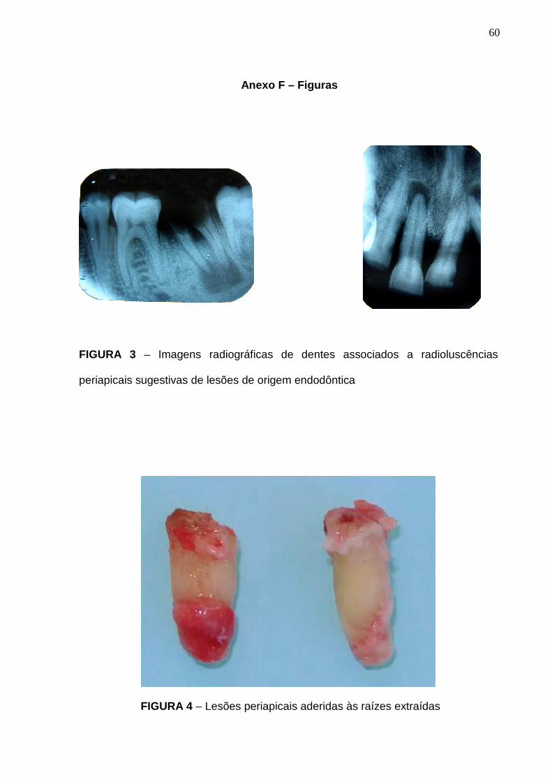
60
Anexo F – Figuras
FIGURA 3 – Imagens radiográficas de dentes associados a radioluscências
periapicais sugestivas de lesões de origem endodôntica
FIGURA 4 – Lesões periapicais aderidas às raízes extraídas