Embed Size (px)
DESCRIPTION
Vol. 5 - N. 1 - Jan/Mar 2012
Citation preview

Vol. 5 - Number 1 January / March 2012
i
Brazilian Journalof Videoendoscopic
Surgery
O f f i c i a l J o u r n a l o f t h e B r a z i l i a n S o c i e t y o f V i d e o s u r g e r y
Production and Distribution - Brazilian Society of VideosurgeryHeadquarters: Avenida das Américas n. 4801, s/ 308
Centro Médico Richet - Barra da Tijuca - Rio de Janeiro, RJ - BrasilCEP: 22.631-004
Telephone and Fax: + 55 21 3325-7724 - [email protected]
Year 5
Vol. 5Number 1
Brazilian Journalof VideoendoscopicSurgery January / March 2012
EDITOR-IN-CHIEFMarco Aurelio Pinho de Oliveira (RJ)
TECHNIQUE EDITORJosé Anacleto Resende Junior (RJ)
ASSISTANT EDITORSMirandolino Batista Mariano (RS)
Marcus Vinicius de Campos Martins (RJ)Sérgio Eduardo Araújo (SP)
ASSOCIATE EDITORS OF SPECIALITIESGeneral Surgery - Miguel Prestes Nácul (RS)
Gynecology - Paulo Augusto Ayroza Galvão Ribeiro (SP)Coloproctology - Fábio Guilherme Campos (SP)
Bariatric Surgery - Sérgio Santoro Santos Pereira (SP)Urology - Mauricio Rubinstein (RJ)
Thoracic Surgery - Rui Haddad (RJ)
NATIONAL EDITORIAL BOARDAlexander Charles Morrell (SP), Alexandre Miranda Duarte (RJ), Antonio de
Pádua Medeiros de Carvalho (AL), Aureo Ludovico de Paula (GO), Celso LuizEmpinotti (SC), Claudio Jose Caldas Bresciani (SP), Claudio Peixoto Crispi (RJ),
Delta Madureira Filho (RJ), Edna Delabio Ferraz (RJ), Edvaldo Fahel (BA),Elizabeth Gomes dos Santos (RJ), Fabricio Borges Carrerette (RJ), Francisco
Luis Altenburg (SC), Francisco Sergio Pinheiro Regadas (CE), Homero LealMeirelles Junior (RJ), Joao de Aguiar Pupo Neto (RJ), Jose de Ribamar Saboia de
Azevedo (RJ), Luis Claudio Pandini (SP), Luiz Augusto Henrique Melki (RJ),Luis Carlos Losso (SP), Lutegarde Vieira Freitas (RJ), Marco Antonio Cezario de
Melo (PE), Marcos Bettini Pitombo (RJ), Maria Cristina Araujo Maya (RJ), MarioRibeiro (MG), Nelson Ary Brandalise (SP), Osorio Miguel Parra (SP), Paulo CezarGalvão do Amaral (BA), Paulo Roberto Cara (RS), Paulo Roberto Savassi Rocha(MG), Renam Catharina Tinoco (RJ), Ricardo Bassil Lasmar (RJ), Ricardo PaivaAraujo Scheiba Zorron (RJ), Roberto Saad Junior (SP), Ronaldo Damião (RJ),
Sergio Brenner (PR), Sergio Carlos Nahas (SP).
Executive Board of DirectorsSOBRACIL - TRIÊNIO 2010-2012
PresidentANTONIO BISPO SANTOS JUNIOR
1st Vice-PresidentFABIO GUILERME C.M. DE CAMPOS
2nd Vice-PresidentHOMERO LEAL DE MEIRELES JUNIOR
General SecretaryCARLOS EDUARDO DOMENE
Assistant SecretaryRENATO LAERCIO TEIXEIRA DOS SANTOS
TreasurerSALVADOR PITCHON
Assistant TreasurerGUILERME XAVIER JACCOUD
North Region Vice-PresidentMARIO RUBENS MACEDO VIANNA
Northeast Region Vice-President
West-Central Region Vice-PresidentRITA DE CASSIA S. DA SILVA TAVARES
Southeast Region Vice-PresidentEDSON RICARDO LOUREIRO
South Region Vice-PresidentARTHUR PACHECO SEABRA
Fiscal CouncilJOSE LUIS DESOUZA VARELA
MARCUS VINICIUS DANTAS C. MARTINSPAULO CESAR GALVÃO DO AMARAL
Total or partial reproduction of this publication isprohibited. Copyright reserved.
Brazilian Journal of Videoendoscopic SurgeryPeriodicity: Trimestral
Circulation: 3.500 exemplaresFree Distribuiton to:
SOBRACIL Associate Members
Subscription and Contact:
ISSN 1983-9901 (press) / 1983-991X (on-line)Eletronic version at:
www.sobracil.org.br
Printing and Publishing: Press Graphic & Publishing LtdRua João Alves, 27 - Saúde - Rio de Janeiro - RJ - Brasil
CEP: 20220-330Phone: + 55 21 2253-8343 [email protected]
INTERNATIONAL EDITORIAL BOARDUrology - Robert Stein (USA), Kenneth Palmer (USA), Fernado Secin (Paraguay),
René Sotelo (Venezuela), Alexis Alva Pinto (Peru)Gynecology - Harry Reich (USA), Keith Isaacson (USA), Resad paya Pasic (USA),
Rudy Leon de Wilde (USA)General Surgery - Eduardo Parra-Davila (USA), Jeffrey M. Marks (USA),
Antonello Forgione (ITA)
English translator - Leigh Jonathan Passman
ii
Vol. 5 - Number 1 January / March 2012Brazilian Journalof Videoendoscopic
Surgery
Cataloging-in-Publication Data
Bras. J. Video-Sur., Rio de Janeiro, v. 5, n. 1, p. 001-054, January / March, 2012
Brazilian Journal of Videoendoscopic Surgery. Brazilian Society ofVideosurgery. Sobracil -- v.5, n1, jan./mar. 2012 --- Rio de Janeiro:Brazilian Journal of Videoendoscopic Surgery. 2012.
Published QuaterlyAbstract
n. 1; 28 cm.
1. Medicine, Videosurgery - Periodicals I. Brazilian Society ofVideosurgery.
CDD 617
References Norms StandardizationLuciana Danielli de Araújo
CRB-7 [email protected]
Grafic Design and ProductionMárcio Alvim de [email protected]

Vol. 5 - Number 1 January / March 2012
i i i
Brazilian Journalof Videoendoscopic
Surgery
January / March 2012
CONTENTS
Brazilian Journalof Videoendoscopic
Surgery
EDITORIAL
A Time of ChangesTempo de MudançasMarco Aurélio Pinho de Oliveira ..................................................................................................................................... 001
ORIGINAL ARTICLE
Comp arison Between Single T rocar Access (SITRACC) Cholecystectomyand Conventional Laparoscopic Cholecystectomy - One year follow-upComparação entre Colecistectomia laparoscópica Single Trocar Access (SITRACC) e Colecistectomialaparoscópica Convencional - Seguimento após um anoJames Skinovsky; Marcus Vinicius Dantas de Campos Martins; Mauricio Chibata; Rogério Cavalieri;Fernanda Tsumanuma; Diogo Falcão ........................................................................................................................... 003
Pelvic Endometriosis – A Survey of Knowledge and Practice Among Gynecologist sEndometriose Pélvica – Enquete com Médicos GinecologistasWilliam Kondo; Paulo Guimarães; Viviane Margareth Scantamburlo; Monica Tessmann Zomer .............................009
Laparoscopic T ransmediastinal Drainage: A Simple and Effective Procedure forReverting an Accident al Pneumothorax During Lap aroscopic Antireflux SurgeryDrenagem transmediastinal laparoscópica: Um procedimento simples e eficaz para reverter umpneumotórax acidental durante uma cirurgia laparoscópica antirefluxoGustavo L. Carvalho; Camila Rocha da Cruz; José Sérgio N. Silva; Diego Laurentino Lima;Rebeca Gonçalves Rocha; Eduardo Felipe de Carvalho Chaves ............................................................................... 024
Laparoscopic Partial Nephrectomy for Renal T umors Larger Than 4cmNefrectomia parcial laparoscópica para tumores renais maiores que 4 cmMirandolino Batista Mariano; Gilvan Neiva Fonseca; Isidoro Henrique Goldaich; Paulo Cerutti Franciscatto;Ana Carolina Brochado Geist; Edna Marin Guimarães Winkler ................................................................................... 029
Does Collagen-Coated Polyester Mesh Decrease the Rate of IntraperitonealAdhesions in Incisional Hernia Repair?A tela de Poliéster recoberta por colágeno diminui a taxa de aderências intraperitoneais no reparoda hérnia incisional?Bárbara Lambert; Bibiana Borges Manna; Juliano Hermes Maeso Montes; André Vicente Bigolin;João Vicente Machado Grossi, Leandro Totti Cavazzola ............................................................................................. 037
Transabdominal Pre-Peritoneal (T APP) Inguinal Hernioplasty by Lap aroendoscopicSingle Site Surgery (LESS). Is it Feasible and Safe?Hernioplastia Inguinal Transabdominal Pré-Peritoneal por Cirurgia Laparoscópica de Acesso Único.É Viável e Segura?James Skinovsky; Marcus Vinicius Dantas de Campos Martins; Sérgio Roll; Mauricio Chibata;Fernanda Tsumanuma; Rogério Cavalliere; Francisco Almeida ................................................................................. 044
SPECIAL SECTION IInformation for Authors .................................................................................................................................................... 049

iv
Vol. 5 - Number 1 January / March 2012Brazilian Journalof Videoendoscopic
Surgery
Dear Contributors,
Publish your manuscript:Original Article, Case Report, Review or Actualization, Preliminary Communications ,
Technique Protocol.Also publish your “Original Image” in videoendoscopic surgery .
Bring and share your experience.Our Journal is On-line!
Manuscript Submission to:
Brazilian Journal of Videoendoscopic SurgeryAvenida das Américas n o 4801, sala 308Centro Médico Richet - Barra da Tijuca22.631-004 Rio de Janeiro - RJ , Brasil
Eletronic Version and fully instructions for submission at:www.sobracil.org.br
Visibility
Future is present at the BJVSYour opinion, experience and scientific investigation are here.
How to be more Efficient with Bibliographic Citations and References 1Vol. 1, Nº 1 EditorialBrazilian Journalof VideoendoscopicSurgery
1
Brazilian Journal of Videoendoscopic Surgery - v. 5 - n. 1 - Jan./Mar. 2012 - Subscription: + 55 21 3325-7724 - E-mail: [email protected] 1983-9901: (Press) ISSN 1983-991X: (on-line) - SOBRACIL - Press Graphic & Publishing Ltd. Rio de Janeiro, RJ-Brasil
Dear Readers,
With this first issue of the 2012 volume weintroduce a new cover design.
The improvements, however, go well beyondthe cover.
The journal has undergone important changes.An international editorial board was established and astandardized model for reviewers has beenimplemented. The lag in publication is narrowing, aswe publish issue No. 1 of the 2012 volume in the samecalendar year. We intend to publish issues No. 2 andNo. 3 in 2012, and No. 4 in early 2013, maintainingthe continuity of the journal and getting it out on time.
Over the course of the past year and a halfwe published a six-part series of scientific methodologyarticles emphasizing study design and basic statisticalconcepts. The series received praise from severalreaders, we hope it stimulates more submissions fromour readers.
Access to the journal through the SOBRACILwebsite has been modernized and readers can nowfind issues from 2010 and 2011. Full versions of articlesfrom these years can be downloaded, for free.
In light of the contributions being made byour new international editorial board, the nationaleditorial board has been streamlined to focus onBrazilian scholars whose CVs are available on theLATTES platform. The vast majority have advancedpost-graduate degrees. All these measures allow usto request indexation by Lilacs and Scielo, the firststep towards the recognition of the journal by theNational Counsel of Technological and ScientificDevelopment (CNPq). Not an easy task, given themany requirement.
Following the global trend, we are in the finalstages of adding tools to the SOBRACIL site that willpermit on-line submission of manuscripts. Indeed,soon, the only way to submit manuscripts will beelectronically. On-line forms will automatically detectsome errors. We expect all this will expedite thereview process, and get our authors contributionspublished faster.
Improving this journal strengthens theBrazilian Society of Videosurgery, We needeveryone’s support!
Give us your feedback. Critiques. Engage.Let’s keep working together.
A Time of Changes
Marco Aurelio Pinho de OliveiraEditor-in-Chief
Brazilian Journal of Videoendoscopic Surgery
Correspondence Address:MARCO AURELIO PINHO DE OLIVEIRA
Rua Coelho Neto, 55 / 201Tel.: (21) 9987-5843
E-mail: [email protected]
Oliveira et al.2 Bras. J. Video-Sur., January / March 2012EditorialBrazilian Journalof VideoendoscopicSurgery
2
Brazilian Journal of Videoendoscopic Surgery - v. 5 - n. 1 - Jan./Mar. 2012 - Subscription: + 55 21 3325-7724 - E-mail: [email protected] 1983-9901: (Press) ISSN 1983-991X: (on-line) - SOBRACIL - Press Graphic & Publishing Ltd. Rio de Janeiro, RJ-Brasil
Caros leitores,
Neste primeiro volume de 2012 estamos apre-sentando um novo desenho de capa. Mas as melhoriasnão devem ficar só na capa.
A revista passou por algumas modificações,incluindo a criação de um conselho editorial internaci-onal e a criação de um modelo padronizado para osrevisores. A defasagem na publicação está diminuin-do, pois publicamos o primeiro número de 2012 den-tro do mesmo ano. A intenção é publicar o 2o e 3onúmeros ainda em 2012 e o 4o no início de 2013, man-tendo a continuidade da revista e entregá-la em dia.
Nas últimas edições foram publicados seiseditoriais sobre os diversos desenhos de estudos, in-cluindo conceitos básicos de estatística. Por contadesta série sobre metodologia científica recebemoselogios de vários leitores, e esperamos que a mesmaajude a aumentar o número de submissões dos nos-sos leitores.
No site da SOBRACIL o acesso para a re-vista foi modernizado e já é possível encontrar todosos números publicados em 2010 e 2011. Todos os ar-
Tempo de Mudanças
tigos destes anos podem ser baixados na íntegra, gra-tuitamente.
Para valorizar as contribuições que vem sen-do feitas pelos nossos novos membros internacionais, oconselho editorial nacional está sendo composto prefe-rencialmente por membros com currículo Lattes. Alémdisso, a grande maioria possui título de doutorado e/oumestrado.Todas estas medidas permitem que a partirde agora possamos pleitear inicialmente uma indexaçãono Lilacs e Scielo, primeiro passo para o reconheci-mento da revista junto ao Conselho Nacional de De-senvolvimento Científico e Tecnológico (CNPq). Nãoé uma tarefa fácil, pois as exigências são muitas.
Seguindo a tendência mundial, estamos na fasefinal da construção de um site dedicado para receberos artigos dos autores. Em breve, a única forma deenvio será o eletrônico. Desta forma esperamosagilizar todo o processo de avaliação e fazer com queas contribuições dos autores possam ser publicadasmais rapidamente.
Valorizar a revista é fortalecer a SOBRACIL.Precisamos do apoio de todos!
Opinem. Critiquem. Enfim, participem.Vamos trabalhar em conjunto.
Mar co Aur elio Pinho de OliveiraEditor-Chefe do Brazilian Journal of Videoendoscopic Surgery
Endereço para Correspondência:MARCO AURELIO PINHO DE OLIVEIRA
Rua Coelho Neto, 55 / 201Tel.: (21) 9987-5843
E-mail: [email protected]
Comparison Between Single Trocar Access (SITRACC) Cholecystectomy andConventional Laparoscopic Cholecystectomy - One year follow-up.
3Vol. 1, Nº 1 Original ArticleBrazilian Journalof VideoendoscopicSurgery
Accepted after revision: january, 13, 2012.Bras. J. V ideo-Sur , 2012, v. 5, n. 1: 003-008
3
Comp arison Between Single T rocar Access (SITRACC)Cholecystectomy and Conventional Lap aroscopic
Cholecystectomy - One year follow-up
Comp aração entre Colecistectomia lap aroscópica Single T rocarAccess (SITRACC) e Colecistectomia lap aroscópica Convencional -
Seguimento após um ano
JAMES SKINOVSKY, PHD1; MARCUS VINICIUS DANTAS DE CAMPOS MARTINS, MD 2;MAURICIO CHIBATA, MD3; ROGÉRIO CAVALIERI 4; FERNANDA TSUMANUMA5; DIOGO FALCÃO 6
Study was conducted at the Red Cross Hospital, Curitiba, PR.1. Head, Surgery Department, Red Cross Hospital and Positivo University, Curitiba, PR; 2. Professor, Surgery
Department, Estácio de Sá University, Rio de Janeiro, RJ; 3. Surgery Department, Red Cross Hospital, PositivoUniversity, Curitiba, PR; 4. Surgery Department, Red Cross Hospital, Curitiba, PR; 5. Surgery Department, Red
Cross Hospital, Curitiba, PR; 6. Surgery Department, Red Cross Hospital, Curitiba, PR.
ABSTRACTObjective: To describe the data obtained after one year follow-up of patients who underwent Single Trocar Access(SITRACC®) cholecystectomy, compared to conventional endoscopic cholecystectomy. Patient s and Method: Twentypatients who underwent SITRACC cholecystectomies and twenty patients who underwent conventionalvideocholecystectomy were questioned using the SF-36 instrument one year after the procedure to evaluate quality of life.The incidence of hernias in the trocar site was also studied. Results: There was no statistically significant differencebetween the groups with regard to quality of life and the trocar hernia rate. There were no major complications in eithergroup. Discussion: The SITRACC device is a new platform for a novel surgical approach. The literature is limitedregarding several important comparative questions, particularly whether this kind of approach truly offers benefits topatients. Studies which compare the SITRACC approach to the conventional laparoscopic approach in term of clinicaloutcomes (quality of life) and complications (the trocar hernia rate) are needed. Conclusions: One year after surgery theSITRACC cholecystectomy group had the same outcomes – in terms of quality of life as measured by the SF-36 – as theconventional laparoscopic cholecystectomy group, at least. There was no increase of trocar hernia cases in the SITRACCgroup. New studies are necessary, using larger series, to compare this new approach to the conventional endoscopicsurgery procedures, especially concerning operative trauma and the metabolic response.
Key words: Videosurgery. Cholecystectomy. Surgery by Single Portal, SITRACC.
INTRODUCTION
Since the 1987 introduction of videosurgery and theconcept of minimally invasive surgery into the
surgical field, it has been amply demonstrated that thisapproach offers patients less suffering, mildermetabolic changes, faster recovery, and superioraesthetic results, and these advances havedisseminating to operating rooms around the worldquickly and enthusiastically.
With constant improvements in the optics andthe instruments available for the performing
videosurgeries, new and more complex proceduresare being carried out successfully using the minimalinvasion approach.
Simultaneously, new technologies andMinimum Access Surgery approaches have emerged,such as Natural Orifice Transluminal EndoscopicSurgery (NOTES), Needlescopy, and Surgery bySingle Access, whose common goal is the search forminimal operative trauma and faster postoperativerecovery, with the fewest complications.
Several platforms for performing SingleAccess Surgery have emerged in recent years1. One
Skinovsky et al.4 Bras. J. Video-Sur., January / March 2012
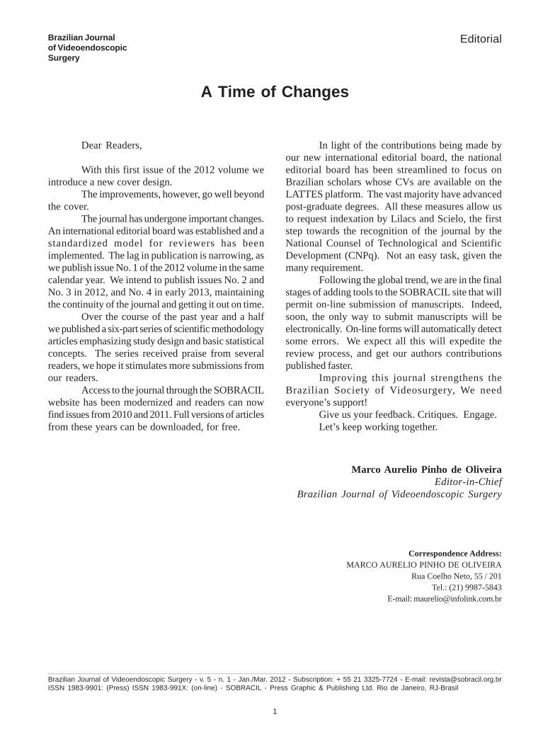
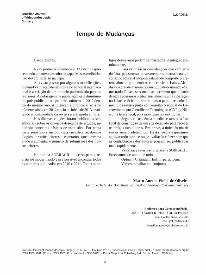
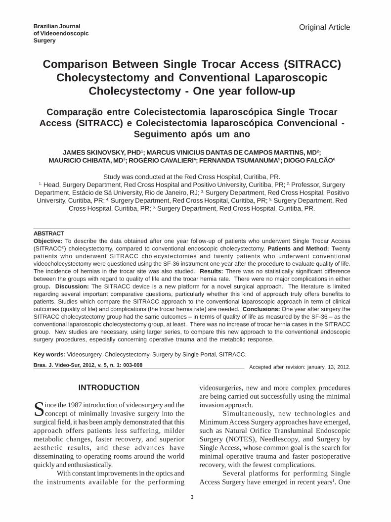
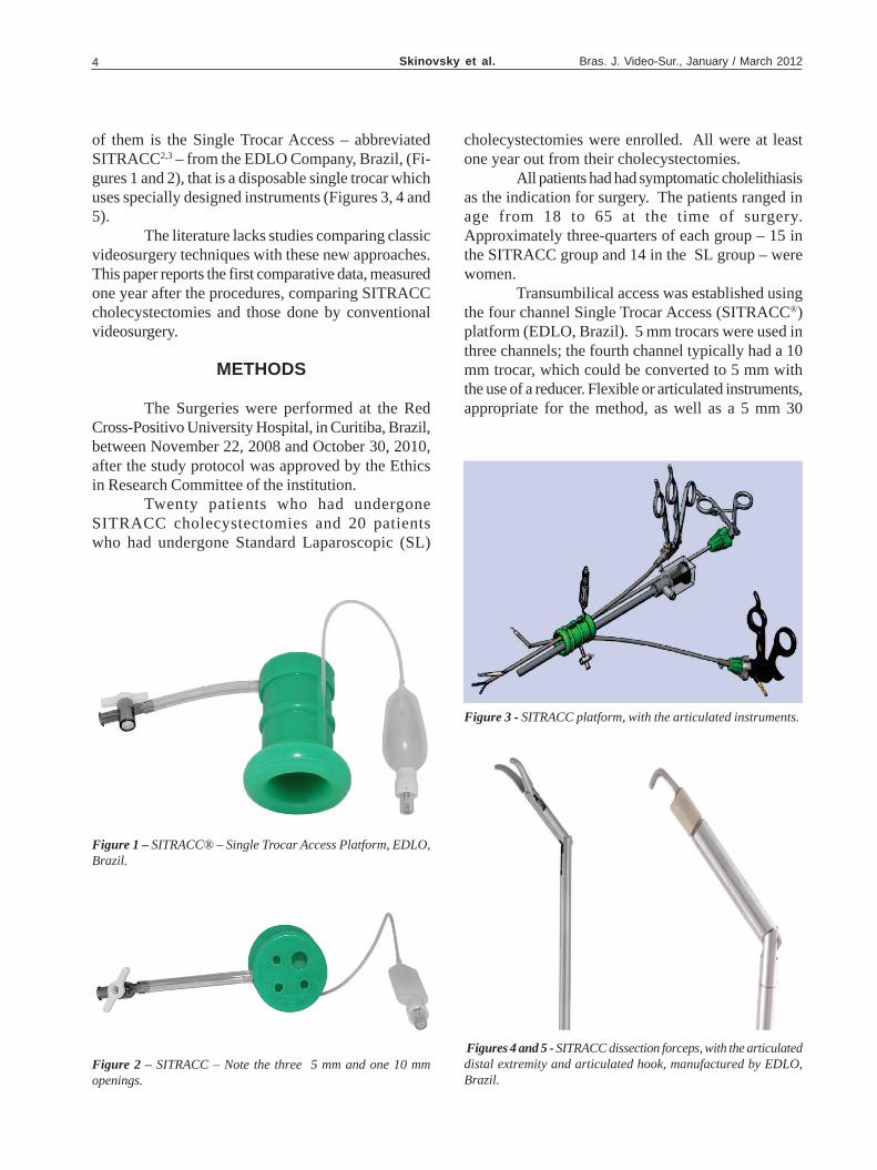
of them is the Single Trocar Access – abbreviatedSITRACC2,3 – from the EDLO Company, Brazil, (Fi-gures 1 and 2), that is a disposable single trocar whichuses specially designed instruments (Figures 3, 4 and5).
The literature lacks studies comparing classicvideosurgery techniques with these new approaches.This paper reports the first comparative data, measuredone year after the procedures, comparing SITRACCcholecystectomies and those done by conventionalvideosurgery.
METHODS
The Surgeries were performed at the RedCross-Positivo University Hospital, in Curitiba, Brazil,between November 22, 2008 and October 30, 2010,after the study protocol was approved by the Ethicsin Research Committee of the institution.
Twenty patients who had undergoneSITRACC cholecystectomies and 20 patientswho had undergone Standard Laparoscopic (SL)
Figure 1 – SITRACC® – Single Trocar Access Platform, EDLO,Brazil.
Figure 2 – SITRACC – Note the three 5 mm and one 10 mmopenings.
Figure 3 - SITRACC platform, with the articulated instruments.
Figures 4 and 5 - SITRACC dissection forceps, with the articulateddistal extremity and articulated hook, manufactured by EDLO,Brazil.
cholecystectomies were enrolled. All were at leastone year out from their cholecystectomies.
All patients had had symptomatic cholelithiasisas the indication for surgery. The patients ranged inage from 18 to 65 at the time of surgery.Approximately three-quarters of each group – 15 inthe SITRACC group and 14 in the SL group – werewomen.
Transumbilical access was established usingthe four channel Single Trocar Access (SITRACC®)platform (EDLO, Brazil). 5 mm trocars were used inthree channels; the fourth channel typically had a 10mm trocar, which could be converted to 5 mm withthe use of a reducer. Flexible or articulated instruments,appropriate for the method, as well as a 5 mm 30
Comparison Between Single Trocar Access (SITRACC) Cholecystectomy andConventional Laparoscopic Cholecystectomy - One year follow-up.
5Vol. 1, Nº 1
degree optic were used. The “StandardLaparoscopic” cholecystectomies were performedfollowing the classic steps.
Quality of life was measured using the ShortForm (36) Health Survey (abbreviated SF-36) whichwas administered by an interviewer by telephonecontact. The occurrence of incisional hernia at thetrocar insertion site was also evaluated.
The SF-36 questionnaire measures healthstatus. It consists of eight scaled scores (rangingfrom 0 to 100), which are calculated as the weightedsums of answers to the questions in each of eightdomains:
1. Functional Capacity2. Physical Aspects3. Pain4. General Health5. Vitality6. Social Aspects7. Emotional Aspects8. Mental Health
The results were tabulated and the groupscores were compared by Mann-Whitney Non-Parametric Test; p values below 0.05 were consideredstatistically significant.
RESULTS
No incisional hernia was reported in eitherthe SITRACC or SL group.
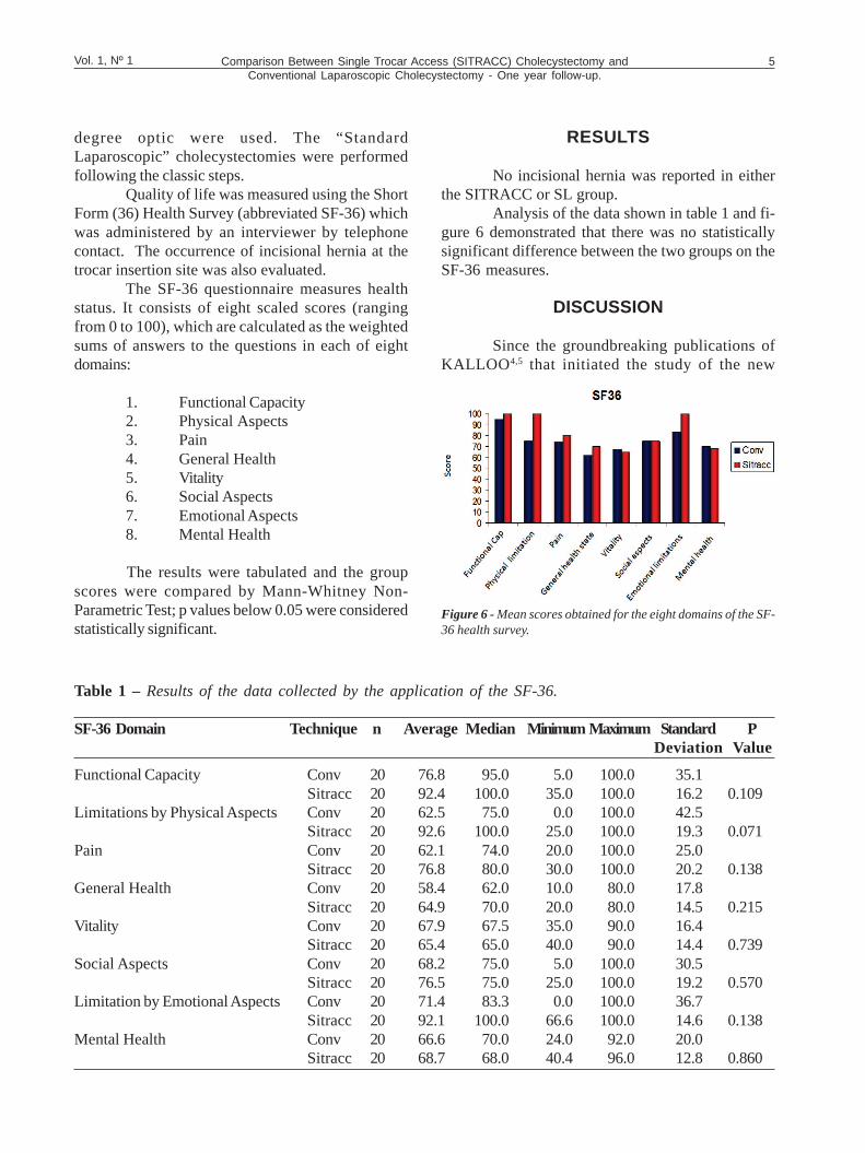
Analysis of the data shown in table 1 and fi-gure 6 demonstrated that there was no statisticallysignificant difference between the two groups on theSF-36 measures.
DISCUSSION
Since the groundbreaking publications ofKALLOO 4,5 that initiated the study of the new
Figure 6 - Mean scores obtained for the eight domains of the SF-36 health survey.
Table 1 – Results of the data collected by the application of the SF-36.
SF-36 Domain Technique n Average Median Minimum Maximum Standard PDeviation Value
Functional Capacity Conv 20 76.8 95.0 5.0 100.0 35.1Sitracc 20 92.4 100.0 35.0 100.0 16.2 0.109
Limitations by Physical Aspects Conv 20 62.5 75.0 0.0 100.0 42.5Sitracc 20 92.6 100.0 25.0 100.0 19.3 0.071
Pain Conv 20 62.1 74.0 20.0 100.0 25.0Sitracc 20 76.8 80.0 30.0 100.0 20.2 0.138
General Health Conv 20 58.4 62.0 10.0 80.0 17.8Sitracc 20 64.9 70.0 20.0 80.0 14.5 0.215
Vitality Conv 20 67.9 67.5 35.0 90.0 16.4Sitracc 20 65.4 65.0 40.0 90.0 14.4 0.739
Social Aspects Conv 20 68.2 75.0 5.0 100.0 30.5Sitracc 20 76.5 75.0 25.0 100.0 19.2 0.570
Limitation by Emotional Aspects Conv 20 71.4 83.3 0.0 100.0 36.7Sitracc 20 92.1 100.0 66.6 100.0 14.6 0.138
Mental Health Conv 20 66.6 70.0 24.0 92.0 20.0Sitracc 20 68.7 68.0 40.4 96.0 12.8 0.860
Skinovsky et al.6 Bras. J. Video-Sur., January / March 2012
approach now known as NOTES, several researchesaround the world have been conducting studies of thenew equipment and instruments for this and evennewer approaches, to determine their viability andpractical application.
The training and demand for newworkstations, access to the abdominal cavity, closureof the stomach and other hollow viscera, the potentialfor infection, the development of new and necessaryequipment, and the orientation difficulty because ofthe use of regular endoscopes have emerged as theprincipal challenges for the development of transluminalsurgery. They need to be overcome to transformNOTES into a common option in clinical and surgicalpractice.
The transumbilical approach now presentsitself as the most viable technology, because thevisualization is similar to conventional videosurgery,and because the development and use of flexible andarticulated instruments allows a degree of triangulation,facilitating surgical maneuvers.
WHEELEES is credited with being the firstto use the principles of single-access surgery, in 1969,to perform tubal ligation.6
In 1997 NAVARRA7 et al.7 describedcholecystectomy performed through two 10 mmtrocars introduced via the umbilicus.
Single Access Surgery entered in a period oflatency, resurfacing in 2007, when ZHU published hisfirst experience using the umbilicus as a single accesspath into the peritoneal cavity, a technique he namedTransumbilical Endoscopic Surgery (TUES).8
In 2008, ZHU et al.9 published a studydescribing new cases of TUES: two cases of hepaticcyst fenestration, six cholecystectomies, and nineappendectomies, using a trocar with three workingchannels.
Also in 2008, Indian authors PALANIVELUet al.10 published a study describing eight transumbilicalappendectomies in which a standard flexible endoscopewas used. The authors considered the technique as apreparatory step for NOTES.
Since then Single Access Surgery techniqueshave been developed for various procedures such anephrectomy and pyeloplasty,11,12,13 adrenalectomy,14
right colectomy,15 sleeve gastrectomy,16,17 adjustablegastric band,18 Roux-en-Y gastric bypass,19
gastrostomy,20 intracorporeal gastrojejunostomy,21 andsplenectomy,22 among others.
Procedures in several surgical subspecialtieshave been successfully performed using single surgicalaccess techniques. Data to date indicate thattransumbilical surgery is feasible and safe,23 but theliterature is quite limited in terms of medium to longterm follow-up and in terms of studies comparing singleaccess surgery and so-called conventionalvideosurgery.
The SF- 36 quality of life health survey wasdeveloped in the USA in the 1980s, and have bewidely used and validated by several studies. TheSF-36 is a multi-purpose, short-form health surveywith 36 questions. It yields an 8-scale profile offunctional health and well-being scores as well aspsychometrically-based physical and mental healthsummary measures and a preference-based healthutility index. It is a generic measure, as opposed toone that targets a specific age, disease, or treatmentgroup. Accordingly, the SF-36 has proven useful insurveys of general and specific populations,comparing the relative burden of diseases, and indifferentiating the health benefits produced by a widerange of different treatments. The experience todate with the SF-36 has been documented in nearly4,000 publications, including surgicalstudies.24,25,26,27,28
The benefits of Single Access Surgery ascompared to NOTES vary, but include from theprinciples of Scarless Surgery – operations that leavelittle or no scar – to the improved vision provided(which surgeons already use in regular laparoscopicprocedures), and the low risk of infection.
We still need large double blind series, thatcompare similar procedures performed using singleaccess surgery techniques with those performedby regular videosurgical methods. Data collectedin this study begins to demonstrate that single accesssurgery has, in the medium and long term,outcomes that are at least comparable to outcomesobtained with the current “gold standard”approach for performing cholecystectomy, thevideocholecystectomy.
Single access surgery procedures need to beviewed as part of an operative arsenal that extendsfrom open surgery to videosurgery and NOTES.Each patient is unique, as is his or her illness. It is upto the experienced surgeon to determine the bestapproach that offers security and better surgicaloutcomes and aesthetic results.
Comparison Between Single Trocar Access (SITRACC) Cholecystectomy andConventional Laparoscopic Cholecystectomy - One year follow-up.
7Vol. 1, Nº 1
RESUMOObjetivos: Descrever os dados obtidos pelo menos um ano após a realização de colecistectomias pela abordagemSingle Trocar Access (SITRACC®), comparadas àquelas realizadas pela abordagem laparoscópica convencional. Paci-entes e Método: Foram estudados vinte pacientes SITRACC e vinte pacientes submetidos à colecistectomia laparoscópicaconvencional, todos eles pelo menos um ano após o procedimento, tendo sido submetidos ao questionário SF-36,classicamente utilizado como medida de aferição da qualidade de vida, bem como também foi avaliada a incidência dehérnia em sítio de trocater. Result ados: Não houve diferença estatística significativa entre ambos os grupos estudados,tanto com relação à qualidade de vida quanto sobre o montante de incidência da hérnia em local de trocater. Igualmenteentre ambos os grupos não foram relatadas complicações maiores. Discussão: A plataforma SITRACC é um novoequipamento para uma nova abordagem, que necessita de estudos comparativos com a abordagem convencionalmais aprofundados, bem como sobre a incidência de hérnia incisional. A literatura disponível é escassa na resposta dediversas questões comparativas, especialmente se este tipo de abordagem realmente representa benefício real paraos pacientes. Conclusões: O grupo submetido a colecistectomia SITRACC apresentou o mesmo nível de satisfação,com relação a qualidade de vida, quando comparado ao grupo convencional, um ano após os procedimentos. Nãohouve aumento de incidência de hérnia incisional no grupo Single Trocar Access. Novos estudos são necessários,utilizando-se séries maiores, para comparar esta nova abordagem aos procedimentos videocirúrgicos convencionais,especialmente no que diz respeito ao trauma cirúrgico e à resposta metabólica.
Palavras chave: Videocirurgia. Colecistetomia. Cirurgia por Portal Único, SITRAC.
REFERENCES
1. Galvão Neto M, Ramos A, Campos J. Single portlaparoscopy Access surgery. Tech Gastrointest Endosc 2009;11(2):84-94.
2. Dantas MVDC, Skinovsky J, Coelho DE, Torres MF.SITRACC – Single Trocar Access: a new device for a newsurgical approach. Bras J Vide-Surg 2008; 1(2):60-63.
3. Dantas MVDC, Skinovsky J, Coelho DE, Ramos A, GalvãoNeto MP, Rodrigues J, de Carli L, Cavazolla, LT, Campos J,Thuller F, Brunetti A. Cholecystectomy by single trocaraccess – SITRACC: The first multicenter study. Surg Innov2009; Dez.
4. Kalloo AN, Sibgh VK, Jagannath SB, Niiyama H, VaughCA, Magee CA, Kantsevoy SV. Flexible transgastricperitoneoscopy: a novel approach to diagnostic andtherapeutic interventions. Gastrointest Endosc 2004; 60:114-7.
5. Giday SA, Kantsevoy SV, Kalloo AN. Principle and historyof natural orifice translumenal endoscopic surgery (NOTES).Minim Invasive Ther Allied Technol 2006; 15:373-377.
6. Wheeless CR. A rapid, inexpensive and effective method ofsurgical sterilization by laparoscopy. J Reprod Med 1969;5:255.
7. Navarra G: One-wound laparoscopic cholecystectomy. Br JSurg 1997; 84(5):695.
8. Zhu JF. Scarless endoscopic surgery: NOTES or TUES.Surg Endosc 2007; 21:1898-1899.
9. Zhu JF, Hu H, Ma YZ, Xu MZ, Li F. Transumbilicalendoscopic surgery: a preliminary clinical report. Surg Endosc[online periodical] 2008; Available from: URL:http//www.springerlink.com/content [consulted on 02/10/2010].
10. Papanivelu C, Rajan PS, Rangarajan M, Parthasarathi R,Senthilnathan P, Praveenraj P. Transumbilical endoscopicappendectomy in humans: on the road to NOTES: aprospective study. J Laparoendosc & Advanced Surg Tech2008; 18(4): 579-582.
11. Desai MM, Rao PP, Aron M, Haber GP, Desai M, MishraS, Kaouk JH, Gill IS. Scarless single port transumbilicalnephrectomy and pyeloplasty: first clinical report. Brit JUrology 2008; 101: 83-88.
12. Kaouk JH, Haber GP, Goel RK. Single-port laparoscopicsurgery in urology: initial experience. Urology 2008; 71(1):3-6.
13. Rané A, Rao P, Rao Pr. Single-Port-Access Nephrectomyand other laparoscopic urologic procedures using a novellaparoscopic port (R-Port). Urology 2008; 72:260-264.
14. Castellucci SA, Curcillo PG, Ginsberg PC, et al: Single-portaccess adrenalectomy. J Endourol 2008; 22:1573-1576.
15. Bucher P, Pugin F, Morel P. Single port access laparoscopicright hemicolectomy. Int J Colorectal Dis 2008; 23:1013-1016.
16. Saber AA, Elgamal MH, Itawi EA, Rao AJ. Single incisionlaparoscopic sleeve gastrectomy (SILS): a novel technique.Obes Surg 2008; 18:1338-1342.
17. Reavis KM, Hinojosa MW, Smith BR, et al: Singlelaparoscopic-incision transabdominal surgery sleevegastrectomy. Obes Surg 2008; 18(11):1492-1494.
18. Teixeira J, McGill K, Binenbaum S, Forrester G. Laparoscopicsingle-site surgery for placement of an adjustable gastric band:initial experience. Surg Endosc 2009; 23:1409-1414.
19. Saber AA, El-Ghazaly T, Minnick D. Single port accesstransumbilical laparoscopic Roux-en-Y gastric bypass usingthe SILS port: first reported case. Surg Innov 2009.
Skinovsky et al.8 Bras. J. Video-Sur., January / March 2012
20. Podolsy ER, Rottman SJ, Curcillo PG. Single Port Access(SPATM) gastrostomy tube in patients unable to receivepercutaneous endoscopic gastrostomy placement. SurgEndosc 2009; 23:1142-1145.
21. Busher P, Pugin F, Morel P. Transumbilical single-incisionlaparoscopic intracorporeal anastomosis forgastrojejunostomy: case report. Surg Endosc 2009; 23:1667-1670.
22. Targarona EM, Balaque C, Martinez C, Pallares L, EstalellaL, Trias M. Single-Port Access: a feasible alternative toconventional laparoscopic splenectomy. Surg Innov 2009.
23. Romanelli JR, Earle DB. Single-port laparoscopic surgery:an overview. Surg Endosc 2009; 23:1419-1427.
24. Ware JE Jr, Gandek B. Overview of the SF-36 health surveyand the international quality of life assessment (IQOLA)project. J Clin Epidemiol 1998; 51(11):903-912.
25. Brazier JE, Harper R, Jones NMB, O’Cathain A, ThomasKJ, Usherwood T, Westlake L. Validating the SF-36 healthsurvey questionnaire: new outcome measure for primarycare. British Medical Journal 1992; 305:160-164.
26. Brazier JE, Roberts J, Deverill M. The estimation of areference-based measure of health from the SF-36. J HealthEconomics 2002; 21:271-292.
27. Dymek MP, le Grange D, Neven K, Alverdy J. Quality oflife after gastric bypass surgery: a cross-sectional study.Obesity Surg 2002; 10:1135-1142.
28. Brennan JC, Steele RJC. Measurement of quality of life insurgery. J R Coll Surg Edinb 1999; 44:252-259.
Corr esponding Author:JAMES SKINOVSKYAv. Iguaçú 2713, Apt. 503, Água VerdeCuritiba, PR, Brazil80240-030E-mail: [email protected]
Brazilian Journal of Videoendoscopic Surgery - v. 5 - n. 1 - Jan./Mar. 2012 - Subscription: + 55 21 3325-7724 - E-mail: [email protected] 1983-9901: (Press) ISSN 1983-991X: (on-line) - SOBRACIL - Press Graphic & Publishing Ltd. Rio de Janeiro, RJ-Brasil
Pelvic Endometriosis – A Survey of Knowledge and Practice Among Gynecologists 9Vol. 1, Nº 1 Original ArticleBrazilian Journalof VideoendoscopicSurgery
Accepted after revision: january, 16, 2012.Bras. J. V ideo-Sur , 2012, v. 5, n. 1: 009-023
9
Pelvic Endometriosis – A Survey of Knowledge andPractice Among Gynecologist s
Endometriose Pélvica – Enquete com Médicos Ginecologist as
WILLIAM KONDO 1; PAULO GUIMARÃES 2; VIVIANE MARGARETH SCANT AMBURLO 3;MONICA TESSMANN ZOMER 4
The Word took place in Sugisawa Hospital and Medical Center.1. Gynecologist and General Surgeon, Sugisawa Hospital and Medical Center, Curitiba, Paraná. Ex-Fellow,
Department de Gynecologic Surgery, CHU Estaing, Clermont-Ferrand, França; 2. Ginecologista;3. Gynecologist, Sugisawa Hospital and Medical Center, Curitiba, Paraná. Specialist in Human Reproduction,
Androlab, Curitiba, Paraná; 4. Gynecologist, Sugisawa Hospital and Medical Center, Curitiba, Paraná
ABSTRACTPurpose: Endometriosis affects a large number of reproductive age women. Clinical manifestations of the disease arecyclic pelvic pain and infertility. Women with endometriosis usually seek medical advice from a gynecologist for theirsymptoms. The role of the gynecologist is therefore crucial in identifying, treating, and, when appropriatey, referring thesepatients promptly to specialised centers. Methods: A brief questionnaire was completed anonymously by 40 Braziliangynecologists. Results: 67.5% of the respondents perform surgery for endometriosis. Approximately half (55%) of therespondents stated that the physical examination can diagnose cases of deeply infiltrating endometriosis; 92.5% do notexclude the possibility of deep endometriosis when serum CA-125 levels are normal. Magnetic resonance imaging,transvaginal ultrasound and colonoscopy are important in the preoperative assessment of the patient for 72.5%, 70%,and 62.5% of the respondents. For 62.5% of the respondents, GnRH analogues are the best medical management forendometriosis. Only 17.5% of the gynecologists think that all hormone-based treatments have similar outcomes.Although 80% of gynecologists responded that complete resection of the disease is the best treatment for deep lesions,44% of the gynecologists that perform surgery for endometriosis recommend only diagnostic laparoscopy in thesecases. Only 7.4% of the respondents are able to treat deep endometriosis with bowel involvement without the aid of ageneral surgeon or a colorectal surgeon. Conclusions: More education is required among gynecologists on the subjectof endometriosis, in order to identify and treat patients with this disease. Referral to a center with the necessary expertiseto offer all available treatments in a multi-disciplinary context is important to improve the surgical outcomes of deepinfiltrating endometriosis.
Key words: Endometriosis. Diagnosis. Treatment. Deep endometriosis. Laparoscopy.
INTRODUCTION
Endometriosis is a benign gynecological diseasedefined as the presence of endometrial tissue –
consisting of gland and/or stroma – outside of theuterine cavity.1 This condition is predominantly foundin women of reproductive age and affectsapproximately seven to ten percent of all women, 71%to 87% of women with chronic pelvic pain, and 38%of women with infertility.2
The deep infiltrating disease has been definedas endometriosis that penetrates more than 5mmbelow the peritoneal surface and is strongly associated
with severe chronic pelvic pain, dyspareunia anddysmenorrhea. In this situation the endometrioticimplants may involve the uterosacral ligaments, pouchof Douglas (retrocervical endometriosis), rectovaginalseptum and even the rectum, bladder and ureters.3
The diagnosis includes deep history, physicalexamination and complementary imaging tests to stagethe disease and plan a possible surgical treatment.4,5
Treatment should be individualized for eachpatient. Hormonal treatments are effective forcontrolling pain related to endometriosis, but are notan option in women wishing to get pregnant.6,7 Surgicaltreatment is indicated for the histopathologic diagnosis
Kondo et al.10 Bras. J. Video-Sur., January / March 2012
of the disease and is necessary in symptomatic patientsto complete medical treatment. In general, theevidence suggests that complete excision ofendometriosis offers prolonged symptom relief,particularly in women with severe or debilitatingsymptoms. To ensure complete removal of diseaseand for the best results in terms of quality of life,several procedures may be required, including surgicalmanipulation of the bladder, ureters, rectum, and va-gina.
The complication rate for laparoscopic surgeryfor deep endometriosis is estimated to be 3.4%, andmay reach 10% to 22% when intestinal resection isnecessary.3 Postoperative intestinal fistula have adeleterious impact on women’s fertility and quality oflife and is the most worrisome complication of thistype of surgery.
In this article, we report our assessment ofthe knowledge of a group of Brazilian gynecologistswith regard to clinical evaluation and treatment ofendometriosis using hypothetical cases.
METHODS
From June to December 2010 we surveyedgynecologists about endometriosis. A questionnaire(available in the Appendix of this article) was sentelectronically to 400 members of the Society ofObstetricians and Gynecologists of Paraná (SOGIPA),chosen randomly, and was hand-delivered to 40physicians taking laparoscopy courses (Gynelaser)held in Brasilia.
Data analysis was conducted using version8.0 of the STATISTICA statistical software package.
Os dados foram inseridos no programaSTATISTICA 8.0 e avaliados.
RESULTS
Only the questionnaires hand-delivered in thelaparoscopy courses were completed (n = 40),accounting for only 9.1% of the 440 questionnairesdistributed. No gynecologist answered the surveyelectronically.
Gynecologists who answered the survey hadbeen in a practice an average of 15.2 ± 10 years(range 1-40 years). Thirty percent of gynecologistsreported having specific training in the treatment ofendometriosis; 45% responded that they performlaparoscopic surgeries. Forty-five percent reportedperforming open surgery for the treatment of deependometriosis, and 30% said they performlaparoscopic surgery for deep endometriosis. Asthree of the 40 gynecologists (7.5%) reportedperforming both open and laparoscopic surgery forendometriosis, the total percentage of gynecologistsamong survey respondents that treat deependometriosis was 67.5%. Fifty percent ofgynecologists have already participated in oraccompanied surgeries of deep endometriosisinvolving the retrocervical region, the rectovaginalseptum, or the intestine.
Regarding the diagnosis of endometriosis, 55%stated that the diagnosis of endometriosis can besuspected or established by the gynecologicalexamination. For 65% of the gynecologists the valueof serum CA-125 levels is important for the diagnosisof endometriosis, but 92.5% do not rule out thediagnosis of endometriosis when the CA-125 level isnormal.
Table 1 shows the imaging studiesgynecologists consider important in the preoperativeinvestigation of deep endometriosis.
Table 1 - Responses to the question: “Which tests do you consider important for the preoperative diagnosisand investigation of deep endometriosis?”
Imaging Test Yes No
Transvaginal Ultrasound 28 (70%) 12 (30%)Transvaginal Ultrasound after bowel prep 22 (55%) 18 (45%)Ultrasound of the urinary tract 16 (40%) 24 (60%)CT of the Pelvis 12 (30%) 28 (70%)Magnetic Resonance Imaging of the pelvis 29 (72.5%) 11 (27.5%)Colonoscopy 25 (62.5%) 15 (37.5%)Transrectal Ultrasound 16 (40%) 24 (60%)
Pelvic Endometriosis – A Survey of Knowledge and Practice Among Gynecologists 11Vol. 1, Nº 1
In the setting of a clinical suspicion ofendometriosis, but without abnormal findings onphysical examination, or in imaging studies orlaboratory tests, 57.5% of gynecologists recommenda diagnostic laparoscopy and 42.5% said they try atherapeutic trial with an oral contraceptive.
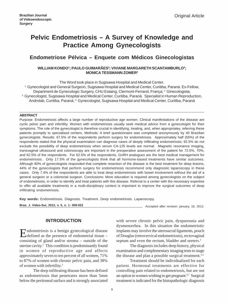
In patients with endometriosis lesions largerthan 1 cm in diameter in the uterosacral ligament, 35%of gynecologists believe that colonoscopy is the mostimportant test to include in the preoperative investigation.For 30% of gynecologists computed tomography of thepelvis is the most important test and for 22.5%ultrasonography of the urinary tract should be orderedas part of the pre-operative evaluation (Figure 1).
For deep endometriosis, 80% of thegynecologists surveyed considered complete resectionof the lesion the ideal surgical treatment and 15% fa-vor bipolar coagulation of lesions. Five percent of thegynecologists responded “I don’t know.” Of the 27gynecologists who reported performing surgery fordeep endometriosis, 81.5% responded that they docomplete resection of the lesions (Table 2).
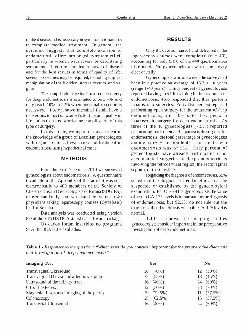
For 52.5% of gynecologists, the deep lesionsof endometriosis in uterosacral ligament exceeding 1cm in diameter should be treated by means of com-plete resection of the lesion associated with ureterolysis(Figure 2).
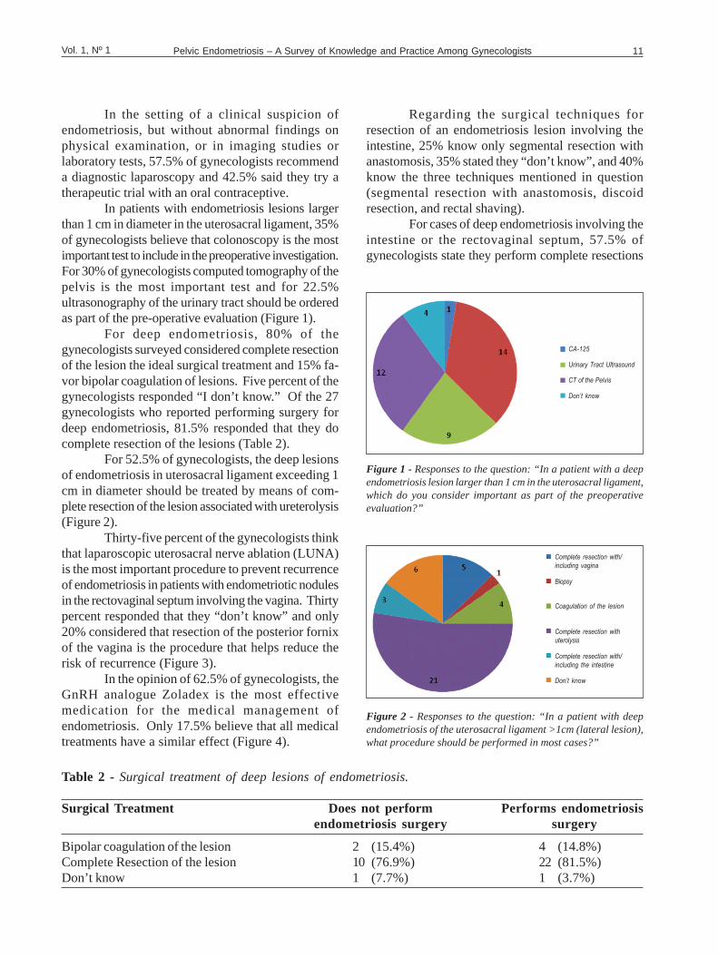
Thirty-five percent of the gynecologists thinkthat laparoscopic uterosacral nerve ablation (LUNA)is the most important procedure to prevent recurrenceof endometriosis in patients with endometriotic nodulesin the rectovaginal septum involving the vagina. Thirtypercent responded that they “don’t know” and only20% considered that resection of the posterior fornixof the vagina is the procedure that helps reduce therisk of recurrence (Figure 3).
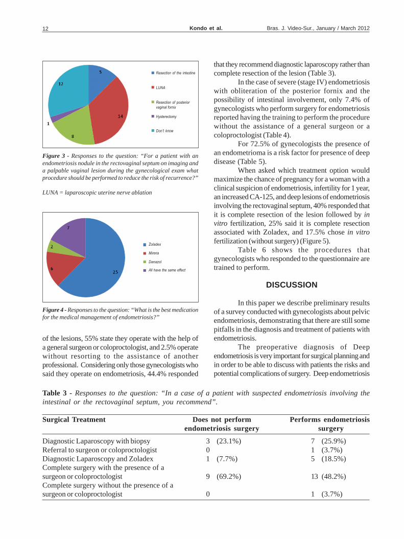
In the opinion of 62.5% of gynecologists, theGnRH analogue Zoladex is the most effectivemedication for the medical management ofendometriosis. Only 17.5% believe that all medicaltreatments have a similar effect (Figure 4).
Regarding the surgical techniques forresection of an endometriosis lesion involving theintestine, 25% know only segmental resection withanastomosis, 35% stated they “don’t know”, and 40%know the three techniques mentioned in question(segmental resection with anastomosis, discoidresection, and rectal shaving).
For cases of deep endometriosis involving theintestine or the rectovaginal septum, 57.5% ofgynecologists state they perform complete resections
Table 2 - Surgical treatment of deep lesions of endometriosis.
Surgical Tr eatment Does not perform Performs endometriosisendometriosis surgery surgery
Bipolar coagulation of the lesion 2 (15.4%) 4 (14.8%)Complete Resection of the lesion 10 (76.9%) 22 (81.5%)Don’t know 1 (7.7%) 1 (3.7%)
Figure 1 - Responses to the question: “In a patient with a deependometriosis lesion larger than 1 cm in the uterosacral ligament,which do you consider important as part of the preoperativeevaluation?”
CA-125
Don’t know
CT of the Pelvis
Urinary Tract Ultrasound
Figure 2 - Responses to the question: “In a patient with deependometriosis of the uterosacral ligament >1cm (lateral lesion),what procedure should be performed in most cases?”
Complete resection with/
including vagina
Don’t know
Complete resection with/
including the intestine
Complete resection with
uterolysis
Coagulation of the lesion
Biopsy
Kondo et al.12 Bras. J. Video-Sur., January / March 2012
that they recommend diagnostic laparoscopy rather thancomplete resection of the lesion (Table 3).
In the case of severe (stage IV) endometriosiswith obliteration of the posterior fornix and thepossibility of intestinal involvement, only 7.4% ofgynecologists who perform surgery for endometriosisreported having the training to perform the procedurewithout the assistance of a general surgeon or acoloproctologist (Table 4).
For 72.5% of gynecologists the presence ofan endometrioma is a risk factor for presence of deepdisease (Table 5).
When asked which treatment option wouldmaximize the chance of pregnancy for a woman with aclinical suspicion of endometriosis, infertility for 1 year,an increased CA-125, and deep lesions of endometriosisinvolving the rectovaginal septum, 40% responded thatit is complete resection of the lesion followed by invitro fertilization, 25% said it is complete resectionassociated with Zoladex, and 17.5% chose in vitrofertilization (without surgery) (Figure 5).
Table 6 shows the procedures thatgynecologists who responded to the questionnaire aretrained to perform.
DISCUSSION
In this paper we describe preliminary resultsof a survey conducted with gynecologists about pelvicendometriosis, demonstrating that there are still somepitfalls in the diagnosis and treatment of patients withendometriosis.
The preoperative diagnosis of Deependometriosis is very important for surgical planning andin order to be able to discuss with patients the risks andpotential complications of surgery. Deep endometriosis
Table 3 - Responses to the question: “In a case of a patient with suspected endometriosis involving theintestinal or the rectovaginal septum, you recommend”.
Surgical Tr eatment Does not perform Performs endometriosisendometriosis surgery surgery
Diagnostic Laparoscopy with biopsy 3 (23.1%) 7 (25.9%)Referral to surgeon or coloproctologist 0 1 (3.7%)Diagnostic Laparoscopy and Zoladex 1 (7.7%) 5 (18.5%)Complete surgery with the presence of asurgeon or coloproctologist 9 (69.2%) 13 (48.2%)Complete surgery without the presence of asurgeon or coloproctologist 0 1 (3.7%)
Figure 3 - Responses to the question: “For a patient with anendometriosis nodule in the rectovaginal septum on imaging anda palpable vaginal lesion during the gynecological exam whatprocedure should be performed to reduce the risk of recurrence?”
LUNA = laparoscopic uterine nerve ablation
Resection of the intestine
Don’t know
Hysterectomy
Resection of posterior
vaginal fornix
LUNA
Figure 4 - Responses to the question: “What is the best medicationfor the medical management of endometriosis?”
Zoladex
All have the same effect
Danazol
Mirena
of the lesions, 55% state they operate with the help ofa general surgeon or coloproctologist, and 2.5% operatewithout resorting to the assistance of anotherprofessional. Considering only those gynecologists whosaid they operate on endometriosis, 44.4% responded
Pelvic Endometriosis – A Survey of Knowledge and Practice Among Gynecologists 13Vol. 1, Nº 1
involving the uterosacral ligaments, retrocervical region,pouch of Douglas, and posterior vaginal fornix can usuallybe palpated on digital vaginal examination when theyexceed 5 to 10mm in diameter. The physical examinationis important because guides the selection of laboratoryand imaging tests to be ordered. The clinical/physicalexamination should include: (1) inspection of theretrocervical area as well as the upper portion of theposterior vaginal wall in search of typical bluish lesions,and (2) vaginal examination in search of nodules in theuterosacral ligaments and pain upon extension of theuterosacral ligaments. By re-examining the patient duringthe menstrual period we can increase the performanceof the exam.9 In our study, only 55% of gynecologistsresponded that deep endometriosis nodules can be felton physical examination, this probably has a direct impacton clinical diagnosis. Gynecologists who think that thedisease cannot be felt on physical examination probablydo not search for retrocervical nodules or nodules in theposterior vaginal fornix during the vaginal digitalexamination, which may be one of the reasons for failureto diagnose this disease and for delay in diagnosis. Thisis consistent with the findings of Arruda et al10 whoshowed that the time between symptom onset anddiagnosis of endometriosis in Brazilian women is aboutseven years.
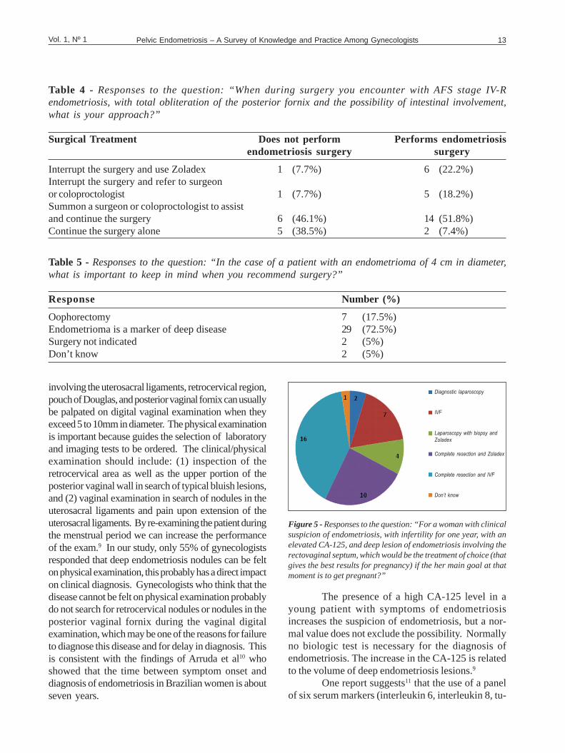
Table 4 - Responses to the question: “When during surgery you encounter with AFS stage IV-Rendometriosis, with total obliteration of the posterior fornix and the possibility of intestinal involvement,what is your approach?”
Surgical Tr eatment Does not perform Performs endometriosisendometriosis surgery surgery
Interrupt the surgery and use Zoladex 1 (7.7%) 6 (22.2%)Interrupt the surgery and refer to surgeonor coloproctologist 1 (7.7%) 5 (18.2%)Summon a surgeon or coloproctologist to assistand continue the surgery 6 (46.1%) 14 (51.8%)Continue the surgery alone 5 (38.5%) 2 (7.4%)
Table 5 - Responses to the question: “In the case of a patient with an endometrioma of 4 cm in diameter,what is important to keep in mind when you recommend surgery?”
Response Number (%)
Oophorectomy 7 (17.5%)Endometrioma is a marker of deep disease 29 (72.5%)Surgery not indicated 2 (5%)Don’t know 2 (5%)
The presence of a high CA-125 level in ayoung patient with symptoms of endometriosisincreases the suspicion of endometriosis, but a nor-mal value does not exclude the possibility. Normallyno biologic test is necessary for the diagnosis ofendometriosis. The increase in the CA-125 is relatedto the volume of deep endometriosis lesions.9
One report suggests11 that the use of a panelof six serum markers (interleukin 6, interleukin 8, tu-
Figure 5 - Responses to the question: “For a woman with clinicalsuspicion of endometriosis, with infertility for one year, with anelevated CA-125, and deep lesion of endometriosis involving therectovaginal septum, which would be the treatment of choice (thatgives the best results for pregnancy) if the her main goal at thatmoment is to get pregnant?”
Don’t know
Diagnostic laparoscopy
IVF
Laparoscopy with biopsy and
Zoladex
Complete resection and Zoladex
Complete resection and IVF
Kondo et al.14 Bras. J. Video-Sur., January / March 2012
mor necrosis factor alpha, high-sensitivity protein C,CA-125 and CA-19-9) to test specimens obtainedduring the secretory phase or during menstruationallow the diagnosis of both minimal and mild andmoderate to severe endometriosis, with high sensitivityand clinically acceptable specificity.
Kafali et al12 showed that it may be possibleto make a clinical diagnosis of endometriosis byevaluating differences in CA-125 levels during themenses with the rest of the menstrual cycle. In theirstudy of 28 women, there was a 22% increase in serumCA-125 levels during the menstrual period (12.2 U /ml) compared with the rest of the menstrual cycle (10U/ml) in the control group. This increase was alsoobserved in women with endometriosis, but the levelsvaried 198.3%. The mean CA-125 levels in thesepatients was 35.8 U/ml during menses compared with12 U/ml during the rest of the menstrual cycle. In oursurvey, 65% of gynecologists said that CA-125 isimportant for the diagnosis of endometriosis, but 92.5%would not rule out the possibility of deep endometriosiswhen the CA-125 value is normal.
Several imaging studies have been used tomap the lesions of deep endometriosis. The mostcommonly reported in the literature are transvaginalpelvic ultrasound (with or without a bowel preparation),magnetic resonance imaging, and transrectalultrasonography. Abrao et al4 evaluated the ability ofthe clinical examination, transvaginal ultrasonography,and magnetic resonance imaging to diagnosisendometriosis with retrocervical or recto-sigmoidinvolvement.
One hundred and four women with a clinicalsuspicion of endometriosis were evaluated using thesethree diagnostic methods which were later correlatedwith the surgical specimen histopathologic findings.Endometriosis was confirmed histologically in 94.2%of patients. Regarding recto-sigmoid or retrocervical
region involvement, respectively, the digitalexamination had a sensitivity of 72% and 68%, aspecificity of 54% and 46%, positive predictive valueof 63% and 45%, negative predictive value of 64%and 69%, and an accuracy of 63% and 55%. Fortransvaginal ultrasound, the sensitivity was 98% and95%, the specificity was 100% and 98%, positivepredictive value was 100% and 98%, negativepredictive value was 98% and 97% and the accuracywas 99% and 97%. MRI had a sensitivity of 83%and 76%, a specificity of 98% and 68%, positivepredictive value of 98% and 61%, negative predictivevalue of 85% and 81% and an accuracy of 90% and71%.
In a similar fashion, Bazot et al5 comparedthe value of the physical examination, transvaginalultrasound, transrectal ultrasound, and magneticresonance imaging in the evaluation of differentlocations of deep infiltrating endometriosis. Ninety-two patients with clinical evidence of pelvicendometriosis were evaluated retrospectively. Thesensitivity and positive and negative likelihood ratiosfor physical examination, transvaginal ultrasound,transrectal ultrasound, and magnetic resonanceimaging were respectively 73.5%, 3.3, and 0.34;78.3%, 2.34, and 0.32; 48.2%, 0.86 and 1.16; and84.4%, 7.59, and 0.18 for endometriosis in uterosacralligaments; 50%, 3.88 and 0.57; 46.7%, 9.64 and 0.56;6.7%, –, 0.93; and 80%, 5.51, and 0.23 for vaginalendometriosis; and 46%, 1.67, and 0.75; 93.6%, – ,and 0.06, 88.9%, 12.89 and 0.12; and 87.3%, 12.66and 0.14 for intestinal endometriosis. The authorsconcluded that MRI has results comparable totransvaginal ultrasound and transrectal ultrasound forthe diagnosis of intestinal endometriosis, but has agreater sensitivity and higher likelihood ratios for thediagnosis of endometriosis in the uterosacral ligamentsand in the vagina.
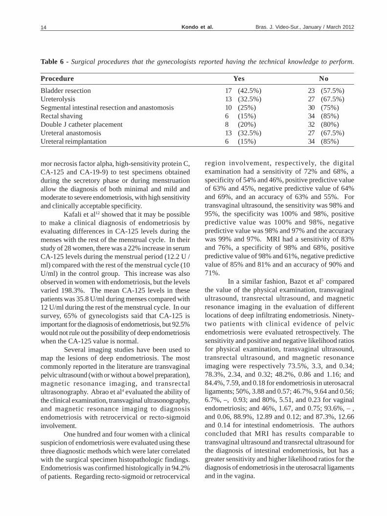
Table 6 - Surgical procedures that the gynecologists reported having the technical knowledge to perform.
Procedure Yes No
Bladder resection 17 (42.5%) 23 (57.5%)Ureterolysis 13 (32.5%) 27 (67.5%)Segmental intestinal resection and anastomosis 10 (25%) 30 (75%)Rectal shaving 6 (15%) 34 (85%)Double J catheter placement 8 (20%) 32 (80%)Ureteral anastomosis 13 (32.5%) 27 (67.5%)Ureteral reimplantation 6 (15%) 34 (85%)
Pelvic Endometriosis – A Survey of Knowledge and Practice Among Gynecologists 15Vol. 1, Nº 1
Several authors13 have shown that injection ofultrasound gel inside the vagina and rectum to performmagnetic resonance imaging can identify rectovaginalendometriosis with a sensitivity of 90.9% and aspecificity of 77.8%. For the presence of a deep lesion,the sensitivity can reach 94.1% and specificity 100%.These findings were confirmed by Chassang et al14
who also showed that the opacification of the vaginaand rectum with ultrasound gel significantly improvedthe sensitivity of MRI for the detection of deependometriosis, allowing better delineation of the pelvicorgans. This was especially apparent for lesions in thevagina and rectovaginal septum.
Ultrasonography of the urinary tract isimportant in cases of lateral lesions infiltrating theuterosacral ligaments and large volume midline lesionsin order to assess ureteral involvement. Donnez etal15 prospectively evaluated 405 women with severedysmenorrhea or deep dyspareunia due to recto-va-ginal endometriosis nodules using intravenouspyelography. Ureteral stenosis with hydronephrosiswas observed in 18 patients (4.4%). A significantlyhigher prevalence (11.2%) was observed in nodulesequal to or exceeding 3 cm in diameter. Five womenhad complete ureteral stenosis. Renal scintigraphyrevealed damage to renal parenchymal function rangingfrom 18 to 42%.
Computed tomography is another potentialoption as an imaging modality for the evaluation ofdeep endometriosis,16 but most groups with experiencein deep endometriosis prefer MRI. Given that theendometriosis deposits have a predilection for the outerlayers of the wall intestinal,17 colonoscopy has a limitedrole in identifying lesions of intestinal deependometriosis since it is better suited for evaluatingthe bowel mucosa.
In the opinion of gynecologists who respondedto the study, transvaginal ultrasonography and pelvicMRIs are the studies most frequently ordered for theinvestigation of deep endometriosis (70% and 72.5%,respectively), which is consistent with the recentliterature. Nevertheless, 62.5% still feel thatcolonoscopy is a study to be used in the investigationof deep endometriosis. Only 22.5% of gynecologistsremembered the importance of urinary tractultrasonography in the evaluation of women withposterolateral lesions of deep endometriosis (in theuterosacral ligaments).
Treatment of endometriosis must becustomized for each patient and can be divided into
medical and surgical, or a combination of both. Thetherapeutic approach varies depending on the wishesof the patient: the treatment for infertility is differentthan the treatment of painful symptoms.
The clinical treatment has a role in the strategyof the management of endometriosis when administeredfor a prolonged period of time. It has been shown thatprogestins can prevent the implantation and growth ofregurgitated endometrial inhibiting the expression ofmatrix metalloproteinases and angiogenesis, as well ashaving various anti-inflammatory effects in vitro andin vivo that may reduce the inflammatory stategenerated by metabolic activity of the ectopicendometrium. Oral contraceptives increase theabnormally low apoptotic activity of the endometriumof women with endometriosis. Furthermore, anovulation,decidualization, amenorrhea, and establishing anestrogen-progestin balance contribute to the quiescenceof the disease.18
Empirical treatment for painful symptomswhose probable cause is endometriosis, but withoutdefinitive pathological diagnosis includes counseling,analgesia, nutritional therapy, and the progestins orcombined oral contraceptives. It is unclear whetherthe latter should be administered in a conventionalmanner, continuously or in a tricyclic regime.19 Amongthe gynecologists who answered the survey, 42.5%said they prescribe a therapeutic trial of an oralcontraceptive when they have a clinical suspicion ofendometriosis; the remaining 57.5% recommendlaparoscopy.
Similar efficacy has been observed withseveral medical treatments for women withendometriosis confirmed by histopathology followingsurgery. Thus, the combined oral contraceptives andprogestins, based on their favorable safety profile,good tolerability and low cost, should be consideredfirst-line agents, both as an alternative to surgery andfor postoperative adjuvant use. In situations wherethe progestin and oral contraceptives are ineffective,poorly tolerated or contraindicated, GnRH analogs,Danazol or gestrinone can be used. As thereproductive prognosis is not improved by medicaltherapy, is not indicated for women who want to getpregnant.7
In a Cochrane review including 4935women,20 the GnRH analogues seem to be moreeffective in relieving pain associated withendometriosis than a placebo or no treatment. Therewas no evidence of a difference in pain relief between
Kondo et al.16 Bras. J. Video-Sur., January / March 2012
GnRH analogues and Danazol, although more sideeffects have been reported in the groups that usedanalogues. There was no evidence of a difference inpain relief between the GnRH analogues andlevonorgestrel. The literature also suggests that thereis no evidence of a difference in the results oftreatment of painful symptoms associated withendometriosis using oral contraceptives and GnRHanalogues.21
For patients with a clinical diagnosis ofadenomyosis, the levonorgestrel IUD appears to beeffective in reducing uterine volume, withimprovement of vascularization and relief ofsymptoms. Sheng et al22 treated severe dysmenorrheadue to adenomyosis using the levonorgestrel IUD andfollowed the patients for three years. There weredeclines in pain scores measured using a visualanalogue scale, a reduction in uterine volumes, and areduction in CA-125 levels. The most common sideeffect was weight gain (28.7%), followed by formationof simple ovarian cysts (22.3%), and pelvic pain(12.8%). In 36 months, the overall satisfaction ratewas 72.5%.
Even in women with rectovaginalendometriosis, the effect of clinical treatment in termsof improvement in pain appears to be substantial, withpain relief, reduction in lesion size during treatment,and improved quality of life.
Progestins and combined oral contraceptiveshave repeatedly been shown to be safe, well tolerated,and effective in the long-term treatment of womenwith symptomatic endometriosis, as have danazol andGnRH analogues.
The best candidates for long-term medicalmanagement are those women who do not wish toget pregnant and those who have undergone surgerywithout success. The patients who have not respondedor adhered to treatment or who do not want to usemedical treatment for a long period of time – even ifwell tolerated – should considered surgery. It isimportant to remember that hormonal treatmentsshould not be offered in the presence of obstructiveuropathy, symptomatic intestinal stenosis, or thepresence of a suspicious adnexal mass.6
Only 17.5% of gynecologists in this surveyresponded that the medical treatments forendometriosis have a comparable clinical responseprofile. The great majority of them (62.5%) still thinkthe best medical treatment for endometriosis is a GnRHanalog.
Surgical treatment of endometriosis is a complexprocedure. While superficial endometriosis can betreated safely and effectively by most gynecologists,the deep infiltrative disease must be treated in specializedendometriosis centers. For women to be treatedappropriately, it is necessary to try to identify pre-operatively whether or not they have deependometriosis.23 Normally the surgery entails acombination of several procedures, including releaseof adhesions, oophoroplasty or oophorectomy, ureteralprocedures (double-J catheter placement, ureterolysis,uretero-ureteral anastomosis or ureteral reimplantation),bladder procedures (partial cystectomy), vaginalprocedures (resection of the posterior vaginal fornix)or intestinal procedures (shaving, discoid resection, orresection with anastomosis). The professional whoperforms surgery for deep endometriosis must bequalified to perform all these procedures or should workin a multidisciplinary team that is able to perform thesesurgical procedures.
The presence of an ovarian endometriomashould make the gynecologist pay attention to the factthat there may be other concomitant lesions of deependometriosis. Among the gynecologists whoanswered the survey, 72.5% agreed with thisstatement. In 1999, Redwine24 noted that superficialor deep ovarian endometriosis is a marker for thepresence of extensive intestinal and pelvic disease.The surgeons who diagnose and treat endometriomasmay be underdiagnosing and undertreating theirpatients. Banerjee et al23 prospectively evaluated 295women with histologically confirmed endometriosis -61 (21%) had ovarian endometriomas. A higherproportion of women with endometrioma hadendometriotic disease involving the intestine comparedwith women without endometrioma (77% vs. 21%, P<0.001).
A strong relationship was observed betweenthe presence of endometrioma and obliteration of theposterior fornix, disease involving the recto-sigmoid,and involvement of the sero-muscular layer of theintestine. The presence of endometrioma significantlyincreased the probability of having the disease in thesigmoid-rectum, with a positive likelihood ratio of 6.96(95% CI: 4.04 to 12). With a negative likelihood ratioof 0.55 (95% CI: 0.45 to 0.67) the absence ofendometrioma, however, did not rule out the presenceof disease in the sigmoid-rectum.
A study by Chapron et al25 included 500women with deeply infiltrating endometriosis. Among
Pelvic Endometriosis – A Survey of Knowledge and Practice Among Gynecologists 17Vol. 1, Nº 1
women with associated ovarian endometrioma, thenumber of isolated lesions of deep endometriosis waslower (41.9% vs. 61.1%). The average number oflesions of deep endometriosis was statistically higherin women with associated ovarian endometrioma (2.51± 1.72. vs. 1.64 ± 1). For women with associatedovarian endometrioma, deep endometriosis lesionswere more severe, with higher rates of lesions in thevagina, intestine, and ureter.
ESHRE guidelines recommend laparoscopicovarian cystectomy in cases of endometrioma equalto or exceeding 4 cm in diameter to confirm thediagnosis histologically, reduce the risk of infection,improve access to follicles and possibly improveovarian response. Coagulation or laser vaporizationwithout the excision of the pseudocapsule is associatedwith an increased risk of cyst recurrence.19
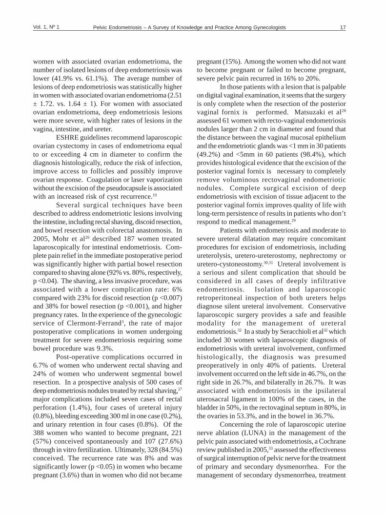
Several surgical techniques have beendescribed to address endometriotic lesions involvingthe intestine, including rectal shaving, discoid resection,and bowel resection with colorectal anastomosis. In2005, Mohr et al26 described 187 women treatedlaparoscopically for intestinal endometriosis. Com-plete pain relief in the immediate postoperative periodwas significantly higher with partial bowel resectioncompared to shaving alone (92% vs. 80%, respectively,p <0.04). The shaving, a less invasive procedure, wasassociated with a lower complication rate: 6%compared with 23% for discoid resection (p <0.007)and 38% for bowel resection (p <0.001), and higherpregnancy rates. In the experience of the gynecologicservice of Clermont-Ferrand3, the rate of majorpostoperative complications in women undergoingtreatment for severe endometriosis requiring somebowel procedure was 9.3%.
Post-operative complications occurred in6.7% of women who underwent rectal shaving and24% of women who underwent segmental bowelresection. In a prospective analysis of 500 cases ofdeep endometriosis nodules treated by rectal shaving,27
major complications included seven cases of rectalperforation (1.4%), four cases of ureteral injury(0.8%), bleeding exceeding 300 ml in one case (0.2%),and urinary retention in four cases (0.8%). Of the388 women who wanted to become pregnant, 221(57%) conceived spontaneously and 107 (27.6%)through in vitro fertilization. Ultimately, 328 (84.5%)conceived. The recurrence rate was 8% and wassignificantly lower (p <0.05) in women who becamepregnant (3.6%) than in women who did not became
pregnant (15%). Among the women who did not wantto become pregnant or failed to become pregnant,severe pelvic pain recurred in 16% to 20%.
In those patients with a lesion that is palpableon digital vaginal examination, it seems that the surgeryis only complete when the resection of the posteriorvaginal fornix is performed. Matsuzaki et al28
assessed 61 women with recto-vaginal endometriosisnodules larger than 2 cm in diameter and found thatthe distance between the vaginal mucosal epitheliumand the endometriotic glands was <1 mm in 30 patients(49.2%) and <5mm in 60 patients (98.4%), whichprovides histological evidence that the excision of theposterior vaginal fornix is necessary to completelyremove voluminous rectovaginal endometrioticnodules. Complete surgical excision of deependometriosis with excision of tissue adjacent to theposterior vaginal fornix improves quality of life withlong-term persistence of results in patients who don’trespond to medical management.29
Patients with endometriosis and moderate tosevere ureteral dilatation may require concomitantprocedures for excision of endometriosis, includingureterolysis, uretero-ureterostomy, nephrectomy oruretero-cystoneostomy.30,31 Ureteral involvement isa serious and silent complication that should beconsidered in all cases of deeply infiltrativeendometriosis. Isolation and laparoscopicretroperitoneal inspection of both ureters helpsdiagnose silent ureteral involvement. Conservativelaparoscopic surgery provides a safe and feasiblemodality for the management of ureteralendometriosis.32 In a study by Seracchioli et al32 whichincluded 30 women with laparoscopic diagnosis ofendometriosis with ureteral involvement, confirmedhistologically, the diagnosis was presumedpreoperatively in only 40% of patients. Ureteralinvolvement occurred on the left side in 46.7%, on theright side in 26.7%, and bilaterally in 26.7%. It wasassociated with endometriosis in the ipsilateraluterosacral ligament in 100% of the cases, in thebladder in 50%, in the rectovaginal septum in 80%, inthe ovaries in 53.3%, and in the bowel in 36.7%.
Concerning the role of laparoscopic uterinenerve ablation (LUNA) in the management of thepelvic pain associated with endometriosis, a Cochranereview published in 2005,33 assessed the effectivenessof surgical interruption of pelvic nerve for the treatmentof primary and secondary dysmenorrhea. For themanagement of secondary dysmenorrhea, treatment
Kondo et al.18 Bras. J. Video-Sur., January / March 2012
with LUNA combined with surgical treatment ofendometrial implants versus surgical treatment ofendometriosis alone showed that the addition ofLUNA did not help in relieving pain. For presacralneurectomy combined with the treatment ofendometriosis versus treatment of endometriosisalone, there was a overall difference in the paincontrol, although the data suggest that this may bespecific for laparoscopy and only for midline abdo-minal pain. Adverse effects were most common forpre-sacral neurectomy; however, most werecomplications such as constipation, which canimprove spontaneously.
With regard to gynecologists’ practice whenfaced with deep endometriosis lesions, 80% said thatthe ideal surgical treatment is complete resection ofthe lesion. However, when we put a scenario of apatient with deep endometriosis with intestinal orrectovaginal septum involvement, 44.4% of thegynecologists who responded said they doendometriosis operations responded that theyrecommend a diagnostic laparoscopy and not a com-plete resection, which contradicts the answer to theprevious question.
Only one of the gynecologists (7.4%) reportedthat they performed this type of surgery without theassistance of a surgeon general or a coloproctologist.Only 20% of gynecologists remembered the need forresection of the posterior fornix of the vagina for deeplesions palpable on digital vaginal examination, and35% still believe that LUNA has an important role in
preventing the recurrence of symptoms and of thelesions.
The relationship between endometriosis andinfertility is still controversal.1 Several factors canaffect spontaneous fertility in women with deependometriosis including the woman’s age34 (especiallyabove age 35), the presence of uterine adenomyosis,35
the presence of associated male infertility, and couples’attitudes about natural conception and infertilitytreatments. Treatment with intrauterine inseminationappears to improve the fertility in cases of minimal ormild endometriosis. In vitro fertilization (IVF)treatment is the appropriate treatment when tubalfunction is compromised, when there is male infertilityor when other treatments have failed, but pregnancyrates with IVF are still lower in patients withendometriosis than those with infertility due to tubalfactors.19
We conclude that more education is necessaryamong gynecologists with respect to endometriosis inorder to identify and treat patients with this disease.Referral to specialized centers that offer all availabletreatments in a multidisciplinary context is importantto improve the surgical results of deep infiltrativeendometriosis.
ACKNOWLEDGEMENTS
We thank the 40 doctors who answered thequestionnaires anonymously and collaborated with thestudy.
RESUMOObjetivo: A endometriose afeta um grande número de mulheres em idade reprodutiva. As manifestações clínicas dadoença são dor pélvica cíclica e infertilidade. As mulheres com endometriose geralmente procuram atendimento médicodevido à sua sintomatologia. O papel do ginecologista é, portanto crucial na identificação, no tratamento e, quandonecessário, no encaminhamento das pacientes para centros especializados. Métodos: Um questionário anônimo foicompletado por 40 ginecologistas brasileiros. Resultados: 67.5% dos avaliados realizam cirurgia para endometriose.Aproximadamente metade (55%) dos avaliados declararam que o exame físico pode diagnosticar casos de endometrioseprofunda e 92,5% não excluem a possibilidade de doença profunda quando os níveis de CA-125 séricos são normais.Ressonância nuclear magnetica, ultra-som transvaginal e colonoscopia são importantes na avaliação pré-operatória daspacientes para 72,5, 70 e 62,5% dos avaliados. Para 62,5% dos avaliados, os análogos de GnRH são o melhor tratamentoclínico para endometriose. Apenas 17,5% dos ginecologistas acham que todos os tratamentos clínicos hormonais têmresultados semelhantes. Embora 80% dos ginecologistas responderam que a ressecção completa da doença é omelhor tratamento para a doença profunda, 44% dos ginecologistas que realizam cirurgia para endometriose indicamapenas laparoscopia diagnóstica nesses casos. Apenas 7,4% dos avaliados são capazes de tratar endometriose profun-da com comprometimento intestinal sem o auxílio de um cirurgião geral ou um cirurgião colo-retal. Conclusões: Maiseducação é necessária entre os ginecologistas com relação à endometriose, a fim de identificar e tratar pacientes comesta doença. O encaminhamento para centros especializados que ofereçam todos os tratamentos disponíveis em umcontexto multi-disciplinar é importante para melhorar os resultados cirúrgicos da endometriose profunda infiltrativa.
Palavras chave: Endometriose. Diagnóstico. Tratamento. Endometriose profunda. Laparoscopia.
Pelvic Endometriosis – A Survey of Knowledge and Practice Among Gynecologists 19Vol. 1, Nº 1
REFERENCES
1. Kondo W, Ferriani RA, Petta CA, Abrão MS, Amaral VF.Endometriose e infertilidade: causa ou consequência? JBRA -Jornal Brasileiro de Reprodução Assistida. 2009; 13(2):33-8.
2. Amsterdam LL, Gentry W, Jobanputra S, Wolf M, RubinSD, Bulun SE. Anastrazole and oral contraceptives: a noveltreatment for endometriosis. Fertil Steril. 2005 ;84(2):300-4.
3. Kondo W, Bourdel N, Tamburro S, Cavoli D, Jardon K,Rabischong B, et al. Complications after surgery for deeplyinfiltrating pelvic endometriosis. BJOG. 2010, in press.
4. Abrao MS, Gonçalves MO, Dias JA Jr, Podgaec S, ChamieLP, Blasbalg R. Comparison between clinical examination,transvaginal sonography and magnetic resonance imaging forthe diagnosis of deep endometriosis. Hum Reprod. 2007;22(12):3092-7.
5. Bazot M, Lafont C, Rouzier R, Roseau G, Thomassin-Naggara I, Daraï E. Diagnostic accuracy of physicalexamination, transvaginal sonography, rectal endoscopicsonography, and magnetic resonance imaging to diagnose deepinfiltrating endometriosis. Fertil Steril. 2009; 92(6):1825-33.
6. Vercellini P, Crosignani PG, Somigliana E, Berlanda N, BarbaraG, Fedele L. Medical treatment for rectovaginalendometriosis: what is the evidence? Hum Reprod. 2009;24(10):2504-14.
7. Vercellini P, Somigliana E, Viganò P, Abbiati A, Barbara G,Crosignani PG. Endometriosis: current therapies and newpharmacological developments. Drugs. 2009; 69(6):649-75.
8. Kondo W, Daraï E, Yazbeck C, Panel P, Tamburro S,Dubuisson J, et al. Do patients manage to achieve pregnancyafter a major complication of deeply infiltratingendometriosis resection? Eur J Obstet Gynecol Reprod Biol.2010, in press.
9. Panel P, Renouvel F. Management of endometriosis: clinicaland biological assessment. J Gynecol Obstet Biol Reprod(Paris). 2007; 36(2):119-28.
10. Arruda MS, Petta CA, Abrão MS, Benetti-Pinto CL. Timeelapsed from onset of symptoms to diagnosis ofendometriosis in a cohort study of Brazilian women. HumReprod. 2003; 18(4):756-9.
11. Mihalyi A, Gevaert O, Kyama CM, Simsa P, Pochet N, DeSmet F, et al. Non-invasive diagnosis of endometriosis basedon a combined analysis of six plasma biomarkers. HumReprod. 2010; 25(3):654-64.
12. Kafali H, Artuc H, Demir N. Use of CA125 fluctuationduring the menstrual cycle as a tool in the clinical diagnosisof endometriosis; a preliminary report. Eur J Obstet GynecolReprod Biol. 2004; 116(1):85-8.
13. Takeuchi H, Kuwatsuru R, Kitade M, Sakurai A, Kikuchi I,Shimanuki H, et al. A novel technique using magneticresonance imaging jelly for evaluation of rectovaginalendometriosis. Fertil Steril. 2005; 83(2):442-7.
14. Chassang M, Novellas S, Bloch-Marcotte C, Delotte J,Toullalan O, Bongain A, et al. Utility of vaginal and rectalcontrast medium in MRI for the detection of deep pelvicendometriosis. Eur Radiol. 2010; 20(4):1003-10.
15. Donnez J, Nisolle M, Squifflet J. Ureteral endometriosis: acomplication of rectovaginal endometriotic (adenomyotic)nodules. Fertil Steril. 2002; 77(1):32-7.
16. Jung SI, Kim YJ, Jeon HJ, Jeong KA. Deep infiltratingendometriosis: CT imaging evaluation. J Comput AssistTomogr. 2010; 34(3):338-42.
17. Levitt MD, Hodby KJ, van Merwyk AJ, Glancy RJ. Cyclicalrectal bleeding in colorectal endometriosis. Aust N Z J Surg.1989; 59(12):941-3.
18. Daguati R, Somigliana E, Viganò P, Vercellini P. Progestogensand estroprogestins in the treatment of pelvic pain associatedwith endometriosis. Minerva Ginecol. 2006; 58(6):499-510.
19. Kennedy S, Bergqvist A, Chapron C, D’Hooghe T,Dunselman G, Greb R, et al. ESHRE guideline for thediagnosis and treatment of endometriosis. Hum Reprod.2005; 20(10):2698-704.
20. Brown J, Pan A, Hart RJ. Gonadotrophin-releasing hormoneanalogues for pain associated with endometriosis. CochraneDatabase Syst Rev. 2010; 8(12):CD008475.
21. Davis L, Kennedy SS, Moore J, Prentice A. Moderncombined oral contraceptives for pain associated withendometriosis. Cochrane Database Syst Rev. 2007;18(3):CD001019.
22. Sheng J, Zhang WY, Zhang JP, Lu D. The LNG-IUS studyon adenomyosis: a 3-year follow-up study on the efficacyand side effects of the use of levonorgestrel intrauterinesystem for the treatment of dysmenorrhea associated withadenomyosis. Contraception. 2009; 79(3):189-93.
23. Banerjee SK, Ballard KD, Wright JT. Endometriomas as amarker of disease severity. J Minim Invasive Gynecol. 2008;15(5):538-40.
24. Redwine DB. Ovarian endometriosis: a marker for moreextensive pelvic and intestinal disease. Fertil Steril. 1999;72(2):310-5.
25. Chapron C, Pietin-Vialle C, Borghese B, Davy C, Foulot H,Chopin N. Associated ovarian endometrioma is a marker forgreater severity of deeply infiltrating endometriosis. FertilSteril. 2009; 92(2):453-7.
26. Mohr C, Nezhat FR, Nezhat CH, Seidman DS, Nezhat CR.Fertility considerations in laparoscopic treatment ofinfiltrative bowel endometriosis. JSLS. 2005; 9(1):16-24.
27. Donnez J, Squifflet J. Complications, pregnancy andrecurrence in a prospective series of 500 patients operatedon by the shaving technique for deep rectovaginalendometriotic nodules. Hum Reprod. 2010; 25(8):1949-58.
28. Matsuzaki S, Houlle C, Botchorishvili R, Pouly JL, MageG, Canis M. Excision of the posterior vaginal fornix isnecessary to ensure complete resection of rectovaginalendometriotic nodules of more than 2 cm in size. Fertil Steril.2009; 91(4 Suppl):1314-5.

Kondo et al.20 Bras. J. Video-Sur., January / March 2012
29. Angioni S, Peiretti M, Zirone M, Palomba M, Mais V, GomelV, et al. Laparoscopic excision of posterior vaginal fornix inthe treatment of patients with deep endometriosis withoutrectum involvement: surgical treatment and long-term follow-up. Hum Reprod. 2006; 21(6):1629-34.
30. Ghezzi F, Cromi A, Bergamini V, Bolis P. Management ofureteral endometriosis: areas of controversy. Curr OpinObstet Gynecol. 2007; 19(4):319-24.
31. Mereu L, Gagliardi ML, Clarizia R, Mainardi P, Landi S,Minelli L. Laparoscopic management of ureteralendometriosis in case of moderate-severehydroureteronephrosis. Fertil Steril. 2010; 93(1):46-51.
32. Seracchioli R, Mabrouk M, Manuzzi L, Guerrini M, Villa G,Montanari G, et al. Importance of retroperitoneal uretericevaluation in cases of deep infiltrating endometriosis. J MinimInvasive Gynecol. 2008; 15(4):435-9.
33. Proctor ML, Latthe PM, Farquhar CM, Khan KS, JohnsonNP. Surgical interruption of pelvic nerve pathways forprimary and secondary dysmenorrhoea. Cochrane DatabaseSyst Rev. 2005; 19(4):CD001896.
34. Ferrero S, Anserini P, Abbamonte LH, Ragni N, Camerini G,Remorgida V. Fertility after bowel resection forendometriosis. Fertil Steril. 2009; 92(1):41-6.
35. Daraï E, Marpeau O, Thomassin I, Dubernard G, BarrangerE, Bazot M. Fertility after laparoscopic colorectal resectionfor endometriosis: preliminary results. Fertil Steril. 2005;84(4):945-50.
Corr esponding Author:WILLIAM KONDOAv. Getúlio Vargas 3163 Apt 21Curitiba, Paraná 80240-041 BrazilTelephone: (41) 9222-1065E-mail: [email protected]
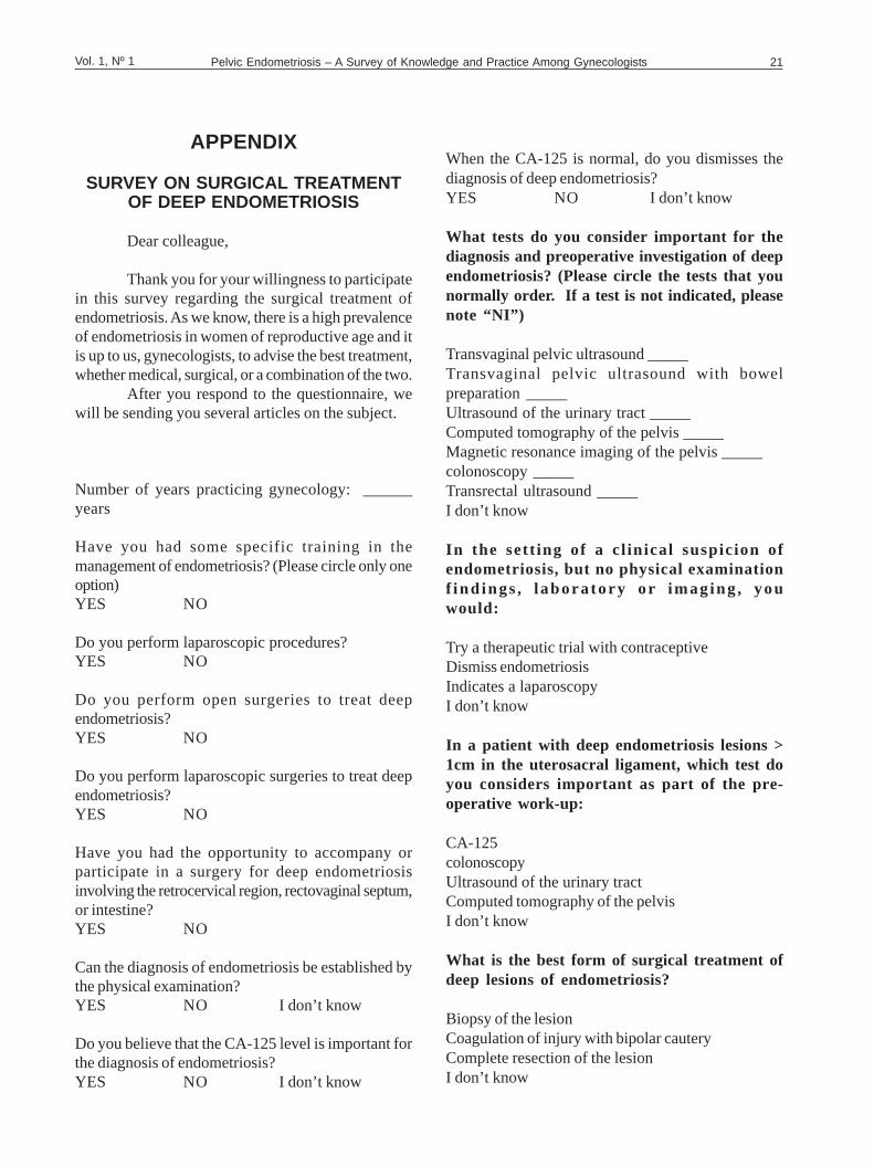
Pelvic Endometriosis – A Survey of Knowledge and Practice Among Gynecologists 21Vol. 1, Nº 1
APPENDIX
SURVEY ON SURGICAL TREATMENTOF DEEP ENDOMETRIOSIS
Dear colleague,
Thank you for your willingness to participatein this survey regarding the surgical treatment ofendometriosis. As we know, there is a high prevalenceof endometriosis in women of reproductive age and itis up to us, gynecologists, to advise the best treatment,whether medical, surgical, or a combination of the two.
After you respond to the questionnaire, wewill be sending you several articles on the subject.
Number of years practicing gynecology: ______years
Have you had some specific training in themanagement of endometriosis? (Please circle only oneoption)YES NO
Do you perform laparoscopic procedures?YES NO
Do you perform open surgeries to treat deependometriosis?YES NO
Do you perform laparoscopic surgeries to treat deependometriosis?YES NO
Have you had the opportunity to accompany orparticipate in a surgery for deep endometriosisinvolving the retrocervical region, rectovaginal septum,or intestine?YES NO
Can the diagnosis of endometriosis be established bythe physical examination?YES NO I don’t know
Do you believe that the CA-125 level is important forthe diagnosis of endometriosis?YES NO I don’t know
When the CA-125 is normal, do you dismisses thediagnosis of deep endometriosis?YES NO I don’t know
What tests do you consider important for thediagnosis and preoperative investigation of deependometriosis? (Please circle the tests that younormally order. If a test is not indicated, pleasenote “NI”)
Transvaginal pelvic ultrasound _____Transvaginal pelvic ultrasound with bowelpreparation _____Ultrasound of the urinary tract _____Computed tomography of the pelvis _____Magnetic resonance imaging of the pelvis _____colonoscopy _____Transrectal ultrasound _____I don’t know
In the sett ing of a c l in ical suspic ion ofendometriosis, but no physical examinationf ind ings , labora to ry o r imag ing , youwould:
Try a therapeutic trial with contraceptiveDismiss endometriosisIndicates a laparoscopyI don’t know
In a patient with deep endometriosis lesions >1cm in the uterosacral ligament, which test doyou considers important as part of the pre-operative work-up:
CA-125colonoscopyUltrasound of the urinary tractComputed tomography of the pelvisI don’t know
What is the best form of surgical treatment ofdeep lesions of endometriosis?
Biopsy of the lesionCoagulation of injury with bipolar cauteryComplete resection of the lesionI don’t know
Kondo et al.22 Bras. J. Video-Sur., January / March 2012
In a patient with deep endometriosis of theuterosacral ligament >1cm (lateral lesion),what procedure should be performed in mostcases?
Complete resection of the lesion with a portion of thevaginal vaultBiopsy of the lesionCoagulation of the lesionComplete resection of the lesion with ureterolysis(release of the ureter)Curative resection with bowel resectionI don’t know
A patient has an endometriosis nodule inrectovaginal septum found on imaging and a va-ginal lesion palpable to the touch. Whatprocedure should be performed to reduce therisk of recurrence?
Bowel resectionLaparoscopic Uterine Nerve Ablation (LUNA)Resection of the posterior fornix of the vaginaHysterectomyI don’t know
What is the best medication for the medicalmanagement of endometriosis?
ZoladexOral contraceptiveProgestin (oral or intramuscular)MirenaDanazolAll have the same effectI don’t know
What are the surgical techniques for resectionof a lesion of endometriosis involving theintestine?
Intestinal resection and anastomosisDiscoid resection of the rectumShavingAll of the aboveI don’t know
In the case of a patient with suspectedendometriosis involving the intestine or therectovaginal septum, you would:
Recommend a diagnostic laparoscopy with biopsyRefer to a digestivesurgeon or coloproctologistRecommend a diagnostic laparoscopy and adjuvanttreatment with ZoladexRecommend a complete surgery including bowelresection or rectal shaving, if necessary, along with adigestive surgeon or coloproctologistaRecommend a complete surgery including bowelresection or rectal shaving, if necessary, withoutresorting to a colleague from another specialty
When you encounter intra-operatively AFS-Rstage IV endometriosis with total obliteration ofthe posterior fornix and the possibility of intes-tinal involvement, what is your approach?
Halt the operation and postoperatively execute medicalmanagement with ZoladexHalt the operation and refer to a digestive surgeon ordigestive coloproctologistaSummon a digestive surgeon or coloproctologist duringthe procedure and continue to surgeryContinue the surgery; as you have experienced in thesurgical manipulation of the urinary and gastrointestinaltracts, address involvement of the ureter or recto-sigmoid as necessary.
In the case of a patient with an endometrioma of4 cm in diameter, which is important to keep inmind when they recommend surgery?
One should remove the entire ovary to preventrecurrence, since the recurrence rate ofendometrioma is high.Endometrioma is a marker of deep endometriosisand there is a possibility of other associated lesions(retrocervical, rectovaginal septum, intestine,ureter)There is no indication for surgery for an endometriomaof this sizeI don’t know
For a woman clinically suspected of havingendometriosis, who has been infertile for oneyear, with an elevated CA-125, and a deeplesion of endometriosis involving therectovaginal septum, which would be thetreatment of choice (with the best success forpregnancy) if the main objective at that timeis to get pregnant?

Pelvic Endometriosis – A Survey of Knowledge and Practice Among Gynecologists 23Vol. 1, Nº 1
Recommend laparoscopy to confirm the diagnosis ofendometriosisRefer for in vitro fertilization (IVF)Recommend laparoscopy with biopsy and takeZoladex post-operativelyRecommend complete resection of the lesion and thentreat with ZoladexRecommend complete resection of the lesion and thenin vitro fertilizationI don’t know
What surgical procedures would you have nodif ficulty performing, if necessary, during anoperation?
Resection of bladderUreterolysisSegmental bowel resection and anastomosisRectal shavingDouble-J catheterUreteral anastomosisUreteral reimplantation
Brazilian Journal of Videoendoscopic Surgery - v. 5 - n. 1 - Jan./Mar. 2012 - Subscription: + 55 21 3325-7724 - E-mail: [email protected] 1983-9901: (Press) ISSN 1983-991X: (on-line) - SOBRACIL - Press Graphic & Publishing Ltd. Rio de Janeiro, RJ-Brasil
THANK YOU FOR YOUR TIME!ALL INFORMATION CONTAINED IN THIS
QUESTIONNAIREIS CONFIDENTIAL
I DECLARE THAT I ACCEPTED TOPARTICIPATE IN THIS SURVEY
VOLUNTARILY
____________________________________signature
THE INITIALS OF YOUR NAME:______________________________________
DATE: _____ / _____/2010
Carvalho et al.24 Bras. J. Video-Sur., January / March 2012Original ArticleBrazilian Journalof VideoendoscopicSurgery
Accepted after revision: march, 12, 2012.Bras. J. V ideo-Sur , 2012, v. 5, n. 1: 024-028
24
Laparoscopic T ransmediastinal Drainage: A Simple andEffective Procedure for Reverting an Accident al
Pneumothorax During Lap aroscopic Antireflux Surgery
Drenagem transmediastinal lap aroscópica: Um procedimentosimples e eficaz p ara reverter um pneumotórax acident al durante
uma cirurgia lap aroscópica antirefluxo
GUSTAVO L. CARVALHO, MD, PHD 1; CAMILA ROCHA DA CRUZ, MD2; JOSÉ SÉRGIO N. SILVA, MD3;DIEGO LAURENTINO LIMA, MD 4; REBECA GONÇALVES ROCHA5; EDUARDO FELIPE DE CAR VALHO
CHAVES, MD6
Department of Surgery, Faculty of Medical Sciences, University of Pernambuco (UPE), and the GustavoCarvalho Videolaparoscopic Surgery Clinic, Recife, Pernambuco, Brazil.
1. Associate Professor of Surgery – Faculty of Medical Sciences, University of Pernambuco. Member ofSAGES, SOBRACIL, ELSA and Brazilian College of Surgeons (CBC); 2. Faculty of Medical Sciences,
University of Pernambuco; 3. Faculty of Medical Sciences, University of Pernambuco; 4. Faculty of MedicalSciences, University of Pernambuco; 5. Medical Student, Faculty of Medical Sciences, University of
Pernambuco; 6. Faculty of Medical Sciences, University of Pernambuco.
ABSTRACTIntroduction: Pneumothorax (PTX) secondary to pleural injury is an uncommon accident in laparoscopic antirefluxsurgery (LARS). Its frequency is approximately 2–6%. The current therapy for transoperative PTX is thoracic intercostaldrainage. AIM: To evaluate the treatment of accidental transoperative PTX using a new procedure: laparoscopictransmediastinal drainage (LTD). Materials and Methods: From January 2000 to September 2010, 400 patients underwentLARS and 18 patients presented accidental pleural injuries. All 18 were treated using LTD. LTD was performed by theinsertion of an 8 to 12 French silicone drain between the diaphragmatic pillars, leaving all the holes of the drain in thepleural cavity. The drain’s exit was made through a 3 to 5 mm trocar hole inserted below the 5 mm work-trocar. Thepatients were kept on high-pressure mechanical ventilation (PEEP) by the anesthesiologist. The drains were removed15 min on average after the end of the surgery and a chest radiograph was performed to confirm the absence of PTX.Results: No significant complication was observed when this technique was used for the treatment of PTX. Radiographicconfirmation of the absence of PTX was obtained in all the patients. Conclusions: The treatment of PTX by LTD is a safe,simple, and effective method and should be considered the procedure of choice for this complication during LARS. Byavoiding traditional intercostal drainage LTD maintains a minimally invasive surgery and preserves the desired estheticoutcomes.
Key words: Pneumothorax. Intraoperative complications. Laparoscopy. Laparoscopy transmediastinal drainage. Antirefluxprocedure. Gastroesophageal reflux disease.
1. INTRODUCTION
L aparoscopic antireflux surgery (LARS) is aneffective alternative to lifelong antireflux medical
therapy and has become the standard approach tohiatal hernia repair and surgical treatment ofgastroesophageal reflux disease (GERD).1-3
Numerous studies have shown that LARS is a safeand effective treatment with excellent intermediateand long term functional outcomes, which results inhigh patient satisfaction, and significantly improvespatients’ long term quality of life.4,5
Although laparoscopic fundoplication surgeryis a safe and effective procedure, it is not free of
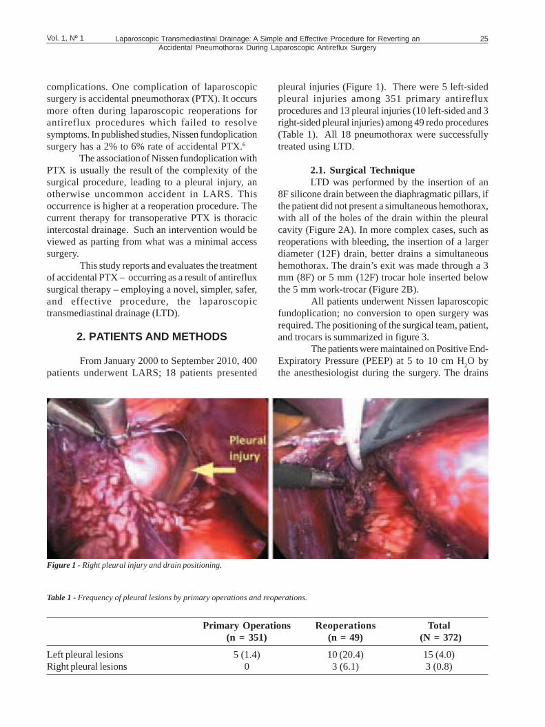
Laparoscopic Transmediastinal Drainage: A Simple and Effective Procedure for Reverting anAccidental Pneumothorax During Laparoscopic Antireflux Surgery
25Vol. 1, Nº 1
complications. One complication of laparoscopicsurgery is accidental pneumothorax (PTX). It occursmore often during laparoscopic reoperations forantireflux procedures which failed to resolvesymptoms. In published studies, Nissen fundoplicationsurgery has a 2% to 6% rate of accidental PTX.6
The association of Nissen fundoplication withPTX is usually the result of the complexity of thesurgical procedure, leading to a pleural injury, anotherwise uncommon accident in LARS. Thisoccurrence is higher at a reoperation procedure. Thecurrent therapy for transoperative PTX is thoracicintercostal drainage. Such an intervention would beviewed as parting from what was a minimal accesssurgery.
This study reports and evaluates the treatmentof accidental PTX – occurring as a result of antirefluxsurgical therapy – employing a novel, simpler, safer,and effective procedure, the laparoscopictransmediastinal drainage (LTD).
2. PATIENTS AND METHODS
From January 2000 to September 2010, 400patients underwent LARS; 18 patients presented
pleural injuries (Figure 1). There were 5 left-sidedpleural injuries among 351 primary antirefluxprocedures and 13 pleural injuries (10 left-sided and 3right-sided pleural injuries) among 49 redo procedures(Table 1). All 18 pneumothorax were successfullytreated using LTD.
2.1. Surgical TechniqueLTD was performed by the insertion of an
8F silicone drain between the diaphragmatic pillars, ifthe patient did not present a simultaneous hemothorax,with all of the holes of the drain within the pleuralcavity (Figure 2A). In more complex cases, such asreoperations with bleeding, the insertion of a largerdiameter (12F) drain, better drains a simultaneoushemothorax. The drain’s exit was made through a 3mm (8F) or 5 mm (12F) trocar hole inserted belowthe 5 mm work-trocar (Figure 2B).
All patients underwent Nissen laparoscopicfundoplication; no conversion to open surgery wasrequired. The positioning of the surgical team, patient,and trocars is summarized in figure 3.
The patients were maintained on Positive End-Expiratory Pressure (PEEP) at 5 to 10 cm H
2O by
the anesthesiologist during the surgery. The drains
Figure 1 - Right pleural injury and drain positioning.
Table 1 - Frequency of pleural lesions by primary operations and reoperations.
Primary Operations Reoperations Total(n = 351) (n = 49) (N = 372)
Left pleural lesions 5 (1.4) 10 (20.4) 15 (4.0)Right pleural lesions 0 3 (6.1) 3 (0.8)
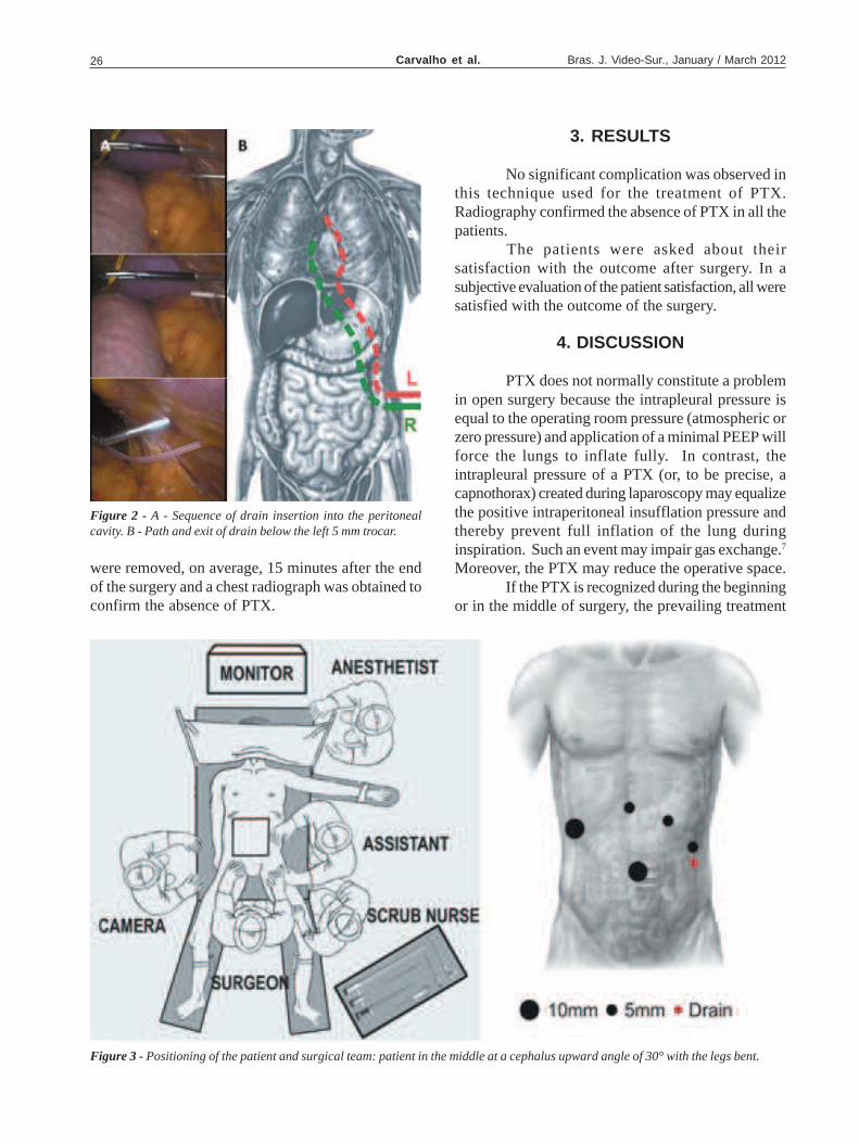
Carvalho et al.26 Bras. J. Video-Sur., January / March 2012
3. RESULTS
No significant complication was observed inthis technique used for the treatment of PTX.Radiography confirmed the absence of PTX in all thepatients.
The patients were asked about theirsatisfaction with the outcome after surgery. In asubjective evaluation of the patient satisfaction, all weresatisfied with the outcome of the surgery.
4. DISCUSSION
PTX does not normally constitute a problemin open surgery because the intrapleural pressure isequal to the operating room pressure (atmospheric orzero pressure) and application of a minimal PEEP willforce the lungs to inflate fully. In contrast, theintrapleural pressure of a PTX (or, to be precise, acapnothorax) created during laparoscopy may equalizethe positive intraperitoneal insufflation pressure andthereby prevent full inflation of the lung duringinspiration. Such an event may impair gas exchange.7
Moreover, the PTX may reduce the operative space.If the PTX is recognized during the beginning
or in the middle of surgery, the prevailing treatment
Figure 2 - A - Sequence of drain insertion into the peritonealcavity. B - Path and exit of drain below the left 5 mm trocar.
Figure 3 - Positioning of the patient and surgical team: patient in the middle at a cephalus upward angle of 30° with the legs bent.
were removed, on average, 15 minutes after the endof the surgery and a chest radiograph was obtained toconfirm the absence of PTX.
Laparoscopic Transmediastinal Drainage: A Simple and Effective Procedure for Reverting anAccidental Pneumothorax During Laparoscopic Antireflux Surgery
27Vol. 1, Nº 1
consists of deflating the abdomen, performing athoracic intercostal drainage, and then proceeding withthe surgery if the patient remains hemodynamicallystable.8 LTD, not only avoids thoracostomy, but theintended esthetic results are maintained; only a minimaladditional incision, usually 3 mm trocar hole in theabdomen, for 8F drainage, is required.
The psychological benefits for the patient arerelevant. Thoracostomy may be viewed as anunexpected complication of the surgery, while anadditional small abdominal incision probably won’ t.Transmediastinal pneumothorax drainage preserves themain goal of laparoscopic procedure: minimalpostoperative morbidity.
Since 2000 the LTD technique described inthis report has been the remedy of choice in ourinstitution to treat all patients who experience anaccidental PTX during antireflux surgical therapy.
5. CONCLUSIONS
Treatment of PTX by LTD is a safe, simple,and effective method and it should be consideredthe procedure of choice for this complication duringLARS. LTD maintains the minimally invasive natureof the surgery, and preserves the desired estheticoutcomes.
This technique merits wide disseminationamong surgeons, so that it benefits more patients.
6. ACKNOWLEDGMENTS
The authors thank all those who have helpedin this study, especially those who have diligently caredfor the patients, and are grateful for the support of theDepartment of Surgery of the Faculty of MedicalSciences, University of Pernambuco, Brazil.
RESUMOIntrodução: Pneumotórax secundário a lesão pleural é um acidente incomum em cirurgia laparoscópica antirrefluxo.Sua frequência é aproximadamente 2-6%. A terapia atual para o pneumotórax transoperatório é a drenagem torácicaintercostal. Objetivo: Avaliar o tratamento de um pneumotórax acidental transoperatório usando um novo procedimento,a drenagem laparoscópica transmediastinal. Materiais e Métodos: De janeiro de 2000 a setembro de 2010, 400pacientes foram submetidos à cirurgia laparoscópica antirrefluxo dos quais 18 apresentaram lesões pleurais acidentais.Todos foram tratados usando a drenagem laparoscópica transmediastinal. Ela é realizada pela inserção de um drenode silicone 8F-12F entre as bordas do pilar diafragmático, deixando todos os orifícios do dreno na cavidade pleural. Asaída do dreno foi criada através de uma incisão feita por um trocar de 3-5 mm inserido abaixo do trocar de 5 mm. Ospacientes foram mantidos em ventilação mecânica de alta pressão pelo anestesista. Os drenos foram removidos emuma media de 15 minutos após o final da cirurgia e uma radiografia de tórax foi realizada para confirmar a ausência depneumotórax. Resultados: Nenhuma complicação foi observada por essa técnica usada para o tratamento dopneumotórax. Radiofragia confirmou a ausência de pneumotórax em todos os pacientes. Conclusões: O tratamento porDrenagem Laparoscópica transmediastinal é um método seguro, simples e efetivo e deve ser considerado umprocedimento de escolha para essa complicação durante a cirurgia laparoscópica antirrefluxo e cirurgia minimamenteinvasive, além de manter o resultado estético, evitando a drenagem intercostal tradicional.
Descritores: Pneumotórax. Complicações intraoperatórias. Laparoscopia. Drenagem transmediastinal laparoscópica.Procedimento antirrefluxo. Doença do refluxo gastroesofageal.
7. REFERENCES
1. Hinder RA, Filipi CJ, Wetscher G, et al. Laparoscopic Nissenfundoplication is an effective treatment for gastroesophagealreflux disease. Ann Surg. 1994; 220:472-483.
2. Arnaud JP, Pessaux P, Ghavami B, et al. Laparoscopicfundoplication for gastroesophageal reflux: multicenter studyof 1470 cases. Surg Endosc. 2000; 14:1024-1027.
3. Lafullarde T, Watson DI, Jamieson G, et al. LaparoscopicNissen fundoplication: five year result and beyond. ArchSurg. 2001; 136:180-184.
4. Granderath FA, Kamolz T, Schweiger UM, et al. Quality oflife, surgical outcome and patient satisfaction three yearsafter laparoscopic Nissen fundoplication. World J Surg. 2002;26:1234-1238.
5. Kamolz T, Granderath FA,BammerT, et al. Mid- and long-termquality of life assessments after laparoscopic fundoplicationand refundoplication: a prospective single unit review of 500antireflux procedures. Dig Liver Dis. 2002; 34:470-476.
6. Perdikis G, Hinder RA, Lund RJ, Raiser F, Katada N.Laparoscopic Nissen fundoplication: Where do we stand?Surg Laparosc Endosc 1997; 7:17–21

Carvalho et al.28 Bras. J. Video-Sur., January / March 2012
Brazilian Journal of Videoendoscopic Surgery - v. 5 - n. 1 - Jan./Mar. 2012 - Subscription: + 55 21 3325-7724 - E-mail: [email protected] 1983-9901: (Press) ISSN 1983-991X: (on-line) - SOBRACIL - Press Graphic & Publishing Ltd. Rio de Janeiro, RJ-Brasil
7. Sandbu, R. 2001. A Laparoscopic Approach to Gastro-Oesophageal Surgery: Experimental and EpidemiologicalStudies. Acta Universitatis Upsaliensis - ComprehensiveSummaries of Uppsala Dissertations from the Faculty ofMedicine 1094. 87 pp.
8. Prystowsky JB, Jericho BG, Epstein HM. Spontaneousbilateral pneumothorax – complication of laparoscopiccholecystectomy. Surgery. 1993; 114:988-92.
Corresponding author:GUSTAVO CARVALHO, MD, PhD.Av. Boa Viagem 5526B Apt.1902Recife, PE, Brazil 51030-000Tel: +55 81 9971-9698 Fax: +55 81 3325-3318E-mail: [email protected]; [email protected]
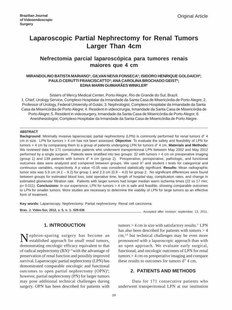
Laparoscopic Partial Nephrectomy for Renal Tumors Larger Than 4cm 29Vol. 1, Nº 1 Original ArticleBrazilian Journalof VideoendoscopicSurgery
Accepted after revision: september, 13, 2011.Bras. J. V ideo-Sur , 2012, v. 5, n. 1: 029-036
29
Laparoscopic Partial Nephrectomy for Renal T umorsLarger Than 4cm
Nefrectomia p arcial lap aroscópica p ara tumores renaismaiores que 4 cm
MIRANDOLINO BATISTA MARIANO 1; GILVAN NEIVA FONSECA2; ISIDORO HENRIQUE GOLDAICH 3;PAULO CERUTTI FRANCISCATTO4; ANA CAROLINA BROCHADO GEIST 5;
EDNA MARIN GUIMARÃES WINKLER 6
Sisters of Mercy Medical Center, Porto Alegre, Rio de Grande do Sul, Brazil.1. Chief, Urology Service, Complexo Hospitalar da Irmandade da Santa Casa de Misericórdia de Porto Alegre; 2.Professor of Urology, Federal University of Goiás; 3. Nephrologist, Complexo Hospitalar da Irmandade da SantaCasa da Misericórdia de Porto Alegre; 4. Resident in videocirurgia, Irmandade da Santa Casa de Misericórdia de
Porto Alegre; 5. Resident in videosurgery, Irmandade da Santa Casa de Misericórdia de Porto Alegre; 6.Anesthesiologist, Complexo Hospitalar da Irmandade da Santa Casa de Misericórdia de Porto Alegre.
ABSTRACTBackground : Minimally invasive laparoscopic partial nephrectomy (LPN) is commonly performed for renal tumors d” 4cm in size. LPN for tumors > 4 cm has not been assessed. Objective : To evaluate the safety and feasibility of LPN fortumors > 4 cm by compariang them to a group of patients undergoing LPN for tumors d” 4 cm. Materials and Methods:We reviewed data for 171 consecutive patients who underwent transperitoneal LPN between May 2002 and May 2012performed by a single surgeon. Patients were stratified into two groups: 32 with tumors > 4 cm on preoperative imaging(group 1) and 139 patients with tumors d” 4 cm (group 2). Preoperative, perioperative, pathologic, and functionaloutcomes data were analyzed and compered between groups. We used X2 and student t tests for categorical andcontinuous variables, respectively. A p value <0.05 was considered statistically significant. Result s: Mean radiographictumor size was 5.9 cm (4.1 – 9.2) for group 1 and 2.3 cm (0.9 – 4.0) for group 2. No significant differences were foundbetween groups for estimated blood loss, total operative time, length of hospital stay, complication rates, and change inestimated glomerular filtration rate. Patients with larger tumors had longer median warm ischemia times (22 vs 17 min;p= 0.011). Conclusions: In our experience, LPN for tumors > 4 cm is safe and feasible, showing comparable outcomesto LPN for smaller tumors. More studies are necessary to determine the viability of LPN for large tumors as an effectiveform of treatment.
Key words: Laparoscopy. Nephrectomy. Partial nephrectomy. Renal cell carcinoma.
1. INTRODUCTION
N ephron-sparing surgery has become anestablished approach for small renal tumors,
demonstrating oncologic efficacy equivalent to thatof radical nephrectomy (RN)1-3 with the advantage ofpreservation of renal function and possibly improvedsurvival. Laparoscopic partial nephrectomy (LPN) hasdemonstrated comparable oncologic and functionaloutcomes to open partial nephrectomy (OPN)4;however, partial nephrectomy (PN) for larger tumorsmay pose additional technical challenges duringsurgery. OPN has been described for patients with
tumors > 4 cm in size with satisfactory results.3 LPNhas also been described for patients with tumors > 4cm,5,6 but technical challenges may be even morepronounced with a laparoscopic approach than withan open approach. We evaluate early surgical,functional, and oncologic outcomes of LPN for renaltumors > 4 cm on preoperative imaging and comparethese results to outcomes for tumors d” 4 cm.
2. PATIENTS AND METHODS
Data for 171 consecutive patients whounderwent transperitoneal LPN at our institution
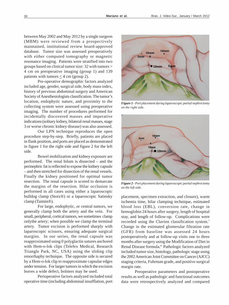
Mariano et al.30 Bras. J. Video-Sur., January / March 2012
between May 2002 and May 2012 by a single surgeon(MBM) were reviewed from a prospectivelymaintained, institutional review board-approveddatabase. Tumor size was assessed preoperativelywith either computed tomography or magneticresonance imaging. Patients were stratified into twogroups based on clinical tumor size: 32 with tumors >4 cm on preoperative imaging (group 1) and 139patients with tumors < 4 cm (group 2).
Pre-operative demographic factors analyzedincluded age, gender, surgical side, body mass index,history of previous abdominal surgery and AmericanSociety of Anesthesiologists classification. The tumor’slocation, endophytic nature, and proximity to thecollecting system were assessed using preoperativeimaging. The number of procedures performed forincidentally discovered masses and imperativeindications (solitary kidney, bilateral renal masses, stage3 or worse chronic kidney disease) was also assessed.
Our LPN technique reproduces the openprocedure step-by-step. Briefly, patients are placedin flank position, and ports are placed as demonstratedin figure 1 for the right side and figure 2 for the leftside.
Bowel mobilization and kidney exposure areperformed. The renal hilum is dissected – and theperinephric fat is reflected to expose the kidney capsule– and then stretched for dissection of the renal vessels.Finally the kidney positioned for optimal tumorresection. The renal capsule is scored to demarcatethe margins of the resection. Hilar occlusion isperformed in all cases using either a laparoscopicbulldog clamp (Storz®) or a laparoscopic Satinskyclamp (Taimin®).
For large, endophytic, or central tumors, wegenerally clamp both the artery and the vein. Forsmall, peripheral, cortical tumors, we sometimes clamponlythe artery; when possible we clamp the terminalartery. Tumor excision is performed sharply withlaparoscopic scissors, ensuring adequate surgicalmargins. In our series, the renal capsule wasreapproximated using 0 polyglactin sutures anchoredwith Hem-o-lok clips (Telefex Medical, ResearchTriangle Park, NC, USA) using the sliding cliprenorrhaphy technique. The opposite side is securedby a Hem-o-lok clip to reapproximate capsular edgesunder tension. For larger tumors in which the excisionleaves a wide defect, bolsters may be used.
Perioperative factors analyzed included totaloperative time (including abdominal insufflation, port
placement, specimen extraction, and closure), warmischemia time, hilar clamping technique, estimatedblood loss (EBL), conversion rate, change inhemoglobin 24 hours after surgery, length of hospitalstay, and length of follow-up. Complications wererecorded using the Clavien classification system.7
Change in the estimated glomerular filtration rate(GFR) from baseline was assessed 24 hourspostoperatively and at follow-up visits one to threemonths after surgery using the Modification of Diet inRenal Disease formula.8 Pathologic factors analyzedincluded tumor size, histology, pathologic stage usingthe 2002 American Joint Committee on Cancer (AJCC)staging criteria, Fuhrman grade, and positive surgicalmargin rate.
Preoperative parameters and postoperativeresults as well as pathologic and functional outcomesdata were retrospectively analyzed and compared
Figure 2 - Port placement during laparoscopic partial nephrectomyon the left side.
Figure 1 - Port placement during laparoscopic partial nephrectomyon the right side.
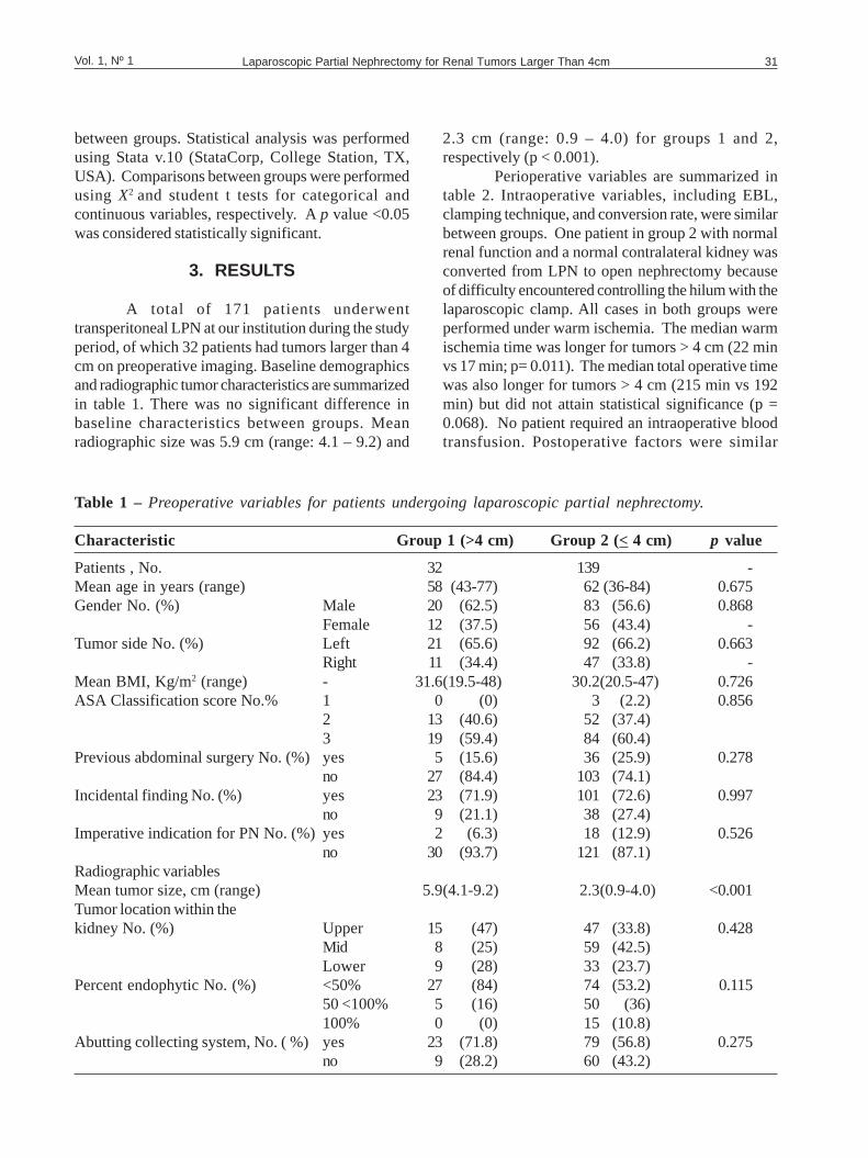
Laparoscopic Partial Nephrectomy for Renal Tumors Larger Than 4cm 31Vol. 1, Nº 1
between groups. Statistical analysis was performedusing Stata v.10 (StataCorp, College Station, TX,USA). Comparisons between groups were performedusing X2 and student t tests for categorical andcontinuous variables, respectively. A p value <0.05was considered statistically significant.
3. RESULTS
A total of 171 patients underwenttransperitoneal LPN at our institution during the studyperiod, of which 32 patients had tumors larger than 4cm on preoperative imaging. Baseline demographicsand radiographic tumor characteristics are summarizedin table 1. There was no significant difference inbaseline characteristics between groups. Meanradiographic size was 5.9 cm (range: 4.1 – 9.2) and
2.3 cm (range: 0.9 – 4.0) for groups 1 and 2,respectively (p < 0.001).
Perioperative variables are summarized intable 2. Intraoperative variables, including EBL,clamping technique, and conversion rate, were similarbetween groups. One patient in group 2 with normalrenal function and a normal contralateral kidney wasconverted from LPN to open nephrectomy becauseof difficulty encountered controlling the hilum with thelaparoscopic clamp. All cases in both groups wereperformed under warm ischemia. The median warmischemia time was longer for tumors > 4 cm (22 minvs 17 min; p= 0.011). The median total operative timewas also longer for tumors > 4 cm (215 min vs 192min) but did not attain statistical significance (p =0.068). No patient required an intraoperative bloodtransfusion. Postoperative factors were similar
Table 1 – Preoperative variables for patients undergoing laparoscopic partial nephrectomy.
Characteristic Group 1 (>4 cm) Group 2 (< 4 cm) p value
Patients , No. 32 139 -Mean age in years (range) 58 (43-77) 62(36-84) 0.675Gender No. (%) Male 20 (62.5) 83 (56.6) 0.868
Female 12 (37.5) 56 (43.4) -Tumor side No. (%) Left 21 (65.6) 92 (66.2) 0.663
Right 11 (34.4) 47 (33.8) -Mean BMI, Kg/m2 (range) - 31.6(19.5-48) 30.2(20.5-47) 0.726ASA Classification score No.% 1 0 (0) 3 (2.2) 0.856
2 13 (40.6) 52 (37.4)3 19 (59.4) 84 (60.4)
Previous abdominal surgery No. (%)yes 5 (15.6) 36 (25.9) 0.278no 27 (84.4) 103 (74.1)
Incidental finding No. (%) yes 23 (71.9) 101 (72.6) 0.997no 9 (21.1) 38 (27.4)
Imperative indication for PN No. (%)yes 2 (6.3) 18 (12.9) 0.526no 30 (93.7) 121 (87.1)
Radiographic variablesMean tumor size, cm (range) 5.9(4.1-9.2) 2.3(0.9-4.0) <0.001Tumor location within thekidney No. (%) Upper 15 (47) 47 (33.8) 0.428
Mid 8 (25) 59 (42.5)Lower 9 (28) 33 (23.7)
Percent endophytic No. (%) <50% 27 (84) 74 (53.2) 0.11550 <100% 5 (16) 50 (36)100% 0 (0) 15 (10.8)
Abutting collecting system, No. ( %)yes 23 (71.8) 79 (56.8) 0.275no 9 (28.2) 60 (43.2)
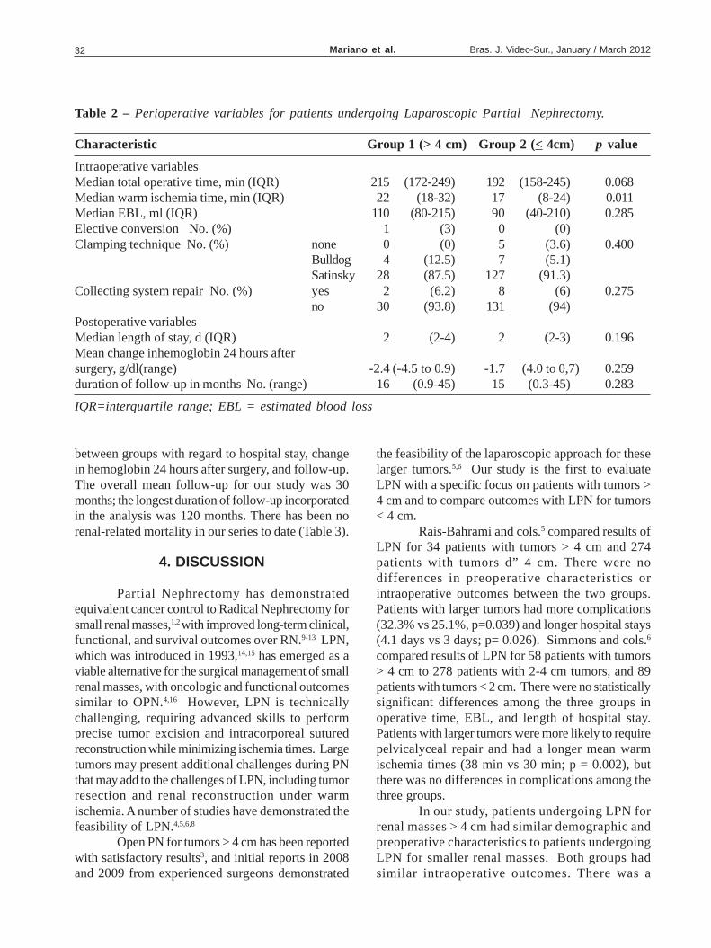
Mariano et al.32 Bras. J. Video-Sur., January / March 2012
between groups with regard to hospital stay, changein hemoglobin 24 hours after surgery, and follow-up.The overall mean follow-up for our study was 30months; the longest duration of follow-up incorporatedin the analysis was 120 months. There has been norenal-related mortality in our series to date (Table 3).
4. DISCUSSION
Partial Nephrectomy has demonstratedequivalent cancer control to Radical Nephrectomy forsmall renal masses,1,2 with improved long-term clinical,functional, and survival outcomes over RN.9-13 LPN,which was introduced in 1993,14,15 has emerged as aviable alternative for the surgical management of smallrenal masses, with oncologic and functional outcomessimilar to OPN.4,16 However, LPN is technicallychallenging, requiring advanced skills to performprecise tumor excision and intracorporeal suturedreconstruction while minimizing ischemia times. Largetumors may present additional challenges during PNthat may add to the challenges of LPN, including tumorresection and renal reconstruction under warmischemia. A number of studies have demonstrated thefeasibility of LPN.4,5,6,8
Open PN for tumors > 4 cm has been reportedwith satisfactory results3, and initial reports in 2008and 2009 from experienced surgeons demonstrated
the feasibility of the laparoscopic approach for theselarger tumors.5,6 Our study is the first to evaluateLPN with a specific focus on patients with tumors >4 cm and to compare outcomes with LPN for tumors< 4 cm.
Rais-Bahrami and cols.5 compared results ofLPN for 34 patients with tumors > 4 cm and 274patients with tumors d” 4 cm. There were nodifferences in preoperative characteristics orintraoperative outcomes between the two groups.Patients with larger tumors had more complications(32.3% vs 25.1%, p=0.039) and longer hospital stays(4.1 days vs 3 days; p= 0.026). Simmons and cols.6
compared results of LPN for 58 patients with tumors> 4 cm to 278 patients with 2-4 cm tumors, and 89patients with tumors < 2 cm. There were no statisticallysignificant differences among the three groups inoperative time, EBL, and length of hospital stay.Patients with larger tumors were more likely to requirepelvicalyceal repair and had a longer mean warmischemia times (38 min vs 30 min; p = 0.002), butthere was no differences in complications among thethree groups.
In our study, patients undergoing LPN forrenal masses > 4 cm had similar demographic andpreoperative characteristics to patients undergoingLPN for smaller renal masses. Both groups hadsimilar intraoperative outcomes. There was a
Table 2 – Perioperative variables for patients undergoing Laparoscopic Partial Nephrectomy.
Characteristic Group 1 (> 4 cm) Group 2 (< 4cm) p value
Intraoperative variablesMedian total operative time, min (IQR) 215 (172-249) 192 (158-245) 0.068Median warm ischemia time, min (IQR) 22 (18-32) 17 (8-24) 0.011Median EBL, ml (IQR) 110 (80-215) 90 (40-210) 0.285Elective conversion No. (%) 1 (3) 0 (0)Clamping technique No. (%) none 0 (0) 5 (3.6) 0.400
Bulldog 4 (12.5) 7 (5.1)Satinsky 28 (87.5) 127 (91.3)
Collecting system repair No. (%) yes 2 (6.2) 8 (6) 0.275no 30 (93.8) 131 (94)
Postoperative variablesMedian length of stay, d (IQR) 2 (2-4) 2 (2-3) 0.196Mean change inhemoglobin 24 hours aftersurgery, g/dl(range) -2.4(-4.5 to 0.9) -1.7 (4.0 to 0,7) 0.259duration of follow-up in months No. (range) 16 (0.9-45) 15 (0.3-45) 0.283
IQR=interquartile range; EBL = estimated blood loss
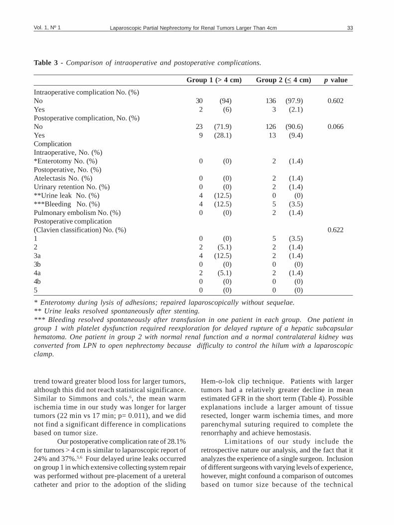
Laparoscopic Partial Nephrectomy for Renal Tumors Larger Than 4cm 33Vol. 1, Nº 1
Table 3 - Comparison of intraoperative and postoperative complications.
Group 1 (> 4 cm) Group 2 (< 4 cm) p value
Intraoperative complication No. (%)No 30 (94) 136 (97.9) 0.602Yes 2 (6) 3 (2.1)Postoperative complication, No. (%)No 23 (71.9) 126 (90.6) 0.066Yes 9 (28.1) 13 (9.4)ComplicationIntraoperative, No. (%)*Enterotomy No. (%) 0 (0) 2 (1.4)Postoperative, No. (%)Atelectasis No. (%) 0 (0) 2 (1.4)Urinary retention No. (%) 0 (0) 2 (1.4)**Urine leak No. (%) 4 (12.5) 0 (0)***Bleeding No. (%) 4 (12.5) 5 (3.5)Pulmonary embolism No. (%) 0 (0) 2 (1.4)Postoperative complication(Clavien classification) No. (%) 0.6221 0 (0) 5 (3.5)2 2 (5.1) 2 (1.4)3a 4 (12.5) 2 (1.4)3b 0 (0) 0 (0)4a 2 (5.1) 2 (1.4)4b 0 (0) 0 (0)5 0 (0) 0 (0)
* Enterotomy during lysis of adhesions; repaired laparoscopically without sequelae.** Urine leaks resolved spontaneously after stenting.*** Bleeding resolved spontaneously after transfusion in one patient in each group. One patient ingroup 1 with platelet dysfunction required reexploration for delayed rupture of a hepatic subcapsularhematoma. One patient in group 2 with normal renal function and a normal contralateral kidney wasconverted from LPN to open nephrectomy because difficulty to control the hilum with a laparoscopicclamp.
trend toward greater blood loss for larger tumors,although this did not reach statistical significance.Similar to Simmons and cols.6, the mean warmischemia time in our study was longer for largertumors (22 min vs 17 min; p= 0.011), and we didnot find a significant difference in complicationsbased on tumor size.
Our postoperative complication rate of 28.1%for tumors > 4 cm is similar to laparoscopic report of24% and 37%.5,6 Four delayed urine leaks occurredon group 1 in which extensive collecting system repairwas performed without pre-placement of a ureteralcatheter and prior to the adoption of the sliding
Hem-o-lok clip technique. Patients with largertumors had a relatively greater decline in meanestimated GFR in the short term (Table 4). Possibleexplanations include a larger amount of tissueresected, longer warm ischemia times, and moreparenchymal suturing required to complete therenorrhaphy and achieve hemostasis.
Limitations of our study include theretrospective nature our analysis, and the fact that itanalyzes the experience of a single surgeon. Inclusionof different surgeons with varying levels of experience,however, might confound a comparison of outcomesbased on tumor size because of the technical
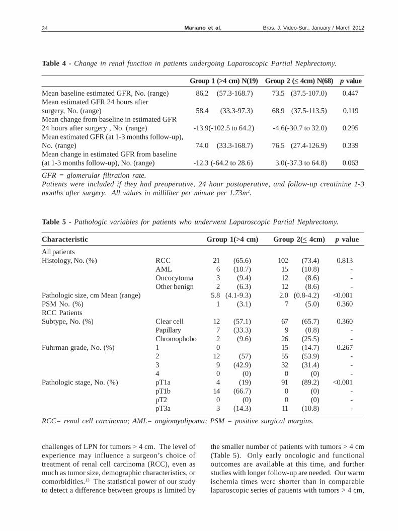
Mariano et al.34 Bras. J. Video-Sur., January / March 2012
Table 4 - Change in renal function in patients undergoing Laparoscopic Partial Nephrectomy.
Group 1 (>4 cm) N(19) Group 2 (< 4cm) N(68) p value
Mean baseline estimated GFR, No. (range) 86.2 (57.3-168.7) 73.5 (37.5-107.0) 0.447Mean estimated GFR 24 hours aftersurgery, No. (range) 58.4 (33.3-97.3) 68.9 (37.5-113.5) 0.119Mean change from baseline in estimated GFR24 hours after surgery , No. (range) -13.9(-102.5 to 64.2) -4.6(-30.7 to 32.0) 0.295Mean estimated GFR (at 1-3 months follow-up),No. (range) 74.0 (33.3-168.7) 76.5 (27.4-126.9) 0.339Mean change in estimated GFR from baseline(at 1-3 months follow-up), No. (range) -12.3(-64.2 to 28.6) 3.0(-37.3 to 64.8) 0.063
GFR = glomerular filtration rate.Patients were included if they had preoperative, 24 hour postoperative, and follow-up creatinine 1-3months after surgery. All values in milliliter per minute per 1.73m2.
Table 5 - Pathologic variables for patients who underwent Laparoscopic Partial Nephrectomy.
Characteristic Group 1(>4 cm) Group 2(< 4cm) p value
All patientsHistology, No. (%) RCC 21 (65.6) 102 (73.4) 0.813
AML 6 (18.7) 15 (10.8) -Oncocytoma 3 (9.4) 12 (8.6) -Other benign 2 (6.3) 12 (8.6) -
Pathologic size, cm Mean (range) 5.8 (4.1-9.3) 2.0 (0.8-4.2) <0.001PSM No. (%) 1 (3.1) 7 (5.0) 0.360RCC PatientsSubtype, No. (%) Clear cell 12 (57.1) 67 (65.7) 0.360
Papillary 7 (33.3) 9 (8.8) -Chromophobo 2 (9.6) 26 (25.5) -
Fuhrman grade, No. (%) 1 0 15 (14.7) 0.2672 12 (57) 55 (53.9) -3 9 (42.9) 32 (31.4) -4 0 (0) 0 (0) -
Pathologic stage, No. (%) pT1a 4 (19) 91 (89.2) <0.001pT1b 14 (66.7) 0 (0) -pT2 0 (0) 0 (0) -pT3a 3 (14.3) 11 (10.8) -
RCC= renal cell carcinoma; AML= angiomyolipoma; PSM = positive surgical margins.
challenges of LPN for tumors > 4 cm. The level ofexperience may influence a surgeon’s choice oftreatment of renal cell carcinoma (RCC), even asmuch as tumor size, demographic characteristics, orcomorbidities.13 The statistical power of our studyto detect a difference between groups is limited by
the smaller number of patients with tumors > 4 cm(Table 5). Only early oncologic and functionaloutcomes are available at this time, and furtherstudies with longer follow-up are needed. Our warmischemia times were shorter than in comparablelaparoscopic series of patients with tumors > 4 cm,
Laparoscopic Partial Nephrectomy for Renal Tumors Larger Than 4cm 35Vol. 1, Nº 1
but a potential criticism is that our total operativetimes were longer. The most important componentof the operative time is the warm ischemia time, asthis factor affects subsequent renal function. Wefeel that the investment of additional time forpreparation to save even a few minutes of warmischemia is time well spent. Other explanations forour longer operatives times include the fact that manyof our patients are obese (mean BMI was 31.6 Kg/m2 in group 1 and 30.2 Kg/m2 in group 2), and 15.6%of group 1 patients and 25.9% of group 2 patientshad undergone prior abdominal surgery.
5. CONCLUSION
In our initial experience, LPN for tumors > 4cm is safe and feasible, showing comparable outcomesto OPN for smaller tumors, although with longer warmischemia times. We do not advocate LPN for allpatients with renal masses, but it may allow selectpatients with larger tumors to achieve theconvalescence benefits of a minimally invasiveapproach. Studies with longer follow-up are neededto more definitively evaluate the efficacy of LPN forlarge tumors.
RESUMOIntrodução: Cirurgia minimamente invasiva por nefrectomia parcial laparoscópica (NPL) normalmente é feita paratumores renais < 4 cm em tamanho. NPL para tumores> 4 cm não tem sido a rotina. Objetivo: Para avaliar a segurançae factibilidade da NPL para tumores > 4 cm comparou-se dois grupos de pacientes: um com tumores < 4 cm e outrocom tumores > 4 cm. Materiais e Métodos: Revisamos dados consecutivos de 171 pacientes que foram submetidos aNPL transperitoneal entre maio de 2002 e maio de 2012 feitas por um mesmo cirurgião. Pacientes foram estratificadosem dois grupos: 32 com tumores> 4 cm na imagem pré-operatória (grupo 1) e 139 com tumores < 4 cm (grupo 2).Dados pré-operatórios, perioperatórios, resultados patológicos e funcionais foram analisados e comparados entre osgrupos. Usamos o teste X2 e student t. O valor p < 0,05 foi considerado estatisticamente significativo. Resultados:Tamanho médio radiográfico do tumor foi 5,9 cm (4,1 – 9,2) para o grupo 1 e 2,3 cm (0,9 – 4,0) para o grupo 2. Não foiencontrada diferença significativa entre os grupos na perda sanguínea estimada, tempo total da cirurgia, tempo dehospitalização, taxa de complicações e mudança na taxa de filtração glomerular. Pacientes com tumores maiores temtempo maior de isquemia quente (22 vs 17 min; p= 0,011). Conclusões: Em nossa experiência, NPL para tumores > 4cm é segura e factível, mostrando resultados comparáveis a NPL para tumores menores. Mais estudos são necessá-rios para determinar a viabilidade da NPL para tumores maiores como uma forma efetiva de tratamento.
Key words: Laparoscopia. Nefrectomia. Nefrectomia parcial. Carcinoma de células renais.
6. REFERENCES
1. Fergany AF, Hafez KS, Novick AC. Long-term results ofnephron sparing
Surgery for localized renal cell carcinoma: 10-year follow-up. JUrol 2000; 163:442-5
2. Uzzo RG, Novick AC. Nephron sparing surgery for renaltumors: indications, techniques and outcomes. J Urol 2001;166:6-18
3. Leibovich BC, Blute ML, Cheville JC, et al. Nephron sparingsurgery for appropriately selected renal cell carcinomabetween 4 and 7 cm results in outcome similar to radicalnephrectomy. J Urol 2004; 171:1066-70
4. Marszalek M, Meixl H, Polajnar M, Rauchenwald M,Jeschke K, Madersbacher S. Laparoscopic and open partialnephrectomy: a matched-pair comparision of 200 patients.Eur Urol 2009; 55:1171-8
5. Rais-Bahrami S, Romero FR, Lima GC, et al. Electivelaparoscopic partial nephrectomy in patients with tumors >4cm . Urology 2008; 72:580-3.
6. Simmons MN, Chung BI, Gill IS. Perioperative efficacy oflaparoscopic partial nephrectomy for tumors larger than 4cm. Eur Urol 2009; 55:199-208.
7. Dindo D, Demartines N, Clavien PA. Classification of surgicalcomplications: a new proposal with evaluation in a cohort of6336 patients and results of a survey. Ann Surg 2004; 240:205-13
8. Levey AS, Bosch JP, Lewis JB, et al. A more accurate methodto estimate glomerular filtration rate from serum creatinine: anew prediction equation. Modification of Diet in RenalDisease Study Group. Ann Intern Med 1999; 130:461-70
9. Go AS, Chertow GM, Fan D, McCulloch CE, Hsu CY.Chronic kidney disease and the risks of death, cardiovascularevents, and hospitalization. N Engl J Med 2004; 351:1296-305.
10. Huang WC, Levey AS, et al. Chronic kidney disease afternephrectomy in patients with renal cortical tumors: aretrospective cohort study. Lancet Oncol 2006; 7:735-40.
11. Thompson RH, Boorjian SA, Lohse CM, et al. Radicalnephrectomy for pT1 a renal masses may be associated with
Mariano et al.36 Bras. J. Video-Sur., January / March 2012
decreased overall survival compared with partialnephrectomy. J Urol 2008; 179:468-71, discussion 472-3.
12. Zini L, Perrotte P, Capitanio U, et al. Radical versus partialnephrectomy: effect on overall and noncancer mortality.Cancer 2009; 115:1465-71.
13. Miller DC, Schonlou M, Litwin MS, Lai J. Saigal CS. Renaland cardiovascular morbidity after partial or radicalnephrectomy. Cancer 2008; 511-20.
14. McDougall EM, Clayman RV, Chanhoke PS, et al.Laparoscopic partial nephrectomy in the pig model. J Urol1993; 149:1633-6
15. Winfield HN. Donovan JF, Lund GO, et al. Laparoscopicpartial nephrectomy: initial experience and comparision tothe open surgical approach. J Urol, 1995;153: 1409-14
Brazilian Journal of Videoendoscopic Surgery - v. 5 - n. 1 - Jan./Mar. 2012 - Subscription: + 55 21 3325-7724 - E-mail: [email protected] 1983-9901: (Press) ISSN 1983-991X: (on-line) - SOBRACIL - Press Graphic & Publishing Ltd. Rio de Janeiro, RJ-Brasil
16. Lane BR, Gill IS. 5 –year outcomes of laparoscopic partialnephrectomy. J Urol 2007; 177:70-4.
Corresponding author:MIRANDOLINO BATISTA MARIANORua Costa 30, conjunto 803, Menino DeusPorto Alegre, RS, Brazil90110-270Email: [email protected].: 55 51 3231-0990 Fax : 55 51 3231-7247
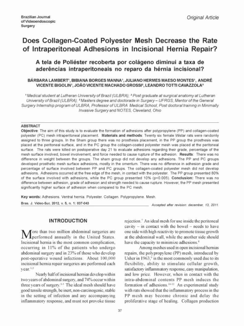
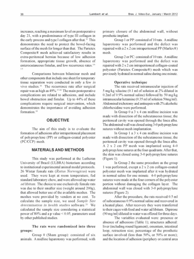
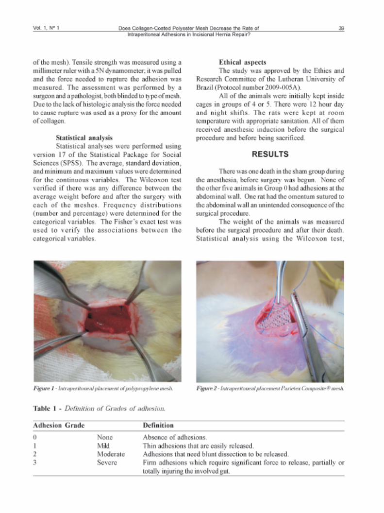
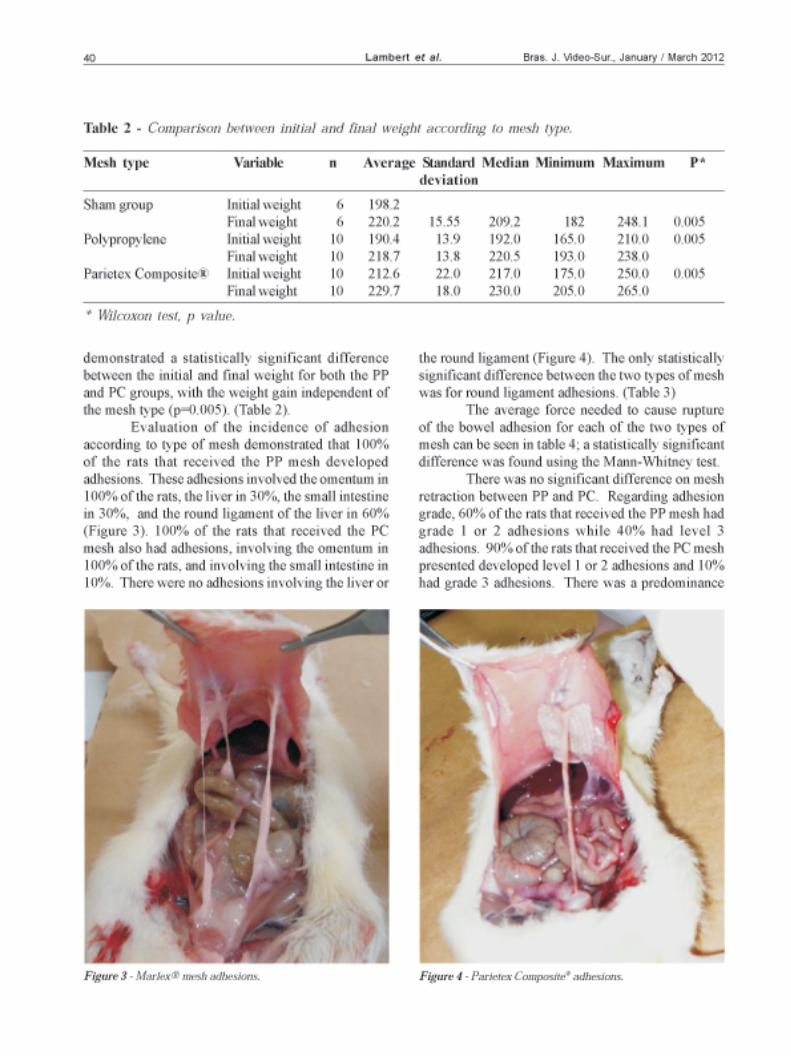
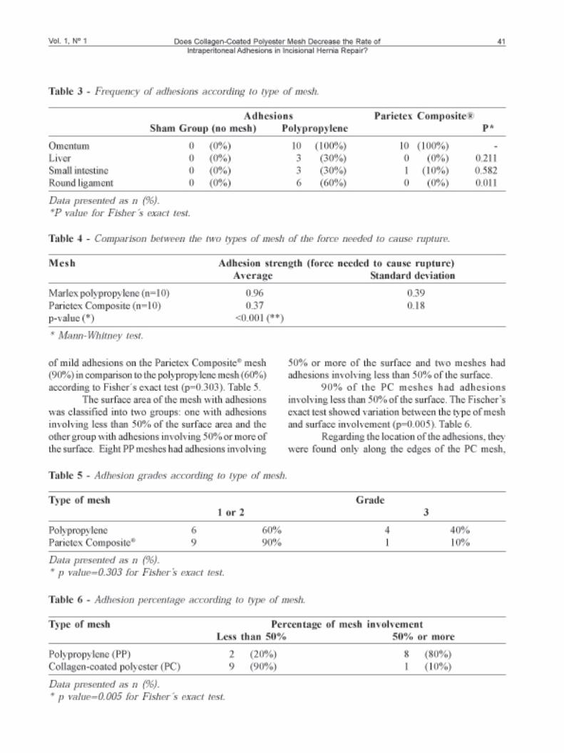
Skinovsky et al.44 Bras. J. Video-Sur., January / March 2012Original ArticleBrazilian Journalof VideoendoscopicSurgery
Accepted after revision: february, 06, 2012.Bras. J. V ideo-Sur , 2012, v. 5, n. 1: 044-048
44
Transabdominal Pre-Peritoneal (T APP) InguinalHernioplasty by Lap aroendoscopic Single Site Surgery
(LESS). Is it Feasible and Safe?
Hernioplastia Inguinal T ransabdominal Pré-Peritoneal por CirurgiaLaparoscópica de Acesso Único. É V iável e Segura?
JAMES SKINOVSKY, PHD1; MARCUS VINICIUS DANTAS DE CAMPOS MARTINS, MD 2;SÉRGIO ROLL, PHD 3; MAURICIO CHIBATA, MD4; FERNANDA TSUMANUMA 5; ROGÉRIO CAVALLIERE 6;
FRANCISCO ALMEIDA 7
Study conducted at the Red Cross / Positivo University Hospital, Curitiba, PR, Brazil.1. Professor of Surgery, Positivo University and Head of Surgery Department, Red Cross University Hospital,
Curitiba, Brazil; 2. Professor of Surgery, Estácio de Sá University, Rio de Janeiro, Brazil; 3. President, AmericanHernia Society; 4. Professor of Surgery, Positivo University, Curitiba,Brazil; 5. Staff Surgeon, Red CrossUniversity Hospital, Curitiba, Brazil; 6. Staff Surgeon, Red Cross University Hospital, Curitiba, Brazil;
7. Surgery Resident, Red Cross University Hospital, Curitiba, Brazil.
ABSTRACTObjectives: To present the first transabdominal pre-peritoneal (TAPP) Laparo-Endoscopic Single-Site Surgery (LESS)inguinal hernioplasties series. Patient s and Methods: From June to December 2011 the first six LESS TAPP inguinalhernioplasties, were performed at the Red Cross University Hospital in Curitiba, Paraná, Brazil. The Single TrocarAccess (SITRACC) Platform (EDLO, Brazil) was used in 5 cases, while the SILS Platform (Covidien, USA) was used inone case. All patients were male; their ages ranged from 18 to 45. Five had NYHUS II hernias and one had a NYHUS IIIahernia. Results: The mean operative time was 44 minutes. None of the surgeries required an extra trocar or conversionto a conventional laparoscopic procedure. All patients were discharged within 24 hours. Conclusions: The TAPP inguinalhernioplasty using a LESS approach is feasible and safe. It constitutes a new option in the scarless surgery field, as wellas a new technique in the ongoing pursuit of surgical innovation that benefits our patients.
Key words: Minimally Invasive Surgery. LESS. Inguinal Hernioplasty. SITRACC.
INTRODUCTION
Since the introduction in 1987 of videosurgery andthe concept of minimally invasive surgery, the
benefits of less discomfort, milder metabolicalterations, faster recovery, and good aestheticresults have been amply demonstrated, and thetechniques have disseminated through theoperating rooms of the world quickly andenthusiastically.
Ongoing improvements in optical equipmentand refinements in the instrumentations employed invideosurgery have made it possible for increasinglycomplex procedures to be performed by minimallyinvasive methods.
Over the same period new technologies andapproaches have emerged, such as Natural OrificeTranslumenal Endoscopic Surgery (NOTES),Needlescopy, and Laparo-Endoscopic Single-SiteSurgery (LESS).
Several platforms to perform LESS havebecome available in recent years. Two of them arethe Single Trocar Access (SITRACC), a disposablemultiport trocar (EDLO, Brazil – Figure 1) that usesinstruments specially designed for this approach, andthe Single Incision Laparoscopic Surgery (SILS)platform (Covidien, USA).
Laparo-Endoscopic Single-Site Surgery(LESS), now with several variations, has emergedas an alternative to NOTES. One multichannel
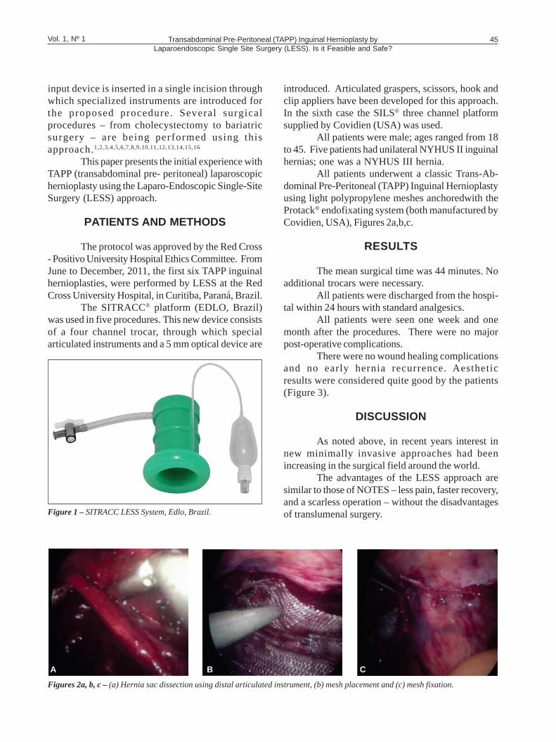
Transabdominal Pre-Peritoneal (TAPP) Inguinal Hernioplasty byLaparoendoscopic Single Site Surgery (LESS). Is it Feasible and Safe?
45Vol. 1, Nº 1
input device is inserted in a single incision throughwhich specialized instruments are introduced forthe proposed procedure. Several surgicalprocedures – from cholecystectomy to bariatricsurgery – are being performed using thisapproach.1,2,3,4,5,6,7,8,9,10,11,12,13,14,15,16
This paper presents the initial experience withTAPP (transabdominal pre- peritoneal) laparoscopichernioplasty using the Laparo-Endoscopic Single-SiteSurgery (LESS) approach.
PATIENTS AND METHODS
The protocol was approved by the Red Cross- Positivo University Hospital Ethics Committee. FromJune to December, 2011, the first six TAPP inguinalhernioplasties, were performed by LESS at the RedCross University Hospital, in Curitiba, Paraná, Brazil.
The SITRACC® platform (EDLO, Brazil)was used in five procedures. This new device consistsof a four channel trocar, through which specialarticulated instruments and a 5 mm optical device are
introduced. Articulated graspers, scissors, hook andclip appliers have been developed for this approach.In the sixth case the SILS® three channel platformsupplied by Covidien (USA) was used.
All patients were male; ages ranged from 18to 45. Five patients had unilateral NYHUS II inguinalhernias; one was a NYHUS III hernia.
All patients underwent a classic Trans-Ab-dominal Pre-Peritoneal (TAPP) Inguinal Hernioplastyusing light polypropylene meshes anchoredwith theProtack® endofixating system (both manufactured byCovidien, USA), Figures 2a,b,c.
RESULTS
The mean surgical time was 44 minutes. Noadditional trocars were necessary.
All patients were discharged from the hospi-tal within 24 hours with standard analgesics.
All patients were seen one week and onemonth after the procedures. There were no majorpost-operative complications.
There were no wound healing complicationsand no early hernia recurrence. Aestheticresults were considered quite good by the patients(Figure 3).
DISCUSSION
As noted above, in recent years interest innew minimally invasive approaches had beenincreasing in the surgical field around the world.
The advantages of the LESS approach aresimilar to those of NOTES – less pain, faster recovery,and a scarless operation – without the disadvantagesof translumenal surgery.Figure 1 – SITRACC LESS System, Edlo, Brazil.
Figures 2a, b, c – (a) Hernia sac dissection using distal articulated instrument, (b) mesh placement and (c) mesh fixation.
A B C
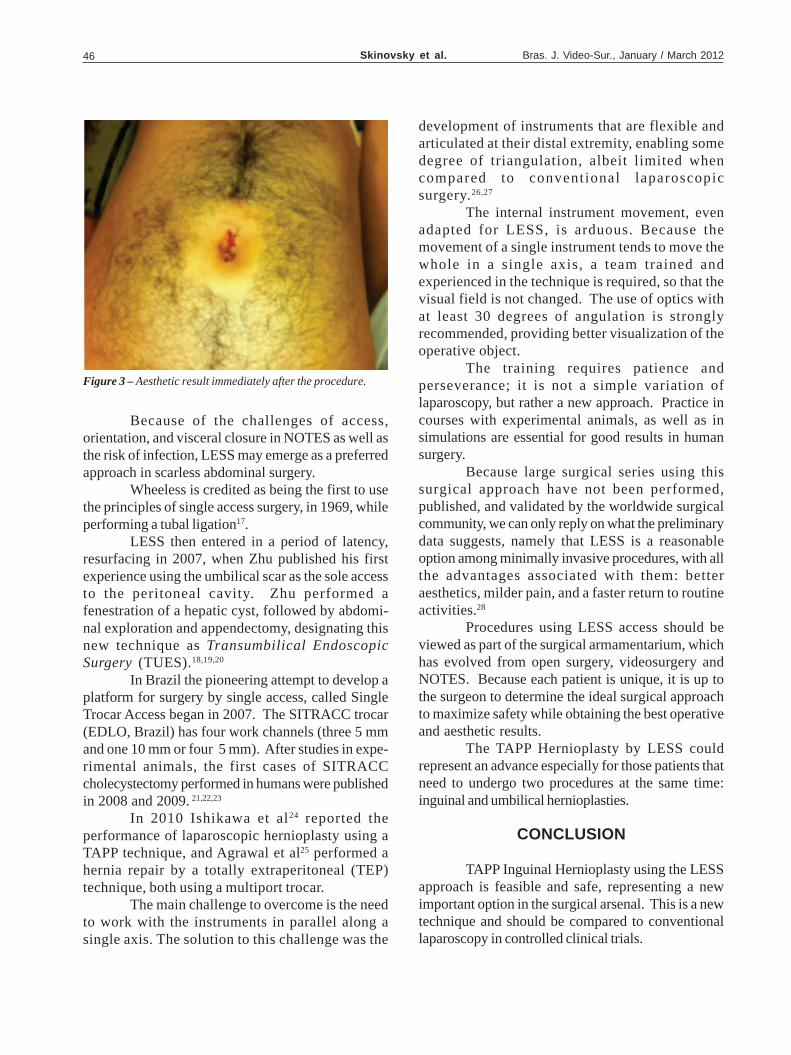
Skinovsky et al.46 Bras. J. Video-Sur., January / March 2012
Because of the challenges of access,orientation, and visceral closure in NOTES as well asthe risk of infection, LESS may emerge as a preferredapproach in scarless abdominal surgery.
Wheeless is credited as being the first to usethe principles of single access surgery, in 1969, whileperforming a tubal ligation17.
LESS then entered in a period of latency,resurfacing in 2007, when Zhu published his firstexperience using the umbilical scar as the sole accessto the peritoneal cavity. Zhu performed afenestration of a hepatic cyst, followed by abdomi-nal exploration and appendectomy, designating thisnew technique as Transumbilical EndoscopicSurgery (TUES).18,19,20
In Brazil the pioneering attempt to develop aplatform for surgery by single access, called SingleTrocar Access began in 2007. The SITRACC trocar(EDLO, Brazil) has four work channels (three 5 mmand one 10 mm or four 5 mm). After studies in expe-rimental animals, the first cases of SITRACCcholecystectomy performed in humans were publishedin 2008 and 2009. 21,22,23
In 2010 Ishikawa et al24 reported theperformance of laparoscopic hernioplasty using aTAPP technique, and Agrawal et al25 performed ahernia repair by a totally extraperitoneal (TEP)technique, both using a multiport trocar.
The main challenge to overcome is the needto work with the instruments in parallel along asingle axis. The solution to this challenge was the
development of instruments that are flexible andarticulated at their distal extremity, enabling somedegree of triangulation, albeit limited whencompared to conventional laparoscopicsurgery.26,27
The internal instrument movement, evenadapted for LESS, is arduous. Because themovement of a single instrument tends to move thewhole in a single axis, a team trained andexperienced in the technique is required, so that thevisual field is not changed. The use of optics withat least 30 degrees of angulation is stronglyrecommended, providing better visualization of theoperative object.
The training requires patience andperseverance; it is not a simple variation oflaparoscopy, but rather a new approach. Practice incourses with experimental animals, as well as insimulations are essential for good results in humansurgery.
Because large surgical series using thissurgical approach have not been performed,published, and validated by the worldwide surgicalcommunity, we can only reply on what the preliminarydata suggests, namely that LESS is a reasonableoption among minimally invasive procedures, with allthe advantages associated with them: betteraesthetics, milder pain, and a faster return to routineactivities.28
Procedures using LESS access should beviewed as part of the surgical armamentarium, whichhas evolved from open surgery, videosurgery andNOTES. Because each patient is unique, it is up tothe surgeon to determine the ideal surgical approachto maximize safety while obtaining the best operativeand aesthetic results.
The TAPP Hernioplasty by LESS couldrepresent an advance especially for those patients thatneed to undergo two procedures at the same time:inguinal and umbilical hernioplasties.
CONCLUSION
TAPP Inguinal Hernioplasty using the LESSapproach is feasible and safe, representing a newimportant option in the surgical arsenal. This is a newtechnique and should be compared to conventionallaparoscopy in controlled clinical trials.
Figure 3 – Aesthetic result immediately after the procedure.
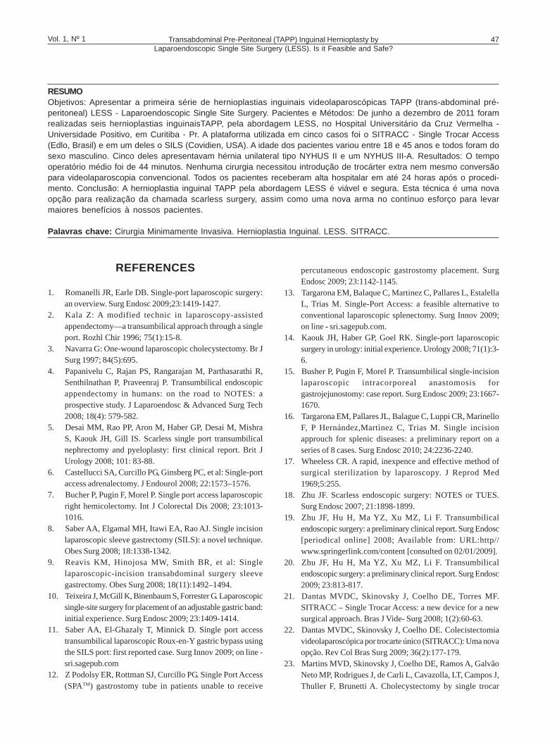
Transabdominal Pre-Peritoneal (TAPP) Inguinal Hernioplasty byLaparoendoscopic Single Site Surgery (LESS). Is it Feasible and Safe?
47Vol. 1, Nº 1
RESUMOObjetivos: Apresentar a primeira série de hernioplastias inguinais videolaparoscópicas TAPP (trans-abdominal pré-peritoneal) LESS - Laparoendoscopic Single Site Surgery. Pacientes e Métodos: De junho a dezembro de 2011 foramrealizadas seis hernioplastias inguinaisTAPP, pela abordagem LESS, no Hospital Universitário da Cruz Vermelha -Universidade Positivo, em Curitiba - Pr. A plataforma utilizada em cinco casos foi o SITRACC - Single Trocar Access(Edlo, Brasil) e em um deles o SILS (Covidien, USA). A idade dos pacientes variou entre 18 e 45 anos e todos foram dosexo masculino. Cinco deles apresentavam hérnia unilateral tipo NYHUS II e um NYHUS III-A. Resultados: O tempooperatório médio foi de 44 minutos. Nenhuma cirurgia necessitou introdução de trocárter extra nem mesmo conversãopara videolaparoscopia convencional. Todos os pacientes receberam alta hospitalar em até 24 horas após o procedi-mento. Conclusão: A hernioplastia inguinal TAPP pela abordagem LESS é viável e segura. Esta técnica é uma novaopção para realização da chamada scarless surgery, assim como uma nova arma no contínuo esforço para levarmaiores benefícios à nossos pacientes.
Palavras chave: Cirurgia Minimamente Invasiva. Hernioplastia Inguinal. LESS. SITRACC.
REFERENCES
1. Romanelli JR, Earle DB. Single-port laparoscopic surgery:an overview. Surg Endosc 2009;23:1419-1427.
2. Kala Z: A modified technic in laparoscopy-assistedappendectomy—a transumbilical approach through a singleport. Rozhl Chir 1996; 75(1):15-8.
3. Navarra G: One-wound laparoscopic cholecystectomy. Br JSurg 1997; 84(5):695.
4. Papanivelu C, Rajan PS, Rangarajan M, Parthasarathi R,Senthilnathan P, Praveenraj P. Transumbilical endoscopicappendectomy in humans: on the road to NOTES: aprospective study. J Laparoendosc & Advanced Surg Tech2008; 18(4): 579-582.
5. Desai MM, Rao PP, Aron M, Haber GP, Desai M, MishraS, Kaouk JH, Gill IS. Scarless single port transumbilicalnephrectomy and pyeloplasty: first clinical report. Brit JUrology 2008; 101: 83-88.
6. Castellucci SA, Curcillo PG, Ginsberg PC, et al: Single-portaccess adrenalectomy. J Endourol 2008; 22:1573–1576.
7. Bucher P, Pugin F, Morel P. Single port access laparoscopicright hemicolectomy. Int J Colorectal Dis 2008; 23:1013-1016.
8. Saber AA, Elgamal MH, Itawi EA, Rao AJ. Single incisionlaparoscopic sleeve gastrectomy (SILS): a novel technique.Obes Surg 2008; 18:1338-1342.
9. Reavis KM, Hinojosa MW, Smith BR, et al: Singlelaparoscopic-incision transabdominal surgery sleevegastrectomy. Obes Surg 2008; 18(11):1492–1494.
10. Teixeira J, McGill K, Binenbaum S, Forrester G. Laparoscopicsingle-site surgery for placement of an adjustable gastric band:initial experience. Surg Endosc 2009; 23:1409-1414.
11. Saber AA, El-Ghazaly T, Minnick D. Single port accesstransumbilical laparoscopic Roux-en-Y gastric bypass usingthe SILS port: first reported case. Surg Innov 2009; on line -sri.sagepub.com
12. Z Podolsy ER, Rottman SJ, Curcillo PG. Single Port Access(SPATM) gastrostomy tube in patients unable to receive
percutaneous endoscopic gastrostomy placement. SurgEndosc 2009; 23:1142-1145.
13. Targarona EM, Balaque C, Martinez C, Pallares L, EstalellaL, Trias M. Single-Port Access: a feasible alternative toconventional laparoscopic splenectomy. Surg Innov 2009;on line - sri.sagepub.com.
14. Kaouk JH, Haber GP, Goel RK. Single-port laparoscopicsurgery in urology: initial experience. Urology 2008; 71(1):3-6.
15. Busher P, Pugin F, Morel P. Transumbilical single-incisionlaparoscopic intracorporeal anastomosis forgastrojejunostomy: case report. Surg Endosc 2009; 23:1667-1670.
16. Targarona EM, Pallares JL, Balague C, Luppi CR, MarinelloF, P Hernández,Martìnez C, Trias M. Single incisionapprouch for splenic diseases: a preliminary report on aseries of 8 cases. Surg Endosc 2010; 24:2236-2240.
17. Wheeless CR. A rapid, inexpence and effective method ofsurgical sterilization by laparoscopy. J Reprod Med1969;5:255.
18. Zhu JF. Scarless endoscopic surgery: NOTES or TUES.Surg Endosc 2007; 21:1898-1899.
19. Zhu JF, Hu H, Ma YZ, Xu MZ, Li F. Transumbilicalendoscopic surgery: a preliminary clinical report. Surg Endosc[periodical online] 2008; Available from: URL:http//www.springerlink.com/content [consulted on 02/01/2009].
20. Zhu JF, Hu H, Ma YZ, Xu MZ, Li F. Transumbilicalendoscopic surgery: a preliminary clinical report. Surg Endosc2009; 23:813-817.
21. Dantas MVDC, Skinovsky J, Coelho DE, Torres MF.SITRACC – Single Trocar Access: a new device for a newsurgical approach. Bras J Vide- Surg 2008; 1(2):60-63.
22. Dantas MVDC, Skinovsky J, Coelho DE. Colecistectomiavideolaparoscópica por trocarte único (SITRACC): Uma novaopção. Rev Col Bras Surg 2009; 36(2):177-179.
23. Martins MVD, Skinovsky J, Coelho DE, Ramos A, GalvãoNeto MP, Rodrigues J, de Carli L, Cavazolla, LT, Campos J,Thuller F, Brunetti A. Cholecystectomy by single trocar
Skinovsky et al.48 Bras. J. Video-Sur., January / March 2012
access – SITRACC: The first multicenter study. Surg Innov2009;Dez – on line - sri.sagepub.com.
24. Ishikawa N, Kawaguchi M, Shimizu S, Matsunoki A, InakiN, watanabe G. Single-incision laparoscopic hernioplastywith the assistance of the Radius Surgical System. Surg Endosc2010; 24:730-731.
25. Agrawal S, Shaw A, Soon Y. Single-Port laparoscopic totallyextraperitoneal inguinal hernia repair with the TriPort system:initial experience. Surg Endosc 2010; 24:952-956.
26. Dominguez G, Durand L, De Rosa J, Danguise E, ArozamenaC, Ferraina PA. Retraction and triangulation with neodymiunmagnetic fórceps for single-port laparoscopiccholecystectomy. Surg Endosc 2009; 23:1660-1666.
27. Galvão Neto M, Ramos A, Campos J. Single portlaparoscopy Access surgery. Tech Gastrointest Endosc 2009;11(2):84-94.
Brazilian Journal of Videoendoscopic Surgery - v. 5 - n. 1 - Jan./Mar. 2012 - Subscription: + 55 21 3325-7724 - E-mail: [email protected] 1983-9901: (Press) ISSN 1983-991X: (on-line) - SOBRACIL - Press Graphic & Publishing Ltd. Rio de Janeiro, RJ-Brasil
28. Tsimoyiannis EC, Tsimogiannis KE, Pappas-Gogos G,Farantos C, Benetatos N, Mavridou P, Manataki A. Dif ferentpain scores in single transumbilical incision laparoscopiccholecystectomy versus classic laparoscopiccholecystectomy: a randomized controled Trial. Surg Endosc2010; 24:1842-1848.
Corresponding author:JAMES SKINOVSKYAv. Iguaçú, 2713/503, Água VerdeCuritiba, PR, Brazil80240-030Phone: 55-41-9971-1644E-mail: [email protected]
Information for Authors 49Vol. 1, Nº 1 Special Section IBrazilian Journalof VideoendoscopicSurgery
INFORMATION FOR AUTHORS
1. ObjectivesBRAZILIAN JOURNAL OF VIDEOENDOSCOPIC SURGERY (BJV) is the official journal of the Brazilian Society of
Videosurgery that publishes scientific articles in order to register results of videosurgery researches and related subjects,encourages study and progress in this area as well as publications to deepen medical knowledge.
2. Analysis, Selection and Exclusiveness of ManuscriptsManuscripts submitted will be analyzed by a Reviewers Committee, the manuscripts should be original and should not
be published elsewhere. A copy of the manuscript is anonymously forwarded by the Editor to 2 or 3 reviewers to be analyzedwithin 30 days.
Peer review includes suggestions to the Editor, reject or accept the manuscript with or without changes. Manuscriptsthat are rejected will be returned to the author. Afterwards, peer review suggestions are forwarded to the main author forapproval who will decide if she/he will resubmit it. Scientific articles describing experiments on human subjects or animalsmust include approval of the appropriate ethics committee of the institution where the study was performed, in accordancewith the Declaration of Helsinki (1964 and 1975,1983 and 1989 amendments), the Animal Protection International Rules andthe National Health Council Resolution no 196/96. Republishing a national or an international journal article is only acceptedin special cases and must be accompanied by written permission for its use from the copyright owner and the author. In thiscase a copy of the first manuscript version should be provided. Manuscripts must have up to 6 authors in order to bepublished.
3. Periodicity & Scientific Matters to be publishedBrazilian Journal of Videoendoscopic Surgery is published quarterly. It is a communication channel of scientific matters such as:· Original Article: original clinical(or experimental) research;· Preliminary Communications: partial results on new researches, techniques and methods in study;· Case Report(or Clinical Meeting): with critical analysis and discussion;· Clinical Observation: should have critical analysis and discussion;· Epidemiologic Statistics: with critical analysis and discussion;· Description and Evaluation: of methods or procedures, with revision, critical analysis and discussion;· Opinion and Analysis: of philosophical, ethical and social aspects regarding the area of study;· Letters to the Editor: including criticisms and suggestions about publications, as well as questions and/or comments
about manuscripts that have already been published.
4. Requirements for preparation and submission of manuscripts Authors should send the manuscript in microsoft word format by email to: [email protected]). Security
Copy: A copy of all materials submitted to the journal will be sent to the author with the approval by the Editorial Boardfor future copyrights warranties. IMPORTANT! Keep a copy of all the material submitted to the publication of yourmanuscript.
Cover letter: A cover letter signed by the main author should be enclosed. If the author have interest in pay for coloredillustrations this should be specified in the cover letter.
Permission for reproduction and copyright transfer statement: Manuscripts must be accompanied by written permissionfor use of copyrighted material or photographs of identifiable persons. Copyright transfer statement must be sent.
Protection sending the manuscript: Manuscripts should be sent in a suitable package, in order to avoid bendingphotographs and illustrations.
5. Standard Format and Print out· Manuscripts should be typed double-spaced with up to 25 lines per page.· Pages should be numbered consecutively (numbers should be in the upper or lower right corner). The first page
should be the Title Page.· Each section should start on a new page.· Manuscripts should be printable on one side of a 216x279mm or A4(212x297mm) white sulphite paper with margins of
25mm.· Manuscripts should include, in sequence and on separate pages:
49
Information for Authors50 Bras. J. Video-Sur., January / March 2012
- Identification Page (Title page – see details bellow);- Abstract/Key words;- Text pages and Acknowledgments;- References;- Tables (one in each page, separately);- Illustrations;- Legends;- Abbreviations.
6. Manuscripts Preparation
6.1. Identification Page (Title Page)All manuscripts will be subject to a process of anonymous editorial review, therefore the name and address of authors
should only be in the Title Page with identification as it is not going to be sent to the reviewers. The authors should verifyif there is any identification on the text to avoid identification.
Title Page “without identification”- Complete Manuscript Title(concise and informative)- Short title ( up to 8 words)
Title Page “with identification”- Name(s) of author(s) and Institutional Scientific Affiliation: provide detailed information about the department and
the institute where the work was conducted. Affiliation and/or Academic degrees of the authors: include name, highestacademic degree and institutional affiliation and position of each author.
- Footnotes:Address, telephone, fax and e-mail of the main author should be given for journal editor contact.Address to request copies and to contact author (include full address information and e-mail of the author who submitted
the material to be published).- Source of Funding: it should be declared any source of funding such as grants, equipments and others.
6.2. AbstractThe abstract is mandatory. It should be up to 250 words. Every abstract should be written in an informative style.
Depending on the abstract it should contain the following headings:Original Ar ticles: Objectives/Materials(Patients) and Methods/Results/Discussion/Conclusion(s).Reviews, Actualization, Opinion: Objectives/History(Scientific Summary)/DiscussionCase report or Clinical Meetings: Objectives/Meetings Summary/DiscussionTechnical Notes or Preliminary Communication: Objectives/Technical Report/ Research Report/ Preliminary Results/Discussion
6.3. Descriptors (Key Words)Identify the manuscript with 3 to 10 key words or short phrases bellow the abstract using DeCS or MESH terminology
which will assist indexers in cross-indexing the article in the data base.For DeCS terms access: http://decs.bvs.br and for MESH (Medical Subject Index) terms access: http://www.nlm.nih.gov/
mesh/meshhome.html . If suitable MeSH terms are not yet available, well known terms or expressions are accepted.6.4. TextThe textual material of clinical or experimental observation manuscripts should be organized whenever possible in a
standard form as follows: Introduction, Patients and Methods, Results, Discussion, Conclusion, Acknowledgment,References. Other types of manuscripts such as case report, editorials and reviews may follow a different format, accordingto the Editorial Board. Long manuscripts in order to provide a better understanding of its contents may include subheadingsin some sections such as Results and Discussion.
Citations and References: Authors citations must appear in the text as superscript numbers placed to the right of aword, sentence or paragraph. Citations of names should be typed in Upper Case. Name of author(s) citation should followthe format bellow:
Information for Authors 51Vol. 1, Nº 1
- One author: KOCK1
- Two authors: KOCK e PENROSE1
- Three or more authors: KOCK and cols. 1
Note: In the body of the text the form “…and cols” is suggested and in the references “… et al”.
Introduction – It should briefly describe the reason to accomplish the article and the objective. Do not include data orconclusions and mention only relevant references.
Materials (or Patients) and Methods – should describe in detail the recruitment of individuals (human subjects andlaboratory animals as well as group control) included in the research. Identify the age, sex and other relevant characteristicsof the subject. Authors should be careful when specify race or ethnic group as their definition and relevance areambiguous. Methods, apparatus (with manufacturer’s name and address in parentheses) and procedures used should beidentified in adequate detail so that other researches can reproduce the experiment. The methods published in otherresearch should be mention and unknown methods briefly described. Statistical methods and protocols used should alsobe described, as well as the computers software used. Authors that submitted reviews should include a section todescribe the methods used for locating, selecting, extracting and synthesizing data. These methods should be summarizedin the abstract. When the paper reports experiments on human subjects it must indicate whether the procedures followedethical standards of the responsible committee on human experimentation. Do not use name, initials or hospital identificationof the patients, especially in illustrative material. When the paper reports experiments on animals, it must indicate thatprotocols were reviewed by the appropriate institutional committee with respect to the care and use of laboratory animalsused in this study.
Results – Provide results in a logical sequence in the text, tables and figures. Do not repeat all tables and figures data inthe text; consider the relevant ones.
Discussion – Emphasize important and new aspects of the study as well as the conclusions originated from them. Avoiddetailed repetition of the data provided in the Introduction or Results. Include findings implications and limitations in theDiscussion Section, mentioning implications for future research. Compare what was observed to other relevant studies.
Conclusions – The conclusions should be based on the study objectives, in order to avoid unqualified statements andconclusions that are not based on the findings. Author(s) should not state the economic benefits and costs unless theirmanuscript includes economic analysis and data. Studies that have not been completed should not be mentioned. Newhypothesis should only be considered if justified. Include recommendations when appropriate.
Acknowledgement: Acknowledgements to people and institutions may be included at the end of the manuscript, statingany type of contribution and/or participation towards the development of the research. Technical support should beacknowledged in a paragraph separate from other types of contributions.
6.5. ReferencesThe references that are stated in the text should be consecutively in alphabetical order or as they are cited in the text.
References, tables and legends must be identified in the text by superscript Arabic numerals. Citation of manuscriptsaccepted but not yet published: mention the journal and add “In press” in the reference list (authors should have writtenpermission to mention these articles, as well as to verify if manuscripts were accepted to publication).
Avoid personal communications citation, unless it provides essential information and it is not possible to be obtainedin printed sources (in such case they should be cited in parentheses in the text with name of the person and date of thecommunication). The Brazilian Journal of Videoendoscopic Surgery is in accordance with “Vancouver Style” (uniformrequirements for manuscripts submitted to biomedical journals), electronic version is available on http://acponline.org/journals/annals/01jan97/unifreqr.htm, also published in N Engl J Med 1997; 336(4): 309-315 and commended by theInternational Committee of Medical Journal Editors.
The Uniform Requirements (Vancouver Style) are based on the American National Standards Institute (ANSI) adaptedby the NLM (National Library of Medicine). Complete information about format of references may be verified in: UniformRequirements for Manuscripts, Journal of Public Health 1999; 33(1), also available in electronic version: http://www.fsp.usp.br/~rsp: http:// www.fsp.usp.br/~rsp.
Information for Authors52 Bras. J. Video-Sur., January / March 2012
Examples of references format:
- Periodical articleInclude only the first 6 authors and add “et al”. Do not use Upper Case or bold or underlined or italics. Journal names are
abbreviated according to the Index Medicus – in the List of Journals Indexed in Index Medicus available at http://www.nlm.nih.gov/tsd/serials/lji.html, and the Latin American Journals available at: http://www.bireme.br/abd/P/lista_geral.htm.
Ex: Parkin DM, Clayton D, Black RJ, Masuyer E, Friedl HP, Ivanov E, et al. Childhood leukemia in Europe after Chernobyl:5 years follow-up. Br J Cancer 1996; 73: 1006-12.
- BookEx: Rigsven MK, Bond D. Gerontology and leadship skills for nurses. 2nd ed. Albany (NY): Delmar Publishers; 1996.
- Chapter in BookEx: Philips SJ, Whiosnant JP. Hypertension and stroke. In: Laragh JH, Brenner BM, editors. Hypertension:
pathophysiology, diagnosis and management. 2nd ed. New York; Raven Press; 1995. p.465-78.
- Conference PaperEx: Bergtson S, Solhein BG. Enforcement of data protection, privacy and security in medical informatics. In: Lun KC,
Degoulet P, Piemme TE, Rienhoff O, editor. MEDINFO 92. Proceedings of the 7th World Congress on Medical Informatics;1992 Sep 6-10; Geneva, Switzerland, Amsterdam: North Holland; 1992. p.1561-5.
- DissertationEx: Carvalho ACP. A contribuição da tomografia computadorizada ao diagnóstico de aneurisma dissecante da aorta
[dissertação - mestrado]. Rio de Janeiro: Faculdade de Medicina, Universidade Federal do Rio de Janeiro; 1993.Kaplan SJ. Post-hospital home health care: the elderly’s access and utilization [dissertation]. St. Louis (Ø): Washington
Univ.; 1995
- Journal article in electronic formatEx: Morse SS. Factors in the emergence of infectious diseases. Emerg Infect Dis [periodical online] 1995; 1(1). Available
from: URL: http://www.cdc.gov/ncidod/EID/eid.htm [consulted on 11/12/2002].
- Opinion or technical articles onlineEx: Carvalho ACP, Marchiori E. Manual de orientação para a elaboração de monografias, dissertações e teses. Avaialabre
from: URL http://www.radiologia.ufrj.br/manual.htm [consulted on 08/12/2002].
6.6 TablesPrint out each Table on a separate sheet of paper. Number tables with Arabic numerals consecutively in the order of their
first citation in the text and supply a brief title for each table. Data that are shown in the table should not be repeated in thegraphics. Follow the “Guidelines for Tabular Presentations” established by the National Statistical Council (Rev Bras Est1963, 24:42-60). Explanatory matter in the footnotes of the tables should be limited and the following symbols should beused in this sequence */+/§/**/§§ etc. Identify the statistical analysis of dispersion such as standard deviation andstandard error of the mean.
6.7. Illustrations (figures, drawings, graphics etc.)Illustrations should be numbered with Arabic numerals consecutively according to the order in which they have been first cited
in the text, they should be mentioned as “Figure”. All photographic documentation should have on its back (in pencil) the number ofthe legend and page in the text indicating the correct position(portrait or landscape) of the figure, that may be glued on a separate sheetof paper. Illustrations (drawings or photographs without mounting) should not be larger than 203x254mm. Legends should be in aseparate sheet of paper. The illustrations should allow a perfect reproduction of the original. Drawings and graphics should be donewith nankim ink in white paper or drawing paper, and normographe fonts should be used for lettering, freehand and typewrittenlettering is unacceptable. High resolution digital photographs printed in high quality photographic paper will be accepted. Copies ofthe digital photographs should be submitted on BMP, JPEG or TIFF format in CD or diskette. Colored photographs will not beaccepted for publication in black and white. Illustrations in color require in real color for reproduction whenever possible.

Information for Authors 53Vol. 1, Nº 1
Legends for IllustrationsPrint out legends for illustrations using double spacing, on a separate page, with Arabic numerals corresponding to the
illustrations. When symbols, arrows, numbers, or letters are used to identify parts of the illustrations, identify and explaineach one clearly in the legend. Explain the internal scale and identify the method of staining in microphotographs.
6.8 AbbreviationsUse only standard abbreviations, avoiding abbreviations in the title and abstract. The first time an abbreviation appears
it should be preceded by the full term for which an abbreviation stands in the text, unless it is a standard unit of measurement.
7. Protection of Patients’ Rights to Privacy - Information that may identify a patient as a subject of a study (descriptions,photographs, and genealogy) should not be published without patient’s informed consent. Photographs with inadequateprotection of anonymity may be rejected by the publisher, if patients’ rights to privacy were infringed. In these cases, thejournal publisher’s may require patient’s informed consent.
8. Appr oval of Local Ethics Committee – Authors should send a letter with approval of the appropriate local ethicscommittee signed by all of them or the main author when the study involves human beings.
9. The Brazilian Journal of Videoendoscopic Surgery has all rights as well as translations reserved under both Internationaland Pan American Copyright Conventions.
10. For the total or partial publication of text of manuscripts published in the Journal in other periodic written authorizationof the editors of these periodic is necessary. It is also required citation of the journal.
11. It is forbidden translation or total or partial reproduction of the manuscripts for commercial purpose.
12. Brazilian Journal of Videoendoscopic Surgery editorial committee neither accept advertising nor pay authors ofmanuscripts published in its pages.
13. Brazilian Journal of Videoendoscopic Surgery reserves the right to reject manuscripts that do not comply with therequirements (presentation, typewrite, number of copies, copy in diskette, requested items …) in addition to suggestchanges to manuscripts under the Editorial Board and Editorial Consultants analysis.
14. The Editorial Board when necessary will automatically adjust all approved manuscripts to the proposed requirements.
15. Conflict of interest disclosure statement: All authors must disclose any commercial interest, financial interest, and/or other relationship with manufacturers of pharmaceuticals, laboratory supplies, and/or medical devices and with commercialproviders of medically related services. All relationships must be disclosed. Off label uses of products must be clearlyidentified.
16. Randomized controlled trial and clinical trials must be registered before submitted to publication. Instructions forregistration can be found in http://www.icmje.or/clin_trialup.htm and the registration can be done in the National Libraryof Medicine clinical trial database (http://clinicaltrials.gov/ct/gui).
Manuscripts submission address:Editors of the Brazilian Journal of Videoendoscopic SurgerySOBRACIL – Av. das Américas, 4.801 room 308Centro Médico Richet, Barra da Tijuca22631-004 – Rio de Janeiro – Brazile-mail: [email protected]
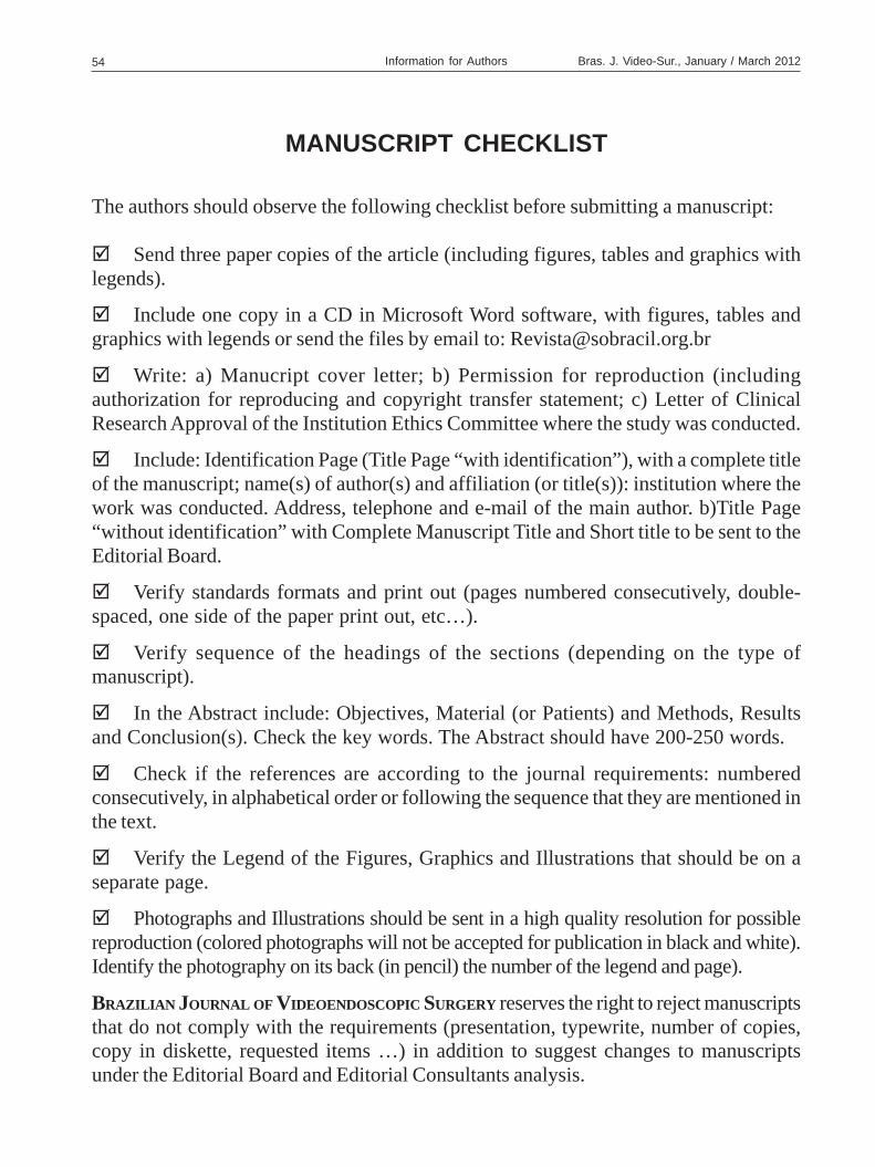
Information for Authors54 Bras. J. Video-Sur., January / March 2012
MANUSCRIPT CHECKLIST
The authors should observe the following checklist before submitting a manuscript:
þ Send three paper copies of the article (including figures, tables and graphics withlegends).
þ Include one copy in a CD in Microsoft Word software, with figures, tables andgraphics with legends or send the files by email to: [email protected]
þ Write: a) Manucript cover letter; b) Permission for reproduction (includingauthorization for reproducing and copyright transfer statement; c) Letter of ClinicalResearch Approval of the Institution Ethics Committee where the study was conducted.
þ Include: Identification Page (Title Page “with identification”), with a complete titleof the manuscript; name(s) of author(s) and affiliation (or title(s)): institution where thework was conducted. Address, telephone and e-mail of the main author. b)Title Page“without identification” with Complete Manuscript Title and Short title to be sent to theEditorial Board.
þ Verify standards formats and print out (pages numbered consecutively, double-spaced, one side of the paper print out, etc…).
þ Verify sequence of the headings of the sections (depending on the type ofmanuscript).
þ In the Abstract include: Objectives, Material (or Patients) and Methods, Resultsand Conclusion(s). Check the key words. The Abstract should have 200-250 words.
þ Check if the references are according to the journal requirements: numberedconsecutively, in alphabetical order or following the sequence that they are mentioned inthe text.
þ Verify the Legend of the Figures, Graphics and Illustrations that should be on aseparate page.
þ Photographs and Illustrations should be sent in a high quality resolution for possiblereproduction (colored photographs will not be accepted for publication in black and white).Identify the photography on its back (in pencil) the number of the legend and page).
BRAZILIAN JOURNAL OF VIDEOENDOSCOPIC SURGERY reserves the right to reject manuscriptsthat do not comply with the requirements (presentation, typewrite, number of copies,copy in diskette, requested items …) in addition to suggest changes to manuscriptsunder the Editorial Board and Editorial Consultants analysis.