Embed Size (px)
Citation preview

Pontifícia Universidade Católica de Minas Gerais
Faculdade de Odontologia
CARACTERÍSTICAS ESTRUTURAIS
DO TRABECULADO ÓSSEO ALVEOLAR
MAXILAR PÓS-CORTICOTOMIA EM CÃES
ANA PAULA DE CARVALHO GOMES FERREIRA
Belo Horizonte
2009

Ana Paula de Carvalho Gomes Ferreira
CARACTERÍSTICAS ESTRUTURAIS
DO TRABECULADO ÓSSEO ALVEOLAR
MAXILAR PÓS-CORTICOTOMIA EM CÃES
Dissertação apresentada ao Programa de Mestrado em Odontologia da Pontifícia Universidade Católica de Minas Gerais, como parte dos requisitos para a obtenção do título de Mestre em Odontologia, área de concentração: Ortodontia. Orientador: Prof. Dr. Dauro Douglas Oliveira Co-orientador: Prof. Dr. Martinho Campolina Rebello Horta
Belo Horizonte 2009

FICHA CATALOGRÁFICA Elaborada pela Biblioteca da Pontifícia Universidade Católica de Minas Gerais
Ferreira, Ana Paula de Carvalho Gomes F383c Características estruturais do trabeculado ósseo alveolar maxilar pós-
corticotomia em cães / Ana Paula de Carvalho Gomes Ferreira. Belo Horizonte, 2009.
78f. : il. Orientador: Dauro Douglas Oliveira Co-Orientador: Martinho Campolina Rebello Horta Dissertação (Mestrado) – Pontifícia Universidade Católica de Minas
Gerais, Programa de Pós-Graduação em Odontologia. 1. Ossos - Cirurgia. 2. Animais. 3. Densidade óssea. 4. Movimentação
dentária. I. Oliveira, Dauro Douglas. II. Horta, Martinho Campolina Rebello. III. Pontifícia Universidade Católica de Minas Gerais. Programa de Pós-Graduação em Odontologia. IV. Título.
CDU: 616.314-089.5

FOLHA DE APROVAÇÃO
Dedico este trabalho aos meus amados pais
Que não têm pressa, mas apressam-se em ajudar-me
Que me ouvem, me entendem e acreditam em mim
Que choram e riem comigo
Que assentam-se ao meu lado, afagam os meus cabelos
Que não medem esforços, esquecem até o sono por mim
Que seguem meus passos, sonham os meus sonhos
Que me fazem sorrir em meio à dor
Que zelam, sofrem e protegem
Que são modelos de renúncia e dedicação sem fim
Que, com um coração sensível e forte, alagam o meu de esperança
Que intercedem à Deus por cada um dos meus dias
Que me ensinam a amar- amor singular

AGRADECIMENTOS
Em primeiro lugar, agradeço ao Senhor Jesus, autor da minha vida, por me
conceder o privilégio de conhecê-lO e de tê-lO como melhor amigo. Reconheço e
louvo Deus pela Sua fidelidade e amor, pela família que me deu, pelo meu marido
e pela oportunidade de poder crescer profissionalmente.
Agradeço ao José, meu amado, por caminhar ao meu lado e sonhar comigo,
por ser meu ombro amigo, por me incentivar e me ajudar a conquistar, por estar
sempre por perto, na alegria ou na tristeza, por tantas palavras amáveis e surpresas
agradáveis, por ouvir meus desabafos e por compreender meus silêncios, por ser
muito mais do que eu sonhei.
Agradeço à minha vó Antônia, que a cada dia me ensina a crer que vale a
pena buscar ao Senhor em primeiro lugar para que as demais coisas me sejam
acrescentadas.
Agradeço à Didi, por ser minha irmã e amiga de todas as horas. Obrigada por
compartilharmos as alegrias e chorarmos as lágrimas uma da outra. À Poliana por
ser tão presente em meu coração.
Agradeço ao Cris pela sua amizade sincera, pelo carinho de irmão e por me
inspirar a querer ser sempre melhor.
Agradeço ao Sr. Vicente, D. Ivone, Miguel e Mauro por torcerem pela minha
vitória.
Agradeço à Pra. Marli, Andréa e a todos da Igreja, que oram comigo as
minhas causas e se alegram com mais esta bênção alcançada.
Agradeço às minhas amigas Fernanda, Flavinha, Chel Brant, Ká, Ana, Soraia,
Chel Venerozo, Fabi ,Polly e Aline, pelo carinho de valor inestimável.
Agradeço aos meus colegas do COP da M7 e M11 pelas boas lembranças
que ficarão; aos colegas da M8, especialmente à Bruna e Lud, por terem sido
minhas companheiras de todas as horas e aos colegas da M9, Bruno Fonseca,
Bruno Gribel, Maria Rita, Paula, Rafael e Sarah e M10, Adriana, Cybelle, Flávia,
Larissa, Lucas e Taty por terem me recebido com tanto carinho e por me
proporcionarem momentos tão agradáveis durante nossa convivência.
Agradeço ao meu orientador, professor Dauro Oliveira, por me apoiar no
momento em que mais precisei e por confiar em mim para continuar seu trabalho tão
precioso. Ao Martinho, meu co-orientador e amigo, por estar
sempre disponível, me ensinando o caminho e estimulando a prosseguir. Ao Manzi,
por sua colaboração tão significativa.
Agradeço aos professores do COP, Armando Lima, Ênio Mazzieiro, Flávio
Almeida, Ildeu Andrade, José Eymard, José Maurício, Júlio Brant , Hélio Brito,
Heloísio Leite e Tarcísio Junqueira, por me ensinarem, principalmente, a por tudo o
que sou no mínimo que faço.
Ao professor Roberval por se empenhar para que o nosso curso seja
reconhecido em todo o Brasil.
Agradeço aos funcionários Reni,Toninha, Mariângela, Andreza, Lorraine, Ana
Paula, Angélica e Silvania, em especial ao Diego e Alcides, por tanto me ajudarem a
prosseguir essa travessia.

“ Não se glorie o sábio em sua sabedoria, nem o forte em sua força,
nem o rico em sua riqueza. Mas quem se gloriar, glorie-se nisto:
em compreender-me e conhecer-me, pois eu sou o Senhor ”
Jeremias 9:23 e 24
APRESENTAÇÃO
Este trabalho se refere à dissertação apresentada ao Programa de Pós-
Graduação em Odontologia da Faculdade de Odontologia da Pontifícia Universidade
Católica de Minas Gerais e representa requisito parcial para a obtenção do título de
mestre.
O tema abordado na elaboração desta dissertação é pertinente, visto que, há
muitos anos, pesquisadores de todo o mundo estudam métodos de alterar o
metabolismo ósseo com o intuito de reduzir sua densidade para possivelvemnte
facilitar a movimentação ortodôntica. Porém, a grande maioria desses trabalhos
envolve a administração de hormônios ou substâncias químicas, que, por exercerem
efeitos sistêmicos, não podem ser aplicados clinicamente.
A execução de um procedimento cirúrgico localizado para diminuir a
resistência óssea foi primeiramente descrita por artigos da área de ortopedia.
Recentemente, esta técnica teve sua utilização ampliada para o campo da
Odontologia. Alguns poucos estudos relataram que a associação da corticotomia à
ortodontia convencional poderia permitir a realização de movimentos ortodônticos
mais complexos e em menor espaço de tempo.
Esses questionamentos culminaram com a elaboração da tese de doutorado
do professor desta instituição, Dr. Dauro Douglas Oliveira, na Universidade Federal
do Rio de Janeiro, conluída em 2006. Sua pesquisa abordou o desenho
experimental de boca dividida (split-mouth) e os estudos foram direcionados para a
avaliação da densidadade óssea do osso mandibular de cães sem raça definida.
Considerando os importantes resultados encontrados pelo autor citado, o
presente estudo objetivou dar sequência à investigação baseada na biologia do
movimento dentário, avaliando o osso maxilar dos respectivos cães.
De acordo com as opções de formato contempladas pelo regulamento do
Programa, essa dissertação se baseia em três artigos produzidos durante o curso,
respectivamente intitulados:
1) “Corticotomia como alternativa para alterar a densidade óssea e facilitar a
movimentação ortodôntica”
2) “Maxillary alveolar bone structure after selective alveolar corticotomy in dogs:
optical and scanning electron microscopy bone density analysis”
3) “Parallel bars, skeletical anchorage and corticotomy to intrude molars”
O primeiro artigo, por meio de uma revisão da literatura, salienta a relevância
do metabolismo ósseo no processo da movimentação ortodôntica e enumera os
fatores passíveis de alterar a densidade óssea, destacando o fator cirúrgico, na qual
a corticotomia é alocada. As aplicações clínicas da corticotomia e o procedimento
cirúrgico propriamente dito são descritos neste capítulo.
O segundo artigo traz resposta à dúvida quanto às eventuais diferenças no
padrão da arquitetura tecidual óssea quando o procedimento de corticotomia é
realizado. Este assunto é original na literatura, e pretende adicionar informações que
possam auxiliar na decisão sobre como abordar pacientes adultos portadores de
maloclusões severas.
O terceiro artigo tem como finalidade ilustrar a aplicação clínica da
corticotomia. Este relato de caso demonstra o benefício da associação da
corticotomia e outras técnicas inovadoras para alcançar o sucesso na intrusão de
dentes posteriores em tempo reduzido, procedimento este, que, até pouco tempo
atrás, era difícil de ser bem sucedido e requeria um longo período de tratamento.
Além dos capítulos referentes aos artigos, esta dissertação traz as
considerações iniciais onde o tema a ser estudado é introduzido e os objetivos da
dissertação são descritos. Ainda, as conclusões deste trabalho são apresentadas ao
final e as referências bibliográficas são enumeradas. A lista de referências
bibliográficas de cada artigo encontra-se ao final dos mesmos.

RESUMO
Corticotomia é definida como uma osteotomia limitada à cortical óssea e tem sido
utilizada como procedimento auxiliar no tratamento ortodôntico de maloclusões
diversas. Consiste de um procedimento cirúrgico de remoção parcial da camada
cortical de osso alveolar seguido imediatamente de aplicações de forças
ortodônticas. Quando esta terapêutica é adotada, o tempo de tratamento é reduzido
consideravelmente devido à diminuição da densidade óssea e consequente
diminuição da resistência ao movimento ortodôntico. O conceito mais novo de
corticotomia é suportado pelo Fenômeno Aceleratório Regional (Rapid Acceleratory
Phenomenon- RAP), caracterizado pela aceleração do metabolismo ósseo e
decréscimo da densidade óssea localizada. Este trabalho analisou as alterações do
trabeculado ósseo alveolar neoformado pós-corticotomia em cães. A análise
qualitativa foi feita através da microscopia eletrônica de varredura, enquanto que a
quantitativa foi obtida através da histomorfometria, obtendo-se o índice de matriz
óssea mineralizada em relação à área tecidual total.
Palavras- chave: Metabolismo ósseo. Densidade óssea. Remodelação óssea.
Corticotomia alveolar.
ABSTRACT
Corticotomy, defined as an osteotomy limited to the cortical bone, has been used as
an auxiliary procedure in the orthodontic treatment of malocclusions. It consists of a
surgical procedure in which a layer of cortical bone is partially removed and
orthodontic forces are immediately applied. This technique should be considered a
therapeutic option, especially for adult patients, because treatment time is
considerably reduced due to bone density decrease and consequently a decrease in
resistance to orthodontic tooth movement. A recent approach in corticotomy is Rapid
Acceleratory Phenomenon (RAP), characterized by bone metabolism acceleration
and localized bone density decrease. This study analysed alterations in newly formed
alveolar bone after performing corticotomy in dogs by electron microscopy. In
addition, a quantitative evaluation was done by measuring the extent of mineralized
bone matrix and by counting osteoblasts and osteoclasts within the bone tissue after
the surgical procedure.
Key- words: Bone turnover. Bone density. Bone remodeling. Alveolar corticotomy
decortication

LISTA DE ARTIGOS
Esta dissertação gerou as seguintes propostas de artigos:
Artigo 1
FERREIRA, A.P.C.G.; OLIVEIRA, D.D.; HORTA, M.C.R; MANZI, F.R. Corticotomia
como alternativa para alterar a densidade óssea facilitar a movimentação
ortodôntica.
Artigo 2
FERREIRA, A.P.C.G.; OLIVEIRA, D.D.; HORTA, M.C.R; MANZI, F.R.; ALVES, J.B.
Maxillary alveolar bone structure after selective alveolar corticotomy in dogs: optical
and scanning electron microscopy bone density analysis.
Artigo 3
FERREIRA, A.P.C.G.; HORTA, M.C.R.; OLIVEIRA B.F. ; OLIVEIRA, D.D.
Parallel bars, skeletical anchorage and corticotomy to intrude molars.

SUMÁRIO INTRODUÇÃO .………………………………………………………………… 13
OBJETIVOS....……………………………………………………….…………. 16
ARTIGO 1 .......………………………………………………………………….. 17
ARTIGO 2 …………………………………………………………………….…. 40
ARTIGO 3 ..…………………………………………………………………....... 62
CONCLUSÕES...……………………………………………………………….. 74
REFERÊNCIAS BIBLIOGRÁFICAS COMPLEMENTARES ....................... 75
ANEXO ........................................................................................................ 77
13
1. INTRODUÇÃO
A movimentação ortodôntica convencional é o resultado de reabsorção e
deposição óssea secundárias às forças mecânicas aplicadas. Fatores individuais,
como intensidade da força, turn over do ligamento periodontal e o metabolismo
ósseo exercem papéis importantes na determinação do índice de movimentação
dentária.
A força ortodôntica é o fator mais facilmente manipulado. Porém, tem sido
demonstrado experimentalmente que, quando a magnitude da força ortodôntica é
excessiva, há uma diminuição do suprimento vascular, o que resulta em morte
celular nas áreas onde as fibras foram demasiadamente comprimidas ou estiradas.
Reabsorção das fibras de Sharpey, invasão de células para dentro da membrana
periodontal, reabsorção do osso alveolar e reabsorção dentária também podem ser
inevitáveis em regiões sob pressão ou tensão em excesso (PILON, 1996). Portanto,
ainda que a magnitude da força possa ser administrada, o movimento biológico dos
dentes pode ser alcançado de forma limitada (KIŞNIŞCI et al., 2002).
O metabolismo ósseo é extremamente relevante no processo da
movimentação ortodôntica. Sabe-se que o tecido ósseo com menor densidade
permite um movimento dentário mais rápido devido à sua menor resistência física. A
atividade e diferenciação das células ósseas são controladas genicamente por uma
cascata de eventos que envolvem hormônios, citocinas e fatores de crescimento.
Por exercerem efeitos sistêmicos, a utilização clínica desses elementos, na tentativa
de diminuir a densidade óssea, é restringida (MIDGET et al., 1981).
Bridges et al. (1988), demonstrou em seu trabalho que a densidade óssea
influencia a movimentação dentária, quando comprovou que ratos mais jovens
tinham índice de movimentação dentário maiores do que a dos ratos de maior idade.
Hasler et al. (1997) e Iseri et al. (2005) demonstraram que a velocidade da
distalização de caninos feita imediatamente após a extração de pré-molares, com o
alvéolo menos denso, foi maior quando comparada à distalização feita muito tempo
após a extração, onde o tecido ósseo já estava completamente regenerado. Esta
teoria é suportada pelo Fenômeno Aceleratório Regional (Rapid Accelleratory
14
Phenomenon- RAP), descrito como um estágio transitório e localizado do osso em
regeneração, onde há aceleração do metabolismo ósseo e decréscimo da densidade
óssea localizada (GERMEÇ et al., 2005). O mecanismo exato que inicia o RAP não
é totalmente conhecido. Ele é provavelmente guiado por demandas bio-físicas, onde
a formação rápida de osso na cavidade medular é mais uma adaptação à
instabilidade mecânica do tecido ósseo em resposta à injúria. A formação de osso
jovem é o principal papel do RAP para melhorar a estabilidade do osso após
qualquer tipo de injúria (SCHILLING et al., 1998).
Similarmente, o turnover ósseo é aumentado pelo RAP após corticotomia, um
procedimento cirúrgico que tem sido combinado com ortodontia convencional para o
melhor tratamento de anormalidades dento-alveolares severas. Esta terapia pode
ser definida como uma osteotomia limitada à cortical óssea, onde há remoção
parcial da camada cortical de osso alveolar e preservação da integridade dos
espaços medulares e osso esponjoso, seguido imediatamente de aplicações de
forças ortodônticas (GANTES et al., 1990; FROST, 1989).
A associação da corticotomia permite a realização de movimentos mais
complexos, como relatado por Hwang et al. (2001) e Oliveira et al. (2006), onde a
intrusão de molares foi realizada com maior rapidez e sem o indesejável efeito de
extrusão dos dentes adjacentes.
Em pacientes adultos, onde a redução do tempo de tratamento é uma
importante demanda, essa técnica pode ser de considerável valia, visto que, a
diminuição da resistência óssea ao movimento ortodôntico faz com que o dente se
movimente mais rapidamente através da área em regeneração. (NAKAMOTO et al.,
2002).
A aplicação clínica da corticotomia associada à ortodontia está sendo
ampliada, modificando abordagens clássicas de tratamento e sugerindo a
otimização da correção de maloclusões classificadas como severas. Apesar de nos
últimos anos terem surgido os primeiros trabalhos bem desenhados para avaliar os
efeitos das corticotomias alveolares na movimentação ortodôntica, vários aspectos
dessa abordagem ainda permanecem sem esclarecimento e devem ser estudados
para que essa técnica cirúrgica seja indicada de forma mais abrangente e com mais
confiança por parte dos ortodontistas clínicos.
Por isso, este trabalho tem como objetivo aprimorar o conhecimento,
pesquisando as possíveis alterações do trabeculado ósseo após a realização de

15
corticotomia em cães. Tais investigações incluem a análise da densidade óssea
através de avaliação histomorfométrica e a comparação visual entre as estruturas
ósseas que foram e não foram submetidas à corticotomia através da microscopia
eletrônica de varredura.
16
2. OBJETIVOS
Os propósitos deste estudo foram:
1. Comparar a área de matriz óssea mineralizada em relação à área tecidual
total nas áreas de osso corticotomizadas e não corticotomizadas através da
histomorfometria;
2. Comparar, por meio de microscopia eletrônica de varredura, a estrutura do
tecido ósseo normal do lado controle com o tecido ósseo em regeneração do
lado experimental;
3. Ilustrar a aplicação clínica e consequentemente a eficiência da associação
da corticotomia à ortodontia para a intrusão de molares supraextruídos.

17
ARTIGO 1
Corticotomia como alternativa para alterar a densid ade óssea e facilitar a
movimentação ortodôntica
Ana Paula de Carvalho Gomes Ferreira*
Martinho Campolina Rebello Horta**
Flávio Ricardo Manzi**
Dauro Douglas Oliveira***
*Mestranda em Ortodontia pela Pontifícia Universidade Católica de Minas
Gerais
** Professor adjunto III da Pontifícia Universidade Católica de Minas Gerais
*** Coordenador do Programa em Ortodontia da Pontifícia Universidade
Católica de Minas Gerais
18
RESUMO
A regulação da remodelação óssea é controlada por uma cascata de eventos que
envolve regulação gênica. Muitos pesquisadores alteraram a densidade do osso
alveolar através da utilização de hormônios e substâncias químicas, que, por
resultarem em efeito sistêmico, não podem ser aplicados clinicamente. Em
contrapartida, outros estudos têm sugerido a associação de procedimentos
cirúrgicos locais que podem diminuir a densidade óssea, sem causar efeitos
sistêmicos. Corticotomia pode ser definida como uma osteotomia restrita à cortical
óssea. A aplicação desta técnica pode ser benéfica no tratamento de adultos com
maloclusão de moderada a severa. Os autores, através de revisão crítica da
literatura, discutem a aplicação deste procedimento em associação à ortodontia
convencional.
Palavras-chave: Corticotomia alveolar. Movimento ortodôntico acelerado.
Metabolismo ósseo. Densidade óssea. Remodelação óssea.
19
ABSTRACT
Bone remodeling regulation is controlled by a series of events that involves genetic
regulation. Previous studies have altered bone density of complex areas such as the
alveolar bone using hormones and chemical substances, such as cytokines and
prostaglandins, which result in systemic effects. Therefore, it is difficult to apply this
method on a clinical basis. To address this treatment obstacle, other researchers
have studied local surgical procedures that might change bone density. Corticotomy
can be defined as an osteotomy limited to the cortical bone, and it consists of a
surgical procedure in which alveolar cortical bone is partially removed. This
procedure can be particularly recommended for treatment of adults or for moderate
to severe malocclusions because decreased bone density might result in a
decreased resistance to orthodontic tooth movement resistance, reducing treatment
time considerably. The authors, by means of literature review, discuss the application
of this procedure in conjunction with conventional orthodontics.
Key words : Alveolar decortications. Corticotomy. Bone turnover. Bone density. Bone
remodeling

20
INTRODUÇÃO
A quantidade de movimento ortodôntico pode ser afetada por vários fatores,
dentre eles, a alteração do metabolismo ósseo. Ossos menos densos são mais
viscoelásticos e mais facilmente remodelados, o que poderia diminuir a resistência
física deste tecido, facilitando a movimentação dentária. A literatura descreve
inúmeros trabalhos em que a redução da densidade mineral do processo alveolar é
alcançada através de alterações hormonais e injeção de substâncias químicas, que
não podem ser aplicadas clinicamente por causarem efeitos sistêmicos. Ao
contrário, a associação de procedimentos cirúrgicos locais à ortodontia
convencional, como a corticotomia alveolar, tem sido utilizada em alguns estudos
animais e parece alcançar resultados mais eficientes no tratamento de
anormalidades dentoalveolares moderadas a severas, sem causar prejuízo
sistêmico. Em pacientes adultos, onde a redução do tempo de tratamento é uma
importante demanda, essa técnica pode ser de considerável valia. Entretanto,
muitas questões relacionadas a este procedimento ainda permanecem
desconhecidas ou possuem respostas divergentes, e, devido à escassez de artigos
que discutem este tema, ainda não há suporte científico suficiente para que esta
técnica cirúrgica seja creditada e indicada como parte do tratamento ortodôntico. Por
isso, os autores deste trabalho têm como objetivo fazer uma revisão crítica da
literatura, discutindo a aplicação clínica deste procedimento associada à ortodontia
convencional.

21
REVISÃO DE LITERATURA
Movimentação ortodôntica
A movimentação ortodôntica convencional é um processo biológico
caracterizado pela reação sequencial do tecido periodontal às forças mecânicas
(KRISHNAN e DAVIDOVITCH, 2006). Há o consenso de que as forças ortodônticas
produzem duas regiões biomecanicamente diferentes no tecido periodontal: os lados
de pressão e tensão, que geram reabsorção e formação óssea, respectivamente
(KING et al., 1995).
Fatores individuais como turnover do ligamento periodontal e o metabolismo
ósseo exercem papéis importantes na determinação do índice de movimentação
dentária.
A força ortodôntica é o fator mais facilmente manipulado. Porém, tem sido
demonstrado experimentalmente que, quando a magnitude da força ortodôntica é
excessiva, há uma diminuição do suprimento vascular, o que resulta em morte
celular nas áreas onde as fibras foram demasiadamente comprimidas ou estiradas.
Reabsorção das fibras de Sharpey, invasão de células inflamatórias para dentro do
ligamento periodontal, reabsorção do osso alveolar e reabsorção dentária também
podem ser inevitáveis em regiões sob pressão ou tensão em excesso (PILON,
1996). Portanto, ainda que a magnitude da força possa ser manipulada, o
movimento biológico dos dentes pode ser alcançado de forma limitada (KIŞNIŞCI et
al., 2002).
Metabolismo ósseo
O metabolismo ósseo é extremamente relevante no processo da
movimentação ortodôntica. O movimento dos dentes através do processo alveolar é
consequência do turnover ósseo, que permite a remodelação deste tecido durante

22
toda a vida do indivíduo e quando submetido ao tratamento ortodôntico (MIDGETT
et al., 1981).
Quando em processo de regeneração, há a aceleração do metabolismo
ósseo e o decréscimo da densidade óssea localizada (GERMEÇ et al., 2005). Esta
teoria é suportada pelo Fenômeno Aceleratório Regional (Rapid Accelleratory
Phenomenon- RAP), descrito como um estágio transitório e localizado do osso em
regeneração. Este fenômeno é provavelmente guiado por demandas biofísicas, onde
a formação rápida de osso na cavidade medular é mais uma adaptação à
instabilidade mecânica do tecido ósseo em resposta ao defeito (FROST, 1989). A
formação de osso jovem é o principal papel do RAP para melhorar a estabilidade do
osso após qualquer tipo de injúria. Essa aceleração das atividades normais, tanto no
tecido mole quanto no duro, tem como objetivo potencializar a cicatrização e reações
defensivas teciduais (SCHILING et al., 1998).
Fatores que alteram o metabolismo ósseo
1. Fatores relacionados ao indivíduo
A quantidade de movimento ortodôntico é afetada por vários fatores inerentes
ao hospedeiro, como número e forma das raízes, morfologia do osso trabecular e
fatores nutricionais. Características hormonais, resposta inflamatória, vascularização
local, presença de doenças que afetam o metabolismo ósseo, deficiência na
absorção intestinal de cálcio e uso de drogas como anti-inflamatórios não esteróides
ou tetraciclinas também podem exercer influência na quantidade de movimentação
(COPE et al., 1999; ISERI et al., 2005).
Em relação à idade, BRIDGES et al. (1988) demonstraram que a
movimentação ortodôntica foi significativamente maior nos ratos mais jovens em
relação aos adultos devido a menor densidade óssea nos primeiros. A
movimentação ortodôntica é retardada em pacientes adultos devido ao decréscimo

23
da atividade proliferativa no ligamento periodontal e no osso alveolar (VERNA et al.,
2000).
2. Fatores químicos
A resposta bioquímica adaptativa à força ortodôntica é um processo
altamente sofisticado, onde centenas de genes e milhares de proteínas, cujas
atividades ainda não são claramente conhecidas, têm participação ativa. Essa
adaptação do tecido ósseo depende da ativação ou supressão de genes para
osteoblastos e osteoclastos. Esses, por sua vez, expressam a necessidade de
proteínas específicas no momento e no lugar adequados (SODEK e MCKEE 2000).
A densidade mineral de um sítio complexo como o processo alveolar pode
ser alterada por vários fatores sistêmicos, que poderiam ter um impacto no ciclo da
movimentação dentária. Este índice de mineralização pode ser influenciado por
diferentes hormônios, como a testosterona, calcitonina, corticosteróides e hormônios
da tireóide e paratireóide, dentre outros (BRIDGES et al., 1988).
HASHIMOTO et al. (2001) verificaram que a administração sistêmica de
osteocalcina em ratos acelerou a movimentação ortodôntica devido a um aumento
da osteoclastogênese no lado de pressão. YAMASHIRO e TAKANO-YAMAMOTO
(2001) observaram que quando há deficiência em estrógeno, provocada pela
ovariectomia em ratas, ocorre também uma alteração do metabolismo ósseo com
consequente aumento da movimentação ortodôntica nas mesmas. TANAKA et al.
(2002), ao estudarem este mesmo assunto, concluíram que a deficiência de
estrógeno causou uma mudança tal do osso alveolar a ponto de torna-lo menos
denso e osteoporótico.
A força ortodôntica aplicada ao tecido ósseo, produz alterações locais na
vascularização assim como na reorganização celular e na matriz extracelular,
levando à síntese e à liberação de vários neurotransmissores, citocinas, fatores de
crescimento, prostaglandinas e leucotrienos (KRISHNAN e DAVIDOVITCH, 2006). A

24
administração local ou sistêmica desses mediadores pode acelerar a regeneração
periodontal através da indução da inflamação ortodôntica, através da estimulação de
osteoblastos, da formação de osteoclastos a partir dos monócitos precursores ou
através da melhora da permeabilidade vascular (NAKAMOTO et al., 2002; REN et
al., 2007).
A conexão entre os sinais indutores de remodelação e a resposta provocada
por eles não é muito bem entendida. Porém, sugere-se que, uma vez iniciado este
processo em um determinado sítio, mecanismos locais se encarregam de completar
os eventos da remodelação sem a necessidade de sinais adicionais. Há indícios de
que o osso tem uma memória à distensão criada por distorção mecânica da sua
matriz extracelular, possivelmente dos proteoglicanos. Este mecanismo pode
explicar porque as células são estimuladas para efetuar a movimentação dentária
mesmo após a remoção dos sinais mecânicos indutores (KING, et al., 1995). A
cascata de transmissão de sinais mediada pelas integrinas e a interação desses
mecanismos reguladores co-participantes do complexo processo de remodelação
óssea deve ser estudado mais a fundo (KRISHNAN e DAVIDOVITCH, 2006).
3. Fatores físicos
A força mecânica, propriamente dita, que pode ser facilmente manipulada
pelo ortodontista, tem papel crucial na remodelação óssea durante o crescimento,
desenvolvimento e especialmente na movimentação ortodôntica (TANG et al., 2006).
Outros fatores físicos que podem influenciar a quantidade de movimentação
ortodôntica são a estimulação física ou mecânica dos tecidos através de correntes
elétricas (KARANTH & SHETTY, 2001), campos eletromagnéticos estáticos
(YAMAMOTO et al., 2003) ou pulsáteis (STARK & SINCLAIR, 1987), magnetos
samarium-cobalto (DARENDELILER et al., 1995) e a baixa energia laser
(SHIMIZU et al., 2007). Tem sido sugerido que estímulos físicos podem aumentar a
25
vascularidade e com isso melhorar o metabolismo ósseo. Porém, há efeitos
colaterais e alguns deles ainda são desconhecidos, especialmente a longo prazo.
4. Fatores cirúrgicos
Cirurgias orais como osteotomias têm sido combinadas à ortodontia
convencional para o melhor tratamento de anormalidades dentoalveolares de
moderadas a severas, pois o turnover ósseo pode ser aumentado pelo RAP após
esses procedimentos (SEBAOUN et al., 2008). Neste caso, supõem-se que o osso
em regeneração permite um movimento dentário mais rápido devido à sua menor
resistência física (GERMEÇ et al., 2005).
Distração Osteogênica (DO) é um método que se utiliza da osteotomia para
induzir a neoformação óssea pelo gradual afastamento ou distração de fragmentos
ósseos através do estiramento do tecido vascularizado pré-existente. Além do
alongamento ósseo, a DO promove a regeneração dos tecidos moles incluindo
músculos, tecidos conectivos e epitélio (NAKAMOTO et al., 2002).
Nas primeiras semanas após a osteotomia, o osso criado é ainda imaturo.
Teoricamente, esse osso jovem e menos denso ofereceria menor resistência física
ao estímulo ortodôntico e, assim, a quantidade de movimentação dentária seria
maior (LIOU et al., 2000).
Corticotomia alveolar é definida como uma osteotomia limitada à cortical
óssea, onde há remoção parcial da camada cortical de osso alveolar e preservação
da integridade dos espaços medulares e osso esponjoso (GERMEÇ et al., 2005). A
osteotomia restrita ao osso cortical na região apical minimiza o risco de injúrias às
estruturas vitais, pois não afeta a circulação sanguínea intraóssea e intrapulpar
(GANTES et al., 1990).
OLIVIERA et al. (2006) observaram que o estímulo criado pela injúria na
cortical óssea alveolar em cães afetou significantemente as estruturas do osso
esponjoso adjacente. O impacto na fisiologia óssea foi localizado e transitório, e teve
como característica principal espaços trabeculares em maior quantidade, o que

26
revelou a diminuição da densidade óssea da área investigada. Através de
microscopia eletrônica de varredura, verificaram que houve decréscimo do conteúdo
mineral do osso esponjoso adjacente à área corticotomizada em cães, evidenciada
pelo aumento de espaços trabeculares e pela marcada descontinuidade entre as
estruturas mineralizadas.
APLICAÇÃO CLÍNICA DA CORTICOTOMIA
1. Acelerar a movimentação dentária
O tempo de tratamento ortodôntico pode ser reduzido a 50 % usando forças
pesadas em combinação com corticotomia, induzindo mudanças histológicas no
ligamento periodontal e no osso alveolar, sem afetar a vitalidade do dente. Em
pacientes adultos, essa técnica pode reduzir consideravelmente o tempo de
tratamento, graças à diminuição da resistência óssea ao movimento ortodôntico
(GERMEÇ et al. 2005).
REN et al. (2007) relataram que a cirurgia alveolar é um procedimento efetivo
e seguro, uma vez que os primeiros pré-molares de cães foram movimentados mais
rapidamente no lado que teve corticotomia realizada sem que reabsorções
radiculares ou injúrias ao tecido pulpar fossem observadas.
LINO et al. (2007) também observaram que foi necessário menor tempo para
a distalização de caninos onde corticotomia alveolar tinha sido feita previamente.
2. Facilitar movimentos dentários complexos
A associação da corticotomia pode permitir a realização de movimentos mais
complexos, como relatado por HWANG et al. (2001), onde a intrusão de molares foi
realizada com maior rapidez e sem o indesejável efeito de extrusão dos dentes
adjacentes, associando-se ao aparelho ortodôntico, corticotomia ao redor das raízes

27
dos molares. O primeiro molar superior direito estava extruído, tocando o rebordo
alveolar inferior. Antes da mecânica de intrusão, foi realizada corticotomia ao redor
da raiz deste dente. Os cortes verticais iniciaram 2-3mm abaixo da crista óssea e
foram extendidos de 2-3mm além do ápice radicular. Uma semana após a cirurgia, o
aparelho foi instalado, e constituia de anel no molar extruído e aparelho removível na
arcada inferior, com magnetos repelentes. Um mês após a aplicação da força
magnética, o molar foi consideravelmente intruído.
OLIVEIRA et al. (2008) também lançaram mão da corticotomia para auxiliar
na intrusão de molares. Cortes verticais interproximais e um corte horizontal
apicalmente localizado, unindo os cortes verticais, foram feitos, além de perfurações
esféricas com broca dentro da área delimitada pelos cortes descritos anteriormente.
Um Splint removível de acrílico foi utilizado, com cobertura oclusal total, exceto nos
molares a serem intruídos. Por meio de molas fechadas apoiadas em ganchos por
vestibular e lingual aos molares, foram aplicadas forças intrusivas constantes
através do centro de resistência dos molares. Dois meses e meio após o início da
mecânica intrusiva, os molares estavam no mesmo nível oclusal dos dentes
adjacentes.
FISCHER (2007), ao realizar tracionamento ortodôntico de caninos maxilares
impactados de seis pacientes, teve a exposição cirúrgica para colagem do acessório
realizada de forma convencional de um lado e, do outro lado, corticotomia alveolar
foi adicionada ao redor de toda a raiz. Utilizando-se da mesma técnica e quantidade
de força para tracionamento dos caninos de ambos os lados, observou que o dente
do lado da corticotomia teve movimentação favorecida em relação ao canino
contralateral, com redução do tempo de tracionamento e incorporação do dente no
arco de 28 a 33%.
GERMEÇ et al. (2006), utilizaram da corticotomia para retrair incisivos
inferiores que estavam protruídos e apinhados. Foi realizada extração de pré-
molares e distalização dos caninos. Cortes verticais, restritos à cortical do osso
alveolar, foram feitos mesiodistalmente em todos os incisivos para facilitar sua
28
distalização e retroinclinação, que se completou com 45 dias, sem qualquer dano ao
dente e ao periodonto.
3. Correção de maloclusões esqueléticas
De acordo com SEBAOUN et al. (2008), esta técnica pode significantemente
expandir os leques de oportunidades de tratamento de discrepâncias esqueléticas
como mordidas abertas e constrições maxilares em pacientes sem crescimento,
condições tipicamente relegadas à cirurgia ortognática.
Corticotomia associada à ortodontia pode ser uma opção de tratamento de
pacientes com mordida aberta anterior, que pode ser fechada ortodonticamente com
a intrusão dos dentes posteriores. Consequentemente, há a rotação no sentido anti-
horário da mandíbula, diminuição da altura facial anterior inferior e diminuição do
ângulo goníaco (SHERWOOD et al., 2002).
Chung et al. (2001) demonstraram que a combinação de corticotomia e tração
ortopédica pode ser efetiva para intrusão de dentes posteriores, corrigindo casos de
poblemas verticais e mordida aberta anterior sem que cirurgia ortognática seja
necessário.
CONSIDERAÇÕES
As contraindicações para a realização deste procedimento cirúrgico são as
mesmas para o tratamento ortodôntico convencional: raízes curtas (KIŞNIŞCI et al.,
2002), pacientes com doença periodontal ou qualquer deformidade, como recessões
gengivais, deiscência óssea lingual ou bucal e dentes com periodonto reduzido
(GANTES et al., 1990).

29
Segundo HWANG et al. (2001), para se obter a movimentação desejada, é
necessária a aplicação de forças ortodônticas convencionais, imediatamente após o
procedimento cirúrgico, com reativações mais frequentes, para que a corticotomia
não perca sua efetividade.
O alto metabolismo ósseo presente no espaço em regeneração pode afetar
não somente a taxa de movimentação ortodôntica mas também o tipo de
movimentação, podendo predominar inclinação ou translação dentária. Esta
alteração do metabolismo, pode, ainda, relocar o centro de rotação (CRot) dos
dentes. VERNA et al. (2000) observaram que diante de metabolismo mais baixo, o
CRot foi localizado mais próximo à coroa, enquanto que na presença de
metabolismo mais alto, o CRot foi posicionado mais apicalmente.
A cicatrização tecidual pós-corticotomia é geralmente normal. Em estudo feito
por GANTES et al.(1990), os pacientes reportaram que os níveis de desconforto
durante e após a cirurgia foram menores que o esperado. Neste mesmo estudo,
todos os dentes movimentados através da área corticotomizada continuaram com
vitalidade. A profundidade do sulco gengival e mudanças de inserção desses
mesmos dentes demonstraram que o procedimento cirúrgico não é excessivamente
danoso ao tecido periodontal. Recessões gengivais foram mínimas e as papilas
interdentais foram preservadas, garantindo um bom resultado estético pós-
tratamento.
LIOU et al. (2000) não verificaram nenhuma evidência radiográfica de
reabsorção externa radicular após a movimentação de dentes em tecido ósseo
resultante da DO em cães. Além disso, o tecido pulpar de todos os dentes
movimentados foram histologicamente comprovados como vitais.
NAKAMOTO et al. (2002) acreditam que o dente pode se mover para a área
regenerada, mas a influência desta movimentação no ligamento periodontal e nas
raízes dos dentes ainda permanece desconhecida. Eles observaram, após a
movimentação dentária sobre uma área corticotomizada em cães, a perda da lâmina
dura desses mesmos dentes na 8ª semana de movimentação. Reabsorção de
cemento também foi observada no ápice radicular, estendendo-se para a dentina no
30
lado de compressão na área logo abaixo da bifurcação. Porém, em geral, houve um
reparo pela formação de cemento na superfície de dentina reabsorvida.
TÉCNICA CIRÚRGICA
Köle, em 1959, descreveu uma técnica cirúrgica que consistia em
corticotomias interradiculares e uma osteotomia supra-apical, unindo os cortes
verticais (FIG. 1). Ele acreditava que blocos ósseos eram criados a partir desses
cortes e que seriam movidos tendo os dentes como braços de apoio. Apesar dos
resultados obtidos por Köle serem estáveis, alterações pulpares não eram raras.
Esta técnica original foi revisada e mudada com o passar dos anos com o objetivo
de diminuir os possíveis riscos deste procedimento, como por exemplo a ocorrência
de danos periodontais, de desvitalização do dente e/ou do segmento ósseo devido a
um suprimento sanguíneo inadequado (GERMEÇ et al.,2005).
Em 1978, Generson reportou uma modificação ao método descrito por Köle,
onde a osteotomia subapical foi substituída por corticotomia que circunscrevia as
raízes dos dentes, por labial e lingual (FIG. 2). O autor relatou que após tais
modificações, não ocorreram problemas relacionados ao suprimento sanguíneo
inadequado, desvitalização pulpar, reabsorções radiculares ou recessão gengival.
Como os resultados clínicos observados foram tão bons quanto aqueles relatados
por Köle, mesmo sem a osteotomia subapical, a teoria da movimentação de blocos
ósseos passou a ser questionada.
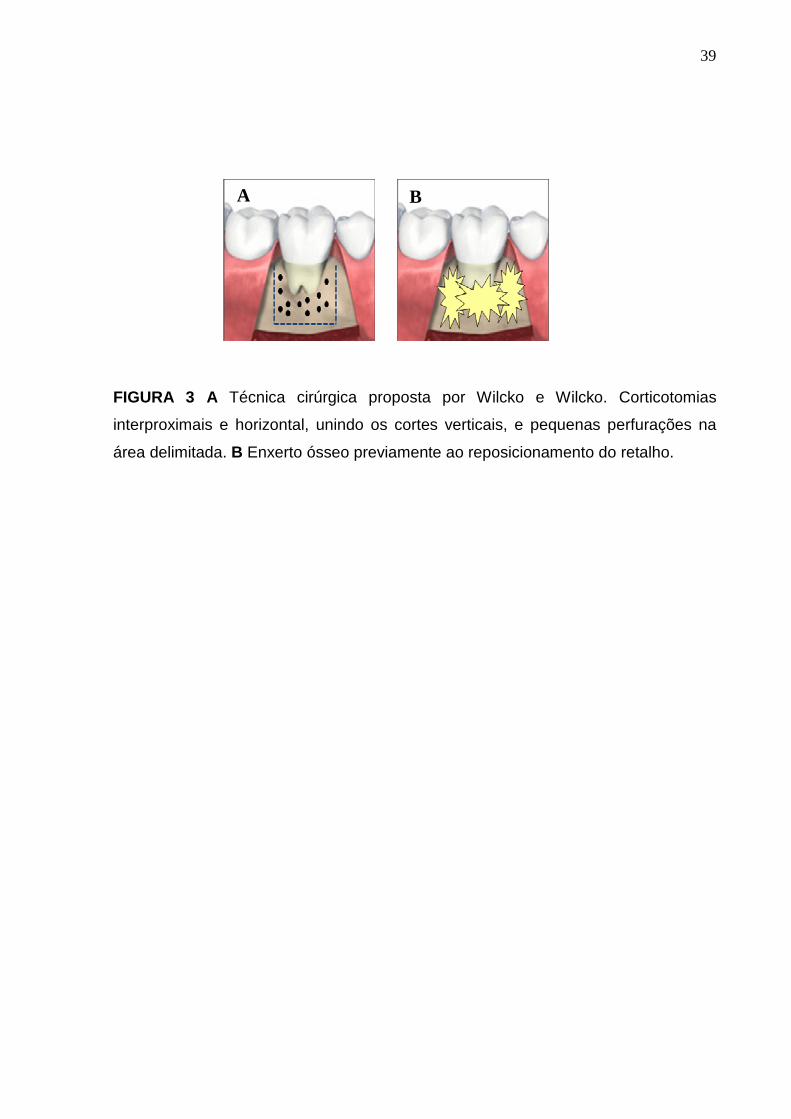
Wilcko et al. (2008) descreveram uma técnica chamada ortodontia
osteogênica acelerada periodontalmente (periodontally accelerated osteogenic
orthodontics -PAOO), que pode ser definida como um procedimento cirúrgico no
qual a corticotomia é realizada ao redor do dente, por lingual e vestibular, com
pequenas perfurações feitas com broca dentro da área previamente delimitada para
estimular o processo de regeneração. A colocação de enxerto ósseo em
seguida, tende a aumentar o volume alveolar, para que, mesmo nas situações de
31
apinhamento severos, haja amplo suporte ósseo para as raízes (FIG. 3). Foram
demonstrados vários casos em que a movimentação dentária ocorreu de duas a três
vezes mais rapidamente do que teria sido alcançada se só ortodontia convencional
fosse aplicada.
CONCLUSÃO
Corticotomia alveolar é um procedimento auxiliar que pode facilitar a
movimentação ortodôntica através do aumento do turnover ósseo e diminuição da
densidade óssea. Porém, há ainda muitas questões a serem respondidas, como
quando começar a aplicação da força ortodôntica após feita a corticotomia, se a
força deve ou não ser mais pesada do que a usual, qual tipo de movimento
predomina quando o dente é movimentado em uma área em regeneração e qual a
influência desta movimentação no ligamento periodontal, no tecido pulpar e nas
raízes dos dentes. Para maiores esclarecimentos, são necessários mais estudos,
inclusive em humanos, a fim de que esta técnica ortocirúrgica tenha respaldo
científico para ser empregada clinicamente com eficácia e segurança.
32
REFERÊNCIAS BIBLIOGRÁFICAS
BRIDGES, T.; KING, G.; MOHAMMED, A. The effect of age on tooth movement and
mineral density in alveolar tissues of the rat. Am J Orthod Dentofac Orthop , v.93,
n.3, p.245-50, Mar. 1988.
COPE, J.B.; HARPER, R. P.; SAMCHUKOV, M. L. Experimental tooth movement
through regenerate alveolar bone: A pilot study. Am J Orthod Dentofacial Orthop ,
v.116, n.5, p.501-5, Nov. 1999.
CHUNG, K.; OH,M.; KO,SU-JIN. Corticotomy-Assisted Orhtodontics. Journal of
Clinical Orthodontics. v.35, n.5, p.331-9. 2001.
DARENDELILER, M.A.; SINCLAIR P.M.;KUSY R.P.The effects of samarium-cobalt
magnets and pulsed electromagnetic fields on tooth movement. Am J Orthod
Dentofacial Orthop . v.107, n.6, p. 578-88, Jun. 1995.
FISCHER,T. J. Orthodontic Treatment Acceleration with Corticotomy-assisted
Exposure of Palatally Impacted Canines. Angle Orthodontist , v. 77, n. 3. 2007.
FROST, H.M. The biology of fracture healing: An overview for clinicians. Part
I.Clinical Orhtopaedics and Related Research . n. 248, p.283-93.Nov. 1989.
FROST, H.M. The biology of fracture healing: An overview for clinicians. Part
II.Clinical Orhtopaedics and Related Research . n. 248, p.294-309.Nov. 1989.
GANTES, B.; RATHBUN, E.; ANHOLM, M. Effects on the periodontium following
corticotomy-facilitated orthodontics. Case Reports. J Periodontol , v.61, n.4. p. 234-
37, Apr. 1990.

33
GENERSON,R.M.;PORTER,J.M.;ZELL,A.;STRATIGOS,G.T.Combined surgical and
orthodontic management of anterior open bite using corticotomy. J Oral Surg . v.36,
n. 3, p.216-9, Mar.1978.
GERMEÇ, D.; GIRAY,B.; KOCADERELLI, I.; ENACART, A. Lower incisor retraction
with a modified corticotomy. The Angle Orthodontist . v.76, n.5, p.882-90, Nov.
2005.
HASHINOMOTO, F. et al. Administration of osteocalcin accelerates orthodontic tooth
movement induced by a closed coil spring in rats. Eur J Orthod , v. 23, n.5, p.535-45.
Oct. 2001.
HWANG, H.S.; LEE, K. H. Intrusion of overerupted molars by corticotomy and
magnets. Am J Orthod Dentofacial Orthop , v.120, n.2, p.209-16, Aug. 2001.
ISERI, H.; KISNISCI, R.; BZIZI, N.; TUZ, H. Rapid canine retraction and orthodontic
treatment with dentoalveolar distraction osteogenesis. Am J Orthod Dentofacial
Orthop , v.127, n.5, p.533-41, May. 2005.
KARANTH, H.S.; SHETT, K.S. Orthodontic tooth movement and bioelectricity. Indian
J Dent . Res, v.12, n.4, p. 212-21, Oct/Dec.2001.
KING, G. J.; KEELING, S. D. Orthodontic bone remodeling in relation to appliance
decay. The Angle Orthodontist v.65, n.2, p.129-40, Apr.1995.
KIŞNIŞCI,R.Ş.; ÌŞERI, H.; TÜZ, H. H.; ALTUG, A. T. Dentoalveolar distraction
osteogenesis for rapid orthodontic canine retraction. J Oral Maxillofac Surg , v.60,
n.4,p.389-94, Apr.2002.
KÖLE H. Surgical operations of the alveolar ridge to correct occlusal abnormalities.
Oral Surg Oral Med Oral Pathol v.12,n.5, p.515-29,May.1959.
34
KRISHNAN, V.; DAVIDOVITCH, A. Cellular, molecular, and tissue-level reactions to
orthodontic force. Am J Orthod Dentofacial Orthop , v.129, n.4, p.469. apr. 2006.
LINO, S et al. Acceleration of orthodontic tooth movement by alveolar corticotomy in
the dog. Am J Orthod Dentofacial Orthop v. 131; p.448.e1-448.e8, 2007.
LIOU, E. J.; FIGUEROA, A. A.; POLLEY, J. W. Rapid orthodontic tooth movement
into newly distracted bone after mandibular distraction osteogenesis in a canine
model. Am J Orthod Dentofacial Orthop ,v.117, n.4, p.391-8. Apr. 2000.
MIDGETT, R. J.; SHAYE, R.; FRUGE, J. F. The effect of altered bone metabolism on
orthodontic tooth movement. Am J Orthod , v.80, n.3, p.256-62, Sep. 1981.
NAKAMOTO, N. et al. Experimental tooth movement through mature and immature
bone regenerates after distraction osteogenesis in dog. Am J Orthod Dentofacial
Orthop v.121, n.4, p. 385-95. Apr. 2002.
OLIVEIRA, D.D. et al. Effects of selective alveolar decortication on cancellous bone
density, 2006. Tese (Doutorado)-Universidade Federal do Rio de Janeiro, Faculdade
de Odontologia, Rio de Janeiro.
OLIVEIRA, D.D. et al. Selective alveolar corticotomy to intrude overerupted molars.
Am J Orthod Dentofacial Orthop v. 133, p. 902-8,2008.
ILON, J.J.G.M; KUIPERS-JAGTMAS, A.M., MALTHA, J.C. Magnitude of orthodontic
forces and rate of bodily tooth movement: an experimental study. Am J Orthod
Dentofacial Orthop , v.110, n.1,p.16-23,Jul. 1996.

35
REN, A.; LV,T.; KANG, NA.; ZHAO, B.; CHEN, Y.; BAI,D. Rapid orthodontic tooth
movement aided by alveolar surgery in beagles. Am J Orthod Dentofacial Orthop
v.131, n.2, p.160.el-10. Feb. 2007.
SCHILLING, T.; MÜLLER, M.; MINNE, H. W.; ZIEGLER, R. Influence of
inflammation-mediated osteopenia on the regional acceleratory phenomenon and the
systemic acceleratory phenomenon during healing of a bone defect in the rat. Calcif
Tissue Int , v.63,n. 2,p.160-6, Aug.1998.
SEBAOUN, J.D. et al. Modeling of trabecular bone and lamina dura following
selective alveolar decortications in rats. J Periodontol ,v.79, n.9, p.1679-
88,Sep.2008.
SHERWOOD K.H. ; Burch J.G. ;THOMPSON W.J. Closing anterior open bites by
intruding molars with titanium miniplate anchorage. Am J Orthod Dentofacial
Orthop, v.122, p. 593-600, 2002.
SHIMIZU N. et al. Low-intensity laser irradiation stimulates bone nodule formation via
insulin-like growth factor-I empression in rat calvarial cells. Lasers Surg Med, v.39,
n.6, p.551-9,Jul.2007.
SODEK, J.; MCKEE, M. D. Molecular and cellular biology of alveolar bone.
Periodontology . v.24,p.99-126.Oct.2000.
STARK, T.M.; SINCLAIR, P.M. Effect of pulsed electromagnetic fields on orthodontic
tooth movement. Am J Orthod Dentofacial Orthop, v.91,n.2 ,p.91-104,Feb.1987.
TANAKA, M. et al. Effects of ovariectomy on trabecular structures of rat alveolar
bone. J Periodontal Res , v.37, n.2, p.161-5. Apr. 2002.
36
TANG, L.; LIN, Z.; LI, Y. M. Effects of different magnitudes of mechanical strain on
osteoblasts in vitro. Biochem Biophys Res Commun , v.344, n.1, p.122-
8,May.2006.
VERNA, C.; DALSTRA,M.; MELSEN,B. The rate and the type of orthodontic tooth
movement is influenced by bone turnover in a rat model. Eur J of
Orthodontics, v.22,p.343-52.Aug. 2000.
WILCKO,M.T.;WILCKO,W.M.;BISSADA,N.F. An evidence-based analysis of
periodontally accelerated orthodontic and osteogenic techniques: a synthesis of
scientific perspectives. Semin Orthod , v.14, n.4,p. 305-316, Dec. 2008.
YAMAMOTO Y. et al. Effects of static magnetic fields on bone formation in rat
osteoblast cultures. J Dent Res , v. 82, n.12, p.962-6, Dec. 2003.
YAMASHIRO, T.; TAKANO-YAMAMOTO,T. Influences of ovariectomy on
experimental tooth movement in the rat. J Dent Res , v.80, n.9, p.1858-61, Sep.2001.

37
LEGENDAS
FIGURA 1 Proposta inicial de Köle, onde corticotomias interproximais eram unidas
por uma osteotomia apical (em azul) a fim de formar “blocos ósseos” para a
movimentação ortodôntica. A vista vestibular. B corte sagital.
FIGURA 2 Modificação proposta por Generson, onde o corte apical, assim como os
cortes interproximais, se restringiam à cortical óssea (em verde), preservando a
vitalidade do dente.
FIGURA 3 Técnica cirúrgica proposta por Wilcko e Wilcko. Corticotomias
interproximais, horizontal e pequenas perfurações na área delimitada. Enxerto ósseo
e reposicionamento do retalho.

38
ILUSTRAÇÕES
FIGURA 1 Proposta inicial de Köle, onde corticotomias interproximais eram unidas
por uma osteotomia apical (em azul) a fim de formar “blocos ósseos” para a
movimentação ortodôntica. A vista vestibular. B corte sagital.
FIGURA 2 Corticotomia, onde os cortes se restringem à cortical óssea (em verde),
preservando a vitalidade dentária.
A B
39
FIGURA 3 A Técnica cirúrgica proposta por Wilcko e Wilcko. Corticotomias
interproximais e horizontal, unindo os cortes verticais, e pequenas perfurações na
área delimitada. B Enxerto ósseo previamente ao reposicionamento do retalho.
A B
40
ARTIGO 2
Maxillary alveolar bone structure after selective a lveolar corticotomy in dogs:
optical and scanning electron microscopy bone densi ty and structural
analysis.
Ana Paula Carvalho Gomes Ferreira, DDS*
Martinho Campolina Rebello Horta, DDS, MS, PhD**
Flávio Ricardo Manzi , DDS, MS, PhD**
José Bento Alves, DDS, MS, PhD***
Dauro Douglas Oliveira, DDS,MS,PhD****
*Master Student in Orthodontics, School of Dentistry, Pontifical Catholic
University of Minas Gerais, Belo Horizonte, Brazil
** Associate Professor, School of Dentistry, Pontifical Catholic University of
Minas Gerais, Belo Horizonte, Brazil
***Associate Professor, School of Dentistry, Federal University of Minas
Gerais, Belo Horizonte, Brazil
**** Program director in Orthodontics, Pontifical Catholic University of Minas
Gerais

41
ABSTRACT
Introduction : Surgical alveolar corticotomies have been used as an alternative to
conventional orthodontic treatment in difficult adult cases. This technique might
dramatically reduce the treatment time because the resistance of the dense cortical
bone to tooth movement is minimized. The purpose of this study was to evaluate the
maxillary alveolar bone structure after selective alveolar decortication in dogs.
Methods: Corticotomy was performed on the left maxillary side in mongrel dogs, with
right side serving as control. The animals were sacrificed after 7 and 14 days post-
surgery in order to obtain maxillary slices. Optical and scanning electron microscopy
(SEM) analysis were performed to compare the cancellous bone structure between
both experimental and control sides. Results: The surgery sides had significantly
less calcified matrix bone when compared to the control sides, for all time points.
There was also a statistical difference when the experimental samples were
compared as a function of time post decortications (P<0.05).
Conclusions: Selective alveolar corticotomy can cause bone structural change due
to a diminished matrix bone percentage in the area adjacent to the injury.
42
INTRODUCTION
Reduction of orthodontic therapy time is considered to be an important goal in
the management of malocclusions and many attempts have been made to shorten
orthodontic tooth movement. Ren et al. (2007) defined these attempts into 3
categories. The first is local or systemic administration of medicines such as
prostaglandins (LEE, 1990), interleukins (IWASAKI et al., 2006) and vitamin D
(COLLINS & SINCLAIR, 1988). They can induce orthodontic inflammation, improve
capillary permeability and participate in the formation of osteoclasts, which can
accelerate periodontal regeneration. The second category is mechanical or physical
stimulation, such as direct electrical current (KARANTH & SHETTY, 2001), pulsed
(STARK & SINCLAIR, 1987) and static (YAMAMOTO et al., 2003) electromagnetic
field, samarium-cobalt magnet (DARENDELILER et al., 1995) and low-energy laser
(SHIMIZU et al., 2007). It has been suggested that the electric currents or the
pulsating electromagnetic field generated by the magnets within the mouth increases
vascularity and enhances bony metabolism through enhanced cellular proliferation
and differentiation. However, physical stimulation also has unwanted effects and
some of those are unknown in the long term. The last category is oral surgery,
including gingival fiberotomy (TUNCAY & KILLIANY, 1986), alveolar surgery
(SEBAOUN et al., 2008) and distraction osteogenesis (LIOU et al., 2000).
Trauma to cortical bone has been shown to be a potentiating factor in
producing a localized osteoporosis. Tissue reorganization after injury leads to a
transient increase of bone turnover, which means a decrease of bone density, which
could facilitate tooth movement (WILCKO et al., 2001).
The alveolar corticotomy technique has been revised and changed over the
years to eliminate possible risks of the procedure, such as periodontal damage and
devitalization of the teeth and osseous segments because of inadequate blood
supply (GANTES et al, 1990). The original technique described by Köle included a
combined interradicular corticotomy and supra-apical osteotomy (KÖLE, 1959).

43
Although his results were stable, pulp mortifications were not rare. Later, the supra-
apical osteotomy was replaced by corticotomy, and labial and lingual corticotomy
cuts were used to circumscribe the roots of the teeth (GENERSON et al., 1978).
The earliest concept of the rapid tooth movement was based on bony block
movement, when buccal and lingual vertical and subapical horizontal cuts
circumscribing the roots of the teeth (WILCKO et al., 2008).The fissure made through
the cortical bone surrounded the tooth so that it was embedded within a bone block
that was connected to the adjacent blocks through only the medullary bone. In this
way, it was believed that the tooth acted like a handle by which the bands of the less
dense medullary bone moved as a block. The bone blocks moved with the teeth,
rather than moving the teeth within the bone (ANHOLM et al., 1986).
On the other hand, the latest concept on the rapid tooth movement after
corticotomy, described by Frost (1989), is characterized by bone turnover increase
supported by the cascade of physiologic healing events, which is termed regional
acceleratory phenomenon (RAP). Verna et al. (2000) comproved that the rate of
tooth movement was higher in cases of high bone turnover and smaller in the rats
with low bone turnover than in normal animals.
The RAP mechanism potentiating tissue healing was shown to occur in the
mandible as well as in long bones, and it leads to an accelerated bone turnover and
decreased regional bone density by means of a local, transient, clinically induced
osteoporosis (WILCKO et al., 2001).
In addition, studies showing how the alveolar bone is structurally affected after
corticotomy are important, in order to provide scientific subsides for the
implementation of this thecnique in the daily orthodontic practice. The aim of this
research is to verify the alterations in bone structure after corticotomy in dogs, by
means of: (1) histomorphometric analysis of bone area and (2) Scanning Electron
Microscopy analysis of cancellous bone structural changes.
44
MATERIAL AND METHODS
Experimental Set-up
Twelve male adult mongrel dogs were submitted to 4 weeks of acclimation
with feedings twice daily with moistened dog chow and water ad libitum. All animals
were individually caged throughout the experiment and the environment was
regulated for light and temperature. Randomly, 3 groups of 4 dogs each were
obtained. The group named G0 had no surgery performed, serving as control. On
this group, we ended up with only 3 dogs because one dog was reported with
clinical signs of leishmaniasis. For the groups named G7 and G14, the dogs were
submitted to corticotomy only on the left maxillary side, while the right maxillary side
served as control. Then, they were sacrificed 7 and 14 days after surgery
respectively.
Before the surgery, each dog was pre-sedated with 0.1mg/Kg of
acepromazine (Acepran, Univet S/A, São Paulo, Brazil). General anesthesia was
achieved with intravenuous 15mg/Kg thiopental sodium (Thiopentax, Cristália
Produtos Químicos e Farmacológicos LTDA, São Paulo, Brazil) and maintained with
halothane inhalation after intubation. Flaps were raised on both buccal and lingual
surfaces to expose the alveolar bone surrounding the maxillary left third molar.
Corticotomy was performed all around this teeth, using a slow speed #4 carbide bur
under copious irrigation with cold sterile saline. Vertical cuts were made in both
mesial and distal interproximal areas, initiating 3 to 4 mm below the alveolar crest
and extending 1 to 2 mm past the estimated root apices. Connecting the interdental
cuts, a bony incision was extended horizontally at the level approximating the roots
apices. Nine round perforations equivalent to the #4 bur diameter were also made
inside the area of the circumscribing cuts in order to increase the traumatic stimulus
(Figure 1). All surgical cuts were performed within the cortical plate and barely
penetrating into cancellous bone. After careful irrigation, gingival flaps were
repositioned and sutured. Following the surgeries, dental prophylaxis was performed
45
twice a week with a gel of 0.32% chlorhexidine digluconate to eliminate gingival
inflammation.
Before sacrifice, all dogs were anesthetized with sodium pentobarbital
(30mg/Kg/IV) and sacrificed by perfusion with 10% buffered formalin through the
external carotid arteries to obtain complete fixation of the jaws.
The bone blocks defined by the surgical cuts and their corresponding contra
lateral controls were divided in half. The distal sections were prepared for histological
evaluation and the mesial half for scanning electron microscopy (SEM) analysis
(Figure 2).
Experimental procedures were approved by the Institutional Animal Research
Committee at the Federal University of Rio de Janeiro, Brazil.
Histological Procedures
The distal halves of the bone block tissue samples were demineralized in 10%
EDTA solution (pH=7), at a constant temperature of 4º Celsius to assure appropriate
sample preservation for future analysis. Paraffin serial sections (5µm), prepared in a
buccal-lingual direction, were obtained and stained with hematoxilin and eosin.
Histomorphometric analysis was performed using a light microscopy image
capture system at 40X magnification. The area evaluated comprised three
magnification fields buccal-lingualy below the alveolar crest and extending occluso-
gingivaly for other three magnification fields (Figure 3).
An image analysis system (UTHSCSA Image Tool, San Antonio, TX, USA) was used
in order to measure the bone density (percentage of mineralized bone area per total
bone area). The sections were blindly presented for measurements by one examiner,
and the data were then averaged to allow for intra and inter-group analysis and
comparisons.
46
Scanning Electron Microscopy
Scanning electron microscopy (SEM) was used in order to evaluate how
alveolar cancellous bone responded to corticotomy. The mesial half of the tissue
sample was immersed in 1% (v/v) Triton- X-100 (Sigma-Aldrich, São Paulo, Brazil)
for 20 minutes at room temperature in an ultrasonic cleaner to partially remove the
organic material. The specimens were dehydrated by acetone (Merck S/A, São
Paulo, Brazil) and mounted on metal stubs. Mounting was carefully performed to
assure that the exposed alveolar cancellous bone was scanned properly. Finally, the
samples were coated with golgdusing a Balzers MED 010 sputter coater (Desk II
Cold Sputter, Denton Vacuum Inc., Moorestown, NJ) and examined with a JEOL
5600 scanning electron microscope (Jeol, Tokyo, Japan).
Statistical Analysis
Differences in bone density in between both left and right sides of each dog were
analyzed using paired t-test. In order to compare the ratio of left side bone density
per right side bone density among the groups (GO, G7 and G14), the one- way
ANOVA test followed by t-test for paired comparison was used. The data were
analyzed by means of Biostat 5.0 software (Optical Digital Technology, Belém,
Brazil). Tests were considered significant when their P-values were < 0.05
47
RESULTS
In this study on how bone response to the alveolar corticotomy, we chose two
time points (7 and 14 days) to determine possible changes in bone density. Inter-
group analysis showed significantly lower bone density in all experimental groups
when compared to the control values.
Histomorphometric evaluation of the slides stained with hematoxylin and eosin
demonstrated that at 7 days, the surgery side had significantly less calcified matrix
bone when compared to the control side (p<0.05) . Surgical injury to the alveolus
induced a significant decrease in bone density by day 7 and had its effects continued
until day 14, which showed a more osteoporotic tissue architecture, when compared
to the sham sides. The control group (G0), in which alveolar corticotomy was not
performed, both left and right sides showed a similar proportion of matrix and
cancellous bone (p>0.05).
Moreover, in order to compare the bone density changes over time,
differences in the ratio of left side bone density per right side bone density in the
dogs of the three groups were evaluated. No difference was observed among G7 and
G14 (p>0.05).
SEM evaluation demonstrated no difference in bone architecture between
right and left side for group zero. However, for the experimental groups, the surgical
side, the alveolar bone was more osteoporotic. There was a structural change, where
trabecular spaces were increased and cancellous bone appeared more numerous.

48
DISCUSSION
The histologic findings of bone density in this study are similar to other reports
of bone response to a noxious stimulus. Our findings demonstrated that the injury to
the bone, even in the absence of any tooth movement, led to a decreased bone
density. These results were also consistent with previous reports in which bone
density was diminished in long bone spongiosa immediately adjacent to corticotomy
incision in the head of the rabbit tibia was demonstrated at 4 weeks (Bogoch et
al.,1993) . A partial removal of tibial cortex of mices also showed that the group with
surgical holes exhibited a higher rate of bone formation than the group without
surgical holes both on the periosteal and endosteal surfaces (Zhang and Yokota,
2007).
Microscopically, the bone density was diminished and bone structure was
found with a more osteoporotic aspect in the experimental sides. This might reflect
that dynamics of alveolar bone change as a consequence of decortication-type injury
or intentional wounding. Oliveira et al. (2006), who studied the mandible alveolar
bone alterations of the very same dogs as ours, observed that the injury stimulus
affected significantly the adjacent cancellous bone structure. This physiologic effect
was mainly characterized by larger and more numerous trabecular spaces, which
revelled a less bone density in the investigated area. Ferguson et al. (2008) observed
after selective alveolar cortications in rats, that the impact of the injury was localized
to the area immediately adjacent to the injury. Histomorphometric analysis of the
spongiosa showed a less calcified trabecular bone surface on the surgery side at 3
weeks compared to control.
Overall, surgical injury to the alveolus induced a dramatic increase in tissue
turnover, which dissipated to steady state. Turnover of bone is related to maturation,
skeletal maintenance, and mineral metabolism and takes place in bone-forming and -
resorbing phases defined as anabolic and catabolic modeling, respectively (Roberts
and Roberts, 2004).
Another key finding is that the increased bone metabolism is localized to the
49
area immediately adjacent to the injury, which supports the suggestion that RAP
(Rapid Acceleratory Phenomenon) is responsible for the observed tissue response to
selective alveolar decortications. The RAP does not seem to provide new processes,
but by increasing the rapidity of the other healing stages, it makes healing occur two
to ten times more quickly than otherwise (Frost, 1989).
Mostafa et al. (2009), undertook a study using dogs to investigate the
influence of corticotomy on tooth movement and to compare tissue changes between
the standard technique and the corticotomy-facilitated technique Histologically, they
found a more active and extensive bone remodeling in the corticotomy-facilitated
group. This finding suggested that the acceleration of tooth movement associated
with corticotomy was due to increased bone turnover and based on a regional
acceleratory phenomenon. RAP refers to reorganizing activity and the cascade of
physiologic healing events that occur in tissues adjacent to the site of injury. It
results in a decrease in regional bone densities (osteopenia) in healthy tissues,
whereas the volume of bone matrix remains constant. According to Wilcko et al.
(2003), selective alveolar decortications results in a transient osteopenia, what was
also found in our study.
RAP led to a five-fold increase in new trabecular bone formation adjacent to
the corticotomy cut in the rabbit tibia, and was so localized that there was no
influence on the contralateral pole of the bipolar head of the tibia (Bogoch et al.,
1993).
The initial phase of RAP is characterized by an increase in cortical bone
porosity and dramatic turnover of trabecular bone surfaces due to increased
osteoclastic activity (Sebaoun et al., 2008). In our study, although we did not
quantified the osteoclastic cells, because the hematoxilin and eosin stain could
underestimate the number of these cells, a bone porosity could be observed,
whereas the sustained response was characterized by increased bone modeling.
Long-term studies are necessary in order to distinguish between transitional
responses and permanent adaption. Further studies using larger samples are
required to extend our knowledge on biologic parameters underlying the alveolar
corticotomy, such as toth movement, gingival response, root resorption and pulp
changes especially in humans.
50
CONCLUSION
This study demonstrated that selective alveolar corticotomy induced
decreased bone density by increasing medullar spaces in the cancellous bone
adjacent to the injury. This physiologic effect was mainly characterized by an
architecture change, where bone structure was found with a more osteoporotic
aspect in the experimental side, due to a diminished matrix bone percentage.
Extrapolation of the results helps to explain how selective alveolar decortication
facilitates clinical orthodontic treatment when applied in conjunction with tooth
movement. This process might be a transient and reversible demineralization
reaction in the bone marrow cavities of the thin layer of bone subjacent to the injured
area. However, the biologic mechanisms underlying the bone dynamics following
selective alveolar corticotomy are not clearly elucidated.
51
REFERENCES
Anholm J M et al. Corticotomy facilitated orthodontics. CDA J1986;14:7-11.
Bogoch E, Gschwend N, Rahn B, Moran E, Perren S. Healing of cancellous bone
osteotomy in rabbits - Part I: regulation of bone volume and the regional acceleratory
phenomenon in normal bone. J Orthop Res 1993; 11:285-91.
Collins MK, Sinclair PM.The local use of vitamin D to increase the rate of orthodontic
tooth movement.Am J Orthod Dentofacial Orthop1988; 94:278-84.
Darendeliler M.A, Sinclair PM, Kusy RP.The effects of samarium-cobalt magnets and
pulsed electromagnetic fields on tooth movement. Am J Orthod Dentofacial
Orthop1995;107:578-88.
Ferguson DJ, et al. Modeling of Trabecular Bone and Lamina Dura Following
Selective Alveolar Decortication in Rats. J Periodontol. 2008; 79: 1679–1688.
Frost HM. The biology of fracture healing: An overview for clinicians. Part I. Clin
Orhtop Rel Res1989; 248:283-93.
Frost HM. The biology of fracture healing: An overview for clinicians.Part II.Clin
Orhtop Rel Res1989; 248:294-309.
Gantes B, Rathbun E, Anholm M.Effects on the periodontium following corticotomy-
facilitated orthodontics.Case Reports.J Periodontol1990; 61:234-37.
Generson, RM ,et al. Combined surgical and orthodontic management of anterior
open bite using corticotomy. J Oral Surg 1978; 34:216-19.
Iwasaki LR et al. Speed of tooth movement is related to stress and IL-1 gene
polymorphisms. Am J Orthod Dentofacial Orthop 2006; 130:698.
52
Karanth HS, Shett KS. Orthodontic tooth movement and bioelectricity. Indian J Dent.
Res 2001; 12:212-21.
King GJ, Keelin SD, Wronski T J. Histomorphometric study of alveolar bone turnover
in orthodontic tooth movement. Bone 1991; 12:401-9.
Köle H. Surgical operations on the alveolar ridge to correct occlusal abnormalities.
Oral Surg Oral Med Oral Path 1959; 12:515-29.
Lee WC. Experimental study of the effect of prostaglandin administration on tooth
movement- with particular emphasis on the relationship to the method of PGE1
administration. Am J Orhtod Dentofacial Orthop1990; 93:231-41.
Liou EJ, Figueroa AA, Polley J W. Rapid orthodontic tooth movement into newly
distracted bone after mandibular distraction osteogenesis in a canine model. Am J
Orthod Dentofacial Orthop 2000; 117:391-8.
Mostafa YA et al. Comparison of corticotomy-facilitated vs standard tooth-movement
techniques in dogs with miniscrews as anchor units. Am J Orthod Dentofacial Orthop
2009;136:570-7.
OLIVEIRA, D.D. et al. Effects of selective alveolar decortication on cancellous bone
density, 2006. PhD Thesis. Universidade Federal do Rio de Janeiro, Dentistry
School, Rio de Janeiro, Brazil.
REN, A.; LV,T.; KANG, NA.; ZHAO, B.; CHEN, Y.; BAI,D. Rapid orthodontic tooth
movement aided by alveolar surgery in beagles. Am J Orthod Dentofacial Orthop
v.131, n.2, p.160.el-10. Feb. 2007.
Roberts, WE; Huja, S; Roberts, JA. Bone modeling: Biomechanics, molecular
mechanisms, and clinical perspectives. Semin Orthod. 2004; 10:123–161.
Yamamoto Y et al. Effects of static magnetic fields on bone formation in rat
osteoblast cultures. J Dent Res 2003; 82:962-6.
53
Zhang P, Yokota H. Effects of surgical holes in mouse tibiae on bone formation
induced by knee loading. Bone 2007; 40:1320–1328.
Sebaoun JD et al. Modeling of trabecular bone and lamina dura following selective
alveolar decortications in rats. J Periodontol 2008; 79:1679-88.
Shimizu N et al. Low-intensity laser irradiation stimulates bone nodule formation via
insulin-like growth factor-I empression in rat calvarial cells. Lasers Surg Med 2007;
39:551-9.
Stark TM, Sinclair PM. Effect of pulsed electromagnetic fields on orthodontic tooth
movement. Am J Orthod Dentofacial Orthop 1987; 91:91-104.
Tuncay OC, Killiany DM. The effect of gingival fiberotomy on the rate of tooth
movement. Am J Orthod 1986; 89:212-5.
Verna C, Dalstra M, Melsen b. The rate and the type of orthodontic tooth movement
is influenced by bone turnover in a rat model. Eur J Orthod 2000; 22:343-52.
Wilcko WM, Wilcko MT,Ferguson DJ. Rapid orthodontics with alveolar reshaping: two
cases reports of decrowding. Int J Periodontics Restorative Dent 2001;21:9-19.
Wilcko WM et al. Rapid orthodontic decrowding with alveolar augmentation: case
report. World J Orthod 2003; 4:197-205.
Wilcko MT, Wilcko WM, Bissada NF. An evidence-based analysis of periodontally
accelerated orthodontic and osteogenic techniques: a synthesis of scientific
perspectives Semin Orthod 2008; 14:305-316.
Yamamoto Y et al. Effects of static magnetic fields on bone formation in rat
osteoblast cultures. J Dent Res 2003; 82:962-6. Zhang P, Yokota H. Effects of
surgical holes in mouse tibiae on bone formation induced by knee loading. Bone
2007; 40:1320–1328.
54
Sebaoun JD et al. Modeling of trabecular bone and lamina dura following selective
alveolar decortications in rats. J Periodontol 2008; 79:1679-88.
Shimizu N et al. Low-intensity laser irradiation stimulates bone nodule formation via
insulin-like growth factor-I empression in rat calvarial cells. Lasers Surg Med 2007;
39:551-9.
Stark TM, Sinclair PM. Effect of pulsed electromagnetic fields on orthodontic tooth
movement. Am J Orthod Dentofacial Orthop 1987; 91:91-104.
Tuncay OC, Killiany DM. The effect of gingival fiberotomy on the rate of tooth
movement. Am J Orthod 1986; 89:212-5.
Verna C, Dalstra M, Melsen b. The rate and the type of orthodontic tooth movement
is influenced by bone turnover in a rat model. Eur J Orthod 2000; 22:343-52.
Wilcko WM, Wilcko MT,Ferguson DJ. Rapid orthodontics with alveolar reshaping: two
cases reports of decrowding. Int J Periodontics Restorative Dent 2001;21:9-19.
Wilcko WM et al. Rapid orthodontic decrowding with alveolar augmentation: case
report. World J Orthod 2003; 4:197-205.
Wilcko MT, Wilcko WM, Bissada NF. An evidence-based analysis of periodontally
accelerated orthodontic and osteogenic techniques: a synthesis of scientific
perspectives Semin Orthod 2008; 14:305-316.

55
LEGENDS
Figure 1. Surgical cuts performed within the cortical plate: two vertical interdental
cuts, a horizontal apical cut and round perforations inside the circumscribing area.
Figure 2. The bone blocks were divided in half (blue line). The distal sections (D)
were prepared for histological evaluation and the mesial half (M) for scanning
electron microscopy (SEM) analysis.
Figure 3. The area evaluated, delimited by blue lines, comprised three magnification
fields buccal-lingualy (B-L) below the alveolar crest and extending occluso-gingivaly
(O-G) for other three magnification fields.
Figure 4. Photomicrographs of alveolar bone tissue on samples of group zero.
Figure 5. Comparison of bone density on group zero.
Figure 6. Photomicrographs of alveolar bone tissue on samples of group seven.
Figure 7. Comparison of bone density on group seven.
Figure 8. Photomicrographs of alveolar bone tissue on samples of group fourteen.
Figure 9. Comparison of bone density on group fourteen.
Figure 10. Comparison of the ratio of left side bone density per right side bone
density.
Figure 11. Scanning electron microscopy images of coronal sections of maxillary
alveolar bone.
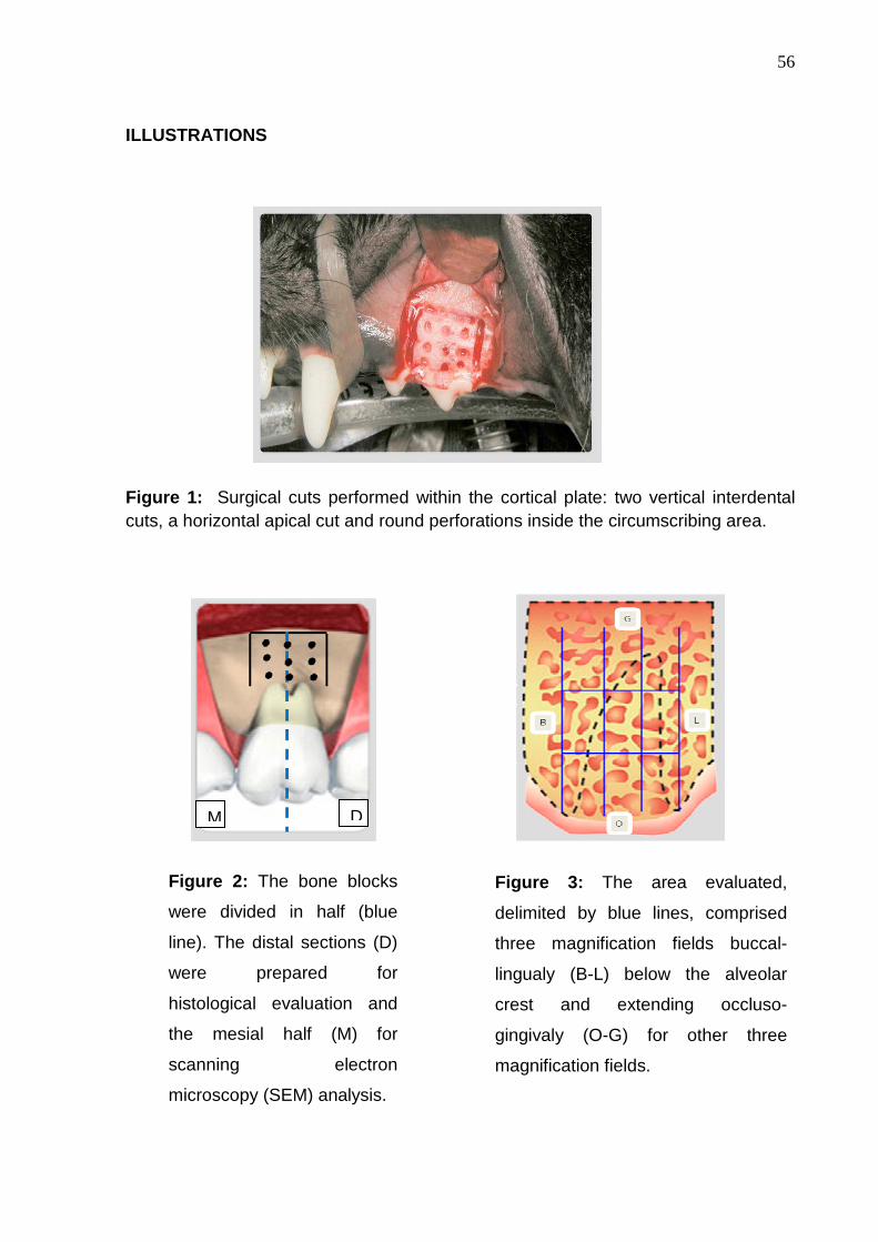
56
ILLUSTRATIONS
Figure 1: Surgical cuts performed within the cortical plate: two vertical interdental cuts, a horizontal apical cut and round perforations inside the circumscribing area.
M D
Figure 2: The bone blocks
were divided in half (blue
line). The distal sections (D)
were prepared for
histological evaluation and
the mesial half (M) for
scanning electron
microscopy (SEM) analysis.
Figure 3: The area evaluated,
delimited by blue lines, comprised
three magnification fields buccal-
lingualy (B-L) below the alveolar
crest and extending occluso-
gingivaly (O-G) for other three
magnification fields.

57
Figure 4: Photomicrophotographs of alveolar bone tissue on right (A) and left (B)
maxillary sides of samples of Group Zero (G0), in which alveolar corticotomy was not
performed (HE x40). Both sides showed similar pattern of bone density.
Figure 5: Comparison of bone density between left (LS) and right maxillary
sides (RS) showed no difference on group zero (G0) (p>0.05).
B
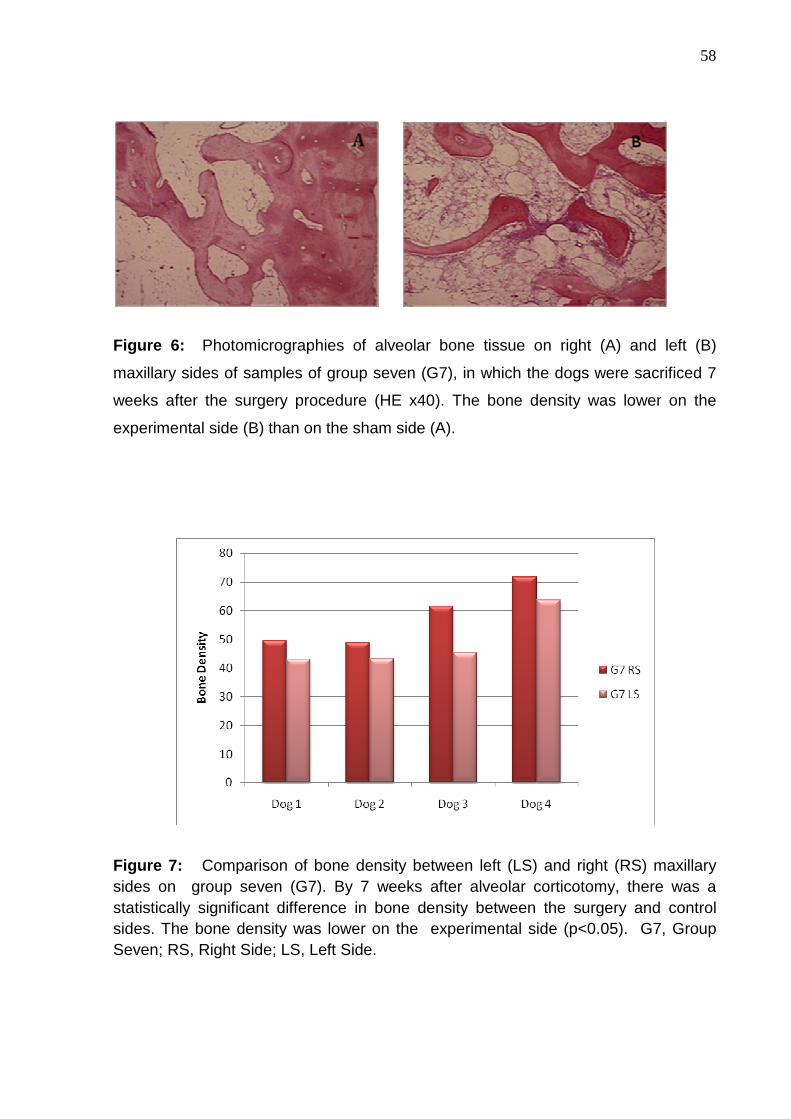
58
Figure 6: Photomicrographies of alveolar bone tissue on right (A) and left (B)
maxillary sides of samples of group seven (G7), in which the dogs were sacrificed 7
weeks after the surgery procedure (HE x40). The bone density was lower on the
experimental side (B) than on the sham side (A).
Figure 7: Comparison of bone density between left (LS) and right (RS) maxillary sides on group seven (G7). By 7 weeks after alveolar corticotomy, there was a statistically significant difference in bone density between the surgery and control sides. The bone density was lower on the experimental side (p<0.05). G7, Group Seven; RS, Right Side; LS, Left Side.
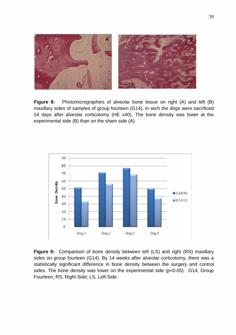
59
Figure 8: Photomicrographies of alveolar bone tissue on right (A) and left (B) maxillary sides of samples of group fourteen (G14), in wich the dogs were sacrificed 14 days after alveolar corticotomy (HE x40). The bone density was lower at the experimental side (B) than on the sham side (A).
Figure 9: Comparison of bone density between left (LS) and right (RS) maxillary sides on group fourteen (G14). By 14 weeks after alveolar corticotomy, there was a statistically significant difference in bone density between the surgery and control sides. The bone density was lower on the experimental side (p<0.05). G14, Group Fourteen; RS, Right Side; LS, Left Side.

60
Figure 10: Ratio of left side bone density per right side bone density in the dogs of
the three groups bone density. Ratio of left side bone density per right side bone
density in the dogs of the three groups. G0, Group Zero; G7, Group Seven; G14,
Group Fourteen; RS, Right Side; LS, Left Side.
Figure 11 : SEM images of coronal sections of maxillary alveolar bone. A: Group
zero, right side. B: Group zero, left side. These pictures show a similar pattern of
medullary bone aspect. C: Group seven, right side; D: Group seven, left side. The
experimental side is characterized by larger and more numerous trabecular spaces,
when compared to the sham side.
A B
C D
61
Parallel bars, skeletical anchorage and corticotomy
to intrude overerupted molars
Ana Paula Carvalho Gomes Ferreira, DDS*
Martinho Campolina Rebello Horta, DDS, MS, PhD**
Bruno Franco de Oliveira***
Dauro Douglas Oliveira, DDS,MS,PhD****
*Master Student in Orthodontics, School of Dentistry, Pontificia Universidade
Católica de Minas Gerais, Belo Horizonte, Brazil
** Associate Professor, School of Dentistry, Pontificia Universidade Católica
de Minas Gerais, Belo Horizonte, Brazil
*** Private practice, Belo Horizonte, Brazil
**** Program director in Orthodontics, Pontifical Catholic University of
Minas Gerais
62
ABSTRACT Intrusion of overerupted molars by conventional orthodontic methods is not always as
effective as desired and the results may be unpredictable and sometimes frustrating.
The purpose of this article is to illustrate the clinical effects of a new multidisciplinary
approach to achieve adequate molar intrusion. Customized buccal and palatal
metallic bars were attached to mini-screws for optimal anchorage and localized
alveolar corticotomies were performed to potentiate the biological response to the
intrusive forces. Five months after intrusive force application through the center of
resistance of the left maxillary molars, a considerable amount of intrusion was
observed. This case report demonstrated an efficient method to intrude overerupted
molars when temporary anchorage devices can not be ideally placed from a
biomechanical perspective.
KEY WORDS: Overerupted molars; Intrusion; Corticotomy; Miniscrews

63
INTRODUCTION
The advancements in Preventive Dentistry have changed the quality of life of a
large amount of the world’s population. Despite these improvements, premature loss
of permanent molars is still a common clinical finding that often leads to the
overeruption of the opposing molar, resulting in functional problems such as occlusal
interferences, periodontal defects and inadequate room to restore the edentulous
space (Melo et al., 2008). An interdisciplinary and comprehensive dental treatment is
necessary to restore adequate posterior occlusion and to maintain good periodontal
health.
Correction of the overerupted molars is a first and essential step before other
procedures can be started. Protocols such as prosthodontic reduction, surgical
impaction, and conventional orthodontic intrusion have been suggested. However,
the limitations of these clinical approaches must be taken into to consideration during
treatment planning.
Prosthodontic reduction often entails the need for significant molar crown
height reduction, frequently demanding costly root canal therapy at the expense of
tooth vitality (Kravitz et al., 2007). Immediate surgical impaction involves a more
aggressive, expensive and morbid approach (Mostafa et al., 1985). Hence,
orthodontic intrusion of the overerupted molars becomes a more plausible procedure
to restore proper occlusion (Yao et al., 2004) because endodontic therapy or
extensive prosthetic restorations could be avoided, minimizing the biological cost to
benefit to the patient.
Intrusion of posterior teeth has been considered one of the most challenging
types of movement in Orthodontics. It often requires longer treatment time and the
application of complex mechanical systems needed to intrude multirooted teeth (Park
et al., 2003; Ng et al., 2006). After the development of temporary skeletal anchorage
systems, molar intrusion has become a more reliable procedure (Carrilo et al., 2007).
However, placing mini-screws in a position that allows the exertion of intrusive forces
passing precisely through the center of resistance of the overerupted molar might be
a difficult task in some patients.
64
Orthodontic treatment combined with corticotomy can attain the most effective
rate and distance for intrusion (Moon et al., 2007). When this combined perio-ortho
approach is implemented, an osteoporotic situation can be induced, leading to less
bone resistance against the desired movement, facilitating orthodontic tooth
movement (Wilcko et al., 2001).
Corticotomy is a surgical technique in which a fissure is made through the
cortical bone, barely touching the cancelous bone. The healing stimulus after this
surgical trauma leads to a rapid acceleratory phenomenon (RAP) characterized by
increased bone turnover and decreased localized bone density (Frost, 1989), leading
to a diminished resistance to the desired tooth movement (Wilcko et al., 2003). The
combination of skeletal anchorage, optimum force systems and increased bone
turnover surrounding the teeth to be moved represent the most efficient scenario for
optimum molar intrusion.
The purpose of this case report is to illustrate a new alternative method to
achieve adequate overerupted molar intrusion when temporary anchorage devices
(TAD’s) can’t be ideally placed from a biomechanical perspective.
CASE PRESENTATION
A 37 year-old Brazilian woman was seeking restoration of her left posterior
occlusion in order to restore her edentulous regions and improve mastication. There
was little vertical space for proper prosthodontic treatment because her maxillary left
first and second molars had overerupted following the premature loss of their
antagonists (Fig.1). Three treatment options were presented to her: (1) elective
endodontic therapy, occlusal reduction, crown lengthening and full crown restorations
of the overerupted molars; (2) subapical osteotomies for immediate bone-block
intrusion and (3) Skeletal anchorage supported with buccal and palatal customized
metallic bars attached to four TAD’s combined with selective alveolar corticotomies
to orthodontic intrude the left maxillary first and second molars.The patient was
65
informed of the risks, advantages, and disadvantages of all therapeutic approaches.
She chose the third treatment alternative and signed a consent form assuming all
responsibilities for her decision.
Laboratory phase
Maxillary alginate impression was taken and a working model was obtained.
The locations of the TAD’s heads were reproduced with red acrylic on both buccal
and palatal sides (Fig. 2A). Two parallel bars were then sculpted with wax, having
their extremity placed right over the red acrylic. Four holes were made in each bar.
The ones on each extremity were named “Safety holes” to allow the use of steel
ligature to firmly tie the bars to the TAD’s (Fig. 2B). The other two holes were located
according to the position of the molars in order to obtain the correct placement of
Closed Sentalloy coils (Dentsply GAC International, Bohemia, NY) to exert 150 g of
intrusive force apically trough the center of resistance of each molar (Fig. 2C). After
the final adjustments of the waxed bars, they were casted into metallic bars (Fig. 2D).
Surgical Procedure
The surgeries were performed with patients under local anesthesia. A full-
thickness flap was raised on the palatal and a modified flap to preserve the papillae
was made on the buccal surfaces to expose the alveolus surrounding the
overerupted maxillary molars. Localized corticotomies were performed around the
maxillary left first and second molars and the adjacent third molar was extracted (Fig.
3). Cortical bone was removed by using a #701 surgical bur under continuous and
abundant irrigation with cold sterile saline solution. Vertical cuts were made on both
mesial and distal interproximal areas starting 2 to 3 mm above the alveolar crest and
extending 2 to 3 mm past the estimated root apices. A horizontal corticotomy was
performed connecting the interdental cuts. All surgical cuts were made in the cortical
plate, barely penetrating the trabecular bone. After careful irrigation, the gingival flaps

66
were repositioned and sutured appropriately. Antibiotics and anti-inflammatories were
prescribed from 24 hours before to 3 days postsurgery. The patient reported mild
postcorticotomy discomfort, describing it as similar to the soreness she felt after
previous tooth extractions.
TAD’s insertion and Orthodontic procedures
The mini-screws were inserted onto the buccal (Neodent, 1.6mm in diameter
and 11mm long) and palatal (Neodent, 1.6mm in diameter and 9mm long) alveolar
ridges 7 days after decortications were performed. The palatal miniscrews were
placed between second bicuspid and first molar and distal to the second molar.
Buccal TAD’s were placed between first and second bicuspids and distal to the
second molar.
Immediately after TAD’s were inserted, the customized metallic bars were
transferred to the patient’s mouth and firmly tied with steel ligatures to the mini-
screws heads. Flowable light cure composite (FlowTain, Reliance Orthodontic
Products, Itasca, IL) was placed covering both extremities of the bars, reinforcing the
TAD-Bars junction as well as improving patient’s comfort. Closed Sentalloy coils
(Dentsply GAC International, Bohemia, NY) were tied to the bars with steel ligatures
to allow constant 150 g intrusive forces apically through the center of resistance of
the molars (Fig.4).
Five months after the force application, a considerable amount of maxillary
molar intrusion was observed, leveling the occlusal plane. Although no force was
applied to the bicuspid, an undesirable intrusion was noticed probably due to an
overcontoured amalgam restoration in the interproximal surface adjacent to the first
molar. After establishing the proper leveling, retention of the intruded maxillary
molars was accomplished with implant supported restorations in the opposite arch,
which reconstructed the vertical dimension (Fig.5). At this point, fixed appliances
were placed to continue treatment. In the meantime, the mini-screws were removed.
67
DISCUSSION
Molar intrusion is not always as effective as desired and the results can be
unpredictable and frustrating sometimes. When conventional fixed orthodontics is
implemented some common unwanted side effects such as extrusion and tipping of
the adjacent anchorage teeth may be observed. Temporary anchorage devices
(TAD’s) can be used to avoid these problems and to obtain pure intrusion of posterior
teeth. However, anatomical variations such as maxillary sinus indentations may limit
the possible areas for mini screws placement. Pure intrusion of overerupted maxillary
molars requires a precise force application from both buccal and lingual sides with
the resultant force vector passing apically through the molar’s center of resistance.
The present case report proposes a new approach to intrude extruded maxillary
molars when TAD’s can not be ideally placed from a biomechanical point of view.
In this patient, miniscrews could not be properly located to achieve the ideal
intrusive force system. The alternative found by the multidisciplinary team involved
was the placement of four miniscrews where they would have the best bony support
and the fabrication of customized casted metallic bars that were attached to the
TAD’s. These bars also presented holes that allowed the adequate positioning of
niquel-titanium closed coil springs to obtain the desired intrusive forces. The use of
customized metallic bars attached to miniscrews and localized corticotomies
represented an attempt to optimize intrusion without the need of patient cooperation.
Although this approach eliminated the need for elastic or headgear wear it made the
maintenance of ideal oral hygiene a more challenging task and calculus
accumulation was noticed over the bars.
Kravitz et al. (2007) suggested creating adequate space between the
overerupted molar and its adjacent teeth prior to the application of intrusive forces.
They observed that intrusion can be compromised if the adjacent teeth lean over the
overerupted molar. In the present case, the second bicuspid was in close contact
with the first molar. No interproximal reduction was performed prior to intrusion and a
possible overcontoured amalgam restoration transferred the intrusive forces to the
bicuspid that presented undesired intrusion. This problem was addressed latter in
treatment when the premolar was left untied to the fixed appliance allowing the
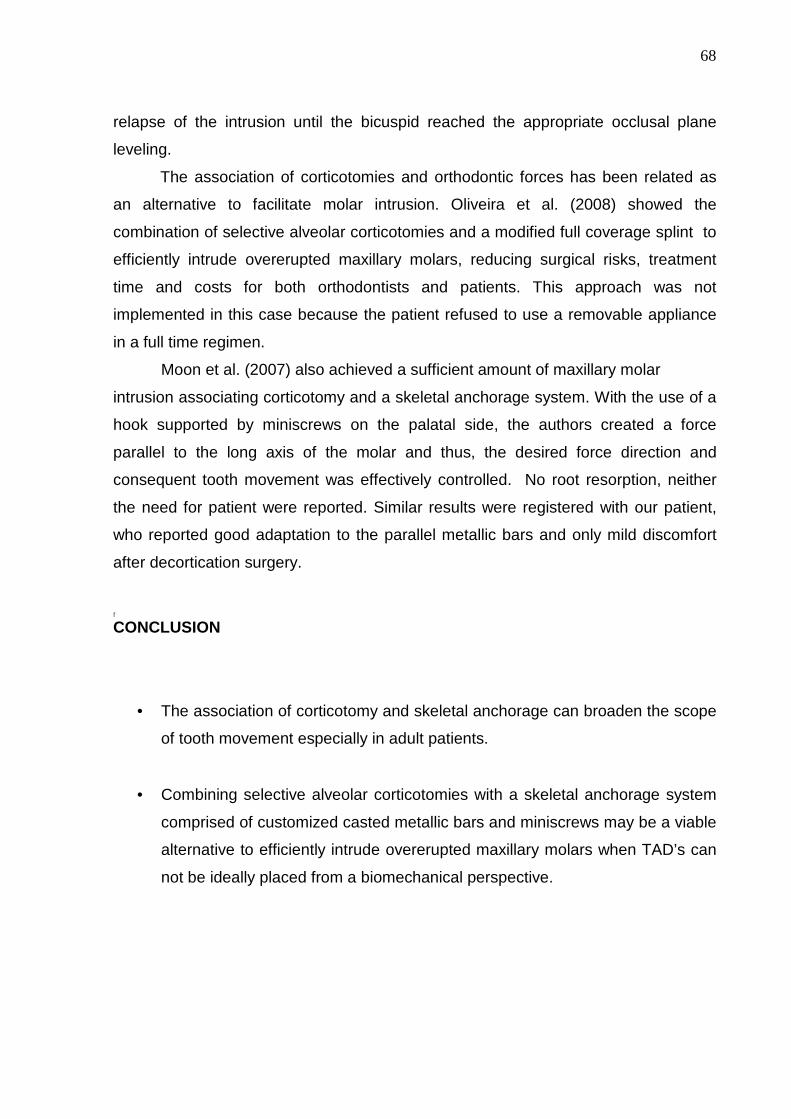
68
relapse of the intrusion until the bicuspid reached the appropriate occlusal plane
leveling.
The association of corticotomies and orthodontic forces has been related as
an alternative to facilitate molar intrusion. Oliveira et al. (2008) showed the
combination of selective alveolar corticotomies and a modified full coverage splint to
efficiently intrude overerupted maxillary molars, reducing surgical risks, treatment
time and costs for both orthodontists and patients. This approach was not
implemented in this case because the patient refused to use a removable appliance
in a full time regimen.
Moon et al. (2007) also achieved a sufficient amount of maxillary molar
intrusion associating corticotomy and a skeletal anchorage system. With the use of a
hook supported by miniscrews on the palatal side, the authors created a force
parallel to the long axis of the molar and thus, the desired force direction and
consequent tooth movement was effectively controlled. No root resorption, neither
the need for patient were reported. Similar results were registered with our patient,
who reported good adaptation to the parallel metallic bars and only mild discomfort
after decortication surgery.
f CONCLUSION
• The association of corticotomy and skeletal anchorage can broaden the scope
of tooth movement especially in adult patients.
• Combining selective alveolar corticotomies with a skeletal anchorage system
comprised of customized casted metallic bars and miniscrews may be a viable
alternative to efficiently intrude overerupted maxillary molars when TAD’s can
not be ideally placed from a biomechanical perspective.

69
REFERENCES
Carrilo R et al. Intrusion of multiradicular teeth and related root resorption with mini-
screw implant anchorage: A radiographic evaluation. Am J Orthod Dentofacial Orthop
2007; 132:647-55.
Frost HM. The biology of fracture healing: An overview for clinicians. Part I. Clin
Orthop Rel Res 1989; 248:283-93.
Frost HM. The biology of fracture healing: An overview for clinicians. Part II. Clin
Orthop Rel Res 1989; 248:294-309.
Kravitz ND et al. Intrusion of overerupted upper first molar using two orthodontic
miniscrews. A case report. Angle Orthod 2007; 77:915-22.
Kravitz ND et al. The use of temporary anchorage devices for molar intrusion. J Am
Dent Assoc 2007; 138:56-64.
Melo AC et al. Upper molar intrusion in rehabilitation patients with the aid of
microscrews. Aust Orthod J 2008; 24:50-3.
Moon CH, Wee JU, Lee HS. Intrusion of Overerupted Molars by
Corticotomy and Orthodontic Skeletal Anchorage. Angle Orthodontist 2007; 77:1119-
25.
Mostafa YA, Tawfik KM, El-Mangoury NH. Surgical Orthodontic treatment for
overerupted maxillary molars. J Clin Orthod 1985;19:350-1.
Ng J, Major P W,Mirc CF. True molar intrusion attained during orthodontic treatment:
A systematic review. Am J Orthod Dentofacial Orthop 2006; 130:709-14.
Oliveira DD et al. Selective alveolar corticotomy to intrude overerupted molars. Am
J Orthod Dentofacial Orthop 2008; 133:902-8.

70
Park YC,Lee SY, KimDH, Jee SH. Intrusion of posterior teeth using miniscrew
implants. Am J Orthod Dentofacial Orthop 2003; 123:690-4.
Wilcko WM, Wilcko MT,Ferguson DJ. Rapid orthodontics with alveolar reshaping: two
cases reports of decrowding. Int J Periodontics Restorative Dent 2001; 21:9-19.
Wilcko WM et al. Rapid orthodontic decrowding with alveolar augmentation: case report. World J Orthod 2003; 4:197-205.
Yao CCJ et al. Intrusion of the Overerupted Upper Left First and Second
Molars by Mini-implants with Partial-Fixed Orthodontic Appliances: A Case Report.
Angle Orthod 2004; 74:550–557.

71
LEGENDS
Figure 1. Pre-treatment photographs revealing significant overeruption of maxillary
left first and second molars.
Figure 2. A, Estimated location of TAD’s heads; B, Customized waxed parallel
bars; C, Determination of safety holes at each extremity and location of force
application holes. D, Metal bars placed in position prior to transferring to the patient’s
mouth.
Figure 3. Surgical photographs. A, Buccal view showing modified gingival flap; B,
Palatal view illustrating decortications and 3rd molar extraction.
Figure 4. Intrusive force system insertion. A, Buccal view; B, Occlusal Photograph
showing mediodistal coil position and composite reinforcements at the TAD-Bar
junctions.
Figure 5. Progress photos. A, 2 months post intrusive force application; B, 5 months
after surgery; C, Retention of intrusion with temporary crowns over lower
conventional implants.
Figure 6. Pretreatment and intermediate panoramic radiographs.
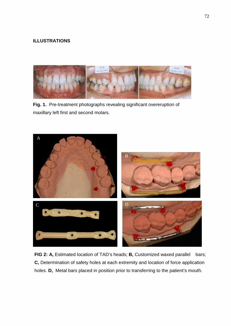
72
ILLUSTRATIONS
Fig. 1. Pre-treatment photographs revealing significant overeruption of
maxillary left first and second molars.
FIG 2: A, Estimated location of TAD’s heads; B, Customized waxed parallel bars;
C, Determination of safety holes at each extremity and location of force application
holes. D, Metal bars placed in position prior to transferring to the patient’s mouth.
A
D C
B
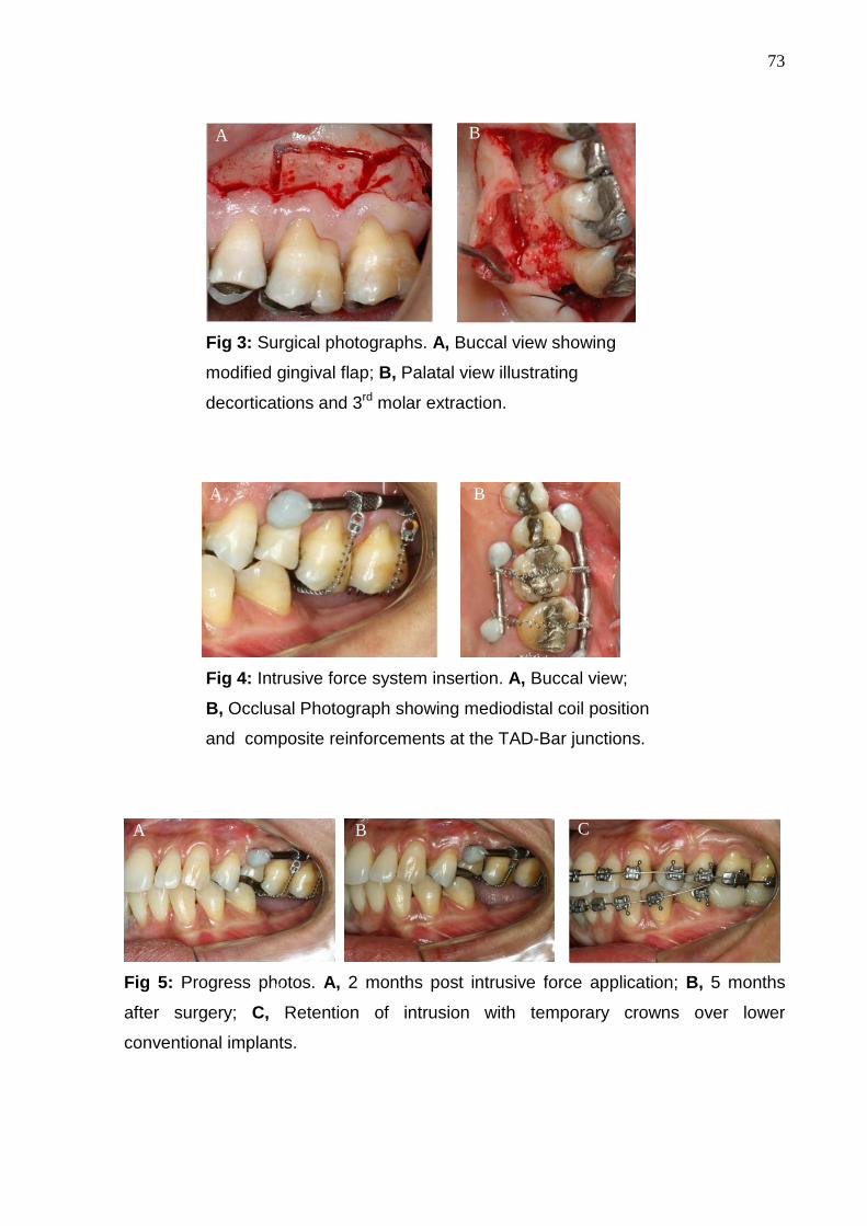
73
Fig 3: Surgical photographs. A, Buccal view showing
modified gingival flap; B, Palatal view illustrating
decortications and 3rd molar extraction.
Fig 4: Intrusive force system insertion. A, Buccal view;
B, Occlusal Photograph showing mediodistal coil position
and composite reinforcements at the TAD-Bar junctions.
Fig 5: Progress photos. A, 2 months post intrusive force application; B, 5 months
after surgery; C, Retention of intrusion with temporary crowns over lower
conventional implants.
B
A B
A B
A B C
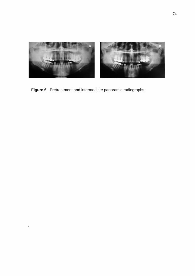
74
Figure 6. Pretreatment and intermediate panoramic radiographs.
.
75
CONCLUSÕES
1. Corticotomias alveolares seletivas podem aumentar os espaços medulares e,
consequentemente, diminuir a densidade óssea nas áreas adjacentes à
injúria;
2. A arquitetura do tecido ósseo quando submetido à corticotomia torna-se mais
osteoporótica;
3. A associação da corticotomia à ortodontia convencional pode, eficientemente,
abordar tratamentos de pacientes adultos com má-oclusões severas, como
molares supraextruídos.
76
REFERÊNCIAS BIBLIOGRÁFICAS COMPLEMENTARES BRIDGES, T.; KING, G.; MOHAMMED, A. The effect of age on tooth movement and
mineral density in alveolar tissues of the rat. Am J Orthod Dentofac Orthop ,v.93,
n.3, p.245-50,Mar.1988.
FROST, H.m. The biology of fracture healing: An overview for clinicians. Part
I.Clinical Orhtopaedics and Related Research . v.248,n.11,p.283-93.Nov.1989.
FROST, H.m. The biology of fracture healing: An overview for clinicians. Part
II.Clinical Orhtopaedics and Related Research . v.248,n.11,p.294-309.Nov.1989.
GANTES, B.; RATHBUN, E.; ANHOLM, M. Effects on the periodontium following
corticotomy-facilitated orthodontics. Case Reports. J Periodontol , v.61, n.4. p.234-
37,Apr.1990.
GERMEÇ, D.; GIRAY,B.; KOCADERELLI, I.; ENACART, A. Lower incisor retraction
with a modified corticotomy. The Angle Orthodontist . v.76,n.5, p.882-90,Nov.2005.
HASLER, R. et al. A clinical comparison of the rate of maxillary canine retraction into
heales and recent extraction sites- a pilot study. Eur J Orthod , v.19, p.711-
19,Dec.1997.
HWANG, H.S.; LEE, K. H. Intrusion of overerupted molars by corticotomy and
magnets. Am J Orthod Dentofacial Orthop , v.120, n.2, p.209-16,Aug. 2001.
ISERI, H.; KISNISCI, R.; BZIZI, N.; TUZ, H. Rapid canine retraction and orthodontic
treatment with dentoalveolar distraction osteogenesis. Am J Orthod Dentofacial
Orthop , v.127, n.5, p.533-41,May.2005.
KIŞNIŞCI,R.Ş.; ÌŞERI, H.; TÜZ, H. H.; ALTUG, A. T. Dentoalveolar distraction
osteogenesis for rapid orthodontic canine retraction. J Oral Maxillofac Surg , v.60,
p.389-94, Apr.2002.
77
MIDGETT, R. J.; SHAYE, R.; FRUGE, J. F. The effect of altered bone metabolism on
orthodontic tooth movement. Am J Orthod , v.80, n.3, p.256-62, Sep.1981.
NAKAMOTO, N. et al. Experimental tooth movement through mature and immature
bone regenerates after distraction osteogenesis in dog. Am J Orthod Dentofacial
Orthop . v.121, n.4, p.385-95.Apr.2002.
OLIVEIRA, D.D. et al. Effects of selective alveolar decortication on cancellous bone
density, 2006. Tese (Doutorado)-Universidade Federal do Rio de Janeiro, Faculdade
de Odontologia, Rio de Janeiro.
PILON, J.J.G.M; KUIPERS-JAGTMAS, A.M., MALTHA, J.C. Magnitude of
orthodontic forces and rate of bodily tooth movement: an experimental study.
Am J Orthod Dentofacial Orthop . v.110, n.1,p.16-23,Jul.1996.
SCHILLING, T.; MÜLLER, M.; MINNE, H. W.; ZIEGLER, R. Influence of
inflammation-mediated osteopenia on the regional acceleratory phenomenon and the
systemic acceleratory phenomenon during healing of a bone defect in the rat. Calcif
Tissue Int , v.63, n.2,p.160-66,Aug.1998.

78