Embed Size (px)
Citation preview

CHRONIC CHAGASIC CARDIOMYOPATHY WITH BROAD COMPLEX QRS TACHYCARDIA
OR WIDE COMPLEX TACHYCARDIA(WCT)
CARDIOMIOPATIA CHAGÁSICA CRÔNICA COM TAQUICARDIA DE QRS LARGO
Raimundo Barbosa Barros MD, Fortaleza – Ceará, Brazil.

Prezado Prof. Andrés, eu gostaria de ouvir a opinião dos colegas do forum sobre este caso quegerou polêmica sobre a conduta quando foi apresentado ontem em sessão clínica.Paciente masculino, 50anos, procedente de Redenção – Ceará- Brasil. História pregressa de palpitações e pré-síncope. Deu entrada com TV com padrão de bloqueio do ramo esquerdo. Optado pela reversão química(discutível) com amiodarona 300mg EV. Atualmente em classe funcional II em fase de otimização terapêutica.Sorologia positiva para Chagas.Rx com cardiomegalia ++/4.ECO: VE =68/59; FE=28% Hipocinesia difusa de grau importante.Qual a conduta adequada neste caso?---------------------------------------------------------------------------------------------------------------------------Dear Prof. Andrés, I would like to hear the valuable opinion of forum´s colleagues about this case that has generated polemic related to management when he was introduced yesterday at the clinics session. Male patient, 50yo, coming from Redenção Ceará - Brazil.Previous history of palpitations and near syncope.He joined our service with broad QRS left bundle branch block pattern pattern tachycardia.Opted for chemicals reversion with intravenous amiodarone 300mg(debatable). Today, the patient is in fuctional Class II with otimization theraphy Positive serology for Chagas disease.Chest Rx cardiomegaly + + / 4.ECO: LV = 68/59; LVEF = 28% diffuse severe hypokinesia.What is appropriate approach in this case?

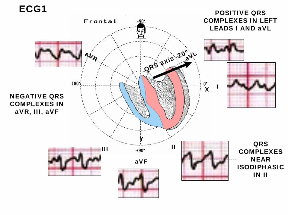
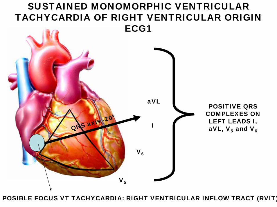
Very rapid Monomorphic Sustained VT with LBBB-like pattern. HR= 270bpm. QRS duration 170ms(>160ms with LBBB morphology), QRS axis ≈ - 20º on PF and to back and leftward on HP. Possible origin focus Right Ventricular Inflow Tract (RVIT).

RBBB
LAE
PVC
Salvos of VT with fusion and capture beats
Anterolateralfibrosis
LOW QRS VOLTAGE ON FP
ECG2: Sinus rhythm, left atrial enlargement (LAE): slow and profound last P portion component in V1), low QRS voltage on FP, LAFB, complete RBBB, PVCs and salvos of VT with fusion and capture beats and similar morphology of right ventricular origin with focus on RVIT. Salvos HR 160bpm consequence of amiodarone administration. Anterolateralfibrosis.

INTERMITTENT VARIABLES DEGREE OF RBBB
PVC
R-on T-phenomenon
Sinus rhythm, HR 90bpm, extreme left axis deviation (LAFB), low QRS voltage tendency, intermittent variables degree of RBBB, frequent monomorphic PVCs with short coupling (R-on-T phenomenon) originate from RVOT (Left BBB-pattern and inferior axis), fusion beats and anterolateral fibrosis.

Estimado Andres,Taquicardia ventricular monomorfa sostenida sintomatica (palpitaciones, presincope). mirar aVR y algoritmo de Vereckei.Miocardiopatia dilatada chagásicaDisfunción ventricular izquierda severa (28%)TratamientoPara insuficiência cardiaca congestiva yCDI (prevencion secundaria de morte cardiaca súbita).No sé si se necesitan más detalles. Saludos. Oscar Pellizzon MD opeli@fibertel.com.ar----------------------------------------------------------------------------------------------------------------------Dear Andrés,Symptomatic sustained monomorphic ventricular tachycardia (palpitation and near syncope).Please see aVR lead and Vereckei’s algorithm.Chagasic dilated cardiomyopathy.Severe left ventricular disfunction (EF=28%).Treatment:Pharmacologic drugs for heart failure and ICD implatation as secondary sudden death prevention.I don’t know if you need others details greatings,Oscar.

Comentarios del caso del paciente chagásico: El primer ECG es un flutter ventricular con una frecuencia cardíaca elevada (260 lpm) lo que ocasionaria una fase diastólica extremadamente corta que en teoria induciria a una disminucion severa del volumen sistólico. En mi experiencia estas TVs rápidas evolucionan rapidamente hacia FV. Siendo supraventricular con esta frecuencia cardiaca, la sobrevida es mayor como ocurre frecuentemente en pacientes con Wolff-Parkinson-White y aleteo auricular o fibrilación auricular En este caso es imposible hacer un diagnóstico con exactitud, pero si el paciente mantuvo la presion arterial en limites aceptables y no sufrió edema agudo de pulmón, tenderia a suponer que era supraventricular y en especial porque el médico tratante tuvo tiempo de medicarlo con drogas y respondió. Excepcionalmente un flutter ventricular le daria todo este tiempo siendo necesário inmediata cardioversión para salvarle la vida. Pero en medicina todo es posible, y mi experiencia muestra excepciones, la cual puede ser esta.
El ECG 2 En V1 la segunda parte de la onda P sugiere dilatación auricular izquierda, talvez indicando una insuficiencia diastólica. Los complejos QRS tienen un patrón de bloqueo bifascicular de tipo BCRD + LAFB.Se observa una extrasistole ventricular, problemente originada en el VD, que induce a un latido de reentrada. Hay una TV no sostenida con morfologia similar al flutter del ECG1, pero mas lenta por el efecto de la amiodarona que reduce la velocidad del circuito de reentrada. En aVR en el ultimo latido se ve una onda P invertida aparentemente no conducida o probablemente una P retrograda. Uno esperaria que sea positiva.Lo interesante es que la arritmia de reentrada fue iniciada por una extrasiste ventricular con morfologia diferente a la arritmia de reentrada.----------------------------------------------------------------------------------------------------------------------------------
-

ECG3 Es muy probable que este patrón electrocardiográfico pertenece a una miocarditis chagásica. El segundo vector en II,III es lento sugeriendo que existe un fenomeno fibrótico en el ventrículo izquierdo. Ademas existe una onda TIII >T1 y onda T invertida en aVL indicando un fenómeno fibrotico en la base del corazón, talvez ocasionada por el proceso fibrótico chagásicoLa extrasistole ventricular se origina en la via de salida del ventriculo derecho y probablemente ha descencadenado la arritmia de reentrada , como lo sugiere el ECG 2Un fraternal abrazo Samuel Sclarovsky---------------------------------------------------------------------------------------------------------Un abrazo Samuel SclarovskyComments related the Chagasic patient: The first ECG is a ventricular flutter with a very high heart rate (260 bpm) which would mean a very short diastolic phase in theory induce a severe decrease in stroke volume. In my experience these fast TVs evolving rapidly toward FV.If this broad QRS tachycardia is supraventricular in origin the survival is greater as frequently occurs in patients with WPW, atrial flutter or AF. In this case it is impossible to diagnose accurately, but if the patient had blood pressure within acceptable limits and suffered no acute pulmonary edema, would tend to believe it was supraventricular and especially since the physician had time to medicate with drugs and responded. Exceptionally ventricular flutter this time would give immediate cardioversion be necessary to save his life. But everything is possible in medicine, and my experience shows exceptions, which may be this.I continue after A hug for allSamuel Sclarovsky

ECG 2 In the second part of the P wave in V1 suggests left atrial enlargement, perhaps indicating a diastolic failure.The QRS complexes have a bifascicular pattern RBBB + LAFB.There is a PVC, problemente originated in the RV, which induces a reentrant beat.There is a non-sustained VT with morphology similar to the flutter of ECG1, but more slowly by the effect of amiodarone slows the reentry circuit.In the last beat aVR I observe an inverted P wave apparently not conducted or probably a retrograde P. One would expect to be positive.The interesting thing is that the reentry arrhythmia was initiated by a PVC with different morphology reentrant arrhythmia.
ECG3 is very likely that this electrocardiographic pattern belongs to a chagasic myocarditis. The second vector in II, III is slow suggesting that there is a fibrotic phenomenon in the left ventricle. There is also a wave TIII> T1 and inverted T wave in aVL indicating a fibrotic phenomenon at the base of the heart, perhaps caused by the fibrotic chagasic process.
The PVC originates in the right ventricular outflow tract and probably break out the reentry, as suggested by the ECG 2
A fraternal hug
Samuel Sclarovsky------------------------------------------------------------------------------------------------------------------------------

Caso Paciente Chagásico:Estimados Colegas, Concuerdo con el Dr. Barbosa que de entrada se trata de una TVMS 240 mseg, (se ve claramente la disociacion AV en D2) y tambien concuerdo que el tartamientode reversion quimica con amiodarona 300 mg/EV es discutible.El segundo electro muestra a las claras un tipico ECG de una miocarditis Chagasica Cronicaen etapas avanzadas: RS, HBA, BRD colgajos de TV no sostenidos Monomorfos que se mesclan con frecuentes EV de otra morfologia. (dando la apariencia que se trata de TV polimorfa).El tratamiento sin lugar a dudas es un CDI, independientemente de la FEVI, ya que la arritmia es Pre-Sincopal. Asimismo dejaria tratamiento con Amiodarona en principio en forma aislada y si continua con arritmia sostenida o comienza con TV lentas cambiaria o asociaria si lo disponen y el paciente lo tolera, la Mexiletina.----------------------------------------------------------------------------------------------------------------------------------Un abrazo para todosEnrique RetykI agree with Dr Barbosa it is a sustained monomorphic VT with HR 240bpm. (I observe clear dissociation in II lead) Also agree that chemical reversion with intravenous amiodarone(300mg) is not consensual. ECG2: typical pattern of chagasic cardiomyopathy: sinus rhythm, Right Bundle Branch Block, Left Anterior Fascicular Block, non-sustained monomorphic VT mixed with frequent premature ventricular contractions (pseudo polymorphic VT).Appropriate approach: Implantable cardioverter-defibrillator without to think in LVEF, because the tachyarrhythmia occasioned near-syncope. I employ amiodarone in association with the cardioverter-defibrillator and if the VT continue or we observe slow VT I associate Mexiletine.
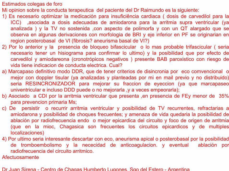
Estimados colegas de foro Mi opinion sobre la conducta terapeutica del paciente del Dr Raimundo es la siguiente: 1) Es necesario optimizar la medicación para insuficiência cardiaca ( dosis de carvedilol para la
ICC) ,asociada a dosis adecuadas de amiodarona para la arritmia supra ventricular (ya analizada ) y la TV no sostenida ,con aspecto de polimorfa y con un QT alargado que se observa en algunas derivaciones con morfologia de BRI y eje inferior en PF se originarian en region posterobasal de VI (fibrosis? aneurisma basal de VI?)
2) Por lo anterior y la presencia de bloqueo bifascicular o lo mas probable trifascicular ( seria necesario tener un hisiograma para confirmar lo ultimo) y la posibilidad que por efecto de carvedilol y amiodareona (cronotrópicos negativos ) presente BAB paroxistico con riesgo de vida tiene indicacion de conducta electrica. Cual?
a) Marcapaso definitivo modo DDR, que de tener criterios de disincronia por eco comvencional o mejor con doppler tisular (ya analizadas y planteadas por mi en mail previo y no distribuido) seria RESINCRONIZADOR para mejorar su fraccion de eyeccion (ya que marcapaseo univentricular e incluso DDD puede o no mejorarla ,y a veces empeorarla);
b) Asociado a CDI por la arritmia ventricular que presenta ,en presencia de FEy menor de 35% para prevencion primaria Ms;
c) De persistir o recurrir arritmia ventricular y posibilidad de TV recurrentes, refractarias a amiodarona y posibilidad de choques frecuentes; y amenaza de vida quedaria la posibilidad de ablación por radiofrecuencia endo o mejor epicardica del circuito y foco de origen de arritmia (que en la mioc, Chagasica son frecuentes los circuitos epicardicos y de multiples localizaciones)
4) Por ultimo seria interesante descartar con eco, aneurisma apical o posterobasal por la posibilidad de tromboembolismo y la nececidad de anticoagulacion. y eventual ablación por radiofrecuencia del circuito arritmico.
Afectuosamente
Dr Juan Sirena - Centro de Chagas Humberto Lugones, Sgo del Estero - Argentina

Dear forum’s colleaguesMy opinion on the patient's therapeutic of Dr. Raimundo:1) It is necessary to optimize HF medications (carvedilol doses), combined with
adequate doses of amiodarone for supra ventricular and non-sustained VT with polymorphic appearance and prolonged QT interval observed in some leads, with BRI morphology and inferior QRS axis on PF suggesting posterobasal origen in the LV (fibrosis? LV basal aneurysm?)
2) Bifascicular or trifascicular block presence (it would be necessary to have an Hisiogram to confirm the latest) and the possibility that the carvedilol and amiodaronaeffect (negative cronotropic effect ) cause a paroxysmal complete AV block has life-threatening indication electrical approach. Which one?
3) In thinking about it, the algorithm would be:a) Permanent pacemaker DDDR mode, which have criteria of dyssynchrony by conventional Echo or better with tissue Doppler (already discussed and raised by me in previous mail and not distributed) would resynchronisation to improve LVEF (as univentricular pacing and even DDD may or may not improve, and sometimes worse)b) Associated ACD presenting ventricular arrhythmia in the presence of LVEF < 35% for sudden death primary prevention. If VT episodes are persistent or recurrent I would be the possibility of endo RFCA or better epicardial circuit focus of arrhythmia (in the Chagasic cardiomyopathy are frequent epicardial and multiple locations.
4) Finally it is important rule out with Echo, apical or posterobasal aneurysm because thromboembolism possibility. anticoagulation)Affectionately Dr Sirena Centro de Chagas Humberto Lugones, Santiago del Estero - Argentina

Estimado Dr SirenaNo tengo el placer de conocerlo personalmente, pero estoy impresionado por su evaluacion y
manejo del paciente. Concuerdo plenamente con su analisis. No estoy seguro que en el pais muchos grupos hagan ablacion epicardica (Pancho? Aguinaga? Favaloro?). Generalemente se llega a ese punto de 2 maneras:
La ablación endocardica fallo y duranteel EPS uno encuentra origen eipcardico. El ECG es altamente sugestivo de origen epicardico.Lo que no se hace es al voleo decidir ir por abordaje epicardico (ni siquiera en Chagas, donde como Ud seniala muy bien, el origen epicardico es frecuente). Digo esto porque el abordaje epicardico requiere tecnica, paciencia y no esta libre de complicaciones. Nosotros tuvimos una muerte post ablacion, durante ablacion epicardica con “auto-transfusion”, en quien la ablacion fue exitosa, debido a perforación y hemolisis por la autotransfusión.
Es un placer contar con sus analisis en el foro. Lo saludo muy cordialmenteAB-----------------------------------------------------------------------------------------------------------------------------------Dear Dr SirenaI have not the pleasure of knowing you personally, but I'm impressed by your assessment and
patient management. I fully agree with your analysis. I'm not sure that many groups in your country preform the epicardial ablation approach (Femenia? Aguinaga? Favaloro?). Generally reached the point of 2 Endocardial ablation failure and is a source during EPS. The ECG is highly suggestive of epicardial origin. What is not done by deciding to broadcast epicardial approach (even Chagas, where some vital signs as you very well, the epicardial origin is common). I say this because the epicardial approach requires technique, patience and not free of complications. We had a death post ablation, during epicardial ablation with "auto-transfusion"in whom ablation was successful dueto hemolysis by drilling and autotransfusion.
It is a pleasure to have with their analysis in the forum. Yours sincerely.AB

Dear Andrés: The ECG's associated with this case suggest the following:ECG1: Monomorphic ventricular tachycardia HR; 270 bpm of right ventricular origin. ECG2: A few sinus beats with RBBB + LAFB morphology, and salvos of VT with similar morphology as ECG1; AV dissociation is present during the salvos of VT.ECG3: Mostly normal sinus rhythm (RBBB+LAFB) HR: around 90 bpm, with PVC's and fusion beats. I'm not sure what the pause in the rhythm strip is, but it follows a premature beat (?PAC).His history and poor LV function indicates the need for CRT + defibrillator. I await the esteemed opinion of others in this case.Best regards,
Frank Yanowitz, MD. Professor of Medicine. University of Utah School of MedicineMedical Director, ECG Department. LDS Hospital Salt Lake City, Utah . 8th Ave. and C Street. Salt Lake City, Utah 84143 USA [email protected]; fyanow@mac.com----------------------------------------------------------------------------------------------------------------------------------Caro Andrés: Os ECGs de este caso, sugerem o seguinte:
ECG1: HR 270 bpm, taquicardia ventricular monomórfica, de origem no ventrículo direito.ECG2: Uns poucos batimentos com morfologia BRD + LAFB, e salvas de TV com morfologia semelhante à ECG1; dissociação AV está presente durante as salvas de VT.ECG3: Ritmo sinusal, BRD + LAFB HR: cerca de 90 bpm, com PVC e batimentos de fusão. Eu não tenho certeza de que a pausa na faixa de ritmo é, mas segue uma batida prematura (? PAC).Pela sua história e função do VE pobres indica a necessidade de implante de desfibrilador + CRT.Aguardo o respeitado parecer dos outros colegas, neste caso.
Atenciosamente,Frank

Andres, The 12 lead ECG tachycardia shows an extremely rapid tachycardia at between 250-300 bpm. It is a wide QRS and the number 1 diagnosis is monomorphic VT. This extremely rapid tachycardia is going to be hemodynamically compromising with a baseline LVEF of 28%. Apparently, rather than emergent electrical cardioversion, IV amiodarone was used, the tachycardia was terminated and we see a low grade sinus tachycardia at a rate of approximately 100 bpm. Within this there were episodes of multifocal PVCs along with one three beat, one four beat run and one 10 beat run of VT. While Amiodarone clearly has worked, it is also associated with a high incidence of side effects and in the SCD-HeFT trial, did no better than a placebo in the long run. These brief runs will also be hemodynamically compromising. I believe that this patient should receive an ICD for the more protracted episode as rescue even though the ICD will not prevent episodes of VT or VF but will rescue the patient. However, one would also want to eliminate the brief salvos of non-sustained VT which may also be hemodynamicallycompromising. As such, the amiodarone should be continued and may be more effective as serum levels rise. If it proves ineffective, other antiarrhythmic agents might be tried and if these fail, I would consider an EP study trying to map the various VT foci or pathways so that they can be ablated. I have offered this approach with the presumption that all facilities are available such as an EP lab with either a NAVX or Cardio navigation system available. I fully realize that this may not be the case or this may be too expensive for the patient and that everyone cannot afford the absolute best of medical care. Any concomitant heart failure should be treated with the combination of beta blockes, ACE inhibitors, diuretics including spironolactone and improvement of heart failure may also be associated with a reduction of ectopy. While the patient is in the hospital under observation, one can meticulously treat one aspect of the patient such as heart failure to assess its effect on the ventricular ectopy and when the heart failure is thought to be optimally managed, then begin to titrate antiarrhythmic agents both as single therapy or combination therapy regimens before considering an EP and mapping study.I await the comments of others and the final discussion.Paul Levine

Andrés, El ECG muestra una taquicardia con QRS ancho muy rápida (FC 250-300 lpm.). El diagnóstico número 1, es TV monomórfica. Esta taquicardia extremadamente rápida ha comprometido hemodinámicamente la FEVI ( 28%.). Al parecer, en lugar de CV eléctrica se utilizó amiodarona IV, la cual terminó con el evento rápido. Se ve una taquicardia sinusal ≈100 lpm. extrasistoles polifocales, junto con colgajos de TV no sustentada que tambien pueden compromenter el desempeño del VI. La amiodarona puede haver sido responsable en parte de estos eventos En el estudio SCD-HeFT esta droga no fue mejor que el placebo a largo plazo. Creo que este paciente debe recibir un CDI a pesar de que el ICD no impedirá que los episodios de TV o FV pero evita la MS. Sin embargo, también quisiera eliminar las salvas breves de TV no sostenidas, que también puede comprometer la hemodinamia. La amiodarona se debe continuar y puede ser más eficaz cuando se eleven los niveles séricos. Si resulta ineficaz, otros agentes antiarrítmicos puede ser empleados, Yo consideraría un estudio del PE tratando de mapear los focos de VT o varias vías para que puedan realizar la ablación. Me han ofrecido este enfoque con la presunción de que todas las instalaciones están disponibles como un laboratorio de EF, ya sea con un de NAVX o el sistema cardiovascular de navegación. Estoy plenamente consciente de que puede no ser el caso, o puede ser demasiado costoso para el paciente y que todo el mundo no puede permitirse la mejor absoluto de atención médica. La ICC concomitante deben ser tratada con la combinación de betabloquedores, inhibidores de la ECA, diuréticos como espironolactona. La mejora de la ICC también puede estar asociada con una reducción de ectopia. Mientras el paciente está en el hospital en observación, uno puede tratar a un meticuloso aspecto del paciente, tales como la ICC para evaluar su efecto sobre la ectopia ventricular y la ICC cuando se piensa que es administrada de manera óptima, a continuación, empezar a valorar los agentes antiarrítmicos tanto como terapia única o regímenes de combinación de la terapia antes de considerar un estudio del EF y la cartografía. Espero los comentarios de los demás y la discusión final. Paul.

FINAL CONCLUSIONS AND THEORETICAL CONSIDERATIONS
ANDRÉS RICARDO PÉREZ RIERA, MDChief of the Electro-vectorcardiography Sector Faculty of Medicine
ABC Foundation Santo André – São Paulo – Brazil

ECGs ANALYSIS
1. ECG 1: Very rapid Monomorphic Sustained VT with LBBB-like pattern. HR= 270bpm. QRS duration 170ms(>160ms with LBBB morphology), QRS axis ≈ - 20º on PF and to back and leftward on HP. Possible origin focus Right Ventricular Inflow Tract (RVIT).
2. ECG2: Sinus rhythm, HR 90bpm, extreme left axis deviation (LAFB), low QRS voltage on frontal plane, intermittent variables degree of RBBB, frequent monomorphic PVCs with short coupling (R-on-T phenomenon) originate from RVOT (Left BBB-pattern and inferior axis), salvos of VT with fusion (they occur when a normal AV node beat fuses with a beat originating from the ventricles) and capture beats (occasional narrow QRS complexes appearing sooner than normal place) in the trace with similar morphology of right ventricular origin (focus on RVIT) and anterolateral fibrosis.
3. ECG3: Sinus rhythm, HR 90bpm, LAFB, intermittent variables degree of RBBB, frequent monomorphic PVCs with short coupling (R-on-T phenomenon) originate from RVOT( Left BBB-pattern with inferior QRS axis), fusion beats and anterolateral fibrosis.
aVR aVL
IIIII
X
Y
I
aVF
QRS axis -20º
QRS COMPLEXES
NEAR ISODIPHASIC
IN II
POSITIVE QRS COMPLEXES IN LEFT
LEADS I AND aVL
ECG1
NEGATIVE QRSCOMPLEXES IN
aVR, III, aVF

V6
V1
V4
V5
V2
X
Z
V3
LBBB-LIKE PATTERN
RV FOCUSRight ventricular origin
ECG1 MONOPHASIC QRS POSITIVES
COMPLEXES ON LEFT PRECORDIAL
LEADS V5-V6
Absence of RS
complexes from V1 to V6
VTNEGATIVE QRS COMPLEXES ON RIGHT
PERCORDIAL LEADS
QRS axis -20ºPOSITIVE QRS
COMPLEXES ON LEFT LEADS I, aVL, V5 and V6
V6
I
aVL
V5
SUSTAINED MONOMORPHIC VENTRICULAR TACHYCARDIA OF RIGHT VENTRICULAR ORIGIN
ECG1
POSIBLE FOCUS VT TACHYCARDIA: RIGHT VENTRICULAR INFLOW TRACT (RVIT)

BRUGADA ALGORITHM TO DIFFERENTIATE VT FROM SVT WITH ABERRANT CONDUCTION
I) Absence of RS complex from V1 to V6.
Yes No
VT
Pedro Brugada, et. al. Circulation 1991; 83:1649-1659.
II) R to S interval greater than 100 ms in any precordial lead?
YesNo
VT
Highly specific of VTLittle sensitive.
>100 ms<100 ms
SVT
SVT
> 100ms
ECG1
ECG1

IV) AV dissociation?
YesNo
VT
V) Are morphological criteria for TV present in V1-V2 and in V6?
Yes NoVT ASVT
The following are criteria that indicate VT:1) Negative complexes from V4 to V6 .2) QR from V2 to V6.3) Demonstration of A-V dissociation.4) SAQRS < 600 and > 1500.
The point V is not suggestive of VT
ECG1+ECG2
BRUGADA ALGORITHM TO DIFFERENTIATE VT FROM SVT WITH ABERRANT CONDUCTION
Pedro Brugada, et. al. Circulation 1991; 83:1649-1659.

BRUGADA ALGORITHM TO DIFFERENTIATE VT FROM SVT WITH ABERRANT CONDUCTION
IV) In the presence of LBBB-like morphology, is duration of r wave from V1 > 40 ms and rS interval greater than 70 ms?
Yes
> 40 ms
VT
V1
>70 ms.
QS PATTERN IN V1: ECG1
>70ms
Pedro Brugada, et. al. Circulation 1991; 83:1649-1659.

aVR
1. Vereckei A, Duray G, Szénási G, Altemose GT, Miller JM. New algorithm using only lead aVR for differential diagnosis of wide QRS complex tachycardia. Heart Rhythm. 2008; 5: 89
FIRST STEP Presence of an initial R wave in lead aVR
NO YES
ALGORITHM USING ONLY LEAD aVR FOR DIFFERENTIAL DIAGNOSIS OF WIDE QRS
COMPLEX TACHYCARDIA.
>98,6% <1,5%VT SVT
SECOND STEP In the presence of LBBB pattern and initial r wave >40 ms
NO YES
>87,7% VT <13,3%SVT
THIRD STEP Presence of a notch or the descending limb of a negative onset and predominantly negative QRS
NO YES
>86% VT <14%SVT

Vi/Vt ratio Vi/Vt
Vi: initial excursion of QRS complex. Vt: final excursion. Ventricular activation-velocity ratio The vertical excursion (in millivolts) recorded during the initial (Vi) and terminal (Vt) 40 ms of the QRS complex. If ≤1= VT or >1=SVT-A. This criteria suggest SVT-A
VtVi
Vi/Vt ratio (if > 1, SVT-A

QRS duration > 160ms
V6
ECG1: QRS DURATION ≥160ms IS SUGGESTIVE OF VT WHEN LBBB-LIKE PATTERN IS PRESENT

PAVA´S CRITERIONECG R-wave peak time (RWPT) on lead II in differentiating VT from SVT in patients with wide QRS complex tachycardia. RWPT ≥ 50 ms at II is sensitive criterion that discriminates VT from SVT in patients with wide QRS complex tachycardia.
II
R-wave peak time
From the beginning of QRS
Until the apex of R
wave
1. Pava LF, Perafán P, Badiel M, Arango JJ, Mont L, Morillo CA, Brugada J. R-wave peak time at DII: a new criterion for differentiating between wide complex QRS tachycardias. Heart Rhythm. 2010 Jul;7:922-926.

MANAGEMENTAlways optimized pharmacologic treatment: Carvedilol, aldosterone antagonists (espironolactone), furosemide, angiotensin converting enzymes (ACE) inhibitors or angiotensinreceptor blockers (ARBs) (commonly used with people who cannot tolerate the side effects of ACE Inhibitors.), anticoagulation and amiodarone. In Chagas disease drug therapy is ineffective for patients with severe HF: 100% recurrence rate/40% mortality in 1 year. Drugs can reduce the frequency of ICD therapies, but have disappointing efficacy and side effects. Implantable Cardioverter-Defibrillator (ICD): LVEF <35% Secondary SD prevention. Patients with Chagas' cardiomyopathy presenting with either sustained VT or NSVT run a major risk for mortality when had concomitant severe or even moderate LV systolic dysfunction1. The potential benefits ICD need assessment in prospective randomized trials2. In subjects with chronic chagasic cardiomyopathy with an ICD, are predictive of arrhythmic storm: LVEF <35%, NYHA functional class III-IV and absence of beta-blocker treatment3. A randomized trial may not be necessary in high-risk patients4.Epicardial RFCA approach tentative? for VT circuits only with an experienced team. Chronic Chagas cardiomyopathy patients have larger epicardial as compared to endocardialsubstrate areas. Combined epicardial endocardial substrate mapping and ablation during sinus rhythm proves effective in preventing VT recurrences and appropriate ICD therapies5.
1. Sarabanda AV, Marin-Neto JA. Predictors of mortality in patients with chagas' cardiomyopathy and ventricular tachycardia not treated with implantable cardioverter-defibrillators. Pacing Clin Electrophysiol. 2011 Jan;34:54-62.
2. Rassi A Jr, Rassi A, Marin-Neto JA. Chagas disease. Lancet. 2010 Apr 17;375(9723):1388-402.3. Flores-Ocampo J, Nava S, Márquez MF, et al. Clinical predictors of ventricular arrhythmia storms in Chagas
cardiomyopathy patients with implantable defibrillators. Arch Cardiol Mex. 2009 Oct-Dec;79:263-267.4. Bestetti R, Cardinalli-Neto A. Implantable cardioverter defibrillator therapy for patients with chronic Chagas' disease: a
randomized trial may not be necessary in high-risk patients. Europace. 2009 Apr;11(4):537.5. Henz BD, do Nascimento TA, Dietrich Cde O, et al. Simultaneous epicardial and endocardial substrate mapping and
radiofrequency catheter ablation as first-line treatment for ventricular tachycardia and frequent ICD shocks in chronic chagasic cardiomyopathy. J Interv Card Electrophysiol. 2009 Dec;26:195-205.

BROAD COMPLEX TACHYCARDIAS or WIDE COMPLEX TACHYCARDIAS(WCT)A tachycardia is defined as a heart rate ≥ 100 bpm). In broad complex tachycardias the QRS complex is ≥120 ms (3 small squares on the ECG). A broad QRS complex is either caused by the ventricular conducting system not working (bundle branch block) or the electrical circuit is not involving the atrioventricular (AV) node correctly. Broad complex tachycardias may be ventricular or supraventricular in origin.CausesVentricular tachycardias (VTs)WCT of supraventricular origin:
With aberrant conduction or ventricular pre-excitation, any supraventricular tachycardia(SVT) may present as a WCT and mimic VT.Atrial tachycardia with aberrant conduction: aberrant conduction usually manifests as either left or right bundle branch block and the bundle branch block may predate the tachycardia. It may be a rate-related functional block, occurring when atrial impulses arrive too rapidly for a bundle branch to conduct normally. Atrial fibrillation (AF) occurring with an aberrant conduction and a rapid ventricular response, produces an irregular WCT.Wolff-Parkinson-White syndrome: WCTs may occur, either as an atrioventricular re-entrant tachycardia or in association with atrial flutter or AF.AF: in the Wolff-Parkinson-White syndrome the atrial impulses are conducted down the accessory pathway, which may allow rapid conduction and consequently very fast ventricular rates with WCT. The QRS pattern is fairly constant, except for occasional normal complexes and fusion beats (see below).
PresentationA ventricular origin for a WCT is suggested if the patient is aged over 35 years and has a history of ischaemic heart disease or congestive cardiac failure.

Symptoms: Depend on the haemodynamic consequences of the arrhythmia rather than the origin of the arrhythmia. In some patients with VT, they may not be in a state of collapse, but present with dizziness, palpitations, syncope, chest pain or HF. Some patients with SVT and poor ventricular function may present in a state of haemodynamic collapse. In the differential diagnosis of broad QRS tachycardias , hemodynamic status, age, ad HR should not be used. VT can occur at all ages and at very rapid rates. Patients with VT tend to be older than those with SVT.(older than 35yo)A history of heart disease –previous MI, angina, or congestive HF-can serve as a rough discrimination between SVT-A an VT.If th history include similar episodes occuring for longer than 3 years , SVT-A is far more likely than VT. Signs: In VT the rhythm is regular or almost regular. An obviously irregular rhythm is most likely due to AF with either aberrant conduction or pre -excitation. (HR 150bpm and >180bpm respectively)
The following are clinical evidence of AV dissociation,( indicates a VT.)1.Cannon a waves in the jugular venous pulse. This beatinf occasionally coincides so that the atria contract agaist closed AV valves and cause a reflux of blood up the jugular veins.2.Variable intensity of the first heart sound3.Beat –to-beat changes in sistolic blood pressure: changing Korotkoff sounds.Their absence does not rule out VT
Physical signs will also vary according to the haemodynamic effects of the tachycardia.Termination of WCT in response to physical maneuvers such as Valsalva or carotid sinus pressure, or intravenous adenosine strongly suggest SVT-A

VARIETIES OF BROAD(OR WIDE) COMPLEX TACHYCARDIA(WCT)I) Ventricular:
Regular or typical: Monomorphic ventricular tachycardia (MVT)Fascicular ventricular tachycardia, Left ventricular idiopathic VT verapamil responsive( Belhansen VT)Bundle branch reentrant VT(BBR-VT)Right ventricular outflow tract tachycardia RVOT-VT
Irregular, polymorphic or atypical VT Torsades de pointes tachycardia TdP with and witout prolonged QT intervalOthers Polymorphic VT
II) Supraventricular:1. SVT with aberrancy (SVT-A) Bundle branch block with aberrant conduction: triphasic
pattern of RBBB, QRS duration≤140ms in V1 positive patterns, QRS duration≤160ms in V1 negative patterns, R:S ratio in V6 >1
2. Atrial fibrillation over and accessory pathway pre-excitation: Broad, very fast(>180bpm) and irregular QRS occurrence
3. Atrial fibrillation with BBB: HR 140 to 150bpm, and the QRS pattern is that of BBB4. Atrial fibrillation with multiple accessory pathways: irregular, rapid, fusion beats, and
polymorphous.5. Atrial flutter with conduction over a left-side accessory pathway: cause positive
precordial concordance6. Antidromic Circus Movemet Tachycardia: Broad, regular, HR 150 to 250bpm, after event
delta wave identica to that of the initial forces of the tachycardia.7. Circus movement tachycardia with two accessory pathwas(rare)8. Broad QRS paroxysmal supraventricular tachycardia using nodoventricular fibers: Broad
with LBBB pattern.

PRACTICAL APPROACH FOR EVALUATE WIDE COMPLEX TACHYCARDIA (WCT)
Step 1: AV relationship ( if dissociated, VT is diagnosed; if not, proceed to next step)
Step 2: Rightward superior axis (if present, VT is diagnosed; if not, proceed to next step)
Step 3: Vi/Vt ratio (if greater than 1, SVT-A is diagnosed; if not, proceed t next step)
Step 4: Precordial RS pattern (if absent, VT is diagnosed; if not, proceed to next step)
Step 5: Precordial RS interval (if RS present and interval grater than 100ms, VT is diagnosed; if not, proceed to next step)
Step 6: In LBBB type WCT, R wave less than 20ms or R onset to S nadir les than 60ms in V1(if present, SVT-A is diagnosed).
SVT-A: Supraventricular Tachycardia with Aberrancy

ECGA baseline 12-lead ECG is always advantageous, especialy when there is preexisting BBB or ECG evidence of previois MI. In patients with VT and preexisting BBB the QRS pattern during the event is clearly different from that recorded during SR. Usually shows monomorphic QRS complexes as seen in most common forms of sustained VT.QRS complex shape is unusual and of prolonged duration (usually > 120ms). Normally, the longer the duration of the QRS complex the more likely it is to be VT (particularly if >160ms). Changing QRS morphology during the tachycardia also indicates a VT.Evidence of AV dissociation through presence of P waves independent of the QRS complex is common but not always present. Rate: usually 120-300 bpm. Rhythm: regular or approximately regular unless affected by capture or fusion beats. A clearly irregular rhythm it most likely to be due to AF. Diagnostic features (absence of these does not exclude VT):
Capture beats: occasional narrow QRS complexes appearing sooner than normal place in the trace, these show normal AV conduction so SVT is unlikely.Fusion beats: QRS complexes somewhere between a standard QRS and the others present on the trace. They occur when a normal AV node beat fuses with a beat originating from the ventricles.QRS concordance: all QRS complexes in chest leads are enterely positive or enterely negative during a WCT.QRS axis between -90º + - 180° (rightward superior quadrant) is unlike to be SVT-A, so VT is the likely diagnosis.V1 with RBBB pattern: Momophasic R wave, qR, a broad R (grater than 30ms) with any following terminal negative QRS forces are indicative of VT.V6 with RBBB pattern: Rs, Qrs, QS, QR, or a monophasic R wave is indicative of VT;

Management Resuscitation Support ABCs. Basic life support and advanced life support may be required. Give oxygen and gain venous access.The following guidance is taken from the Resuscitation Council guideline for Peri-Arrest Arrhythmias: Monitor ECG, BP, oxygen saturation. Record 12-lead ECG if possible; if not, record rhythm stripIdentify and treat reversible causes (e.g. electrolyte abnormalities - monitor potassium and, if low, start infusions of potassium chloride and magnesium sulfate)If the patient is unstable (signs of instability include reduced conscious level, chest pain, systolic BP below 90 mmHg, HF (rate-related symptoms are uncommon at less than 150 bpm): Synchronized Electrical Cardioversion DC Shock: up to 3 attempts; attempted electrical is always undertaken under sedation or general anaesthesia. If the patient still has a pulse, it is usually possible to terminate a VT episode with a direct current shock across the heart. This is ideally synchronised to the patient's heartbeat. As this is quite uncomfortable, shocks should be delivered only to an unconscious or sedated patient. As a reminder, this is different from defibrillating the patient. If the patient still had a pulse, defibrillating the patient could potentially send him into asystole.Amiodarone 300 mg IV over 10-20 minutes and repeat shock; followed by amiodarone 900 mg over 24 hours. If the patient is stable:
Irregular: Seek expert help; possibilities include:AF with bundle branch block: treat as for narrow complexPre-excited AF: consider amiodaronePolymorphic VT e.g. TdP: give magnesium 2 g over 10 minutes
Regular: If VT - or uncertain rhythm: amiodarone 300 mg IV over 20-60 minutes; then 900 mg over 24 hours.If previously confirmed SVT with bundle branch block: give adenosine as for regular narrow complex tachycardia.
Drugs such as amiodarone or procainamide may be used in addition to defibrillation to terminate VT while the underlying cause of the VT can be determined. When in doubt (VT pr SVT with aberrancy) use procainamide. Do not administer digoxin or verapamil when confronted with a WCT that is more tha 200bpm an irregular; administer procainamide . Qhat is probably present in AF with conduciton over an AP. IF procainamide does not slow the HR and convert the rhythm, CV and refer for evaluation RFCA.
As hypomagnesia is a common cause of VT, stat dose magnesium sulphate can be given for torsades or if hypomagnesemia is found/suspected. Long term anti-arrhythmic therapy may be indicated to prevent recurrence of VT.
Beta-blockers and a number of class III anti-arrhythmics are commonly used. Lidocaine is now being replaced by amiodarone as the first line anti-arrhythmic treatment of VT.
The implantation of an ICD is more effective than drug therapy for prevention of SCD due to VT and VF, but may be constrained by cost issues,(RDM) and well as patient co-morbidities and patient preference.For refractory cases, consider additional pharmacological agents, e.g. lidocaine or sotalol, or overdrive pacing.
Caution is required because pharmacological agents may cause myocardial depression. The treatment of a WCT depends on the origin of the tachycardia and should be treated
as sustained VT until proven otherwise. Vagal stimulation, e.g. carotid sinus massage or the Valsalva manoeuvre, does not usually affect a VT but may affect arrhythmias of supraventricular origin. By transiently slowing or blocking conduction through the atrioventricular node, an atrioventricular nodal re-entrant tachycardia or atrioventricular re-entrant tachycardia may be terminated. In atrial flutter, transient block may reveal the underlying flutter waves.

RFCA is a key therapeutic modality for patients with recurrent VT. A task force consisting of the European Heart Rhythm Association (EHRA) in conjunction with the Heart Rhythm Society (HRS) developed an expert consensus paper that carefully defines the indications, techniques, and outcomes of this procedure1.There was consensus among the task force members that RFCA for VT should generally be considered early in the treatment of patients with recurrent VT. In the past, RFCA was often not considered until pharmacological options had been exhausted, often after the patient had suffered substantial morbidity from recurrent episodes of VT and ICD shocks. Antiarrhythmic medications can reduce the frequency of ICD therapies, but have disappointing efficacy and side effects. Advances in technology and understanding of VT substrates now allow RFCA of multiple and unstable VTs with acceptable safety and efficacy, even in patients with advanced heart disease.Remote magnetic navigation is recognized as an important method for delivery of ablation therapy for these patients due to the ability of the flexible magnetic catheter to carefully map the diseased tissue without inadvertently inducing abnormal ventricular rhythms. In a series of 110 patients that included all morphologies of VT, 85% of patients treated with magnetic ablation were free from VT at one year after the intervention and were exposed to statistically reduced levels of radiation when compared to non-magnetic VT ablations at the same center2.
1. Aliot EM, Stevenson WG, Almendral-Garrote JM, Bogun F, Calkins CH, Delacretaz E, Bella PD, Hindricks G, Jaïs P, Josephson ME, Kautzner J, Kay GN, Kuck KH, Lerman BB, Marchlinski F, Reddy V, Schalij MJ, Schilling R, Soejima K, Wilber D; European Heart Rhythm Association; European Society of Cardiology; Heart Rhythm Society. “EHRA/HRS Expert Consensus on Catheter Ablation of Ventricular Arrhythmias: developed in a partnership with the European Heart Rhythm Association (EHRA), a Registered Branch of the European Society of Cardiology (ESC), and the Heart Rhythm Society (HRS); in collaboration with the American College of Cardiology (ACC) and the American Heart Association (AHA).” Europace. 2009 Jun;11:771-817.
2. Di Biase, L; Santangeli, P; Astudillo, V; Conti, S; Mohanty, P; Mohanty, S; Sanchez, JE; Horton, R et al. "Endo-epicardial ablation of ventricular arrhythmias in the left ventricle with the Remote Magnetic Navigation System and the 3.5-mm open irrigated magnetic catheter: results from a large single-center case-control series.". Heart rhythm : the official journal of the Heart Rhythm Society 2010; 7: 1029–1035.

In patients with myocardial scarring from a previous heart attack who were receiving excessive shocks from an ICD, magnetic ablation was shown to be successful in reducing these shocks and demonstrated a 67% reduction in imaging radiation needed to complete the procedure compared to a historical non-magnetic group1.For monomorphic idiopathic VT which may originate in thin-walled tissues, magnetic ablation offers catheter flexibility, steering accuracy and reproducibility to navigate to a desired location with a low probability of perforating the myocardium2.
PrognosisMonomorphic ventricular tachycardia (VT) usually occurs after myocardial infarction and is a sign of extensive myocardial damage; there is a high mortality, often resulting from impaired ventricular function.
1. Haghjoo, M; Hindricks, G; Bode, K; Piorkowski, C; Bollmann, A; Arya, A (2009). "Initial clinical experience with the new irrigated tip magnetic catheter for ablation of scar-related sustained ventricular tachycardia: a small case series.". Journal of cardiovascular electrophysiology 20 : 935–939
2. Schwagten, BK; Szili-Torok, T; Rivero-Ayerza, M; Jessurun, E; Valk, S; Jordaens, LJ "Usefulness of remote magnetic navigation for ablation of ventricular arrhythmias originating from outflow regions.". Netherlands heart journal : monthly journal of the Netherlands Society of Cardiology and the Netherlands Heart Foundation. 2009; 17 : 245–249.
RISK SCORE FOR PREDICTING DEATH IN CHAGAS' HEART DISEASE.
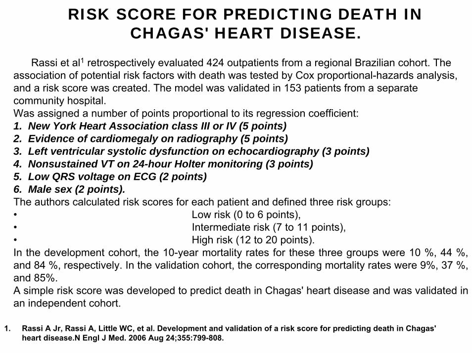
Rassi et al1 retrospectively evaluated 424 outpatients from a regional Brazilian cohort. The association of potential risk factors with death was tested by Cox proportional-hazards analysis, and a risk score was created. The model was validated in 153 patients from a separate community hospital.Was assigned a number of points proportional to its regression coefficient: 1. New York Heart Association class III or IV (5 points)2. Evidence of cardiomegaly on radiography (5 points)3. Left ventricular systolic dysfunction on echocardiography (3 points)4. Nonsustained VT on 24-hour Holter monitoring (3 points)5. Low QRS voltage on ECG (2 points)6. Male sex (2 points).The authors calculated risk scores for each patient and defined three risk groups: • Low risk (0 to 6 points), • Intermediate risk (7 to 11 points), • High risk (12 to 20 points). In the development cohort, the 10-year mortality rates for these three groups were 10 %, 44 %, and 84 %, respectively. In the validation cohort, the corresponding mortality rates were 9%, 37 %, and 85%. A simple risk score was developed to predict death in Chagas' heart disease and was validated in an independent cohort.
1. Rassi A Jr, Rassi A, Little WC, et al. Development and validation of a risk score for predicting death in Chagas' heart disease.N Engl J Med. 2006 Aug 24;355:799-808.

Epicardial Ventricular TachycardiaEpicardial VT is defined as VT in which the critical sites of the reentrant circuit (or the ‘sites of origin’) are located exclusively in the subepicardial tissue, as shown by entrainment maneuvers or VT that is terminated within 10 s with standard radiofrequency (RF) pulses, or both. E. SOSA,M. SCANAVACCA et al. http://www.springerlink.com/content/w608142674154tp5/A possible epicardial site of origin may be the reason for unsuccessful endocardial application of radiofrequency catheter ablation (RFCA) to control recurrent VT. Epicardial mapping can be safely performed through a pericardial puncture in the electrophysiology laboratory. Epicardial mapping is performed through a pericardial puncture as an epidural introducer needle is advanced into the pericardial space under fluoroscopic guidance. Medium contrast is injected to demonstrate the position of the needle tip, and a guidewire is introduced until its tip lay within the pericardial space. A 8-French Hemaquet is advanced and 4-mm deflectable tip catheter introduced into the pericardial sac to map the right and left ventricular epicardium. Transthoracic echocardiographic monitoring is performed on the day of the procedure and on the day of hospital discharge. With this electrophysiologic approach is possible detect the existence of an epicardial circuit1 and preformed the transthoracic epicardial RFCA. Transthoracic epicardial RFCA effectively controls recurrent VT in patients with Chagas' disease in whom epicardial circuits predominate. Epicardial circuits also occur in postinfarction VT2.
1. Sosa E, Scanavacca M, d'Avila A, Pilleggi F. A new technique to perform epicardial mapping in the electrophysiology laboratory. J Cardiovasc Electrophysiol. 1996 Jun;7:531-536.
2. Sosa E, Scanavacca M, d'Avila A, Oliveira F, Ramires JA. Nonsurgical transthoracic epicardial catheter ablation to treat recurrent ventricular tachycardia occurring late after myocardial infarction. J Am Coll Cardiol. 2000 May;35:1442-1449.

How to recognize epicardial origin of VT by surface ECG ?Suggest epicardial origin of the VTs.
1. Terminal S wave in V2 and q in lead 1 strongly suggest VT of sub epicardial origin.
2. Pseudo delta wave3. Intrinsicoid deflection time of 85 ms 4. RS complex duration of; 120ms
Important Linkshttp://www.circ.ahajournals.org/cgi/content/full/113/13/1659 http://circ.ahajournals.org/cgi/content/full/109/15/1842 ( Must read)http://cogprints.org/4222/2/tada.pdf
What is the clinical significance of epicardial VT ?Endo cardial ablation not likely to be successful. Trans pericardial approach may be needed.

Chronic Chagasic cardiomyopathy can alter the myocardial substrate in a way that facilitates the emergence of fatal VT in a way similar to the long-term consequences of MI. Post-MI and Chagasic VT share many similarities: they are both macroreentrant circuits, entrainable, involving any wall segment from the endocardium to the epicardium. However, as compared to patients with post-MI VT, Chagasic patients tend to be younger and have a higher LVEF( in this case the LVEF is very low). It is assumed, therefore, that their prognosis is closely related to VT treatment rather than the progression of the myocardial damage caused by the disease itself. Although SCD is a rare event in patients in NYHA functional class I and II treated with amiodarone, VT recurrence rate is 30% a year. Pharmacological approach is ineffective for patients with sever HF: 100% recurrence rate and 40% mortality in 1 year. Open-chest surgery is effective but requires very specialized centers and great expertise making its widespread use unrealistic. The results of combining RF endo/epicardial catheter ablation are still disappointing. Thus, research protocols on the search for new ablation technologies may greatly impact overall mortality in this subset of patients. An alternative approach is urgently needed. Experimental evidence of the efficacy of near infrared Lasers for catheter ablation will be reported along with investigations of the optical properties of the chagasic myocardium in the near infrared region to indicate that it might be not only feasible but also an appropriate choice to treat these patients1.
1. d'Avila A, Splinter R, Svenson RH, Scanavacca M, Pruitt E, Kasell J, Sosa E. New perspectives on catheter-based ablation of ventricular tachycardia complicating Chagas' disease: experimental evidence of the efficacy of near infrared lasers for catheter ablation of Chagas' VT. J Interv Card Electrophysiol. 2002 Aug;7:23-38.

Sosa et al1 reported the efficacy and safety of epicardial and endocardial approaches to treat 10 consecutive patients with VT and Chagas' disease. Epicardial mapping was carried out with a regular steerable catheter introduced into the pericardial space. An epicardial circuit was found in 14 of 18 mapable VTs induced in 10 patients. Epicardial mapping was used to guide endocardialablation in 4 patients and epicardial ablation in 6. The epicardial earliest activation site occurred 107+/-60 msec earlier than the onset of the QRS complex. At the epicardial site used to guide endocardial ablation, earliest activation occurred 75+/-55 msec before the QRS complex. Epicardial mid-diastolic potentials and/or continuous electrical activity were seen in 7 patients. After 4.8+/-2.9 seconds of epicardial RFCA, VT was rendered noninducible. Hemopericardium requiring drainage occurred in 1 patient; 3 others developed pericardial friction without hemopericardium. Experimentally D'Avila et al 2 shows that the effects of RFCA delivered adjacent to the LAD islimited to the media but when delivered above the artery, severe intimal hyperplasia and intravascular thrombosis may occur. Susceptibility to damage is inversely proportional to the vessel size. Sosa´s patients remain asymptomatic 5 to 9 months after the procedure. Interruption during endocardial pulses occurred after 20.2+/-14 seconds, but VT was always reinducible and the patients experienced a poor outcome. Epicardial mapping does not enhance the effectiveness of endocardial pulses of RFCA. The authors conclude that epicardial applications of RFCA can safely and effectively treat patients with VT and Chagas' disease.
1. Sosa E, Scanavacca M, D'Avila A, et al. Endocardial and epicardial ablation guided by nonsurgical transthoracicepicardial mapping to treat recurrent ventricular tachycardia. J Cardiovasc Electrophysiol. 1998 Mar;9:229-239.
2. D'Avila A, Gutierrez P, Scanavacca M, Reddy V, Lustgarten DL, Sosa E, Ramires JA. Effects of radiofrequency pulses delivered in the vicinity of the coronary arteries: implications for nonsurgical transthoracic epicardial catheter ablation to treat ventricular tachycardia. Pacing Clin Electrophysiol. 2002 Oct;25:1488-1495