Embed Size (px)
Citation preview

CONSIDERAÇÕES CIRÚRGICAS NO REPARO DA COARCTAÇÃO DE AORTA
COARCTAÇÃO DA AORTA >>> estenose congdistal à ASE, ou mesmo arco aórtico ou emsegmento de hipoplasia do arco como ocorre em
DESLOCAMENTO DISTAL DA ASEDESLOCAMENTO DISTAL DA ASE
DESCRIÇÃO INICIAL – MORGAGNI EM 1760 SEXTA LESÃO CARDIACA MAIS
TRATAMENTO CIRURGICO INICIAL
DEFEITOS ASSOCIADOS MAIS FREQUENTES:1. PCA2.
3. VALVA AÓRTICA BICÚSPIDE 464. LESÕES OBSTRUTIVAS DO LADO ESQUERDO
NCCK
ÇÕES CIRÚRGICAS NO REPARO DA COARCTAÇÃO DE AORTA
congênita da aorta, justaductal, imediatamenem aorta abdominal, ou ser parte de um long
em várias lesões obstrutivas esquerdas - SCE
1
MORGAGNI EM 1760 - PATOLOGIALESÃO CARDIACA MAIS COMUM – 4 a 6 % das CC
TRATAMENTO CIRURGICO INICIAL – CRAFOORD/Nylan 1944 GROSS EM 1945
PCACIV
VALVA AÓRTICA BICÚSPIDE 46-62 % LESÕES OBSTRUTIVAS DO LADO ESQUERDO – SHCE
MAI1

”Native coarctation of the aorta
The classic native C describes the discrete
aorta resulting from ridge-like thickening
protrudes into the lumen opposite theprotrudes into the lumen opposite the
(Figure 2). The origin of the subclavian
with post-stenotic dilatation of the aorta
hypoplasia is a less common form of native
and part of the transverse arch. ”
2
discrete narrowing of the descending
thickening of the media of the aortic wall that
the insertion of the ductus arteriosusthe insertion of the ductus arteriosus
artery can occasionally be involved
aorta commonly encountered. Tubular
native C which involves the isthmus

NCCK
3
MAI1

HAS - METADE SUPERIOR DO CORPO – REPOUSO OU EXERCAssintomáticos >>> HVE, FONTAN
AUMENTO DA PÓS-CARGA DE VE
DISTÚRBIOS DE FLUXO EM AORTA TORÁCICA
PERFUSÃO REDUZIDA NA ½ INFERIOR DO CORPO - PULSOS MMII REDUZIDOS OU ABOLIDOS
PULSO MSD (ABERRÂNCIA DA ASD) – REDUZIDO –
ACHADOS CLÍNICOS
NCCK
PULSO MSD (ABERRÂNCIA DA ASD) – REDUZIDO –
RETARDO NO PULSO FEMORAL EM RELAÇAO AO DAS EXTREMIDADES SUPERIORES E GRADIENTE PRESSÓRICO
CANAL PODE GERAR PULSO EM MMII – FALHA DE DIAGNÓSTICO NEONATAL
FECHAMENTO CANAL – COAO NEONATAL – CHOQUE
DISFUNÇÃO VENTRICULAR - ICC REFRATÁRIA
4EXERCÍCIO
>>> HVE, FONTAN
PULSOS MMII REDUZIDOS OU ABOLIDOS
– PODE INCLUIR ASE
MAI1
– PODE INCLUIR ASE
ÇAO AO DAS EXTREMIDADES SUPERIORES E GRADIENTE PRESSÓRICO
ÓSTICO NEONATAL
CHOQUE

� DAC PREMATURA
� ACIDENTE VASCULAR CEREBRAL
� ENDOCARDITE
� DISSECÇÃO AÓRTICA
� ICC
COMPLICAÇÕES - HN - HIPERTENSÃO DE LONGO TERMO
NCCK
� ICC
PÓS OPERATÓRIO TARDIO – CIRURGIA OU INTERVENÇÃO
� RECOARCTAÇÃO – 10 % PO CIRURGIA
� ANEURISMA
MORTALIDADE 75% ~ DOS 46 ANOS DE VIDASOBREVIDA MÉDIA ~35 ANOS
5LONGO TERMO
MAI1
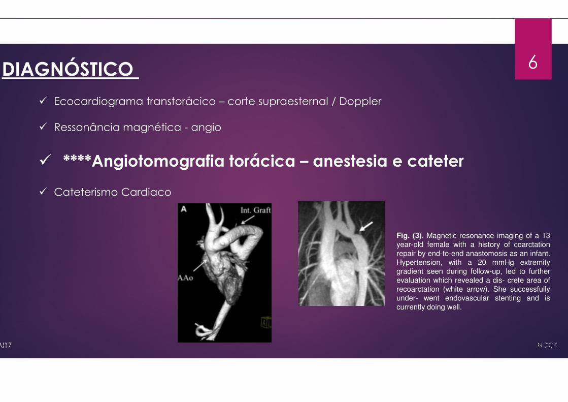
� Ecocardiograma transtorácico – corte supraesternal
� Ressonância magnética - angio
� ****Angiotomografia torácica
� Cateterismo Cardiaco
DIAGNÓSTICO 6
supraesternal / Doppler
torácica – anestesia e cateter
Fig. (3). Magnetic resonance imaging of a 13year-old female with a history of coarctationrepair by end-to-end anastomosis as an infant.Hypertension, with a 20 mmHg extremitygradient seen during follow-up, led to furtherevaluation which revealed a dis- crete area ofrecoarctation (white arrow). She successfullyunder- went endovascular stenting and iscurrently doing well.

INDICAÇÕES TERAPÊUTICAS
1. Neonato – risco de colapso hemodinâmico1. Grau de obstrução2. Evidência de multiplas lesões obstrutivas
2. Pacientes assintomáticos com HAS leve – com 1. ”Campbell’s natural history study” – aumentoanosanos
Gradiente pressão > / = 20 mmHg ➤➤➤➤ coarctação
3. Presença de colaterais – número e calibre
7
âmico com fechamento do canal
obstrutivas do lado esquerdo
com gradiente MMSS/MMII documentadoaumento mortalidade e sobrevida média 31
coarctação significativa➤➤➤➤ Intervenção
➤ ➤ ➤GRADIENTE!!!!

GUIDELINE 2008 – ACC/ AHA
Guidelines for adults with congenital heart disease
following settings3:
� Peak-to peak coarctation gradient >/= 20mg;
and beyond the narrowed segment.
� Peak-to-peak coarctation gradient < 20 mg with
radiologic evidence of significant collateral flow
indicator of severity when there is significant collateral
NCCK
ACC/ AHA
disease recommended intervention for coarctation in the
8
>/= 20mg; which is the difference in peak pressure proximal
with imaging evidence of significant coarctation and
flow. The resting gradient alone may be an unreliable
collateral circulation.
MAI1
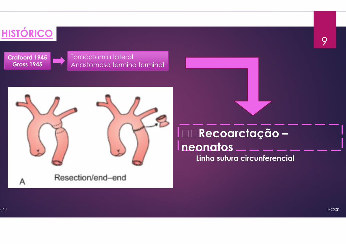
HISTÓRICO
Crafoord 1945Gross 1945
Toracotomia lateral Anastomose termino terminal
9
55Recoarctação –neonatos
Linha sutura circunferencial
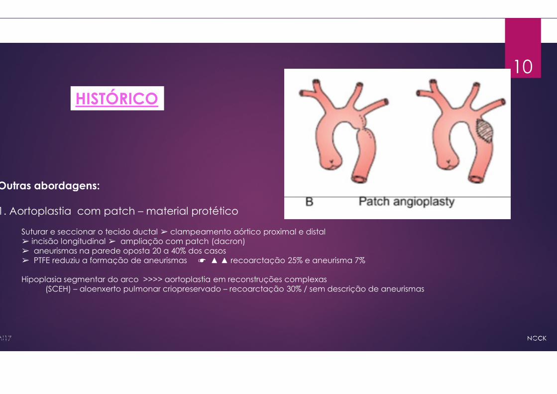
HISTÓRICO
Outras abordagens:
1. Aortoplastia com patch – material protético
Suturar e seccionar o tecido ductal ➢ clampeamento aórtico proximal e distal ➢ incisão longitudinal ➢ ampliação com patch (dacron) ➢ aneurismas na parede oposta 20 a 40% dos casos➢ PTFE reduziu a formação de aneurismas ☛ ▲▲ recoarctação
Hipoplasia segmentar do arco >>>> aortoplastia em reconstruções(SCEH) – aloenxerto pulmonar criopreservado – recoarctação
10
proximal e distal
recoarctação 25% e aneurisma 7%
reconstruções complexasrecoarctação 30% / sem descrição de aneurismas

2. Aortoplastia FLAP SUBCLÁVIA - apenas tecido autólogo
Toracotomia lateral > clampeamento > secção tecido ductallongitudinal do coto proximal da ASE transcoarctação > suturafor para ampliar o arcoMenos recoarctação – 23% / 0 a 3% - cças maioresAneurismaIsquemia braço – rara – perfusão colateralDiscrepância comprimento braço esquerdo e claudicação no exercício
Outras abordagens:
Fig. (4). Angiogram demonstratingFig. (4). Angiogram demonstrating
aneurysm development
black arrow) in a 13 year-
who previously underwent
flap aortoplasty for coarctation
aorta. Magnetic resonance
a 30 year-old patient in Panel
demonstrates aneurysm
(white arrow) after subclavian
aortoplasty. Note the absence
left subclavian artery in both
11autólogo (Walhausen -1966)
ductal > secção da ASE na emergência da vetebral > incisãosutura do flap ampliando aorta distal à subclávia E ou proximal se
exercício
demonstratingdemonstrating
(Panel A,
-old patient
subclavian
coarctation of the
resonance imaging of
Panel B also
formation
subclavian flap-
absence of the
both images.
Jeffrey E. Vergales. Current Cardiology Reviews, 2013, Vol. 9, No. 3

3. Anastomose Termino Terminal Extendida - apenas� Toracotomia lateral > clampeamento > envolver a CE com dissecção
aorta decendente >> ressecção da zona de caorctação com
bisel >> encaixe da aorta descendente sob o arco transverso
Outras abordagens:
� Toracotomia mediana >> dissecção de aorta ascendente, arco transverso
istmo aórtico, canal ou ligamento arterioso e aorta descendente
22-24 graus retal >> mobilizção de canula arterial para o TBC com
cerebral seletiva >> clampeamento de CE, SE e aorta descendentecerebral seletiva >> clampeamento de CE, SE e aorta descendente
cm além da coarctação >> ressecção do tecido ductal, área coarctada
em direção distal em Bisel. Incisão longitudinal do arco em direção
origem do TBC >> anastomose do arco aberto com aorta descendente
do arco transverso com PA fixado em formaldeido ou PB ou PTFE
Baixa mortalidade – mesmo em < 2 Kg
Clampeamento mais curto
Menos recoarctação – 4-13% em 5-10 anos
Anastomose termino lateral
12tecido autólogo (Amato -1977)dissecção ampla dos vasos supra-aórticos, arco transverso e
com todo tecido ductal possivel > sutura termino terminal em
- SOBREVIDA 98% EM 4,8 ANOS DE SEGMENTO
transverso, vasos supraórticos,
descendente > CEC com resfriamento até
com garroteamento – perfusão
descendente mobilizada pelo menos 3descendente mobilizada pelo menos 3
coarctada e ampliada incisão
direção proximal ultrapassando a
descendente e ampliação anterior

Outras abordagens: 4. Ressecção Coarctação – interposição enxerto
Pacientes nos quais crescimento não será problemaCoarctação longa ou segmentar
Suturar e seccionar o tecido ductal ➢ clampeamento aórtico➢ ressecção segmento doente➢ interposição tubo – homoenxerto
� Requer clampeamento mais longo❗❗❗❗❗❗- PARAPLEGIA� Crescimento� Reoperações
Fig. (6). This patient with a ventricularunderwent ventricular septal defect surgicalthe coarctation in a long-segment, however(black arrow). Recurrent obstructionNote pacemaker wires related to post-
� Preferida para adultos
13enxerto - Gross
aórtico proximal e distal homoenxerto aórtico ou Dacron ou tubo PB ou PTFE
PARAPLEGIA
ventricular septal defect and coarctation of the aorta previouslysurgical patch closure and coarctation repair. Recurrence of
however, necessitated placement of an interposi- tion graftis seen at the proxi- mal end of the graft (white arrow).-surgical heart block.
Jeffrey E. Vergales. Current Cardiology Reviews, 2013, Vol. 9, No. 3
adultos com longos segmentos de coarctação

NCCK
14
MAI1

NCCK
15
MAI1
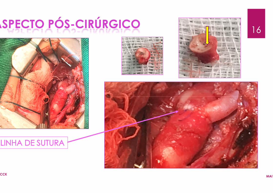
ASPECTO PÓS-CIRÚRGICO
NCCK
16CIRÚRGICO
MAI1

Intervenção percutânea
Crianças < 1 ano – solução temporária pré operatória em ptes
Recoarctação - melhor resultado e menor taxa complicações
Adultos e crianças maiores > 25 KG (INJÚRIA AORTA E FEMORAL)
Aneurismas – injúria à parede aórtica – 6% ATÉ MAIOR 9%Aneurismas – injúria à parede aórtica – 6% ATÉ MAIOR 9%
RNM – stents // TC
”Open ring” ou growth stents – overdilatation – requer F-up mais
Stents biodegradáveis – 3 a 6 meses
17
ptes alto risco (<4 MESES)
complicações - 5 A 12 SEMANAS
> 25 KG (INJÚRIA AORTA E FEMORAL)
6% ATÉ MAIOR 9%6% ATÉ MAIOR 9%
mais longo
� HAS crônica
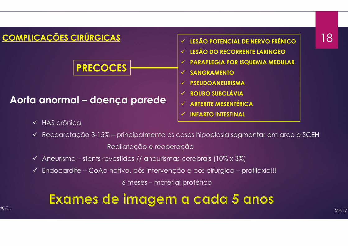
COMPLICAÇÕES CIRÚRGICAS
PRECOCES
Aorta anormal – doença parede
� HAS crônica
� Recoarctação 3-15% – principalmente os
Redilatação e reoperação
� Aneurisma – stents revestidos // aneurismas
� Endocardite – CoAo nativa, pós intervenção
6 meses – material
18� LESÃO POTENCIAL DE NERVO FRÊNICO
� LESÃO DO RECORRENTE LARINGEO
� PARAPLEGIA POR ISQUEMIA MEDULAR
� SANGRAMENTO
� PSEUDOANEURISMA
� ROUBO SUBCLÁVIA
� ARTERITE MESENTÉRICA
� INFARTO INTESTINAL
casos hipoplasia segmentar em arco e SCEH
reoperação
aneurismas cerebrais (10% x 3%)
intervenção e pós cirúrgico – profilaxia!!!
material protético

”MANAGEMENT APPROACH
Societal guidelines recommend correction
optimally early in childhood, to reduce
survival. The choice of intervention shouldsurvival. The choice of intervention should
team experienced in treating patients
dependent on the underlying morphology
absence of other cardiac lesions ”3,14,24.
19
correction of coarctation as early as possible,
reduce the long-term morbidity and improve
should be determined by a multidisciplinaryshould be determined by a multidisciplinary
with congenital heart disease and is
morphology, age of the patient, and the presence or
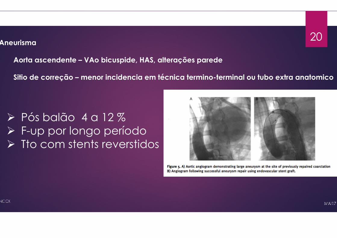
Aneurisma
Aorta ascendente – VAo bicuspide, HAS, altera
Sitio de correção – menor incidencia em técnica
� Pós balão 4 a 12 %� Pós balão 4 a 12 %� F-up por longo período� Tto com stents reverstidos
20
, HAS, alterações parede
técnica termino-terminal ou tubo extra anatomico

NCCK
21
MAI1

Jeffrey E. Vergales. Current Cardiology Reviews, 2013, Vol. 9, No. 3
Current management of coarctation of the aortaHussam Suradi1,3,*, Ziyad M. Hijazi1,2 uradi and Hijazi. Global Cardiology
Burch PT, Cowley CG, Holubkov R, Null D, Lambert LM, Kouretas PC, et al., young infants: is small size or low weight still a risk factor? The Journal2009;138(3):547–552, Epub 2009/08/25.
Coarctation of the Aorta Strategies for Improving OutcomesLan Nguyen, MDa, Stephen C. Cook, MDb,* Cardiol Clin -
Coarctation of the aorta: Management from infancy to adulthoodRachel D Torok, Michael J Campbell, Gregory A Fleming, Kevin
Coarctation of the Aorta can no Longer be Considered a Benign Condition Melissa G.Y. Lee, MBBS, Yves d’Udekem, MD, PhD* Heart, Lung and Circulation (2014) 23,
22
Cardiology Science and Practice 2015:44
PC, et al., Coarctation repair in neonates andJournal of thoracic and cardiovascular surgery.
Outcomes(2015)
adulthood, Michael J Campbell, Gregory A Fleming, Kevin D Hill
of the Aorta can no Longer be Considered a Benign Condition , MD, PhD* Heart, Lung and Circulation (2014) 23, 297–298