Embed Size (px)
Citation preview
Cysteine-rich intestinal protein 2 (CRIP2) acts as arepressor of NF-κB–mediated proangiogenic cytokinetranscription to suppress tumorigenesis and angiogenesisArthur Kwok Leung Cheunga, Josephine M. Y. Koa, Hong Lok Lunga, Kwok Wah Chanb, Eric J. Stanbridgec,Eugene Zabarovskyd,e, Takashi Tokinof, Lisa Kashimaf, Toshiharu Suzukig, Dora Lai-Wan Kwonga, Daniel Chuaa,Sai Wah Tsaoh, and Maria Li Lunga,1
aDepartment of Clinical Oncology and Center for Cancer Research and bDepartment of Pathology, University of Hong Kong, Hong Kong, People’s Republic ofChina; cDepartment of Microbiology and Molecular Genetics, University of California, Irvine, CA 92697; dDepartment of Microbiology, Tumor and Cell Biologyand Department of Clinical Science and Education, Karolinska Institute, 141 86 Stockholm, Sweden; eLaboratory of Structural and Functional Genomics,Engelhardt Institute of Molecular Biology, Russian Academy of Sciences, Moscow 119991, Russia; fDepartment of Molecular Biology, Cancer Research Institute,Sapporo Medical University, Sapporo 060-8556, Japan; gLaboratory of Neuroscience, Graduate School of Pharmaceutical Sciences, Hokkaido University,Sapporo 060-0812, Japan; and hDepartment of Anatomy, University of Hong Kong, Hong Kong, People’s Republic of China
Edited* by George Klein, Karolinska Institute, Stockholm, Sweden, and approved April 13, 2011 (received for review February 01, 2011)
Chromosome 14 was transferred into tumorigenic nasopharyngealcarcinoma and esophageal carcinoma cell lines by a microcell-mediated chromosome transfer approach. Functional complemen-tation of defects present in the cancer cells suppressed tumor for-mation. A candidate tumor-suppressor gene, cysteine-rich intestinalprotein 2 (CRIP2), located in the hot spot for chromosomal loss at14q32.3, was identified as an important candidate gene capable offunctionally suppressing tumor formation. Previous studies haveshown that CRIP2 is associated with development. To date, no re-port has provided functional evidence supporting a role for CRIP2 intumor development. The present study provides unequivocal evi-dence that CRIP2 can functionally suppress tumorigenesis. CRIP2 issignificantly down-regulated in nasopharyngeal carcinoma cell linesand tumors. CRIP2 reexpression functionally suppresses in vivo tu-morigenesis and angiogenesis; these effects are induced by its tran-scription-repressor capability. It interacts with the NF-κB/p65 toinhibit its DNA-binding ability to the promoter regions of the majorproangiogenesis cytokines critical for tumor progression, includingIL6, IL8, and VEGF. In conclusion, we provide compelling evidencethat CRIP2 acts as a transcription repressor of the NF-κB–mediatedproangiogenic cytokine expression and thus functionally inhibitstumor formation and angiogenesis.
transcription regulator | antiangiogenesis
Functional complementation of internal defects present in can-cer cells can be used to identify candidate tumor-suppressor
genes (TSGs) contributing to tumor development. The microcell-mediated chromosome transfer (MMCT) approach can be used totransfer a chromosome to a cancer cell. The resulting hybrid cellscontaining the exogenous transferred chromosome, known asmicrocell hybrids (MCHs), can be used to investigate the ability ofthat particular chromosome to induce tumor suppression andidentify TSGs (1).Using a panel of tumor-suppressive chromosome14 MCH cell lines established in a previous study (1), a novelcandidate TSG, cysteine-rich intestinal protein 2 (CRIP2), wasidentified and shown to induce tumor suppression in nasopharyn-geal carcinoma (NPC). CRIP2 is located in the chromosome14q32.3 region, which often shows high allelic loss in many cancers,including NPC (2, 3) and esophageal, renal, and colon carcinomas(4–7). In fact, because of its location in a hot spot for chromosometruncation in tumor development, it was reported to be a candidate forleukemic translocation (8). However, there have been no follow-upstudies to examine the functional role of this gene in tumor de-velopment. Our earlier chromosome 14 studies indicate that CRIP2may be a potential TSG and provide the impetus for further in-depthstudy of a new functional role for CRIP2 in NPC.
CRIP2 is a member of the LIM domain protein family (9).Members in this family encode different proteins, including tran-scription factors, adhesion molecules, and cytoskeleton proteins(10, 11). CRIP2 belongs to the cysteine-rich intestine protein familyl and specifically to the second class of LIM domain proteins, whichcontains between one and three LIM domains but usually lacksDNA-binding homeodomains (9). Interestingly, the mouse CRIP2protein was found to interact with a protein tyrosine phosphatase,PTP-BL, which is important in cancer development (12).CRIP2 is highly expressed in theheart aswell as in theovaries, brain,
skeletal muscle, spleen, prostate, small intestine, pancreas, testis, andneuronal ganglia (9, 13). CRIP2 expression has been detected in theheart endothelium during development and in the adult heart (14).CRIP2 also has been identified as a heart vascular marker (15).The present study shows that CRIP2 acts as a transcriptional
repressor of NF-κB. NF-κB is an important and well-studiedtranscription factor for various genes involved in the regulationof cancer development and angiogenesis. Loss of regulation of thisgene is commonly seen in various types of cancer. The dominantNF-κB complex is p50/p65. This complex is mainly controlled byIκB, which binds to NF-κB to inactivate its transcription function.Phosphorylation of IκB proteins by the upstream IκB kinaseresults in degradation of IκB protein. NF-κB is then translocatedto the nucleus and activates proangiogenesis and cell proliferationtarget gene transcription. Inactivation of NF-κB transcriptionfactor regulation is an important event contributing to tumorsuppression (16, 17).We have investigated the possible role of CRIP2 in regulating
angiogenesis in cancer and associated molecular pathways forCRIP2-inhibited angiogenesis. This report investigates thefunctional role of CRIP2 in cancer development and providescritical evidence of its crucial role in regulating angiogenesisduring tumor development.
ResultsIdentification of a Candidate TSG Using a Chromosome 14 MMCTApproach. Our earlier study of NPC chromosome 14 MCH celllines established by the MMCT approach demonstrated these
Author contributions: A.K.L.C., J.M.Y.K., H.L.L., and M.L.L. designed research; A.K.L.C. andK.W.C. performed research; A.K.L.C., E.J.S., E.Z., T.T., L.K., T.S., D.L.K., D.C., and S.W.T.contributed new reagents/analytic tools; A.K.L.C., J.M.Y.K., H.L.L., and M.L.L. analyzeddata; and A.K.L.C., J.M.Y.K., H.L.L., and M.L.L. wrote the paper.
The authors declare no conflicts of interest.
*This Direct Submission article had a prearranged editor.1To whom correspondence should be addressed. E-mail: [email protected].
This article contains supporting information online at www.pnas.org/lookup/suppl/doi:10.1073/pnas.1101747108/-/DCSupplemental.
8390–8395 | PNAS | May 17, 2011 | vol. 108 | no. 20 www.pnas.org/cgi/doi/10.1073/pnas.1101747108
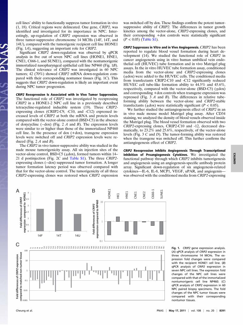
cell lines’ ability to functionally suppress tumor formation in vivo(1, 18). Critical regions were delineated. One gene, CRIP2, wasidentified and investigated for its importance in NPC. Inter-estingly, up-regulation of CRIP2 expression was observed inthree tumor-suppressive chromosome 14 MCHs (14F, 14T, and14U), compared with the tumorigenic recipient cell line HONE1(Fig. 1A), suggesting an important role for CRIP2.Significant CRIP2 down-regulation was observed by qPCR
analysis in five out of seven NPC cell lines (HONE1, HNE1,CNE1, C666-1, and SUNE1), compared with the nontumorigenicimmortalized nasopharyngeal epithelial cell line NP460 (Fig. 1B).The clinical relevance of CRIP2 was investigated in 60 NPCtumors; 42 (70%) showed CRIP2 mRNA down-regulation com-pared with their corresponding nontumor tissues (Fig. 1C). Thissuggests that CRIP2 down-regulation is likely an important eventduring NPC tumor progression.
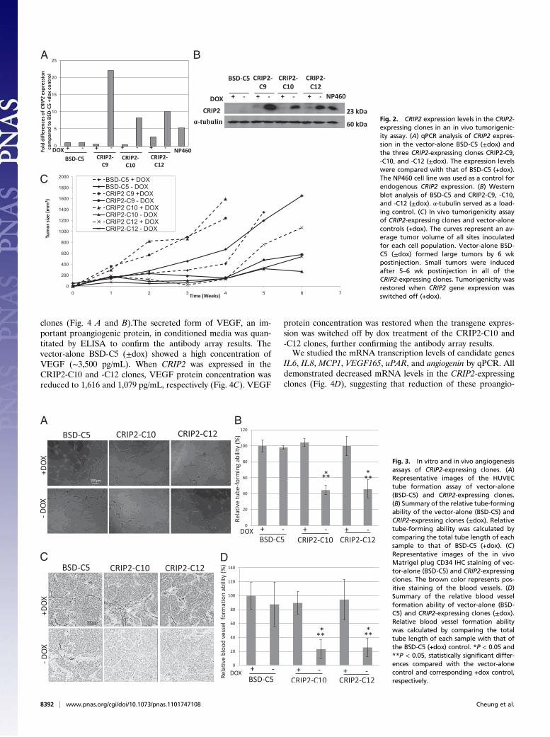
CRIP2 Reexpression Is Associated with in Vivo Tumor Suppression.The functional role of CRIP2 was investigated by reexpressingCRIP2 in a HONE1-2 NPC cell line in a previously describedtetracycline-regulated inducible system (19). Three CRIP2-expressing clones (CRIP2-C9, -C10, and -C12) expressed in-creased levels of CRIP2 at both the mRNA and protein levelscompared with the vector-alone control (BSD-C5) in the absenceof doxycycline (−dox) (Fig. 2 A and B). The expression levelswere similar to or higher than those of the immortalized NP460cell line. In the presence of dox (+dox), transgene expressionlevels were switched off and CRIP2 expression levels were re-duced (Fig. 2 A and B).The CRIP2 in vivo tumor-suppressive ability was studied in the
nude mouse tumorigenicity assay. All six injection sites of thevector-alone control, BSD-C5 (±dox), formed tumors within 14–21 d postinjection (Fig. 2C and Table S1). The three CRIP2-expressing clones (−dox) suppressed tumor formation. A longertumor formation latency period was observed compared withthat for the vector-alone control. The tumorigenicity of all threeCRIP2-expressing clones was restored when CRIP2 expression
was switched off by dox. These findings confirm the potent tumor-suppressive ability of CRIP2. The differences in tumor growthkinetics among the vector-alone, CRIP2-expressing clones, andtheir corresponding +dox controls were statistically significant(P < 0.05) (Table S1).
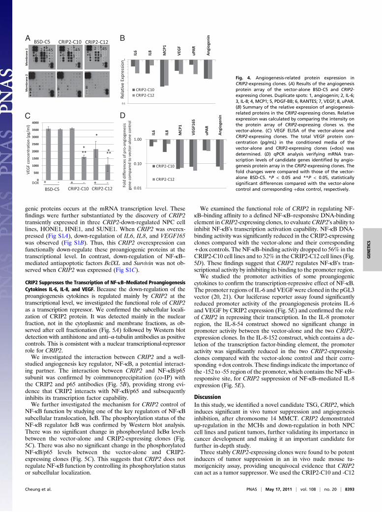
CRIP2 Suppresses in Vitro and in Vivo Angiogenesis. CRIP2 has beenreported to regulate blood vessel formation during heart de-velopment (14). We studied the functional role of CRIP2 incancer angiogenesis using in vitro human umbilical vein endo-thelial cell (HUVEC) tube formation and in vivo Matrigel plugassays. In the in vitro HUVEC tube formation assay, conditionedmedia from the vector-alone and CRIP2-expressing clones(±dox) were added to the HUVEC cells. The conditioned mediafrom transfectants CRIP2-C10 and -C12 significantly reducedHUVEC cell tube-like formation ability to 44.5% and 45.6%,respectively, compared with the vector-alone (BSD-C5) (±dox)and corresponding +dox controls when transgene expression wasrepressed (Fig. 3 A and B). The differences in relative tube-forming ability between the vector-alone and CRIP2-stabletransfectants (±dox) were statistically significant (P < 0.05).We further studied the antiangiogenesis effect of CRIP2 in an
in vivo nude mouse model Matrigel plug assay. After CD34staining, we analyzed the density of blood vessels observed insidethe Matrigel plug. The blood vessel formation observed with twoCRIP2-expressing clones, CRIP2-C10 and -12, decreased dra-matically, to 23.2% and 25.6%, respectively, of the vector-alonelevels (Fig. 3 C and D). The tumor-forming ability was restoredwhen the transgene was switched off. This further confirms theantiangiogenesis effect of CRIP2.
CRIP2 Reexpression Inhibits Angiogenesis Through TranscriptionalInhibition of Proangiogenesis Cytokines. We investigated thefunctional pathway through which CRIP2 inhibits tumorigenesisand angiogenesis using an angiogenesis-specific antibody proteinarray. Significant down-regulation of six angiogenesis-relatedcytokines—IL-6, IL-8, MCP1, VEGF, uPAR, and angiogenin—was observed with the conditioned media from CRIP2-expressing
A B
CFig. 1. CRIP2 gene expression analysis.(A) qPCR analysis of CRIP2 expression inthree chromosome 14 MCHs. The ex-pression fold changes were comparedwith the recipient HONE1 cell line. (B)qPCR analysis of CRIP2 expression inseven NPC cell lines. The expression foldchanges of the NPC cell lines werecompared with that of the immortalizednontumorigenic cell line NP460. (C)qPCR analysis of CRIP2 expression in 60NPC paired biopsy specimens. The foldchanges of the NPC tumor tissues werecompared with their correspondingnontumor tissues.
Cheung et al. PNAS | May 17, 2011 | vol. 108 | no. 20 | 8391
GEN
ETICS
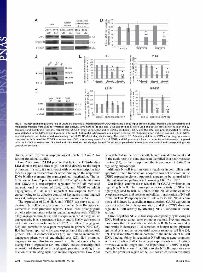
clones (Fig. 4 A and B).The secreted form of VEGF, an im-portant proangiogenic protein, in conditioned media was quan-titated by ELISA to confirm the antibody array results. Thevector-alone BSD-C5 (±dox) showed a high concentration ofVEGF (∼3,500 pg/mL). When CRIP2 was expressed in theCRIP2-C10 and -C12 clones, VEGF protein concentration wasreduced to 1,616 and 1,079 pg/mL, respectively (Fig. 4C). VEGF
protein concentration was restored when the transgene expres-sion was switched off by dox treatment of the CRIP2-C10 and-C12 clones, further confirming the antibody array results.We studied the mRNA transcription levels of candidate genes
IL6, IL8, MCP1, VEGF165, uPAR, and angiogenin by qPCR. Alldemonstrated decreased mRNA levels in the CRIP2-expressingclones (Fig. 4D), suggesting that reduction of these proangio-
A
C D
B
Fig. 3. In vitro and in vivo angiogenesisassays of CRIP2-expressing clones. (A)Representative images of the HUVECtube formation assay of vector-alone(BSD-C5) and CRIP2-expressing clones.(B) Summary of the relative tube-formingability of the vector-alone (BSD-C5) andCRIP2-expressing clones (±dox). Relativetube-forming ability was calculated bycomparing the total tube length of eachsample to that of BSD-C5 (+dox). (C)Representative images of the in vivoMatrigel plug CD34 IHC staining of vec-tor-alone (BSD-C5) and CRIP2-expressingclones. The brown color represents pos-itive staining of the blood vessels. (D)Summary of the relative blood vesselformation ability of vector-alone (BSD-C5) and CRIP2-expressing clones (±dox).Relative blood vessel formation abilitywas calculated by comparing the totaltube length of each sample with that ofthe BSD-C5 (+dox) control. *P < 0.05 and**P < 0.05, statistically significant differ-ences compared with the vector-alonecontrol and corresponding +dox control,respectively.
A B
C
Fig. 2. CRIP2 expression levels in the CRIP2-expressing clones in an in vivo tumorigenic-ity assay. (A) qPCR analysis of CRIP2 expres-sion in the vector-alone BSD-C5 (±dox) andthe three CRIP2-expressing clones CRIP2-C9,-C10, and -C12 (±dox). The expression levelswere compared with that of BSD-C5 (+dox).The NP460 cell line was used as a control forendogenous CRIP2 expression. (B) Westernblot analysis of BSD-C5 and CRIP2-C9, -C10,and -C12 (±dox). α-tubulin served as a load-ing control. (C) In vivo tumorigenicity assayof CRIP2-expressing clones and vector-alonecontrols (+dox). The curves represent an av-erage tumor volume of all sites inoculatedfor each cell population. Vector-alone BSD-C5 (±dox) formed large tumors by 6 wkpostinjection. Small tumors were inducedafter 5–6 wk postinjection in all of theCRIP2-expressing clones. Tumorigenicity wasrestored when CRIP2 gene expression wasswitched off (+dox).
8392 | www.pnas.org/cgi/doi/10.1073/pnas.1101747108 Cheung et al.
genic proteins occurs at the mRNA transcription level. Thesefindings were further substantiated by the discovery of CRIP2transiently expressed in three CRIP2-down-regulated NPC celllines, HONE1, HNE1, and SUNE1. When CRIP2 was overex-pressed (Fig S1A), down-regulation of IL6, IL8, and VEGF165was observed (Fig S1B). Thus, this CRIP2 overexpression canfunctionally down-regulate these proangiogenic proteins at thetranscriptional level. In contrast, down-regulation of NF-κB–mediated antiapoptotic factors BclXL and Survivin was not ob-served when CRIP2 was expressed (Fig S1C).
CRIP2 Suppresses the Transcription of NF-κB–Mediated ProangiogenesisCytokines IL-6, IL-8, and VEGF. Because the down-regulation of theproangiogenesis cytokines is regulated mainly by CRIP2 at thetranscriptional level, we investigated the functional role of CRIP2as a transcription repressor. We confirmed the subcellular locali-zation of CRIP2 protein. It was detected mainly in the nuclearfraction, not in the cytoplasmic and membrane fractions, as ob-served after cell fractionation (Fig. 5A) followed by Western blotdetection with antihistone and anti–α-tubulin antibodies as positivecontrols. This is consistent with a nuclear transcriptional-repressorrole for CRIP2.We investigated the interaction between CRIP2 and a well-
studied angiogenesis key regulator, NF-κB, a potential interact-ing partner. The interaction between CRIP2 and NF-κB/p65subunit was confirmed by coimmunoprecipitation (co-IP) withthe CRIP2 and p65 antibodies (Fig. 5B), providing strong evi-dence that CRIP2 interacts with NF-κB/p65 and subsequentlyinhibits its transcription factor capability.We further investigated the mechanism for CRIP2 control of
NF-κB function by studying one of the key regulators of NF-κBsubcellular translocation, IκB. The phosphorylation status of theNF-κB regulator IκB was confirmed by Western blot analysis.There was no significant change in phosphorylated IκBα levelsbetween the vector-alone and CRIP2-expressing clones (Fig.5C). There was also no significant change in the phosphorylatedNF-κB/p65 levels between the vector-alone and CRIP2-expressing clones (Fig. 5C). This suggests that CRIP2 does notregulate NF-κB function by controlling its phosphorylation statusor subcellular localization.
We examined the functional role of CRIP2 in regulating NF-κB–binding affinity to a defined NF-κB–responsive DNA-bindingelement inCRIP2-expressing clones, to evaluateCRIP2’s ability toinhibit NF-κB’s transcription activation capability. NF-κB DNA-binding activity was significantly reduced in the CRIP2-expressingclones compared with the vector-alone and their corresponding+dox controls. The NF-κB–binding activity dropped to 56% in theCRIP2-C10 cell lines and to 32% in the CRIP2-C12 cell lines (Fig.5D). These findings suggest that CRIP2 regulates NF-κB’s tran-scriptional activity by inhibiting its binding to the promoter region.We studied the promoter activities of some proangiogenic
cytokines to confirm the transcription-repressive effect of NF-κB.The promoter regions of IL-6 and VEGFwere cloned in the pGL3vector (20, 21). Our luciferase reporter assay found significantlyreduced promoter activity of the proangiogenesis proteins IL-6and VEGF by CRIP2 expression (Fig. 5E) and confirmed the roleof CRIP2 in repressing their transcription. In the IL-8 promoterregion, the IL-8-54 construct showed no significant change inpromoter activity between the vector-alone and the two CRIP2-expression clones. In the IL-8-152 construct, which contains a de-letion of the transcription factor-binding element, the promoteractivity was significantly reduced in the two CRIP2-expressingclones compared with the vector-alone control and their corre-sponding+dox controls. These findings indicate the importance ofthe -152 to -55 region of the promoter, which contains the NF-κB–responsive site, for CRIP2 suppression of NF-κB–mediated IL-8expression (Fig. 5E).
DiscussionIn this study, we identified a novel candidate TSG, CRIP2, whichinduces significant in vivo tumor suppression and angiogenesisinhibition, after chromosome 14 MMCT. CRIP2 demonstratedup-regulation in the MCHs and down-regulation in both NPCcell lines and patient tumors, further validating its importance incancer development and making it an important candidate forfurther in-depth study.Three stably CRIP2-expressing clones were found to be potent
inducers of tumor suppression in an in vivo nude mouse tu-morigenicity assay, providing unequivocal evidence that CRIP2can act as a tumor suppressor. We used the CRIP2-C10 and -C12
A B
C D
Fig. 4. Angiogenesis-related protein expression inCRIP2-expressing clones. (A) Results of the angiogenesisprotein array of the vector-alone BSD-C5 and CRIP2-expressing clones. Duplicate spots: 1, angiogenin; 2, IL-6;3, IL-8; 4, MCP1; 5, PDGF-BB; 6, RANTES; 7, VEGF; 8, uPAR.(B) Summary of the relative expression of angiogenesis-related proteins in the CRIP2-expressing clones. Relativeexpression was calculated by comparing the intensity onthe protein array of CRIP2-expressing clones vs. thevector-alone. (C) VEGF ELISA of the vector-alone andCRIP2-expressing clones. The total VEGF protein con-centration (pg/mL) in the conditioned media of thevector-alone and CRIP2-expressing clones (+dox) wasdetermined. (D) qPCR analysis verifying mRNA tran-scription levels of candidate genes identified by angio-genesis protein array in the CRIP2-expressing clones. Thefold changes were compared with those of the vector-alone BSD-C5. *P < 0.05 and **P < 0.05, statisticallysignificant differences compared with the vector-alonecontrol and corresponding +dox control, respectively.
Cheung et al. PNAS | May 17, 2011 | vol. 108 | no. 20 | 8393
GEN
ETICS
clones, which express near-physiological levels of CRIP2, forfurther functional studies.CRIP2 is a group 2 LIM protein that lacks the DNA-binding
LIM domain (9) and thus might not bind directly to the targetpromoters. Instead, it can interact with other transcription fac-tors to suppress transcription or affect binding to the responsiveDNA-binding elements for transcriptional inactivation. The in-teraction of CRIP2 protein with the NF-κB/p65 subunit showsthat CRIP2 is a transcription regulator for NF-κB–mediatedtranscriptional activation of IL-6, IL-8, and VEGF to inhibitangiogenesis. NF-κB is an important transcription factor incancer owing to its effective control of key genes promoting cellsurvival, antiapoptosis, angiogenesis, and invasion (16).The expression of IL-6, IL-8, and VEGF can serve as an in-
dicator of NF-κB activity, because they contain NF-κB–responsiveelements in their promoter regions. These three proangiogenicproteins play important roles in regulating angiogenesis. VEGF isa key angiogenic stimulator, and its expression can directly induceangiogenesis. It is a prognostic factor and is highly expressed inmetastatic cancers, including NPC (22, 23). IL-8 is proangiogenic(24) and contributes to a poor prognosis in primary NPC (25).IL-8 has been reported to increase expression of the antiapoptoticprotein Bcl-2 in endothelial cells (26) and thus to maintain theangiogenic phenotype of endothelial cells (27). IL-6 promotesangiogenesis and also tumor growth in different cancers by in-ducing VEGF expression (28–30). CRIP2 induces transcriptionalrepression of these three proangiogenic proteins, resulting in re-duction of stimulating signals to induce angiogenesis. CRIP2 has
been detected in the heart endothelium during development andin the adult heart (14) and has been identified as a heart vascularmarker (15), further supporting the importance of CRIP2 inregulating angiogenesis.Although NF-κB is an important regulator in controlling anti-
apoptotic protein transcription, apoptosis was not observed in theCRIP2-expressing clones. Apoptosis appears to be controlled bydifferent signaling pathways not involving CRIP2 in NPC.Our findings confirm the mechanism for CRIP2 involvement in
regulating NF-κB. The transcription factor activity of NF-κB istightly regulated by IκB. IκB binds to the NF-κB complex in thecytoplasmic region and prevents subcellular translocation of NF-κBto the nucleus. Phosphorylation of I-κB releases the NF-κB com-plex and induces its subcellular translocation. CRIP2 expressiondoes not affect I-κB phosphorylation, and thus CRIP2 does notregulate NF-κB activity by affecting NF-κB subcellular locali-zation.CRIP2 regulates NF-κB’s transcription capability by blocking its
DNA binding to target gene promoter regions. Previous studieshave shown that 17 β-estradiol inhibits NF-κBDNA-binding abilityand results in decreased IL-6 secretion in human retinal pigmentepithelial cells and an endometrial adenocarcinoma cell line (31,32). This demonstrates the importance of the inhibition of NF-κBDNA-binding ability in regulating NF-κB target gene promoteractivities to critically affect target gene expression levels. This studyprovides valuable insight into the importance of CRIP2 in regu-lating NF-κB function. In addition to the NF-κB–responsive ele-ment, the promoter region of the IL-8 construct used in this study
A
B
C
D
E
Fig. 5. Transcriptional regulatory role of CRIP2. (A) Subcellular fractionation of CRIP2-expressing clones. Input protein, nuclear fraction, and cytoplasmic andmembrane fraction were used for Western blot analysis. Anti–histone-1H and anti–α-tubulin antibodies were used as positive controls for nuclear and cy-toplasmic and membrane fractions, respectively. (B) Co-IP assay using CRIP2 and NF-κB/p65 antibodies. CRIP2 and the total and phosphorylated NF-κB/p65were detected in the CRIP2-expressing clones after co-IP. Anti-rabbit IgG was used as a negative control. (C) Phosphorylation status of p65 and IκBα in CRIP2-expressing clones. α-tubulin served as a loading control. (D) NF-κB–binding ability assay. The relative NF-κB–binding abilities of CRIP2-expressing clones werecompared with those of the BSD-C5 (+dox) control. (E) Promoter assay results for IL-6, VEGF, and IL-8 promoters. Relative promoter activities were comparedwith the BSD-C5 (+dox) control. *P < 0.05 and **P < 0.05, statistically significant differences compared with the vector-alone control and corresponding +doxcontrol, respectively.
8394 | www.pnas.org/cgi/doi/10.1073/pnas.1101747108 Cheung et al.
also contains the AP1- and NF-IL-6–binding elements (33); thissuggests that CRIP2, like NF-κB, may have the ability to regulateAP1 and NF-IL-6 transcriptional activities. Taken together, ourfindings indicate that NF-κB is an important CRIP2 target re-sponsible for its function.In conclusion, we have provided irrefutable evidence that
CRIP2 plays an essential role in suppressing tumor developmentthrough inhibiting angiogenesis. This interesting gene has hith-erto been associated only with developmental processes. Wehave shown that CRIP2 plays a key role as a transcriptional re-pressor of NF-κB–mediated transcription, and that its sub-sequent functional ramifications in cell regulation mediate itscritical role in cancer development.
Materials and MethodsSee SI Materials and Methods for more detailed information. Table S2contains qPCR primer sequences.
Gene Transfection. The CRIP2 cDNA clone was purchased from the Mam-malian Gene Collection (Invitrogen). The CRIP2 gene ORF was cloned intothe pETE-BSD (19) vector to establish stable transfectants. Transfection into
HONE1-2 was performed with Lipofectamine 2000 Reagent (Invitrogen), asdescribed previously (34).
Luciferase Reporter Assay. The promoter region of the proangiogenesiscytokines IL-6 and VEGF was cloned into pGL3-basic vector as describedpreviously (20, 21), The IL-8 promoter plasmids pGL-IL-8-54 and -152 weredescribed previously (33). Luciferase activity was measured 48 h aftertransfection with the Promega Dual-Luciferase Reporter assay systemaccording to the manufacturer’s instructions. The relative promoter ex-pression was calculated after normalization with Renilla luciferase activity toeliminate the effect of differences in transfection efficiency.
NF-κB/p65 Transcription Factor Assay. The NF-κB/p65 transcription factor assaywas performed with the Millipore Universal EZ-TFA Transcription FactorAssay Chemiluminescent Kit according to the manufacturer’s instructionsand as described previously (35).
ACKNOWLEDGMENTS. This work was supported by the Research GrantsCouncil and the University Grants Council of the Hong Kong SpecialAdministrative Region, People’s Republic of China (Grants 6615/07M andAoE/M-06/08, to M.L.L.); the University of Hong Kong Small Project Fund(Grant 200907176081, to A.K.L.C.); and the Swedish Cancer Society, SwedishResearch Council, Swedish Institute, Royal Swedish Academy of Sciences, andKarolinska Institute (E.Z.).
1. Cheung AK, et al. (2009) Chromosome 14 transfer and functional studies identifya candidate tumor-suppressor gene, Mirror image polydactyly 1, in nasopharyngealcarcinoma. Proc Natl Acad Sci USA 106:14478–14483.
2. Hui AB, et al. (1999) Detection of recurrent chromosomal gains and losses in primarynasopharyngeal carcinoma by comparative genomic hybridisation. Int J Cancer 82:498–503.
3. Lo KW, et al. (2000) High-resolution allelotype of microdissected primary nasopharyngealcarcinoma. Cancer Res 60:3348–3353.
4. Hoshi M, et al. (2000) Detailed deletion mapping of chromosome band 14q32 inhuman neuroblastoma defines a 1.1-Mb region of common allelic loss. Br J Cancer 82:1801–1807.
5. Ohta M, et al. (2002) Monocyte chemoattractant protein-1 expression correlates withmacrophage infiltration and tumor vascularity in human esophageal squamous cellcarcinomas. Int J Cancer 102:220–224.
6. Yoshimoto T, et al. (2007) High-resolution analysis of DNA copy number alterationsand gene expression in renal clear cell carcinoma. J Pathol 213:392–401.
7. Mourra N, et al. (2007) High frequency of chromosome 14 deletion in early-onsetcolon cancer. Dis Colon Rectum 50:1881–1886.
8. Tsui SK, et al. (1996) A novel cDNA encoding for a LIM domain protein located athuman chromosome 14q32 as a candidate for leukemic translocation. Biochem MolBiol Int 39:747–754.
9. Karim MA, et al. (1996) Human ESP1/CRP2, a member of the LIM domain proteinfamily: Characterization of the cDNA and assignment of the gene locus to chromosome14q32.3. Genomics 31:167–176.
10. Sadler I, Crawford AW, Michelsen JW, Beckerle MC (1992) Zyxin and cCRP: Twointeractive LIM domain proteins associated with the cytoskeleton. J Cell Biol 119:1573–1587.
11. Wang X, Lee G, Liebhaber SA, Cooke NE (1992) Human cysteine-rich protein: Amember of the LIM/double-finger family displaying coordinate serum induction withc-myc. J Biol Chem 267:9176–9184.
12. van Ham M, et al. (2003) Cloning and characterization of mCRIP2, a mouse LIM-onlyprotein that interacts with PDZ domain IV of PTP-BL. Genes Cells 8:631–644.
13. Bourane S, et al. (2007) A SAGE-based screen for genes expressed in sub-populationsof neurons in the mouse dorsal root ganglion. BMC Neurosci 8:97.
14. Yu TS, Moctezuma-Anaya M, Kubo A, Keller G, Robertson S (2002) The heart LIMprotein gene (Hlp), expressed in the developing and adult heart, defines a new tissue-specific LIM-only protein family. Mech Dev 116:187–192.
15. Zhang L, Hoffman JA, Ruoslahti E (2005) Molecular profiling of heart endothelialcells. Circulation 112:1601–1611.
16. Karin M (2006) Nuclear factor-κB in cancer development and progression. Nature 441:431–436.
17. Karin M (2006) NF-κB and cancer: Mechanisms and targets. Mol Carcinog 45:355–361.18. Ko JM, et al. (2005) Functional evidence of decreased tumorigenicity associated with
monochromosome transfer of chromosome 14 in esophageal cancer and the mappingof tumor-suppressive regions to 14q32. Genes Chromosomes Cancer 43:284–293.
19. Protopopov AI, et al. (2002) Human cell lines engineered for tetracycline-regulatedexpression of tumor-suppressor candidate genes from a frequently affected chro-mosomal region, 3p21. J Gene Med 4:397–406.
20. Watters KM, Dean J, Gautier V, Hall WW, Sheehy N (2010) Tax 1-independentinduction of vascular endothelial growth factor in adult T-cell leukemia caused byhuman T-cell leukemia virus type 1. J Virol 84:5222–5228.
21. Smith AJ, et al. (2008) Association of serum interleukin-6 concentration witha functional IL-6 -6331T>C polymorphism. Clin Chem 54:841–850.
22. Krishna SM, James S, Balaram P (2006) Expression of VEGF as prognosticator inprimary nasopharyngeal cancer and its relation to EBV status. Virus Res 115:85–90.
23. Guang-Wu H, et al. (2000) The relationship between microvessel density, theexpression of vascular endothelial growth factor (VEGF), and the extension ofnasopharyngeal carcinoma. Laryngoscope 110:2066–2069.
24. Harada K, et al. (2009) Cepharanthine inhibits angiogenesis and tumorigenicity ofhuman oral squamous cell carcinoma cells by suppressing expression of vascularendothelial growth factor and interleukin-8. Int J Oncol 35:1025–1035.
25. Xie LQ, Bian LJ, Li Z, Li Y, Liang YJ (2010) Co-elevated expression of hepatocytegrowth factor and interleukin-8 contributes to poor prognosis of patients withprimary nasopharyngeal carcinoma. Oncol Rep 23:141–150.
26. Nör JE, et al. (2001) Up-regulation of Bcl-2 in microvascular endothelial cells enhancesintratumoral angiogenesis and accelerates tumor growth. Cancer Res 61:2183–2188.
27. Strieter RM, et al. (2006) Cancer CXC chemokine networks and tumour angiogenesis.Eur J Cancer 42:768–778.
28. Adachi Y, et al. (2006) Interleukin-6 induces both cell growth and VEGF production inmalignant mesotheliomas. Int J Cancer 119:1303–1311.
29. Cohen T, Nahari D, Cerem LW, Neufeld G, Levi BZ (1996) Interleukin 6 induces theexpression of vascular endothelial growth factor. J Biol Chem 271:736–741.
30. Loeffler S, Fayard B, Weis J, Weissenberger J (2005) Interleukin-6 induces trans-criptional activation of vascular endothelial growth factor (VEGF) in astrocytes in vivoand regulates VEGF promoter activity in glioblastoma cells via direct interactionbetween STAT3 and Sp1. Int J Cancer 115:202–213.
31. Paimela T, et al. (2007) The effect of 17beta-estradiol on IL-6 secretion and NF-κBDNA-binding activity in human retinal pigment epithelial cells. Immunol Lett 110:139–144.
32. Ray P, Ghosh SK, Zhang DH, Ray A (1997) Repression of interleukin-6 gene expressionby 17 beta-estradiol: Inhibition of the DNA-binding activity of the transcriptionfactors NF-IL-6 and NF-κB by the estrogen receptor. FEBS Lett 409:79–85.
33. Kashima L, et al. (2009) CHFR, a potential tumor suppressor, down-regulatesinterleukin-8 through the inhibition of NF-κB. Oncogene 28:2643–2653.
34. Cheung AK, et al. (2008) Functional analysis of a cell cycle-associated tumor-suppressive gene, protein tyrosine phosphatase receptor type G, in nasopharyngealcarcinoma. Cancer Res 68:8137–8145.
35. Yamini B, et al. (2007) Inhibition of nuclear factor-κB activity by temozolomideinvolves O6-methylguanine–induced inhibition of p65 DNA binding. Cancer Res 67:6889–6898.
Cheung et al. PNAS | May 17, 2011 | vol. 108 | no. 20 | 8395
GEN
ETICS


![Chromosome (mis)segregation is biased by kinetochore sizephosphorylated Knl1, a bona fide Aurora B substrate at the kinetochores [34]. We were unable to detect any significant difference](https://img.document.onl/doc/110x75/60b48e10dd35e64fa700eb1e/chromosome-missegregation-is-biased-by-kinetochore-size-phosphorylated-knl1-a.jpg)