Embed Size (px)
Citation preview
UNIVERSIDADE FEDERAL DE UBERLÂNDIA
INSTITUTO DE BIOTECNOLOGIA
PROGRAMA DE PÓS-GRADUAÇÃO EM BIOTECNOLOGIA
FERNANDA CARDOSO DA SILVA
DISFUNÇÃO ENDOTELIAL PELA INIBIÇÃO DA SÍNTESE
DE ÓXIDO NÍTRICO: proposta e caracterização de um modelo
celular in vitro
Declaramos que os resultados aqui apresentados ainda estão em fase de desenvolvimento
e não foram publicados.
PATOS DE MINAS - MG
FEVEREIRO DE 2020
FERNANDA CARDOSO DA SILVA
DISFUNÇÃO ENDOTELIAL PELA INIBIÇÃO DA SÍNTESE
DE ÓXIDO NÍTRICO: proposta e caracterização de um modelo
celular in vitro
Dissertação de Mestrado apresentada ao
Programa de Pós-graduação em Biotecnologia
como requisito parcial para a obtenção do
Título de Mestre em Biotecnologia.
Orientadora: Profª. Drª. Cristina Ribas
Fürstenau
Declaramos que os resultados aqui apresentados ainda estão em fase de desenvolvimento
e não foram publicados.
PATOS DE MINAS - MG
FEVEREIRO DE 2020
Silva, Fernanda Cardoso da, 1996-S5862020 Disfunção endotelial pela inibição da síntese de óxido nítrico
[recurso eletrônico] : proposta e caracterização de um modelocelular in vitro / Fernanda Cardoso da Silva. - 2020.
Orientadora: Cristina Ribas Fürstenau .Coorientadora: Thaise Gonçalves de Araújo.Dissertação (Mestrado) - Universidade Federal de Uberlândia,
Pós-graduação em Biotecnologia.Modo de acesso: Internet.
CDU: 60
1. Biotecnologia. I. , Cristina Ribas Fürstenau,1979-, (Orient.).II. Araújo, Thaise Gonçalves de ,1984-, (Coorient.). III.Universidade Federal de Uberlândia. Pós-graduação emBiotecnologia. IV. Título.
Disponível em: http://doi.org/10.14393/ufu.di.2020.283Inclui bibliografia.Inclui ilustrações.
Ficha Catalográfica Online do Sistema de Bibliotecas da UFUcom dados informados pelo(a) próprio(a) autor(a).
Bibliotecários responsáveis pela estrutura de acordo com o AACR2:Gizele Cristine Nunes do Couto - CRB6/2091
Nelson Marcos Ferreira - CRB6/3074

“O sucesso nasce do querer, da determinação
e persistência em se chegar a um objetivo.
Mesmo não atingindo o alvo, quem busca e
vence obstáculos, no mínimo fará coisas
admiráveis." José de Alencar

Dedico esse trabalho aos meus pais,
Belchior e Celina, vocês são tudo para
mim! Amo vocês!
AGRADECIMENTOS
À Deus, nosso pai criador, pela oportunidade de crescimento intelectual, pessoal e
espiritual. Agradeço pelas oportunidades e desafios enfrentados ao longo desse período.
Ao meu pai, Belchior, que é meu exemplo de boa conduta, seriedade e perseverança.
Agradeço por todo esforço que fez para que eu tivesse a oportunidade de estudar e me dedicar
à um sonho.
À minha mãe Celina, o grande amor de minha vida, meu exemplo de força, fé,
esperança, coragem e caridade. Agradeço por sempre estar ao meu lado, me incentivando e me
consolando quando as coisas não dão certo. Devo tudo o que conquistei até hoje a senhora.
Espero um dia poder retribuir tudo o que fez e faz por mim e te dar o prazer de se orgulhar.
À Flávia, minha irmã e grande amiga, obrigada por estar ao meu lado, me incentivar,
me mostrar o caminho a seguir. Agradeço por sempre me ensinar coisas boas, por ser meu porto
seguro, por me ensinar a gostar de livros, músicas e filmes.
Ao meu irmão Fabrício, obrigada pelo carinho e pelo esforço em me auxiliar.
À minha tia Maria, minha segunda mãe, que nunca me deixa faltar nada e que sempre
está a cuidar de mim. Agradeço por sempre estar disposta a me ajudar.
Ao meu avô José Cardoso (in memoriam), que sempre me ensinou a persistir, jamais
desistir, me dedicar aos estudos e ao trabalho no bem. A saudade que sinto é gigantesca, mas a
gratidão é muito maior. O senhor é meu exemplo de homem de bem. Obrigada por ser meu
porto seguro por tanto tempo, por me oferecer um colo de avô e de amigo. Suas histórias e
ensinamentos estão guardados em minha memória e no meu coração para sempre.
À minha avó, Maria Natalícia que sempre esteve ao meu lado, me oferecendo carinho,
amor e apoio.
À todos os meus demais familiares, que sempre me incentivaram.
Aos meus amigos, que tornaram essa jornada mais suave. Em especial agradeço, ao
Arthur, meu melhor amigo e meu grande amor, obrigada por estar sempre ao meu lado, obrigada
pelo carinho e amor. À Isabella, minha companheira na vida e melhor amiga, agradeço por ser
uma grande confidente e conselheira. À Andreia e Rodolfo, que estiveram ao meu lado e muito
me auxiliaram.
À Janayne, Juliana, Thamara, que mesmo a distância sempre estiveram muito presentes
durante esse tempo. Amo vocês!

À Cristina, que aceitou a incumbência de me orientar e muito bem o fez. Agradeço
muitíssimo por todo esforço que aplicou para que tivéssemos bons resultados. Agradeço
também pelo carinho e paciência que teve comigo.
À professora Thaise que aceitou contribuir nessa pesquisa como coorientadora.
Agradeço pelo apoio, pelo carinho e pelo acolhimento.
À professora Joyce que muito contribuiu com esse trabalho e aceitou participar na banca
examinadora. À professora Ana Paula que aceitou participar da banca e contribuir para a
melhoria do trabalho.
À Bruna Juber, minha companheira de laboratório e amiga, que em diversos momentos
me sustentou e me auxiliou a continuar. Agradeço imensamente por todos os dias que se propôs
a estar comigo, a me auxiliar, por sempre se mostrar prestativa, mesmo nos finais de semana e
feriados. Não tenho palavras para mostrar minha gratidão.
Aos colegas de laboratório que direta ou indiretamente contribuíram para a realização
dessa pesquisa. Em especial, agradeço à Carina, Vinícius e Bruno que me muito me auxiliaram
na parte experimental.
Ao grupo de pesquisa do laboratório GBIO, que me acolheram e me auxiliaram nos
momentos mais difíceis.
Aos técnicos de laboratório, em especial à Luciana. Agradeço pelo auxílio e pelo
incentivo que sempre me deu.
Aos professores e alunos do Programa de Pós-graduação em Biotecnologia. Em especial
agradeço aos meus colegas, Matheus Ribeiro, Lourayne, Natália e Vanessa, que estiveram ao
meu lado e que muito me auxiliaram.
À CAPES e as demais instituições de fomento pelo auxílio financeiro ofertado durante
esses meses, sem ele não seria possível a realização de tal pesquisa.
À UFU, a instituição que me acolheu desde a graduação e que muito tem me ofertado
de aprendizados.
RESUMO
O endotélio é um tipo de tecido epitelial, formado por uma única camada de células, que recobre
internamente os vasos sanguíneos e é essencial para a manutenção da homeostase vascular,
garantida pela produção de biomoléculas vasoativas. Um desequilíbrio na biodisponibilidade
dessas biomoléculas pode desencadear um estado patológico, como a disfunção endotelial
(DE), que se caracteriza, principalmente, pela redução na disponibilidade do óxido nítrico (NO)
e por um aumento na produção de espécies reativas de oxigênio (EROs). Dada a importância
do entendimento da disfunção endotelial para o progresso de patologias como a aterosclerose,
por exemplo, faz-se necessário o desenvolvimento de modelos experimentais que simulem tal
condição. Assim, esse estudo foi realizado visando desenvolver e caracterizar um modelo
celular in vitro de DE pela inibição da síntese de NO. Inicialmente, realizou-se o tratamento das
células endoteliais derivadas do timo (linhagem tEnd.1) com o inibidor da síntese de NO (L-
NAME) nas concentrações 1µM, 10 µM, 100 µM e 1mM por 12, 24, 48, 72, 96 e 120 horas,
com e sem retratamento a cada 24 horas. Posteriormente, determinou-se as melhores condições
de tratamento para indução de um estado de DE nas células, a partir da avaliação da viabilidade
celular e da concentração de nitrito. Os tratamentos com 10 µM e 100 µM de L-NAME, nos
tempos de 72 horas sem retratamento e de 96 horas com retratamento foram selecionados. Em
seguida, realizou-se a qPCR para os genes p22phox, p47phox e eNOS. Os resultados foram
significativos para o p22phox, em que os níveis relativos de mRNA do gene foram reduzidos em
65% em resposta ao tratamento com L-NAME 100 µM por 96 horas com retratamento. A
análise da expressão proteica da eNOS por Western blotting não foi bem-sucedida. Por fim, os
resultados de avaliação de peroxidação lipídica pela técnica de Buege e Aust não foram
estatisticamente significativos. Em conjunto, os resultados apontam que o tratamento com L-
NAME 100 µM por 96 horas com retratamento foi capaz de induzir a DE, diminuindo a
disponibilidade de NO, a qual foi verificada tanto pela redução na concentração de nitrito
quanto pela redução na expressão de p22phox. Entretanto, estudos adicionais, sobretudo visando
aprofundar o papel das EROs nas células submetidas ao tratamento com L-NAME, são
necessários para o aprimoramento e caracterização do modelo experimental proposto.
Palavras-chave: Óxido nítrico. L-NAME. Estresse oxidativo. p22phox. Disfunção endotelial.
ABSTRACT
Vascular endothelium is a type of epithelial tissue, formed by a monolayer of cells, which
internally covers blood vessels and is essential for the maintenance of vascular homeostasis,
guaranteed by the production of vasoactive biomolecules. An imbalance in the bioavailability
of these biomolecules may trigger a pathological state, such as endothelial dysfunction (ED),
which is mainly characterized by a reduction in the availability of nitric oxide (NO) and an
increase in the production of reactive oxygen species (ROS). Considering the importance of
endothelial dysfunction for the progress of pathologies such as atherosclerosis, for example, it
is necessary to develop experimental models that simulate such a condition. This study was
then carried out to develop and characterize an in vitro cellular model of ED by inhibiting NO
synthesis. Initially, the thymus-derived endothelial cells (tEnd.1 line) were treated with the NO
synthesis inhibitor (L-NAME) at 1µM, 10 µM, 100 µM and 1mM for 12, 24, 48, 72 , 96 and
120 hours, with and without retreatment every 24 hours. Subsequently, the best treatment
conditions for inducing an ED state in the cells were determined based on the evaluation of cell
viability and nitrite concentration. Treatments with 10 µM and 100 µM of L-NAME for 72
hours without retreatment and 96 hours with retreatment were selected. Real-time PCR was
performed for p22phox, p47phox and eNOS genes. Results were significant for p22phox, in which
the relative levels of gene mRNA were reduced by 65% in response to 100 µM L-NAME for
96 hours with retreatment. Western blotting analysis of eNOS protein expression was not
successful. Finally, results of the evaluation of lipid peroxidation using the Buege and Aust
technique were not statistically significant. Together, the results show that treatment with 100
µM L-NAME for 96 hours with retreatment was able to induce ED, decreasing the availability
of NO, which was verified by the reduction in the concentration of nitrite and by the reduction
in the expression of p22phox. However, additional studies aiming to determine the role of ROS
in cells submitted to treatment with L-NAME are necessary for the improvement and
characterization of the proposed experimental model.
Keywords: Nitric oxide. L-NAME. Oxidative stress. p22phox. Endothelial dysfunction.
0LISTA DE ILUSTRAÇÕES
Figura 1- Representação das camadas que constituem a parede do vaso sanguíneo: túnica
adventícia, túnica média e túnica íntima.
Figura 2 – Representação das principais biomoléculas produzidas pelas células endoteliais e
suas funções
Figura 3 – Representação da vasodilatação induzida pelo óxido nítrico produzido pelas células
endoteliais.
Figura 4- Representação das duas fontes de espécies reativas de oxigênio (EROs) na parede
vascular.
Figura 5 - Produção de biomoléculas em um endotélio saudável e em um endotélio disfuncional.
Figura 6 - Morfologia da linhagem de células tEnd.1 em cultura.
LISTA DE ABREVIATURAS E SIGLAS
5-HT: 5-hidroxitriptamina
Ach: Acetilcolina
ADMA: inibidor de eNOS
AT-I: Angiotensina I
AT-II: Angiotensina II
B2: Receptor beta-2
BK: Bradicinina
cAMP: 3’5’-adenosina-monofosfato-cíclico
CAT: Catalase
CE: Células endoteliais
cGMP: Monofosfato cíclico de guanosina
CMLVs: Células musculares lisas vasculares
COX: Ciclooxigenase
DE: Disfunção endotelial
DILA: Dilatação fluxo-mediada
DPI: Difenilenoiodônio
eNOS: Óxido nítrico sintase endotelial
ECA: Enzima conversora de angiotensina
EDHF: Fator hiperpolarizante derivado do endotélio
EROs: Espécies reativas de oxigênio
ERON: espécie reativa de oxigênio e nitrogênio
ET: Endotelina
ET-1: Endotelina 1
ETB: Receptor de endotelina

FMN: Mononucleotídeo de flavina
GSH-Px: Glutationa peroxidase
GTP: Trifosfato de guanosina
HAS: Hipertensão arterial sistêmica
iNOS: Óxido nítrico sintase induzível
K+: Íon potássio
L-arg: L-arginina
L-cit: L-citrulina
L-NAME: Nω-Nitro-L-arginina metil éster
M: Receptor muscarínico
nNOS: Óxido nítrico sintase neuronal
NO: Óxido nítrico
NOS: Óxido nítrico sintase
NOX: NADPH oxidase
O2º-: Ânion superóxido
PGH2: Prostaglandina H2
PGI2: Prostaciclinas
SOD: Superóxido dismutases
tEnd.1: Linhagem de células endoteliais derivadas de timo
TP: Receptor para tromboxano-prostanóide
TXA2: Tromboxano A2
SUMÁRIO
CAPÍTULO 1 ........................................................................................................................... 13
1. INTRODUÇÃO ................................................................................................................ 13
1.1 Problema .................................................................................................................... 13
1.2 Hipóteses .................................................................................................................... 14
1.3 Objetivos .................................................................................................................... 14
1.3.1 Objetivo Geral ......................................................................................................... 14
1.3.2 Objetivos Específicos ......................................................................................... 14
1.4 Justificativa ................................................................................................................ 14
2. REFERENCIAL TEÓRICO ............................................................................................. 15
2.1. Funções do endotélio vascular ................................................................................... 15
2.2. Via do óxido nítrico derivado do endotélio ............................................................... 18
2.3. Espécies reativas de oxigênio (EROs): fontes e funções biológicas .......................... 20
2.4. Disfunção endotelial (DE) e patologias associadas ................................................... 23
2.5. Estratégias para o teste da função endotelial ............................................................. 26
2.6. Inibição da síntese de NO e indução de disfunção endotelial .................................... 27
CAPÍTULO 2 ........................................................................................................................... 29
ENDOTHELIAL DYSFUNCTION BY INHIBITING NITRIC OXIDE SYNTHESIS:
PROPOSAL AND CHARACTERIZATION OF AN IN VITRO CELLULAR MODEL ... 29
CONCLUSÃO .......................................................................................................................... 60
REFERÊNCIAS ....................................................................................................................... 61
ANEXO 1 ................................................................................................................................. 68
13
CAPÍTULO 1
1. INTRODUÇÃO
1.1 Problema
O endotélio vascular é constituído por uma monocamada de células que reveste
internamente os vasos sanguíneos e que atua como uma interface dinamicamente mutável
(KHADDAJ MALLAT et al., 2017; GIMBRONE JÚNIOR; GARCÍA-CARDEÑA, 2013),
sendo essencial para a manutenção da integridade e da homeostase vascular (DAIBER et al.,
2019). Nesse sentido, a perda do equilíbrio entre a produção e a liberação de moléculas
vasoconstritoras e vasodilatadoras definem um estado denominado disfunção endotelial (DE)
(GIMBRONE JÚNIOR; GARCÍA-CARDEÑA, 2013).
A DE é uma condição caracterizada, principalmente, por uma redução na
biodisponibilidade de óxido nítrico (NO), devido ao desacoplamento da NO sintase (NOS)
(INCALZA et al., 2018). Consequentemente, há o aumento da produção de espécies reativas
de oxigênio (EROs) e o desencadeamento de um processo inflamatório associado (DAIBER et
al., 2019; INCALZA et al., 2018; RUDIC; SESSA, 1999; VANHOUTTE et al., 2017). A DE
pode, então, causar um aumento da vasoconstrição, oxidação e alteração da permeabilidade da
membrana plasmática, acúmulo de células do sistema imune (GIMBRONE JÚNIOR;
GARCÍA-CARDEÑA, 2013; KONUKOGLU; UZUN, 2016), aumento da formação de
agregados plaquetários e proliferação das células musculares lisas vasculares (CMLVs)
(YUYUN; NG; NG, 2018).
A DE pode se apresentar como um fator de risco inicial para o desencadeamento de
doenças vasculares e metabólicas (DAIBER et al., 2017), sendo um marcador importante em
patologias como aterosclerose, hipercolesterolemia, hipertensão arterial sistêmica (HAS),
doença arterial periférica e diabetes (FALCONER et al., 2018; HONG et al., 2019). Essas
doenças são apontadas como a principal causa de morte no mundo, sendo que, em 2017, foram
estimadas 17,8 milhões de morte tendo como causa as doenças cardiovasculares (KAPTOGE
et al., 2019).
Portanto, considerando-se que a DE é um evento comum aos distúrbios metabólicos que
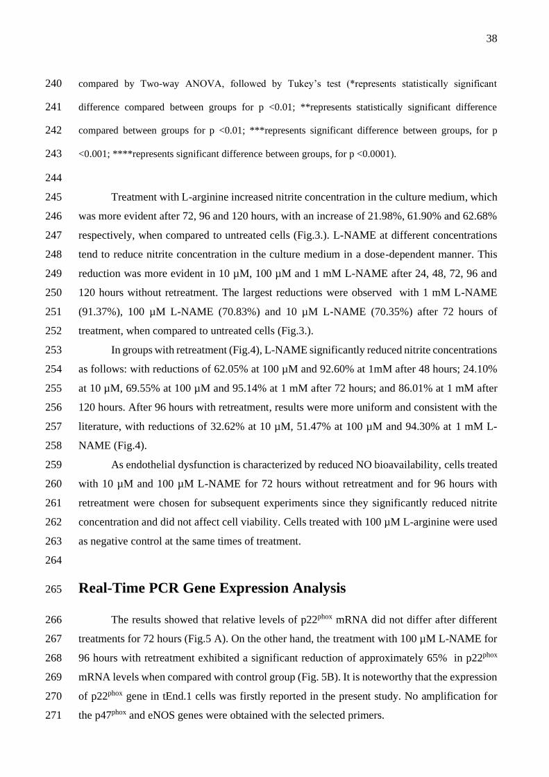
culminam no desenvolvimento e progresso das patologias que mais matam em todo o mundo,
14
a compreensão sobre os mecanismos celulares e moleculares envolvidos nessa condição é de
extrema importância para a condução dos tratamentos, bem como para a elaboração de novas
estratégias terapêuticas.
1.2 Hipóteses
• O tratamento de células endoteliais (CE) com Nω-Nitro-L-arginina metil éster (L-NAME)
induz um estado semelhante à DE em CE;
• O tratamento com L-NAME reduz a biodisponibilidade de NO e aumenta a produção de
EROs.
1.3 Objetivos
1.3.1 Objetivo Geral
Desenvolver e caracterizar um modelo celular in vitro de DE por meio da inibição da
síntese de NO.
1.3.2 Objetivos Específicos
• Padronizar as condições de cultivo in vitro das CE derivadas de timo (tEnd.1);
• A partir do tratamento com o inibidor da síntese de NO (L-NAME), determinar as
melhores condições (concentração e tempo de tratamento) para indução de um estado
semelhante à DE;
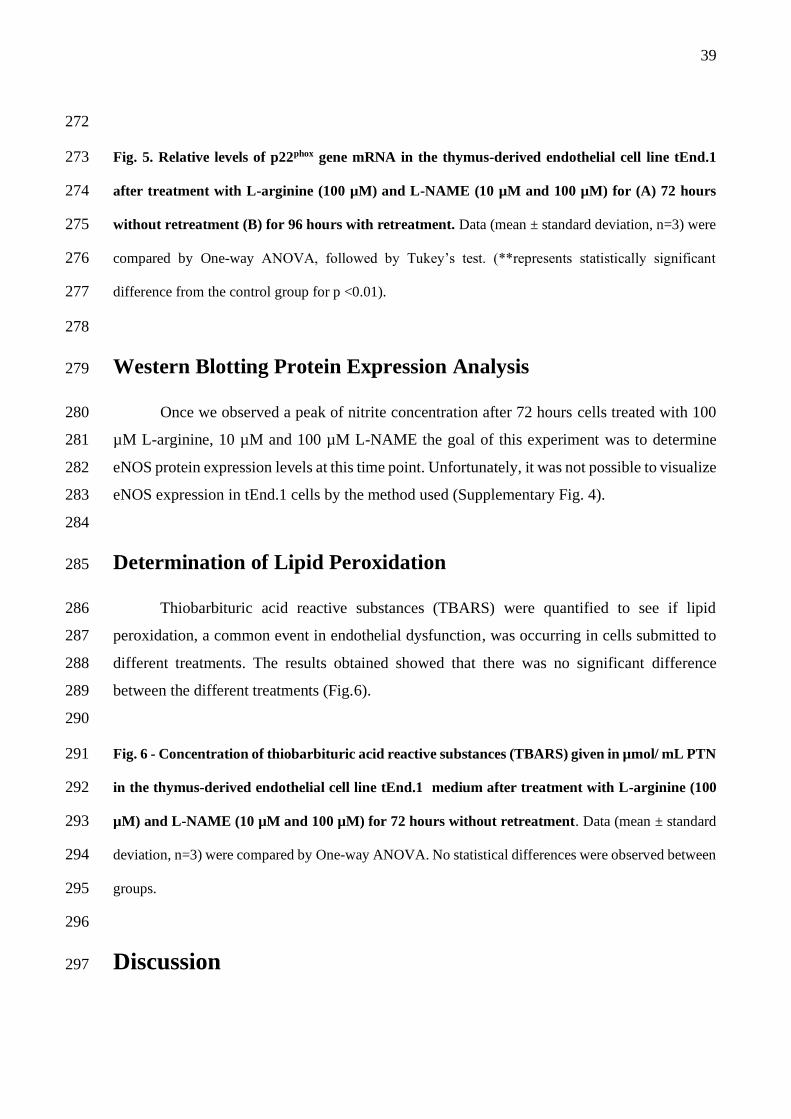
• Caracterizar o modelo de DE in vitro proposto observando as principais alterações na
síntese de NO, expressão de NOS e parâmetros de estresse oxidativo.
1.4 Justificativa
Conforme mencionado anteriormente, as doenças cardiovasculares são as doenças não
transmissíveis mais frequentes em todo o mundo e estão relacionadas com uma alta taxa de
morbidade e mortalidade (KAPTOGE et al., 2019). A DE é um evento comum que precede o
estabelecimento de patologias metabólicas e vasculares. Os mecanismos inerentes à DE ainda
15
não estão completamente elucidados e os modelos experimentais in vitro disponíveis para o
estudo dessa condição permanecem escassos. Portanto, o desenvolvimento de um modelo
celular de disfunção endotelial poderá permitir o avanço das pesquisas na área, bem como
contribuir para a redução de custos e demandas necessárias para a manutenção de modelos
animais.
2. REFERENCIAL TEÓRICO
2.1. Funções do endotélio vascular
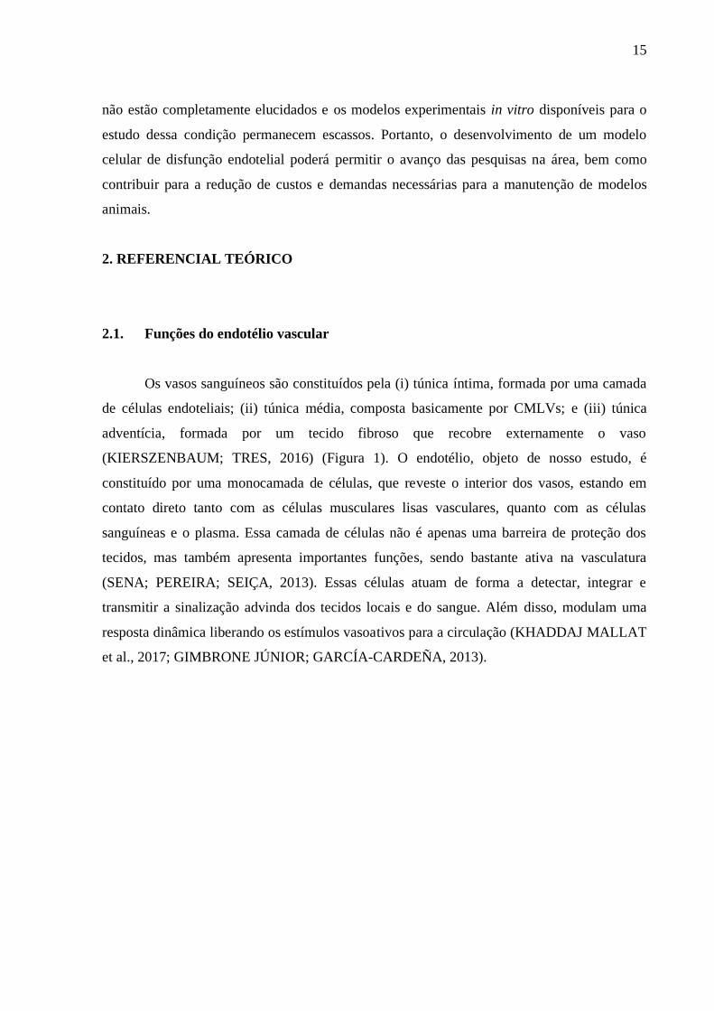
Os vasos sanguíneos são constituídos pela (i) túnica íntima, formada por uma camada
de células endoteliais; (ii) túnica média, composta basicamente por CMLVs; e (iii) túnica
adventícia, formada por um tecido fibroso que recobre externamente o vaso
(KIERSZENBAUM; TRES, 2016) (Figura 1). O endotélio, objeto de nosso estudo, é
constituído por uma monocamada de células, que reveste o interior dos vasos, estando em
contato direto tanto com as células musculares lisas vasculares, quanto com as células
sanguíneas e o plasma. Essa camada de células não é apenas uma barreira de proteção dos
tecidos, mas também apresenta importantes funções, sendo bastante ativa na vasculatura
(SENA; PEREIRA; SEIÇA, 2013). Essas células atuam de forma a detectar, integrar e
transmitir a sinalização advinda dos tecidos locais e do sangue. Além disso, modulam uma
resposta dinâmica liberando os estímulos vasoativos para a circulação (KHADDAJ MALLAT
et al., 2017; GIMBRONE JÚNIOR; GARCÍA-CARDEÑA, 2013).
16
Figura 1- Representação das camadas que constituem a parede do vaso sanguíneo: túnica adventícia,
túnica média e túnica íntima.
Fonte: (TORTORA, 2013).
O endotélio é essencial para a integridade e manutenção da homeostase vascular
(DAIBER et al., 2019) ao regular a angiogênese, tônus vascular, permeabilidade celular e
transporte capilar. Ainda, atua na resposta imune, regulando a adesão e infiltração de linfócitos
(KHADDAJ MALLAT et al., 2017). Além disso, o endotélio é responsável por produzir
biomoléculas capazes de realizar o controle entre a vasodilatação e vasoconstrição, liberação
de procoagulantes e anticoagulantes e de protrombóticos e antitrombóticos (FALCONER et al.,
2018; JAMWAL; SHARMA, 2018; YUYUN; NG; NG, 2018).
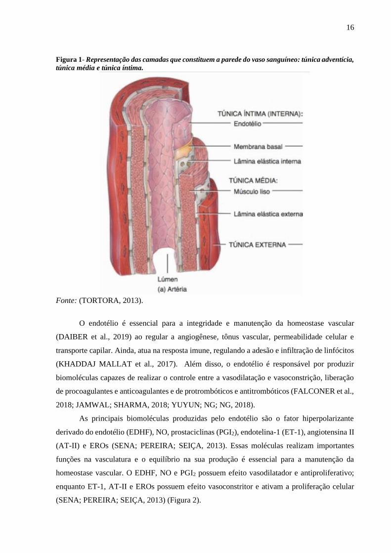
As principais biomoléculas produzidas pelo endotélio são o fator hiperpolarizante
derivado do endotélio (EDHF), NO, prostaciclinas (PGI2), endotelina-1 (ET-1), angiotensina II
(AT-II) e EROs (SENA; PEREIRA; SEIÇA, 2013). Essas moléculas realizam importantes
funções na vasculatura e o equilíbrio na sua produção é essencial para a manutenção da
homeostase vascular. O EDHF, NO e PGI2 possuem efeito vasodilatador e antiproliferativo;
enquanto ET-1, AT-II e EROs possuem efeito vasoconstritor e ativam a proliferação celular
(SENA; PEREIRA; SEIÇA, 2013) (Figura 2).
17
Figura 2 – Representação das principais biomoléculas produzidas pelas células endoteliais e suas
funções.
Fonte: Adaptado de SOARES, 2020.
O EDHF é importante no controle do tônus vascular, sendo produzido e liberado pelas
CE a partir da ligação de acetilcolina e/ ou bradicinina aos seus receptores. A atuação desse
fator através dos receptores presentes na musculatura lisa causa a hiperpolarização e o
relaxamento desta, mediado pela ativação dos segundos mensageiros, monofosfato cíclico de
guanosina (GMPc) e 3’5’-adenosina-monofosfato-cíclico (AMPc). Esses, por sua vez,
desencadeiam a ativação de canais de potássio e a diminuição da concentração de cálcio no
interior das células (BATLOUNI, 2001; KHADDAJ MALLAT et al., 2017).
O NO é um importante vasodilatador e será melhor descrito adiante. Já as PGI2 atuam
contribuindo para a vasodilatação e inibindo a agregação plaquetária. Isso se deve ao fato de
que tanto as CMLVs quanto as plaquetas possuem os receptores de prostaciclinas. A ligação
dessas moléculas aos seus receptores específicos conduz à ativação de adenilato ciclase e
proteína quinase e promove o relaxamento da musculatura (ARNAL et al., 1999; BATLOUNI,
2001; KHADDAJ MALLAT et al., 2017). É importante ressaltar que o EDHF e o NO são os
responsáveis majoritários pelo relaxamento vascular, com uma pequena contribuição das PGI2
(BATLOUNI, 2001).
Entre os agentes vasoconstritores destacam-se as endotelinas (ET), as quais são
peptídeos vasopressores que atuam diretamente em receptores nas CMLVs, ativando segundos
18
mensageiros e causando a vasoconstrição. Podem atuar também em receptores presentes nas
CE, sendo responsáveis pela manutenção da função endotelial. Em condições fisiológicas, a
concentração desses peptídeos é relativamente baixa, porém são os vasoconstritores mais
potentes (KHADDAJ MALLAT et al., 2017).
A AT-II é obtida através da conversão da angiotensina I por ação da enzima conversora
de angiotensina (ECA) e pode ocorrer em vários tecidos e células, inclusive nas células
endoteliais (KRINSKI et al., 2007). Essa molécula desempenha diversas funções na
vasculatura, como vasoconstrição, hipertrofia cardíaca, proliferação celular e formação de
matriz extracelular (BATLOUNI, 2001).
Finalmente, as EROs apresentam importante função como vasoconstritores diretos e
indiretos, podendo diminuir a biodisponibilidade de NO, reduzindo a vasodilatação dependente
de NO. A produção e as funções das EROs na vasculatura serão melhor abordadas adiante
(BATLOUNI, 2001; KHADDAJ MALLAT et al., 2017).
Diante da importância do endotélio para a manutenção da homeostase vascular, fica
evidente a necessidade de se manter o equilíbrio entre a biodisponibilidade de moléculas
vasoconstritoras e vasodilatadoras, uma vez que a DE decorre de uma alteração nessa
homeostase. Na DE há um desbalanço entre a produção e a degradação de NO e EROs,
representando um fator de risco preponderante no desencadeamento de doenças vasculares
(GIMBRONE JÚNIOR; GARCÍA-CARDEÑA, 2013).
2.2. Via do óxido nítrico derivado do endotélio
O NO é um radical livre, de caráter inorgânico, que se encontra no estado gasoso em
temperatura ambiente (DUSSE; VIEIRA; CARVALHO, 2003). A molécula é constituída por
uma ligação covalente do nitrogênio com o oxigênio e apresenta um elétron desemparelhado,
isso faz com que essa molécula possua uma alta reatividade (DUSSE; VIEIRA; CARVALHO,
2003). O NO derivado do endotélio é uma importante biomolécula vasomotora, que regula o
tônus vascular, inibe a adesão e agregação plaquetária, inibe a inflamação e ativa a angiogênese
de pequenos vasos (GHIMIRE et al., 2016).
Essa molécula é sintetizada por ação da enzima óxido nítrico sintase (NOS), a qual
converte L-arginina em L-citrulina com a redução de oxigênio e concomitante produção de NO
(YUYUN; NG; NG, 2018). São reconhecidas três isoformas de NOS nos mamíferos: 1) a óxido
nítrico sintase neuronal (nNOS); 2) a óxido nítrico sintase induzível (iNOS); e 3) a óxido nítrico
sintase endotelial (eNOS) (HONG et al., 2019). Todas as isoformas das NOS se encontram
19
ativas na forma de dímeros (YUYUN; NG; NG, 2018) e necessitam de cofatores e coenzimas
para a sua ativação como O2, NADPH, flavinas e biopterinas (FLORA FILHO; ZILBERSTEIN,
2000).
A nNOS é expressa principalmente no sistema nervoso central e periférico, mas também
em algumas organelas como as mitocôndrias, complexo de Golgi, retículo endoplasmático e
membrana plasmática de alguns tipos celulares (YUYUN; NG; NG, 2018). Essa isoforma é
codificada pelo gene NOS1 (YUYUN; NG; NG, 2018). Já a iNOS, codificada pelo gene NOS2,
está expressa, principalmente, em macrófagos, regulando a resposta imune; e na musculatura
lisa vascular, cuja atividade é modulada por agentes indutores (YUYUN; NG; NG, 2018). Por
fim, a eNOS, codificada pelo gene NOS3, é expressa principalmente no endotélio vascular,
miócitos cardíacos e epitélio renal, sendo responsável pela produção de cerca de 70% do NO
presente no plasma sanguíneo (FALCONER et al., 2018; GHIMIRE et al., 2016; SIRAGUSA;
FLEMING, 2016) (YUYUN; NG; NG, 2018).
Após sua síntese, o NO pode atuar como vasodilatador direto ou indireto. Para sua
atuação direta é necessária sua difusão através da membrana da célula endotelial, atuando nas
CMLVs e ativando a enzima guanilato ciclase, que, por sua vez, converte trifosfato de
guanosina (GTP) em GMPc. Este conduz à uma cascata de ativação enzimática que culmina
na remoção de cálcio das células musculares e, consequentemente, no relaxamento da
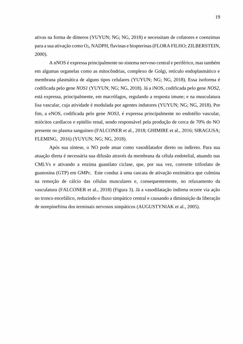
vasculatura (FALCONER et al., 2018) (Figura 3). Já a vasodilatação indireta ocorre via ação
no tronco encefálico, reduzindo o fluxo simpático central e causando a diminuição da liberação
de norepinefrina dos terminais nervosos simpáticos (AUGUSTYNIAK et al., 2005).
20
Figura 3 – Representação da vasodilatação induzida pelo óxido nítrico produzido pelas células
endoteliais. IP3: Inositol trifosfato; Ca2+: Íon cálcio; GTP: Guanosina trifosfato.
Fonte: Adaptado de PEARSON EDUCATION, 2012.
Além de ser um potente agente vasodilatador, o NO induz a angiogênese, permitindo a
síntese de vasos colaterais, e inibe a adesão de leucócitos no endotélio regulando processos
inflamatórios (FALCONER et al., 2018). Pode também atuar prevenindo a agregação
plaquetária e a ativação de fatores trombogênicos, inibindo a aterogênese (KHADDAJ
MALLAT et al., 2017).
Nesse contexto, o NO e drogas doadoras de NO são investigadas quanto ao seu potencial
para o tratamento de diversas doenças, como a hipertensão pulmonar e a aterosclerose, devido
a sua ação como agente anti-inflamatório, vasodilatador e antitrombótico (EVORA et al., 2012;
GHIMIRE et al., 2016; JAMWAL; SHARMA, 2018). Porém, a produção em excesso dessa
biomolécula também pode ser prejudicial, visto que, um aumento descomunal na produção de
NO pela iNOS, por exemplo, tem sido relacionado com a progressão do câncer de mama
(GHIMIRE et al., 2016), um vez que o excesso de NO atua como espécie reativa de oxigênio e
nitrogênio (ERON) (QUEIROZ; BATISTA, 1999). Logo, a manutenção da função endotelial e
da correta produção de NO é essencial para a manutenção da saúde do indivíduo.
2.3. Espécies reativas de oxigênio (EROs): fontes e funções biológicas
O oxigênio molecular (O2) é o aceptor final de elétrons na cascata de respiração celular
dos seres aeróbicos, devido à sua configuração eletrônica e sua alta eletronegatividade
21
(RIBEIRO et al., 2005). Ao longo da cascata de respiração são produzidas moléculas
intermediárias a partir do O2, caracterizadas por sua alta reatividade, as quais são denominadas
de EROs (INCALZA et al., 2018).
Nos seres aeróbicos, a produção de EROs se dá de forma natural e contínua, sendo o
ânion superóxido (O2º-), o peróxido de hidrogênio (H2O2), o radical hidroxila (OH-) e o
peroxinitrito (ONOO-) as principais EROs produzidas. A síntese de EROS é mediada por
enzimas específicas que podem atuar utilizando outra molécula considerada ERO como
substrato, ou a própria a produção de uma ERO pode desencadear a produção de outra por meio
de sinalização celular (RIBEIRO et al., 2005).
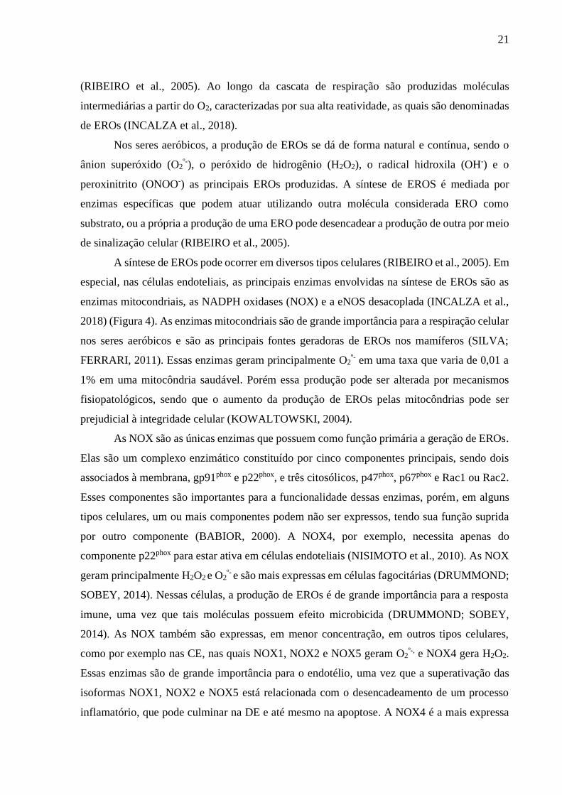
A síntese de EROs pode ocorrer em diversos tipos celulares (RIBEIRO et al., 2005). Em
especial, nas células endoteliais, as principais enzimas envolvidas na síntese de EROs são as
enzimas mitocondriais, as NADPH oxidases (NOX) e a eNOS desacoplada (INCALZA et al.,
2018) (Figura 4). As enzimas mitocondriais são de grande importância para a respiração celular
nos seres aeróbicos e são as principais fontes geradoras de EROs nos mamíferos (SILVA;
FERRARI, 2011). Essas enzimas geram principalmente O2º- em uma taxa que varia de 0,01 a
1% em uma mitocôndria saudável. Porém essa produção pode ser alterada por mecanismos
fisiopatológicos, sendo que o aumento da produção de EROs pelas mitocôndrias pode ser
prejudicial à integridade celular (KOWALTOWSKI, 2004).
As NOX são as únicas enzimas que possuem como função primária a geração de EROs.
Elas são um complexo enzimático constituído por cinco componentes principais, sendo dois
associados à membrana, gp91phox e p22phox, e três citosólicos, p47phox, p67phox e Rac1 ou Rac2.
Esses componentes são importantes para a funcionalidade dessas enzimas, porém, em alguns
tipos celulares, um ou mais componentes podem não ser expressos, tendo sua função suprida
por outro componente (BABIOR, 2000). A NOX4, por exemplo, necessita apenas do
componente p22phox para estar ativa em células endoteliais (NISIMOTO et al., 2010). As NOX
geram principalmente H2O2 e O2º- e são mais expressas em células fagocitárias (DRUMMOND;
SOBEY, 2014). Nessas células, a produção de EROs é de grande importância para a resposta
imune, uma vez que tais moléculas possuem efeito microbicida (DRUMMOND; SOBEY,
2014). As NOX também são expressas, em menor concentração, em outros tipos celulares,
como por exemplo nas CE, nas quais NOX1, NOX2 e NOX5 geram O2º-, e NOX4 gera H2O2.
Essas enzimas são de grande importância para o endotélio, uma vez que a superativação das
isoformas NOX1, NOX2 e NOX5 está relacionada com o desencadeamento de um processo
inflamatório, que pode culminar na DE e até mesmo na apoptose. A NOX4 é a mais expressa
22
nas células endoteliais e apontada como uma enzima de efeito vasoprotetor (DRUMMOND;
SOBEY, 2014; LANGBEIN et al., 2015; LIAO et al., 2018).
A alteração na função da eNOS, conhecida por desacoplamento, faz com que essa
enzima produza O2º- ao invés de NO nas CE (SENA; PEREIRA; SEIÇA, 2013). O
desacoplamento da eNOS pode ocorrer devido à ausência de L-arginina, oxidação de
tetraidrobiopterina (BH4) (Figura 4), processo inflamatório ou estresse oxidativo (SENA;
PEREIRA; SEIÇA, 2013). Esse desacoplamento causa a perda da conformação dimérica da
enzima, que passa a ser um monómero que sintetiza mais O2º- do que NO. Essa condição
aumenta as concentrações de EROs, o que afeta ainda mais a atividade endotelial (COUTO et
al., 2014; GHIMIRE et al., 2016; VARADHARAJ et al., 2015).
Figura 4- Representação da produção de Espécies Reativas de Oxigênio (EROs) na parede vascular
pela eNOS e pela NOX. No lado esquerdo, tem-se a representação da produção de ânion superóxido
via eNOS e a via de síntese de tetra-hidrobiopterina (BH4), um importante cofator. À direita, a
encontra-se representada a ativação da NAD(P)H oxidase (NOX). (FMN: mononucleotídeo de
flavina; GTP: guanosina 5’-trifosfato; ADMA: inibidor de eNOS; DPI: difenilenoiodônio inibidor
inespecífico das NOX).
Fonte: Adaptado de VANHOUTE et al., 2017
É importante ressaltar que as EROs regulam a expressão de genes sensíveis ao sinais
redox e também controlam a homeostase vascular por serem moléculas vasoativas
(BATLOUNI, 2001; INCALZA et al., 2018; KHADDAJ MALLAT et al., 2017; RIBEIRO et
al., 2005). Contudo, em concentrações supra-fisiológicas, essas moléculas podem reagir com
lipídeos e carboidratos, conduzindo à sua oxidação; e com o DNA, causando danos genéticos
(BONOMINI; RODELLA; REZZANI, 2015; RIBEIRO et al., 2005). Nesse sentido, a célula
possui um sistema de controle da biodisponibilidade de EROs, que pode incluir mecanismos
antioxidantes enzimáticos e não enzimáticos. O excesso de EROs ativa e/ ou induz a expressão
23
de enzimas antioxidantes como superóxido dismutases (SOD), catalase (CAT), glutationa
peroxidase (GSH-Px), peroxirredoxinas e tioredoxina (RIBEIRO et al., 2005). O mecanismo
antioxidante não enzimático, por sua vez, é uma proteção das biomoléculas realizada por
moléculas (como carotenoides, compostos fenólicos e ascorbato) que podem evitar a produção
de radicais livres ou causar a redução das EROs comprometendo sua reatividade (RIBEIRO et
al., 2005).
Os mecanismos de controle antioxidantes existem para evitar que ocorra o estresse
oxidativo (INCALZA et al., 2018), caracterizado pela produção excessiva e sustentada de EROs
e/ou à baixa disponibilidade de agentes antioxidantes (FÖRSTERMANN; XIA; LI, 2017;
WEFERS; SIES, 1983). Tal situação encontra-se associada a condições patológicas e ao próprio
envelhecimento celular (SIES, 2015). Em especial, evidências apontam que, nas doenças
cardiovasculares, a produção de EROs é aumentada, contribuindo para a sintomatologia das
doenças (HARRISON; GONGORA, 2009; SINHA; KUMAR DABLA, 2015) e para os danos
vasculares (MONTEZANO et al., 2015).
2.4. Disfunção endotelial (DE) e patologias associadas
A DE é uma condição caracterizada por um processo inflamatório concomitante ao
estresse oxidativo, o que ocasiona a perda da função endotelial decorrente do dano celular
(DAIBER et al., 2017). Diante do importante papel realizado pelo NO, a DE é geralmente
relacionada a uma redução da biodisponibilidade dessa biomolécula, a qual é o primeiro e um
dos mais significativos eventos que caracterizam a DE (INCALZA et al., 2018; RUDIC;
SESSA, 1999; VANHOUTTE et al., 2017).
Adicionalmente, a DE está intimamente ligada ao aumento da concentração de EROs
(DAVEL; BRUM; ROSSONI, 2014; INCALZA et al., 2018), sendo esse aumento uma das
causas do desacoplamento da eNOS, da redução de sua expressão (DAIBER et al., 2019) e do
aumento da degradação de NO (SENA; PEREIRA; SEIÇA, 2013). Em conjunto, esses eventos
ocasionam um comprometimento na regulação do tônus vascular e outras alterações fenotípicas
nas CE (INCALZA et al., 2018), tais como redução da vasodilatação, aumento da inflamação
e indução de um estado pró-trombótico (ENDEMANN; SCHIFFRIN, 2004).
A DE e os sintomas relacionados, entretanto, não incluem apenas a alteração no
metabolismo do NO e da consequente vasoconstrição, mas também a oxidação das
lipoproteínas com alteração da permeabilidade da membrana plasmática, acúmulo de
leucócitos, alteração no metabolismo extracelular (GIMBRONE JÚNIOR; GARCÍA-
24
CARDEÑA, 2013; KONUKOGLU; UZUN, 2016), agregação e adesão plaquetária,
proliferação das CMLVs e inflamação vascular (YUYUN; NG; NG, 2018) (Figura 5). A DE
representa, portanto, um mecanismo patológico relacionado a diferentes fatores e é apontada
como uma condição presente em várias doenças metabólicas e cardiovasculares como
aterosclerose, doença arterial periférica, diabetes, hipercolesterolemia e HAS (DAIBER et al.,
2019; FALCONER et al., 2018; HONG et al., 2019).
Figura 5 - Produção de biomoléculas em um endotélio saudável e em um endotélio disfuncional. Ach:
Acetilcolina; BK: Bradicinina; ET-1:Endotelina 1; M: Receptor muscarínico; B2: Receptor beta-2; ETB:
Receptor de Endotelina; 5-HT: 5-hidroxitriptamina; AT-I: Angiotensina I; AT-II: Angiotensina II;
TXA2: Tromboxano A2; PGH2: Prostaglandina H2; TP: Receptor para tromboxano-prostanóide.
Fonte: Adaptado de PARK; PARK, 2015.
A aterosclerose tem a DE como um importante marcador. A produção ineficiente e
anormal de NO, concomitante ao aumento dos níveis de EROs, desencadeiam o dano no
endotélio, que, por sua vez, causa agregação plaquetária, oxidação de lipídeos, inflamação
vascular e progressão da doença (GHOSH et al., 2017; YUYUN; NG; NG, 2018). Pesquisas
atuais buscam estratégias de tratamento que visam atuar na via do NO, assim como na sua
síntese, com o intuito de substituir as drogas tradicionais. Essas novas terapias visam
principalmente aumentar a expressão de eNOS e a inibição de seu desacoplamento (HONG et
al., 2019; KHADDAJ MALLAT et al., 2017).
25
A doença arterial periférica é caracterizada pela oclusão progressiva de vasos grandes e
médios dos membros inferiores (WILLIANS et al., 2012). Nessa patologia, a DE também é
uma condição primária e pode predizer o prognóstico dos pacientes. Portanto, drogas capazes
de aumentar os níveis de NO podem ser potentes no tratamento da doença arterial periférica
(FALCONER et al., 2018).
O diabetes tipo 2 está comumente associado ao estresse oxidativo na vasculatura,
somado a uma baixa concentração de antioxidantes naturais, acarretando na disfunção
endotelial, a qual, em um sistema de cascata, agrava a condição do paciente (KHADDAJ
MALLAT et al., 2017). É comum que pacientes diabéticos apresentem uma redução na
elasticidade dos vasos e hipertensão arterial secundária (KHADDAJ MALLAT et al., 2017).
De fato, o diabetes causa a ativação da enzima aldose redutase, que afeta a fosforilação e a
expressão da eNOS (JAMWAL; SHARMA, 2018).
Outra patologia importante é a hipercolesterolemia, uma condição que estimula a
oxidação do colesterol, a liberação de ET e a geração de EROs (JAMWAL; SHARMA, 2018).
Todos esses fatores estão relacionados à uma redução na expressão de eNOS e à perturbação
da integridade do endotélio vascular, ou seja, estão associados com a DE (JAMWAL;
SHARMA, 2018).
Finalmente, na HAS, a DE atua diretamente no quadro da doença, sendo o NO uma
molécula chave para a adequada manutenção dos níveis de pressão arterial. Dessa forma, em
pacientes com HAS essencial ou primária observam-se baixos níveis de NO circulante, edema
subendotelial causado por aumento da permeabilidade vascular, aumento da aderência
leucocitária à parede vascular, aumento da agregação plaquetária e aumento da proliferação de
CMLVs (BATLOUNI, 2001; CARVALHO et al., 2001; DAVEL et al., 2011; HAMILTON et
al., 2001). Além disso, pacientes com HAS apresentam altos níveis de liberação de ET,
prostanóides vasoconstritores, AT-II, citocinas inflamatórias e xantina oxidase, culminando
com o aumento na degradação de NO (JAMWAL; SHARMA, 2018). Nesse sentido, moléculas
doadoras de NO atuam compensando a redução ocorrida durante a DE e têm se mostrado
potentes agentes terapêuticos para a HAS (GHEIBI et al., 2018; KONUKOGLU; UZUN, 2016;
WENCESLAU; ROSSONI, 2014).
Compreende-se, portanto, que a DE é uma condição presente em diversas doenças
cardiovasculares e metabólicas. Levando em conta que tais patologias podem coexistir em um
mesmo indivíduo e que a DE é um evento comum nessas doenças, a DE pode tratada com um
potencial alvo para o desenvolvimento de novas estratégias terapêuticas (JAMWAL;
SHARMA, 2018). Faz-se necessário, então, o estudo mais aprofundado da disfunção endotelial
26
e dos mecanismos e vias envolvidos no seu estabelecimento e progressão. Nesse sentido,
abordagens metodológicas capazes de avaliar a função endotelial in vitro e in vivo se fazem
necessárias.
2.5. Estratégias para o teste da função endotelial
Diferentes abordagens são adotadas para avaliar a função endotelial e identificar
características gerais da DE. Esses métodos possibilitam o desenvolvimento de métodos
diagnósticos para diversas doenças relacionadas à DE (GIRIBELA et al., 2011). Nesse
contexto, algumas análises permitem identificar CE inteiras ou apoptóticas na circulação,
marcadores circulantes da função endotelial e a dilatação fluxo-mediada (DILA) da artéria
braquial (GIRIBELA et al., 2011; INCALZA et al., 2018).
Durante a DE, o estresse oxidativo, concomitante com a inflamação crônica, causa a
lesão celular, formando corpos apoptóticos ou até mesmo a liberação de CE inteiras na
circulação. Essas células podem ser identificadas pela coleta e análise do sangue (GIRIBELA
et al., 2011; INCALZA et al., 2018). Além disso, marcadores da função endotelial circulantes,
como o NO, moléculas de adesão celular, citocinas inflamatórias, agentes trombóticos e
antitrombóticos, podem ser identificados (INCALZA et al., 2018). Vale ressaltar, no entanto,
que alguns desses marcadores são de difícil identificação e dosagem, como o NO que possui
uma meia-vida muito curta, o que limita a viabilidade dessa técnica (GIRIBELA et al., 2011;
PREMER et al., 2019).
In vivo, a dosagem de NO em amostras biológicas é um processo complexo e com uma
alta margem de erro. A integridade funcional do endotélio pode ser observada por meio da
DILA, em que utiliza-se uma ultrassonografia Doppler para observar a dilatação ou não da
artéria após uma isquemia induzida pela inflação de um manguito colocado no membro superior
(CELERMAJER et al., 1992). Esse estímulo é suficiente para a liberação de NO pelo endotélio,
caso esse esteja íntegro, causando a dilatação da artéria (FALCONER et al., 2018; GIRIBELA
et al., 2011; PREMER et al., 2019).
Entretanto, é importante ressaltar que ainda existe a necessidade de desenvolvimento de
novos métodos e estratégias, pois muitas das ferramentas disponíveis são de difícil execução
e/ou de alto custo (GIRIBELA et al., 2011). Dessa forma, a concepção de condições
experimentais que se assemelham à DE in vitro possibilita maior compreensão dessa condição,
facilitando a análise in vivo e o desenvolvimento de técnicas de diagnóstico e terapia para essa
doença.
27
2.6. Inibição da síntese de NO e indução de disfunção endotelial
O L-NAME é um pró-fármaco com capacidade inibitória da síntese de NO (PFEIFFER
et al, 1996), pois é um análogo de L-arginina, o substrato para a síntese de NO. A inibição da
NOS pelo L-NAME ocorre por meio da ligação desse inibidor à enzima no sítio catalítico,
competindo com o substrato e impedindo a ligação da L-arginina (PETERSON et al, 1992;
REES, et al., 1990). Além disso, o L-NAME é um inibidor reversível e não seletivo das NOSs
comumente utilizado em experimentos a longo e curto prazo, seja in vitro ou in vivo, com o
objetivo de identificar os eventos associados à restrição da produção de NO (KOPINCOVÁ;
PÚZSEROVÁ; BERNÁTOVÁ, 2012). Dados da literatura descrevem os efeitos do L-NAME
em modelos animais. A administração de L-NAME, seja injetável ou na água de beber, conduz
ao aumento na vasoconstrição, de pressão arterial e bradicardia (REES, et al., 1990); além do
aumento da resistência vascular periférica, hipertrofia do ventrículo esquerdo do coração,
redução da resposta relaxante nos vasos calibrosos, aumento da resposta vascular à estímulos
adrenérgicos e inflamação perivascular nos indivíduos experimentais (KOPINCOVÁ;
PÚZSEROVÁ; BERNÁTOVÁ, 2012; TÖRÖK, 2008).
Sabendo-se que a DE é comum em pacientes com hipertensão essencial e que está
intimamente relacionada à redução da biodisponibilidade de NO, um modelo animal de indução
de hipertensão foi previamente desenvolvido (REES, et al., 1990). Este consiste na
administração de L-NAME de forma crônica em doses altas, induzindo uma hipertensão
denominada hipertensão por deficiência de NO em ratos que antes não eram hipertensos (REES,
et al., 1990). In vitro, por sua vez, resultados demonstraram que a inibição causada pelo L-
NAME da produção de NO em anéis de aorta é dose-dependente e leva a um aumento da
contração dependente do endotélio, redução de vasodilatação dependente de acetilcolina (Ach)
(REES, et al., 1990), desestabilização da membrana lisossomal (FOMINA et al., 2018) e a
redução da biodisponibilidade de NO (KOPINCOVÁ; PÚZSEROVÁ; BERNÁTOVÁ, 2012).
Até o presente, todavia, não foi descrito um modelo de indução de condição semelhante à DE
em cultura de células.
Dessa forma, sabendo-se que CE em cultura são expostas a diferentes situações e
estímulos próprios da DE, esse tipo celular se torna mais suscetível ao desacoplamento da eNOS
e à indução de um estado semelhante à DE. Estudos apontam que o aumento da oxidação de
lipoproteínas de baixa densidade, alta concentração de glicose, homocisteína e angiostatina são
capazes de induzir tal condição (INCALZA et al., 2018). Por isso, buscou-se observar nesse

28
trabalho se o tratamento com o L-NAME, e consequente diminuição na biodisponibilidade de
NO, também seria capaz de realizar essa indução.

29
CAPÍTULO 2
ENDOTHELIAL DYSFUNCTION BY INHIBITING NITRIC OXIDE SYNTHESIS:
PROPOSAL AND CHARACTERIZATION OF AN IN VITRO CELLULAR MODEL
Fernanda Cardoso da Silva, Bruna Juber de Araújo, Carina Santos Cordeiro, Vinícius
Marques Arruda, Bruno Quintanilha Faria, Joyce Ferreira da Costa Guerra, Thaise Gonçalves
de Araújo, Cristina Ribas Fürstenau
30
1
2
3
Endothelial dysfunction by inhibiting nitric oxide synthesis: Proposal and 4
characterization of an in vitro cellular model 5
6
Fernanda Cardoso da Silva¹, Bruna Juber de Araújo¹, Carina Santos Cordeiro¹, Vinícius 7
Marques Arruda¹, Bruno Quintanilha Faria¹, Joyce Ferreira da Costa Guerra¹, Thaise 8
Gonçalves de Araújo¹, Cristina Ribas Fürstenau²* 9
10
11
¹ Institute of Biotechnology, Federal University of Uberlândia, Patos de Minas, MG, Brazil 12
² Center for Natural and Human Sciences (CCNH), Federal University of ABC (UFABC), 13
Santo André, SP, Brazil. 14
15
16
17
18
19
*Corresponding author 20
E-mail: [email protected] (CRF) 21

31
Abstract 22
Vascular endothelium plays an important role in the maintenance of vascular 23
homeostasis, mediated by vasoactive molecules produced by endothelial cells. The balance 24
between vasoconstrictor and vasodilator biomolecules is what guarantees this equilibrium. 25
Therefore, an increase in the bioavailability of vasoconstrictors together with a reduction in 26
vasodilators may indicate a condition called endothelial dysfunction. Endothelial dysfunction 27
is mainly characterized by a reduction in nitric oxide (NO) bioavailability and an increase in 28
the production of reactive oxygen species (ROS), triggering an inflammatory process. This 29
condition is a predictive marker of several cardiovascular diseases (e.g. atherosclerosis, 30
hypertension and diabetes), but research is affected by the scarcity of suitable in vitro models 31
that simulates such condition. In this context, the goal of this study was to induce an in vitro 32
condition that mimics endothelial dysfunction by inhibiting NO synthesis. Thymus-derived 33
endothelial cells (tEnd.1) were treated with different concentrations of L-NAME (1µM, 10 µM, 34
100 µM and 1mM) for different times (12, 24, 48, 72, 96 and 120 hours with and without 35
retreatment every 24 hours). Cell viability, nitrite concentration, p22phox gene expression and 36
lipid peroxidation were evaluated. Results indicated that treatment with 100 µM L-NAME for 37
96 hours with retreatment was able to trigger a reduction in NO concentration by 94% and 38
reduce p22phox expression by 65%. We thus propose that 100 µM L-NAME treatment for 96 39
hours caused a condition similar to endothelial dysfunction. Besides, our results show for the 40
first time, the p22phox expression in tEnd.1 cell line. However, additional studies aiming to 41
determine the role of ROS in cells submitted to treatment with L-NAME are necessary for the 42
improvement and characterization of the proposed experimental model. 43
44
Introduction 45
The endothelium consists of a cell monolayer, that not only acts as a tissue barrier, but 46
also has important biological functions. In this sense, endothelial cells are very active in the 47
vasculature [1] and essential for the integrity and maintenance of vascular homeostasis. Also, 48
they perform important functions as regulator of angiogenesis, vascular tone, and cellular 49
permeability, influencing in capillary transport [2]. 50
To perform such diverse functions, endothelial cells produce a range of important 51
biomolecules. The main biomolecules produced by the endothelium are endothelium-derived 52
hyperpolarizing factor (EDHF), nitric oxide (NO) and prostacyclins (PGI2), which have 53
vasodilatory and antiproliferative effects on vascular smooth muscle cells. On the other hand, 54
endothelial cells also generate endothelin-1 (ET-1), angiotensin II (AT-II), and reactive oxygen 55
species (ROS), which present vasoconstrictor effects and promote vascular smooth muscle cells 56
proliferation [1]. 57
Endothelial dysfunction is a condition characterized by an inflammatory process 58
concomitant with oxidative stress, which causes loss of endothelial function and consequent 59
32
imbalance in the production of biomolecules [2]. Two of the most important events that 60
characterize endothelial dysfunction are the reduction in NO bioavailability and a considerable 61
increase in the production of ROS, such as superoxide anion [3-5]. This condition represents a 62
pathological mechanism that is related to a variety of factors and is pointed as a predictor of 63
distinct cardiovascular and metabolic diseases [2]. 64
It is thus clear that endothelial dysfunction is a conserved target in metabolic disorders. 65
In line with that, given that different metabolic and cardiovascular diseases may coexist in the 66
same patient and that endothelial dysfunction is a common event in these diseases, endothelial 67
dysfunction may also be used as a target for new therapies [6]. It is therefore necessary to 68
deepen the studies regarding endothelial dysfunction to better understand the mechanisms and 69
pathways involved in this condition. 70
Different in vitro and in vivo approaches may be used to access endothelial function. At 71
present, however, there is no specific protocol to mimic in vitro endothelial dysfunction, which 72
limits the study of this condition at the cellular level and to search for biomolecules targeting 73
diagnosis and treatment. Therefore, this study aimed to develop a protocol to simulate 74
endothelial dysfunction in vitro by inhibiting NO synthesis in thymus-derived endothelial cells 75
(tEnd.1). 76
Materials and methods 77
Cell Culture 78
Murine thymus-derived endothelial cell line tEnd.1 (RRID: CVCL_6272) was cultured 79
in Dulbecco’s modified Eagle’s medium (DMEM, GIBCO®), enriched with 10% fetal bovine 80
serum (FBS, GIBCO®), 100 U/mL penicillin and 100 µg/mL streptomycin (GIBCO®) at 37 81
°C, 5% CO2 in an humidified incubator until reaching 80 to 90% confluence. Cell morphology 82
during cultivation is shown in Supplementary Figure 1. Cells were used between the 3rd and 8th 83
passages. Depending on the experiment, cells were plated in 6, 12 or 96-well plates, with 2 x 84
105 cells/ well, 4 x 10⁴ cells/ well and 1 x 10³ cells/ well, respectively. 85
86
L-NAME Treatment 87
After 24 hours of plating and prior to treatments, cells were made quiescent by fetal 88
bovine serum deprivation (0.5%) for 3 hours and subsequently subjected to L-NAME 89
33
(SIGMA®) treatment (1µM, 10 µM, 100 µM and 1mM ) or 100 µM L-arginine (SIGMA®) 90
treatment, as a negative control [7] for 12, 24, 48, 72, 96 and 120 hours with and without 91
retreatment every 24 hours. Retreatments were performed every 24 hours by replacing the “old 92
culture medium” by the “new culture medium” with the same treatment. 93
94
Cell Viability 95
Following cell treatments, 25 μL of Tetrazolium Blue Thiazolyl Bromide (MTT) 96
(Ludwig Biotec®) was added at a concentration of 5 mg/ mL in PBS (w/v) to each well and 97
plates were left for 4 hours in the incubator. The culture medium with MTT excess was then 98
aspirated, followed by the addition of dimethyl sulfoxide (DMSO) to dissolve formazan crystals 99
[8]. MTT method is based on the ability of living cells to reduce the yellow tetrazolium salt to 100
the purple insoluble formazan, which precipitates thanks to the action of the mitochondrial 101
enzyme succinyl dehydrogenase, active only on living cells [8]. Optical reading was performed 102
on an automatic plate reader at 560 nm (Readwell PLATE, ROBONIK®). Cell viability results 103
were obtained according to Equation 1. 104
105
% 𝐶𝑒𝑙𝑙 𝑣𝑖𝑎𝑏𝑖𝑙𝑖𝑡𝑦 =(𝐴𝑡 - 𝐴𝑏 )
(𝐴𝑐 - 𝐴𝑏 ) × 100 106
Where: 107
𝐴𝑡: Absorbance at 560 nm of treat𝑒d cells (cells + treatment) 108
𝐴𝑏 : Absorbance at 560 nm of blank wells (only DMSO) 109
𝐴𝑐: Absorbance at 560 nm of control cells (culture medium + cells) 110
111
Nitrite Quantification 112
Nitrite quantification was performed as an indirect measurement of NO levels. The 113
treatments were performed as described before, but using phenol red free DMEM (GIBCO, 114
Grand Island, New York, USA) to not influence the readings. Nitrite content was determined 115
using a Griess reagent kit (Thermo Fisher Scientific®) according to the manufacturer's 116
instructions. The culture medium from each well was collected, centrifuged at 16,000 rpm, 4°C 117
(Hermle Labor Technik, Z 36 HK) for 10 minutes and the supernatant was kept for further 118
analysis. In a 96-well microplate, 20 µL Griess reagent, 150 µL of nitrite-containing sample 119
(1)
34
and 130 µL deionized water were mixed. After 30 minutes of incubation in the dark at room 120
temperature, the plate was read on an ELISA plate reader (Readwell PLATE, ROBONIK®) at 121
560 nm. Nitrite concentration in the samples was calculated based on a standard curve of 122
different sodium nitrite concentrations (1, 5, 10, 30, 50 and 100 µM). 123
124
Real-Time PCR Gene Expression Analysis 125
After treatments, total cellular RNAs were extracted with Trizol® reagent (Invitrogen, 126
Carlsbad, CA, USA) following the manufacturer’s instructions. The quality of the extracted 127
RNAs is represented in Supplementary Figure 2. Reverse transcription was performed as 128
previously described [9] (Supplementary Figure 3). The reference gene B-actin was used 129
(5'GGGAAATCGTGCGTGACATC3' and 5'GCCACAGGATTCCATACCCAA3') to validate 130
RNA quality of each sample and for normalization of qPCR assays. For validation, 131
conventional PCR reactions were performed as follows: 2.0 µL of cDNA amplicons, 1.0 U of 132
Taq DNA Polymerase Platinum (Invitrogen), 50 mM KCl; 10 mM Tris– HCl pH 8.3, 2.0 mM 133
MgCl2, 200 μM dNTPs, 5.0 pmol of each primer, all components incubated for 25 cycles at 94 134
°C 30 s, 60 °C 1 min, 72 °C 1 min, preceded by an initial denaturation at 95 °C for 5 min 135
(Supplementary Figure 3). 136
For qPCR, each 2 µL aliquot of cDNA was amplified with 5 pmol of each specific 137
primer (EXXTEND®) for eNOS [10], p47phox (5'ATCCCCAGCCAGCACTATGTG3' and 138
5'GAGATCCACACAAGAGAACAGAG3') and 139
p22phox(5'CCAGTGTGATCTATCTGCTGGCA3' and 5'GCCTCCTCTTCACCCTCACTC3'). 140
The reactions were conducted in six replicates in a total volume of 10 μL containing Power 141
SYBR_ Green PCR Master Mix (Applied Biosystems - Carlsbad, CA, USA) in a thermal cycler 142
(StepOnePlus™ Real-Time PCR System, Applied Biosystems). Standard relative curves for all 143
primers were constructed and expression of each gene was quantified through comparative Cq 144
method. 145
146
Western Blotting Protein Expression Analysis 147
Following cells treatment, total proteins were obtained using an extraction buffer (1% 148
Triton X-100, 135 mM NaCl, 20 mM Tris, pH 8.0 and 10% glycerol). The crude lysate was 149
centrifuged (14000 x g, 10 minutes, 4 °C). The supernatant was then collected, and 20 µg of 150
35
total protein was separated by 10% SDS-polyacrylamide gel electrophoresis. After separation, 151
proteins were transferred to nitrocellulose membranes (Amersham Protran Premium - GE 152
Healthcare, Life Science, USA). Firstly, membranes were blocked with 3% bovine serum 153
albumin (SIGMA®) for 1 hour at room temperature. Primary antibodies to proteins of interest 154
eNOS (Purified Mouse Anti-eNOS / NOS Type III, BD transduction Laboratories™) and actin 155
(Anti-Actin Antibody, clone C4, MERCK®) were incubated overnight at 4 °C. Peroxidase-156
conjugated IgG secondary antibodies were used to detect the primary antibody (Jackson 157
Immunoresearch, West Grove, PA, USA). Bands were revealed by chemiluminescence on X-158
ray films (T-MAT G/ RA Film, KODAK, Sao Jose dos Campos, Sao Paulo, Brazil) using the 159
ECL detection system (WESTAR SUN, Cyanagen, Bologna, Italy). 160
161
Determination of Lipid Peroxidation 162
The quantification of lipid peroxidation is essential to evaluate oxidative stress in 163
pathophysiological processes. One of the main products of lipid peroxidation is 164
malondialdehyde (MDA), the most abundant aldehyde generated by the attack of reactive 165
species on polyunsaturated fatty acids in cell membranes [11]. MDA levels were determined 166
by testing thiobarbituric acid reactive substances using the method of Buege & Aust (1978) 167
[11], which is based on the ability of thiobarbituric acid (TBA) to bind to oxidized lipids. 168
Briefly, 2 x 10⁷ cells submitted to different treatments were homogenized in 1 mL of cold 169
20mM Tris HCL (pH 7.4) buffer. All homogenate was mixed with trichloroacetic acid (28% 170
w/v in 0.25 N HCl), TBA (1% in 0.25 M acetic acid) and BHT (125 mM in ethanol), heated for 171
1 hour at 95 °C and then placed in an ice bath. The precipitate was then removed by 172
centrifugation at 10,000 x g for 15 minutes at 4 °C, and the supernatant absorbance was 173
determined at 535 nm in a spectrophotometer (Gehaka, UV-340 G). MDA levels were 174
calculated using 1,1,3,3-tetramethoxypropane as standard for constructing the calibration curve 175
(12.5, 6.25, 3.125, 1.562, 0.781 and 0.390 µmoL/L ). 176
177
Statistical Analysis 178
Data are presented as mean ± standard deviation for each of the measurements 179
performed. Sample number (n) represents the number of experiments performed with different 180
treatments in the cell line culture. For the comparison between groups, Two-way and One-way 181
36
analysis of variance (ANOVA) was applied and Tukey's multiple comparisons test was used as 182
post-hoc test, because the distribution was normal. Differences between groups were considered 183
significant at p <0.05. Data were analyzed using GraphPad Prism software, version 7.00 for 184
Windows. 185
186
Results 187
Cell Viability 188
The MTT assay results show how different treatments affected the viability of the tEnd.1 189
cells. In groups without retreatment, 100 µM L-arginine increased cell viability (18.67%) after 190
48 hours, while reduced cell viability (28.77% and 37.52%) after 96 and 120 hours, respectively 191
(Fig.1). In general, L-NAME increased cell viability after 24, 48, 96 and 120 hours of treatment. 192
Differences were most evident in L-NAME-treated cells at any concentration for 24 hours (cell 193
viability approximately 140%); 10 µM and 100 µM L-NAME for 48 hours (cell viability 194
133.85% and 134.83%, respectively); 100 µM for 96 hours (cell viability 122.88%); 10 µM, 195
100 µM and 1 mM for 120 hours (cell viability 163.52%, 121.76% and 122.29%, respectively) 196
(Fig.1). Treatments performed for 72 hours without retreatment had the least influence on cell 197
viability when compared to control (Fig.1). 198
199
Fig. 1. Cell viability of the thymus-derived endothelial cell line tEnd.1 after treatment with L-200
arginine (100 µM) and L-NAME (1 µM, 10 µM, 100 µM, 1 mM) for 12, 24, 48, 72, 96 and 120 201
hours without retreatment. Data (mean ± standard deviation, n=5) were analyzed by Two-way 202
ANOVA, followed by Tukey’s multiple comparisons test. (*represents significant difference between 203
groups, for p <0.05; **represents significant difference between groups, for p <0.01; ***represents 204
significant difference between groups, for p <0.001; ****represents significant difference from the 205
control group, for p <0.0001). 206
207
In groups that experienced retreatment every 24 hours (Fig.2), 100 µM L-arginine 208
generally does not affect cell viability, except after 72 hours with reduction of 26,04% of cell 209
viability. Once again, L-NAME increased cell viability after 24, 48 and 72 hours of treatment, 210
37
in most L-NAME treated (Fig.2). Comparing to untreated cells, after 24 hours, L-NAME at any 211
concentration increased cell viability by approximately 40%. In a same manner, 100 µM and 1 212
mM L-NAME after 48 hours also increased cell viability around 38.22% and 27.51%, 213
respectively. The treatments for 96 and 120 hours with retreatment every 24 hours were the 214
ones that least influenced cell viability when compared to control cells (Fig.2). 215
216
Fig. 2. Cell viability of the thymus-derived endothelial cell line tEnd.1 after treatment with L-217
arginine (100 µM) and L-NAME (1 µM, 10 µM, 100 µM, 1 mM) for 48, 72, 96 and 120 hours with 218
retreatment. Data (mean ± standard deviation, n=5) were analyzed by Two-way ANOVA, followed by 219
Tukey’s multiple comparisons test. (*represents significant difference between groups, for p <0.05; 220
**represents significant difference between groups, for p <0.01; ***represents significant difference 221
between groups, for p <0.001; ****represents significant difference between groups, for p <0.0001). 222
223
Nitrite Quantification 224
Results presented in Figures 3 and 4 show that in both groups (without and with 225
retreatment) nitrite concentration increases in control group until it reaches a peak after 72 226
hours, and then decreases. 227
228
Fig. 3. Nitrite concentration in culture medium of the thymus-derived endothelial cell line 229
tEnd.1after treatment with L-arginine (100 µM) and L-NAME (1 µM, 10 µM, 100 µM, 1 mM) for 230
12, 24 , 48, 72, 96 and 120 hours without retreatment. Data (mean ± standard deviation, n=5) were 231
compared by Two-way ANOVA, followed by Tukey’s test. (*represents statistically significant 232
difference compared between groups for p <0.01; **represents statistically significant difference 233
compared between groups for p <0.01; ***represents significant difference compared between groups, 234
for p <0.001; ****represents significant difference compared between groups, for p <0.0001). 235
236
Fig. 4. Nitrite concentration in culture medium of the thymus-derived endothelial cell line tEnd.1 237
after treatment with L-arginine (100 µM) and L-NAME (1 µM, 10 µM, 100 µM, 1 mM) for 48, 72, 238
96 and 120 hours with retreatment every 24 hours. Data (mean ± standard deviation, n=5) were 239
38
compared by Two-way ANOVA, followed by Tukey’s test (*represents statistically significant 240
difference compared between groups for p <0.01; **represents statistically significant difference 241
compared between groups for p <0.01; ***represents significant difference between groups, for p 242
<0.001; ****represents significant difference between groups, for p <0.0001). 243
244
Treatment with L-arginine increased nitrite concentration in the culture medium, which 245
was more evident after 72, 96 and 120 hours, with an increase of 21.98%, 61.90% and 62.68% 246
respectively, when compared to untreated cells (Fig.3.). L-NAME at different concentrations 247
tend to reduce nitrite concentration in the culture medium in a dose-dependent manner. This 248
reduction was more evident in 10 µM, 100 µM and 1 mM L-NAME after 24, 48, 72, 96 and 249
120 hours without retreatment. The largest reductions were observed with 1 mM L-NAME 250
(91.37%), 100 µM L-NAME (70.83%) and 10 µM L-NAME (70.35%) after 72 hours of 251
treatment, when compared to untreated cells (Fig.3.). 252
In groups with retreatment (Fig.4), L-NAME significantly reduced nitrite concentrations 253
as follows: with reductions of 62.05% at 100 µM and 92.60% at 1mM after 48 hours; 24.10% 254
at 10 µM, 69.55% at 100 µM and 95.14% at 1 mM after 72 hours; and 86.01% at 1 mM after 255
120 hours. After 96 hours with retreatment, results were more uniform and consistent with the 256
literature, with reductions of 32.62% at 10 µM, 51.47% at 100 µM and 94.30% at 1 mM L-257
NAME (Fig.4). 258
As endothelial dysfunction is characterized by reduced NO bioavailability, cells treated 259
with 10 µM and 100 µM L-NAME for 72 hours without retreatment and for 96 hours with 260
retreatment were chosen for subsequent experiments since they significantly reduced nitrite 261
concentration and did not affect cell viability. Cells treated with 100 µM L-arginine were used 262
as negative control at the same times of treatment. 263
264
Real-Time PCR Gene Expression Analysis 265
The results showed that relative levels of p22phox mRNA did not differ after different 266
treatments for 72 hours (Fig.5 A). On the other hand, the treatment with 100 µM L-NAME for 267
96 hours with retreatment exhibited a significant reduction of approximately 65% in p22phox 268
mRNA levels when compared with control group (Fig. 5B). It is noteworthy that the expression 269
of p22phox gene in tEnd.1 cells was firstly reported in the present study. No amplification for 270
the p47phox and eNOS genes were obtained with the selected primers. 271
39
272
Fig. 5. Relative levels of p22phox gene mRNA in the thymus-derived endothelial cell line tEnd.1 273
after treatment with L-arginine (100 µM) and L-NAME (10 µM and 100 µM) for (A) 72 hours 274
without retreatment (B) for 96 hours with retreatment. Data (mean ± standard deviation, n=3) were 275
compared by One-way ANOVA, followed by Tukey’s test. (**represents statistically significant 276
difference from the control group for p <0.01). 277
278
Western Blotting Protein Expression Analysis 279
Once we observed a peak of nitrite concentration after 72 hours cells treated with 100 280
µM L-arginine, 10 µM and 100 µM L-NAME the goal of this experiment was to determine 281
eNOS protein expression levels at this time point. Unfortunately, it was not possible to visualize 282
eNOS expression in tEnd.1 cells by the method used (Supplementary Fig. 4). 283
284
Determination of Lipid Peroxidation 285
Thiobarbituric acid reactive substances (TBARS) were quantified to see if lipid 286
peroxidation, a common event in endothelial dysfunction, was occurring in cells submitted to 287
different treatments. The results obtained showed that there was no significant difference 288
between the different treatments (Fig.6). 289
290
Fig. 6 - Concentration of thiobarbituric acid reactive substances (TBARS) given in µmol/ mL PTN 291
in the thymus-derived endothelial cell line tEnd.1 medium after treatment with L-arginine (100 292
µM) and L-NAME (10 µM and 100 µM) for 72 hours without retreatment. Data (mean ± standard 293
deviation, n=3) were compared by One-way ANOVA. No statistical differences were observed between 294
groups. 295
296
Discussion 297
40
Endothelial dysfunction is a primary condition of many cardiovascular diseases but is 298
still little explored as a target for diagnosis and treatment [12-14]. Given this, the interest in the 299
study of this condition has gradually grown over the years and studies focusing on the 300
evaluation of endothelial function have shown to be very promising [15]. 301
Researchers have already proposed that certain treatments may induce a condition 302
similar to endothelial dysfunction in cell culture when they mimic the metabolic changes 303
inherent to this pathological state. It is well known that treatment with native and oxidized low 304
density lipoproteins, angiostatin, homocysteine and high glucose are capable of causing eNOS 305
uncoupling, inducing a state similar to endothelial dysfunction [3]. However, this is the first 306
time that endothelial dysfunction has been proposed using a NO synthesis inhibitor (L-NAME) 307
as an inducer of this condition. 308
Most research regarding endothelial dysfunction is performed in vivo and little is known 309
about what happens at the cellular level. In vitro research is also of great importance for a better 310
understanding of this condition and to elucidate the pathways involved in endothelial cells. For 311
these reasons, there is a crescent need for a standardization of a method of inducing in vitro 312
endothelial dysfunction [16]. 313
Throughout the present research, we proposed the development of a protocol to mimic 314
endothelial dysfunction in vitro by inhibiting NO synthesis using L-NAME. Initially, we 315
evaluated how treatments affected endothelial cells viability. It is already known that NO affects 316
the viability of endothelial cells and inhibits cell apoptosis induced by inflammation or 317
atherosclerotic factors [17]. In addition, when NO synthesis is induced by vasoactive agents, 318
there is autocrine regulation of microvascular events, causing neovascularization, resulting 319
from endothelial cell angiogenesis, growth and migration [18]. This inhibition of apoptosis is 320
what contributes to NO having a significant effect as an anti-inflammatory and pro-angiogenic 321
molecule [17]. L-arginine is an important amino acid that is considered versatile due to the fact 322
that it is the substrate for the synthesis of many molecules, including NO. Studies indicate that 323
L-arginine supplementation increases endometrial cell proliferation by a NO-dependent 324
mechanism [19]. In endothelial cells, L-arginine is known to play an important role in cell 325
survival during oxidative stress [20]. 326
Considering the above mentioned, a reduction in cell viability in L-NAME-treated cells 327
and an increase in L-arginine-treated cells was expected, but this did not occur. Cell viability 328
was increased in most L-NAME-treated cells. This can be justified by the fact that NOS 329
isoforms can also generate superoxide anion [21], an important ROS, which is cytotoxic and 330
capable of affecting the organization of cellular plasma membrane, leading to apoptosis or even 331
41
necrosis [22-23]. Thus, treatment with L-NAME inhibits both NO and superoxide production, 332
and inhibition of superoxide synthesis may contribute to greater cell proliferation, as noted in 333
the results [24]. 334
Despite the fact that endothelial dysfunction may reduce cell viability, in vitro studies 335
should pay attention to treatments that affect cell survival, since the viability may be affected 336
directly by the drug used in the treatment and not by the pathological condition. In addition, it 337
is known that numerous pathways are involved in cell proliferation and death processes [25]. 338
Therefore, for a better study of endothelial dysfunction we proposed the use of concentrations 339
and treatment times that least altered cell viability which were 72 hours without L-NAME 340
retreatment and at 96 hours with L-NAME retreatment. 341
NO plays an important role in protecting the endothelium against abnormal constrictions 342
and atherosclerosis of the larger caliber arteries [5], and is, therefore, considered one of the 343
essential biomolecules for the maintenance of vascular homeostasis [3]. One of the striking 344
features of endothelial dysfunction is the reduction in NO bioavailability, which can occur 345
either by a reduction in its synthesis or by an increase in its degradation [5]. A reduction in NO 346
bioavailability could be achieved by treatment with L-NAME because it reduces NO synthesis 347
in a dose-dependent manner, as observed from nitrite quantification results. This is due to the 348
fact that L-NAME, one of the first synthetic inhibitors of NOS, has good experimental 349
application and is already widespread in investigating NO involvement in different processes 350
[26]. 351
Treatment with L-arginine in most of time points showed an increase in nitrite 352
concentration, indicating a possible NO synthesis. NO is known to be synthesized from L-353
arginine as a substrate, and the absence or impairment of L-arginine could reduce the synthesis 354
of NO, characterizing a classic endothelial dysfunction. L-arginine supplementation has been 355
shown to be beneficial for patients with vascular disease, as it contributes to increase NO 356
synthesis [5]. In addition, this increase in NO synthesis caused by L-arginine indicates good 357
NOS activity in these cells [27]. 358
Based on the results of cell viability and indirect NO quantification, it was hypothesized 359
that cells treated with 10µM and 100µM L-NAME for 72 hours without retreatment and for 96 360
hours with retreatment would be able to mimic a condition similar to endothelial dysfunction. 361
However, endothelial dysfunction is not only characterized by reduced NO bioavailability and 362
another important feature of this condition is the increased synthesis of ROS, and consequent 363
oxidative stress [3, 28]. In endothelial cells it is known that the main sources of ROS are 364
NADPH oxidases (NOX), xanthine oxidase, COXs and eNOS itself, when uncoupled [5]. NOX 365
42
and uncoupled eNOS are pointed as the main sources of ROS in an endothelial dysfunction 366
condition [29]. 367
Endothelial NOS can be uncoupled due to lack of L-arginine substrate or lack of 368
tetrahydrobiopterin (BH4) cofactor [5], as well as constant hyperactivation [30]. Enzyme 369
uncoupling produces superoxide instead of NO. However, the protocol performed in this study 370
by treatment with L-NAME did not allow the observation of superoxide production by eNOS, 371
since L-NAME also inhibits superoxide synthesis as mentioned before, being used in 372
experiments aimed at identifying the source of superoxide [31-32]. 373
Therefore, we investigated the expression of NOX isoforms components, p22phox and 374
p47phox, already described as expressed in endothelial cells [33-34] and as a component of the 375
main isoforms present in these cells, NOX1, NOX2, NOX4 and NOX5 [29]. NOX1, NOX2 and 376
NOX5 are characterized by their direct involvement in the onset of inflammation, apoptosis and 377
endothelial dysfunction, while NOX4, by contrast, is characterized as an important 378
vasoprotective agent, involved in suppression of cell death pathways and increased NO 379
bioavailability [29, 35]. 380
Langbein and collaborators (2015) observed in in vivo studies that the endothelial 381
function was compromised in the thoracic aorta of mice knockout for the Nox4 gene and in a 382
hyperlipidic diet, causing an endothelial dysfunction [36]. NOX isoforms are generally 383
characterized by synthesizing O2º-, with the exception of NOX4 which synthesizes H2O2 [37]. 384
While O2º- can react with NO and cause the formation of another ROS, peroxynitrite, reducing 385
the bioavailability of NO, H2O2 does not interact with NO. In addition, research highlights the 386
importance of producing H2O2, which has a vasodilating action in pathological conditions, 387
acting as a hyperpolarizing factor derived from the endothelium [38-39]. 388
Our results pointed to a significant reduction of p22phox in cells treated with 100 µM L-389
NAME for 96 hours with retreatment, which may be an indicative of a reduction in NOX4 390
expression. Since NOX4 is the most expressed NADPH oxidase in endothelial cells [29], the 391
only isoform that is constitutively expressed and that requires only the p22phox component to be 392
active [37; 40], our results may indicate a possible reduction in the expression and activity of 393
this enzyme, causing a reduction in vasoprotection, reinforcing the signals of a condition of 394
endothelial dysfunction [36]. Importantly, we must highlight that the expression of p22phox is 395
being demonstrated for the first time in tEnd.1 cells. 396
Reduction of eNOS protein expression or in the dimer/ monomer eNOS ratio may be an 397
indicative of eNOS uncoupling with consequent endothelial dysfunction [41]. Therefore, this 398
ratio is often checked by Western Blotting analysis in studies searching for endothelial 399
43
dysfunction, as in the research developed by Couto and collaborators (2018) [42]. Some authors 400
have already demonstrated the expression of eNOS in tEnd.1 cells [27;43], but we could not 401
reproduce such results in our experimental design. Therefore, higher protein loading per well 402
or prior immunoprecipitation may be necessary, as the expression of this enzyme seems to be 403
low in our conditions [27; 43]. 404
In addition to checking gene expression, we sought to visualize another indicative of 405
oxidative stress, which is lipid peroxidation. No significant differences in TBARS test were 406
observed after 72 hours without retreatment. Importantly, the absence of lipid peroxidation does 407
not exclude oxidative stress, and other antioxidant enzymes, such as glutathione peroxidase 408
may have acted, preventing lipid peroxidation [44]. Glutathione peroxidase is an important 409
enzyme that acts by minimizing the effects of ROS on endothelial cells. Several studies indicate 410
how supplementation with different antioxidants can activate the expression/ activity of this 411
enzyme and reduce the damage caused by oxidative stress, preventing endothelial dysfunction 412
[45-46]. Our results suggest that after treatment with L-NAME, ROS production has increased, 413
but antioxidant enzymes may have acted to prevent lipid peroxidation [47]. 414
415
Conclusion 416
Treatment of endothelial cells with 100µM L-NAME for 96 hours with retreatment was 417
able to decrease NO bioavailability that was verified by diminished nitrite concentrations and 418
reduced p22phox expression. We thus propose for the first time a protocol to simulate endothelial 419
dysfunction based in NO synthesis inhibition, using a cellular in vitro approach. Finally, we 420
understand that additional studies specially aiming to determine the role of ROS in cells 421
submitted to treatment with L-NAME are necessary for the improvement and characterization 422
of the proposed experimental model. 423
424
425
426
427
428
429
44
References 430
431
1. Sena CM, Pereira AM, Seiça R. Endothelial dysfunction—a major mediator of diabetic 432
vascular disease. Biochim Biophys Acta. 2013 Dec;1832(12):2216-31. 433
https://doi.org/10.1016/j.bbadis.2013.08.006 434
2. Daiber A, Xia N, Steven S, Oelze M, Hanf A, Kröller-Schön S, et al. New Therapeutic 435
Implications of Endothelial Nitric Oxide Synthase (eNOS) Function/Dysfunction in 436
Cardiovascular Disease. Int J Mol Sci. 2019 Jan 7;20(1). pii: E187. 437
3. Incalza MA, D'Oria R, Natalicchio A, Perrini S, Laviola L, Giorgino F. Oxidative stress 438
and reactive oxygen species in endothelial dysfunction associated with cardiovascular 439
and metabolic diseases. Vascul Pharmacol. 2018 Jan;100:1-19. 440
https://doi.org/10.1016/j.vph.2017.05.005 441
4. Rudic RD, Sessa WC. Nitric oxide in endothelial dysfunction and vascular remodeling: 442
clinical correlates and experimental links. Am J Hum Genet. 1999 Mar;64(3):673-7. 443
https://doi.org/10.1086/302304 444
5. Vanhoutte PM, Shimokawa H, Feletou M, Tang EH. Endothelial dysfunction and 445
vascular disease–a 30th anniversary update. Acta Physiol (Oxf). 2017 Jan;219(1):22-446
96. https://doi.org/10.1111/apha.12646 447
6. Jamwal S, Sharma S. Vascular endothelium dysfunction: a conservative target in 448
metabolic disorders. Inflamm Res. 2018 May;67(5):391-405. 449
https://doi.org/10.1007/s00011-018-1129-8 450
7. Klawitter J, Hildreth KL, Christians U, Kohrt WM, Moreau KL. A relative L‐arginine 451
deficiency contributes to endothelial dysfunction across the stages of the menopausal 452
transition. Physiol Rep. 2017 Sep;5(17). pii: e13409. 453
https://doi.org/10.14814/phy2.13409 454
8. Mosmann T. Rapid colorimetric assay for cellular growth and survival: application to 455
proliferation and cytotoxicity assays. J Immunol Methods. 1983 Dec 16;65(1-2):55-63. 456
https://doi.org/10.1016/0022-1759(83)90303-4 457
9. Mota STS, Vecchi L, Zóia MAP, Oliveira FM, Alves DA, Dornelas BC, et al. New 458
Insights into the Role of Polybromo-1 in Prostate Cancer. Int J Mol Sci. 2019 Jun 459
12;20(12). pii: E2852. https://doi.org/10.3390/ijms20122852 460
45
10. Srinivasan S, Hatley ME, Bolick DT, Palmer LA, Edelstein D, Brownlee M, et al. 461
Hyperglycaemia-induced superoxide production decreases eNOS expression via AP-1 462
activation in aortic endothelial cells. Diabetologia. 2004 Oct;47(10):1727-34. 463
https://doi.org/10.1007/s00125-004-1525-1 464
11. Buege JA, Aust SD. Microsomal lipid peroxidation. Methods Enzymol. 1978;52:302-465
10. https://doi.org/10.1016/S0076-6879(78)52032-6 466
12. Mordi I, Mordi N, Delles C, Tzemos N. Endothelial dysfunction in human essential 467
hypertension. J Hypertens. 2016 Aug;34(8):1464-72. 468
https://doi.org/10.1097/HJH.0000000000000965 469
13. Poredos P; Jezovnik MK. Endothelial dysfunction and venous thrombosis. Angiology. 470
2018 Aug;69(7):564-567. https://doi.org/10.1177/0003319717732238 471
14. Yang X, Chang Y, Wei W. Endothelial dysfunction and inflammation: immunity in 472
rheumatoid arthritis. Mediators Inflamm. 2016;2016:6813016. 473
https://doi.org/10.1155/2016/6813016 474
15. Todiras M, Alenina N, Bader M. Evaluation of endothelial dysfunction in vivo. Methods 475
Mol Biol. 2017;1527:355-367. https://doi.org/10.1007/978-1-4939-6625-7_28 476
16. Aman J, Weijers EM, van Nieuw Amerongen GP, Malik, AB, van Hinsbergh, VW. 477
Using cultured endothelial cells to study endothelial barrier dysfunction: challenges and 478
opportunities. Am J Physiol Lung Cell Mol Physiol. 2016 Aug 1;311(2):453-66. 479
https://doi.org/10.1152/ajplung.00393.2015 480
17. Dimmeler S, Zeiher AM. Nitric oxide–an endothelial cell survival factor. Cell Death 481
Differ. 1999 Oct;6(10):964-8. https://doi.org/10.1038/sj.cdd.4400581 482
18. Ziche M, Morbidelli L, Masini E, Amerini S, Granger HJ, Maggi CA, et al. Nitric oxide 483
mediates angiogenesis in vivo and endothelial cell growth and migration in vitro 484
promoted by substance P. J Clin Invest. 1994 Nov;94(5):2036-44. 485
https://doi.org/10.1172/JCI117557 486
19. Greene JM, Feugang JM, Pfeiffer KE, Stokes JV, Bowers SD, Ryan PL. L-arginine 487
Enhances Cell Proliferation and Reduces Apoptosis in Human Endometrial Cells. 488
Reprod Biol Endocrinol. 2013 Feb 26;11:15. https://doi.org/10.1186/1477-7827-11-15 489
20. Suschek CV, Schnorr O, Hemmrich K, Aust O, Klotz LO, Sies H, et al. Critical role of 490
L-arginine in endothelial cell survival during oxidative stress. Circulation. 2003 May 491
27;107(20):2607-14. https://doi.org/10.1161/01.CIR.0000066909.13953.F1 492
21. Xia Y, Tsai AL, Berka V, Zweier JL. Superoxide generation from endothelial nitric-493
oxide synthase A Ca2+/calmodulin-dependent and tetrahydrobiopterin regulatory 494
46
process. J Biol Chem. 1998 Oct 2;273(40):25804-8. 495
https://doi.org/10.1074/jbc.273.40.25804 496
22. Freeman BA, Rosen GM, Barber MJ. Superoxide perturbation of the organization of 497
vascular endothelial cell membranes. J Biol Chem. 1986 May 15;261(14):6590-3. 498
23. Suo Z, Fang C, Crawford F, Mullan M. Superoxide free radical and intracellular calcium 499
mediate Aβ1–42 induced endothelial toxicity. Brain Res. 1997 Jul 11;762(1-2):144-52. 500
https://doi.org/10.1016/S0006-8993(97)00383-1 501
24. Kaesemeyer WH, Ogonowski AA, Jin L, Caldwell RB, Caldwell RW. Endothelial nitric 502
oxide synthase is a site of superoxide synthesis in endothelial cells treated with glyceryl 503
trinitrate. Br J Pharmacol. 2000 Nov;131(5):1019-23. 504
https://doi.org/10.1038/sj.bjp.0703665 505
25. Anazetti MC, Melo PS. Morte Celular por Apoptose: uma visão bioquímica e 506
molecular. Metrocamp pesquisa. 2007 Jun; 1 (1): 37-58. 507
26. Víteček J, Lojek A, Valacchi G, Kubala L. Arginine-based inhibitors of nitric oxide 508
synthase: therapeutic potential and challenges. Mediators Inflamm. 2012;2012:318087. 509
https://doi.org/10.1155/2012/318087 510
27. Arese M, Strasly M, Ruva C, Costamagna C, Ghigo D, MacAllister R, et al. Regulation 511
of nitric oxide synthesis in uraemia. Nephrol Dial Transplant. 1995;10(8):1386-97. 512
28. Tian R, Ding Y, Peng YY, Lu N. Myeloperoxidase amplified high glucose-induced 513
endothelial dysfunction in vasculature: role of NADPH oxidase and hypochlorous 514
acid. Biochem Biophys Res Commun. 2017 Mar 11;484(3):572-578. 515
https://doi.org/10.1016/j.bbrc.2017.01.132 516
29. Drummond GR, Sobey CG. Endothelial NADPH oxidases: which NOX to target in 517
vascular disease?. Trends Endocrinol Metab. 2014 Sep;25(9):452-63. 518
https://doi.org/10.1016/j.tem.2014.06.012 519
30. Potje SR, Chen Z, Oliveira SDS, Bendhack LM, da Silva RS, Bonini MG, et al. Nitric 520
oxide donor [Ru (terpy)(bdq) NO] 3+ induces uncoupling and phosphorylation of 521
endothelial nitric oxide synthase promoting oxidant production. Free Radic Biol Med. 522
2017 Nov;112:587-596. https://doi.org/10.1016/j.freeradbiomed.2017.09.004 523
31. Vanhoutte PM, Shimokawa H, Tang EH, Feletou M. Endothelial dysfunction and 524
vascular disease. Acta Physiol (Oxf). 2009 Jun;196(2):193-222. 525
https://doi.org/10.1111/j.1748-1716.2009.01964.x 526
32. Varadharaj S, Porter K, Pleister A, Wannemacher J, Sow A, Jarjoura D, et al. 527
Endothelial nitric oxide synthase uncoupling: a novel pathway in OSA induced vascular 528
47
endothelial dysfunction. Respir Physiol Neurobiol. 2015 Feb 1;207:40-7. 529
https://doi.org/10.1016/j.resp.2014.12.012 530
33. Babior BM. The NADPH oxidase of endothelial cells. IUBMB Life. 2000 Oct-531
Nov;50(4-5):267-9. https://doi.org/ 10.1080/713803730 532
34. Jones SA, O'Donnell VB, Wood JD, Broughton JP, Hughes EJ, Jones OT. Expression 533
of phagocyte NADPH oxidase components in human endothelial cells. Am J Physiol. 534
1996 Oct;271(4 Pt 2):1626-34. https://doi.org/10.1152/ajpheart.1996.271.4.H1626 535
35. Liao Y, Gou L, Chen L, Zhong X, Zhang D, Zhu H, et al. NADPH oxidase 4 and 536
endothelial nitric oxide synthase contribute to endothelial dysfunction mediated by 537
histone methylations in metabolic memory. Free Radic Biol Med. 2018 Feb 1;115:383-538
394. https://doi.org/10.1016/j.freeradbiomed.2017.12.017 539
36. Langbein H, Brunssen C, Hofmann A, Cimalla P, Brux M, Bornstein SR, et al. NADPH 540
oxidase 4 protects against development of endothelial dysfunction and atherosclerosis 541
in LDL receptor deficient mice. Eur Heart J. 2016 Jun 7;37(22):1753-61. 542
https://doi.org/10.1093/eurheartj/ehv564 543
37. Martyn KD, Frederick LM, Loehneysen von, K, Dinauer MC, Knaus UG. 544
Functional analysis of Nox4 reveals unique characteristics compared to other 545
NADPH oxidases. Cell Signal. 2006 Jan;18(1):69-82. 546
https://doi.org/10.1016/j.cellsig.2005.03.023 547
38. Ray R, Murdoch CE, Wang M, Santos CX, Zhang M, Alom-Ruiz S, et al. Endothelial 548
Nox4 NADPH oxidase enhances vasodilatation and reduces blood pressure in vivo. 549
Arterioscler Thromb Vasc Biol. 2011 Jun;31(6):1368-76. 550
https://doi.org/10.1161/ATVBAHA.110.219238 551
39. Matoba T, Shimokawa H, Nakashima M, Hirakawa Y, Mukai Y, Hirano K, et al. 552
Hydrogen peroxide is an endothelium-derived hyperpolarizing factor in mice. J Clin 553
Invest. 2000 Dec;106(12):1521-30. https://doi.org/10.1172/JCI10506 554
40. Nisimoto Y, Jackson HM, Ogawa H, Kawahara T, Lambeth JD. Constitutive 555
NADPH-dependent electron transferase activity of the Nox4 dehydrogenase 556
domain. Biochemistry. 2010;49:2433–2442. https://doi.org/10.1021/bi9022285 557
41. Hong FF, Liang XY, Liu W, Lv S, He SJ, Kuang HB, et al. Roles of eNOS in 558
atherosclerosis treatment. Inflamm Res. 2019 Jun;68(6):429-41. 559
https://doi.org/10.1007/s00011-019-01229-9 560
48
42. Couto GK, Paula SM, Gomes-Santos IL, Negrão CE, Rossoni LV. Exercise training 561
induces eNOS coupling and restores relaxation in coronary arteries of heart failure rats. 562
Am J Physiol Heart Circ Physiol. 2018 Apr 1;314(4):878-87. 563
https://doi.org/10.1152/ajpheart.00624.2017 564
43. Ghigo D, Arese M, Todde R, Vecchi A, Silvagno F, Costamagna C, et al. Middle T 565
antigen-transformed endothelial cells exhibit an increased activity of nitric oxide 566
synthase. J Exp Med. 1995 Jan 1;181(1):9-19. https://doi.org/10.1084/jem.181.1.9 567
44. Giblin FJ.Glutathione: a vital lens antioxidant. J Ocul Pharmacol Ther. 2000 568
Apr;16(2):121-35. https://doi.org/10.1089/jop.2000.16.121 569
45. Spanier G, Xu H, Xia N, Tobias S, Deng S, Wojnowski L, et al. Resveratrol reduces 570
endothelial oxidative stress by modulating the gene expression of superoxide dismutase 571
1 (SOD1), glutathione peroxidase 1 (GPx1) and NADPH oxidase subunit (Nox4). J 572
Physiol Pharmacol. 2009 Oct;60 Suppl 4:111-6. 573
46. Steinbrenner H, Bilgic E, Alili L, Sies H, Brenneisen P. Selenoprotein P protects 574
endothelial cells from oxidative damage by stimulation of glutathione peroxidase 575
expression and activity. Free Radic Res. 2006 Sep;40(9):936-43. 576
https://doi.org/10.1080/10715760600806248 577
47. Zhou HG, Liu L, Zhang Y, Huang YY, Tao YH, Zhang S, et al. Glutathione Prevents 578
Free Fatty Acids‐Induced Oxidative Stress and Apoptosis in Human Brain Vascular 579
Endothelial Cells Through A kt Pathway. CNS Neurosci Ther. 2013 Apr;19(4):252-61. 580
https://doi.org/10.1111/cns.12068 581
582
49
Supporting information 583
S1 Fig. Morphology of the thymus-derived endothelial cell line tEnd.1 in Culture. In (A) and 584
(B) are the images recorded by a camera attached to the optical microscope with an 585
augmentation of 100 and 200 x, respectively. Cells were 80 to 90% confluent. 586
S2 Fig. Quality of total RNA extraction. Three microliters of diluted RNA, 3 µL of loading 587
buffer, 3 µL of formamide and 1 µL of 20 x gel red were added to each well. The run was 588
performed at 90 V for 30 minutes + 100 V for 20 min. The image was taken by a transluminator. 589
S3 Fig. β-actin amplification through conventional PCR (203 pb) to validate total RNA 590
extraction. Five microliters of sample, 3 µL of loading buffer and 1 µL of 20 x gel red were 591
added to each well. The run was performed at 100 V for 30 minutes. The image was taken by a 592
transluminator. 593
S4 Fig. Protein expression of eNOS in tEnd.1 cells after treatment with L-arginine (100 594
µM) and L-NAME (10 µM and 100 µM) for 72 hours without retreatment. About 20 µg of 595
protein were addet to each well as follows: 1) Control; 2) L-arginine (100µM); 3) L-NAME (10 596
µM); and 4) L-NAME (100 µM). 597
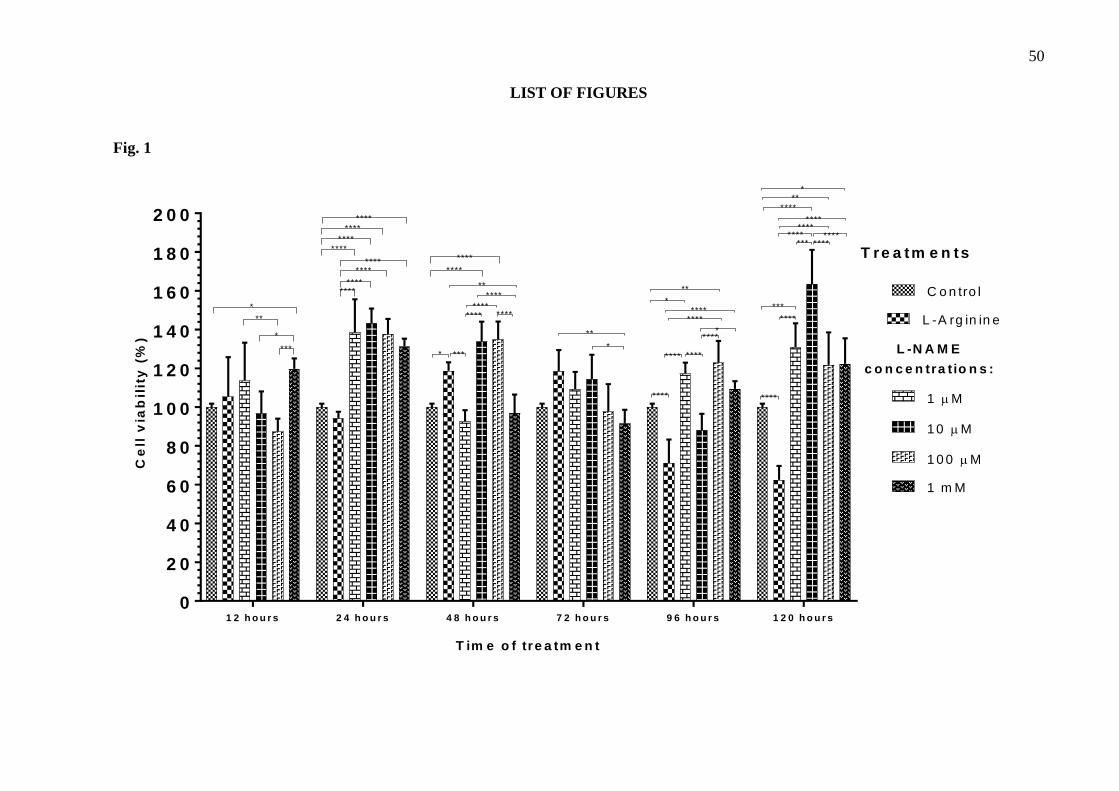
50
LIST OF FIGURES
Fig. 1
1 2 h o u r s 2 4 h o u r s 4 8 h o u r s 7 2 h o u r s 9 6 h o u r s 1 2 0 h o u r s
0
2 0
4 0
6 0
8 0
1 0 0
1 2 0
1 4 0
1 6 0
1 8 0
2 0 0
T im e o f tre a tm e n t
Ce
ll v
iab
ilit
y (
%)
C o n tro l
L -A rg in in e
1 M
10 M
1 0 0 M
1 m M
*
**
*
***
********
********
********
********
*
****
****
***
**
********
****
****
**
*
*****
****
********
****
**
*
****
***********
********
****
****
***
****
***
****
T re a tm e n ts
L -N A M E
c o n c e n tra t io n s :
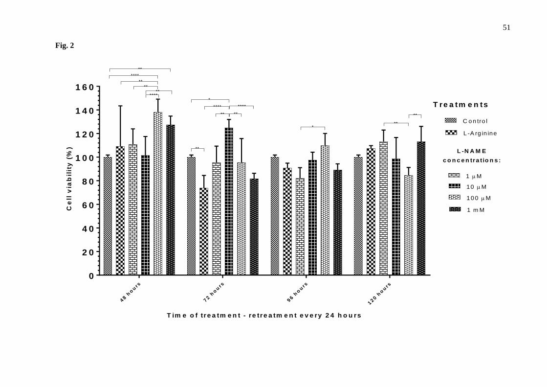
51
Fig. 2
48 h
ou
rs
72 h
ou
rs
96 h
ou
rs
120 h
ou
rs
0
2 0
4 0
6 0
8 0
1 0 0
1 2 0
1 4 0
1 6 0
T im e o f tre a tm e n t - re tre a tm e n t e v e ry 2 4 h o u rs
Ce
ll v
iab
ilit
y (
%)
C o n tro l
L -A rg in ine
1 M
10 M
100 M
1 m M
**
**
**
****
T re a tm e n ts
L -N A M E
c o n c e n tra t io n s :
******
*
**
** **
**** ****
***
**
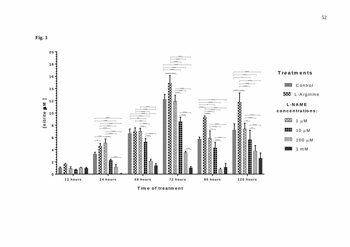
52
Fig. 3
1 2 h o u r s 2 4 h o u r s 4 8 h o u r s 7 2 h o u r s 9 6 h o u r s 1 2 0 h o u r s
0
2
4
6
8
1 0
1 2
1 4
1 6
1 8
2 0
T im e o f tre a tm e n t
[nit
rit
e
M ]
C o n tro l
L -A rg in in e
1 M
10 M
1 0 0 M
1 m M
*
*** ****
****
****
****
****
****
****
********
****
********
***
****
***
******
****
********
****
********
****
********
********
********
****
****
****
****
****
*
********
****
*****
********
****
*
****
****
****
****
****
****
**
****
****
**
****
T re a tm e n ts
L -N A M E
c o n c e n tra t io n s :****
********
****
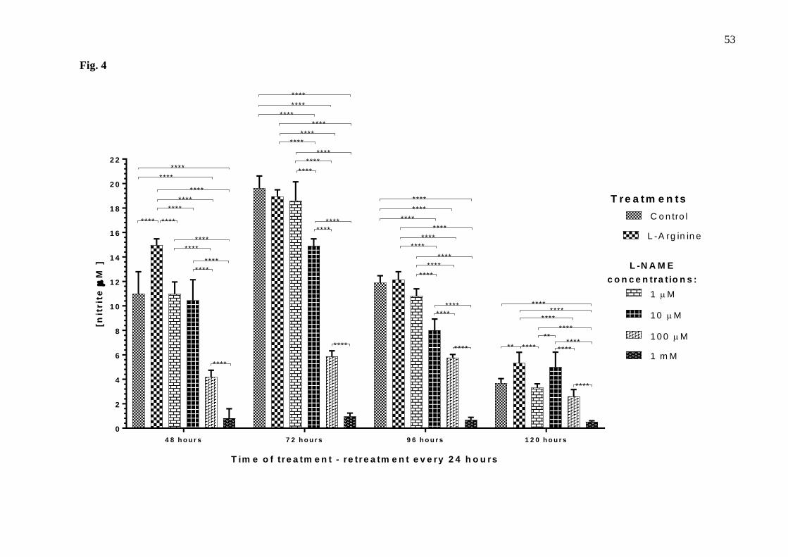
53
Fig. 4
4 8 h o u r s 7 2 h o u r s 9 6 h o u r s 1 2 0 h o u r s
0
2
4
6
8
1 0
1 2
1 4
1 6
1 8
2 0
2 2
T im e o f tre a tm e n t - re tre a tm e n t e v e ry 2 4 h o u rs
[nit
rit
e
M ]
C o n tro l
L -A rg in in e
1 M
10 M
1 0 0 M
1 m M
T re a tm e n ts
L -N A M E
c o n c e n tra t io n s :
**** ****
****
****
****
****
****
****
****
****
****
****
****
****
****
****
****
****
********
****
********
****
****
****
****
****
****
****
********
****
********
**** ** ****
**
****
****
****
****
********
****
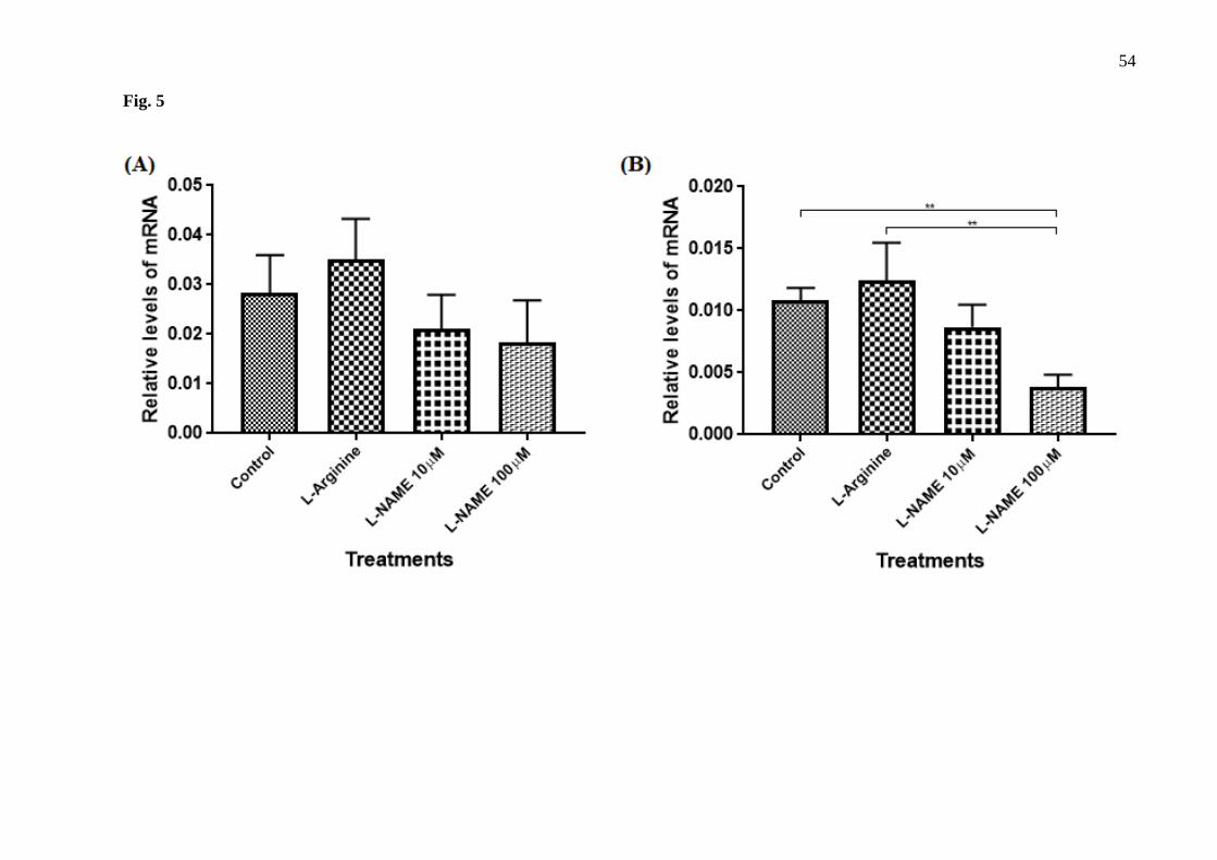
54
Fig. 5
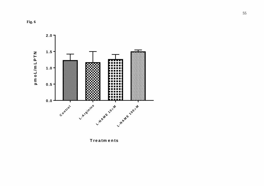
55
Fig. 6
Co
ntr
ol
L-A
rgin
ine
L-N
AM
E 1
0
M
L-N
AM
E 1
00
M
0 .0
0 .5
1 .0
1 .5
2 .0
T re a tm e n ts
µm
oL
/mL
PT
N
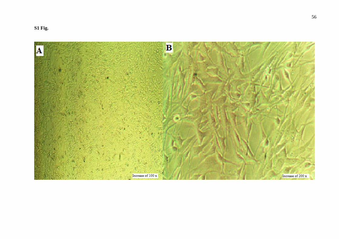
56
S1 Fig.

57
S2 Fig.
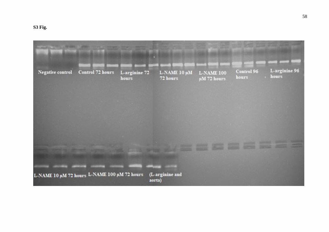
58
S3 Fig.
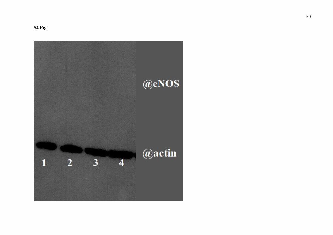
59
S4 Fig.
60
CONCLUSÃO
De acordo com os resultados aqui apresentados, pode-se concluir que o tratamento de
células endoteliais com o L-NAME parece ser capaz de induzir nessas células um estado
semelhante ao que ocorre na disfunção endotelial, reduzindo a biodisponibilidade de NO e,
aparentemente, aumentando o estresse oxidativo e a degradação de NO. Sendo assim, os dados
apontaram que o tratamento com L-NAME 100µM por 96 horas com retratamento foi capaz de
reduzir a concentração de nitritos no meio de cultura celular e reduzir a expressão de p22phox,
que pode estar relacionado à uma redução na expressão e atividade da NOX4, a qual tem função
protetora na vasculatura.
Dessa forma, propomos aqui, pela primeira vez, um método que permite a investigação
de algumas das vias envolvidas na disfunção endotelial in vitro e mostramos a expressão do
gene p22phox na linhagem tEnd.1. Outros experimentos são necessários para uma melhor
caracterização da abordagem proposta, especialmente visando esclarecer o papel das EROs nas
células endoteliais tratadas com L-NAME. Reforçamos, entretanto, que os resultados obtidos
são bastante promissores e contribuem para o entendimento da DE e para pesquisas voltadas
para sua utilização como alvo para o tratamento de patologias cardiovasculares e metabólicas,
como a HAS, aterosclerose e diabetes.
61
REFERÊNCIAS
ARESE, Marco et al. Regulation of nitric oxide synthesis in uraemia. Nephrology Dialysis
Transplantation, v. 10, n. 8, p. 1386-1397, 1995.
ARNAL, J. F. et al. Endothelium-derived nitric oxide and vascular physiology and
pathology. Cellular and Molecular Life Sciences CMLS, v. 55, n. 8-9, p. 1078-1087, 1999.
https://doi.org/10.1007/s000180050358
AUGUSTYNIAK, R. A. et al. Nitric oxide pathway as new drug targets for refractory
hypertension. Current pharmaceutical design, v. 11, n. 25, p. 3307-3315, 2005.
https://doi.org/10.2174/138161205774424672
BABIOR, B. M. The NADPH oxidase of endothelial cells. IUBMB life, v. 50, n. 4‐5, p. 267-
269, 2000. https://doi.org/10.1080/15216540051080976
BATLOUNI, M. Endotélio e hipertensão arterial. Revista brasileira de hipertensão, v. 8, n.
3, p. 328-338, 2001.
BONOMINI, F.; RODELLA, L. F.; REZZANI, R. Metabolic syndrome, aging and involvement
of oxidative stress. Aging and disease, v. 6, n. 2, p. 109, 2015.
https://doi.org/10.14336/AD.2014.0305
CARVALHO, M. H. C. et al. Hipertensão arterial: o endotélio e suas múltiplas funções. Rev
Bras Hipertens, v. 8, n. 1, p. 76-88, 2001.
CELERMAJER, David S. et al. Non-invasive detection of endothelial dysfunction in children
and adults at risk of atherosclerosis. The lancet, v. 340, n. 8828, p. 1111-1115, 1992.
https://doi.org/10.1016/0140-6736(92)93147-F
COUTO, G. K. et al. Double disruption of α2A‐and α2C‐adrenoceptors induces endothelial
dysfunction in mouse small arteries: role of nitric oxide synthase uncoupling. Experimental
physiology, v. 99, n. 10, p. 1427-1438, 2014. https://doi.org/10.1113/expphysiol.2014.079236
DAIBER, A. et al. New Therapeutic Implications of Endothelial Nitric Oxide Synthase (eNOS)
Function/Dysfunction in Cardiovascular Disease. International journal of molecular
sciences, v. 20, n. 1, p. 187, 2019. https://doi.org/10.3390/ijms20010187
62
DAIBER, A. et al. Targeting vascular (endothelial) dysfunction. British journal of
pharmacology, v. 174, n. 12, p. 1591-1619, 2017. https://doi.org/10.1111/bph.13517
DAVEL, A. P. et al. Endothelial dysfunction in cardiovascular and endocrine-metabolic
diseases: an update. Brazilian Journal of Medical and Biological Research, v. 44, n. 9, p.
920-932, 2011. https://doi.org/10.1590/S0100-879X2011007500104
DAVEL, A. P.; BRUM, P. C.; ROSSONI, L. V. Isoproterenol induces vascular oxidative stress
and endothelial dysfunction via a Giα-coupled β2-adrenoceptor signaling pathway. PLoS One,
v. 9, n. 3, p. e91877, 2014. https://doi.org/10.1371/journal.pone.0091877
DRUMMOND, G. R.; SOBEY, C. G. Endothelial NADPH oxidases: which NOX to target in
vascular disease?. Trends in Endocrinology & Metabolism, v. 25, n. 9, p. 452-463, 2014.
https://doi.org/10.1016/j.tem.2014.06.012
DUSSE, L. M. S. A.; VIEIRA, L.M.; CARVALHO, M. G. Revisão sobre óxido nítrico. Jornal
Brasileiro de Patologia e Medicina Laboratorial, v. 39, n. 4, p. 343-50, 2003.
https://doi.org/10.1590/S1676-24442003000400012
ENDEMANN, D. H.; SCHIFFRIN, E. L. Endothelial dysfunction. Journal of the American
Society of Nephrology, v. 15, n. 8, p. 1983-1992, 2004.
https://doi.org/10.1097/01.ASN.0000132474.50966.DA
EVORA, R. B. et al. Cardiovascular therapeutics targets on the NO–sGC–cGMP signaling
pathway: a critical overview. Current drug targets, v. 13, n. 9, p. 1207-1214, 2012.
https://doi.org/10.2174/138945012802002348
FALCONER, D. et al. Nitric oxide donors for peripheral artery disease. Current opinion in
pharmacology, v. 39, p. 77-85, 2018. https://doi.org/10.1016/j.coph.2018.02.009
FLORA FILHO, R.; ZILBERSTEIN, B. Óxido nítrico: o simples mensageiro percorrendo a
complexidade. Metabolismo, síntese e funções. Revista da Associação Médica Brasileira, v.
46, n. 3, p. 265-271, 2000. https://doi.org/10.1590/S0104-42302000000300012
FOMINA, M. A. et al. In Vitro Effects of Sodium Nitroprusside and LN ω-Nitroarginine
Methyl Ester (L-NAME) on Activity of Lysosomal Cysteine Proteinases and Lysosomal
Membrane Permeability. Bulletin of Experimental Biology and Medicine, v. 165, n. 1, p. 36-
39, 2018. https://doi.org/10.1007/s10517-018-4093-y
63
FÖRSTERMANN, U.; XIA, N.; LI, H. Roles of vascular oxidative stress and nitric oxide in the
pathogenesis of atherosclerosis. Circulation research, v. 120, n. 4, p. 713-735, 2017.
https://doi.org/10.1161/CIRCRESAHA.116.309326
FRANCELINO, A. A. et al. Estudo da expressão de ligantes e receptores de matriz
extracelular nas células endoteliais tímicas e sua participação na migração. 2008. 78 f.
Dissertação. Programa de Pós-Graduação em Ciências da Saúde, Universidade Federal de
Alagoas. 2008.
GHEIBI, S. et al. Regulation of vascular tone homeostasis by NO and H2S: Implications in
hypertension. Biochemical pharmacology, v. 149, p. 42-59, 2018.
https://doi.org/10.1016/j.bcp.2018.01.017
GHIMIRE, K. et al. Nitric oxide: what’s new to NO?. American Journal of Physiology-Cell
Physiology, v. 312, n. 3, p. 254-C262, 2016. https://doi.org/10.1152/ajpcell.00315.2016
GHOSH, A. et al. Role of free fatty acids in endothelial dysfunction. Journal of biomedical
science, v. 24, n. 1, p. 50, 2017. https://doi.org/10.1186/s12929-017-0357-5
GIMBRONE JÚNIOR, M. A.; GARCÍA-CARDEÑA, G. Vascular endothelium,
hemodynamics, and the pathobiology of atherosclerosis. Cardiovascular pathology, v. 22, n.
1, p. 9-15, 2013. https://doi.org/10.1016/j.carpath.2012.06.006
GIRIBELA, C. R. G. et al. Função e disfunção endotelial: da fisiopatologia às perspectivas de
uso em pesquisa e na prática clínica. Revista Brasileira de Hipertensão, v. 18, n. 1, p. 27-32,
2011.
HAMILTON, C. A. et al. Superoxide excess in hypertension and aging: a common cause of
endothelial dysfunction. Hypertension, v. 37, n. 2, p. 529-534, 2001.
https://doi.org/10.1161/01.HYP.37.2.529
HARRISON, D. G.; GONGORA, M. C. Oxidative stress and hypertension. Medical Clinics,
v. 93, n. 3, p. 621-635, 2009. https://doi.org/10.1016/j.mcna.2009.02.015
HONG, F. et al. Roles of eNOS in atherosclerosis treatment. Inflammation Research, p. 1-13,
2019.
INCALZA, M. A. et al. Oxidative stress and reactive oxygen species in endothelial dysfunction
associated with cardiovascular and metabolic diseases. Vascular pharmacology, v. 100, p. 1-
19, 2018. https://doi.org/10.1016/j.vph.2017.05.005
64
JAMWAL; SHARMA. Vascular endothelium dysfunction: a conservative target in metabolic
disorders. Inflammation Research, v. 67, n. 5, p. 391-405, 2018.
https://doi.org/10.1007/s00011-018-1129-8
KAPTOGE, S. et al. World Health Organization cardiovascular disease risk charts: revised
models to estimate risk in 21 global regions. The Lancet Global Health, v. 7, n. 10, p. e1332-
e1345, 2019.
KHADDAJ MALLAT, R. et al. The vascular endothelium: a regulator of arterial tone and
interface for the immune system. Critical reviews in clinical laboratory sciences, v. 54, n. 7-
8, p. 458-470, 2017. https://doi.org/10.1080/10408363.2017.1394267
KIERSZENBAUM, A.; TRES, L. Histologia e biologia celular: uma introdução à patologia.
Elsevier Brasil, 2016.
KONUKOGLU, D.; UZUN, H.. Endothelial dysfunction and hypertension. In: Hypertension:
from basic research to clinical practice. Springer, Cham, p. 511-540, 2016.
https://doi.org/10.1007/5584_2016_90
KOPINCOVÁ, J.; PÚZSEROVÁ, A. BERNÁTOVÁ, I. L-NAME in the cardiovascular
system–nitric oxide synthase activator?. Pharmacological Reports, v. 64, n. 3, p. 511-520,
2012. https://doi.org/10.1016/S1734-1140(12)70846-0
KOWALTOWSKI, Alicia Juliana. Participação da mitocôndria na regulação da
viabilidade celular. 2004. Tese de Doutorado. Universidade de São Paulo.
KRINSKI, K. C. et al. Tratamento da disfunção endotelial. Arquivos de Ciências da Saúde
da UNIPAR, v. 11, n. 1, 2007.
LANGBEIN, H. et al. NADPH oxidase 4 protects against development of endothelial
dysfunction and atherosclerosis in LDL receptor deficient mice. European heart journal, v.
37, n. 22, p. 1753-1761, 2015.
https://doi.org/10.1093/eurheartj/ehv564
LIAO, Y. et al. NADPH oxidase 4 and endothelial nitric oxide synthase contribute to
endothelial dysfunction mediated by histone methylations in metabolic memory. Free Radical
Biology and Medicine, v. 115, p. 383-394, 2018.
https://doi.org/10.1016/j.freeradbiomed.2017.12.017
65
LINS, M. P. et al. Participação do hormônio do crescimento nas interações entre
timócitos/endotélio tímico envolvidas no processo migratório. 2015. Dissertação. Instituto
de Ciências Biológicas e da Saúde, Universidade Federal de Alagoas. 2015
Growth hormone modulates in vitro endothelial cell migration and formation of capillary‐like
structures. Cell biology international, v. 41, n. 5, p. 577-584, 2017.
https://doi.org/10.1002/cbin.10747
MONTEZANO, A. C. et al. Oxidative stress and human hypertension: vascular mechanisms,
biomarkers, and novel therapies. Canadian Journal of Cardiology, v. 31, n. 5, p. 631-641,
2015. https://doi.org/10.1016/j.cjca.2015.02.008
NISIMOTO, Yukio et al. Constitutive NADPH-dependent electron transferase activity of the
Nox4 dehydrogenase domain. Biochemistry, v. 49, n. 11, p. 2433-2442, 2010.
https://doi.org/10.1021/bi9022285
PARK, K.; PARK, W. J. Endothelial dysfunction: clinical implications in cardiovascular
disease and therapeutic approaches. Journal of Korean medical science, v. 30, n. 9, p. 1213-
1225, 2015. https://doi.org/10.3346/jkms.2015.30.9.1213
PEARSON EDUCATION. Nitric Oxide Vasodilation. 2012. Disponível em:
<http://kalde.bwong.co/nitric-oxide-vasodilation/>. Acesso em: Jan. 2020.
PETERSON, D. A. et al. The non specificity of specific nitric oxide synthase
inhibitors. Biochemical and biophysical research communications, v. 187, n. 2, p. 797-801,
1992. https://doi.org/10.1016/0006-291X(92)91266-S
PFEIFFER, S. et al. Inhibition of nitric oxide synthesis by NG‐nitro‐L‐arginine methyl ester
(L‐NAME): requirement for bioactivation to the free acid, NG‐nitro‐L‐arginine. British
journal of pharmacology, v. 118, n. 6, p. 1433-1440, 1996. https://doi.org/10.1111/j.1476-
5381.1996.tb15557.x
PREMER, C. et al. Rethinking endothelial dysfunction as a crucial target in fighting heart
failure. Mayo Clinic Proceedings: Innovations, Quality & Outcomes, v. 3, n. 1, p. 1-13,
2019. https://doi.org/10.1016/j.mayocpiqo.2018.12.006
QUEIROZ, S. L.; BATISTA, A. A. Funções biológicas do óxido nítrico. Química Nova, v. 22,
n. 4, p. 584-590, 1999. https://doi.org/10.1590/S0100-40421999000400017
66
REES, D. D. et al. Characterization of three inhibitors of endothelial nitric oxide synthase in
vitro and in vivo. British journal of pharmacology, v. 101, n. 3, p. 746-752, 1990.
https://doi.org/10.1111/j.1476-5381.1990.tb14151.x
RIBEIRO, S. M. R. et al. A formação e os efeitos das espécies reativas de oxigênio no meio
biológico. Bioscience journal, v. 21, n. 3, 2005.
RUDIC, R. D.; SESSA, W. C. Human genetics 99: the cardiovascular system. Nitric oxide in
endothelial dysfunction and vascular remodeling: clinical correlates and experimental
links. Am J Hum Genet, v. 64, p. 673-7, 1999. https://doi.org/10.1086/302304
SANTOS, J. E. et al. Ação conjunta do fator-1 de crescimento semelhante à insulina (IGF-
1) e da quimiocina CXCL12 na migração de linfócitos.72f. 2017.
SENA, C. M.; PEREIRA, A. M.; SEIÇA, R. Endothelial dysfunction—a major mediator of
diabetic vascular disease. Biochimica et Biophysica Acta (BBA)-Molecular Basis of Disease,
v. 1832, n. 12, p. 2216-2231, 2013. https://doi.org/10.1016/j.bbadis.2013.08.006
SIES, H. Oxidative stress: a concept in redox biology and medicine. Redox biology, v. 4, p.
180-183, 2015.
https://doi.org/10.1016/j.redox.2015.01.002
SILVA, Wallison Junio Martins; FERRARI, Carlos Kusano Bucalen. Metabolismo
mitocondrial, radicais livres e envelhecimento. Revista Brasileira de Geriatria e
Gerontologia, v. 14, n. 3, p. 441-451, 2011. https://doi.org/10.1590/S1809-
98232011000300005
SINHA, N.; KUMAR DABLA, P. Oxidative stress and antioxidants in hypertension–a current
review. Current hypertension reviews, v. 11, n. 2, p. 132-142, 2015.
https://doi.org/10.2174/1573402111666150529130922
SIRAGUSA, M.; FLEMING, I. The eNOS signalosome and its link to endothelial dysfunction.
Pflügers Archiv-European Journal of Physiology, v. 468, n. 7, p. 1125-1137, 2016.
https://doi.org/10.1007/s00424-016-1839-0
SOARES, N. P. Células endoteliais. Disponível em:
<https://www.infoescola.com/biologia/celula-endotelial/>. Acesso em: Janeiro 2020
TÖRÖK, J. Participation of nitric oxide in different models of experimental
hypertension. Physiological Research, v. 57, n. 6, 2008.
67
TORTORA, G. J. Princípios de anatomia humana. Tradução de: Principles of
human anatomy, Rio de Janeiro: Guanabara Koogan. 12, 523, 2013.
VANHOUTTE, P. M. et al. Endothelial dysfunction and vascular disease–a 30th anniversary
update. Acta physiologica, v. 219, n. 1, p. 22-96, 2017. https://doi.org/10.1111/apha.12646
VARADHARAJ, S. et al. Endothelial nitric oxide synthase uncoupling: a novel pathway in
OSA induced vascular endothelial dysfunction. Respiratory physiology & neurobiology, v.
207, p. 40-47, 2015. https://doi.org/10.1016/j.resp.2014.12.012
WEFERS, H.; SIES, H. Oxidation of glutathione by the superoxide radical to the disulfide and
the sulfonate yielding singlet oxygen. European Journal of Biochemistry, v. 137, n. 1‐2, p.
29-36, 1983. https://doi.org/10.1111/j.1432-1033.1983.tb07791.x
WENCESLAU, C. F.; ROSSONI, L. V. Rostafuroxin ameliorates endothelial dysfunction and
oxidative stress in resistance arteries from deoxycorticosterone acetate-salt hypertensive rats:
the role of Na+ K+-ATPase/cSRC pathway. Journal of hypertension, v. 32, n. 3, p. 542-554,
2014. https://doi.org/10.1097/HJH.0000000000000059
WILLIAMS, G. et al. Nitric oxide manipulation: a therapeutic target for peripheral arterial
disease?. Cardiology research and practice, v. 2012, 2012.
https://doi.org/10.1155/2012/656247
WILLIAMS, R. L.; COURTNEIDGE, S. A.; WAGNER, E. F. Embryonic lethalities and
endothelial tumors in chimeric mice expressing polyoma virus middle T oncogene. Cell, v. 52,
n. 1, p. 121-131, 1988. https://doi.org/10.1016/0092-8674(88)90536-3
YUYUN, M. F.; NG, L. L.; NG, G. A. Endothelial dysfunction, endothelial nitric oxide
bioavailability, tetrahydrobiopterin, and 5-methyltetrahydrofolate in cardiovascular disease.
Where are we with therapy?. Microvascular research, v. 119, p. 7-12, 2018.
https://doi.org/10.1016/j.mvr.2018.03.012
68
ANEXO 1
Normas para publicação de artigo no periódico PLOS ONE
PUBLISH WITH PLOS ONE
Submission Guidelines
Related information for authors
• Submission system
• Journal scope and publication criteria
• Getting started guide
• Guidelines for revisions
• Publication fees
Style and Format
File
format
Manuscript files can be in the following formats: DOC, DOCX, or RTF. Microsoft
Word documents should not be locked or protected.
LaTeX manuscripts must be submitted as PDFs. Read the LaTeX guidelines.
Length Manuscripts can be any length. There are no restrictions on word count, number of
figures, or amount of supporting information.
We encourage you to present and discuss your findings concisely.
Font Use a standard font size and any standard font, except for the font named “Symbol”.
To add symbols to the manuscript, use the Insert → Symbol function in your word
processor or paste in the appropriate Unicode character.
Headin
gs
Limit manuscript sections and sub-sections to 3 heading levels. Make sure heading
levels are clearly indicated in the manuscript text.
Layout
and
spacing
Manuscript text should be double-spaced.
Do not format text in multiple columns.
Page
and
line
number
s
Include page numbers and line numbers in the manuscript file. Use continuous line
numbers (do not restart the numbering on each page).
69
Footno
tes
Footnotes are not permitted. If your manuscript contains footnotes, move the
information into the main text or the reference list, depending on the content.
Langua
ge
Manuscripts must be submitted in English.
You may submit translations of the manuscript or abstract as supporting
information. Read the supporting information guidelines.
Abbrev
iations
Define abbreviations upon first appearance in the text.
Do not use non-standard abbreviations unless they appear at least three times in the
text.
Keep abbreviations to a minimum.
Refere
nce
style
PLOS uses “Vancouver” style, as outlined in the ICMJE sample references.
See reference formatting examples and additional instructions below.
Equati
ons
We recommend using MathType for display and inline equations, as it will provide
the most reliable outcome. If this is not possible, Equation Editor or
Microsoft's Insert→Equation function is acceptable.
Avoid using MathType, Equation Editor, or the Insert→Equation function to insert
single variables (e.g., “a² + b² = c²”), Greek or other symbols (e.g., β, Δ, or ′
[prime]), or mathematical operators (e.g., x, ≥, or ±) in running text. Wherever
possible, insert single symbols as normal text with the correct Unicode (hex) values.
Do not use MathType, Equation Editor, or the Insert→Equation function for only a
portion of an equation. Rather, ensure that the entire equation is included. Equations
should not contain a mix of different equation tools. Avoid “hybrid” inline or
display equations, in which part is text and part is MathType, or part is MathType
and part is Equation Editor.
Nomen
clature
Use correct and established nomenclature wherever possible.
Units of
measurement
Use SI units. If you do not use these exclusively, provide the SI
value in parentheses after each value. Read more about SI units.
Drugs Provide the Recommended International Non-Proprietary Name (rINN).
Species names
Write in italics (e.g., Homo sapiens). Write out in full the genus and
species, both in the title of the manuscript and at the first mention of an
organism in a paper. After first mention, the first letter of the genus
name followed by the full species name may be used (e.g., H. sapiens).
70
Genes,
mutations,
genotypes, and
alleles
Write in italics. Use the recommended name by consulting the
appropriate genetic nomenclature database (e.g., HGNC for human
genes; we strongly recommend using this tool to check against
previously approved names). It is sometimes advisable to indicate the
synonyms for the gene the first time it appears in the text. Gene prefixes
such as those used for oncogenes or cellular localization should be
shown in roman typeface (e.g., v-fes, c-MYC).
Allergens
The systematic allergen nomenclature of the World Health
Organization/International Union of Immunological Societies
(WHO/IUIS) Allergen Nomenclature Sub-committee should be used
for manuscripts that include the description or use of allergenic
proteins. For manuscripts describing new allergens, the systematic
name of the allergen should be approved by the WHO/IUIS Allergen
Nomenclature Sub-Committee prior to manuscript publication.
Examples of the systematic allergen nomenclature can be found
at the WHO/IUIS Allergen Nomenclature site.
Copyediting manuscripts
Prior to submission, authors who believe their manuscripts would benefit from professional
editing are encouraged to use language-editing and copyediting services. Obtaining this
service is the responsibility of the author, and should be done before initial submission. These
services can be found on the web using search terms like “scientific editing service” or
“manuscript editing service.”
Submissions are not copyedited before publication.
Submissions that do not meet the PLOS ONE publication criterion for language
standards may be rejected.
Manuscript Organization
Manuscripts should be organized as follows. Instructions for each element appear below the
list.
Beginning
section
The following elements are required, in order:
• Title page: List title, authors, and affiliations as first page of manuscript
71
• Abstract
• Introduction
Middle
section
The following elements can be renamed as needed and presented in any order:
• Materials and Methods
• Results
• Discussion
• Conclusions (optional)
Ending
section
The following elements are required, in order:
• Acknowledgments
• References
• Supporting information captions (if applicable)
Other
elements
• Figure captions are inserted immediately after the first paragraph in
which the figure is cited. Figure files are uploaded separately.
• Tables are inserted immediately after the first paragraph in which they
are cited.
• Supporting information files are uploaded separately.
Please refer to our downloadable sample files to ensure that your submission meets our
formatting requirements:
• Download sample title, author list, and affiliations page (PDF)
• Download sample manuscript body (PDF)
Viewing Figures and Supporting Information in the compiled submission PDF
The compiled submission PDF includes low-resolution preview images of the figures after the
reference list. The function of these previews is to allow you to download the entire
submission as quickly as possible. Click the link at the top of each preview page to download
a high-resolution version of each figure. Links to download Supporting Information files are
also available after the reference list.
Parts of a Submission
• Title
Include a full title and a short title for the manuscript.
Title Length Guidelines Examples
72
Full
title
250
characters
Specific, descriptive, concise,
and comprehensible to readers
outside the field
Impact of cigarette smoke exposure on innate
immunity: A Caenorhabditis elegans model
Solar drinking water disinfection (SODIS) to
reduce childhood diarrhoea in rural Bolivia: A
cluster-randomized, controlled trial
Short
title
100
characters
State the topic of the study Cigarette smoke exposure and innate
immunity
SODIS and childhood diarrhoea
Titles should be written in sentence case (only the first word of the text, proper nouns, and
genus names are capitalized). Avoid specialist abbreviations if possible. For clinical trials,
systematic reviews, or meta-analyses, the subtitle should include the study design.
• Author list
Authorship requirements
All authors must meet the criteria for authorship as outlined in the authorship policy. Those
who contributed to the work but do not meet the criteria for authorship can be mentioned in
the Acknowledgments. Read more about Acknowledgments.
The corresponding author must provide an ORCID iD at the time of submission by entering it
in the user profile in the submission system. Read more about ORCID.
Author names and affiliations
Enter author names on the title page of the manuscript and in the online submission system.
On the title page, write author names in the following order:
• First name (or initials, if used)
• Middle name (or initials, if used)
• Last name (surname, family name)
Each author on the list must have an affiliation. The affiliation includes department,
university, or organizational affiliation and its location, including city, state/province (if
applicable), and country. Authors have the option to include a current address in addition to
the address of their affiliation at the time of the study. The current address should be listed in
the byline and clearly labeled “current address.” At a minimum, the address must include the
author’s current institution, city, and country.
73
If an author has multiple affiliations, enter all affiliations on the title page only. In the
submission system, enter only the preferred or primary affiliation. Author affiliations will be
listed in the typeset PDF article in the same order that authors are listed in the submission.
Author names will be published exactly as they appear in the manuscript file. Please double-
check the information carefully to make sure it is correct.
Corresponding author
The submitting author is automatically designated as the corresponding author in the
submission system. The corresponding author is the primary contact for the journal office and
the only author able to view or change the manuscript while it is under editorial consideration.
The corresponding author role may be transferred to another coauthor. However, note that
transferring the corresponding author role also transfers access to the manuscript. (To
designate a new corresponding author while the manuscript is still under consideration, watch
the video tutorial below.)
Only one corresponding author can be designated in the submission system, but this does not
restrict the number of corresponding authors that may be listed on the article in the event of
publication. Whoever is designated as a corresponding author on the title page of the
manuscript file will be listed as such upon publication. Include an email address for each
corresponding author listed on the title page of the manuscript.
How to select a new corresponding author in Editorial Manager
Consortia and group authorship
If a manuscript is submitted on behalf of a consortium or group, include its name in the
manuscript byline. Do not add it to the author list in the submission system. You may include
the full list of members in the Acknowledgments or in a supporting information file.
PubMed only indexes individual consortium or group author members listed in the article
byline. If included, these individuals must qualify for authorship according to our criteria.
Read the group authorship policy.
• Author contributions
Provide at minimum one contribution for each author in the submission system. Use the
CRediT taxonomy to describe each contribution. Read the policy and the full list of roles.
Contributions will be published with the final article, and they should accurately reflect
contributions to the work. The submitting author is responsible for completing this
74
information at submission, and we expect that all authors will have reviewed, discussed, and
agreed to their individual contributions ahead of this time.
PLOS ONE will contact all authors by email at submission to ensure that they are aware of the
submission.
• Cover letter
Upload a cover letter as a separate file in the online system. The length limit is 1 page.
The cover letter should include the following information:
• Summarize the study’s contribution to the scientific literature
• Relate the study to previously published work
• Specify the type of article (for example, research article, systematic review, meta-
analysis, clinical trial)
• Describe any prior interactions with PLOS regarding the submitted manuscript
• Suggest appropriate Academic Editors to handle your manuscript (see the full list of
Academic Editors)
• List any opposed reviewers
IMPORTANT: Do not include requests to reduce or waive publication fees in the cover letter.
This information will be entered separately in the online submission system.
Read about publication fee assistance.
• Title page
The title, authors, and affiliations should all be included on a title page as the first page of the
manuscript file.
Download our sample title, author list, and affiliations page (PDF)
• Abstract
The Abstract comes after the title page in the manuscript file. The abstract text is also entered
in a separate field in the submission system.
The Abstract should:
• Describe the main objective(s) of the study
• Explain how the study was done, including any model organisms used, without
methodological detail
• Summarize the most important results and their significance
• Not exceed 300 words
Abstracts should not include:
• Citations
75
• Abbreviations, if possible
• Introduction
The introduction should:
• Provide background that puts the manuscript into context and allows readers outside
the field to understand the purpose and significance of the study
• Define the problem addressed and why it is important
• Include a brief review of the key literature
• Note any relevant controversies or disagreements in the field
• Conclude with a brief statement of the overall aim of the work and a comment about
whether that aim was achieved
• Materials and Methods
The Materials and Methods section should provide enough detail to allow suitably skilled
investigators to fully replicate your study. Specific information and/or protocols for new
methods should be included in detail. If materials, methods, and protocols are well
established, authors may cite articles where those protocols are described in detail, but the
submission should include sufficient information to be understood independent of these
references.
Protocol documents for clinical trials, observational studies, and other non-
laboratory investigations may be uploaded as supporting information. We recommend
depositing laboratory protocols at protocols.io. Read detailed instructions for depositing and
sharing your laboratory protocols.
Human or animal subjects and/or tissue or field sampling
Methods sections describing research using human or animal subjects and/or tissue or field
sampling must include required ethics statements. For details, consult the reporting guidelines
for specific study types.
Data
PLOS journals require authors to make all data underlying the findings described in their
manuscript fully available without restriction, with rare exception.
Large data sets, including raw data, may be deposited in an appropriate public repository. See
our list of recommended repositories.
For smaller data sets and certain data types, authors may provide their data within supporting
information files accompanying the manuscript. Authors should take care to maximize the
accessibility and reusability of the data by selecting a file format from which data can be
76
efficiently extracted (for example, spreadsheets or flat files should be provided rather than
PDFs when providing tabulated data).
For more information on how best to provide data, read our policy on data availability. PLOS
does not accept references to “data not shown.”
Cell lines
Methods sections describing research using cell lines must state the origin of the cell lines
used. See the reporting guidelines for cell line research.
Laboratory protocols
To enhance the reproducibility of your results, we recommend and encourage you to deposit
laboratory protocols in protocols.io, where protocols can be assigned their own persistent
digital object identifiers (DOIs).
To include a link to a protocol in your article:
1. Describe your step-by-step protocol on protocols.io
2. Select Get DOI to issue your protocol a persistent digital object identifier (DOI)
3. Include the DOI link in the Methods section of your manuscript using the following
format provided by protocols.io: http://dx.doi.org/10.17504/protocols.io.[PROTOCOL
DOI]
At this stage, your protocol is only visible to those with the link. This allows editors and
reviewers to consult your protocol when evaluating the manuscript. You can make your
protocols public at any time by selecting Publish on the protocols.io site. Any referenced
protocol(s) will automatically be made public when your article is published.
New taxon names
Methods sections of manuscripts adding new zoological, botanical, or fungal taxon names to
the literature must follow the guidelines for new taxon names.
• Results, Discussion, Conclusions
These sections may all be separate, or may be combined to create a mixed Results/Discussion
section (commonly labeled “Results and Discussion”) or a mixed Discussion/Conclusions
section (commonly labeled “Discussion”). These sections may be further divided into
subsections, each with a concise subheading, as appropriate. These sections have no word
limit, but the language should be clear and concise.
Together, these sections should describe the results of the experiments, the interpretation of
these results, and the conclusions that can be drawn.
77
Authors should explain how the results relate to the hypothesis presented as the basis of the
study and provide a succinct explanation of the implications of the findings, particularly in
relation to previous related studies and potential future directions for research.
PLOS ONE editorial decisions do not rely on perceived significance or impact, so authors
should avoid overstating their conclusions. See the PLOS ONE Criteria for Publication for
more information.
• Acknowledgments
Those who contributed to the work but do not meet our authorship criteria should be listed in
the Acknowledgments with a description of the contribution.
Authors are responsible for ensuring that anyone named in the Acknowledgments agrees to be
named.
PLOS journals publicly acknowledge the indispensable efforts of our editors and reviewers on
an annual basis. To ensure equitable recognition and avoid any appearance of partiality, do
not include editors or peer reviewers—named or unnamed—in the Acknowledgments.
Do not include funding sources in the Acknowledgments or anywhere else in the manuscript
file. Funding information should only be entered in the financial disclosure section of the
submission system.
• References
Any and all available works can be cited in the reference list. Acceptable sources include:
• Published or accepted manuscripts
• Manuscripts on preprint servers, providing the manuscript has a citable DOI or arXiv
URL.
Do not cite the following sources in the reference list:
• Unavailable and unpublished work, including manuscripts that have been submitted
but not yet accepted (e.g., “unpublished work,” “data not shown”). Instead, include
those data as supplementary material or deposit the data in a publicly available
database.
• Personal communications (these should be supported by a letter from the relevant
authors but not included in the reference list)
References are listed at the end of the manuscript and numbered in the order that they appear
in the text. In the text, cite the reference number in square brackets (e.g., “We used the
techniques developed by our colleagues [19] to analyze the data”). PLOS uses the numbered
citation (citation-sequence) method and first six authors, et al.
78
Do not include citations in abstracts.
Make sure the parts of the manuscript are in the correct order before ordering the citations.
Formatting references
Because all references will be linked electronically as much as possible to the papers they
cite, proper formatting of the references is crucial.
PLOS uses the reference style outlined by the International Committee of Medical Journal
Editors (ICMJE), also referred to as the “Vancouver” style. Example formats are listed below.
Additional examples are in the ICMJE sample references.
A reference management tool, EndNote, offers a current style file that can assist you with the
formatting of your references. If you have problems with any reference management program,
please contact the source company's technical support.
Journal name abbreviations should be those found in the National Center for Biotechnology
Information (NCBI) databases.
Source Format
Published
articles
Hou WR, Hou YL, Wu GF, Song Y, Su XL, Sun B, et al. cDNA, genomic
sequence cloning and overexpression of ribosomal protein gene L9 (rpL9) of
the giant panda (Ailuropoda melanoleuca). Genet Mol Res. 2011;10: 1576-
1588.
Devaraju P, Gulati R, Antony PT, Mithun CB, Negi VS. Susceptibility to
SLE in South Indian Tamils may be influenced by genetic selection pressure
on TLR2 and TLR9 genes. Mol Immunol. 2014 Nov 22. pii: S0161-
5890(14)00313-7. doi: 10.1016/j.molimm.2014.11.005.
Note: A DOI number for the full-text article is acceptable as an alternative
to or in addition to traditional volume and page numbers. When providing a
DOI, adhere to the format in the example above with both the label and full
DOI included at the end of the reference (doi:
10.1016/j.molimm.2014.11.005). Do not provide a shortened DOI or the
URL.
Accepted,
unpublished
articles
Same as published articles, but substitute “Forthcoming” for page numbers
or DOI.

79
Source Format
Online articles Huynen MMTE, Martens P, Hilderlink HBM. The health impacts of
globalisation: a conceptual framework. Global Health. 2005;1: 14. Available
from: http://www.globalizationandhealth.com/content/1/1/14
Books Bates B. Bargaining for life: A social history of tuberculosis. 1st ed.
Philadelphia: University of Pennsylvania Press; 1992.
Book chapters Hansen B. New York City epidemics and history for the public. In: Harden
VA, Risse GB, editors. AIDS and the historian. Bethesda: National Institutes
of Health; 1991. pp. 21-28.
Deposited
articles (preprint
s, e-prints, or
arXiv)
Krick T, Shub DA, Verstraete N, Ferreiro DU, Alonso LG, Shub M, et al.
Amino acid metabolism conflicts with protein diversity. arXiv:1403.3301v1
[Preprint]. 2014 [cited 2014 March 17]. Available
from: https://128.84.21.199/abs/1403.3301v1
Kording KP, Mensh B. Ten simple rules for structuring papers. BioRxiv
[Preprint]. 2016 bioRxiv 088278 [posted 2016 Nov 28; revised 2016 Dec
14; revised 2016 Dec 15; cited 2017 Feb 9]: [12 p.]. Available
from: https://www.biorxiv.org/content/10.1101/088278v5 doi:
10.1101/088278
Published media
(print or online
newspapers and
magazine
articles)
Fountain H. For Already Vulnerable Penguins, Study Finds Climate Change
Is Another Danger. The New York Times. 2014 Jan 29 [Cited 2014 March
17]. Available
from: http://www.nytimes.com/2014/01/30/science/earth/climate-change-
taking-toll-on-penguins-study-finds.html
New media
(blogs, web
sites, or other
written works)
Allen L. Announcing PLOS Blogs. 2010 Sep 1 [cited 17 March 2014]. In:
PLOS Blogs [Internet]. San Francisco: PLOS 2006 - . [about 2 screens].
Available from: http://blogs.plos.org/plos/2010/09/announcing-plos-blogs/.
Masters' theses
or doctoral
dissertations
Wells A. Exploring the development of the independent, electronic,
scholarly journal. M.Sc. Thesis, The University of Sheffield. 1999.
Available from: http://cumincad.scix.net/cgi-bin/works/Show?2e09
Databases and
repositories
Roberts SB. QPX Genome Browser Feature Tracks; 2013 [cited 2013 Oct
5]. Database: figshare [Internet]. Available
80
Source Format
(Figshare,
arXiv)
from: http://figshare.com/articles/QPX_Genome_Browser_Feature_Tracks/7
01214
Multimedia
(videos, movies,
or TV shows)
Hitchcock A, producer and director. Rear Window [Film]; 1954. Los
Angeles: MGM.
• Supporting information
Authors can submit essential supporting files and multimedia files along with their
manuscripts. All supporting information will be subject to peer review. All file types can be
submitted, but files must be smaller than 20 MB in size.
Authors may use almost any description as the item name for a supporting information file as
long as it contains an “S” and number. For example, “S1 Appendix” and “S2 Appendix,” “S1
Table” and “S2 Table,” and so forth.
Supporting information files are published exactly as provided, and are not copyedited.
Supporting information captions
List supporting information captions at the end of the manuscript file. Do not submit captions
in a separate file.
The file number and name are required in a caption, and we highly recommend including a
one-line title as well. You may also include a legend in your caption, but it is not required.
Example caption
S1 Text. Title is strongly recommended. Legend is optional.
In-text citations
We recommend that you cite supporting information in the manuscript text, but this is not a
requirement. If you cite supporting information in the text, citations do not need to be in
numerical order.
Read the supporting information guidelines for more details about submitting supporting
information and multimedia files.
• Figures and tables
Figures
Do not include figures in the main manuscript file. Each figure must be prepared and
submitted as an individual file.
Cite figures in ascending numeric order at first appearance in the manuscript file.
81
Read the guidelines for figures and requirements for reporting blot and gel results.
Figure captions
Figure captions must be inserted in the text of the manuscript, immediately following the
paragraph in which the figure is first cited (read order). Do not include captions as part of the
figure files themselves or submit them in a separate document.
At a minimum, include the following in your figure captions:
• A figure label with Arabic numerals, and “Figure” abbreviated to “Fig” (e.g. Fig 1,
Fig 2, Fig 3, etc). Match the label of your figure with the name of the file uploaded at
submission (e.g. a figure citation of “Fig 1” must refer to a figure file named
“Fig1.tif”).
• A concise, descriptive title
The caption may also include a legend as needed.
Read more about figure captions.
Tables
Cite tables in ascending numeric order upon first appearance in the manuscript file.
Place each table in your manuscript file directly after the paragraph in which it is first cited
(read order). Do not submit your tables in separate files.
Tables require a label (e.g., “Table 1”) and brief descriptive title to be placed above the table.
Place legends, footnotes, and other text below the table.
Read the guidelines for tables.
• Statistical reporting
Manuscripts submitted to PLOS ONE are expected to report statistical methods in sufficient
detail for others to replicate the analysis performed. Ensure that results are rigorously reported
in accordance with community standards and that the statistical methods employed are
appropriate for the study design.
Consult the following resources for additional guidance:
• SAMPL guidelines, for general guidance on statistical reporting
• PLOS ONE guidelines, for clinical trials requirements
• PLOS ONE guidelines, for systematic review and meta-analysis requirements
• EQUATOR, for specific reporting guidelines for a range of other study types
Reporting of statistical methods
In the methods, include a section on statistical analysis that reports a detailed description of
the statistical methods. In this section:
82
• List the name and version of any software package used, alongside any relevant
references
• Describe the technical details or procedures required to reproduce the analysis
• Provide the repository identifier for any code used in the analysis (See our code-
sharing policy.)
Statistical reporting guidelines:
• Identify research design and independent variables as being between- or within-
subjects
• For pre-processed data:
o Describe any analysis carried out to confirm the data meets the assumptions of
the analysis performed (e.g. linearity, co-linearity, normality of the
distribution).
o If data were transformed include this information, with a reason for doing so
and a description of the transformation performed
• Provide details of how outliers were treated and your analysis, both with the full
dataset and with the outliers removed
• If relevant, describe how missing/excluded data were handled
• Define the threshold for significance (alpha)
• If appropriate, provide sample sizes, along with a description of how they were
determined. If a sample size calculation was performed, specify the inputs for power,
effect size and alpha. Where relevant, report the number of independent replications
for each experiment.
• For analyses of variance (ANOVAs), detail any post hoc tests that were performed
• Include details of any corrections applied to account for multiple comparisons. If
corrections were not applied, include a justification for not doing so
• Describe all options for statistical procedures. For example, if t-tests were performed,
state whether these were one- or two-tailed. Include details of the type of t-test
conducted (e.g. one sample, within-/between-subjects).
• For step-wise multiple regression analyses:
o Report the alpha level used
o Discuss whether the variables were assessed for collinearity and interaction
o Describe the variable selection process by which the final model was
developed (e.g., forward-stepwise; best subset). See SAMPL guidelines.
83
• For Bayesian analysis explain the choice of prior trial probabilities and how they were
selected. Markov chain Monte Carlo settings should be reported.
Reporting of statistical results
Results must be rigorously and appropriately reported, in keeping with community standards.
• Units of measurement. Clearly define measurement units in all tables and figures.
• Properties of distribution. It should be clear from the text which measures of variance
(standard deviation, standard error of the mean, confidence intervals) and central
tendency (mean, median) are being presented.
• Regression analyses. Include the full results of any regression analysis performed as a
supplementary file. Include all estimated regression coefficients, their standard error,
p-values, and confidence intervals, as well as the measures of goodness of fit.
• Reporting parameters. Test statistics (F/t/r) and associated degrees of freedom should
be provided. Effect sizes and confidence intervals should be reported where
appropriate. If percentages are provided, the numerator and denominator should also
be given.
• P-values. Report exact p-values for all values greater than or equal to 0.001. P-values
less than 0.001 may be expressed as p < 0.001, or as exponentials in studies of genetic
associations.
• Displaying data in plots. Format plots so that they accurately depict the sample
distribution. 3D effects in plots can bias and hinder interpretation of values, so avoid
them in cases where regular plots are sufficient to display the data.
• Open data. As explained in PLOS’s Data Policy, be sure to make individual data
points, underlying graphs and summary statistics available at the time of publication.
Data can be deposited in a repository or included within the Supporting Information
files.
• Data reporting
All data and related metadata underlying the findings reported in a submitted manuscript
should be deposited in an appropriate public repository, unless already provided as part of the
submitted article.
See instructions on providing underlying data to support blot and gel results
Read our policy on data availability.
Repositories may be either subject-specific (where these exist) and accept specific types of
structured data, or generalist repositories that accept multiple data types. We recommend that
authors select repositories appropriate to their field. Repositories may be subject-specific
84
(e.g., GenBank for sequences and PDB for structures), general, or institutional, as long as
DOIs or accession numbers are provided and the data are at least as open as CC BY. Authors
are encouraged to select repositories that meet accepted criteria as trustworthy digital
repositories, such as criteria of the Centre for Research Libraries or Data Seal of Approval.
Large, international databases are more likely to persist than small, local ones.
See our list of recommended repositories.
To support data sharing and author compliance of the PLOS data policy, we have integrated
our submission process with a select set of data repositories. The list is neither representative
nor exhaustive of the suitable repositories available to authors. Current repository integration
partners include Dryad and FlowRepository. Please contact [email protected] to make
recommendations for further partnerships.
Instructions for PLOS submissions with data deposited in an integration partner repository:
• Deposit data in the integrated repository of choice.
• Once deposition is final and complete, the repository will provide you with a dataset
DOI (provisional) and private URL for reviewers to gain access to the data.
• Enter the given data DOI into the full Data Availability Statement, which is requested
in the Additional Information section of the PLOS submission form. Then provide the
URL passcode in the Attach Files section.
If you have any questions, please email us.
• Accession numbers
All appropriate data sets, images, and information should be deposited in an appropriate
public repository. See our list of recommended repositories.
Accession numbers (and version numbers, if appropriate) should be provided in the Data
Availability Statement. Accession numbers or a citation to the DOI should also be provided
when the data set is mentioned within the manuscript.
In some cases authors may not be able to obtain accession numbers of DOIs until the
manuscript is accepted; in these cases, the authors must provide these numbers at acceptance.
In all other cases, these numbers must be provided at full submission.
Identifiers
As much as possible, please provide accession numbers or identifiers for all entities such as
genes, proteins, mutants, diseases, etc., for which there is an entry in a public database, for
example:
• Ensembl
• Entrez Gene
85
• FlyBase
• InterPro
• Mouse Genome Database (MGD)
• Online Mendelian Inheritance in Man (OMIM)
• PubChem
Identifiers should be provided in parentheses after the entity on first use.
• Striking image
You can choose to upload a “Striking Image” that we may use to represent your article online
in places like the journal homepage or in search results.
The striking image must be derived from a figure or supporting information file from the
submission, i.e., a cropped portion of an image or the entire image. Striking images should
ideally be high resolution, eye-catching, single panel images, and should ideally avoid
containing added details such as text, scale bars, and arrows.
If no striking image is uploaded, we will designate a figure from the submission as the
striking image.
Striking images should not contain potentially identifying images of people. Read our policy
on identifying information.
The PLOS licenses and copyright policy also applies to striking images.
Additional Information Requested at Submission
• Financial Disclosure Statement
This information should describe sources of funding that have supported the work. It is
important to gather these details prior to submission because your financial disclosure
statement cannot be changed after initial submission without journal approval. If your
manuscript is published, your statement will appear in the Funding section of the article.
Enter this statement in the Financial Disclosure section of the submission form. Do not
include it in your manuscript file.
The statement should include:
• Specific grant numbers
• Initials of authors who received each award
• Full names of commercial companies that funded the study or authors
• Initials of authors who received salary or other funding from commercial companies
• URLs to sponsors’ websites
Also state whether any sponsors or funders (other than the named authors) played any role in:
86
• Study design
• Data collection and analysis
• Decision to publish
• Preparation of the manuscript
If they had no role in the research, include this sentence: “The funders had no role in study
design, data collection and analysis, decision to publish, or preparation of the manuscript.”
If the study was unfunded, include this sentence as the Financial Disclosure statement: “The
author(s) received no specific funding for this work."
Read our policy on disclosure of funding sources.
• Competing interests
This information should not be in your manuscript file; you will provide it via our submission
system.
All potential competing interests must be declared in full. If the submission is related to
any patents, patent applications, or products in development or for market, these details,
including patent numbers and titles, must be disclosed in full.
Read our policy on competing interests.
• Manuscripts disputing published work
For manuscripts disputing previously published work, it is PLOS ONE policy to invite a
signed review by the disputed author during the peer review process. This procedure is aimed
at ensuring a thorough, transparent, and productive review process.
If the disputed author chooses to submit a review, it must be returned in a timely fashion and
contain a full declaration of all competing interests. The Academic Editor will consider any
such reviews in light of the competing interest.
Authors submitting manuscripts disputing previous work should explain the relationship
between the manuscripts in their cover letter, and will be required to confirm that they accept
the conditions of this review policy before the manuscript is considered further.
• Related manuscripts
Upon submission, authors must confirm that the manuscript, or any related manuscript, is not
currently under consideration or accepted elsewhere. If related work has been submitted
to PLOS ONE or elsewhere, authors must include a copy with the submitted article.
Reviewers will be asked to comment on the overlap between related submissions.
We strongly discourage the unnecessary division of related work into separate manuscripts,
and we will not consider manuscripts that are divided into “parts.” Each submission to PLOS
ONE must be written as an independent unit and should not rely on any work that has not
87
already been accepted for publication. If related manuscripts are submitted to PLOS ONE, the
authors may be advised to combine them into a single manuscript at the editor's discretion.
Read our policies on related manuscripts.
• Preprints
PLOS encourages authors to post preprints as a way to accelerate the dissemination of
research and supports authors who wish to share their work early and receive feedback before
formal peer review. Deposition of manuscripts with preprint servers does not impact
consideration of the manuscript at any PLOS journal.
Authors posting on bioRxiv or medRxiv may submit directly to relevant PLOS journals
through the direct transfer to journal service.
Authors submitting manuscripts in the life sciences to PLOS ONE may opt-in to post their
work on bioRxiv during the PLOS ONE initial submission process.
Read more about preprints.
Learn how to post a preprint to bioRxiv during PLOS ONE initial submission.
Guidelines for Specific Study Types
• Human subjects research
All research involving human participants must have been approved by the
authors’ Institutional Review Board (IRB) or by equivalent ethics committee(s), and must
have been conducted according to the principles expressed in the Declaration of Helsinki.
Authors should be able to submit, upon request, a statement from the IRB or ethics committee
indicating approval of the research. We reserve the right to reject work that we believe has not
been conducted to a high ethical standard, even when formal approval has been obtained.
Subjects must have been properly instructed and have indicated that they consent to
participate by signing the appropriate informed consent paperwork. Authors may be asked to
submit a blank, sample copy of a subject consent form. If consent was verbal instead of
written, or if consent could not be obtained, the authors must explain the reason in the
manuscript, and the use of verbal consent or the lack of consent must have been approved by
the IRB or ethics committee.
All efforts should be made to protect patient privacy and anonymity. Identifying information,
including photos, should not be included in the manuscript unless the information is crucial
and the individual has provided written consent by completing the Consent Form for

88
Publication in a PLOS Journal (PDF). Download additional translations of the form from
the Downloads and Translations page. More information about patient privacy, anonymity,
and informed consent can be found in the International Committee of Medical Journal
Editors (ICMJE) Privacy and Confidentiality guidelines.
Manuscripts should conform to the following reporting guidelines:
• Studies of diagnostic accuracy: STARD
• Observational studies: STROBE
• Microarray experiments: MIAME
• Other types of health-related research: Consult the EQUATOR web site for
appropriate reporting guidelines
Methods sections of papers on research using human subjects or samples must include ethics
statements that specify:
• The name of the approving institutional review board or equivalent committee(s). If
approval was not obtained, the authors must provide a detailed statement explaining
why it was not needed
• Whether informed consent was written or oral. If informed consent was oral, it must
be stated in the manuscript:
o Why written consent could not be obtained
o That the Institutional Review Board (IRB) approved use of oral consent
o How oral consent was documented
For studies involving humans categorized by race/ethnicity, age, disease/disabilities, religion,
sex/gender, sexual orientation, or other socially constructed groupings, authors should:
• Explicitly describe their methods of categorizing human populations
• Define categories in as much detail as the study protocol allows
• Justify their choices of definitions and categories, including for example whether any
rules of human categorization were required by their funding agency
• Explain whether (and if so, how) they controlled for confounding variables such as
socioeconomic status, nutrition, environmental exposures, or similar factors in their
analysis
In addition, outmoded terms and potentially stigmatizing labels should be changed to more
current, acceptable terminology. Examples: “Caucasian” should be changed to “white” or “of
[Western] European descent” (as appropriate); “cancer victims” should be changed to
“patients with cancer.”
89
For papers that include identifying, or potentially identifying, information, authors
must download the Consent Form for Publication in a PLOS Journal, which the individual,
parent, or guardian must sign once they have read the paper and been informed about the
terms of PLOS open-access license. The signed consent form should not be submitted with
the manuscript, but authors should securely file it in the individual's case notes and the
methods section of the manuscript should explicitly state that consent authorization for
publication is on file, using wording like:
The individual in this manuscript has given written informed consent (as outlined in PLOS
consent form) to publish these case details.
For more information about PLOS ONE policies regarding human subjects research, see
the Publication Criteria and Editorial Policies.
• Clinical trials
Clinical trials are subject to all policies regarding human research. PLOS ONE follows
the World Health Organization's (WHO) definition of a clinical trial:
A clinical trial is any research study that prospectively assigns human participants or groups
of humans to one or more health-related interventions to evaluate the effects on health
outcomes [...] Interventions include but are not restricted to drugs, cells and other biological
products, surgical procedures, radiologic procedures, devices, behavioural treatments,
process-of-care changes, preventive care, etc.
All clinical trials must be registered in one of the publicly-accessible registries approved by
the WHO or ICMJE (International Committee of Medical Journal Editors). Authors must
provide the trial registration number. Prior disclosure of results on a clinical trial registry site
will not affect consideration for publication. We reserve the right to inform authors'
institutions or ethics committees, and to reject the manuscript, if we become aware of
unregistered trials.
PLOS ONE supports prospective trial registration (i.e. before participant recruitment has
begun) as recommended by the ICMJE's clinical trial registration policy. Where trials were
not publicly registered before participant recruitment began, authors must:
• Register all related clinical trials and confirm they have done so in the Methods
section
• Explain in the Methods the reason for failing to register before participant recruitment
Clinical trials must be reported according to the relevant reporting guidelines,
i.e. CONSORT for randomized controlled trials, TREND for non-randomized trials, and other
specialized guidelines as appropriate. The intervention should be described according to the
90
requirements of the TIDieR checklist and guide. Submissions must also include the study
protocol as supporting information, which will be published with the manuscript if accepted.
Authors of manuscripts describing the results of clinical trials must adhere to
the CONSORT reporting guidelines appropriate to their trial design, available on
the CONSORT Statement web site. Before the paper can enter peer review, authors must:
• Provide the registry name and number in the methods section of the manuscript
• Provide a copy of the trial protocol as approved by the ethics committee and a
completed CONSORT checklist as supporting information (which will be published
alongside the paper, if accepted). This should be named S1 CONSORT Checklist.
• Include the CONSORT flow diagram as the manuscript's “Fig 1”
Any deviation from the trial protocol must be explained in the paper. Authors must explicitly
discuss informed consent in their paper, and we reserve the right to ask for a copy of the
patient consent form.
The methods section must include the name of the registry, the registry number, and the URL
of your trial in the registry database for each location in which the trial is registered.
• Animal research
All research involving vertebrates or cephalopods must have approval from the authors'
Institutional Animal Care and Use Committee (IACUC) or equivalent ethics committee(s),
and must have been conducted according to applicable national and international guidelines.
Approval must be received prior to beginning research.
Manuscripts reporting animal research must state in the Methods section:
• The full name of the relevant ethics committee that approved the work, and the
associated permit number(s).
• Where ethical approval is not required, the manuscript should include a clear
statement of this and the reason why. Provide any relevant regulations under which the
study is exempt from the requirement for approval.
• Relevant details of steps taken to ameliorate animal suffering.
Example ethics statement
This study was carried out in strict accordance with the recommendations in the Guide for the
Care and Use of Laboratory Animals of the National Institutes of Health. The protocol was
approved by the Committee on the Ethics of Animal Experiments of the University of
Minnesota (Protocol Number: 27-2956). All surgery was performed under sodium
pentobarbital anesthesia, and all efforts were made to minimize suffering.
91
Authors should always state the organism(s) studied in the Abstract. Where the study may be
confused as pertaining to clinical research, authors should also state the animal model in the
title.
To maximize reproducibility and potential for re-use of data, we encourage authors to follow
the Animal Research: Reporting of In Vivo Experiments (ARRIVE) guidelines for all
submissions describing laboratory-based animal research and to upload a completed ARRIVE
Guidelines Checklist to be published as supporting information.
Non-human primates
Manuscripts describing research involving non-human primates must report details of
husbandry and animal welfare in accordance with the recommendations of the Weatherall
report, The use of non-human primates in research, including:
• Information about housing, feeding, and environmental enrichment.
• Steps taken to minimize suffering, including use of anesthesia and method of sacrifice,
if appropriate.
Random source animals
Manuscripts describing studies that use random source (e.g. Class B dealer-sourced in the
USA), shelter, or stray animals will be subject to additional scrutiny and may be rejected if
sufficient ethical and scientific justification for the study design is lacking.
Unacceptable euthanasia methods and anesthetic agents
Manuscripts reporting use of a euthanasia method(s) classified as unacceptable by
the American Veterinary Medical Association or use of an anesthesia method(s) that is widely
prohibited (e.g., chloral hydrate, ether, chloroform) must include at the time of initial
submission, scientific justification for use in the specific study design, as well as confirmation
of approval for specific use from their animal research ethics committee. These manuscripts
may be subject to additional ethics considerations prior to publication.
Humane endpoints
Manuscripts reporting studies in which death of a regulated animal (vertebrate, cephalopod) is
a likely outcome or a planned experimental endpoint, must comprehensively report details of
study design, rationale for the approach, and methodology, including consideration of humane
endpoints. This applies to research that involves, for instance, assessment of survival, toxicity,
longevity, terminal disease, or high rates of incidental mortality.
Definition of a humane endpoint
A humane endpoint is a predefined experimental endpoint at which animals are euthanized

92
when they display early markers associated with death or poor prognosis of quality of life, or
specific signs of severe suffering or distress. Humane endpoints are used as an alternative to
allowing such conditions to continue or progress to death following the experimental
intervention (“death as an endpoint”), or only euthanizing animals at the end of an
experiment. Before a study begins, researchers define the practical observations or
measurements that will be used during the study to recognize a humane endpoint, based on
anticipated clinical, physiological, and behavioral signs. Please see the NC3Rs guidelines for
more information. Additional discussion of humane endpoints can be found in this
article: Nuno H. Franco, Margarida Correia-Neves, I. Anna S. Olsson (2012) How “Humane”
Is Your Endpoint? — Refining the Science-Driven Approach for Termination of Animal
Studies of Chronic Infection. PLoS Pathog 8(1):
e1002399 doi.org/10.1371/journal.ppat.1002399.
Full details of humane endpoints use must be reported for a study to be reproducible and for
the results to be accurately interpreted.
For studies in which death of an animal is an outcome or a planned experimental endpoint,
authors should include the following information in the Methods section of the manuscript:
• The specific criteria (i.e. humane endpoints) used to determine when animals should
be euthanized.
• The duration of the experiment.
• The numbers of animals used, euthanized, and found dead (if any); the cause of death
for all animals.
• How frequently animal health and behavior were monitored.
• All animal welfare considerations taken, including efforts to minimize suffering and
distress, use of analgesics or anaesthetics, or special housing conditions.
If humane endpoints were not used, the manuscript should report:
• A scientific justification for the study design, including the reasons why humane
endpoints could not be used, and discussion of alternatives that were considered.
• Whether the institutional animal ethics committee specifically reviewed and approved
the anticipated mortality in the study design.
• Observational and field studies
Methods sections for submissions reporting on any type of field study must include ethics
statements that specify:
• Permits and approvals obtained for the work, including the full name of the authority
that approved the study; if none were required, authors should explain why
93
• Whether the land accessed is privately owned or protected
• Whether any protected species were sampled
• Full details of animal husbandry, experimentation, and care/welfare, where relevant
• Paleontology and archaeology research
Manuscripts reporting paleontology and archaeology research must include descriptions of
methods and specimens in sufficient detail to allow the work to be reproduced. Data sets
supporting statistical and phylogenetic analyses should be provided, preferably in a format
that allows easy re-use. Read the policy.
Specimen numbers and complete repository information, including museum name and
geographic location, are required for publication. Locality information should be provided in
the manuscript as legally allowable, or a statement should be included giving details of the
availability of such information to qualified researchers.
If permits were required for any aspect of the work, details should be given of all permits that
were obtained, including the full name of the issuing authority. This should be accompanied
by the following statement:
All necessary permits were obtained for the described study, which complied with all relevant
regulations.
If no permits were required, please include the following statement:
No permits were required for the described study, which complied with all relevant
regulations.
Manuscripts describing paleontology and archaeology research are subject to the following
policies:
• Sharing of data and materials. Any specimen that is erected as a new species,
described, or figured must be deposited in an accessible, permanent repository (i.e.,
public museum or similar institution). If study conclusions depend on specimens that
do not fit these criteria, the article will be rejected under PLOS ONE's data availability
criterion.
• Ethics. PLOS ONE will not publish research on specimens that were obtained without
necessary permission or were illegally exported.
• Systematic reviews and meta-analyses
A systematic review paper, as defined by The Cochrane Collaboration, is a review of a clearly
formulated question that uses explicit, systematic methods to identify, select, and critically
appraise relevant research, and to collect and analyze data from the studies that are included
in the review. These reviews differ substantially from narrative-based reviews or synthesis
94
articles. Statistical methods (meta-analysis) may or may not be used to analyze and
summarize the results of the included studies.
Reports of systematic reviews and meta-analyses must include a completed PRISMA
(Preferred Reporting Items for Systematic Reviews and Meta-Analyses) checklist and flow
diagram to accompany the main text. Blank templates are available here:
• Checklist: PDF or Word document
• Flow diagram: PDF or Word document
Authors must also state in their “Methods” section whether a protocol exists for their
systematic review, and if so, provide a copy of the protocol as supporting information and
provide the registry number in the abstract.
If your article is a systematic review or a meta-analysis you should:
• State this in your cover letter
• Select “Research Article” as your article type when submitting
• Include the PRISMA flow diagram as Fig 1 (required where applicable)
• Include the PRISMA checklist as supporting information
• Meta-analysis of genetic association studies
Manuscripts reporting a meta-analysis of genetic association studies must report results of
value to the field and should be reported according to the guidelines presented in Systematic
Reviews of Genetic Association Studies by Sagoo et al.
On submission, authors will be asked to justify the rationale for the meta-analysis and how it
contributes to the base of scientific knowledge in the light of previously published results.
Authors will also be asked to complete a checklist (DOCX) outlining information about the
justification for the study and the methodology employed. Meta-analyses that replicate
published studies will be rejected if the authors do not provide adequate justification.
• Personal data from third-party sources
For all studies using personal data from internet-based and other third-party sources (e.g.,
social media, blogs, other internet sources, mobile phone companies), data must be collected
and used according to company/website Terms and Conditions, with appropriate permissions.
All data sources must be acknowledged clearly in the Materials and Methods section.
Read our policy on data availability.
In the Ethics Statement, authors should declare any potential risks to individuals or individual
privacy, or affirm that in their assessment, the study posed no such risks. In addition, the
following Ethics and Data Protection requirements must be met.
95
For interventional studies, which impact participants’ experiences or data, the study design
must have been prospectively approved by an Ethics Committee, and informed consent is
required. The Ethics Committee may waive the requirement for approval and/or consent.
For observational studies in which personal experiences and accounts are not manipulated,
consultation with an Ethics or Data Protection Committee is recommended. Additional
requirements apply in the following circumstances:
• If information used could threaten personal privacy or damage the reputation of
individuals whose data are used, an Ethics Committee should be consulted and
informed consent obtained or specifically addressed.
• If authors accessed any personal identifying information, an Ethics or Data Protection
Committee should oversee data anonymization. If data were anonymized and/or
aggregated before access and analysis, informed consent is generally not required.
Note that Terms of Use contracts do not qualify as informed consent, even if they address the
use of personal data for research.
See our reporting guidelines for human subjects research.
• Cell lines
Authors reporting research using cell lines should state when and where they obtained the
cells, giving the date and the name of the researcher, cell line repository, or commercial
source (company) who provided the cells, as appropriate.
Authors must also include the following information for each cell line:
For de novo (new) cell lines, including those given to the researchers as a gift, authors must
follow our policies for human subjects research or animal research, as appropriate. The ethics
statement must include:
• Details of institutional review board or ethics committee approval; AND
• For human cells, confirmation of written informed consent from the donor, guardian,
or next of kin
For established cell lines, the Methods section should include:
• A reference to the published article that first described the cell line; AND/OR
• The cell line repository or company the cell line was obtained from, the catalogue
number, and whether the cell line was obtained directly from the repository/company
or from another laboratory
Authors should check established cell lines using the ICLAC Database of Cross-contaminated
or Misidentified Cell Lines to confirm they are not misidentified or contaminated. Cell line
96
authentication is recommended – e.g., by karyotyping, isozyme analysis, or short tandem
repeats (STR) analysis – and may be required during peer review or after publication.
• Blots and gels
Please review PLOS ONE’s requirements for reporting blot and gel results and providing the
underlying raw images.
• Antibodies
Manuscripts reporting experiments using antibodies should include the following information:
• The name of each antibody, a description of whether it is monoclonal or polyclonal,
and the host species.
• The commercial supplier or source laboratory.
• The catalogue or clone number and, if known, the batch number.
• The antigen(s) used to raise the antibody.
• For established antibodies, a stable public identifier from the Antibody Registry.
The manuscript should also report the following experimental details:
• The final antibody concentration or dilution.
• A reference to the validation study if the antibody was previously validated. If not,
provide details of how the authors validated the antibody for the applications and
species used.
We encourage authors to consider adding information on new validations to a publicly
available database such as Antibodypedia or CiteAb.
• Small and macromolecule crystal data
Manuscripts reporting new and unpublished three-dimensional structures must include
sufficient supporting data and detailed descriptions of the methodologies used to allow the
reproduction and validation of the structures. All novel structures must have been deposited in
a community endorsed database prior to submission (please see our list of recommended
repositories).
Small molecule single crystal data
Authors reporting X-Ray crystallographic structures of small organic, metal-organic, and
inorganic molecules must deposit their data with the Cambridge Crystallographic Data Centre
(CCDC), the Inorganic Crystal Structure Database (ICSD), or similar community databases
providing a recognized validation functionality. Authors are also required to include the
relevant structure reference numbers within the main text (e.g. the CCDC ID number), as well
as the crystallographic information files (.cif format) as Supplementary Information, along
97
with the checkCIF validation reports that can be obtained via the International Union of
Crystallography (IUCr).
Macromolecular structures
Authors reporting novel macromolecular structures must have deposited their data prior to
initial submission with the Worldwide Protein Data Bank (wwPDB), the Biological Magnetic
Resonance Data Bank (BMRB), the Electron Microscopy Data Bank (EMDB), or other
community databases providing a recognized validation functionality. Authors must include
the structure reference numbers within the main text and submit as Supplementary
Information the official validation reports from these databases.
• Methods, software, databases, and tools
PLOS ONE will consider submissions that present new methods, software, databases, or tools
as the primary focus of the manuscript if they meet the following criteria:
Utility
The tool must be of use to the community and must present a proven advantage over existing
alternatives, where applicable. Recapitulation of existing methods, software, or databases is
not useful and will not be considered for publication. Combining data and/or functionalities
from other sources may be acceptable, but simpler instances (i.e. presenting a subset of an
already existing database) may not be considered. For software, databases, and online tools,
the long-term utility should also be discussed, as relevant. This discussion may include
maintenance, the potential for future growth, and the stability of the hosting, as applicable.
Validation
Submissions presenting methods, software, databases, or tools must demonstrate that the new
tool achieves its intended purpose. If similar options already exist, the submitted manuscript
must demonstrate that the new tool is an improvement over existing options in some way.
This requirement may be met by including a proof-of-principle experiment or analysis; if this
is not possible, a discussion of the possible applications and some preliminary analysis may
be sufficient.
Availability
If the manuscript’s primary purpose is the description of new software or a new software
package, this software must be open source, deposited in an appropriate archive, and conform
to the Open Source Definition. If the manuscript mainly describes a database, this
database must be open-access and hosted somewhere publicly accessible, and any software
98
used to generate a database should also be open source. If relevant, databases should be open
for appropriate deposition of additional data. Dependency on commercial software such as
Mathematica and MATLAB does not preclude a paper from consideration, although complete
open source solutions are preferred. In these cases, authors should provide a direct link to the
deposited software or the database hosting site from within the paper. If the primary focus of a
manuscript is the presentation of a new tool, such as a newly developed or modified
questionnaire or scale, it should be openly available under a license no more restrictive than
CC BY.
Software submissions
Manuscripts whose primary purpose is the description of new software must provide full
details of the algorithms designed. Describe any dependencies on commercial products or
operating system. Include details of the supplied test data and explain how to install and run
the software. A brief description of enhancements made in the major releases of the software
may also be given. Authors should provide a direct link to the deposited software from within
the paper.
Database submissions
For descriptions of databases, provide details about how the data were curated, as well as
plans for long-term database maintenance, growth, and stability. Authors should provide a
direct link to the database hosting site from within the paper.
Read the PLOS policy on sharing materials and software.
• New taxon names
Zoological names
When publishing papers that describe a new zoological taxon name, PLOS aims to comply
with the requirements of the International Commission on Zoological Nomenclature (ICZN).
Effective 1 January 2012, the ICZN considers an online-only publication to be legitimate if it
meets the criteria of archiving and is registered in ZooBank, the ICZN's official registry.
For proper registration of a new zoological taxon, we require two specific statements to be
included in your manuscript.
In the Results section, the globally unique identifier (GUID), currently in the form of a Life
Science Identifier (LSID), should be listed under the new species name, for example:
Anochetus boltoni Fisher sp. nov. urn:lsid:zoobank.org:act:B6C072CF-1CA6-40C7-8396-
534E91EF7FBB
You will need to contact Zoobank to obtain a GUID (LSID). Please do this as early as
possible to avoid delay of publication upon acceptance of your manuscript. It is your
99
responsibility to provide us with this information so we can include it in the final published
paper.
Please also insert the following text into the Methods section, in a sub-section to be called
“Nomenclatural Acts”:
The electronic edition of this article conforms to the requirements of the amended
International Code of Zoological Nomenclature, and hence the new names contained herein
are available under that Code from the electronic edition of this article. This published work
and the nomenclatural acts it contains have been registered in ZooBank, the online
registration system for the ICZN. The ZooBank LSIDs (Life Science Identifiers) can be
resolved and the associated information viewed through any standard web browser by
appending the LSID to the prefix “http://zoobank.org/”. The LSID for this publication is:
urn:lsid:zoobank.org:pub: XXXXXXX. The electronic edition of this work was published in a
journal with an ISSN, and has been archived and is available from the following digital
repositories: PubMed Central, LOCKSS [author to insert any additional repositories].
All PLOS articles are deposited in PubMed Central and LOCKSS. If your institute, or those
of your co-authors, has its own repository, we recommend that you also deposit the published
online article there and include the name in your article.
Botanical names
When publishing papers that describe a new botanical taxon, PLOS aims to comply with the
requirements of the International Code of Nomenclature for algae, fungi, and plants (ICN).
The following guidelines for publication in an online-only journal have been agreed such that
any scientific botanical name published by us is considered effectively published under the
rules of the Code. Please note that these guidelines differ from those for zoological
nomenclature, and apply only to seed plants, ferns, and lycophytes.
Effective January 2012, the description or diagnosis of a new taxon can be in either Latin or
English. This does not affect the requirements for scientific names, which are still to be Latin.
Also effective January 2012, the electronic PDF represents a published work according to the
ICN for algae, fungi, and plants. Therefore the new names contained in the electronic
publication of PLOS article are effectively published under that Code from the electronic
edition alone, so there is no longer any need to provide printed copies.
Additional information describing recent changes to the Code can be found here.
For proper registration of the new taxon, we require two specific statements to be included in
your manuscript.
100
In the Results section, the globally unique identifier (GUID), currently in the form of a Life
Science Identifier (LSID), should be listed under the new species name, for example:
Solanum aspersum S.Knapp, sp. nov. [urn:lsid:ipni.org:names:77103633-1] Type: Colombia.
Putumayo: vertiente oriental de la Cordillera, entre Sachamates y San Francisco de Sibundoy,
1600-1750 m, 30 Dec 1940, J. Cuatrecasas 11471 (holotype, COL; isotypes, F [F-1335119],
US [US-1799731]).
Journal staff will contact IPNI to obtain the GUID (LSID) after your manuscript is accepted
for publication, and this information will then be added to the manuscript during the
production phase
In the Methods section, include a sub-section called “Nomenclature” using the following
wording:
The electronic version of this article in Portable Document Format (PDF) in a work with an
ISSN or ISBN will represent a published work according to the International Code of
Nomenclature for algae, fungi, and plants, and hence the new names contained in the
electronic publication of a PLOS article are effectively published under that Code from the
electronic edition alone, so there is no longer any need to provide printed copies.
In addition, new names contained in this work have been submitted to IPNI, from where they
will be made available to the Global Names Index. The IPNI LSIDs can be resolved and the
associated information viewed through any standard web browser by appending the LSID
contained in this publication to the prefix http://ipni.org/. The online version of this work is
archived and available from the following digital repositories: [INSERT NAMES OF
DIGITAL REPOSITORIES WHERE ACCEPTED MANUSCRIPT WILL BE SUBMITTED
(PubMed Central, LOCKSS etc)].
All PLOS articles are deposited in PubMed Central and LOCKSS. If your institute, or those
of your co-authors, has its own repository, we recommend that you also deposit the published
online article there and include the name in your article.
Fungal names
When publishing papers that describe a new botanical taxon, PLOS aims to comply with the
requirements of the International Code of Nomenclature for algae, fungi, and plants (ICN).
The following guidelines for publication in an online-only journal have been agreed such that
any scientific botanical name published by us is considered effectively published under the
rules of the Code. Please note that these guidelines differ from those for zoological
nomenclature.
101
Effective January 2012, the description or diagnosis of a new taxon can be in either Latin or
English. This does not affect the requirements for scientific names, which are still to be Latin.
Also effective January 2012, the electronic PDF represents a published work according to the
ICN for algae, fungi, and plants. Therefore the new names contained in the electronic
publication of PLOS article are effectively published under that Code from the electronic
edition alone, so there is no longer any need to provide printed copies.
Additional information describing recent changes to the Code can be found here.
For proper registration of the new taxon, we require two specific statements to be included in
your manuscript.
In the Results section, the globally unique identifier (GUID), currently in the form of a Life
Science Identifier (LSID), should be listed under the new species name, for example:
Hymenogaster huthii. Stielow et al. 2010, sp. nov.
[urn:lsid:indexfungorum.org:names:518624]
You will need to contact either Mycobank or Index Fungorum to obtain the GUID (LSID).
Please do this as early as possible to avoid delay of publication upon acceptance of your
manuscript. It is your responsibility to provide us with this information so we can include it in
the final published paper. Effective January 2013, all papers describing new fungal species
must reference the identifier issued by a recognized repository in the protologue in order to be
considered effectively published.
In the Methods section, include a sub-section called “Nomenclature” using the following
wording. Note that this example is for taxon names submitted to MycoBank; please substitute
appropriately if you have submitted to Index Fungorum using the
prefix http://www.indexfungorum.org/Names/NamesRecord.asp?RecordID=.
The electronic version of this article in Portable Document Format (PDF) in a work with an
ISSN or ISBN will represent a published work according to the International Code of
Nomenclature for algae, fungi, and plants, and hence the new names contained in the
electronic publication of a PLOS article are effectively published under that Code from the
electronic edition alone, so there is no longer any need to provide printed copies.
In addition, new names contained in this work have been submitted to MycoBank from where
they will be made available to the Global Names Index. The unique MycoBank number can
be resolved and the associated information viewed through any standard web browser by
appending the MycoBank number contained in this publication to the prefix
http://www.mycobank.org/MB/. The online version of this work is archived and available
102
from the following digital repositories: [INSERT NAMES OF DIGITAL REPOSITORIES
WHERE ACCEPTED MANUSCRIPT WILL BE SUBMITTED (PubMed Central, LOCKSS
etc)].
All PLOS articles are deposited in PubMed Central and LOCKSS. If your institute, or those
of your co-authors, has its own repository, we recommend that you also deposit the published
online article there and include the name in your article.
• Qualitative research
Qualitative research studies use non-quantitative methods to address a defined research
question that may not be accessible by quantitative methods, such as people's interpretations,
experiences, and perspectives. The analysis methods are explicit, systematic, and
reproducible, but the results do not involve numerical values or use statistics. Examples of
qualitative data sources include, but are not limited to, interviews, text documents,
audio/video recordings, and free-form answers to questionnaires and surveys.
Qualitative research studies should be reported in accordance to the Consolidated criteria for
reporting qualitative research (COREQ) checklist or Standards for reporting qualitative
research (SRQR) checklist. Further reporting guidelines can be found in the Equator
Network's Guidelines for reporting qualitative research.