Embed Size (px)
Citation preview
RE
VISÃ
O R
EV
IEW
4001
1 Programa de Pós-Graduação em Saúde Coletiva, Universidade do Extremo Sul Catarinense (UNESC). Av. Universitária 1105, Bairro Universitário. 88806-000 Criciúma SC Brasil. [email protected] Programa de Pós-Graduação em Ciências da Saúde, UNESC. Criciúma SC Brasil.3 Laboratório de Epidemiologia, UNESC. Criciúma SC Brasil.
IgM ELISA for leptospirosis diagnosis: a systematic review and meta-analysis
ELISA IgM para diagnóstico de leptospirose: revisão sistemática e meta-análise
Resumo O objetivo desta revisão sistemática e meta-análise foi avaliar a acurácia do ELISA IgM para o diagnóstico precoce da leptospirose em hu-manos. A busca foi realizada nas seguintes bases de dados: Medline, PubMed, LILACS, Embase e Cochrane Central Register of Controlled Trials e Grey literature (Google Scholar and British Li-brary). As palavras-chaves usadas foram: “lep-tospirosis”, “human leptospirosis” e “IgM ELI-SA”. Foram analisados 52 estudos, que incluíram 10.775 amostras. A sensibilidade e especificidade combinada de todos os estudos foram 86% (CI 95%, 85%-87%) e 90% (CI 95%, 89%-91%), respectivamente. Nos estudos de fase aguda, a sen-sibilidade e especificidade foram, respectivamente, 84% (CI 95%, 82%-85%) e 91% (CI 95%, 90%-91%). Conclui-se que o ELISA IgM é um teste sensível para rastreamento inicial da leptospirose. Palavras-chave Leptospirose humana, ELISA IgM, Diagnóstico, Meta-análise
Abstract A systematic review with meta-anal-ysis was performed to estimate the accuracy of IgM ELISA for Leptospirosis diagnosis. A search of Medline, Lilacs, Embase, Cochrane Central Register of Controlled Trials and Grey literature (Google Scholar and British Library) was con-ducted. The medical subject headings (MeSHs) and the words “leptospirosis”, “human leptospiro-sis” and “IgM ELISA” were used. Fifty-two studies were analyzed, which included 10,775 samples. The pooled sensitivity of all the studies was 86% (CI 95%, 85%-87%) and specificity was 90% (CI 95%, 89%-91%). In studies of the acute phase, the sensitivity and specificity were 84% (CI 95%, 82%-85%) and 91% (CI 95%, 90%-91%), re-spectively. In conclusion, IgM ELISA is sensitive for use as an initial screen for leptospiral infec-tions.Key words Human leptospirosis, IgM ELISA, Leptospirosis diagnosis, Meta-analysis
Maria Ines Rosa 1
Maria Fernandes dos Reis 2
Carla Simon 3
Eduardo Dondossola 3
Maria Cecília Alexandre 3
Tamy Colonetti 2
Fernanda Oliveira Meller 1
DOI: 10.1590/1413-812320172212.14112016
4002R
osa
MI
et a
l.
Introduction
Leptospirosis is a neglected infectious disease caused by spirochetes from the genus Leptospi-ra. It constitutes the most widespread zoonosis and is emerging as a major public health problem with outcomes ranging from subclinical infec-tions to fatal pulmonary hemorrhage and Weil´s syndrome1.
Leptospirosis has a broad geographical distri-bution, occurring in both rural and urban areas of tropical, subtropical and temperate regions. The disease outbreaks in developed countries are usually associated with occupational exposure, tourism or sporting events1.
Leptospirosis is transmitted by contact of abraded skin or mucous membranes with water or soil contaminated with urine from reservoir animals, such as rodents2. More than 500.000 cases of severe leptospirosis are reported each year, with mortality rates exceeding 10%3. A new global estimate estimates that the overall annual incidence is 1 million cases and 60,000 deaths4.
The microscopic agglutination test (MAT) is most often used as a reference test5. Standard tests are tedious, laborious and require well-equipped laboratories with experienced staff and are there-fore restricted to a few centers. Because the initial presentation of leptospirosis may be difficult to discern from other infectious diseases, rapid and accurate diagnosis is essential to prevent the pro-gression of the more severe form of the disease, particularly in developing countries2.
Traditional serological methods, such as the ELISA, are widely used to diagnose leptospiro-sis. Antileptospires IgM may be detected 4 to 5 days after the onset of symptoms, before detec-tion of IgG and agglutinating antibodies, and persist at least 5 months in patients6. ELISA can be performed with minimal training and typi-cally provides results in 2–4 hours. The aim of this study was to perform a systematic review and meta-analysis of the literature to verify the accu-racy of the IgM ELISA for leptospirosis diagnosis.
Methods
All methods for analysis, inclusion/exclusion cri-teria, data extraction and quality assessment were specified in advance. It was performed a system-atic review according to a prospective protocol using PRISMA–statement guidelines7,8. The re-view protocol is registered at PROSPERO (Inter-national prospective register of systemic reviews,
http://www.crd.york.ac.uk/prospero; CRD42014 009784).
The electronic databases Medline via Pubmed, Lilacs (through Scielo interface), Cochrane Cen-tral Register of Controlled Trials, Embase and Grey literature (Google Scholar and British Li-brary) were searched for papers published from January 1969 to July 2014. The following terms were used, both as text words and, as appropriate, Medical Subjects Heading (MeSH), or equivalent subject heading/thesaurus terms: Leptospirosis, Human Leptospirosis and IgM ELISA.
This sensitive filter was created by combining three filters to identify diagnostic studies via the Boolean operators “OR” and “AND”. The search was limited to human studies and had no lan-guage restrictions. Reference lists of all available primary studies were reviewed to identify addi-tional relevant citations. The complete search strategy is available on request.
Abstracts/titles identified from the search were screened by two reviewers. Disagreements about study inclusion or exclusion were initially solved by consensus, and if agreement was not possible, they were arbitrarily resolved by a third reviewer.
Cross-sectional and cohort studies, prospec-tive and retrospective, which evaluated IgM en-zyme-linked immunosorbent assay (Elisa) in Leptospirosis diagnosis were included. Studies that used the index test IgM Elisa to diagnose leptospirosis in patients were analyzed. The di-agnostic reference standard was the result of the MAT with confirmation based on the result on the same serum sample as used for the index test. Therefore, the primary outcome analyzed was the presence of Leptospirosis.
It was extracted data on the studies, patients and test characteristics using a standardized form. Data were abstracted as 2 x 2 tables regard-ing IgM Elisa vs MAT in leptospirosis diagnosis (positive vs negative by cut-off). It was also cal-culated the sensitivities, specificities, and Odds Ratio diagnostic (DOR). Studies that lacked the data needed to construct 2 x 2 contingency tables were excluded. The assessment of non–English–language articles was performed independently following translation (if necessary). Any dis-agreement was resolved by consensus for stud-ies published in all languages. Final inclusion or exclusion was made with reference to a selection criteria checklist.
Disagreements about study inclusion or ex-clusion were initially solved by consensus, and if agreement was not possible, they were arbitrarily
4003C
iência &
Saúde C
oletiva, 22(12):4001-4012, 2017
resolved by another reviewer. The agreement sta-tistics among reviewers were computed.
The methodological quality assessment for diagnostic accuracy was performed according to criteria from the Quality Assessment of Diagnos-tic Accuracy Studies (QUADAS 2)9. QUADAS-2 is designed to assess the quality of primary di-agnostic accuracy studies, and it consists of four domains: patient selection, index test, reference standard, and flow of patients through the study and timing of the index tests(s) and reference standard “flow and timing”. Signaling questions are included to help judge the risk of bias8. The Quality assessment of studies was independently performed using the Review Manager 5.2 soft-ware10.
The rates were calculated as true positive (TPR, sensitivity), false positive (FPR, 1 – spec-ificity), true negative (TN) and false negative (FN)11. If any cell containing “0” was present in the contingency table, 0,5 was added to each cell to facilitate the calculations; if the study con-tained two cells with “0”, the study was excluded from the analysis12.
Bivariate analysis was used to calculate pooled estimates of sensitivity, specificity, and DOR in addition to 95% confidence intervals (CIs) for the summary estimates13. The bivariate model preserves the 2-dimensional nature of di-agnostic data by analyzing the logit transformed sensitivity and specificity of each study in a single model and considers both within-study and be-tween-study variability, in contrast to the Litten-berg and Moses method that departs from a fixed effects model14. To detect cut-off threshold effects, the relationship between sensitivity and specific-ity was evaluated by the Spearman’s correlation coefficient. Pooled estimates were only calculated for studies showing sufficient clinical and statisti-cal homogeneity. I2 or Q tests (commonly used in meta-analysis) are not recommended for assess-ing statistical homogeneity in diagnostic reviews because they do not consider the association between sensitivity and specificity15. The DOR can relate to different combinations of sensitiv-ities and specificities and describes the odds of the positive test resulting in participants with the disease compared with the odds of a positive test resulting in those without disease. A single diag-nostic odds ratio corresponds to a set of sensi-tivities and specificities depicted by the SROC. It can change according to the threshold and to the ROC curve used to define an abnormal examina-tion resulted in the expected trade-off between sensitivity and specificity.
A summary receiver operating characteristic curve was generated using data from all thresh-olds using the Littenberg and Moses method. Additionally, the area under the curve (AUC) can summarize the inherent capacity of a test for dis-criminating a diseased from a non-diseased sub-ject. Accurate tests usually have AUCs close to 1, and poor tests usually have AUCs close to 0.516. Sensitivity analyses were performed to assess ex-cluding studies with a high risk of verification bias according to QUADAS 2. To analyze publi-cation bias, inverted funnel plots of the logarith-mic odds ratio (OR) of individual studies were plotted against sample size15.
The statistical analysis was performed with the software Stata 1117, Meta-DiSc®18 (version 1.4), and Review Manager 5.210.
Results
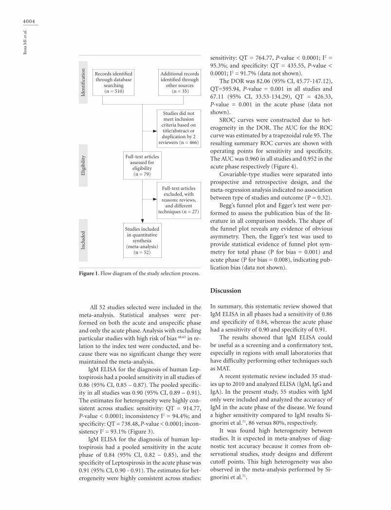
A total of 545 studies were identified: 510 studies were identified using the database search and 35 additional records were identified through other sources. Seventy-nine full-text articles were re-trieved; 27 were excluded after further scrutiny. Fifty-two primary studies, including 10,775 se-rum samples, met the criteria for inclusion and were included in the meta-analysis 19-69 (Figure 1).
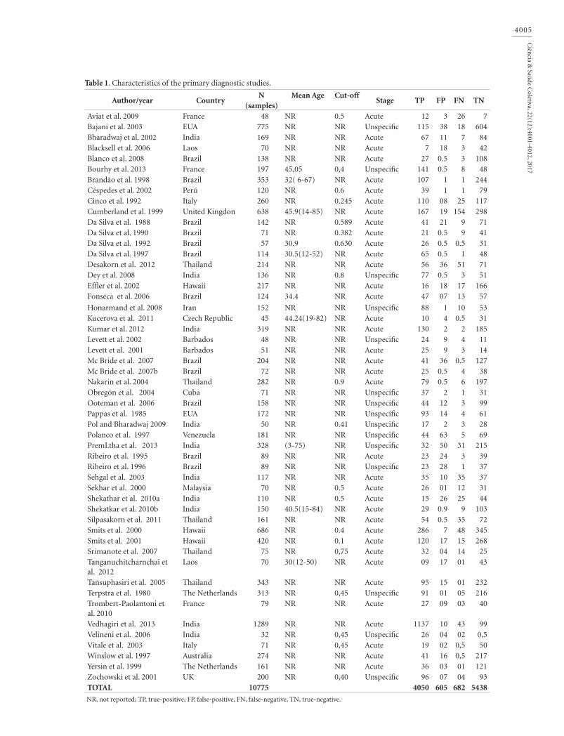
Details of the participants and interventions are summarized in Table 118-69. Most studies were prospective, except for two41,44.
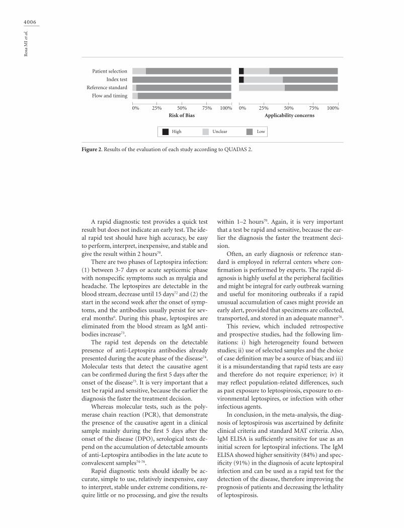
The quality assessment results are presented in Figure 219-69. Thirteen studies fulfilled all crite-ria of QUADAS 219,20,27,28,36,41,52,56,57,59,61,63,70. In five studies, the risk of bias was in the patient selec-tion31,44,55,58,62. Two studies showed unclear risk of bias in the reference standard22,44 and two stud-ies showed unclear risk of bias in the flow tim-ing39,45. Two studies have indicated high risk of bias in the patient selection in the applicability criteria50,51, and two studies demonstrated a high risk of bias in evaluating the index test48,65. In the other studies, there were some unclear applica-bility criteria in the index test and reference stan-dard19,21,23-26,29,30,32,34,35,38,40,42,43,46,47,49,53,54,57,64,67-69.
The robustness of the results was tested by repeating the analysis using a different statisti-cal model (random effects model). Some studies were identified as outliers, and one re-analysis was performed without them. However, no sig-nificant difference was found in the sensitivity or specificity; therefore, those papers were not ex-cluded from the meta-analysis.
4004R
osa
MI
et a
l.
All 52 studies selected were included in the meta-analysis. Statistical analyses were per-formed on both the acute and unspecific phase and only the acute phase. Analysis with excluding particular studies with high risk of bias 48,65 in re-lation to the index test were conducted, and be-cause there was no significant change they were maintained the meta-analysis.
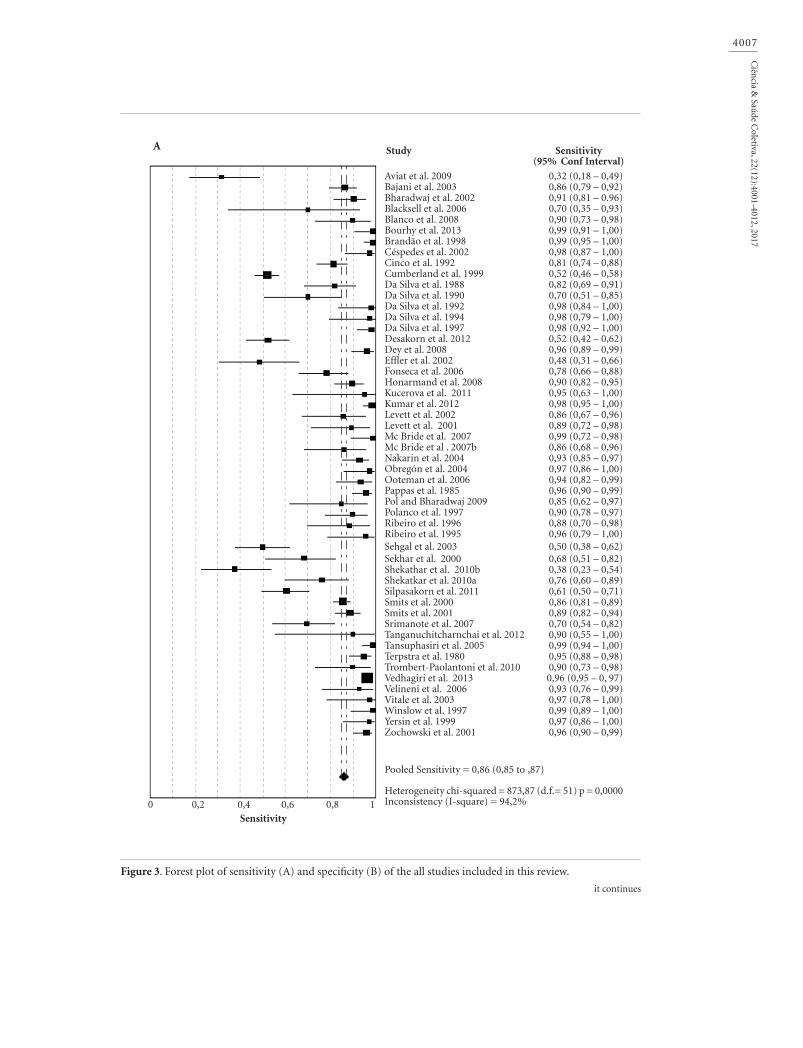
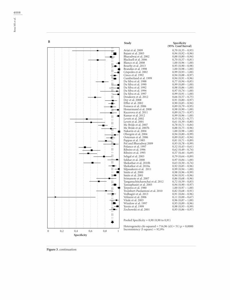
IgM ELISA for the diagnosis of human Lep-tospirosis had a pooled sensitivity in all studies of 0.86 (95% CI, 0.85 – 0.87). The pooled specific-ity in all studies was 0.90 (95% CI, 0.89 – 0.91). The estimates for heterogeneity were highly con-sistent across studies: sensitivity: QT = 914.77, P-value < 0.0001; inconsistency I2 = 94.4%; and specificity: QT = 738.48, P-value < 0.0001; incon-sistency I2 = 93.1% (Figure 3).
IgM ELISA for the diagnosis of human lep-tospirosis had a pooled sensitivity in the acute phase of 0.84 (95% CI, 0.82 – 0.85), and the specificity of Leptospirosis in the acute phase was 0.91 (95% CI, 0.90 - 0.91). The estimates for het-erogeneity were highly consistent across studies:
sensitivity: QT = 764.77, P-value < 0.0001; I2 = 95.3%; and specificity: QT = 435.55, P-value < 0.0001; I2 = 91.7% (data not shown).
The DOR was 82.06 (95% CI, 45.77-147.12), QT=595.94, P-value = 0.001 in all studies and 67.11 (95% CI, 33.53-134.29), QT = 426.33, P-value = 0.001 in the acute phase (data not shown).
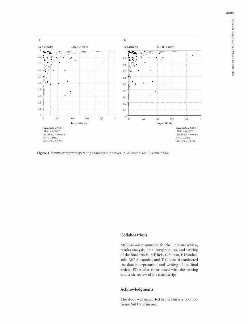
SROC curves were constructed due to het-erogeneity in the DOR. The AUC for the ROC curve was estimated by a trapezoidal rule 95. The resulting summary ROC curves are shown with operating points for sensitivity and specificity. The AUC was 0.960 in all studies and 0.952 in the acute phase respectively (Figure 4).
Covariable-type studies were separated into prospective and retrospective design, and the meta-regression analysis indicated no association between type of studies and outcome (P = 0.32).
Begg’s funnel plot and Egger’s test were per-formed to assess the publication bias of the lit-erature in all comparison models. The shape of the funnel plot reveals any evidence of obvious asymmetry. Then, the Egger’s test was used to provide statistical evidence of funnel plot sym-metry for total phase (P for bias = 0.001) and acute phase (P for bias = 0.008), indicating pub-lication bias (data not shown).
Discussion
In summary, this systematic review showed that IgM ELISA in all phases had a sensitivity of 0.86 and specificity of 0.84, whereas the acute phase had a sensitivity of 0.90 and specificity of 0.91.
The results showed that IgM ELISA could be useful as a screening and a confirmatory test, especially in regions with small laboratories that have difficulty performing other techniques such as MAT.
A recent systematic review included 35 stud-ies up to 2010 and analyzed ELISA (IgM, IgG and IgA). In the present study, 55 studies with IgM only were included and analyzed the accuracy of IgM in the acute phase of the disease. We found a higher sensitivity compared to IgM results Si-gnorini et al.71, 86 versus 80%, respectively.
It was found high heterogeneity between studies. It is expected in meta-analyses of diag-nostic test accuracy because it comes from ob-servational studies, study designs and different cutoff points. This high heterogeneity was also observed in the meta-analysis performed by Si-gnorini et al.71.
Figure 1. Flow diagram of the study selection process.
Records identified through database
searching (n = 510)
Additional records identified through
other sources (n = 35)
Studies did not meet inclusion
criteria based on title/abstract or duplication by 2
reviewers (n = 466)
Full-text articles excluded, with
reasons: reviews, and different
techniques (n = 27)
Full-text articles assessed for eligibility (n = 79)
Studies included in quantitative
synthesis (meta-analysis)
(n = 52)
Iden
tifi
cati
onE
ligib
ility
Incl
ude
d
4005C
iência &
Saúde C
oletiva, 22(12):4001-4012, 2017
Table 1. Characteristics of the primary diagnostic studies.
Author/year CountryN
(samples) Mean Age Cut-off
Stage TP FP FN TN
Aviat et al. 2009 France 48 NR 0.5 Acute 12 3 26 7Bajani et al. 2003 EUA 775 NR NR Unspecific 115 38 18 604Bharadwaj et al. 2002 India 169 NR NR Acute 67 11 7 84Blacksell et al. 2006 Laos 70 NR NR Acute 7 18 3 42Blanco et al. 2008 Brazil 138 NR NR Acute 27 0.5 3 108Bourhy et al. 2013 France 197 45,05 0,4 Unspecific 141 0.5 8 48Brandão et al. 1998 Brazil 353 32( 6-67) NR Acute 107 1 1 244Céspedes et al. 2002 Perú 120 NR 0.6 Acute 39 1 1 79Cinco et al. 1992 Italy 260 NR 0.245 Acute 110 08 25 117Cumberland et al. 1999 United Kingdon 638 45.9(14-85) NR Acute 167 19 154 298Da Silva et al. 1988 Brazil 142 NR 0.589 Acute 41 21 9 71Da Silva et al. 1990 Brazil 71 NR 0.382 Acute 21 0.5 9 41Da Silva et al. 1992 Brazil 57 30.9 0.630 Acute 26 0.5 0.5 31Da Silva et al. 1997 Brazil 114 30.5(12-52) NR Acute 65 0.5 1 48Desakorn et al. 2012 Thailand 214 NR NR Acute 56 36 51 71Dey et al. 2008 India 136 NR 0.8 Unspecific 77 0.5 3 51Effler et al. 2002 Hawaii 217 NR NR Acute 16 18 17 166Fonseca et al. 2006 Brazil 124 34.4 NR Acute 47 07 13 57
Honarmand et al. 2008 Iran 152 NR NR Unspecific 88 1 10 53Kucerova et al. 2011 Czech Republic 45 44.24(19-82) NR Acute 10 4 0.5 31Kumar et al. 2012 India 319 NR NR Acute 130 2 2 185Levett et al. 2002 Barbados 48 NR NR Unspecific 24 9 4 11Levett et al. 2001 Barbados 51 NR NR Acute 25 9 3 14Mc Bride et al. 2007 Brazil 204 NR NR Acute 41 36 0.5 127Mc Bride et al. 2007b Brazil 72 NR NR Acute 25 0.5 4 38Nakarin et al. 2004 Thailand 282 NR 0.9 Acute 79 0.5 6 197Obregón et al. 2004 Cuba 71 NR NR Unspecific 37 2 1 31Ooteman et al. 2006 Brazil 158 NR NR Unspecific 44 12 3 99Pappas et al. 1985 EUA 172 NR NR Unspecific 93 14 4 61Pol and Bharadwaj 2009 India 50 NR 0.41 Unspecific 17 2 3 28Polanco et al. 1997 Venezuela 181 NR NR Unspecific 44 63 5 69PremLtha et al. 2013 India 328 (3-75) NR Unspecific 32 50 31 215Ribeiro et al. 1995 Brazil 89 NR NR Acute 23 24 3 39Ribeiro et al. 1996 Brazil 89 NR NR Unspecific 23 28 1 37Sehgal et al. 2003 India 117 NR NR Acute 35 10 35 37Sekhar et al. 2000 Malaysia 70 NR 0.5 Acute 26 01 12 31Shekathar et al. 2010a India 110 NR 0.5 Acute 15 26 25 44Shekatkar et al. 2010b India 150 40.5(15-84) NR Acute 29 0.9 9 103Silpasakorn et al. 2011 Thailand 161 NR NR Acute 54 0.5 35 72Smits et al. 2000 Hawaii 686 NR 0.4 Acute 286 7 48 345Smits et al. 2001 Hawaii 420 NR 0.1 Acute 120 17 15 268Srimanote et al. 2007 Thailand 75 NR 0,75 Acute 32 04 14 25Tanganuchitcharnchai et al. 2012
Laos 70 30(12-50) NR Acute 09 17 01 43
Tansuphasiri et al. 2005 Thailand 343 NR NR Acute 95 15 01 232Terpstra et al. 1980 The Netherlands 313 NR 0,45 Unspecific 91 01 05 216Trombert-Paolantoni et al. 2010
France 79 NR NR Acute 27 09 03 40
Vedhagiri et al. 2013 India 1289 NR NR Acute 1137 10 43 99Velineni et al. 2006 India 32 NR 0,45 Unspecific 26 04 02 0,5Vitale et al. 2003 Italy 71 NR 0,45 Acute 19 02 0,5 50Winslow et al. 1997 Australia 274 NR NR Acute 41 16 0,5 217Yersin et al. 1999 The Netherlands 161 NR NR Acute 36 03 01 121Zochowski et al. 2001 UK 200 NR 0,40 Unspecific 96 07 04 93TOTAL 10775 4050 605 682 5438
NR, not reported; TP, true-positive; FP, false-positive, FN, false-negative, TN, true-negative.
4006R
osa
MI
et a
l.
A rapid diagnostic test provides a quick test result but does not indicate an early test. The ide-al rapid test should have high accuracy, be easy to perform, interpret, inexpensive, and stable and give the result within 2 hours70.
There are two phases of Leptospira infection: (1) between 3-7 days or acute septicemic phase with nonspecific symptoms such as myalgia and headache. The leptospires are detectable in the blood stream, decrease until 15 days72 and (2) the start in the second week after the onset of symp-toms, and the antibodies usually persist for sev-eral months6. During this phase, leptospires are eliminated from the blood stream as IgM anti-bodies increase73.
The rapid test depends on the detectable presence of anti-Leptospira antibodies already presented during the acute phase of the disease74. Molecular tests that detect the causative agent can be confirmed during the first 5 days after the onset of the disease75. It is very important that a test be rapid and sensitive, because the earlier the diagnosis the faster the treatment decision.
Whereas molecular tests, such as the poly-merase chain reaction (PCR), that demonstrate the presence of the causative agent in a clinical sample mainly during the first 5 days after the onset of the disease (DPO), serological tests de-pend on the accumulation of detectable amounts of anti-Leptospira antibodies in the late acute to convalescent samples74-76.
Rapid diagnostic tests should ideally be ac-curate, simple to use, relatively inexpensive, easy to interpret, stable under extreme conditions, re-quire little or no processing, and give the results
within 1–2 hours70. Again, it is very important that a test be rapid and sensitive, because the ear-lier the diagnosis the faster the treatment deci-sion.
Often, an early diagnosis or reference stan-dard is employed in referral centers where con-firmation is performed by experts. The rapid di-agnosis is highly useful at the peripheral facilities and might be integral for early outbreak warning and useful for monitoring outbreaks if a rapid unusual accumulation of cases might provide an early alert, provided that specimens are collected, transported, and stored in an adequate manner76.
This review, which included retrospective and prospective studies, had the following lim-itations: i) high heterogeneity found between studies; ii) use of selected samples and the choice of case definition may be a source of bias; and iii) it is a misunderstanding that rapid tests are easy and therefore do not require experience; iv) it may reflect population-related differences, such as past exposure to leptospirosis, exposure to en-vironmental leptospires, or infection with other infectious agents.
In conclusion, in the meta-analysis, the diag-nosis of leptospirosis was ascertained by definite clinical criteria and standard MAT criteria. Also, IgM ELISA is sufficiently sensitive for use as an initial screen for leptospiral infections. The IgM ELISA showed higher sensitivity (84%) and spec-ificity (91%) in the diagnosis of acute leptospiral infection and can be used as a rapid test for the detection of the disease, therefore improving the prognosis of patients and decreasing the lethality of leptospirosis.
Figure 2. Results of the evaluation of each study according to QUADAS 2.
Patient selection
Index test
Reference standard
Flow and timing
0% 25% 50% 75% 100% 0% 25% 50% 75% 100%
High Unclear Low
Risk of Bias Applicability concerns
4007C
iência &
Saúde C
oletiva, 22(12):4001-4012, 2017
Figure 3. Forest plot of sensitivity (A) and specificity (B) of the all studies included in this review.
Study Sensitivity (95% Conf Interval)
Aviat et al. 2009 0,32 (0,18 – 0,49)Bajani et al. 2003 0,86 (0,79 – 0,92)Bharadwaj et al. 2002 0,91 (0,81 – 0.96)Blacksell et al. 2006 0,70 (0,35 – 0,93)Blanco et al. 2008 0,90 (0,73 – 0,98)Bourhy et al. 2013 0,99 (0,91 – 1,00)Brandão et al. 1998 0,99 (0,95 – 1,00)Céspedes et al. 2002 0,98 (0,87 – 1,00)Cinco et al. 1992 0,81 (0,74 – 0,88) Cumberland et al. 1999 0,52 (0,46 – 0,58)Da Silva et al. 1988 0,82 (0,69 – 0,91)Da Silva et al. 1990 0,70 (0,51 – 0,85) Da Silva et al. 1992 0,98 (0,84 – 1,00)Da Silva et al. 1994 0,98 (0,79 – 1,00)Da Silva et al. 1997 0,98 (0,92 – 1,00)Desakorn et al. 2012 0,52 (0,42 – 0,62) Dey et al. 2008 0,96 (0,89 – 0,99) Effler et al. 2002 0,48 (0,31 – 0,66)Fonseca et al. 2006 0,78 (0,66 – 0,88)Honarmand et al. 2008 0,90 (0,82 – 0,95) Kucerova et al. 2011 0,95 (0,63 – 1,00)Kumar et al. 2012 0,98 (0,95 – 1,00)Levett et al. 2002 0,86 (0,67 – 0,96) Levett et al. 2001 0,89 (0,72 – 0,98)Mc Bride et al. 2007 0,99 (0,72 – 0,98)Mc Bride et al . 2007b 0,86 (0,68 – 0,96) Nakarin et al. 2004 0,93 (0,85 – 0,97)Obregón et al. 2004 0,97 (0,86 – 1,00)Ooteman et al. 2006 0,94 (0,82 – 0,99)Pappas et al. 1985 0,96 (0,90 – 0,99)Pol and Bharadwaj 2009 0,85 (0,62 – 0,97)Polanco et al. 1997 0,90 (0,78 – 0,97)Ribeiro et al. 1996 0,88 (0,70 – 0,98)Ribeiro et al. 1995 0,96 (0,79 – 1,00)Sehgal et al. 2003 0,50 (0,38 – 0,62)Sekhar et al. 2000 0,68 (0,51 – 0,82)Shekathar et al. 2010b 0,38 (0,23 – 0,54)Shekatkar et al. 2010a 0,76 (0,60 – 0,89)Silpasakorn et al. 2011 0,61 (0,50 – 0,71)Smits et al. 2000 0,86 (0,81 – 0,89)Smits et al. 2001 0,89 (0,82 – 0,94)Srimanote et al. 2007 0,70 (0,54 – 0,82)Tanganuchitcharnchai et al. 2012 0,90 (0,55 – 1,00)Tansuphasiri et al. 2005 0,99 (0,94 – 1,00)Terpstra et al. 1980 0,95 (0,88 – 0,98)Trombert-Paolantoni et al. 2010 0,90 (0,73 – 0,98)Vedhagiri et al. 2013 0,96 (0,95 – 0, 97)Velineni et al. 2006 0,93 (0,76 – 0,99) Vitale et al. 2003 0,97 (0,78 – 1,00)Winslow et al. 1997 0,99 (0,89 – 1,00)Yersin et al. 1999 0,97 (0,86 – 1,00)Zochowski et al. 2001 0,96 (0,90 – 0,99)
Pooled Sensitivity = 0,86 (0,85 to ,87)
Heterogeneity chi-squared = 873,87 (d.f.= 51) p = 0,0000Inconsistency (I-square) = 94,2%
A
Sensitivity0 0,2 0,4 0,6 0,8 1
it continues
4008R
osa
MI
et a
l.
B
Specificity0 0,2 0,4 0,6 0,8 1
Study Specificity (95% Conf Iterval)
Aviat et al. 2009 0,70 (0,35 – 0,93)Bajani et al. 2003 0,94 (0,92 – 0,96)Bharadwaj et al. 2002 0,88 (0,80 – 0,94)Blacksell et al. 2006 0,70 (0,57 – 0,81)Blanco et al. 2008 1,00 (0,96 – 1,00)Bourhy et al .2013 0,95 (0,90 – 0,98)Brandão et al. 1998 1,00 (0,98 – 1,00)Céspedes et al. 2002 0,99 (0,93 – 1,00)Cinco et al. 1992 0,94 (0,88 – 0,97)Cumberland et al. 1999 0,94 (0,91 – 0,96)Da Silva et al. 1988 0,77 (0,94 – 0,85)Da Silva et al. 1990 0,99 (0,89 – 1,00)Da Silva et al. 1992 0,98 (0,86 – 1,00)Da Silva et al. 1994 0,97 (0,74 – 1,00)Da Silva et al. 1997 0,99 (0,91 – 1,00)Desakorn et al. 2012 0,66 (0,57 – 0,75) Dey et al. 2008 0,91 (0,80 – 0,97)Effler et al. 2002 0,90 (0,85 – 0,94) Fonseca et al. 2006 0,89 (0,79 – 0,95)Honarmand et al. 2008 0,98 (0,90 – 1,00)Kucerova et al. 2011 0,89 (0,73 – 0,97)Kumar et al. 2012 0,99 (0,96 – 1,00)Levett et al. 2002 0,55 (0,32 – 0,77)Levett et al. 2001 0,61 (0,39 – 0,80)Mc Bride et al. 2007 0,78 (0,71 – 0,84)Mc Bride et al. 2007b 0,88 (0,75 – 0,96) Nakarin et al. 2004 1,00 (0,98 – 1,00)Obregón et al. 2004 0,94 (0,80 – 0,99)Ooteman et al. 2006 0,89 (0,82 – 0,94)Pappas et al. 1985 0,81 (0,71 – 0,89)Pol and Bharadwaj 2009 0,93 (0,78 – 0,99)Polanco et al. 1997 0,52 (0,43 – 0,61)Ribeiro et al. 1996 0,62 (0,49 – 0,74)Ribeiro et al. 1995 0,57 (0,44 – 0,69)Sehgal et al. 2003 0,79 (0,64 – 0,89)Sekhar et al. 2000 0,97 (0,84 – 1,00)Shekathar et al. 2010b 0,63 (0,50 – 0,74)Shekatkar et al. 2010a 0,92 (0,85 – 0,96)Silpasakorn et al. 2011 0,99 (0,94 – 1,00)Smits et al. 2000 0,98 (0,96 – 0,99)Smits et al. 2001 0,94 (0,91 – 0,96)Srimanote et al. 2007 0,86 (0,68 – 0,96)Tanganuchitcharnchai et al. 2012 0,72 (0,59 – 0,83)Tansuphasiri et al. 2005 0,94 (0,90 – 0,97)Terpstra et al. 1980 1,00 (0,97 – 1,00)Trombert-Paolantoni et al. 2010 0,82 (0,68 – 0,91)Vedhagiri et al. 2013 0,91 (0,84 – 0,96)Velineni et al. 2006 0,11 (0,00 – 0,67)Vitale et al. 2003 0,96 (0,87 – 1,00)Winslow et al. 1997 0,93 (0,89 – 0,96)Yersin et al. 1999 0,98 (0,93 – 0,99)Zochowski et al. 2001 0,93 (0,86 – 0,97)
Pooled Specificity = 0,90 (0,90 to 0,91)
Heterogeneity chi-squared = 716,96 (d.f.= 51) p = 0,0000Inconsistency (I-square) = 92,9%
Figure 3. continuation
4009C
iência &
Saúde C
oletiva, 22(12):4001-4012, 2017
Collaborations
MI Rosa was responsible for the literature review, results analysis, data interpretation, and writing of the final article. MF Reis, C Simon, E Dondos-sola, MC Alexandre, and T Colonetti conducted the data interpretation and writing of the final article. FO Meller contributed with the writing and critic review of the manuscript.
Acknowledgments
The study was supported by the University of Ex-tremo Sul Catarinense.
Figure 4. Summary receiver operating characteristic curves. A: all studies and B: acute phase.
1-specificity
Sensitivity SROC Curve
Symmetric SROCAUC = 0,9527SE(AUC) = 0,0148Q* = 0,8942SE(Q*) = 0,0204
0 0,2 0,4 0,6 0,8 1
1
0,9
0,8
0,7
0,6
0,5
0,4
0,3
0,2
0,1
0
A
1-specificity
Symmetric SROCAUC = 0,9607SE(AUC) = 0,0098Q* = 0,9055SE(Q*) = 0,0144
0 0,2 0,4 0,6 0,8 1
Sensitivity SROC Curve
1
0,9
0,8
0,7
0,6
0,5
0,4
0,3
0,2
0,1
0
B
4010R
osa
MI
et a
l.
References
1. Pappas G, Papadimitriou P, Siozopoulou V, Christou L, Akritidis N. The globalization of leptospirosis: world-wide incidence trends. Int J Infect Dis 2008;12(4):351-357.
2. Bharti AR, Nally JE, Ricaldi JN, Matthias MA, Diaz MM, Lovett MA, Levett PN, Gilman RH, Willig MR, Gotuzzo E, Vinetz JM; Peru-United States Lepto-spirosis Consortium. Leptospirosis: a zoonotic disease of global importance. Lancet Infect Dis 2003;3(12):757-771.
3. World Health Organization (WHO). Report of the Sec-ond Meeting of the Leptospirosis Burden Epidemiology Reference Group (LERG).WHO: Geneva; 2011.
4. Hagan JE, Costa J, Calcagno M, Kane M, Torgerson P, Martinez-Silveira MS, Stein C, Abela-Ridder B, Ko AI. Global morbidity and mortality of leptospi-rosis: a systematic review. PLoS Negl Trop Dis 2015; 9(9):e0003898
5. Limmathurotsakul D, Turner EL, Wuthiekanun V, Thaipadungpanit J, Suputtamongkol Y, Chierakul W, Smythe LD, Day NP, Cooper B, Peacock SJ.Fool’s gold: why imperfect reference tests are undermining the evaluation of novel diagnostics: a re-evaluation of 5 diagnostic tests for leptospirosis. Clin Infect Dis 2012;55(3):322-331.
6. Silva MV, Camargo ED, Batista L, Vaz AJ, Brandão AP, Nakamura PM, Negrão JM. Behaviour of specific IgM, IgG and IgA class antibodies in human leptospi-rosis during the acute phase of the disease and during convalescence. J Trop Med Hyg 1995;98(4):268-272.
7. Liberati A, Altman DG, Tetzlaff J, Mulrow C, Gøtzsche PC, Ioannidis JP, Clarke M, Devereaux PJ, Kleijnen J, Moher D. The PRISMA statement for reporting sys-tematic reviews and meta-analysis of studies that eval-uate healthcare interventions: explanation and elabo-ration. BMJ 2009;339:b2700.
8. Medeiros LR, Rosa MI, Silva BR, Reis ME, Simon CS, Dondossola ER, Cunha Filho JS. Accuracy of magnetic resonance in deeply infiltrating endometriosis: a sys-tematic review and meta-analysis. Arch Gynecol Obstet 2015;291(3):611-621.
9. Whiting PF, Rutjes AW, Westwood ME, Mallett S, Deeks JJ, Reitsma JB, Leeflang MM, Sterne JA, Bossuyt PM; QUADAS-2 Group. QUADAS-2: a revised tool for the quality assessment of diagnostic accuracy studies. Ann Intern Med 2011;155(8):529-536.
10. Review Manager (RevMan) [computer program]. Version 5.2. 2012 Copenhagen: The Nordic Cochrane Centre, The Cochrane Collaboration.Altman DG. Some common problems in medical research. In: Alt-man DG, editor. Practical statistics for medical research. 9ª ed. London: Chapman; 1999. p. 396-439.
11. Altman DG. Some common problems in medical re-search. In: Altman DG (eds). Practical statistics for medical research, 9 ed. London: Chapman; 1999. p. 396-439.
12. Irwig L, Tosteson AN, Gatsonis C, Lau J, Colditz G, Chalmers TC, Mosteller F. Guidelines for meta-anal-yses evaluating diagnostic tests. Ann Intern Med 1994; 120(8):667-676.
13. Reitsma JB, Glas AS, Rutjes AWS, Scholten RJ, Bossuyt PM, Zwinderman AH. Bivariate analysis of sensitivity and specificity produce informative summary mea-sures in diagnostic reviews. J Clin Epidemiol 2005; 58(10):982-990.
14. Gatsonis C, Paliwal P. Meta-analysis of diagnostic and screening test accuracy evaluations: methodologic primer. AJR Am J Roentgenol 2006; 187(2):271-281.
15. Deeks JJ, Macaskill P, Irwig L. The performance of tests of publication bias and other sample size effects in systematic reviews of diagnostic test accuracy was as-sessed. J Clin Epidemiol 2005; 58(9):882-893.
16. Stata Corporation. Stata Statistical Software version 11: College Station, TX/ Stata Corporation; 2009.
17. Zamora J, Abraira V, Muriel A, Khan K, Coomarasamy A. Meta-DiSc: a software for meta-analysis of test accu-racy data. BMC Med Res Methodol 2006; 6:31.
18. Terpstra WJ, Ligthart GS, Schoone GJ. Serodiagnosis of human leptospirosis by enzyme-linked-immuno-sorrbent-assay (ELISA). Zentralbl Bakteriol A 1980; 247(3):400-405.
19. Pappas MG, Ballou WR, Gray MR, Takafuji ET, Miller RN, Hockmeyer WT. Rapid serodiagnosis of leptospi-rosis using the IgM-specific Dot-ELISA: comparison with the microscopic agglutination test. Am J Trop Med Hyg 1985; 34(2):346-354.
20. Da Silva MV, Camargo ED, Vaz AJ, Souza, AMC, Ueda M, Sakata, EE. Teste imunoenzimático (ELISA) para detecção de anticorpos circulantes da classe IgM na leptospirose humana. Rev Inst Med Trop São Paulo [on-line]1988; [acessado 2017 Ago 25];30(2):95-100. Dis-ponível em: http://www.scielo.br/scielo.php?script=s-ci_arttext&pid=S0036-46651988000200008&lng=pt.
21. Da Silva MV, Camargo ED, Vaz AJ, Souza AMC, Chie-ffi PP, Sakata EE. Imunodiagnóstico da leptospirose humana através do teste ELISA-IgM, empregando-se diferentes preparações antigênicas a partir de soroti-pos prevalentes de Leptospira interrogans. Rev Inst Med Trop São Paulo [online] 1990 [acessado 2017 Ago 25]; 32(4):233-239. Disponível em: http://www.scielo.br/scielo.php?pid=S0036-46651990000400001&script=s-ci_abstract&tlng=pt
22. Silva MV, Camargo ED. Enzyme-linked immunosor-bent assay ELISA for the detection of antibodies in the human leptospirosis. Rev Inst Med Trop São Paulo [online]. 1992[acessado 2017 Ago 25]; 34(3):239-242. Disponível em: http://www.scielo.br/scielo.php?scrip-t=sci_arttext&pid=S0036-46651992000300010
23. Cinco M, Balanzin A, Banfi DAE. Evaluation of an im-munoenzimatic test (ELISA) for the diagnosis of lep-tospirosis in Italy. Eur J of Epidemiology 1992;8(5):677-682.
24. Cumberland P, Everard CO, Levett PN. Assessment of the efficacy of anIgM-elisa and microscopic agglutina-tion test (MAT) in the diagnosis of acute leptospirosis. Am J Trop Med Hyg 1999;61(5):731-734.
25. Ribeiro MA, Souza CC, Almeida SH. Dot-ELISA for human leptospirosis employing immunodominant an-tigen. J Trop Med Hyg. 1995;98(6):452-456.
4011C
iência &
Saúde C
oletiva, 22(12):4001-4012, 2017
26. Ribeiro MA, Brandão AP, Romero EC. Evaluation of diagnostic tests for human leptospirosis. Braz J Med Biol Res 1996;29(6):773-777.
27. Polanco J, Aguirre L, Marcano E, Pantoja A. Diagnós-tico de la Leptospirosis Humana Mediante el uso de la técnica dot-ELISA. Veterinaria Tropical 1997;22(1):65-75.
28. Winslow WE, Merry DJ, Pirc ML, Devine PL. Evalua-tion of a commercial enzyme-linked immunosorbent assay for detection of immunoglobulin M antibody in diagnosis of human leptospiral infection. J Clin Micro-biol 1995;35(8):1938-1942.
29. Da Silva MV, Nakamura PM, Camargo ED, Batista L, Vaz AJ, Romero EC, Brandao AP. Immunodiagnosis of human leptospirosis by dot-ELISA for the detection of IgM, IgG, and IgA antibodies. Am J Trop Med Hyg 1997;56(6):650-655.
30. Brandão AP, Camargo ED, da Silva ED, Silva MV, Abrão RV. Macroscopic agglutination test for rap-id diagnosis of human leptospirosis. J Clin Microbiol 1998;36(11):3138-3142.
31. Yersin C, Bovet P, Smits HL, Perolat P. Field evaluation of a one-step dipstick assay for the diagnosis of human leptospirosis in the Seychelles. Trop Med Int Health 1999;4(1):38-45.
32. Smits HL, van der Hoorn MA, Goris MG. Simple latex agglutination assay for rapid serodiagnosis of human leptospirosis. J Clin Microbiol 2000;38(3):1272-1275.
33. Sekhar WY, Soo EH, Gopalakrishnan V, Devi S. Lepto-spirosis in Kuala Lumpur and the comparative evalu-ation of two rapid commercial diagnostic kits against the MAT test for the detection of antibodies to lepto-spira interrogans. Singapore Med J 2000;41(8):370-375.
34. Levett PN, Branch SL, Whittington CU, Edwards CN, Paxton H. Two methods for rapid serological diag-nosis of acute leptospirosis. Clin Diagn Lab Immunol 2001;8(2):349-351.
35. Smits HL, Chee HD, Eapen CK, Kuriakose M, Sugath-an S, Gasem MH, Yersin C, Sakasi D, Lai-A-Fat RF, Hartskeerl RA, Liesdek B, Abdoel TH, Goris MG, Gussenhoven GC. Latex based, rapid and easy as-say for human leptospirosis in a single test format. Trop Med Int Health 2001;6(2):114-118.
36. Zochowski WJ, Palmer MF, Coleman TJ. An evaluation of three commercial kits for use as screening methods for the detection of leptospiral antibodies in the UK. J Clin Pathol 2001;54(1):25-30.
37. Bharadwaj R, Bal AM, Joshi SA, Kagal A, Pol SS, Ga-rad G, Arjunwadkar V, Katti R. An urban outbreak of leptospirosis in Mumbai, India. Jpn J Infect Dis 2002;55(6):194-196.
38. Céspedes Z M, Glenny A M, Felices AV, Balda J L, Suá-rez MV. Prueba de Elisa indirecta para la detección de anticuerpos IgM para el diagnóstico de Leptospirosis humana. Revista Peruana de Medicina Experimental y Salud Pública 2002;19(1):1924-1927.
39. Effler PV, Bogard AK, Domen HY, Katz AR, Higa HY, Sasaki DM. Evaluation ofeight rapid screening tests for acute leptospirosis in Hawaii. J Clin Microbiol 2002;40(4):1464-1469.
40. Levett PN, Branch SL. Evaluation of two enzyme-linked immunosorbent assay methods for detection of immu-noglobulin M antibodies in acute leptospirosis. Am J Trop Med Hyg 2002;66(6):745-748.
41. Bajani MD, Ashford DA, Bragg SL, Woods CW, Aye T, Spiegel RA, Plikaytis BD, Perkins BA, Phelan M, Le-vett PN, Weyant RS. Evaluation of four commercially available rapid serologic tests for diagnosis of leptospi-rosis. J Clin Microbiol 2003;41(2):803-809.
42. Sehgal SC, Vijayachari P, Sugunan AP, Umapathi T. Field application of Lepto lateral flow for rapid di-agnosis of leptospirosis. J Med Microbiol 2003;52(Pt 10):897-901.
43. Nakarin J, Pradutkanchana S. Evaluation of en-zyme-linked immunosorbent assay and indirect hem-agglutination assay for detection of leptospiral anti-body by using three different antigens. J Med Assoc Thai 2004;87(10):1218-1224.
44. Obregón AM, Martínez G, Martínez R, Llop A, Ro-dríguez I, Rodríguez J, Fernández C. Serological re-sponse by ELISA and MAT in Cuban volunteers vaccinated with vax SPIRAL. Rev Cubana Med Trop 2004;56(2):148-1451.
45. Vitale G, La Russa C, Galioto A, Chifari N, Moccia-ro C, Caruso R, Micalizzi A, Mansueto P, Di Rosa S, Mansueto S.Evaluation of an IgM-ELISA test for the diagnosis of human leptospirosis. New Microbiol 2004;27(2):149-154.
46. Tansuphasiri U, Deepradit S, Phulsuksombati D, Tang-kanakul W. Two simple immunoassays using endemic leptospiral antigens for serodiagnosis of human lep-tospirosis. Southeast Asian J Trop Med Public Healt. 2005;36(2):302-311.
47. Blacksell SD, Smythe L, Phetsouvanh R, Dohnt M, Hartskeerl R, Symonds M, Slack A, Vong-souvath M, Davong V, Lattana O, Phongmany S, Ke-olouangkot V, White NJ, Day NP, Newton PN. Limited diagnostic capacities of two commercial assays for the detection of Leptospira immunoglobulin M antibodies in Laos. Clin Vaccine Immunol 2006;13(10):1166-1169.
48. de Abreu Fonseca C, Teixeira de Freitas VL, Caló Ro-mero E, Spinosa C, Arroyo Sanches MC, da Silva MV, Shikanai-Yasuda MA. Polymerase chain reaction incomparison with serological tests for early diag-nosis of human leptospirosis. Trop Med Int Health 2006;11(11):1699-1707.
49. Ooteman MC, Vago AR, Koury MC. Evaluation of MAT, IgM ELISA and PCR methodsfor the diag-nosis of human leptospirosis. J Microbiol Methods 2006;65(2):247-257.
50. McBride AJ, Santos BL, Queiroz A, Santos AC, Hart-skeerl RA, Reis MG, Ko AI. Evaluation of four whole-cell Leptospira-based serological tests for di-agnosis of urban leptospirosis. Clin Vaccine Immunol 2007;14(9):1245-1248.
51. McBride AJ, Pereira FA, da Silva ED, Ferreira AG, Reis MG, Ko AI. Evaluation of the EIE-IgM-Leptospirose assay for the serodiagnosis of leptospirosis. Acta Trop 2007;102(3):206-211.
4012R
osa
MI
et a
l.
52. Velineni S, Asuthkar S, Umabala P, Lakshmi V, Sritha-ran M. Serological evaluation of leptospirosis in Hyder-abad Andhra Pradesh: a retrospective hospital-based study. Indian J Med Microbiol 2007;25(1):24-27.
53. Dey S, Mohan CM, Ramadass P, Nachimuthu K. Di-agnosis of leptospirosis by recombinant antigen based single serum dilution ELISA. Indian J Med Res 2008;128(2):172-177.
54. Honarmand HR, Nezafat TM, Heydarzadeh A, Sol-tani B, Mirzajani E, Asmar M. Evaluation an in–house IgM-ELISA for the diagnosis of human leptospiro-sis. Journal of Semnan University of Medical Sciences 2008;9(4):309-313.
55. Srimanote P, Wongdeethai N, Jieanampunkul P, Samonkiert S, Leepiyasakulchai C, Kalambaheti T, Pra-chayasittikul V.Recombinant ligA for leptospirosis diagnosis and ligA among the Leptospira spp. clinical isolates. J Microbiol Methods 2008;72(1):73-78.
56. Blanco RM, Takei K, Romero EC. Leptospiral glycolipo-protein as a candidate antigen for serodiagnosis of hu-man leptospirosis. Lett Appl Microbiol 2009;49(2):267-273.
57. Pol S, Bharadwaj R. Evaluation of high performance liquid chromatography purified leptospiral anti-gen for the diagnosis of leptospirosis. Jpn J Infect Dis 2009;62(6):428-431.
58. Aviat F, Rochereau-Roulet S, Branger C, Estavoyer JM, Chatrenet B, Orsonneau JL, Thorin C, Andre-Fon-taine G. Synthetic peptide issued from Hap1/LipL32 for new early serodiagnosis of human leptospirosis. Comp Immunol Microbiol Infect Dis 2010;33(5):375-387.
59. Shekatkar SB, Harish BN, Menezes GA, Parija SC. Clinical and serological evaluation of Leptospiro-sis in Puducherry, India. J Infect Dev Ctries 2010a 29;4(3):139-143.
60. Shekatkar S, Acharya NS, Harish BN, Parija SC. Com-parison of an in-house latex agglutination test with IgM ELISA and MAT in the diagnosis of leptospirosis. Indian J Med Microbiol 2010;28(3):238-240.
61. Trombert-Paolantoni S, Thomas P, Hermet F, Clairet V, Litou N, Maury L..Leptospirosis screening: perfor-mance of the Serion Elisa Classic Leptospira IgM KIT. Pathol Biol (Paris) 2010;58(1):95-99.
62. Kučerová P, Cermáková Z, Plíšková L, Valenta Z, Pavliš O, Kubíčková P. Comparison of results of two serolog-ical methods for diagnosing leptospirosis - microag-glutination test and ELISA. Klin Mikrobiol Infekc Lek 2011;17(5):173-178.
63. Silpasakorn S, Waywa D, Hoontrakul S, Suttinont C, Losuwanaluk K, Suputtamongkol Y. Performance of Leptospira immunoglobulin M ELISA and rapid im-munoglobulin G immunochromatographic assays for the diagnosis of leptospirosis. J Med Assoc Thai 2011;94(Suppl.1):203-206.
64. Desakorn V, Wuthiekanun V, Thanachartwet V, Sa-hassananda D, Chierakul W, Apiwattanaporn A, Day NP, Limmathurotsakul D, Peacock SJ.Accuracy of a commercial IgM ELISA for the diagnosis of hu-man leptospirosis in Thailand. Am J Trop Med Hyg 2012;86(3):524-527.
65. Kumar A, Rajasekar V, Selvakumar G, Dhanalaksh-mi DP. Comparison study between in-house IgM DOT-ELISA and the microscopic agglutination test (MAT) for the diagnosis of human leptospirosis. Int Research J of Pharmacy 2012;3(4):314-317.
66. Tanganuchitcharnchai A, Smythe L, Dohnt M , Hartskeerl R, Vongsouvath M, Davong V, Lattana O, Newton PN, Blacksell SD. Evaluation of the Standard DiagnosticsLeptospira IgM ELISA for diagnosis of acute leptospirosis in Lao PDR. Trans R Soc Trop Med Hyg 2012;106(9-2):563-566.
67. Bourhy P, Vray M, Picardeau M. Evaluation of an in-house ELISA using the intermediate species Leptospira fainei for diagnosis of leptospirosis. J Med Microbiol 2013;62(Pt 6):822-857.
68. Premlatha MM, Kaur IR, Avasthi R, Dey AB, Chaudhry R. A newer approach for the serodiagnosis of lepto-spirosis using outer membrane proteins of leptospira interrogans serovar tarassovi. Asian Journal of Medical Sciences 2013;4(2):41-46.
69. Vedhagiri K, Velineni S, Timoney JF, Shanmughapriya S, 1 Vijayachari P, Narayanan R, Natarajaseenivasan K. Detection of LipL32-specific IgM by ELISA in sera of patients with a clinical diagnosis of leptospirosis. Pat-hog Glob Health 2013;107(3):130-135.
70. Banoo S, Bell D, Bossuyt P, Herring A, Mabey D, Poole F, Smith PG, Sriram N, Wongsrichanalai C, Linke R, O’Brien R, Perkins M, Cunningham J, Matso-so P, Nathanson CM, Olliaro P, Peeling RW, Ramsay A.Evaluation of diagnostic tests for infectious diseas-es: general principles. Nat Rev Microbiol 2010;8(Sup-pl.):S17-29.
71. Agampodi SB, Matthias MA, Moreno AC, Vinetz JM. Utility of quantitative polymerase chain reaction in leptospirosis diagnosis: association of level of lepto-spiremia and clinical manifestations in Sri Lanka. Clin Infect Dis 2012;54(9):1249-1255.
72. Signorini ML, Lottersberger J, Tarabla HD, Vanasco NB. Enzyme-linked immunosorbent assay to diagnose human leptospirosis: a meta-analysis of the published literature. Epidemiol Infect. 2013;141(1):22-32
73. Levett PN. Leptospirosis. Clinical Microbiology Reviews 2001; 14(2):296-326.
74. Ahmed A, Anthony RM, Hartskeerl RA. A simple and rapid molecular method for Leptospira species identi-fication. Infect Genet Evol 2010;10(7):955-962.
75. Ahmed A, Klaassen HLMB, Van der Veen M, van der Linden H , Goris MGA , Hartskeerl RA. Evaluation of real-time PCR and culturing for the detection of lep-tospires in canine samples. Advances in Microbiology 2012;2(2):162-170.
76. Goris MGA, Leeflang MMG, Boer KR, Goeijenbier M, van Gorp ECM4, Wagenaar JFP, Hartskeerl RA. Estab-lishment of valid laboratory case definition for human leptospirosis. J Bacteriol Parasitol [periódico na Inter-net]. 2012 [acessado 2017 Ago 25];3. Disponível em: doi: 10.4172/2155-9597.1000132
Artigo apresentado em 25/05/2016Aprovado em 23/08/2016Versão final apresentada em 25/08/2016


![[XLS] · Web viewAna Maria Caetano de Faria Ana Maria Caldeira Oliveira Ana Maria Covolam Ana Maria de Freitas Pinheiro Ana Maria Maniero Moreira Ana Maria Mouad Ana Maria Petraitis](https://img.document.onl/doc/110x75/5ad51e2a7f8b9a1a028cb048/xls-viewana-maria-caetano-de-faria-ana-maria-caldeira-oliveira-ana-maria-covolam.jpg)