Embed Size (px)
Citation preview
Estrutura e função da célula procariotica
Cell Diagram: Mariana Ruiz, pub domain
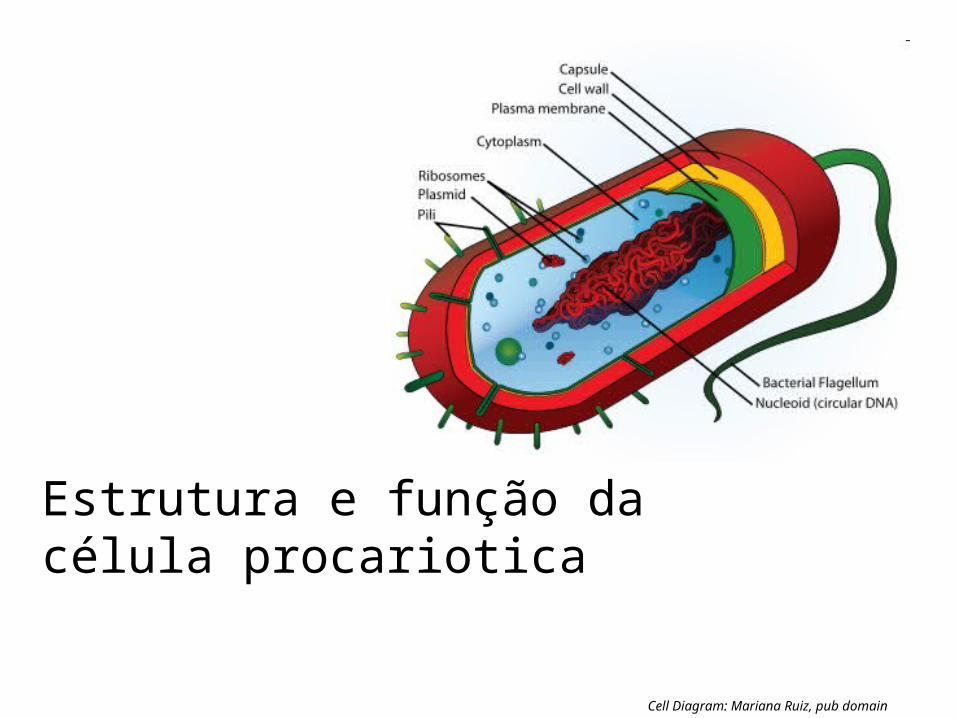
a BIOMASSA DE BACTERIAS NA TERRA É ESTIMADA A SER IGUAL QUE A DE PLANTAS. O NUMERO DE PROCARIOTES NA TERRA ESTIMADO É DE 5 × 1030, QUE REPRESENTA A METADE DA BIOMASSA GLOBAL.
^ Whitman W, Coleman D, Wiebe W (1998). "Prokaryotes: the unseen majority". Proc Natl Acad Sci USA 95 (12): 6578–83. doi:10.1073/pnas.95.12.6578. PMC 33863. PMID 961845

pH ácido pH alcalino
HABITAT

Outros habitats
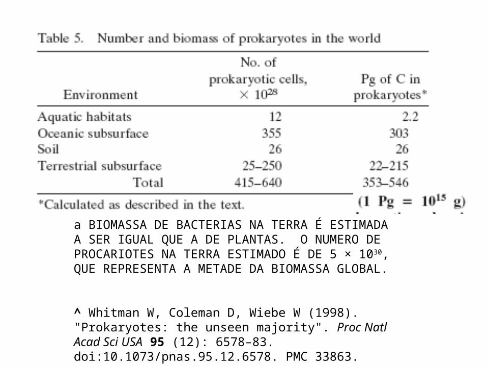
Arvore universal da vida derivada do sequenciamento de ssRNA. N. Pace

Arvore universal da vida derivada do sequenciamento de ssRNA. N. Pace
--Archaea are the least evolved type of cell (they remain closest to the common point of origin). This helps explainwhy contemporary Archaea are inhabitants of environments that are something like the earth 3.86 billion years ago(hot, salty, acidic, anaerobic, low in organic material, etc.) Ref. Todar microbiology.
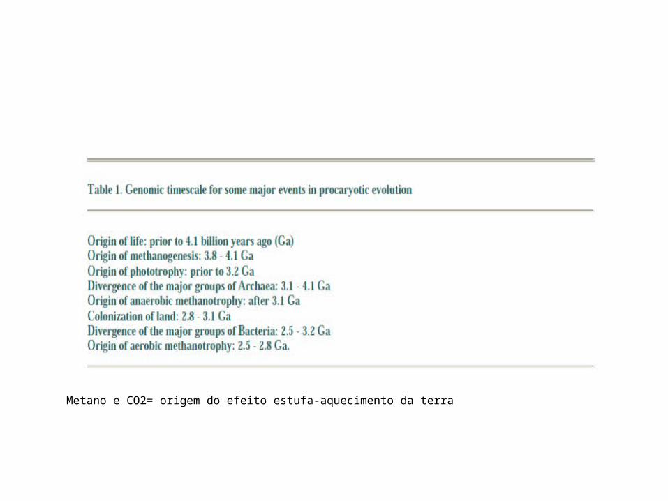
Metano e CO2= origem do efeito estufa-aquecimento da terra
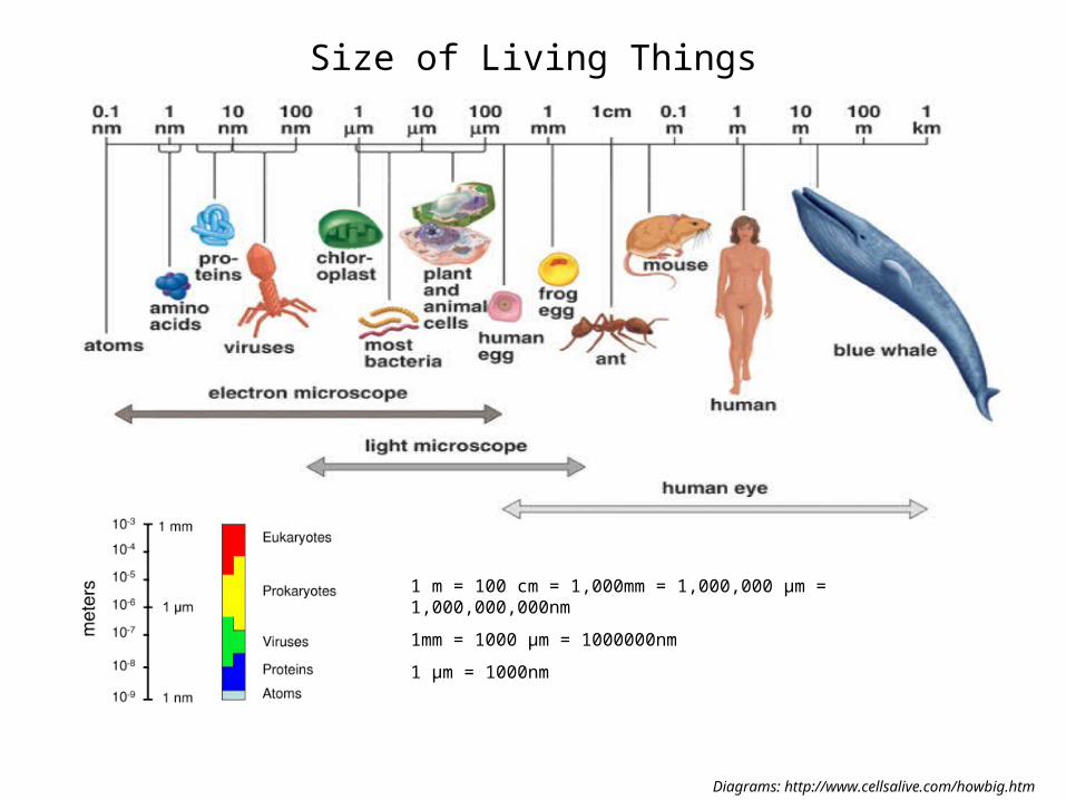
Size of Living Things
Diagrams: http://www.cellsalive.com/howbig.htm
1 m = 100 cm = 1,000mm = 1,000,000 µm = 1,000,000,000nm
1mm = 1000 µm = 1000000nm
1 µm = 1000nm

Célula típica: diametro 1 - 2 μm (~ tamanho de
mitocôndria)
Megabactéria (Epulopiscium fishelsoni): 80 x 600 μm,
portanto visível ao olho nú
Nanobactéria
Tamanho da célula bacteriana


Streptooccus pneumoniae

Bacillus antrax=bacteria gram +

E. coli
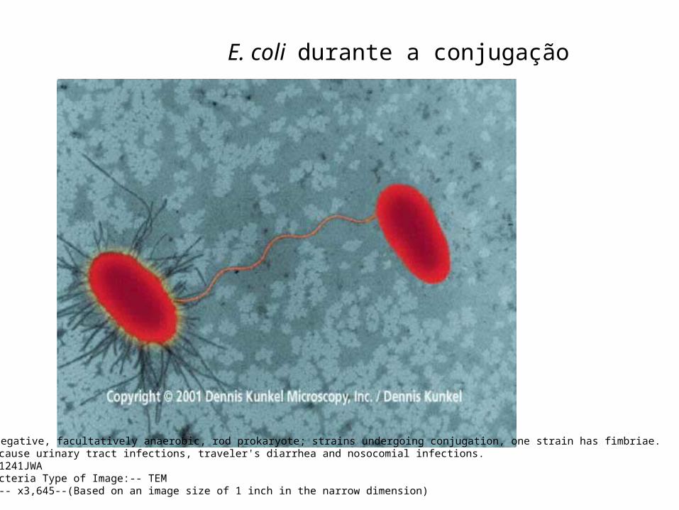
E. coli durante a conjugação
-E. coli - Gram-negative, facultatively anaerobic, rod prokaryote; strains undergoing conjugation, one strain has fimbriae.- E. coli can cause urinary tract infections, traveler's diarrhea and nosocomial infections.File Name:-- 71241JWA Category:-- Bacteria Type of Image:-- TEMMagnification:-- x3,645--(Based on an image size of 1 inch in the narrow dimension)

Two basic types of cells
Diagrams:
Prokaryotic & Eukaryotic Cell, Mariana Ruiz
__________________________________________

Peptidoglycan - Rigid mechanical support - Freely permeable to solutes
Peptidoglycan is a huge polymer of interlocking chains of identical peptidoglycan monomers.
Backbone of peptidoglycan molecule composed of two derivatives of glucose:
N-acetylglucosamine (NAG)N-acetlymuramic acid (NAM)
NAG / NAM strands are connected by interpeptide bridges.
Prokaryotes – Cell Wall
Image:
Peptindoglycan Structure: NicolasGrandjean

Prokaryotes - Cell Wall: Gram-Negative & Gram-Positive
Image:
Prokaryotic Cell, Mariana Ruiz
Gram +-, Julian Onions

N-acetil glucosamina e acido muramico


Chapter 4

Chapter 4
Cell Wall
• Peptido-glycan Polymer (amino acids + sugars)
• Unique to bacteria
• Sugars; NAG & NAM– N-acetylglucosamine– N-acetymuramic acid
• D form of Amino acids used not L form– Hard to break down D form
• Amino acids cross link NAG & NAM

Video Clip parede celular

Chapter 4

Chapter 4

Chapter 4
Cell Wall Summary
• Determine shape of bacteria
• Strength prevents osmotic rupture
• 20-40% of bacteria
• Unique to bacteria
• Some antibiotics effect directly– Penicillin

Why are these differences in cell wall structure so important?
Images: Sources unknown
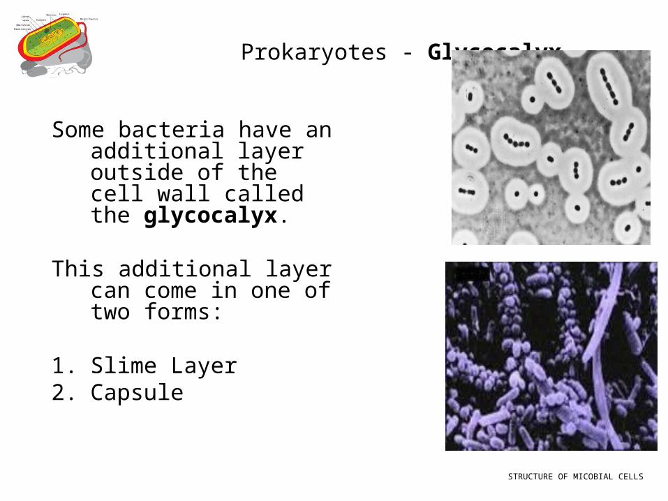
Prokaryotes - Glycocalyx
Some bacteria have an additional layer outside of the cell wall called the glycocalyx.
This additional layer can come in one of two forms:
1. Slime Layer2. Capsule
STRUCTURE OF MICOBIAL CELLS

Prokaryotes - Glycocalyx Some bacteria have an additional layer outside of the cell
wall called the glycocalyx.
This additional layer can come in one of two forms:
glycoproteins loosely associated with the cell wall.
Slime layers cause bacteria to adhere to solid surfaces and help prevent the cell from drying out.
StreptococcusThe slime layer of Gram+ Streptococcus mutans allows it to accumulate on tooth enamel (yuck mouth and one of the causes of cavities).
Other bacteria in the mouth become trapped in the slime and form a biofilm & eventually a buildup of plaque.
StaphylococcusThe slime layer of Gram+ Staphylococcus allows it to thrive in the salty, hypertonic environment of the skin.
Glycocalyces are not specific to Gram+ or Gram- bacteria, sometimes only some members of a certain species (strains) have a glycocalyx, whereas others don’t.
STRUCTURE OF MICOBIAL CELLS
Prokaryotes - Glycocalyx
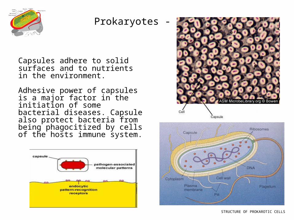
Capsules adhere to solid surfaces and to nutrients in the environment.
Adhesive power of capsules is a major factor in the initiation of some bacterial diseases. Capsule also protect bacteria from being phagocitized by cells of the hosts immune system.
STRUCTURE OF PROKAROTIC CELLS
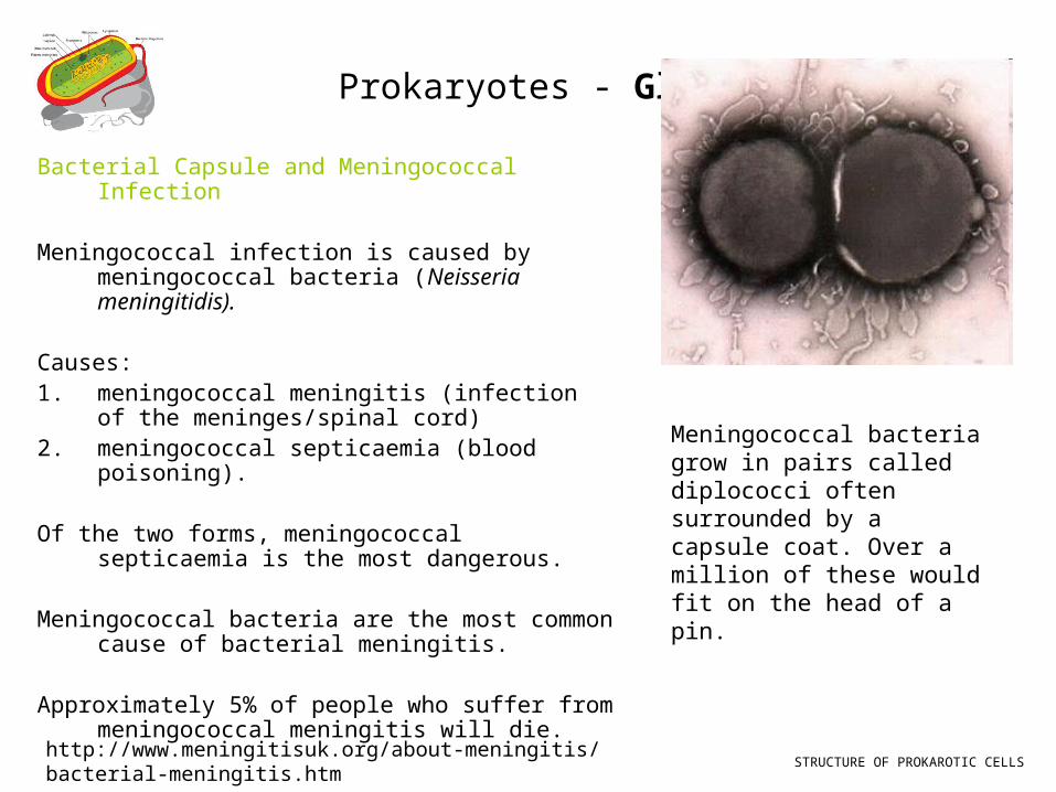
Prokaryotes - Glycocalyx Bacterial Capsule and Meningococcal Infection
Meningococcal infection is caused by meningococcal bacteria (Neisseria meningitidis).
Causes:1. meningococcal meningitis (infection of the
meninges/spinal cord)2. meningococcal septicaemia (blood
poisoning).
Of the two forms, meningococcal septicaemia is the most dangerous.
Meningococcal bacteria are the most common cause of bacterial meningitis.
Approximately 5% of people who suffer from meningococcal meningitis will die.
STRUCTURE OF PROKAROTIC CELLS
Meningococcal bacteria grow in pairs called diplococci often surrounded by a capsule coat. Over a million of these would fit on the head of a pin.
http://www.meningitisuk.org/about-meningitis/bacterial-meningitis.htm
Prokaryotes - Endospores
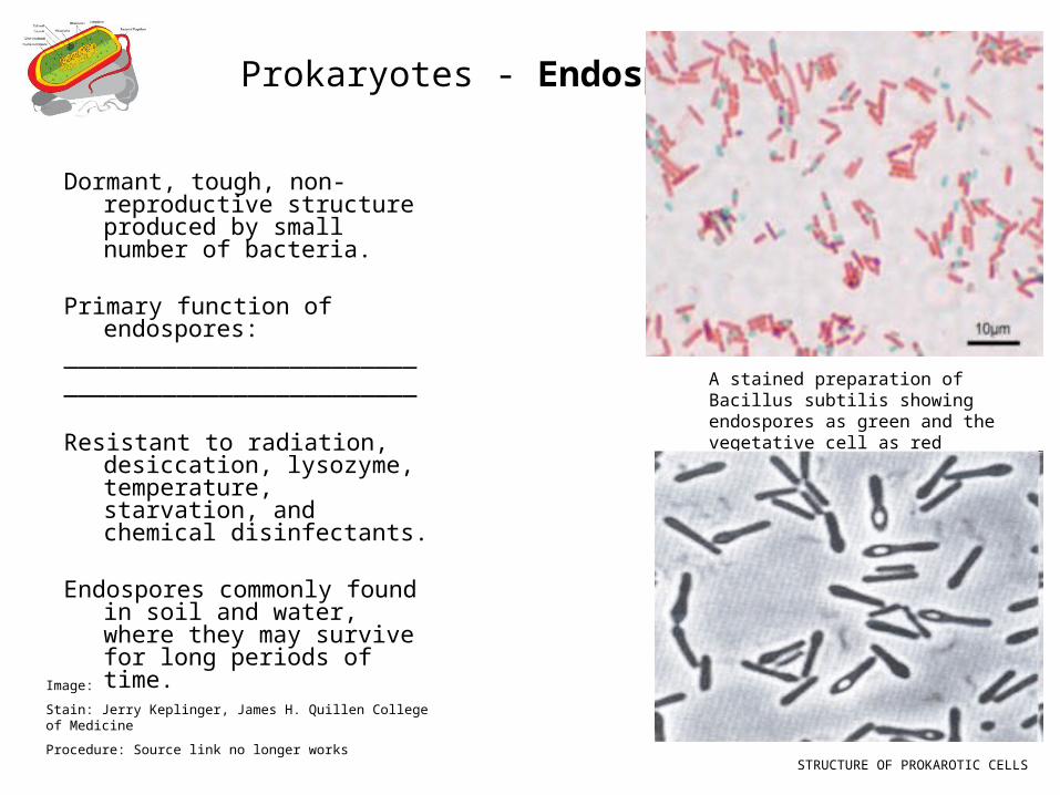
Dormant, tough, non-reproductive structure produced by small number of bacteria.
Primary function of endospores:__________________________________________________
Resistant to radiation, desiccation, lysozyme, temperature, starvation, and chemical disinfectants.
Endospores commonly found in soil and water, where they may survive for long periods of time.
A stained preparation of Bacillus subtilis showing endospores as green and the vegetative cell as red
Image:
Stain: Jerry Keplinger, James H. Quillen College of Medicine
Procedure: Source link no longer works
STRUCTURE OF PROKAROTIC CELLS


![89023611-Wiring diagram VM EM-EU5[1].pdf](https://img.document.onl/doc/110x75/55cf94e1550346f57ba50dac/89023611-wiring-diagram-vm-em-eu51pdf.jpg)