Embed Size (px)
Citation preview
Pontifícia Universidade Católica do Rio Grande do Sul
Faculdade de Biociências
Programa de Pós-Graduação em Biologia Celular e Molecular
Dissertação de Mestrado
DAIANA RENCK
ESTUDOS BIOQUÍMICOS E GENÉTICOS DA ENZIMA URIDINA
FOSFORILASE-1 HUMANA (E.C. 2.4.2.3)
Porto Alegre
2010

DAIANA RENCK
ESTUDOS BIOQUÍMICOS E GENÉTICOS DA ENZIMA URIDINA
FOSFORILASE-1 HUMANA (E.C. 2.4.2.3)
Dissertação apresentada como requisito para a obtenção do grau de Mestre pelo Programa de Pós-Graduação em Biologia Celular e Molecular da Pontifícia Universidade Católica do Rio Grande do Sul.
Orientador: Prof. Dr. Luiz Augusto Basso
Co-Orientador: Prof. Dr. Diógenes Santiago Santos
Porto Alegre
2010

DAIANA RENCK
ESTUDOS BIOQUÍMICOS E GENÉTICOS DA ENZIMA URIDINA
FOSFORILASE-1 HUMANA (E.C. 2.4.2.3)
Dissertação apresentada como requisito para a obtenção do grau de Mestre pelo Programa de Pós-Graduação em Biologia Celular e Molecular da Pontifícia Universidade Católica do Rio Grande do Sul.
Aprovado em _____ de ___________________ de _________.
BANCA EXAMINADORA:
Profa. Dra.
_________Nadja Schöreder__________
Prof. Dr.
_____Osmar Norberto de Souza______
Prof. Dr.
_________Mário Sérgio Palma_______
AGRADECIMENTOS
Ao Prof. Diógenes Santiago Santos, agradeço pela oportunidade de
integrar seu grupo de pesquisas e por me proporcionar um maior aprendizado.
Ao Prof. Luiz Augusto Basso, agradeço por todo o conhecimento
compartilhado, pelas correções e sugestões que fizeram com que este trabalho
fosse concluído.
Um agradecimento especial ao colaborador e amigo Dr. Rodrigo G.
Ducati, por toda a paciência e por todos os ensinamentos que proporcionaram
a realização deste trabalho e acrescentaram muito em minha vida acadêmica.
Aos Drs. Gaby Renard, Claudia P. Nunes e Eraldo Batista Jr, por todos os
conhecimentos e conselhos que foram passados com muita paciência e
carinho, que com certeza foram muito importantes para a realização desse
trabalho, mas acima de tudo pela amizade cultivada por todos esses anos.
Aos demais colegas e amigos do CPBMF e da Quatro G pela ajuda,
amizade, companheirismo e pelos momentos de descontração, pois de alguma
forma todos contribuíram para a realização desse trabalho.
Ao colega e amigo, Thiago M. de Assunção, por toda a ajuda, troca de
experiências e compreensão que foram essenciais para a execução desse
trabalho; e por estar presente em todos os momentos, não só como colega de
profissão e trabalho, mas como amigo de tanto tempo, faço-lhe esse
agradecimento mais que especial. Ao meu namorado, Leonardo Rosado,
agradeço pela troca de experiências, ensinamentos, carinho, compreensão,
dedicação e amor que foram essenciais para concluir essa etapa.
Aos meus pais, Ary Renck e Thereza Laux, agradeço pelo amor, carinho
e dedicação que recebi, estando presente ou não, durante todos esses anos;
pelos momentos em que a saudade teve que ser guardada e pelo exemplo de
caráter e valores que levarei para o resto da vida. Agradeço também a minha
irmã, Deise Renck, pela ajuda, pelos conselhos e pelo carinho durante essa
etapa, que com certeza nunca serão esquecidos.
i

SUMÁRIO
Agradecimentos i
Resumo iii
Abstract iv
1 INTRODUÇÃO .........................................................................................................1
1.1 A Visão Geral das Estimativas do Câncer..........................................................1
1.1.1Câncer de Mama e Cólorretal.......................................................................3
1.2 Metabolismo de nucleotídeos pirimídicos ..........................................................4
1.3 A uridina e seu papel protetor no uso do 5-FU ..................................................8
1.4 Uridina fosforilase humana ..............................................................................10
1.4.1 Uridina fosforilase é regulada pela p53 ....................................................13
1.4.2 As duas isoformas da UP em mamíferos: UP1 e UP2 .............................14
2 OBJETIVOS...........................................................................................................17
2.1 Objetivo geral...................................................................................................17
2.2 Objetivos específicos .......................................................................................17
3. ARTIGO CIENTÍFICO ...........................................................................................18
Abstract..................................................................................................................21
Introduction ............................................................................................................22
Materials and Methods...........................................................................................25
Results and Discussion..........................................................................................31
Summary ...............................................................................................................39
References ............................................................................................................42
Legends, figures and tables...................................................................................50
4.CONSIDERAÇÕES FINAIS...................................................................................61
ii

ANEXO 1. TESTE DE PURIFICAÇÃO DA PROTEÍNA RECOMBINANTE
HUP1........................................................................................................................64
ANEXO 2. TESTE DE CRISTALIZAÇÃO PARA DETERMINAÇÃO DA
ESTRUTURA TRIDIMENSIONAL DA PROTEÍNA RECOMBINANTE HUP1....... .65
ANEXO 3. EXPRESSÃO DIFERENCIAL EM TECIDOS NEOPLÁSICOS..............66
ANEXO 4. CONFIRMAÇÃO DA SUBMISSÃO DO MANUSCRITO....................... 69
ANEXO 5. APROVAÇÃO DO COMITÊ EM ÉTICA E PESQUISA..........................70
REFERÊNCIAS BIBLIOGRÁFICA...........................................................................71
iii

RESUMO
A uridina fosforilase (UP) é uma enzima chave na rota de salvamento das
pirimidinas, catalisando a fosforólise reversível de uridina a uracil e ribose-1-
fosfato (R1P). A UP humana do tipo 1 (hUP1) é um alvo molecular para o
desenho de inibidores com a finalidade de aumentar os níveis endógenos
de uridina para “resgatar” os tecidos normais da toxicidade produzida pelo
uso de agentes quimioterápicos análogos de pirimidinas, como o 5-
fluorouracil e a capecitabina. Neste trabalho, descrevemos o método de
obtenção da proteína recombinante homogênea hUP1, dados de velocidade
inicial, inibição pelo produto e ensaios de ligação em equilíbrio. Esses
resultados sugerem que a hUP1 catalisa a fosforólise de uridina por um
mecanismo cinético bi bi ordenado, no qual o fosfato inorgânico se liga
primeiro seguido pela ligação da uridina, e o uracil de dissocia primeiro,
seguido pela dissociação da R1P. Os ensaios de ligação por fluorescência
em equilíbrio mostraram uma ligação cooperativa tanto do PI como da R1P.
Os resíduos de aminoácidos envolvidos tanto na catálise como na ligação
foram propostos pelo perfil de pH.
Palavras chave: Quimioterapia do câncer, velocidade inicial, inibição pelo
produto, fluorimetria, perfil de pH, mecanismo cinético da uridina fosforilase.
iv

ABSTRACT
Uridine phosphorylase (UP) is a key enzyme in the pyrimidine salvage pathway,
catalyzing the reversible phosphorolysis of uridine to uracil and ribose-1-
phosphate (R1P). The human UP type 1 (hUP1) is a molecular target for the
design of inhibitors intended to boost endogenous uridine levels to rescue
normal tissues from the toxicity of fluoropyrimidine nucleoside
chemotherapeutic agents, such as capecitabine and 5-fluorouracil. Here, we
describe a method to obtain homogeneous recombinant hUP1, and present
initial velocity, product inhibition, and equilibrium binding data. These results
suggest that hUP1 catalyzes uridine phosphorolysis by a steady-state ordered
bi bi kinetic mechanism, in which inorganic phosphate binds first followed by the
binding of uridine, and uracil dissociates first, followed by R1P release.
Fluorescence titration at equilibrium showed cooperative binding of either Pi or
R1P binding to hUP1. Amino acid residues involved in either catalysis or
substrate binding were proposed based on pH-rate profiles.
Keywords: Cancer chemotherapy; Initial velocity; Product inhibition;
Fluorescence spectroscopy; pH-rate profiles; Uridine phosphorylase kinetic
mechanism
v

1. INTRODUÇÃO
1.1 A Visão Geral das Estimativas do Câncer
O câncer é uma das doenças que mais atinge a população mundial e
que está, a cada ano, fazendo mais vítimas. Segundo o relatório da Agência
Internacional para Pesquisa em Câncer (IARC)/Organização Mundial da Saúde
(OMS) [World Cancer Report 2008], o impacto global que o câncer gera dobrou
nos últimos 30 anos, e de 2000 à 2020 esse impacto dobrará novamente. Em
2008, a IARC/OMS estimou que ocorressem 12,4 milhões de novos casos e
7,6 milhões de óbitos por câncer no mundo. Destes, os mais incidentes foram o
câncer de pulmão (1,52 milhões de novos casos), câncer de mama (1,29
milhões) e câncer de cólon e reto (1,15 milhões).
Para América do Sul e Central, em 2008, foram cerca de 1 milhão de
novos casos de câncer e 589 mil óbitos. Em homens, o câncer que teve maior
prevalência foi o câncer de próstata, seguido por pulmão, estômago e cólon e
reto. No sexo feminino, o mais freqüente foi o câncer de mama, seguido do
colo do útero, cólon e reto, estômago e pulmão.
A distribuição dos novos casos de câncer segundo localização primária
mostra-se heterogênea entre estados e capitais do Brasil; o que fica em
evidência ao observarem-se as diferentes taxas brutas de incidência, como
ilustrado nas tabelas apresentadas a seguir. As regiões Sul e Sudeste, de
maneira geral, apresentam as maiores taxas, enquanto que as regiões Norte e
Nordeste mostram as menores taxas. As taxas da região Centro-Oeste
1

apresentam um padrão intermediário [INCA: Estimativa 2010: Incidência de
Câncer no Brasil].
Estimativas para o ano 2010 das taxas brutas de incidência por 100.000 e de número de casos novos por câncer, em homens, segundo localização primária. Adaptado: INCA.
Estimativas para o ano 2010 das taxas brutas de incidência por 100.000 e de número de casos novos por câncer, em mulheres, segundo localização primária. Adaptado: INCA.
2

1.1.2 Câncer de Mama e Colorretal
O câncer de mama, o segundo tipo mais freqüente de câncer no mundo,
é o mais comum entre as mulheres, respondendo por 22% dos casos novos a
cada ano. No Brasil, as taxas de mortalidade por câncer de mama continuam
elevadas, provavelmente porque a doença ainda é diagnosticada tardiamente,
gerando, apenas em 2007, um valor de 11.194 mil mortes. Na população
mundial, a sobrevida média após cinco anos é de 61%. O câncer de mama é
relativamente raro antes dos 35 anos, acima desta faixa etária sua incidência
cresce rápida e progressivamente. Estatísticas indicam aumento de sua
incidência tanto nos países desenvolvidos quanto nos em desenvolvimento,
segundo a OMS. As estimativas para 2010 no Brasil geram um valor de 49.240
mil novos casos.
Com uma incidência um pouco menor, mas também preocupante, os
tumores malignos que acometem o intestino grosso (cólon) e o reto ocupam o
quarto lugar como o câncer mais comum no mundo e a segunda em países
desenvolvidos. Grande parte desses tumores se inicia a partir de pólipos,
lesões benignas que podem crescer na parede interna do intestino grosso.
Uma maneira de prevenir o aparecimento dos tumores seria a detecção e a
remoção dos pólipos antes de eles se tornarem malignos [Santos et al, 2005].
Segundo dados do Instituto Nacional de Câncer (INCA), o número de novos
casos de câncer de cólon e reto estimados para o Brasil em 2010 é de 28.110
mil casos, sendo destes 13.310 em homens e 14.800 em mulheres. As mortes
totalizadas por este tipo de câncer atingiram, em 2007, 11.322 mil pessoas,
sendo destes 5.305 homens e 6.017 mulheres [www.inca.gov.br].
3
Dentro do panorama destes dois principais tipos de câncer, a cirurgia e o
posterior tratamento com quimioterápicos são as escolhas de primeira linha.
Dentre os quimioterápicos, um dos mais utilizados é o 5-Fluorouracil (5-FU),
introduzido no mercado há mais de 40 anos e será discutido mais
detalhadamente a seguir. Neste cenário, necessita-se de avanços aplicados ao
tratamento e a prevenção, tornando-se fundamental que os recursos e esforços
sejam direcionados para a busca de novos medicamentos de combate ao
câncer assim como medicamentos que aumentem a qualidade de vida dos
pacientes durante os longos e desgastantes tratamentos quimioterápicos.
A efetividade clínica do 5-FU é limitada por seus severos efeitos
adversos como mielossupressão, trombocitopenia e lesões gastrointestinais e
nas mucosas, entre outras. A uridina tem sido utilizada para reduzir esses
efeitos adversos severos levando também a um aumento do índice terapêutico
[Cao et al, 2002], no qual a disponibilidade e a concentração são reguladas
pela uridina fosforilase (UP). Porém, o uso clínico desse “resgate da uridina” é
dificultado pela sua rápida degradação, iniciada pela UP no fígado e pela
toxicidade dose-limitante resultando na necessidade do uso de altas
concentrações para obter a dose desejada à proteção dos tecidos. BAU, um
inibidor da UP, tem sido capaz de aumentar a concentração endógena de
uridina levando a uma proteção similar aos tecidos normais [Cao et al, 2002].
1.2 Metabolismo de nucleotídeos pirimídicos
Os nucleotídeos são compostos que fazem parte de um grande número
de processos bioquímicos nas diferentes formas de vida e são classificados em
4

purínicos e pirimídicos. A importância desses nucleotídeos no metabolismo
celular é suportada pelo fato de que quase todas as células podem sintetizá-los
tanto de novo como a partir dos produtos de degradação dos ácidos nucléicos,
denominando-se salvamento ou recuperação [Voet e Voet, 2006]. Os
nucleotídeos purínicos e pirimídicos são encontrados em todos os organismos
vivos onde suas funções estão relacionadas principalmente com a estrutura
dos ácidos nucléicos (DNA e RNA), atuando como fonte de energia em muitos
sistemas, na sinalização celular, na regulação de rotas de biossíntese, entre
outras [Shambaugh, 1979].
Existem separadas vias enzimáticas para a produção destes
nucleotídeos como mencionado acima, ocorrendo ou através da via de novo, a
partir de precursores simples e com uma demanda maior de energia, ou
através da via de salvamento (Salvage Pathway) (Figura 1), a partir de
nucleosídeos e bases livres pré-formadas e com um gasto menor de energia
devido a este processo de reciclagem [Shambaugh, 1976; Connolly e Duley,
1999]. As duas rotas, através de múltiplos passos enzimáticos envolvendo
fosforilases e quinases, produzem um produto comum, uridina 5’monofosfato
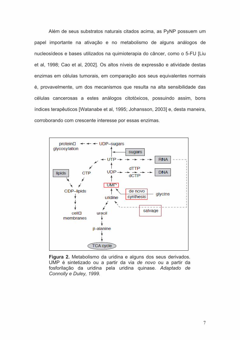
(UMP), o ponto no qual as duas rotas se encontram (Figura 2) [Connolly e
Duley, 1999].
As fosforilases de nucleosídeos pirimídicos (PyNP) são enzimas chave
na rota de salvamento destes nucleotídeos. Há dois tipos de PyNP na maioria
dos organismos: a Uridina Fosforilase (UP) e a Timidina Fosforilase (TP), as
quais catalisam a fosforólise reversível de uridina (Urd) e timidina ou
desoxiuridina, respectivamente, para suas bases livres e ribose-1-fosfato (R1P)
ou desoxiribose-1-fosfato [Watanabe e Uchida, 1995; Pizzorno et al, 2002;
5

Johansson, 2003]. Esses produtos de degradação são, então, utilizados ou
como fontes de carbono e origem de energia ou para o resgate de bases
pirimídicas para a síntese de nucleotídeos [Watanabe et al, 1995].
Figura 1. Catabolismo dos nucleotídeos pirimídicos. Adaptado: Voet e Voet, 2006.
6
Além de seus substratos naturais citados acima, as PyNP possuem um
papel importante na ativação e no metabolismo de alguns análogos de
nucleosídeos e bases utilizados na quimioterapia do câncer, como o 5-FU [Liu
et al, 1998; Cao et al, 2002]. Os altos níveis de expressão e atividade destas
enzimas em células tumorais, em comparação aos seus equivalentes normais
é, provavelmente, um dos mecanismos que resulta na alta sensibilidade das
células cancerosas a estes análogos citotóxicos, possuindo assim, bons
índices terapêuticos [Watanabe et al, 1995; Johansson, 2003] e, desta maneira,
corroborando com crescente interesse por essas enzimas.
Figura 2. Metabolismo da uridina e alguns dos seus derivados. UMP é sintetizado ou a partir da via de novo ou a partir da fosforilação da uridina pela uridina quinase. Adaptado de Connolly e Duley, 1999.
7

1.3 A uridina e seu papel protetor no uso do 5-FU
A Urd, um nucleosídeo pirimídico, é um importante precursor da rota de
salvamento das pirimidinas [Balestri et al, 2007; Cao et al, 2005]. Além de a
Urd ser essencial para a síntese de RNA e de biomembranas, através da
formação de conjugados pirimidina-lipídeo e pirimidina-açúcar [Connolly e
Duley, 1999], há também evidências experimentais e clínicas que sugerem que
ela é um elemento crucial na regulação de processos fisiológicos normais
[Connolly e Duley, 1999; Cansey, 2006], especialmente no sistema nervoso
central [Cansey, 2006], e em alguns estados patológicos [Connolly e Duley,
1999].
Farmacologicamente, a Urd tem sido utilizada para proteger os tecidos
normais dos efeitos colaterais tóxicos da quimioterapia anti câncer (baseada
em pirimidinas), principalmente como uma terapia de “resgate” para a
toxicidade produzida pelo 5-FU justamente por ela ser um competidor natural
[Krenitsky et al, 1965; Martin et al, 1982; Leyva et al, 1984; Pizzorno et al,
1998; Cao et al, 1999; el Kouni et al, 2000; Zhang et al, 2001; Pizzorno et al,
2002].
O 5-FU consiste em um análogo da base uracil possuindo como
diferença um átomo de flúor no carbono 5 no lugar de um hidrogênio e teve sua
introdução na clínica há mais de 40 anos. Ele rapidamente entra nas células
através de difusão facilitada e é convertido aos seus metabólitos ativos. O 5-FU
é uma droga que interfere na duplicação e transcrição do DNA [Pandolfo et al,
2005] e ainda representa um dos agentes mais ativos no tratamento de
8

tumores sólidos como mama, cólon e câncer de cabeça e pescoço, entre
outros [Cao et al, 2002].
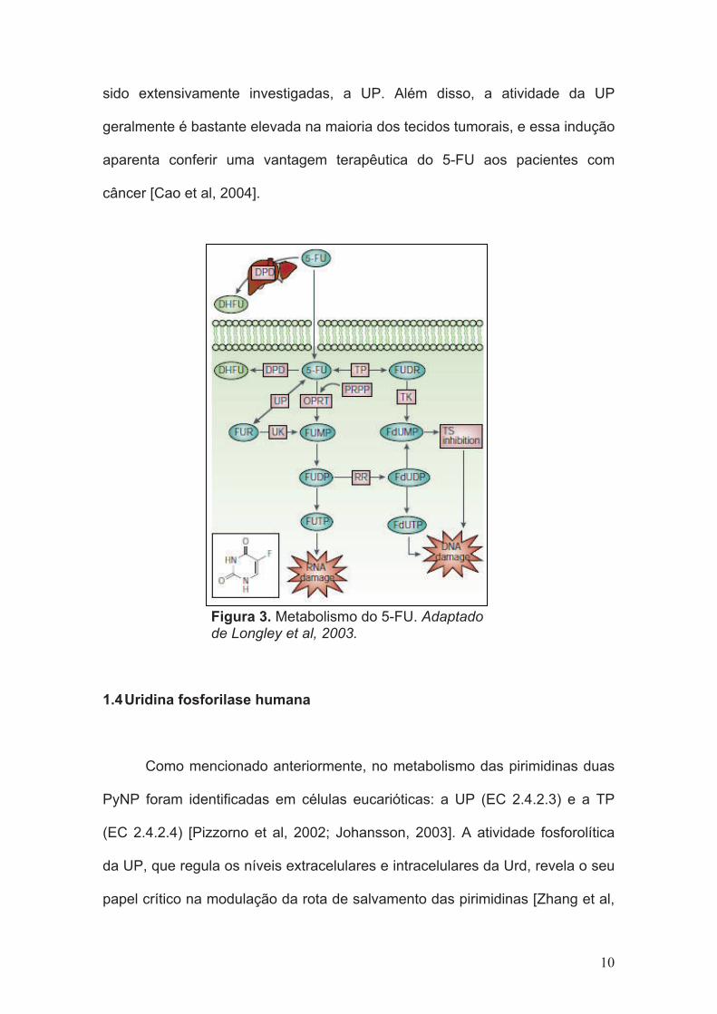
Dois mecanismos de ação contribuem para o efeito citotóxico do 5-FU:
a) toxicidade direta ao DNA, onde o 5-FluorodesoxiUMP (5-FdUMP) se liga
fortemente à Timidilato Sintase (TS), resultando na inibição da síntese de DNA
e no crescimento celular e com menor freqüência a incorporação de
fluorodesoxinucleotídeos no DNA levando à sua fragmentação e morte celular;
b) citotoxicidade direta ao RNA com incorporação do FUTP em várias espécies
de RNA, incluindo RNA polissomal, RNA nuclear e RNA mensageiro, por meio
disso interrompendo a maturação e algumas funções do RNA [Cao et al, 2002]
(Figura 3). Há hipóteses de que a atividade antitumoral do 5-FU consiste na
inibição da TS enquanto que a sua toxicidade se dá pela incorporação do
nucleotídeo fluorado, gerado a partir da ativação do 5-FU, no RNA.
Se altas doses exógenas de Urd forem administradas algumas horas
após o medicamento, a ligação do FdUMP à TS não será afetada, mas o UTP
substituirá o FUTP no RNA [Pinedo e Peters, 1988], revelando que a atividade
antitumoral do medicamento não é afetada, porém seus efeitos adversos
diminuem. Entretanto, esses elevados níveis exógenos de Urd não são bem
tolerados em humanos [Pizzorno et al, 1998], causando mais efeitos adversos
ao paciente além daqueles já produzidos pela medicação [Cao et al, 1999];
porém esses altos níveis são essenciais para que ocorra o efeito de proteção
desejado devido à meia vida curta [Leyva et al, 1984; Miyashita et al, 2002] da
Urd no organismo (10-15 minutos) [Miyashita et al, 2002].
Por esta razão, uma das enzimas que metaboliza o fármaco em tecidos
normais e tumorais e ainda regula a concentração homeostática da Urd tem
9
sido extensivamente investigadas, a UP. Além disso, a atividade da UP
geralmente é bastante elevada na maioria dos tecidos tumorais, e essa indução
aparenta conferir uma vantagem terapêutica do 5-FU aos pacientes com
câncer [Cao et al, 2004].
Figura 3. Metabolismo do 5-FU. Adaptadode Longley et al, 2003.
1.4 Uridina fosforilase humana
Como mencionado anteriormente, no metabolismo das pirimidinas duas
PyNP foram identificadas em células eucarióticas: a UP (EC 2.4.2.3) e a TP
(EC 2.4.2.4) [Pizzorno et al, 2002; Johansson, 2003]. A atividade fosforolítica
da UP, que regula os níveis extracelulares e intracelulares da Urd, revela o seu
papel crítico na modulação da rota de salvamento das pirimidinas [Zhang et al,
10
2001]. A enzima está distribuída em procariotos, leveduras e organismos
superiores e a sua seqüência de aminoácidos é altamente conservada entre
enzimas bacterianas, humana e de camundongos [el Kouni et al, 2000]. A
atividade fosforolítica básica é conservada ao longo de toda a hierarquia
evolucionária, de bactérias a humanos, embora a estrutura gênica e protéica e
o tamanho da proteína variem, se apresentando, por exemplo, como um
hexâmero em Escherichia coli [Caradoc-Davies et al, 2004] e Salmonella
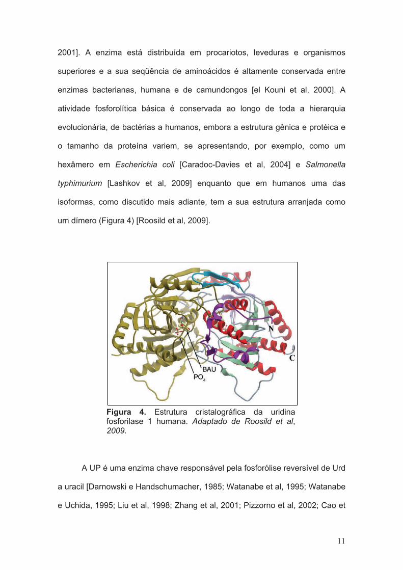
typhimurium [Lashkov et al, 2009] enquanto que em humanos uma das
isoformas, como discutido mais adiante, tem a sua estrutura arranjada como
um dímero (Figura 4) [Roosild et al, 2009].
Figura 4. Estrutura cristalográfica da uridina fosforilase 1 humana. Adaptado de Roosild et al, 2009.
A UP é uma enzima chave responsável pela fosforólise reversível de Urd
a uracil [Darnowski e Handschumacher, 1985; Watanabe et al, 1995; Watanabe
e Uchida, 1995; Liu et al, 1998; Zhang et al, 2001; Pizzorno et al, 2002; Cao et
11

al, 2005] e, na presença de R1P, ela pode também catalisar a reação reversa,
formando o nucleosídeo a partir da base livre (Figura 5) [Grem, 2000; Cao et al,
2002; Pizzorno et al, 2002].
Figura 5. Reação catalisada pela UP.
Altos níveis de atividade da UP são encontrados em diversas espécies
de tumores sólidos e o nível de expressão dela pode estar correlacionado com
a progressão da doença [Miyashita et al, 2002; Johansson, 2003]. Zhang e
colaboradores [Zhang et al, 2001] clonaram e caracterizaram parcialmente a
região promotora da UP e verificaram que ela apresenta supostos elementos
regulatórios para alguns fatores oncogênicos e genes supressores de tumor,
incluindo a p53, como descrito a seguir.
Deste modo, o entendimento da regulação do gene UP, afetando tanto
a atividade catalítica como a expressão, tornou-se crítico para a elucidação do
seu potencial papel na tumorigênese e para a modulação do tratamento de
câncer.
12

1.4.1 A Uridina Fosforilase é regulada pela p53
O supressor tumoral p53, possui um papel crucial no controle do
crescimento celular, no reparo do DNA danificado e na regulação da apoptose
[Ko e Prives, 1996]. A regulação supressora da p53 sobre o gene UP indica a
presença de um controle negativo na rota de salvamento de pirimidinas,
provavelmente como um mecanismo celular de auto-proteção em caso de
depleção de ribonucleotídeos [Zhang et al, 2001; Pizzorno et al, 2002] (Figura
6). Um dano celular causando uma perda ou um desequilíbrio no “pool” desses
nucleotídeos pode causar a ativação da p53, conduzindo a uma supressão da
expressão da UP e à ativação da rota de salvamento de pirimidinas para
reabastecer o “pool” de nucleotídeos de pirimidinas afetado [Pizzorno et al,
2002].
O esclarecimento da regulação do controle negativo da p53 sobre o
gene promotor da UP e a sua expressão podem ter uma implicação
considerável a níveis clínicos, desde no presente promotor do DNA da UP até
no resultado terapêutico em tumores com mutação específica em p53, quando
submetido a terapias anti-câncer baseadas em anti metabólitos [Zhang et al,
2001].
13
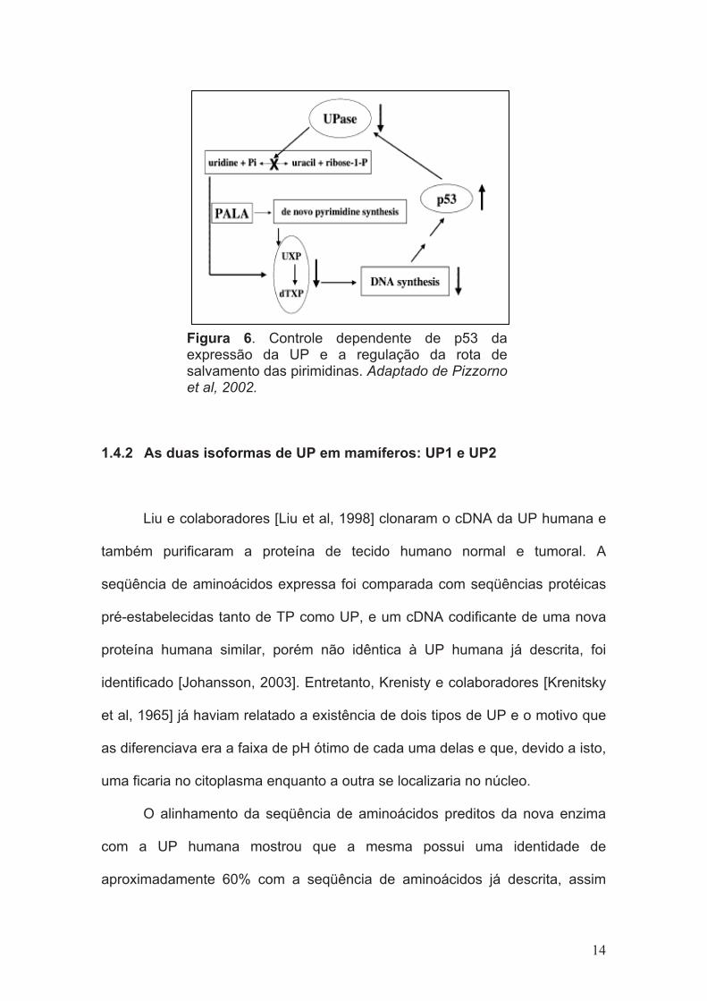
Figura 6. Controle dependente de p53 da expressão da UP e a regulação da rota de salvamento das pirimidinas. Adaptado de Pizzornoet al, 2002.
1.4.2 As duas isoformas de UP em mamíferos: UP1 e UP2
Liu e colaboradores [Liu et al, 1998] clonaram o cDNA da UP humana e
também purificaram a proteína de tecido humano normal e tumoral. A
seqüência de aminoácidos expressa foi comparada com seqüências protéicas
pré-estabelecidas tanto de TP como UP, e um cDNA codificante de uma nova
proteína humana similar, porém não idêntica à UP humana já descrita, foi
identificado [Johansson, 2003]. Entretanto, Krenisty e colaboradores [Krenitsky
et al, 1965] já haviam relatado a existência de dois tipos de UP e o motivo que
as diferenciava era a faixa de pH ótimo de cada uma delas e que, devido a isto,
uma ficaria no citoplasma enquanto a outra se localizaria no núcleo.
O alinhamento da seqüência de aminoácidos preditos da nova enzima
com a UP humana mostrou que a mesma possui uma identidade de
aproximadamente 60% com a seqüência de aminoácidos já descrita, assim
14

como as enzimas de camundongo, também possui uma elevada similaridade
com as duas isoformas humanas [Johansson, 2003] (Figura 7).
Baseado nos altos níveis de similaridade da seqüência, a nova enzima
foi nomeada Uridina Fosforilase-2 (UP2) enquanto que a Uridina Fosforilase
humana previamente caracterizada parcialmente foi nomeada UP1 [Johansson,
2003]. Assim, houve a identificação da terceira fosforilase de nucleosídeos
pirimídicos, presente em células de mamíferos, além da UP1 e TP.
Figura 7. Alinhamento da seqüência de aminoácidos preditos da UP-2 humana (H. UPase-2) e de camundongo (M. UPase-2) com a UP-1 humana (H. UPase-1) e de camundongo (M. UPase-1), respectivamente. As porções destacadas em preto indicam os resíduos de aminoácidos idênticos, conservados em comparação à seqüência da UP-1 humana. Adaptado de Johansson et al, 2003.
Em humanos, as duas enzimas se encontram em genes e cromossomos
distintos e o local de maior expressão também difere entre as duas isoformas.
Enquanto que a UP2 é predominantemente expressa no rim, a UP1 encontra-
se distribuída de uma maneira mais generalizada no organismo. Esta
15

expressão diferenciada entre as duas isoformas ainda não está bem
esclarecida, não se sabendo ao certo o motivo pelo qual ocorre no organismo
visto que as duas realizam a mesma função [Cao et al, 2005].
Assim, a caracterização da UP1 humana poderá contribuir para o
desenvolvimento de inibidores seletivos e para a quimioterapia do câncer.
16

2. OBJETIVOS
2.1 Obejtivo Geral
Este trabalho tem por objetivo geral a expressão, a purificação e a
caracterização da enzima Uridina Fosforilase-1 humana realizados no Instituto
Nacional de Ciência e Tecnologia em Tubérculos (INCT-TB) - Centro de
Pesquisas em Biologia Molecular e Funcional da PUCRS (CP-BMF). Tais
experimentos fazem parte de um projeto maior existente no laboratório que visa
à caracterização de enzimas (humanas e de M. tuberculosis) da via de
salvamento de purinas e pirimidinas para o desenvolvimento racional de
possíveis inibidores.
2.2 Objetivos Específicos
- A amplificação do gene UPP1 através da reação em cadeia da
polimerase (PCR) utilizando oligonucleotídeos iniciadores;
- Clonagem do amplicon em vetor de clonagem e posteriormente em
vetor de expressão;
- Estabelecimento de um protocolo de expressão e de purificação da
proteína UP1 por meio de Cromatografia Líquida de Rápida Performance
(FPLC);
- Análise por espectrometria de massas e pelo método de Degradação
de Édman para a determinação da identidade da proteína homogênea;
- Teste de atividade e caracterização da enzima através de cinética
enzimática;
17

3. ARTIGO CIENTÍFICO
TITLE OF THE ARTICLE
The kinetic mechanism of human uridine phosphorylase 1: towards the
development of new enzyme inhibitors for cancer chemotherapy.
PERIODIC CHOSEN FOR SUBMISSION
Archives of Biochemistry and Biophysics (impact factor: 2.626).
18

The kinetic mechanism of human uridine phosphorylase 1: towards the
development of enzyme inhibitors for cancer chemotherapy†
Daiana Renck1,2, Rodrigo G. Ducati1, Mario S. Palma3, Diógenes S. Santos1,2,*, Luiz A.
Basso1,2,*
1Centro de Pesquisas em Biologia Molecular e Funcional (CPBMF), Instituto Nacional
de Ciência e Tecnologia em Tuberculose (INCT-TB), Pontifícia Universidade Católica
do Rio Grande do Sul (PUCRS), 6681/92-A Av. Ipiranga, 90619-900, Porto Alegre, RS,
Brazil.
2Programa de Pós-Graduação em Biologia Celular e Molecular, Pontifícia Universidade
Católica do Rio Grande do Sul (PUCRS), Porto Alegre, RS, Brazil.
3Laboratório de Biologia Estrutural e Zooquímica, Centro de Estudos de Insetos Sociais,
Departamento de Biologia, Instituto de Biociências de Rio Claro, Universidade Estadual
Paulista (UNESP), Rio Claro, SP, Brazil.
†This work was supported by the National Institute of Science and Technology on
Tuberculosis (DECIT/SCTIE/MS-MCT-CNPq-FNDCT-CAPES) and the Millennium
Initiative Program (CNPq) to D.S.S. and L.A.B. D.S.S. (CNPq, 304051/1975-06),
L.A.B. (CNPq, 520182/99-5), and M.S.P. (CNPq, 500079/90-0) are Research Career
Awardees of the National Research Council of Brazil (CNPq). R.G.D. is a postdoctoral
fellow of CNPq. D.R. is recipient of an MSc scholarship awarded by BNDES.
Running Title: Kinetic mechanism of human uridine phosphorylase 1
19

*Corresponding authors. Telephone/Fax: +55-51-33203629.
E-mail addresses: [email protected] (Luiz A. Basso); [email protected] (Diógenes
S. Santos).
20

Abstract
Uridine phosphorylase (UP) is a key enzyme in the pyrimidine salvage pathway,
catalyzing the reversible phosphorolysis of uridine to uracil and ribose-1-phosphate
(R1P). The human UP type 1 (hUP1) is a molecular target for the design of inhibitors
intended to boost endogenous uridine levels to rescue normal tissues from the toxicity
of fluoropyrimidine nucleoside chemotherapeutic agents, such as capecitabine and 5-
fluorouracil. Here, we describe a method to obtain homogeneous recombinant hUP1,
and present initial velocity, product inhibition, and equilibrium binding data. These
results suggest that hUP1 catalyzes uridine phosphorolysis by a steady-state ordered bi
bi kinetic mechanism, in which inorganic phosphate binds first followed by the binding
of uridine, and uracil dissociates first, followed by R1P release. Fluorescence titration at
equilibrium showed cooperative binding of either Pi or R1P binding to hUP1. Amino
acid residues involved in either catalysis or substrate binding were proposed based on
pH-rate profiles.
Keywords: Cancer chemotherapy; Initial velocity; Product inhibition; Fluorescence
spectroscopy; pH-rate profiles; Uridine phosphorylase kinetic mechanism
21

Pyrimidine nucleoside phosphorylases are key enzymes in the pyrimidine
salvage pathway. Two types of enzymes have been identified in human cells: uridine
phosphorylase (UP1; EC 2.4.2.3) and thymidine phosphorylase (EC 2.4.2.4) [1,2,3]. UP
belongs to the nucleoside phosphorylase (NP) super-family of proteins, in the NP-1
subset [4], and plays an important role in nucleoside metabolism, catalyzing the
phosphorolysis of uridine (Urd) to uracil and ribose-1-phosphate (R1P) (Fig. 1). These
products can be further utilized for nucleoside synthesis [5,6]. In humans, there are two
isoforms of UP, hUP1 [3] and hUP2 [2], which are encoded by two different genes in
distinct chromosomes. The alignment of the isoforms showed that they share
approximately 60% amino acid sequence identity [2]. The hUP1 cDNA contains an
open reading frame coding for a sequence of 310 amino acid residues corresponding to
a protein with a subunit molecular mass of 33.9 kDa that is dimeric in solution [3,7],
which is in contrast to the hexameric Escherichia coli enzyme [8,9].
Although UP is present in most normal and tumoral tissues, its activity as well as
its expression is elevated in certain tumors, a feature that may contribute to selectivity
of chemotherapeutic agents [3,10,11,12]. Studies have shown that this expression can be
up-regulated by treating tumor cells with cytokines such as interferon-!, interferon-",
tumor necrosis factor-!, and interleukin-1! [3,13]. RT-PCR analysis demonstrated that
basic fibroblast growth factor (bFGF) increases the expression level of mouse UP1 in
osteolineage cell lines; the induction by bFGF is dependent of NF#B activity [14]. In
1Abbreviations used: Arg, arginine; BAU, 5-benzylacyclouridine; CV, column volume; bFGF, basic fibroblast growth factor; ESI-MS, electrospray ionization mass spectrometry; EWS, Ewing’s sarcomas; 5-FU, 5-fluorouracil; FUrd, fluorouridine; FUMP, fluorouridine monophosphate; FUDP, fluorouridine diphosphate; FUTP, fluorouridine triphosphate; FdUrd, fluorodeoxyuridine; FdUMP, fluorodeoxyuridine monophosphate; FdUDP, fluorodeoxyuridine diphosphate; FdUTP, fluorodeoxyuridine triphosphate; Hepes, N-2-hydroxyethylpiperazyne-N’-2-ethanesulfonic acid; His, histidine; hUP1, human uridine phosphorylase 1; IPTG, isopropyl-$-D-thiogalactopyranoside; LB, Luria-Bertani; NP, nucleoside phosphorylase; OPRT, orotate phosphoribosyltransferase; Pi, inorganic phosphate; PNP, purine nucleoside phosphorylase; R1P, ribose-1-phosphate; SDS-PAGE, sodium dodecyl sulfate-polyacrylamide gel eletrophoresis; TB, Terrific Broth; TK, thymidine kinase; TP, thymidine phosphorylase; Tris, tris(Hydroxymethyl)aminomethane; Urd, uridine; UK, uridine kinase; UP, uridine phosphorylase.
22

addition, it has been shown that tumor-associated chromosomal translocation in
Ewing’s family tumors, where the N-terminus of Ewing’s sarcoma (EWS) gene fuses
with the C-terminus of some ETS transcription factors, up-regulates UP promoter in
vivo, suggesting that UP could be a direct target of EWS/ETS fusion proteins [15,16]. In
contrast, the activity and gene promoter levels of UP have been shown to be down-
regulated by wild-type p53, a tumor suppressor gene that plays a key role in cell growth
control, DNA damage repair, and apoptosis [1,17].
UP plays an important role in the homeostatic regulation of Urd concentration in
plasma (1 - 5 %M) and tissues, and affects activation and catabolism of several
nucleoside analogues used in cancer chemotherapy [2]. These analogues include
fluoropyrimidines, such as 5-fluorouracil (5-FU) [1,8], which is a uracil analogue with a
fluorine atom at the C-5 position. This compound was developed in 1957 [18] after the
observation that rat tumoral tissues use uracil more rapidly than normal tissues, which
indicated that uracil metabolism is a potential target for chemotherapy [8]. Since then,
5-FU has been used in clinical practice against many types of solid tumors; yet, most of
its use is related to colorectal cancer [8,19]. 5-FU is converted to fluorodeoxyuridine
monophosphate (FdUMP), fluorodeoxyuridine triphosphate (FdUTP) and fluorouridine
triphosphate (FUTP), the three main active metabolites. The main mechanism of 5-FU
activation is either directly by orotate phosphoribosyltransferase (OPRT), or indirectly
by the sequential action of UP converting 5-FU to fluorouridine (FUrd) which, in turn,
is converted to fluorouridine monophosphate (FUMP) by uridine kinase (UK) [8,14].
FUMP is phosphorylated to fluorouridine diphosphate (FUDP), which can be either
phosphorylated to FUTP or converted by ribonucleotide reductase into
fluorodeoxyuridine diphosphate (FdUDP) [8]. The latter, in turn, can either be
phosphorylated to FdUTP or dephosphorylated to FdUMP, both of which are active
23

metabolites. There is also an alternative pathway that involves conversion of 5-FU to
fluorodeoxyuridine (FdUrd) by thymidine phosphorylase (TP), and conversion of
FdUrd to FdUMP by thymidine kinase (TK). The main metabolites of 5-FU exert their
anti-cancer activity either by disrupting RNA synthesis (FUTP), or inhibiting
thymidylate synthase (TS) enzyme activity (FdUMP) that converts dUMP to
deoxythymidine monophosphate (dTMP), which is needed for DNA synthesis and
repair. In addition, inhibition of TS by FdUMP results in accumulation of dUMP
leading to increased levels of dUTP. The latter and the 5-FU metabolite FdUMP can be
misincorporated into DNA. Clinical evidences have demonstrated the ability of Urd to
reduce bone marrow and gastrointestinal toxicity induced by 5-FU, called “rescue of 5-
FU toxicity” [2,6,10,20]. High doses of uridine are required to produce the “rescue”
effect due to the short half-life of plasma uridine of only two minutes and the regulation
of plasma uridine homeostasis at the 2-4 !M level by the activity of hepatic UP [6,10].
However, these high doses needed to produce this effect are not well tolerated and
produce dose-limiting effect in humans. It is thus necessary to develop a drug capable of
maintaining elevated endogenous levels of Urd [6,7,10] to protect the normal tissues
through Urd rescue effect. Accordingly, inhibition of UP activity appears an attractive
therapeutic strategy to rescue 5-FU toxicity. Acyclouridine analogues, including 5-
benzylacyclouridine (BAU), have been designed as hUP1 inhibitors, and BAU has been
shown in clinical trials to be capable of increasing plasma Urd concentration, thereby
increasing the therapeutic index of 5-FU [7].
Enzyme inhibitors make up roughly 25 % of the drugs marketed in United States
[21]. Enzymes catalyze multistep chemical reactions to achieve rate accelerations by
stabilization of the transition state structure [22]. Accordingly, mechanistic analysis
(kinetic, chemical, and catalytic mechanisms) should always be a top priority for
24

enzyme-targeted drug programs aiming at the rational design of potent enzyme
inhibitors. Here we describe amplification, cloning, and sequencing of the recombinant
hUP1 coding gene. We also present heterologous protein expression in Escherichia coli,
purification to homogeneity, N-terminal amino acid sequencing, electrospray ionization
mass spectrometry analysis, determination of true steady-state kinetic parameters,
product inhibition, equilibrium fluorescence of substrate/product binding, and pH-rate
profiles of functional recombinant hUP1 enzyme. These results provide a solid
foundation on which to base function-guided hUP1 enzyme inhibitors with potential
anti-cancer activity.
Materials and methods
Amplification and cloning of the human UPP1 gene
The human UPP1 coding sequence was searched on the GeneBank (BC007348)
of the National Institute for Biotechnology Information (http://www.ncbi.nlm.nhi.gov).
The cDNA of UPP1 was obtained by RT-PCR amplification of colorectal RNA
(from Ambion; Austin, TX, USA). The oligonucleotide primers used (forward primer,
5’-CAGTTGGCCATATGGCGGCCACGGGAGC-3’; and reverse primer, 5’-
GCGGAGAAGCTTGGCAGCGCTCAGGCC-3’) contained, respectively, NdeI and
HindIII (New England Biolabs) restriction sites (underlined). The PCR product was
analyzed on 1% agarose gel, and a 930-bp band was detected and purified. The DNA
fragment was cloned into pCR-Blunt cloning vector (Invitrogen), cleaved with NdeI and
HindIII restriction enzymes, and subcloned into the pET-23a(+) expression vector
(Novagen). The complete UPP1 gene sequence was determined by automated DNA-
25

sequencing to confirm sequence integrity and the absence of mutations in the cloned
fragment.
Expression and purification of recombinant hUP1
The pET-23a(+)::UPP1 recombinant plasmid was transformed into Escherichia
coli Rosetta (DE3) competent cells (Novagen) and selected on Luria-Bertani (LB) agar
plates containing 50 %g mL-1 ampicillin and 34 %g mL-1 cloranfenicol. A single colony
was grown overnight in LB medium pH 7.2 (60 mL) containing the same antibiotics, at
37°C. An aliquot of this culture (10 mL) was used to inoculate Terrific Broth (TB)
medium (2.5 L, with the same antibiotics) and grown for 36 h at 30°C after reaching an
OD600 nm of 0.4 - 0.6, without isopropyl-$-D-thiogalactopyranoside (IPTG) induction.
The same procedure was employed for E. coli Rosetta (DE3) cells transformed with
pET-23a(+) (control). The cells (40 g) were harvested by centrifugation at 11,800g for
30 min at 4°C and stored at –20°C. Soluble protein expression was analyzed by 12%
sodium dodecyl sulfate-polyacrylamide gel eletrophoresis (SDS-PAGE) stained with
Coomassie Brilliant Blue [23].
All purification steps were performed at 4°C and sample elution was monitored
by UV detection. Frozen cells (5 g) were suspended in 25 mL of 50 mM N-2-
hydroxyethylpiperazyne-N’-2-ethanesulfonic acid (Hepes) pH 7.0 (buffer A) and
incubated with 0.2 mg mL-1 lysozyme (Sigma) for 30 min. The cells were disrupted by
sonication (5 pulses of 10 sec) and the solution was cleared by centrifugation at 48,000g
for 30 min. The supernatant was treated with 1% (wt/vol) streptomycin sulfate (Sigma;
final concentration) for 30 min to precipitate the nucleic acids, and centrifuged (48,000g
for 30 min). The supernatant was dialyzed against buffer A (2 x 2 L, 3 h each). Residual
26

precipitate was removed by centrifugation (48,000g for 30 min) and the supernatant was
loaded onto a SP Sepharose Fast Flow cation exchange column (GE Healthcare) pre-
equilibrated with buffer A. The column was washed with 5 column volumes (CV) of
buffer A and the adsorbed material was eluted with 15 CV linear gradient (0 - 100%) of
50 mM Hepes 200 mM NaCl pH 7.0 (buffer B) at a 1 mL min-1 flow rate. The target
recombinant protein was eluted at approximately half of the gradient, where a single
SDS-PAGE band could be observed. The homogenous recombinant protein was
dialyzed against 100 mM tris(Hydroxymethyl)aminomethane (Tris) pH 7.4 (3 x 2 L, 3 h
each), concentrated, and stored at –80°C. Protein concentration was determined with
Bradford Protein Assay Kit (Bio-Rad Laboratories) and bovine serum albumin was used
as standard [24]. All subsequent activity and binding assays were performed in 100 Tris
pH 7.4, unless stated otherwise.
Amino acid sequence and mass spectrometry analysis
The N-terminal amino acid sequence of homogeneous recombinant hUP1
protein was analyzed by automated Edman degradation sequencing using a gas-phase
sequencer PPSQ-21 A (Shimadzu) [25].
hUP1 was assessed by electrospray ionization mass spectrometry (ESI-MS)
according to Chassaigne and Lobinski, with some adaptations [26]. The sample was
analyzed on Quattro-II triple-quadrupole mass spectrometer (Micromass; Altrincham,
United Kingdom). During all experiments, the source temperature was maintained at –
80°C and the capillary voltage at 3.6 kV; a drying nitrogen gas flow (200 L h-1) and a
nebulizer gas flow (20 L h-1) were used. Intact horse heart myoglobin was used to
calibrate the mass spectrometer and its typical cone voltage-induced fragments. hUP1
27

subunit molecular mass was determined by adjusting the mass spectrometer to yield a
peak with a half-height of 1 mass unit, and the sampling cone-to-skimmer lens voltage
controlling the transfer of ions to the mass analyzer was set to 38 V. Approximately 50
pmol of each sample were injected into the electrospray transport solvent. The ESI
spectrum was obtained in the multichannel acquisition mode, with scanning from 500 to
1,800 m/z at a scan time of 7 sec. The mass spectrometer is equipped with MassLynx
and Transform softwares for data acquisition and spectrum handling.
hUP1 enzymatic assay
Recombinant hUP1 enzyme activity was monitored in an UV-2550 UV/Visible
spectrophotometer (Shimadzu). All assays were performed under initial rate conditions
at 37°C and 100 mM Tris pH 7.4, in 500 µL total reaction volumes for 60 sec. This
assay was based on the maximum difference in absorbance at 280 nm between Urd and
uracil (&AM-1
cm-1
= 2100), in which a decrease in absorbance is observed upon
conversion of Urd to uracil [27].
Initial velocity measurements
Initial velocity studies were carried out to determine the true steady-state kinetic
parameters, in the forward direction. Saturation curves were performed varying
concentrations of Urd (20 - 500 µM) against several fixed-varying concentrations of
inorganic phosphate (Pi) (1 - 10 mM).
Product inhibition patterns
28

To provide an additional experimental approach to distinguish between the
possible kinetic mechanisms, product inhibition studies were carried out at varying
concentrations of one substrate, fixed concentrations of the co-substrate (in non-
saturating levels), and fixed-varying concentrations of products (either R1P or uracil).
The experimental conditions were as follows: varying Urd concentrations (20 - 500
µM), fixed Pi concentration (2 mM), and fixed-varying concentrations of either uracil
(50 - 600 µM) or R1P (24 - 160 µM); varying Pi concentrations (1 - 10 mM), fixed Urd
concentration (50 µM), and fixed-varying concentrations of either uracil (50 - 300 µM )
or R1P (80 - 320 µM).
Equilibrium binding by fluorescence spectroscopy
Fluorescence measurements were carried out in a RF-5301 PC
Spectrofluorophotometer (Shimadzu) at 25°C. Measurements of intrinsic hUP1 protein
fluorescence employed excitation wavelength at 280 nm in each binding experiment,
and the emission wavelength ranged from 285 to 350 nm. The slits for excitation and
emission were both 3 nm. Fluorescence titrations of binary complex formation were
carried out by making microliter additions of the following compounds to 2 mL
containing 10 µM hUP1: 40 mM Pi stock solution (19.99 - 434.2 µM final
concentration); 5 mM Urd stock solution (2.498 - 97.92 µM final concentration); 10
mM uracil stock solution (4.997 - 123.3 µM final concentration); 4 mM R1P stock
solution (1.99 - 41.53 µM final concentration). Control experiments were employed to
both determine the maximum ligand concentrations to be used with no inner filter effect
and to account for any dilution effect on protein fluorescence.
29
pH-rate profiles
To determine the dependence of the kinetic parameters on pH, initial velocities
were measured in the presence of varying concentrations of one substrate and a
saturating level of the other in a buffer mixture of 2-(N-morpholino)ethanesulfonic acid
/Hepes/2-(N-cyclohexylamino)ethanesulfonic acid over the following pH values: 5.0,
5.5, 6.0, 6.5, 7.0, 7.5, 8.0, 8.5 and 9.0 [28]. The data were plotted as pH values versus
either log kcat or log kcat/KM. As the KM values changed as a function of pH, different
concentration ranges of the variable substrate as well as the fixed substrate had to be
employed. For varying Urd concentrations the experimental conditions were: at pH 5.0:
Pi = 20 mM, 200 !M " Urd " 900 !M; at pH 5.5: Pi = 20 mM, 100 !M " Urd " 800
!M; at pH 6.0: Pi = 12 mM, 20 !M " Urd " 800 !M; at pH 6.5: Pi = 10 mM, 20 !M "
Urd " 700 !M; at pH 7.0 and 7.5: Pi = 10 mM, 20 !M " Urd " 500 !M; at pH 8.0: Pi =
10 mM, 20 !M " Urd " 700 !M; at pH 8.5: Pi = 10 mM, 20 !M " Urd " 600 !M; and
at pH 9.0: Pi = 10 mM, 20 !M " Urd " 800 !M. For varying Pi concentrations the
experimental ranges employed were as follows: at pH 5.0, Urd = 800 !M, 2 mM " Pi "
20 mM; at pH 5.5, Urd = 700 !M, 2 mM " Pi " 16 mM; at pH 6.0, Urd = 700 !M, 0.5
mM " Pi " 8 mM; at pH 6.5, Urd = 600 !M, 0.05 mM " Pi " 8 mM; at pH 7.0 and 7.5,
Urd = 500 !M, 0.1 mM " Pi " 8 mM; at pH 8.0, Urd = 600 !M, 0.1 mM " Pi " 8 mM;
at pH 8.5, Urd = 500 !M, 0.05 mM " Pi " 5 mM; and at pH 9.0, Urd = 700 !M, 0.05
mM " Pi " 5 mM.
30
Results and discussion
Amplification, cloning, and DNA sequencing
The PCR amplification protocol yielded a product with an expected size
corresponding to the human UPP1 (930-bp) DNA coding sequence (data not shown).
The fragment was purified from the agarose gel and ligated into the pET-23a(+)
expression vector. Nucleotide sequence analysis confirmed both identity and integrity of
human UPP1 DNA coding sequence.
Expression of recombinant hUP1 protein
The pET-23a(+)::UPP1 recombinant plasmid was transformed into E. coli
Rosetta (DE3) host cells by electroporation. Analysis by SDS-PAGE (Fig. 2) showed
that the cell extracts contained the recombinant protein, in the insoluble and soluble
fraction, with an apparent molecular mass of 33 kDa, in agreement with the expected
size of 33.934 kDa for hUP1. Among a number of protocols tested, the best
experimental condition for expression of recombinant hUP1 occurred with E. coli
Rosetta (DE3) cells grown for 36 h (after reaching an OD600 nm of 0.4 - 0.6, without
IPTG induction) at 30°C in TB medium. The pET expression vector system (Novagen)
has a strong IPTG-inducible bacteriophage T7 lacUV5 late promoter that controls the
T7 RNA polymerase to transcribe cloned target genes [29]. It has been shown that lac-
controlled systems could have high-levels of protein expression in the absence of
inducer [30,31]. It has been proposed that leaky protein expression is due to
derepression of the lac-controlled system when cells approach stationary phase in
31
complex medium and that cyclic AMP, acetate, and low pH are required to achieve
high-level expression in the absence of IPTG induction, which may be part of a general
cellular response to nutrition limitation [32]. However, more recently, it has been shown
that unintended induction in the pET system is due to the presence of as little as
0.0001% of lactose in the medium [33]. It is noteworthy that a large amount of
recombinant hUP1 protein remained in the insoluble fraction (Fig. 2, lane 3). Although
inclusion body formation can greatly simplify protein purification, there is no guarantee
that the in vitro refolding will yield large amounts of biologically active protein.
Moreover, inclusion body purification schemes present a number of problems such as:
use of denaturants that are expensive and can cause irreversible modifications of protein
structure that will elude all of the most sophisticated analytical tests, refolding usually
must be done in very dilute solution and the protein reconcentrated, and refolding
encourages protein isomerization leading to precipitation during storage [34]. Since we
aimed at determining the mode of action of recombinant hUP1 enzyme, we deemed
more appropriate to avoid solubilizing agents.
Purification of recombinant hUP1 protein
Recombinant hUP1 protein was efficiently purified to homogeneity (Fig. 3) by a
single-step purification protocol, using a cation exchange column. The target protein
eluted at approximately 50% of buffer B. The 3.8-fold purification protocol resulted in a
protein yield of 43% and 20.8 mg of active recombinant hUP1 protein from 5 g of cells
(Table 1). In contrast to Roosild and co-workers [7], we found no need to add potassium
salt in the purification buffers to prevent protein aggregation and precipitation. The
homogeneous protein was stored at –80°C, with no loss of activity.
32
Mass spectrometry and N-terminal amino acid sequencing
The subunit molecular mass value for hUP1 was determined to be 33,934.00 Da
by electrospray ionization mass spectrometry (ESI-MS). Since the predicted molecular
mass is 33,934.00 Da, this result indicates removal of the N-terminal methionine
(predicted methionine molecular mass = 131.20 Da). These results provide evidence
that confirm the identity of recombinant hUP1.
The Edman degradation method identified the first 21 N-terminal amino acid
residues of the recombinant hUP1 as: AATGANAEKAESHNDCPVRLL. This result
unambiguously demonstrates that the purified protein is hUP1, and confirms the
removal of the N-terminal methionine. Protein N-terminal methionine excision is a
common type of post-translational modification process that occurs in the cytoplasm of
many organisms displaying protein synthesis. The cleavage of the initiator methionine
is usually directed by the penultimate amino acid residues with the smallest side chain
radii of gyration (Gly, Ala, Ser, Thr, Pro, Val, and Cys) [35], which is in agreement
with removal of the N-terminal methionine from hUP1 since alanine is the penultimate
N-terminal amino acid residue.
Initial velocity and steady-state kinetic parameters
The double-reciprocal plots for the forward reaction (phosphorolysis) showed a
family of lines intersecting to the left of the y-axis (Fig. 4 A and B), which is consistent
with ternary complex formation and a sequential mechanism [36]. Ping-pong and rapid
equilibrium ordered mechanisms could be ruled out, since these mechanisms display
33

parallel lines and intersecting lines at the y-axis, respectively. The data were fitted to the
following equation: v = VAB/(KiaKb + KaB + KbA + AB), yielding the following true
steady-state kinetic parameters: kcat = 7.5 (± 0.2) s-1, KUrd = 51 (± 4) µM, KPi = 2462 (±
228) µM, kcat/KUrd = 14.7 (± 1.2) x 104 M-1s-1, and kcat/KPi = 3.05 (± 0.28) x 103 M-1s-1.
These values are different from the apparent steady-state kinetic constants reported for
human liver UP1 [10]. Although it is not possible to offer a clear explanation for these
differences, here we present the true steady-state kinetic parameters whereas Liu and
co-workers [10] presented apparent steady-state kinetic parameters.
Product inhibition
The initial velocity results described above cannot distinguish between a steady-
state ordered bi bi mechanism and rapid equilibrium random bi bi system. Accordingly,
product (either R1P or uracil) inhibition measurements were carried out to determine the
order of substrate addition to the enzyme. The data were fitted to an equation for either
competitive or noncompetitive inhibition: v = VA/[Ka(1 + I/Kis) + A] or v = VA/[Ka(1 +
I/Kis) + A(1 + Kii)], respectively. The double-reciprocal plots revealed a pattern of three
noncompetitive and one competitive inhibition (Table 2). This pattern is in agreement
with a steady-state ordered bi bi kinetic mechanism [36], in which Pi binds first to free
hUP1 enzyme followed by Urd binding to form the catalytically competent ternary
complex. This pattern also suggests that uracil is released first followed by R1P
dissociation from the R1P-hUP1 binary complex. In addition, a steady-state random bi
bi mechanism could be discarded because double reciprocal plots were linear in initial
velocity studies and the pattern of product inhibition would be noncompetitive for all
substrate-product pairs. The steady-state ordered bi bi kinetic mechanism for hUP1 is in
34

agreement with the cytoplasmic rat liver UP [19,37]. However, this mechanism is in
disagreement with the random order of substrate addition for UP from both E. coli
[38,39] and Lactobacillus casei [40], and in disagreement with the ordered mechanism
for UP from guinea pig, in which binding of uracil precedes that of Pi [41].
Equilibrium binding of ligands to hUP1
Binding experiments were employed to confirm the order of substrate addition
proposed by product inhibition for hUP1, and to reinforce the proposal of the order of
product dissociation from the catalytically competent ternary complex. Intrinsic hUP1
fluorescence enhanced upon either Pi or R1P binding to free hUP1. Plots of either Pi or
R1P concentration versus relative protein fluorescence variation upon binary complex
formation (Fig. 5 A and B) were sigmoidal, and the data were fitted to the Hill equation
[42]: F/Fmax = An/(K’ + A
n), yielding values of K’ = 106 (± 56) mM and n = 2.0 (± 0.1)
for Pi, and K’ = 297 (± 102) µM and n = 2.0 (± 0.1) for R1P. K’ represents the mean
dissociation constant for hUP1:ligand binary complex formation, which is comprised of
interaction factors and the intrinsic dissociation constant, and n represents the total
number of binding sites [36]. The value of 2 for n is in agreement with the dimeric form
of hUP1 in solution demonstrated by size-exclusion chromatography and multi-angle
static light scattering [7]. Positive cooperativity in the binding of Pi or R1P to hUP1 was
supported by upward-curved double-reciprocal plots (insets in Fig 5 A and B,
respectively). No enhancement in intrinsic protein fluorescence could be detected upon
binding of either uridine or uracil to free hUP1, thereby lending support to the proposed
kinetic mechanism for hUP1. Interestingly, based on crystal structures of ternary
complexes of E. coli UP either with 5-FU and R1P or FUrd and Pi, it has been proposed
35

that there appears to be a cooperative pattern of substrate binding [18]. Our results
demonstrate that there is indeed cooperativity that is brought about by Pi or R1P binding
to hUP1.
The initial velocity, product inhibition, and equilibrium binding results are
consistent with a steady-state ordered bi bi kinetic mechanism, in which Pi binds first to
the free enzyme, followed by the binding of Urd to form the catalytically competent
ternary complex, and uracil is the first product to dissociate from the complex, followed
by the release of R1P (Fig. 6).
pH-rate profiles
pH Dependence of the kinetic parameters was evaluated to probe acid-base
catalysis in hUP1 mode of action. The bell-shaped pH-rate profiles were fitted to the
following equation: log y = log[C/(1 + H/Ka + Kb/H)], yielding Ka and Kb, respectively,
the apparent acid and base dissociation constants for ionizing groups. In this equation, y
represents the apparent kinetic parameter (kcat or kcat/KM), C is the pH-independent
plateau value of y, and H is the proton concentration. The bell-shaped pH-rate profiles
showed values of 1 for the acidic limb and -1 for the basic limb, indicating participation
of a single ionizable group in each limb. The data from pH 5.0 - 6.0 for kcat/KPi, and pH
5.0 for kcat/KUrd were not included in the analysis, since these saturation curves were
sigmoidal. It is interesting to note that, as has been pointed out in a recent review
showing a timeline of evolution of allostery as a concept [43], pH is now considered an
allosteric effector. At any rate, as the intracellular pH is near neutral, we deemed more
appropriate to consider only the pH values that yielded hyperbolic curves to allow a
more straightforward data analysis.
36

The pH-rate profile for kcat indicates that protonation of a group with pKa value
of 5.5 (± 0.6) and deprotonation of another group with pKa value of 8.2 (± 0.9) play a
critical role in hUP1 enzyme catalysis (Fig. 7A). The amino acid side chain having a
pKa value of 5.5 that has to be deprotonated for efficient catalysis may tentatively be
ascribed to the conserved His36 residue showed by crystallography to interact with the
acyclic moiety of 5-benzylacyclouridine (BAU, an inhibitor of hUP1) [7]. In the usually
predominant nonionized tautomeric form of His36, the N-3 nitrogen (#2) with the
hydrogen atom may act as an electrophile and H-bond donor, whereas N-1 nitrogen ($1)
atom may act as a nucleophile and acceptor for H-bonding. However, the position of the
hydrogen atom can vary with conditions in the local environment of hUP1 active site.
The conserved Tyr35, Lys271, and Lys272 are candidates for the residue having a pKa
value of 8.2 that has to be protonated for efficient hUP1 catalysis to occur.
The pH dependence of kcat/KPi (Fig. 7B) indicates that protonation of a group
and deprotonation of another group with an average pKa value of 7.7 (± 0.8) abolish Pi
binding. These data suggest that there is no pH plateau in which hUP1 would be fully in
its active form as regards Pi binding. In other words, with increasing pH, before one
group has been fully deprotonated to give its active form, another group required in the
protonated form has started to lose its proton. Amino acid sequence comparison
demonstrates conservation of three arginine residues (Arg64, Arg94, and Arg138) in
hUP1. Although these residues are involved in Pi binding, the crystal structure of hUP1
has shown that Arg64 is bent away from the Pi anion in the active site, leaving two
guanidinium groups (Arg94 and Arg138), one from each subunit of the dimer, to bind
the substrate [7]. Based on this finding, the Arg64 residue has been suggested not to
participate in Pi binding [7]. The pKa value of the $-guanido group of arginine in
solution is usually about 12. How can one reconcile the pKa values of 7.7 for amino acid
37

side chains involved in Pi binding? It has been pointed out by Copeland [44] that in
some cases the pKa values that are measured cannot be correctly ascribed to a particular
amino acid, but rather reflect a specific set of residue interactions within an enzyme
molecule that create in situ a unique acid-base center. Moreover, the hydrophobic
interior of enzyme active sites that undergo domain closure can greatly perturb the pKa
values of amino acid side chains relative to their typical pKa values in aqueous solution.
At any rate, site-directed mutagenesis of Arg64, Arg94, and Arg138 residues of hUP1
and crystal structure determination of these mutants will have to be carried out to
ascertain the role, if any, of these residues in Pi binding.
The pH dependence of kcat/KUrd (Fig. 7C) indicates that protonation of group
with pKa of 6.5 (± 0.6) and deprotonation of another group with pKa of 8.4 (± 0.9)
abolish uridine binding. The side chains of His8 and Glu198 have been shown to be
involved in E. coli UP ribose binding site [18], corresponding to His36 and Glu250 in
hUP1. The side chain of His36 is a more likely candidate for the group with pKa value
of 6.5 that has to be deprotonated to interact with the 5'-OH group of the ribose moiety
of Urd in hUP1. There are also H-bonds formed between the 2'-OH group of ribose and
the main-chain nitrogen of Met197 (Met249 in hUP1) and the side-chain of Arg91
(Arg138 in hUP1) [18]. As Arg138 has been shown to be involved in Pi binding in
hUP1 [7], it is not likely that Arg138 represents the group with pKa of 8.4 whose
deprotonation abolishes Urd binding. The crystal structure of E. coli UP has also shown
that Gln166 and Arg168 are key residues in the uracil binding pocket and together with
a tightly bound water molecule are seen to be involved in the substrate specificity of UP
[18]. These residues correspond to Gln217 and Arg219 in human UP1 [7]. It is unlikely
that this role is played by Gln217 because its amine side chain does not ionize. On the
other hand, the $-guanidinium group of the side chain of the conserved Arg168 amino
38

acid in E. coli UP (Arg219 in hUP1) interacts with the O4 of the carbonyl group of
uracil, and it has been proposed to play a role in substrate selectivity via an electrostatic
effect [18]. It is thus tempting to assign to the guanidinium side chain of Arg219 residue
the pKa value of 8.4 that has to be protonated for Urd binding to occur.
Notwithstanding, site-directed mutagenesis will have to be carried out to assign any role
in substrate binding and catalysis to a particular amino acid residue in hUP1.
Summary
Here we describe an efficient method to obtain homogeneous recombinant
hUP1. We also present initial velocity, product inhibition, and equilibrium binding data
that show that hUP1 catalyzes the phosphorolysis of Urd by a steady-state ordered bi bi
kinetic mechanism, in which Pi binds first to free enzyme, followed by the binding of
Urd to form the catalytically competent ternary complex, and uracil is the first product
to dissociate from the complex, followed by R1P release. Amino acid residues involved
in either catalysis or substrate binding were proposed based on pH-rate profiles. A
comparison between the crystal structure of free hUP1 and BAU-hUP1 binary complex
showed that there is a large inter-domain motion between monomers of dimeric hUP1
[7]. These findings prompted the authors to propose that the “open” conformation of
hUP1 provides an opportunity to develop a novel class of allosteric inhibitors of this
enzyme that lock the protein in a functionally disabled form [7]. However, there was no
experimental evidence showing that hUP1 exhibits allostery. Thermodynamic
dissociation constants were assessed by the enhancement of intrinsic protein
fluorescence upon Pi or R1P binding to hUP1, and the results demonstrated that these
substrates exhibit cooperative binding to the enzyme. These results lend support to the
39

proposal of designing allosteric inhibitors of hUP1 enzyme activity. It has been
proposed that the conservation of key residues and interactions with substrate in the
phosphate and ribose binding pockets would indicate that ribooxocarbenium ion
formation during catalysis of UP may be similar to that proposed for E. coli purine
nucleoside phosphorylase (PNP) [18]. Human PNP catalyzes the phosphorolysis of N-
ribosidic bonds of 6-oxy-purine nucleosides and deoxynucleosides to the corresponding
purine base and %-D-ribose 1-phosphate. Transition-state analogues that have picomolar
inhibition constants have been developed for human PNP based on the transition-state
structure for calf spleen PNP [45]. For instance, Immucillin-H possesses features of the
transition state the include an elevated pKa at the N7 position of the 9-
deazahypoxanthine ring, a positive charge in the protonated iminoribitol moiety to
mimic the ribooxocarbenium ion, and an enzymatically stable carbon-carbon ribosidic
bond [45]. Human PNP has a later transition state and DADMe-Immucillin-H has been
synthesized to be a mimicry of the proposed transition state. On the other hand, bovine
PNP has an earlier transition state and Immucillin-H has been synthesized to be a
mimicry of this transition state. It has been shown that DADMe-Immucillin-H binds
more tightly to human PNP than Immucillin-H, thereby showing that even though
bovine and human PNPs share 87% sequence identity and have totally conserved active
site residues, inhibitors with differential specificity can be designed [46]. The crystal
structure of human PNP in complex with Immucillin-H showing the amino acid
residues that interact with this transition-state analogue has been reported [47].
Accordingly, the transition state analogues of human PNP could serve as blueprints for
the design of inhibitors of hUP1 enzyme activity. As hUP1 is a molecular target for the
design of specific inhibitors intended to boost endogenous uridine levels for the purpose
of rescuing normal tissues from the toxicity of fluoropyrimidine nucleoside
40

chemotherapeutic agents, the data here presented provide pivotal data for the design of
function-based inhibitors. Understanding the mode of action of hUP1 will inform us on
how to better design inhibitors targeting hUP1 with potential therapeutic application in
cancer chemotherapy.
41

References
[1] G. Pizzorno, D. Cao, J.J. Leffert, R.L. Russel, D. Zhang, R.E. Handschumacher,
Homeostatic control of uridine and the role of uridine phosphorylase: a biological and
clinical update, Biochim. Biophys. Acta 1587 (2002) 133-144.
[2] M. Johansson, Identification of a novel human uridine phosphorylase, Biochem.
Biophys. Res. Commun. 307 (2003) 41-46.
[3] S.I. Watanabe, T. Uchida, Cloning and expression of human uridine phosphorylase.
Biochem. Biophys. Res. Commun. 216 (1995) 265-272.
[4] M.J. Pugmire, S.E. Ealick, Structural analyses reveal two distinct families of
nucleoside phosphorylases, Biochem. J. 361 (2002) 1-25.
[5] D. Cao, J.J Leffert, J. McCabe, B. Kim, G. Pizzorno, Abnormalities in uridine
homeostatic regulation and pyrimidine nucleotide metabolism as a consequence of the
deletion of the uridine phosphorylase gene, J. Biol. Chem. 280 (2005) 21169-21175.
[6] D. Cao, R.L. Russell, D. Zhang, J.J. Leffert, G. Pizzorno, Uridine phosphorylase (-/-
) murine embryonic stem cells clarify the key role of this enzyme in the regulation of
the pyrimidine salvage pathway and in the activation of fluoropyrimidines, Cancer Res.
62 (2002) 2313-2317.
42
[7] T.P. Roosild, S. Castronovo, M. Fabbiani, G. Pizzorno, Implications of the structure
of human uridine phosphorylase 1 on the development of novel inhibitors for improving
the therapeutic window of fluoropyrimidine chemotherapy, BMC Struct. Biol. 16
(2009) 9-14.
[8] D.B. Longley, D.P. Harkin, P.G. Johnston, 5-fluorouracil: mechanisms of action and
clinical strategies, Nat. Rev. Cancer 3 (2003) 330-338.
[9] E.Y. Morgunova, A.M. Mikhailov, A.N. Popov, E.V. Blagova, E.A. Smirnova, B.K.
Vainshtein, C. Mao, S.R. Armstrong, S.E. Ealick, A.A. Komissarov, E.V. Linkova,
A.A. Burlakova, A.S. Mironov, V.G. Debabov, Atomic structure at 2.5 Å resolution of
uridine phosphorylase from E. coli as refined in the monoclinic crystal lattice, FEBS
Lett. 367 (1995) 183-187.
[10] M. Liu, D. Cao, R. Russell, R.E. Handschumacher, G. Pizzorno, Expression,
characterization, and detection of human uridine phosphorylase and identification of
variant uridine phosphorolytic activity in selected human tumors, Cancer Res. 58 (1998)
5418-5424.
[11] H. Miyashita, Y. Takebayashi, J.F. Eliason, F. Fujimori, Y. Nitta, A. Sato, H.
Morikawa, A. Ohashi, K. Motegi, M. Fukumoto, S. Mori, T. Uchida, Uridine
phosphorylase is a potential prognostic factor in patients with oral squamous cell
carcinoma, Cancer 94 (2002) 2959-2966.
43

[12] T.A. Krenitsky, M. Barclay, J.A. Jacquez, Specificity of mouse uridine
phosphorylase, chromatography, purification, and properties, J. Biol. Chem. 239 (1964)
805-812.
[13] D. Cao, M.A. Nimmakayalu, F. Wang, D. Zhang, R.E. Handschumacher, P. Bray-
Ward, G. Pizzorno, Genomic structure, chromosomal mapping, and promoter region
analysis of murine uridine phosphorylase gene, Cancer Res. 59 (1999) 4997-5001.
[14]Y. Im, H.K. Shin, H. Kim, S. Jeong, S. Kim, Y. Kim, D.H. Lee, S. Jeon, H. Lee, J.
Choi, Enhanced cytotoxicity of 5-FU by bFGF through up-regulation of uridine
phosphorylase 1, Mol. Cells 28 (2009) 119-124.
[15] B. Deneen, H. Hamidi, C.T. Denny, Functional analysis of the EWS/ETS target
gene uridine phosphorylase, Cancer Res. 63 (2003) 4268-4274.
[16] A. Arvand, C.T. Denny, Biology of EWS/ETS fusions in Ewing’s family tumors,
Oncogene. 20 (2001) 5747-5754.
[17] D. Zhang, D. Cao, R. Russell, G. Pizzorno, p53-dependent suppression of uridine
phosphorylase gene expression through direct promoter interaction, Cancer Res. 61
(2001) 6899-6905.
[18] T.T. Caradoc-Davies, S.M. Cutfield, I.L. Lamont, J.F. Cutfield, Crystal strutures of
Escherichia coli uridine phosphorylase in two native and three complexed forms reveal
44

basis of substrate specificity, induced conformation changes and influence of potassium,
J. Mol. Biol. 337 (2004) 337-354.
[19] R. Bose, E.W. Yamada, Uridine phosphorylase, molecular properties and
mechanism of catalysis, Biochemistry 13 (1974) 2051-2056.
[20] G.P. Connolly, J.A. Duley, Uridine and its nucleotides: biological actions,
therapeutic potentials, Trends Pharmacol. Sci. 20 (1999) 218-225.
[21] J.G. Robertson, Mechanistic basis of enzyme-targeted drugs. Biochemistry 44
(2005) 5561-5571.
[22] J.G. Robertson, Enzymes as a special class of therapeutic target: clinical drugs and
modes of action. Curr. Opin. Struct. Biol. 17 (2007) 674-679.
[23] U.K. Laemmli, Cleavage of structural proteins during the assembly of the head of
bacteriophage T4, Nature 227 (1970) 680-685.
[24] M.M. Bradford, A rapid and sensitive method for the quantitation of microgram
quantities of protein utilizing the principle of protein-dye binding, Anal. Biochem. 72
(1976) 248-254.
[25] B.M. de Souza, M.S. Palma, Monitoring the positioning of short polycationic
peptides in model lipid bilayers by combining hydrogen/deuterium exchange and
45

electrospray ionization mass spectrometry, Biochim. Biophys. Acta 1778 (2008) 2797-
2805.
[26] H. Chassaigne, R. Lobinski, Characterization of horse kidney metallothionein
isoforms by electrospray MS and reversed-phase HPLC-electrospray MS, Analyst. 123
(1998) 2125-2130.
[27] G. Magni, Uridine nucleosidase from yeast, Methods Enzymol. 51 (1978) 290-296.
[28] P.F. Cook, W.W. Cleland, Enzyme Kinetics and Mechanisms, Garland Science,
London, New York, 2007.
[29] K.C. Kelley, K.J. Huestis, D.A. Austen, C.T. Sanderson, M.A. Donohue, S.K.
Stickel, E.S. Kawasaki, M.S. Osburne, Regulation of CD4-183 gene expression from
phage-T7-based vectors in Escherichia coli, Gene 156 (1995) 33–36.
[30] C. Rizzi, J. Frazzon, F. Ely, P.G. Weber, I.O. Fonseca, M. Gallas, J.S. Oliveira,
M.A. Mendes, B.M. Souza, M.S. Palma, D.S. Santos, L.A. Basso, DAHP synthase from
Mycobacterium tuberculosis H37Rv: cloning, expression, and purification of functional
enzyme, Protein Expr. Purif. 40 (2005) 23-30.
[31] R.G. Silva, L.P. Carvalho, J.S. Oliveira, C.A. Pinto, M.A. Mendes, M.S. Palma,
L.A. Basso, D.S. Santos, Cloning, overexpression, and purification of functional human
purine nucleoside phosphorylase, Protein Exp. Purif. 27 (2003) 158-164.
46

[32] T.H. Grossman, E.S. Kawasaki, S.R. Punreddy, M.S. Osburne, Spontaneous
cAMP-dependent derepression of gene expression in stationary phase plays a role in
recombinant expression instability, Gene. 209 (1998) 95–103.
[33] F.W. Studier, Protein production by auto-induction in high-density shaking
cultures, Protein Expr. Purif. 41 (2005) 207-234.
[34] C.H. Schein, Production of soluble recombinant proteins in bacteria,
Biotechnology 7 (1989) 1141-1149.
[35] P.H. Hirel, M.J. Schmitter, P. Dessen, G. Fayat, S. Blanquet, Extent of N-terminal
methionine excision from Escherichia coli proteins is governed by the side-chain length
of the penultimate amino acid, Proc. Natl. Acad. Sci. 86 (1989) 8247-8251.
[36] I.H. Segel, Enzyme Kinetics, Behavior and analysis of rapid equilibrium and
steady-state enzyme systems, John Wiley and Sons, Inc., New York, 1975.
[37] A. Kraut, E.W. Yamada, Cytoplasmic uridine phosphorylase of rat liver -
characterization and kinetic, J. Biol. Chem. 246 (1971) 2021-2030.
[38] T.A. Krenitsky, Uridine phosphorylase from Escherichia coli - Kinetic properties
and mechanism. Biochim. Biophys. Acta 429 (1976) 352-358.
47

[39] A. Vita, C.Y. Huang, G. Magni, Uridine phosphorylase from Escherichia coli B.:
kinetic studies on the mechanism of catalysis, Arch. Biochem. Biophys. 226 (1983)
687-692.
[40] Y. Avraham, N. Grossovicz, J. Yashphe, Purification and characterization of
uridine and thymidine phosphorylase from Lactobacillus casei, Biochem. Biophys. Acta
1040 (1990) 287-293.
[41] T.A. Krenitsky, Pentosyl transfer mechanisms of the mammalian nucleoside
phosphorylase, J. Biol. Chem. 243 (1968) 2871-2875.
[42] A.V. Hill, The Combinations of Haemoglobin with Oxygen and with Carbon
Monoxide. I, Biochem. J. 7 (1913) 471-480.
[43] N.M. Goodey, S.J. Benkovic, Allosteric regulation and catalysis emerge via a
common route, Nat. Chem. Biol. 4 (2008) 474-482.
[44] R.A. Copeland, Enzymes: A Pratical Introduction to Structure, Mechanism, and
Data Analysis, Wiley-VCH, New York, 2000.
[45] V.L. Schramm, Enzymatic transition states: thermodynamics, dynamics and
analogue design, Arch. Biochem. Biophys. 433 (2005) 13-26.
48

[46] E.A. Taylor Ringia, P.C. Tyler, G.B. Evans, R.H. Furneaux, A.S. Murkin, V.L.
Schramm, Transition state analogue discrimination by related purine nucleoside
phosphorylases, J. Am. Chem. Soc. 128 (2006) 7126-7127.
[47] Azevedo W.F.Jr, F. Canduri, D.M. Santos, J.H. Pereira, M.V. Dias, R.G. Silva,
M.A. Mendes, L.A. Basso, M.S. Palma, D.S. Santos, Structural basis for inhibition of
human PNP by immucillin-H, Biochem. Biophys. Res. Commun. 309 (2003) 917-922.
49

Figure legends
Fig. 1. Chemical reaction catalyzed by hUP1.
Fig. 2. 12% SDS-PAGE analysis of total insoluble and soluble proteins. Expression of
hUP1 after 36 h of cell growth after reaching an OD600 nm of 0.4 - 0.6 in TB medium
without addition of IPTG. Lane 1, Protein Molecular Weight Marker (Fermentas); lane
2, insoluble E. coli Rosetta (DE3) [pET-23a(+) (control)] extract; lane 3, insoluble E.
coli Rosetta (DE3) [pET-23a(+)::UPP1] extract; lane 4, soluble E. coli Rosetta (DE3)
[pET-23a(+) (control)] extract; lane 5, soluble E. coli Rosetta (DE3) [pET-
23a(+)::UPP1] extract.
Fig. 3. 12% SDS-PAGE analysis of pooled fractions from hUP1 purification steps. Lane
1, Protein Molecular Weight Marker (Fermentas); lane 2, crude extract; lane 3, SP
Sepharose Fast Flow cation exchange elution.
Fig. 4. Intersecting initial velocity patterns for hUP1 with either Urd (A) or Pi (B) as the
variable substrate. Each curve represents fixed-varying levels of the co-substrate.
Fig. 5. Dissociation constant for hUP1 with Pi (A) and with R1P (B) binary complex
formation monitoring changes in intrinsic protein fluorescence. Insets represent the fit
of double-reciprocal plots of the fluorescence data to an exponential growth equation.
Fig. 6. Proposed kinetic mechanism for hUP1.
50
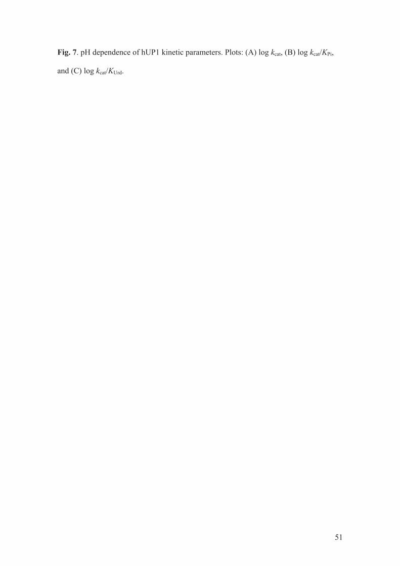
Fig. 7. pH dependence of hUP1 kinetic parameters. Plots: (A) log kcat, (B) log kcat/KPi,
and (C) log kcat/KUrd.
51

Figure 1.
52
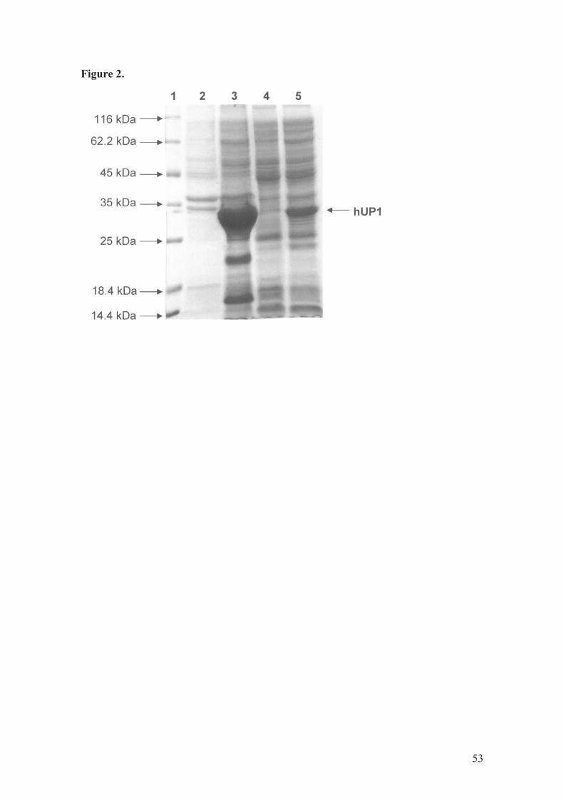
Figure 2.
53

Figure 3.
54
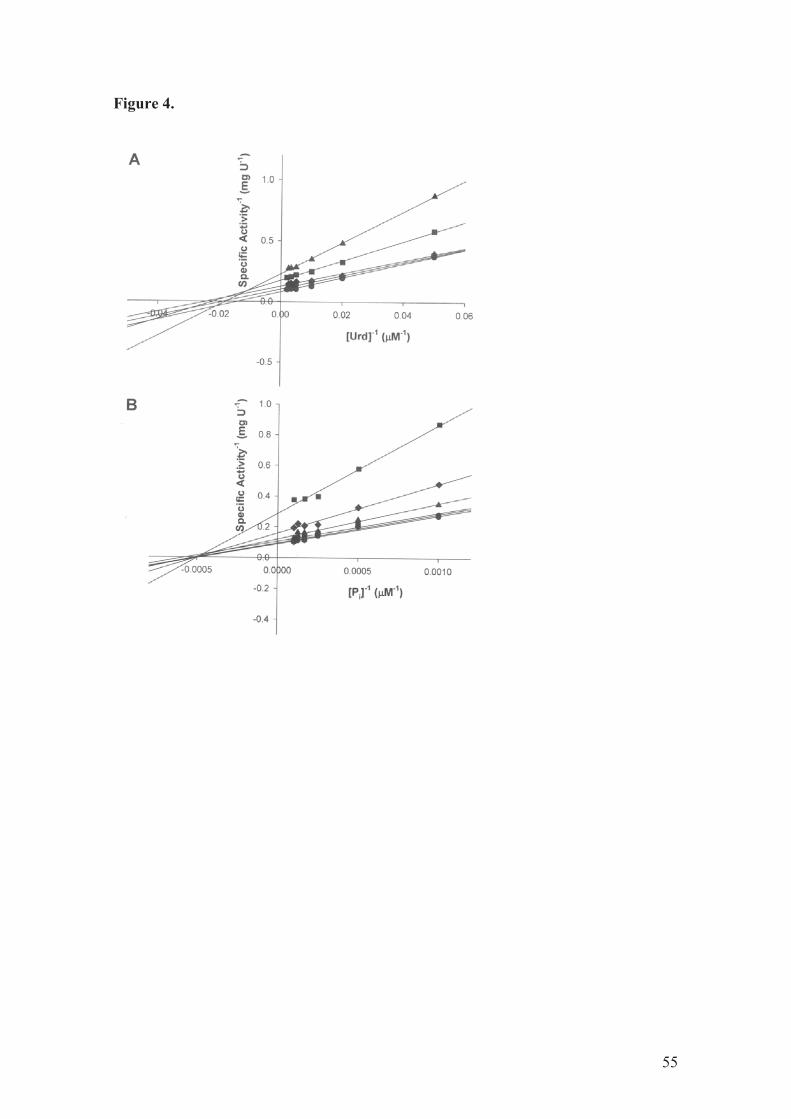
Figure 4.
55

Figure 5.
56

Figure 6.
57
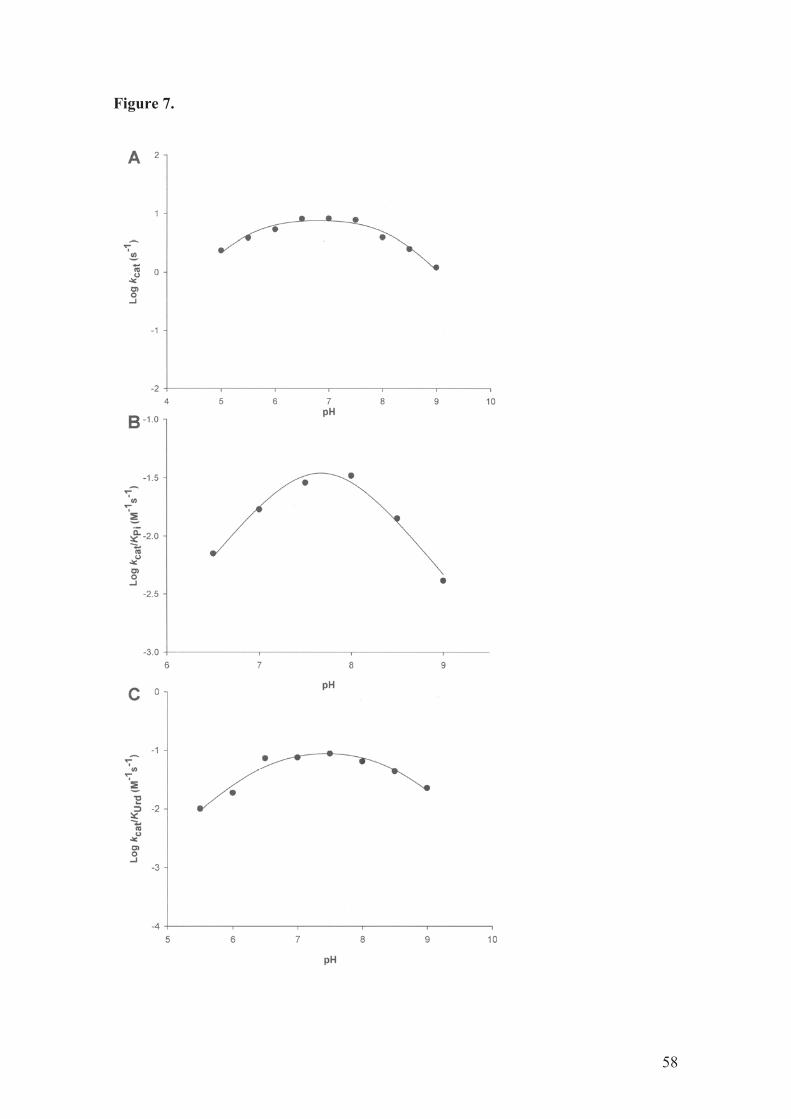
Figure 7.
58

Table 1.
Purification of hUP1 from E. coli Rosetta (DE3). Typical purification protocol from 5 g
wet cell paste (300 mL of culture).
Purification
step
Total protein
(mg)
Total
enzyme
activity (U)
Specific
activity
(U mg-1)
Purification
fold
Yield
(%)
Crude extract 180.3 367.5 2.04 1.0 100
SP Sepharose
Fast Flow 20.8 159.9 7.70 3.8 43
59

Table 2
Product inhibition patterns for hUP1.a
Variable substrate Product inhibitor Inhibition typebKis (µM)c
Kii (µM)d
Uridine Uracil NC 50 ± 7 618 ± 119
Uridine R1P NC 34 ± 7 2.16 x 10⁶ e
Pi Uracil NC 137 ± 27 251 ± 20
Pi R1P C 494 ± 61 -----------
aAt 37°C and 100 mM Tris pH 7.4.
bNC = noncompetitive, C = competitive.
cKis is the slope inhibition constant.
dKii is the intercept inhibition constant.
e This value was poorly defined due to a large SE value
60

4. CONSIDERAÇÕES FINAIS
A importância da uridina fosforilase à quimioterapia do câncer advém do
fato dela ser uma das enzimas responsáveis pela ativação ou degradação de
alguns análogos quimioterápicos, como as 5-fluoropirimidinas.
A importância da uridina fosforilase na quimioterapia do câncer tem
gerado um grande interesse no desenvolvimento de inibidores para essa
enzima. Desse modo, os inibidores podem aumentar a efetividade
quimioterápica dessas drogas pela prevenção de sua degradação e/ou
toxicidade ao hospedeiro [el Kouni et al, 2000], assim como podem atuar
separadamente inibindo parcialmente a via de salvamento de pirimidinas
através da não reciclagem da base uracil.
Durante este trabalho foi possível realizar a amplificação do gene UPP1,
a partir de cDNA obtido por RT-PCR de um RNA colorretal e a sua clonagem
em vetor de expressão pET23a(+). Determinamos que a melhor condição de
expressão solúvel para a proteína humana UP1 foi com a cepa de E. coli
Rosetta(DE3), utilizando o meio Terrific Broth, à 30C° por 36h e sem indução
com IPTG. A obtenção da proteína aparentemente homogênea foi através de
apenas uma etapa cromatográfica utilizando uma coluna de troca catiônica, e
sua identidade e massa molecular também foram determinados. Com a
proteína homogênea, foi possível fazer sua parcial caracterização através de
cinética enzimática, revelando-nos um mecanismo enzimático ordenado bi bi,
onde o fosfato inorgânico se liga primeiro seguido da uridina, para formar o
complexo ternário cataliticamente competente, com o uracil se dissociando
primeiro seguido pela liberação de ribose-1-fosfato. Também foi possível a
61

determinação das constantes cinéticas verdadeiras (Km, kcat e Vmax) assim
como a influência do pH nesses parâmetros e os possíveis aminoácidos
envolvidos na catálise e na ligação aos substratos, como visualizado na figura
8.
Figura 8. Ilustração das duas subunidades do dímero, mostrando a sítio ativo entre as duas subunidades. Ampliado estão os possíveis resíduos envolvidos na ligação e/ou catálise, como descrito no manuscrito. Fonte: PDB, código de acesso 3EUF.
Após uma análise mais acurada através de sua estrutura tridimensional,
foi verificado que os resíduos de aminoácidos conservados no sítio ativo
(Lys271 e Lys272) sugeridos durante a realização do manuscrito, na realidade
seriam os resíduos Leu272 e Leu273. Deste modo, as Leu272 e Leu273 não
62

podem ser os resíduos candidatos ao pKa de 8.2, como sugerido no
manuscrito, pois estes resíduos não podem sofrer protonação ou
deprotonação. Sendo assim, como sugerido no manuscrito, apenas o resíduo
de Tyr35 envolvido na catálise.
Neste sentido, a realização deste projeto amplia o conhecimento da
uridina fosforilase 1 com a finalidade de, futuramente, identificar inibidores
naturais e/ou sintéticos, que permitam o posterior desenvolvimento de drogas
para o tratamento quimioterápico do câncer.
Como perspectivas, temos a finalização de sua caracterização cinética,
tentativas de cristalografia, virtual screening para a busca de inibidores e estes
serão, então, analisados cineticamente e em células e animais. Serão
igualmente realizadas as mesmas análises para a outra isoforma humana,
UP2, para determinar suas semelhanças e suas diferenças, que poderão ser
úteis para o desenho de inibidores seletivos.
63

ANEXO 1. TESTE DE PURIFICAÇÃO DA PROTEÍNA REOMBIANTE hUP1
Os testes de purificação foram realizados utilizando o sistema de
purificação de Cromatografia Líquida de Rápida Eficiência (FPLC) ÄKTA (GE
Healthcare) para o controle do fluxo, pressão da coluna cromatográfica,
detecção de vários comprimentos de onda, pH, temperatura, controle de
gradiente, aplicação e coleta de amostras. As células foram preparadas
conforme descrito no manuscrito e para a determinação do protocolo final de
purificação diversas colunas cromatográficas e perfis de corrida foram testados,
a fim de se obter o melhor rendimento e a otimização do processo.
Para o teste inicial, com colunas de 1mL, foram utilizadas as colunas
cromatográficas de troca catiônica (HiTrap SPFF e HiTrap SPXL). As duas
trocas catiônicas apresentaram um padrão semelhante, sendo muito eficientes
para separar a proteína alvo das proteínas “contaminantes” da própria E. coli. A
coluna escolhida para os próximos testes foi a SPFF de 58mL e para a
obtenção da proteína recombinante aparentemente homogênea vários perfis de
corrida foram testados. Desta maneira, apenas uma coluna, a SPFF, foi
utilizada para obter a proteína aparentemente homogênea, como descrito no
manuscrito.
64

ANEXO 2. TESTES DE CRISTALIZAÇÃO PARA DETERMINAÇÃO DA
ESTRUTURA TRIDIMENSIONAL DA PROTEÍNA RECOMBINANTE hUP1
As tentativas de cristalização para determinação da estrutura
tridimensional foram feitas pelo método de difusão de vapor por gota em
suspensão, sob uma temperatura controlada de 20°C. Os kits utilizados para o
screening inicial foram Hampton Crystal Screen e Crystal Screen 2 (Hampton
Research). As gotas em suspensão foram preparadas misturando 2!L de uma
solução contendo 2mg/mL da proteína recombinante hUP1 em 50mM Tris-HCl
pH7.5 e 1!L da solução do reservatório.
Não foi verificada a formação de nenhum cristal de proteína em
nenhuma das condições testadas. Serão realizadas algumas mudanças no
protocolo para a tentativa de obter os cristais, visto que já existe a estrutura
tridimensional da hUP1 publicada [Roosild et al, 2009], porém com uma
seqüência de aminoácidos a mais no N-terminal; o que justifica o nosso
interesse na obtenção dos cristais.
Estes estudos de determinação da estrutura tridimensional da proteína
foram realizados no laboratório de Bioquímica LaBioQuest, na PUCRS,
laboratório sob coordenação do Prof. Dr. Walter Figueira de Azevedo Jr. da
PUCRS.
65

ANEXO 3. EXPRESSÃO DIFERENCIAL EM TECIDOS NEOPLÁSICOS
Para realizarmos estudos genéticos de expressão diferencial, optamos
pela técnica de PCR em Tempo Real. Para isso, utilizamos amostras normais e
tumorais de tecido colorretal que já estavam disponíveis por terem sido
utilizadas em outro experimento pelo nosso grupo. Os tecidos obtidos já
possuem aprovação do projeto pelo Comitê de Ética em Pesquisa da PUCRS
(CEP N°: 797/09, documento anexado) e a autorização por parte dos pacientes
ou responsáveis pela assinatura de termo de consentimento livre e esclarecido.
Essas amostras, depois de removidas dos pacientes, foram armazenadas em
solução para estabilização de RNA (RNA later™ RNA Stabilization Reagent,
Ambion, Austin, TX, USA). Amostras pareadas de tecido normal e tumoral
foram obtidas de 10 pacientes e pesavam aproximadamente 30 mg. Elas foram
trituradas sob incubação em nitrogênio líquido e solubilizadas com tampão de
lise fornecido pelo fabricante (Qiagen, Valencia, CA, USA). O tecido lisado foi
transferido para colunas de afinidade cromatográfica e processado de acordo
com o protocolo de isolamento do RNA fornecido. O RNA purificado foi eluído
em 30µl de água livre de RNase e armazenado a -80&C.
A quantidade e a qualidade do RNA foram estimadas através de
espectrofotometria e gel de agarose desnaturante respectivamente. As
amostras que apresentaram uma OD igual ou maior que 1.6 e que
apresentavam seu RNA íntegro (inspeção das bandas do RNA ribossomal 18s
e 28s, através de eletroforese em gel de formaldeído em condições de
desnaturamento corado com SYBRGold®) foram selecionadas.
66

A partir desses RNA selecionados, foi feita a transcrição reversa
utilizando primers oligoméricos, dNTPs, MgCl2 e a enzima derivada do vírus da
leucemia mielóide aviária em 20uL de reação. O cDNA sintetizado foi
armazenado a -20°C.
A técnica de PCR em Tempo Real foi utilizada para a quantificação
relativa das enzimas UP1 e UP2 humanas em tecido tumoral e normal de cólon
e reto a partir de material de arquivo mencionado anteriormente. O estudo foi
baseado na comparação da expressão dos genes de interesse em tecidos
tumorais em relação a tecidos normais (calibrador), utilizando o GAPDH como
controle endógeno da reação. Uma placa de 96 poços foi preparada para cada
gene de interesse, sendo que cada uma tinha 10 pares (Normal e Tumoral) de
amostras diferentes, para a amplificação do gene alvo e para a amplificação do
controle endógeno. Para cada amostra analisada foram preparadas duplicatas
contendo 25ng de cDNA, produto resultante da transcrição reversa e uma
mistura previamente preparada contendo tampão, dNTPs, MgCl2 e enzima Taq
(Universal PCR Master Mix TaqMan®, Applied Biosystems, Carlsbad, CA,
USA), primers e sondas com o marcador de fluorescência para UP1 e UP2
humana (TaqMan® Gene Expression Assays, Assays-on-Demand, Applied
Biosystems, Foster City, CA, USA), incluindo também, primers e sondas para
GAPDH (controle endógeno). O volume final da reação para cada duplicata foi
de 25µl.
As condições de temperatura para a realização da PCR em tempo real
foram divididas em quatro estágios: primeiramente as misturas contendo as
reações foram submetidas a uma temperatura de 30ºC por 2 minutos, o
segundo estágio seguiu a 95ºC por 10 minutos, o terceiro estágio com 50 ciclos
67

a 95ºC por 15segundos e 60ºC por 1 minuto. A expressão da UP1 e UP2 em
tecido tumoral foi fornecida através de quantificação relativa utilizando o
método ''Ct como anteriormente descrito por Livak [Livak e Schittgen, 2001].
A calibragem do sistema e eficiência das reações foram calculados a partir de
uma diluição seriada de cDNA tumoral e de tecido normal para UP1 e UP2 e
GAPDH, devendo o valor do coeficiente de determinação (r2) ser igual ou maior
que 98% para ambos os alvos.
Os resultados obtidos por esses experimentos, tanto para UP1 como
para UP2, não apresentaram diferença significativa de expressão destas
enzimas entre as amostras normais e tumorais de colorretal. Esses
experimentos foram repetidos para a confirmação dos resultados encontrados.
O fato de não haver diferença de expressão gênica entre as amostras normais
e tumorais não significa que não há diferença quanto à atividade da enzima; o
que já é descrito pela literatura como elevada nos tecidos tumorais. Não se
sabe ao certo quais os fatores que regulam a expressão da enzima e como
ocorre esse aumento da atividade sem haver necessariamente o aumento da
taxa de transcrição dessa proteína, poderia ser algum mecanismo de
reciclagem, mas essa informação ainda não é bem descrita.
68

4. CONFIRMAÇÃO DA SUBMISSÃO DO MANUSCRITO
From: [email protected] on behalf of ABB
Sent: Tue 1/19/2010 2:50 PM
To: Luiz Augusto Basso
Subject: Manuscript number assigned
Dear Dr Basso,
Your submission entitled "The kinetic mechanism of human uridine phosphorylase 1: towards
the development of enzyme inhibitors for cancer chemotherapy" has been assigned the
following manuscript number: ABBI-10-32.
You will be able to check on the progress of your paper by logging on to the Elsevier Editorial
System as an author.
The URL is http://ees.elsevier.com/yabbi/.
Thank you for submitting your work to Archives of Biochemistry and Biophysics.
Kind regards,
Jon Stein
Journal Manager
Archives of Biochemistry and Biophysics
69

5. APROVAÇÃO DO COMITÊ DE ÉTICA EM PESQUISA
70

REFERÊCIAS BIBLIOGRÁFICAS
Balestri F, Barsotti C, Lutzemberger L, Camici M, Ipata PL. Key role of uridine
kinase and uridine phosphorylase in the homeostatic regulation of purine and
pyrimidine salvage in brain. Neurochem Int. 2007; 51(8):517-23. Epub 2007 Jun
22.
Cansey M. Uridine and cytidine in the brain: their transport and utilization. Brain
Res Rev. 2006; 52(2):389-97.
Cao D, Nimmakayalu MA, Wang F, Zhang D, Handschumacher RE, Bray-Ward
P, Pizzorno G. Genomic structure, chromosomal mapping, and promoter region
analysis of murine uridine phosphorylase gene. Cancer Res. 1999;
59(19):4997-5001.
Cao D, Russell RL, Zhang D, Leffert JJ, Pizzorno G. Uridine phosphorylase (-/-)
murine embryonic stem cells clarify the key role of this enzyme in the regulation
of the pyrimidine salvage pathway and in the activation of fluoropyrimidines.
Cancer Res. 2002; 62(8):2313-7.
Cao D, Pizzorno G. Uridine Phosphorylase: an important enzyme in pyrimidine
metabolism and fluoropyrimidine activation. Drugs of Today. 2004; 40(5):431-
443.
Cao D, Leffert JJ, McCabe J, Kim B, Pizzorno G. Abnormalities in uridine
homeostatic regulation and pyrimidine nucleotide metabolism as a
consequence of the deletion of the uridine phosphorylase gene. J Biol Chem.
2005; 280(22):21169-75. Epub 2005 Mar 16.
Caradoc-Davies TT, Cutfield SM, Lamont IL, Cutfield JF, Crystal strutures of
Escherichia coli uridine phosphorylase in two native and three complexed forms
reveal basis of substrate specificity, induced conformation changes and
influence of potassium, J. Mol. Biol. 2004; (337):337-354.
71
Connolly GP, Duley JA. Uridine and its nucleotides: biological actions,
therapeutic potentials. Trends Pharmacol Sci. 1999; 20(5):218-25.
Darnowski JW, Handschumacher RE. Tissue-specific enhancement of uridine
utilization and 5-fluorouracil therapy in mice by benzylacyclouridine. Cancer
Res. 1985; 45(11 Pt 1):5364-8.
Grem JL. 5-Fluorouracil: forty-plus and still ticking. A review of its preclinical and
clinical development. Invest New Drugs. 2000; 18(4):299-313.
Instituto Nacional de Câncer, INCA. Endereço eletrônico:
http://www1.inca.gov.br/estimativa/2010/index.asp?link=conteudo_view.asp&ID
=1. Acessado em 19 de janeiro de 2010.
Johansson M. Identification of a novel human uridine phosphorylase. Biochem
Biophys Res Commun. 2003; 307(1):41-6.
Krenitsky TA, Mellors JW, Barclay RK. Pyrimidine nucleosidases. Their
classification and relationship to uric acid ribonucleoside phosphorylase. J Biol
Chem. 1965; 240:1281-6.
Ko LJ, Prives C. p53: puzzle and paradigm. Genes Dev. 1996; 10(9):1054-72.
el Kouni MH, Goudgaon NM, Rafeeq M, Al Safarjalani ON, Schinazi RF, Naguib
FN. 5-phenylthioacyclouridine: a potent and specific inhibitor of uridine
phosphorylase. Biochem Pharmacol. 2000; 60(6):851-6.
Lashkov AA, Gabdoulkhakov AG, Shtil AA, Mikhailov AM. Crystallization and
reliminary X-ray diffraction analysis of Salmonella typhimurium uridine
phosphorylase complexed with 5-fluorouracil. Acta Crystallogr Sect F Struct Biol
Cryst Commun. 2009; 65(Pt 6):601-3.
Leyva A, van Groeningen CJ, Kraal I, Gall H, Peters GJ, Lankelma J, Pinedo
HM. Phase I and pharmacokinetic studies of high-dose uridine intended for
rescue from 5-fluorouracil toxicity. Cancer Res. 1984; 44(12 Pt 1):5928-33.
72

Liu M, Cao D, Russell R, Handschumacher RE, Pizzorno G. Expression,
characterization, and detection of human uridine phosphorylase and
identification of variant uridine phosphorolytic activity in selected human tumors.
Cancer Res. 1998; 58(23):5418-24.
Livak KJ, Schittgen TD. Analysis of relative gene expression data using real-
time quantitative PCR and the 2(-Delta Delta C(T)) Method. Methods. 2001;
25(4):402-8.
Martin DS, Stolfi RL, Sawyer RC, Spiegelman S, Young CW. High-dose 5-
fluorouracil with delayed uridine "rescue" in mice. Cancer Res. 1982;
42(10):3964-70.
Miyashita H, Takebayashi Y, Eliason JF, Fujimori F, Nitta Y, Sato A, et al.
Uridine phosphorylase is a potential prognostic factor in patients with oral
squamous cell carcinoma. Cancer Res. 2002; 94(11):2959-66.
Pandolfo M, Araújo RA, Nascimento L, Goulart. Farmacogenoma do
fluorouracil. Análise de polimorfismo no gene da diidropirimidina
desidrogenase. Infarma. 2005; 17( 3-4).
Pinedo HM, Peters GF. Fluorouracil: biochemistry and pharmacology. J Clin
Oncol. 1988; 6(10):1653-64.
Pizzorno G, Yee L, Burtness BA, Marsh JC, Darnowski JW, Chu MY, et al.
Phase I clinical and pharmacological studies of benzylacyclouridine, a uridine
phosphorylase inhibitor. Clin Cancer Res. 1998; 4(5):1165-75.
Pizzorno G, Cao D, Leffert JJ, Russell RL, Zhang D, Handschumacher RE.
Homeostatic control of uridine and the role of uridine phosphorylase: a
biological and clinical update. Biochim Biophys Acta. 2002; 1587(2-3):133-44.
Roosild TP, Castronovo S, Fabbiani M, Pizzorno G, Implications of the structure
of human uridine phosphorylase 1 on the development of novel inhibitors for
improving the therapeutic window of fluoropyrimidine chemotherapy, BMC
Struct. Biol.; 2009 (16): 9-14.
73

Santos AMR, Ferreira, J. M. O., Oliveira, J. F. P., Duarte, K. S., Santos, M. O.,
Rebelo, M. S., Reis, R. S. Estimativa/ 2006 Incidencia de câncer no Brasil.
Shambaugh GE 3rd. Pyrimidine biosynthesis. Am J Clin Nutr. 1979; 32(6):1290-
7.
Voet D, Voet J. Bioquímica. Porto Alegre: Artes médicas; 2006.
Zhang D, Cao D, Russell R, Pizzorno G. p53-dependent suppression of uridine
phosphorylase gene expression through direct promoter interaction. Cancer
Res. 2001; 61(18):6899-905.
Watanabe S, Hino A, Wada K, Eliason JF, Uchida T. Purification, cloning, and
expression of murine uridine phosphorylase. J Biol Chem. 1995;
270(20):12191-6.
Watanabe S, Uchida T. Cloning and expression of human uridine
phosphorylase. Biochem Biophys Res Commun. 1995; 216(1):265-72.
World Cancer Report 2008, acessado pela World Health Organization, WHO. Endereço eletrônico: http://apps.who.int/bookorders/anglais/detart1.jsp?sesslan=1&codlan=1&codcol=76&codcch=26. Acessado em 19 de janeiro de 2010.
74