Embed Size (px)
Citation preview

BRUNA BENSO
AVALIAÇÃO DAS ATIVIDADES ANTIBACTERIANA, ANTI-
INFLAMATÓRIA, ANTI-OSTEOCLASTOGÊNICA E ANTI-HIV DA
Malva sylvestris
EVALUATION OF THE ANTIBACTERIAL, ANTI-INFLAMMATORY,
ANTI-OSTEOCLASTOGENIC AND ANTI-HIV ACTIVITIES OF Malva
sylvestris
PIRACICABA 2016
UNIVERSIDADE ESTADUAL DE CAMPINAS FACULDADE DE ODONTOLOGIA DE PIRACICABA
BRUNA BENSO
AVALIAÇÃO DAS ATIVIDADES ANTIBACTERIANA, ANTI-INFLAMATÓRIA, ANTI-OSTEOCLASTOGÊNICA E ANTI-HIV DA
Malva sylvestris
EVALUATION OF THE ANTIBACTERIAL, ANTI-INFLAMMATORY,
ANTI-OSTEOCLASTOGENIC AND ANTI-HIV ACTIVITIES OF Malva
sylvestris
Tese apresentada à Faculdade de Odontologia de Piracicaba da Universidade Estadual de Campinas como parte dos requisitos exigidos para obtenção do título de Doutora em Odontologia, na Área de Farmacologia, Anestesiologia e Terapêutica.
Thesis presented to the Piracicaba Dental School of the University of Campinas in partial fulfillment of the requirements for the degree of Doctor in Dentistry, in the Pharmacology, Anesthesiology and Therapeutics Area.
Orientador: Prof. Dr. Pedro Luiz Rosalen
Co-orientador: Prof. Dr. Gilson César Nobre Franco ESTE EXEMPLAR CORRESPONDE À VERSÃO FINAL DA TESE DEFENDIDA PELA ALUNA BRUNA BENSO, ORIENTADA PELO PROF. DR. PEDRO LUIZ ROSALEN E CO-ORIENTADADA PELO PROF. DR GILSON CÉSAR NOBRE FRANCO.
PIRACICABA 2016


DEDICATÓRIA
Dedico este trabalho aos meus pais,
Luiz Carlos (in memoriam) e Rose Benso; que me
guiaram e educaram com todo o seu amor e carinho.
Agradeço pelo incentivo contínuo e pelo grande exemplo
de força e superação.

AGRADECIMENTOS
A Deus, pela proteção e bondade, por iluminar a minha vida e se mostrar sempre
presente.
À Universidade Estadual de Campinas, na pessoa do seu Magnífico Reitor, Prof. Dr. José Tadeu Jorge.
À Faculdade de Odontologia de Piracicaba, na pessoa do Diretor, Prof. Dr.
Guilherme Elias Pessanha Henriques.
À Profa. Cinthia Pereira Machado Tabchoury, Coordenadora Geral da Pós-
Graduação da FOP-UNICAMP.
À Profa. Dra. Juliana Trindade Clemente Napimoga, Coordenadora do
Programa de Pós-Graduação em Odontologia.
Ao meu orientador Prof. Dr. Pedro Luiz Rosalen, pelo incentivo e todas as
oportunidades a mim oferecidas. Agradeço por participar tão ativamente na minha formação
científica e intelectual, sendo um exemplo de competência, honestidade, profissionalismo e
dedicação pelo trabalho.
Ao Prof. Dr. Gilson César Nobre Franco, por sua orientação e participação
indispensável em todos os momentos da pesquisa.
Ao Prof. Dr. Ramiro Mendonça Murata, por me receber em seu laboratório na
University of Southern California durante período de estágio Doutorado Sanduíche. Meu
reconhecimento pela oportunidade, confiança, e ainda, por sempre se mostrar solícito, foi um
ano de intenso trabalho, mas de grande contribuição para minha formação pessoal e
profissional.
Ao Prof. Dr. Severino Matias de Alencar, por sua orientação e participação
indispensável em todos os momentos da pesquisa.
Aos Professores do Programa de Pós-Graduação em Odontologia, em especial
da área de Farmacologia, pela participação na minha formação e constante incentivo.

Aos Profs. Drs., Ana Paula de Souza Pardo, Karina Cogo Müller, Marcelo
Rocha Marques pelas sugestões e contribuições no exame de qualificação.
Aos Profs. Drs., Carina Denny, Francisco Carlos Groppo, Janaina Orlando Sardi e Severino Matias de Alencar pelas sugestões e contribuições no exame de defesa.
Ao meu irmão Luiz Eduardo pela amizade apesar da distância e por me alegrar
com sua forma simples de viver a vida.
A Pedro Aravena pelo carinho, amor, companheirismo e especialmente pela
paciência durante esta jornada.
À Juliana Botelho, Patrícia Lauer, Talita Graziano, pelo companheirismo,
palavras sinceras e amizade em todos os momentos.
À Vanessa Pardi por me receber juntamente com sua família em Los Angeles.
Obrigada por amenizar a difícil saudade de casa.
À Adna Massarioli pela disposição e colaboração com as análises químicas deste
trabalho.
Ao técnico do laboratório de Patologia da FOP-UNICAMP, Fábio Téo pela
colaboração e paciência com as análises de biologia molecular.
Ao professor Masaharu Ikegaki pela colaboração na busca de produtores de
Malva sylvestris.
Ao produtor Jonas Pereira pela disposição e fornecimento de material vegetal de
boa qualidade que permitiu a execução desta pesquisa.
Aos técnicos do laboratório de Farmacologia da FOP-UNICAMP, Sra. Eliane Melo pela alegria que transborda todos os dias e Sr. José Carlos Gregório, pela colaboração
durante estes anos.
A Christopher Patuwo, Dalia Saleem, Diana Levya, Emily Chen, Juliana
Noguti, Maria Marquezin, Meng Lin, Silvana Pasetto, Sthephanie Ting, Keane Young e
Vivian Oliveira por momentos agradáveis de laboratório e amizade nos EUA. Fica meu
carinho especial à todos!
Aos amigos da área de farmacologia: Aline Castilho, Ana Paula Bentes,
Andréia Scriboni, Bruno Nani, Bruno Vilela, Camila Batista, Carina Denny, Cleiton
Pita, Fabiano Brito, Felipe LLoret, Giovana Fiorito, Irlan Freires, Janaina Sardi, Jonny Burga, Josy Goldony, Karina Cogo, Laila Facin, Larissa Shiozawa, Leandro Pereira,
Leilane Iwamoto, Lívia Galvão, Luciana Berto, Luciano Serpe, Luiz Ferreira, Marcelo Franchin, Marcos Cunha, Michelle Leite, Paula Sampaio, Rodrigo Girondo, Salete
Fernandes, Sérgio Rochelle e Verônica Freitas pela agradável convivência.
Às Sras. Ana Paula Carone, Érica Alessandra Pinho Sinhoreti e Raquel Quintana Marcondes Cesar Sacchi e secretárias da Coordenadoria Geral dos Programas de
Pós-Graduação, Maria Elisa dos Santos secretária da Farmacologia, e Eliete Rigueto Roque, secretária do Departamento de Ciências Fisiológicas, por todas as orientações e
indispensável ajuda.
A Fundação de Amparo à Pesquisa do Estado de Pesquisa do Estado de São Paulo (FAPESP), pela concessão da bolsa de doutorado (2011/ 23980-5) e à Coordenação
de Aperfeiçoamento de Pessoal de Nível Superior (CAPES) pela concessão de bolsa de
doutorado modalidade sanduíche (2317/2014-01).
Meu eterno reconhecimento a todos que de alguma forma contribuíram para a
realização deste trabalho.

RESUMO
A natureza é fonte de descoberta de novos fármacos há séculos, originando
inúmeras drogas de utilidade clínica. Plantas são reconhecidas por seu valor medicinal e
nutracêutico, a exemplo, a Malva sylvestris possui literatura etnofarmacológica, que relata
histórico de suas propriedades biológicas. Desta forma, o objetivo deste estudo foi realizar um
screening das atividades farmacológicas da Malva sylvestris, portanto, investigou-se: (1) As
atividades antibacteriana e anti-inflamatória do extrato de M. sylvestris (MSE) e frações
utilizando método de cultura de células epiteliais e de tecido gengivais infectadas pelo micro-
organismo Aggregatibacter actinomycetemcomitans e a quantificação da expressão de genes e
citocinas relacionados ao processo inflamatório; (2) A atividade do MSE e frações quanto a
atividade anti-inflamatória in vivo (migração de neutrófilos para cavidade peritoneal, edema
de pata e quantificação de citocinas), capacidade de ação anti-osteoclastogênica (análise de
expressão de gênica, contagem das células TRAP positivas e zimografia), atividade
antioxidante (método DPPH e ABTS•+), e identificação química e confirmação da fração
bioativa (MS/MS); (3) A ação anti-HIV da fração aquosa (AF) em células infectadas por
HIV-BaL em modelo dual chamber in vitro por meio da quantificação antígeno p24,
expressão gênica, citocinas e mecanismo de ação por transcriptase reversa. A análise
estatística de variáveis quantitativas foram comparadas por análise de variância (ANOVA) e
post-hoc de Dunnet. O nível de significância adotado foi de alfa= 0,05. Os resultados
demonstraram que a fração clorofórmica (CLF) na concentração de 75 µg/mL foi eficaz na
redução da colonização bacteriana e no controle dos mediadores inflamatórios promovendo a
regulação dos genes IL-1beta, IL-6, IL-10, CD14, PTGS, MMP-1 e FOS, bem como, na
redução da expressão das proteínas IL-1beta, IL-6, IL-8 e GM-CSF. A ação anti-inflamatória
in vivo foi significativa (dose de 30 mg/kg, via oral) para todas as frações (CLF, EAF e AF) e
extrato (MSE) estudados, com exceção da fração hexânica, na redução na migração de
neutrófilos para cavidade peritoneal. AF (dose 30 mg/kg, via oral) reduziu o edema de pata
nas 3 primeiras horas analisadas, apresentando uma ação mais rápida que o controle positivo,
e ainda, reduziu os níveis de expressão de IL-1β. A análise da atividade de M. sylvestris sobre
o processo de remodelação óssea demonstrou que AF na concentração de 10 µg/mL regulou a
transcrição dos genes analisados (anidrase carbônica, catepsina K e fosfatase ácido-tártaro
resistente), promoveu a redução no número de osteoclastos TRAP-positivos/área e controlou
expressão de enzimas proteolíticas específicas MMP-9. Para a atividade antioxidante a AF e a

fração acetato de etila (EAF) apresentaram a melhor capacidade em capturar radicais livres. A
identificação química revelou a presença do composto bioativo rutina na AF. Os resultados
para atividade antiviral demonstraram uma redução na expressão de antígeno p24, ação sobre
transcriptase reversa, controle da transcrição do genes CD4, Bcl-2 e TRIM5, e redução da
expressão citocinas IL1-alpha, IL-beta, IL-6, IL-8 e GM-CSF após o tratamento com AF (50
µg/mL). Portanto, podemos concluir que a M. sylvestris e as frações bioativas encontradas
apresentam compostos promissores como novos agentes terapêuticos.
Palavras-chave: Malvaceae. Infecções Bacterianas. Osteoclastos. Antioxidantes. Inflamação.
Infecções por HIV

ABSTRACT
Nature has been a source of medicinal products for centuries, yielding many
useful drugs. A wide variety of plants are well recognized for their medicinal and
nutraceutical value, Malva sylvestris being one example; the ethnopharmacological literature
has reported a long history of recognition of biological properties. The aim of this study was
to conduct a pharmacological screening of Malva sylvestris and its interest to dentistry.
Therefore, we investigated: (1) the antibacterial and anti-inflammatory activity of M.
sylvestris extract (MSE) and fractions using a cell culture technique with epithelial and
gingival cells infected with Aggregatibacter actinomycetemcomitans and a gene expression
and cytokine quantification related to the inflammatory response; (2) The activity of MSE and
fractions in the in vivo anti-inflammatory activity (neutrophil migration, paw edema and
cytokine quantification, anti-osteoclastogenic action (gene expression, number of positive
TRAP positive cells and zymography), antioxidant activity (DPPH and ABTS•+), and
chemical identification of the bioactive fraction (MS/MS); (3) anti-HIV activity of aqueous
fraction (AF) in cells infected with HIV-Bal using the in vitro dual chamber model,
quantifying p24 antigen, gene expression and cytokines. Statistical analysis was performed by
analysis of variance (ANOVA) and Dunnett’s post-hoc test.The significance level adopted
was alfa = 0.05. The results showed that chloroform fraction CLF (75 µg/mL) was efficient in
reducing the bacteria colonization and inflammatory mediators, promoting the gene regulation
of IL-1beta, IL-6, IL-10, CD14, PTGS, MMP-1 and FOS, as well as reducing protein
expression IL-1beta, IL-6, IL-8 and GM-CSF. The in vivo reduction of anti-inflammatory
effect (30 mg/kg, orally) was significant for the extract (MSE) and all fractions (CLF, EAF
and AF) with the exception of the hexane fraction in the neutrophil migration assay. The AF
(30 mg/kg, orally) reduced the paw edema in the first 3 hours analyzed, with a faster action
than the positive control, reducing the levels of IL-1β expression. The activity of M. sylvestris
in the bone remodeling assay demonstrated that the aqueous fraction (AF) in the
concentration of 10 µg/mL regulated the gene transcription of the study genes (carbonic
anhydrase, cathepsin K and tartrate-resistant acid phosphatase) and reduced the number of
TRAP-positive osteoclasts and the specific proteolytic enzyme MMP-9. In terms of the
antioxidant activity, the AF and the ethyl acetate fraction (EAF) had the best ability to capture
free radicals. The chemical identification revealed rutin as the bioactive compound in the AF.
Results for the antiviral activity showed a p24 antigen reduction, reverse transcriptase
mechanism of action, controlled transcription of the genes CD4, Bcl-2 and TRIM5, and a
reduction in the cytokines IL-beta, IL-6, IL-8 and GM-CSF after treatment with AF (50
µg/mL). Therefore, we can conclude that M. sylvestris and its bioactive fractions are
promising compounds as novel therapeutic agents.
Keywords: Malvaceae. Bacterial Infections. Osteoclasts. Antioxidants. Inflammation. HIV
Infections.

SUMÁRIO
1 INTRODUÇÃO........................................................................................................... 14
2 ARTIGOS.....................................................................................................................
2.1 Artigo 1: Malva sylvestris inhibits inflammatory response in oral human cells.
An in vitro infection model............................................................................... 18
2.2 Artigo 2: Anti-inflammatory, bone remodeling and antioxidant effects of Malva
sylvestris extract and fractions: in vitro and in vivo studies 43
2.3 Artigo 3: Evaluation of Malva sylvestris as inhibitor of HIV-1 BaL in a dual
chamber in vitro model.......................................................................................... 72
3 DISCUSSÃO…........................................................................................................... 92
4 CONCLUSÃO 98
REFERÊNCIAS.............................................................................................................. 99
ANEXOS.....................................................................................................................
Anexo 1 – Correspondência periódico PlosOne 107
Anexo 2 – Certificado do Comitê de Ética em Animais 108

14
1 INTRODUÇÃO
Historicamente, os produtos naturais proveniente de plantas e animais são os
responsáveis por cerca de 25 % dos medicamentos disponíveis no mercado (Cragg et al.,
2014). As plantas, em particular, tem formado a base da medicina tradicional com registros de
uso milenar que originam desde a Mesopotâmia, 2600 a.C, no entanto, apenas no século XIX
iniciou-se a busca por princípios ativos, originando assim, os primeiros fármacos com
características semelhantes aos atuais (Dias et al., 2012; Harvey, 2008).
As plantas constituem uma valiosa fonte de recursos para a síntese orgânica
devido ao seu mecanismo de biossíntese de compostos chamado metabolismo secundário
(Harvey, 2007). O metabolismo secundário, geralmente não é essencial para o crescimento,
desenvolvimento ou reprodução dos organismos e são produzidos devido ao processo de
adaptação ao meio ambiente, ou ainda, podem ser produzidos como mecanismo de defesa
para sobrevivência (Dewick, 2001). A biossíntese pode ocorrer por fotossíntese, glicólise ou
pelo ciclo de Krebs e podem produzir intermediários biossintéticos, que podem ser infinitos e
reconhecidos como produtos naturais (Maplestone et al., 1992). Esse processo diferenciado de
biossíntese proporciona características únicas na estrutura química e inúmeras atividades
biológicas (Dewick, 2001).
O desenvolvimento de técnicas analíticas de separação e elucidação estrutural
permitiram o isolamento de diversos metabólitos secundários com potencial farmacológico
(Cragg et al., 2014). Há muitas áreas de conhecimento que se beneficiaram dos esforços de
descobertas de novas drogas, entre elas, os antimicrobianos (Molinari, 2009). A medicina
tradicional mostra interesse cada vez maior na utilização de drogas antimicrobianas derivadas
de plantas, pois o antibióticos tradicionais, aqueles originados de produtos de micro-
organismos ou derivados sinteticamente, tem se mostrado ineficazes no tratamento infeccioso
em diversos momentos (Lai e Roy, 2004). A diversidade estrutural nos compostos derivados
de plantas é imenso e o impacto produzido nos microrganismos é dependente da configuração
química (Harvey, 2007). Para exemplificar, nas flavonas a presença do grupo (-OH) na
posição 5´ da fórmula estrutural confere atividade contra cepas Staphylococcus aureus
resistentes a meticilina. Esses achados mostram a relação direta entre a estrutura química e a
atividade antimicrobiana (Lai e Roy, 2004).

15
Na lista das doenças crônicas que apresentam maior prevalência na população
mundial estão presentes as doenças infecciosas de origem bucal (Petersen et al., 2005). A
microbiota da cavidade oral tem estrutura complexa e é composta por mais de 600 espécies
diferentes de bactérias (Dewhirst et al., 2010; Moore e Moore, 1994). As populações
microbianas das estruturas dos dentes (biofilme dental) e o sistema de defesa do hospedeiro se
mantem em equilíbrio dinâmico, no entanto, em algumas situações há colonização de novas
espécies e um desequilíbrio pode ser iniciado, causando inflamação destrutiva dos tecidos
circundantes, periodontais (Darveau, 2010; Alani e Seymour 2014).
O início e a manutenção da inflamação periodontal é determinado por bactérias
que estão presentes no biofilme dentário e, em maior proporção, as gram-negativas
(Johansson, 2011). Aggregatibacter actinomycetemcomitans é o micro-organismo relacionado
de forma específica à periodontite agressiva, no entanto, também exerce papel na doença
crônica (Slots, 1999; Kachlany, 2010). A. actinomycetemcomitans e outros micro-organismos
incluindo Porphyromonas gingivalis, Treponema denticola, Tannerela forsythia estimulam a
resposta imune promovendo inflamação dos tecidos moles e consequente destruição óssea
(Darveau, 2010). Estas bactérias periodontopatoge ̂nicas produzem fatores de virulência como
os lipopolissacarídeos e peptideoglicanos, que induzem a produção de citocinas pro-
inflamatórias pelo hospedeiro (Salvi e Lang, 2005).
Os lipopolissacarídeos são macromoléculas que se associam a proteína CD14,
formando o complexo LPS-CD14 que ativa o receptor de proteína tool-like (TLR-4),
estimulando a sinalização intracelular e ativação de fosfolipase A, fosfolipase C e aumento
dos níveis intracelulares de cálcio, p42/p44, e ainda, p38 (Han et al., 1993; Lima et al., 2010).
Além disso, estimulam a liberação de diversos mediadores inflamatórios como:
prostaglandinas (PGs), óxido nítrico (NO) e interleucinas (ILs) (Henderson et al., 1996;
Johansson, 2011).
A ativação do sistema imune pode induzir ao estresse oxidativo e promover a
produção e liberação de NO e espécies reativas de oxigênio (ROS) é um mecanismo utilizado
para atrair mediadores para o local da inflamação (Conner e Grisham, 1996; Khansari et al.,
2009). Uma ativação genética pode resultar na expressão de ROS pouco regulada e
promover a apoptose de osteoblastos e a consequente reabsorção óssea e ativação do
sinalizador NF-κB, responsável pelo mecanismo de osteoclasteogênse (Conner e Grisham,
1996). A progressão da doença periodontal permite a ativação dos osteoclastos e consequente

16
destruição óssea, fatores estimulatórios são reguladores do processo, como por exemplo,
interleucina tipo 1 (IL-1), fator estimulador das colônias de macrófagos (MCSF), monócitos e
células T (Henderson et al., 1996; Salvi and Lang, 2005).
O tratamento da doença periodontal é baseado nos fatores de virulência, nos
micro-organismos que se estabelecem nos processos de saúde e doença, desta maneira, as
terapias devem ser direcionadas para o controle desses micro-organismos (Seymour, 2006).
Embora seja indiscutível o papel do biofilme bacteriano na etiologia das doenças
periodontais, a severidade e a progressão destas doenças são determinadas por fatores
relacionados a resposta do hospedeiro (Haffajee et al., 1997; Batchelor, 2015). Agentes
moduladores são estudados como coadjuvantes no tratamento da doença periodontal não
cirúrgica (Alani e Seymour, 2014). A partir da década de 90 foi incluído a terapia de
modulação da resposta do hospedeiro como uma opção adjunta ao tratamento convencional
da doença periodontal, são exemplos de moduladores: anti-inflamatórios sistêmicos e tópicos,
sub-doses de doxicilina e o uso de bifosfonatos (Golub et al., 1992; Gokhale e Padhye, 2013).
A terapia periodontal é realizada com sucesso, porém a recolonização da área subgengival
pelos periodontopatógenos, resulta em uma terapia preventiva de manutenção falha e isto
pode levar ao processo de doença recorrente (Teles et al., 2006).
O sistema imune também é desafiado por infecções de origem viral. Em
condições fisiológicas a maior parte das células do sistema imune estão em repouso, no
entanto, vários fatores podem participar como ativadores, e o vírus da imunodeficiência
humana (HIV-1) é um exemplo (Younas et al., 2015). O processo infeccioso resulta na
ativação de longa duração do sistema imunológico incluindo a perda progressiva de células de
defesa T-CD4+ e a produção elevada de citocinas pró-inflamatórias e quimiocinas que não são
totalmente restabelecidos por terapias antirretrovirais (TARV) (Dagenais-Lussier et al., 2015).
O efeito da TARV no tratamento de pacientes portadores de HIV trouxe inúmeros benefícios,
em especial, diminuindo a mortalidade e risco de transmissão (Bahr, 2005). A terapia consiste
na combinação de 3 classes de drogas: inibidores da transcriptase reversa, inibidores não-
nucleosídeos da transcriptase reversa e inibidores de protease. A inserção desta terapia
farmacológica permitiu que a doença infecciosa se transformasse em doença crônica
(Maartens et al., 2014). No entanto, um significante número de novas terapias ainda não
curativas e o alto custo ainda impede que algumas populações tenham acesso ao tratamento
(Günthard et al., 2014).

17
A resposta imunológica mediante algumas patologias pode representar um desafio
para a terapêutica, por exemplo, o tratamento de inflamações crônicas e infecções virais
(Harvey, 2008). Há um interesse crescente no uso de plantas medicinais para a modulação do
sistema imune e na prevenção de infecções relacionadas (Molinari, 2009). Compostos como
flavonóides, polissacarídeos, lactonas, alcalóides, diterpenóides e glicosídeos presentes em
muitas plantas, tem sido reportados pelas propriedades imunomoduladoras (Jantan et al.,
2015).
A Malva sylvestris, popularmente conhecida como malva, é nativa da Europa,
Norte da África e Ásia e tem o uso reportado desde 3000 a.C., devido a sua relevância
terapêutica partes da planta tem sido empregadas na medicina tradicional e veterinária
(Gasparetto et al., 2012). Etnofarmacologicamente é conhecida por suas propriedades anti-
inflamatórias, antioxidantes, anticâncer, tratamento de bronquites e de lesões de pele
(Gasparetto et al., 2012; Razavi et a., 2011). As folhas, flores e as parte aéreas da malva são
conhecidas para tratamentos de doenças que afetam cavidade bucal como abcessos e dores
dentárias (Guarrera, 2005). No Brasil a M. sylvestris é registrada na ANVISA como
medicamento fitoterápico, na categoria para uso oral com expectorante, tratamento de
inflamações e antisséptico da cavidade oral (Gasparetto et al., 2012; Kaileh et al., 2007). O
uso da malva é disseminado e suas propriedades biológicas conhecidas ao redor do mundo
(Romojaro et al., 2013). Desta forma, é necessária a investigação do potencial farmacológico
e de interesse odontológico da M. sylvestris, constituindo uma base para o uso clínico desta
planta, e ao mesmo tempo, um modelo para identificação de compostos bioativos.
Assim, a proposta deste trabalho foi realizar um screening de diferentes
atividades farmacológicas da planta Malva sylvestris e como objetivos específicos, investigar:
(1) As atividades antibacteriana e anti-inflamatória do extrato de M. sylvestris (MSE) e
frações em células infectadas por Aggregatibacter actinomycetemcomitans; (2) A atividade do
MSE e frações quanto a atividade anti-inflamatória, anti-osteoclástica, antioxidante, e
finalmente, identificar quimicamente a fração ativa; (3) A ação anti-HIV da fração aquosa de
Malva sylvestris em células infectadas por HIV-BaL.

18
2 ARTIGOS
2.1 ARTIGO (artigo publicado – Anexo 1)1
Malva sylvestris inhibits inflammatory response in oral human cells. An in vitro
infection model
Bruna Benso1; Pedro Luiz Rosalen1; Severino Matias Alencar2; Ramiro Mendonça Murata3*
1Department of Physiological Sciences, Piracicaba Dental School, University of Campinas, Piracicaba, Sao Paulo, Brazil. 2Department of Agri-food Industry, Food and Nutrition, “Luiz de Queiroz” College of Agriculture, University of Sao Paulo, Piracicaba, Sao Paulo, Brazil 3Division of Periodontology, Diagnostic Sciences & Dental Hygiene and Division of Biomedical Sciences Herman Ostrow School of Dentistry, University of Southern California, Los Angeles, United States of America *Corresponding author E-mail: [email protected]
1 Benso B, Rosalen PL, Alencar SM, Murata RM. Malva sylvestris Inhibits Inflammatory Response in Oral
Human Cells. An In Vitro Infection Model. PLoS One. 2015 Oct 19;10(10):e0140331.

19
Abstract
The aim of this study was to investigate the in vitro anti-inflammatory activity of
Malva sylvestris extract (MSE) and fractions in a co-culture model of cells infected by
Aggregatibacter actinomycetemcomitans. In addition, we evaluated the phytochemical
content in the extract and fractions of M. sylvestris and demonstrated that polyphenols were
the most frequent group in all samples studied. An in vitro dual-chamber model to mimic the
periodontal structure was developed using a monolayer of epithelial keratinocytes (OBA-9)
and a subepithelial layer of fibroblasts (HGF-1). The invasive periodontopathogen A.
actinomycetemcomitans (D7S-1) was applied to migrate through the cell layers and induce the
synthesis of immune factors and cytokines in the host cells. In an attempt to analyze the
antimicrobial properties of MSE and fractions, a susceptibility test was carried out. The
extract (MIC 175 μg/mL, MBC 500μg/ mL) and chloroform fraction (MIC 150 μg/mL, MBC
250 μg/mL) were found to have inhibitory activity. The extract and all fractions were assessed
using a cytotoxicity test and results showed that concentrations under 100 μg/mL did not
significantly reduce cell viability compared to the control group (p > 0.05, viability > 90%).
In order to analyze the inflammatory response, transcriptional factors and cytokines were
quantified in the supernatant released from the cells. The chloroform fraction was the most
effective in reducing the bacterial colonization (p< 0.05) and controlling inflammatory
mediators, and promoted the down-regulation of genes including IL-1beta, IL-6, IL-10,
CD14, PTGS, MMP-1 and FOS as well as the reduction of the IL-1beta, IL-6, IL-8 and GM-
CSF protein levels (p< 0.05). Malva sylvestris and its chloroform fraction minimized the A.
actinomycetemcomitans infection and inflammation processes in oral human cells by a
putative pathway that involves important cytokines and receptors. Therefore, this natural
product may be considered as a successful dual anti-inflammatory–antimicrobial candidate.

20
Introduction
Periodontal disease is characterized by bacterial infection associated with the
presence of biofilm, resulting in chronic inflammation of the tooth-supporting tissues and
leading to progressive destruction of periodontal tissue. This disease affects up to 90% of the
world’s population [1], [2]. Dental biofilm with a large quantity of gram-negative bacteria is
responsible for the initiation and maintenance of periodontal inflammation [3].
Aggregatibacter actinomycetemcomitans has been described as an important agent of
localized aggressive periodontic lesions, but is also related to chronic periodontitis [4], [5]. In
addition, A. actinomycetemcomitans and other pathogenic microbiota including
Porphyromonas gingivalis, Treponema denticola, Tannerela forsythia trigger both innate and
acquired immune responses, resulting in the progression of periodontal disease, and promote
soft tissue inflammation and destruction with consequent bone resorption [6].
The development of new therapeutic agents that can inhibit biofilm formation and
modulate the inflammatory response will have a major impact on the prevention and
treatment of periodontal disease [7].
Nature has been a source of medicinal products for centuries, yielding many
useful drugs [8]. A wide variety of plants are well recognized for their medicinal and
nutraceutical value, and the exploration of biodiversity from rich environments has led to the
discovery of many pharmacologically active chemicals [9], [10]. Malva sylvestris is one
example. Commonly known as mallow, it is a plant native to Europe, North Africa and Asia.
The ethnopharmacological literature has reported a long history of recognition for its potent
anti-inflammatory, antioxidant, anticancer and antiulcerogenic properties [11], [12]. Some
reports have indicated that M. sylvestris contains phytochemicals including several classes of
terpenoids, including monoterpenes, diterpenes, sesquiterpenes and norterpenes [11], [13],
[14]. Since natural products do not have a standard composition, there is increasing interest in

21
identifying biological therapeutic potential in new plant extracts [15]. Thus, the aim of this
study was to investigate in vitro the antimicrobial and anti-inflammatory activity of Malva
sylvestris extract and fractions in a dual chamber model of epithelial and subepithelial cells
infected by A. actinomycetemcomitans.
Material and Methods
Preparation of the extract and fractions
Malva sylvestris leaves were purchased from a local farmer in the municipality of
Princesa Isabel, Paraiba (northeast Brazil) in March and April 2013. This plant is not an
endangered or protected species and was registered in the herbarium of the University of Sao
Paulo (USP), receiving an identification number (ESA voucher # 121403). Absolute ethanol
(800 mL) at room temperature was used to create extracts of M. sylvestris leaves (100 g)
using exhaustive maceration (for 7 days). Filtration was used to obtain the ethanolic extract of
M. sylvestris (MSE). The material was lyophilized, homogenized, weighed and stored at -
20oC. The MSE was successively partitioned using liquid-liquid extraction with hexane,
chloroform, and ethyl acetate solvents. The final residue obtained after ethyl acetate
fractionation was totally soluble in water and thus was called the aqueous fraction (AF)[16].
The extract (MSE), chloroform fraction (CLF) and aqueous fraction were re-suspended in 1%
ethanol and used in the biological assays.
Determination of total flavonoid, phenol and condensed tannin content
For the flavonoid determination, the aluminum chloride method was used. Total
flavonoid contents were calculated using quercetin for the calibration curve. The absorbance
was measured at 425 nm with a microplate reader (SpectraMax M5, Molecular Devices

22
Sunnyvale, CA, USA). The polyphenol content was measured by the Folin-Ciocalteu method
and gallic acid was used as a standard equivalent [17]. For the content of condensed tannins, a
vanillin solution was added to the extract, followed by 37% hydrochloric acid. The calibration
curve was determined based on catechin as a reference.
Bacterial Strains
The A. actinomycetemcomitans (D7S-1) was cultivated from the subgingival
plaque of an African American female patient diagnosed with generalized aggressive
periodontitis. The strain was kindly donated by Dr. Casey Chen (University of Southern
California) [18]. In addition, the following reference strains were used: Fusobacterium
nucleatum ATCC 25586, Prevotella intermedia 25611 and Porphyromonas gingivalis ATCC
BAA-308.
Cell culture
Keratinocytes were processed and isolated, and the cell line established was
named OBA-9 [19]. The cell line was kindly donated by Dr. Kusumoto. OBA-9 cells used in
this experiment were cultured in a specific medium for keratinocytes (Defined Keratinocyte-
SFM, Life Technologies, Carlsbad, CA, USA). The human gingival fibroblasts HGF-1
(ATCC CRL-2014) were cultured in Dulbecco’s modified Eagle’s medium (DMEM) with
10% fetal bovine serum (Gibco, Life Technologies, Carlsbad, CA, USA), 100 U/mL penicillin
and 100 µg/mL streptomycin (Invitrogen Life Technologies, CA, USA). Cells were
maintained in a humidified incubator at 37 °C in 5% CO2.
Susceptibility testing
The susceptibility of four potential periodontopathogenic bacteria (A.
actinomycetemcomitans DS7-1, Fusobacterium nucleatum ATCC 25586, Prevotella

23
intermedia 25611 and Porphyromonas gingivalis ATCC BAA-308) to MSE extract and
fractions were tested. Tests were performed according to Clinical and Laboratory Standards
Institute guidelines [20]. The minimum inhibitory concentration (MIC) was determined as
follows. Bacteria were inoculated at a concentration of 5 × 10⁵ CFU/mL in 96-well
microplates, using a trypticase soy broth and yeast extract medium (TSB, YE, Difco, Franklin
Lakes, NJ, USA) for A. actinomycetemcomitans and enriched with 5 μg/mL hemin and
1 μg/mL of menadione for the other microorganisms. The concentrations of MSE and
fractions ranged from 3.125 to 1000 μg/mL. The vehicle control was ethanol (final ethanol
concentration: 1%, v/v), and the positive control was gentamicin (1 mg/mL, Sigma-Aldrich,
St. Louis, MO, USA). The plates for the evaluation of antimicrobial activity against
facultative aerobes were incubated at 37°C, 5% CO2 and the plates for evaluation of activity
against strict anaerobes were placed in an anaerobic chamber at 37°C, 10% H2, 10% CO2 and
80% N2. The MIC was defined as the lowest concentration of MSE or fraction that allowed no
visible growth, confirmed by 0.01% resazurin dye (Promega, Madison, WI, USA). The
minimum bactericidal concentration (MBC) was determined by subculturing in trypticase soy
agar (TSA, Difco, Franklin Lakes, NJ, USA) or TSA containing 2 μg/mL hemin, 1 μg/mL
menadione and sheep blood (5.0%) and 20 μL aliquots from each incubated well with a
concentration equal to or greater than the MIC. The experiments were conducted in triplicate
in three independent assays.
Cell viability test
HGF-1 cells were seeded (~ 1x10⁵ cells/mL) in a 96-well plate and incubated for
24 h at 37oC with 5% CO2. M. sylvestris extract and fractions (0.1-1000 μg/mL) were added to
the cell culture and incubated for 24 h. After the incubation time, the supernatant was
discarded and the cells were washed with PBS (Lonza, Walkersville, MD, USA). Fresh
24
medium and 20 μL of CellTiter-Blue (Promega Corp, Madson, WI, USA) were added and
incubated at 37°C and 5% CO2. The CellTiter-Blue test is a fluorescent assay that measures
cell viability via non-specific redox enzyme activity. After incubation, the well contents were
transferred to a new microplate and the fluorescence was read in a microplate reader
(SpectraMax M5 Molecular Devices Sunnyvale, CA, USA) with 550 nm excitation, 585 nm
emission [21].
Invasion dual chamber assay
The activity of MSE and its fractions in cells infected by A.
actinomycetemcomitans were investigated using an adapted dual chamber model to mimic the
periodontum [22]. Keratinocytes (OBA-9) were seeded in a transwell insert with an 8 μm
pore and 0.3 cm2 culture surface (Grenier Bio-One, Monroe, NC, USA) and positioned in a 24
well plate. The basal chamber was seeded with HGF-1 fibroblasts. After 24 h the
transepithelial resistance (TEER) was measured for each cell layer using a Millicell-ERS
Volt-Ohm Meter (Millipore, Bedford, MA, USA). The cell layer confluence in the transwell
insert was measured to reach the optimal TEER (>150 Ohm/cm2). On day 2, an overnight A.
actinomycetemcomitans culture was harvested by centrifugation at 900 X g for 10 min at
room temperature and incubated in the dual chamber with KSFM culture medium (~1x10⁶
CFU/mL) passing through the upper layer of cells (OBA-9) and reaching the bottom layer
(HGF-1) for 2h. Extracellular, unattached bacteria were removed by washing with saline
buffer (PBS) two times. After this initial incubation, epithelial and subepithelial cell layers
were incubated with gentamicin 100 μg/mL (Sigma, St Louis, MO, USA) to kill the
extracellular bacteria. The medium was removed and washed with saline buffer. Fresh new
culture medium was added and the culture was treated with MSE or fractions at a
concentration of 75 μg/mL. In light of the dose-dependent effects of MSE and CLF

25
treatments, this concentration was determined to be the highest concentration that possessed
antimicrobial activity but was still non-cytotoxic after an exposure time of 24 h.
Sample analysis
Antimicrobial activity
The antimicrobial activity of MSE and fractions in the co-culture model was
accessed after 24 h of treatment. Aliquots of 20 μL were cultured from each sample in TSB-
YE plates to determine the CFU/mL and quantify the numbers of viable bacterial cells.
Analysis using the RT2 Profiler PCR Array
One microgram of RNA was converted in cDNA using RT2 First Strand Kit
(Qiagen, Valencia, CA, USA) according to the manufacture’s instructions. 84 genes were
analyzed using inflammatory response & Autoimmunity Array RT2 profiler (Qiagen
Sabiosciences, Valencia, CA, USA) with buffers supplied by the manufacturer. The full list of
genes detected by the SYBR Green-optimized primer assays is shown in (Table 1). A reaction
mixture was prepared using 102 μL cDNA, 1248 μL water and 1350 μL SYBR Green/ROX.
Analysis was performed using the Sabioscences web portal
(http://pcrdataanalysis.sabiosciences.com/pcr/arrayanalysis.php), according to the 2∆∆CT
method. DataSet is assigned a GEO accession number GSE72443.

26
Functional Gene Grouping Subgroup Gene symbol
Cytokine Chemokines
CCL11 (eotaxin), CCL13 (MCP-4), CCL16 (HCC-4), CCL17 (TARC), CCL19, CCL2 (MCP-1), CCL21 (MIP-2), CCL22 (MDC), CCL23 (MPIF-1), CCL24 (MPIF-2), (Eotaxin-2), CCL3 (MIP-1A), CCL4 (MIP-1B), CCL5 (RANTES), CCL7 (MCP-3), CCL8 (MCP-2), CXCL1 (GRO1, GROa, SCYB1), CXCL10 (INP10), CXCL2 (GRO2, GROb, SCYB2), CXCL3, CXCL5 (ENA-78, LIX), CXCL6 (GCP-2), CXCL9 (MIG).
Interleukins IL10, IL15, IL17A, IL18, IL1A, IL1B, IL1RN, IL22, IL23A, IL5, IL6, CXCL8
Other Cytokines IL10, IL15, IL17A, IL18, IL1A, IL1B, IL1RN, IL22, IL23A, IL5, IL6, CXCL8, CSF1(MCSF), FASLG (TNFSF6), LTB, TNFSF14
Cytokines Receptors Cytokine Receptor IL10RB, IL1R1, IL1RAP, IL23R,IL6R.
Chemokine Receptors
CCR1, CCR2, CCR3, CCR4, CCR7, CXCR1 (IL8RA), CXCR2 (IL8RB), CXCR4.
Cytokine Metabolism - IL10, IL18, TLR1, TLR3, TLR4, TLR6
Cytokine-Mediated Signaling -
CCL2 (MCP-1), CCL5 (RANTES), CCR1, CCR2, IFNG, IL1A, IL1B, IL1R1, IL1RN, IL5, IL6, IL6R, MYD88, RIPK2, TNF
Acute-phase response CEBP, CRP, PTGS2
Chronic Inflammatory Response
- CCL11 (eotaxin), CCL5 (RANTES), IL1B, LTA (TNFB), TNF
Humoral Immune Response -
C3, CCL16 (HCC-4), CCL2 (MCP-1), CCL22 (MDC), CCL3 (MIP-1A), CCL7 (MCP-3), CCR2, CCR7, CD40 (TNFRSF5), IL10, IL18, IL1B, IL6, ITGB2, LY96 (MD-2), NFKB1.
Regulation of the inflammatory response
- BCL6, C3AR1, CD14, CD40LG, FOS, IL9, KNG91, NOS2, NR3C1, SELE, TIRAP, TLR3, TLR5, TLR7, TOLLIP
Table 1. The Human Inflammatory Response & Autoimmunity RT² Profiler PCR Array. This
assay profiles 84 key genes involved in autoimmune and inflammatory immune responses. It profiles
genes related to inflammatory cytokines and chemokines as well as their receptors and also genes
related to the metabolism of cytokines and those involved in cytokine-cytokine receptor interactions.
Quantitative Real-Time PCR
Quantitative PCR (qPCR) was performed to evaluate the possible effects of the A.
actinomycetemcomitans invasion in the lower chamber compartment upon reaching the
subepithelial cells (HGF-1). In addition, we aimed to analyze genes related to the
inflammation process to verify whether MSE and fractions could promote some biological
activity in the infection process. RNA was isolated from cell culture after 24h of treatment

27
using the RNeasy Mini Kit (Qiagen; Valencia, CA, USA). Purity and quantity of RNA were
measured in the NanoPhotometer P360 (Implen; Westlake Village, CA, USA). RNA sample
has been treated with DNase. Reverse transcription of RNA to cDNA was performed using
the QuantiTect Reverse Transcription Kit (Qiagen, Valencia, CA, USA) according to the
manufacturer’s instructions. Based on PCR array analysis, genes were selected that presented
significant levels of down-regulation (Quantitech Primers, Qiagen). The threshold was
manually adjusted within the logarithmic curve above the background level and below the
plateau phase. A comparative Ct method was used to calculate the relative gene number.The
relative gene copy number was calculated using the 2∆∆CT method.
Cytokine assay
Cytokine assays were performed on all samples using specific enzyme-linked
immunosorbent assay (ELISA) kits (Qiagen, Valencia, CA, USA). The cytokines were
selected in order to confirm the encoded genes that exhibited down-regulation in the gene
expression analysis. The concentration of IL-1alpha, IL-1beta, IL6, IL8, IL10 and GM-CSF
were measured according to the manufacturer’s instructions.
Results
Chemical analysis
Determination of total flavonoid, phenol and condensed tannin content
The phytochemical characterization revealed that the total polyphenol contents of
MSE, CLF and AF were 38%, 26% and 22% gallic acid equivalents, respectively. Tannin
represented 0.02%, 0.54% and 0.8% catechin equivalent in MSE, CLF and AF respectively;
flavonoid content was 2.7%, 7% and 5.6% quercetin equivalent in MSE, CLF and AF
respectively.

28
Susceptibility testing
Table 2 shows the MIC and the MBC values of MSE and fractions screened for
different periodontopathogenic bacteria. The results demonstrated that MSE and CLF had
inhibitory activity for the all microorganisms tested: A. actinomycetemcomitans,
Fusobacterium nucleatum, Prevotella intermedia and Porphyromonas gingivalis. CLF was
the most potent, with an MIC against A. actinomycetemcomitans of 150 μg/mL, an MIC
against F. nucleatum of 500 μg/mL and an MIC against P. intermedia of 125 μg/mL. The
MSE had the lowest MIC against P. gingivalis (15.6 μg/mL). AF had no inhibitory activity
against any of the bacteria tested. Gentamicin was used as the positive control (10 μg/mL).
Extract/fraction A. actinomycetemcomitans F. nucleatum P. intermedia P. gingivalis
MIC MBC MIC MBC MIC MBC MIC MBC
MSE 175 500 1000 - 250 - 15.6 125
CLF 150 250 500 - 125 500 62.5 1000
AF - - - - - - - -
Table 2. Minimum inhibitory concentration (MIC) and minimum bactericidal concentration
(MBC). MIC and MBC for the ethanolic extract of Malva sylvestris and its chloroform and aqueous
fraction against four different periodontopathogens: Aggregatibacter actinomycetemcomitans D7S1,
Fusobacterium nucleatum ATCC 25586, Prevotella intermedia ATCC 25611 and Porphyromonas
gingivalis ATCC BAA-308. The highest concentration evaluated was 1000 μg/mL and the minus
symbol (-) means no inhibitory activity.
Cell viability test
The cytotoxicity of the extract and all fractions was assessed at concentrations of
0.1, 1, 10, 100 and 1000 µg/mL. AF did not affect the cell viability (p>0.05) at any of the
concentrations tested when compared to the control group (non-treated). MSE and CLF
29
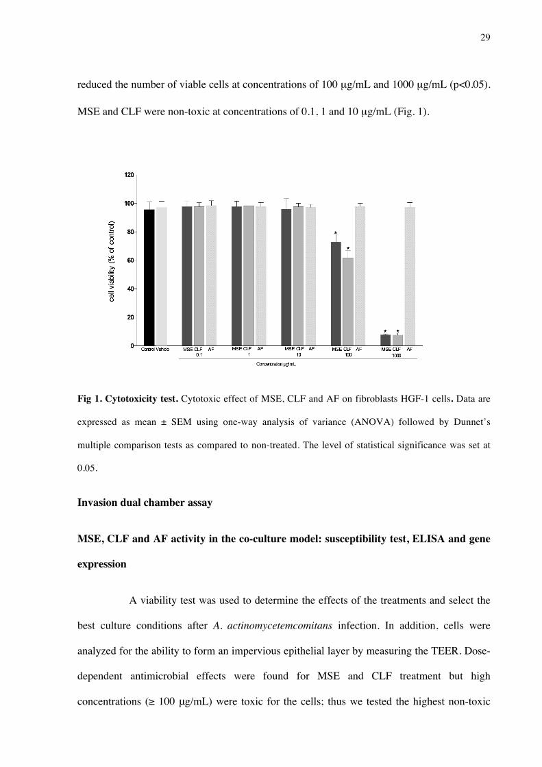
reduced the number of viable cells at concentrations of 100 µg/mL and 1000 µg/mL (p<0.05).
MSE and CLF were non-toxic at concentrations of 0.1, 1 and 10 µg/mL (Fig. 1).
Fig 1. Cytotoxicity test. Cytotoxic effect of MSE, CLF and AF on fibroblasts HGF-1 cells. Data are
expressed as mean ± SEM using one-way analysis of variance (ANOVA) followed by Dunnet’s
multiple comparison tests as compared to non-treated. The level of statistical significance was set at
0.05.
Invasion dual chamber assay
MSE, CLF and AF activity in the co-culture model: susceptibility test, ELISA and gene
expression
A viability test was used to determine the effects of the treatments and select the
best culture conditions after A. actinomycetemcomitans infection. In addition, cells were
analyzed for the ability to form an impervious epithelial layer by measuring the TEER. Dose-
dependent antimicrobial effects were found for MSE and CLF treatment but high
concentrations (≥ 100 μg/mL) were toxic for the cells; thus we tested the highest non-toxic
30
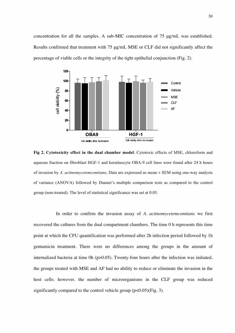
concentration for all the samples. A sub-MIC concentration of 75 μg/mL was established.
Results confirmed that treatment with 75 μg/mL MSE or CLF did not significantly affect the
percentage of viable cells or the integrity of the tight epithelial conjunction (Fig. 2).
Fig 2. Cytotoxicity effect in the dual chamber model. Cytotoxic effects of MSE, chloroform and
aqueous fraction on fibroblast HGF-1 and keratinocyte OBA-9 cell lines were found after 24 h hours
of invasion by A. actinomycetemcomitans. Data are expressed as mean ± SEM using one-way analysis
of variance (ANOVA) followed by Dunnet’s multiple comparison tests as compared to the control
group (non-treated). The level of statistical significance was set at 0.05.
In order to confirm the invasion assay of A. actinomycetemcomitans we first
recovered the cultures fromthe dual compartment chambers. The time 0 h represents this time
point at which the CFU quantification was performed after 2h infection period followed by 1h
gentamicin treatment. There were no differences among the groups in the amount of
internalized bacteria at time 0h (p>0.05). Twenty-four hours after the infection was initiated,
the groups treated with MSE and AF had no ability to reduce or eliminate the invasion in the
host cells; however, the number of microorganisms in the CLF group was reduced
significantly compared to the control vehicle group (p<0.05)(Fig. 3).

31
Fig 3. Comparison of colony-forming units. Comparison of colony-forming units (CFU/mL) among
groups treated with MSE, CLF and AF after A. actinomycetemcomitans infection. Data are expressed
as mean ± SEM using one-way analysis of variance (ANOVA) followed by Dunnet’s multiple
comparison tests as compared to vehicle control. The level of statistical significance was set at 0.05.
Analysis Using the RT2 Profiler PCR Array
Alterations in the transcript levels for all treatment groups were initially analyzed
using the RT2 Profiler PCR Array and 84 genes were screened and analyzed using the
SABiosciences web portal software. The transcriptional profile in the lower chamber cell
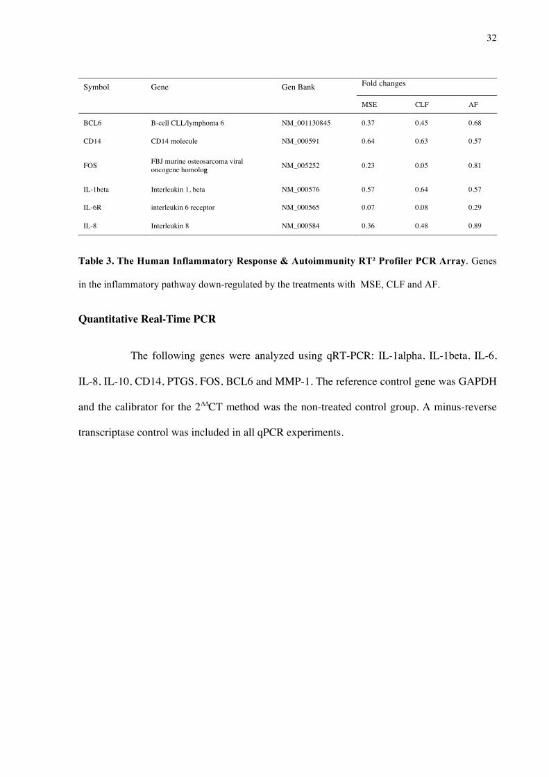
lines after 24 hours invasion assay is showed on Table 3. It was found a down-regulation of 6
different genes among 84 target genes. For the fold changes, values less than 1 are considered
down regulated.
32
Symbol Gene Gen Bank Fold changes
MSE CLF AF
BCL6 B-cell CLL/lymphoma 6 NM_001130845 0.37 0.45 0.68
CD14 CD14 molecule NM_000591 0.64 0.63 0.57
FOS FBJ murine osteosarcoma viral oncogene homolog NM_005252 0.23 0.05 0.81
IL-1beta Interleukin 1, beta NM_000576 0.57 0.64 0.57
IL-6R interleukin 6 receptor NM_000565 0.07 0.08 0.29
IL-8 Interleukin 8 NM_000584 0.36 0.48 0.89
Table 3. The Human Inflammatory Response & Autoimmunity RT² Profiler PCR Array. Genes
in the inflammatory pathway down-regulated by the treatments with MSE, CLF and AF.
Quantitative Real-Time PCR
The following genes were analyzed using qRT-PCR: IL-1alpha, IL-1beta, IL-6,
IL-8, IL-10, CD14, PTGS, FOS, BCL6 and MMP-1. The reference control gene was GAPDH
and the calibrator for the 2∆∆CT method was the non-treated control group. A minus-reverse
transcriptase control was included in all qPCR experiments.
33
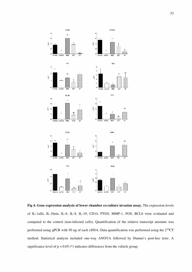
Fig 4. Gene expression analysis of lower chamber co-culture invasion assay. The expression levels
of IL-1alfa, IL-1beta, IL-6, IL-8, IL-19, CD14, PTGS, MMP-1, FOS, BCL6 were evaluated and
compared to the control (non-infected cells). Quantification of the relative transcript amounts was
performed using qPCR with 50 ng of each cDNA. Data quantification was performed using the 2∆∆CT
method. Statistical analysis included one-way ANOVA followed by Dunnet’s post-hoc tests. A
significance level of p < 0.05 (*) indicates differences from the vehicle group.

34
The results showed that cells invaded by A. actinomycetemcomitans and treated
with MSE had statistically significant down-regulation of the genes IL6, IL8, CD14 compared
to the control group (non-infected cells) (p<0.05). The chloroform fraction down-regulated
the gene expression of IL-1beta, IL6, IL10, PTGS, CD14, PTGS, FOS and BCL6 (p<0.05)
compared to the control group. The group treated with AF showed a down-regulation of
transcript levels for the genes IL-1alpha, IL-1beta, CD14, PTGS and FOS (p<0.05) (Fig. 4).
Cytokine assay
The concentrations of IL-1 alpha, IL-1beta, IL-6, IL-8, IL-10 and GM-CSF were
quantified by ELISA to confirm whether the proteins encoded by the down-regulated genes
were found at reduced levels in the supernatant. MSE reduced the expression of IL-6 in the
infected cells compared to the vehicle control group (p<0.05). Significant differences in the
levels of a number of cytokines were found between the CLF and control groups. Specifically,
reduced expression levels were observed for the cytokines IL-1beta, IL-6, IL-8 and GM-CSF
(reductions of 32%, 80, 3%, 30, 58 and 4% compared to the vehicle control, respectively).
Infected cells expressed low levels of IL-1 alpha when treated with the AF, a decrease of 31%
compared to the vehicle control group (Fig. 5).

35
Fig. 5 Cytokine assay. Quantification of IL-1alpha, IL-1beta, IL-6, IL-8, IL-10 and GM-CSF in the
co-culture supernant after 24 h of A. actinomycetemcomitans invasion. Cells were treated with 75
µg/mL of MSE and fractions and bacteria inocula were established at 2x10⁶ CFU/mL. Data are
expressed as mean±SD, n=6. Symbols indicate statistical differences (p<0.05, Dunnet’s test). #
indicates p<0.05 compared to non-treated group; * indicates p<0.05 compared to vehicle group.
Discussion
Periodontal disease is an oral infectious inflammatory disease and the most
common human chronic disorder [23]. The relationship between this disease and many
systemic diseases (including cardiovascular disease, diabetes, adverse pregnancy outcomes
and others) is well recognized [24], [25]. Based on current knowledge of inflammation
36
pathways, it appears that natural products may be a good source for developing multi-target
drugs with activity against the microorganisms responsible for periodontal disease [26], [27].
For this study, we evaluated the toxicity, antimicrobial and anti-inflammatory
activity of compounds naturally occurring in the plant M. sylvestris [28]. A screening assay
simulating the effect of A. actinomycetemcomitans, a species known to be associated with
periodontal disease, was used to model the infection of epithelial and subepithelial cell lines
[22]. These results confirmed the internalization of the bacteria, indicating the possible
activation of the membrane and intracellular receptors [29]. Transcriptional factors and
cytokines identified in the infection process suggested signaling and host response pathways
were involved in the bacteria challenge and during the treatment with M. sylvestris extract and
fractions.
The antimicrobial susceptibility test showed that MSE and CLF had activity not
only against A. actinomycetemcomitans but also against other periodontopathogens (F.
nucleatum, P. gingivalis and P. intermedia) that are implicated in the development and
virulence of periodontal disease [1]. In addition, this demonstrates that M. sylvestris works
against both microaerophiles and anaerobes. In the literature, it has been shown that the
ethanolic extract of M. sylvestris is effective as a bacteriostatic agent against methicillin-
resistant S. aureus (I50 ≤ 32 μg/ml) [30], and moderate to low activity was reported against
strains of Helicobacter pylori (MIC ranged from 0.625 to >5.0 mg/mL)[31]; moreover, the
aqueous fraction was reported to have anti-fungal activity, though not against Candida
albicans [32]. Overall, though antimicrobial effects of M. sylvestris have been reported in the
literature for a few microorganisms [30],[31],[32] these studies used different extract
preparations and the majority were based on agar-diffusion tests, making inter-study
comparisons difficult.

37
Our findings also demonstrate that the bioguided fractionation was successful and
may be a model for bioprospecting new drugs, as long as the active fraction (CLF) presented
enhanced antimicrobial activity relative to the unfractionated extract. In addition, our data
describe the cytotoxicity of the extract and fractions in vitro to provide better estimation of
the potential of the compound as favorable therapeutic agent. The viability test showed that
the CLF fraction was non-toxic at concentrations up to 100 µg/mL and AF had no toxic
effects at any of the concentrations tested. The LD50 of the extract for cell lines OBA-9 and
HGF (250 µg/mL and 210 µg/mL, respectively) gave insight into the safe concentrations for
use in the biological assays. M. sylvestris is widely known as a food or condiment and has
been used for millennia in traditional medicine; however, only one in vivo test of its toxicity
has been reported in the literature [33].
The bacterial products from A. actinomycetemcomitans affected the cell immune
response and increased the production of local cytokines. All the treatments tested affected
different signaling pathways. Upon treatment with the aqueous fraction, both the IL-1alpha
gene and protein expression levels were reduced. The pro-inflammatory cytokine IL-1 and
tumor necrosis factor alpha (TNF alpha) are modulators of the host response to microbial
infection. It has previously [34] been demonstrated that IL-1 specific marker is a strong
indicator of susceptibility to severe periodontal disease in adults. Furthermore, it has been
established that IL-1 is involved in the induction of bone resorption by promoting the
differentiation of osteoclast precursors in active osteoclasts [35].
A statistical reduction of IL-6 gene expression and protein levels were found after
treatment with the chloroform fraction (CLF). The higher expression levels of IL-6 in
untreated periodontal disease might induce an increase in matrix metalloproteinases (MMPs)
that are related to tissue destruction [36], [37]. IL-6 has been reported as a principal regulator

38
in the acute phase of inflammation and may promote osteoclastogenesis by increasing
tRANKL expression [38].
In addition, the CLF treatment regulated the expression of other
immunomodulatory genes (CD14, MMP1 and FOS), which indicates an effect on more than
one signaling pathway and may result in a good therapeutic outcome. Finding compounds that
trigger CD14 or toll-like receptors (TLRs) is potentially useful in periodontal disease. The
binding of lipopolysaccharides (LPS) with CD14 might induce the temporary activation of
many protein kinases and the phosphorylation of intracellular proteins essential for LPS
activation in monocytes/macrophages [39].
The MSE could regulate the transcription of IL-8 but not the same cytokine
expression. The answer to the question of how genomic information can be processed
differently to produce a specific cellular proteome to date remains unanswered [40], [41]. The
literature has been demonstrated that M. sylvestris may regulated the expression of cytokines
in the inflammatory process. In a pre-clinical study, important anti-inflammatory action of the
hydroalcoholic extract was found to interfere with the production of IL-1beta and
consequently block leukocyte migration [42]. Furthermore, the aqueous extract of M.
sylvestris was found to have an immunomodulatory property, acting as a macrophage
activators and promoting both IL-12 and (IFN) interferon transcripts [42]. Overall, the
literature and present data highlight the biological activity of M. sylvestris in treating
inflammation.
The phytochemical investigation of M. sylvestris showed a high occurrence of
phenolic compounds in all studied extracts and fractions. This is consistent with a previous
report [14], in which 4-hydroxybenzoic acid, 4-methoxybenzoic acid, 4-hydrocycinnamic acid
and tyrosol were isolated from M. sylvestris. Furthermore, the interest in phenolic compounds
has increased in recent years due to their possible implications for human heath, such as in

39
treating and preventing cancer, cardiovascular disease and other pathologies [11]. Overall,
phenolic compounds are particularly potent natural products with a wide range of biological
properties known in the literature that could be used extensively in dentistry.
The results of the present study showed that the low-polarity fraction CLF has
relevant dual activity, simultaneously controlling infection and inflammation processes. Thus,
M. sylvestris may be considered as a potential drug candidate for use as a new therapeutic
approach in the treatment of the periodontal disease.
Conclusion
In our study we found that Malva sylvestris and its chloroform fraction were able
to minimize the infection and inflammation process in oral human cells by a putative pathway
that may involve the antimicrobial effect and modulation of cytokines and receptors.
Therefore, this natural product may be considered as a successful dual anti-inflammatory–
antimicrobial candidate.
Funding
Research reported in this publication was supported by: National Center for
Complementary and Integrative Health of the National Institutes of Health under award
number R00AT006507, São Paulo Research Foundation FAPESP (Grant #2011/23980-5) and
Brazilian Federal Agency for the Support and Evaluation of Graduate Education CAPES
(Grant #2317/2014-01). The funders had no role in study design, data collection and analysis,
decision to publish, or preparation of the manuscript.
References
1. Pihlstrom BL, Michalowicz BS, Johnson NW. Periodontal diseases. Lancet. 2005 Nov 19; 366 (9499):1809–20. PMID: 16298220

40
2. Susin C, Haas AN, Albandar JM. Epidemiology and demographics of aggressive periodontitis. Period- ontol 2000. 2014 Jun; 65(1):27–45 doi: 10.1111/prd.12019 PMID: 24738585 3. Van Dyke TE, Serhan CN. Resolution of inflammation: a new paradigm for the pathogenesis of peri- odontal diseases. J Dent Res. 2003 Feb; 82(2):82–90. PMID: 12562878 4. Slots J, Reynolds HS, Genco RJ. Actinobacillus actinomycetemcomitans in human periodontal dis- ease: a cross-sectional microbiological investigation. Infect Immun. 1980 Sep; 29(3):1013–20. PMID: 6968718 5. Johansson A. Aggregatibacter actinomycetemcomitans leukotoxin: a powerful tool with 5. capacity to cause imbalance in the host inflammatory response. Toxins (Basel). 2011 Mar; 3(3):2459. 6. Darveau RP, Tanner A, Page RC. The microbial challenge in periodontitis. Periodontol 2000. 1997 Jun; 14:12–32. PMID: 9567964 7. Offenbacher S, Barros SP, Singer RE, Moss K, Williams RC, Beck JD. Periodontal disease at the bio- film-gingival interface. J Periodontol. 2007 Oct; 78(10):1911–25. PMID: 18062113 8. Cragg GM, Grothaus PG, Newman DJ. New horizons for old drugs and drug leads. J Nat Prod. 2014 Mar 28; 77(3):703–23. doi: 10.1021/np5000796 PMID: 24499205 9. Freires IA, Denny C, Benso B, de Alencar SM, Rosalen PL. Antibacterial Activity of Essential Oils and Their Isolated Constituents against Cariogenic Bacteria: A Systematic Review. Molecules. 2015 Apr 22; 20(4):7329–7358. doi: 10.3390/molecules20047329 PMID: 25911964 10.Jeon JG, Rosalen PL, Falsetta ML, Koo H. Natural products in caries research: current (limited) knowl- edge, challenges and future perspective. Caries Res. 2011; 45(3):243–63. doi: 10.1159/000327250 PMID: 21576957 11. Gasparetto JC, Martins CA, Hayashi SS, Otuky MF, Pontarolo R. Ethnobotanical and scientific aspects of Malva sylvestris L.: a millennial herbal medicine. J Pharm Pharmacol. 2012 Feb; 64(2):172–89. doi: 10.1111/j.2042-7158.2011.01383.x PMID: 22221093 12. DellaGreca M, Cutillo F, D'Abrosca B, Fiorentino A, Pacifico S, Zarrelli A. Antioxidant and radical scav- enging properties of Malva sylvestris. Nat Prod Commun. 2009 Jul; 4(7):893–6. PMID: 19731587 13. Barros L, Carvalho AM, Ferreira IC. Leaves, flowers, immature fruits and leafy flowered stems of Malva sylvestris: a comparative study of the nutraceutical potential and composition. Food Chem Toxicol. 2010 Jun; 48(6):1466–72. doi: 10.1016/j.fct.2010.03.012 PMID: 20233600 14. Cutillo F, D'Abrosca B, Dellagreca M, Fiorentino A, Zarrelli A. Terpenoids and phenol derivatives from Malva silvestris. Phytochemistry. 2006 Mar; 67(5):481–5. PMID: 16403542 15. Cragg GM, Newman DJ. Natural products: a continuing source of novel drug leads. Biochim Biophys Acta. 2013 Jun; 1830(6):3670–95. doi: 10.1016/j.bbagen.2013.02.008 PMID: 23428572
16. da Cunha MG, Franchin M, de Carvalho Galva ̃o LC, de Ruiz AL, de Carvalho JE, Ikegaki M et al. Anti- microbial and antiproliferative activities of stingless bee Melipona scutellaris geopropolis. BMC Com- plement Altern Med. 2013 Jan 28; 13:23. doi: 10.1186/1472-6882-13-23 PMID: 23356696

41
17. Folin O, Denis W. On phosphotungstic-phosphomolybdic compounds as color reagents. J Biol Chem. 1912; 12:239–243. 18. Chen C, Kittichotirat W, Chen W, Downey JS, Si Y, Bumgarner R. Genome sequence of naturally com- petent Aggregatibacter actinomycetemcomitans serotype a strain D7S-1. J Bacteriol. 2010 May; 192 (10):2643–4. doi: 10.1128/JB.00157-10 PMID: 20348265 19. Kusumoto Y, Hirano H, Saitoh K, Yamada S, Takedachi M, Nozaki T et al. Human gingival epithelial cells produce chemotactic factors interleukin-8 and monocyte chemoattractant protein-1 after stimula- tion with Porphyromonas gingivalis via toll-like receptor 2. J Periodontol. 2004 Mar; 75(3):370–9. PMID: 15088874 20. Clinical Laboratory Standards Institute 2009. Performance standards for antimicrobial susceptibility testing; 19th informational supplement. Document M100-S19. Clinical Laboratory Standards Institute, Wayne, PA. 21. Pasetto S, Pardi V, Murata RM. Anti-HIV-1 activity of flavonoid myricetin on HIV-1 infection in a dual- chamber in vitro model. PLoS One. 2014 Dec 29; 9(12):e115323. doi: 10.1371/journal.pone.0115323 PMID: 25546350 22. Zhao L, Wu Y, Tan L, Xu Z, Wang J, Zhao Z et al. Coculture with endothelial cells enhances osteogenic differentiation of periodontal ligament stem cells via cyclooxygenase-2/prostaglandin E2/vascular endothelial growth factor signaling under hypoxia. J Periodontol. 2013 Dec; 84(12):1847–57. doi: 10. 1902/jop.2013.120548 PMID: 23537125 23. Socransky SS. Microbiologycal of periodontal disease- present status and future considerations. J Peri- odontol, 1977 Sep; 48(9):497–504. PMID: 333085
24. Negrato CA, Tarzia O, Jovanovic ̌ L, Chinellato LE. Periodontal disease and diabetes mellitus. J Appl Oral Sci. 2013 Jan-Feb; 21(1):1–12. PMID: 23559105 25. Rautemaa R, Lauhio A, Cullinan MP, Seymour GJ. Oral infections and systemic disease—an emerging problem in medicine. Clin Microbiol Infect. 2007 Nov; 13(11):1041–7. PMID: 17714525 26. Koeberle A, Werz O. Multi-target approach for natural products in inflammation. Drug Discov Today. 2014 Dec; 19(12):1871–82. doi: 10.1016/j.drudis.2014.08.006 PMID: 25172801 27. Freires Ide A, Murata RM, Furletti VF, Sartoratto A, Alencar SM, Figueira GM et al. Coriandrum sativum L. (Coriander) essential oil: antifungal activity and mode of action on Candida spp., and molecular tar- gets affected in human whole-genome expression. PLoS One. 2014 Jun 5; 9(6):e99086. doi: 10.1371/ journal.pone.0099086 PMID: 24901768 28. Jain S, Darveau RP. Contribution of Porphyromonas gingivalis lipopolysaccharide to periodontitis. Peri- odontol 2000. 2010 Oct; 54(1):53–70. doi: 10.1111/j.1600-0757.2009.00333.x PMID: 20712633 29. Stathopoulou Panagiota G., Benakanakere Manjunatha R., Galicia Johnah C. et al. Epithelial cell pro- inflammatory cytokine response differs across dental plaque bacterial species. J Clin Periodontol. 2010 Jan; 37(1): 24–29. doi: 10.1111/j.1600-051X.2009.01505.x PMID: 20096064 30. Quave CL, Plano LR, Pantuso T, Bennett BC. Effects of extracts from Italian medicinal plants on plank- tonic growth, biofilm formation and adherence of methicillin-resistant Staphylococcus aureus. J Ethno- pharmacol. 2008 Aug 13; 118(3):418–28 doi: 10.1016/j.jep.2008.05.005 PMID: 18556162
42
31. Cogo LL, Monteiro CL, Miguel MD Miguel OG, Cunico MM et al. Anti-Helicobacter pylori activity of plant extracts traditionally used for the treatment of gastrointestinal disorders. Braz J Microbiol. 2010 Apr; 41 (2):304–9. doi: 10.1590/S1517-83822010000200007 PMID: 24031496 32. Magro A, Carolino M, Bastos M, Mexia A. Efficacy of plant extracts against stored products fungi. Effi- cacy of plant extracts against stored-products fungi. Rev Iberoam Micol. 2006 Sep; 23(3):176–8. PMID: 17196025 33. Seiberg M et al. Enhancing production of mucus of mucosal tissue, for administering to mucosal tissue, a composition comprising a safe effective amount of Malva sylvestris extract. Patent Number(s): US2006088616-A1; WO2006047470-A2; EP1811955-A2, 2006. 34. Kornman KS, Crane A, Wang HY, di Giovine FS, Newman MG, Pirk FW et al. The interleukin-1 geno- type as a severity factor in adult periodontal disease. J Clin Periodontol. 1997 Jan; 24(1):72–7. PMID: 9049801 35. Graves DT, Li J, Cochran DL. Inflammation and uncoupling as mechanisms of periodontal bone loss. J Dent Res. 2011 Feb; 90(2):143–53. doi: 10.1177/0022034510385236 PMID: 21135192 36. Kang JH, Ko HM, Moon JS, Yoo HI, Jung JY, Kim MS et al. Osteoprotegerin expressed by osteoclasts: an autoregulator of osteoclastogenesis. J Dent Res. 2014 Nov; 93(11):1116–23. doi: 10.1177/ 0022034514552677 PMID: 25256714 37. Scapoli L, Girardi A, Palmieri A, Carinci F, Testori T, Zuffetti F et al. IL6 and IL10 are genetic susceptibil- ity factors of periodontal disease. Dent Res J (Isfahan). 2012 Dec; 9(Suppl 2):S197–201. 38. Irwin CR, Myrillas T, Smyth M, Doogan J, Rice C, Schor SL. Regulation of fibroblast-induced collagen gel contraction by interleukin-1beta. J Oral Pathol Med. 1998 Jul; 27(6):255–9. PMID: 9707277 39. Wang PL, Ohura K. Porphyromonas gingivalis lipopolysaccharide signaling in gingival fibroblasts- CD14 and Toll-like receptors. Crit Rev Oral Biol Med. 2002; 13(2):132–42. PMID: 12097356 40. Reynier F, Petit F, Paye M, Turrel-Davin F, Imbert PE, Hot A et al. Importance of correlation between gene expression levels: application to the type I interferon signature in rheumatoid arthritis. PLoS One. 2011; 6(10):e24828. doi: 10.1371/journal.pone.0024828 PMID: 22043277 41. Prudente AS, Loddi AM, Duarte MR, Santos AR, Pochapski MT et al. Pre-clinical anti-inflammatory aspects of a cuisine and medicinal millennial herb: Malva sylvestris L. Food Chem Toxicol. 2013 Aug; 58:324–31. doi: 10.1016/j.fct.2013.04.042 PMID: 23684757. 42. El Ghaoui WB, Ghanem EB, Chedid LA, Abdelnoor AM. The effects of Alcea rosea L., Malva sylvestris L. and Salvia libanotica L. water extracts on the production of anti-egg albumin antibodies, interleukin- 4, gamma interferon and interleukin-12 in BALB/c mice. Phytother Res. 2008 Dec; 22(12):1599–604 doi: 10.1002/ptr.2530 PMID: 18688815

43
2.2 ARTIGO2
Anti-inflammatory, anti-osteoclastogenic and antioxidant effects of Malva
sylvestris extract and fractions: in vitro and in vivo studies
Bruna Benso1; Marcelo Franchin1; Adna Prado Masaroli2; Jonas Augusto Rizzato Paschoal3; Severino Matias Alencar2; Gilson César Nobre Franco4; Pedro Luiz Rosalen1
1Department of Physiological Sciences, Piracicaba Dental School, University of Campinas, Piracicaba, Sao Paulo, Brazil. 2Department of Agri-food Industry, Food and Nutrition, “Luiz de Queiroz” College of Agriculture, University of São Paulo, Piracicaba, SP, Brazil. 3Departments of Physics and Chemistry, School of Pharmaceutical Sciences of Ribeirão Preto, University of São Paulo, Piracicaba, SP, Brazil 4Department of General Biology, State University of Ponta Grossa, Ponta Grossa, PR, Brazil
Corresponding Author Email: [email protected] (PLR)
2Benso B, Franchin M; Massarioloi AP, Paschoal JAR, Alencar SM, Franco GC, Rosalen PL, será submetido para publicação ao periódico
PLoS One.

44
Abstract
Given their medical importance, natural products represent a tremendous source
of drug discovery. Malva sylvestris is a plant cited extensively in the ethnopharmacological
literature and is known worldwide. The aim of this study was to investigate the extract (MSE)
and fractions (HF, CLF, EAF and AF) of M. sylvestris for anti-inflammatory, anti-
osteoclastogenic, antioxidant effects and a chemical identification of the bioactive fraction.
The in vivo experiments consisted of the quantification of neutrophil migration to the
peritoneal cavity, paw edema and cytokine release. M. sylvestris extract (MSE) and fractions
at 3, 10 and 30 mg/kg were administered orally. Macrophages were cultured by cell viability
assay to determine the concentration of MSE and fractions for all cell-based experiments.
Transcriptional factors were quantified by qPCR and the expression of the following genes
were studied: carbonic anhydrase II (CAII), cathepsin K and tartrate-resistant acid
phosphatase (TRAP). Gel zymography with collagen as the substrate was used to identify the
latent and the active gelatinase MMP-9 secreted in the media stimulated with LPS (E. coli) in
RAW 264.7 cells. TRAP staining was employed to evaluate osteoclast (OC) formation and
TRAP-positive multinuclear macrophages with more than three nuclei were counted as OCs.
Antioxidant activities measured for all extract and fractions for the two most common radical
scavenging assays using 1,1-diphenyl-2-picrylhydrazyl (DPPH) and 2,2-azino-bis-3-
ethylbenzthiazoline-6-sulfonic acid (ABTS). The chemical analysis was performed using the
MS/MS technique. The aqueous fraction (AF) was identified as the bioactive fraction, with
the oral treatment significantly reducing the neutrophil migration to the peritoneal cavity,
antiedematogenic and IL-1B cytokine level (54% reduction). The viability tests showed a
concentration-dependent effect, where the MSE and fractions at concentrations equal to 10
μg/mL were not toxic for the cells. In the TRAP gene expression analysis, all the treatments
tested presented a downregulation of the transcription levels. CLF (chloroform fraction) and

45
AF treatments had the ability to reduce the osteoclastogenesis on RAW 264.7 cell lines
(p<0.05) measured in the TRAP staining assay. In our study, the activity of MMP-9 decreased
when treated with the AF and EAF, with a reduction of 69% and 75%, respectively.
Moreover, the bioactive fraction had the ability to regulate the oxidation pathway, eliminating
the radicals of ABTS and DPPH method. Mass spectrometry identified rutin as the bioactive
compound in the AF. The AF of M. sylvestris presented anti-inflammatory, anti-
osteoclastogenic and antioxidant abilities in different in vitro and in vivo methods. In addition,
we suggest that given its multi-target activity the bioactive fraction may be a good candidate
in the therapy of chronic inflammatory diseases.
Introduction
Inflammation is a biological process that involves vascular and cellular events
coordinated by mediators such as prostaglandins, leukotrienes and cytokines (Perretti et al.,
2015). This is an organism’s essential and protective mechanism in response to injury,
infection and trauma (Ward, 1974). Thus, inflammation appears to be an inherently self-
perpetuating event in terms of possible transformation due to the largely biological chemokine
attraction (Souza and Lerner, 2013).
A prolonged inflammation process may lead to chronic diseases such as
periodontal disease and rheumatoid arthritis, which are associated with tissue injury and bone
resorption (Crotti et al., 2015). In some of these chronic inflammations, pro-inflammatory
mediators and reactive oxygen species (ROS) can promote osteoblast apoptosis and bone
resorption through the activation of the NF-κB signaling pathway, which plays an important
role in osteoclastogenesis (Conner and Grisham, 1996). The classic NF-κB pathway
stimulation includes the receptor activator of nuclear factor kappa-B ligand (RANKL), the
osteoclastogenic cytokine, as well as TNF-α and other inflammatory mediators (Takeshita et

46
al., 2000). These cytokines may induce bone resorption, affecting the production of the
essential osteoclast differentiation (Henderson et al., 1996).
Osteoclast differentiation and the activation of bone resorption function by mature
osteoclasts are events that require RANKL and its permissive macrophage colony-stimulating
factor (M-CSF) to induce the expression of RANK, a receptor for RANKL. RANKL plays an
essential role in the differential, recruitment, activation and survival of osteoclasts by binding
to its receptor (RANK) on osteoclasts or progenitor cells (Ohshiba et al., 2003). A number of
the RANK-induced signaling pathways in osteoclasts ultimately induce the expression of
several genes, including TRAP, cathepsin K and carbonic anhydrase, which are enzymes
involved in the regulation of the dissolution of mineral and collagen (Franco et al., 2011;
Zhang et al., 2011).
In addition, matrix metalloproteinase (MMP) is a family of proteolytic enzymes
involved in the role of extracellular matrix degradation that includes a variety of tissues and
bone (Ohshiba et al., 2003). In the group of MMPs, MMP-9 is an important proteinase that
osteoclasts express in high levels. Moreover, there are studies showing the relation of MMP-9
activity in bone destruction, including in some diseases such as rheumatoid arthritis (Franco et
al., 2011; Takeshita et al., 2000).
Traditionally, anti-inflammatory therapy has focused on controlling cytokine and
adhesion molecule expression, including non-steroidal drugs and glucocorticoids
(Georgakopoulou and Scully, 2014; Rainsford, 2007). However, in the past few years it has
been recognized that the inflammation resolution may be based on multi-target drugs
(Koeberle and Werz, 2014). Multiple signaling pathways are a way to improve the pro-
inflammatory, immunomodulatory and proresolving cascades, which define the aspects of the
inflammation (Kohli and Levy, 2009). Thus, natural products have played an important role in

47
the development of new sources in the treatment of inflammatory diseases (Cragg et al.,
2014).
The screening of extracts from natural sources has historically led to the discovery
of many clinical drugs in current therapy (Molinari, 2009). Since natural products do not have
a standard composition, there is interest in identifying biological therapeutic potential in new
plant extracts (Harvey, 2008). The ethnopharmacological literature has reported a wide use of
Malva sylvestris since ancient times for its emollient, antioxidant and anti-inflammatory
properties (Gasparetto et al., 2012). Given its widespread and medicinal importance, the aim
of this study was to investigate the extract and fractions of Malva sylvestris for anti-
inflammatory, anti-osteoclastogenic, antioxidant and chemical identification of the bioactive
fraction.
Material and Methods
Preparation of the extract and fractions
Malva sylvestris leaves were collected in the inner region, municipality of
“Princesa Isabel”, state of Paraiba, in northeastern Brazil. The leaves were extracted with
absolute ethanol at room temperature and then filtered to obtain the ethanol extract of M.
sylvestris (MSE). The MSE was further fractioned using liquid-liquid extraction. The
fractions obtained were: hexane (HF), chloroform (CLF), ethyl acetate (EAF) and aqueous
(AF), and these were monitored with thin-layer chromatography (TLC) using the
anisaldehyde reagent (4-methoxybenzaldhyde, acetic acid, sulfuric acid, 1.0:48.5:0.5),
followed by heating at 100oC for 5 min. Fluorescent substances were visualized under
ultraviolet (UV) at wavelengths of 254 and 366 nm. All the extract and fractions were re-
suspended in 1% ethanol and used in the biological assays [17].
48
Anti-inflammatory analysis
Animals
Male Balb/c albino mice (20–25g), SPF, were purchased from
CEMIB/UNICAMP (Multidisciplinary Center for Biological Research, SP, Brazil). The mice
were maintained in a room with a controlled temperature (22 ± 2°C) for a 12 h light/12 h dark
cycle, humidity 40-60%, with food (standard pellet diet) and water provided ad libitum. The
experiments were conducted in accordance with the Guide for the Care and Use of Laboratory
Animals and had received prior approval from the local Animal Ethics Committee (CEUA,
Ethics Committee on Animal Use/UNICAMP, process number 2790–1).
Neutrophils migration in the peritoneal cavity
To determine the neutrophil migration into the peritoneal cavity of the MSE and
fractions, 3, 10 and 30 mg/kg were administered orally and 2 mg/kg dexamethasone was
administered by subcutaneous (s.c.) injection 1h before administration of inflammatory
stimulation by intraperitoneal (i.p.) injection of carrageenan at 500 μg/cavity. The vehicle
(0.9% NaCl) was used as the negative control. The mice were euthanized 4 h after the
challenge (carrageenan administration) and the peritoneal cavity cells were harvested by
washing the cavity with 3 mL of phosphate-buffered saline (PBS) containing EDTA. The
volumes recovered were similar in all experimental groups and equal to approximately 95%
of the injected volume. In order to count the total number of cells, a Neubauer chamber was
used. Smears were prepared using a cytocentrifuge (Cytospin 3; Shandon Lipshaw), stained
with a Panoptic staining kit and neutrophils cells were counted (until 100 cells) using an
optical microscope (1000X). The results are presented as the number of neutrophils per cavity
[18].

49
Carrageenan-induced paw edema
A paw edema was induced by subplantar injection of 0.05 mL of lambda
carrageenan (1% w/v in 0.9% of saline) into the left hind paw in the mice. An equal volume
of vehicle was injected into the contralateral paw. The volume of both hind paws up to the
ankle joint was measured with a plethysmometer (UGO Basile, Model 7140) immediately
before the 0, 1, 2, 3, 4 and 5 hours post-carrageenan. The difference in the volumes between
the hind paws was a measure of the edema (mL). The MSE and the bioactive fraction
previously selected in the neutrophil migration model were administered by oral treatment (30
mg/kg), the reference drug, indomethacin (10 mg/kg), or the vehicle (10 mL/kg of 0.9% of
saline), were given intraperitoneally 1/2 h or orally 1 h before the subplantar injection of the
phlogistic agent [19].
Cytokines quantification
Based on a previous test (neutrophil migration assay), the MSE and the bioactive
AF were selected for the quantification of proinflammatory cytokines produced in the
peritoneal cavity. The mice were treated with the MSE or the AF (30 mg/kg, oral) 1h before
the administration of inflammatory stimulation by intraperitoneal (i.p.) injection of
carrageenan at 500 μg/cavity. After 4 h, the animals were euthanized and the samples were
homogenized in 500 μL of the appropriate buffer containing protease inhibitors (Sigma, St.
Louis, MO, USA). Levels of TNF-α and IL-1β were determined by ELISA using protocols
supplied by the manufacturers (Peprotech, Rocky Hill, NJ, USA) from both the experiments.

50
Osteoclasteogenic assays
Cell culture
RAW 264.7 cells were purchased from the Rio de Janeiro cell bank (Rio de
Janeiro, Brazil) and cultured in Dulbecco’s Modified Eagle’s Medium (DMEM) with 10%
fetal bovine serum (Gibco, Life Technologies, CA), 100 U/mL penicillin, and 100 µg/mL
streptomycin (Invitrogen Life Technologies, CA). Cells were maintained in a humidified
incubator at 37°C in 5% CO2 [7].
Cell viability
A cell-based assay to screen the MSE and fractions (HF, CLF, EAF, AF) to
measure the enzyme activity as a marker of viable cells. RAW 264.7 cells were seeded (~
1x10⁵ cells/mL) in a 96-well plate and incubated for 24 h at 37oC with 5% CO2. The MSE and
fractions (0.1-1000 μg/mL) were added to the cell culture and incubated for 24 h. After the
incubation time, the supernatant was discarded and the cells were washed with PBS. Fresh
medium with 3-(4,5-dimethylthiazol-2-yl)-2,5-diphenyltetrazolium (MTT) 0.5 mg/mL were
then incubated for an additional 4 h. After the incubation time, the cell growth medium was
replaced by ethanol and colorimetric measurements were performed with a microplate reader
at 570 nm [20]. The extract MSE and all fractions and aqueous were re-suspended in 1%
ethanol and used in the biological assays.
Analysis of gene expression
Quantitative PCR (qPCR) was performed to evaluate the possible effects of MSE
and fractions on the expression of predominant osteoclast marker genes. In addition, we also
aimed to evaluate whether our natural products could control the transcription of genes
involved in bone metabolism. The genes analyzed were: carbonic anhydrase II (CAII),

51
cathepsin K, tartrate-resistant acid phosphatase (TRAP) and glycerol 3 phosphate
dehydrogenase (GAPDH). The primers sequences were: CAII (forward:
TGGTTCACTGGAACACCAA, reverse: CACGCTTCCCCTTTGTTTTA), cathepsin K
(forward: CAGCTTCCCCAAGATGTGAT, reverse: AGCACCAACGAGAGGAGAAA),
TRAP (forward: CCCTCTGCAACTCTGGACTC, reverse:
TAGAGGCGAACAGGAAGGAA), GAPDH (forward: AACTTTGGCATTGTGGAAGG,
reverse: ACACATTGGGGGTAGGAACA). RAW 264.7 cells (~ 1x10⁶ cells/mL) were
seeded into 24-well plates for 24 h and treated with a 10 µg/mL concentration of the MSE and
fractions in serum-free medium for 24 h and the stimulatory response was induced by 1
µg/mL LPS (Sigma Aldrich, St. Louis, Mo). Cultures were washed twice with PBS and RNA
was subsequently isolated using RNeasy Mini Kit (Qiagen, Valencia, CA, USA) following
the manufacturer’s protocols. RNA was treated with DNase Set (Qiagen, Valencia, CA,
USA). The cDNA was synthesized from total RNA using the SuperScript® III First-Strand
Synthesis System (Invitrogen, Carlsbad, CA, USA) and random primers, as previously
described [17]. Quantification of the relative transcript amounts performed by qPCR with 10
ng of each cDNA and SYBR Green PCR Master Mix (Applied Biosystems, Foster City, CA,
USA). The reactions were performed in the instrument StepOnePlus™ (Applied Biosystems,
Foster City, CA, USA). GAPDH was used as an endogenous control. The relative gene copy
number was calculated using the 2∆∆CT method and o primer-dimers were generated during
the applied 40 real-time PCR amplification cycles.
TRAP staining
To examine the effect of the MSE and fractions on sRANKL-induced
osteoclastogenesis in RAW 264.7 macrophage cells, a quantitative measurement was
conducted. Osteoclast formation was measured by the quantification of TRAP+

52
multinucleated osteoclasts per well, using light microscopy. RAW 264.7 cells were seeded in
96-well plates (~ 5x10³ cells/mL) and stimulated with sRANKL (50 ng/mL). Treatments were
MSE and fractions at 10 µg/mL, and the cell culture medium was α-MEM with 10% fetal
bovine serum (FBS). After 6 days, cells were fixed with 4% paraformaldehyde, washed with
PBS, and stained for TRAP (Sigma Aldrich, St. Louis, MO, USA). TRAP-positive
multinucleated (>3 nuclei) cells were counted as osteoclast-like cells [7].
Gelatin zymography
RAW 264.7 cells (~ 1x10⁶ cells/mL) were seeded into 24-well plates for 24h.
Inflammatory response was induced by 1 µg/mL LPS for 48 h (Sigma Aldrich, St. Louis,
Mo). The test concentrations of the MSE and the HF, CLF, EAF and AF were 10 µg/mL for
48 h. The supernatant was collected and the amount of total protein was measured using the
Pierce BCA Protein Assay Kit (Thermo Scientific, Rockford, IL, USA). An equal amount of
protein was designed by electrophoresis in Tris-Glycine Gels (Novex®, Life Technologies,
Carlsbad, CA) under non-reducing conditions. The protein separated in the gel was developed
using Developing Buffer supplied by the manufacture (Novex®, Life Technologies, Carlsbad,
CA). Subsequently the developed gelatin gel was stained with Coomassie R-250 Stain [7].
Antioxidants assays
DPPH
The reaction mixture consisted of 0.5 mL of extract and fractions, 3.0 mL of pure
ethanol, and 0.3 mL of DPPH radical in a 0.5 mM ethanol solution, which was incubated at
room temperature for 45 min, and the activity was expressed in µmol Trolox/g of sample per
dry weight. The calibration curve was constructed with the standard Trolox in the
concentration range of 0 to 200 µM Trolox. Several MSE and fractions concentrations were

53
used, and readings were monitored at 517 nm using a spectrophotometer (Shimadzu, Japan).
The antioxidant activity measured by the DPPH free radical method can be expressed as
IC50, i.e., the antioxidant concentration required to reduce the initial DPPH radical by 50%.
The sample concentration required to reduce the initial DPPH radical by 50% (Alencar et al.,
2007).
ABTS•+
The antioxidant activity by the ABTS•+ method (2,2′-azinobis-3-
ethylbenzothiazoline-6-sulfonic acid) was assessed according to the method described by Re
et al. (1999) with modifications. The ABTS radical was formed through the reaction of 7
mM ABTS•+ solution with 140 mM potassium persulfate solution, incubated at 25 °C in the
dark for 12–16 h. Once formed, the radical was diluted with ethanol P.A. to an absorbance of
0.700 ± 0.020 at 734 nm. Three different dilutions of each vegetable extract were prepared in
triplicate. After that, 30 µL of the MSE and fraction dilution were transferred to test tubes
with 3.0 mL of ABTS radical in the dark. The absorbance was read at 734 nm after 6 min of
reaction using ethanol as a blank. Trolox, a synthetic water-soluble antioxidant analogue of
vitamin E, was used as the reference at concentrations ranging from 100 to 2000 µM and the
results were expressed as µM Trolox/g sample.
Chemical analysis
HPLC analysis
A Shimadzu Prep 6AD LC system equipped with SPD-M10Avp photodiode array
detector (PDA), a 10AF auto injector and FRC-10A fraction collector were used to perform
the high-performance liquid chromatography (HPLC) analysis. For the analytical test, diluted
solutions of the AF were filtered (Millipore – 0.22 μm), and 10 μL aliquots were injected into
a Shimadzu reverse-phase analytical column of 250 mm × 4.6 mm × 5 μm (particle size). For
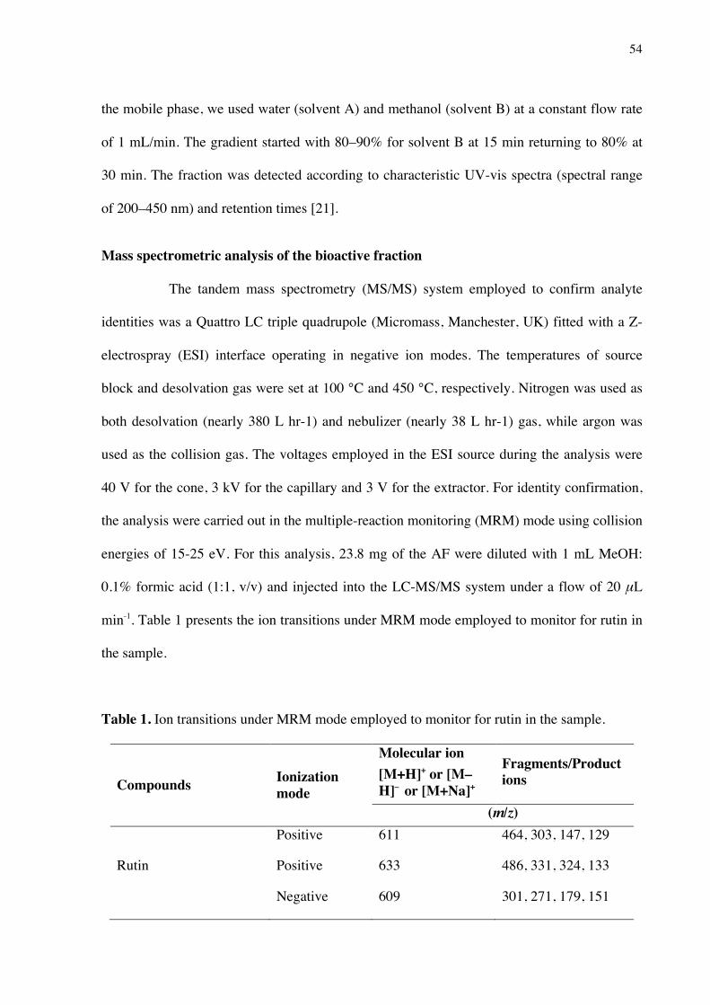
54
the mobile phase, we used water (solvent A) and methanol (solvent B) at a constant flow rate
of 1 mL/min. The gradient started with 80–90% for solvent B at 15 min returning to 80% at
30 min. The fraction was detected according to characteristic UV-vis spectra (spectral range
of 200–450 nm) and retention times [21].
Mass spectrometric analysis of the bioactive fraction
The tandem mass spectrometry (MS/MS) system employed to confirm analyte
identities was a Quattro LC triple quadrupole (Micromass, Manchester, UK) fitted with a Z-
electrospray (ESI) interface operating in negative ion modes. The temperatures of source
block and desolvation gas were set at 100 °C and 450 °C, respectively. Nitrogen was used as
both desolvation (nearly 380 L hr-1) and nebulizer (nearly 38 L hr-1) gas, while argon was
used as the collision gas. The voltages employed in the ESI source during the analysis were
40 V for the cone, 3 kV for the capillary and 3 V for the extractor. For identity confirmation,
the analysis were carried out in the multiple-reaction monitoring (MRM) mode using collision
energies of 15-25 eV. For this analysis, 23.8 mg of the AF were diluted with 1 mL MeOH:
0.1% formic acid (1:1, v/v) and injected into the LC-MS/MS system under a flow of 20 μL
min-1. Table 1 presents the ion transitions under MRM mode employed to monitor for rutin in
the sample.
Table 1. Ion transitions under MRM mode employed to monitor for rutin in the sample.
Compounds Ionization mode
Molecular ion [M+H]+ or [M–H]– or [M+Na]+
Fragments/Product ions
(m/z)
Rutin
Positive
Positive
Negative
611
633
609
464, 303, 147, 129
486, 331, 324, 133
301, 271, 179, 151
55
Statistical analysis
Continuous variables are presented as mean values + SD. The Shapiro–Wilk test was
used for the assessment of normality. All reported p- values are compared to a significance
level of 0.05. For multiple group comparisons, the data were subjected to one-way analysis of
variance (ANOVA). To determine overall difference between the group means and Tukey's
significant difference for pair-wise differences for within group comparisons. the Bonferroni
post-test indicated a significant difference between the controls. Data were analysed using
STATATM (Version 10.0, Stata Corporation, College Station, TX, USA) and GraphPad
(Version5.0,GraphPad Software Inc., San Diego, CA).
Results Neutrophils migration in the peritoneal cavity
The MSE and fractions were primarily tested for anti-inflammatory effects using a
neutrophil migration model as an in vivo model to screen anti-inflammatory compounds. The
results showed that the oral administration of 30mg/kg of the MSE decreased the migration of
neutrophils into the peritoneal cavity compared to the carrageenan group (ρ<0.05).
The fractionation method used for the MSE was effective, showing that the CLF,
EAF and AF concentrated the biological activity and reduced the neutrophil influx in all doses
tested. However, the AF yielded the best results, showing in all doses tested with a significant
reduction of neutrophil migration and the lowest percentages of inflammatory cells compared
to the other fractions (Figure 1).

56
Figure 1. Inhibitory effect of the MSE, HF, CLF, EAF, AF on neutrophil migration into the
peritoneal cavity induced by carrageenan. Neutrophil migration was determined 4 h after the
injection of carrageenan 500 µg/cavity. Mice previously treated with the vehicle, MSE, HF, CLF, EAF,
AF. The data are expressed as mean ± SD, n = 6. Symbols indicate statistical difference (p < 0.05,
ANOVA, Tukey post test) ** compared to the carrageenan group; * comparison between the
concentrations tested 3, 10 and 30 mg/kg.
Saline CG Dexa 3 10 300
4
8
12
Neu
troph
ils x
106
**
**
Carrageenan (500 μg/cavity)MSE (mg/kg)
MSE*
*
**
Saline - Dexa 3 10 300
4
8
12
Neu
troph
ils x
106
**
**
CLF (mg/kg)
** **
**
Carrageenan (500 μg/cavity)
CLF
Saline CG Dexa 3 10 300
4
8
12
Neu
troph
ils x
106
**
**
AF (mg/kg)
****
**
Carrageenan (500 μg/cavity)
AF
Saline - Dexa 3 10 300
4
8
12
Neu
troph
ils x
106
**
HF (mg/kg)Carrageenan (500 μg/cavity)
**
HF
Saline CG Dexa 3 10 300
4
8
12
Neu
troph
ils x
106
**
**
EAF (mg/kg)
** **
**
Carrageenan (500 μg/cavity)
EAF
*

57
Carrageenan-induced paw edema
The MSE and the bioactive AF were verified for antiedematogenic activity. The
results for the AF demonstrated a biological activity in the first hour analyzed. AF was
comparable to the positive control indomethacin for second and third hours analyzed as
shown in (Table 2).
Table 2. Effect of MSE and the AF on carrageenan-induced paw edema on mice.
Treatment
(mg/kg)
Time (h) after injection of carrageenan
1 2 3 4 5
Carrageenan 0.08±0.02 0.13±0.02 0.17±0.01 0.12±0.02 0.12±0.02
Indo 10 0.05±0.03 0.05±0.03* 0.08±0.02* 0.08±0.02* 0.08±0.02*
MSE 30 0.06±0.02 0.08±0.02* 0.07±0.01* 0.11±0.03 0.11±0.03
AF 30 0.04±0.01* 0.06±0.02* 0.07±0.02* 0.10±0.02 0.09±0.02
The data are expressed as mean ± SD, n = 6. Symbols indicate statistical difference (p < 0.05,
ANOVA, Bonferroni post test) * compared to the carrageenan group.
Cytokines assay
The administration of the AF at a dose of 30 mg/kg significantly reduced the level
of cytokine IL-1β (54%) compared to the control group vehicle (p<0.05). Thus, non-
significant differences were found at TNF-α expression levels (Figure 2).
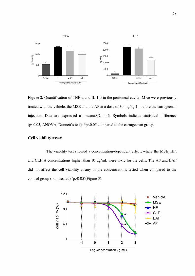
58
Figure 2. Quantification of TNF-α and IL-1 β in the peritoneal cavity. Mice were previously
treated with the vehicle, the MSE and the AF at a dose of 30 mg/kg 1h before the carrageenan
injection. Data are expressed as mean±SD, n=6. Symbols indicate statistical difference
(p<0.05, ANOVA, Dunnett’s test); *p<0.05 compared to the carrageenan group.
Cell viability assay
The viability test showed a concentration-dependent effect, where the MSE, HF,
and CLF at concentrations higher than 10 μg/mL were toxic for the cells. The AF and EAF
did not affect the cell viability at any of the concentrations tested when compared to the
control group (non-treated) (p>0.05)(Figure 3).
Saline - MSE AF0
50
100
150
pg /
cavi
ty
Carrageneenan (500 ug/cavity)
TNF-A
*
Saline - MSE AF0
500
1000
1500
2000
2500
pg/ c
avity
Carrageenan (500 ug/cavity)
*
IL-1B
*
-1 0 1 2 30
40
80
120
cell
viab
ility
(%)
VehicleMSEHFCLFEAFAF
Log (concentration µg/mL)
**
**

59
Figure 3. Effect of MSE and fractions on cell viability. Log dose response of MSE, HF, EAF, and
AF on RAW 264.7 cells. Significant difference between fractions at the indicated dose (p<0.05).
**Significant difference between EC50 values (p < 0.05). Data were expressed as mean±SD, n=9.
Analysis of gene expression
Gene expression analysis showed that the AF was the only treatment that had the
ability to downregulate all the study genes: ACII, cathepsin K and TRAP (p>0.05) (Figures 4,
5, 6). The MSE, CLF, EAF and AF downregulated the gene expression of TRAP (p>0.05)
(Figure 6). The gene transcription of cathepsin K was controlled by the treatments with the
AF and EAF (p>0.05) (Figure 5).
Figure 4. Effect of MSE and fractions on CAII (carbonic anhydrase) expression levels.
Quantification of the relative transcript amounts performed by qPCR with 10 ng of each cDNA. Fold
regulation was calculated in comparison to the LPS control group. Statistical analysis were performed
by one-way ANOVA followed by Dunnett’s post-hoc tests. *p < 0.05 significantly different from
LPS-stimulated cells.
LPS - MSE CLF EAF AF0
2
4
6
fold
regu
latio
n
* *
*
ACII
M. sylvestris extract and fractions

60
Figure 5. Effect of MSE and fractions on cathepsin K expression levels. Quantification of the
relative transcript amounts performed by qPCR with 10 ng of each cDNA. Fold regulation was
calculated in comparison to the LPS control group. Statistical analysis was performed by one-way
ANOVA followed by Dunnett’s post-hoc tests. *p < 0.05 significantly different from LPS-stimulated
cells.
Figure 6. MSE and fractions effect on TRAP expression levels. Quantification of the relative
transcript amounts performed by qPCR with 10 ng of each cDNA. Fold regulation was calculated in
comparison to the LPS control group.Statistical analysis were performed by one-way ANOVA
followed by Dunnett’s post-hoc tests. *p < 0.05 significantly different from LPS-stimulated cells.
LPS - MSE CLF EAF AF0
1
2
3
4
5
fold
regu
latio
n
**
*
**
M. sylvestris extract and fractions
Catepsina K
TRAP
LPS - MSE CLF EAF AF0
2
4
6
8
10
fold
regu
latio
n
**
M. sylvestris extract and fractions

61
TRAP staining
The number of TRAP-positive cells levels had high levels of expression in the
stimulated control group (p<0.05). The CLF and AF treatments had the ability to reduce the
osteoclastogenesis on RAW 264.7 cell lines compared to the vehicle control group (p<0.05)
(Figure 7).
Figure 7. Activity of M. sylvestris and fractions on RANKL-mediated osteoclast differentiation in
vitro. RAW 264.7 cells were stimulated with sRANKL (50 ng/ml) for 6 days. Cells were fixed and
stained for TRAP. TRAP+ multinuclear cells were counted. Data represent mean ± SD of three
cultures. * p < 0.05; significantly lower than sRANKL-stimulated group.
Gelatin zymography
The results showed that two different fractions caused the reduction of
gelatinolytic activity: the AF and EAF at the 10 μg/mL concentration. The non-treated group
and the AF, EAF were not significantly different (p>0.05). In our study, MMP-9 activity
decreased 69% and 75% (AF and EAF, respectively). In contrast, HF and CLF increased the
enzyme activity (Figure 8).
TRAP staining
- Vehicle MSE HF CLF EAF AF0
20
40
60
TRA
P+ #
cells
/ w
ell
*
*
*
M. sylvestris extract and fractions
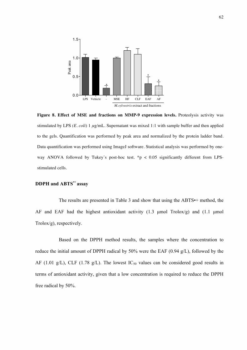
62
Figure 8. Effect of MSE and fractions on MMP-9 expression levels. Proteolysis activity was
stimulated by LPS (E. coli) 1 μg/mL. Supernatant was mixed 1:1 with sample buffer and then applied
to the gels. Quantification was performed by peak area and normalized by the protein ladder band.
Data quantification was performed using ImageJ software. Statistical analysis was performed by one-
way ANOVA followed by Tukey’s post-hoc test. *p < 0.05 significantly different from LPS-
stimulated cells.
DDPH and ABTS•+ assay
The results are presented in Table 3 and show that using the ABTS•+ method, the
AF and EAF had the highest antioxidant activity (1.3 µmol Trolox/g) and (1.1 µmol
Trolox/g), respectively.
Based on the DPPH method results, the samples where the concentration to
reduce the initial amount of DPPH radical by 50% were the EAF (0.94 g/L), followed by the
AF (1.01 g/L), CLF (1.78 g/L). The lowest IC50 values can be considered good results in
terms of antioxidant activity, given that a low concentration is required to reduce the DPPH
free radical by 50%.
LPS Vehicle - MSE HF CLF EAF AF0.0
0.5
1.0
1.5
*
M. sylvestris extract and fractions
*
*
Peak
area

63
Table 3. Antioxidant activity of MSE and fractions using ABTS•+ and DPPH method.
Antioxidant activity
Groups E50 DPPH (g/L) ABTS•+(µmol Trolox/g)
MSE 2.62±0.08a 0.34±0.03a
HF 6.01±1.78b 0.25±0.02a
CLF 1.78±0.20c 0.64±0.10a
EAF 0.94±0.04d 1.10±0.20b
AF 1.01±0.06d 1.30±0.20b
*Averages of triplicates ± SD / means followed by letters showing the columns that differ statistically (p <0.05). Tukey's multiple comparison test. DPPH: 2,2-diphenyl-1-picryl-hydrazine; ABTS: 2,2′-azinobis-3-ethylbenzothiazoline-6-sulfonic acid.
Chemical analysis
HPLC analysis
The HPLC analysis was used for the bioassay-guided fractionation and the major
compound of the AF was identified and represented (Figure 9). The peak corresponding to
rutin was identified at retention time 47.58 min and UV absorbance at 350 nm.

64
Figure 9. Analytical HPLC-PAD chromatogram of the AF of the ethanolic extract of M. sylvestris
recorded at UV 350 nm.
Mass Spectrometric Analysis of Bioactive Fraction (Aqueous Fraction)
For identity confirmation of rutin, ESI positive and negative modes were
employed. For ESI positive mode, protonated ion [M+H]+ (m/z 611, Figure 10) and sodiated
ion [M+Na]+ (m/z 633, Figure 11) as well as their respective product ions were monitored.
For ESI negative mode, the deprotonated ion [M-H]- (m/z 609, Figure 12) and its respective
product ions were monitored.
Figure 10. Mass spectrum on ESI positive mode under MRM monitoring for molecular ion
m/z 611.

65
Figure 11. Mass spectrum on ESI positive mode under MRM monitoring for molecular ion
m/z 633.
Figure 12. Mass spectrum on ESI negative mode under MRM monitoring for molecular ion
m/z 609.
Discussion
The complexity of non-resolving inflammation and the lack of understanding it
render pharmacological therapy difficult [1]. Multipronged therapeutic approaches are needed
to treat chronic inflammatory diseases, and the most potent drug interventions may increase
the risk of adverse effects [22]. Glucocorticoids are commonly used as a reference drug when
treating chronic inflammation, and their major adverse effect is additional bone loss [23].
Many epidemiological and experimental studies suggest that natural products may
have the ability to decrease oxidative and inflammatory process, thereby helping to prevent

66
chronic diseases as a dietary product [24]. The MSE and fractions were primarily tested for
anti-inflammatory effects using the neutrophil migration model as an in vivo activity
screening model. Our findings show that the extract and all the fractions, except the HF, had
the capacity to reduce the number of neutrophil migrating into the carrageenan-induced
peritonitis. However, with the bioguided method of fractionation, we were able to establish
that the AF concentrated the active compound and presented the most significant anti-
inflammatory reduction. The mechanisms involved in the inhibition of neutrophil migration to
the damage site are accompanied by the reduction in inflammatory markers and oxidative
stress [25]. The AF promoted the significant reduction of cytokine IL-1β, which plays a role
in the release of prostanoids [26]. The release of pro-inflammatory cytokines is related to the
migration process, and induces the rolling and adhesion of neutrophils in the vascular
endothelium and transmigration to the inflammatory site [1]. In this way, the putative
mechanism associated with this activity may be inhibition of the synthesis of cytokines such
as IL-1β, which are involved in cell migration [27].
The carrageenan paw edema was accessed to verify the potency of the AF in the
inflammation process [28]. The biphasic nature of edema is key in the role of the
inflammatory response [29]. In the first phase (0-3h) there is an increase in histamine,
serotonin and chemical mediators, and these are related to the greater vascular permeability
and production of cytokines such as IL-1β and TNF-α (Winter et al., 1962). The second phase
is sensitive to most clinically anti-inflammatory drugs and related to the release of
prostaglandins, protease, and lysosome [30]. The results of the AF (30 mg/kg, orally.) showed
an anti-inflammatory activity statistically equal to the positive control indomethacin (10
mg/kg i.p.). In this way, the anti-inflammatory effect of the fraction may be due to the
suppression of cyclooxygenases involved in the formation of prostaglandins.

67
This study demonstrated that the AF and EAF had a significant inhibitory effect
on MMP-9 accessed by zymography. MMP-9 (gelatinase B, 92 KDa Type IV) is produced in
the cell environment and is activated after release into the extracellular space [7]. This protein
is involved in the breakdown of the extracellular process of bone development and may be
found in many pathological conditions, such as arthritis and tumor metastasis [31]. The use of
natural products to inhibit MMPs may contribute to attenuating the proteolysis of the
extracellular matrix and the role in bone osteoclastic resorption [32].
Osteoclasts are formed by the fusion of hematopoietic cells of monocyte-
macrophage lineage during the differentiation process [33]. RAW 264.7 cells are precursors
and can express phenotype marker genes such as TRAP and cathepsin K, which represent the
expression of mature osteoclasts [34]. The AF was able to reduce the expression of both genes
and the enzyme anhydrase carbonic II that is overexpressed during the bone resorption
process. Despite the lack scientific information about natural products and bone-protective
effects, it is known that flavonoids, a common class of natural products, have shown promise
in the area of health promotion related to dietary components like calcium and vitamin D [35].
Flavonoids have been related to the activity of the signaling pathways that influence the
osteoblast in an osteoclastic difference. Consistent with our study, authors have tested an
osteoclastic differential in vitro model on RAW 264.7 cells and found a therapeutic potential
in a Chinese traditional herb for reducing the TRAP, MMP-9, cathepsin K genes in this way,
showing an ability to directly interact with the bone cells (osteoblasts, osteoclasts, osteocytes)
[36].
Some tests that evaluate radical scavenging capacity may contribute to the
etiology of various degenerative diseases, particularly those related to the chronic
inflammation process [22]. When sequestering, radical antioxidants can regulate the oxidation
pathway [37]. The ABTS and DPPH assays conducted on M. sylvestris and fractions screened
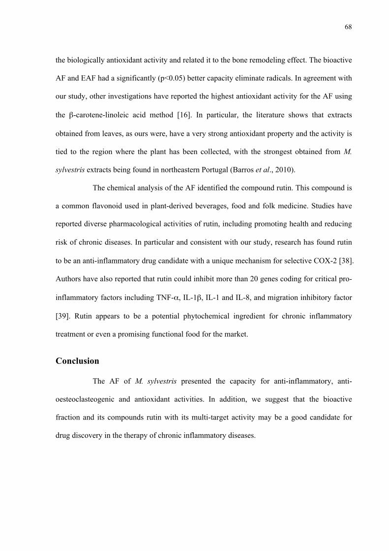
68
the biologically antioxidant activity and related it to the bone remodeling effect. The bioactive
AF and EAF had a significantly (p<0.05) better capacity eliminate radicals. In agreement with
our study, other investigations have reported the highest antioxidant activity for the AF using
the β-carotene-linoleic acid method [16]. In particular, the literature shows that extracts
obtained from leaves, as ours were, have a very strong antioxidant property and the activity is
tied to the region where the plant has been collected, with the strongest obtained from M.
sylvestris extracts being found in northeastern Portugal (Barros et al., 2010).
The chemical analysis of the AF identified the compound rutin. This compound is
a common flavonoid used in plant-derived beverages, food and folk medicine. Studies have
reported diverse pharmacological activities of rutin, including promoting health and reducing
risk of chronic diseases. In particular and consistent with our study, research has found rutin
to be an anti-inflammatory drug candidate with a unique mechanism for selective COX-2 [38].
Authors have also reported that rutin could inhibit more than 20 genes coding for critical pro-
inflammatory factors including TNF-α, IL-1β, IL-1 and IL-8, and migration inhibitory factor
[39]. Rutin appears to be a potential phytochemical ingredient for chronic inflammatory
treatment or even a promising functional food for the market.
Conclusion
The AF of M. sylvestris presented the capacity for anti-inflammatory, anti-
oesteoclasteogenic and antioxidant activities. In addition, we suggest that the bioactive
fraction and its compounds rutin with its multi-target activity may be a good candidate for
drug discovery in the therapy of chronic inflammatory diseases.

69
Acknowledgement
Research reported in this publication was supported by São Paulo Research
Foundation FAPESP (Grant #2011/23980-5). The funders had no role in study design, data
collection and analysis, decision to publish, or preparation of the manuscript.
References
1. Ward PA. The inflammatory mediators. Ann N Y Acad Sci. 1974;221: 290–8. Available: http://www.ncbi.nlm.nih.gov/pubmed/4132121
2. Crotti TN, Dharmapatni AASSK, Alias E, Haynes DR. Osteoimmunology: Major and Costimulatory Pathway Expression Associated with Chronic Inflammatory Induced Bone Loss. J Immunol Res. 2015;2015: 281287. doi:10.1155/2015/281287
3. Conner EM, Grisham MB. Inflammation, free radicals, and antioxidants. Nutrition. 1996;12: 274–7.
4. Henderson B, Poole S, Wilson M. Bacterial modulins: a novel class of virulence factors which cause host tissue pathology by inducing cytokine synthesis. Microbiol Rev. 1996;60: 316–41.
5. Ohshiba T, Miyaura C, Inada M, Ito A. Role of RANKL-induced osteoclast formation and MMP-dependent matrix degradation in bone destruction by breast cancer metastasis. Br J Cancer. 2003;88: 1318–26. doi:10.1038/sj.bjc.6600858
6. Zhang F, Tanaka H, Kawato T, Kitami S, Nakai K, Motohashi M, et al. Interleukin-17A induces cathepsin K and MMP-9 expression in osteoclasts via celecoxib-blocked prostaglandin E2 in osteoblasts. Biochimie. 2011;93: 296–305. doi:10.1016/j.biochi.2010.10.001
7. Franco GCN, Kajiya M, Nakanishi T, Ohta K, Rosalen PL, Groppo FC, et al. Inhibition of matrix metalloproteinase-9 activity by doxycycline ameliorates RANK ligand-induced osteoclast differentiation in vitro and in vivo. Exp Cell Res. 2011;317: 1454–64. doi:10.1016/j.yexcr.2011.03.014
8. Takeshita S, Kaji K, Kudo A. Identification and characterization of the new osteoclast progenitor with macrophage phenotypes being able to differentiate into mature osteoclasts. J Bone Miner Res. 2000;15: 1477–88. doi:10.1359/jbmr.2000.15.8.1477
9. Rainsford KD. Anti-inflammatory drugs in the 21st century. Subcell Biochem. 2007;42: 3–27.
10. Georgakopoulou EA, Scully C. Systemic use of non-biologic corticosteroids in orofacial diseases. Oral Dis. 2014;20: 127–35. doi:10.1111/odi.12132
11. Koeberle A, Werz O. Multi-target approach for natural products in inflammation. Drug Discov Today. 2014;19: 1871–82. doi:10.1016/j.drudis.2014.08.006
12. Kohli P, Levy BD. Resolvins and protectins: mediating solutions to inflammation. Br J Pharmacol. 2009;158: 960–71. doi:10.1111/j.1476-5381.2009.00290.x
70
13. Cragg GM, Grothaus PG, Newman DJ. New horizons for old drugs and drug leads. J Nat Prod. 2014;77: 703–23. doi:10.1021/np5000796
14. Molinari G. Natural products in drug discovery: present status and perspectives. Adv Exp Med Biol. 2009;655: 13–27. doi:10.1007/978-1-4419-1132-2_2
15. Harvey AL. Natural products in drug discovery. Drug Discov Today. 2008;13: 894–901. doi:10.1016/j.drudis.2008.07.004
16. Gasparetto JC, Martins CAF, Hayashi SS, Otuky MF, Pontarolo R. Ethnobotanical and scientific aspects of Malva sylvestris L.: a millennial herbal medicine. J Pharm Pharmacol. 2012;64: 172–89. doi:10.1111/j.2042-7158.2011.01383.
17. Benso B, Rosalen PL, Alencar SM, Murata RM. Malva sylvestris Inhibits Inflammatory Response in Oral Human Cells. An In Vitro Infection Model. Kreth J, editor. PLoS One. 2015;10: e0140331. doi:10.1371/journal.pone.0140331
18. Franchin M, da Cunha MG, Denny C, Napimoga MH, Cunha TM, Koo H, et al. Geopropolis from Melipona scutellaris decreases the mechanical inflammatory hypernociception by inhibiting the production of IL-1β and TNF-α. J Ethnopharmacol. 2012;143: 709–15. doi:10.1016/j.jep.2012.07.040
19. Winter CA, Risley EA, Nuss GW. Carrageenin-induced edema in hind paw of the rat as an assay for antiiflammatory drugs. Proc Soc Exp Biol Med. 1962;111: 544–7.
20. Mosmann T. Rapid colorimetric assay for cellular growth and survival: application to proliferation and cytotoxicity assays. J Immunol Methods. 1983;65: 55–63.
21. Alencar SM, Oldoni TLC, Castro ML, Cabral ISR, Costa-Neto CM, Cury JA, et al. Chemical composition and biological activity of a new type of Brazilian propolis: red propolis. J Ethnopharmacol. 2007;113: 278–83. doi:10.1016/j.jep.2007.06.005
22. Khansari N, Shakiba Y, Mahmoudi M. Chronic inflammation and oxidative stress as a major cause of age-related diseases and cancer. Recent Pat Inflamm Allergy Drug Discov. 2009;3: 73–80.
23. Hardy RS, Raza K, Cooper MS. Glucocorticoid metabolism in rheumatoid arthritis. Ann N Y Acad Sci. 2014;1318: 18–26. doi:10.1111/nyas.12389
24. Pandey KB, Rizvi SI. Plant polyphenols as dietary antioxidants in human health and disease. Oxid Med Cell Longev. 2: 270–8. doi:10.4161/oxim.2.5.9498
25. Franchin M, da Cunha MG, Denny C, Napimoga MH, Cunha TM, Bueno-Silva B, et al. Bioactive Fraction of Geopropolis from Melipona scutellaris Decreases Neutrophils Migration in the Inflammatory Process: Involvement of Nitric Oxide Pathway. Evid Based Complement Alternat Med. 2013;2013: 907041. doi:10.1155/2013/907041
26. Dal Secco D, Moreira AP, Freitas A, Silva JS, Rossi MA, Ferreira SH, et al. Nitric oxide inhibits neutrophil migration by a mechanism dependent on ICAM-1: role of soluble guanylate cyclase. Nitric Oxide. 2006;15: 77–86. doi:10.1016/j.niox.2006.02.004
27. Yan B, Han P, Pan L, Lu W, Xiong J, Zhang M, et al. IL-1β and reactive oxygen species differentially regulate neutrophil directional migration and Basal random motility in a zebrafish injury-induced inflammation model. J Immunol. 2014;192: 5998–6008. doi:10.4049/jimmunol.1301645

71
28. Levy L. Carrageenan paw edema in the mouse. Life Sci. 1969;8: 601–606. doi:10.1016/0024-3205(69)90021-6
29. Posadas I, Bucci M, Roviezzo F, Rossi A, Parente L, Sautebin L, et al. Carrageenan-induced mouse paw oedema is biphasic, age-weight dependent and displays differential nitric oxide cyclooxygenase-2 expression. Br J Pharmacol. 2004;142: 331–8. doi:10.1038/sj.bjp.0705650
30. Morris CJ. Carrageenan-induced paw edema in the rat and mouse. Methods Mol Biol. 2003;225: 115–21. doi:10.1385/1-59259-374-7:115
31. Cataldo DD, Gueders MM, Rocks N, Sounni NE, Evrard B, Bartsch P, et al. Pathogenic role of matrix metalloproteases and their inhibitors in asthma and chronic obstructive pulmonary disease and therapeutic relevance of matrix metalloproteases inhibitors. Cell Mol Biol (Noisy-le-grand). 2003;49: 875–84.
32. Al Mamun MA, Islam K, Alam MJ, Khatun A, Alam MM, Al-Bari MAA. Flavonoids isolated from Tridax procumbens (TPF) inhibit osteoclasts differentiation and bone resorption. Biol Res. 2015;48: 51. doi:10.1186/s40659-015-0043-6
33. Syggelos SA, Aletras AJ, Smirlaki I, Skandalis SS. Extracellular matrix degradation and tissue remodeling in periprosthetic loosening and osteolysis: focus on matrix metalloproteinases, their endogenous tissue inhibitors, and the proteasome. Biomed Res Int. 2013;2013: 230805. doi:10.1155/2013/230805
34. Kim H-S, Suh KS, Sul D, Kim B-J, Lee SK, Jung W-W. The inhibitory effect and the molecular mechanism of glabridin on RANKL-induced osteoclastogenesis in RAW264.7 cells. Int J Mol Med. 2012;29: 169–77. doi:10.3892/ijmm.2011.822
35. Wu C, Wang W, Tian B, Liu X, Qu X, Zhai Z, et al. Myricetin prevents titanium particle-induced osteolysis in vivo and inhibits RANKL-induced osteoclastogenesis in vitro. Biochem Pharmacol. 2015;93: 59–71. doi:10.1016/j.bcp.2014.10.019
36. Shim K-S, Ma CJ, Kim D-S, Ma JY. Yukmijihwang-tang inhibits receptor activator for nuclear Factor-κB ligand-induced osteoclast differentiation. J Med Food. 2011;14: 1439–47. doi:10.1089/jmf.2010.1502
37. Bocci V, Valacchi G. Free radicals and antioxidants: how to reestablish redox homeostasis in chronic diseases? Curr Med Chem. 2013;20: 3397–415.
38. Choi K-S, Kundu JK, Chun K-S, Na H-K, Surh Y-J. Rutin inhibits UVB radiation-induced expression of COX-2 and iNOS in hairless mouse skin: p38 MAP kinase and JNK as potential targets. Arch Biochem Biophys. 2014;559: 38–45. doi:10.1016/j.abb.2014.05.016
39. Kauss T, Moynet D, Rambert J, Al-Kharrat A, Brajot S, Thiolat D, et al. Rutoside decreases human macrophage-derived inflammatory mediators and improves clinical signs in adjuvant-induced arthritis. Arthritis Res Ther. 2008;10: R19. doi:10.1186/ar2372

72
2.3 ARTIGO3
Evaluation of Malva sylvestris as inhibitor of HIV-1 BaL in a dual chamber in vitro
model
Benso Ba; Rosalen PLa; Pasetto Sb, Marquezin MCSa; Alencar SMc; Murata RMb*
aDepartment of Physiological Sciences, Piracicaba Dental School, State University of
Campinas, Piracicaba, SP, Brazil.
bDivision of Periodontology, Diagnostic Sciences & Dental Hygiene and Division of
Biomedical Sciences Herman Ostrow School of Dentistry, University of Southern California,
Los Angeles, USA. Electronic address: [email protected]
cDepartment of Agri-food Industry, Food and Nutrition, “Luiz de Queiroz” College of
Agriculture, University of Sao Paulo, Piracicaba, Sao Paulo, Brazil
*Corresponding Author
Ramiro Mendonca Murata
Division of Periodontology, Diagnostic Sciences, Dental Hygiene and Biomedical Science,
Ostrow School of Dentistry, University of Southern California , Los Angeles, CA , EUA,
email: [email protected]
3Benso B, Rosalen PL, Pasetto S, Alencar SM, Marquezin MCSa, Murata RM, será submetido para publicação ao periódico Journal of
Ethnopharmacology.

73
Abstract
Ethnopharmacological relevance- Emphasis is now being placed on identifying novel
molecular agents, including microbicides that can be applied topically and protect against
sexually transmitted infections, especially HIV. Natural products are a good source of drug
discovery.
Aim of the study- to investigate the potential anti-HIV activity of aqueous fraction of
Malva sylvestris on cells infected by HIV-1BaL using a dual-chamber model.
Material and Methods- Extract and fractions were screened for anti-HIV activity
measuring the antibody-mediated neutralization of HIV-1. The in vitro cytotoxicity of the
aqueous fraction (AF) was assessed using a fluorescent assay for the TZM-bl, PBMC and
HeLa cell lines. Antiviral activity was supported by p24 quantification in the supernatant of a
dual-chamber model, cytokine release, and transcription of specific targets CD4, TRIM5 and
Bcl-2 and tested against HIV-RT.
Results- The AF demonstrated potential anti-HIV activity on the TZM-bl cell line, showing
a reduction score higher than 60% infectivity. Results showed no cytotoxicity effect for AF in
all cell lines studied (p> 0.05). Quantification of p24 in the supernatant of the dual-chamber
model demonstrated a reduction in viral particles after AF treatment (p< 0.05). Cytokines
were quantified and AF reduced the expression of all signaling related to the inflammatory
process, such as IL1-alpha, IL-beta, IL-6, IL-8 and GM-CSF (p<0.05) compared to the
control group. In particular, IL-6 had low levels of expression when compared to the AZT
positive control group. The molecular pathways used by AF to regulate the HIV-1BaL
infection showed a controlled transcription of the genes CD4, Bcl-2 and TRIM5 and HIV-
RT.

74
Conclusions- M. sylvestris contains highly potential anti-HIV components, non toxic effects
and anti-inflammatory properties, thereby characteristics to be considered as a potential
microbicide.
1. Malva sylvestris action in a HIV infectivity model in vitro. Introduction
Human immunodeficiency virus (HIV) is responsible for the acquired
immunodeficiency syndrome (AIDS). The first cases of AIDS were reported in 1981 and
today, more than 34 years later, it is one of the world’s most serious health problems.
According to estimates by the World Organization, approximately 78 million people have
been infected with HIV worldwide and about 39 million people have died of HIV. Sub-
Saharam Africa remains most severely affected, with 1 in every 20 adults living with the virus
(WHO, 2010). The epidemic continues to expand in countries and regions where HIV
treatment is insufficient coverage (Maartens et al., 2014).
The advent of antiretroviral therapy modified the epidemiology of HIV leading to
decreased mortality and risk of transmission (Simon et al., 2006). Currently, therapy consists
of a combination of three classes of drugs that transformed the infection disease into a chronic
event: nucleoside reverse transcriptase inhibitors, non-nucleoside reverse transcriptase

75
inhibitors and protease inhibitors (Pomerantz and Horn, 2003). However, a significant number
of therapies are still not curative and financial obstacles may limit access by some populations
to prevention techniques and treatment (Cutler and Justman, 2008). The focus has been placed
on discovery and development of novel agents, including microbicides that can be applied
topically and protect against sexually transmitted infections (STIs), especially HIV(Pirrone et
al., 2012). The future distribution of microbicides may prove to have an important social
impact, reducing health care costs and risk of HIV infection (D’Cruz and Uckun, 2004).
Natural products are the most productive source of drug discovery and the
bioactivity-guided fractionation of crude extracts may generate new molecules with biological
activity, especially with anti-HIV activity (Cragg et al., 2014). Many natural compounds have
a specific biological activity and low toxicity effects working on a particular target that may
act to complement the traditional antiviral drugs (Yang et al., 2001). Studies have shown that
a natural product native to Europe, North Africa and Asia named Malva sylvestris has the
potential to treat inflammation, and has antimicrobial properties and antioxidant, activity
(Gasparetto et al., 2012). Its use in folk medicine is widespread and given its medical
importance it is a good candidate for drug discovery. Therefore, the aim of this study was to
investigate the potential anti-HIV activity of the aqueous fraction of M. sylvestris on cells
infected by HIV-1BaL using a dual-chamber model.
Material and Methods
Preparation of the extract and fractions
M. sylvestris leaves were registered in the herbarium of the University of Sao
Paulo (USP, Piracicaba, Sao Paulo, Brazil) under an identification number (ESA #1214403).
All material was collected in northeast Brazil (07º44'12” S and 37º59'36" W) and the extract

76
was prepared with absolute ethanol using constant shaking (100 rpm, 1 week, in the dark at
room temperature) and exhaustive maceration technique. The ethanolic extract of M.
sylvestris (MSE) was homogenized, lyophilized, weighed for the fraction using liquid-liquid
partitioning by chemical polarity gradient with hexane, chloroform and ethyl acetate solvents.
The final residue obtained was totally soluble in water and thus was called the aqueous
fraction (AF) (Benso et al., 2015b).
Cell lines
The cell lines included in this study were: TZM-bl obtained through the NIH
AIDS Reagent Program, Division of AIDS, NIAID, NIH and peripheral blood mononuclear
cells (PBMC) (Astarte Biologics, LLC, Redmond, USA, Catalog number 1001, Lot number
2536DE13) obtained from a healthy donor and HeLa from ATCC (CCL-2).
Virus strain
HIV-1BaL was originally explanted from a primary culture of cells from infant
explanted lung and obtained through NIH (AIDS Reagent Program, Division of AIDS,
NIAID, NIH). All virus samples were stored at -80oC and serial dilutions were conducted
(1:10). After 3 days, the multiplicity infection of HIV-1BaL was verified using the enzyme-
linked immunosorbent assay (ELISA) assay to establish the tissue culture infective dose
(TCID)(Pasetto et al., 2014). The multiplicity of infection (MOI) was obtained and corrected
to MOI=1, for all assays. The positive control treatment was Zidovudine (AZT) (Sigma
Aldrich, St Louis, USA). For all assays the vehicle was (1% DMSO, v/v; Sigma Aldrich, St
Louis, USA).

77
Anti-HIV activity to evaluate microbicide candidate
Cytotoxicity activity
Cell viability assay was conducted using the CellTiter-Blue® reagent (Promega,
Corp, Madison, WI, USA) that provides an estimate of viable cells through a dye indicating
the metabolic capacity. Cells were cultured (200 μL, 1x10⁵ cells/mL) in a 96-well plate and
incubated for 24 h at 37oC with 5% CO2. The AF at the biological activity concentrations (25-
50 μg/mL) were added to cell culture and incubated for 24 h. Subsequently, the culture
medium was discarded and the cells washed with PBS (Lonza, Walkersville, MD, USA) and a
fresh new medium with 20 μL of CellTiter-Blue was added and incubated for 4 h (Pasetto et
al., 2014). The contents were transferred to a new microplate and the fluorescence reading
conducted in a microplate reader (SpectraMax M5, Molecular Devices®) with 550 nm
excitation, 585 emission.
Anti-HIV activity in infected TZM-bl cells
The in vitro activity of AF was conducted on the TZM-bl cell line. The assay was
previously described by (Ochsenbauer-Jambor et al., 2006). These cells are genetically
engineered from a HeLa cell that expresses CD4, CXCR4, and CCR5 and contains reporter
genes for luciferase (Luc) upon infection and therefore enables the measurement of relative
HIV-1 infectivity by quantifying luminescence units. Cells were cultured in a 96-well plate
(1x10⁵ cells/well) and infected with HIV-1BaL (MOI=1) for 48 h. The cytotoxicity activity
was measured by fluorescence as previously described. The cells were washed with PBS and
treated with lysis buffer. In the following step 20 μL/well luciferase reagent was added. The
luminescence reading was conducted in a microplate reader (SpectraMax M5 Molecular
Devices) with 500 ms of integration time.
78
Dual-chamber model
Quantification of HIV-1 p24 antigen by ELISA
Aiming to mimic the epithelium of the female genital tract and to verify the
potential of AF as a microbicide, the dual-chamber model was developed(Gali et al., 2010;
Pasetto et al., 2014). Transwell assay inserts with 8 μM diameter pores and 0.3 cm2 of culture
surface (Greiner Bio-One®) were positioned in the wells of a 24-well plate. The first layer,
(1×10⁵ cells/mL) HeLa cells were culture into each transwell insert/apical chamber. PBMC
cells (1×10⁵ cells/mL) were seeded into the basal chambers below the inserts, and the plates
were incubated at 37°C, 5% CO2. To assess confluence, the transepithelial electrical
resistance (TEER) of each HeLa cell layer was measured with a Millicell-ERS Volt-Ohm
Meter (Millipore, Bedford, MA,) before and after AF treatments (Gali et al., 2010). Cell layer
confluence in the transwell inserts was measured daily until the optimal TEER (>150
Ohm/cm2) was reached on day 4. The plate was incubated at 37°C, in humid air containing
5% CO2. After 24 h (day 5), the cell viability and luciferase assays were performed to analyze
the toxicity and anti-HIV activity of the AF.
Transcription analysis of genes involved in the role of HIV-1 disease
RNA was isolated from the cell culture (day 5) of PBMC cells using the RNeasy Mini
Kit (Qiagen, Valencia, CA). RNA quantification was measured in the NanoPhotometer P360
(Implen®, Westlake Village, CA, USA). Reverse transcription of RNA to cDNA was
performed using the QuantiTect Reverse Transcription Kit (Qiagen, Valencia, CA) according
to the manufacturer’s instructions. Three primers CD4, Bcl-2 and TRIM5 were selected to
comprehend generally the mechanism of action of the AF in the infection caused by HIV-
1BaL. The threshold was manually adjusted within the logarithmic curve above the

79
background level and below the plateau phase. A comparative Ct method was used to
calculate the relative gene number. The relative gene copy number was calculated using the
2∆∆CT method.
Quantification of cytokines
Cytokines were measured for all samples using an ELISA assay with a specific kit
(Qiagen, Valencia, CA). The cytokines were measured by standard ELISA protocol using a
panel of 6 inflammatory cytokines (IL-1alpha, IL-1beta, IL-6, IL-8, IL-10, GM-CSF).
Anti- HIV reverse transcriptase activity
Reverse transcriptase assay (RT) is an in vitro colorimetric enzyme immunoassay for
screening anti-viral agents. The (RT) is a crucial enzyme for retrovirus replication, and its
presence in the virion is indispensable for infectiity. In a microcentrifuge tube, 20 µL of
recombinant HIV-1-RT (Roche Diagnostics, Indianapolis, IN, USA) was diluted in lysis
buffer and 20 μL of the appropriate AF dilutions or vehicle control was added and incubated
at 37°C. After 1 hour, 60 μL of each of sample was added into a microplate (Roche
Diagnostics, Indianapolis, IN) precoated with streptavidin and incubated at 37°C for 1 hour.
The wells were washed with washing buffer and 200 µL of anti-DIG-POD (antibody to
digoxigenin conjugated to peroxidase) working solution was added to each well and then
incubated at 37°C to bind to the digoxigenin-labeled DNA. After 1 hour, the wells were
washed and 200 µL of peroxidase substrate ABTS solution was added to each well and was
incubated at room temperature for 30 min. The resulting signal intensity is directly
proportional to the RT activity and was determined by the absorbance using a microplate
reader at 405 nm.
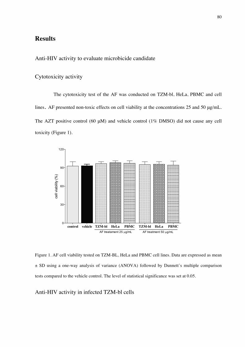
80
Results
Anti-HIV activity to evaluate microbicide candidate
Cytotoxicity activity
The cytotoxicity test of the AF was conducted on TZM-bl, HeLa, PBMC and cell
lines. AF presented non-toxic effects on cell viability at the concentrations 25 and 50 μg/mL.
The AZT positive control (60 μM) and vehicle control (1% DMSO) did not cause any cell
toxicity (Figure 1).
Figure 1. AF cell viability tested on TZM-BL, HeLa and PBMC cell lines. Data are expressed as mean
± SD using a one-way analysis of variance (ANOVA) followed by Dunnett’s multiple comparison
tests compared to the vehicle control. The level of statistical significance was set at 0.05.
Anti-HIV activity in infected TZM-bl cells
control vehicle TZM-bl HeLa PBMC TZM-bl HeLa PBMC0
30
60
90
120
cell
viab
ility
(%)
AF treatament 25 µg/mL AF treatment 50 µg/mL

81
AF decreased significantly the HIV-1 infectivity on TZM-bl cells (p<0.05),
blocking 67% of HIV-1BaL infection at the 25 μg/mL concentration. The inhibition effect of
between the 25 and 50 μg/mL concentrations were not statistically different (p>0.05). The
positive control (AZT 60 μM) inhibited 80% the HIV-1BaL infection, and the vehicle control
(1% DMSO) did not affect the infectivity of the HIV-1BaL (Figure 2). The cell viability was
assessed and non-toxic effects were found in the test groups (p>0.05).
Figure 2. Anti-HIV-1 activity of AF on TZM-bl cell line infected by HIV-1BaL. Data are expressed as
mean ± SD using a one-way ANOVA followed by Tukey’s multiple comparison tests. Symbols
indicate statistical difference (p<0.05) * compared to the vehicle group; ** comparison between the
concentrations tested 25 and 50 µg/kg.
Dual-chamber model
Quantification of HIV-1 p24 antigen by ELISA
The results of p24 quantification by ELISA after PBMC cells were treated with
the AF showed a statistically significant reduction in the infection (p<0.05). The AZT positive
control and AF 50 µg/mL are not statistically different (p>0.05) (Figure 3).
Vehicle TZM BaL AZT 25 500
20
40
60
80
100
(%) H
IV1
BaL
inhi
bitio
n
*
*
*
*
AF treatment µg/mL
**

82
Figure 3. AF-mediated inhibition of HIV-1BaL infection in PBMC cells. Raw data is showed. Data
are expressed as mean ± SD using a one-way ANOVA followed by Tukey’s multiple comparison tests.
Symbols indicate statistical difference (p<0.05) * compared to the vehicle group; ** comparison
between the AF treatment and the AZT group.
Transcription analysis of genes involved in the role of HIV-1 disease
The AF showed a significant downregulation expression (p<0.05) of CD4,
TRIM5 and Bcl-2 when compared to the control group (vehicle) (Figure 4).
Vehicle PBMC BaL AZT AF 25 AF 500.0
0.5
1.0
1.5
2.0
Sign
al in
tens
ity
AF treatment µg/mL
* *
*
*
**
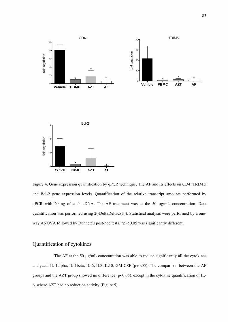
83
Figure 4. Gene expression quantification by qPCR technique. The AF and its effects on CD4, TRIM 5
and Bcl-2 gene expression levels. Quantification of the relative transcript amounts performed by
qPCR with 20 ng of each cDNA. The AF treatment was at the 50 μg/mL concentration. Data
quantification was performed using 2(-DeltaDeltaC(T)). Statistical analysis were performed by a one-
way ANOVA followed by Dunnett’s post-hoc tests. *p < 0.05 was significantly different.
Quantification of cytokines
The AF at the 50 μg/mL concentration was able to reduce significantly all the cytokines
analyzed: IL-1alpha, IL-1beta, IL-6, IL8, IL10, GM-CSF (p<0.05). The comparison between the AF
groups and the AZT group showed no difference (p<0.05), except in the cytokine quantification of IL-
6, where AZT had no reduction activity (Figure 5).
Vehicle PBMC AZT AF0
2
4
6
8
10fo
ld re
gulat
ion
*
** *
CD4
Vehicle PBMC AZT AF0
5
10
15
fold
regu
latio
n
** *
Bcl-2
Vehicle PBMC AZT AF0
10
20
30
40
* **
fold
regu
latio
n
*
TRIM5

84
Figure 5. Cytokine quantification in the supernatant released from the cells in the dual-chamber
model. The AF treatment was at the 50 μg/mL concentration. Data are expressed as mean±SD.
Symbols indicate statistical differences (p<0.05, ANOVA, Dunnett’s test); * indicates p<0.05
compared to the vehicle group.
Vehicle PBMC AZT AF0
500
1000
1500
2000
2500
* *
pg/mL
IL-1α
*
Vehicle PBMC AZT AF0
500
1000
1500
2000
2500
**
pg/mL
IL-6
*
Vehicle PBMC AZT AF0
500
1000
1500
2000
2500
pg/mL
***
IL-10
Vehicle PBMC AZT AF0
500
1000
1500
2000
2500
** *
pg/mL
IL-1β
*
Vehicle PBMC AZT AF0
500
1000
1500
2000
2500
**
pg/mL
IL-8
*
Vehicle PBMC AZT AF0
500
1000
1500
2000
2500
pg/mL
* ***
GM-CSF

85
Anti- HIV reverse transcriptase activity
Quantification of the inhibitory effect on HIV reverse transcriptase was done for AF
(25-50 μg/mL) and the results are shown in Fig. 6. The inhibitory activity of AF against HIV-
1 reverse transcriptase decreased signicantly compared to the vehicle control group.
Figure 6. Anti HIV-1–RT effect of AF. The data are expressed as mean ± SD, n = 6. Symbols indicate
statistical difference (p < 0.05, ANOVA, Tukey post test) ** compared to the reverse transcriptase
group (RT 4 ng); * comparison between the treatments AF (50) and (AZT 120).
Discussion
Natural products and derivates have been considered potential candidates for drug
discovery (Cragg et al., 2014) and in particular new insights into the treatment of HIV-1
(Pasetto et al., 2014). Plant extracts with strong activity acting in the nanomolar/picomolar
range can be used to enhance the activity of synthetic compounds and work as anti-HIV
agents (Singh and Bodiwala, 2010).
The development of potent and safe topical formulations of anti-HIV and against
HIV-associated pathogens, referred to as microbicides, has become a major priority in HIV
RT Vehicle AZT 120 AF 25 AF 500
40
80
120
% ex
pres
sion
**
****
*
(µg/mL)(µM)

86
research (NIH, 2012). According to WHO, the microbicibe must have low citotoxicity, anti-
viral activity and not induce inflammatory response of the epithelial monolayer (Mauck et al.,
2001).
Following the guidelines, our first evaluation of the cytotoxicity of M. sylvestris
extracts. The AF, showed no cytotoxicity in different cell lines (TZM, HeLa and PBMCs).
Other authors supported our findings, showing that M. sylvestris extract from Iran had no
lethal dose possible to estimate in several concentrations tested. The anti-HIV activity of AF
and the relative potency was based on effective dose (IC50). In a dose dependent manner, AF
(IC50=18 μg/mL) displayed anti HIV effect against HIV-1 strain BaL and maintained the
number of viable cells. Some naturally occurring products demonstrated to be a important
source of anti-HIV activity in this same in vitro model. The extract prepared from the stem of
Acacia catechu inhibited HIV infection in low concentrations and the IC50 values were found
in concentrations lower than 2 μg/mL (Nutan et al., 2013). The compound Myrcetin revealed
high therapeutic index (137.4) acting with a non cytotoxicity effects, indicating a potential
and safe biological activity (Pasetto, et al, 2014).
The AF potential microbicide activity was assessed in a dual chamber model using
epithelial and blood cells lines. The purpose of the model is to measure anti-HIV activity, cell
viability, and inflammatory response of the epithelial monolayer; this was of particular
importance for microbicides that tended to increase rather than decrease HIV transmission
rates (Smith-McCune et al., 2015). The AF acts against HIV, the amount of p24 protein
expressed in the cell culture supernatant decreased significantly by AF treatment.
Additionally, the putative pathway by which AF affect HIV-1 infection involve the
modulation cell chemokine receptor interaction (CD4), modulates the apoptosis process
affecting virus survival (BCL-2) and affect the human retrovirus restriction factor (TRIM 5)
(Pierson et al., 2000).

87
The cytotoxicity profile, and their potential to induce an inflammatory response were
assessed. The AF did not induce the inflammatory response and reduced the expression of all
cytokines studied - IL1-alpha, IL-beta, IL-6, IL-8 and GM-CSF - compared to the vehicle
control group. In particular, IL-6 had low expression levels compared to the negative control
group. In addition, cytokines modulate the monocyte function as well as HIV replication with
these cells and they are considered to be major reservoirs of HIV-1, playing an important role
in the pathogenesis of AIDS (Badley et al., 2013).
To elucidate the mechanism of the anti-HIV effects of the AF, the compound's
inhibition of HIV-1 reverse transcriptase enzymatic activity was tested. The HIV-1 RT is
unique to the virus, and is the enzyme that controls HIV-1 replication infected cells (Tao et
al., 2007). We found that AF inhibited HIV-1-RT activity. Phytochemical investigations AF
of M. sylvestris resulted in the identification of potentially bioactive flavonoid rutin (Benso et
al., 2015a). It is important to note that flavonoids, beyond action against reverse transcriptase,
also modulate several steps of HIV-1 life cycle, including entry, integration and maturation
phases (Li et al., 2000).
In 2007, (Tao et al., 2007) found no anti-HIV activity for rutin in the TZM-bl HIV-
1BaL infection model; however, for the modified structure sodium rutin sulfate, a polyanionic
compound, a significant virus inhibition IC50 8.5 μM (5.19 μg/mL) was demonstrated. The
authors correlated the activity with the presence of sulfated polysaccharides in the structure,
such as dextran sulfate, that may preferably bind to the V3 loop of X4 gp120 rather than that
of R5 gp120.
Reports have shown a variety of pharmacological activities for the flavonoid group
and the association between chemical structure and biological activity (Asada et al., 2013;
Soto-Cabrera et al., 2015). Moreover, the biochemical effects are caused by the ability to
inhibit a number of enzymes, such as reductase, lipoxygenase, cyclooxygenase and different

88
hormones (Rathee et al., 2009). Quercetin, rutin and catechin are flavonoids that possess
antiviral action, which may be related to the non-glycosidic compounds and hydroxylation at
the 3-position permitting the inhibition activity. Overall, they are proven to have biological
activity and they have been important components in traditional medicine for many years
(Gerdin and Svensjö, 1983).
The discovery of therapeutic agents acting as microbicide is promising in preventing
interventions in AIDS research. AF has shown three important requirements for a compound
to become a microbicide, as according to WHO regulatory guidelines for microbicide
development (Mauck et al., 2001). The first requirement is low toxicity. AF showed low
cytotoxicity on eukaryotic cell, indicating that it is safe. The second requirement is the
inhibitory activity on HIV infection. AF demonstrated anti-HIV-1 activity in the dual-
chamber model. The third requirement is low potential to induce inflammatory response. The
AF did not induce proinflammatory cytokines production on HeLa cell monolayer.
Therefore, this study, having determined the cytotoxicity and anti-HIV activity of AF
against HIV-1. Nevertheless, further studies with AF of M. sylvestris are still needed with
different HIV-1 strains in order to identify the molecular targets.
Conclusion
M. sylvestris contains highly potential anti-HIV components, non toxic effects and
anti-inflammatory properties, thereby characteristics to be considered as a potential
microbicide.
Acknowledgement
Research reported in this publication was supported by: National Center for
Complementary and Integrative Health of the National Institutes of Health under award

89
number R00AT006507, São Paulo Research Foundation FAPESP (Grant #2011/23980-5) and
Brazilian Federal Agency for the Support and Evaluation of Graduate Education CAPES
(Grant #2317/2014-01). The funders had no role in study design, data collection and analysis,
decision to publish, or preparation of the manuscript.
References
Asada, Y., Sukemori, A., Watanabe, T., Malla, K.J., Yoshikawa, T., Li, W., Kuang, X., Koike, K., Chen, C.-H., Akiyama, T., Qian, K., Nakagawa-Goto, K., Morris-Natschke, S.L., Lu, Y., Lee, K.-H., 2013. Isolation, structure determination, and anti-HIV evaluation of tigliane-type diterpenes and biflavonoid from Stellera chamaejasme. J Nat Prod 76, 852–7. doi:10.1021/np300815t
Badley, A.D., Sainski, A., Wightman, F., Lewin, S.R., 2013. Altering cell death pathways as an approach to cure HIV infection. Cell Death Dis 4, e718. doi:10.1038/cddis.2013.248
Benso, B., Franchin, M., Massarioli, A., Franco, Gilson César Nobre Alencar, Severino Matias Rosalen, P.L., 2015a. Avaliação da atividade farmacológica e identificação de composto ativo de Malva silvestres. Braz Oral Res. URL http://www.sbpqo.org.br/hotsite/anais/BOR-v029-Suppl-Book_FINAL.pdf.
Benso, B., Rosalen, P.L., Alencar, S.M., Murata, R.M., 2015b. Malva sylvestris Inhibits Inflammatory Response in Oral Human Cells. An In Vitro Infection Model. PLoS One 10, e0140331. doi:10.1371/journal.pone.0140331
Cragg, G.M., Grothaus, P.G., Newman, D.J., 2014. New horizons for old drugs and drug leads. J Nat Prod 77, 703–23. doi:10.1021/np5000796
Cutler, B., Justman, J., 2008. Vaginal microbicides and the prevention of HIV transmission. Lancet Infect Dis 8, 685–97. doi:10.1016/S1473-3099(08)70254-8
D’Cruz, O.J., Uckun, F.M., 2004. Clinical development of microbicides for the prevention of HIV infection. Curr Pharm Des 10, 315–36.
Gali, Y., Ariën, K.K., Praet, M., Van den Bergh, R., Temmerman, M., Delezay, O., Vanham, G., 2010. Development of an in vitro dual-chamber model of the female genital tract as a screening tool for epithelial toxicity. J Virol Methods 165, 186–97. doi:10.1016/j.jviromet.2010.01.018
Gasparetto, J.C., Martins, C.A.F., Hayashi, S.S., Otuky, M.F., Pontarolo, R., 2012. Ethnobotanical and scientific aspects of Malva sylvestris L.: a millennial herbal medicine. J Pharm Pharmacol 64, 172–89. doi:10.1111/j.2042-7158.2011.01383.x
Gerdin, B., Svensjö, E., 1983. Inhibitory effect of the flavonoid O-(beta-hydroxyethyl)-

90
rutoside on increased microvascular permeability induced by various agents in rat skin. Int J Microcirc Clin Exp 2, 39–46.
Li BQ, Fu T, Dongyan Y, Mikovits JA, Ruscetti FW, Wang JM. Flavonoid baicalin inhibits HIV-1 infection at the level of viral entry. Biochem Biophys Res Commun.2000 Sep 24;276(2):534-8.
Maartens, G., Celum, C., Lewin, S.R., 2014. HIV infection: epidemiology, pathogenesis, treatment, and prevention. Lancet 384, 258–71. doi:10.1016/S0140-6736(14)60164-1
Mauck, C., Rosenberg, Z., Van Damme, L., 2001. Recommendations for the clinical development of topical microbicides: an update. AIDS 15, 857–68.
Ochsenbauer-Jambor, C., Jones, J., Heil, M., Zammit, K.P., Kutsch, O., 2006. T-cell line for HIV drug screening using EGFP as a quantitative marker of HIV-1 replication. Biotechniques 40, 91–100.
Pasetto, S., Pardi, V., Murata, R.M., 2014. Anti-HIV-1 activity of flavonoid myricetin on HIV-1 infection in a dual-chamber in vitro model. PLoS One 9, e115323. doi:10.1371/journal.pone.0115323
Pierson, T., McArthur, J., Siliciano, R.F., 2000. Reservoirs for HIV-1: mechanisms for viral persistence in the presence of antiviral immune responses and antiretroviral therapy. Annu Rev Immunol 18, 665–708. doi:10.1146/annurev.immunol.18.1.665
Pirrone, V., Passic, S., Wigdahl, B., Krebs, F.C., 2012. Application and removal of polyanionic microbicide compounds enhances subsequent infection by HIV-1. Virol J 9, 33. doi:10.1186/1743-422X-9-33
Pomerantz, R.J., Horn, D.L., 2003. Twenty years of therapy for HIV-1 infection. Nat Med 9, 867–73. doi:10.1038/nm0703-867
Rathee, P., Chaudhary, H., Rathee, S., Rathee, D., Kumar, V., Kohli, K., 2009. Mechanism of action of flavonoids as anti-inflammatory agents: a review. Inflamm Allergy Drug Targets 8, 229–35.
Simon, V., Ho, D.D., Abdool Karim, Q., 2006. HIV/AIDS epidemiology, pathogenesis, prevention, and treatment. Lancet 368, 489–504. doi:10.1016/S0140-6736(06)69157-5
Singh, I.P., Bodiwala, H.S., 2010. Recent advances in anti-HIV natural products. Nat Prod Rep 27, 1781–800. doi:10.1039/c0np00025f
Smith-McCune, K., Chen, J.C., Greenblatt, R.M., Shanmugasundaram, U., Shacklett, B.L., Hilton, J.F., Johnson, B., Irwin, J.C., Giudice, L.C., 2015. Unexpected Inflammatory Effects of Intravaginal Gels (Universal Placebo Gel and Nonoxynol-9) on the Upper Female Reproductive Tract: A Randomized Crossover Study. PLoS One 10, e0129769. doi:10.1371/journal.pone.0129769
Soto-Cabrera, D., Salazar, J.R., Nogueda-Gutiérrez, I., Torres-Olvera, M., Cerón-Nava, A.,

91
Rosales-Guevara, J., Terrazas, T., Rosas-Acevedo, H., 2015. Quantification of polyphenols and flavonoid content and evaluation of anti-inflammatory and antimicrobial activities of Stenocereus stellatus extracts. Nat Prod Res 1–5. doi:10.1080/14786419.2015.1084302
Tao, J., Hu, Q., Yang, J., Li, R., Li, X., Lu, C., Chen, C., Wang, L., Shattock, R., Ben, K., 2007. In vitro anti-HIV and -HSV activity and safety of sodium rutin sulfate as a microbicide candidate. Antiviral Res 75, 227–33. doi:10.1016/j.antiviral.2007.03.008
Yang, S.S., Cragg, G.M., Newman, D.J., Bader, J.P., 2001. Natural product-based anti-HIV drug discovery and development facilitated by the NCI developmental therapeutics program. J Nat Prod 64, 265–77.

92
3 DISCUSSÃO
Para facilitar a análise do estudo apresentado na forma de três artigos, esta
discussão reunirá os resultados de acordo como os objetidos específicos da tese de forma a
tornar mais claro a compreensão dos achados para cada proposta, a saber: (1) As atividades
antimicrobiana e anti-inflamatória do extrato de M. sylvestris (MSE) e frações em células
infectadas por Aggregatibacter actinomycetemcomitans; (2) A atividade do MSE e frações
quanto a capacidade anti-inflamatória anti-osteoclástica, antioxidante, e finalmente,
identificar quimicamente o composto ativo; e (3) A ação anti-HIV da fração aquosa de M.
sylvestris em células infectadas por HIV-BaL.
(1) As atividades antimicrobiana e anti-inflamatória do extrato de M. sylvestris
(MSE) e frações em células infectadas por Aggregatibacter actinomycetemcomitans;
A Malva sylvestris é uma fonte promissora na descoberta de moléculas bioativas
com potencial aplicação farmacológica em diversas áreas (Gasparetto et al., 2012). Os dados
apresentados neste trabalho sugerem que a M. sylvestris e suas frações bioativas possuem
constituintes de interesse para o desenvolvimento de novas formulações com atividade
antimicrobiana e antiinflamatória de interesse em Odontologia e em Medicina.
Os agentes antimicrobianos podem contribuir na prevenção e controle de doenças
orais, tais como: cárie e doença periodontal (Wu, 2009). Os resultados do extrato e da CLF de
M. sylvestris demonstraram atividade inibitória para os microrganismos F. nucleatum, P.
gingivalis e P. intermedia, que estão diretamente relacionados ao mecanismo de virulência na
doença periodontal (Pihlstrom et al., 2005). A atividade antimicrobiana pareceu não ser
exclusiva a microrganismos periodontapatogênicos, pois a literatura mostra resultados
inibitórios para o extrato etanólico de M. sylvestris em cepas de Staphylococcus aureus
resistentes a meticilina e ainda em cepas de Helicobacter pylori, no entanto, outros resultados
demonstram atividade para fração de maior polaridade, a aquosa, em diferentes cepas
fúngicas exceto Candida albicans (Cogo et al., 2010; Quave et al., 2008). Embora a
especificidade do mecanismo de ação do MSE e frações não estar elucidado, é conhecido que
o grupo de compostos fenólicos, presentes em grande quantidade no extrato e frações de M.
sylvestris tem a habilidade de produzir metabólitos quelantes de ferro. Estes quelantes
formam complexos estáveis com íons metálicos e consequentemente reduz o número de

93
espécies bacterianas, o que poderia explicar, em parte, a ação inibitória dos compostos
estudados (Xia et al., 2010).
O tratamento com o MSE e as frações CLF e AF afetou diretamente na resposta
do hospedeiro por meio de diferentes mediadores inflamatórios. AF teve a capacidade
controlar a expressão de citocinas IL-1alfa e TNF-alfa que são moduladores da resposta do
hospedeiro frente a um desafio infeccioso (Graves et al., 2011). A capacidade de reduzir a
expressão de citocinas pró-inflamatórias, a exemplo, TNF-alfa foi demonstrada in vivo por
polifenóis provenientes do chá verde que atuaram bloqueando diretamente a expressão de NF-
KB (Yang et al., 1998). Esta atividade biológica, além de atenuar o processo inflamatório tem
a capacidade de promover redução nos níveis de mediatores resposáveis pela osteoclastegênse
envolvidas na doença periodontal (Gennaro et al., 2015).
A literatura tem identificado metabóltios de M. sylvestris e provado a sua ação
anti-inflamatória (Gasparetto et al., 2012). Alguns desses compostos pertencem a classe das
antocianinas, incluindo o marcador químico malvidina-3-glucosídeo, que demonstrou
potencial biológico pelo mecanismo de redução nos níveis de expressão de citocina IL-6
(Prudente et al., 2013). Em acordo com estes resultados, o presente estudo demonstrou que o
tratamento de CLF em células infectadas por A. actinomycetencomitans promoveu controle da
transcrição gênica e da expressão da citocina IL-6, e ainda, reduziu a colonização bacteriana
em células epiteliais e gengivais. A IL-6, tem papel fundamental na instalação da doença
periodontal, pois atua na indução de atividade proteolítica das matriz de metaloproteinases
(MMPs). Além disso, sua atividade promove a degradação de matriz celular e
consequentemente destruição tecidual e a significativa expressão durante a fase aguda do
processo inflamatório promovendo osteoclasteogênese pelo aumento da expressão de
tRANKL (Graves et al., 2011; Kang et al., 2014).
O MSE reduziu a transcrição gênica de IL-8 que é considerada um dos maiores
ativadores de eventos celulares da resposta inflamatória devido ao seu mecanismo de ação ser
responsável pelo início de eventos celulares que atraem leucócitos para o local da inflamação
(Pihlstrom et al., 2005). Esse potencial biológico é considerado de interesse no
desenvolvimento de novos alvos terapêuticos, pois inibidores de IL-8 atuam em em diferentes
níveis celulares como macrófagos, endotélio e células epiteliais promovendo um decrécimo
na ativação e atuando em uma série de doenças de origem inflamatória aguda e crônica
(Perretti et al., 2015).

94
Os resultados demonstraram que o MSE e AF presentaram potencial
farmacológico modulando genes e citocinas envolvidas no processo de regulação da resposta
inflamatória. No entanto, fração de baixa polaridade CLF, apresentou uma relevante dupla
atividade controlando o processo inflamatório e infeccioso ao mesmo tempo, condição
necessária nas atuais abordagens terapêuticas na doença periodontal.
(2) A atividade do MSE e frações quanto a capacidade anti-inflamatória, anti-
osteoclástica, antioxidante, e finalmente, identificação química do composto ativo;
Os resultados do primeiro estudo demonstraram potencial farmacológico para o
extrato e frações de M. sylvestris atuando no controle de transcrição e expressão de citocinas
reguladoras do evento inflamatório. Neste segundo estudo, avaliou-se a capacidade do extrato
e frações em eventos inflamatórios in vivo, metabolismo ósseo e ação no sequ ̈estramento de
radicais livres in vitro, fenômenos que caracterizam doenças inflamatórias crônicas de difícil
abordagem terapêutica.
A atividade anti-inflamatória in vivo, mostrou a ação do extrato e frações sobre o
modelo de migração de neutrófilos para a cavidade peritoneal. Os resultados, mostram que o
MSE e todas as frações, com exceção da hexânica, reduziram o número de neutrófilos no
processo inflamatório. Este modelo de fracionamento bioguiado permitiu concentrar o
composto ativo na fração de maior polaridade, a fração aquosa, que apresentou as melhores
porcentagens de redução de migração de neutrófilos. A migração de células para cavidade
peritoneal é pode depender da via do óxido nítrico que expressa moléculas de adesão.
Utilizando o mesmo modelo in vivo pesquisadores analisaram a atividade anti-inflamatória da
fração aquosa do extrato de geoprópolis que apresentou mecanismo de ação de regulação do
óxido nítrico e consequetemente redução no número de leucócitos que migraram na cavidade
peritoneal (Dal Secco et al., 2006; Franchin et al., 2013).
A inibição da inflamação no modelo induzido por carragenina tem sido mostrada
como preditivo para atividade de drogas anti-inflamatórias e as doses utilizadas dos anti-
inflamatórios não esteroides podem ser correlacionadas com doses efetivas em pacientes. Foi
demonstrado que o modelo de edema de pata é um modelo bifásico que tem importante papel
na resposta inflamatória (Posadas et al., 2004). A primeira fase, ou inicial (0-3h), é o período
que há aumento de histamina, serotonina e mediadores químicos, estes relacionados com o
aumento da permeabilidade vascular e a produção de citocinas IL-1β and TNF-α. Na segunda

95
fase, ou tardia (3-5h), há aumento de prostaglandinas, proteases e lisossomos, neste momento
atuam a maior parte das drogas anti-inflamatórias que estão no mercado (Levy, 1969; Morris,
2003). A fração bioativa AF foi avalida neste modelo e apresentou uma resposta mais rápida
que o controle positivo indometacina reduzindo significativamente a resposta inflamatória já
na primeira hora avalida, esta atividade farmacológica que foi mantida durante toda a fase
inicial do modelo. O efeito anti-inflamatório da fração pode estar relacionado a supressão da
citocina quantificada IL-1β e ao mecanismo de cicloxigenases, pois ambos os precursores de
prostaglandinas e traboxanos são derivados da via do ácido araquidônico e drogas inibidoras
desta via tem mostrado efetividade neste modelo e potencial uso clínico (Morris, 2003).
O presente estudo ainda demonstrou que AF e a EAF tem atividade inibidora de
MMP-9 no modelo de de zimografia que caracteriza o processo de proteólise (Franco et al.,
2011). Essas enzimas proteolíticas estão classicamente envolvidas no processo de degradação
extracelular e no metabolismo ósseo, sendo encontradas em diversas patologias,
especialmente relacionados a doenças inflamatórias crônicas (Cataldo et al., 2003). Os
osteoclastos são formados pela fusão de células hematopoiéticas de monócitos durante o
processo de diferenciação (Syggelos et al., 2013). Células RAW 264.7 são precursoras e
podem expressar genes marcadores como TRAP e Catepsina K que representam a expressão
de osteoclastos maduros (Kim et al., 2012). O tratamento com AF reduziu a expressão de
ambos os genes citados anteriormente e a enzima AC II que é super expressa durante o
processo de reabsorção óssea (Al Mamun et al., 2015). Pouco se conhece sobre o mecanismo
protetor dos produtos naturais na saúde óssea, no entanto, estudos tem demonstrado que a
classe fitoquímica dos flavonóides tem melhorado o metabolismo ósseo por ser fonte de
componentes alimentares fornecedores de cálcio e vitamina D (Wu et al., 2015). Estes
compostos conseguem atuar nas vias de sinalização durante o processo de diferenciação do
osteoclasto em osteoblasto, isto foi demonstrado no trabalho com o extrato de Yukmijihwang-
tang, origem chinesa, em avaliação in vitro em células Raw 264.7 e a regulação dos genes
TRAP, MMP-9, catepsina K com atuação direta na interação das células envolvidas no
metabolismo ósseo que são os osteoblastos, osteoclastos e osteócitos (Shim et al., 2011).
Evidências científicas tem relacionado a atividade biológica de eliminação de
radicais livres com a atuação em diversas doenças degenerativas, especialmente relacionadas
a inflamação crônica, isso porque, os antioxidantes atuam sequestrando os radicais livres e
podem regular a via de oxidação (Bocci and Valacchi, 2013). Essa relação pode ser observada

96
no estudo da M. sylvestris sendo que a fração bioativa foi a mesma nos modelos de
reabsorção óssea e atividade antioxidante. Pesquisadores relataram atividade antioxidante na
fração aquosa de M. sylvestris e relacionam a presença de atividade ao tipo de solo de sua
produção, pois os melhores resultados foram encontrados na região nordeste de Portugal. A M.
sylvestris apresenta atividade promissora de investigação clínica, especialmente como
alimento funcional, pois estudos sugerem que os alimentos com a capacidade de modular
processo oxidativo combinado a atividade de remodelação óssea e anti-inflamatória pode
contribuir a prevenção de doenças crônicas (Barros et al., 2010; Gasparetto et al., 2012).
A identificação da rutina como composto majoritário na fração aquosa confirma
os achados da literatura que a identificam como flavonóide com importantes atividades
farmacológicas incluindo antioxidante e anti-inflamatória, controlando a transcrição de genes
e proteínas pró-inflamatórios (Choi et al., 2014). Estudos recentes, demonstram potencial no
tratamento de doenças crônicas como diabetes, hipertensão e hipercolesterolemia (Moukette
et al., 2015; Nafees et al., 2015). Desta forma, a rutina apresenta potencial farmacológico de
interesse a serem investigado no tratamento de doenças crônicas, ou ainda, como um
alimento funcional para o mercado (Chua, 2013).
(3) A ação anti-HIV da fração aquosa de Malva sylvestris em células infectadas
por HIV-BaL.
Os produtos naturais são fontes de compostos bioativos que podem atuar
estimulando o sistema imunológico (Cragg et al., 2014). Compostos naturais com atividade
anti-HIV são reportados, em especial, no grupo dos flavonóides. Estes compostos atuam por
mecanismos de inibição da transcriptase reversa, inativação da protease viral, ou ainda, na
indução da síntese de interferons que promovem a ativação de linfócitos e estimulam o
sistema imune (Wang et al., 1998; Yu and Zhao, 2012). O melhoramento da atividade
farmacológica de compostos sintéticos ou a descoberta de novos agentes pode contribuir para
que novos fármacos estejam disponíveis no mercado, e ainda, aumentar a eficiência no
tratamento anti-HIV (Yang et al., 2001).
A OMS fornece pareces técnicos das exigências e orientações para o
desenvolvimentos de novas formulações com ação anti-HIV. Para agentes microbicidas os
critérios são: apresentar atividade antiviral, baixa citotoxicidade e não induzir resposta
inflamatória aos tecidos (Gilks et al., 2006). Desta forma, o primeiro ensaio conduzido neste
trabalho foi a análise de viabilidade celular e os resultados demonstraram que a AF não

97
apresentou atividade citotóxica nas linhagens celulares testadas. No entanto, os possíveis
efeitos tóxicos do uso do extrato ou frações de Malva sylestris são pouco explorados na
literatura. Apenas um estudo in vivo (Microtox bioassay) foi conduzido para avaliar a
toxicidade do extrato etanólico da planta e os autores concluiram que o efeito encontrado foi
próximo ao máximo estabelecido para o teste- 20% de inibição de bioluminescência (Razavi
et al., 2011).
O potencial efeito anti-HIV da fração aquosa foi demonstrado em método in vitro pela
quantificação da proteína viral p24 no sobrenadante de células infectadas por HIV-1 BaL. Os
compostos naturais que apresentam forte atividade biológica e em baixas concentrações
podem contribuir para potencializar o efeito de compostos sintéticos e atuar como agentes
anti-HIV (Singh and Bodiwala, 2010). Na literatura podem ser identificados compostos com
características desejáveis, a exemplo, o extrato preparado das folhas de Acacia catechu que
apresentou atividade antiviral em baixas concentrações 2 μg/mL e o flavonoide Miricetina
com alto índice terapêutico (137.4) e baixa citotoxicidade (Nutan et al., 2013; Pasetto et al.,
2014).
A entrada do vírus no hospedeiro e a replicação envolve uma série proteínas e a
produção de inúmeros subprodutos que podem interferir a infecção viral a nível celular
(Demers et al., 2013). A AF apresentou atividade regulatória de citocinas pró-inflamatórias,
estas possuem papel no controle da homeostase do sistema imune e os seus efeitos podem ser
inibitórios, estimulatórios ou bifuncional (Marsili et al. 2012). Isto indica que a infecção e a
replicação podem acontecer continuamente pela regulação de um complexo de citocinas e por
uma variedade de células (Altfeld and Gale, 2015). Os mecanismos moleculares envolvidos
na regulação da AF na infecção viral demonstraram regulação na transcrição dos genes CD4
Bcl-2 e TRIM5 que são responsáveis por codificar fatores de sobrevivência e a latência do
virus durante o processo infeccioso (Asaoka et al., 2005; Hu et al., 2013).
Desta forma, a fração bioativa de M. sylvestris AF possui compostos com potencial
atividade anti-HIV, baixa citotoxicidade, e a capacidade de modular a expressão de citocinas,
o que implica em características de interesse no desenvolvimento de novas formulações com
aplicabilidade clínica.

98
4 CONCLUSÃO
Os resultados deste estudo permitiram concluir que:
a) A fração clorórmica da M. sylvestris possui compostos bioativos com atividade
promissora como novos agentes terapêuticos com atividade antimicrobiana e
anti-inflamatória;
b) A fração aquosa da M. sylvestris apresentou atividade anti-inflamatória, anti-
osteoclástica e antioxidante em modelos experimentais in vitro e in vivo, desta
forma, a fração bioativa apresenta potencial biológico para desenvolvimentos
de novas terapias no tratamento de doenças crônicas;¡
c) A fração aquosa apresentou atividade anti-HIV, não foi citotóxica em modelo
in vitro e modulou da expressão de citocinas, o que implica em características
de interesse para o desenvolvimento de novas formulações de aplicabilidade
clínica.

99
REFERENCIAS4
Al Mamun MA, Islam K, Alam MJ, Khatun A, Alam MM, Al-Bari MA, Alam MJ.
Flavonoids isolated from Tridax procumbens (TPF) inhibit osteoclasts differentiation and
bone resorption. Biol Res. 2015 Sep 12;48:51. doi: 10.1186/s40659-015-0043-6.
Alani A, Seymour R. Systemic medication and the inflammatory cascade. Periodontol 2000.
2014 Feb;64(1):198-210. doi: 10.1111/j.1600 0757.2012.00454.x.
Altfeld M, Gale M Jr. Innate immunity against HIV-1 infection. Nat Immunol. 2015
Jun;16(6):554-62. doi: 10.1038/ni.3157.
Asaoka K, Ikeda K, Hishinuma T, Horie-Inoue K, Takeda S, Inoue S. A retrovirus restriction
factor TRIM5alpha is transcriptionally regulated by interferons. Biochem Biophys Res
Commun. 2005 Dec 30;338(4):1950-6.
Bahr GM. Immune deficiency in HIV-1 infection: novel therapeutic approaches targeting
innate and adaptive responses. Expert Rev Clin Immunol. 2005 Nov;1(4):529-47. doi:
10.1586/1744666X.1.4.529.
Barros L, Carvalho AM, Ferreira IC. Leaves, flowers, immature fruits and leafy flowered
stems of Malva sylvestris: a comparative study of the nutraceutical potential and composition.
Food Chem Toxicol. 2010 Jun;48(6):1466-72. doi:10.1016/j.fct.2010.03.012.
Batchelor P. The changing epidemiology of oral diseases in the elderly, their growing
importance for care and how they can be managed. Age Ageing. 2015 Nov;44(6):1064-70.
doi: 10.1093/ageing/afv148.
Bocci V, Valacchi G. Free radicals and antioxidants: how to reestablish redox homeostasis in
chronic diseases? Curr Med Chem. 2013;20(27):3397-415.
Cataldo DD, Gueders MM, Rocks N, Sounni NE, Evrard B, Bartsch P, et al. Pathogenic role
of matrix metalloproteases and their inhibitors in asthma and chronic obstructive pulmonary
disease and therapeutic relevance of matrix metalloproteases inhibitors. Cell Mol Biol (Noisy-
le-grand). 2003Sep;49(6):875-84. 4*De acordo com a norma utilizada na FOP/UNICAMP, baseada na norma do International Commitee of Medical Journal Editors – grupo Vancouver. Abreviatura dos periódicos em conformidade com o Medline.
100
Choi KS, Kundu JK, Chun KS, Na HK, Surh YJ. Rutin inhibits UVB radiation-induced
expression of COX-2 and iNOS in hairless mouse skin: p38 MAP kinase and JNK as potential
targets. Arch Biochem Biophys. 2014 Oct 1;559:38-45. doi: 10.1016/j.abb.2014.05.016.
Chua LS. A review on plant-based rutin extraction methods and its pharmacological activities.
J Ethnopharmacol. 2013 Dec 12;150(3):805-17. doi: 10.1016/j.jep.2013.10.036.
Cogo LL, Monteiro CL, Miguel MD, Miguel OG, Cunico MM, Ribeiro ML, et al. Anti-
Helicobacter pylori activity of plant extracts traditionally used for the treatment of
gastrointestinal disorders. Braz J Microbiol. 2010 Apr;41(2):304-9. doi: 10.1590/S1517-
83822010000200007.
Conner EM, Grisham MB. Inflammation, free radicals, and antioxidants. Nutrition. 1996
Apr;12(4):274-7.
Cragg GM, Grothaus PG, Newman DJ. New horizons for old drugs and drug leads. J Nat
Prod. 2014 Mar 28;77(3):703-23. doi: 10.1021/np5000796.
Crotti TN, Dharmapatni AA, Alias E, Haynes DR. Osteoimmunology: Major and
Costimulatory Pathway Expression Associated with Chronic Inflammatory Induced Bone
Loss. J Immunol Res. 2015;2015:281287. doi: 10.1155/2015/281287.
Dal Secco D, Moreira AP, Freitas A, Silva JS, Rossi MA, Ferreira SH, et al. Nitric oxide
inhibits neutrophil migration by a mechanism dependent on ICAM-1: role of soluble
guanylate cyclase. Nitric Oxide. 2006 Aug;15(1):77-86.
Dagenais-Lussier X, Mouna A, Routy JP, Tremblay C, Sekaly RP, El-Far M, et al. Current
topics in HIV-1 pathogenesis: The emergence of deregulated immuno-metabolism in HIV-
infected subjects. Cytokine Growth Factor Rev. 2015 Dec;26(6):603-13. doi:
10.1016/j.cytogfr.2015.09.001.
Darveau RP. Periodontitis: a polymicrobial disruption of host homeostasis. Nat Rev
Microbiol. 2010 Jul;8(7):481-90. doi: 10.1038/nrmicro2337.
Demers KR, Reuter MA, Betts MR. CD8(+) T-cell effector function and transcriptional
regulation during HIV pathogenesis. Immunol Rev. 2013 Jul;254(1):190-206. doi:
10.1111/imr.12069.

101
Dewhirst FE, Chen T, Izard J, Paster BJ, Tanner AC, Yu WH, et al. The human oral
microbiome. J Bacteriol. 2010 Oct;192(19):5002-17. doi: 10.1128/JB.00542-10.
Dewick PM. Medicinal Natural Products. Chichester, UK: John Wiley & Sons, Ltd; 2001.
doi:10.1002/0470846275.
Dias DA, Urban S, Roessner U. A historical overview of natural products in drug discovery.
Metabolites. 2012 Apr 16;2(2):303-36. doi: 10.3390/metabo2020303.
Franchin M, da Cunha MG, Denny C, Napimoga MH, Cunha TM, Bueno-Silva B, et al.
Bioactive Fraction of Geopropolis from Melipona scutellaris Decreases Neutrophils
Migration in the Inflammatory Process: Involvement of Nitric Oxide Pathway. Evid Based
Complement Alternat Med. 2013;2013:907041. doi: 10.1155/2013/907041.
Franco GC, Kajiya M, Nakanishi T, Ohta K, Rosalen PL, Groppo FC, et al. Inhibition of
matrix metalloproteinase-9 activity by doxycycline ameliorates RANK ligand-induced
osteoclast differentiation in vitro and in vivo. Exp Cell Res. 2011 Jun 10;317(10):1454-64.
doi: 10.1016/j.yexcr.2011.03.014.
Gallant JE, Glesby MJ, Reiss P, Saag MS, Thomas DL, Jacobsen DM, et al. Antiretroviral
treatment of adult HIV infection: 2014 recommendations of the International Antiviral
Society-USA Panel. JAMA. 2014 Jul 23-30;312(4):410-25. doi: 10.1001/jama.2014.8722.
Gasparetto JC, Martins CA, Hayashi SS, Otuky MF, Pontarolo R. Ethnobotanical and
scientific aspects of Malva sylvestris L.: a millennial herbal medicine. J Pharm Pharmacol.
2012 Feb;64(2):172-89. doi: 10.1111/j.2042-7158.2011.01383.x.
Gennaro G, Claudino M, Cestari TM, Ceolin D, Germino P, Garlet GP, et al. Green Tea
Modulates Cytokine Expression in the Periodontium and Attenuates Alveolar Bone
Resorption in Type 1 Diabetic Rats. PLoS One. 2015 Aug 13;10(8):e0134784. doi:
10.1371/journal.pone.0134784.
Gilks CF, Crowley S, Ekpini R, Gove S, Perriens J, Souteyrand Y, et al. The WHO public-
health approach to antiretroviral treatment against HIV in resource-limited settings. Lancet.
2006 Aug 5;368(9534):505-10.
Gokhale SR, Padhye AM. Future prospects of systemic host modulatory agents in
periodontal therapy. Br Dent J. 2013 May;214(9):467-71. doi: 10.1038/sj.bdj.2013.432.

102
Golub LM, Suomalainen K, Sorsa T. Host modulation with tetracyclines and their chemically
modified analogues. Curr Opin Dent. 1992 Mar;2:80-90.
Graves DT, Li J, Cochran DL. Inflammation and uncoupling as mechanisms of periodontal
bone loss. J Dent Res. 2011 Feb;90(2):143-53. doi: 10.1177/0022034510385236.
Guarrera PM. Traditional phytotherapy in Central Italy (Marche, Abruzzo, and Latium).
Fitoterapia. 2005 Jan;76(1):1-25.
Günthard HF, Aberg JA, Eron JJ, Hoy JF, Telenti A, Benson CA, et al. The effect of SRP on
the clinical and microbiological parameters of periodontal diseases. J Clin Periodontol. 1997
May;24(5):324-34.
Han J, Lee JD, Tobias PS, Ulevitch RJ. Endotoxin induces rapid protein tyrosine
phosphorylation in 70Z/3 cells expressing CD14. J Biol Chem. 1993 Nov 25;268(33):25009-
14.
Harvey AL. Natural products as a screening resource. Curr Opin Chem Biol. 2007
Oct;11(5):480-4.
Harvey AL. Natural products in drug discovery. Drug Discov Today. 2008 Oct;13(19-
20):894-901. doi: 10.1016/j.drudis.2008.07.004.
Henderson B, Poole S, Wilson M. Bacterial modulins: a novel class of virulence factors
which cause host tissue pathology by inducing cytokine synthesis. Microbiol Rev. 1996
Jun;60(2):316-41.
Hu H, Nau M, Ehrenberg P, Chenine AL, Macedo C, Zhou Y, et al. Distinct gene-expression
profiles associated with the susceptibility of pathogen-specific CD4 T cells to HIV-1
infection. Blood. 2013 Feb 14;121(7):1136-44. doi: 10.1182/blood-2012-07-446278
Jantan I, Ahmad W, Bukhari SN. Plant-derived immunomodulators: an insight on their
preclinical evaluation and clinical trials. Front Plant Sci. 2015 Aug 25;6:655. doi:
10.3389/fpls.2015.00655.
Johansson A. Aggregatibacter actinomycetemcomitans leukotoxin: a powerful tool with
capacity to cause imbalance in the host inflammatory response. Toxins (Basel). 2011
Mar;3(3):242-59. doi: 10.3390/toxins3030242.

103
Kachlany SC. Aggregatibacter actinomycetemcomitans leukotoxin: from threat to therapy. J
Dent Res. 2010 Jun;89(6):561-70. doi: 10.1177/0022034510363682.
Kaileh M, Vanden Berghe W, Boone E, Essawi T, Haegeman G. Screening of indigenous
Palestinian medicinal plants for potential anti-inflammatory and cytotoxic activity. J
Ethnopharmacol. 2007 Sep 25;113(3):510-6.
Kang JH, Ko HM, Moon JS, Yoo HI, Jung JY, Kim MS, et al. Osteoprotegerin expressed by
osteoclasts: an autoregulator of osteoclastogenesis. J Dent Res. 2014 Nov;93(11):1116-23.
doi: 10.1177/0022034514552677.
Kedzierska K, Crowe SM. Cytokines and HIV-1: interactions and clinical implications.
Antivir Chem Chemother. 2001 May;12(3):133-50.
Kim HS, Suh KS, Sul D, Kim BJ, Lee SK, Jung WW. The inhibitory effect and the
molecular mechanism of glabridin on RANKL-induced osteoclastogenesis in RAW264.7
cells. Int J Mol Med. 2012 Feb;29(2):169-77. doi: 10.3892/ijmm.2011.822.
Khansari N, Shakiba Y, Mahmoudi M. Chronic inflammation and oxidative stress as a major
cause of age-related diseases and cancer. Recent Pat Inflamm Allergy Drug Discov. 2009
Jan;3(1):73-80.
Lai PK, Roy J. Antimicrobial and chemopreventive properties of herbs and spices. Curr Med
Chem. 2004 Jun;11(11):1451-60.
Levy L. Carrageenan paw edema in the mouse. Life Sci. 1969 Jun 1;8(11):601-6.
Lima HR, Gelani V, Fernandes AP, Gasparoto TH, Torres SA, Santos CF, et al. The essential
role of toll like receptor-4 in the control of Aggregatibacter actinomycetemcomitans infection
in mice. J Clin Periodontol. 2010 Mar;37(3):248-54. doi: 10.1111/j.1600-051X.2009.01531.x.
Maartens G, Celum C, Lewin SR. HIV infection: epidemiology, pathogenesis, treatment, and
prevention. Lancet. 2014 Jul 19;384(9939):258-71. doi: 10.1016/S0140-6736(14)60164-1.
Maplestone RA, Stone MJ, Williams DH. The evolutionary role of secondary metabolites--a
review. Gene. 1992 Jun 15;115(1-2):151-7.
104
Marsili G, Remoli AL, Sgarbanti M, Perrotti E, Fragale A, Battistini A. HIV-1, interferon and
the interferon regulatory factor system: an interplay between induction, antiviral responses
and viral evasion. Cytokine Growth Factor Rev. 2012 Aug-Oct;23(4-5):255-70. doi:
10.1016/j.cytogfr.2012.06.001.
Molinari G. Natural products in drug discovery: present status and perspectives. Adv Exp
Med Biol. 2009;655:13-27. doi: 10.1007/978-1-4419-1132-2_2.
Moore WE, Moore LV. The bacteria of periodontal diseases. Periodontol 2000.1994
Jun;5:66-77.
Morris CJ. Carrageenan-induced paw edema in the rat and mouse. Methods Mol Biol.
2003;225:115-21.
Moukette BM, Pieme CA, Njimou JR, Biapa CP, Marco B, Ngogang JY. In vitro antioxidant
properties, free radicals scavenging activities of extracts and polyphenol composition of a
non-timber forest product used as spice: Monodora myristica. Biol Res. 2015 Mar 14;48:15.
doi: 10.1186/s40659-015-0003-1.
Nafees S, Rashid S, Ali N, Hasan SK, Sultana S. Rutin ameliorates cyclophosphamide
induced oxidative stress and inflammation in Wistar rats: role of NFκB/MAPK pathway.
Chem Biol Interact. 2015 Apr 25;231:98-107. doi: 10.1016/j.cbi.2015.02.021.
Nutan, Modi M, Dezzutti CS, Kulshreshtha S, Rawat AK, Srivastava SK, et al. Extracts from
Acacia catechu suppress HIV-1 replication by inhibiting the activities of the viral protease
and Tat. Virol J. 2013 Oct 18;10:309. doi: 10.1186/1743-422X-10-309.
Pasetto S, Pardi V, Murata RM. Anti-HIV-1 activity of flavonoid myricetin on HIV-1
infection in a dual-chamber in vitro model. PLoS One. 2014 Dec 29;9(12):e115323. doi:
10.1371/journal.pone.0115323.
Perretti M, Leroy X, Bland EJ, Montero-Melendez T. Resolution Pharmacology:
Opportunities for Therapeutic Innovation in Inflammation. Trends Pharmacol Sci. 2015
Nov;36(11):737-55. doi: 10.1016/j.tips.2015.07.007.
Petersen PE, Bourgeois D, Ogawa H, Estupinan-Day S, Ndiaye C. The global burden of oral
diseases and risks to oral health. Bull World Health Organ. 2005 Sep;83(9):661-9.

105
Pihlstrom BL, Michalowicz BS, Johnson NW. Periodontal diseases. Lancet. 2005 Nov
19;366(9499):1809-20.
Posadas I, Bucci M, Roviezzo F, Rossi A, Parente L, Sautebin L, et al. Carrageenan-induced
mouse paw oedema is biphasic, age-weight dependent and displays differential nitric oxide
cyclooxygenase-2 expression. Br J Pharmacol. 2004 May;142(2):331-8.
Prudente AS, Loddi AM, Duarte MR, Santos AR, Pochapski MT, Pizzolatti MG, et al. Pre-
clinical anti-inflammatory aspects of a cuisine and medicinal millennial herb: Malvas
ylvestris L. Food Chem Toxicol. 2013 Aug;58:324-31. doi: 10.1016/j.fct.2013.04.042.
Quave CL, Plano LR, Pantuso T, Bennett BC. Effects of extracts from Italian medicinal
plants on planktonic growth, biofilm formation and adherence of methicillin-resistant
Staphylococcus aureus. J Ethnopharmacol. 2008 Aug 13;118(3):418-28. doi:
10.1016/j.jep.2008.05.005.
Razavi SM, Zarrini G, Molavi G, Ghasemi G. Bioactivity of malva sylvestris L., a medicinal
plant from iran. Iran J Basic Med Sci. 2011 Nov;14(6):574-9.
Romojaro A, Botella MÁ, Obón C, Pretel MT. Nutritional and antioxidant properties of wild
edible plants and their use as potential ingredients in the modern diet. Int J Food Sci Nutr.
2013 Dec;64(8):944-52. doi: 10.3109/09637486.2013.821695.
Salvi GE, Lang NP. Host response modulation in the management of periodontal diseases. J
Clin Periodontol. 2005;32 Suppl 6:108-29.
Seymour RA. Effects of medications on the periodontal tissues in health and disease.
Periodontol 2000. 2006;40:120-9.
Shim KS, Ma CJ, Kim DS, Ma JY. Yukmijihwang-tang inhibits receptor activator for nuclear
Factor-κB ligand-induced osteoclast differentiation. J Med Food. 2011 Nov;14(11):1439-47.
doi: 10.1089/jmf.2010.1502.
Slots J. Update on Actinobacillus Actinomycetemcomitans and Porphyromonas gingivalis in
human periodontal disease. J Int Acad Periodontol. 1999 Oct;1(4):121-6.

106
Syggelos SA, Aletras AJ, Smirlaki I, Skandalis SS. Extracellular matrix degradation and
tissue remodeling in periprosthetic loosening and osteolysis: focus on matrix
metalloproteinases, their endogenous tissue inhibitors, and the proteasome. Biomed Res Int.
2013;2013:230805. doi: 10.1155/2013/230805.
Teles RP, Haffajee AD, Socransky SS. Microbiological goals of periodontal therapy.
Periodontol 2000. 2006;42:180-218.
Wang HK, Xia Y, Yang ZY, Natschke SL, Lee KH. Recent advances in the discovery and
development of flavonoids and their analogues as antitumor and anti-HIV agents. Adv Exp
Med Biol. 1998;439:191-225.
Wu C, Wang W, Tian B, Liu X, Qu X, Zhai Z, et al. Myricetin prevents titanium particle-
induced osteolysis in vivo and inhibits RANKL-induced osteoclastogenesis in vitro. Biochem
Pharmacol. 2015 Jan 1;93(1):59-71. doi: 10.1016/j.bcp.2014.10.019.
Wu CD. Grape products and oral health. J Nutr. 2009 Sep;139(9):1818S-23S.
doi:10.3945/jn.109.107854.
Xia EQ, Deng GF, Guo YJ, Li HB. Biological activities of polyphenols from grapes. Int J
Mol Sci. 2010 Feb 4;11(2):622-46. doi: 10.3390/ijms11020622.
Yang F, de Villiers WJ, McClain CJ, Varilek GW. Green tea polyphenols block endotoxin-
induced tumor necrosis factor-production and lethality in a murine model. J Nutr. 1998
Dec;128(12):2334-40.
Yang SS, Cragg GM, Newman DJ, Bader JP. Natural product-based anti-HIV drug discovery
and development facilitated by the NCI developmental therapeutics program. J Nat Prod.
2001 Feb;64(2):265-77.
Younas M, Psomas C, Reynes J, Corbeau P. Immune activation in the course of HIV-1
infection: Causes, phenotypes and persistence under therapy. HIV Med. 2015 Oct 10. doi:
10.1111/hiv.12310.
Yu S, Zhao G. Development of polyphenols as HIV-1 integrase inhibitors: a summary and
perspective. Curr Med Chem. 2012;19(32):5536-61.
107
Anexo 1 – Correspondência periódico PlosOne
De: plosone <[email protected]> Data: 9 de dezembro de 2015 09:43:17 BRST Para: "[email protected]" <[email protected]> Assunto: Editorial Permission [ ref:_00DU0Ifis._500U0PowPO:ref ] Dear Mrs. Benso, Thank you for your message. PLOS ONE publishes all of the content in the articles under an open access license called “CC-BY.” This license allows you to download, reuse, reprint, modify, distribute, and/or copy articles or images in PLOS journals, so long as the original creators are credited (e.g., including the article’s citation and/or the image credit). Additional permissions are not required. You can read about our open access license here: http://www.plos.org/about/open-access/. There are many ways to access our content, including HTML, XML, and PDF versions of each article. Higher resolution versions of figures can be downloaded directly from the article. Thank you for your interest in PLOS ONE and for your continued support of the Open Access model. Please do not hesitate to be in touch with any additional questions. Kind Regards, Julie Coleman Staff EO PLoS ONE Case Number: 04298805

108
Anexo 2 - Certificado do Comitê de Ética em Animais