Embed Size (px)
Citation preview
1
Fernando José Milhazes Mar
NOVEL TARGETS TO IMPROVE AXONAL REGENERATION IN THE
CNS: THE ROLE OF MYELIN LIPID INHIBITORS, INJURY SIGNALS
AND AXONAL TRANSPORT.
Tese de Candidatura ao grau de Doutor em
Ciências Biomédicas submetida ao Instituto de
Ciências Biomédicas Abel Salazar da
Universidade do Porto.
Orientador – Doutor Mónica Mendes Sousa
Categoria – Investigadora Principal
Afiliação – Instituto de Biologia Molecular e
Celular da Universidade do Porto.

2

3
Este trabalho foi financiado pela Fundação para a Ciência e Tecnologia
SFRH/BD/43484/2008, FCOMP-01-0124-FEDER-017455 (HMSP-ICT/0020/2010) e
pela International Foundation for Research in Paraplegia (Research grant P140).

4

5
“Mais vale ser que parecer”
(Provérbio popular português)

6

7
Foram utilizados nesta dissertação os artigos publicados ou em vias de
publicação abaixo indicados:
Mar FM, Bonni A and Sousa MM (2014). Cell intrinsic control of axon
regeneration. EMBO Rep. 2014 Mar;15(3):254-63. doi: 10.1002/embr.201337723.
Fleming CE, Mar FM, Franquinho F, Saraiva MJ and Sousa MM (2009).
Transthyretin internalization by sensory neurons is megalin mediated and
necessary for its neuritogenic activity. J Neurosci. 2009 Mar 11;29(10):3220-32.
doi: 10.1523/JNEUROSCI.6012-08.2009.
Mar FM, Ferreira da Silva T, Morgado MM, Rodrigues LG, Rodrigues D, Marques A,
Sousa VF, Coentro J, Sá- Miranda C, Sousa MM and Brites P. Inhibition of axonal
regeneration by the myelin lipids cholesterol and sphingomyelin is ameliorated by
cyclodextrin treatment following spinal cord injury. In prep.
Mar FM, Simões AR and Sousa MM. Differential activation and transport of injury
signals contributes to the failure of a dorsal root injury to increase the intrinsic
growth capacity of DRG neurons. In prep.
Mar FM, Simões AR, Leite S, Morgado MM, Santos TE, Rodrigo IS, Teixeira CA,
Misgeld T and Sousa MM (2014). CNS axons globally increase axonal transport
after peripheral conditioning. Accepted.

8
Table of contents
9
TABLE OF CONTENTS
AGRADECIMENTOS ......................................................................................... 11
SUMMARY ...................................................................................................... 13
SUMÁRIO ....................................................................................................... 15
ABBREVIATION LIST ........................................................................................ 17
INTRODUCTION.............................................................................................. 23
1. HISTORIC PERSPECTIVE ON AXONAL REGENERATION ............................................................................... 25
2. AXONAL ELONGATION DURING DEVELOPMENT ........................................................................................ 27
2.1. Cytoskeletal dynamics in neuron polarization and axonal growth ........................... 29
2.2. Growth cone, leading the way .............................................................................................. 31
3. REGENERATION OF PNS AXONS – AXONAL REGENERATION IS POSSIBLE. ................................................. 33
3.1. Extrinsic factors – the importance of Wallerian degeneration .................................... 33 3.1.1. Axonal degeneration and break down. ....................................................................................... 35 3.1.2. The importance of Schwann cells ................................................................................................. 36 3.1.3. The immune response ...................................................................................................................... 38
3.2 Intrinsic factors .......................................................................................................................... 39 3.2.1. Important neuronal features that allow a regenerative response to be mounted .......... 39
3.2.1.1. Axonal transport ....................................................................................................................... 40 3.2.1.1.1. Anterograde transport .................................................................................................... 41 3.2.1.1.2. Retrograde transport ....................................................................................................... 44 3.2.1.1.3. Defects in axonal transport as the cause of pathological conditions ............... 46
3.2.1.2. Local protein synthesis in the axon ..................................................................................... 47 3.2.2. Injury mechanisms ............................................................................................................................ 49
3.2.2.1. Axonal depolarization ............................................................................................................. 50 3.2.2.2. Negative injury signals ............................................................................................................ 51 3.2.2.3. Positive injury signals .............................................................................................................. 52
3.2.3. Neuronal response to injury and expression of regeneration associated genes (RAGs)
............................................................................................................................................................................. 55 3.2.4. PNS regeneration: a robust but incomplete process ............................................................... 56
4. CNS REGENERATION – WHY DOES IT FAIL? .............................................................................................. 57
4.1. Slow Wallerian degeneration and the formation of the glial scar .............................. 57
4.2. Inhibitory components are present in the CNS following injury ................................ 59 4.2.1. Myelin associated inhibitors (MAIs) .............................................................................................. 60 4.2.2. Guidance cues..................................................................................................................................... 63
4.2.2.1.Semaphorins ................................................................................................................................ 63 4.2.2.2. Ephrins ......................................................................................................................................... 63 4.2.2.3. Repulsive guidance molecules (RGM) .................................................................................. 64
4.2.3. Chondroitin sulphate proteoglycans (CSPGs) ............................................................................ 64 4.3. CNS neurons are not able to increase their intrinsic ability to regenerate ............. 65
5. CONDITIONING INJURY MODEL ................................................................................................................ 66
6. IMPORTANT TOOLS TO STUDY CNS AXONAL REGENERATION ................................................................. 69
6.1. Axonal plasticity: regeneration vs sprouting ................................................................... 70
6.2. Injury models to study CNS axonal regeneration. .......................................................... 71 6.2.1. Dorsal column fibers ........................................................................................................................ 71 6.2.2. Raphespinal fibers ............................................................................................................................. 72 6.2.3. Rubrospinal fibers ............................................................................................................................. 73 6.2.4. Corticospinal tract (CST) .................................................................................................................. 73 6.2.5. Optic nerve .......................................................................................................................................... 74
6.3. In vivo imaging, a new tool to study axonal regeneration........................................... 75
Table of contents
10
7. POSSIBLE TREATMENTS TO ACHIEVE AXONAL REGENERATION FOLLOWING SPINAL CORD INJURY. ............ 76
7.1. Modulation of the injury site environment ....................................................................... 77
7.2. Increase of the intrinsic regeneration ability of CNS neurons .................................... 78
7.3. Combined therapies ................................................................................................................ 81
RESEARCH GOALS ........................................................................................... 95
PROLOGUE – CHARACTERIZATION OF TRANSTHYRETIN AS AN AXONAL
REGENERATION ENHANCER IN THE PNS ........................................................... 97
CHAPTER I ................................................................................................... 115
CHAPTER II .................................................................................................. 141
CHAPTER III .................................................................................................. 169
GENERAL CONCLUSION AND FUTURE PERSPECTIVES ....................................... 189
REFERENCES ................................................................................................. 193
Agradecimentos
11
Agradecimentos
Como em tudo na vida, o doutoramento não resulta de um processo linear, mas
sim uma onda senoidal com altos e baixos. Apesar do resultado ser uma tese
individual, foram muitas as pessoas que contribuíram para a conclusão desta tese
e por isso merecem o meu agradecimento.
À Mónica, por me ter acolhido como aluno de doutoramento. Deu-me
oportunidade de conhecer o mundo científico nacional e internacional. Nunca
colocou qualquer obstáculo às minhas sugestões cientificas, e ajudou-me a
melhora-las com os seus conselhos.
À Márcia com quem comecei por partilhar a secretária, depois a bancada de
trabalho, mais tarde longas horas no biotério. Mais importante que isso…
Acabamos por partilhar muito mais que histórias de laboratório. Um obrigado
especial à minha colega miss Charmalidade!
Ao Migas e ao Tigas. Irmãos gémeos separados à nascença que o destino juntou
novamente por mero acaso. Ambos tornam o ambiente mais amigável. Cada um
por si trás uma boa vibe, mas o efeito sinergético de ambos é notável. Obrigado a
ambos.
À Vera Sousa por ter facilitado todo o meu trabalho histológico e por estar
sempre disponível para qualquer experiência.
À Telma Santos por acreditar que ser feliz é possível.
À Carla Teixeira pela sua calma inabalável e pensamento sempre positivo.
À Catarina Miranda com quem partilhei o laboratório durante vários anos sempre
(ou quase sempre) com um sorriso nos lábios.
Ao Pedro por ter sempre uma visão particular sobre todos os assuntos.
À Marlene pela ajuda em inúmeras quantificações de neurite outgrowth e
medições de velocidade de transporte.
À Filipa Ferreira com quem inicialmente formei a “equipa de técnicos”.
À Ana Rita que com o seu espirito optimista forma sempre bom ambiente.
À Anabel e Inês, alunas de mestrado que comigo partilharam parte deste
trabalho.
À professora Maria João Saraiva, por ter permitido usar o seu laboratório durante
a fase inicial deste trabalho e para todas as experiências com radioactividade.
À Paula Sampaio por toda a ajuda com a microscopia de fluorescência, que me
permitiu fazer live imaging que faz inveja mesmo aos grupos expert na área.
Agradecimentos
12
À Isabel Carvalho, a minha mentora durante longos meses, com quem aprendi a
essência de experimentação animal, o que me permitiu estabelecer diversos
modelos de experimentação animal. Possivelmente a pessoa mais sui generis que
alguma vez conheci a seguir a mim próprio.
À Sofia Lamas, sucessora da Isabel, que sempre se mostrou disponível para
aperfeiçoar/desenvolver todas as técnicas envolvendo experimentação animal.
A todas as tratadoras do Biotério pela ajuda na manutenção das colónias.
À Paula Magalhães pela ajuda na genotipagem e PCR em tempo real.
À Lorena e ao Daniel pela ajuda na quantificação dos lípidos por HPTLC.
To Julius Anckar for remembering me that science although not all people in
science are extraordinary, the extraordinary are the ones to whom I should look
up to.
À Filipa Nunes, com quem partilhei o laboratório poucos meses, mas me mostrou
que existem pessoas genuinamente boas.
Aos Colegas cuja convivência me permitiu sempre manter um pé dentro da
realidade.
A toda a minha família que apesar de não fazer a mínima ideia do que eu faço
sempre acreditou que era algo com valor.
Aos meus sogros que com a sua calma olímpica conseguem sempre mostrar o
lado positivo de qualquer problema.
À minha Mãe, por me ter dado o mais importante. A oportunidade e o incentivo
para continuar a estudar. Vai valer a pena!
À Daniela, minha companhia, meu apoio. Diz a sabedoria popular que os últimos
são os primeiros. Neste caso sem dúvida isso é verdade. Obrigado por
compreenderes todas as vezes em que alguma coisa ficou primeiro que nós.
Summary
13
Summary
In the adult central nervous system (CNS), injured axons have limited ability to
regenerate. This regenerative failure is accounted in part by the fact that upon
lesion, an inhibitory glial scar is formed. Besides, injured CNS neurons are not
able to mount a regenerative program and express the genes needed to promote
axonal elongation. In contrast, in the peripheral nervous system (PNS), axons are
able to regenerate given the efficient Wallerian degeneration, the removal of
inhibitory molecules by Schwann cells and macrophages, and the ability to
express regeneration-associated genes as a response to the retrograde transport
of injury signals.
In this Thesis we aimed at further dissecting mechanisms related to the
failure of axonal regeneration in the CNS namely: i) the inhibitory action of
different myelin components; ii) the retrograde transport of injury signals and iii)
the anterograde transport of regeneration enhancers.
Most of the inhibitory molecules identified following CNS injury are myelin-
associated proteins and chondroitin sulphate proteoglycans. By using shiverer
mice, we aimed at further unraveling the importance of myelin during axonal
regeneration, as this model lacks compact myelin in the CNS. Although shiverer
mice displayed a standard glial scar, an increased axonal regeneration and
sprouting was observed after spinal cord injury. In vitro, we demonstrated that
besides myelin proteins, myelin lipids, that are severely decreased in the shiverer
spinal cord, specifically cholesterol and sphingomyelin, were inhibitory for axon
outgrowth through a mechanism involving Rho activation. In vivo, and supporting
the importance of myelin lipids in repressing axonal regeneration, 2-
hydroxypropyl-β-cyclodextrin, a drug that reduced lipid levels in the injury site,
promoted axonal regeneration following spinal cord injury. In summary, our work
supports that myelin lipids should be considered together with myelin proteins as
targets to improve axonal growth following injury.
ERK, JNK and STAT-3 are positive injury signals that trigger a regenerative
response following PNS lesion. To further understand the importance of injury
signaling, we used dorsal root ganglia (DRG) neurons that comprise a central
branch that does not regenerate and a peripheral branch that regrows after
lesion. Whereas injury to the central branch of DRG neurons also led to activation
and retrograde transport of ERK, JNK and STAT-3, only injury to the peripheral
branch was able to elicit a gain in intrinsic growth capacity. By analysis of dynein-
Summary
14
bound axoplasm proteins through antibody microarrays following either
peripheral or central branch injury, a broad differential activation and transport of
signals after each injury type was observed. From these, increased levels of Hsp-
40, ROCK-II and GSK3β after central branch injury were identified not only in the
axoplasm but also in DRG cell bodies. In summary, activation and transport of
canonical positive injury signals is not sufficient to increase intrinsic growth
capacity, and limited regenerative response may be accounted by activation of
inhibitory injury signals including ROCKII and GSK3β.
To study the anterograde transport of regeneration enhancers, in vivo
radiolabeling of DRG neurons coupled to mass spectrometry and kinesin
immunoprecipitation of spinal cord extracts was performed. Following peripheral
conditioning lesion, increased intrinsic growth capacity was accompanied by
increased anterograde transport of cytoskeleton components, metabolic enzymes
and potential axonal regeneration enhancers, in the central branch of DRG
neurons. Changes in axonal transport induced by peripheral conditioning were
broad including mitochondria, lysosomes and synaptic vesicles. In summary, a
peripheral injury induces a global increase in axonal transport that by extending
to the central branch, allows a rapid and sustained support of regenerating
central axons.
Overall, in this Thesis we contributed to: i) characterize cholesterol and
sphingomyelin as novel axonal regeneration inhibitors; ii) identify ROCK-II and
GSK3β as repressors of axonal regeneration that are linked to the retrograde
transport machinery and iii) identify augmented axonal transport as a key feature
of the increased regeneration ability produced after a peripheral conditioning
injury.
Sumário
15
Sumário
No sistema nervoso central (SNC), após lesão os axónios têm pouca capacidade
de regeneração. Este facto deve-se à formação de uma cicatriz glial inibitória após
lesão. Além disso, os neurónios do SNC não são capazes de produzir uma
resposta regenerativa e expressar os genes necessário à regeneração quando
lesionados. Pelo contrário, no sistema nervoso periférico (SNP), os axónios são
capazes de regenerar devido a uma eficiente degeneração Walleriana onde há a
remoção de moléculas inibitórias pelas células de Schwann e macrófagos, e
devido ainda à sua capacidade para expressar genes associados à regeneração
como resposta ao transporte retrogrado de sinais positivos de regeneração.
Nesta Tese, os nossos objectivos foram desvendar mecanismos
relacionados com a ausência de regeneração após lesão no SNC nomeadamente:
i) a acção inibitória de diferentes componentes da mielina; ii) o transporte
retrogrado de sinais de regeneração e iii) o transporte anterogrado de
potenciadores de regeneração.
A maior parte das moléculas inibitórias identificadas após lesão no SNC
são proteínas associadas à mielina e proteoglicanos de sulfato de condroítina.
Usando ratinhos shiverer procuramos perceber a importância da mielina na
regeneração axonal, uma vez que este modelo não apresenta mielina compacta
no SNC. Apesar de os ratinhos shiverer formarem uma cicatriz glial semelhante a
ratinhos selvagens, observamos que os seus axónios têm maior capacidade de
regeneração e maior plasticidade após uma lesão na espinal medula. In vitro,
mostrámos que além das proteínas da mielina, os seus lípidos que estão bastante
diminuídos na espinal medula de ratinhos shiverer, especificamente colesterol e
esfingomielina, são inibitórios para o crescimento axonal por um mecanismo
dependente na activação de Rho. Suportando o facto de os lípidos da mielina
serem inibidores da regeneração axonal, o uso in vivo de 2-hidroxipropil-β-
ciclodextrina, um fármaco capaz de reduzir a quantidade de lípidos acumulada na
zona lesionada, aumentou a regeneração axonal após lesão na espinal medula.
Em suma, o nosso trabalho mostra que além das proteínas da mielina, os seus
lípidos também deveriam ser considerados alvos para promover regeneração
axonal após lesão.
ERK, JNK e STAT-3 são sinais positivos de regeneração que são activados
após lesão no SNP e desencadeiam uma resposta regenerativa. Para melhor
compreender a importância dos sinais de regeneração, usámos neurónios dos
Sumário
16
gânglios da raiz dorsal que possuem um ramo central que não é capaz de
regenerar e um ramo periférico que regenera quando lesionado. Apesar de uma
lesão no ramo central dos neurónios dos gânglios da raiz dorsal também levar à
activação e transporte retrogrado de ERK, JNK e STAT-3, apenas a lesão no ramo
periférico é capaz de promover um aumento na capacidade regenerativa desses
neurónios. A análise através de um microarray de anticorpos das proteínas
axoplasmáticas ligadas à dineína após lesão no ramo periférico ou central
permitiu a identificação de alterações extensas na activação e transporte de sinais
de lesão. Destes, podemos destacar um aumento de Hsp-40, ROCK-II e GSK3β
após lesão no ramo central, não apenas no axoplasma, mas também no corpo
celular dos neurónios dos gânglios da raiz dorsal. Em suma, a activação e
transporte de sinais positivos de lesão não é suficiente para aumentar a
capacidade intrínseca de regeneração dos neurónios. A baixa capacidade
regenerativa poderá também ser devida à activação de sinais de lesão inibitórios
como ROCK-II e GSK3β.
Para estudar o transporte anterogrado de potenciadores de regeneração,
os neurónios dos gânglios da raiz dorsal foram marcados com radioactividade in
vivo e posteriormente amostras de espinal medula foram analisadas por
espectrometria de massa e imunoprecipitação de quinesina. Após lesão periférica
condicionante, o aumento na capacidade intrínseca de regeneração foi
acompanhada por um aumento do transporte anterogrado de componentes do
citoesqueleto, enzimas metabólicas e possíveis potenciadores de regeneração no
ramo central dos neurónios dos gânglios da raiz dorsal. As alterações no
transporte axonal despoletadas por uma lesão periférica condicionante foram
bastante extensas e incluem aumento no transporte de mitocôndrias, lisossomas
e vesiculas sinápticas. Em suma, uma lesão periférica condicionante induz um
aumento global no transporte axonal no ramo central dos neurónios dos gânglios
da raiz dorsal, que permitem uma resposta regenerativa rápida e continuada.
Globalmente, nesta tese nós contribuímos para: i) a caracterização o
colesterol e esfingomielina como novos inibidores de regeneração; ii) a
identificação de ROCK-II e GSK3β como estando ligados à maquinaria de
transporte retrogrado levando à repressão da regeneração axonal e iii) a
identificação do aumento do transporte axonal como uma característica
fundamental para o aumento da capacidade regenerativa produzida por uma
lesão condicionante periférica.
Abbreviation list
17
Abbreviation list
5HT 5-hydroxytyptamine
9CPA 9-cyclopentyladenine
aCSF Artificial CSF
ALS Amyotrophic lateral sclerosis
AKT Protein kinase B
APC Anaphase promoting complex
Arg-1 Arginase 1
ATF-3 Activating transcription factor 3
ATP Adenosine-5'-triphosphate
BBB Blood-brain-barrier
BDNF Brain-derived neurotrophic factor
C3 C3-ADP-ribosyltransferase
cAMP Cyclic adenosine monophosphate
CE Cholesteryl esters
Cer Ceramide
cGMP Cyclic guanosine monophosphate
CGN Cerebellar granule neurons
CNS Central nervous system
CO Cholesterol
CREB cAMP response element-binding protein
CRMP-2 Collapsin response mediator protein 2
Csk C-terminal Src kinase
CSPG Chondroitin sulfate proteoglycans
Abbreviation list
18
CST Corticospinal tract
CTB Cholera toxin B
db-cAMP Dibutyryl cyclic adenosine monophosphate
DLK Dual leucine zipper kinase
DREZ Dorsal root entry zone
DRG Dorsal root ganglia
DRI Dorsal root injury
ECM Extracellular matrix
EGL External plexiform layer
Elk-1 E twenty-six like transcription factor 1
ERK Extracellular signal regulated kinase
ESCs Embryonic stem cells
GalCer Galactocerebroside
GAP-43 Growth associated protein 43
Gb4 Globotetrahexosylceramide
GDNF Glial-derived neurotrophic factor
GFAP Glial fibrillary acidic protein
GM1 Ganglioside GM1
GS Sulfatide
GSK3β Glycogen synthase kinase 3 beta
HPβCD 2-hydroxypropyl-β-cyclodextrin
HDAC5 Histone deacetylase-5
HPRT Hypoxanthine-guanine phosphoribosyltransferase
HPTLC High-performance thin-layer chromatography
Abbreviation list
19
IGL Inner granule layer
IL-1α Interleukin-1α
IL-6 Interleukin-6
IL-10 Interleukin-10
JNK c-Jun N-terminal kinases
Lac Lactocerebroside
LIF Leukemia inhibitory factor
MAG Myelin associated glycoprotein
MAIs Myelin associated inhibitors
MAP1b Microtubule-associated protein 1B
MAPKKK Mitogen-activated protein kinase kinase kinase
MBP Myelin basic protein
MCP-1 Monocyte chemoattractant protein-1
MHC-II Major histocompatibility complex II
mRNA Messenger RNA
MSC Bone marrow stromal cells
mTOR Mammalian target of rapamycin
NCAM Neural cell adhesion molecule
NFIL3 Nuclear factor, interleukin 3 regulated
NGF Nerve growth factor
NgR Nogo-66 receptor
NLS Nuclear localization signal
Npc1 Niemann-Pick type C1
NPY Neuropeptide Y
Abbreviation list
20
NSCs Neural stem cells
NT-3 Neurotrophin 3
NT-4 Neurotrophin 4
OEC Olfactory ensheating cells
OMgp Oligodendrocyte myelin glycoprotein
PC Phosphatidylcholine
PE Phosphatidylethanolamine
PI Phosphatidylinositol
PI3K Phosphoinositide 3-kinase
Plk3 Polo-like kinase 3
PNS Peripheral nervous system
PS Phosphatidylserine
PTEN Phosphatase and tensin homologue
RAGs Regeneration associated genes
RGM Repulsive Guidance Molecules
RhoGDI Rho GDP dissociation inhibitor
ROCK Rho-associated kinase
SEMA-3 Class 3 semaphorins
Shi Shiverer
Smad1 Mothers against decapentaplegic homolog 1
SOCS-3 Suppressor of cytokine signaling 3
Sox11 sex determining region Y-box 11
SCa Slow component a
SCb Slow component b
Abbreviation list
21
SCI Spinal cord injury
SNI Sciatic nerve injury
SpH Sphingomyelin
Src Proto-oncogenic cytoplasmic tyrosine kinase
STAT-3 Signal transducer and activator of transcription 3
Syd Sunday Driver
TG Triglycerides
TNF-α Tumor necrosis factor-α
TNFR Tumor necrosis factor receptor
TTR Transthyretin
UTR Untranslated regions
Vip Vasointestine peptide
Wlds
Slow wallerian degeneration
WT Wild type
ZBP-1 Zipcode-binding protein-1

22

23
Introduction

24
Introduction
25
1. Historic perspective on axonal regeneration
For many years, the nervous system was seen as unalterable following
maturation. Axonal elongation of central nervous system (CNS) axons was
thought to be exclusive of the development stage. Ramon y Cajal made the first
descriptions on axonal regeneration of peripheral nerves by the end of the XIX
century, and went on to characterize the abortive regeneration of CNS axons both
in spinal cord and in the cerebral cortex (Ramon y Cajal and May, 1928).
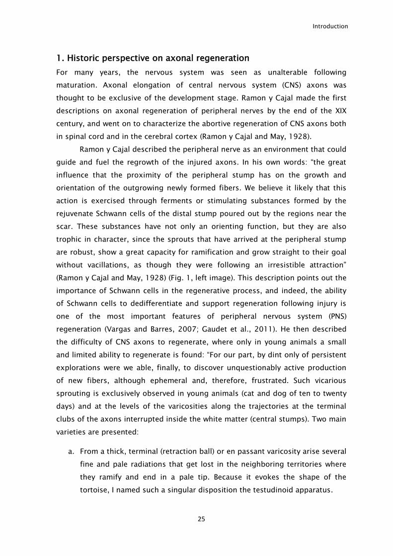
Ramon y Cajal described the peripheral nerve as an environment that could
guide and fuel the regrowth of the injured axons. In his own words: “the great
influence that the proximity of the peripheral stump has on the growth and
orientation of the outgrowing newly formed fibers. We believe it likely that this
action is exercised through ferments or stimulating substances formed by the
rejuvenate Schwann cells of the distal stump poured out by the regions near the
scar. These substances have not only an orienting function, but they are also
trophic in character, since the sprouts that have arrived at the peripheral stump
are robust, show a great capacity for ramification and grow straight to their goal
without vacillations, as though they were following an irresistible attraction”
(Ramon y Cajal and May, 1928) (Fig. 1, left image). This description points out the
importance of Schwann cells in the regenerative process, and indeed, the ability
of Schwann cells to dedifferentiate and support regeneration following injury is
one of the most important features of peripheral nervous system (PNS)
regeneration (Vargas and Barres, 2007; Gaudet et al., 2011). He then described
the difficulty of CNS axons to regenerate, where only in young animals a small
and limited ability to regenerate is found: “For our part, by dint only of persistent
explorations were we able, finally, to discover unquestionably active production
of new fibers, although ephemeral and, therefore, frustrated. Such vicarious
sprouting is exclusively observed in young animals (cat and dog of ten to twenty
days) and at the levels of the varicosities along the trajectories at the terminal
clubs of the axons interrupted inside the white matter (central stumps). Two main
varieties are presented:
a. From a thick, terminal (retraction ball) or en passant varicosity arise several
fine and pale radiations that get lost in the neighboring territories where
they ramify and end in a pale tip. Because it evokes the shape of the
tortoise, I named such a singular disposition the testudinoid apparatus.
Introduction
26
b. At the frontiers of a necrosed axon segment, the surviving neurofibrils of
the neighboring varicosity enter into active proliferation, generating certain
tufts of small branches that invade the dead protoplasm, where they end
my means of boutons or rings. Because of its shape which somewhat
recalls that of the cuttlefish, I baptized such an unusual disposition with
the name of cephalopodic apparatus” (Ramon y Cajal and May, 1928) (Fig.
1 right image).
Figure 1. Ramon y Cajal drawings of peripheral and central nervous system injuries. Left: injured
sciatic nerve. In the left nerve, both ends are kept together, allowing axonal regrowth to the distal
portion, while in the right nerve, the gap created does not allow axonal regrowth. Right: injured
spinal cord displaying scar tissue and absence of axonal growth through the formation of retraction
bulbs. Adapted from (Ramon y Cajal and May, 1928).
Although many advances have been made in the axonal regeneration field,
and despite that several molecular mechanisms underlying axonal growth have
been dissected, the general view has not changed greatly since the first
descriptions by Ramon y Cajal. To date, despite of the several ongoing clinical
trials, still only very limited axonal regeneration is achieved in the CNS. As such,
new studies to understand and improve regeneration of CNS axons are of the
utmost importance.
Introduction
27
2. Axonal elongation during development
During development, axonal elongation occurs both in PNS and CNS. During this
process, neurons express a genetic program that allows a robust elongation and
the correct interpretation of the guidance cues to reach their post-synaptic
targets (Polleux and Snider, 2010). In many ways, regeneration can be viewed, at
least in part, as a recapitulation of the developmental process, since axons need
to regrow towards their targets. Axonal elongation declines during development
due to loss of the neuronal intrinsic ability to elongate (Cai et al., 2001; Goldberg
et al., 2002). As such, the developing nervous system has been used as a model
to identify genes that may be crucial for the control of axon elongation (Moore et
al., 2009).
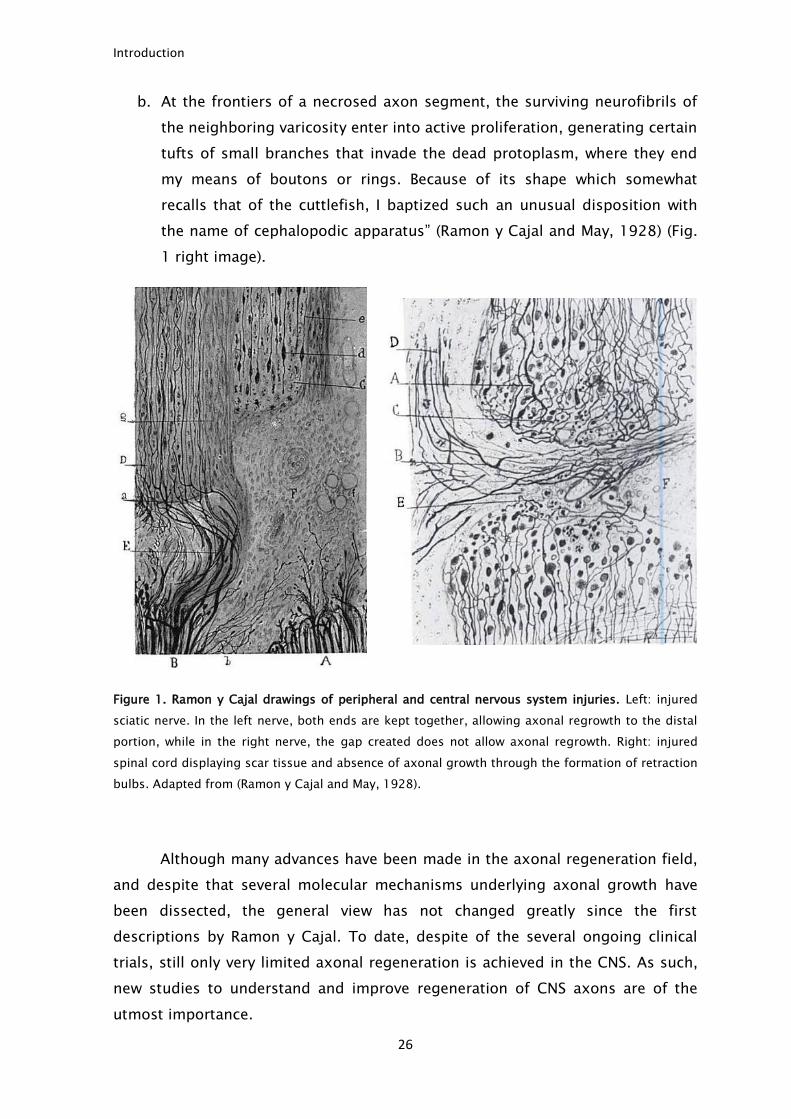
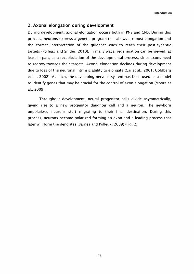
Throughout development, neural progenitor cells divide asymmetrically,
giving rise to a new progenitor daughter cell and a neuron. The newborn
unpolarized neurons start migrating to their final destination. During this
process, neurons become polarized forming an axon and a leading process that
later will form the dendrites (Barnes and Polleux, 2009) (Fig. 2).
Introduction
28
Figure 2. Pattern of the in vivo neuronal polarization and migration of cerebellum granule neurons
(CGN) and pyramidal cortical neurons. A. Representation of the polarization of the CGN in the
mammalian cerebellum. The progenitor cells divide rapidly in the external plexiform layer (EGL, 1)
and upon cell cycle exit, start to adopt a bipolar morphology (2). Then, progenitors migrate
tangentially with a leading and a trailing process (3). Another process emerges orthogonally from
the cell body (4), and becomes the leading process, directing migration towards the inner granule
layer (IGL, 5). The trailing processes form a characteristic T-shaped axon (purple), whereas the
leading process gives rise to the dendritic domain (green, 6). B. Representation of the radially
migrating pyramidal neurons in the mammalian neocortex. Neurons are generated between E11 and
E17 in the ventricular zone by asymmetric division of radial glial progenitors. The progenitor cells
have a long basal process attached to the basal membrane and a short apical process on the
ventricle side (1). Upon division (2), the post mitotic neuron (blue) goes through a multipolar
transition where multiple neurites emerge from the cell body (3), before one major process forms in
the radial direction and becomes the leading process (4). The neuron initiates a radial migration
along the radial glial process and leaves behind a trailing process that elongates tangentially in the
intermediate zone (purple, 5). The cell body continues to migrate towards its final destination (the
top of the cortical plate), while the axon rapidly elongates (6). The leading process gives rise to the
apical dendrite (green, 7), which initiates local branching in the marginal zone (until radial
A
B
Introduction
29
migration ends). During the first postnatal week, the cell body will then translocate ventrally (8-9)
and neurons born at later stages (orange) will bypass them (inside-out accumulation pattern, 10).
Adapted from (Barnes and Polleux, 2009).
In this stage, neurons possess a high intrinsic ability to elongate. This
ability is transcription dependent and relies on the activity of several signaling
pathways, like phosphoinositide 3-kinase (PI3K), phosphatase and tensin
homologue (PTEN) and glycogen synthase kinase 3 beta (GSK3β) (Barnes and
Polleux, 2009; Polleux and Snider, 2010). Local protein synthesis and degradation
also play an important role for correct axonal elongation (Campbell and Holt,
2001). In this context, the ubiquitin-proteasome system emerged as a key player
in the control of axon growth during development.
The ubiquitin ligase anaphase promoting complex (APC) and its activator
protein, Cdh1 were shown to be important regulators of axon growth during
development in post-mitotic neurons (Konishi et al., 2004), acting on the nucleus,
limiting axon growth (Stegmuller et al., 2006). A reduction in Cdh1 in CGN leads
to an increase in axon length, both in vitro and in vivo (Konishi et al., 2004) which
is achieved by targeting both SnoN and Id2 for degradation (Lasorella et al.,
2006; Stegmuller et al., 2006). However, it is not clear if upon completion of
development of the nervous system, Cdh1-APC plays a similar role namely, if it
limits axon overgrowth in the adult nervous system, or even if it inhibits axonal
regeneration.
2.1. Cytoskeletal dynamics in neuron polarization and axonal growth
Upon formation, the immature neurons polarize, giving rise to one axon and
several dendrites. Upon polarization, maturation proceeds with axon and dendrite
elongation (Polleux and Snider, 2010). Most players responsible for axon
initiation and elongation were identified using in vitro cultures of hippocampal
neurons, a system where the timeline for polarization and elongation are very
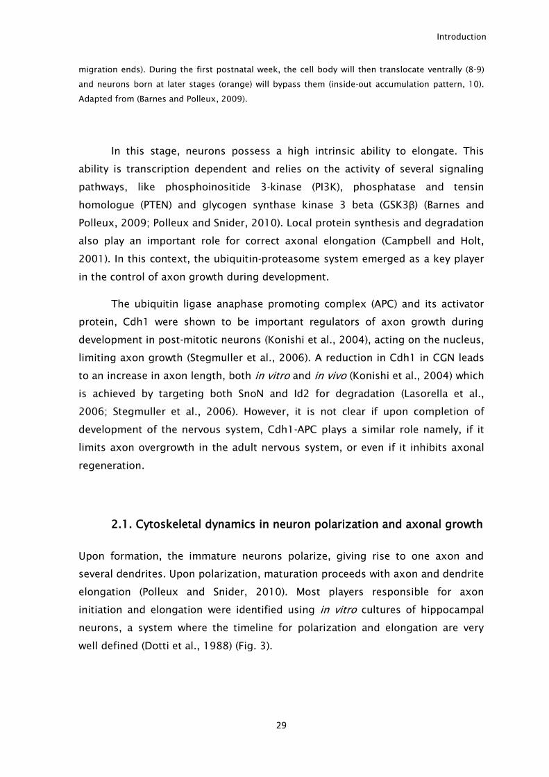
well defined (Dotti et al., 1988) (Fig. 3).
Introduction
30
Figure 3. Neuronal polarization of hippocampal neurons in vitro. In dissociated cultures of
postmitotic hippocampal neurons, stage 1 neurons present an intense lamellipodia and filopodia
that leads to the emergence of multiple immature neurites, stage 2. Then a critical step occurs, and
neurons become asymmetric and a single neurite grows rapidly originating the axon (red), stage 3.
Subsequently there is a rapid elongation of both axon and dendrites (stage 4), and finally neurons
present dendritic spines and the axonal initial segment. Adapted from (Polleux and Snider, 2010)
The in vitro studies led to the notion that this polarization was mainly due
to intrinsic neuronal factors. Cytoskeleton dynamics was found to be crucial for
polarization and growth. In immature hippocampal neurons (stage 2), before
axon formation, there is local actin destabilization in one of the neurites that will
then become the axon. The importance of actin stability was shown by the use of
actin-destabilizing agents like lactrunculin B and cytochalasin D that promoted
axon formation (Bradke and Dotti, 1999). The local stability of actin is regulated
by profilin that promotes actin polymerization and cofilin that promotes actin
depolimerization (Witte and Bradke, 2008). Contrary to actin, during axon
formation there is microtubule stabilization that is needed for microtubule
protrusion. In fact, local microtubule stabilization by the application of taxol is
able to induce axon formation (Witte et al., 2008). GSK3β activity is essential for
the regulation of microtubule stability. It does so by regulating the activity of
several microtubule binding proteins like microtubule-associated protein 1B
(MAP1b), collapsin response mediator protein 2 (CRMP-2) and adenomatous
polyposis coli protein.
Besides axonal polarization, cytoskeleton dynamics is also important for
axonal growth. In fact, microtubule destabilization underlies the formation of a
retraction bulb, a structure that impedes axonal growth. Furthermore, stabilizing
microtubules with the use of taxol promotes axonal growth (Erturk et al., 2007).
Introduction
31
The role of actin dynamics during axonal growth still needs to be further
addressed.
2.2. Growth cone, leading the way
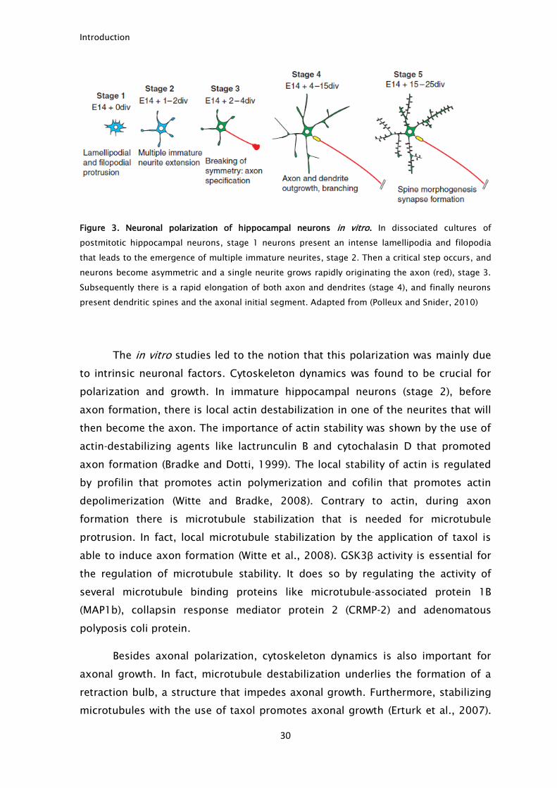
The distal tip of the growing axon, the growth cone, is the structure responsible
for axonal elongation (Fig. 4). At the growth cone there is the integration of the
extracellular cues and this structure is responsible for the elongation through the
correct pathway (Jung et al., 2011).
Figure 4. Growth cone structure. The scheme represents a mature growth cone composed of
dynamic actin filaments at the periphery, forming the lamellipodia and stable microtubules in the
axon shaft. Adapted from (Bradke et al., 2012).
Extracellular cues guide growing axons to reach their correct targets (Fig.
5). Neurotrophins stimulate axonal growth and are required for the transcription
of genes important for elongation to take place. Although the initial phase of
axonal growth is independent of neurotrophins, the later stages of axonal growth
require neurotrophins, and their absence leads to cell death and innervation
failure (Polleux and Snider, 2010). Besides neurotrophins, during development
the growth cone is exposed to several guidance cues, like semaphorins, ephrins
Introduction
32
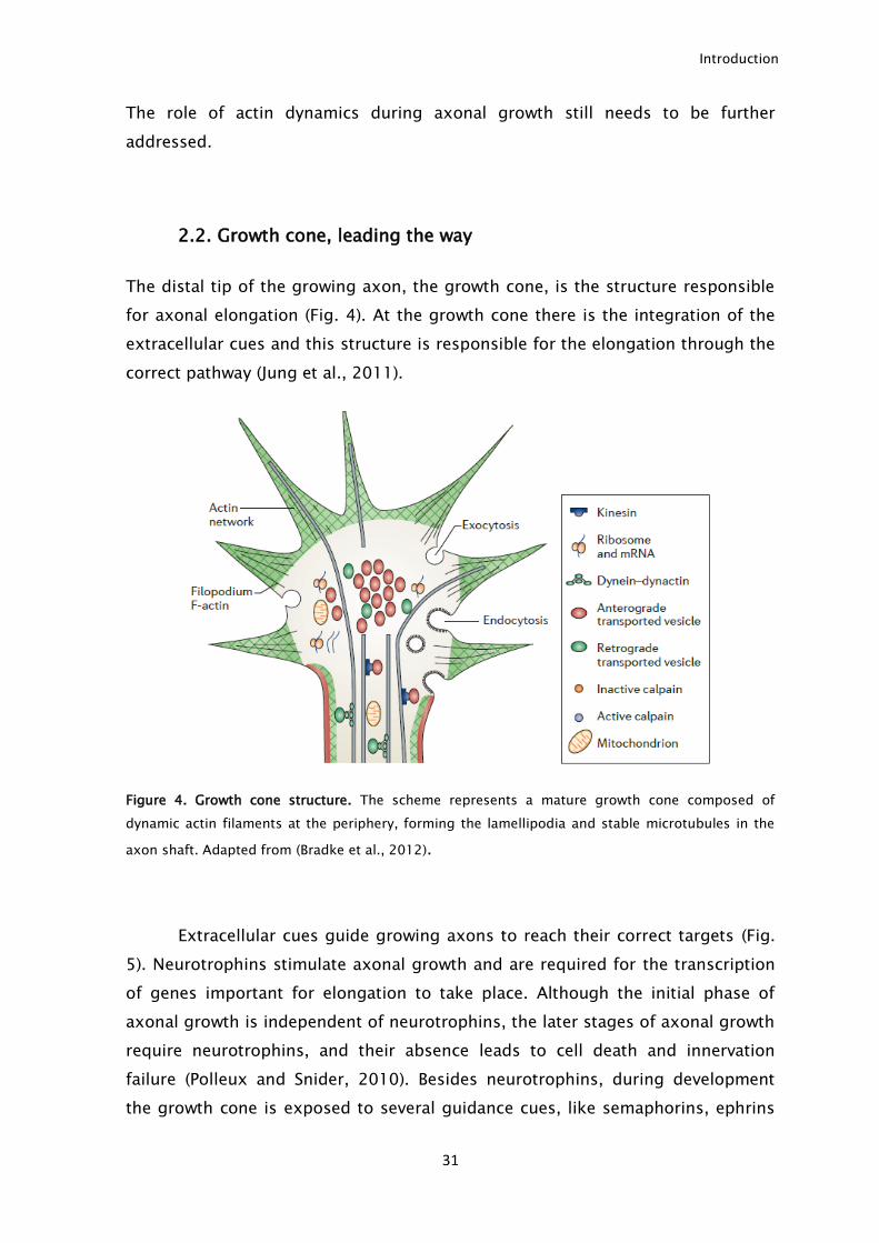
and netrin (Song and Poo, 1999). The optic nerve formation and development has
been the typical model to study the importance and the mechanism of action of
these molecules. It was shown that guidance cues influence developing axons by
promoting either local protein synthesis (attraction) or degradation (repulsion)
(Jung et al., 2011).
Figure 5. Action of guidance cues in the growth cone. Attractive guidance cues induce growth cone
attraction by the stimulation of local protein synthesis (left) and repulsive guidance cues induce
repulsion by stimulation of protein degradation (right). Adapted from (Jung et al., 2011).
Furthermore, it has been described that the attractive/repulsive behavior of
the extracellular cues is dependent on the cyclic adenosine monophosphate
(cAMP): cyclic guanosine monophosphate (cGMP) ratio (Nishiyama et al., 2003).
Guidance cues can be divided in cAMP- or cGMP- responsive. In the cAMP-
responsive group are included netrin-1, myelin associated glycoprotein (MAG),
nerve growth factor (NGF) and brain-derived neurotrophic factor (BDNF), which
attract axons with high levels of cAMP, and repulse axons with low levels of cAMP
(Song and Poo, 1999). In the cGMP-responsive group are included semaphorins
Introduction
33
and neurotrophin-3 (NT-3), which attract axons with high levels of cGMP, but
repulse axons with low levels of cGMP (Song and Poo, 1999; van Horck et al.,
2004). Since the levels of cAMP vary from development to adulthood, this
mechanism allows understanding how some molecules that attract axons during
development become inhibitory in the adult nervous system.
3. Regeneration of PNS axons – axonal regeneration is possible.
Injured PNS neurons are able to regrow to a significant extent and are often used
as a model to understand how axonal regeneration can be achieved in the
nervous system. This regenerative ability is supported by numerous factors that
can be divided in two categories: extrinsic and intrinsic. The extrinsic factors
include the role played by the supporting glia and the immune response triggered
by the injury. The intrinsic factors comprise the ability of the neurons to express
genes that allow their survival and increase their ability to regrow their axons
following injury. These are known as regeneration-associated genes (RAGs). Below
we will discuss in detail the importance of these mechanisms in the successful
PNS regeneration.
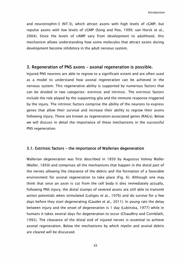
3.1. Extrinsic factors – the importance of Wallerian degeneration
Wallerian degeneration was first described in 1850 by Augustus Volney Waller
(Waller, 1850) and comprises all the mechanisms that happen in the distal part of
the nerves allowing the clearance of the debris and the formation of a favorable
environment for axonal regeneration to take place (Fig. 6). Although one may
think that once an axon is cut from the cell body it dies immediately actually,
following PNS injury, the distal stumps of severed axons are still able to transmit
action potentials when stimulated (Luttges et al., 1976) and do survive for a few
days before they start degenerating (Gaudet et al., 2011). In young rats the delay
between injury and the onset of degeneration is 1 day (Lubinska, 1977) while in
humans it takes several days for degeneration to occur (Chaudhry and Cornblath,
1992). The clearance of the distal end of injured nerves is essential to achieve
axonal regeneration. Below the mechanisms by which myelin and axonal debris
are cleared will be discussed.
Introduction
34
Figure 6. Wallerian degeneration. Following PNS injury there is dying back of the distal axons and
Schwann cell dedifferentiation (2). Then macrophages invade the nerves and together with Schwann
cells they clear the myelin debris (3). The Schwann cells align forming the bands of Bungner which
serve as a rail for the regenerating axons (4) culminating in successful regeneration (5). Adapted
from (Gaudet et al., 2011).
Introduction
35
3.1.1. Axonal degeneration and break down.
Calcium influx is one of the first signals that an injury has occurred. It leads to
calpain activation which triggers the resealing of the membrane of the injured
axons (Krause et al., 1994; Howard et al., 1999). Upon injury, membrane
disruption promotes a transient calcium influx that activates the intracellular
signaling responsible for the membrane reseal (Krause et al., 1994) and for local
protein synthesis (Chierzi et al., 2005). Calcium influx activates calcium-
dependent enzymes including adenylate cyclase, promoting increased cAMP
levels that signal to the downstream effector dual leucine zipper kinase (DLK)
promoting cytoskeleton rearrangements needed for growth cone assembly
(Ghosh-Roy et al., 2010). Besides cytoskeleton rearrangements, local protein
synthesis and degradation are important for growth cone formation. These
processes are tightly regulated by the mammalian target of rapamycin (mTOR),
p38MAPK and caspase-3 (Verma et al., 2005). Following axonal injury, the correct
formation of a growth cone is a key step leading to axonal regeneration.
An elegant study using in vivo imaging showed that approximately 20 min
following injury, both proximal and distal ends suffer 200-300μm fast
degeneration in a process called acute axonal degeneration. Following this
process, the proximal end is stabilized and can start regenerating as early as 30h
after injury, while the distal ends can persist up to 48h before starting to
degenerate (Kerschensteiner et al., 2005).
At the distal part of injured axons, the first visible sign of injury is the
axonal membrane beading and swelling. The mechanisms by which the beading
and swelling occur are independent of calcium and of the ubiquitin-proteasome
system, since they are not inhibited by calcium chelators or by ubiquitin-
proteasome inhibitors (George et al., 1995; Zhai et al., 2003). These
morphological changes occur before the onset of cytoskeleton disintegration, a
process called granular disintegration of the axonal cytoskeleton where the
microtubules and neurofilaments are dismantled leading to axonal fragmentation
(Gaudet et al., 2011). This process is calcium and ubiquitin-proteasome system-
dependent, and it can be inhibited by blocking the ubiquitin-proteasome system
(Zhai et al., 2003) or by blocking the ion-sensitive protease calpain (George et al.,
1995). These two mechanisms have different roles. The ubiquitin-proteasome
system disassembles microtubules, while the calcium-activated calpain response
secures neurofilament degradation. Once this process starts, the complete
Introduction
36
destruction of the cytoskeleton components into fine debris is completed in one
hour (George et al., 1995; Beirowski et al., 2005; Kerschensteiner et al., 2005).
The degenerative process was initially envisaged as a passive process,
where axons died given the lack of connectivity to the cell body. It is now known
that it is an active process, intrinsic to the axons that can be controlled and
delayed. This paradigm shift has emerged with the studies in the slow wallerian
degeneration (Wlds
) mouse (Perry et al., 1990), in which the injured distal nerve
ends degenerate much slower and are able to conduct action potentials for 2-3
weeks while wild type animals only conduct action potentials for 3 days (Lunn et
al., 1989; Tsao et al., 1999). The study of the Wlds
mouse also revealed the
importance of axonal degeneration in the regenerative process. The delayed
cytoskeleton disintegration is accompanied by a delay in myelin sheath
breakdown and macrophage influx, ultimately leading to impaired axonal
regeneration (Bisby and Chen, 1990).
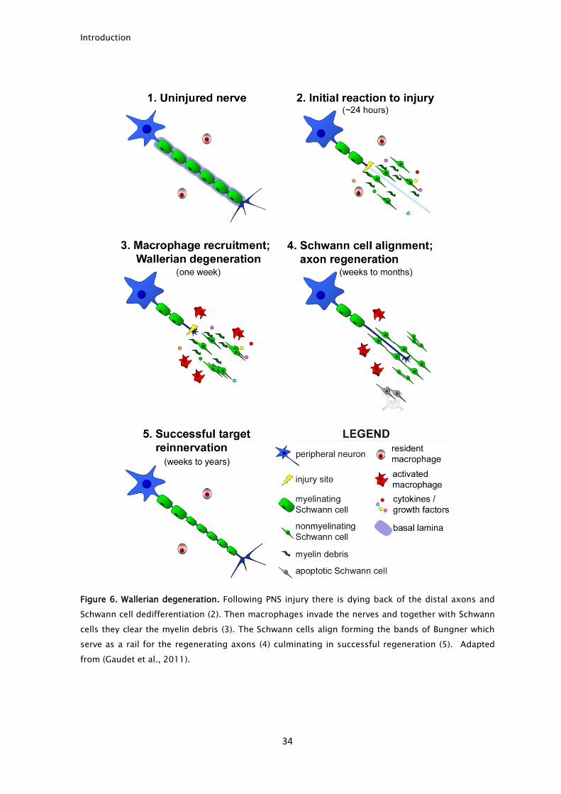
3.1.2. The importance of Schwann cells
Schwann cells are the glial cells of the PNS. They play an essential role in nerve
physiology since they are responsible for the trophic support of developing and
mature neurons, and also for myelin insulation (Bhatheja and Field, 2006). There
are two types of Schwann cells: myelinating and non-myelinating Schwann cells.
Myelination occurs only in large caliber axons (>1μm), with each Schwann cell
myelinating a single axon (Fig. 7, left image). Myelin insulation allows the axon to
conduct action potentials much faster. The small caliber axons are ensheathed in
a structure called Remak bundle, where a single Schwann cell wraps multiple
axons, separating them by a thin layer of cytoplasm (Fig. 7, right image).
Figure 7. Schwann cell organization in PNS axons. In PNS axons, Schwann cells can either myelinate
a single axon (diameter >1µm) or wrap several axons (diameter <1µm) with a thin layer of
cytoplasm forming a remak bundle. Adapted from (Salzer, 2008).
Introduction
37
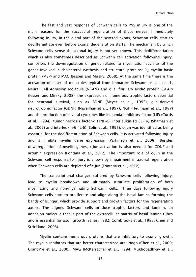
The fast and vast response of Schwann cells to PNS injury is one of the
main reasons for the successful regeneration of these nerves. Immediately
following injury, in the distal part of the severed axons, Schwann cells start to
dedifferentiate even before axonal degeneration starts. The mechanism by which
Schwann cells sense the axonal injury is not yet known. This dedifferentiation
which is also sometimes described as Schwann cell activation following injury,
comprises the downregulation of genes related to myelination such as of the
genes involved in cholesterol synthesis and structural proteins: P0
, myelin basic
protein (MBP) and MAG (Jessen and Mirsky, 2008). At the same time there is the
activation of a set of molecules typical from immature Schwann cells, like L1,
Neural Cell Adhesion Molecule (NCAM) and glial fibrillary acidic protein (GFAP)
(Jessen and Mirsky, 2008), the expression of numerous trophic factors essential
for neuronal survival, such as BDNF (Meyer et al., 1992), glial-derived
neurotrophic factor (GDNF) (Naveilhan et al., 1997), NGF (Heumann et al., 1987)
and the production of several cytokines like leukemia inhibitory factor (LIF) (Curtis
et al., 1994), tumor necrosis factor-α (TNF-α), interleukin-1α (IL-1α) (Shamash et
al., 2002) and interleukin-6 (IL-6) (Bolin et al., 1995). c-Jun was identified as being
essential for the dedifferentiation of Schwann cells. It is activated following injury
and it inhibits myelin gene expression (Parkinson et al., 2008). Besides
downregulation of myelin genes, c-Jun activation is also needed for GDNF and
artemin expression (Fontana et al., 2012). The important role of c-Jun in the
Schwann cell response to injury is shown by impairment in axonal regeneration
when Schwann cells are depleted of c-Jun (Fontana et al., 2012).
The transcriptional changes suffered by Schwann cells following injury,
lead to myelin breakdown and ultimately stimulate proliferation of both
myelinating and non-myelinating Schwann cells. Three days following injury
Schwann cells start to proliferate and align along the basal lamina forming the
bands of Bunger, which provide support and growth factors for the regenerating
axons. The aligned Schwann cells produce trophic factors and laminin, an
adhesion molecule that is part of the extracellular matrix of basal lamina tubes
and is essential for axon growth (Sanes, 1982; Cornbrooks et al., 1983; Chen and
Strickland, 2003).
Myelin contains numerous proteins that are inhibitory to axonal growth.
The myelin inhibitors that are better characterized are: Nogo (Chen et al., 2000;
GrandPre et al., 2000), MAG (McKerracher et al., 1994; Mukhopadhyay et al.,
Introduction
38
1994) and oligodendrocyte myelin glycoprotein (OMgp) (Kottis et al., 2002; Wang
et al., 2002b). From these three proteins, only MAG is present in PNS myelin
(McKerracher et al., 1994). Following injury, there is an accumulation of myelin
and of its axonal growth inhibitors that, if not removed, impairs axonal
regeneration. This has been shown in animals with slow Wallerian degeneration
where the delay in myelin clearance delays PNS regeneration (Brown et al., 1991).
Schwann cells and macrophages are responsible for myelin clearance after PNS
injury (Vargas and Barres, 2007). Schwann cells degrade myelin using hydrolytic
enzymes in intracellular vacuoles (Holtzman and Novikoff, 1965). During the first
days following injury, the contribution of macrophages for myelin clearance is
small and Schwann cells are responsible for almost all myelin removal (Perry et
al., 1995). Schwann cells are able to clear their own myelin, phagocyte
extracellular debris and also may function as antigen presenting cells by
presenting myelin through major histocompatibility complex II (MHC-II) although
this last function is not clear (Holtzman and Novikoff, 1965; Hirata et al., 1999).
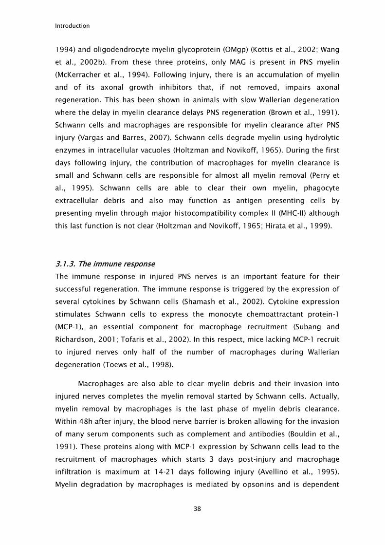
3.1.3. The immune response
The immune response in injured PNS nerves is an important feature for their
successful regeneration. The immune response is triggered by the expression of
several cytokines by Schwann cells (Shamash et al., 2002). Cytokine expression
stimulates Schwann cells to express the monocyte chemoattractant protein-1
(MCP-1), an essential component for macrophage recruitment (Subang and
Richardson, 2001; Tofaris et al., 2002). In this respect, mice lacking MCP-1 recruit
to injured nerves only half of the number of macrophages during Wallerian
degeneration (Toews et al., 1998).
Macrophages are also able to clear myelin debris and their invasion into
injured nerves completes the myelin removal started by Schwann cells. Actually,
myelin removal by macrophages is the last phase of myelin debris clearance.
Within 48h after injury, the blood nerve barrier is broken allowing for the invasion
of many serum components such as complement and antibodies (Bouldin et al.,
1991). These proteins along with MCP-1 expression by Schwann cells lead to the
recruitment of macrophages which starts 3 days post-injury and macrophage
infiltration is maximum at 14-21 days following injury (Avellino et al., 1995).
Myelin degradation by macrophages is mediated by opsonins and is dependent
Introduction
39
on the complement system (Bruck and Friede, 1990), such that the invasion of
complement components and antibodies into injured nerves is essential, since
they “label” myelin debris making them more visible for macrophage removal
(Bruck and Friede, 1990).
By far, macrophages are the immune cell type that plays the most
important role in PNS regeneration. Other leukocytes play small roles in PNS
regeneration. Neutrophils invade injured nerves early after injury and are able to
degrade myelin. However their importance in nerve regeneration is not known. In
the last phase of the immune response, T cells fine tune immune response
producing several pro- and anti-inflammatory cytokines (Gaudet et al., 2011).
3.2 Intrinsic factors
This section served as the basis for the review manuscript: Mar FM, Bonni A and
Sousa MM (2014). Cell intrinsic control of axon regeneration. EMBO Rep. In press
doi: 10.1002/embr.201337723 that is reprinted at the end of the introduction
section. The successful regeneration of PNS axons is not only due to the removal
of myelin negative cues during Wallerian degeneration. PNS neurons also respond
to injury by increasing their ability to regrow. Neurons are able to sense an
axonal injury many centimeters away from the cell body and to change their
transcriptional profile and express several RAGs that increase their ability to
regenerate. Bellow I will discuss in detail how PNS axons sense an injury and what
are the important features of their regenerative response.
3.2.1. Important neuronal features that allow a regenerative response to be
mounted
Neurons are highly polarized cells, where in many cases the cell body and the
axon terminal can be separated by many centimeters. Most of the protein
synthesis occurs in the cell body following which proteins need to be “shipped” to
their correct location. As such, axonal transport is of the utmost importance to
deliver proteins along the axons, but also to transmit signals from the axon
terminal to the cell body. Although limited, protein synthesis can occur along the
axon and in the growth cone during elongation. The local protein synthesis is of
extreme importance for local early responses to axonal injury and to growth cone
Introduction
40
guidance cues. The importance of axonal transport and local protein synthesis
will be discussed in detail.
3.2.1.1. Axonal transport
The axoplasm represents 99% of the neuronal cytoplasm, but protein synthesis in
the axon is limited. As such, most of the axoplasm constituents are synthesized
in the cell body and then transported to their final destination. Also, axon
terminals often receive target derived signals that need to be communicated to
the cell body. Given the great length of the axons, those activities pose a great
challenge for the neuron. Most of the axonal transport is made along the
microtubules in an adenosine-5'-triphosphate (ATP)-dependent way by cargo
binding to molecular motors (Fig. 8). In axons, microtubules are uniformly
arranged with their plus ends facing the axon terminal (plus-end-out orientation).
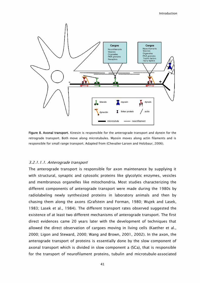
Two main motors are capable of binding microtubules: kinesin, that is
responsible for the transport towards the distal axon tip (plus-end), known as
anterograde transport and dynein that is responsible for the transport from the
axon tip to the cell body (minus-end), known as retrograde transport (Guzik and
Goldstein, 2004).
Besides kinesin and dynein, myosin is another motor performing axonal
transport. Contrary to kinesin and dynein that move along microtubules, myosin
moves along actin filaments (Seabra and Coudrier, 2004). Myosin is incapable of
long distance transport, a function performed by kinesin and dynein. Instead, it is
responsible for short distance transport, like local insertion of proteins in the
plasma membrane. It also interacts with neurofilaments being responsible for
their organization (Bridgman, 2004).
Introduction
41
Figure 8. Axonal transport. Kinesin is responsible for the anterograde transport and dynein for the
retrograde transport. Both move along microtubules. Myosin moves along actin filaments and is
responsible for small range transport. Adapted from (Chevalier-Larsen and Holzbaur, 2006).
3.2.1.1.1. Anterograde transport
The anterograde transport is responsible for axon maintenance by supplying it
with structural, synaptic and cytosolic proteins like glycolytic enzymes, vesicles
and membranous organelles like mitochondria. Most studies characterizing the
different components of anterograde transport were made during the 1980s by
radiolabeling newly synthesized proteins in laboratory animals and then by
chasing them along the axons (Grafstein and Forman, 1980; Wujek and Lasek,
1983; Lasek et al., 1984). The different transport rates observed suggested the
existence of at least two different mechanisms of anterograde transport. The first
direct evidences came 20 years later with the development of techniques that
allowed the direct observation of cargoes moving in living cells (Kaether et al.,
2000; Ligon and Steward, 2000; Wang and Brown, 2001, 2002). In the axon, the
anterograde transport of proteins is essentially done by the slow component of
axonal transport which is divided in slow component a (SCa), that is responsible
for the transport of neurofilament proteins, tubulin and microtubule-associated
Introduction
42
proteins at a rate of 0,2-1 mm/day; and the slow component b (SCb) that
transports glycolytic enzymes and actin, among others, at a rate of 2-8 mm/day
(Lasek et al., 1984; Brown, 2003). Vesicles and membranous organelles are
transported in the fast component at 50-400 mm/day (Lasek et al., 1984; Brown,
2003). Surprisingly, the motors and the kinetics of both slow and fast
components of axonal transport are similar and the different average rates are
explained by an intermittent behavior of cargoes during transport (Brown, 2003).
Slow transport components, although traveling at the same rate as the fast
component, spend more time in a stationary stage along the way resulting in a
lower average rate (Brown, 2003; Roy et al., 2007). These findings raised new
questions such as: How is the intermittent behavior controlled? What triggers
movement and stoppage? These questions remain unanswered.
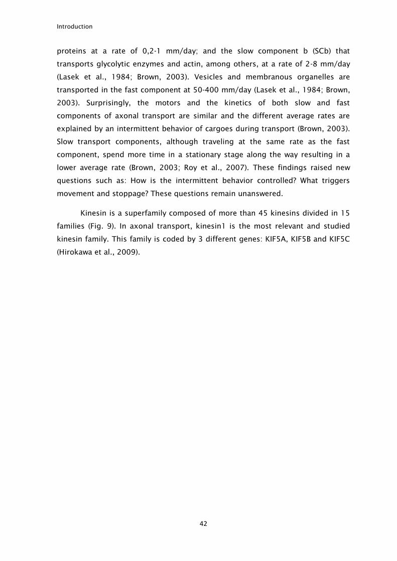
Kinesin is a superfamily composed of more than 45 kinesins divided in 15
families (Fig. 9). In axonal transport, kinesin1 is the most relevant and studied
kinesin family. This family is coded by 3 different genes: KIF5A, KIF5B and KIF5C
(Hirokawa et al., 2009).
Introduction
43
Figure 9. Kinesin protein family. Adapted from (Hirokawa et al., 2009).
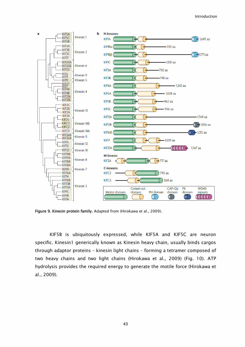
KIF5B is ubiquitously expressed, while KIF5A and KIF5C are neuron
specific. Kinesin1 generically known as Kinesin heavy chain, usually binds cargos
through adaptor proteins – kinesin light chains – forming a tetramer composed of
two heavy chains and two light chains (Hirokawa et al., 2009) (Fig. 10). ATP
hydrolysis provides the required energy to generate the motile force (Hirokawa et
al., 2009).
Introduction
44
Figure 10. Kinesin structure. Kinesin is composed by two heavy chains and two light chains.
Adapted from (Hirokawa et al., 2010).
Besides axonal maintenance, anterograde transport is of particular
importance during axonal regeneration since it supplies the necessary proteins
for this process, particularly the structural components tubulin, actin and
neurofilament (Tashiro and Komiya, 1992; Jacob and McQuarrie, 1996). In fact the
speed of axonal regeneration is similar to the rate of the slow component of
anterograde transport, confirming the idea that the anterograde transport
supplies regrowing axons (Wujek and Lasek, 1983).
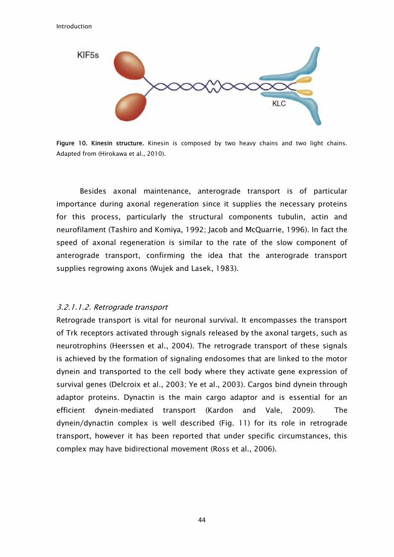
3.2.1.1.2. Retrograde transport
Retrograde transport is vital for neuronal survival. It encompasses the transport
of Trk receptors activated through signals released by the axonal targets, such as
neurotrophins (Heerssen et al., 2004). The retrograde transport of these signals
is achieved by the formation of signaling endosomes that are linked to the motor
dynein and transported to the cell body where they activate gene expression of
survival genes (Delcroix et al., 2003; Ye et al., 2003). Cargos bind dynein through
adaptor proteins. Dynactin is the main cargo adaptor and is essential for an
efficient dynein-mediated transport (Kardon and Vale, 2009). The
dynein/dynactin complex is well described (Fig. 11) for its role in retrograde
transport, however it has been reported that under specific circumstances, this
complex may have bidirectional movement (Ross et al., 2006).

Introduction
45
Figure 11. Dynein/dynactin complex structure. Dynein is composed by two heavy chains, two light-
intermediate chains, two intermediate chains and two light chains that bind to dynactin for cargo
transport. Adapted from (Hirokawa et al., 2010).
The importance of retrograde transport is shown by the fact that its
disruption leads to the onset of neurodegeneration (LaMonte et al., 2002;
Hafezparast et al., 2003). In fact, in many neurodegenerative diseases such as
Amyotrophic lateral sclerosis (ALS), defects in retrograde transport are found
even before the first symptoms appear (Kieran et al., 2005; Ligon et al., 2005).
Retrograde transport is extremely important to signal injury following nerve
lesion. Upon injury, there is the local activation of injury signals that are linked to
dynein (Hanz et al., 2003). This linkage ensures the retrograde transport of the
injury signals to the cell body (Fig. 12) where they can signal the injury and
trigger a regenerative response (Ambron et al., 1992; Schmied and Ambron,
1997; Hanz et al., 2003; Perlson et al., 2005).
Introduction
46
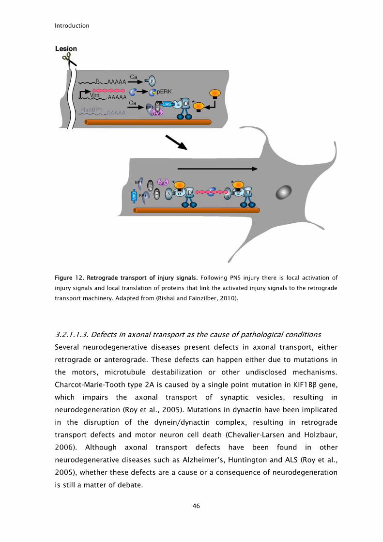
Figure 12. Retrograde transport of injury signals. Following PNS injury there is local activation of
injury signals and local translation of proteins that link the activated injury signals to the retrograde
transport machinery. Adapted from (Rishal and Fainzilber, 2010).
3.2.1.1.3. Defects in axonal transport as the cause of pathological conditions
Several neurodegenerative diseases present defects in axonal transport, either
retrograde or anterograde. These defects can happen either due to mutations in
the motors, microtubule destabilization or other undisclosed mechanisms.
Charcot-Marie-Tooth type 2A is caused by a single point mutation in KIF1Bβ gene,
which impairs the axonal transport of synaptic vesicles, resulting in
neurodegeneration (Roy et al., 2005). Mutations in dynactin have been implicated
in the disruption of the dynein/dynactin complex, resulting in retrograde
transport defects and motor neuron cell death (Chevalier-Larsen and Holzbaur,
2006). Although axonal transport defects have been found in other
neurodegenerative diseases such as Alzheimer’s, Huntington and ALS (Roy et al.,
2005), whether these defects are a cause or a consequence of neurodegeneration
is still a matter of debate.
Introduction
47
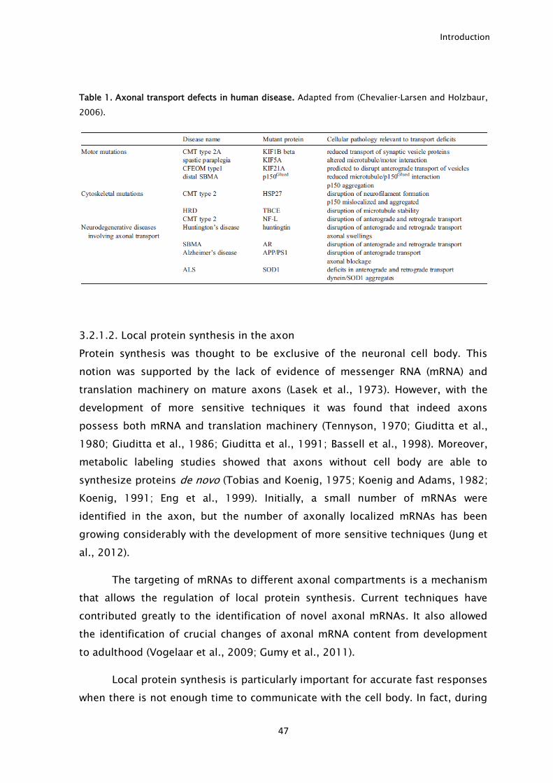
Table 1. Axonal transport defects in human disease. Adapted from (Chevalier-Larsen and Holzbaur,
2006).
3.2.1.2. Local protein synthesis in the axon
Protein synthesis was thought to be exclusive of the neuronal cell body. This
notion was supported by the lack of evidence of messenger RNA (mRNA) and
translation machinery on mature axons (Lasek et al., 1973). However, with the
development of more sensitive techniques it was found that indeed axons
possess both mRNA and translation machinery (Tennyson, 1970; Giuditta et al.,
1980; Giuditta et al., 1986; Giuditta et al., 1991; Bassell et al., 1998). Moreover,
metabolic labeling studies showed that axons without cell body are able to
synthesize proteins de novo (Tobias and Koenig, 1975; Koenig and Adams, 1982;
Koenig, 1991; Eng et al., 1999). Initially, a small number of mRNAs were
identified in the axon, but the number of axonally localized mRNAs has been
growing considerably with the development of more sensitive techniques (Jung et
al., 2012).
The targeting of mRNAs to different axonal compartments is a mechanism
that allows the regulation of local protein synthesis. Current techniques have
contributed greatly to the identification of novel axonal mRNAs. It also allowed
the identification of crucial changes of axonal mRNA content from development
to adulthood (Vogelaar et al., 2009; Gumy et al., 2011).
Local protein synthesis is particularly important for accurate fast responses
when there is not enough time to communicate with the cell body. In fact, during
Introduction
48
development, growing axons are exposed to several guidance cues, like netrin-1,
ephrin B, semaphorin 3A, NGF and BDNF, which need to be interpreted rapidly for
the correct pathfinding (Lin and Holt, 2008; Jung et al., 2012). The growth cone is
the structure responsible for the integration of the environment signals in a
process dependent on local protein synthesis (Campbell and Holt, 2001; Ming et
al., 2002). Although CNS axons are able to synthesize proteins in the growth cone
during development, studies with adult CNS axons fail to show the presence of
ribosomes, suggesting that adult CNS axons are not able to synthesize locally
proteins or that this ability is very limited (Steward and Ribak, 1986; Verma et al.,
2005). This can underlie in part their limited ability to regenerate. In contrast,
adult PNS axons do possess ribosomes distributed unevenly along the axoplasm,
close to the plasma membrane (Koenig et al., 2000; Li et al., 2005; Kun et al.,
2007). Schwann cells have also been suggested as a source of ribosomes for PNS
axons following injury, indicating that Schwann cells may promote local protein
synthesis (Court et al., 2008; Twiss and Fainzilber, 2009). Protein synthesis is
generally decreased with axonal ageing, that coincides with reduced axonal
regeneration potential (Gumy et al., 2010). This evidence further reinforces the
importance of local axonal synthesis in regeneration.
As such, besides its importance for axonal steering during development,
local protein synthesis has been shown to be important following peripheral
nerve injury by two different mechanisms. It enables the synthesis of vimentin
and importin-β that can be linked to the retrograde transport machinery along
with locally activated proteins and transported back to the cell body where they
can trigger a robust regenerative response (Hanz et al., 2003; Perlson et al.,
2005). Also, the initial steps of axonal regeneration are achieved by local protein
synthesis, since the arrival to the injury site of proteins synthesized in the cell
body can take a few days. As such, the formation of a growth cone, an essential
structure for successful regeneration, as well as the provision of the first building
blocks of the regrowing axons is obtained by local protein synthesis (Verma et al.,
2005; Willis and Twiss, 2006; Gumy et al., 2010).
Axonal mRNAs face great challenges: they need to be actively transported,
stored and protected from degradation at their final destination. The RNA-binding
proteins play an essential role in this process, by binding to cis-elements in the
5’- or 3’- untranslated regions (UTR). Upon RNA binding they control its transport,
stability and translation (Bassell and Kelic, 2004; Patel et al., 2012). The best
Introduction
49
studied mRNA with axonal localization is β-actin. Its mRNA is controlled by the
zipcode-binding protein-1 (ZBP-1) that binds to a cis element in the 3’-UTR. This
cis element in the 3’-UTR is essential for local translation of β-actin in response to
guidance cues (Leung et al., 2006; Yao et al., 2006). The importance of ZBP-1 has
been shown by the observation that reduced ZBP-1 activity leads to decreased
axon regeneration (Donnelly et al., 2011). Additionally, the overexpression of β-
actin 3′UTR competed in vivo with other ZBP-1 cargo mRNAs such as growth
associated protein 43 (GAP-43) (Yoo et al., 2013). Recently, it has also been
shown that axonal translation of β-actin supports axon branching, while that of
GAP-43 promotes elongation of sensory neurons (Donnelly et al., 2013).
In summary, the recent findings support that although local axonal protein
synthesis is limited, it is of great importance to axonal regeneration.
3.2.2. Injury mechanisms
The mechanism by which an axon can “warn” the cell body that it has been
injured is a question that puzzled neuroscientists for many years. Elegant studies
made in Aplysia neurons in the 90’s described the first injury signals capable of
increasing the regeneration ability (Ambron et al., 1995; Ambron et al., 1996).
Since then, many advances were made in how PNS axons are able to sense an
injury, although many of the proposed signals still lack robust experimental
evidence (Fig. 13).
Introduction
50
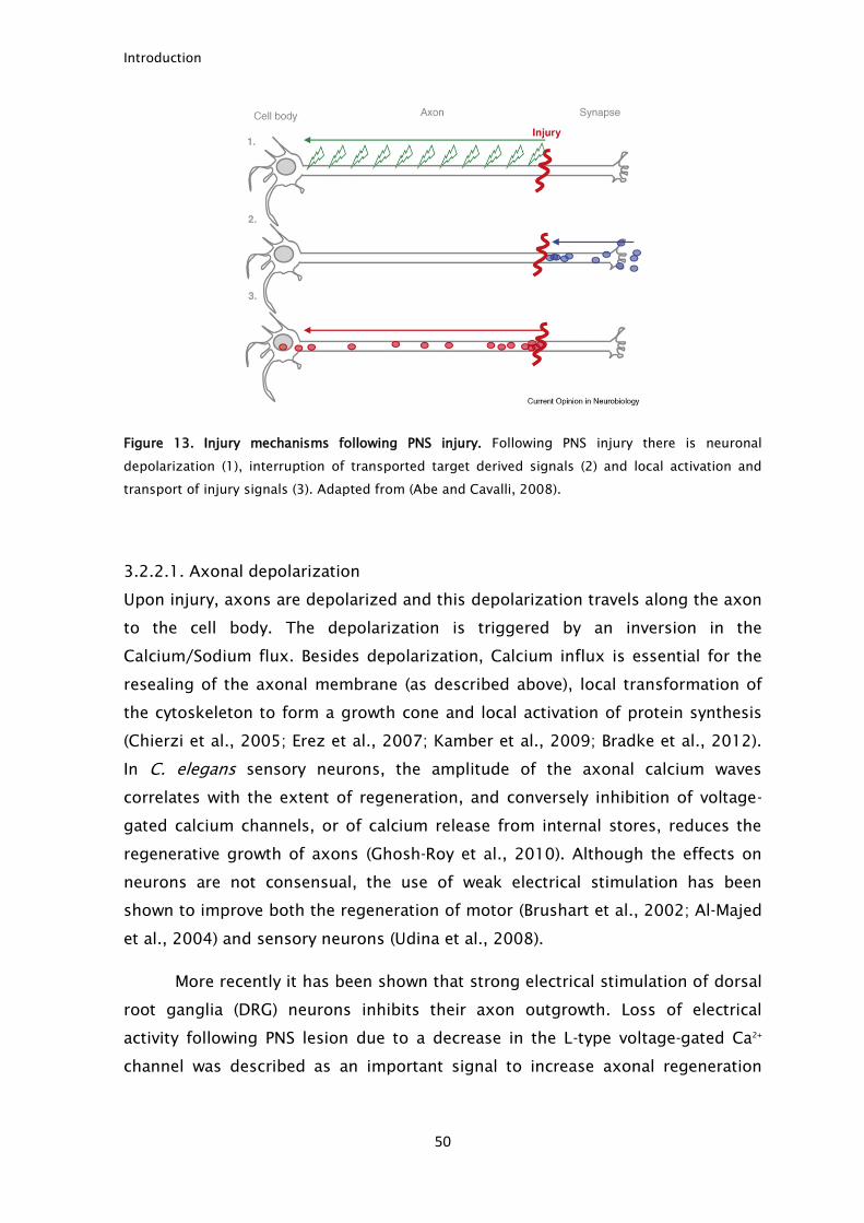
Figure 13. Injury mechanisms following PNS injury. Following PNS injury there is neuronal
depolarization (1), interruption of transported target derived signals (2) and local activation and
transport of injury signals (3). Adapted from (Abe and Cavalli, 2008).
3.2.2.1. Axonal depolarization
Upon injury, axons are depolarized and this depolarization travels along the axon
to the cell body. The depolarization is triggered by an inversion in the
Calcium/Sodium flux. Besides depolarization, Calcium influx is essential for the
resealing of the axonal membrane (as described above), local transformation of
the cytoskeleton to form a growth cone and local activation of protein synthesis
(Chierzi et al., 2005; Erez et al., 2007; Kamber et al., 2009; Bradke et al., 2012).
In C. elegans sensory neurons, the amplitude of the axonal calcium waves
correlates with the extent of regeneration, and conversely inhibition of voltage-
gated calcium channels, or of calcium release from internal stores, reduces the
regenerative growth of axons (Ghosh-Roy et al., 2010). Although the effects on
neurons are not consensual, the use of weak electrical stimulation has been
shown to improve both the regeneration of motor (Brushart et al., 2002; Al-Majed
et al., 2004) and sensory neurons (Udina et al., 2008).
More recently it has been shown that strong electrical stimulation of dorsal
root ganglia (DRG) neurons inhibits their axon outgrowth. Loss of electrical
activity following PNS lesion due to a decrease in the L-type voltage-gated Ca2+
channel was described as an important signal to increase axonal regeneration
Introduction
51
(Enes et al., 2010). These results suggested that the electrical activity may be a
negative signal that once lost can trigger the injury response in DRG neurons.
In summary, axonal depolarization is thought to be the first signal of
injury. However it is seen as a transient signal that, if not followed by other injury
signaling mechanisms, cannot trigger a robust, sustained regenerative response.
3.2.2.2. Negative injury signals
During development axons elongate until they find their targets. Once an axon
finds its target, the neuron receives several target-derived factors through
retrograde transport that repress the elongation machinery of neurons converting
the growth cone into a presynaptic terminal.
Upon injury, the neurons are disconnected from their targets and there is
an interruption of the normal supply of target-derived signals. This decrease in
the supply of target derived signals is thought to alleviate the repression of axon
elongation allowing regeneration. These signals are known as negative injury
signals since their absence can trigger a regenerative response. It has been
shown that neurotrophins are dramatically decreased in DRG neurons following
injury (Raivich et al., 1991). Although neurotrophins fulfill many of the
requirements for a negative injury signal, their role in nerve regeneration is still
unclear. In accordance with a negative injury signal function, it has been
described that administration of NGF to injured nerves could delay regeneration
(Gold, 1997). However, in other studies, the administration of neurotrophins
following injury was linked to an improvement in PNS regeneration (Molteni et al.,
2004). Other studies have also described that PNS regeneration may be
independent of NGF (Diamond et al., 1992; Tannemaat et al., 2008).
The notion that naïve neurons continuously receive signals that repress
axonal growth that are relieved following PNS injury is an appealing idea. The
identification of these signals may lead to the development of new methods to
improve axon regeneration. However, this hypothesis still lacks robust
experimental evidence and so far no clear negative injury signals have been
identified.
Introduction
52
3.2.2.3. Positive injury signals
The first evidence that axons can produce molecules capable of triggering a
regenerative response following injury emerged in the 90s using Aplysia neurons
as a model. It was shown that injecting axoplasm extracted from injured nerves in
naïve neurons, an injury-like behavior was triggered in the injected neurons. By
labeling the injured nerves with γ32
P ATP there was evidence that these signals
were dependent on phosphorylation (Ambron et al., 1995). Combining these
results with previous data on a SV40-type nuclear localization signal (NLS) that
targeted axonal proteins to the nucleus through the retrograde transport
machinery (Ambron et al., 1992; Schmied and Ambron, 1997), the authors
proposed that under normal circumstances, the signal proteins have an hidden
NLS, that after injury is exposed by phosphorylation leading to targeting of the
signal to the nucleus triggering a response to injury. The importance of
retrograde transport of NLS proteins was shown later when an NLS synthetic
peptide was injected into the injured nerve precluding the increase in the
regeneration capacity by competition with the intrinsic signals (Hanz et al., 2003).
A decade later, the molecular mechanisms underlying the activation of
positive injury signals and for their transport were described, but the initial idea
was maintained. One of the most important features added to the injury signaling
mechanism was the importance of local translation in injured axons. The concept
that axons are able to synthesize proteins allowed to understand how a protein
containing a NLS is able to undergo retrograde transport only following injury.
The NLS-mediated import of proteins to the nucleus is mediated by their binding
to importins (Gorlich and Kutay, 1999). It is also important to note that NLS
proteins bind with low affinity to importin-α, but with high affinity to importin-α/β
heterodimers (Kohler et al., 1999). In normal nerves only importin-α is present
making it unlikely that NLS proteins are retrogradely transported to the nucleus.
However, following injury, there is the local translation of importin-β. The
synthesis of importin-β allows for the formation of importin-α/β heterodimers
which bind to NLS and link those proteins to the retrograde transport machinery
through dynein binding (Hanz et al., 2003). Recently, it has been shown that the
axonal localization of importin-β mRNA is essential for the correct assembly of
the retrograde transport machinery of injury signals (Ben-Tov Perry et al., 2012).
Besides importin-β, RanBP1 is also locally synthesized following injury and able to
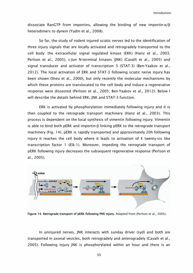
Introduction
53
dissociate RanGTP from importins, allowing the binding of new importin-α/β
heterodimers to dynein (Yudin et al., 2008).
So far, the study of rodent injured sciatic nerves led to the identification of
three injury signals that are locally activated and retrogradely transported to the
cell body: the extracellular signal regulated kinase (ERK) (Hanz et al., 2003;
Perlson et al., 2005), c-Jun N-terminal kinases (JNK) (Cavalli et al., 2005) and
signal transducer and activator of transcription 3 (STAT-3) (Ben-Yaakov et al.,
2012). The local activation of ERK and STAT-3 following sciatic nerve injury has
been shown (Sheu et al., 2000), but only recently the molecular mechanisms by
which these proteins are translocated to the cell body and induce a regenerative
response were dissected (Perlson et al., 2005; Ben-Yaakov et al., 2012). Below I
will describe the details behind ERK, JNK and STAT-3 function.
ERK is activated by phosphorylation immediately following injury and it is
then coupled to the retrograde transport machinery (Hanz et al., 2003). This
process is dependent on the local synthesis of vimentin following injury. Vimentin
is able to bind both pERK and importin-β linking pERK to the retrograde transport
machinery (Fig. 14). pERK is rapidly transported and approximately 20h following
injury it reaches the cell body where it leads to activation of E twenty-six like
transcription factor 1 (Elk-1). Moreover, impeding the retrograde transport of
pERK following injury decreases the subsequent regenerative response (Perlson et
al., 2005).
Figure 14. Retrograde transport of pERK following PNS injury. Adapted from (Perlson et al., 2005).
In uninjured nerves, JNK interacts with sunday driver (syd) and both are
transported in axonal vesicles, both retrogradely and anterogradely (Cavalli et al.,
2005). Following injury JNK is phosphorylated within an hour and there is an
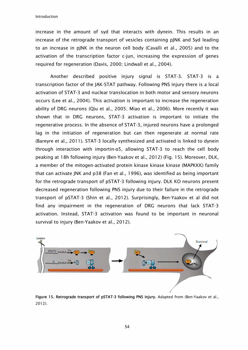
Introduction
54
increase in the amount of syd that interacts with dynein. This results in an
increase of the retrograde transport of vesicles containing pJNK and Syd leading
to an increase in pJNK in the neuron cell body (Cavalli et al., 2005) and to the
activation of the transcription factor c-jun, increasing the expression of genes
required for regeneration (Davis, 2000; Lindwall et al., 2004).
Another described positive injury signal is STAT-3. STAT-3 is a
transcription factor of the JAK-STAT pathway. Following PNS injury there is a local
activation of STAT-3 and nuclear translocation in both motor and sensory neurons
occurs (Lee et al., 2004). This activation is important to increase the regeneration
ability of DRG neurons (Qiu et al., 2005; Miao et al., 2006). More recently it was
shown that in DRG neurons, STAT-3 activation is important to initiate the
regenerative process. In the absence of STAT-3, injured neurons have a prolonged
lag in the initiation of regeneration but can then regenerate at normal rate
(Bareyre et al., 2011). STAT-3 locally synthesized and activated is linked to dynein
through interaction with importin-α5, allowing STAT-3 to reach the cell body
peaking at 18h following injury (Ben-Yaakov et al., 2012) (Fig. 15). Moreover, DLK,
a member of the mitogen-activated protein kinase kinase kinase (MAPKKK) family
that can activate JNK and p38 (Fan et al., 1996), was identified as being important
for the retrograde transport of pSTAT-3 following injury. DLK KO neurons present
decreased regeneration following PNS injury due to their failure in the retrograde
transport of pSTAT-3 (Shin et al., 2012). Surprisingly, Ben-Yaakov et al did not
find any impairment in the regeneration of DRG neurons that lack STAT-3
activation. Instead, STAT-3 activation was found to be important in neuronal
survival to injury (Ben-Yaakov et al., 2012).
Figure 15. Retrograde transport of pSTAT-3 following PNS injury. Adapted from (Ben-Yaakov et al.,
2012).
Introduction
55
The transport of activated injury signals for such a long distance as an
entire axon represents a great challenge to the cell. Protein phosphorylation is a
reversible process. Cells need to protect the phospho active signals to avoid the
inactivation of the signals during their course. The function of vimentin in ERK
transport is not only to link ERK to the retrograde transport machinery. Through
its binding to ERK, vimentin hides the phosphorylated ERK sites protecting them
from phosphatase activity (Perlson et al., 2005; Perlson et al., 2006). Another
possible method to avoid dephosphorylation is by hitchhiking in transport
vesicles. Indeed, vesicles are used by toxins to avoid degradation (Pellizzari et al.,
1999; Baldwin and Barbieri, 2009). This protection method is also used by JNK
which is transported in axonal vesicles (Cavalli et al., 2005).
3.2.3. Neuronal response to injury and expression of regeneration associated
genes (RAGs)
Although not all injury mechanisms are yet identified, an injury to the PNS
triggers a robust response in the neuron cell body. Injury induces a transient
increase in cAMP (Qiu et al., 2002) followed by expression and activation of
several transcription factors, namely cAMP response element-binding protein
(CREB) (Gao et al., 2004), c-Jun (Jenkins and Hunt, 1991; Leah et al., 1991), Elk-1
(Perlson et al., 2005), STAT-3 (Lee et al., 2004; Ben-Yaakov et al., 2012),
activating transcription factor 3 (ATF-3) (Tsujino et al., 2000; Seijffers et al.,
2006), sex determining region Y-box 11(Sox11) (Tanabe et al., 2003; Jankowski
et al., 2006) and Mothers against decapentaplegic homolog 1 (Smad1) (Zou et al.,
2009), which lead to a robust alteration of the transcription profile needed to
increase the neuronal growth competence (Smith and Skene, 1997; Costigan et
al., 2002; Xiao et al., 2002). Several RAGs have been identified as being
expressed following injury. Among these genes are: cytoskeleton genes like
tubulin and actin (Hoffman, 2010), Arginase-1 (Deng et al., 2009), neuropeptide-
Y, vasointestine peptide (Vip) (Xiao et al., 2002), IL-6 (Cao et al., 2006), growth
associated protein 43 (GAP-43) and CAP-23 (Bomze et al., 2001).
The changes induced by an injury can be seen in vitro. Naïve adult DRG
neurons when cultured extend small, highly ramified neurites, while pre-injured
DRG neurons extend long unbranched neurites. The transcription changes
induced by an injury promote a transition from branching to elongation (Hu-Tsai
Introduction
56
et al., 1994; Smith and Skene, 1997). One of the most striking features of injured
DRG neurons is their ability to overcome myelin inhibition. Pre-injured DRG
neurons are able to extend neurites even in the presence of an inhibitory
environment, such as when plated on myelin (Qiu et al., 2002).
Several groups have tried to find a master regulator of the injury induced
response. In this respect, cAMP was a major candidate for this role. Besides the
increase of cAMP levels following injury, the fact that during development
neurons maintain high levels of cAMP (Cai et al., 2001) led to the idea of cAMP as
having a central role in the induction of axonal elongation. It was described that
treatment with dibutyryl cyclic adenosine monophosphate (db-cAMP), a cell
permeable analog of cAMP increased the regeneration ability of DRG neurons,
even allowing to overcome myelin inhibition (Neumann et al., 2002; Qiu et al.,
2002). However, cAMP induction does not reproduce the changes produced by a
PNS injury, falling short to PNS injured neurons (Han et al., 2004; Blesch et al.,
2012). Nevertheless, cAMP is still seen as one of the main targets to improve
axonal regeneration. So far, manipulation of several of the RAGs led to
improvements in axonal regeneration but none of the genes identified was able
to mimic the changes produced by an injury (Blesch et al., 2012).
3.2.4. PNS regeneration: a robust but incomplete process
Following injury to the PNS, both motor and sensory neurons survive and are able
to regrow past the injury site. Although the PNS is described as being able to fully
regenerate, usually this regrowth does not lead to a complete functional recovery
(de Ruiter et al., 2008; Hamilton et al., 2011). One of the main causes for the
incomplete functional recovery is the misdirection of the regenerative axons.
During regeneration of mixed nerves (composed of both sensory and motor
tracts), injured axons are not able to find their previous targets, leading to an
incorrect reinnervation. In practical terms, muscles can be reinnervated by
motoneurons that previously were innervating a different muscle (English, 2005)
or even by sensory neurons (Brushart et al., 2005). These abnormalities promote
both motor deficits like contraction of muscles or synkinesis and disturbed
sensory function (Dyck et al., 1988). Different surgical approaches have been
tested unsuccessfully to improve correct axonal targeting (Evans et al., 1991;
Bodine-Fowler et al., 1997; de Ruiter et al., 2008). The use of electrical
Introduction
57
stimulation to improve targeting is controversial with beneficial (Brushart et al.,
2005) or detrimental (Hamilton et al., 2011) outcomes. Furthermore, methods
used to improve regeneration tend to increase axonal misdirection (English,
2005; Hamilton et al., 2011).
PNS regeneration is seen has a powerful process, particularly when
compared to regeneration after a CNS injury. However this comparison can give
the wrong impression that axonal regeneration in the PNS is a perfect process. We
should not forget that although robust, PNS regeneration still has some
drawbacks, namely the incorrect re-innervation.
4. CNS regeneration – why does it fail?
In contrast to the PNS, following injury CNS axons present limited ability to
regenerate. This abortive regeneration translates in permanent sensory-motor
impairments. Several differences have been pointed out to explain the lack of
regenerative ability of CNS neurons, namely the inefficient Wallerian degeneration
followed by the formation of the glial scar, that is a barrier to regenerating axons,
and the limited neuronal cell-autonomous response to injury as CNS neurons are
unable to activate many of the genes necessary for regeneration to take place.
In the following paragraphs the current view on the ineffective
regeneration of the CNS will be explored.
4.1. Slow Wallerian degeneration and the formation of the glial scar
Support of CNS neurons is provided by oligodendrocytes and astrocytes.
Oligodendrocytes provide myelin insulation to the CNS axons with a single cell
being able to myelinate several axons, while astrocytes provide trophic support
and are responsible for the homeostasis of the CNS. Following injury,
oligodendrocyte apoptosis leads to an accumulation of myelin debris which
together with astrocyte activation leads to the formation of a glial scar (Fig. 16).
Furthermore, injury triggers a robust immune response with the production of
cytokines and chemokines with coordinated infiltration of leukocytes.
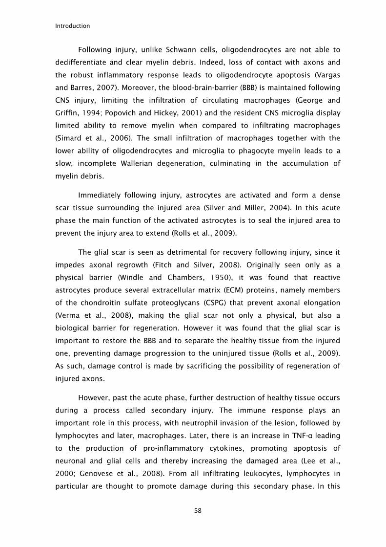
Introduction
58
Following injury, unlike Schwann cells, oligodendrocytes are not able to
dedifferentiate and clear myelin debris. Indeed, loss of contact with axons and
the robust inflammatory response leads to oligodendrocyte apoptosis (Vargas
and Barres, 2007). Moreover, the blood-brain-barrier (BBB) is maintained following
CNS injury, limiting the infiltration of circulating macrophages (George and
Griffin, 1994; Popovich and Hickey, 2001) and the resident CNS microglia display
limited ability to remove myelin when compared to infiltrating macrophages
(Simard et al., 2006). The small infiltration of macrophages together with the
lower ability of oligodendrocytes and microglia to phagocyte myelin leads to a
slow, incomplete Wallerian degeneration, culminating in the accumulation of
myelin debris.
Immediately following injury, astrocytes are activated and form a dense
scar tissue surrounding the injured area (Silver and Miller, 2004). In this acute
phase the main function of the activated astrocytes is to seal the injured area to
prevent the injury area to extend (Rolls et al., 2009).
The glial scar is seen as detrimental for recovery following injury, since it
impedes axonal regrowth (Fitch and Silver, 2008). Originally seen only as a
physical barrier (Windle and Chambers, 1950), it was found that reactive
astrocytes produce several extracellular matrix (ECM) proteins, namely members
of the chondroitin sulfate proteoglycans (CSPG) that prevent axonal elongation
(Verma et al., 2008), making the glial scar not only a physical, but also a
biological barrier for regeneration. However it was found that the glial scar is
important to restore the BBB and to separate the healthy tissue from the injured
one, preventing damage progression to the uninjured tissue (Rolls et al., 2009).
As such, damage control is made by sacrificing the possibility of regeneration of
injured axons.
However, past the acute phase, further destruction of healthy tissue occurs
during a process called secondary injury. The immune response plays an
important role in this process, with neutrophil invasion of the lesion, followed by
lymphocytes and later, macrophages. Later, there is an increase in TNF-α leading
to the production of pro-inflammatory cytokines, promoting apoptosis of
neuronal and glial cells and thereby increasing the damaged area (Lee et al.,
2000; Genovese et al., 2008). From all infiltrating leukocytes, lymphocytes in
particular are thought to promote damage during this secondary phase. In this
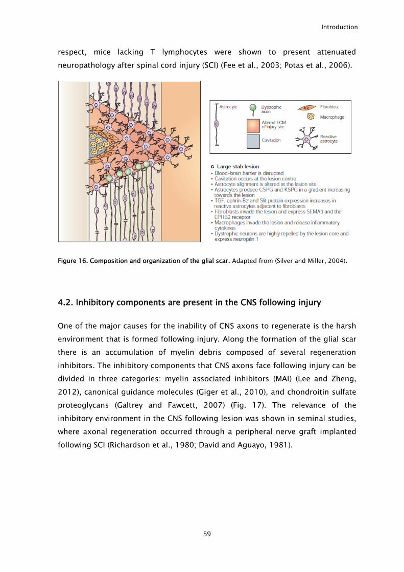
Introduction
59
respect, mice lacking T lymphocytes were shown to present attenuated
neuropathology after spinal cord injury (SCI) (Fee et al., 2003; Potas et al., 2006).
Figure 16. Composition and organization of the glial scar. Adapted from (Silver and Miller, 2004).
4.2. Inhibitory components are present in the CNS following injury
One of the major causes for the inability of CNS axons to regenerate is the harsh
environment that is formed following injury. Along the formation of the glial scar
there is an accumulation of myelin debris composed of several regeneration
inhibitors. The inhibitory components that CNS axons face following injury can be
divided in three categories: myelin associated inhibitors (MAI) (Lee and Zheng,
2012), canonical guidance molecules (Giger et al., 2010), and chondroitin sulfate
proteoglycans (Galtrey and Fawcett, 2007) (Fig. 17). The relevance of the
inhibitory environment in the CNS following lesion was shown in seminal studies,
where axonal regeneration occurred through a peripheral nerve graft implanted
following SCI (Richardson et al., 1980; David and Aguayo, 1981).
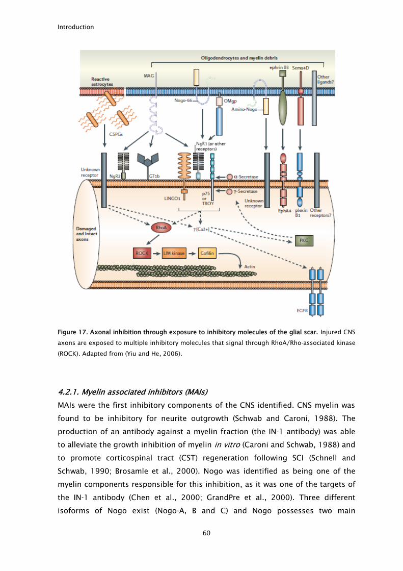
Introduction
60
Figure 17. Axonal inhibition through exposure to inhibitory molecules of the glial scar. Injured CNS
axons are exposed to multiple inhibitory molecules that signal through RhoA/Rho-associated kinase
(ROCK). Adapted from (Yiu and He, 2006).
4.2.1. Myelin associated inhibitors (MAIs)
MAIs were the first inhibitory components of the CNS identified. CNS myelin was
found to be inhibitory for neurite outgrowth (Schwab and Caroni, 1988). The
production of an antibody against a myelin fraction (the IN-1 antibody) was able
to alleviate the growth inhibition of myelin in vitro (Caroni and Schwab, 1988) and
to promote corticospinal tract (CST) regeneration following SCI (Schnell and
Schwab, 1990; Brosamle et al., 2000). Nogo was identified as being one of the
myelin components responsible for this inhibition, as it was one of the targets of
the IN-1 antibody (Chen et al., 2000; GrandPre et al., 2000). Three different
isoforms of Nogo exist (Nogo-A, B and C) and Nogo possesses two main
Introduction
61
inhibitory regions, a putative extracellular amino acid loop called Nogo-66, which
is common to all isoforms, and a segment specific for Nogo-A (Oertle et al.,
2003). Besides Nogo, MAG was also identified as a myelin inhibitor (McKerracher
et al., 1994; Mukhopadhyay et al., 1994). MAG is the only inhibitor that is present
both in PNS and in CNS myelin which could also account for the better
regeneration of PNS axons. Although MAG is able to inhibit neurite outgrowth of
several classes of neurons, it plays a dual role in axonal growth. Adult DRG
neurons are inhibited by MAG, but DRG from newborn animals are stimulated by
this protein. This behavior shift occurs within the first days of life (P4) and is
thought to be related to the decrease in cAMP levels in neurons following
maturation (Mukhopadhyay et al., 1994; DeBellard et al., 1996). More recently
OMgp was also found to be another member of this family (Kottis et al., 2002;
Wang et al., 2002b).
Extensive in vitro work led to the characterization of the molecular
mechanisms by which MAIs promote growth cone collapse and outgrowth
inhibition. The Nogo-66 receptor (NgR) was the first identified receptor for MAIs.
It is a GPI-linked protein expressed in many types of neurons initially described as
interacting with Nogo-66 (Fournier et al., 2001). Although the three known MAIs
do not share any structural feature, MAG and OMgp are also able to interact with
NgR (Domeniconi et al., 2002; Liu et al., 2002; Wang et al., 2002b). NgR lacks a
cytosolic domain capable of transducing the inhibitory signals from MAIs and as
such it needs a co-receptor capable of transducing the inhibitory signals. p75 was
found to be a NgR co-receptor as neurons lacking p75 presented lower inhibition
when facing MAIs (Wang et al., 2002a; Wong et al., 2002; Yamashita et al., 2002;
Park et al., 2005; Shao et al., 2005). Nevertheless, p75 is not expressed in many
neuron populations limiting its relevance in the in vivo inhibitory effect of MAIs.
Later, Troy, another member of the tumor necrosis factor receptor (TNFR) family
which is expressed in most of the adult neurons was identified as the co-receptor
of NgR (Park et al., 2005; Shao et al., 2005). Furthermore, Lingo-1 was also found
to be necessary for the complex NgR/p75 or NgR/Troy to be active and
responsive to MAIs as neurons expressing a dominant negative Lingo-1 present
lower responsiveness to MAIs (Mi et al., 2004). Although NgR was identified as
being a MAI receptor mediating MAI inhibition, NgR KO mice are also inhibited by
MAIs and do not present any improvement in CNS regeneration, suggesting the
existence of other MAI receptors (Zheng et al., 2005). Indeed, PirB was later
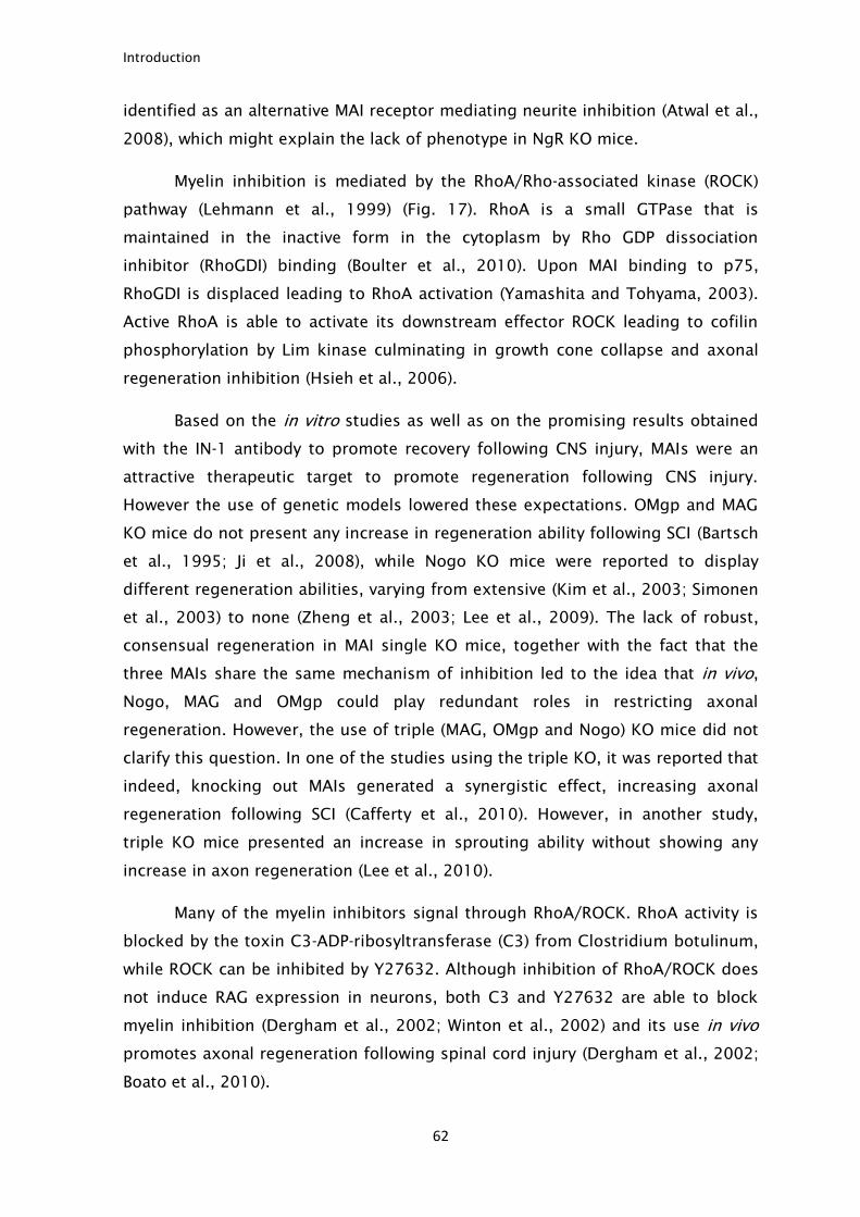
Introduction
62
identified as an alternative MAI receptor mediating neurite inhibition (Atwal et al.,
2008), which might explain the lack of phenotype in NgR KO mice.
Myelin inhibition is mediated by the RhoA/Rho-associated kinase (ROCK)
pathway (Lehmann et al., 1999) (Fig. 17). RhoA is a small GTPase that is
maintained in the inactive form in the cytoplasm by Rho GDP dissociation
inhibitor (RhoGDI) binding (Boulter et al., 2010). Upon MAI binding to p75,
RhoGDI is displaced leading to RhoA activation (Yamashita and Tohyama, 2003).
Active RhoA is able to activate its downstream effector ROCK leading to cofilin
phosphorylation by Lim kinase culminating in growth cone collapse and axonal
regeneration inhibition (Hsieh et al., 2006).
Based on the in vitro studies as well as on the promising results obtained
with the IN-1 antibody to promote recovery following CNS injury, MAIs were an
attractive therapeutic target to promote regeneration following CNS injury.
However the use of genetic models lowered these expectations. OMgp and MAG
KO mice do not present any increase in regeneration ability following SCI (Bartsch
et al., 1995; Ji et al., 2008), while Nogo KO mice were reported to display
different regeneration abilities, varying from extensive (Kim et al., 2003; Simonen
et al., 2003) to none (Zheng et al., 2003; Lee et al., 2009). The lack of robust,
consensual regeneration in MAI single KO mice, together with the fact that the
three MAIs share the same mechanism of inhibition led to the idea that in vivo,
Nogo, MAG and OMgp could play redundant roles in restricting axonal
regeneration. However, the use of triple (MAG, OMgp and Nogo) KO mice did not
clarify this question. In one of the studies using the triple KO, it was reported that
indeed, knocking out MAIs generated a synergistic effect, increasing axonal
regeneration following SCI (Cafferty et al., 2010). However, in another study,
triple KO mice presented an increase in sprouting ability without showing any
increase in axon regeneration (Lee et al., 2010).
Many of the myelin inhibitors signal through RhoA/ROCK. RhoA activity is
blocked by the toxin C3-ADP-ribosyltransferase (C3) from Clostridium botulinum,
while ROCK can be inhibited by Y27632. Although inhibition of RhoA/ROCK does
not induce RAG expression in neurons, both C3 and Y27632 are able to block
myelin inhibition (Dergham et al., 2002; Winton et al., 2002) and its use in vivo
promotes axonal regeneration following spinal cord injury (Dergham et al., 2002;
Boato et al., 2010).
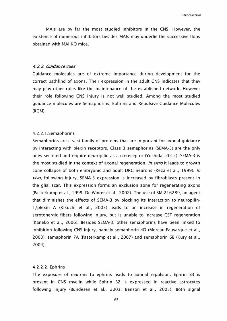
Introduction
63
MAIs are by far the most studied inhibitors in the CNS. However, the
existence of numerous inhibitors besides MAIs may underlie the successive flops
obtained with MAI KO mice.
4.2.2. Guidance cues
Guidance molecules are of extreme importance during development for the
correct pathfind of axons. Their expression in the adult CNS indicates that they
may play other roles like the maintenance of the established network. However
their role following CNS injury is not well studied. Among the most studied
guidance molecules are Semaphorins, Ephrins and Repulsive Guidance Molecules
(RGM).
4.2.2.1.Semaphorins
Semaphorins are a vast family of proteins that are important for axonal guidance
by interacting with plexin receptors. Class 3 semaphorins (SEMA-3) are the only
ones secreted and require neuropilin as a co-receptor (Yoshida, 2012). SEMA-3 is
the most studied in the context of axonal regeneration. In vitro it leads to growth
cone collapse of both embryonic and adult DRG neurons (Reza et al., 1999). In
vivo, following injury, SEMA-3 expression is increased by fibroblasts present in
the glial scar. This expression forms an exclusion zone for regenerating axons
(Pasterkamp et al., 1999; De Winter et al., 2002). The use of SM-216289, an agent
that diminishes the effects of SEMA-3 by blocking its interaction to neuropilin-
1/plexin A (Kikuchi et al., 2003) leads to an increase in regeneration of
serotonergic fibers following injury, but is unable to increase CST regeneration
(Kaneko et al., 2006). Besides SEMA-3, other semaphorins have been linked to
inhibition following CNS injury, namely semaphorin 4D (Moreau-Fauvarque et al.,
2003), semaphorin 7A (Pasterkamp et al., 2007) and semaphorin 6B (Kury et al.,
2004).
4.2.2.2. Ephrins
The exposure of neurons to ephrins leads to axonal repulsion. Ephrin B3 is
present in CNS myelin while Ephrin B2 is expressed in reactive astrocytes
following injury (Bundesen et al., 2003; Benson et al., 2005). Both signal

Introduction
64
inhibition through the EphA4 receptor and lead to neurite growth inhibition
(Benson et al., 2005; Fabes et al., 2006). In vivo, blockage of Ephrin A4 receptor
does not promote regeneration, but CST sprouting is observed (Fabes et al.,
2007).
Besides axonal growth inhibition, ephrins may be important in the glial
scar formation. This hypothesis is supported by the fact that the EphA3 receptor
and EphA7 are expressed in astrocytes following injury (Irizarry-Ramirez et al.,
2005; Figueroa et al., 2006) and also by the reduced glial scar formation in
Ephrin A4 receptor KO mice (Goldshmit et al., 2004).
4.2.2.3. Repulsive guidance molecules (RGM)
RGM are a family of cell membrane associated glycosylphosphatidylinositol
anchored glycoproteins. This family was identified as being repulsive and,
important for the guidance of chicken temporal retinal axons (Monnier et al.,
2002). In mice, 3 RGM have been identified (RGMa, RGMb and RGMc). Following
SCI, there is an increased expression of RGM around the injured area leading to
axon growth inhibition (Schwab et al., 2005; Hata et al., 2006). Local
neutralization of RGMa with a specific antibody promotes axonal regeneration
and functional recovery (Hata et al., 2006). Neogenin receptor was identified as
being the only receptor for RGMa, and it mediates its inhibitory effect
(Rajagopalan et al., 2004) by a mechanism dependent on the RhoA/ROCK
pathway (Conrad et al., 2007). Besides axonal guidance and inhibition several
other functions were described for RGMa such as importance in neural tube
morphogenesis, cell adhesion, cell migration, cell polarity and cell differentiation
(Key and Lah, 2012).
4.2.3. Chondroitin sulphate proteoglycans (CSPGs)
Upon injury, astrocytes are activated and start the production of extracellular
matrix proteins, the CSPGs. These proteins represent a vast family of ECM
proteins composed of a protein core linked to sulphated glycosaminoglycan
chains. Some members of this family are: aggrecan, brevican, phosphacan,
neurocan, versican and NG-2 (Silver and Miller, 2004; Rolls et al., 2009). Their
importance as inhibitors of axonal regeneration is supported by three evidences:
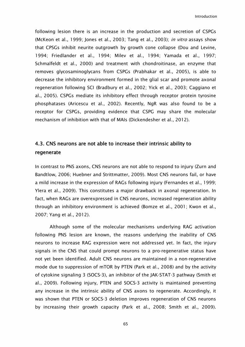
Introduction
65
following lesion there is an increase in the production and secretion of CSPGs
(McKeon et al., 1999; Jones et al., 2003; Tang et al., 2003); in vitro assays show
that CPSGs inhibit neurite outgrowth by growth cone collapse (Dou and Levine,
1994; Friedlander et al., 1994; Milev et al., 1994; Yamada et al., 1997;
Schmalfeldt et al., 2000) and treatment with chondroitinase, an enzyme that
removes glycosaminoglycans from CSPGs (Prabhakar et al., 2005), is able to
decrease the inhibitory environment formed in the glial scar and promote axonal
regeneration following SCI (Bradbury et al., 2002; Yick et al., 2003; Caggiano et
al., 2005). CSPGs mediate its inhibitory effect through receptor protein tyrosine
phosphatases (Aricescu et al., 2002). Recently, NgR was also found to be a
receptor for CSPGs, providing evidence that CSPG may share the molecular
mechanism of inhibition with that of MAIs (Dickendesher et al., 2012).
4.3. CNS neurons are not able to increase their intrinsic ability to
regenerate
In contrast to PNS axons, CNS neurons are not able to respond to injury (Zurn and
Bandtlow, 2006; Huebner and Strittmatter, 2009). Most CNS neurons fail, or have
a mild increase in the expression of RAGs following injury (Fernandes et al., 1999;
Ylera et al., 2009). This constitutes a major drawback in axonal regeneration. In
fact, when RAGs are overexpressed in CNS neurons, increased regeneration ability
through an inhibitory environment is achieved (Bomze et al., 2001; Kwon et al.,
2007; Yang et al., 2012).
Although some of the molecular mechanisms underlying RAG activation
following PNS lesion are known, the reasons underlying the inability of CNS
neurons to increase RAG expression were not addressed yet. In fact, the injury
signals in the CNS that could prompt neurons to a pro-regenerative status have
not yet been identified. Adult CNS neurons are maintained in a non-regenerative
mode due to suppression of mTOR by PTEN (Park et al., 2008) and by the activity
of cytokine signaling 3 (SOCS-3), an inhibitor of the JAK-STAT-3 pathway (Smith et
al., 2009). Following injury, PTEN and SOCS-3 activity is maintained preventing
any increase in the intrinsic ability of CNS axons to regenerate. Accordingly, it
was shown that PTEN or SOCS-3 deletion improves regeneration of CNS neurons
by increasing their growth capacity (Park et al., 2008; Smith et al., 2009).
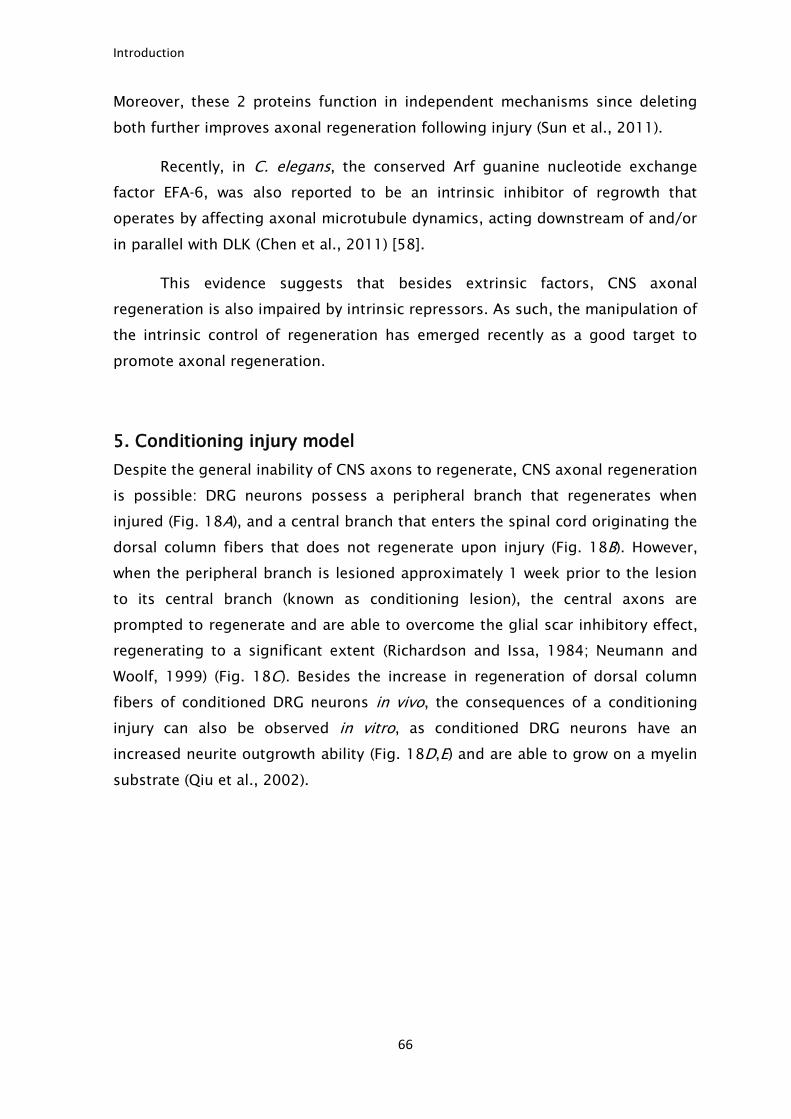
Introduction
66
Moreover, these 2 proteins function in independent mechanisms since deleting
both further improves axonal regeneration following injury (Sun et al., 2011).
Recently, in C. elegans, the conserved Arf guanine nucleotide exchange
factor EFA-6, was also reported to be an intrinsic inhibitor of regrowth that
operates by affecting axonal microtubule dynamics, acting downstream of and/or
in parallel with DLK (Chen et al., 2011) [58].
This evidence suggests that besides extrinsic factors, CNS axonal
regeneration is also impaired by intrinsic repressors. As such, the manipulation of
the intrinsic control of regeneration has emerged recently as a good target to
promote axonal regeneration.
5. Conditioning injury model
Despite the general inability of CNS axons to regenerate, CNS axonal regeneration
is possible: DRG neurons possess a peripheral branch that regenerates when
injured (Fig. 18A), and a central branch that enters the spinal cord originating the
dorsal column fibers that does not regenerate upon injury (Fig. 18B). However,
when the peripheral branch is lesioned approximately 1 week prior to the lesion
to its central branch (known as conditioning lesion), the central axons are
prompted to regenerate and are able to overcome the glial scar inhibitory effect,
regenerating to a significant extent (Richardson and Issa, 1984; Neumann and
Woolf, 1999) (Fig. 18C). Besides the increase in regeneration of dorsal column
fibers of conditioned DRG neurons in vivo, the consequences of a conditioning
injury can also be observed in vitro, as conditioned DRG neurons have an
increased neurite outgrowth ability (Fig. 18D,E) and are able to grow on a myelin
substrate (Qiu et al., 2002).
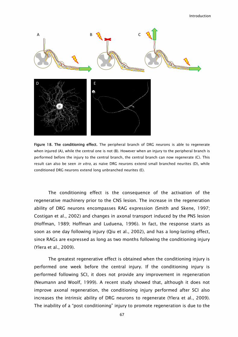
Introduction
67
Figure 18. The conditioning effect. The peripheral branch of DRG neurons is able to regenerate
when injured (A), while the central one is not (B). However when an injury to the peripheral branch is
performed before the injury to the central branch, the central branch can now regenerate (C). This
result can also be seen in vitro, as naïve DRG neurons extend small branched neurites (D), while
conditioned DRG neurons extend long unbranched neurites (E).
The conditioning effect is the consequence of the activation of the
regenerative machinery prior to the CNS lesion. The increase in the regeneration
ability of DRG neurons encompasses RAG expression (Smith and Skene, 1997;
Costigan et al., 2002) and changes in axonal transport induced by the PNS lesion
(Hoffman, 1989; Hoffman and Luduena, 1996). In fact, the response starts as
soon as one day following injury (Qiu et al., 2002), and has a long-lasting effect,
since RAGs are expressed as long as two months following the conditioning injury
(Ylera et al., 2009).
The greatest regenerative effect is obtained when the conditioning injury is
performed one week before the central injury. If the conditioning injury is
performed following SCI, it does not provide any improvement in regeneration
(Neumann and Woolf, 1999). A recent study showed that, although it does not
improve axonal regeneration, the conditioning injury performed after SCI also
increases the intrinsic ability of DRG neurons to regenerate (Ylera et al., 2009).
The inability of a “post conditioning” injury to promote regeneration is due to the
A B C
D E
Introduction
68
establishment of a thick glial scar that does not allow the regrowth of axons, even
when expressing RAGs. In fact, the authors elegantly show that when performing
laser single axon lesion, that cause minimal scarring, a conditioning lesion
performed two weeks after is able to promote the regeneration of the central
axons (Ylera et al., 2009). These experiments suggest that following a SCI, there
is a small window of opportunity for treatment, before the glial scar becomes too
thick. A prolonged delay in treatment, allows the formation of a thick glial scar
that prevents the regeneration of central axons, even when these have an
increased intrinsic ability to regrow.
The conditioning effect represents a good model on how increasing the
intrinsic ability of axons to regrow can improve axonal regeneration. As such,
understanding the mechanism by which a conditioning injury prompts CNS axons
to regenerate has been the focus of attention on the axonal regeneration field for
many years. The most relevant finding was the fact that at least in part, the
conditioning effect was mediated by cAMP (Qiu et al., 2002). This finding
together with others made cAMP the main target to promote regeneration. In fact,
the use of phosphodiesterase (enzyme responsible for cAMP degradation)
inhibitors, such as rolipram was shown to increase cAMP levels and improve
recovery following SCI (Nikulina et al., 2004; Pearse et al., 2004). However, the
use of phosphodiesterase inhibitors leads to disabling nausea, limiting its clinical
application.
Several studies using array techniques identified broad changes in gene
expression of conditioned neurons (Costigan et al., 2002; Cao et al., 2006). In
fact these changes are regulated by the activation of multiple transcription factors
including c-Jun, sox11, CREB, Smad1, ATF-3, AKRD1, nuclear factor interleukin 3
regulated (NFIL3) and STAT-3 (Kiryu-Seo and Kiyama, 2011). The conditioning
injury triggers the expression of traditional RAGs such as GAP-43 and CAP-23
(Hoffman, 1989), but it also allowed the identification of novel RAGs such as Arg-
1 (Cai et al., 2002; Deng et al., 2009), leukemia inhibitory factor (Cafferty et al.,
2001), IL-6 (Cafferty et al., 2004; Cao et al., 2006) and tissue plasminogen
activator (Minor et al., 2009). However, none of the identified transcription factor
or RAGs is able to reproduce the extension of the conditioning effect. As such,
the quest for clinically relevant molecules that mimic the conditioning lesion
should be pursued.
Introduction
69
Besides the transcription changes observed following conditioning injury,
another important alteration occurs that may contribute to axonal regeneration. It
has been shown that a conditioning injury is able to increase axonal transport in
both DRG branches (Hoffman and Luduena, 1996; Hoffman, 2010). Furthermore,
these alterations were identified as being independent of cAMP, since cAMP is
able to trigger transcription changes but it does not alter axonal transport (Han et
al., 2004). It has also been shown that conditioned neurons present increased
protein synthesis in their axons following injury, and this local synthesis may also
underlie their high ability to regenerate (Zheng et al., 2001; Verma et al., 2005).
Recently, histone deacetylase 5 (HDAC5) has been described as a key
element in the conditioning response. Upon injury, the initial depolarization wave
is able to activate protein kinase Cµ at the cell body promoting nuclear export of
HDAC5. Absence of HDAC5 at the nucleus leads to increase histone acetylation
activating the pro-regenerative program (Cho et al., 2013). This epigenetic
mechanism controls the switch from non-regenerative to growth-competent
axons. This initial phase where histones are acetylated prime the neuronal DNA to
the positive injury signals that will be conveyed later through the retrograde
transport machinery (Cho et al., 2013). In the proposed mechanism, the initial
depolarization alters neurons so that they can respond rapidly to further signals,
explaining why the initial depolarization by itself cannot trigger a long-lasting
response.
In summary a conditioning injury triggers a robust response to induce
regeneration. Transcriptional changes together with changes in local protein
synthesis and axonal transport make the conditioning injury a powerful paradigm
that can hardly be reproduced by the stimulation of a single gene/protein.
6. Important tools to study CNS axonal regeneration
The complexity of the CNS makes the study of CNS axonal regeneration a great
challenge. It is important to distinguish the different types of axonal
regeneration, as well as the different models available to study it. In this chapter
such features will be discussed.
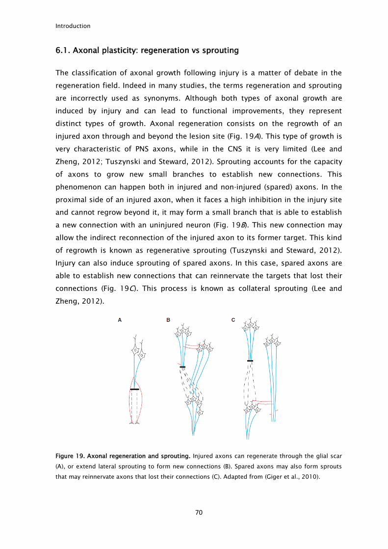
Introduction
70
6.1. Axonal plasticity: regeneration vs sprouting
The classification of axonal growth following injury is a matter of debate in the
regeneration field. Indeed in many studies, the terms regeneration and sprouting
are incorrectly used as synonyms. Although both types of axonal growth are
induced by injury and can lead to functional improvements, they represent
distinct types of growth. Axonal regeneration consists on the regrowth of an
injured axon through and beyond the lesion site (Fig. 19A). This type of growth is
very characteristic of PNS axons, while in the CNS it is very limited (Lee and
Zheng, 2012; Tuszynski and Steward, 2012). Sprouting accounts for the capacity
of axons to grow new small branches to establish new connections. This
phenomenon can happen both in injured and non-injured (spared) axons. In the
proximal side of an injured axon, when it faces a high inhibition in the injury site
and cannot regrow beyond it, it may form a small branch that is able to establish
a new connection with an uninjured neuron (Fig. 19B). This new connection may
allow the indirect reconnection of the injured axon to its former target. This kind
of regrowth is known as regenerative sprouting (Tuszynski and Steward, 2012).
Injury can also induce sprouting of spared axons. In this case, spared axons are
able to establish new connections that can reinnervate the targets that lost their
connections (Fig. 19C). This process is known as collateral sprouting (Lee and
Zheng, 2012).
Figure 19. Axonal regeneration and sprouting. Injured axons can regenerate through the glial scar
(A), or extend lateral sprouting to form new connections (B). Spared axons may also form sprouts
that may reinnervate axons that lost their connections (C). Adapted from (Giger et al., 2010).
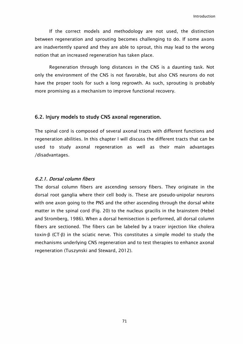
Introduction
71
If the correct models and methodology are not used, the distinction
between regeneration and sprouting becomes challenging to do. If some axons
are inadvertently spared and they are able to sprout, this may lead to the wrong
notion that an increased regeneration has taken place.
Regeneration through long distances in the CNS is a daunting task. Not
only the environment of the CNS is not favorable, but also CNS neurons do not
have the proper tools for such a long regrowth. As such, sprouting is probably
more promising as a mechanism to improve functional recovery.
6.2. Injury models to study CNS axonal regeneration.
The spinal cord is composed of several axonal tracts with different functions and
regeneration abilities. In this chapter I will discuss the different tracts that can be
used to study axonal regeneration as well as their main advantages
/disadvantages.
6.2.1. Dorsal column fibers
The dorsal column fibers are ascending sensory fibers. They originate in the
dorsal root ganglia where their cell body is. These are pseudo-unipolar neurons
with one axon going to the PNS and the other ascending through the dorsal white
matter in the spinal cord (Fig. 20) to the nucleus gracilis in the brainstem (Hebel
and Stromberg, 1986). When a dorsal hemisection is performed, all dorsal column
fibers are sectioned. The fibers can be labeled by a tracer injection like cholera
toxin-β (CT-β) in the sciatic nerve. This constitutes a simple model to study the
mechanisms underlying CNS regeneration and to test therapies to enhance axonal
regeneration (Tuszynski and Steward, 2012).
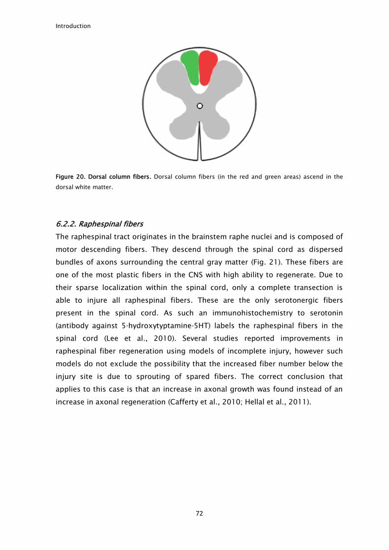
Introduction
72
Figure 20. Dorsal column fibers. Dorsal column fibers (in the red and green areas) ascend in the
dorsal white matter.
6.2.2. Raphespinal fibers
The raphespinal tract originates in the brainstem raphe nuclei and is composed of
motor descending fibers. They descend through the spinal cord as dispersed
bundles of axons surrounding the central gray matter (Fig. 21). These fibers are
one of the most plastic fibers in the CNS with high ability to regenerate. Due to
their sparse localization within the spinal cord, only a complete transection is
able to injure all raphespinal fibers. These are the only serotonergic fibers
present in the spinal cord. As such an immunohistochemistry to serotonin
(antibody against 5-hydroxytyptamine-5HT) labels the raphespinal fibers in the
spinal cord (Lee et al., 2010). Several studies reported improvements in
raphespinal fiber regeneration using models of incomplete injury, however such
models do not exclude the possibility that the increased fiber number below the
injury site is due to sprouting of spared fibers. The correct conclusion that
applies to this case is that an increase in axonal growth was found instead of an
increase in axonal regeneration (Cafferty et al., 2010; Hellal et al., 2011).
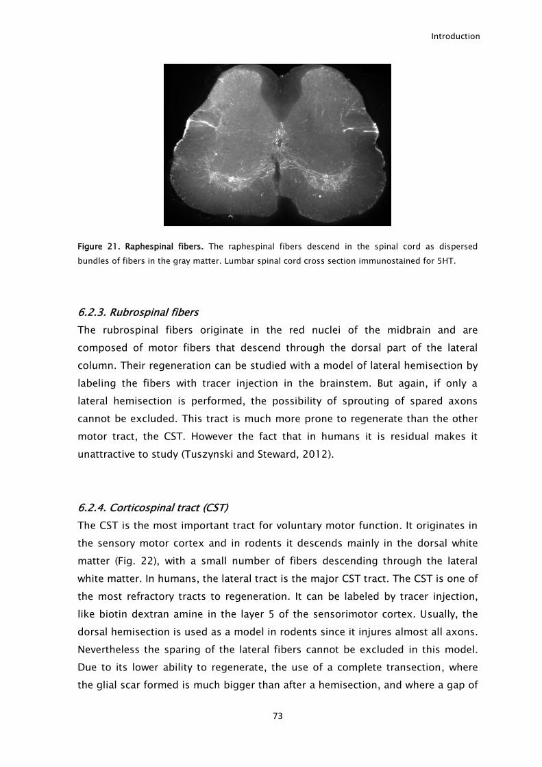
Introduction
73
Figure 21. Raphespinal fibers. The raphespinal fibers descend in the spinal cord as dispersed
bundles of fibers in the gray matter. Lumbar spinal cord cross section immunostained for 5HT.
6.2.3. Rubrospinal fibers
The rubrospinal fibers originate in the red nuclei of the midbrain and are
composed of motor fibers that descend through the dorsal part of the lateral
column. Their regeneration can be studied with a model of lateral hemisection by
labeling the fibers with tracer injection in the brainstem. But again, if only a
lateral hemisection is performed, the possibility of sprouting of spared axons
cannot be excluded. This tract is much more prone to regenerate than the other
motor tract, the CST. However the fact that in humans it is residual makes it
unattractive to study (Tuszynski and Steward, 2012).
6.2.4. Corticospinal tract (CST)
The CST is the most important tract for voluntary motor function. It originates in
the sensory motor cortex and in rodents it descends mainly in the dorsal white
matter (Fig. 22), with a small number of fibers descending through the lateral
white matter. In humans, the lateral tract is the major CST tract. The CST is one of
the most refractory tracts to regeneration. It can be labeled by tracer injection,
like biotin dextran amine in the layer 5 of the sensorimotor cortex. Usually, the
dorsal hemisection is used as a model in rodents since it injures almost all axons.
Nevertheless the sparing of the lateral fibers cannot be excluded in this model.
Due to its lower ability to regenerate, the use of a complete transection, where
the glial scar formed is much bigger than after a hemisection, and where a gap of
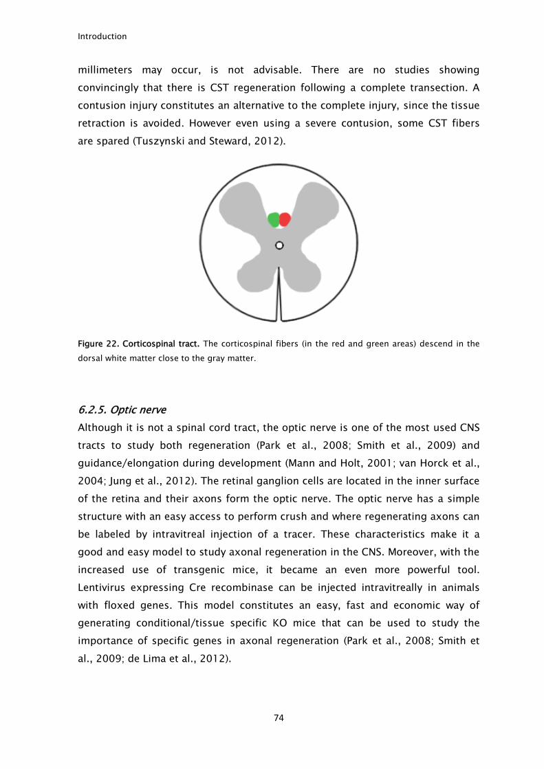
Introduction
74
millimeters may occur, is not advisable. There are no studies showing
convincingly that there is CST regeneration following a complete transection. A
contusion injury constitutes an alternative to the complete injury, since the tissue
retraction is avoided. However even using a severe contusion, some CST fibers
are spared (Tuszynski and Steward, 2012).
Figure 22. Corticospinal tract. The corticospinal fibers (in the red and green areas) descend in the
dorsal white matter close to the gray matter.
6.2.5. Optic nerve
Although it is not a spinal cord tract, the optic nerve is one of the most used CNS
tracts to study both regeneration (Park et al., 2008; Smith et al., 2009) and
guidance/elongation during development (Mann and Holt, 2001; van Horck et al.,
2004; Jung et al., 2012). The retinal ganglion cells are located in the inner surface
of the retina and their axons form the optic nerve. The optic nerve has a simple
structure with an easy access to perform crush and where regenerating axons can
be labeled by intravitreal injection of a tracer. These characteristics make it a
good and easy model to study axonal regeneration in the CNS. Moreover, with the
increased use of transgenic mice, it became an even more powerful tool.
Lentivirus expressing Cre recombinase can be injected intravitreally in animals
with floxed genes. This model constitutes an easy, fast and economic way of
generating conditional/tissue specific KO mice that can be used to study the
importance of specific genes in axonal regeneration (Park et al., 2008; Smith et
al., 2009; de Lima et al., 2012).
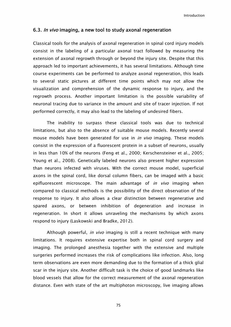
Introduction
75
6.3. In vivo imaging, a new tool to study axonal regeneration
Classical tools for the analysis of axonal regeneration in spinal cord injury models
consist in the labeling of a particular axonal tract followed by measuring the
extension of axonal regrowth through or beyond the injury site. Despite that this
approach led to important achievements, it has several limitations. Although time
course experiments can be performed to analyze axonal regeneration, this leads
to several static pictures at different time points which may not allow the
visualization and comprehension of the dynamic response to injury, and the
regrowth process. Another important limitation is the possible variability of
neuronal tracing due to variance in the amount and site of tracer injection. If not
performed correctly, it may also lead to the labeling of undesired fibers.
The inability to surpass these classical tools was due to technical
limitations, but also to the absence of suitable mouse models. Recently several
mouse models have been generated for use in in vivo imaging. These models
consist in the expression of a fluorescent protein in a subset of neurons, usually
in less than 10% of the neurons (Feng et al., 2000; Kerschensteiner et al., 2005;
Young et al., 2008). Genetically labeled neurons also present higher expression
than neurons infected with viruses. With the correct mouse model, superficial
axons in the spinal cord, like dorsal column fibers, can be imaged with a basic
epifluorescent microscope. The main advantage of in vivo imaging when
compared to classical methods is the possibility of the direct observation of the
response to injury. It also allows a clear distinction between regenerative and
spared axons, or between inhibition of degeneration and increase in
regeneration. In short it allows unraveling the mechanisms by which axons
respond to injury (Laskowski and Bradke, 2012).
Although powerful, in vivo imaging is still a recent technique with many
limitations. It requires extensive expertise both in spinal cord surgery and
imaging. The prolonged anesthesia together with the extensive and multiple
surgeries performed increases the risk of complications like infection. Also, long
term observations are even more demanding due to the formation of a thick glial
scar in the injury site. Another difficult task is the choice of good landmarks like
blood vessels that allow for the correct measurement of the axonal regeneration
distance. Even with state of the art multiphoton microscopy, live imaging allows
Introduction
76
only observation of relatively superficial axons (50µm deep), limiting the axonal
tracts evaluated.
Recently two methods were described for chronic imaging of spinal cord
injury. They consist in the implantation of a homemade chamber in the vertebral
column (Farrar et al., 2012) or of a glass window attached to the spinal cord
(Fenrich et al., 2012). Both methods present minimal scarring enabling successful
imaging for as much as 22 imaging sessions up to 350 days following surgery.
The other main advantage is the possibility of visualizing chronic injury with a
single surgery. It is exciting to see if the next developments of this technique
fulfill its great potential.
7. Possible treatments to achieve axonal regeneration following
spinal cord injury.
In the last two decades, methylprednisolone was the only drug administered
following SCI. Although initially described as improving functional recovery, in
recent studies its benefits have been questioned (Coleman et al., 2000; Hurlbert,
2000; Bracken, 2001; Miller, 2008), which led to abandon methylprednisolone as
a treatment to SCI. Nowadays the only treatment available following SCI is
rehabilitation. The main goal of rehabilitation is to prevent further complications
and maximize physical function. As such, there is a total absence of therapies to
promote axonal regeneration and functional recovery of patients with acute or
chronic SCI.
The limitations for axonal regeneration in the CNS are well identified. The
lack of an intrinsic regenerative ability, together with the formation of a thick glial
scar, where injured axons are exposed to several inhibitors poses a great
challenge to achieve axonal regeneration. As such, most pre-clinical strategies
consist in overcoming these two main obstacles, either by stimulating neurons to
increase the intrinsic ability to regrow their axons or by modulating the injury site
environment so that it becomes less inhibitory.
Introduction
77
7.1. Modulation of the injury site environment
Modulation of the injury environment was the first strategy used successfully in
animal models. The neutralization of the inhibitors present in the spinal cord
following injury is an attractive strategy for treatment. Initial studies using IN-1,
an antibody produced against myelin components showed promising results,
promoting axonal regeneration following SCI (Schnell and Schwab, 1990). As
described above, IN-1 recognizes Nogo-A, the most studied regeneration inhibitor
(Chen et al., 2000), but it is not specific for Nogo-A (Caroni and Schwab, 1988).
Other antibodies produced against Nogo showed less pronounced improvements
(Liebscher et al., 2005). Even when neutralizing Nogo, several other inhibitors are
still present. Many of the inhibitors like other MAIs or CSPG share a common
receptor, NgR. The blockage of NgR should then have a broader effect since it
may block the effect of several inhibitors. Indeed peptide blocking of NgR was
reported to improve axonal regeneration (GrandPre et al., 2002; Li and
Strittmatter, 2003; Cao et al., 2008). However, the use of the blocking peptide
was re-assessed and no relevant improvements were found (Steward et al., 2008).
Besides Nogo, antibody blocking of other inhibitors like RGMa (Hata et al., 2006)
and proteoglycan NG2 (Tan et al., 2006) also diminishes the inhibitory
environment following injury promoting axonal regeneration.
CSPGs are one of the most important inhibitory components of the glial
scar. The degradation of the glycosaminoglycan chains by chondroitinase ABC
diminishes the inhibitory effect of CSPGs and its use in vivo has led to significant
functional improvements in CNS regeneration (Moon et al., 2001; Bradbury et al.,
2002; Yick et al., 2003; Caggiano et al., 2005; Tester and Howland, 2008).
Although blocking a particular inhibitor may lead to improvements, the reality is
that several other inhibitory molecules are still present and may take the place of
the inhibitor neutralized. Multi-targeting several inhibitory classes should
promote better outcomes.
Another important approach to overcome the inhibitory environment at the
lesion site is to provide “bridges” that provide axons an alternative path without
contacting the inhibitors. The first bridge successfully used was a peripheral
nerve graft which allowed for regeneration of CNS axons (Richardson et al., 1980;
David and Aguayo, 1981). Transplant of fetal spinal cord to the injury site also
promotes the growth of axons within the injury site (Jakeman and Reier, 1991).
Introduction
78
The use of cell transplantation has caught most of the attention in an attempt to
“build bridges” in the injury site. The main goal is to have cells that integrate well
within the injury and form a cellular matrix capable of linking the rostral and
distal injury borders (Lu and Tuszynski, 2008). The successful use of PNS graft by
(David and Aguayo, 1981), raised the possibility that Schwann cells could
ameliorate the environment following SCI improving functional recovery. Indeed it
has been shown that Schwann cell transplantation produces functional recovery
(Pinzon et al., 2001; Takami et al., 2002). In fact, the studies with cell
transplantation following SCI to improve recovery have been growing extensively.
In this respect, bone marrow stromal cell (MSC) transplantation has been widely
used to facilitate the bridging following injury. When used in combination with
other treatments that increase the intrinsic regeneration ability of neurons, MSCs
have a synergistic effect (Lu et al., 2004; Kadoya et al., 2009). Transplantation of
olfactory ensheating cells (OEC) has also been reported to promote improvements
following SCI. It has been shown that OEC transplantation improves both survival
and regeneration following SCI by producing neurotrophic factors, as well as by
reducing the CSPG production by astrocytes (Tohda and Kuboyama, 2011). One of
the most attractive features of MSC and OEC transplantation is the possibility of
autologous transplant. Besides trophic support, cell transplantation can be used
to replace the lost cells following injury. In this case, stem cell transplantation
becomes important in doing so. Both embryonic stem cells (ESCs) and neural
stem cells (NSCs) have been used successfully, improving functional recovery and
being able to differentiate in oligodendrocytes and neurons (Pego et al., 2012).
Recently, the implantation of embryonic neural stem cells following spinal cord
injury has been shown to increase regeneration and functional recovery
remarkably. The implanted cells were able to extend axons and build new circuits
that led to improved function (Lu et al., 2012).
7.2. Increase of the intrinsic regeneration ability of CNS neurons
cAMP is one of the most important intracellular signals. It is synthesized from
ATP by adenylyl cyclase at the plasma membrane and it is degraded by
phosphodiesterase. For the past 15 years, cAMP got most of the attention in the
axonal growth and regeneration field. Young CNS neurons have high ability to
Introduction
79
elongate. This ability is lost during development due to a decrease in cAMP levels
(Cai et al., 2001). The high levels of cAMP during development also make axon
growth cones attracted to guidance cues like netrin-1 and SEMA-3, that are
repellent to adult neurons (Ming et al., 1997; Song et al., 1998). Two of the most
relevant findings were that cAMP can prompt neurons to overcome myelin
inhibition in vitro (Cai et al., 1999; Qiu et al., 2002), and that treatment with db-
cAMP, a cell permeable analog of cAMP increases the regeneration of dorsal
column fibers following SCI (Neumann et al., 2002; Qiu et al., 2002). This led to
the notion that the manipulation of cAMP levels in neurons could allow them to
overcome the inhibitory environment following SCI. The use of phosphodiesterase
inhibitor drugs like rolipram increases cAMP levels by preventing its degradation
(Udina et al., 2010), leading to increased axonal regeneration and functional
recovery (Nikulina et al., 2004; Bretzner et al., 2010). However, the use of such
drugs induces disabling nausea making them unsuitable for treatment.
Furthermore, the robust effects of cAMP initially reported have been questioned
by recent studies (Han et al., 2004; Blesch et al., 2012).
Besides cAMP, some of its downstream targets such as Arg-1 or IL-6 have
been tested as possible enhancers. IL-6 was identified as being able to induce
regeneration (Cafferty et al., 2004; Cao et al., 2006) and daidzein (a soy
isoflavone) has been identified as a potent inducer of Arg-1 expression that can
induce axonal protection and growth (Ma et al., 2010). However, none of them
has shown enough potential to be relevant in an in vivo human condition.
Recently, the regeneration field has been focused on PTEN and SOCS-3.
These 2 proteins were identified as potent regeneration inhibitors, as their
removal promotes a robust increase in regeneration (Park et al., 2008; Sun et al.,
2011; de Lima et al., 2012). Despite the robust effects described so far, further
studies are needed to strengthen their role in axonal regeneration.
Neurotrophins are a class of small molecules that are important for the
regulation of neuronal functions. NGF was the first neurotrophin identified. After
NGF, many others were identified: BDNF, neurotrophin 3 and 4 (NT-3/4) and
GDNF. They regulate neuronal survival and the interaction of neurons with their
targets (Kirstein and Farinas, 2002). Moreover, they are also capable of inducing
axonal growth and RAG expression (Gillespie, 2003) and to suppress myelin
inhibition by increasing cAMP levels (Cai et al., 1999; Gao et al., 2003). When
Introduction
80
applied following SCI, neurotrophins lead to functional improvements and to
axonal regeneration of different tracts. NGF, BDNF and NT-3 are the most used
ones with reports of all of them showing increased axonal regeneration (Schnell
et al., 1994; Oudega and Hagg, 1996; Bregman et al., 1997; Bradbury et al.,
1999; Oudega and Hagg, 1999; Song et al., 2008). The injection of lentivirus
expressing NT-3 distally to the injury site was shown to create a chemoattractant
gradient allowing axonal bridging of the injury site (Taylor et al., 2006).
Inflammation is a crucial step following injury encompassing the
production of pro-inflammatory cytokines like TNF-α and IL-1β, and leukocyte
recruitment. Usually its effects are seen as deleterious, and as discussed above,
methylprednisolone a potent anti-inflammatory drug was previously seen as the
only treatment available. Although it was reported initially as improving
functional recovery, later studies questioned such evidences. The use of the anti-
inflammatory cytokines interleukin 10 (IL-10) and IL-6 has shown improvements
in recovery following SCI. IL-10 has a neuroprotective effect and its use promotes
neuronal survival of injured neurons (Zhou et al., 2009), while IL-6 increases the
axonal growth ability of neurons and overcomes myelin inhibition (Cafferty et al.,
2004; Cao et al., 2006). Its use following injury promotes functional recovery by
increasing axonal regeneration (Cafferty et al., 2004; Cao et al., 2006; Yang et al.,
2012). Another evidence that the inflammatory response could be modulated to
improve axonal regeneration is the ability of activated macrophages to promote
regeneration of retinal ganglion cells following optic nerve injury. Activated
macrophages are able to produce oncomodulin that increases the ability of
neurons to grow their axons (Yin et al., 2006). However, in the context of SCI, the
use of zymosan, a yeast cell wall preparation known for its ability to activate
macrophages, leads to improvements by reducing the injured area and not by
improving the ability of neurons to regenerate (Benowitz and Popovich, 2011).
Injured CNS axons usually fail to form a growth cone and instead originate
a non-regenerative structure called retraction bulb. The formation of a growth
cone instead of a retraction bulb is absolutely essential to start regeneration
(Bradke et al., 2012). Recent studies have focused on promoting regeneration by
the direct action on the injured axon tip, increasing the possibility of growth cone
formation. Cytoskeleton components are the strongest candidates to promote the
formation of a growth cone. Indeed, the use of taxol stabilizes microtubules and
promotes the formation of growth cones which can overcome the myelin
Introduction
81
inhibitory effect (Erturk et al., 2007). The use of taxol was also shown to improve
axonal regeneration following SCI (Hellal et al., 2011).
Most of the therapies referred so far require local administration at the
injury site, either by local injection of cells, or by the implantation of osmotic
pumps to deliver antibodies or drugs. This type of treatment, involves at least
one invasive procedure, increasing the risk of complications in patient’s recovery.
As such, the use of electrical stimulation in a non-invasive manner for treatment
poses an attractive alternative. However, the effects of electrical stimulation are
not consensual, although it has been reported as beneficial following SCI being
able to promote regeneration of dorsal column fibers by increasing their intrinsic
ability to regenerate (Udina et al., 2008).
7.3. Combined therapies
Although many single therapies are able to improve recovery following SCI in
animal models, the effects produced are usually minimal. The use of multiple
approaches might be able to combine all the small improvements and in many
instances have a synergistic effect on regeneration. Usually combinatorial
therapies are constituted at least by one method to improve the intrinsic
regeneration ability of neurons and a therapy to reduce the inhibitory effects
within the injury site.
The use of tissue or cell transplantation for bridging, together with
phosphodiesterase inhibitor to increase the levels of cAMP promote recovery to
levels higher than any individual therapy (Nikulina et al., 2004; Pearse et al.,
2004). MSC transplantation together with NT-3 treatment was also reported to
promote robust regeneration within the glial scar (Lu et al., 2007). Other methods
to decrease the inhibitory effect of the glial scar like the use of chondroitinase
ABC together with other therapies like zymosan or neurotrophins act
synergistically to promote regeneration (Tropea et al., 2003; Steinmetz et al.,
2005; Garcia-Alias et al., 2011).
The use of combinatorial therapies provides a wide range of possibilities
without a limit to the number of therapies that can be used at the same time. Two
recent studies show cumulative benefits of using four different therapies:
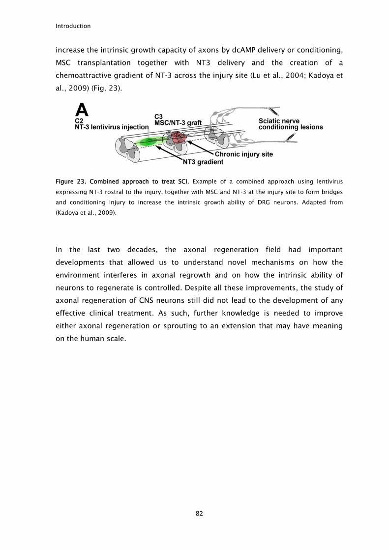
Introduction
82
increase the intrinsic growth capacity of axons by dcAMP delivery or conditioning,
MSC transplantation together with NT3 delivery and the creation of a
chemoattractive gradient of NT-3 across the injury site (Lu et al., 2004; Kadoya et
al., 2009) (Fig. 23).
Figure 23. Combined approach to treat SCI. Example of a combined approach using lentivirus
expressing NT-3 rostral to the injury, together with MSC and NT-3 at the injury site to form bridges
and conditioning injury to increase the intrinsic growth ability of DRG neurons. Adapted from
(Kadoya et al., 2009).
In the last two decades, the axonal regeneration field had important
developments that allowed us to understand novel mechanisms on how the
environment interferes in axonal regrowth and on how the intrinsic ability of
neurons to regenerate is controlled. Despite all these improvements, the study of
axonal regeneration of CNS neurons still did not lead to the development of any
effective clinical treatment. As such, further knowledge is needed to improve
either axonal regeneration or sprouting to an extension that may have meaning
on the human scale.
Review
Cell intrinsic control of axon regenerationFernando M Mar1, Azad Bonni2,3 & M�onica M Sousa1,*
Abstract
Although neurons execute a cell intrinsic program of axonalgrowth during development, following the establishment of con-nections, the developmental growth capacity declines. Besidesenvironmental challenges, this switch largely accounts for the fail-ure of adult central nervous system (CNS) axons to regenerate.Here, we discuss the cell intrinsic control of axon regeneration,including not only the regulation of transcriptional and epigeneticmechanisms, but also the modulation of local protein translation,retrograde and anterograde axonal transport, and microtubuledynamics. We further explore the causes underlying the failure ofCNS neurons to mount a vigorous regenerative response, and theparadigms demonstrating the activation of cell intrinsic axongrowth programs. Finally, we present potential mechanisms tosupport axon regeneration, as these may represent future thera-peutic approaches to promote recovery following CNS injury anddisease.
Keywords axon regeneration; axonal transport; conditioning lesion;
microtubule dynamics
DOI 10.1002/embr.201337723 | Received 4 July 2013 | Revised 22 January
2014 | Accepted 22 January 2014
See the Glossary for abbreviations used in this article.
Introduction
During development, by implementing a transcription-dependent
program relying on multiple signaling pathways, neurons display
robust elongation capacity that allows them to reach their targets.
Following this initial phase, with the establishment of connections
the developmental axon elongation ability declines. The view that
neurons in the adult CNS permanently lose their intrinsic ability to
grow axons was challenged by seminal studies by the Albert Aguayo
group, showing that the use of peripheral nerve “bridges” in the
spinal cord permits CNS axons to grow for considerable distances
following injury [1]. These studies demonstrated that adult CNS
neurons can activate a cell intrinsic program that supports axonal
regrowth, provided that a permissive environment is created. These
initial reports fueled efforts to characterize the extrinsic cues that
inhibit axon growth in the CNS, while the cell intrinsic mechanisms
that govern axon regeneration remained poorly understood. Several
decades later, the body of knowledge gathered supports the view
that counteracting or removing the extracellular inhibitory mole-
cules results in incomplete axon regeneration in vivo and that a bet-
ter understanding of cell intrinsic mechanisms regulating axon
growth following injury is needed [2].
In contrast to the CNS, adult peripheral nervous system (PNS)
axons can spontaneously regrow to a significant extent and are
often used as a model to identify the players that promote axon
regeneration. The regenerative capacity of the PNS is supported by
the combination of extrinsic and intrinsic factors that generate a
growth-permissive milieu where the execution of a cell intrinsic pro-
gram leads to successful axonal regrowth. Cell intrinsic changes
induced by a PNS injury can be observed in vitro and in vivo, as will
be discussed in the context of the conditioning lesion paradigm. In
CNS neurons, the combined action of repressors of axonal growth,
the limited injury signaling mechanisms, and the lack of robust
expression of regeneration-associated genes (RAGs) results in a
restricted potential to regenerate. Here, we will provide a critical
perspective of our understanding of the intrinsic mechanisms con-
trolling axonal regeneration in the adult nervous system. With the
term cell intrinsic, we refer to mechanisms that do not strictly
depend on external cues, although external cues can influence their
activity. As such, this review is not restricted to the discussion of
changes in the expression of the neuronal genetic program, that is,
transcriptional and epigenetic mechanisms and regulation of transla-
tion, but is extended to the analyses of intracellular pathways and
mechanisms—including axonal transport and microtubule dynamics
—that regulate axon growth and regeneration.
Cell intrinsic mechanisms of axonal regeneration inthe PNS
Calcium influx into the axoplasm is one of the first signals caused by
injury, and the depolarization triggered by the inversion of the
calcium/sodium flux travels along the axon to the cell body.
Calcium influx is here discussed in the context of the cell intrinsic
factors that govern axon regeneration as it elicits various cell auton-
omous mechanisms necessary for successful axon growth, ranging
from the regulation of intracellular pathways to the generation of
1 Nerve Regeneration Group, Instituto de Biologia Molecular e Celular - IBMC, University of Porto, Porto, Portugal2 Harvard Medical School, Boston, MA, USA3 Washington University School of Medicine, St. Louis, MO, USA
*Corresponding author. Tel: +351 22 6074900; Fax: +351 22 6099157; E-mail: [email protected]
ª 2014 The Authors EMBO reports 1
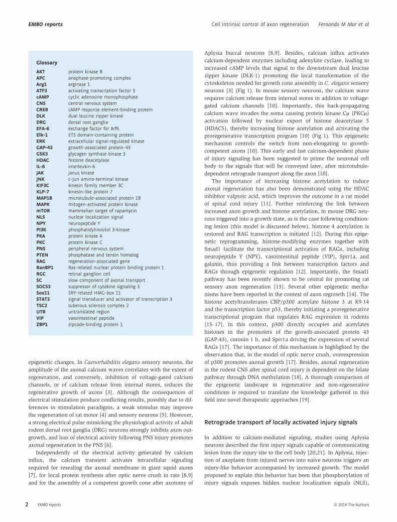
epigenetic changes. In Caenorhabditis elegans sensory neurons, the
amplitude of the axonal calcium waves correlates with the extent of
regeneration, and conversely, inhibition of voltage-gated calcium
channels, or of calcium release from internal stores, reduces the
regenerative growth of axons [3]. Although the consequences of
electrical stimulation produce conflicting results, possibly due to dif-
ferences in stimulation paradigms, a weak stimulus may improve
the regeneration of rat motor [4] and sensory neurons [5]. However,
a strong electrical pulse mimicking the physiological activity of adult
rodent dorsal root ganglia (DRG) neurons strongly inhibits axon out-
growth, and loss of electrical activity following PNS injury promotes
axonal regeneration in the PNS [6].
Independently of the electrical activity generated by calcium
influx, the calcium transient activates intracellular signaling
required for resealing the axonal membrane in giant squid axons
[7], for local protein synthesis after optic nerve crush in rats [8,9]
and for the assembly of a competent growth cone after axotomy of
Aplysia buccal neurons [8,9]. Besides, calcium influx activates
calcium-dependent enzymes including adenylate cyclase, leading to
increased cAMP levels that signal to the downstream dual leucine
zipper kinase (DLK-1) promoting the local transformation of the
cytoskeleton needed for growth cone assembly in C. elegans sensory
neurons [3] (Fig 1). In mouse sensory neurons, the calcium wave
requires calcium release from internal stores in addition to voltage-
gated calcium channels [10]. Importantly, this back-propagating
calcium wave invades the soma causing protein kinase Cl (PKCl)activation followed by nuclear export of histone deacetylase 5
(HDAC5), thereby increasing histone acetylation and activating the
proregenerative transcription program [10] (Fig 1). This epigenetic
mechanism controls the switch from non-elongating to growth-
competent axons [10]. This early and fast calcium-dependent phase
of injury signaling has been suggested to prime the neuronal cell
body to the signals that will be conveyed later, after microtubule-
dependent retrograde transport along the axon [10].
The importance of increasing histone acetylation to induce
axonal regeneration has also been demonstrated using the HDAC
inhibitor valproic acid, which improves the outcome in a rat model
of spinal cord injury [11]. Further reinforcing the link between
increased axon growth and histone acetylation, in mouse DRG neu-
rons triggered into a growth state, as is the case following condition-
ing lesion (this model is discussed below), histone 4 acetylation is
restored and RAG transcription is initiated [12]. During this epige-
netic reprogramming, histone-modifying enzymes together with
Smad1 facilitate the transcriptional activation of RAGs, including
neuropeptide Y (NPY), vasointestinal peptide (VIP), Sprr1a, and
galanin, thus providing a link between transcription factors and
RAGs through epigenetic regulation [12]. Importantly, the Smad1
pathway has been recently shown to be central for promoting rat
sensory axon regeneration [13]. Several other epigenetic mecha-
nisms have been reported in the context of axon regrowth [14]. The
histone acetyltransferases CBP/p300 acetylate histone 3 at K9-14
and the transcription factor p53, thereby initiating a proregenerative
transcriptional program that regulates RAG expression in rodents
[15–17]. In this context, p300 directly occupies and acetylates
histones in the promoters of the growth-associated protein 43
(GAP-43), coronin 1 b, and Sprr1a driving the expression of several
RAGs [17]. The importance of this mechanism is highlighted by the
observation that, in the model of optic nerve crush, overexpression
of p300 promotes axonal growth [17]. Besides, axonal regeneration
in the rodent CNS after spinal cord injury is dependent on the folate
pathway through DNA methylation [18]. A thorough comparison of
the epigenetic landscape in regenerative and non-regenerative
conditions is required to translate the knowledge gathered in this
field into novel therapeutic approaches [19].
Retrograde transport of locally activated injury signals
In addition to calcium-mediated signaling, studies using Aplysia
neurons described the first injury signals capable of communicating
lesion from the injury site to the cell body [20,21]. In Aplysia, injec-
tion of axoplasm from injured nerves into na€ıve neurons triggers an
injury-like behavior accompanied by increased growth. The model
proposed to explain this behavior has been that phosphorylation of
injury signals exposes hidden nuclear localization signals (NLS),
Glossary
AKT protein kinase BAPC anaphase-promoting complexArg1 arginase 1ATF3 activating transcription factor 3cAMP cyclic adenosine monophosphateCNS central nervous systemCREB cAMP response element-binding proteinDLK dual leucine zipper kinaseDRG dorsal root gangliaEFA-6 exchange factor for Arf6Elk-1 ETS domain-containing proteinERK extracellular signal-regulated kinaseGAP-43 growth-associated protein-43GSK3 glycogen synthase kinase 3HDAC histone deacetylaseIL-6 interleukin-6JAK janus kinaseJNK c-Jun amino-terminal kinaseKIF3C kinesin family member 3CKLP-7 kinesin-like protein 7MAP1B microtubule-associated protein 1BMAPK mitogen-activated protein kinasemTOR mammalian target of rapamycinNLS nuclear localization signalNPY neuropeptide YPI3K phosphatidylinositol 3-kinasePKA protein kinase APKC protein kinase CPNS peripheral nervous systemPTEN phosphatase and tensin homologRAG regeneration-associated geneRanBP1 Ras-related nuclear protein binding protein 1RGC retinal ganglion cellSC slow component of axonal transportSOCS3 suppressor of cytokine signaling 3Sox11 SRY-related HMG-box 11STAT3 signal transducer and activator of transcription 3TSC2 tuberous sclerosis complex 2UTR untranslated regionVIP vasointestinal peptideZBP1 zipcode-binding protein 1
EMBO reports ª 2014 The Authors
EMBO reports Cell intrinsic control of axon regeneration Fernando M Mar et al
2
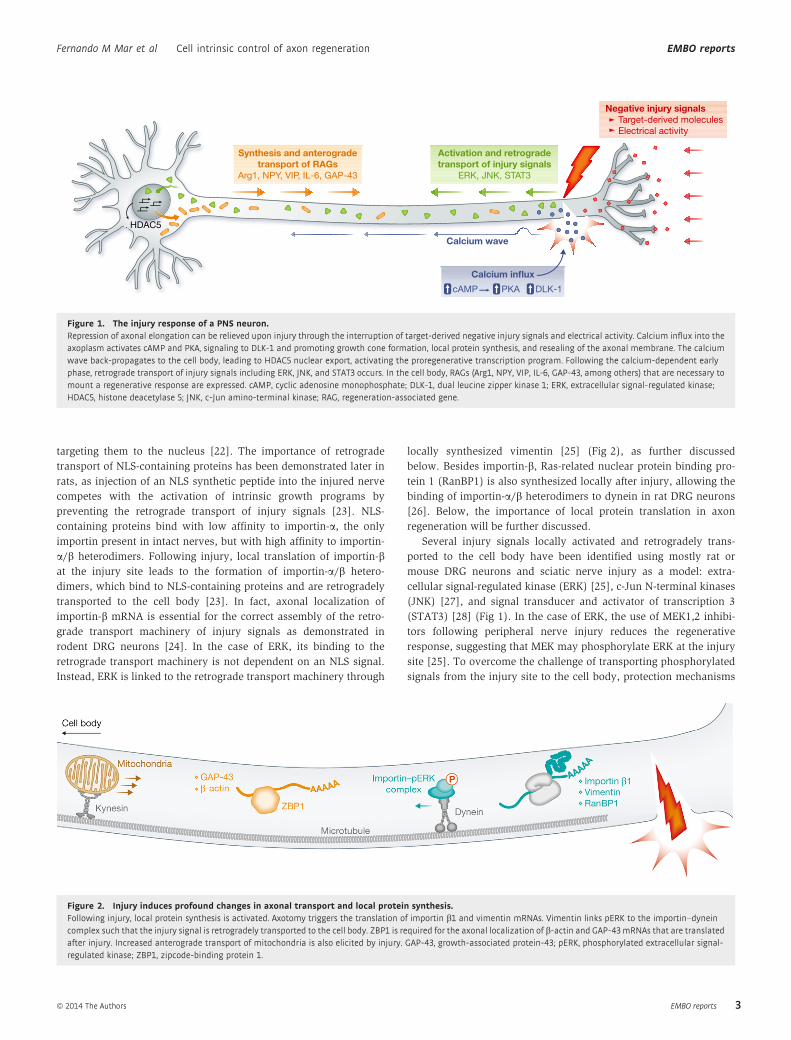
targeting them to the nucleus [22]. The importance of retrograde
transport of NLS-containing proteins has been demonstrated later in
rats, as injection of an NLS synthetic peptide into the injured nerve
competes with the activation of intrinsic growth programs by
preventing the retrograde transport of injury signals [23]. NLS-
containing proteins bind with low affinity to importin-a, the only
importin present in intact nerves, but with high affinity to importin-
a/b heterodimers. Following injury, local translation of importin-bat the injury site leads to the formation of importin-a/b hetero-
dimers, which bind to NLS-containing proteins and are retrogradely
transported to the cell body [23]. In fact, axonal localization of
importin-b mRNA is essential for the correct assembly of the retro-
grade transport machinery of injury signals as demonstrated in
rodent DRG neurons [24]. In the case of ERK, its binding to the
retrograde transport machinery is not dependent on an NLS signal.
Instead, ERK is linked to the retrograde transport machinery through
locally synthesized vimentin [25] (Fig 2), as further discussed
below. Besides importin-b, Ras-related nuclear protein binding pro-
tein 1 (RanBP1) is also synthesized locally after injury, allowing the
binding of importin-a/b heterodimers to dynein in rat DRG neurons
[26]. Below, the importance of local protein translation in axon
regeneration will be further discussed.
Several injury signals locally activated and retrogradely trans-
ported to the cell body have been identified using mostly rat or
mouse DRG neurons and sciatic nerve injury as a model: extra-
cellular signal-regulated kinase (ERK) [25], c-Jun N-terminal kinases
(JNK) [27], and signal transducer and activator of transcription 3
(STAT3) [28] (Fig 1). In the case of ERK, the use of MEK1,2 inhibi-
tors following peripheral nerve injury reduces the regenerative
response, suggesting that MEK may phosphorylate ERK at the injury
site [25]. To overcome the challenge of transporting phosphorylated
signals from the injury site to the cell body, protection mechanisms
HDAC5
Synthesis and anterogradetransport of RAGs
Arg1, NPY, VIP, IL-6, GAP-43
Activation and retrogradetransport of injury signals
ERK, JNK, STAT3
Negative injury signals Target-derived molecules
Electrical activity
Calcium wave
Calcium influx
cAMP PKA DLK-1
Figure 1. The injury response of a PNS neuron.Repression of axonal elongation can be relieved upon injury through the interruption of target-derived negative injury signals and electrical activity. Calcium influx into theaxoplasm activates cAMP and PKA, signaling to DLK-1 and promoting growth cone formation, local protein synthesis, and resealing of the axonal membrane. The calciumwave back-propagates to the cell body, leading to HDAC5 nuclear export, activating the proregenerative transcription program. Following the calcium-dependent earlyphase, retrograde transport of injury signals including ERK, JNK, and STAT3 occurs. In the cell body, RAGs (Arg1, NPY, VIP, IL-6, GAP-43, among others) that are necessary tomount a regenerative response are expressed. cAMP, cyclic adenosine monophosphate; DLK-1, dual leucine zipper kinase 1; ERK, extracellular signal-regulated kinase;HDAC5, histone deacetylase 5; JNK, c-Jun amino-terminal kinase; RAG, regeneration-associated gene.
Figure 2. Injury induces profound changes in axonal transport and local protein synthesis.Following injury, local protein synthesis is activated. Axotomy triggers the translation of importin b1 and vimentin mRNAs. Vimentin links pERK to the importin–dyneincomplex such that the injury signal is retrogradely transported to the cell body. ZBP1 is required for the axonal localization of b-actin and GAP-43mRNAs that are translatedafter injury. Increased anterograde transport of mitochondria is also elicited by injury. GAP-43, growth-associated protein-43; pERK, phosphorylated extracellular signal-regulated kinase; ZBP1, zipcode-binding protein 1.
ª 2014 The Authors EMBO reports
Fernando M Mar et al Cell intrinsic control of axon regeneration EMBO reports
3
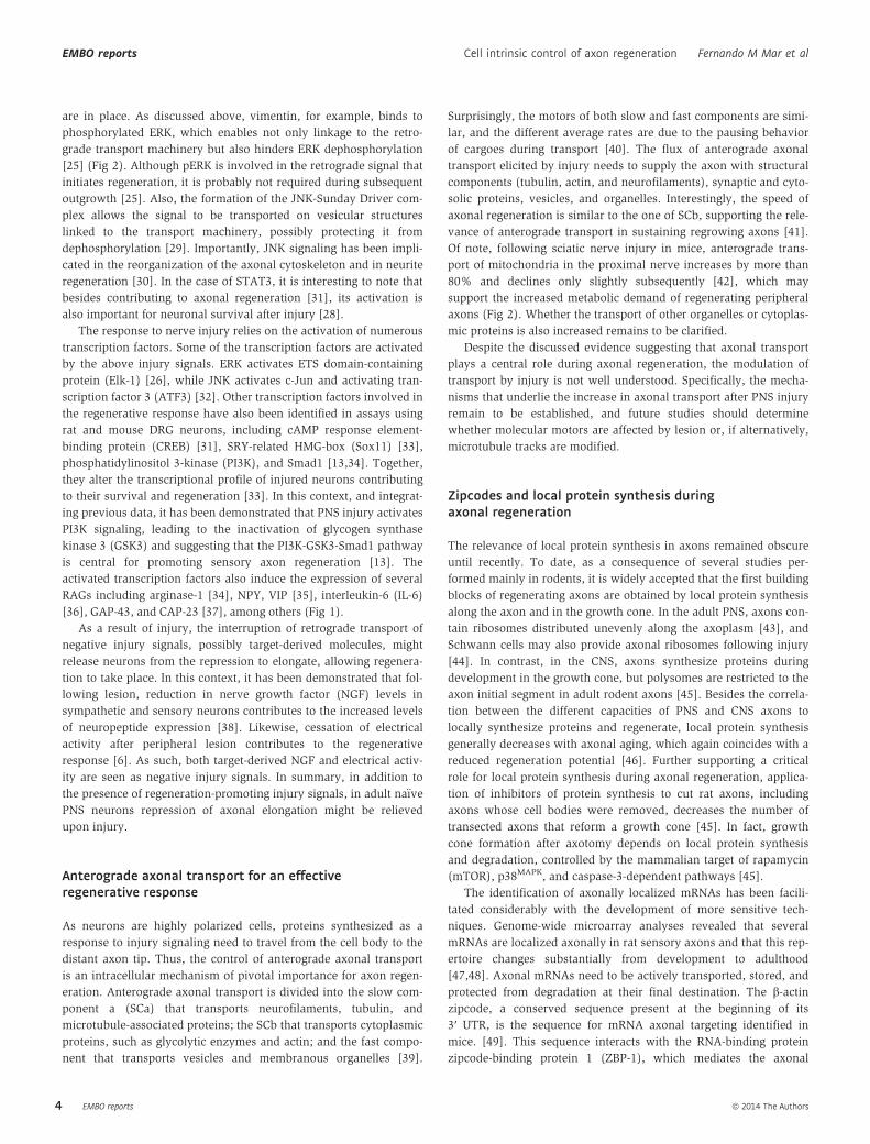
are in place. As discussed above, vimentin, for example, binds to
phosphorylated ERK, which enables not only linkage to the retro-
grade transport machinery but also hinders ERK dephosphorylation
[25] (Fig 2). Although pERK is involved in the retrograde signal that
initiates regeneration, it is probably not required during subsequent
outgrowth [25]. Also, the formation of the JNK-Sunday Driver com-
plex allows the signal to be transported on vesicular structures
linked to the transport machinery, possibly protecting it from
dephosphorylation [29]. Importantly, JNK signaling has been impli-
cated in the reorganization of the axonal cytoskeleton and in neurite
regeneration [30]. In the case of STAT3, it is interesting to note that
besides contributing to axonal regeneration [31], its activation is
also important for neuronal survival after injury [28].
The response to nerve injury relies on the activation of numerous
transcription factors. Some of the transcription factors are activated
by the above injury signals. ERK activates ETS domain-containing
protein (Elk-1) [26], while JNK activates c-Jun and activating tran-
scription factor 3 (ATF3) [32]. Other transcription factors involved in
the regenerative response have also been identified in assays using
rat and mouse DRG neurons, including cAMP response element-
binding protein (CREB) [31], SRY-related HMG-box (Sox11) [33],
phosphatidylinositol 3-kinase (PI3K), and Smad1 [13,34]. Together,
they alter the transcriptional profile of injured neurons contributing
to their survival and regeneration [33]. In this context, and integrat-
ing previous data, it has been demonstrated that PNS injury activates
PI3K signaling, leading to the inactivation of glycogen synthase
kinase 3 (GSK3) and suggesting that the PI3K-GSK3-Smad1 pathway
is central for promoting sensory axon regeneration [13]. The
activated transcription factors also induce the expression of several
RAGs including arginase-1 [34], NPY, VIP [35], interleukin-6 (IL-6)
[36], GAP-43, and CAP-23 [37], among others (Fig 1).
As a result of injury, the interruption of retrograde transport of
negative injury signals, possibly target-derived molecules, might
release neurons from the repression to elongate, allowing regenera-
tion to take place. In this context, it has been demonstrated that fol-
lowing lesion, reduction in nerve growth factor (NGF) levels in
sympathetic and sensory neurons contributes to the increased levels
of neuropeptide expression [38]. Likewise, cessation of electrical
activity after peripheral lesion contributes to the regenerative
response [6]. As such, both target-derived NGF and electrical activ-
ity are seen as negative injury signals. In summary, in addition to
the presence of regeneration-promoting injury signals, in adult na€ıve
PNS neurons repression of axonal elongation might be relieved
upon injury.
Anterograde axonal transport for an effectiveregenerative response
As neurons are highly polarized cells, proteins synthesized as a
response to injury signaling need to travel from the cell body to the
distant axon tip. Thus, the control of anterograde axonal transport
is an intracellular mechanism of pivotal importance for axon regen-
eration. Anterograde axonal transport is divided into the slow com-
ponent a (SCa) that transports neurofilaments, tubulin, and
microtubule-associated proteins; the SCb that transports cytoplasmic
proteins, such as glycolytic enzymes and actin; and the fast compo-
nent that transports vesicles and membranous organelles [39].
Surprisingly, the motors of both slow and fast components are simi-
lar, and the different average rates are due to the pausing behavior
of cargoes during transport [40]. The flux of anterograde axonal
transport elicited by injury needs to supply the axon with structural
components (tubulin, actin, and neurofilaments), synaptic and cyto-
solic proteins, vesicles, and organelles. Interestingly, the speed of
axonal regeneration is similar to the one of SCb, supporting the rele-
vance of anterograde transport in sustaining regrowing axons [41].
Of note, following sciatic nerve injury in mice, anterograde trans-
port of mitochondria in the proximal nerve increases by more than
80% and declines only slightly subsequently [42], which may
support the increased metabolic demand of regenerating peripheral
axons (Fig 2). Whether the transport of other organelles or cytoplas-
mic proteins is also increased remains to be clarified.
Despite the discussed evidence suggesting that axonal transport
plays a central role during axonal regeneration, the modulation of
transport by injury is not well understood. Specifically, the mecha-
nisms that underlie the increase in axonal transport after PNS injury
remain to be established, and future studies should determine
whether molecular motors are affected by lesion or, if alternatively,
microtubule tracks are modified.
Zipcodes and local protein synthesis duringaxonal regeneration
The relevance of local protein synthesis in axons remained obscure
until recently. To date, as a consequence of several studies per-
formed mainly in rodents, it is widely accepted that the first building
blocks of regenerating axons are obtained by local protein synthesis
along the axon and in the growth cone. In the adult PNS, axons con-
tain ribosomes distributed unevenly along the axoplasm [43], and
Schwann cells may also provide axonal ribosomes following injury
[44]. In contrast, in the CNS, axons synthesize proteins during
development in the growth cone, but polysomes are restricted to the
axon initial segment in adult rodent axons [45]. Besides the correla-
tion between the different capacities of PNS and CNS axons to
locally synthesize proteins and regenerate, local protein synthesis
generally decreases with axonal aging, which again coincides with a
reduced regeneration potential [46]. Further supporting a critical
role for local protein synthesis during axonal regeneration, applica-
tion of inhibitors of protein synthesis to cut rat axons, including
axons whose cell bodies were removed, decreases the number of
transected axons that reform a growth cone [45]. In fact, growth
cone formation after axotomy depends on local protein synthesis
and degradation, controlled by the mammalian target of rapamycin
(mTOR), p38MAPK, and caspase-3-dependent pathways [45].
The identification of axonally localized mRNAs has been facili-
tated considerably with the development of more sensitive tech-
niques. Genome-wide microarray analyses revealed that several
mRNAs are localized axonally in rat sensory axons and that this rep-
ertoire changes substantially from development to adulthood
[47,48]. Axonal mRNAs need to be actively transported, stored, and
protected from degradation at their final destination. The b-actinzipcode, a conserved sequence present at the beginning of its
3′ UTR, is the sequence for mRNA axonal targeting identified in
mice. [49]. This sequence interacts with the RNA-binding protein
zipcode-binding protein 1 (ZBP-1), which mediates the axonal
EMBO reports ª 2014 The Authors
EMBO reports Cell intrinsic control of axon regeneration Fernando M Mar et al
4
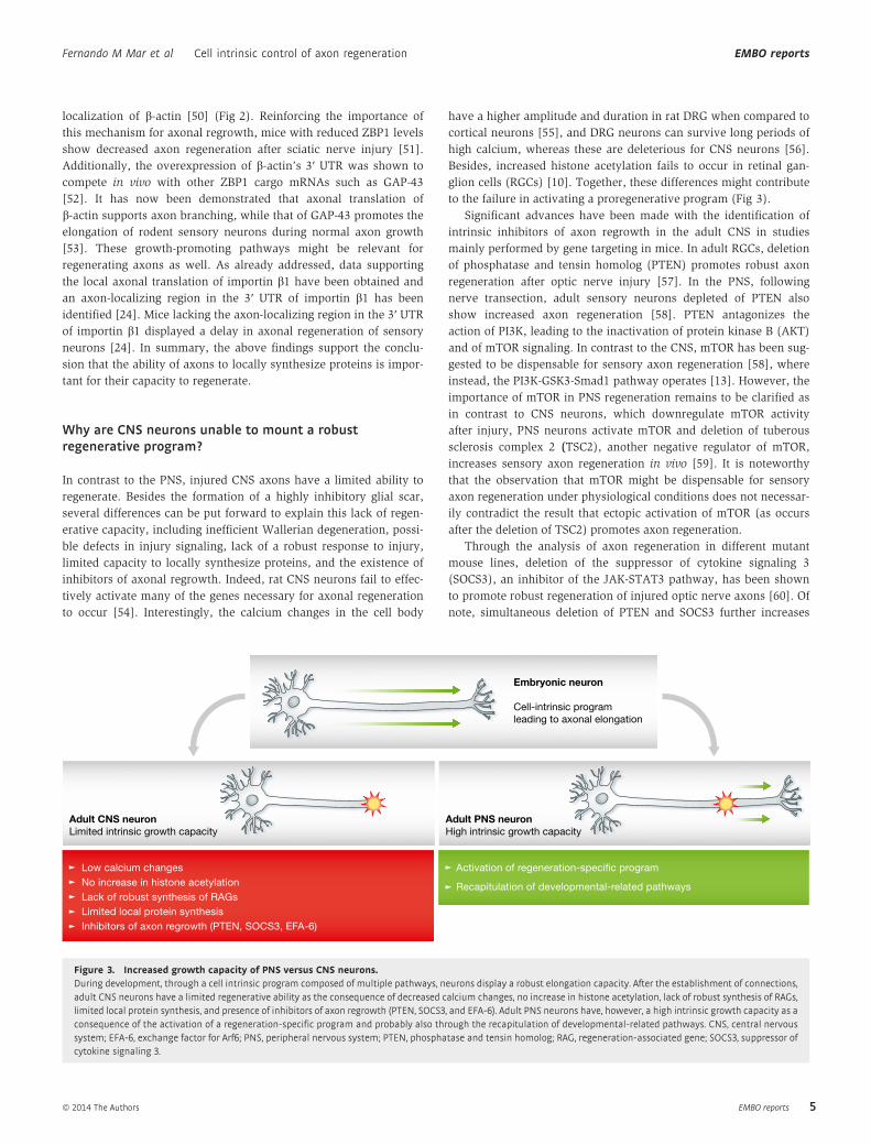
localization of b-actin [50] (Fig 2). Reinforcing the importance of
this mechanism for axonal regrowth, mice with reduced ZBP1 levels
show decreased axon regeneration after sciatic nerve injury [51].
Additionally, the overexpression of b-actin’s 3′ UTR was shown to
compete in vivo with other ZBP1 cargo mRNAs such as GAP-43
[52]. It has now been demonstrated that axonal translation of
b-actin supports axon branching, while that of GAP-43 promotes the
elongation of rodent sensory neurons during normal axon growth
[53]. These growth-promoting pathways might be relevant for
regenerating axons as well. As already addressed, data supporting
the local axonal translation of importin b1 have been obtained and
an axon-localizing region in the 3′ UTR of importin b1 has been
identified [24]. Mice lacking the axon-localizing region in the 3′ UTR
of importin b1 displayed a delay in axonal regeneration of sensory
neurons [24]. In summary, the above findings support the conclu-
sion that the ability of axons to locally synthesize proteins is impor-
tant for their capacity to regenerate.
Why are CNS neurons unable to mount a robustregenerative program?
In contrast to the PNS, injured CNS axons have a limited ability to
regenerate. Besides the formation of a highly inhibitory glial scar,
several differences can be put forward to explain this lack of regen-
erative capacity, including inefficient Wallerian degeneration, possi-
ble defects in injury signaling, lack of a robust response to injury,
limited capacity to locally synthesize proteins, and the existence of
inhibitors of axonal regrowth. Indeed, rat CNS neurons fail to effec-
tively activate many of the genes necessary for axonal regeneration
to occur [54]. Interestingly, the calcium changes in the cell body
have a higher amplitude and duration in rat DRG when compared to
cortical neurons [55], and DRG neurons can survive long periods of
high calcium, whereas these are deleterious for CNS neurons [56].
Besides, increased histone acetylation fails to occur in retinal gan-
glion cells (RGCs) [10]. Together, these differences might contribute
to the failure in activating a proregenerative program (Fig 3).
Significant advances have been made with the identification of
intrinsic inhibitors of axon regrowth in the adult CNS in studies
mainly performed by gene targeting in mice. In adult RGCs, deletion
of phosphatase and tensin homolog (PTEN) promotes robust axon
regeneration after optic nerve injury [57]. In the PNS, following
nerve transection, adult sensory neurons depleted of PTEN also
show increased axon regeneration [58]. PTEN antagonizes the
action of PI3K, leading to the inactivation of protein kinase B (AKT)
and of mTOR signaling. In contrast to the CNS, mTOR has been sug-
gested to be dispensable for sensory axon regeneration [58], where
instead, the PI3K-GSK3-Smad1 pathway operates [13]. However, the
importance of mTOR in PNS regeneration remains to be clarified as
in contrast to CNS neurons, which downregulate mTOR activity
after injury, PNS neurons activate mTOR and deletion of tuberous
sclerosis complex 2 (TSC2), another negative regulator of mTOR,
increases sensory axon regeneration in vivo [59]. It is noteworthy
that the observation that mTOR might be dispensable for sensory
axon regeneration under physiological conditions does not necessar-
ily contradict the result that ectopic activation of mTOR (as occurs
after the deletion of TSC2) promotes axon regeneration.
Through the analysis of axon regeneration in different mutant
mouse lines, deletion of the suppressor of cytokine signaling 3
(SOCS3), an inhibitor of the JAK-STAT3 pathway, has been shown
to promote robust regeneration of injured optic nerve axons [60]. Of
note, simultaneous deletion of PTEN and SOCS3 further increases
Embryonic neuron
Cell-intrinsic program
leading to axonal elongation
Adult CNS neuronLimited intrinsic growth capacity
Adult PNS neuronHigh intrinsic growth capacity
Low calcium changes
No increase in histone acetylation
Lack of robust synthesis of RAGs
Limited local protein synthesis
Inhibitors of axon regrowth (PTEN, SOCS3, EFA-6)
Activation of regeneration-specific program
Recapitulation of developmental-related pathways
Figure 3. Increased growth capacity of PNS versus CNS neurons.During development, through a cell intrinsic program composed of multiple pathways, neurons display a robust elongation capacity. After the establishment of connections,adult CNS neurons have a limited regenerative ability as the consequence of decreased calcium changes, no increase in histone acetylation, lack of robust synthesis of RAGs,limited local protein synthesis, and presence of inhibitors of axon regrowth (PTEN, SOCS3, and EFA-6). Adult PNS neurons have, however, a high intrinsic growth capacity as aconsequence of the activation of a regeneration-specific program and probably also through the recapitulation of developmental-related pathways. CNS, central nervoussystem; EFA-6, exchange factor for Arf6; PNS, peripheral nervous system; PTEN, phosphatase and tensin homolog; RAG, regeneration-associated gene; SOCS3, suppressor ofcytokine signaling 3.
ª 2014 The Authors EMBO reports
Fernando M Mar et al Cell intrinsic control of axon regeneration EMBO reports
5
axon regeneration, as these signals regulate two independent path-
ways that act synergistically [61]. In summary, the mTOR and
STAT3 pathways emerged as key regulators promoting long-
distance axon regeneration in the adult CNS. Reinforcing this view,
PTEN or SOCS3 deletion improves regeneration of distinct CNS
axons [57,60,62,63], and sustained activation of STAT3 promotes
corticospinal remodeling and functional recovery after spinal cord
injury [64]. Recently, in C. elegans mechanosensory neurons, the
conserved Arf guanine nucleotide-exchange factor EFA-6 has been
reported to be an intrinsic inhibitor of axon regrowth that operates
by affecting axonal microtubule dynamics, acting downstream of
and/or in parallel with DLK-1 [65].
Together, these studies indicate that axon regeneration is
restrained not only by extrinsic inhibitory cues but also by intrinsic
factors. As such, the manipulation of intrinsic growth control path-
ways is actively being pursued as a therapeutic approach to promote
axon regeneration after CNS injury.
The cell intrinsic growth capacity of adult CNS neuronscan be activated
Despite the general inability of CNS axons to regenerate, regrowth
can be activated under specific conditions, for example the condi-
tioning lesion effect. DRG neurons possess a peripheral axon branch
that regenerates when injured, and a central axon branch that enters
the spinal cord (forming the dorsal column fibers) and does not
regenerate upon injury. However, when the peripheral branch is
lesioned prior to lesioning the central branch (conditioning lesion),
the central axon can overcome the glial scar inhibitory effect, regen-
erating to a significant extent both in vivo and in vitro [66,67], when
plated on inhibitory substrates, including myelin [68]. The condi-
tioning effect is probably due to the activation of the regenerative
machinery prior to CNS lesion. Studies performed in rodents have
shown that the increase in regeneration capacity encompasses RAG
expression [33,69] and possibly changes in axonal transport [70]. In
fact, the response to injury starts as soon as 1 day following lesion
[68] and has a long-lasting effect, as RAGs are still expressed
2 months following the priming injury [71]. Although a peripheral
lesion performed subsequently to the CNS injury does not improve
axonal regeneration due to the assembly of a thick glial scar, it still
increases the intrinsic regenerative ability of DRG neurons [71].
The conditioning effect certainly represents a good model to
identify the mechanisms underlying the cell intrinsic regenerative
capacity. Numerous molecular pathways have been shown to be
regulated by a conditioning lesion, in accordance with the robust
and broad transcriptional change that conditioning causes in DRG
neurons. The initial unifying concept was that the conditioning
effect was mediated by increased cAMP levels induced by injury
[68]. cAMP prompted neurons to overcome myelin inhibition in
vitro, and treatment with dibutyryl-cAMP, a cell-permeable analog
of cAMP, increased the regeneration of dorsal column fibers fol-
lowing spinal cord injury [68,72]. Further supporting the pivotal
role of cAMP, the phosphodiesterase inhibitor rolipram increases
cAMP levels, leading to enhanced regeneration of serotonergic
axons and functional recovery following spinal cord injury in rats
[73]. However, the robust effect of cAMP in axon regeneration
has been questioned by recent studies, as the use of cAMP
analogs has failed to reproduce the full effect of a conditioning
lesion [74]. Increased levels of cAMP appear to promote axon
regeneration by overcoming myelin-based inhibitors rather than
by modulating the intrinsic ability of neurons to support axon
regeneration. It has been shown that elevation of cAMP fails to
increase the SCb of axonal transport, the rate-limiting step of axon
growth [75].
Besides cAMP, several studies have identified broad changes in
gene expression in rodent conditioned neurons [36,69], regulated
by the activation of multiple transcription factors [76]. Down-
stream, the expression of traditional RAGs such as GAP-43 and
CAP-23 [77] is induced and novel RAGs have been identified using
this model, including arginase-1 [34] and IL-6 [36]. However, none
of the identified transcription factors or RAGs reproduces the entire
conditioning effect [74], suggesting that conditioning cannot be
mimicked by manipulating a single pathway. Supporting this view,
epigenetic changes elicited by HDAC5 nuclear export partially
reproduce the conditioning lesion effect [10]. Several questions
remain open, including the differences in injury-induced signaling
that allow a peripheral injury to elicit a strong regeneration
response, whereas a central lesion to the same neuron fails to do so
(Sidebar A).
Manipulating axonal microtubule dynamics topromote regeneration
Among the multiple processes involved in the generation of a
new growth cone, cytoskeleton reorganization is crucial for the
intrinsic ability to regenerate. While upon injury CNS axons form
a retraction bulb with a disorganized network of microtubules,
PNS axons form a growth cone with stable microtubules in the
backbone and dynamic microtubules in the tip [78]. Pharmaco-
logical destabilization of microtubules converts a growth cone into
a retraction bulb, and taxol-induced stabilization generates growth
cones that can overcome myelin inhibition [78] and regenerate
following spinal cord injury [79]. Also, HDAC6 inhibition in
rodents results in increased levels of acetylated/stable micro-
tubules and enhances the growth of sensory neurons on myelin
[80]. It has been recently suggested that instead of inducing the
normal mode of repair, where the axon tip might behave more
dynamically, taxol might promote axonal regeneration by enabling
the axon tip to become more forceful [81]. In rodent sensory neu-
rons, HDAC5 accumulates at the tip of injured axons where local
tubulin deacetylation induces growth cone microtubule dynamics
and axon regeneration [82]. The varying effects that microtubule
stability might have on axon regeneration do not allow for a clear
causal relationship between axon regeneration and microtubule
stability. Moreover, at this point, the available literature does not
provide sufficient detail to allow for a comparison between the
effects of HDAC5, HDAC6, and taxol on tubulin dynamics in the
axon shaft versus axon tip. Although the results obtained with
studies using taxol and HDAC6 seem contradictory to the data
reported for HDAC5 and the kinesin family member KIF3C [83],
previous studies have shown that efficient developmental axon
growth requires an optimal level of microtubule dynamics. Thus,
destabilizing or overstabilizing microtubules could both impair
axon growth.
EMBO reports ª 2014 The Authors
EMBO reports Cell intrinsic control of axon regeneration Fernando M Mar et al
6
Many of the pathways that contribute to cell intrinsic control of
regeneration participate in the remodeling of the axonal cytoskeleton,
specifically by modulating microtubule dynamics. As such,
potential therapeutic strategies intervening at the level of micro-
tubule-related proteins have been actively pursued. In this respect,
and besides those already discussed in this review including HDAC5
and 6, several other possible targets have been identified, including
GSK3b, as many of its substrates are microtubule-interacting
proteins [84].
Members of the kinesin family have also been put forward as
important players in regulating microtubule dynamics during axonal
regeneration. KIF3C has been shown to be an injury-specific kinesin
with microtubule-destabilizing function, playing a key role during
axon regrowth [83]. Depletion of KIF3C in adult neurons leads to an
increase in stable and looped microtubules and delays axonal regen-
eration after injury [83]. In addition to KIF3C, in C. elegans
mechanosensory axons, the depolymerizing kinesin-like protein
family member 7 (KLP-7) restricts microtubule growth in the steady
state [85]. After axon injury, the number of growing microtubules is
increased at the injury site, simultaneously with the downregulation
of KLP-7, in a cascade coordinated by DLK-1 [85]. This mechanism
has been proposed to allow the stable microtubule cytoskeleton of a
mature neuron to be converted into the dynamically growing micro-
tubule cytoskeleton of a regenerating axon.
Besides the importance of identifying microtubule-interacting
proteins that participate in either the formation of a retraction bulb
or of a growth cone, further analysis of the regulation of post-
translational microtubule modifications following injury is needed,
as these control microtubule dynamics and may also interfere with
axonal transport, therefore impacting on axonal regrowth.
Conclusions and outlook
Recent evidence obtained by systematic genetic screening in
C. elegans shows that besides triggering developmental programs
that may be repressed in mature neurons, regenerative growth
involves specific pathways that sense and specifically respond to
damage [65] (Fig 3). Although axonal regeneration cannot be
viewed as mere recapitulation of axonal elongation during develop-
ment, some of the operating mechanisms are shared. Axon growth
during development occurs in two different phases: an initial phase
of growth and a later phase that takes place during pruning [86].
Axon growth during developmental remodeling appears to be mech-
anistically distinct from initial axon outgrowth. Interestingly,
common pathways, for example mTOR signaling, operate during
developmental axon regrowth and axonal regeneration [86].
The ubiquitin ligase Cdh1-anaphase-promoting complex
(Cdh1-APC) pathway is also a critical cell intrinsic mechanism that
regulates axon growth in the rodent developing cerebellar cortex
[87]. The inhibition of Cdh1-APC in primary neurons enhances
axonal growth and also overrides myelin inhibition of axon
regrowth [87]. Conceivably, Cdh1-APC might therefore potentially
limit axonal growth in the adult CNS. In key follow-up studies, the
transcriptional regulator SnoN has been identified as a critical sub-
strate of Cdh1-APC in neurons [88], operating in a pathway that is
regulated by transforming growth factor b-Smad2 signaling [89].
Smad2 knockdown also overrides myelin inhibition of axon growth
[89]. More recently, expression of a mutant SnoN resistant to degra-
dation has been shown to enhance axonal regeneration following
spinal cord injury in rats [90]. Together, these findings raise the
exciting prospect that pathways operating during development
might also drive axonal regeneration following injury. Therapeutic
strategies aiming at the reactivation of these pathways in injured
CNS neurons might be successful in enhancing our capacity to
regenerate neurons in response to injury or disease.
AcknowledgementsWe apologize to all colleagues whose work could not be cited due to space
constraints. We thank Dr. Valeria Cavalli (Washington University School of
Medicine) for the critical reading of the manuscript. The authors were sup-
ported by FEDER through COMPETE and by FCT (Project FCOMP-01-0124-
FEDER-017455; HMSP-ICT/0020/2010 to M.M.S. and A.B.). M.M.S. is also
supported by FCOMP-01-0124-FEDER-015781; PTDC/SAU-GMG/111761/2009,
and by the International Foundation for Research in Paraplegia. A.B. is also
supported by NIH Grant NS051255.
Conflict of interestThe authors declare that they have no conflict of interest.
References
1. Richardson PM, McGuinness UM, Aguayo AJ (1980) Axons from CNS
neurons regenerate into PNS grafts. Nature 284: 264 – 265
2. Liu K, Tedeschi A, Park KK, He Z (2011) Neuronal intrinsic mechanisms
of axon regeneration. Annu Rev Neurosci 34: 131 – 152
3. Ghosh-Roy A, Wu Z, Goncharov A, Jin Y, Chisholm AD (2010) Calcium
and cyclic AMP promote axonal regeneration in Caenorhabditis elegans
and require DLK-1 kinase. J Neurosci 30: 3175 – 3183
4. Brushart TM, Hoffman PN, Royall RM, Murinson BB, Witzel C, Gordon T
(2002) Electrical stimulation promotes motoneuron regeneration with-
out increasing its speed or conditioning the neuron. J Neurosci 22:
6631 – 6638
5. Udina E, Furey M, Busch S, Silver J, Gordon T, Fouad K (2008)
Electrical stimulation of intact peripheral sensory axons in rats
promotes outgrowth of their central projections. Exp Neurol 210:
238 – 247
6. Enes J, Langwieser N, Ruschel J, Carballosa-Gonzalez MM, Klug A, Traut
MH, Ylera B, Tahirovic S, Hofmann F, Stein V et al (2010) Electrical
Sidebar A. In need of answers
(i) How is axonal transport modulated by injury? Specifically, what are
the mechanisms underlying the increased axonal transport after PNS
injury? Are molecular motors affected or are post-translational micro-
tubule modifications altered?
(ii) What are the differences in injury-induced signaling that allow an
injury to the peripheral branch of DRG neurons to elicit a strong regen-
eration response, whereas lesion to the central branch fails to elicit this
response?
(iii) Is there a causal relationship between axon regeneration and microtu-
bule dynamics?
(iv) Are developmental pathways recapitulated during axonal regeneration
following injury?
ª 2014 The Authors EMBO reports
Fernando M Mar et al Cell intrinsic control of axon regeneration EMBO reports
7
activity suppresses axon growth through Ca(v)1.2 channels in adult pri-
mary sensory neurons. Curr Biol 20: 1154 – 1164
7. Krause TL, Fishman HM, Ballinger ML, Bittner GD (1994) Extent and
mechanism of sealing in transected giant axons of squid and earth-
worms. J Neurosci 14: 6638 – 6651
8. Chierzi S, Ratto GM, Verma P, Fawcett JW (2005) The ability of axons
to regenerate their growth cones depends on axonal type and age,
and is regulated by calcium, cAMP and ERK. Eur J Neurosci 21:
2051 – 2062
9. Kamber D, Erez H, Spira ME (2009) Local calcium-dependent mecha-
nisms determine whether a cut axonal end assembles a retarded end-
bulb or competent growth cone. Exp Neurol 219: 112 – 125
10. Cho Y, Sloutsky R, Naegle KM, Cavalli V (2013) Injury-induced HDAC5
nuclear export is essential for axon regeneration. Cell 155: 894 – 908
11. Lv L, Han X, Sun Y, Wang X, Dong Q (2012) Valproic acid improves loco-
motion in vivo after SCI and axonal growth of neurons in vitro. Exp
Neurol 233: 783 – 790
12. Finelli MJ, Wong JK, Zou H (2013) Epigenetic regulation of sensory axon
regeneration after spinal cord injury. J Neurosci 33: 19664 – 19676
13. Saijilafu, Hur EM, Liu CM, Jiao Z, Xu WL, Zhou FQ (2013) PI3K-GSK3 sig-
nalling regulates mammalian axon regeneration by inducing the
expression of Smad1. Nat Commun 4: 2690
14. Trakhtenberg EF, Goldberg JL (2012) Epigenetic regulation of axon and
dendrite growth. Front Mol Neurosci 5: 24
15. Tedeschi A, Nguyen T, Puttagunta R, Gaub P, Di Giovanni S (2009) A
p53-CBP/p300 transcription module is required for GAP-43 expression,
axon outgrowth, and regeneration. Cell Death Differ 16: 543 – 554
16. Gaub P, Tedeschi A, Puttagunta R, Nguyen T, Schmandke A, Di Giovanni
S (2010) HDAC inhibition promotes neuronal outgrowth and counter-
acts growth cone collapse through CBP/p300 and P/CAF-dependent p53
acetylation. Cell Death Differ 17: 1392 – 1408
17. Gaub P, Joshi Y, Wuttke A, Naumann U, Schnichels S, Heiduschka P, Di
Giovanni S (2011) The histone acetyltransferase p300 promotes intrinsic
axonal regeneration. Brain 134: 2134 – 2148
18. Iskandar BJ, Rizk E, Meier B, Hariharan N, Bottiglieri T, Finnell RH,
Jarrard DF, Banerjee RV, Skene JH, Nelson A et al (2010) Folate regula-
tion of axonal regeneration in the rodent central nervous system
through DNA methylation. J Clin Investig 120: 1603 – 1616
19. Lindner R, Puttagunta R, Di Giovanni S (2013) Epigenetic regulation of
axon outgrowth and regeneration in CNS injury: the first steps forward.
Neurotherapeutics 10: 771 – 781
20. Ambron RT, Dulin MF, Zhang XP, Schmied R, Walters ET (1995)
Axoplasm enriched in a protein mobilized by nerve injury induces
memory-like alterations in Aplysia neurons. J Neurosci 15: 3440 – 3446
21. Ambron RT, Zhang XP, Gunstream JD, Povelones M, Walters ET (1996)
Intrinsic injury signals enhance growth, survival, and excitability of
Aplysia neurons. J Neurosci 16: 7469 – 7477
22. Schmied R, Ambron RT (1997) A nuclear localization signal targets pro-
teins to the retrograde transport system, thereby evading uptake into
organelles in aplysia axons. J Neurobiol 33: 151 – 160
23. Hanz S, Perlson E, Willis D, Zheng JQ, Massarwa R, Huerta JJ, Koltzen-
burg M, Kohler M, van-Minnen J, Twiss JL et al (2003) Axoplasmic im-
portins enable retrograde injury signaling in lesioned nerve. Neuron 40:
1095 – 1104
24. Perry RB, Doron-Mandel E, Iavnilovitch E, Rishal I, Dagan SY, Tsoory M,
Coppola G, McDonald MK, Gomes C, Geschwind DH et al (2012) Subcel-
lular knockout of importin beta1 perturbs axonal retrograde signaling.
Neuron 75: 294 – 305
25. Perlson E, Hanz S, Ben-Yaakov K, Segal-Ruder Y, Seger R, Fainzilber M
(2005) Vimentin-dependent spatial translocation of an activated MAP
kinase in injured nerve. Neuron 45: 715 – 726
26. Yudin D, Hanz S, Yoo S, Iavnilovitch E, Willis D, Gradus T, Vuppalanchi
D, Segal-Ruder Y, Ben-Yaakov K, Hieda M et al (2008) Localized regula-
tion of axonal RanGTPase controls retrograde injury signaling in periph-
eral nerve. Neuron 59: 241 – 252
27. Cavalli V, Kujala P, Klumperman J, Goldstein LS (2005) Sunday driver
links axonal transport to damage signaling. J Cell Biol 168: 775 – 787
28. Ben-Yaakov K, Dagan SY, Segal-Ruder Y, Shalem O, Vuppalanchi D,
Willis DE, Yudin D, Rishal I, Rother F, Bader M et al (2012) Axonal tran-
scription factors signal retrogradely in lesioned peripheral nerve. EMBO
J 31: 1350 – 1363
29. Abe N, Almenar-Queralt A, Lillo C, Shen Z, Lozach J, Briggs SP,
Williams DS, Goldstein LS, Cavalli V (2009) Sunday driver interacts
with two distinct classes of axonal organelles. J Biol Chem 284:
34628 – 34639
30. Barnat M, Enslen H, Propst F, Davis RJ, Soares S, Nothias F (2010) Dis-
tinct roles of c-Jun N-terminal kinase isoforms in neurite initiation and
elongation during axonal regeneration. J Neurosci 30: 7804 – 7816
31. Bareyre FM, Garzorz N, Lang C, Misgeld T, Buning H, Kerschensteiner M
(2011) In vivo imaging reveals a phase-specific role of STAT3 during
central and peripheral nervous system axon regeneration. Proc Natl
Acad Sci USA 108: 6282 – 6287
32. Lindwall C, Kanje M (2005) Retrograde axonal transport of JNK signaling
molecules influence injury induced nuclear changes in p-c-Jun and
ATF3 in adult rat sensory neurons. Mol Cell Neurosci 29: 269 – 282
33. Smith DS, Skene JH (1997) A transcription-dependent switch controls
competence of adult neurons for distinct modes of axon growth. J Neu-
rosci 17: 646 – 658
34. Deng K, He H, Qiu J, Lorber B, Bryson JB, Filbin MT (2009) Increased
synthesis of spermidine as a result of upregulation of arginase I
promotes axonal regeneration in culture and in vivo. J Neurosci 29:
9545 – 9552
35. Xiao HS, Huang QH, Zhang FX, Bao L, Lu YJ, Guo C, Yang L, Huang WJ,
Fu G, Xu SH et al (2002) Identification of gene expression profile of dor-
sal root ganglion in the rat peripheral axotomy model of neuropathic
pain. Proc Natl Acad Sci USA 99: 8360 – 8365
36. Cao Z, Gao Y, Bryson JB, Hou J, Chaudhry N, Siddiq M, Martinez J,
Spencer T, Carmel J, Hart RB et al (2006) The cytokine interleukin-6 is
sufficient but not necessary to mimic the peripheral conditioning lesion
effect on axonal growth. J Neurosci 26: 5565 – 5573
37. Bomze HM, Bulsara KR, Iskandar BJ, Caroni P, Skene JH (2001) Spinal
axon regeneration evoked by replacing two growth cone proteins in
adult neurons. Nat Neurosci 4: 38 – 43
38. Shadiack AM, Sun Y, Zigmond RE (2001) Nerve growth factor antiserum
induces axotomy-like changes in neuropeptide expression in intact
sympathetic and sensory neurons. J Neurosci 21: 363 – 371
39. Lasek RJ, Garner JA, Brady ST (1984) Axonal transport of the cytoplasmic
matrix. J Cell Biol 99: 212s – 221s
40. Roy S, Winton MJ, Black MM, Trojanowski JQ, Lee VM (2007) Rapid and
intermittent cotransport of slow component-b proteins. J Neurosci 27:
3131 – 3138
41. Wujek JR, Lasek RJ (1983) Correlation of axonal regeneration and slow
component B in two branches of a single axon. J Neurosci 3: 243 – 251
42. Misgeld T, Kerschensteiner M, Bareyre FM, Burgess RW, Lichtman JW
(2007) Imaging axonal transport of mitochondria in vivo. Nat Methods
4: 559 – 561
EMBO reports ª 2014 The Authors
EMBO reports Cell intrinsic control of axon regeneration Fernando M Mar et al
8
43. Kun A, Otero L, Sotelo-Silveira JR, Sotelo JR (2007) Ribosomal distribu-
tions in axons of mammalian myelinated fibers. J Neurosci Res 85:
2087 – 2098
44. Court FA, Hendriks WT, MacGillavry HD, Alvarez J, van Minnen J (2008)
Schwann cell to axon transfer of ribosomes: toward a novel under-
standing of the role of glia in the nervous system. J Neurosci 28:
11024 – 11029
45. Verma P, Chierzi S, Codd AM, Campbell DS, Meyer RL, Holt CE, Fawcett
JW (2005) Axonal protein synthesis and degradation are necessary for
efficient growth cone regeneration. J Neurosci 25: 331 – 342
46. Gumy LF, Tan CL, Fawcett JW (2010) The role of local protein synthesis
and degradation in axon regeneration. Exp Neurol 223: 28 – 37
47. Gumy LF, Yeo GS, Tung YC, Zivraj KH, Willis D, Coppola G, Lam BY,
Twiss JL, Holt CE, Fawcett JW (2011) Transcriptome analysis of embry-
onic and adult sensory axons reveals changes in mRNA repertoire local-
ization. RNA 17: 85 – 98
48. Vogelaar CF, Gervasi NM, Gumy LF, Story DJ, Raha-Chowdhury R, Leung
KM, Holt CE, Fawcett JW (2009) Axonal mRNAs: characterisation and
role in the growth and regeneration of dorsal root ganglion axons and
growth cones. Mol Cell Neurosci 42: 102 – 115
49. Patel VL, Mitra S, Harris R, Buxbaum AR, Lionnet T, Brenowitz M,
Girvin M, Levy M, Almo SC, Singer RH et al (2012) Spatial arrange-
ment of an RNA zipcode identifies mRNAs under post-transcriptional
control. Genes Dev 26: 43 – 53
50. Welshhans K, Bassell GJ (2011) Netrin-1-induced local beta-actin syn-
thesis and growth cone guidance requires zipcode binding protein 1. J
Neurosci 31: 9800 – 9813
51. Donnelly CJ, Willis DE, Xu M, Tep C, Jiang C, Yoo S, Schanen NC,
Kirn-Safran CB, van Minnen J, English A et al (2011) Limited availability
of ZBP1 restricts axonal mRNA localization and nerve regeneration
capacity. EMBO J 30: 4665 –4677
52. Yoo S, Kim HH, Kim P, Donnelly CJ, Kalinski AL, Vuppalanchi D, Park M,
Lee SJ, Merianda TT, Perrone-Bizzozero NI et al (2013) A HuD-ZBP1
ribonucleoprotein complex localizes GAP-43 mRNA into axons through
its 3′ untranslated region AU-rich regulatory element. J Neurochem 126:
792 – 804
53. Donnelly CJ, Park M, Spillane M, Yoo S, Pacheco A, Gomes C, Vuppalan-
chi D, McDonald M, Kim HH, Merianda TT et al (2013) Axonally synthe-
sized beta-actin and GAP-43 proteins support distinct modes of axonal
growth. J Neurosci 33: 3311 – 3322
54. Fernandes KJ, Fan DP, Tsui BJ, Cassar SL, Tetzlaff W (1999) Influence of
the axotomy to cell body distance in rat rubrospinal and spinal moto-
neurons: differential regulation of GAP-43, tubulins, and neurofila-
ment-M. J Comp Neurol 414: 495 – 510.
55. Mandolesi G, Madeddu F, Bozzi Y, Maffei L, Ratto GM (2004) Acute
physiological response of mammalian central neurons to axotomy: ionic
regulation and electrical activity. FASEB J 18: 1934 – 1936
56. Zundorf G, Reiser G (2011) Calcium dysregulation and homeostasis of
neural calcium in the molecular mechanisms of neurodegenerative dis-
eases provide multiple targets for neuroprotection. Antioxid Redox Signal
14: 1275 – 1288
57. Park KK, Liu K, Hu Y, Smith PD, Wang C, Cai B, Xu B, Connolly L,
Kramvis I, Sahin M et al (2008) Promoting axon regeneration in the
adult CNS by modulation of the PTEN/mTOR pathway. Science 322:
963 – 966
58. Christie KJ, Webber CA, Martinez JA, Singh B, Zochodne DW (2010)
PTEN inhibition to facilitate intrinsic regenerative outgrowth of adult
peripheral axons. J Neurosci 30: 9306 – 9315
59. Abe N, Borson SH, Gambello MJ, Wang F, Cavalli V (2010)
Mammalian target of rapamycin (mTOR) activation increases axonal
growth capacity of injured peripheral nerves. J Biol Chem 285:
28034 – 28043
60. Smith PD, Sun F, Park KK, Cai B, Wang C, Kuwako K, Martinez-Carrasco
I, Connolly L, He Z (2009) SOCS3 deletion promotes optic nerve regener-
ation in vivo. Neuron 64: 617 – 623
61. Sun F, Park KK, Belin S, Wang D, Lu T, Chen G, Zhang K, Yeung C, Feng
G, Yankner BA et al (2011) Sustained axon regeneration induced by
co-deletion of PTEN and SOCS3. Nature 480: 372 – 375
62. Zukor K, Belin S, Wang C, Keelan N, Wang X, He Z (2013) Short hairpin
RNA against PTEN enhances regenerative growth of corticospinal tract
axons after spinal cord injury. J Neurosci 33: 15350 – 15361
63. Liu K, Lu Y, Lee JK, Samara R, Willenberg R, Sears-Kraxberger I, Tedeschi A,
Park KK, Jin D, Cai B et al (2010) PTEN deletion enhances the regenera-
tive ability of adult corticospinal neurons. Nat Neurosci 13: 1075 – 1081
64. Lang C, Bradley PM, Jacobi A, Kerschensteiner M, Bareyre FM (2013)
STAT3 promotes corticospinal remodelling and functional recovery after
spinal cord injury. EMBO Rep 14: 931 – 937
65. Chen L, Wang Z, Ghosh-Roy A, Hubert T, Yan D, O’Rourke S, Bowerman
B, Wu Z, Jin Y, Chisholm AD (2011) Axon regeneration pathways identi-
fied by systematic genetic screening in C. elegans. Neuron 71:
1043 – 1057
66. Richardson PM, Issa VM (1984) Peripheral injury enhances central
regeneration of primary sensory neurones. Nature 309: 791 – 793
67. Neumann S, Woolf CJ (1999) Regeneration of dorsal column fibers into
and beyond the lesion site following adult spinal cord injury. Neuron 23:
83 – 91
68. Qiu J, Cai D, Dai H, McAtee M, Hoffman PN, Bregman BS, Filbin MT
(2002) Spinal axon regeneration induced by elevation of cyclic AMP.
Neuron 34: 895 – 903
69. Costigan M, Befort K, Karchewski L, Griffin RS, D’Urso D, Allchorne A,
Sitarski J, Mannion JW, Pratt RE, Woolf CJ (2002) Replicate high-density
rat genome oligonucleotide microarrays reveal hundreds of regulated
genes in the dorsal root ganglion after peripheral nerve injury. BMC
Neurosci 3: 16
70. Hoffman PN, Luduena RF (1996) The axonal transport of beta III-tubulin
is altered in both branches of sensory axons after injury of the rat sci-
atic nerve. Brain Res 708: 182 – 184
71. Ylera B, Erturk A, Hellal F, Nadrigny F, Hurtado A, Tahirovic S, Oudega
M, Kirchhoff F, Bradke F (2009) Chronically CNS-injured adult sensory
neurons gain regenerative competence upon a lesion of their peripheral
axon. Curr Biol 19: 930 – 936
72. Neumann S, Bradke F, Tessier-Lavigne M, Basbaum AI (2002) Regenera-
tion of sensory axons within the injured spinal cord induced by intra-
ganglionic cAMP elevation. Neuron 34: 885 – 893
73. Nikulina E, Tidwell JL, Dai HN, Bregman BS, Filbin MT (2004) The phos-
phodiesterase inhibitor rolipram delivered after a spinal cord lesion pro-
motes axonal regeneration and functional recovery. Proc Natl Acad Sci
USA 101: 8786 – 8790
74. Blesch A, Lu P, Tsukada S, Alto LT, Roet K, Coppola G, Geschwind D,
Tuszynski MH (2012) Conditioning lesions before or after spinal cord
injury recruit broad genetic mechanisms that sustain axonal
regeneration: Superiority to camp-mediated effects. Exp Neurol 235:
162 – 173
75. Han PJ, Shukla S, Subramanian PS, Hoffman PN (2004) Cyclic AMP ele-
vates tubulin expression without increasing intrinsic axon growth
capacity. Exp Neurol 189: 293 – 302
ª 2014 The Authors EMBO reports
Fernando M Mar et al Cell intrinsic control of axon regeneration EMBO reports
9
76. Kiryu-Seo S, Kiyama H (2011) The nuclear events guiding successful
nerve regeneration. Front Mol Neurosci 4: 53
77. Hoffman PN (1989) Expression of GAP-43, a rapidly transported
growth-associated protein, and class II beta tubulin, a slowly trans-
ported cytoskeletal protein, are coordinated in regenerating neurons. J
Neurosci 9: 893 – 897
78. Erturk A, Hellal F, Enes J, Bradke F (2007) Disorganized microtubules
underlie the formation of retraction bulbs and the failure of axonal
regeneration. J Neurosci 27: 9169 – 9180
79. Hellal F, Hurtado A, Ruschel J, Flynn KC, Laskowski CJ, Umlauf M, Kapi-
tein LC, Strikis D, Lemmon V, Bixby J et al (2011) Microtubule stabiliza-
tion reduces scarring and causes axon regeneration after spinal cord
injury. Science 331: 928 – 931
80. Rivieccio MA, Brochier C, Willis DE, Walker BA, D’Annibale MA, McLaugh-
lin K, Siddiq A, Kozikowski AP, Jaffrey SR, Twiss JL et al (2009) HDAC6 is a
target for protection and regeneration following injury in the nervous
system. Proc Natl Acad Sci USA 106: 19599 – 19604
81. Baas PW, Ahmad FJ (2013) Beyond taxol: microtubule-based treatment
of disease and injury of the nervous system. Brain 136: 2937 – 2951
82. Cho Y, Cavalli V (2012) HDAC5 is a novel injury-regulated tubulin de-
acetylase controlling axon regeneration. EMBO J 31: 3063 – 3078
83. Gumy LF, Chew DJ, Tortosa E, Katrukha EA, Kapitein LC, Tolkovsky AM,
Hoogenraad CC, Fawcett JW (2013) The kinesin-2 family member KIF3C
regulates microtubule dynamics and is required for axon growth and
regeneration. J Neurosci 33: 11329 – 11345
84. Liu CM, Hur EM, Zhou FQ (2012) Coordinating gene expression and
axon assembly to control axon growth: potential role of GSK3 signaling.
Front Mol Neurosci 5: 3
85. Ghosh-Roy A, Goncharov A, Jin Y, Chisholm AD (2012) Kinesin-13 and
tubulin posttranslational modifications regulate microtubule growth in
axon regeneration. Dev Cell 23: 716 – 728
86. Yaniv SP, Issman-Zecharya N, Oren-Suissa M, Podbilewicz B,
Schuldiner O (2012) Axon regrowth during development and regenera-
tion following injury share molecular mechanisms. Curr Biol 22:
1774 – 1782
87. Konishi Y, Stegmuller J, Matsuda T, Bonni S, Bonni A (2004) Cdh1-APC
controls axonal growth and patterning in the mammalian brain. Science
303: 1026 – 1030
88. Stegmuller J, Konishi Y, Huynh MA, Yuan Z, Dibacco S, Bonni A (2006)
Cell-intrinsic regulation of axonal morphogenesis by the Cdh1-APC
target SnoN. Neuron 50: 389 – 400
89. Stegmuller J, Huynh MA, Yuan Z, Konishi Y, Bonni A (2008) TGFbe-
ta-Smad2 signaling regulates the Cdh1-APC/SnoN pathway of axonal
morphogenesis. J Neurosci 28: 1961 – 1969
90. Do JL, Bonni A, Tuszynski MH (2013) SnoN facilitates axonal regenera-
tion after spinal cord injury. PLoS ONE 8: e71906
EMBO reports ª 2014 The Authors
EMBO reports Cell intrinsic control of axon regeneration Fernando M Mar et al
10

93

94
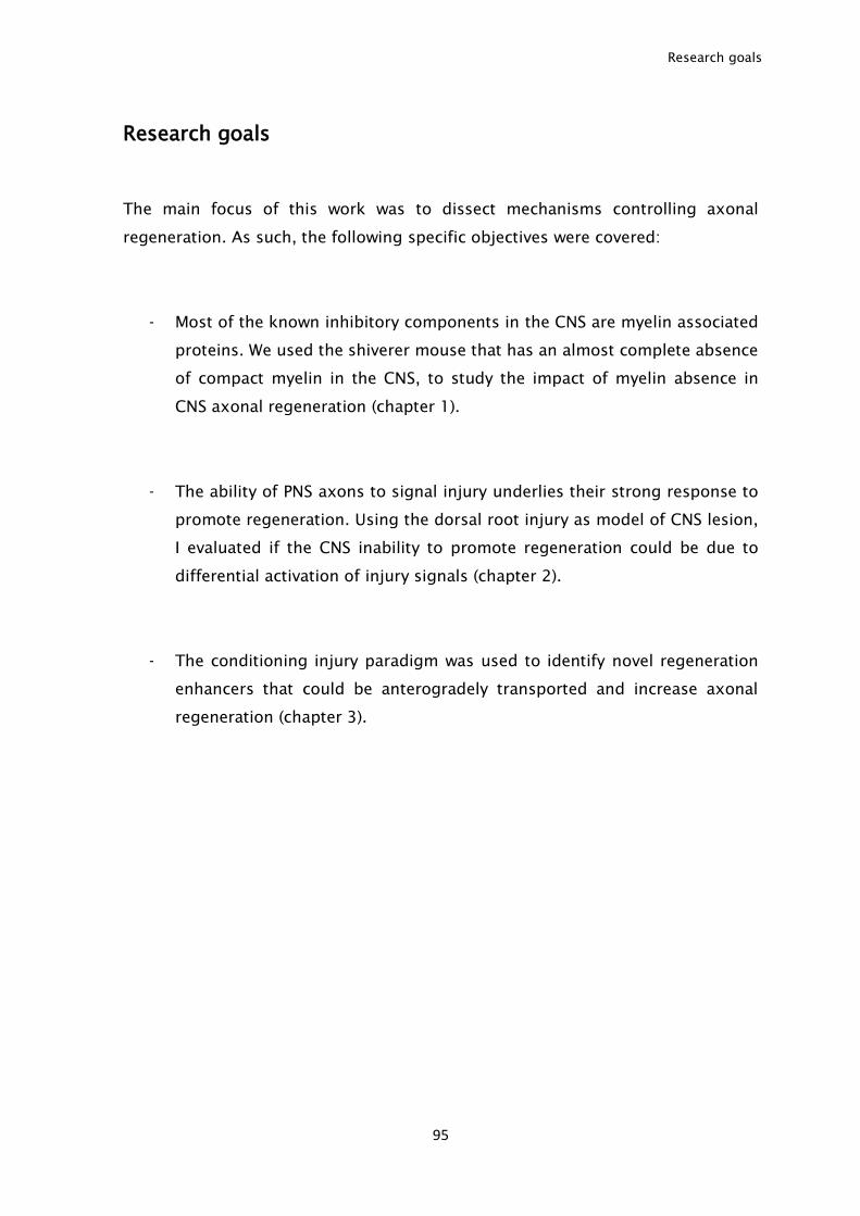
Research goals
95
Research goals
The main focus of this work was to dissect mechanisms controlling axonal
regeneration. As such, the following specific objectives were covered:
- Most of the known inhibitory components in the CNS are myelin associated
proteins. We used the shiverer mouse that has an almost complete absence
of compact myelin in the CNS, to study the impact of myelin absence in
CNS axonal regeneration (chapter 1).
- The ability of PNS axons to signal injury underlies their strong response to
promote regeneration. Using the dorsal root injury as model of CNS lesion,
I evaluated if the CNS inability to promote regeneration could be due to
differential activation of injury signals (chapter 2).
- The conditioning injury paradigm was used to identify novel regeneration
enhancers that could be anterogradely transported and increase axonal
regeneration (chapter 3).

96
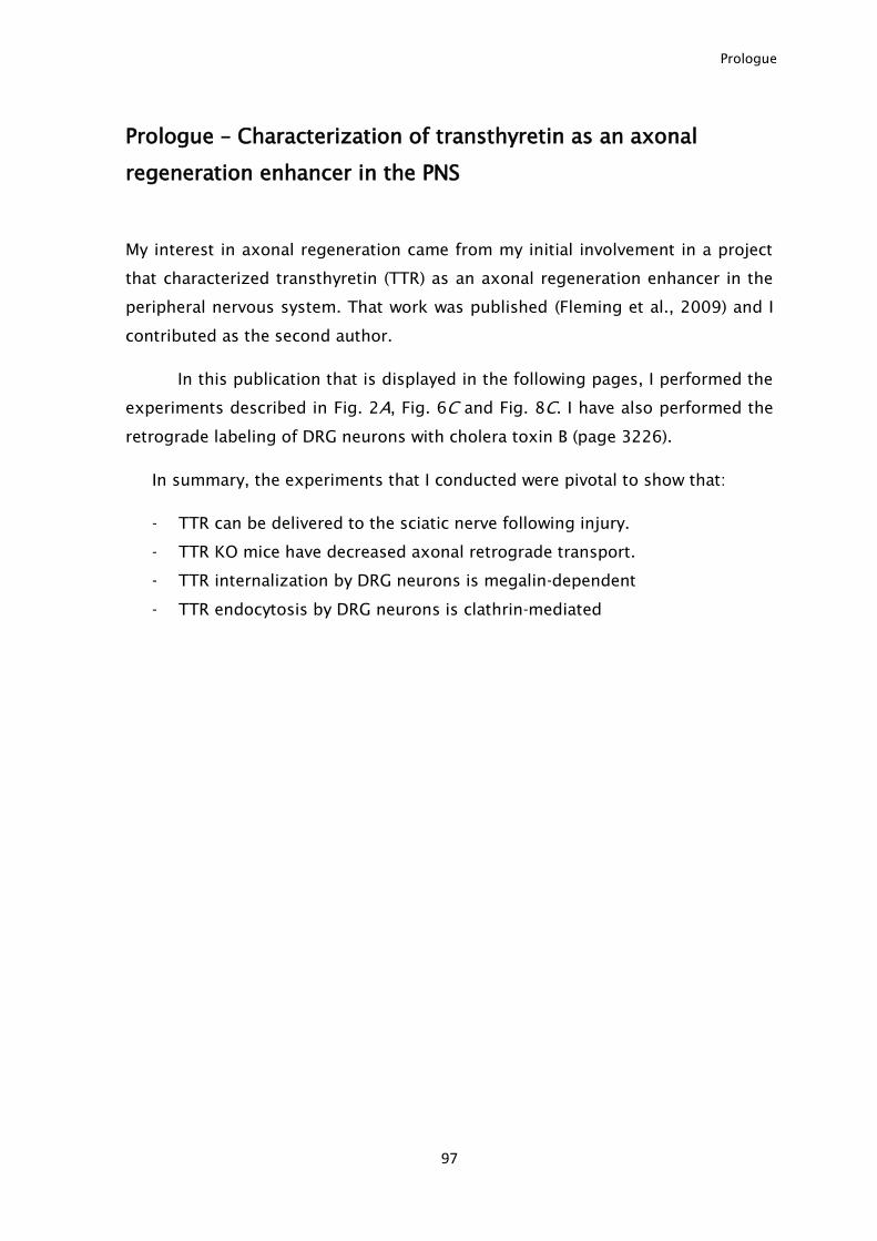
Prologue
97
Prologue – Characterization of transthyretin as an axonal
regeneration enhancer in the PNS
My interest in axonal regeneration came from my initial involvement in a project
that characterized transthyretin (TTR) as an axonal regeneration enhancer in the
peripheral nervous system. That work was published (Fleming et al., 2009) and I
contributed as the second author.
In this publication that is displayed in the following pages, I performed the
experiments described in Fig. 2A, Fig. 6C and Fig. 8C. I have also performed the
retrograde labeling of DRG neurons with cholera toxin B (page 3226).
In summary, the experiments that I conducted were pivotal to show that:
- TTR can be delivered to the sciatic nerve following injury.
- TTR KO mice have decreased axonal retrograde transport.
- TTR internalization by DRG neurons is megalin-dependent
- TTR endocytosis by DRG neurons is clathrin-mediated
Development/Plasticity/Repair
Transthyretin Internalization by Sensory Neurons Is MegalinMediated and Necessary for Its Neuritogenic Activity
Carolina E. Fleming,1,3 Fernando Milhazes Mar,1 Filipa Franquinho,1 Maria J. Saraiva,1,2 and Monica M. Sousa1
1Instituto de Biologia Molecular e Celular–IBMC, Nerve Regeneration Group and Molecular Neurobiology Group, 4150-180 Porto, Portugal, 2Instituto deCiencias Biomedicas Abel Salazar, Universidade do Porto, 4099-003 Porto, Portugal, and 3Programa Doutoral em Biologia Experimental e Biomedicina,Universidade de Coimbra, 3004-517 Coimbra, Portugal
Mutated transthyretin (TTR) causes familial amyloid polyneuropathy, a neurodegenerative disorder characterized by TTR deposition inthe peripheral nervous system (PNS). The origin/reason for TTR deposition in the nerve is unknown. Here we demonstrate that bothendogenous mouse TTR and TTR injected intravenously have access to the mouse sciatic nerve. We previously determined that in theabsence of TTR, both neurite outgrowth in vitro and nerve regeneration in vivo were impaired. Reinforcing this finding, we now show thatlocal TTR delivery to the crushed sciatic nerve rescues the regeneration phenotype of TTR knock-out (KO) mice. As the absence of TTRwas unrelated to neuronal survival, we further evaluated the Schwann cell and inflammatory response to injury, as well as axonalretrograde transport, in the presence/absence of TTR. Only retrograde transport was impaired in TTR KO mice which, in addition to theneurite outgrowth impairment, might account for the decreased regeneration in this strain. Moreover, we show that in vitro, in dorsal rootganglia neurons, clathrin-dependent megalin-mediated TTR internalization is needed for TTR neuritogenic activity. Supporting thisobservation, we demonstrate that in vivo, decreased levels of megalin lead to decreased nerve regeneration and that megalin’s action asa regeneration enhancer is dependent on TTR. In conclusion, our work unravels the mechanism of TTR action during nerve regeneration.Additionally, TTR presence in the nerve, as is here shown, may underlie its preferential deposition in the PNS of familial amyloidpolyneuropathy patients.
IntroductionWhen mutated, transthyretin (TTR) is related to familial amyloidpolyneuropathy (FAP) (Saraiva 2001), a neurodegenerative dis-order characterized by extracellular deposition of TTR aggregatesand amyloid fibrils, particularly in the peripheral nervous system(PNS) (Andrade, 1952). As a consequence of TTR deposition,axonal degeneration arises, ending up in neuronal loss. Severalclues have emerged as to the molecular mechanisms of TTR-mediated cellular toxicity leading to neurodegeneration in FAP(Sousa and Saraiva, 2003). The origin of TTR deposited in thePNS of FAP patients is however unknown. TTR is mainly synthe-sized by the liver and the choroid plexus that are respectively thesources of TTR in the plasma and CSF. TTR was reported as beingexpressed in the PNS, namely by glial cells of dorsal root ganglia(DRG) (Murakami et al., 2008). However, this issue was clarified
in a subsequent study showing that TTR synthesis does not occurin DRG cells (Sousa and Saraiva, 2008). Under physiological con-ditions serum-free human and rat nerve endoneurial fluid dis-play TTR immunoreaction (Saraiva et al., 1988). TTR may haveaccess to the nerve through the blood-nerve barrier (BNB),and/or through contact between peripheral nerve roots and CSF,where TTR is present in high levels. In fact, in recipients of FAPlivers, i.e., after domino liver transplantation, TTR deposits arefound within the nerve of the recipients, suggesting that plasmaTTR (synthesized in the liver) can cross the BNB (M. M. Sousa etal., 2004).
Apart from being a transporter of thyroxine (T4) and retinol,TTR has been described as having functions related to the ner-vous system, namely to be involved in cognition (Brouillette andQuirion, 2008), behavior (J. C. Sousa et al., 2004), and neuropep-tide processing (Nunes et al., 2006). In the case of the PNS, it wasdemonstrated that TTR enhances peripheral nerve regeneration(Fleming et al., 2007). In that study, TTR knock-out (KO) micepresented delayed functional and morphological recovery afternerve injury, as assessed from a decreased number of myelinatedand unmyelinated axons in the course of regeneration. TTR ca-pacity to enhance nerve regeneration was unrelated to neuronalsurvival as the absence of TTR was not accompanied by increasedneuronal loss. In transgenic mice expressing human TTR in neu-rons, in a TTR KO background, the delayed regeneration of TTRKO mice was rescued, reinforcing that TTR enhances nerve re-generation (Fleming et al., 2007). Additionally, absence of TTRwas found to be related to a decreased ability of DRG neurons to
Received Dec. 18, 2008; revised Feb. 5, 2009; accepted Feb. 10, 2009.This work was supported by Association Francaise contre les Myopathies (AFM), France, and Fundacao para a
Ciencia e a Tecnologia (FCT), Portugal (Grants PTDC/BIA-PRO/64437/2006 and SAU-OSM/64093/2006). C.E.F. wasthe recipient of a Programa Doutoral em Biologia Experimental e Biomedicina fellowship (SFRH/BD/9682/2002)from Centro de Neurosciencias de Coimbra/FCT (POCI 2010, FSE), Portugal, and of an AFM fellowship (AM/NM/2006.1272/FN12047). F.M.M. is the recipient of an FCT fellowship (SFRH/BD/43484/2008). We thank Rui Fernandesand Vera Sousa [Instituto de Biologia Molecular e Celular (IBMC)] for tissue processing and electron microscopy, Dr.Paula Sampaio (IBMC) for help in confocal microscopy, Dr. Rosario Almeida (IBMC) for help in T4 binding assays, Dr.Marcia Liz (IBMC) for the production of TTR, and Dr. Pedro Brites (AMC, Amsterdam) for critical reading of thismanuscript.
Correspondence should be addressed to Monica M. Sousa, Nerve Regeneration Group, Instituto de BiologiaMolecular e Celular–IBMC, R. Campo Alegre 823, 4150-180 Porto, Portugal. E-mail: [email protected].
DOI:10.1523/JNEUROSCI.6012-08.2009Copyright © 2009 Society for Neuroscience 0270-6474/09/293220-13$15.00/0
3220 • The Journal of Neuroscience, March 11, 2009 • 29(10):3220 –3232
grow neurites in vitro: cells grown without TTR displayed a re-duced number of neurites, as well as a decreased size of the long-est neurite (Fleming et al., 2007). This TTR neuritogenic effectwas demonstrated to be independent of its major ligands, retinoland T4. The newly described TTR role in peripheral nerve biologymight explain why, when mutated, the protein preferentially ac-cumulates in the PNS. Yet, the means through which TTR in-creases regeneration is unknown.
In the current work, we aimed at finding the mechanismthrough which TTR enhances nerve regeneration and neuriteoutgrowth.
Materials and MethodsMice. Mice were handled according to European Union and Nationalrules. WT and TTR KO (Episkopou et al., 1993) littermates (in the 129/Svbackground), as well as megalin heterozygous [MEG (�/�), kindly pro-vided by Dr. Thomas Willnow, Max-Delbrueck Center for MolecularMedicine, Berlin, Germany] and TTR KO/MEG (�/�) littermate mice(in the 129/Sv background), were obtained from the offspring of het-erozygous breeding pairs. All animals were maintained under a 12 hlight/dark cycle and fed with regular rodent’s chow and tap water adlibitum. Genotypes were determined from tail extracted genomic DNA.Unless otherwise stated, all comparisons comprised groups of 6 animalsper genotype, age- and sex-matched. For TTR detection in the nerve andnerve injury experiments, either 3 or 6 months old animals were used.For DRG neuron cultures, animals with 1– 4 weeks of age were used. Allexperiments were performed with the observer blinded to the animal’sgenotype.
Nerve injury. Mice were anesthetized with medetomidine/ketamineand a 4-mm-long incision was made in the shaved thigh skin. For nervecrush, the sciatic nerve was exposed and crush was performed using Peanforceps, twice during 15 s. To standardize the procedure, the crush sitewas maintained constant for each animal at 35 mm from the tip of thethird digit. A single skin suture, immediately above the crush site, servedas an additional reference. After surgery, animals were allowed to recoverfor 5, 15, 30, or 75 d. For chronic constriction injury (CCI), the sciaticnerve was exposed and one ligature was tied around the nerve using 4/0silk suture material (Braun). Skin was sutured and mice were allowed torecover for 24 h. Mice were perfused for 20 min with PBS through thevena cava at a flow rate of 2 ml/min and the sciatic nerve was subse-quently collected.
Assessment of mouse TTR presence in the nerve after nerve crush. WTmice underwent nerve crush and were allowed to recover for 3 d. Micewere perfused for 20 min with PBS through the vena cava at a flow rate of2 ml/min and the distal nerve segments were subsequently collected.Western blot was performed as described below. The primary antibodyused was a custom made rabbit anti-mouse TTR antibody (producedagainst recombinant mouse TTR, 1:500). Electronic microscopy usingWT distal nerve segments was performed as described below.
Assessment of TTR access to the nerve. TTR was produced as previouslydescribed (Liz et al., 2004), and 1 mg of recombinant human TTR wasconjugated with Alexa 488 using the Alexa Fluor 488 labeling kit (Invitro-gen), according to the manufacturer’s instructions. hTTR-Alexa 488 wasseparated from free Alexa 488 by fine size exclusion chromatography inBio-Rad BioGel P-30 resin columns. Subsequently, 1 �g of hTTR-Alexa488 was run in a 15% SDS polyacrylamide gel and, after electrophoresis,hTTR-Alexa 488 was visualized in a Typhoon 8600 (Amersham) to checklabeling efficacy. hTTR-Alexa 488 (100 �g) was injected intravenously inthe tail vein of WT and TTR KO mice. The next day, mice were subjectedto unilateral nerve crush as described above. The following day, micewere killed, and both crushed and contralateral sciatic nerves were col-lected and cryoprotected with a 1.2% L-lysine solution containing 2%formalin. For immunohistochemistry using the primary rabbit anti-human TTR (Dako; 1:1500), and rabbit anti-mouse TTR (1:2000) poly-clonal antibodies, 10 �m-thick sections were incubated in 0.1% sodiumborohydride (Sigma) for 5 min. Sections were then blocked in blockingbuffer (1% bovine serum albumin and 4% fetal bovine serum in PBS) for1 h at room temperature, and incubated with primary antibody diluted in
blocking buffer overnight. As a negative control, slides were left over-night at 4°C with either anti-mouse (TTR previously adsorbed with re-combinant mouse TTR, produced as previously described (Liz et al.,2004), or with blocking buffer alone. The adsorption of anti-mouse TTRwas performed by incubation of 200 �g of recombinant mouse TTR with1 �l of anti-mouse TTR overnight at room temperature with agitation.After centrifugation at 16,000 g, the supernatant, diluted 1:2000 in block-ing buffer, was used to perform immunohistochemistry. Subsequently,slide incubation with the anti-rabbit IgG-Alexa 568 secondary antibody(Invitrogen, 1:1000) diluted in blocking buffer was performed for 1 h atroom temperature. Coverslips were mounted with VectaShield Mount-ing Medium with DAPI (Vector), and images were taken using a LeicaSP2 AOBS SE (Leica) confocal laser scanning microscope.
Local delivery of TTR to the crushed nerve. WT and TTR KO miceunderwent bilateral nerve crush as described above. Immediately aftercrush and before suturing, 80 �l of Matrigel Basement Membrane Matrix(BD Biosciences) was applied to the left sciatic nerve crush site; the rightsciatic nerve crush site received 80 �l of Matrigel Basement MembraneMatrix supplemented with 60 �g of recombinant WT TTR, produced aspreviously described (Liz et al., 2004). Ten minutes after Matrigel appli-cation, when it had already gelled, skin was sutured. The animals wereallowed to recover for either 15 or 30 d. Mice were killed using a lethalanesthesia dosage, and the left and right nerve distal stumps were col-lected and processed for morphometric analysis as described below. Toassess for the presence of TTR in the nerve after Matrigel application,similar experiments were performed where 80 �l of Matrigel supple-mented with 60 �g of hTTR-Alexa 488 (produced as described above)were applied to the right sciatic nerve crush site of TTR KO mice; to theleft sciatic nerve crush site, Matrigel supplemented with free Alexa 488(equivalent to the amount of fluorophore present in 60 �g of hTTR-Alexa 488) was applied. As an additional negative control, a group ofanimals received Matrigel alone in the sciatic nerve crush site. Mice werekilled 1 d after Matrigel application and the nerve (3– 4 mm upstreamand downstream of the crush site) was collected, cryoprotected with a1.2% L-lysine solution containing 2% formalin and sectioned at 8 �m.The presence of hTTR-Alexa 488 was detected using a using a Leica SP2AOBS SE (Leica Microsystems) confocal laser scanning microscope.
Morphometric analysis. The 3 mm segments immediately distal to thecrush site were fixed overnight in 1.25% glutaraldehyde in 0.1 M sodiumcacodylate, washed in 0.1 M sodium cacodylate for 30 min, postfixed in1% osmium tetroxide in 0.2 M sodium cacodylate for 60 min, washedagain in 0.1 M sodium cacodylate for 30 min, dehydrated using a series ofgraded alcohols and propylene oxide, and embedded in epon. Transversesections (1.0 �m thick) were cut with a SuperNova, Reichest, Leica ul-tramicrotome, and stained with 1% toluidine blue in an 80°C heatingplate for 20 s. For each animal, the total number of myelinated fiberspresent in one semithin section was determined by counting 50� mag-nified photographs covering the whole nerve area. To determine thedensity of unmyelinated fibers, ultrathin transverse sections were cut andstained with uranyl acetate and lead citrate. For each animal, 20 nonover-lapping photomicrographs (7000� amplification) corresponding to�9000 �m 2 of each ultrathin section were taken using a transmissionelectron microscope (Zeiss 10C) and analyzed. To assess possible differ-ences in nerve total areas between strains, these were determined from10� magnified photos of sciatic nerve transverse sections.
Analysis of Schwann cell proliferation. WT and TTR KO mice under-went bilateral nerve crush and were allowed to recover for 5 d. To labeldividing cells, 100 �g/g body weight of BrdU (5-bromo-2�-deoxyuridine,Sigma) was injected intraperitoneally 4 and 2 h before kill. Labeling ofproliferating cells was performed 5 d after injury, since at this time point,Schwann cells reach their maximum proliferative activity in injured sci-atic nerves (Cheng and Zochodne, 2002). Mice were killed using a lethalanesthesia dosage and the sciatic nerves were collected and processed forimmunohistochemistry. Nerves were excised, fixed in 4% neutral buff-ered formalin and embedded in paraffin. Sections (5 �m thick) weredeparafinated in histoclear (National Diagnostics) and hydrated in adescendent alcohol series. Antigen unmasking was done by incubation in2N HCl for 20 min at 37°C, followed by neutralization with 0.1 M
Na2B4O7, and incubation with trypsin-EDTA (Invitrogen) for 10 min at
Fleming et al. • Transthyretin Internalization Is Megalin Mediated J. Neurosci., March 11, 2009 • 29(10):3220 –3232 • 3221
37°C. Immunohistochemistry using the primary anti-BrdU monoclonalantibody (1:1000, Sigma) was performed with the MOM kit (Vector)according to the manufacturer’s instructions, using diaminobenzidine(Sigma) as substrate. Slides were counterstained with hematoxylin(Merck). Two longitudinal nerve sections per animal were immuno-stained; four 50� magnified photographs per section distal to the crushsite were taken, covering an area of �0.5 mm 2, and the number ofBrdU-labeled nucleus was determined, as well as the total number ofnucleus stained with hematoxilin. The percentage of BrdU-labeled nu-cleus was then calculated for each animal.
Assessment of apoptosis by TUNEL analysis. The comparison of apo-ptotic cells in crushed nerves from WT and TTR KO mice was performedwith the ApopTag Peroxidase In Situ Apoptosis Detection Kit (MilliporeBioscience Research Reagents), according to the manufacturer’s instruc-tions, using diaminobenzidine (Sigma) as substrate. Slides were counter-stained with hematoxylin (Merck). Eight 10� magnified photographsper section near the crush site were taken, covering an area of �0.15mm 2. For each animal, the number of labeled nucleus was determined, aswell as the total number of nucleus stained with hematoxilin. The per-centage of apoptotic nucleus was then calculated for each animal.
Determination of macrophage number. Semithin sections stained withtoluidine blue of WT and TTR KO sciatic nerve distal segments wereobtained as described above for morphometric analysis. For each animal,the density of macrophages present in one semithin section covering thewhole nerve area was determined by observation of 20� magnified pho-tographs 15, 30, and 75 d after nerve crush.
Western blot. Intact and CCI nerves were sonicated in 0.5% TritonX-100 (Sigma) containing protease inhibitor mix (Amersham). Protein(12 �g total per lane) was run in 15% SDS polyacrylamide gels. Afterelectrophoresis, samples were transferred to a nitrocellulose membrane(Amersham), blocked with blocking buffer (5% nonfat dried milk inPBS), and incubated overnight at 4°C with primary antibodies diluted inblocking buffer, namely, rabbit polyclonal anti-p75 NTR (Santa Cruz Bio-technology, 1:200), and mouse monoclonal anti-�-actin (Sigma;1:5000). Subsequently, incubation with horseradish peroxidase (HRP)-labeled secondary antibodies diluted in blocking buffer, namely eitheranti-rabbit IgG-HRP (The Binding Site; 1:10,000) or anti-mouse IgG-HRP (The Binding Site; 1:5000), was performed for 1 h at room temper-ature. Blots were developed using the ECL PlusTM Western blottingreagents (Amersham) and exposed to Hyperfilm ECL (Amersham).Quantitative analysis of Western blots was performed using the Image-Quant software (Amersham). Results are shown as the ratio between p75NTR and �-actin signals.
Primary cultures of DRG neurons. Primary cultures of DRG neuronswere performed as described previously (Lindsay, 1988). Briefly, DRGwere dissected aseptically from WT or TTR KO mice, freed of roots andtreated with 0.125% collagenase (Sigma) for 3 h at 37°C. After enzymetreatment, a single-cell suspension was obtained by trituration with afire-polished Pasteur pipette. The cell suspension was centrifuged into a15% albumin gradient for 10 min at 200 g. The obtained pellet wasresuspended in Neurobasal medium supplemented with B27, penicillin-streptomicin, glutamine, fungizone (all from Invitrogen) and 50 ng/mlNGF (Sigma), plated in poly-L-lysine coated 13 mm coverslips and main-tained at 37°C.
Transferrin transport assay. The use of human transferrin conjugatedwith Texas red (Tf-TR, Invitrogen) as a tracer to examine retrogradetransport in cultures of DRG neurons has been previously documented(Liu et al., 2003). WT and TTR KO DRG neurons were cultivated asdescribed above. After 3 d in culture, cells were incubated with mediumcontaining 50 �g/ml Tf-TR for 2 h at 37°C to allow uptake. Cells werethen washed and incubated with Neurobasal medium supplementedwith B27, penicillin-streptomicin, glutamine, fungizone (all from In-vitrogen), and 50 ng/ml NGF (Sigma). Twenty-seven hours later, neu-rons were fixed in 2% neutral buffered formalin for 30 min, washed withPBS, and kept at 4°C until use. Slides were mounted in VectaShieldMounting Medium with DAPI (Vector). Images were taken using a LeicaSP2 AOBS SE (Leica Microsystems) confocal laser-scanning microscope.For the quantification of Tf-TR labeling in DRG neurons, a semiquanti-tative scale ranging from 1 to 5 was used as follows: 1, Tf-TR present in
100% of the neurites; 2, Tf-TR present in �50% of the neurites; 3, Tf-TRpresent in 50% of the neurites; 4, Tf-TR present in �50% of the neurites;5, Tf-TR absent from neurites (i.e., only present in the cell body).
In vivo analysis of retrograde transport using cholera toxin B. The sciaticnerve of WT and TTR KO mice was exposed and transected at the mid-tight level; a solution of the retrogradely transported cholera toxin Bsubunit (0.5 mg/ml, List Biological) was applied to the proximal end ofthe transected sciatic nerve for 35 min. The skin was subsequently su-tured and mice were allowed to recover for 72 h, after which the L4 – 6DRG were collected and fixed in 4% neutral buffered formalin. To detectretrogradely labeled sensory neurons, serial 4-�m-thick DRG sectionswere cut and processed for anti-cholera toxin immunohistochemistry.Briefly, sections were blocked in blocking buffer (1% bovine serum al-bumin and 4% fetal bovine serum in PBS) for 30 min at 37°C and incu-bated with anti-cholera toxin antibody (Calbiochem; 1:1000) diluted inblocking buffer overnight at 4°C. Antigen visualization was performedwith the biotin-extravidin-peroxidase kit (Sigma). For each animal, todetermine the percentage of retrogradely labeled sensory neurons, thetotal number of DRG neurons, as well as the number of labeled DRGneurons presenting visible nuclei, were counted every 24 �m.
Analysis of TTR endocytosis by DRG neuron cultures. Primary culturesof DRG neurons were performed as described above (Lindsay, 1988).Cells were maintained for 96 h at 37°C. After this period, DRG neuronswere supplemented with 300 �g/ml recombinant human TTR conju-gated with Alexa 488 (hTTR-Alexa 488, produced as described above) for3 h at 4°C or 37°C. When mentioned, neuronal cells were additionallyincubated with 50 �g/ml transferrin conjugated with Texas red (Tf-TR,Invitrogen) or with 5 �g/ml cholera toxin subunit B conjugated withAlexa Fluor 647 (CT-B, Invitrogen). Cells were fixed in 2% neutral buff-ered formalin for 30 min, washed with PBS and kept at 4°C until immu-nostaining. For immunocytochemistry, DRG neurons were permeabil-ized with 0.2% Triton X-100 (Sigma) and were then incubated with 0.1%sodium borohydride (Sigma). For immunocytochemistry using the pri-mary antibody rabbit anti-PGP 9.5 (1:500, Ab Serotec), coverslips wereblocked in MOM IgG Blocking reagent (Vector) for 1 h at room temper-ature, and incubated with primary antibody diluted in MOM diluent(Vector) overnight at 4°C. Subsequently, incubation with the secondaryantibody anti-rabbit IgG-Alexa 568 (1:1000, Invitrogen) diluted inMOM diluent was performed for 1 h at room temperature. Coverslipswere mounted with VectaShield Mounting Medium containing DAPI(Vector), and images were taken using a Leica SP2 AOBS SE (Leica Mi-crosystems) confocal laser scanning microscope.
Analysis of TTR endocytosis by transfected DRG neurons. Primary DRGneurons were obtained as already detailed. After DRG neuron isolationand prior plating, �250,000 neurons were transfected with 10 �g ofGFP-tagged Eps15 constructs (a kind gift from Dr. Benmerah, InstitutCochin, Paris, France), isolated using the Endofree Plasmid Maxi kit(Qiagen, Portugal). Transfection was performed in a Amaxa Nucleofec-tor (Amaxa Biosystems), using program A-033 and the Mouse NeuronNucleofector kit (Amaxa Biosystems). Transfected cells were subse-quently plated in 24-well plates at a density of �30,000 cells/well. Cellswere maintained for 48 h at 37°C after which they were supplementedwith 300 �g/ml recombinant human TTR conjugated with Alexa 568(hTTR-Alexa 568, produced similarly as described above for hTTR-Alexa 488) for 3 h at 37°C. Cells were then processed for PGP 9.5 immu-nocytochemistry, as described above, using as secondary antibody anti-rabbit IgG-Alexa 647 (1:1000, Invitrogen). For quantification of hTTR-Alexa 568 internalization, images of randomly selected PGP 9.5 labeledcells were taken along the z-axis in a Leica SP2 AOBS SE confocal laserscanning microscope, using the same laser intensity for all conditions.hTTR-Alexa 568 internalization in cells photographed in each of theconditions was then determined using the ImageJ software (http://rs-bweb.nih.gov/ij/) and calculated as total brightness intensity in the 568channel (expressed as arbitrary units)/cell.
Measurement of neurite outgrowth using TTR coupled to FluoSpheres.Recombinant WT TTR (2 mg) was covalently bound to 1 �m-diametercarboxylate- or amine-modified FluoSpheres (Invitrogen), using a car-bodiimide cross-linking method, according to the manufacturer’s in-structions. FluoSpheres bound to TTR were washed three times with PBS
3222 • J. Neurosci., March 11, 2009 • 29(10):3220 –3232 Fleming et al. • Transthyretin Internalization Is Megalin Mediated
and the efficacy of the method was evaluated by running the three washescontaining free uncoupled WT TTR in a 15% SDS polyacrylamide gel;�90% of WT TTR was bound to the FluoSpheres and no detectable freeTTR was found within the last wash. PC12 cells (European Collection ofCell Cultures), a rat adrenal cell line with a neuronal-like phenotype,were grown in six-well plates in DMEM (Invitrogen) supplemented with10% fetal bovine serum (Invitrogen). When cells reached 50% conflu-ence, fetal bovine serum was withdrawn and cells were supplementedwith either: (1) 10% of either WT or TTR KO mouse serum, (2) 10% TTRKO mouse serum containing 300 �g/ml recombinant WT TTR, (3) 10%of TTR KO mouse serum containing 0.12% of free FluoSpheres, (4) 10%of TTR KO mouse serum containing 300 �g/ml free recombinant WTTTR and 0.12% of free FluoSpheres, and (5) 10% of TTR KO mouseserum containing 300 �g/ml recombinant WT TTR coupled to 0.12%FluoSpheres. Fifty hours later, PC12 cells were fixed in 2% neutral buff-ered formalin for 30 min, washed with PBS and kept at 4°C until furtheranalysis. Neurite size was determined from 50� magnified fields. At least150 cells were analyzed for each condition. To check whether TTR wasbiologically active when coupled to FluoSpheres, its ability to bind T4 wasevaluated. Briefly, a 100 �l suspension of either free FluoSpheres (0.12%)or 8.25 �g/ml TTR coupled to FluoSpheres (0.12%) was incubated with50,000 cpm of [125I]T4 (Perkin-Elmer) in triplicates overnight at 4°C. Asan internal control to ascertain the specificity of T4 binding to TTR, 100�l suspension of the same samples were coincubated with 50,000 cpm of[ 125I]T4 and nonradioactive T4 in molar excess. Subsequently, Fluo-Spheres were washed three times with PBS and radioactivity of the pel-lets, corresponding to T4 bound to TTR, was counted in a gamma-spectrometer (Wallac).
Analysis of TTR endocytosis in DRG neurons blocked with sheep anti-ratmegalin antibody. After DRG neuron isolation, cells were plated in 24-well plates at a density of �10,000 cells/well. Cells were maintained for96 h at 37°C after which they were preincubated for 2 h at 37°C with sheepanti-rat megalin (1:200, kindly provided by Dr. Pierre Verroust, CHUSaint Antoine, Paris, France) or nonimmune sheep serum (using anequal volume as the one used in the case of sheep anti-rat megalin).Subsequently, DRG neurons were supplemented with 300 �g/ml hTTR-Alexa 488 in the presence of sheep anti-rat megalin or nonimmune sheepserum (in the same conditions as used for the preincubation) for 3 h at37°C. It is noteworthy that this anti-megalin antibody has been usedpreviously to successfully inhibit megalin-dependent internalization(Klassen et al., 2004). DRG neurons were then processed for PGP 9.5immunocytochemistry as described above using as secondary antibodyanti-rabbit IgG-Alexa 568 (1:1000, Invitrogen). For quantification ofhTTR-Alexa 488 internalization, images of randomly selected PGP 9.5labeled cells were taken along the z-axis in a Leica SP2 AOBS SE confocallaser scanning microscope, using the same laser intensity for all condi-tions. The intensity of internalized hTTR-Alexa 488 in DRG neuronstreated with either sheep anti-rat megalin or nonimmune sheep serumwas then determined using the ImageJ software (http://rsbweb.nih.gov/ij/), as described above.
Measurement of neurite outgrowth in the presence of TTR and anti-ratmegalin. TTR KO DRG neurons were maintained for 48 h at 37°C, afterwhich DRG neurons were supplemented with (1) B27 or B27 containingeither (2) 300 �g/ml recombinant TTR, (3) 300 �g/ml recombinant TTRand 200 �g/ml sheep anti-rat megalin (kindly provided by Dr. PierreVerroust), (4) 200 �g/ml sheep anti-rat megalin, and (5) 300 �g/mlrecombinant TTR and 200 �g/ml IgG (IgG, Sigma). As an additionalcontrol, in an independent experiment, DRG neurons were supple-mented with B27 containing a volume of nonimmune sheep serum equalto the one of sheep anti-rat megalin used in condition (5). Fifty hourslater, cells were fixed in 2% neutral buffered formalin for 30 min, washedwith PBS and kept at 4°C until further analysis. Neurite size was deter-mined from 50� magnified fields. At least 180 cells were analyzed foreach condition.
Megalin immunohistochemistry and RT-PCR. For RT-PCR, total RNAfrom mouse DRG and kidney was isolated using Trizol (Invitrogen).cDNA was obtained using the Superscript II kit (Invitrogen). PCR wasperformed using the following sense and antisense primers: for mousemegalin, 5�-CCTTGCCAAACCCTCTGAAAAT-3� and 5�-CACAAG-
GTTTGCGGTGTCTTTA-3� and for mouse HPRT, 5�-GTAAT-GATCAGTCAACGGGGGAC-3� and 5�-CCAGCAAGCTTGCAAC-CTTAACCA-3�. Ethidium bromide-stained gels were scanned using aTyphoon 8600 (Amersham). For immunohistochemistry using the sheepanti-megalin primary antibody (1:2000, kindly provided by Dr. PierreVerroust), antigen unmasking was done by boiling 3� in 0.5 mM EDTA10 mM Tris pH � 9.0 solution. Sections were blocked in blocking buffer(1% bovine serum albumin and 4% fetal bovine serum in PBS) for 30min at 37°C and incubated with primary antibody diluted in blockingbuffer overnight at 4°C. Antigen visualization was performed with thebiotin-extravidin-peroxidase kit (Sigma). Slides were counterstainedwith hematoxylin (Merck).
Statistical analysis. Statistical analysis was performed using the Stu-dent’s t test. Results were expressed as average SEM.
ResultsTTR is detectable within the endoneurium of both controland crushed nervesWe recently determined that TTR enhances nerve regeneration(Fleming et al., 2007). Upon nerve injury, disruption of the BNBoccurs, leading to the exposure of the nerve to plasma proteins.To verify the disruption of the BNB by nerve crush, ultrathinnerve sections were analyzed, 3 d after crush; blood residues wereobserved near the crush site (Fig. 1A). To check whether TTR waspresent in the nerve 3 d after crush, mouse TTR immunoblottingof crushed nerves from perfused WT mice was performed. Asillustrated in Figure 1B, TTR is indeed present in the nerve aftercrush, in the course of regeneration. As positive and negativecontrols, CSF from WT mice (WT CSF) and nerve extracts fromTTR KO mice (TTR KO nerve) were used, respectively (Fig. 1B).
To further unveil the presence of TTR in the nerve in thesettings of nerve injury, we performed immunohistochemistryagainst mouse TTR (mTTR) in crushed nerves from WT mice.mTTR was readily detected within the nerve, along nerve fibers(Fig. 1Ca). To confirm the specificity of mTTR immunohisto-chemistry, three different negative controls were performed:mTTR immunohistochemistry of (1) TTR KO crushed nerves(Fig. 1Cb), (2) WT crushed nerves using the anti-mTTR primaryantibody previously adsorbed with recombinant mTTR (Fig.1Cc), and (3) WT crushed nerves in the absence of primary anti-body (Fig. 1Cd). As expected, no reactivity was present in thenegative controls. Considering the prompt detection of TTR inthe crushed nerve, we followed by investigating whether TTR wasdetectable in the nerve under physiological conditions. mTTRimmunohistochemistry of WT intact nerves revealed that TTR isreadily detectable in the nerve (Fig. 1Da) with a similar immu-nostaining pattern as the one observed after nerve crush (Fig.1Ca). The analysis of transverse sections of WT intact nervesstained for mTTR suggests that TTR is probably present in theextracellular matrix surrounding nerve fibers, as visualized bycolocalization with brightfield images (Fig. 1Db– d).
To further understand how TTR gains access to the nerve afterinjury, hTTR-Alexa 488 was injected intravenously 1 d beforenerve crush; 1 d after crush, nerves were collected and cryopre-served. Subsequently, crushed nerves were screened for greenfluorescence. Interestingly, hTTR-Alexa 488 was found withinthe nerve, along fibers (Fig. 1Eb). The same pattern was found inWT and TTR KO nerves (data not shown). To verify that thegreen fluorescence corresponded to injected hTTR-Alexa 488,immunohistochemistry against human TTR (hTTR) was per-formed, showing that both signals colocalized (Fig. 1Eb– d), con-firming that plasma TTR enters the sciatic nerve after crush.
Fleming et al. • Transthyretin Internalization Is Megalin Mediated J. Neurosci., March 11, 2009 • 29(10):3220 –3232 • 3223
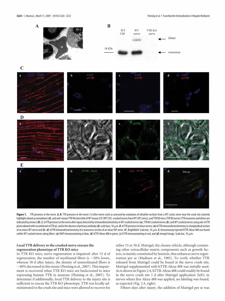
Local TTR delivery to the crushed nerve rescues theregeneration phenotype of TTR KO miceIn TTR KO mice, nerve regeneration is impaired: after 15 d ofregeneration, the number of myelinated fibers is �20% lower,whereas 30 d after injury, the density of unmyelinated fibers is�40% decreased in this strain (Fleming et al., 2007). This impair-ment is recovered when TTR KO mice are backcrossed to miceexpressing human TTR in neurons (Fleming et al., 2007). Todetermine if additionally, local TTR delivery to the injury site issufficient to rescue the TTR KO phenotype, TTR was locally ad-ministrated to the crush site and mice were allowed to recover for
either 15 or 30 d. Matrigel, the chosen vehicle, although contain-ing other extracellular matrix components such as growth fac-tors, is mainly constituted by laminin, that enhances nerve regen-eration per se (Madison et al., 1985). To verify whether TTRreleased from Matrigel could be found at the nerve crush site,Matrigel supplemented with hTTR-Alexa 488 was initially used.As is shown in Figure 2A, hTTR-Alexa 488 could readily be foundin the nerve crush site 1 d after Matrigel application (left); innerves where free Alexa 488 was applied, no labeling was found,as expected (Fig. 2A, right).
Fifteen days after injury, the addition of Matrigel per se was
Figure 1. TTR presence in the nerve. A, B, TTR presence in the nerve 3 d after nerve crush as assessed by evaluation of ultrathin sections from a WT sciatic nerve near the crush site (asteriskhighlights blood accumulation) (A), and anti-mouse TTR Western blot of WT mouse CSF (WT CSF), crushed nerves from WT (WT nerve), and TTR KO mice (TTR KO nerve) (TTR monomer and dimer areindicated by arrows) (B). C, mTTR presence in the nerve after injury detected by immunohistochemistry in WT crushed nerves (a), TTR KO crushed nerves (b), and WT crushed nerves using anti-mTTRpreincubated with recombinant mTTR (c), and in the absence of primary antibody (d); scale bars, 10 �m. D, mTTR presence in intact nerves: (a) mTTR immunohistochemistry in a longitudinal sectionof an intact WT nerve and (b– d) mTTR immunohistochemistry of a transverse section of an intact WT nerve. BF, Brightfield. Scale bar, 10 �m. E, Intravenously injected hTTR-Alexa 488 was foundwithin WT crushed nerves along fibers: (a) DAPI immunostaining in blue, (b) hTTR-Alexa 488 in green, (c) hTTR immunostaining in red, and (d) merged image. Scale bar, 10 �m.
3224 • J. Neurosci., March 11, 2009 • 29(10):3220 –3232 Fleming et al. • Transthyretin Internalization Is Megalin Mediated
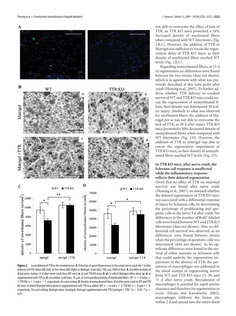
not able to overcome the effect of lack ofTTR, as TTR KO mice presented a 24%decreased density of myelinated fiberswhen compared with WT littermates (Fig.2B,C). However, the addition of TTR toMatrigel was sufficient to rescue the regen-eration delay of TTR KO mice, as theirdensity of myelinated fibers reached WTlevels (Fig. 2B,C).
Regarding unmyelinated fibers, at 15 dof regeneration no differences were foundbetween the two strains (data not shown)which is in agreement with what was pre-viously described at this time point aftercrush (Fleming et al., 2007). To further ad-dress whether TTR delivery to crushednerves of WT and TTR KO mice could res-cue the regeneration of unmyelinated fi-bers, their density was determined 30 d af-ter injury. Similarly to what was observedfor myelinated fibers, the addition of Ma-trigel per se was not able to overcome thelack of TTR, as 30 d after injury TTR KOmice presented a 30% decreased density ofunmyelinated fibers when compared withWT littermates (Fig. 2D). However, theaddition of TTR to Matrigel was able torescue the regeneration impairment ofTTR KO mice, as their density of unmyeli-nated fibers reached WT levels (Fig. 2D).
In TTR KO mice, after nerve crush, theSchwann cell response is unaffectedwhile the inflammatory responsereflects their delayed regenerationGiven that no effect of TTR on neuronalsurvival was found after nerve crush(Fleming et al., 2007), we assessed whetherthe delayed regeneration of TTR KO micewas associated with a differential responseto injury by Schwann cells, by determiningthe percentage of proliferating and apo-ptotic cells in the nerve 5 d after crush. Nodifferences in the number of BrdU-labeledcells were found between WT and TTR KOlittermates (data not shown). Also, no dif-ferential cell survival was observed, as nodifferences were found between strainswhen the percentage of apoptotic cells wasdetermined (data not shown). As no sig-nificant differences were found in the sur-vival of either neurons or Schwann cellsthat could underlie the regeneration im-pairment in the absence of TTR, the per-sistence of macrophages was addressed inthe distal stumps of regenerating nervesfrom WT and TTR KO mice 15, 30, and75 d after nerve crush. Recruitment ofmacrophages is essential for rapid myelinclearance and therefore for regeneration tooccur (Hirata and Kawabuchi, 2002);macrophages infiltrate the lesion sitewithin 2 d and spread into the entire distal
Figure 2. Local delivery of TTR to the crushed nerve. A, Detection of green fluorescence in the sciatic nerve crush site 1 d afterdelivery of hTTR-Alexa 488 (left) or free Alexa 488 (right) in Matrigel. Scale bars, 100 �m; DAPI in blue. B, Semithin sections ofdistal nerve stumps 15 d after nerve crush from WT mice (a, c) and TTR KO mice (b, d), in which Matrigel either alone (a, b) orsupplemented with TTR (c, d) was added. Scale bars, 10 �m. C, Corresponding density of myelinated fibers. WT: n � 6 and n �7; TTR KO: n � 5 and n � 7, respectively, for each setting. D, Density of unmyelinated fibers 30 d after nerve crush in WT and TTRKO mice, in which Matrigel either alone or supplemented with TTR was added. WT: n � 6 and n � 5; TTR KO: n � 8 and n � 8,respectively, for each setting; Matrigel alone (matrigel), Matrigel supplemented with TTR (matrigel�TTR). *p � 0.05, **p �0.01.
Fleming et al. • Transthyretin Internalization Is Megalin Mediated J. Neurosci., March 11, 2009 • 29(10):3220 –3232 • 3225
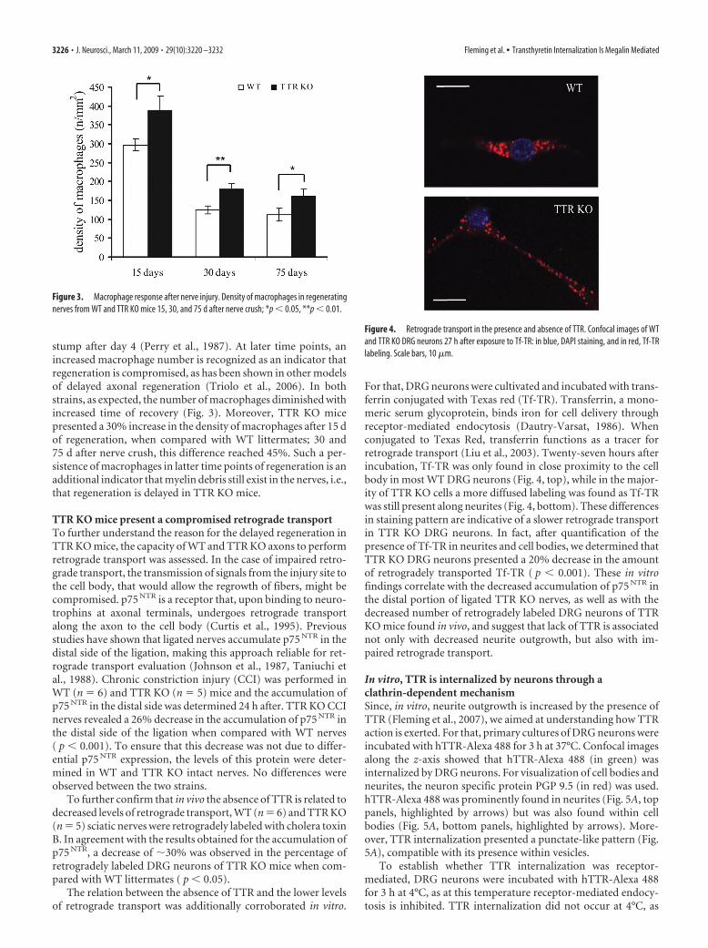
stump after day 4 (Perry et al., 1987). At later time points, anincreased macrophage number is recognized as an indicator thatregeneration is compromised, as has been shown in other modelsof delayed axonal regeneration (Triolo et al., 2006). In bothstrains, as expected, the number of macrophages diminished withincreased time of recovery (Fig. 3). Moreover, TTR KO micepresented a 30% increase in the density of macrophages after 15 dof regeneration, when compared with WT littermates; 30 and75 d after nerve crush, this difference reached 45%. Such a per-sistence of macrophages in latter time points of regeneration is anadditional indicator that myelin debris still exist in the nerves, i.e.,that regeneration is delayed in TTR KO mice.
TTR KO mice present a compromised retrograde transportTo further understand the reason for the delayed regeneration inTTR KO mice, the capacity of WT and TTR KO axons to performretrograde transport was assessed. In the case of impaired retro-grade transport, the transmission of signals from the injury site tothe cell body, that would allow the regrowth of fibers, might becompromised. p75 NTR is a receptor that, upon binding to neuro-trophins at axonal terminals, undergoes retrograde transportalong the axon to the cell body (Curtis et al., 1995). Previousstudies have shown that ligated nerves accumulate p75 NTR in thedistal side of the ligation, making this approach reliable for ret-rograde transport evaluation (Johnson et al., 1987, Taniuchi etal., 1988). Chronic constriction injury (CCI) was performed inWT (n � 6) and TTR KO (n � 5) mice and the accumulation ofp75 NTR in the distal side was determined 24 h after. TTR KO CCInerves revealed a 26% decrease in the accumulation of p75 NTR inthe distal side of the ligation when compared with WT nerves( p � 0.001). To ensure that this decrease was not due to differ-ential p75 NTR expression, the levels of this protein were deter-mined in WT and TTR KO intact nerves. No differences wereobserved between the two strains.
To further confirm that in vivo the absence of TTR is related todecreased levels of retrograde transport, WT (n � 6) and TTR KO(n � 5) sciatic nerves were retrogradely labeled with cholera toxinB. In agreement with the results obtained for the accumulation ofp75 NTR, a decrease of �30% was observed in the percentage ofretrogradely labeled DRG neurons of TTR KO mice when com-pared with WT littermates ( p � 0.05).
The relation between the absence of TTR and the lower levelsof retrograde transport was additionally corroborated in vitro.
For that, DRG neurons were cultivated and incubated with trans-ferrin conjugated with Texas red (Tf-TR). Transferrin, a mono-meric serum glycoprotein, binds iron for cell delivery throughreceptor-mediated endocytosis (Dautry-Varsat, 1986). Whenconjugated to Texas Red, transferrin functions as a tracer forretrograde transport (Liu et al., 2003). Twenty-seven hours afterincubation, Tf-TR was only found in close proximity to the cellbody in most WT DRG neurons (Fig. 4, top), while in the major-ity of TTR KO cells a more diffused labeling was found as Tf-TRwas still present along neurites (Fig. 4, bottom). These differencesin staining pattern are indicative of a slower retrograde transportin TTR KO DRG neurons. In fact, after quantification of thepresence of Tf-TR in neurites and cell bodies, we determined thatTTR KO DRG neurons presented a 20% decrease in the amountof retrogradely transported Tf-TR ( p � 0.001). These in vitrofindings correlate with the decreased accumulation of p75 NTR inthe distal portion of ligated TTR KO nerves, as well as with thedecreased number of retrogradely labeled DRG neurons of TTRKO mice found in vivo, and suggest that lack of TTR is associatednot only with decreased neurite outgrowth, but also with im-paired retrograde transport.
In vitro, TTR is internalized by neurons through aclathrin-dependent mechanismSince, in vitro, neurite outgrowth is increased by the presence ofTTR (Fleming et al., 2007), we aimed at understanding how TTRaction is exerted. For that, primary cultures of DRG neurons wereincubated with hTTR-Alexa 488 for 3 h at 37°C. Confocal imagesalong the z-axis showed that hTTR-Alexa 488 (in green) wasinternalized by DRG neurons. For visualization of cell bodies andneurites, the neuron specific protein PGP 9.5 (in red) was used.hTTR-Alexa 488 was prominently found in neurites (Fig. 5A, toppanels, highlighted by arrows) but was also found within cellbodies (Fig. 5A, bottom panels, highlighted by arrows). More-over, TTR internalization presented a punctate-like pattern (Fig.5A), compatible with its presence within vesicles.
To establish whether TTR internalization was receptor-mediated, DRG neurons were incubated with hTTR-Alexa 488for 3 h at 4°C, as at this temperature receptor-mediated endocy-tosis is inhibited. TTR internalization did not occur at 4°C, as
Figure 3. Macrophage response after nerve injury. Density of macrophages in regeneratingnerves from WT and TTR KO mice 15, 30, and 75 d after nerve crush; *p � 0.05, **p � 0.01.
Figure 4. Retrograde transport in the presence and absence of TTR. Confocal images of WTand TTR KO DRG neurons 27 h after exposure to Tf-TR: in blue, DAPI staining, and in red, Tf-TRlabeling. Scale bars, 10 �m.
3226 • J. Neurosci., March 11, 2009 • 29(10):3220 –3232 Fleming et al. • Transthyretin Internalization Is Megalin Mediated
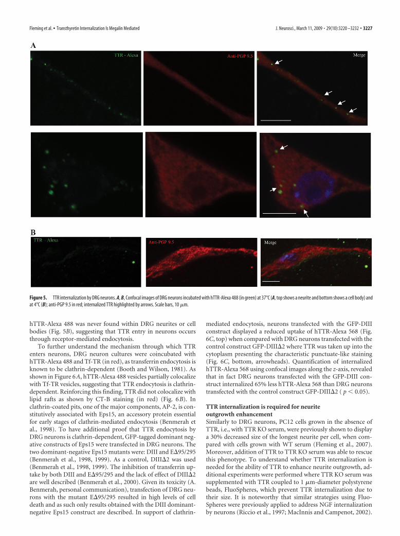
hTTR-Alexa 488 was never found within DRG neurites or cellbodies (Fig. 5B), suggesting that TTR entry in neurons occursthrough receptor-mediated endocytosis.
To further understand the mechanism through which TTRenters neurons, DRG neuron cultures were coincubated withhTTR-Alexa 488 and Tf-TR (in red), as transferrin endocytosis isknown to be clathrin-dependent (Booth and Wilson, 1981). Asshown in Figure 6A, hTTR-Alexa 488 vesicles partially colocalizewith Tf-TR vesicles, suggesting that TTR endocytosis is clathrin-dependent. Reinforcing this finding, TTR did not colocalize withlipid rafts as shown by CT-B staining (in red) (Fig. 6B). Inclathrin-coated pits, one of the major components, AP-2, is con-stitutively associated with Eps15, an accessory protein essentialfor early stages of clathrin-mediated endocytosis (Benmerah etal., 1998). To have additional proof that TTR endocytosis byDRG neurons is clathrin-dependent, GFP-tagged dominant neg-ative constructs of Eps15 were transfected in DRG neurons. Thetwo dominant-negative Eps15 mutants were: DIII and E95/295(Benmerah et al., 1998, 1999). As a control, DIII2 was used(Benmerah et al., 1998, 1999). The inhibition of transferrin up-take by both DIII and E95/295 and the lack of effect of DIII2are well described (Benmerah et al., 2000). Given its toxicity (A.Benmerah, personal communication), transfection of DRG neu-rons with the mutant E95/295 resulted in high levels of celldeath and as such only results obtained with the DIII dominant-negative Eps15 construct are described. In support of clathrin-
mediated endocytosis, neurons transfected with the GFP-DIIIconstruct displayed a reduced uptake of hTTR-Alexa 568 (Fig.6C, top) when compared with DRG neurons transfected with thecontrol construct GFP-DIII2 where TTR was taken up into thecytoplasm presenting the characteristic punctuate-like staining(Fig. 6C, bottom, arrowheads). Quantification of internalizedhTTR-Alexa 568 using confocal images along the z-axis, revealedthat in fact DRG neurons transfected with the GFP-DIII con-struct internalized 65% less hTTR-Alexa 568 than DRG neuronstransfected with the control construct GFP-DIII2 ( p � 0.05).
TTR internalization is required for neuriteoutgrowth enhancementSimilarly to DRG neurons, PC12 cells grown in the absence ofTTR, i.e., with TTR KO serum, were previously shown to displaya 30% decreased size of the longest neurite per cell, when com-pared with cells grown with WT serum (Fleming et al., 2007).Moreover, addition of TTR to TTR KO serum was able to rescuethis phenotype. To understand whether TTR internalization isneeded for the ability of TTR to enhance neurite outgrowth, ad-ditional experiments were performed where TTR KO serum wassupplemented with TTR coupled to 1 �m-diameter polystyrenebeads, FluoSpheres, which prevent TTR internalization due totheir size. It is noteworthy that similar strategies using Fluo-Spheres were previously applied to address NGF internalizationby neurons (Riccio et al., 1997; MacInnis and Campenot, 2002).
Figure 5. TTR internalization by DRG neurons. A, B, Confocal images of DRG neurons incubated with hTTR-Alexa 488 (in green) at 37°C (A, top shows a neurite and bottom shows a cell body) andat 4°C (B); anti-PGP 9.5 in red; internalized TTR highlighted by arrows. Scale bars, 10 �m.
Fleming et al. • Transthyretin Internalization Is Megalin Mediated J. Neurosci., March 11, 2009 • 29(10):3220 –3232 • 3227
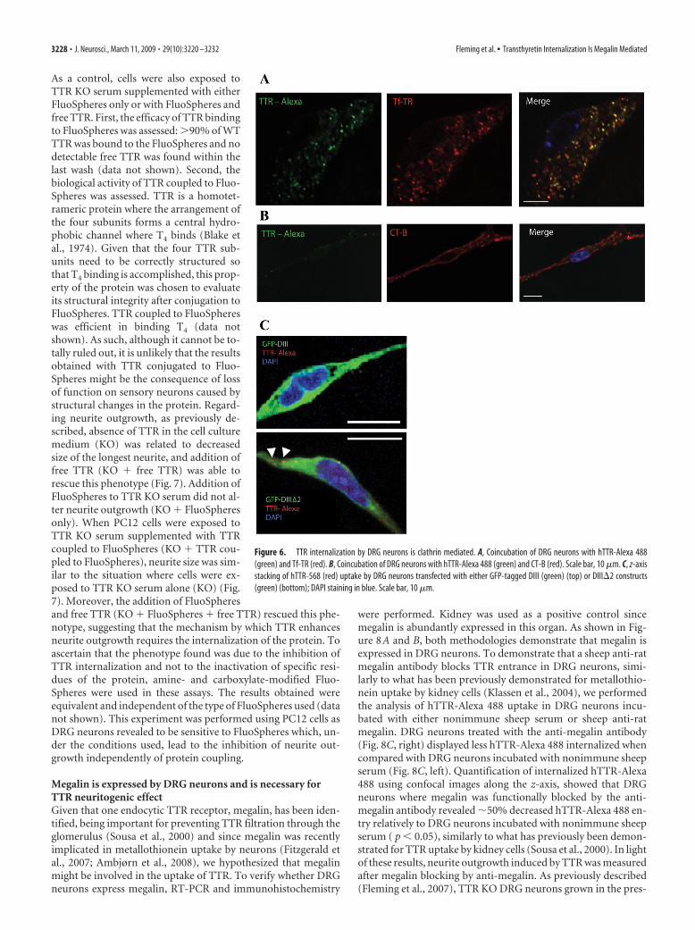
As a control, cells were also exposed toTTR KO serum supplemented with eitherFluoSpheres only or with FluoSpheres andfree TTR. First, the efficacy of TTR bindingto FluoSpheres was assessed: �90% of WTTTR was bound to the FluoSpheres and nodetectable free TTR was found within thelast wash (data not shown). Second, thebiological activity of TTR coupled to Fluo-Spheres was assessed. TTR is a homotet-rameric protein where the arrangement ofthe four subunits forms a central hydro-phobic channel where T4 binds (Blake etal., 1974). Given that the four TTR sub-units need to be correctly structured sothat T4 binding is accomplished, this prop-erty of the protein was chosen to evaluateits structural integrity after conjugation toFluoSpheres. TTR coupled to FluoSphereswas efficient in binding T4 (data notshown). As such, although it cannot be to-tally ruled out, it is unlikely that the resultsobtained with TTR conjugated to Fluo-Spheres might be the consequence of lossof function on sensory neurons caused bystructural changes in the protein. Regard-ing neurite outgrowth, as previously de-scribed, absence of TTR in the cell culturemedium (KO) was related to decreasedsize of the longest neurite, and addition offree TTR (KO � free TTR) was able torescue this phenotype (Fig. 7). Addition ofFluoSpheres to TTR KO serum did not al-ter neurite outgrowth (KO � FluoSpheresonly). When PC12 cells were exposed toTTR KO serum supplemented with TTRcoupled to FluoSpheres (KO � TTR cou-pled to FluoSpheres), neurite size was sim-ilar to the situation where cells were ex-posed to TTR KO serum alone (KO) (Fig.7). Moreover, the addition of FluoSpheresand free TTR (KO � FluoSpheres � free TTR) rescued this phe-notype, suggesting that the mechanism by which TTR enhancesneurite outgrowth requires the internalization of the protein. Toascertain that the phenotype found was due to the inhibition ofTTR internalization and not to the inactivation of specific resi-dues of the protein, amine- and carboxylate-modified Fluo-Spheres were used in these assays. The results obtained wereequivalent and independent of the type of FluoSpheres used (datanot shown). This experiment was performed using PC12 cells asDRG neurons revealed to be sensitive to FluoSpheres which, un-der the conditions used, lead to the inhibition of neurite out-growth independently of protein coupling.
Megalin is expressed by DRG neurons and is necessary forTTR neuritogenic effectGiven that one endocytic TTR receptor, megalin, has been iden-tified, being important for preventing TTR filtration through theglomerulus (Sousa et al., 2000) and since megalin was recentlyimplicated in metallothionein uptake by neurons (Fitzgerald etal., 2007; Ambjørn et al., 2008), we hypothesized that megalinmight be involved in the uptake of TTR. To verify whether DRGneurons express megalin, RT-PCR and immunohistochemistry
were performed. Kidney was used as a positive control sincemegalin is abundantly expressed in this organ. As shown in Fig-ure 8A and B, both methodologies demonstrate that megalin isexpressed in DRG neurons. To demonstrate that a sheep anti-ratmegalin antibody blocks TTR entrance in DRG neurons, simi-larly to what has been previously demonstrated for metallothio-nein uptake by kidney cells (Klassen et al., 2004), we performedthe analysis of hTTR-Alexa 488 uptake in DRG neurons incu-bated with either nonimmune sheep serum or sheep anti-ratmegalin. DRG neurons treated with the anti-megalin antibody(Fig. 8C, right) displayed less hTTR-Alexa 488 internalized whencompared with DRG neurons incubated with nonimmune sheepserum (Fig. 8C, left). Quantification of internalized hTTR-Alexa488 using confocal images along the z-axis, showed that DRGneurons where megalin was functionally blocked by the anti-megalin antibody revealed �50% decreased hTTR-Alexa 488 en-try relatively to DRG neurons incubated with nonimmune sheepserum ( p � 0.05), similarly to what has previously been demon-strated for TTR uptake by kidney cells (Sousa et al., 2000). In lightof these results, neurite outgrowth induced by TTR was measuredafter megalin blocking by anti-megalin. As previously described(Fleming et al., 2007), TTR KO DRG neurons grown in the pres-
Figure 6. TTR internalization by DRG neurons is clathrin mediated. A, Coincubation of DRG neurons with hTTR-Alexa 488(green) and Tf-TR (red). B, Coincubation of DRG neurons with hTTR-Alexa 488 (green) and CT-B (red). Scale bar, 10 �m. C, z-axisstacking of hTTR-568 (red) uptake by DRG neurons transfected with either GFP-tagged DIII (green) (top) or DIII2 constructs(green) (bottom); DAPI staining in blue. Scale bar, 10 �m.
3228 • J. Neurosci., March 11, 2009 • 29(10):3220 –3232 Fleming et al. • Transthyretin Internalization Is Megalin Mediated
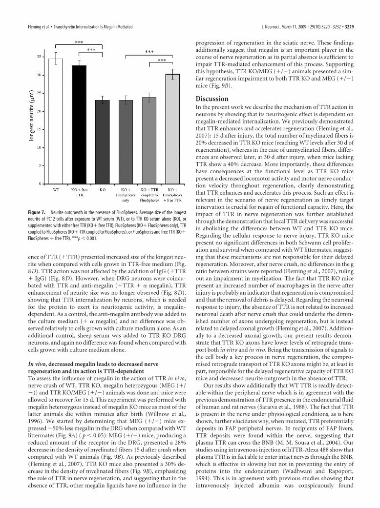
ence of TTR (�TTR) presented increased size of the longest neu-rite when compared with cells grown in TTR-free medium (Fig.8D). TTR action was not affected by the addition of IgG (�TTR� IgG) (Fig. 8D). However, when DRG neurons were coincu-bated with TTR and anti-megalin (�TTR � � megalin), TTRenhancement of neurite size was no longer observed (Fig. 8D),showing that TTR internalization by neurons, which is neededfor the protein to exert its neuritogenic activity, is megalin-dependent. As a control, the anti-megalin antibody was added tothe culture medium (� � megalin) and no difference was ob-served relatively to cells grown with culture medium alone. As anadditional control, sheep serum was added to TTR KO DRGneurons, and again no difference was found when compared withcells grown with culture medium alone.
In vivo, decreased megalin leads to decreased nerveregeneration and its action is TTR-dependentTo assess the influence of megalin in the action of TTR in vivo,nerve crush of WT, TTR KO, megalin heterozygous (MEG (�/�)) and TTR KO/MEG (�/�) animals was done and mice wereallowed to recover for 15 d. This experiment was performed withmegalin heterozygous instead of megalin KO mice as most of thelatter animals die within minutes after birth (Willnow et al.,1996). We started by determining that MEG (�/�) mice ex-pressed �50% less megalin in the DRG when compared with WTlittermates (Fig. 9A) ( p � 0.05). MEG (�/�) mice, producing areduced amount of the receptor in the DRG, presented a 28%decrease in the density of myelinated fibers 15 d after crush whencompared with WT animals (Fig. 9B). As previously described(Fleming et al., 2007), TTR KO mice also presented a 30% de-crease in the density of myelinated fibers (Fig. 9B), emphasizingthe role of TTR in nerve regeneration, and suggesting that in theabsence of TTR, other megalin ligands have no influence in the
progression of regeneration in the sciatic nerve. These findingsadditionally suggest that megalin is an important player in thecourse of nerve regeneration as its partial absence is sufficient toimpair TTR-mediated enhancement of this process. Supportingthis hypothesis, TTR KO/MEG (�/�) animals presented a sim-ilar regeneration impairment to both TTR KO and MEG (�/�)mice (Fig. 9B).
DiscussionIn the present work we describe the mechanism of TTR action inneurons by showing that its neuritogenic effect is dependent onmegalin-mediated internalization. We previously demonstratedthat TTR enhances and accelerates regeneration (Fleming et al.,2007): 15 d after injury, the total number of myelinated fibers is20% decreased in TTR KO mice (reaching WT levels after 30 d ofregeneration), whereas in the case of unmyelinated fibers, differ-ences are observed later, at 30 d after injury, when mice lackingTTR show a 40% decrease. More importantly, these differenceshave consequences at the functional level as TTR KO micepresent a decreased locomotor activity and motor nerve conduc-tion velocity throughout regeneration, clearly demonstratingthat TTR enhances and accelerates this process. Such an effect isrelevant in the scenario of nerve regeneration as timely targetinnervation is crucial for regain of functional capacity. Here, theimpact of TTR in nerve regeneration was further establishedthrough the demonstration that local TTR delivery was successfulin abolishing the differences between WT and TTR KO mice.Regarding the cellular response to nerve injury, TTR KO micepresent no significant differences in both Schwann cell prolifer-ation and survival when compared with WT littermates, suggest-ing that these mechanisms are not responsible for their delayedregeneration. Moreover, after nerve crush, no differences in the gratio between strains were reported (Fleming et al., 2007), rulingout an impairment in myelination. The fact that TTR KO micepresent an increased number of macrophages in the nerve afterinjury is probably an indicator that regeneration is compromisedand that the removal of debris is delayed. Regarding the neuronalresponse to injury, the absence of TTR is not related to increasedneuronal death after nerve crush that could underlie the dimin-ished number of axons undergoing regeneration, but is insteadrelated to delayed axonal growth (Fleming et al., 2007). Addition-ally to a decreased axonal growth, our present results demon-strate that TTR KO axons have lower levels of retrograde trans-port both in vitro and in vivo. Being the transmission of signals tothe cell body a key process in nerve regeneration, the compro-mised retrograde transport of TTR KO axons might be, at least inpart, responsible for the delayed regenerative capacity of TTR KOmice and decreased neurite outgrowth in the absence of TTR.
Our results show additionally that WT TTR is readily detect-able within the peripheral nerve which is in agreement with theprevious demonstration of TTR presence in the endoneurial fluidof human and rat nerves (Saraiva et al., 1988). The fact that TTRis present in the nerve under physiological conditions, as is hereshown, further elucidates why, when mutated, TTR preferentiallydeposits in FAP peripheral nerves. In recipients of FAP livers,TTR deposits were found within the nerve, suggesting thatplasma TTR can cross the BNB (M. M. Sousa et al., 2004). Ourstudies using intravenous injection of hTTR-Alexa 488 show thatplasma TTR is in fact able to enter intact nerves through the BNB,which is effective in slowing but not in preventing the entry ofproteins into the endoneurium (Wadhwani and Rapoport,1994). This is in agreement with previous studies showing thatintravenously injected albumin was conspicuously found
Figure 7. Neurite outgrowth in the presence of FluoSpheres. Average size of the longestneurite of PC12 cells after exposure to WT serum (WT), or to TTR KO serum alone (KO), orsupplemented with either free TTR (KO� free TTR), FluoSpheres (KO� FluoSpheres only), TTRcoupled to FluoSpheres (KO� TTR coupled to FluoSpheres), or FluoSpheres and free TTR (KO�FluoSpheres � free TTR). ***p � 0.001.
Fleming et al. • Transthyretin Internalization Is Megalin Mediated J. Neurosci., March 11, 2009 • 29(10):3220 –3232 • 3229
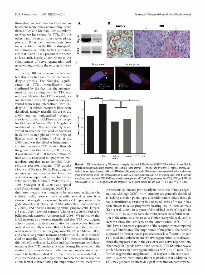
throughout nerve connective tissue and inbasement membranes surrounding nervefibers (Allen and Kiernan, 1994), similarlyto what we here show for TTR. On theother hand, when an injury takes place,plasma TTR has its entrance in the nervoustissue facilitated, as the BNB is disrupted.In summary, our data further substanti-ates that in vivo TTR is present in the nerveand, as such, is able to contribute to theenhancement of nerve regeneration andneurite outgrowth in the settings of nerveinjury.
In vitro, DRG neurons were able to in-ternalize TTR by a clathrin-dependent en-docytic process. The biological signifi-cance of TTR internalization wasconfirmed by the fact that the enhance-ment of neurite outgrowth by TTR wasonly possible when free TTR was used, be-ing abolished when the protein was pre-vented from being internalized. Two en-docytic TTR-related receptors have beendescribed, namely megalin (Sousa et al.,2000) and an unidentified receptor-associated protein (RAP)-sensitive recep-tor (Sousa and Saraiva, 2001). Megalin, amember of the LDL receptor family, is in-volved in receptor-mediated endocytosisin clathrin-coated pits of a wide range ofligands, such as albumin (Zhai et al.,2000), and was described as being impor-tant for preventing TTR filtration throughthe glomerulus (Sousa et al., 2000). Later,it was shown that TTR internalization byliver cells is associated to lipoprotein me-tabolism, and that an unidentified RAP-sensitive receptor mediates TTR uptake(Sousa and Saraiva, 2001). Regarding thenervous system, megalin has been de-scribed as an important protein for the de-velopment of the forebrain (Willnow et al.,1996; Spoelgen et al., 2005) and spinalcord (Wicher and Aldskogius, 2008). Fur-thermore, megalin was thought to be expressed exclusively byepithelial cells; however, very recently, several reports haveshown that megalin is expressed by other cell types, namely oli-godendrocytes (Wicher et al., 2006), astrocytes (Bento-Abreu etal., 2008), and neurons, including retinal ganglion cells (Fitzger-ald et al., 2007), cortical neurons (Chung et al., 2008), and cere-bellar granule neurons (Ambjørn et al., 2008). We now show thatDRG neurons also express megalin and that TTR neuritogenicactivity depends on its internalization by this receptor. Interest-ingly, it was recently reported that metallothionein stimulation ofneurite outgrowth in retinal ganglion cells (Fitzgerald et al., 2007)and cerebellar granule neurons (Ambjørn et al., 2008) is medi-ated by megalin. Considering that TTR interacts with metallo-thionein (Goncalves et al., 2008) and that the present work dem-onstrates that TTR neuritogenic effect is megalin dependent, therelationship between these molecules and neurite outgrowthshould be further studied. The present work also reveals that, invivo, decreased levels of megalin lead to decreased nerve regener-ation, further substantiating the importance of this receptor in
the nervous system and particularly in the course of nerve regen-eration. Although MEG (�/�) mutants are generally describedas lacking a major phenotype, a semidominant effect throughhaplo-insufficiency resulting in decreased levels of megalin hasbeen shown to cause progressive hearing loss in these animals(Konig et al., 2008). In support of diminished levels of megalin inMEG (�/�) mice, these were shown to present transferrin excre-tion in the urine, in contrast to WT mice (Kozyraki et al., 2001).Here we show that similarly to the latter tissues, MEG (�/�)DRG have a decreased expression of the receptor when comparedwith WT littermates. The importance of megalin in the nerve issupported by the fact that its partial absence is sufficient to impairTTR-mediated enhancement of nerve regeneration. Our data ad-ditionally suggests that, in the case of sciatic nerve regeneration,other megalin ligands have no influence, as TTR KO mice have asimilar decrease in nerve regeneration as MEG (�/�) mice. Thisfurther suggests that megalin and TTR may act in the same path-way. It is worth mentioning that it is possible that additionally,TTR may generate its effect via signal transduction pathways ac-
Figure 8. TTR internalization by DRG neurons is megalin mediated. A, Megalin and HPRT RT-PCR of kidney (�) and DRG. B,Megalin immunohistochemistry of kidney (left), and DRG in the absence (�, middle) and presence (�, right) of primary anti-body. Scale bar, 5 �m. C, z-axis stacking of hTTR-Alexa 488 (green) uptake by DRG neurons preincubated with either nonimmunesheep serum (sheep serum, left) or sheep anti-rat megalin (� megalin, right); red: anti PGP 9.5 staining, blue: DAPI. D, Averagesize of the longest neurite of TTR KO DRG neurons cells after exposure to B27 or B27 supplemented with TTR (�TTR), with TTR andanti-megalin (�TTR � � megalin), with anti-megalin (�� megalin), or with TTR and IgG (�TTR � IgG). ***p � 0.001.
3230 • J. Neurosci., March 11, 2009 • 29(10):3220 –3232 Fleming et al. • Transthyretin Internalization Is Megalin Mediated

tivated by the NpxY motifs of the cytoplasmic tail of megalin,which interact with signaling molecules involved in the regula-tion of endocytosis (Qiu et al., 2006). It is also noteworthy thatmegalin is part of a ligand-dependent signaling pathway by enzy-matic processing, linking receptor-mediated endocytosis withcell signaling (Zou et al., 2004).
In conclusion, our work further establishes the mechanism ofTTR action in the nerve by showing that it has a direct effect onneurons, as its absence leads to impaired retrograde transportand decreased axonal growth. Also, we show that TTR effect inneurite outgrowth and nerve regeneration is mediated bymegalin-dependent internalization. Finally, the relevance of TTRpresence in the nerve may underlie its preferential deposition,when mutated, in the PNS of FAP patients.
ReferencesAllen DT, Kiernan JA (1994) Permeation of proteins from the blood into
peripheral nerves and ganglia. Neuroscience 59:755–764.Ambjørn M, Asmussen JW, Lindstam M, Gotfryd K, Jacobsen C, Kiselyov
VV, Moestrup SK, Penkowa M, Bock E, Berezin V (2008) Metallothio-nein and a peptide modeled after metallothionein, EmtinB, induce neu-ronal differentiation and survival through binding to receptors of thelow-density lipoprotein receptor family. J Neurochem 104:21–37.
Andrade C (1952) A peculiar form of peripheral neuropathy. Familial atyp-ical generalized amyloidosis with special involvement of the peripheralnerves. Brain 75:408 – 427.
Benmerah A, Lamaze C, Begue B, Schmid SL, Dautry-Varsat A, Cerf-Bensussan N (1998) AP-2/Eps15 interaction is required for receptor-mediated endocytosis. J Cell Biol 140:1055–1062.
Benmerah A, Bayrou M, Cerf-Bensussan N, Dautry-Varsat A (1999) Inhi-bition of clathrin-coated pit assembly by an Eps15 mutant. J Cell Sci112:1303–1311.
Benmerah A, Poupon V, Cerf-Bensussan N, Dautry-Varsat A (2000) Map-ping of Eps15 domains involved in its targeting to clathrin-coated pits. JBiol Chem 275:3288 –3295.
Bento-Abreu A, Velasco A, Polo-Hernandez E, Perez-Reyes PL, Tabernero A,Medina JM (2008) Megalin is a receptor for albumin in astrocytes and isrequired for the synthesis of the neurotrophic factor oleic acid. J Neuro-chem 106:1149 –1159.
Blake CC, Geisow MJ, Swan ID, Rerat C, Rerat B (1974) Structure of humanplasma prealbumin at 2–5 A resolution. A preliminary report on thepolypeptide chain conformation, quaternary structure and thyroxinebinding. J Mol Biol 88:1–12.
Booth AG, Wilson MJ (1981) Human placental coated vesicles containreceptor-bound transferrin. Biochem J 196:355–362.
Brouillette J, Quirion R (2008) Transthyretin: a key gene involved in themaintenance of memory capacities during aging. Neurobiol Aging29:1721–1732.
Cheng C, Zochodne DW (2002) In vivo proliferation, migration and phe-notypic changes of Schwann cells in the presence of myelinated fibers.Neuroscience 115:321–329.
Chung RS, Penkowa M, Dittmann J, King CE, Bartlett C, Asmussen JW,Hidalgo J, Carrasco J, Leung YK, Walker AK, Fung SJ, Dunlop SA, Fitzger-ald M, Beazley LD, Chuah MI, Vickers JC, West AK (2008) Redefiningthe role of metallothionein within the injured brain: extracellular metal-lothioneins play an important role in the astrocyte-neuron response toinjury. J Biol Chem 283:15349 –15358.
Curtis R, Adryan KM, Stark JL, Park JS, Compton DL, Weskamp G, Huber LJ,Chao MV, Jaenisch R, Lee K (1995) Differential role of the low affinityneurotrophin receptor (p75) in retrograde axonal transport of the neu-rotrophins. Neuron 14:1201–1211.
Dautry-Varsat A (1986) Receptor-mediated endocytosis: the intracellularjourney of transferrin and its receptor. Biochimie 68:375–381.
Episkopou V, Maeda S, Nishiguchi S, Shimada K, Gaitanaris GA, GottesmanME, Robertson EJ (1993) Disruption of the transthyretin gene results inmice with depressed levels of plasma retinol and thyroid hormone. ProcNatl Acad Sci U S A 90:2375–2379.
Fitzgerald M, Nairn P, Bartlett CA, Chung RS, West AK, Beazley LD (2007)Metallothionein-IIA promotes neurite growth via the megalin receptor.Exp Brain Res 183:171–180.
Fleming CE, Saraiva MJ, Sousa MM (2007) Transthyretin enhances nerveregeneration. J Neurochem 103:831– 839.
Goncalves I, Quintela T, Baltazar G, Almeida MR, Saraiva MJ, Santos CR(2008) Transthyretin interacts with metallothionein 2. Biochemistry47:2244 –2251.
Hirata K, Kawabuchi M (2002) Myelin phagocytosis by macrophages andnonmacrophages during wallerian degeneration. Microsc Res Tech57:541–547.
Johnson EM Jr, Taniuchi M, Clark HB, Springer JE, Koh S, Tayrien MW, LoyR (1987) Demonstration of the retrograde transport of nerve growthfactor receptor in the peripheral and central nervous system. J Neurosci7:923–929.
Klassen RB, Crenshaw K, Kozyraki R, Verroust PJ, Tio L, Atrian S, Allen PL,Hammond TG (2004) Megalin mediates renal uptake of heavy metalmetallothionein complexes. Am J Physiol Renal Physiol 287:F393–F403.
Konig O, Ruttiger L, Muller M, Zimmermann U, Erdmann B, Kalbacher H,Gross M, Knipper M (2008) Estrogen and the inner ear: megalin knock-out mice suffer progressive hearing loss. FASEB J 22:410 – 417.
Kozyraki R, Fyfe J, Verroust PJ, Jacobsen C, Dautry-Varsat A, Gburek J,Willnow TE, Christensen EI, Moestrup SK (2001) Megalin-dependentcubilin-mediated endocytosis is a major pathway for the apical uptake oftransferrin in polarized epithelia. Proc Natl Acad Sci U S A98:12491–12496.
Lindsay RM (1988) Nerve growth factors (NGF, BDNF) enhance axonalregeneration but are not required for survival of adult sensory neurons.J Neurosci 8:2394 –2405.
Liu JJ, Ding J, Kowal AS, Nardine T, Allen E, Delcroix JD, Wu C, Mobley W,Fuchs E, Yang Y (2003) BPAG1n4 is essential for retrograde axonaltransport in sensory neurons. J Cell Biol 163:223–229.
Liz MA, Faro CJ, Saraiva MJ, Sousa MM (2004) Transthyretin, a new crypticprotease. J Biol Chem 279:21431–21438.
Figure 9. Decreased megalin levels lead to decreased nerve regeneration. A, RT-PCR analysisof megalin and HPRT expression in MEG(�/�) and WT mouse DRG. B, Morphometric analysisof sciatic nerves from WT, TTR KO, MEG(�/�), and TTR KO/MEG(�/�) mice. Density ofmyelinated fibers 15 d after nerve crush. *p � 0.05.
Fleming et al. • Transthyretin Internalization Is Megalin Mediated J. Neurosci., March 11, 2009 • 29(10):3220 –3232 • 3231
MacInnis BL, Campenot RB (2002) Retrograde support of neuronal sur-vival without retrograde transport of nerve growth factor. Science295:1536 –1539.
Madison R, da Silva CF, Dikkes P, Chiu TH, Sidman RL (1985) Increasedrate of peripheral nerve regeneration using bioresorbable nerve guidesand a laminin-containing gel. Exp Neurol 88:767–772.
Murakami T, Ohsawa Y, Sunada Y (2008) The transthyretin gene is ex-pressed in human and rodent dorsal root ganglia. Neurosci Lett436:335–339.
Nunes AF, Saraiva MJ, Sousa MM (2006) Transthyretin knockouts are anew mouse model for increased neuropeptide Y. FASEB J 20:166 –168.
Perry VH, Brown MC, Gordon S (1987) The macrophage response to cen-tral and peripheral nerve injury. A possible role for macrophages in re-generation. J Exp Med 165:1218 –1223.
Qiu S, Korwek KM, Weeber EJ (2006) A fresh look at an ancient receptorfamily: emerging roles for low density lipoprotein receptors in synapticplasticity and memory formation. Neurobiol Learn Mem 85:16 –29.
Riccio A, Pierchala BA, Ciarallo CL, Ginty DD (1997) An NGF-TrkA-mediated retrograde signal to transcription factor CREB in sympatheticneurons. Science 277:1097–1100.
Saraiva MJ (2001) Transthyretin mutations in hyperthyroxinemia and amy-loid diseases. Hum Mutat 17:493–503.
Saraiva MJ, Makover A, Moriwaki H, Blaner W, Costa PP, Goodman DS(1988) Studies on transthyretin metabolism in the nervous system. In:Amyloid and smyloidosis (Isobe T, Araki S, Uchino F, Kito S, Tsubura E,eds), pp 343–348. New York: Plenum.
Sousa JC, Grandela C, Fernandez-Ruiz J, de Miguel R, de Sousa L, MagalhaesAI, Saraiva MJ, Sousa N, Palha JA (2004) Transthyretin is involved indepression-like behaviour and exploratory activity. J Neurochem88:1052–1058.
Sousa MM, Saraiva MJ (2001) Internalization of transthyretin. Evidence ofa novel yet unidentified receptor associated protein (RAP)-sensitive re-ceptor. J Biol Chem 276:14420 –14425.
Sousa MM, Saraiva MJ (2003) Neurodegeneration in familial amyloid poly-neuropathy: from pathology to molecular signalling. Prog Neurobiol71:385– 400.
Sousa MM, Saraiva MJ (2008) Transthyretin is not expressed by dorsal rootganglia cells. Exp Neurol 214:362–365.
Sousa MM, Norden AG, Jacobsen C, Willnow TE, Christensen EI, Thakker
RV, Verroust PJ, Moestrup SK, Saraiva MJ (2000) Evidence for the roleof megalin in renal uptake of transthyretin. J Biol Chem275:38176 –38181.
Sousa MM, Ferrao J, Fernandes R, Guimaraes A, Geraldes JB, Perdigoto R,Tome L, Mota O, Negrao L, Furtado AL, Saraiva MJ (2004) Depositionand passage of transthyretin through the blood-nerve barrier in recipientsof familial amyloid polyneuropathy livers. Lab Invest 84:865– 873.
Spoelgen R, Hammes A, Anzenberger U, Zechner D, Andersen OM, JerchowB, Willnow TE (2005) LRP2/megalin is required for patterning of theventral telencephalon. Development 132:405– 414.
Taniuchi M, Clark HB, Schweitzer JB, Johnson EM Jr (1988) Expression ofnerve growth factor receptors by Schwann cells of axotomized peripheralnerves: ultrastructural location, suppression by axonal contact, and bind-ing properties. J Neurosci 8:664 – 681.
Triolo D, Dina G, Lorenzetti I, Malaguti M, Morana P, Del Carro U, Comi G,Messing A, Quattrini A, Previtali SC (2006) Loss of glial fibrillary acidicprotein (GFAP) impairs Schwann cell proliferation and delays nerve re-generation after damage. J Cell Sci 119:3981–3993.
Wadhwani KC, Rapoport SI (1994) Transport properties of vertebrateblood-nerve barrier: comparison with blood– brain barrier. Prog Neuro-biol 43:235–279.
Wicher G, Aldskogius H (2008) Megalin deficiency induces critical changesin mouse spinal cord development. Neuroreport 19:559 –563.
Wicher G, Larsson M, Svenningsen AF, Gyllencreutz E, Rask L, Aldskogius H(2006) Low density lipoprotein receptor-related protein-2/megalin is ex-pressed in oligodendrocytes in the mouse spinal cord white matter. J Neu-rosci Res 83:864 – 873.
Willnow TE, Hilpert J, Armstrong SA, Rohlmann A, Hammer RE, Burns DK,Herz J (1996) Defective forebrain development in mice lacking gp330/megalin. Proc Natl Acad Sci U S A 93:8460 – 8464.
Zhai XY, Nielsen R, Birn H, Drumm K, Mildenberger S, Freudinger R,Moestrup SK, Verroust PJ, Christensen EI, Gekle M (2000) Cubilin- andmegalin-mediated uptake of albumin in cultured proximal tubule cells ofopossum kidney. Kidney Int 58:1523–1533.
Zou Z, Chung B, Nguyen T, Mentone S, Thomson B, Biemesderfer D (2004)Linking receptor-mediated endocytosis and cell signaling: evidence forregulated intramembrane proteolysis of megalin in proximal tubule.J Biol Chem 279:34302–34310.
3232 • J. Neurosci., March 11, 2009 • 29(10):3220 –3232 Fleming et al. • Transthyretin Internalization Is Megalin Mediated
Prologue
111
Considering the knowledge that I have acquired since the publication of this
paper, I propose several experiments that could be performed in order to
understand the molecular mechanisms through which TTR increases axonal
growth.
- Determine whether in the adult, the effect of TTR is restricted to DRG
neurons
The role of TTR in the PNS has been extensively studied due to its involvement in
familial amyloid polyneuropathy (FAP), a neuropathy characterized by the
deposition of TTR amyloid fibrils in the PNS (Sousa and Saraiva, 2003). The
preference for TTR accumulation in peripheral nerves led to the hypothesis that
WT TTR may play a role in PNS physiology. TTR is important for axonal growth
since TTR KO DRG neurons have decreased neurite outgrowth, and TTR KO mice
present impaired retrograde transport and delayed axonal regeneration (Fleming
et al., 2009). This novel role of TTR in nerve physiology raises the question of
whether TTR might also be important in the CNS. In this respect, it is important to
note that TTR is one of the most abundant proteins in the cerebrospinal fluid
(CSF). As such, it would be interesting to know if TTR is also important for axonal
elongation of CNS neurons, particularly in hippocampal neurons, as the presence
and expression of TTR by this neuronal subtype has been suggested (Li et al.,
2011).
- Dissect the involvement of TTR in axonal transport
One of the most important features in TTR KO mice is that they present
impairment in retrograde axonal transport as determined by retrograde labeling
with cholera toxin and by analysis of transport of p75NTR
. Nevertheless, very little
is known about the mechanism by which TTR interferes with axonal transport. It
is not even known if the defects in axonal transport are exclusive to retrograde
transport or whether TTR KO mice also have defects in anterograde transport. To
investigate this hypothesis, the accumulation of an anterogradely transported
protein like amyloid precursor protein (APP) (Cavalli et al., 2005) could be studied
in the proximal stump of TTR KO mice following ligation of the sciatic nerve.
Moreover, different components of the axonal transport could be tested in vitro,
namely organelles, vesicles and proteins to identify which components are
TTR as a regeneration enhancer
112
impaired in TTR KO mice. To assess organelle transport, mitochondria labeling
could be performed with mitoTracker (Invitrogen); for vesicles, fluorescently
labeled synaptophysin or APP transfected neurons could be used; and for protein
transport studies with, fluorescently labeled tubulin could be performed. These
experiments could further disclose the involvement of TTR in axonal transport.
Additionally, whether the defects in axonal transport are a direct cause of
the absence of TTR or are a secondary result from the lack of this protein should
be further investigated.
Besides, at the molecular level many questions can be raised. Are
microtubules correctly assembled in axons of TTR KO mice? To evaluate these
issues, microtubules could be extracted from nerves of TTR KO mice and WT
littermates and the ratio of microtubule/tubulin monomer could be established.
- Identify downstream targets of TTR that could mediate axonal growth
The dissection of downstream targets of TTR might unravel novel
regeneration enhancers that could be modulated to increase nerve regeneration.
Since TTR was found to be important in axonal growth of DRG neurons (Fleming
et al., 2009), a screening could be done on DRG neurons, from naïve and PNS
injured TTR KO mice and WT littermates. One of two approaches could be
performed. Either microarray to evaluate different patterns of gene expression or
a proteomic approach such as iTRAQ or 2D gels coupled to mass spectrometry.
The proteins/genes differentially regulated should then be validated. Moreover,
since TTR has also been described as a protease (Liz et al., 2012), and since its
activity is important for its ability to induce neurite outgrowth (Liz et al., 2009), it
would be interesting to know if the identified proteins are TTR substrates.
- Determine whether in FAP, loss of function (as an axonal regeneration
enhancer) of mutated TTR aggregates/fibrils also contributes to
neuropathology.
In FAP, there is the accumulation of mutated TTR in peripheral nerves that leads
to neurodegeneration. Neurodegeneration is attributed to the toxicity of TTR
aggregates (Sousa and Saraiva, 2003). Nevertheless, it cannot be excluded that
Prologue
113
loss of function of TTR fibrils, may also contribute to neuropathology. In
particular, given that TTR is important in retrograde transport, impairment in this
function may contribute to pathology. In fact, defects in axonal transport have
been associated to neurodegenerative diseases (Chevalier-Larsen and Holzbaur,
2006). This could be addressed by using animal models of FAP to check whether
these present defects in axonal transport. It would be interesting to know if these
putative defects precede amyloid deposition and neurodegeneration.

114

115
Chapter I

116
Chapter I
117
Cholesterol and sphingomyelin are axon regeneration inhibitors that can be
counteracted by cyclodextrin delivery
Abbreviated title: Myelin lipids modulate axonal regeneration
Fernando M. Mar1,3
, Tiago Ferreira da Silva1,3
, Marlene M. Morgado1
, Lorena G.
Rodrigues2
, Daniel Rodrigues2
, Ana Marques1
, Vera F. Sousa1,3
, João Coentro1
,
Clara Sá- Miranda2
, Mónica M. Sousa1
*, Pedro Brites1
*
1
Nerve Regeneration group, Instituto de Biologia Molecular e Celular - IBMC,
University of Porto; Rua do Campo Alegre 823, 4150-180 Porto, Portugal
2
Lysosome and Peroxisome Biology group, Instituto de Biologia Molecular e
Celular - IBMC, University of Porto; Rua do Campo Alegre 823, 4150-180 Porto,
Portugal
3
Instituto de Ciências Biomédicas Abel Salazar – ICBAS, Rua Jorge Viterbo Ferreira
228, 4050-313 Porto, Portugal
* These authors contributed equally to this work
Myelin lipids modulate axonal regeneration
118
Abstract
Lack of axonal regeneration following spinal cord injury has been mainly ascribed
to the inhibitory environment of the injury site i.e., to chondroitin sulphate
proteoglycans and myelin-associated inhibitors. Here, we used shiverer mice to
assess axonal regeneration following spinal cord injury in the presence of myelin-
associated inhibitors and chondroitin sulphate proteoglycans, but in the absence
of compact myelin. Although in vitro shiverer neurons displayed a similar intrinsic
neurite outgrowth to wild-type neurons, in vivo, shiverer fibers had increased
regenerative ability, suggesting that the wild-type spinal cord contains additional
inhibitors besides myelin-associated inhibitors and chondroitin sulphate
proteoglycans. Our data shows that besides myelin protein, myelin lipids are
highly inhibitory for neurite outgrowth and demonstrates that this inhibitory
effect is released in the shiverer spinal cord given its decreased lipid content.
Specifically, we identified cholesterol and sphingomyelin as novel myelin-
associated inhibitors with activity in multiple neuron types. We further
demonstrated the inhibitory action of cholesterol and sphingomyelin in vivo, by
showing that delivery of 2-hydroxypropyl-β-cyclodextrin, a drug that reduces the
levels of lipids in the injury site, leads to increased axonal regeneration following
spinal cord injury. In summary, our work shows that myelin lipids are important
modulators of axonal regeneration that should be considered together with
protein myelin-associated inhibitors as critical targets in strategies aiming at
improving axonal growth following injury. In this respect, our study provides the
initial preclinical data needed to evaluate the possible use of 2-hydroxypropyl-β-
cyclodextrin in clinical trials with spinal cord injury patients.
Chapter I
119
Introduction
The inability of adult vertebrate CNS axons to regenerate is seen as a
consequence of the highly inhibitory environment at the injury site (Silver and
Miller, 2004), and of the failure in activating a cell-intrinsic program leading to
the expression of regeneration-associated genes (Sun and He, 2010). Upon CNS
injury, the glial scar functions as an inhibitory barrier composed of multiple
components, generally divided in 3 categories: myelin associated inhibitors
(MAIs), namely Nogo, myelin associated glycoprotein (MAG) and oligodendrocyte
myelin glycoprotein (OMgp) (Lee and Zheng, 2012); canonical axon guidance
molecules, such as semaphorin 3A, ephrin B3, netrin-1 and repulsive guidance
molecule A (RGMa) (Giger et al., 2010); and chondroitin sulfate proteoglycans
(CSPGs), produced by astrocytes (Jones et al., 2003).
Studies from different groups using triple knockout mice for MAG, Nogo
and OMgp produced conflicting results ranging from limited (Lee et al., 2010) to
extensive regeneration abilities (Cafferty et al., 2010). Although the field has
largely concentrated on MAIs, other inhibitors play important roles in vivo:
blocking CSPG or RGMa or deleting ephrinB3 increases axonal regeneration
following spinal cord injury (SCI) (Bradbury et al., 2002; Hata et al., 2006; Duffy et
al., 2012). As such, the wide variety of inhibitors present in the spinal cord milieu
is thought to underlie the absence of a robust axonal regeneration when blocking
either single or a limited combination of inhibitors. Despite the structural
differences, several MAIs share receptors and an inhibitory mechanism dependent
on RhoA/ROCK activation (Yiu and He, 2006), as demonstrated mainly through
the use of RhoA/ROCK inhibitors (Kubo and Yamashita, 2007).
To further explore the importance of myelin in the inhibition of axonal
regeneration, we used shiverer (shi) mice which lack myelin basic protein (MBP), a
key player in myelin compaction in the CNS (Boggs, 2006). In the shi CNS, the
absence of MBP results in a complete lack of compact myelin (Rosenbluth, 1980)
which is accompanied by a severe phenotype comprising shivering, convulsions
and early death. Here we show that in shi mice, despite the presence of canonical
MAIs and of axonal abnormalities generally related to decreased regeneration
capacity, CNS axons have an increased ability to regenerate through the spinal
cord glial scar. This increase in regeneration was ascribed to a decreased
abundance of myelin lipids, namely cholesterol and sphingomyelin. Moreover we
Myelin lipids modulate axonal regeneration
120
show that delivery of 2-hydroxypropyl-β-cyclodextrin (HPβCD), reduces the levels
of cholesterol and sphingomyelin in the SCI site, leading to increased axonal
regeneration. In summary, our work demonstrates that myelin lipids are
important modulators of axonal regeneration that can be targeted through
HPβCD delivery.
Chapter I
121
Materials and methods
Animals. Mice were handled according to European Union and National rules. All
procedures were approved by the IBMC Ethics Committee and by the Portuguese
General Veterinarian Board. Six week-old-WT and shi (Jackson laboratories)
littermates of either sex, on a Swiss Webster:C3HeB/Fe5 background, were
obtained from heterozygous breeding pairs. For drug-delivery studies 8 weeks-
old C57BL/6 mice of either sex were used.
Electron microscopy. The thoracic spinal cord of WT and shi littermates (11
weeks-old) was embedded in Epon. Semi-thin 1µm-thick sections were stained
with p-phenylene-diamine to visualize myelin and white matter tracts. 60nm-thick
sections of the dorsal funiculus were stained with uranyl acetate and lead citrate
and examined in a JEOL JEM-1400 transmission electron microscope.
Western blotting. Twenty five μg of thoracic spinal cord protein from naïve (6
weeks-old) or injured (11 weeks-old) WT and shi littermates were separated in 3-
8% Tris-Acetate gels (Bio-Rad) and transferred to nitrocellulose. Antibodies used:
mouse anti-MAG (Dr. Richard Quarles, NINDS, Bethesda, 1:1,000), rat anti-OMgp
(R&D Systems, 1:250), rabbit anti-Nogo-A (Dr. Stephen Strittmatter, Yale
University, New Haven, 1:10,000), rabbit anti-Ephrin B3 (Santa Cruz
Biotechnology, 1:200), rabbit anti-RGMa (Immuno-Biological Laboratories, 1:200)
and mouse anti-GAPDH (Santa Cruz Biotechnology, 1:2,000).
MAG and Nogo immunohistochemistry. Paraffin-embedded spinal cords from WT
and shi littermates (6 weeks-old) were used for immunohistochemistry against
MAG (1:500) or Nogo-A (1:5,000) using ABC Vectastain and DAB (Vector labs).
Spinal cord injury (SCI). Animals were anesthetized with ketamine
(75mg/kg)/medetomidine (1mg/kg) and a laminectomy was performed at the T9
level. Complete transection, dorsal hemisection or left lateral hemisection were
done using a micro feather ophthalmic scalpel. Analgesia was performed for 72h
with buprenorphine (0.08mg/kg).
Assessment of the lesion area following SCI. Five weeks after complete SCI, 10µm
sagittal spinal cord cryosections were immunostained for CSPG (Sigma-Aldrich,
1:200) and GFAP (DAKO, 1:500). Collagen was visualized with Masson trichrome
staining (Sigma-Aldrich). The injury area was measured using Photoshop CS3.
Myelin lipids modulate axonal regeneration
122
Regeneration of dorsal column fibers. Animals with dorsal hemisection were
allowed to recover for 4 weeks. Four days before sacrifice, animals were
anesthetized as described above and 2µl of 1% cholera toxin B (CT-B, List
Biologicals) were injected in the left sciatic nerve. Fifty µm sagittal free floating
sections were immunostained for CT-B (List Biologicals, 1:30,000). From each
animal, the section displaying the highest number of regenerating axons was
selected. Axonal regeneration was quantified by counting the number of CT-B
labeled axons within the glial scar and by measuring the length of the longest
fiber found rostrally to the injury border. All quantifications of axonal
regeneration were performed with the researcher blinded for genotype and
experimental group.
Regeneration of raphespinal fibers. Five weeks after complete spinal cord
transection, free floating sections were immunostained for serotonin (5-HT)
(1:20,000, Immunostar). Only animals where a complete injury was present in all
sections were analyzed. Axonal regeneration was quantified by counting 5-HT
positive fibers caudally to the injury site. Raphespinal fiber sprouting was
assessed 4 weeks after lateral spinal cord hemisection by 5-HT immunostaining in
cross-sections of the lumbar enlargement. 5-HT immunoreactivity was quantified
in 4 sections/animal using FeatureJ software. Compensatory sprouting was
quantified by 5-HT immunoreactivity in the ipsilateral ventral horn. For each
genotype, 5-HT immunoreactivity was normalized against 5-HT immunoreactivity
in the naïve spinal cord. All quantifications of axonal regeneration were
performed with the researcher blinded for genotype.
Crude membrane isolation. Crude membranes were isolated from the spinal cord
of 6 weeks old WT and shi littermates, as described (Shen et al., 1998). Briefly,
tissues were homogenized in 0.32M sucrose, and nuclei removed by
centrifugation at 500g for 30 min. Membranes were pelleted by centrifugation at
100,000g for 1 h, resuspended in H2O and protein concentration was
determined.
Myelin isolation. Myelin was isolated from the spinal cord of 16 weeks-old WT
mice as detailed (Norton and Poduslo, 1973). Briefly, the tissue was homogenized
in 0.32M sucrose and after centrifugation at 900g, the post-nuclear supernatant
was collected and carefully overlaid on an ultracentrifuge tube containing a 0.85M
sucrose solution on top of a 50% (w/v) sucrose cushion. After centrifugation for 1
Chapter I
123
h at 37,000g at 4ºC, the interphase between sucrose solutions was transferred to
a new ultracentrifuge tube. Two rounds of osmotic shocks were performed by
adding ice-cold water and centrifugation at 20,000g. The final myelin pellet was
stored at -80ºC until further use. Before use, myelin was sonicated and the
protein concentration determined.
Lipid isolation. Lipids were isolated from myelin and spinal cord extracts by a
modified Folch two solvent system (Mirza et al., 2007). Briefly, 150µg of protein
extract (from either myelin or total spinal cord extract) were dissolved in
methanol:water (1:1) and lipids were separated by liquid extraction with
chloroform (Merck). The chloroform layer containing the lipids was then dried
under nitrogen stream and stored at -20°C. Absence of proteins in the lipid
fraction was confirmed by western blotting against Nogo, MAG, Ephrin B3 and
RGMa. The aqueous layer containing the protein sample was precipitated with
0.2M perchloric acid following Folch extraction. Following centrifugation the
protein was resuspended in H2O, sonicated and its concentration was
determined.
Lipid extraction and thin layer chromatography. Thoracic spinal cords from WT
and shi littermates were collected and lipids were extracted as described (Folch et
al., 1957). The spinal cord injury site and a rostral uninjured region of WT spinal
cord treated with artificial CSF (aCSF) or HPβCD was also collected for lipid
extraction. Acidic and neutral lipids were further separated by reverse-phase
chromatography (Vance and Sweeley, 1967; Seyfried et al., 1978; Rodrigues et al.,
2009). All lipids were analyzed by high-performance thin-layer chromatography
(HPTLC) as detailed (Rodrigues et al., 2009) alongside with lipid standards. The
lipids analyzed were: ceramide (Cer), phosphatidylserine (PS), cholesterol (CO),
sulfatide (GS), galactocerebroside (GalCer), phosphatidylcholine (PC), triglycerides
(TG), sphingomyelin (SpH), cholesteryl esters (CE), phosphatidylethanolamine (PE),
globotetrahexosylceramide (Gb4), lactocerebroside (Lac), phosphatidylinositol (PI)
and GM1 ganglioside (GM1).
Neuron cultures and inhibition assays. Primary cultures of DRG neurons from P6
mice were performed as described (Miranda et al., 2011). Glass coverslips were
coated with poly-L-lysine (20µg/mL) and laminin (0.5µg/mL) followed by crude
membranes (0.1µg protein), myelin (0.5µg protein), myelin protein fraction
(0.5µg), MBP (Upstate, 0.9µg) or total protein from spinal cord extracts (3µg). To
Myelin lipids modulate axonal regeneration
124
test the effect of lipids, the myelin lipid fraction (corresponding to 0.5µg of
protein) and lipids extracted from the spinal cord of either WT or shi mice
(corresponding to 2µg of protein) were used. Coverslips were coated with poly-L-
lysine followed by lipids in chloroform/methanol/water (2:1:0.1) and laminin
(0.5µg/mL). The amount of individual lipids present in 2µg protein from WT
spinal cord lysates was determined by HPTLC and is referred to as 1x (PI= 2 ng,
GalCer= 200ng, CO= 120ng, GS= 120ng, Gb4= 24ng, SpH= 50ng, GM1= 0.6ng,
Lac= 20ng, PE= 40ng). In the analysis of single lipids, 1x, 10-fold less (0.1x) or
10-fold more (10x) were used per coverslip. Gb4, PE, SpH and Lac were from
Matreya LLC and CO, GalCer, GS, GM1 and PI were from Sigma-Aldrich. GalCer, GS
and PI were dissolved in chloroform/methanol (2:1), and CO, PE, SpH, Gb4, GM1
and Lac in chloroform/methanol (1:2). For each lipid, the corresponding solvent
was used as control. To confirm the presence of lipids following coating, lipid
extraction of the coverslip was performed, and lipid content was quantified. Lipid-
coating efficacy was measured by HPTLC and approximately 90% of the coated
lipids were present in the coverslips. For each condition, 5,000 cells/coverslip
were plated in triplicate and immunostained 12h later against βIII-tubulin
(Promega, 1:2,000). In at least 100 neurons/condition, the longest neurite was
traced using NeuronJ. Similar experiments were performed with cortical and
hippocampal neurons isolated from E17.5 WT and shi embryos as described (Dent
et al., 1999; Kaech and Banker, 2006), respectively. For cortical neurons, 50,000
cells/coverslip were maintained for 48 h and for hippocampal neurons, 16,500
cells/coverslip were maintained for 72 h.
Evaluation of toxicity. DRG neurons were plated on top of either laminin, solvent,
CO or SpH. Twelve hours later, cells were washed and incubated with 1µg/mL
calcein (Invitrogen) for 30 min followed by 10µg/mL propidium iodide for 5 min.
Cells were then washed and 10x magnification pictures were taken. The
percentage of viable cells (cells where nonfluorescent calcein AM is converted to a
green-fluorescent calcein) was determined.
Rho inactivation assay. WT DRG neurons were plated onto coverslips coated with
laminin, myelin, solvent, CO or SpH, as described above. Thirty minutes before
plating, cells were incubated with 1 µg/mL of C3 transferase (Cytoskeleton, Inc.),
a Rho inhibitor that was maintained during the entire experiment. Twelve hours
later, cells were fixed and neurite outgrowth was evaluated.
Chapter I
125
ROCK activation assay. 16,500 hippocampal neurons/coverslip were plated onto
coverslips coated with either solvent or SpH (10x and 100x), as described above.
A pool of 12 wells/condition was used. Seventy two hours after plating, cells were
lysed in 62.5mM Tris pH 6.8 containing 2% SDS, 12.5% glycerol and 5% β-
mercaptoethanol. Lysates were then immunobloted against phospho-Ser19-
myosin light chain (Cell Signaling, 1:1,000), the primary phosphorylation site of
MLC by ROCK, and against β-actin (Sigma-Aldrich, 1:5,000).
HPβCD treatment. C57BL/6 mice (n=24) were subjected to SCI dorsal hemisection
as described above. At the time of injury, osmotic minipumps (Alzet 2006) were
placed subcutaneously with a tube allowing perfusion of the injury site at a rate
of 0.15μl/hour with either 27μg/g/day of HPβCD in aCSF (128mM NaCl, 2.5mM
KCl, 0.95mM CaCl2, 1.9mM MgCl2; n=12), or vehicle (aCSF; n=12). Besides
delivery at the injury site, 4mg/g of HPβCD were injected subcutaneously twice a
week. Five weeks following injury, the SCI site was collected and lipids were
extracted and quantified as described above (n=6, HPβCD-treated mice; n=6,
aCSF-treated mice). In n=6 HPβCD-treated mice and n=6 aCSF-treated mice, dorsal
column fibers were labeled with CT-B, and axonal regeneration was assessed as
previously described.
Data analysis. Data is shown as mean±SEM. Statistical significance was
determined by Student’s t test or Tukey’s test (One-way ANOVA) for multiple
comparisons.
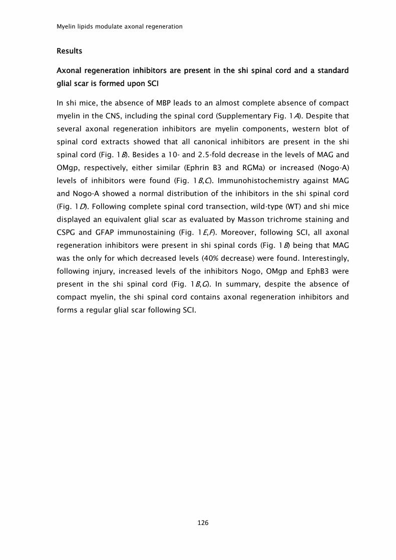
Myelin lipids modulate axonal regeneration
126
Results
Axonal regeneration inhibitors are present in the shi spinal cord and a standard
glial scar is formed upon SCI
In shi mice, the absence of MBP leads to an almost complete absence of compact
myelin in the CNS, including the spinal cord (Supplementary Fig. 1A). Despite that
several axonal regeneration inhibitors are myelin components, western blot of
spinal cord extracts showed that all canonical inhibitors are present in the shi
spinal cord (Fig. 1B). Besides a 10- and 2.5-fold decrease in the levels of MAG and
OMgp, respectively, either similar (Ephrin B3 and RGMa) or increased (Nogo-A)
levels of inhibitors were found (Fig. 1B,C). Immunohistochemistry against MAG
and Nogo-A showed a normal distribution of the inhibitors in the shi spinal cord
(Fig. 1D). Following complete spinal cord transection, wild-type (WT) and shi mice
displayed an equivalent glial scar as evaluated by Masson trichrome staining and
CSPG and GFAP immunostaining (Fig. 1E,F). Moreover, following SCI, all axonal
regeneration inhibitors were present in shi spinal cords (Fig. 1B) being that MAG
was the only for which decreased levels (40% decrease) were found. Interestingly,
following injury, increased levels of the inhibitors Nogo, OMgp and EphB3 were
present in the shi spinal cord (Fig. 1B,G). In summary, despite the absence of
compact myelin, the shi spinal cord contains axonal regeneration inhibitors and
forms a regular glial scar following SCI.
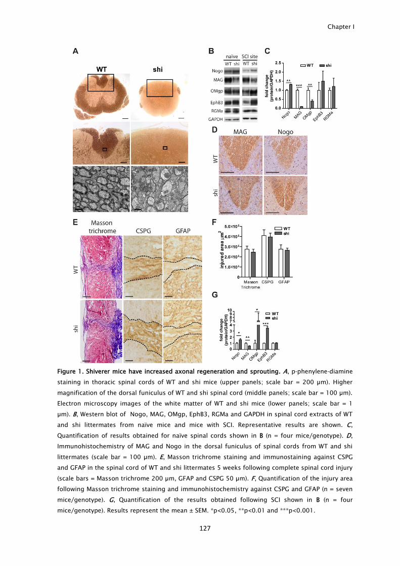
Chapter I
127
Figure 1. Shiverer mice have increased axonal regeneration and sprouting. A, p-phenylene-diamine
staining in thoracic spinal cords of WT and shi mice (upper panels; scale bar = 200 µm). Higher
magnification of the dorsal funiculus of WT and shi spinal cord (middle panels; scale bar = 100 µm).
Electron microscopy images of the white matter of WT and shi mice (lower panels; scale bar = 1
µm). B, Western blot of Nogo, MAG, OMgp, EphB3, RGMa and GAPDH in spinal cord extracts of WT
and shi littermates from naïve mice and mice with SCI. Representative results are shown. C,
Quantification of results obtained for naïve spinal cords shown in B (n = four mice/genotype). D,
Immunohistochemistry of MAG and Nogo in the dorsal funiculus of spinal cords from WT and shi
littermates (scale bar = 100 µm). E, Masson trichrome staining and immunostaining against CSPG
and GFAP in the spinal cord of WT and shi littermates 5 weeks following complete spinal cord injury
(scale bars = Masson trichrome 200 µm, GFAP and CSPG 50 µm). F, Quantification of the injury area
following Masson trichrome staining and immunohistochemistry against CSPG and GFAP (n = seven
mice/genotype). G, Quantification of the results obtained following SCI shown in B (n = four
mice/genotype). Results represent the mean ± SEM. *p<0.05, **p<0.01 and ***p<0.001.
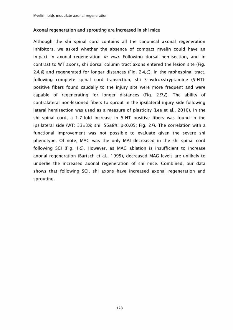
Myelin lipids modulate axonal regeneration
128
Axonal regeneration and sprouting are increased in shi mice
Although the shi spinal cord contains all the canonical axonal regeneration
inhibitors, we asked whether the absence of compact myelin could have an
impact in axonal regeneration in vivo. Following dorsal hemisection, and in
contrast to WT axons, shi dorsal column tract axons entered the lesion site (Fig.
2A,B) and regenerated for longer distances (Fig. 2A,C). In the raphespinal tract,
following complete spinal cord transection, shi 5-hydroxytryptamine (5-HT)-
positive fibers found caudally to the injury site were more frequent and were
capable of regenerating for longer distances (Fig. 2D,E). The ability of
contralateral non-lesioned fibers to sprout in the ipsilateral injury side following
lateral hemisection was used as a measure of plasticity (Lee et al., 2010). In the
shi spinal cord, a 1.7-fold increase in 5-HT positive fibers was found in the
ipsilateral side (WT: 33±3%; shi: 56±8%; p<0.05; Fig. 2F). The correlation with a
functional improvement was not possible to evaluate given the severe shi
phenotype. Of note, MAG was the only MAI decreased in the shi spinal cord
following SCI (Fig. 1G). However, as MAG ablation is insufficient to increase
axonal regeneration (Bartsch et al., 1995), decreased MAG levels are unlikely to
underlie the increased axonal regeneration of shi mice. Combined, our data
shows that following SCI, shi axons have increased axonal regeneration and
sprouting.
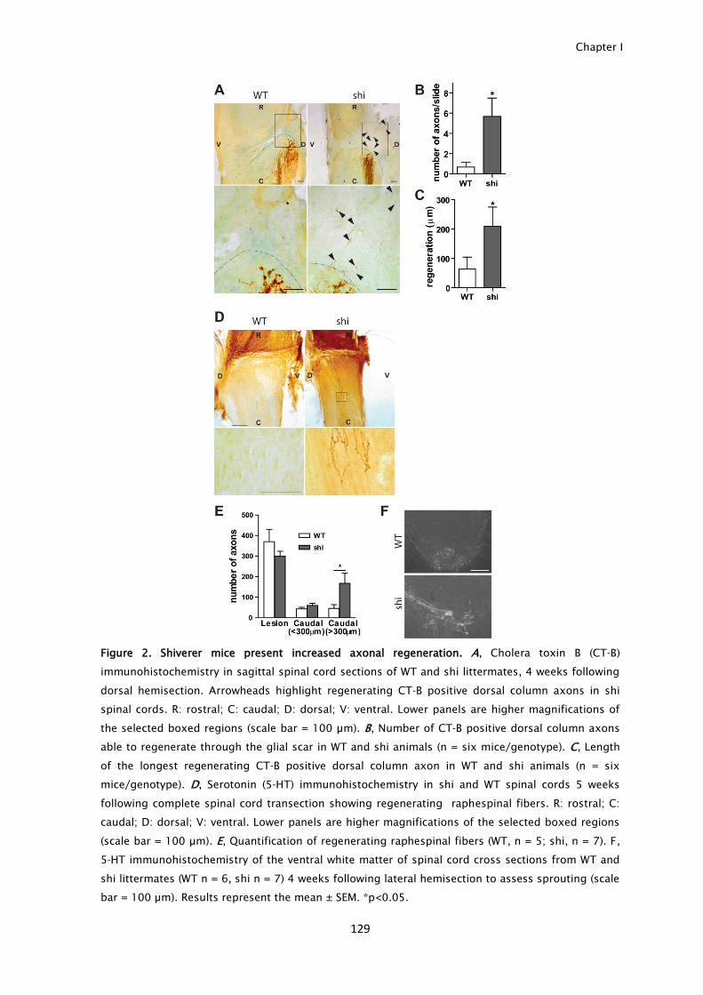
Chapter I
129
Figure 2. Shiverer mice present increased axonal regeneration. A, Cholera toxin B (CT-B)
immunohistochemistry in sagittal spinal cord sections of WT and shi littermates, 4 weeks following
dorsal hemisection. Arrowheads highlight regenerating CT-B positive dorsal column axons in shi
spinal cords. R: rostral; C: caudal; D: dorsal; V: ventral. Lower panels are higher magnifications of
the selected boxed regions (scale bar = 100 µm). B, Number of CT-B positive dorsal column axons
able to regenerate through the glial scar in WT and shi animals (n = six mice/genotype). C, Length
of the longest regenerating CT-B positive dorsal column axon in WT and shi animals (n = six
mice/genotype). D, Serotonin (5-HT) immunohistochemistry in shi and WT spinal cords 5 weeks
following complete spinal cord transection showing regenerating raphespinal fibers. R: rostral; C:
caudal; D: dorsal; V: ventral. Lower panels are higher magnifications of the selected boxed regions
(scale bar = 100 µm). E, Quantification of regenerating raphespinal fibers (WT, n = 5; shi, n = 7). F,
5-HT immunohistochemistry of the ventral white matter of spinal cord cross sections from WT and
shi littermates (WT n = 6, shi n = 7) 4 weeks following lateral hemisection to assess sprouting (scale
bar = 100 µm). Results represent the mean ± SEM. *p<0.05.
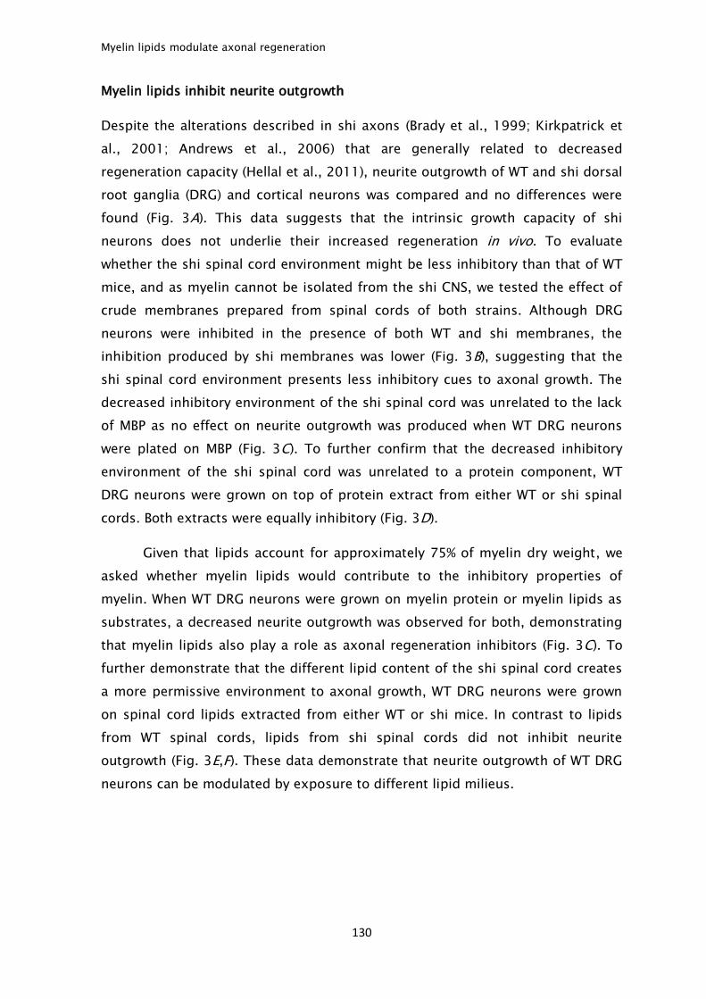
Myelin lipids modulate axonal regeneration
130
Myelin lipids inhibit neurite outgrowth
Despite the alterations described in shi axons (Brady et al., 1999; Kirkpatrick et
al., 2001; Andrews et al., 2006) that are generally related to decreased
regeneration capacity (Hellal et al., 2011), neurite outgrowth of WT and shi dorsal
root ganglia (DRG) and cortical neurons was compared and no differences were
found (Fig. 3A). This data suggests that the intrinsic growth capacity of shi
neurons does not underlie their increased regeneration in vivo. To evaluate
whether the shi spinal cord environment might be less inhibitory than that of WT
mice, and as myelin cannot be isolated from the shi CNS, we tested the effect of
crude membranes prepared from spinal cords of both strains. Although DRG
neurons were inhibited in the presence of both WT and shi membranes, the
inhibition produced by shi membranes was lower (Fig. 3B), suggesting that the
shi spinal cord environment presents less inhibitory cues to axonal growth. The
decreased inhibitory environment of the shi spinal cord was unrelated to the lack
of MBP as no effect on neurite outgrowth was produced when WT DRG neurons
were plated on MBP (Fig. 3C). To further confirm that the decreased inhibitory
environment of the shi spinal cord was unrelated to a protein component, WT
DRG neurons were grown on top of protein extract from either WT or shi spinal
cords. Both extracts were equally inhibitory (Fig. 3D).
Given that lipids account for approximately 75% of myelin dry weight, we
asked whether myelin lipids would contribute to the inhibitory properties of
myelin. When WT DRG neurons were grown on myelin protein or myelin lipids as
substrates, a decreased neurite outgrowth was observed for both, demonstrating
that myelin lipids also play a role as axonal regeneration inhibitors (Fig. 3C). To
further demonstrate that the different lipid content of the shi spinal cord creates
a more permissive environment to axonal growth, WT DRG neurons were grown
on spinal cord lipids extracted from either WT or shi mice. In contrast to lipids
from WT spinal cords, lipids from shi spinal cords did not inhibit neurite
outgrowth (Fig. 3E,F). These data demonstrate that neurite outgrowth of WT DRG
neurons can be modulated by exposure to different lipid milieus.
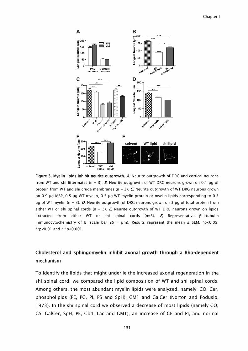
Chapter I
131
Figure 3. Myelin lipids inhibit neurite outgrowth. A, Neurite outgrowth of DRG and cortical neurons
from WT and shi littermates (n = 3). B, Neurite outgrowth of WT DRG neurons grown on 0.1 µg of
protein from WT and shi crude membranes (n = 3). C, Neurite outgrowth of WT DRG neurons grown
on 0.9 µg MBP, 0.5 µg WT myelin, 0.5 µg WT myelin protein or myelin lipids corresponding to 0.5
µg of WT myelin (n = 3). D, Neurite outgrowth of DRG neurons grown on 3 µg of total protein from
either WT or shi spinal cords (n = 3). E, Neurite outgrowth of WT DRG neurons grown on lipids
extracted from either WT or shi spinal cords (n=3). F, Representative βIII-tubulin
immunocytochemistry of E (scale bar 25 = µm). Results represent the mean ± SEM. *p<0.05,
**p<0.01 and ***p<0.001.
Cholesterol and sphingomyelin inhibit axonal growth through a Rho-dependent
mechanism
To identify the lipids that might underlie the increased axonal regeneration in the
shi spinal cord, we compared the lipid composition of WT and shi spinal cords.
Among others, the most abundant myelin lipids were analyzed, namely: CO, Cer,
phospholipids (PE, PC, PI, PS and SpH), GM1 and GalCer (Norton and Poduslo,
1973). In the shi spinal cord we observed a decrease of most lipids (namely CO,
GS, GalCer, SpH, PE, Gb4, Lac and GM1), an increase of CE and PI, and normal
Myelin lipids modulate axonal regeneration
132
amounts of Cer+PE, PS, PC and TG (Fig. 4A). From all the lipids with abnormal
concentrations in the shi spinal cord, only CO and SpH inhibited DRG neurite
outgrowth in a dose-response manner (Fig. 4B,C). This effect was unrelated to
toxicity as no dead cells were found in any condition, as examined by calcein
viability assay (Supplementary Fig. 4D). The effect of both CO and SpH was not
restricted to DRG neurons as these lipids also produced a dose-dependent
inhibitory effect in neurite outgrowth of hippocampal (Fig. 4E) and cortical
neurons (Fig. 4F). Interestingly, when hippocampal neurons were grown on CE,
that shares the same backbone structure as CO, the esterification of CO reduced
its ability to inhibit neurite outgrowth (Fig. 4E), suggesting a specific inhibitory
effect of unesterified CO.
The RhoA/ROCK pathway is the major mediator of myelin inhibition (Yiu
and He, 2006), and the inhibitory effect of myelin can be reverted by the Rho
inhibitor C3 transferase (Winton et al., 2002). To assess whether myelin lipids
share similar pathways to block axonal regeneration than those described for
myelin proteins, DRG neurons were grown on the inhibitory substrates myelin, CO
and SpH and in the presence of C3 transferase. As expected, C3 was able to
overcome myelin inhibition (Fig. 4G). Inhibition by both CO and SpH was also
relieved by C3 treatment (Fig. 4G) suggesting that lipid inhibition of axonal
growth occurs, at least in part, through a Rho-dependent mechanism. Despite
that treatment with C3 improved neurite outgrowth in DRG neurons plated onto
CO and SpH, given that the rescue was complete in neurons plated on SpH, we
evaluated the Rho-mediated signaling cascade by measuring the levels of
phosphorylated myosin light chain (p-MLC). Increased p-MLC is the downstream
outcome of the activation of the effector molecule of Rho, Rho-kinase (ROCK)
(Mueller et al., 2005). In the presence of SpH, p-MLC was increased (Fig. 4H,I),
further supporting a lipid-based Rho-mediated inhibition.
Chapter I
133
Figure 4. Cholesterol and sphingomyelin inhibit neurite outgrowth through a Rho dependent
mechanism. A, Quantification of lipids extracted from WT and shi spinal cords analyzed by high
performance thin layer chromatography (n = three mice/genotype). B, Effect of individual lipids in
neurite outgrowth of DRG neurons (n = 3). C, Representative βIII-tubulin immunocytochemistry of
DRG neurons grown on top of solvent (control), CO and SpH coated coverslips (scale bar = 50µm).
D, Representative images of live/dead assay of DRG neuron cultures grown on top of solvent, CO or
SpH. Green- calcein, red- Propidium iodide (scale bar = 100 μm). E, Neurite outgrowth of
hippocampal neurons plated on CO, CE and SpH (n = 3). F, Neurite outgrowth of cortical neurons
plated on CO and SpH (n = 3). G, Neurite outgrowth of DRG neurons plated on top of myelin, CO or
SpH in the presence or absence of the Rho inhibitor C3 transferase (n = 3). H, Western blot against
p-myosin light chain (p-MLC) and β-actin of hippocampal neurons grown on top of either solvent or
different SpH concentrations (10x and 100x). I, Quantification of H. Results represent the mean ±
SEM. *p<0.05, **p<0.01, ***p<0.001, ns: non statistical and ng: no growth detected.
Reduction of lipid levels in the spinal cord injury site through 2-hydroxypropyl-β-
cyclodextrin delivery promotes axonal regeneration
To further confirm the inhibitory role of CO and SpH in vivo, WT mice with SCI
were treated with 2-hydroxypropyl-β-cyclodextrin (HPβCD), a drug capable of
reducing the levels of CO and sphingolipids, as already demonstrated in models
of Niemann-Pick type C (Abi-Mosleh et al., 2009; Liu et al., 2009; Aqul et al.,
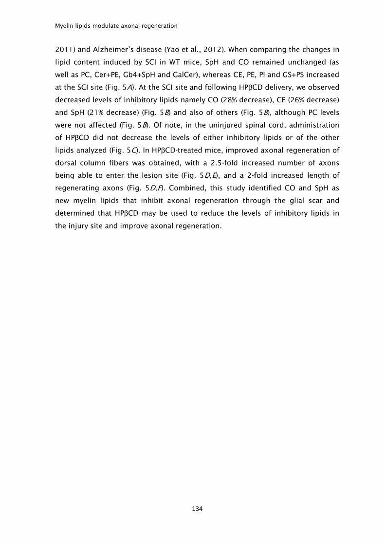
Myelin lipids modulate axonal regeneration
134
2011) and Alzheimer’s disease (Yao et al., 2012). When comparing the changes in
lipid content induced by SCI in WT mice, SpH and CO remained unchanged (as
well as PC, Cer+PE, Gb4+SpH and GalCer), whereas CE, PE, PI and GS+PS increased
at the SCI site (Fig. 5A). At the SCI site and following HPβCD delivery, we observed
decreased levels of inhibitory lipids namely CO (28% decrease), CE (26% decrease)
and SpH (21% decrease) (Fig. 5B) and also of others (Fig. 5B), although PC levels
were not affected (Fig. 5B). Of note, in the uninjured spinal cord, administration
of HPβCD did not decrease the levels of either inhibitory lipids or of the other
lipids analyzed (Fig. 5C). In HPβCD-treated mice, improved axonal regeneration of
dorsal column fibers was obtained, with a 2.5-fold increased number of axons
being able to enter the lesion site (Fig. 5D,E), and a 2-fold increased length of
regenerating axons (Fig. 5D,F). Combined, this study identified CO and SpH as
new myelin lipids that inhibit axonal regeneration through the glial scar and
determined that HPβCD may be used to reduce the levels of inhibitory lipids in
the injury site and improve axonal regeneration.
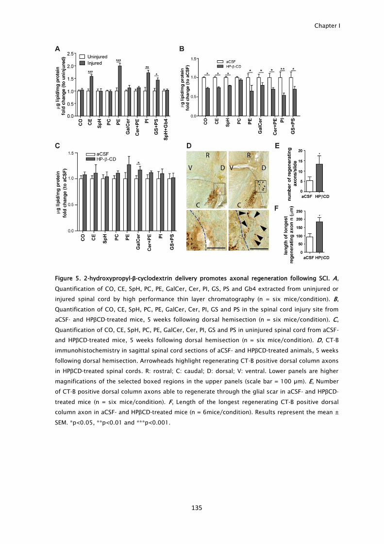
Chapter I
135
Figure 5. 2-hydroxypropyl-β-cyclodextrin delivery promotes axonal regeneration following SCI. A,
Quantification of CO, CE, SpH, PC, PE, GalCer, Cer, PI, GS, PS and Gb4 extracted from uninjured or
injured spinal cord by high performance thin layer chromatography (n = six mice/condition). B,
Quantification of CO, CE, SpH, PC, PE, GalCer, Cer, PI, GS and PS in the spinal cord injury site from
aCSF- and HPβCD-treated mice, 5 weeks following dorsal hemisection (n = six mice/condition). C,
Quantification of CO, CE, SpH, PC, PE, GalCer, Cer, PI, GS and PS in uninjured spinal cord from aCSF-
and HPβCD-treated mice, 5 weeks following dorsal hemisection (n = six mice/condition). D, CT-B
immunohistochemistry in sagittal spinal cord sections of aCSF- and HPβCD-treated animals, 5 weeks
following dorsal hemisection. Arrowheads highlight regenerating CT-B positive dorsal column axons
in HPβCD-treated spinal cords. R: rostral; C: caudal; D: dorsal; V: ventral. Lower panels are higher
magnifications of the selected boxed regions in the upper panels (scale bar = 100 µm). E, Number
of CT-B positive dorsal column axons able to regenerate through the glial scar in aCSF- and HPβCD-
treated mice (n = six mice/condition). F, Length of the longest regenerating CT-B positive dorsal
column axon in aCSF- and HPβCD-treated mice (n = 6mice/condition). Results represent the mean ±
SEM. *p<0.05, **p<0.01 and ***p<0.001.

Myelin lipids modulate axonal regeneration
136
Discussion
Our work shows that CO and SpH are myelin-associated lipid inhibitors that
modulate axonal regeneration, and demonstrates that HPβCD delivery reduces
the levels of lipids in the SCI site promoting axonal regrowth. Lipids represent
75% of myelin dry weight. From these, CO is the most abundant, accounting for
28% of the myelin lipid content, whereas SpH accounts for 4%. Although CO is an
essential component of the plasma membrane crucial for axonal growth, CO
accumulation in the mammalian brain is a risk factor for neurodegenerative
diseases including Alzheimer’s disease (Puglielli et al., 2003) and Niemann-Pick
(Aqul et al., 2011). In the scenario of injury, with the presence of myelin debris
within the injury site, myelin-derived CO is presented to regenerating axons in a
different form from glial-derived CO. Whereas glial-derived CO can stimulate axon
growth by providing lipoproteins as a source of both CO and apolipoprotein E to
regenerating axons, free CO i.e., without the context of a lipoprotein particle, as
is the case of myelin-derived CO, fails to enhance axon extension (Hayashi et al.,
2004). In the case of SpH, its accumulation, which is the hallmark of Niemann-
Pick disease, leads to changes in plasma membrane that similarly to CO, also
culminate in neurodegeneration (Ledesma et al., 2011). As such, we propose that
following CNS injury, when growing axons need to elongate through an
environment filled with myelin debris, exposure to free CO and to SpH
contributes to axonal growth inhibition. This mechanism may also underlie, at
least in part, the increased axonal regeneration in the PNS. Despite that
peripheral and central myelin have similar CO and SpH content, as
dedifferentiated Schwann cells and invading macrophages are able to phagocyte
and clear myelin debris following PNS injury (Stoll et al., 1989), regenerating
axons are probably not exposed to a lipid-rich environment, in contrast to the
CNS where such clearance is not as effective.
Accumulation of unesterified CO and SpH is the hallmark of Niemann-Pick
disease, which culminates in neurodegeneration (Aqul et al., 2011; Ledesma et
al., 2011). Interestingly, cultured neurons from mice lacking Niemann-Pick type
C1 protein (npc1 knockout mice) displayed increased rate of growth cone
collapse that was mediated by ROCK activation and reverted by ROCK inhibition
(Qin et al., 2010). In npc1 mice, administration of HPβCD delays the onset of
clinical symptoms by reducing the buildup of CO and sphingolipids within the
nervous tissue (Davidson et al., 2009; Aqul et al., 2011). HPβCD is a well-tolerated
Chapter I
137
FDA-approved drug used in animal models and in human clinical trials, known for
its ability to form inclusion complexes, remove cholesterol from membranes and
increase cholesterol trafficking (Davidson et al., 2009; Aqul et al., 2011; Matsuo
et al., 2013). Here we show that following HPβCD delivery, decreased levels of
inhibitory lipids, namely CO, CE and SpH are specifically obtained in the SCI site
whereas uninjured spinal cord segments have unaltered lipid composition. Upon
spinal cord injury, HPβCD may exert a generalized sequestering effect allowing
the removal of lipids from the injury site, and thus relieve the inhibitory level
within the glial scar. Although HPβCD did not seem to have a clearly defined lipid
specificity, we did not observe any lipid changes in the treated un-injured spinal
cord, indicating that this non-toxic agent does not lead to lipid dysregulation in
normal tissues. In addition, HPβCD treatment led to increased axonal
regeneration, supporting that HPβCD delivery should be considered as a
therapeutic option in the context of SCI.
At the molecular level, inhibition induced by CO and SpH could be reverted
by the Rho inhibitor C3 transferase. In the case of SpH, for which a stronger effect
was observed with C3 transferase, we further demonstrate the downstream
activation of ROCK as increased phosphorylated levels of its substrate, MLC, are
generated. These data supports that both myelin proteins and lipids signal
inhibition through similar pathways involving Rho activation. CO and SpH may act
as ligands to induce receptor-mediated activation of the Rho signaling cascade, or
alternatively they can augment receptor activation leading to increased Rho
activity. Whether CO and SpH engage the same receptors (e.g. Nogo receptor and
PirB) as several structurally unrelated myelin proteins is to be unraveled. Specific
binding sites for CO have been identified in G-protein-coupled receptors
(Cherezov et al., 2007; Liu et al., 2012). As such, CO and/or SpH at the
extracellular matrix in which axons are growing may interact and engage other
receptors eliciting a signaling cascade that impairs axonal growth. Sulfatide has
also been identified as a lipid that specifically inhibits neurite outgrowth of retinal
ganglion cells through a Rho-mediated mechanism (Winzeler et al., 2011),
although the pathway by which this occurs remains poorly understood. The next
challenge will be to further characterize the pathways by which lipids mediate the
repression of neurite outgrowth.
In summary our work shows that myelin lipids are important modulators of
axonal regeneration that should be considered as critical targets in strategies
Myelin lipids modulate axonal regeneration
138
aiming at improving axonal growth following injury. Moreover, we provide the
initial data supporting that the FDA-approved HPβCD delivery could be tested in
the context of SCI.
Chapter I
139
Acknowledgments
We thank Dr. Richard Quarles (NINDS, Bethesda) and Dr. Stephen Strittmatter
(Yale University, New Haven) for generously sharing reagents, Dr Carla Teixeira
(IBMC) for help in preparing osmotic pumps and Dr Sofia Lamas (IBMC) for
supporting animal experiments. Mar FM was supported by FCT
(SFRH/BD/43484/2008). Brites P is an Investigator FCT. This work was funded by
the European Leukodystrophy Association – Research Foundation (ELA 2010-
042C5) and FEDER through the Operational Competitiveness Program – COMPETE
and National Funds through FCT – Fundação para a Ciência e a Tecnologia under
the projects FCOMP-01-0124-FEDER-015970 (PTDC/SAU-ORG/112406/2009) and
FCOMP-01-0124-FEDER-017455 (HMSP-ICT/0020/2010).
Conflicts of interest: The authors declare no competing financial interests.

140

141
Chapter II

142
Chapter II
143
Differential activation and transport of injury signals contributes to the failure of
a dorsal root injury to increase the intrinsic growth capacity of DRG neurons
Abbreviated title: Negative injury signals inhibit regeneration after dorsal root
injury
Fernando M. Mar1,2
, Anabel R. Simões1
, Inês S. Rodrigo1
, Mónica M Sousa1
1
Nerve Regeneration group, Instituto de Biologia Molecular e Celular - IBMC,
University of Porto; Rua do Campo Alegre 823, 4150-180 Porto, Portugal
2
Instituto de Ciências Biomédicas Abel Salazar – ICBAS, Rua Jorge Viterbo Ferreira
228, 4050-313 Porto, Portugal
Negative injury signals inhibit regeneration after dorsal root injury
144
Abstract
Following peripheral nervous system injury, besides increased cAMP, the positive
injury signals ERK, JNK and STAT-3 are locally activated and retrogradely
transported to the cell body, where they induce a pro-regenerative program. Here,
we used dorsal root ganglia (DRG) neurons which comprise a central branch that
does not regenerate and a peripheral branch that regrows after lesion, to further
understand the importance of injury signaling for successful axon regeneration.
Although injury to the central branch of DRG neurons (dorsal root injury-DRI)
activates the above positive injury signals and increases cAMP levels, it does not
elicit the gain of intrinsic growth capacity nor the ability to overcome myelin
inhibition, as occurs after injury to the peripheral branch (sciatic nerve injury-
SNI). Besides, by blocking ERK activation and adenylyl cyclase activity, we show
that the gain of intrinsic growth capacity of DRG neurons after injury is
independent of ERK and cAMP. Antibody microarray analysis of axoplasm from
rats with either DRI or SNI following dynein immunoprecipitation revealed a broad
differential activation and transport of signals after each injury type and further
supported that ERK, JNK, STAT-3 and cAMP signaling pathways are probably
minor contributors to the differences in the intrinsic axon growth capacity
observed in these injury models. From the injury signals differentially activated
after DRI and SNI, we specifically identified increased levels of Hsp-40, ROCK-II
and GSK3β after DRI, not only in axons but also in DRG cell bodies. In summary,
our work shows that activation and transport of canonical positive injury signals
is not sufficient to promote increased axonal growth capacity, and that a limited
regenerative response after DRI may be accounted by the differential activation of
inhibitory injury signals including ROCKII and GSK3β.
Chapter II
145
Introduction
While axon regeneration in the central nervous system (CNS) is unsuccessful,
adult peripheral nervous system (PNS) axons can spontaneously regenerate to a
considerable extent and are therefore generally used as a model to identify
players that promote axon regrowth. Within the PNS, sensory dorsal root ganglia
(DRG) neurons have been widely used in axonal regeneration studies given their
peculiar nature. DRG neurons are pseudounipolar, possessing two branches: a
peripheral branch that innervates sensory organs and a central branch that enters
the spinal cord through the dorsal root and ascends to the brainstem, forming
the dorsal column fibers (Devor, 1999). When injured, the two branches have
different regenerative capacities. While an injury to the peripheral branch is
followed by successful regeneration, injury to the central branch, either dorsal
root injury (DRI) or spinal cord injury, fails to display a similar effect (Smith and
Skene, 1997). In the case of DRI, sensory axons regenerate within the dorsal root
but stop after contact with the dorsal root entry zone (DREZ; the transition zone
between the spinal cord and the dorsal root), making the DRI a simplified model
to study CNS regeneration. Initially, the inability of sensory axons to regenerate
after DRI has been attributed to the presence of inhibitory molecules in the DREZ
(Zhang et al., 2001; Beggah et al., 2005). However, recent reports suggest that
the formation of new synapses when regenerating axons enter the spinal cord
stabilizes them, inhibiting further growth (Di Maio et al., 2011). Besides
extracellular cues, cell-intrinsic differences have been described in DRG neurons
subjected to either DRI or peripheral injury. Whereas a peripheral injury triggers
the expression of several regeneration associated genes (RAGs), including
activating transcription factor 3 (ATF-3), growth-associated protein 43 (GAP-43)
and CAP-23 among others (Schreyer and Skene, 1993; Mason et al., 2002;
Seijffers et al., 2006), DRI fails to elicit a similar response. The reasons underlying
the differential activation of a pro-regenerative program after SNI and DRI remain
elusive.
It is widely acknowledged that injury signaling is pivotal to mount a pro-
regenerative program (Mar et al., 2014). Seminal studies done in Aplysia have
shown that injection of axoplasm from injured nerves into naïve neurons is
accompanied by increased growth. The hypothesis raised to explain this
observation was that axonal injury triggers the local activation of injury signals
that are retrogradely transported to the cell body and then imported to the
Negative injury signals inhibit regeneration after dorsal root injury
146
nucleus (Ambron et al., 1995). Later, and further supporting this hypothesis,
extracellular-signal-regulated kinase (ERK), c-Jun N-terminal kinase (JNK) and
signal transducer and activator of transcription 3 (STAT-3) have been identified as
positive injury signals locally activated following peripheral injury and
retrogradely transported to the cell body, playing a crucial role in mounting the
response to injury (Cavalli et al., 2005; Perlson et al., 2006; Rishal and Fainzilber,
2010; Ben-Yaakov et al., 2012). Specifically, pERK is involved in the retrograde
signal that initiates regeneration (Perlson et al., 2005); JNK signaling has been
implicated in the reorganization of the axonal cytoskeleton and in neurite
regeneration (Barnat et al., 2010) and STAT-3 contributes to the initiation of
axonal regeneration (Bareyre et al., 2011) and to neuronal survival after injury
(Ben-Yaakov et al., 2012). Besides injury signaling, increased levels of cAMP in
DRG neurons after PNS injury have long been associated with increased axonal
elongation (Cai et al., 1999; Cai et al., 2001; Neumann et al., 2002; Qiu et al.,
2002; Nikulina et al., 2004). Nevertheless, recent reports have questioned the
extension of cAMP effects (Blesch et al., 2012). Downstream of injury signaling,
major transcriptional alterations underlie the increased regeneration ability
following peripheral injury (Costigan et al., 2002). Some of the transcription
factors are activated by the above injury signals: ERK activates ETS domain-
containing protein (Elk-1) (Perlson et al., 2005), JNK activates c-Jun and ATF3
(Lindwall and Kanje, 2005) and the cAMP pathway activates cAMP response
element-binding protein (CREB) (Gao et al., 2004). Together, these will lead to a
profound change of the transcriptional profile of injured neurons contributing to
their survival and regeneration through the expression of an array of RAGs
including neuropeptide Y (NPY), vasoactive intestinal peptide (Vip), ATF-3,
arginase 1 (Arg-1) and GAP-43, among others (Ylera et al., 2009).
Here we observed that despite DRI is unable to elicit a robust increase in
axon growth, it is accompanied by the activation and retrograde transport of the
canonical positive injury signals ERK, JNK and STAT-3 and increased levels of
cAMP. Besides, we identified broad signaling differences between DRI and
peripheral injury including increased levels of signals associated with decreased
axonal growth namely, Rho-associated kinase (ROCK-II) and glycogen synthase
kinase 3 β (GSK3β), that may contribute to the limited regeneration capacity after
a central branch injury.
Chapter II
147
Materials and methods
Surgical procedures. Rats were handled according to European Union and
National rules. All animals were maintained under a 12-h light/dark cycle and fed
with regular rodent's chow and tap water ad libitum. For the animal models of
injury 8-10 weeks old Wistar rats were used. For sciatic nerve injury (SNI), rats
were anesthetized with ketamine (75 mg/kg)/medetomidine (0.5 mg/kg). The
sciatic nerve was exposed at the mid-thigh and crush was then performed using
Pean forceps, twice for 15 seconds. Analgesia (butorphanol 3 mg/kg, twice a day)
was performed for 48 h following injury. For dorsal root injury (DRI), rats were
anesthetized and laminectomy at lumbar vertebrae 2 was performed exposing the
dorsal roots of DRG L4,5. The L4,5 dorsal roots were then crushed using fine
forceps, twice for 15 seconds. Analgesia was performed as described above.
Quantitative PCR. One week after either SNI or DRI, rats were sacrificed with an
overdose of ketamine/medetomidine and L4,5 DRGs were collected; naïve rats
were used as control (n=4 rats per group were used). RNA was extracted from
DRGs (Qiagen), and cDNA was synthesized by reverse transcription (Invitrogen)
using 1 μg of RNA. qRT PCR was then performed using iQ supermix (Bio-Rad) and
primers designed using Beacon designer: NPY (sense: 5’-
GCTCGTGTGTTTGGGCATTCTG-3’; antisense: 5’-GTGTCTCAGGGCTGGATCTCTTG-
3’), Vip (sense: 5’-GTCACTCATTGGCAAACGAATCAG-3’; antisense: 5’-
CTCCCTCACTGCTCCTCTTCC-3’), GAP-43 (sense: 5’-
GATAACTCGCCGTCCTCCAAG-3’; antisense: 5’-CTACAGCTTCTTTCTCCTCCTCAG-
3’); Arg-1 (sense: 5’-GACATCAACACTCCGCTGACAAC-3’; antisense: 5’-
CCAGGGTCCACATCTCGCAAG-3’); ATF-3 (sense: 5’-
TCTGGAGATGTCAGTCACCAAGTC-3’; antisense: 5’-
CCTTCAGTTCGGCATTCACACTC-3’); Hypoxanthine-guanine
phosphoribosyltransferase (Hprt) (sense: 5’-ATGGACTGATTATGGACAGGACTG-3’;
antisense: 5’-GCAGGTCAGCAAAGAACTTATAGC-3’).
Neurite outgrowth assays. One week after either SNI or DRI, animals were
sacrificed with an overdose of ketamine/medetomidine and L4,5 DRGs were
collected. DRG neuron cultures were performed as described (Miranda et al.,
2011). Briefly, DRG were collected and digested with 0.125% collagenase IV-S
(Sigma-Aldrich) for 3 h at 37°C. A single-cell suspension was obtained by
trituration with a fire-polished Pasteur pipette and centrifuged over a 15%
Negative injury signals inhibit regeneration after dorsal root injury
148
albumin cushion for 10 minutes at 200g. Cells were then plated in poly-L-lysine
(20 µg/mL)/laminin (5 µg/mL; Sigma-Aldrich) or in poly-L-lysine (20
µg/mL)/myelin-coated (2.4 µg/coverslip; myelin was isolated from the spinal cord
of 16 weeks-old WT mice as detailed (Norton and Poduslo, 1973)) 13 mm
coverslips and maintained at 37 °C for 12 h. Subsequently, cells were fixed,
immunostained against βIII tubulin (1:2000, Promega) and traced with NeuronJ
(an ImageJ plugin). The length of the longest neurite was measured in at least
100 neurons per condition.
Axoplasm extraction. Immediately after SNI or DRI, a knot was performed with
4/0 suture thread proximally to the DRG to restrain the axonal transport of injury
signals (Fig. 2A). Eight or 24 h after each injury type, rats were sacrificed and
nerves were collected to 100 μl of PBS containing protease inhibitors (GE
Healthcare) and 1 mM ortovanadate (Sigma-Aldrich). Nerves from naïve rats were
used as control. Axoplasm was collected by gently squeezing the nerves with a
pestle followed by a centrifugation at 15,000 g at 4 ºC for 10 minutes, as
described (Hanz et al., 2003). This cycle was repeated 2 more times. After the last
centrifugation, supernatant was collected and 25 μg of each protein sample were
run in 12% SDS-PAGE.
DRG protein extract. L4,5 DRG of animals with either SNI or DRI were collected
17-20 h after lesion, respectively; naïve animals were used as a control
(n=4/group). The tissue was then homogenized in PBS containing protease
inhibitors (GE Healthcare), 1mM ortovanadate and 0.3% Triton X-100, sonicated
and centrifuged for 10 minutes at 15,000 g at 4 °C. The supernatant was
collected and 25 μg of each protein sample were run in 12% SDS-PAGE.
Dynein immunoprecipitation. Dynein immunoprecipitation was performed as
described elsewhere (Perlson et al., 2005). Briefly, 500 μg of axoplasm protein
was precleared for 1 h at 4 °C with protein G magnetic beads (GE Healthcare) and
then incubated with 10 μg of anti-dynein antibody (Millipore) overnight at 4 °C.
The immune complex formed was collected by incubation for 2 h at room
temperature with protein G magnetic beads (GE Healthcare), and then boiled in
sample buffer (31.25 mM Tris-HCL pH 6.8; 1 % SDS; 12.5 % glycerol; 0.02 %
bromophenol blue; 1.25 % β-mercaptoethanol). The immunoprecipitated samples
were then run in 12% SDS-PAGE.
Chapter II
149
Immunoblots. Twenty five µg of either axoplasm, DRG extract or dynein
immunoprecipate were immunobloted against pERK (T202/Y204) and total-ERK
(both 1:2,000, Cell Signaling), pJNK ((T183/Y185) and total-JNK (both 1:1,000,
Cell Signaling), pSTAT-3 (Y705) and total-STAT-3 (both 1:2,000, Cell Signaling), β-
actin (1:5,000, Sigma-Aldrich), GSK3α/β (1:2,000, Santa-Cruz Biotechnologies), C-
terminal Src kinase (Csk) (1:1,000, Santa-Cruz Biotechnologies), proto-oncogenic
cytoplasmic tyrosine kinase (Src) (1:1,000, Signalway Antibody), Hsp-40 (1:1,000,
Santa-Cruz Biotechnologies), mitogen-activated protein kinase kinase (MEK6)
(1:1,000, Enzo Life Sciences), Caspase 4 (1:1,000, Enzo Life Sciences), Polo-like
kinase 3 (Plk3) (1:500, Abgent), ROCK-II (1:1,000, Millipore), total-collapsin
response mediator protein-2 (CRMP-2), pCRMP-2 (both 1:1,000, Kinasource) and
GAPDH (1:1,000, Santa-Cruz Biotechnologies). Quantitative analysis of Western
blots was performed using PDQuest (Bio-rad).
In vitro conditioning assays. DRG from 8 weeks-old Wistar rats were collected and
DRG cultures were performed as described above. Cells were either fixed 12 h
after plating (naïve type of growth), or grown for 24 h and treated with trypsin-
EDTA (Invitrogen) for 5 min, replated and allowed to grow for 12 h (conditioned
type of growth). The in vitro assay was performed in the presence of the ERK
inhibitor U0126 (Calbiochem; 10 μM in 0.1 % DMSO), or of the adenylyl cyclase
inhibitor 9-cyclopentyladenine monomethanesulfonate (9-CPA; 100 μM in H2O,
Sigma-Aldrich). During the assay, cells were exposed to the drug immediately
following tissue collection and the drug was present in all culture mediums.
Vehicle was used as control for both drugs. ERK inhibition was confirmed by
Western blot against pERK. The efficiency of 9-CPA was assessed by measuring
the levels of cAMP, as described below.
cAMP quantification and immunohistochemistry. L4,5 DRG of rats with either SNI
or DRI were collected 1 day after lesion; naïve animals were used as control (n=4
per group). Tissues were immediately collected to liquid nitrogen, homogenized
in ice cold 6 % trichloroacetic acid solution and centrifuged for 15 minutes at
2,000 g at 4 °C. The supernatant was washed 4 times with 1 ml of water saturated
diethyl ether. The aqueous phase was then dried under a stream of Nitrogen.
cAMP levels were measured by an enzyme immune assay according to the
manufacturer’s instructions (GE Healthcare). For cAMP immunohistochemistry,
L4,5 DRG of animals with either SNI or DRI were collected 1 day after lesion and
were formalin fixed, processed and paraffin embedded; naïve animals were used
Negative injury signals inhibit regeneration after dorsal root injury
150
as control (n=4 per group). Paraffin sections were cleared, hydrated and
incubated in blocking buffer (10 % fetal bovine serum, with 0.3 %Triton X-100) for
1 h at room temperature and then incubated with anti-cAMP primary antibody
diluted in blocking buffer (1:500 Millipore) overnight at 4 ºC, followed by
incubation with biotinylated secondary antibody in blocking buffer (1:200, Vector
laboratories) and avidin peroxidase (Elite ABC kit, Vector laboratories). Antigen
visualization was performed using DAB (Vector laboratories). Sections were then
dehydrated, mounted in DPX and pictures were taken at 10 x magnification
(Olympus).
Antibody microarray analysis. Axoplasm of L4,5 ligated nerves from rats with
either SNI (a pool of 18 animals) or DRI (a pool of 6 animals) was collected 8 h
after injury. Dynein immunoprecipitation was performed as described above. The
immune complexes formed were dissociated by two incubations of 10 minutes
with 0.1 M glycine pH 2.5, at room temperature. The solution was neutralized
with 1 M Tris pH 9 and then incubated with protein G magnetic beads (GE
Healthcare) to remove the IgGs. The collected supernatant (immunoprecipitation
product without IgGs) was analyzed using the Kinex™ Antibody Microarray
(Kinexus). The Kinex™ Antibody Microarray tracks both protein expression (with
~510 pan-specific antibodies) and phosphorylation (with ~340 phospho-site-
specific antibodies) using highly validated commercial antibodies. Briefly, 50 μg
of lysate protein from each sample (axoplasm following dynein
immunoprecipitation from animals with either DRI or SNI) was covalently labeled
with a Kinexus proprietary fluorescent dye and loaded side by side on the same
chip (each microarray consists of 2 identical fields). Signal quantification was
performed with ImaGene 8.0 from BioDiscovery (El Segundo, CA). Z scores were
calculated by subtracting the overall average intensity of all spots within a sample
from the raw intensity for each spot, and dividing it by the standard deviations
(SD) of all of the measured intensities within each sample. Z ratios were further
calculated by taking the difference between the averages of the observed protein
Z scores and dividing by the SD of all of the differences for that particular
comparison. A short list of 46 candidates was selected based on the following
parameters: Z-ratio = >+/- 1.0, Error Range for adjacent duplicate spots of the
same protein <30%, Globally Normalized Median Value >254, Flags (indication of
the quality of the spot based on its morphology and background) = 0.0. To
validate the microarray analysis, additional samples of axoplasm of ligated nerves

Chapter II
151
from rats with either SNI (a pool of 14 animals) or DRI (a pool of 7 animals) was
collected 8h after injury and validation was done by Western blot- Kinetworks™.
DRG immunohistochemistry. L4,5 DRG of animals with either SNI or DRI were
collected 17-20 h after lesion, formalin fixed and processed for paraffin blocks;
naïve animals were used as controls (n=4/group). Paraffin sections were cleared
and hydrated, antigen retrieval was performed, blocking was done with 5 % fetal
bovine serum for 1 h at room temperature and the following primary antibodies
were incubated overnight at 4ºC: pSTAT-3 (1:200, Cell Signaling), GSK3β (1:100,
Cell Signaling), Csk (1:50, Santa-Cruz Biotechnologies), Src (1:200, Signalway
Antibody), Hsp-40 (1:50, 10 mM sodium citrate, Santa-Cruz Biotechnologies),
MEK6 (1:200, Enzo Life Sciences), Caspase 4 (1:100, Thermo Scientific) and ROCK-
II (1:100, Millipore). Sections were then incubated with anti-rabbit IgG-alexa488
(1:1,000, Invitrogen) and mounted with vectashield (Vector laboratories). Images
were taken in a fluorescence microscope (Zeiss Axio Imager) with a 10 x
objective. Quantification was performed by determining the percentage of DRG
neurons with positive staining.
Lentivirus production and neurite outgrowth analysis. HEK293T cells were
transfected with packaging plasmids pPAX and pLP/VSVG (Dr. Relvas, IBMC) and a
lentiviral vector (pLKO) coding for puromycin resistance and expressing a
scrambled shRNA (Sarbassov et al., 2005) or a shRNA for Hsp-40 (Sigma-Aldrich).
Forty eight hours later, viral titration was done by infecting HEK 293T cells. DRG
neuron cultures from 8-weeks-old rats were performed as described above. One
day after plating, cells were infected with lentivirus (5,000 IU/well) for 12-16 h,
and 24 h later treated with puromycin (5μg/mL) for 48 h. For neurite outgrowth,
cells were trypsinized, replated for 12 h and immunocytochemistry against βIII-
tubulin (1:2,000, Promega) was conducted. At least 60 neurons/condition were
traced using NeuronJ.
Statistical analysis. All values are presented as mean ± SEM. Student’s t-test or 1-
way-anova with Tukey’s test was used.
Negative injury signals inhibit regeneration after dorsal root injury
152
Results
A dorsal root injury elicits the activation and retrograde transport of positive
injury signals to the soma
After DRI in adult animals, sensory axons regenerate within the dorsal root but
stop after contact with the spinal cord. Besides the inhibitory extrinsic
environment, DRI fails to activate an intrinsic regenerative program (Seijffers et
al., 2006). In accordance with previous findings (Seijffers et al., 2006), DRG
neurons plated on the permissive substrate laminin collected from rats with a
conditioning SNI had a robust 1.6-fold increased neurite outgrowth, whereas after
DRI a marginal 1.1-fold increase was observed (Fig. 1A,B). Moreover, while DRG
collected form rats primed with a SNI were able to overcome myelin inhibition,
DRI was unable to generate a similar effect (Fig. 1A,C). The failure of DRI in
increasing the growth capacity of DRG neurons and of overcoming myelin
inhibition correlated with a decreased capacity in inducing the expression of
RAGs, namely NPY, Vip, GAP-43, Arg-1 and ATF-3 (Fig. 1D). In summary, besides
the extrinsic inhibitory milieu, axonal regeneration failure after DRI might also
result from its inability to prime DRG neurons to increase their intrinsic growth
capacity.
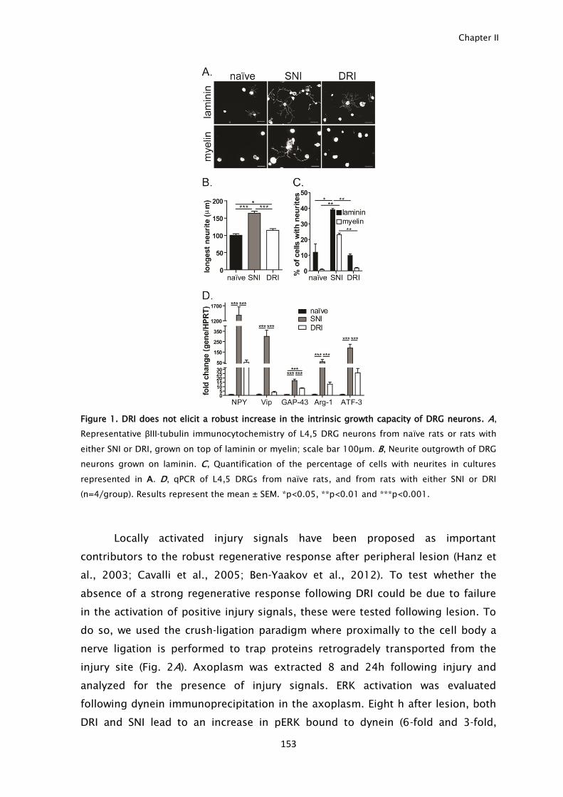
Chapter II
153
Figure 1. DRI does not elicit a robust increase in the intrinsic growth capacity of DRG neurons. A,
Representative βIII-tubulin immunocytochemistry of L4,5 DRG neurons from naïve rats or rats with
either SNI or DRI, grown on top of laminin or myelin; scale bar 100µm. B, Neurite outgrowth of DRG
neurons grown on laminin. C, Quantification of the percentage of cells with neurites in cultures
represented in A. D, qPCR of L4,5 DRGs from naïve rats, and from rats with either SNI or DRI
(n=4/group). Results represent the mean ± SEM. *p<0.05, **p<0.01 and ***p<0.001.
Locally activated injury signals have been proposed as important
contributors to the robust regenerative response after peripheral lesion (Hanz et
al., 2003; Cavalli et al., 2005; Ben-Yaakov et al., 2012). To test whether the
absence of a strong regenerative response following DRI could be due to failure
in the activation of positive injury signals, these were tested following lesion. To
do so, we used the crush-ligation paradigm where proximally to the cell body a
nerve ligation is performed to trap proteins retrogradely transported from the
injury site (Fig. 2A). Axoplasm was extracted 8 and 24h following injury and
analyzed for the presence of injury signals. ERK activation was evaluated
following dynein immunoprecipitation in the axoplasm. Eight h after lesion, both
DRI and SNI lead to an increase in pERK bound to dynein (6-fold and 3-fold,
Negative injury signals inhibit regeneration after dorsal root injury
154
respectively) (Fig. 2B,C). Besides pERK, pJNK (Fig. 2B,D) and pSTAT-3 (Fig. 2B,E)
were activated in the axoplasm after DRI to a similar extent as after SNI. These
signals were not only activated by DRI but were also retrogradely transported and
able to reach the cell body. In DRG extracts, following either DRI or SNI, increased
pERK (20- and 10-fold, respectively- Fig. 2F,G) and pSTAT-3 (2.1- and 2.8-fold,
respectively- Fig. 2H,I) were observed. Similar levels of pJNK in the DRG were also
observed following SNI and DRI. Furthermore, immunohistochemistry of DRG
neurons showed that pSTAT-3 is translocated to the nucleus following both injury
types (Fig. 2J,K). In summary DRI is able to induce to a similar extent as SNI, the
activation and retrograde transport of injury signals to the DRG cell body.
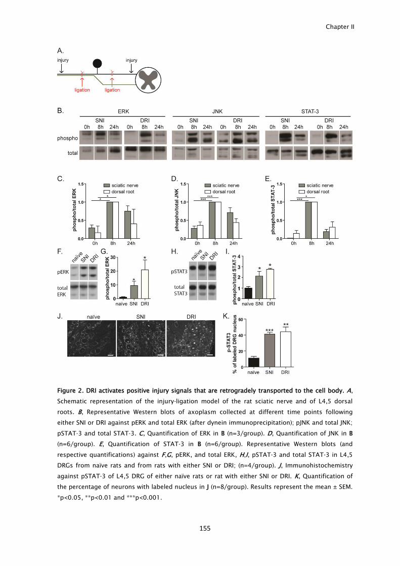
Chapter II
155
Figure 2. DRI activates positive injury signals that are retrogradely transported to the cell body. A,
Schematic representation of the injury-ligation model of the rat sciatic nerve and of L4,5 dorsal
roots. B, Representative Western blots of axoplasm collected at different time points following
either SNI or DRI against pERK and total ERK (after dynein immunoprecipitation); pJNK and total JNK;
pSTAT-3 and total STAT-3. C, Quantification of ERK in B (n=3/group). D, Quantification of JNK in B
(n=6/group). E, Quantification of STAT-3 in B (n=6/group). Representative Western blots (and
respective quantifications) against F,G, pERK, and total ERK, H,I, pSTAT-3 and total STAT-3 in L4,5
DRGs from naïve rats and from rats with either SNI or DRI; (n=4/group). J, Immunohistochemistry
against pSTAT-3 of L4,5 DRG of either naïve rats or rat with either SNI or DRI. K, Quantification of
the percentage of neurons with labeled nucleus in J (n=8/group). Results represent the mean ± SEM.
*p<0.05, **p<0.01 and ***p<0.001.

Negative injury signals inhibit regeneration after dorsal root injury
156
ERK activation is not essential for DRG neurons to trigger a regenerative response
The fact that positive injury signals are activated by DRI in the absence of an
increase in the intrinsic axonal growth questions their relevance as crucial injury
signals sufficient to trigger a regenerative response. Preventing ERK activation
with the MEK inhibitor U0126 has been shown to inhibit axonal regeneration
following peripheral nerve injury (Perlson et al., 2005). To further clarify the role
of ERK in the gain of axonal growth capacity, we performed an in vitro assay
mimicking a peripheral injury by trypsin treatment and replating naïve DRG
neurons after 24 h in culture (Saijilafu et al., 2013), grown with or without U0126.
The efficiency of the inhibitor was confirmed by immunoblot against pERK in
cultured DRG neurons (Fig. 3A). Trypsin treatment increased neurite outgrowth
(Fig. 3B), similarly to the in vivo priming of DRG neurons after peripheral injury.
Although U0126 lead to a small decrease in neurite outgrowth of naïve DRG
neurons (Fig. 3B), trypsin-treated DRG neurons had similar increase in neurite
outgrowth with or without U0126 treatment (Fig. 3B), suggesting that priming
DRG neurons triggers ERK-independent mechanisms that can override its
absence.
Figure 3. ERK activation is not essential for DRG neurons to trigger a regenerative response. A,
Western blot against pERK and total ERK in DRG neuron cultures either untreated or treated with the
MEK inhibitor U0126. B, Neurite outgrowth of DRG neurons treated with U0126 before or after
trypsin treatment (n=3/group). Results represent the mean ± SEM. *p<0.05 and ***p<0.001.
cAMP increases after DRI and is not central for the gain of axonal growth capacity
of conditioned DRG neurons
High levels of cAMP following peripheral injury have been described as essential
to increase axonal regeneration (Qiu et al., 2002). Given the importance of cAMP
in axon growth, we next asked if a DRI could increase the cAMP levels of DRG
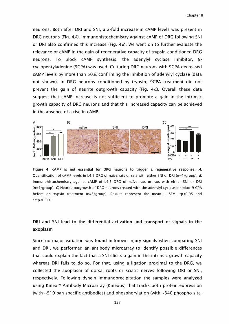
Chapter II
157
neurons. Both after DRI and SNI, a 2-fold increase in cAMP levels was present in
DRG neurons (Fig. 4A). Immunohistochemistry against cAMP of DRG following SNI
or DRI also confirmed this increase (Fig. 4B). We went on to further evaluate the
relevance of cAMP in the gain of regenerative capacity of trypsin-conditioned DRG
neurons. To block cAMP synthesis, the adenylyl cyclase inhibitor, 9-
cyclopentyladenine (9CPA) was used. Culturing DRG neurons with 9CPA decreased
cAMP levels by more than 50%, confirming the inhibition of adenylyl cyclase (data
not shown). In DRG neurons conditioned by trypsin, 9CPA treatment did not
prevent the gain of neurite outgrowth capacity (Fig. 4C). Overall these data
suggest that cAMP increase is not sufficient to promote a gain in the intrinsic
growth capacity of DRG neurons and that this increased capacity can be achieved
in the absence of a rise in cAMP.
Figure 4. cAMP is not essential for DRG neurons to trigger a regenerative response. A,
Quantification of cAMP levels in L4,5 DRG of naïve rats or rats with either SNI or DRI (n=4/group). B,
Immunohistochemistry against cAMP of L4,5 DRG of naïve rats or rats with either SNI or DRI
(n=4/group). C, Neurite outgrowth of DRG neurons treated with the adenylyl cyclase inhibitor 9-CPA
before or trypsin treatment (n=3/group). Results represent the mean ± SEM. *p<0.05 and
***p<0.001.
DRI and SNI lead to the differential activation and transport of signals in the
axoplasm
Since no major variation was found in known injury signals when comparing SNI
and DRI, we performed an antibody microarray to identify possible differences
that could explain the fact that a SNI elicits a gain in the intrinsic growth capacity
whereas DRI fails to do so. For that, using a ligation proximal to the DRG, we
collected the axoplasm of dorsal roots or sciatic nerves following DRI or SNI,
respectively. Following dynein immunoprecipitation the samples were analyzed
using Kinex™ Antibody Microarray (Kinexus) that tracks both protein expression
(with ~510 pan-specific antibodies) and phosphorylation (with ~340 phospho-site-
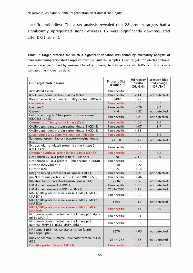
Negative injury signals inhibit regeneration after dorsal root injury
158
specific antibodies). The array analysis revealed that 28 protein targets had a
significantly upregulated signal whereas 16 were significantly downregulated
after DRI (Table 1).
Table 1: Target proteins for which a significant variation was found by microarray analysis of
dynein-immunoprecipitated axoplasm from DRI and SNI samples. Grey- targets for which additional
analysis was performed by Western blot of axoplasm; Red- targets for which Western blot results
validated the microarray data.
Full Target Protein Name Phospho Site
(Human)
Microarray
Z-ratio
(DRI/SNI)
Western blot
fold change
(DRI/SNI)
Acetylated Lysine Pan-specific 1,28
B-cell lymphoma protein 2 alpha (Bcl2) Pan-specific 2,14 not detected
Breast cancer type 1 susceptibility protein (BRCA1) S1497 1,29
Caspase 4 Pan-specific -1,2 0,7
Caspase 5 Pan-specific -1,26 2,2
Caveolin 1 Y14 -1,32 not detected
Cell division cycle 2-like protein-serine kinase 5
(CDC2L5, CHED) Pan-specific -1,21 not detected
C-terminus of Src tyrosine kinase (Csk) Pan-specific 1,05 1,9
Cyclin-dependent protein-serine kinase 5 (CDK5) Pan-specific -1,07 1,7
Cyclin-dependent protein-serine kinase 8 (CDK8) Pan-specific 4,35
DnaJ homolog, subfamily B member 1(Hsp40) Pan-specific 1,4 1,4
Epidermal growth factor receptor-tyrosine kinase
(EGFR) Y1110 1,79 not detected
Extracellular regulated protein-serine kinase 2
(Erk1 + Erk2) Pan-specific 1,32
Glycogen synthase-serine kinase 3 beta (GSK3b) Pan-specific 1,04 3,2
Heat shock 27 kDa protein beta 1 (Hsp27) S78 2,12 0,9
Heat shock 60 kDa protein 1 (chaperonin, CPN60) Pan-specific 1,37
Histone H2A variant X S140 -1,22
Histone H2B S15 -1,14
Integrin-linked protein-serine kinase 1 (ILK1) Pan-specific -1,21 not detected
Jun N-terminus protein-serine kinase (JNK1/2/3) Pan-specific -1,46
Kit/Steel factor receptor-tyrosine kinase (Kit) Y936 1,7 not detected
LIM domain kinase 1 (LIMK1) Pan-specific 1,86 not detected
LIM domain kinase 2 (LIMK1 + LIMK2) Y504+T505 1,19 not detected
MAPK/ERK protein-serine kinase 1 (MKK1, MEK1,
MAP2K1) Pan-specific 1,09
MAPK/ERK protein-serine kinase 2 (MKK2, MEK2,
MAP2K2) T394 1,14 not detected
MAPK/ERK protein-serine kinase 6 (MKK6, MEK6,
MAP2K6) Pan-specific 1,11 3,0
Mitogen-activated protein-serine kinase p38 alpha
(p38a MAPK ) Pan-specific 1,21
Mitogen-activated protein-serine kinase p38
gamma (MAPK12, p38g MAPK, Erk6) Pan-specific 1,03
NF-kappa-B p65 nuclear transcription factor
(NFkappaB p65) S276 -1,03 not detected
nucleophosmin, numatrin, nucleolar protein NO38
(B23) T234/T237 1,68 not detected
Polo-like protein kinase 3 (Plk3) Pan-specific 1,36 2,2
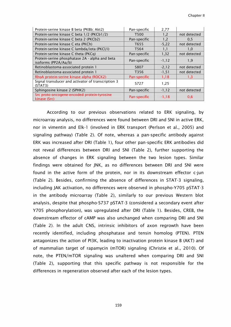
Chapter II
159
Protein-serine kinase B beta (PKBb; Akt2) Pan-specific 2,77
Protein-serine kinase C beta 1/2 (PKCb1/2) T500 1,2 not detected
Protein-serine kinase C beta 2 (PKCb2) Pan-specific 1,2 0,5
Protein-serine kinase C eta (PKCh) T655 -5,22 not detected
Protein-serine kinase C lambda/iota (PKCl/i) T564 1,1 1,0
Protein-serine kinase C theta (PKCq) Pan-specific 1,32 not detected
Protein-serine phosphatase 2A - alpha and beta
isoforms (PP2A/Aa/b) Pan-specific -1,12 1,9
Retinoblastoma-associated protein 1 S807 -2,12 not detected
Retinoblastoma-associated protein 1 T356 -1,51 not detected
RhoA protein-serine kinase alpha (ROCK2) Pan-specific 1,18 1,3
Signal transducer and activator of transcription 3
(STAT3) S727 1,25
Sphingosine kinase 2 (SPHK2) Pan-specific -1,12 not detected
Src proto-oncogene-encoded protein-tyrosine
kinase (Src) Pan-specific -1,18 0,6
According to our previous observations related to ERK signaling, by
microarray analysis, no differences were found between DRI and SNI in active ERK,
nor in vimentin and Elk-1 (involved in ERK transport (Perlson et al., 2005) and
signaling pathway) (Table 2). Of note, whereas a pan-specific antibody against
ERK was increased after DRI (Table 1), four other pan-specific ERK antibodies did
not reveal differences between DRI and SNI (Table 2), further supporting the
absence of changes in ERK signaling between the two lesion types. Similar
findings were obtained for JNK, as no differences between DRI and SNI were
found in the active form of the protein, nor in its downstream effector c-jun
(Table 2). Besides, confirming the absence of differences in STAT-3 signaling,
including JAK activation, no differences were observed in phospho-Y705 pSTAT-3
in the antibody microarray (Table 2), similarly to our previous Western blot
analysis, despite that phospho-S737 pSTAT-3 (considered a secondary event after
Y705 phosphorylation), was upregulated after DRI (Table 1). Besides, CREB, the
downstream effector of cAMP was also unchanged when comparing DRI and SNI
(Table 2). In the adult CNS, intrinsic inhibitors of axon regrowth have been
recently identified, including phosphatase and tensin homolog (PTEN). PTEN
antagonizes the action of PI3K, leading to inactivation protein kinase B (AKT) and
of mammalian target of rapamycin (mTOR) signaling (Christie et al., 2010). Of
note, the PTEN/mTOR signaling was unaltered when comparing DRI and SNI
(Table 2), supporting that this specific pathway is not responsible for the
differences in regeneration observed after each of the lesion types.
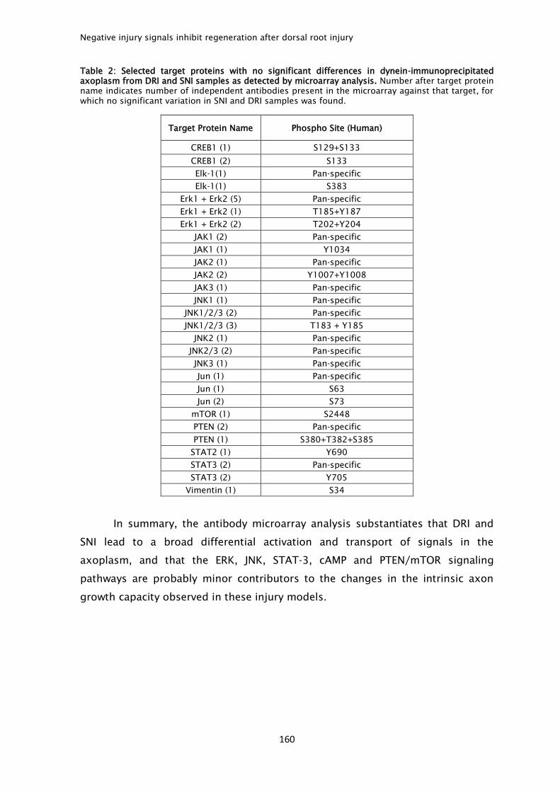
Negative injury signals inhibit regeneration after dorsal root injury
160
Table 2: Selected target proteins with no significant differences in dynein-immunoprecipitated
axoplasm from DRI and SNI samples as detected by microarray analysis. Number after target protein
name indicates number of independent antibodies present in the microarray against that target, for
which no significant variation in SNI and DRI samples was found.
Target Protein Name Phospho Site (Human)
CREB1 (1) S129+S133
CREB1 (2) S133
Elk-1(1) Pan-specific
Elk-1(1) S383
Erk1 + Erk2 (5) Pan-specific
Erk1 + Erk2 (1) T185+Y187
Erk1 + Erk2 (2) T202+Y204
JAK1 (2) Pan-specific
JAK1 (1) Y1034
JAK2 (1) Pan-specific
JAK2 (2) Y1007+Y1008
JAK3 (1) Pan-specific
JNK1 (1) Pan-specific
JNK1/2/3 (2) Pan-specific
JNK1/2/3 (3) T183 + Y185
JNK2 (1) Pan-specific
JNK2/3 (2) Pan-specific
JNK3 (1) Pan-specific
Jun (1) Pan-specific
Jun (1) S63
Jun (2) S73
mTOR (1) S2448
PTEN (2) Pan-specific
PTEN (1) S380+T382+S385
STAT2 (1) Y690
STAT3 (2) Pan-specific
STAT3 (2) Y705
Vimentin (1) S34
In summary, the antibody microarray analysis substantiates that DRI and
SNI lead to a broad differential activation and transport of signals in the
axoplasm, and that the ERK, JNK, STAT-3, cAMP and PTEN/mTOR signaling
pathways are probably minor contributors to the changes in the intrinsic axon
growth capacity observed in these injury models.
Chapter II
161
The negative injury signals ROCK-II and GSK3β are upregulated in the DRG cell
body following DRI
From the microarray shortlist, most of the candidates were chosen for further
validation by immunoblot of total axoplasm collected from additional rats with
either SNI or DRI (Table 1, highlighted in grey). Given the extensive analysis
already described, ERK, JNK and STAT-3 were excluded from further validation.
From the selected candidates, 17 were not detected by Western blot of axoplasm
samples given its lower sensitivity when compared to antibody microarray
analysis. From the remaining 14 candidates, 8 were detected with similar
differential levels in total axoplasm as those observed by antibody microarray
analysis of dynein-immunoprecipitated axoplasm: Hsp-40, Plk3, ROCK-II, MEK6,
Csk and GSK3β- increased after DRI; Src and Caspase 4- decreased after SNI
(Table 1). Although we excluded from subsequent analysis the 6 candidates
where no consistent results were found, one should note that whereas microarray
analysis was performed in dynein-immunoprecipitated axoplasm i.e., allowing the
identification of proteins bound to the transport machinery, Western blot was
done on total axoplasm samples, which may underlie the differences observed.
From the above 8 candidates with consistent changes detected both by
microarray analysis and Western blot, clear alterations in the DRG were found by
immunoblot against Hsp-40 and GSK3β where a 2.0- and 1.3- fold increase
following DRI respectively, was observed (Fig. 5A,B). Besides, by immunostaining
of DRG, following DRI, a 1.4-, 1.6- and 2.1-fold increase in the immunostaining of
Hsp-40, GSK3β and ROCK-II was detected when comparing to SNI, respectively
(Fig. 5C,D). This data further supports that DRI and SNI elicit differential
activation and transport of injury signals to the DRG.
Inhibition of growth cone formation and axonal regeneration is widely
described as occurring through activation of the Rho pathway, that signals
through its effector ROCK (Mueller et al., 2005). In this context, increased ROCK-II
activity in the axoplasm after DRI may certainly contribute to the decreased
intrinsic growth capacity of neurons after this type of injury. Consistent with
increased ROCK-II activity after DRI, increased phosphorylation of the ROCK-II
substrate myosin-light chain was found after DRI (data not shown). In the case of
GSK3, its activation has been linked to axon growth inhibition (Dill et al., 2008;
Alabed et al., 2010). GSK3 activity is regulated by phosphorylation; under resting
conditions, the kinase is constitutively active following phosphorylation of its
Negative injury signals inhibit regeneration after dorsal root injury
162
Tyr216 residue and as a response to the activation of the PI3K pathway, GSK3 is
inactivated by phosphorylation of Ser9 (Zhou and Snider, 2005). We tried to infer
GSK3 activity on DRG by checking its phosphorylation status. Increased levels of
both Ser9 and Tyr216 phosphorylation following DRI were detected by Western
blot (Fig. 5A). To further determine whether increased GSK3β activity was present
after DRI, we assessed the levels of pCRMP2, a GSK3β target. Following DRI, p-
CRMP-2 was increased supporting a higher GSK3β activity (data not shown). Hsp-
40 is a member of the heat shock protein family where in contrast to ROCK-II and
GSK3β, limited information is available as to its role in axon growth and
regeneration. To elucidate whether Hsp-40 plays a direct role in axon growth, we
tested neurite outgrowth in vitro following knockdown of Hsp-40 by ShRNA.
However, as expected from its function as a stimulator of the activity of the
chaperone Hsp-70 (Fan et al., 2003), downregulation of Hsp-40 led to neuronal
toxicity (data not shown). This data suggests that increased Hsp-40 following DRI
is probably related to neuroprotection and not to a direct effect in axon growth.
Supporting this hypothesis, it is known that induction of Hsp protein expression
improves motoneuron survival following injury (Kalmar et al., 2002). In summary,
upregulation of ROCK-II and GSK3β following DRI might act as a negative injury
signaling contributing to the inability of DRG neurons to regenerate robustly.
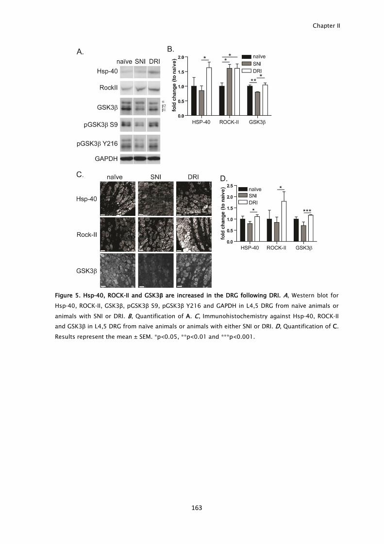
Chapter II
163
Figure 5. Hsp-40, ROCK-II and GSK3β are increased in the DRG following DRI. A, Western blot for
Hsp-40, ROCK-II, GSK3β, pGSK3β S9, pGSK3β Y216 and GAPDH in L4,5 DRG from naïve animals or
animals with SNI or DRI. B, Quantification of A. C, Immunohistochemistry against Hsp-40, ROCK-II
and GSK3β in L4,5 DRG from naïve animals or animals with either SNI or DRI. D, Quantification of C.
Results represent the mean ± SEM. *p<0.05, **p<0.01 and ***p<0.001.

Negative injury signals inhibit regeneration after dorsal root injury
164
Discussion
CNS injury does not trigger a gain of intrinsic growth capacity, as it fails to induce
the expression of RAGs. In the PNS, ERK, JNK and STAT-3 are activated following
injury, and their transport to the cell body is important for the regenerative
response (Cavalli et al., 2005; Perlson et al., 2006; Ben-Yaakov et al., 2012). Here,
we show that after DRI, a simplified model of CNS injury, activation of the
intrinsic growth capacity of DRG neurons fails despite that activation of injury
signaling occurs. This observation raises intriguing questions on the relevance of
ERK, JNK and STAT-3 as master regulators of the regenerative response. Following
activation, ERK retrograde transport is dependent on vimentin, and further
supporting the importance of this signal, vimentin knockout nerves, lack a
regenerative response (Perlson et al., 2005). Nevertheless, this signaling pathway
is not present in all DRG neurons, since 40% of DRG neurons do not express
vimentin (Perlson et al., 2005). This evidence suggests that ERK alternative
pathways may be important to mount the regenerative response which is further
supported by our results since when ERK phosphorylation is inhibited, DRG
neurons are still able to increase their intrinsic growth capacity. Given the robust
response increase in pJNK following injury, it would be interesting to know
whether downregulation of JNK signaling is sufficient to impair the regenerative
response. In the case of STAT-3, its activity is inhibited by suppressor of cytokine
signaling 3 (SOCS3), and further supporting the importance of STAT-3 signaling,
axonal regeneration can be improved by SOCS3 deletion (Sun et al., 2011), or by
increasing STAT-3 activity (Lang et al., 2013). However, the presence of activated
STAT-3 following DRI, when the intrinsic growth capacity is not increased, raises
the possibility that other inhibitory pathways may suppress its function. Although
ERK, JNK and STAT-3 are the most studied retrograde injury signals, recent
analysis of axoplasm following peripheral injury revealed that many other
retrograde signals may exist, supporting the robustness and redundancy of the
regenerative response to peripheral injury (Michaelevski et al., 2010). Inactivation
of PKCα, and activation of ABL, AKT and p38 were identified as signaling
pathways that improve axonal regeneration following peripheral injury
(Michaelevski et al., 2010). In our antibody microarray, we did not find major
differences in PKCα, ABL, AKT and p38, suggesting that regenerative differences
between SNI and DRI are probably not due to alterations in these signaling
pathways.

Chapter II
165
In our model, by comparing the axoplasm from nerves with different
regeneration abilities, we identified several signals upregulated and
downregulated after DRI. From these, we show that ROCK-II, GSK3β and Hsp-40
have an increased transport up to the cell body following DRI. Their presence in
the cell body in conditions where there is no gain in the intrinsic regeneration
capacity after injury suggests that they may act as axonal growth inhibitors. In
fact, both ROCK-II and GSK3β have been widely associated to axonal growth
inhibition. Myelin proteins, namely Nogo, MAG and OMgp, are strong inhibitors of
axonal growth (Yiu and He, 2006). Following receptor binding, there is activation
of the small GTPase RhoA that signals through its effector ROCK. The usage of
RhoA/ROCK pathway inhibitors, such as the Rho inhibitor C3 transferase
(Dergham et al., 2002) and ROCK inhibitors as Y-27632 (Dergham et al., 2002)
and fasudil hydrochloride (Alabed et al., 2006), have been largely used to surpass
myelin inhibition and induce axonal regeneration. Further reinforcing the
importance of this pathway in signaling axon growth inhibition, ROCK-II dominant
negative neurons are able to surpass myelin inhibition (Alabed et al., 2006), and
ROCK-II KO mice present increased axonal regeneration (Duffy et al., 2009). The
inhibitory effect produced by ROCK is attributed to local alterations in the
cytoskeleton at the axon tip (Yiu and He, 2006). We observed that following DRI,
increase levels of ROCK-II are also observed at the cell body. Further studies
should unravel how ROCK-II transport to the cell body may lead to axon growth
inhibition. Besides the RhoA/ROCK pathway, activation of GSK3β has been linked
to axonal growth inhibition and GSK3β inhibitors were successfully used to
promote axonal growth in the presence of inhibitory substrates in vitro, and in
vivo following spinal cord injury (Dill et al., 2008). GSK3β is able to regulate
axonal growth by manipulating microtubule dynamics (Zhou and Snider, 2005). In
our model, similarly to ROCK-II, we observed that there is increased GSK3β in the
cell body. In fact, a small fraction of GSK3β has been described as having a
nuclear localization, with a higher kinase activity (Bijur and Jope, 2003). How this
specific distribution of GSK3β impairs axon growth remains elusive. Hsp-40 is a
member of the heat shock protein family, which is known to be upregulated
under pathologic conditions such as neurodegenerative diseases where it seems
to play a protective role (Jana et al., 2000). Following a central injury, neurons
may require an increase in protection mechanisms such as the ones provided by
heat shock proteins, justifying the increased levels of Hsp-40 following DRI.
Negative injury signals inhibit regeneration after dorsal root injury
166
The reasons underlying the broad differences in injury signaling response
after lesion to the central or to the peripheral branch of a DRG neuron remain
elusive. Whether these arise as a consequence of intrinsic differences of both DRG
branches and/or have a contribution of their distinct extracellular environments
should be further addressed.
In summary, our data shows that DRI and SNI lead to a broad differential
activation and transport of signals in the axoplasm, and that the ERK, JNK, STAT-3
and cAMP signaling pathways are probably minor contributors to the changes in
the intrinsic axon growth capacity observed in these injury models. Besides, we
provide evidence supporting the existence of inhibitory injury signals that include
ROCK-II and GSK3β, which are increased in conditions where a pro-regenerative
program is not activated and suggest that their retrograde transport following
DRI may work as a break to regeneration.
Chapter II
167
Acknowledgments
We thank Dr Paula Sampaio (IBMC) for help with microscopy and Dr Pedro Brites
(IBMC) for providing myelin. Funding was from FEDER through COMPETE and
Fundação para a Ciência e a Tecnologia (project FCOMP-01-0124-FEDER-017455;
HMSP-ICT/0020/2010M.M.S.). F.M.M. was funded by FCT (SFRH/BD/43484/2008).
Conflicts of interest: The authors declare no competing financial interests.

168

169
Chapter III

170
Chapter III
171
CNS axons globally increase axonal transport after peripheral conditioning
Abbreviated title: Increased axonal transport after conditioning
Fernando M. Mar1,2
, Anabel R. Simões1
, Sérgio Leite1,2
, Marlene M. Morgado1
, Telma
E. Santos1
, Inês S. Rodrigo1
, Carla A. Teixeira1
, Thomas Misgeld3
and Mónica M.
Sousa1
1
Nerve Regeneration group, Instituto de Biologia Molecular e Celular - IBMC,
University of Porto, 4150-180 Porto, Portugal;
2
Instituto de Ciências Biomédicas Abel Salazar – ICBAS, 4050-313 Porto, Portugal
3
Institute of Neuronal Cell Biology, Munich Cluster for Systems Neurology and
German Center for Neurodegenerative Diseases, Technische Universität München,
80802 München, Germany.
Increased axonal transport after conditioning
172
Abstract
Despite the inability of CNS axons to regenerate, an increased regenerative
capacity can be elicited following conditioning lesion to the peripheral branch of
dorsal root ganglia neurons (DRGs). By in vivo radiolabeling of rat DRGs, coupled
to mass spectrometry and kinesin immunoprecipitation of spinal cord extracts,
we determined that the anterograde transport of cytoskeleton components,
metabolic enzymes and axonal regeneration enhancers, was increased in the
central branch of DRGs following a peripheral conditioning lesion. Axonal
transport of mitochondria was also increased in the central branch of Thy1-
MitoCFP mice following a peripheral injury. This effect was generalized and
included augmented transport of lysosomes, synaptophysin- and APP-carrying
vesicles. Changes in axonal transport were only elicited by a peripheral lesion and
not by spinal cord injury. In mice, elevated levels of motors and of
polyglutamylated and tyrosinated tubulin were present following a peripheral
lesion and can explain the increase in axonal transport induced by conditioning.
In summary, our work shows that a peripheral injury induces a global increase in
axonal transport that is not restricted to the peripheral branch, and that, by
extending to the central branch, allows a rapid and sustained support of
regenerating central axons.
Chapter III
173
Introduction
Although in the adult CNS axon regeneration fails, some CNS axons can be
prompted to regenerate. For example, the central branch of dorsal root ganglia
neurons (DRGs) gains regenerative capacity when the peripheral branch is injured-
a paradigm known as conditioning lesion (Neumann and Woolf, 1999).
Numerous molecular pathways are regulated by conditioning lesion but none
alone is able to fully replicate its effect.
As neurons are highly polarized, many newly synthesized regeneration-
associated genes (RAGs), and cytoskeleton proteins need to be transported
anterogradely to the axon tip. In fact, the speed of axonal regeneration is similar
to the rate of the slow component b (SCb) of anterograde transport, supporting
the relevance of transport in sustaining regeneration (Wujek and Lasek, 1983).
Anterograde axonal transport is divided into the slow component a (SCa), which
transports neurofilaments, tubulin and microtubule-associated proteins, and SCb,
which transports cytoplasmic proteins, such as glycolytic enzymes and actin,
whereas vesicles and membranous organelles are transported in the fast
component (Lasek et al., 1984). The motors of slow and fast components are
similar, and the different rates result from intermittent pausing behavior of
cargoes (Roy et al., 2007). Retrograde transport is also critical for regeneration,
as it participates in injury signaling (Abe and Cavalli, 2008).
Despite the above evidence suggesting a central role of axonal transport
during regeneration, the modulation of transport by injury is not well understood.
Initial studies suggested that enhanced axonal growth after conditioning lesion
was related to increased transport of cytoskeleton components (McQuarrie and
Grafstein, 1982; Oblinger and Lasek, 1988; McQuarrie and Jacob, 1991).
However, a more detailed interrogation using current approaches is missing. In
this work we used the conditioning lesion to further understand whether axonal
transport is regulated in conditions of increased axonal regeneration.
Increased axonal transport after conditioning
174
Materials and Methods
Surgeries. 8-10 weeks-old male Wistar rats were handled according to European
Union rules. For sciatic nerve injury (SNI), the sciatic nerve was transected at the
mid-thigh. For spinal cord injury (SCI), a dorsal hemisection was performed at T8.
Rats with conditioning were subjected to SNI 1 week prior SCI.
Radiolabeling of DRG neurons. L4,5 DRGs of 8-10 weeks-old naïve rats and of rats
with a SNI performed 1 day before (n=6 rats/group), were injected with [35
S]-
methionine/cysteine (2 μl [22 µCi])/DRG). Six days later, SCI was performed. One
week later, spinal cords were collected from L5 up to the injury site, and
segmented into 6 fragments. Each fragment was homogenized and counts per
minute of 50 μg of protein were measured. Two-dimensional gels of the spinal
cord fragment L4,5 were performed and exposed to a phosphor screen for 5
weeks. Radioactive spots were trypsin-digested and identified by MALDI-TOF/TOF.
qRT-PCR. L4-5 DRGs from 8-10 weeks-old naïve rats and rats 1 day or 1 week
following SNI were collected. qRT-PCR was performed using iQ supermix (Bio-
Rad).
Immunoprecipitation. 8-10 weeks-old Wistar rats, with and without a conditioning
SNI (performed 1 week before SCI), were subjected to SCI and 1 week later the
spinal cord (T9-T12) was collected (pool of 5 animals/group). Samples were
homogenized in PBS containing 0.3% Triton X-100, protease inhibitors (GE
Healthcare) and 1mM sodium ortovanadate. For immunoprecipitation, anti-kinesin
heavy chain (Millipore) was used. As negative control, mouse IgG were used. The
immune complex was collected using protein G magnetic beads (GE Healthcare).
For Western blots, antibodies against RhoGDI (1:2,000 Santa Cruz Biotechnology),
CRMP-5 (1:3,000, Prof Améli-Moradi, Université Lyon1, France), pan 14-3-3
(1:1,000, Cell Signaling) and PGP9.5 (1:1,000, AbD Serotec) were used.
Lentivirus production. HEK293T cells were transfected with packaging plasmids
pPAX and pLP/VSVG (Dr. Relvas, IBMC) and a lentiviral vector (pLKO) coding for
puromycin resistance and expressing the shRNA of interest (Sigma-Aldrich). 48 h
later viral titration was done by infecting HEK 293T cells.
Neurite outgrowth. DRG from naïve rats were isolated as described (Miranda et
al., 2011). One day after plating, cells were infected with lentivirus (5,000 IU/well)
for 12-16 h, and 24 h later treated with puromycin (5μg/mL) for 48 h. To confirm
Chapter III
175
shRNA efficiency, qRT-PCR for RhoGDI, CRMP-5 and PGP9.5 was performed; a
reduction of 85%, 60% and 95% was obtained, respectively. For neurite outgrowth,
cells were trypsinized, replated for 12 h and βIII-tubulin immunocytochemistry
(1:2000, Promega) was done. At least 150 neurons/condition were traced using
NeuronJ. For transfection, 200,000 cells were nucleofected (Amaxa 4D) with R18-
EGFP or control plasmid (Dr. Fournier, McGill University, Canada). Cells recovered
for 48 h, and 12 h after being trypsinized and replated, neurite outgrowth was
measured.
Mitochondria transport. The MitoMouse line P (mice of either sex expressing a
mitochondrial targeting sequence fused to CFP under the neuronal thy1
promoter) (Misgeld et al., 2007) was used. DRG explants from 8-10 weeks-old
mice subjected to SNI were analyzed 1 day or 1 week following injury. In
additional experiments, MitoMice were subjected to SCI and 2 weeks later a SNI
was performed; DRG explants were analyzed 1 week later. Naïve animals served
as controls. Explants of L4,5 DRGs with attached dorsal roots and peripheral
branches were imaged for up to 2 h. Transport of axonal mitochondria was
analyzed by confocal microscopy with acquisition at 0.8Hz for 5 min. The number
of transported mitochondria was determined as described (Misgeld et al., 2007).
The percentage of moving mitochondria was evaluated in 3 different time
frames/axon in at least 12 axons/condition. Mitochondria moving in 10
consecutive frames were used to calculate velocities in at least 50
mitochondria/condition. To image DRG explants from animals with SCI, injured
axons were labeled with 1% cholera toxin B (B)-Alexa488 (Invitrogen) by
performing 3 injections (total volume 7 µl) in the injured dorsal caudal spinal
cord, immediately following lesion. Four days later, CTB-Alexa488-positive axons
in the branches of L4,5 DRGs were imaged.
Transport in DRG neurons. Neuron cultures from L4,5 DRG of naïve or
conditioned mice (DRG collected 1 week following SNI) of either sex were
performed. Cells were either infected with baculovirus expressing synaptophysin-
GFP (Invitrogen), incubated with 100 nM lysotracker (Invitrogen) or nucleofected
with APP-YFP (Dr Kaether, Leibniz Institute for Age Research, Germany). Vesicle
velocity was measured as above, and number of moving APP-containing vesicles
was determined as the number of vesicles crossing a vertical line in either
direction/minute. The effect of rolipram (0.5µM in DMSO) in axonal transport was
Increased axonal transport after conditioning
176
tested in DRGs, by analysis of synaptophysin and lysosome transport as
described above.
Western blotting. One day or 1 week following SNI, mouse dorsal roots were
collected. Naïve animals served as controls. Five animals/group were used.
Antibodies against kinesin heavy chain (1:500, Millipore), cytoplasmic dynein
(1:250, Millipore), β-actin (1:5,000 Sigma-Aldrich), α-tubulin (1:1,000, Sigma-
Aldrich), acetylated tubulin (1:5,000, Sigma-Aldrich), tyrosinated tubulin (1:2,000,
Arium) and polyglutamylated tubulin (1:4,000, Adipogen) were used.
Statistical analysis. All values are mean ± SEM. Student’s t-test was used.
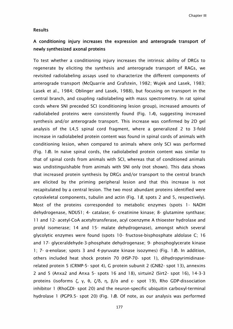
Chapter III
177
Results
A conditioning injury increases the expression and anterograde transport of
newly synthesized axonal proteins
To test whether a conditioning injury increases the intrinsic ability of DRGs to
regenerate by eliciting the synthesis and anterograde transport of RAGs, we
revisited radiolabeling assays used to characterize the different components of
anterograde transport (McQuarrie and Grafstein, 1982; Wujek and Lasek, 1983;
Lasek et al., 1984; Oblinger and Lasek, 1988), but focusing on transport in the
central branch, and coupling radiolabeling with mass spectrometry. In rat spinal
cords where SNI preceded SCI (conditioning lesion group), increased amounts of
radiolabeled proteins were consistently found (Fig. 1A), suggesting increased
synthesis and/or anterograde transport. This increase was confirmed by 2D gel
analysis of the L4,5 spinal cord fragment, where a generalized 2 to 3-fold
increase in radiolabeled protein content was found in spinal cords of animals with
conditioning lesion, when compared to animals where only SCI was performed
(Fig. 1B). In naïve spinal cords, the radiolabeled protein content was similar to
that of spinal cords from animals with SCI, whereas that of conditioned animals
was undistinguishable from animals with SNI only (not shown). This data shows
that increased protein synthesis by DRGs and/or transport to the central branch
are elicited by the priming peripheral lesion and that this increase is not
recapitulated by a central lesion. The two most abundant proteins identified were
cytoskeletal components, tubulin and actin (Fig. 1B, spots 2 and 5, respectively).
Most of the proteins corresponded to metabolic enzymes (spots 1- NADH
dehydrogenase, NDUS1; 4- catalase; 6- creatinine kinase; 8- glutamine synthase;
11 and 12- acetyl-CoA acetyltransferase, acyl coenzyme A thioester hydrolase and
prolyl isomerase; 14 and 15- malate dehydrogenase), amongst which several
glycolytic enzymes were found (spots 10- fructose-bisphosphate aldolase C; 16
and 17- glyceraldehyde-3-phosphate dehydrogenase; 9- phosphoglycerate kinase
1; 7- α-enolase; spots 3 and 4-pyruvate kinase isozymes) (Fig. 1B). In addition,
others included heat shock protein 70 (HSP-70- spot 1), dihydropyrimidinase-
related protein 5 (CRMP-5- spot 4), G protein subunit 2 (GNB2- spot 13), annexins
2 and 5 (Anxa2 and Anxa 5- spots 16 and 18), sirtuin2 (Sirt2- spot 16), 14-3-3
proteins (isoforms ζ, γ, θ, ζ/δ, η, β/α and ε- spot 19), Rho GDP-dissociation
inhibitor 1 (RhoGDI- spot 20) and the neuron-specific ubiquitin carboxyl-terminal
hydrolase 1 (PGP9.5- spot 20) (Fig. 1B). Of note, as our analysis was performed
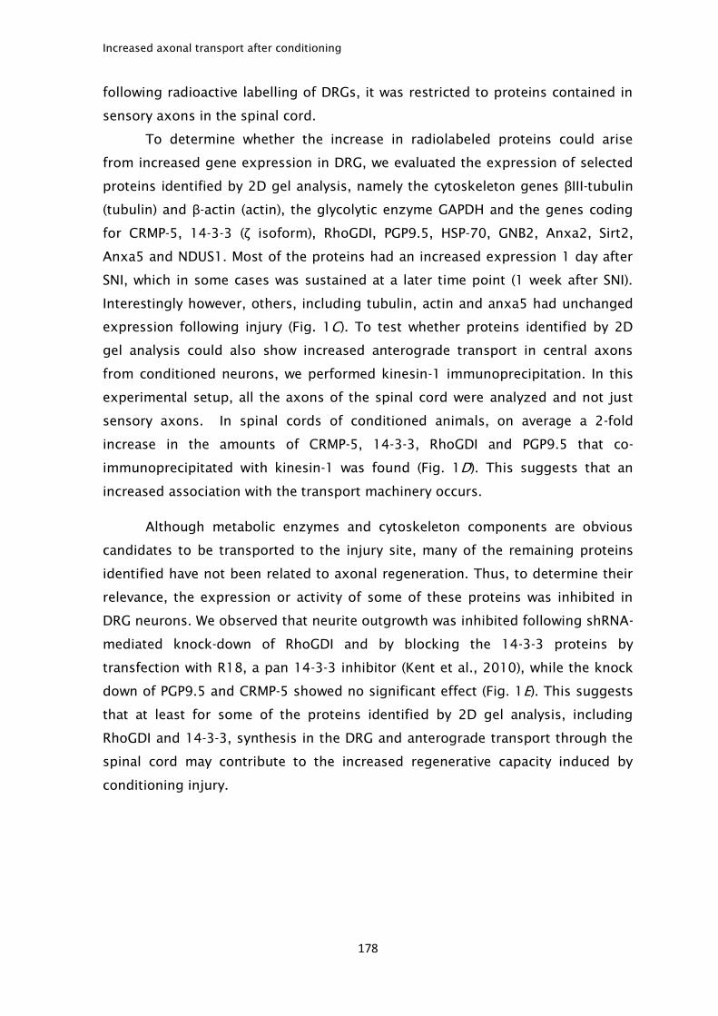
Increased axonal transport after conditioning
178
following radioactive labelling of DRGs, it was restricted to proteins contained in
sensory axons in the spinal cord.
To determine whether the increase in radiolabeled proteins could arise
from increased gene expression in DRG, we evaluated the expression of selected
proteins identified by 2D gel analysis, namely the cytoskeleton genes βIII-tubulin
(tubulin) and β-actin (actin), the glycolytic enzyme GAPDH and the genes coding
for CRMP-5, 14-3-3 (ζ isoform), RhoGDI, PGP9.5, HSP-70, GNB2, Anxa2, Sirt2,
Anxa5 and NDUS1. Most of the proteins had an increased expression 1 day after
SNI, which in some cases was sustained at a later time point (1 week after SNI).
Interestingly however, others, including tubulin, actin and anxa5 had unchanged
expression following injury (Fig. 1C). To test whether proteins identified by 2D
gel analysis could also show increased anterograde transport in central axons
from conditioned neurons, we performed kinesin-1 immunoprecipitation. In this
experimental setup, all the axons of the spinal cord were analyzed and not just
sensory axons. In spinal cords of conditioned animals, on average a 2-fold
increase in the amounts of CRMP-5, 14-3-3, RhoGDI and PGP9.5 that co-
immunoprecipitated with kinesin-1 was found (Fig. 1D). This suggests that an
increased association with the transport machinery occurs.
Although metabolic enzymes and cytoskeleton components are obvious
candidates to be transported to the injury site, many of the remaining proteins
identified have not been related to axonal regeneration. Thus, to determine their
relevance, the expression or activity of some of these proteins was inhibited in
DRG neurons. We observed that neurite outgrowth was inhibited following shRNA-
mediated knock-down of RhoGDI and by blocking the 14-3-3 proteins by
transfection with R18, a pan 14-3-3 inhibitor (Kent et al., 2010), while the knock
down of PGP9.5 and CRMP-5 showed no significant effect (Fig. 1E). This suggests
that at least for some of the proteins identified by 2D gel analysis, including
RhoGDI and 14-3-3, synthesis in the DRG and anterograde transport through the
spinal cord may contribute to the increased regenerative capacity induced by
conditioning injury.
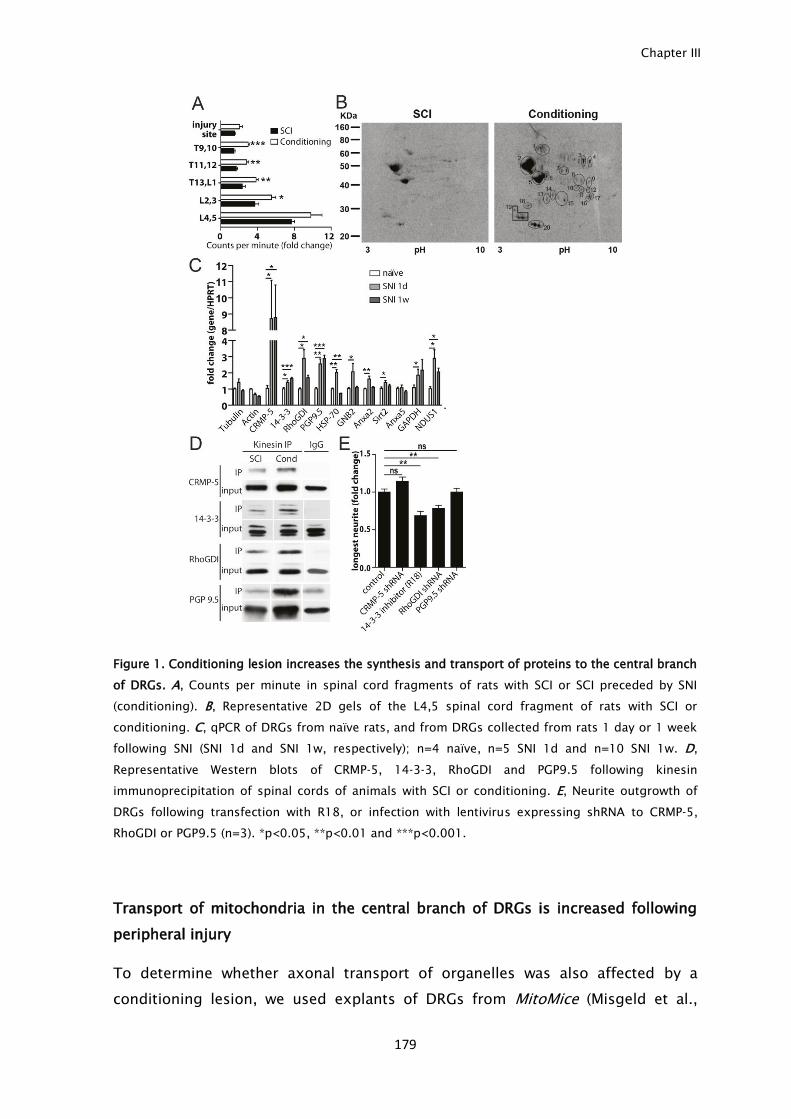
Chapter III
179
Figure 1. Conditioning lesion increases the synthesis and transport of proteins to the central branch
of DRGs. A, Counts per minute in spinal cord fragments of rats with SCI or SCI preceded by SNI
(conditioning). B, Representative 2D gels of the L4,5 spinal cord fragment of rats with SCI or
conditioning. C, qPCR of DRGs from naïve rats, and from DRGs collected from rats 1 day or 1 week
following SNI (SNI 1d and SNI 1w, respectively); n=4 naïve, n=5 SNI 1d and n=10 SNI 1w. D,
Representative Western blots of CRMP-5, 14-3-3, RhoGDI and PGP9.5 following kinesin
immunoprecipitation of spinal cords of animals with SCI or conditioning. E, Neurite outgrowth of
DRGs following transfection with R18, or infection with lentivirus expressing shRNA to CRMP-5,
RhoGDI or PGP9.5 (n=3). *p<0.05, **p<0.01 and ***p<0.001.
Transport of mitochondria in the central branch of DRGs is increased following
peripheral injury
To determine whether axonal transport of organelles was also affected by a
conditioning lesion, we used explants of DRGs from MitoMice (Misgeld et al.,
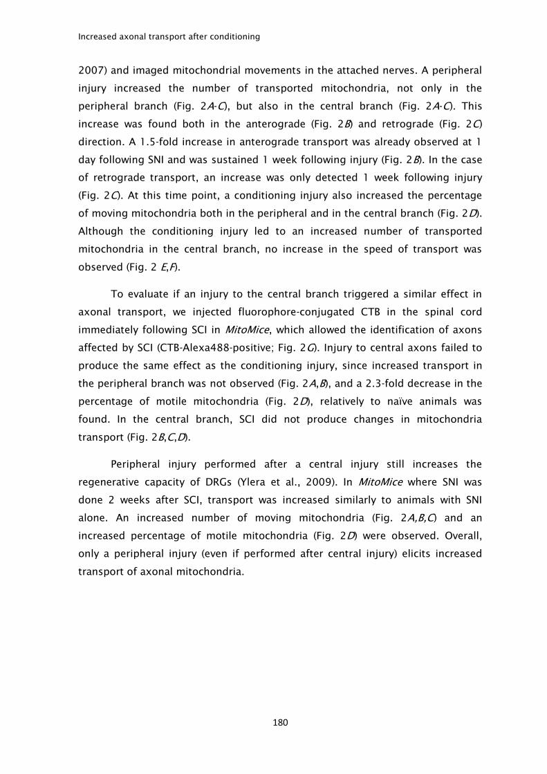
Increased axonal transport after conditioning
180
2007) and imaged mitochondrial movements in the attached nerves. A peripheral
injury increased the number of transported mitochondria, not only in the
peripheral branch (Fig. 2A-C), but also in the central branch (Fig. 2A-C). This
increase was found both in the anterograde (Fig. 2B) and retrograde (Fig. 2C)
direction. A 1.5-fold increase in anterograde transport was already observed at 1
day following SNI and was sustained 1 week following injury (Fig. 2B). In the case
of retrograde transport, an increase was only detected 1 week following injury
(Fig. 2C). At this time point, a conditioning injury also increased the percentage
of moving mitochondria both in the peripheral and in the central branch (Fig. 2D).
Although the conditioning injury led to an increased number of transported
mitochondria in the central branch, no increase in the speed of transport was
observed (Fig. 2 E,F).
To evaluate if an injury to the central branch triggered a similar effect in
axonal transport, we injected fluorophore-conjugated CTB in the spinal cord
immediately following SCI in MitoMice, which allowed the identification of axons
affected by SCI (CTB-Alexa488-positive; Fig. 2G). Injury to central axons failed to
produce the same effect as the conditioning injury, since increased transport in
the peripheral branch was not observed (Fig. 2A,B), and a 2.3-fold decrease in the
percentage of motile mitochondria (Fig. 2D), relatively to naïve animals was
found. In the central branch, SCI did not produce changes in mitochondria
transport (Fig. 2B,C,D).
Peripheral injury performed after a central injury still increases the
regenerative capacity of DRGs (Ylera et al., 2009). In MitoMice where SNI was
done 2 weeks after SCI, transport was increased similarly to animals with SNI
alone. An increased number of moving mitochondria (Fig. 2A,B,C) and an
increased percentage of motile mitochondria (Fig. 2D) were observed. Overall,
only a peripheral injury (even if performed after central injury) elicits increased
transport of axonal mitochondria.
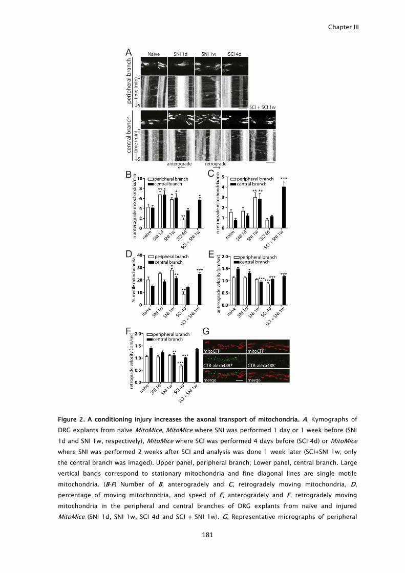
Chapter III
181
Figure 2. A conditioning injury increases the axonal transport of mitochondria. A, Kymographs of
DRG explants from naïve MitoMice, MitoMice where SNI was performed 1 day or 1 week before (SNI
1d and SNI 1w, respectively), MitoMice where SCI was performed 4 days before (SCI 4d) or MitoMice
where SNI was performed 2 weeks after SCI and analysis was done 1 week later (SCI+SNI 1w; only
the central branch was imaged). Upper panel, peripheral branch; Lower panel, central branch. Large
vertical bands correspond to stationary mitochondria and fine diagonal lines are single motile
mitochondria. (B-F) Number of B, anterogradely and C, retrogradely moving mitochondria, D,
percentage of moving mitochondria, and speed of E, anterogradely and F, retrogradely moving
mitochondria in the peripheral and central branches of DRG explants from naïve and injured
MitoMice (SNI 1d, SNI 1w, SCI 4d and SCI + SNI 1w). G, Representative micrographs of peripheral
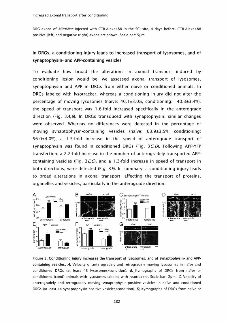
Increased axonal transport after conditioning
182
DRG axons of MitoMice injected with CTB-Alexa488 in the SCI site, 4 days before. CTB-Alexa488
positive (left) and negative (right) axons are shown. Scale bar: 5μm.
In DRGs, a conditioning injury leads to increased transport of lysosomes, and of
synaptophysin- and APP-containing vesicles
To evaluate how broad the alterations in axonal transport induced by
conditioning lesion would be, we assessed axonal transport of lysosomes,
synaptophysin and APP in DRGs from either naïve or conditioned animals. In
DRGs labeled with lysotracker, whereas a conditioning injury did not alter the
percentage of moving lysosomes (naïve: 40.1±3.0%, conditioning: 40.3±3.4%),
the speed of transport was 1.6-fold increased specifically in the anterograde
direction (Fig. 3A,B). In DRGs transduced with synaptophysin, similar changes
were observed. Whereas no differences were detected in the percentage of
moving synaptophysin-containing vesicles (naïve: 63.9±3.5%, conditioning:
56.0±4.0%), a 1.5-fold increase in the speed of anterograde transport of
synaptophysin was found in conditioned DRGs (Fig. 3C,D). Following APP-YFP
transfection, a 2.2-fold increase in the number of anterogradely transported APP-
containing vesicles (Fig. 3E,G), and a 1.3-fold increase in speed of transport in
both directions, were detected (Fig. 3F). In summary, a conditioning injury leads
to broad alterations in axonal transport, affecting the transport of proteins,
organelles and vesicles, particularly in the anterograde direction.
Figure 3. Conditioning injury increases the transport of lysosomes, and of synaptophysin- and APP-
containing vesicles. A, Velocity of anterogradely and retrogradely moving lysosomes in naïve and
conditioned DRGs (at least 48 lysosomes/condition). B, Kymographs of DRGs from naïve or
conditioned (cond) animals with lysosomes labeled with lysotracker. Scale bar: 2µm. C, Velocity of
anterogradely and retrogradely moving synaptophysin-positive vesicles in naïve and conditioned
DRGs (at least 44 synaptophysin-positive vesicles/condition). D, Kymographs of DRGs from naïve or
Chapter III
183
conditioned (cond) animals transduced with synaptophysin-GFP. Scale bar: 2µm. E, Number of
anterogradely and retrogradely moving APP-positive vesicles in naïve and conditioned DRGs. F,
Velocity of anterogradely and retrogradely moving APP-positive vesicles in naïve and conditioned
DRGs (at least 42 APP-positive vesicles/condition). G, Kymographs of DRGs from naïve or
conditioned animals transfected with APP-YFP. Scale bar: 2µm. *p<0.05, **p<0.01 and ***p<0.001.
Increased levels of molecular motors and of polyglutamylated tubulin can
underlie the early increase in axonal transport elicited by a conditioning lesion
To understand the mechanism underlying the increased axonal transport in the
central branch of conditioned DRGs, we analyzed whether conditioning would
elicit alterations previously associated to the control of axonal transport, namely
in molecular motor availability and suitable microtubule tracks. In DRGs, SNI was
accompanied by 1.5- and 1.8-fold increased RNA levels of kinesin-1B and dynein-
1, respectively, as early as 1 day following SNI (Fig. 4A). In the case of kinesin-1B,
this increase was not sustained, but dynein-1 was still increased at 1 week
following SNI (Fig. 4A). At the protein level, only kinesin-1 was significantly
increased in the dorsal root 1 day following SNI but not 1 week following SNI (Fig.
4B). The anterograde transport of cargoes can be regulated by phosphorylation of
kinesin light chain 1 (KLC1), as increased phosphorylation leads to disruption of
cargoes from the transport complex (Morfini et al., 2004). However, decreased
levels of dephosphorylated KLC1 were present in dorsal roots 1 week after SNI
(Fig. 4C), failing to explain increased anterograde transport.
Since tubulin modifications can alter the binding affinity to microtubules,
we tested whether changes in tubulin in the dorsal root could explain the
conditioning-induced transport alterations. Tyrosination of tubulin is a marker of
dynamic microtubules and can inhibit kinesin-1 binding to microtubules (Dunn et
al., 2008). Interestingly, increased levels of tyrosinated tubulin were found 1 day
and 1 week following conditioning injury (Fig. 4D,E), suggesting a more dynamic
state of dorsal root microtubules. Tubulin acetylation has been associated with
increased anterograde transport given improved kinesin-1 binding to
microtubules (Reed et al., 2006). However, no alterations in tubulin acetylation
were found (Fig. 4D,F). As kinesin-1 also binds more effectively to
polyglutamylated tubulin (Ikegami et al., 2007), this modification was
subsequently evaluated. Indeed, 1 day following conditioning injury,
polyglutamylated tubulin was increased in dorsal roots (Fig. 4D,G). However, this
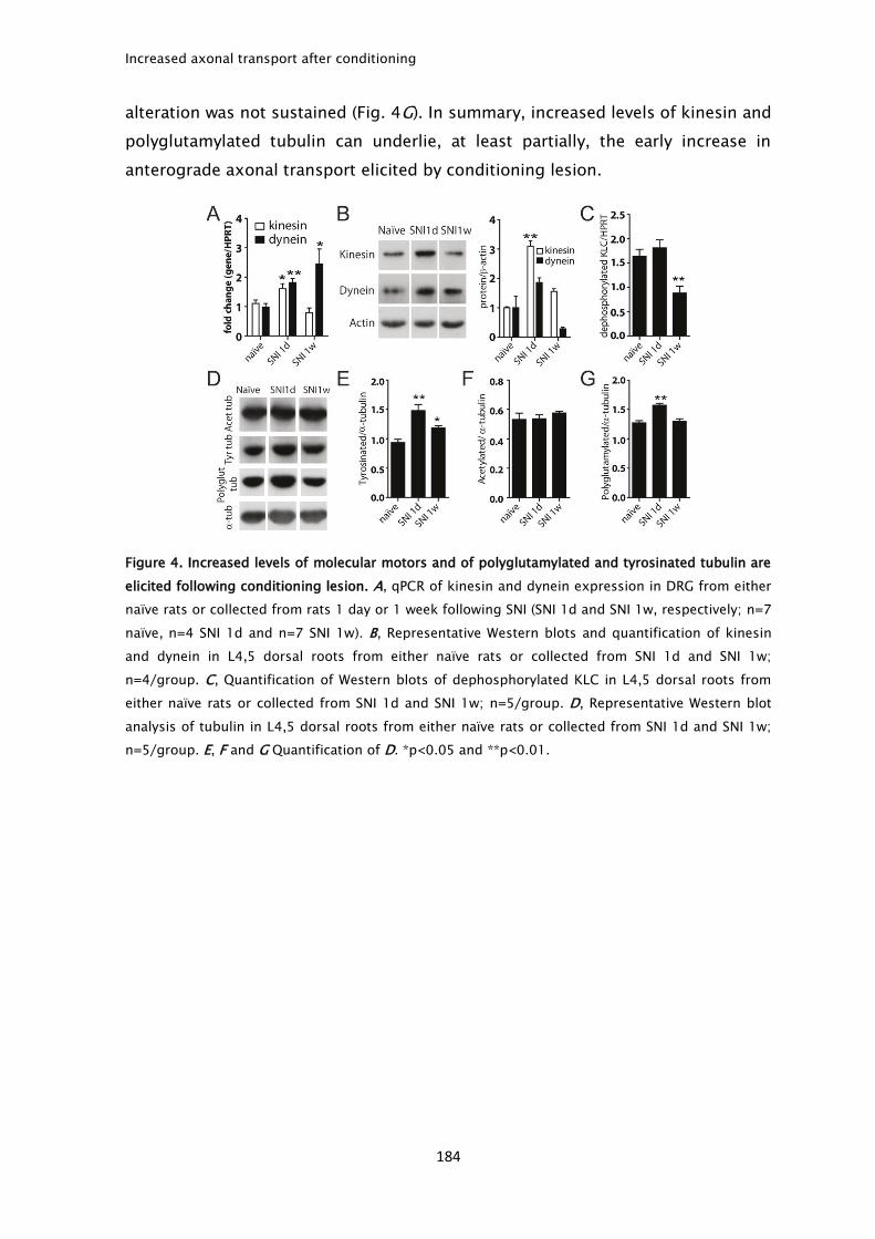
Increased axonal transport after conditioning
184
alteration was not sustained (Fig. 4G). In summary, increased levels of kinesin and
polyglutamylated tubulin can underlie, at least partially, the early increase in
anterograde axonal transport elicited by conditioning lesion.
Figure 4. Increased levels of molecular motors and of polyglutamylated and tyrosinated tubulin are
elicited following conditioning lesion. A, qPCR of kinesin and dynein expression in DRG from either
naïve rats or collected from rats 1 day or 1 week following SNI (SNI 1d and SNI 1w, respectively; n=7
naïve, n=4 SNI 1d and n=7 SNI 1w). B, Representative Western blots and quantification of kinesin
and dynein in L4,5 dorsal roots from either naïve rats or collected from SNI 1d and SNI 1w;
n=4/group. C, Quantification of Western blots of dephosphorylated KLC in L4,5 dorsal roots from
either naïve rats or collected from SNI 1d and SNI 1w; n=5/group. D, Representative Western blot
analysis of tubulin in L4,5 dorsal roots from either naïve rats or collected from SNI 1d and SNI 1w;
n=5/group. E, F and G Quantification of D. *p<0.05 and **p<0.01.
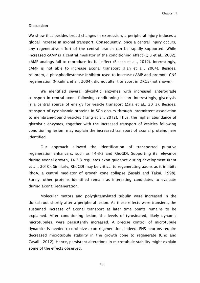
Chapter III
185
Discussion
We show that besides broad changes in expression, a peripheral injury induces a
global increase in axonal transport. Consequently, once a central injury occurs,
any regenerative effort of the central branch can be rapidly supported. While
increased cAMP is a central mediator of the conditioning effect (Qiu et al., 2002),
cAMP analogs fail to reproduce its full effect (Blesch et al., 2012). Interestingly,
cAMP is not able to increase axonal transport (Han et al., 2004). Besides,
rolipram, a phosphodiesterase inhibitor used to increase cAMP and promote CNS
regeneration (Nikulina et al., 2004), did not alter transport in DRGs (not shown).
We identified several glycolytic enzymes with increased anterograde
transport in central axons following conditioning lesion. Interestingly, glycolysis
is a central source of energy for vesicle transport (Zala et al., 2013). Besides,
transport of cytoplasmic proteins in SCb occurs through intermittent association
to membrane-bound vesicles (Tang et al., 2012). Thus, the higher abundance of
glycolytic enzymes, together with the increased transport of vesicles following
conditioning lesion, may explain the increased transport of axonal proteins here
identified.
Our approach allowed the identification of transported putative
regeneration enhancers, such as 14-3-3 and RhoGDI. Supporting its relevance
during axonal growth, 14-3-3 regulates axon guidance during development (Kent
et al., 2010). Similarly, RhoGDI may be critical to regenerating axons as it inhibits
RhoA, a central mediator of growth cone collapse (Sasaki and Takai, 1998).
Surely, other proteins identified remain as interesting candidates to evaluate
during axonal regeneration.
Molecular motors and polyglutamylated tubulin were increased in the
dorsal root shortly after a peripheral lesion. As these effects were transient, the
sustained increase of axonal transport at later time points remains to be
explained. After conditioning lesion, the levels of tyrosinated, likely dynamic
microtubules, were persistently increased. A precise control of microtubule
dynamics is needed to optimize axon regeneration. Indeed, PNS neurons require
decreased microtubule stability in the growth cone to regenerate (Cho and
Cavalli, 2012). Hence, persistent alterations in microtubule stability might explain
some of the effects observed.
Increased axonal transport after conditioning
186
Our work stressed that understanding the intersection between injury-
induced signaling and axonal transport is an important aspect to consider. In
summary, we propose that a broad increase in axonal transport is a major player
during axon regeneration.
Chapter III
187
Acknowledgments
This work was funded by FEDER through COMPETE and by National funds through
FCT –Fundação para a Ciência e a Tecnologia under the Project FCOMP-01-0124-
FEDER-017455 (HMSP ICT/0020/2010). Mar FM and Leite S were supported by
FCT (SFRH/BD/43484/2008 and SFRH/BD/72240/2010) and Teixeira CA by
Programa Ciência, funded by POPH-QREN and MCTES. We thank the help of Dr.
Vitor Costa (IBMC) with 2D gels and Dr. Paula Sampaio (IBMC) with confocal
microscopy. Misgeld T was supported by the Deutsche Forschungsgemeinschaft
(Center for Integrated Protein Science Munich, EXC 114, and Munich Center for
Systems Neurology, EXC 1010).
Conflicts of interest: The authors declare no competing financial interests.

188
Conclusion and perspectives
189
General conclusion and future perspectives
SCI is a severe condition that usually results in loss of sensitive and motor
function, as well as autonomic functions, culminating in lifetime medical care, as
the available treatments are limited. As such, the development of novel therapies
that may lead to improvements in the patient quality of life are needed. For that,
our basic knowledge on both extrinsic and intrinsic mechanisms controlling axon
growth and regeneration need to be expanded.
Initially, the limited regeneration of CNS axons was attributed mainly to the highly
inhibitory environment formed following SCI. Upon injury, the glial scar functions
as a physical barrier, but also as a biological barrier, since it contains numerous
axonal regeneration inhibitors, namely myelin proteins and CSPGs. We were able
to identify novel axonal regeneration inhibitors present in the injury milieu.
Specifically we determined that besides myelin proteins, myelin lipids are able to
inhibit axonal growth, particularly cholesterol and sphingomyelin. The use of 2-
hydroxypropyl-β-cyclodextrin, a drug capable of sequestering cholesterol and
sphingomyelin, in vivo, allowed increased axonal regeneration of dorsal column
fibers following spinal cord hemisection. It will be interesting to determine now
whether 2-hydroxypropyl-β-cyclodextrin is able to promote axonal regeneration
of other axonal tracts and to produce functional recovery.
Although several studies have shown improved axonal regeneration by blocking
inhibitors present in the glial scar, such results are still shy from a major effect
that could be translated to clinical trials. Recently, modifying the intrinsic ability
of CNS neurons emerged has an interesting target to promote regeneration. While
PNS neurons are able to mount a strong regenerative response following injury,
CNS neurons usually fail to produce such response. A key aspect of the ability of
PNS neurons to respond to injury is the generation of injury signals upon lesion.
Whether these signals are present following a CNS injury is not known. Here, we
used the dorsal root injury as a model of CNS injury, since it produces only a mild
response to injury and does not result in axon regeneration. Surprisingly, ERK,
JNK and STAT-3, the best described injury signals in the PNS, were also activated
and retrogradely transported following dorsal root injury. To identify possible
differences in the injury signaling mechanism following peripheral and dorsal
Conclusion and perspectives
190
root injury, we used an antibody microarray to analyze the axoplasm content
upon each lesion. We were able to determine that a dorsal root injury elicits the
differential activation of signaling molecules. From these, ROCK-II and Hsp-40
were increased following dorsal root injury whereas GSK3β was decreased after
sciatic nerve injury. ROCK-II and GSK3β are known inhibitors of axonal
regeneration. Our results, especially for ROCK-II, suggest that following dorsal
root injury besides transport of positive injury signals, there is also the transport
of signals that may repress regeneration. Hsp-40 has been associated with
protection in neurodegenerative diseases. Its knock down in neurons showed
signs of toxicity, suggesting that the increase in Hsp-40 following dorsal root
injury is probably not a direct player in the regenerative process but instead,
participates in protection mechanisms.
The conditioning injury model has been extensively used to dissect the increase
in the intrinsic ability triggered by a peripheral injury. By in vivo radiolabeling we
identified newly synthesized proteins that are anterogradely transported upon
conditioning injury. From these, several potential novel RAGs were identified,
from which we were able to validate RhoGDI and 14-3-3 as playing a role in
neurite outgrowth. There are certainly other candidates identified in our
screening that are worth studying in the context of axon growth and
regeneration.
Furthermore, our data demonstrated that a conditioning lesion elicited the
transport of newly synthesized proteins, organelles and synaptic vesicles. These
transport alterations were independent of motor levels, however the changes
observed in microtubule post-translation modifications, such as tyrosination and
polyglutamylation could at least in part explain the increased axonal transport. At
this stage, the molecular mechanisms controlling the increased transport should
be further dissected. Although the levels of the motors are similar, following
injury is there an increase in the percentage of motile motors? Is the increased
transport due to higher run lengths and decreased pauses among run lengths?
Does tyrosination and polyglutamylation increase motors binding and transport?
Answering these questions could help to further understand the mechanisms that
participate in improving axonal transport. Besides, whether increased axonal
transport is a general feature of conditions where increased axonal regeneration
occurs, as is the case for instance in PTEN and SOCS-3 KO mice, should be further
investigated. Combined, this knowledge may allow the future targeting of the

Conclusion and perspectives
191
transport machinery as a means of promoting axonal regeneration. Also, it would
improve treatment of several neurodegenerative diseases where defficient axonal
transport is present.
In summary, we were able to identify novel extrinsic cues that can impair axonal
growth such as cholesterol and sphingomyelin, but also novel mechanisms that
can contribute to increase axonal regeneration following conditioning injury,
namely a robust increase in axonal transport of proteins, mitochondria and
vesicles.

192

193
References

194
References
195
Abe N, Cavalli V (2008) Nerve injury signaling. Curr Opin Neurobiol 18:276-283.
Abi-Mosleh L, Infante RE, Radhakrishnan A, Goldstein JL, Brown MS (2009)
Cyclodextrin overcomes deficient lysosome-to-endoplasmic reticulum transport
of cholesterol in Niemann-Pick type C cells. Proc Natl Acad Sci USA 106:19316-
19321.
Al-Majed AA, Tam SL, Gordon T (2004) Electrical stimulation accelerates and
enhances expression of regeneration-associated genes in regenerating rat
femoral motoneurons. Cell Mol Neurobiol 24:379-402.
Alabed YZ, Grados-Munro E, Ferraro GB, Hsieh SH, Fournier AE (2006) Neuronal
responses to myelin are mediated by rho kinase. J Neurochem 96:1616-1625.
Alabed YZ, Pool M, Ong Tone S, Sutherland C, Fournier AE (2010) GSK3 beta
regulates myelin-dependent axon outgrowth inhibition through CRMP4. J
Neurosci 30:5635-5643.
Ambron RT, Schmied R, Huang CC, Smedman M (1992) A signal sequence
mediates the retrograde transport of proteins from the axon periphery to the cell
body and then into the nucleus. J Neurosci 12:2813-2818.
Ambron RT, Dulin MF, Zhang XP, Schmied R, Walters ET (1995) Axoplasm
enriched in a protein mobilized by nerve injury induces memory-like alterations
in Aplysia neurons. J Neurosci 15:3440-3446.
Ambron RT, Zhang XP, Gunstream JD, Povelones M, Walters ET (1996) Intrinsic
injury signals enhance growth, survival, and excitability of Aplysia neurons. J
Neurosci 16:7469-7477.
Andrews H, White K, Thomson C, Edgar J, Bates D, Griffiths I, Turnbull D, Nichols
P (2006) Increased axonal mitochondrial activity as an adaptation to myelin
deficiency in the Shiverer mouse. J Neurosci Res 83:1533-1539.
Aqul A, Liu B, Ramirez CM, Pieper AA, Estill SJ, Burns DK, Liu B, Repa JJ, Turley SD,
Dietschy JM (2011) Unesterified cholesterol accumulation in late
References
196
endosomes/lysosomes causes neurodegeneration and is prevented by driving
cholesterol export from this compartment. J Neurosci 31:9404-9413.
Aricescu AR, McKinnell IW, Halfter W, Stoker AW (2002) Heparan sulfate
proteoglycans are ligands for receptor protein tyrosine phosphatase sigma. Mol
Cell Biol 22:1881-1892.
Atwal JK, Pinkston-Gosse J, Syken J, Stawicki S, Wu Y, Shatz C, Tessier-Lavigne M
(2008) PirB is a functional receptor for myelin inhibitors of axonal regeneration.
Science 322:967-970.
Avellino AM, Hart D, Dailey AT, MacKinnon M, Ellegala D, Kliot M (1995)
Differential macrophage responses in the peripheral and central nervous system
during wallerian degeneration of axons. Exp Neurol 136:183-198.
Baldwin MR, Barbieri JT (2009) Association of botulinum neurotoxins with
synaptic vesicle protein complexes. Toxicon 54:570-574.
Bareyre FM, Garzorz N, Lang C, Misgeld T, Buning H, Kerschensteiner M (2011) In
vivo imaging reveals a phase-specific role of STAT3 during central and peripheral
nervous system axon regeneration. Proc Natl Acad Sci U S A 108:6282-6287.
Barnat M, Enslen H, Propst F, Davis RJ, Soares S, Nothias F (2010) Distinct roles of
c-Jun N-terminal kinase isoforms in neurite initiation and elongation during
axonal regeneration. J Neurosci 30:7804-7816.
Barnes AP, Polleux F (2009) Establishment of axon-dendrite polarity in
developing neurons. Annu Rev Neurosci 32:347-381.
Bartsch U, Bandtlow CE, Schnell L, Bartsch S, Spillmann AA, Rubin BP, Hillenbrand
R, Montag D, Schwab ME, Schachner M (1995) Lack of evidence that myelin-
associated glycoprotein is a major inhibitor of axonal regeneration in the CNS.
Neuron 15:1375-1381.
References
197
Bassell GJ, Kelic S (2004) Binding proteins for mRNA localization and local
translation, and their dysfunction in genetic neurological disease. Curr Opin
Neurobiol 14:574-581.
Bassell GJ, Zhang H, Byrd AL, Femino AM, Singer RH, Taneja KL, Lifshitz LM,
Herman IM, Kosik KS (1998) Sorting of beta-actin mRNA and protein to neurites
and growth cones in culture. J Neurosci 18:251-265.
Beggah AT, Dours-Zimmermann MT, Barras FM, Brosius A, Zimmermann DR, Zurn
AD (2005) Lesion-induced differential expression and cell association of
Neurocan, Brevican, Versican V1 and V2 in the mouse dorsal root entry zone.
Neuroscience 133:749-762.
Beirowski B, Adalbert R, Wagner D, Grumme DS, Addicks K, Ribchester RR,
Coleman MP (2005) The progressive nature of Wallerian degeneration in wild-type
and slow Wallerian degeneration (WldS) nerves. BMC Neurosci 6:6.
Ben-Tov Perry R, Doron-Mandel E, Iavnilovitch E, Rishal I, Dagan SY, Tsoory M,
Coppola G, McDonald MK, Gomes C, Geschwind DH et al. (2012) Subcellular
knockout of importin beta1 perturbs axonal retrograde signaling. Neuron
75:294-305.
Ben-Yaakov K, Dagan SY, Segal-Ruder Y, Shalem O, Vuppalanchi D, Willis DE,
Yudin D, Rishal I, Rother F, Bader M et al. (2012) Axonal transcription factors
signal retrogradely in lesioned peripheral nerve. EMBO J 31:1350-1363.
Benowitz LI, Popovich PG (2011) Inflammation and axon regeneration. Curr Opin
Neurol 24:577-583.
Benson MD, Romero MI, Lush ME, Lu QR, Henkemeyer M, Parada LF (2005)
Ephrin-B3 is a myelin-based inhibitor of neurite outgrowth. Proc Natl Acad Sci U S
A 102:10694-10699.
References
198
Bhatheja K, Field J (2006) Schwann cells: origins and role in axonal maintenance
and regeneration. The international journal of biochemistry & cell biology
38:1995-1999.
Bijur GN, Jope RS (2003) Glycogen synthase kinase-3 beta is highly activated in
nuclei and mitochondria. Neuroreport 14:2415-2419.
Bisby MA, Chen S (1990) Delayed wallerian degeneration in sciatic nerves of
C57BL/Ola mice is associated with impaired regeneration of sensory axons. Brain
Res 530:117-120.
Blesch A, Lu P, Tsukada S, Alto LT, Roet K, Coppola G, Geschwind D, Tuszynski
MH (2012) Conditioning lesions before or after spinal cord injury recruit broad
genetic mechanisms that sustain axonal regeneration: superiority to camp-
mediated effects. Exp Neurol 235:162-173.
Boato F, Hendrix S, Huelsenbeck SC, Hofmann F, Grosse G, Djalali S,
Klimaschewski L, Auer M, Just I, Ahnert-Hilger G et al. (2010) C3 peptide
enhances recovery from spinal cord injury by improved regenerative growth of
descending fiber tracts. J Cell Sci 123:1652-1662.
Bodine-Fowler SC, Meyer RS, Moskovitz A, Abrams R, Botte MJ (1997) Inaccurate
projection of rat soleus motoneurons: a comparison of nerve repair techniques.
Muscle Nerve 20:29-37.
Boggs JM (2006) Myelin basic protein: a multifunctional protein. Cell Mol Life Sci
63:1945-1961.
Bolin LM, Verity AN, Silver JE, Shooter EM, Abrams JS (1995) Interleukin-6
production by Schwann cells and induction in sciatic nerve injury. J Neurochem
64:850-858.
Bomze HM, Bulsara KR, Iskandar BJ, Caroni P, Skene JH (2001) Spinal axon
regeneration evoked by replacing two growth cone proteins in adult neurons. Nat
Neurosci 4:38-43.
References
199
Bouldin TW, Earnhardt TS, Goines ND (1991) Restoration of blood-nerve barrier in
neuropathy is associated with axonal regeneration and remyelination. J
Neuropathol Exp Neurol 50:719-728.
Boulter E, Garcia-Mata R, Guilluy C, Dubash A, Rossi G, Brennwald PJ, Burridge K
(2010) Regulation of Rho GTPase crosstalk, degradation and activity by RhoGDI1.
Nat Cell Biol 12:477-483.
Bracken MB (2001) Methylprednisolone and acute spinal cord injury: an update of
the randomized evidence. Spine (Phila Pa 1976) 26:S47-54.
Bradbury EJ, Khemani S, Von R, King, Priestley JV, McMahon SB (1999) NT-3
promotes growth of lesioned adult rat sensory axons ascending in the dorsal
columns of the spinal cord. The European journal of neuroscience 11:3873-3883.
Bradbury EJ, Moon LD, Popat RJ, King VR, Bennett GS, Patel PN, Fawcett JW,
McMahon SB (2002) Chondroitinase ABC promotes functional recovery after spinal
cord injury. Nature 416:636-640.
Bradke F, Dotti CG (1999) The role of local actin instability in axon formation.
Science 283:1931-1934.
Bradke F, Fawcett JW, Spira ME (2012) Assembly of a new growth cone after
axotomy: the precursor to axon regeneration. Nature reviews 13:183-193.
Brady ST, Witt AS, Kirkpatrick LL, de Waegh SM, Readhead C, Tu PH, Lee VM
(1999) Formation of compact myelin is required for maturation of the axonal
cytoskeleton. The Journal of neuroscience : the official journal of the Society for
Neuroscience 19:7278-7288.
Bregman BS, McAtee M, Dai HN, Kuhn PL (1997) Neurotrophic factors increase
axonal growth after spinal cord injury and transplantation in the adult rat. Exp
Neurol 148:475-494.
References
200
Bretzner F, Plemel JR, Liu J, Richter M, Roskams AJ, Tetzlaff W (2010) Combination
of olfactory ensheathing cells with local versus systemic cAMP treatment after a
cervical rubrospinal tract injury. J Neurosci Res 88:2833-2846.
Bridgman PC (2004) Myosin-dependent transport in neurons. J Neurobiol 58:164-
174.
Brosamle C, Huber AB, Fiedler M, Skerra A, Schwab ME (2000) Regeneration of
lesioned corticospinal tract fibers in the adult rat induced by a recombinant,
humanized IN-1 antibody fragment. J Neurosci 20:8061-8068.
Brown A (2003) Axonal transport of membranous and nonmembranous cargoes:
a unified perspective. J Cell Biol 160:817-821.
Brown MC, Perry VH, Lunn ER, Gordon S, Heumann R (1991) Macrophage
dependence of peripheral sensory nerve regeneration: possible involvement of
nerve growth factor. Neuron 6:359-370.
Bruck W, Friede RL (1990) Anti-macrophage CR3 antibody blocks myelin
phagocytosis by macrophages in vitro. Acta Neuropathol 80:415-418.
Brushart TM, Jari R, Verge V, Rohde C, Gordon T (2005) Electrical stimulation
restores the specificity of sensory axon regeneration. Exp Neurol 194:221-229.
Brushart TM, Hoffman PN, Royall RM, Murinson BB, Witzel C, Gordon T (2002)
Electrical stimulation promotes motoneuron regeneration without increasing its
speed or conditioning the neuron. J Neurosci 22:6631-6638.
Bundesen LQ, Scheel TA, Bregman BS, Kromer LF (2003) Ephrin-B2 and EphB2
regulation of astrocyte-meningeal fibroblast interactions in response to spinal
cord lesions in adult rats. J Neurosci 23:7789-7800.
Cafferty WB, Duffy P, Huebner E, Strittmatter SM (2010) MAG and OMgp synergize
with Nogo-A to restrict axonal growth and neurological recovery after spinal cord
trauma. J Neurosci 30:6825-6837.
References
201
Cafferty WB, Gardiner NJ, Das P, Qiu J, McMahon SB, Thompson SW (2004)
Conditioning injury-induced spinal axon regeneration fails in interleukin-6
knock-out mice. J Neurosci 24:4432-4443.
Cafferty WB, Gardiner NJ, Gavazzi I, Powell J, McMahon SB, Heath JK, Munson J,
Cohen J, Thompson SW (2001) Leukemia inhibitory factor determines the growth
status of injured adult sensory neurons. J Neurosci 21:7161-7170.
Caggiano AO, Zimber MP, Ganguly A, Blight AR, Gruskin EA (2005) Chondroitinase
ABCI improves locomotion and bladder function following contusion injury of the
rat spinal cord. J Neurotrauma 22:226-239.
Cai D, Shen Y, De Bellard M, Tang S, Filbin MT (1999) Prior exposure to
neurotrophins blocks inhibition of axonal regeneration by MAG and myelin via a
cAMP-dependent mechanism. Neuron 22:89-101.
Cai D, Qiu J, Cao Z, McAtee M, Bregman BS, Filbin MT (2001) Neuronal cyclic AMP
controls the developmental loss in ability of axons to regenerate. J Neurosci
21:4731-4739.
Cai D, Deng K, Mellado W, Lee J, Ratan RR, Filbin MT (2002) Arginase I and
polyamines act downstream from cyclic AMP in overcoming inhibition of axonal
growth MAG and myelin in vitro. Neuron 35:711-719.
Campbell DS, Holt CE (2001) Chemotropic responses of retinal growth cones
mediated by rapid local protein synthesis and degradation. Neuron 32:1013-
1026.
Cao Y, Shumsky JS, Sabol MA, Kushner RA, Strittmatter S, Hamers FP, Lee DH,
Rabacchi SA, Murray M (2008) Nogo-66 receptor antagonist peptide (NEP1-40)
administration promotes functional recovery and axonal growth after lateral
funiculus injury in the adult rat. Neurorehabil Neural Repair 22:262-278.
Cao Z, Gao Y, Bryson JB, Hou J, Chaudhry N, Siddiq M, Martinez J, Spencer T,
Carmel J, Hart RB et al. (2006) The cytokine interleukin-6 is sufficient but not

References
202
necessary to mimic the peripheral conditioning lesion effect on axonal growth. J
Neurosci 26:5565-5573.
Caroni P, Schwab ME (1988) Antibody against myelin-associated inhibitor of
neurite growth neutralizes nonpermissive substrate properties of CNS white
matter. Neuron 1:85-96.
Cavalli V, Kujala P, Klumperman J, Goldstein LS (2005) Sunday Driver links axonal
transport to damage signaling. J Cell Biol 168:775-787.
Chaudhry V, Cornblath DR (1992) Wallerian degeneration in human nerves: serial
electrophysiological studies. Muscle Nerve 15:687-693.
Chen L, Wang Z, Ghosh-Roy A, Hubert T, Yan D, O'Rourke S, Bowerman B, Wu Z,
Jin Y, Chisholm AD (2011) Axon regeneration pathways identified by systematic
genetic screening in C. elegans. Neuron 71:1043-1057.
Chen MS, Huber AB, van der Haar ME, Frank M, Schnell L, Spillmann AA, Christ F,
Schwab ME (2000) Nogo-A is a myelin-associated neurite outgrowth inhibitor and
an antigen for monoclonal antibody IN-1. Nature 403:434-439.
Chen ZL, Strickland S (2003) Laminin gamma1 is critical for Schwann cell
differentiation, axon myelination, and regeneration in the peripheral nerve. J Cell
Biol 163:889-899.
Cherezov V, Rosenbaum DM, Hanson MA, Rasmussen SG, Thian FS, Kobilka TS,
Choi HJ, Kuhn P, Weis WI, Kobilka BK et al. (2007) High-resolution crystal
structure of an engineered human beta2-adrenergic G protein-coupled receptor.
Science 318:1258-1265.
Chevalier-Larsen E, Holzbaur EL (2006) Axonal transport and neurodegenerative
disease. Biochim Biophys Acta 1762:1094-1108.
Chierzi S, Ratto GM, Verma P, Fawcett JW (2005) The ability of axons to
regenerate their growth cones depends on axonal type and age, and is regulated
by calcium, cAMP and ERK. The European journal of neuroscience 21:2051-2062.
References
203
Cho Y, Cavalli V (2012) HDAC5 is a novel injury-regulated tubulin deacetylase
controlling axon regeneration. EMBO J 31:3063-3078.
Cho Y, Sloutsky R, Naegle KM, Cavalli V (2013) Injury-induced HDAC5 nuclear
export is essential for axon regeneration. Cell 155:894-908.
Christie KJ, Webber CA, Martinez JA, Singh B, Zochodne DW (2010) PTEN
inhibition to facilitate intrinsic regenerative outgrowth of adult peripheral axons. J
Neurosci 30:9306-9315.
Coleman WP, Benzel D, Cahill DW, Ducker T, Geisler F, Green B, Gropper MR,
Goffin J, Madsen PW, 3rd, Maiman DJ et al. (2000) A critical appraisal of the
reporting of the National Acute Spinal Cord Injury Studies (II and III) of
methylprednisolone in acute spinal cord injury. J Spinal Disord 13:185-199.
Conrad S, Genth H, Hofmann F, Just I, Skutella T (2007) Neogenin-RGMa signaling
at the growth cone is bone morphogenetic protein-independent and involves
RhoA, ROCK, and PKC. J Biol Chem 282:16423-16433.
Cornbrooks CJ, Carey DJ, McDonald JA, Timpl R, Bunge RP (1983) In vivo and in
vitro observations on laminin production by Schwann cells. Proc Natl Acad Sci U S
A 80:3850-3854.
Costigan M, Befort K, Karchewski L, Griffin RS, D'Urso D, Allchorne A, Sitarski J,
Mannion JW, Pratt RE, Woolf CJ (2002) Replicate high-density rat genome
oligonucleotide microarrays reveal hundreds of regulated genes in the dorsal root
ganglion after peripheral nerve injury. BMC Neurosci 3:16.
Court FA, Hendriks WT, MacGillavry HD, Alvarez J, van Minnen J (2008) Schwann
cell to axon transfer of ribosomes: toward a novel understanding of the role of
glia in the nervous system. J Neurosci 28:11024-11029.
Curtis R, Scherer SS, Somogyi R, Adryan KM, Ip NY, Zhu Y, Lindsay RM, DiStefano
PS (1994) Retrograde axonal transport of LIF is increased by peripheral nerve
References
204
injury: correlation with increased LIF expression in distal nerve. Neuron 12:191-
204.
David S, Aguayo AJ (1981) Axonal elongation into peripheral nervous system
"bridges" after central nervous system injury in adult rats. Science 214:931-933.
Davidson CD, Ali NF, Micsenyi MC, Stephney G, Renault S, Dobrenis K, Ory DS,
Vanier MT, Walkley SU (2009) Chronic cyclodextrin treatment of murine Niemann-
Pick C disease ameliorates neuronal cholesterol and glycosphingolipid storage
and disease progression. PloS one 4:e6951.
Davis RJ (2000) Signal transduction by the JNK group of MAP kinases. Cell
103:239-252.
de Lima S, Koriyama Y, Kurimoto T, Oliveira JT, Yin Y, Li Y, Gilbert HY, Fagiolini M,
Martinez AM, Benowitz L (2012) Full-length axon regeneration in the adult mouse
optic nerve and partial recovery of simple visual behaviors. Proc Natl Acad Sci U S
A 109:9149-9154.
de Ruiter GC, Malessy MJ, Alaid AO, Spinner RJ, Engelstad JK, Sorenson EJ,
Kaufman KR, Dyck PJ, Windebank AJ (2008) Misdirection of regenerating motor
axons after nerve injury and repair in the rat sciatic nerve model. Exp Neurol
211:339-350.
De Winter F, Oudega M, Lankhorst AJ, Hamers FP, Blits B, Ruitenberg MJ,
Pasterkamp RJ, Gispen WH, Verhaagen J (2002) Injury-induced class 3 semaphorin
expression in the rat spinal cord. Exp Neurol 175:61-75.
DeBellard ME, Tang S, Mukhopadhyay G, Shen YJ, Filbin MT (1996) Myelin-
associated glycoprotein inhibits axonal regeneration from a variety of neurons via
interaction with a sialoglycoprotein. Mol Cell Neurosci 7:89-101.
Delcroix JD, Valletta JS, Wu C, Hunt SJ, Kowal AS, Mobley WC (2003) NGF signaling
in sensory neurons: evidence that early endosomes carry NGF retrograde signals.
Neuron 39:69-84.
References
205
Deng K, He H, Qiu J, Lorber B, Bryson JB, Filbin MT (2009) Increased synthesis of
spermidine as a result of upregulation of arginase I promotes axonal regeneration
in culture and in vivo. J Neurosci 29:9545-9552.
Dent EW, Callaway JL, Szebenyi G, Baas PW, Kalil K (1999) Reorganization and
movement of microtubules in axonal growth cones and developing interstitial
branches. The Journal of neuroscience : the official journal of the Society for
Neuroscience 19:8894-8908.
Dergham P, Ellezam B, Essagian C, Avedissian H, Lubell WD, McKerracher L (2002)
Rho signaling pathway targeted to promote spinal cord repair. J Neurosci
22:6570-6577.
Devor M (1999) Unexplained peculiarities of the dorsal root ganglion. Pain Suppl
6:S27-35.
Di Maio A, Skuba A, Himes BT, Bhagat SL, Hyun JK, Tessler A, Bishop D, Son YJ
(2011) In vivo imaging of dorsal root regeneration: rapid immobilization and
presynaptic differentiation at the CNS/PNS border. J Neurosci 31:4569-4582.
Diamond J, Foerster A, Holmes M, Coughlin M (1992) Sensory nerves in adult rats
regenerate and restore sensory function to the skin independently of endogenous
NGF. J Neurosci 12:1467-1476.
Dickendesher TL, Baldwin KT, Mironova YA, Koriyama Y, Raiker SJ, Askew KL,
Wood A, Geoffroy CG, Zheng B, Liepmann CD et al. (2012) NgR1 and NgR3 are
receptors for chondroitin sulfate proteoglycans. Nat Neurosci 15:703-712.
Dill J, Wang H, Zhou F, Li S (2008) Inactivation of glycogen synthase kinase 3
promotes axonal growth and recovery in the CNS. J Neurosci 28:8914-8928.
Domeniconi M, Cao Z, Spencer T, Sivasankaran R, Wang K, Nikulina E, Kimura N,
Cai H, Deng K, Gao Y et al. (2002) Myelin-associated glycoprotein interacts with
the Nogo66 receptor to inhibit neurite outgrowth. Neuron 35:283-290.
References
206
Donnelly CJ, Park M, Spillane M, Yoo S, Pacheco A, Gomes C, Vuppalanchi D,
McDonald M, Kim HH, Merianda TT et al. (2013) Axonally synthesized beta-actin
and GAP-43 proteins support distinct modes of axonal growth. J Neurosci
33:3311-3322.
Donnelly CJ, Willis DE, Xu M, Tep C, Jiang C, Yoo S, Schanen NC, Kirn-Safran CB,
van Minnen J, English A et al. (2011) Limited availability of ZBP1 restricts axonal
mRNA localization and nerve regeneration capacity. EMBO J 30:4665-4677.
Dotti CG, Sullivan CA, Banker GA (1988) The establishment of polarity by
hippocampal neurons in culture. J Neurosci 8:1454-1468.
Dou CL, Levine JM (1994) Inhibition of neurite growth by the NG2 chondroitin
sulfate proteoglycan. J Neurosci 14:7616-7628.
Duffy P, Schmandke A, Schmandke A, Sigworth J, Narumiya S, Cafferty WB,
Strittmatter SM (2009) Rho-associated kinase II (ROCKII) limits axonal growth
after trauma within the adult mouse spinal cord. J Neurosci 29:15266-15276.
Duffy P, Wang X, Siegel CS, Tu N, Henkemeyer M, Cafferty WB, Strittmatter SM
(2012) Myelin-derived ephrinB3 restricts axonal regeneration and recovery after
adult CNS injury. Proc Natl Acad Sci USA 109:5063-5068.
Dunn S, Morrison EE, Liverpool TB, Molina-Paris C, Cross RA, Alonso MC,
Peckham M (2008) Differential trafficking of Kif5c on tyrosinated and
detyrosinated microtubules in live cells. J Cell Sci 121:1085-1095.
Dyck PJ, Lambert EH, Wood MB, Linscheid RL (1988) Assessment of nerve
regeneration and adaptation after median nerve reconnection and digital
neurovascular flap transfer. Neurology 38:1586-1591.
Enes J, Langwieser N, Ruschel J, Carballosa-Gonzalez MM, Klug A, Traut MH, Ylera
B, Tahirovic S, Hofmann F, Stein V et al. (2010) Electrical activity suppresses axon
growth through Ca(v)1.2 channels in adult primary sensory neurons. Curr Biol
20:1154-1164.
References
207
Eng H, Lund K, Campenot RB (1999) Synthesis of beta-tubulin, actin, and other
proteins in axons of sympathetic neurons in compartmented cultures. J Neurosci
19:1-9.
English AW (2005) Enhancing axon regeneration in peripheral nerves also
increases functionally inappropriate reinnervation of targets. J Comp Neurol
490:427-441.
Erez H, Malkinson G, Prager-Khoutorsky M, De Zeeuw CI, Hoogenraad CC, Spira
ME (2007) Formation of microtubule-based traps controls the sorting and
concentration of vesicles to restricted sites of regenerating neurons after
axotomy. J Cell Biol 176:497-507.
Erturk A, Hellal F, Enes J, Bradke F (2007) Disorganized microtubules underlie the
formation of retraction bulbs and the failure of axonal regeneration. J Neurosci
27:9169-9180.
Evans PJ, Bain JR, Mackinnon SE, Makino AP, Hunter DA (1991) Selective
reinnervation: a comparison of recovery following microsuture and conduit nerve
repair. Brain Res 559:315-321.
Fabes J, Anderson P, Brennan C, Bolsover S (2007) Regeneration-enhancing
effects of EphA4 blocking peptide following corticospinal tract injury in adult rat
spinal cord. The European journal of neuroscience 26:2496-2505.
Fabes J, Anderson P, Yanez-Munoz RJ, Thrasher A, Brennan C, Bolsover S (2006)
Accumulation of the inhibitory receptor EphA4 may prevent regeneration of
corticospinal tract axons following lesion. The European journal of neuroscience
23:1721-1730.
Fan CY, Lee S, Cyr DM (2003) Mechanisms for regulation of Hsp70 function by
Hsp40. Cell stress & chaperones 8:309-316.
References
208
Fan G, Merritt SE, Kortenjann M, Shaw PE, Holzman LB (1996) Dual leucine zipper-
bearing kinase (DLK) activates p46SAPK and p38mapk but not ERK2. J Biol Chem
271:24788-24793.
Farrar MJ, Bernstein IM, Schlafer DH, Cleland TA, Fetcho JR, Schaffer CB (2012)
Chronic in vivo imaging in the mouse spinal cord using an implanted chamber.
Nat Methods 9:297-302.
Fee D, Crumbaugh A, Jacques T, Herdrich B, Sewell D, Auerbach D, Piaskowski S,
Hart MN, Sandor M, Fabry Z (2003) Activated/effector CD4+ T cells exacerbate
acute damage in the central nervous system following traumatic injury. Journal of
neuroimmunology 136:54-66.
Feng G, Mellor RH, Bernstein M, Keller-Peck C, Nguyen QT, Wallace M, Nerbonne
JM, Lichtman JW, Sanes JR (2000) Imaging neuronal subsets in transgenic mice
expressing multiple spectral variants of GFP. Neuron 28:41-51.
Fenrich KK, Weber P, Hocine M, Zalc M, Rougon G, Debarbieux F (2012) Long-
term in vivo imaging of normal and pathological mouse spinal cord with
subcellular resolution using implanted glass windows. J Physiol 590:3665-3675.
Fernandes KJ, Fan DP, Tsui BJ, Cassar SL, Tetzlaff W (1999) Influence of the
axotomy to cell body distance in rat rubrospinal and spinal motoneurons:
differential regulation of GAP-43, tubulins, and neurofilament-M. J Comp Neurol
414:495-510.
Figueroa JD, Benton RL, Velazquez I, Torrado AI, Ortiz CM, Hernandez CM, Diaz JJ,
Magnuson DS, Whittemore SR, Miranda JD (2006) Inhibition of EphA7 up-
regulation after spinal cord injury reduces apoptosis and promotes locomotor
recovery. J Neurosci Res 84:1438-1451.
Fitch MT, Silver J (2008) CNS injury, glial scars, and inflammation: Inhibitory
extracellular matrices and regeneration failure. Exp Neurol 209:294-301.
References
209
Fleming CE, Mar FM, Franquinho F, Saraiva MJ, Sousa MM (2009) Transthyretin
internalization by sensory neurons is megalin mediated and necessary for its
neuritogenic activity. J Neurosci 29:3220-3232.
Folch J, Lees M, Sloane Stanley GH (1957) A simple method for the isolation and
purification of total lipides from animal tissues. The Journal of biological
chemistry 226:497-509.
Fontana X, Hristova M, Da Costa C, Patodia S, Thei L, Makwana M, Spencer-Dene
B, Latouche M, Mirsky R, Jessen KR et al. (2012) c-Jun in Schwann cells promotes
axonal regeneration and motoneuron survival via paracrine signaling. J Cell Biol
198:127-141.
Fournier AE, GrandPre T, Strittmatter SM (2001) Identification of a receptor
mediating Nogo-66 inhibition of axonal regeneration. Nature 409:341-346.
Friedlander DR, Milev P, Karthikeyan L, Margolis RK, Margolis RU, Grumet M
(1994) The neuronal chondroitin sulfate proteoglycan neurocan binds to the
neural cell adhesion molecules Ng-CAM/L1/NILE and N-CAM, and inhibits
neuronal adhesion and neurite outgrowth. J Cell Biol 125:669-680.
Galtrey CM, Fawcett JW (2007) The role of chondroitin sulfate proteoglycans in
regeneration and plasticity in the central nervous system. Brain Res Rev 54:1-18.
Gao Y, Nikulina E, Mellado W, Filbin MT (2003) Neurotrophins elevate cAMP to
reach a threshold required to overcome inhibition by MAG through extracellular
signal-regulated kinase-dependent inhibition of phosphodiesterase. J Neurosci
23:11770-11777.
Gao Y, Deng K, Hou J, Bryson JB, Barco A, Nikulina E, Spencer T, Mellado W,
Kandel ER, Filbin MT (2004) Activated CREB is sufficient to overcome inhibitors in
myelin and promote spinal axon regeneration in vivo. Neuron 44:609-621.
Garcia-Alias G, Petrosyan HA, Schnell L, Horner PJ, Bowers WJ, Mendell LM,
Fawcett JW, Arvanian VL (2011) Chondroitinase ABC combined with neurotrophin
References
210
NT-3 secretion and NR2D expression promotes axonal plasticity and functional
recovery in rats with lateral hemisection of the spinal cord. J Neurosci 31:17788-
17799.
Gaudet AD, Popovich PG, Ramer MS (2011) Wallerian degeneration: gaining
perspective on inflammatory events after peripheral nerve injury. J
Neuroinflammation 8:110.
Genovese T, Mazzon E, Crisafulli C, Di Paola R, Muia C, Esposito E, Bramanti P,
Cuzzocrea S (2008) TNF-alpha blockage in a mouse model of SCI: evidence for
improved outcome. Shock (Augusta, Ga 29:32-41.
George EB, Glass JD, Griffin JW (1995) Axotomy-induced axonal degeneration is
mediated by calcium influx through ion-specific channels. J Neurosci 15:6445-
6452.
George R, Griffin JW (1994) Delayed macrophage responses and myelin clearance
during Wallerian degeneration in the central nervous system: the dorsal
radiculotomy model. Exp Neurol 129:225-236.
Ghosh-Roy A, Wu Z, Goncharov A, Jin Y, Chisholm AD (2010) Calcium and cyclic
AMP promote axonal regeneration in Caenorhabditis elegans and require DLK-1
kinase. J Neurosci 30:3175-3183.
Giger RJ, Hollis ER, 2nd, Tuszynski MH (2010) Guidance molecules in axon
regeneration. Cold Spring Harb Perspect Biol 2:a001867.
Gillespie LN (2003) Regulation of axonal growth and guidance by the
neurotrophin family of neurotrophic factors. Clin Exp Pharmacol Physiol 30:724-
733.
Giuditta A, Cupello A, Lazzarini G (1980) Ribosomal RNA in the axoplasm of the
squid giant axon. J Neurochem 34:1757-1760.
Giuditta A, Hunt T, Santella L (1986) Rapid important paper Messenger RNA in
squid axoplasm. Neurochem Int 8:435-442.
References
211
Giuditta A, Menichini E, Perrone Capano C, Langella M, Martin R, Castigli E, Kaplan
BB (1991) Active polysomes in the axoplasm of the squid giant axon. J Neurosci
Res 28:18-28.
Gold BG (1997) Axonal regeneration of sensory nerves is delayed by continuous
intrathecal infusion of nerve growth factor. Neuroscience 76:1153-1158.
Goldberg JL, Klassen MP, Hua Y, Barres BA (2002) Amacrine-signaled loss of
intrinsic axon growth ability by retinal ganglion cells. Science 296:1860-1864.
Goldshmit Y, Galea MP, Wise G, Bartlett PF, Turnley AM (2004) Axonal
regeneration and lack of astrocytic gliosis in EphA4-deficient mice. J Neurosci
24:10064-10073.
Gorlich D, Kutay U (1999) Transport between the cell nucleus and the cytoplasm.
Annu Rev Cell Dev Biol 15:607-660.
Grafstein B, Forman DS (1980) Intracellular transport in neurons. Physiol Rev
60:1167-1283.
GrandPre T, Li S, Strittmatter SM (2002) Nogo-66 receptor antagonist peptide
promotes axonal regeneration. Nature 417:547-551.
GrandPre T, Nakamura F, Vartanian T, Strittmatter SM (2000) Identification of the
Nogo inhibitor of axon regeneration as a Reticulon protein. Nature 403:439-444.
Gumy LF, Tan CL, Fawcett JW (2010) The role of local protein synthesis and
degradation in axon regeneration. Exp Neurol 223:28-37.
Gumy LF, Yeo GS, Tung YC, Zivraj KH, Willis D, Coppola G, Lam BY, Twiss JL, Holt
CE, Fawcett JW (2011) Transcriptome analysis of embryonic and adult sensory
axons reveals changes in mRNA repertoire localization. Rna 17:85-98.
Guzik BW, Goldstein LS (2004) Microtubule-dependent transport in neurons:
steps towards an understanding of regulation, function and dysfunction. Curr
Opin Cell Biol 16:443-450.
References
212
Hafezparast M, Klocke R, Ruhrberg C, Marquardt A, Ahmad-Annuar A, Bowen S,
Lalli G, Witherden AS, Hummerich H, Nicholson S et al. (2003) Mutations in dynein
link motor neuron degeneration to defects in retrograde transport. Science
300:808-812.
Hamilton SK, Hinkle ML, Nicolini J, Rambo LN, Rexwinkle AM, Rose SJ, Sabatier MJ,
Backus D, English AW (2011) Misdirection of regenerating axons and functional
recovery following sciatic nerve injury in rats. J Comp Neurol 519:21-33.
Han PJ, Shukla S, Subramanian PS, Hoffman PN (2004) Cyclic AMP elevates tubulin
expression without increasing intrinsic axon growth capacity. Exp Neurol
189:293-302.
Hanz S, Perlson E, Willis D, Zheng JQ, Massarwa R, Huerta JJ, Koltzenburg M,
Kohler M, van-Minnen J, Twiss JL et al. (2003) Axoplasmic importins enable
retrograde injury signaling in lesioned nerve. Neuron 40:1095-1104.
Hata K, Fujitani M, Yasuda Y, Doya H, Saito T, Yamagishi S, Mueller BK, Yamashita
T (2006) RGMa inhibition promotes axonal growth and recovery after spinal cord
injury. J Cell Biol 173:47-58.
Hayashi H, Campenot RB, Vance DE, Vance JE (2004) Glial lipoproteins stimulate
axon growth of central nervous system neurons in compartmented cultures. The
Journal of biological chemistry 279:14009-14015.
Hebel R, Stromberg MW (1986) Anatomy and embryology of the laboratory rat:
BioMed Verlag.
Heerssen HM, Pazyra MF, Segal RA (2004) Dynein motors transport activated Trks
to promote survival of target-dependent neurons. Nat Neurosci 7:596-604.
Hellal F, Hurtado A, Ruschel J, Flynn KC, Laskowski CJ, Umlauf M, Kapitein LC,
Strikis D, Lemmon V, Bixby J et al. (2011) Microtubule stabilization reduces
scarring and causes axon regeneration after spinal cord injury. Science 331:928-
931.
References
213
Heumann R, Korsching S, Bandtlow C, Thoenen H (1987) Changes of nerve growth
factor synthesis in nonneuronal cells in response to sciatic nerve transection. J
Cell Biol 104:1623-1631.
Hirata K, Mitoma H, Ueno N, He JW, Kawabuchi M (1999) Differential response of
macrophage subpopulations to myelin degradation in the injured rat sciatic
nerve. J Neurocytol 28:685-695.
Hirokawa N, Niwa S, Tanaka Y (2010) Molecular motors in neurons: transport
mechanisms and roles in brain function, development, and disease. Neuron
68:610-638.
Hirokawa N, Noda Y, Tanaka Y, Niwa S (2009) Kinesin superfamily motor proteins
and intracellular transport. Nat Rev Mol Cell Biol 10:682-696.
Hoffman PN (1989) Expression of GAP-43, a rapidly transported growth-
associated protein, and class II beta tubulin, a slowly transported cytoskeletal
protein, are coordinated in regenerating neurons. J Neurosci 9:893-897.
Hoffman PN (2010) A conditioning lesion induces changes in gene expression and
axonal transport that enhance regeneration by increasing the intrinsic growth
state of axons. Exp Neurol 223:11-18.
Hoffman PN, Luduena RF (1996) The axonal transport of beta III-tubulin is altered
in both branches of sensory axons after injury of the rat sciatic nerve. Brain Res
708:182-184.
Holtzman E, Novikoff AB (1965) Lysomes in the rat sciatic nerve following crush. J
Cell Biol 27:651-669.
Howard MJ, David G, Barrett JN (1999) Resealing of transected myelinated
mammalian axons in vivo: evidence for involvement of calpain. Neuroscience
93:807-815.
References
214
Hsieh SH, Ferraro GB, Fournier AE (2006) Myelin-associated inhibitors regulate
cofilin phosphorylation and neuronal inhibition through LIM kinase and Slingshot
phosphatase. J Neurosci 26:1006-1015.
Hu-Tsai M, Winter J, Emson PC, Woolf CJ (1994) Neurite outgrowth and GAP-43
mRNA expression in cultured adult rat dorsal root ganglion neurons: effects of
NGF or prior peripheral axotomy. J Neurosci Res 39:634-645.
Huebner EA, Strittmatter SM (2009) Axon regeneration in the peripheral and
central nervous systems. Results Probl Cell Differ 48:339-351.
Hurlbert RJ (2000) Methylprednisolone for acute spinal cord injury: an
inappropriate standard of care. J Neurosurg 93:1-7.
Ikegami K, Heier RL, Taruishi M, Takagi H, Mukai M, Shimma S, Taira S, Hatanaka
K, Morone N, Yao I et al. (2007) Loss of alpha-tubulin polyglutamylation in
ROSA22 mice is associated with abnormal targeting of KIF1A and modulated
synaptic function. Proc Natl Acad Sci U S A 104:3213-3218.
Irizarry-Ramirez M, Willson CA, Cruz-Orengo L, Figueroa J, Velazquez I, Jones H,
Foster RD, Whittemore SR, Miranda JD (2005) Upregulation of EphA3 receptor
after spinal cord injury. J Neurotrauma 22:929-935.
Jacob JM, McQuarrie IG (1996) Assembly of microfilaments and microtubules from
axonally transported actin and tubulin after axotomy. J Neurosci Res 43:412-419.
Jakeman LB, Reier PJ (1991) Axonal projections between fetal spinal cord
transplants and the adult rat spinal cord: a neuroanatomical tracing study of local
interactions. J Comp Neurol 307:311-334.
Jana NR, Tanaka M, Wang G, Nukina N (2000) Polyglutamine length-dependent
interaction of Hsp40 and Hsp70 family chaperones with truncated N-terminal
huntingtin: their role in suppression of aggregation and cellular toxicity. Human
molecular genetics 9:2009-2018.
References
215
Jankowski MP, Cornuet PK, McIlwrath S, Koerber HR, Albers KM (2006) SRY-box
containing gene 11 (Sox11) transcription factor is required for neuron survival
and neurite growth. Neuroscience 143:501-514.
Jenkins R, Hunt SP (1991) Long-term increase in the levels of c-jun mRNA and jun
protein-like immunoreactivity in motor and sensory neurons following axon
damage. Neurosci Lett 129:107-110.
Jessen KR, Mirsky R (2008) Negative regulation of myelination: relevance for
development, injury, and demyelinating disease. Glia 56:1552-1565.
Ji B, Case LC, Liu K, Shao Z, Lee X, Yang Z, Wang J, Tian T, Shulga-Morskaya S,
Scott M et al. (2008) Assessment of functional recovery and axonal sprouting in
oligodendrocyte-myelin glycoprotein (OMgp) null mice after spinal cord injury.
Mol Cell Neurosci 39:258-267.
Jones LL, Margolis RU, Tuszynski MH (2003) The chondroitin sulfate
proteoglycans neurocan, brevican, phosphacan, and versican are differentially
regulated following spinal cord injury. Exp Neurol 182:399-411.
Jung H, O'Hare CM, Holt CE (2011) Translational regulation in growth cones. Curr
Opin Genet Dev 21:458-464.
Jung H, Yoon BC, Holt CE (2012) Axonal mRNA localization and local protein
synthesis in nervous system assembly, maintenance and repair. Nature reviews
13:308-324.
Kadoya K, Tsukada S, Lu P, Coppola G, Geschwind D, Filbin MT, Blesch A,
Tuszynski MH (2009) Combined intrinsic and extrinsic neuronal mechanisms
facilitate bridging axonal regeneration one year after spinal cord injury. Neuron
64:165-172.
Kaech S, Banker G (2006) Culturing hippocampal neurons. Nat Protoc 1:2406-
2415.
References
216
Kaether C, Skehel P, Dotti CG (2000) Axonal membrane proteins are transported
in distinct carriers: a two-color video microscopy study in cultured hippocampal
neurons. Molecular biology of the cell 11:1213-1224.
Kalmar B, Burnstock G, Vrbova G, Urbanics R, Csermely P, Greensmith L (2002)
Upregulation of heat shock proteins rescues motoneurones from axotomy-
induced cell death in neonatal rats. Exp Neurol 176:87-97.
Kamber D, Erez H, Spira ME (2009) Local calcium-dependent mechanisms
determine whether a cut axonal end assembles a retarded endbulb or competent
growth cone. Exp Neurol 219:112-125.
Kaneko S, Iwanami A, Nakamura M, Kishino A, Kikuchi K, Shibata S, Okano HJ,
Ikegami T, Moriya A, Konishi O et al. (2006) A selective Sema3A inhibitor
enhances regenerative responses and functional recovery of the injured spinal
cord. Nat Med 12:1380-1389.
Kardon JR, Vale RD (2009) Regulators of the cytoplasmic dynein motor. Nat Rev
Mol Cell Biol 10:854-865.
Kent CB, Shimada T, Ferraro GB, Ritter B, Yam PT, McPherson PS, Charron F,
Kennedy TE, Fournier AE (2010) 14-3-3 proteins regulate protein kinase a activity
to modulate growth cone turning responses. J Neurosci 30:14059-14067.
Kerschensteiner M, Schwab ME, Lichtman JW, Misgeld T (2005) In vivo imaging of
axonal degeneration and regeneration in the injured spinal cord. Nat Med
11:572-577.
Key B, Lah GJ (2012) Repulsive guidance molecule A (RGMa): a molecule for all
seasons. Cell adhesion & migration 6:85-90.
Kieran D, Hafezparast M, Bohnert S, Dick JR, Martin J, Schiavo G, Fisher EM,
Greensmith L (2005) A mutation in dynein rescues axonal transport defects and
extends the life span of ALS mice. J Cell Biol 169:561-567.
References
217
Kikuchi K, Kishino A, Konishi O, Kumagai K, Hosotani N, Saji I, Nakayama C,
Kimura T (2003) In vitro and in vivo characterization of a novel semaphorin 3A
inhibitor, SM-216289 or xanthofulvin. J Biol Chem 278:42985-42991.
Kim JE, Li S, GrandPre T, Qiu D, Strittmatter SM (2003) Axon regeneration in
young adult mice lacking Nogo-A/B. Neuron 38:187-199.
Kirkpatrick LL, Witt AS, Payne HR, Shine HD, Brady ST (2001) Changes in
microtubule stability and density in myelin-deficient shiverer mouse CNS axons.
The Journal of neuroscience : the official journal of the Society for Neuroscience
21:2288-2297.
Kirstein M, Farinas I (2002) Sensing life: regulation of sensory neuron survival by
neurotrophins. Cell Mol Life Sci 59:1787-1802.
Kiryu-Seo S, Kiyama H (2011) The nuclear events guiding successful nerve
regeneration. Front Mol Neurosci 4:53.
Koenig E (1991) Evaluation of local synthesis of axonal proteins in the goldfish
Mauthner cell axon and axons of dorsal and ventral roots of the rat in vitro. Mol
Cell Neurosci 2:384-394.
Koenig E, Adams P (1982) Local protein synthesizing activity in axonal fields
regenerating in vitro. J Neurochem 39:386-400.
Koenig E, Martin R, Titmus M, Sotelo-Silveira JR (2000) Cryptic peripheral
ribosomal domains distributed intermittently along mammalian myelinated
axons. J Neurosci 20:8390-8400.
Kohler M, Speck C, Christiansen M, Bischoff FR, Prehn S, Haller H, Gorlich D,
Hartmann E (1999) Evidence for distinct substrate specificities of importin alpha
family members in nuclear protein import. Mol Cell Biol 19:7782-7791.
Konishi Y, Stegmuller J, Matsuda T, Bonni S, Bonni A (2004) Cdh1-APC controls
axonal growth and patterning in the mammalian brain. Science 303:1026-1030.
References
218
Kottis V, Thibault P, Mikol D, Xiao ZC, Zhang R, Dergham P, Braun PE (2002)
Oligodendrocyte-myelin glycoprotein (OMgp) is an inhibitor of neurite outgrowth.
J Neurochem 82:1566-1569.
Krause TL, Fishman HM, Ballinger ML, Bittner GD (1994) Extent and mechanism of
sealing in transected giant axons of squid and earthworms. J Neurosci 14:6638-
6651.
Kubo T, Yamashita T (2007) Rho-ROCK inhibitors for the treatment of CNS injury.
Recent Pat CNS Drug Discov 2:173-179.
Kun A, Otero L, Sotelo-Silveira JR, Sotelo JR (2007) Ribosomal distributions in
axons of mammalian myelinated fibers. J Neurosci Res 85:2087-2098.
Kury P, Abankwa D, Kruse F, Greiner-Petter R, Muller HW (2004) Gene expression
profiling reveals multiple novel intrinsic and extrinsic factors associated with
axonal regeneration failure. The European journal of neuroscience 19:32-42.
Kwon BK, Liu J, Lam C, Plunet W, Oschipok LW, Hauswirth W, Di Polo A, Blesch A,
Tetzlaff W (2007) Brain-derived neurotrophic factor gene transfer with adeno-
associated viral and lentiviral vectors prevents rubrospinal neuronal atrophy and
stimulates regeneration-associated gene expression after acute cervical spinal
cord injury. Spine (Phila Pa 1976) 32:1164-1173.
LaMonte BH, Wallace KE, Holloway BA, Shelly SS, Ascano J, Tokito M, Van Winkle T,
Howland DS, Holzbaur EL (2002) Disruption of dynein/dynactin inhibits axonal
transport in motor neurons causing late-onset progressive degeneration. Neuron
34:715-727.
Lang C, Bradley PM, Jacobi A, Kerschensteiner M, Bareyre FM (2013) STAT3
promotes corticospinal remodelling and functional recovery after spinal cord
injury. EMBO reports 14:931-937.
Lasek RJ, Dabrowski C, Nordlander R (1973) Analysis of axoplasmic RNA from
invertebrate giant axons. Nat New Biol 244:162-165.
References
219
Lasek RJ, Garner JA, Brady ST (1984) Axonal transport of the cytoplasmic matrix. J
Cell Biol 99:212s-221s.
Laskowski CJ, Bradke F (2012) In vivo imaging: A dynamic imaging approach to
study spinal cord regeneration. Exp Neurol.
Lasorella A, Stegmuller J, Guardavaccaro D, Liu G, Carro MS, Rothschild G, de la
Torre-Ubieta L, Pagano M, Bonni A, Iavarone A (2006) Degradation of Id2 by the
anaphase-promoting complex couples cell cycle exit and axonal growth. Nature
442:471-474.
Leah JD, Herdegen T, Bravo R (1991) Selective expression of Jun proteins
following axotomy and axonal transport block in peripheral nerves in the rat:
evidence for a role in the regeneration process. Brain Res 566:198-207.
Ledesma MD, Prinetti A, Sonnino S, Schuchman EH (2011) Brain pathology in
Niemann Pick disease type A: insights from the acid sphingomyelinase knockout
mice. Journal of neurochemistry 116:779-788.
Lee JK, Zheng B (2012) Role of myelin-associated inhibitors in axonal repair after
spinal cord injury. Exp Neurol 235:33-42.
Lee JK, Chan AF, Luu SM, Zhu Y, Ho C, Tessier-Lavigne M, Zheng B (2009)
Reassessment of corticospinal tract regeneration in Nogo-deficient mice. J
Neurosci 29:8649-8654.
Lee JK, Geoffroy CG, Chan AF, Tolentino KE, Crawford MJ, Leal MA, Kang B, Zheng
B (2010) Assessing spinal axon regeneration and sprouting in Nogo-, MAG-, and
OMgp-deficient mice. Neuron 66:663-670.
Lee N, Neitzel KL, Devlin BK, MacLennan AJ (2004) STAT3 phosphorylation in
injured axons before sensory and motor neuron nuclei: potential role for STAT3
as a retrograde signaling transcription factor. J Comp Neurol 474:535-545.
References
220
Lee YB, Yune TY, Baik SY, Shin YH, Du S, Rhim H, Lee EB, Kim YC, Shin ML,
Markelonis GJ et al. (2000) Role of tumor necrosis factor-alpha in neuronal and
glial apoptosis after spinal cord injury. Exp Neurol 166:190-195.
Lehmann M, Fournier A, Selles-Navarro I, Dergham P, Sebok A, Leclerc N, Tigyi G,
McKerracher L (1999) Inactivation of Rho signaling pathway promotes CNS axon
regeneration. J Neurosci 19:7537-7547.
Leung KM, van Horck FP, Lin AC, Allison R, Standart N, Holt CE (2006)
Asymmetrical beta-actin mRNA translation in growth cones mediates attractive
turning to netrin-1. Nat Neurosci 9:1247-1256.
Li S, Strittmatter SM (2003) Delayed systemic Nogo-66 receptor antagonist
promotes recovery from spinal cord injury. J Neurosci 23:4219-4227.
Li X, Masliah E, Reixach N, Buxbaum JN (2011) Neuronal production of
transthyretin in human and murine Alzheimer's disease: is it protective? J
Neurosci 31:12483-12490.
Li YC, Li YN, Cheng CX, Sakamoto H, Kawate T, Shimada O, Atsumi S (2005)
Subsurface cisterna-lined axonal invaginations and double-walled vesicles at the
axonal-myelin sheath interface. Neurosci Res 53:298-303.
Liebscher T, Schnell L, Schnell D, Scholl J, Schneider R, Gullo M, Fouad K, Mir A,
Rausch M, Kindler D et al. (2005) Nogo-A antibody improves regeneration and
locomotion of spinal cord-injured rats. Ann Neurol 58:706-719.
Ligon LA, Steward O (2000) Movement of mitochondria in the axons and
dendrites of cultured hippocampal neurons. J Comp Neurol 427:340-350.
Ligon LA, LaMonte BH, Wallace KE, Weber N, Kalb RG, Holzbaur EL (2005) Mutant
superoxide dismutase disrupts cytoplasmic dynein in motor neurons.
Neuroreport 16:533-536.
Lin AC, Holt CE (2008) Function and regulation of local axonal translation. Curr
Opin Neurobiol 18:60-68.
References
221
Lindwall C, Kanje M (2005) Retrograde axonal transport of JNK signaling
molecules influence injury induced nuclear changes in p-c-Jun and ATF3 in adult
rat sensory neurons. Mol Cell Neurosci 29:269-282.
Lindwall C, Dahlin L, Lundborg G, Kanje M (2004) Inhibition of c-Jun
phosphorylation reduces axonal outgrowth of adult rat nodose ganglia and dorsal
root ganglia sensory neurons. Mol Cell Neurosci 27:267-279.
Liu B, Turley SD, Burns DK, Miller AM, Repa JJ, Dietschy JM (2009) Reversal of
defective lysosomal transport in NPC disease ameliorates liver dysfunction and
neurodegeneration in the npc1-/- mouse. Proceedings of the National Academy
of Sciences of the United States of America 106:2377-2382.
Liu BP, Fournier A, GrandPre T, Strittmatter SM (2002) Myelin-associated
glycoprotein as a functional ligand for the Nogo-66 receptor. Science 297:1190-
1193.
Liu W, Chun E, Thompson AA, Chubukov P, Xu F, Katritch V, Han GW, Roth CB,
Heitman LH, AP IJ et al. (2012) Structural basis for allosteric regulation of GPCRs
by sodium ions. Science 337:232-236.
Liz MA, Leite SC, Juliano L, Saraiva MJ, Damas AM, Bur D, Sousa MM (2012)
Transthyretin is a metallopeptidase with an inducible active site. Biochem J
443:769-778.
Liz MA, Fleming CE, Nunes AF, Almeida MR, Mar FM, Choe Y, Craik CS, Powers JC,
Bogyo M, Sousa MM (2009) Substrate specificity of transthyretin: identification of
natural substrates in the nervous system. Biochem J 419:467-474.
Lu P, Tuszynski MH (2008) Growth factors and combinatorial therapies for CNS
regeneration. Exp Neurol 209:313-320.
Lu P, Jones LL, Tuszynski MH (2007) Axon regeneration through scars and into
sites of chronic spinal cord injury. Exp Neurol 203:8-21.
References
222
Lu P, Yang H, Jones LL, Filbin MT, Tuszynski MH (2004) Combinatorial therapy
with neurotrophins and cAMP promotes axonal regeneration beyond sites of
spinal cord injury. J Neurosci 24:6402-6409.
Lu P, Wang Y, Graham L, McHale K, Gao M, Wu D, Brock J, Blesch A, Rosenzweig
ES, Havton LA et al. (2012) Long-distance growth and connectivity of neural stem
cells after severe spinal cord injury. Cell 150:1264-1273.
Lubinska L (1977) Early course of Wallerian degeneration in myelinated fibres of
the rat phrenic nerve. Brain Res 130:47-63.
Lunn ER, Perry VH, Brown MC, Rosen H, Gordon S (1989) Absence of Wallerian
Degeneration does not Hinder Regeneration in Peripheral Nerve. The European
journal of neuroscience 1:27-33.
Luttges MW, Kelly PT, Gerren RA (1976) Degenerative changes in mouse sciatic
nerves: electrophoretic and electrophysiologic characterizations. Exp Neurol
50:706-733.
Ma TC, Campana A, Lange PS, Lee HH, Banerjee K, Bryson JB, Mahishi L, Alam S,
Giger RJ, Barnes S et al. (2010) A large-scale chemical screen for regulators of the
arginase 1 promoter identifies the soy isoflavone daidzeinas a clinically approved
small molecule that can promote neuronal protection or regeneration via a cAMP-
independent pathway. J Neurosci 30:739-748.
Mann F, Holt CE (2001) Control of retinal growth and axon divergence at the
chiasm: lessons from Xenopus. Bioessays 23:319-326.
Mar FM, Bonni A, Sousa MM (2014) Cell intrinsic control of axon regeneration.
EMBO reports 15:254-263.
Mason MR, Lieberman AR, Grenningloh G, Anderson PN (2002) Transcriptional
upregulation of SCG10 and CAP-23 is correlated with regeneration of the axons
of peripheral and central neurons in vivo. Mol Cell Neurosci 20:595-615.

References
223
Matsuo M, Togawa M, Hirabaru K, Mochinaga S, Narita A, Adachi M, Egashira M,
Irie T, Ohno K (2013) Effects of cyclodextrin in two patients with Niemann-Pick
Type C disease. Molecular genetics and metabolism 108:76-81.
McKeon RJ, Jurynec MJ, Buck CR (1999) The chondroitin sulfate proteoglycans
neurocan and phosphacan are expressed by reactive astrocytes in the chronic
CNS glial scar. J Neurosci 19:10778-10788.
McKerracher L, David S, Jackson DL, Kottis V, Dunn RJ, Braun PE (1994)
Identification of myelin-associated glycoprotein as a major myelin-derived
inhibitor of neurite growth. Neuron 13:805-811.
McQuarrie IG, Grafstein B (1982) Protein synthesis and axonal transport in
goldfish retinal ganglion cells during regeneration accelerated by a conditioning
lesion. Brain Res 251:25-37.
McQuarrie IG, Jacob JM (1991) Conditioning nerve crush accelerates cytoskeletal
protein transport in sprouts that form after a subsequent crush. J Comp Neurol
305:139-147.
Meyer M, Matsuoka I, Wetmore C, Olson L, Thoenen H (1992) Enhanced synthesis
of brain-derived neurotrophic factor in the lesioned peripheral nerve: different
mechanisms are responsible for the regulation of BDNF and NGF mRNA. J Cell Biol
119:45-54.
Mi S, Lee X, Shao Z, Thill G, Ji B, Relton J, Levesque M, Allaire N, Perrin S, Sands B
et al. (2004) LINGO-1 is a component of the Nogo-66 receptor/p75 signaling
complex. Nat Neurosci 7:221-228.
Miao T, Wu D, Zhang Y, Bo X, Subang MC, Wang P, Richardson PM (2006)
Suppressor of cytokine signaling-3 suppresses the ability of activated signal
transducer and activator of transcription-3 to stimulate neurite growth in rat
primary sensory neurons. J Neurosci 26:9512-9519.
References
224
Michaelevski I, Segal-Ruder Y, Rozenbaum M, Medzihradszky KF, Shalem O,
Coppola G, Horn-Saban S, Ben-Yaakov K, Dagan SY, Rishal I et al. (2010)
Signaling to transcription networks in the neuronal retrograde injury response.
Science signaling 3:ra53.
Milev P, Friedlander DR, Sakurai T, Karthikeyan L, Flad M, Margolis RK, Grumet M,
Margolis RU (1994) Interactions of the chondroitin sulfate proteoglycan
phosphacan, the extracellular domain of a receptor-type protein tyrosine
phosphatase, with neurons, glia, and neural cell adhesion molecules. J Cell Biol
127:1703-1715.
Miller SM (2008) Methylprednisolone in acute spinal cord injury: a tarnished
standard. J Neurosurg Anesthesiol 20:140-142.
Ming GL, Song HJ, Berninger B, Holt CE, Tessier-Lavigne M, Poo MM (1997) cAMP-
dependent growth cone guidance by netrin-1. Neuron 19:1225-1235.
Ming GL, Wong ST, Henley J, Yuan XB, Song HJ, Spitzer NC, Poo MM (2002)
Adaptation in the chemotactic guidance of nerve growth cones. Nature 417:411-
418.
Minor K, Phillips J, Seeds NW (2009) Tissue plasminogen activator promotes
axonal outgrowth on CNS myelin after conditioned injury. J Neurochem 109:706-
715.
Miranda CO, Teixeira CA, Liz MA, Sousa VF, Franquinho F, Forte G, Di Nardo P,
Pinto-Do OP, Sousa MM (2011) Systemic delivery of bone marrow-derived
mesenchymal stromal cells diminishes neuropathology in a mouse model of
Krabbe's disease. Stem cells 29:1738-1751.
Mirza SP, Halligan BD, Greene AS, Olivier M (2007) Improved method for the
analysis of membrane proteins by mass spectrometry. Physiol Genomics 30:89-
94.

References
225
Misgeld T, Kerschensteiner M, Bareyre FM, Burgess RW, Lichtman JW (2007)
Imaging axonal transport of mitochondria in vivo. Nat Methods 4:559-561.
Molteni R, Zheng JQ, Ying Z, Gomez-Pinilla F, Twiss JL (2004) Voluntary exercise
increases axonal regeneration from sensory neurons. Proc Natl Acad Sci U S A
101:8473-8478.
Monnier PP, Sierra A, Macchi P, Deitinghoff L, Andersen JS, Mann M, Flad M,
Hornberger MR, Stahl B, Bonhoeffer F et al. (2002) RGM is a repulsive guidance
molecule for retinal axons. Nature 419:392-395.
Moon LD, Asher RA, Rhodes KE, Fawcett JW (2001) Regeneration of CNS axons
back to their target following treatment of adult rat brain with chondroitinase
ABC. Nat Neurosci 4:465-466.
Moore DL, Blackmore MG, Hu Y, Kaestner KH, Bixby JL, Lemmon VP, Goldberg JL
(2009) KLF family members regulate intrinsic axon regeneration ability. Science
326:298-301.
Moreau-Fauvarque C, Kumanogoh A, Camand E, Jaillard C, Barbin G, Boquet I,
Love C, Jones EY, Kikutani H, Lubetzki C et al. (2003) The transmembrane
semaphorin Sema4D/CD100, an inhibitor of axonal growth, is expressed on
oligodendrocytes and upregulated after CNS lesion. J Neurosci 23:9229-9239.
Morfini G, Szebenyi G, Brown H, Pant HC, Pigino G, DeBoer S, Beffert U, Brady ST
(2004) A novel CDK5-dependent pathway for regulating GSK3 activity and
kinesin-driven motility in neurons. EMBO J 23:2235-2245.
Mueller BK, Mack H, Teusch N (2005) Rho kinase, a promising drug target for
neurological disorders. Nature reviews Drug discovery 4:387-398.
Mukhopadhyay G, Doherty P, Walsh FS, Crocker PR, Filbin MT (1994) A novel role
for myelin-associated glycoprotein as an inhibitor of axonal regeneration. Neuron
13:757-767.
References
226
Naveilhan P, ElShamy WM, Ernfors P (1997) Differential regulation of mRNAs for
GDNF and its receptors Ret and GDNFR alpha after sciatic nerve lesion in the
mouse. The European journal of neuroscience 9:1450-1460.
Neumann S, Woolf CJ (1999) Regeneration of dorsal column fibers into and
beyond the lesion site following adult spinal cord injury. Neuron 23:83-91.
Neumann S, Bradke F, Tessier-Lavigne M, Basbaum AI (2002) Regeneration of
sensory axons within the injured spinal cord induced by intraganglionic cAMP
elevation. Neuron 34:885-893.
Nikulina E, Tidwell JL, Dai HN, Bregman BS, Filbin MT (2004) The
phosphodiesterase inhibitor rolipram delivered after a spinal cord lesion
promotes axonal regeneration and functional recovery. Proc Natl Acad Sci U S A
101:8786-8790.
Nishiyama M, Hoshino A, Tsai L, Henley JR, Goshima Y, Tessier-Lavigne M, Poo
MM, Hong K (2003) Cyclic AMP/GMP-dependent modulation of Ca2+ channels
sets the polarity of nerve growth-cone turning. Nature 423:990-995.
Norton WT, Poduslo SE (1973) Myelination in rat brain: method of myelin
isolation. J Neurochem 21:749-757.
Oblinger MM, Lasek RJ (1988) Axotomy-induced alterations in the synthesis and
transport of neurofilaments and microtubules in dorsal root ganglion cells. J
Neurosci 8:1747-1758.
Oertle T, van der Haar ME, Bandtlow CE, Robeva A, Burfeind P, Buss A, Huber AB,
Simonen M, Schnell L, Brosamle C et al. (2003) Nogo-A inhibits neurite outgrowth
and cell spreading with three discrete regions. J Neurosci 23:5393-5406.
Oudega M, Hagg T (1996) Nerve growth factor promotes regeneration of sensory
axons into adult rat spinal cord. Exp Neurol 140:218-229.
Oudega M, Hagg T (1999) Neurotrophins promote regeneration of sensory axons
in the adult rat spinal cord. Brain Res 818:431-438.
References
227
Park JB, Yiu G, Kaneko S, Wang J, Chang J, He XL, Garcia KC, He Z (2005) A TNF
receptor family member, TROY, is a coreceptor with Nogo receptor in mediating
the inhibitory activity of myelin inhibitors. Neuron 45:345-351.
Park KK, Liu K, Hu Y, Smith PD, Wang C, Cai B, Xu B, Connolly L, Kramvis I, Sahin
M et al. (2008) Promoting axon regeneration in the adult CNS by modulation of
the PTEN/mTOR pathway. Science 322:963-966.
Parkinson DB, Bhaskaran A, Arthur-Farraj P, Noon LA, Woodhoo A, Lloyd AC,
Feltri ML, Wrabetz L, Behrens A, Mirsky R et al. (2008) c-Jun is a negative
regulator of myelination. J Cell Biol 181:625-637.
Pasterkamp RJ, Kolk SM, Hellemons AJ, Kolodkin AL (2007) Expression patterns of
semaphorin7A and plexinC1 during rat neural development suggest roles in axon
guidance and neuronal migration. BMC Dev Biol 7:98.
Pasterkamp RJ, Giger RJ, Ruitenberg MJ, Holtmaat AJ, De Wit J, De Winter F,
Verhaagen J (1999) Expression of the gene encoding the chemorepellent
semaphorin III is induced in the fibroblast component of neural scar tissue
formed following injuries of adult but not neonatal CNS. Mol Cell Neurosci
13:143-166.
Patel VL, Mitra S, Harris R, Buxbaum AR, Lionnet T, Brenowitz M, Girvin M, Levy M,
Almo SC, Singer RH et al. (2012) Spatial arrangement of an RNA zipcode identifies
mRNAs under post-transcriptional control. Genes & development 26:43-53.
Pearse DD, Pereira FC, Marcillo AE, Bates ML, Berrocal YA, Filbin MT, Bunge MB
(2004) cAMP and Schwann cells promote axonal growth and functional recovery
after spinal cord injury. Nat Med 10:610-616.
Pego AP, Kubinova S, Cizkova D, Vanicky I, Mar FM, Sousa MM, Sykova E (2012)
Regenerative medicine for the treatment of spinal cord injury: more than just
promises? Journal of cellular and molecular medicine 16:2564-2582.
References
228
Pellizzari R, Rossetto O, Schiavo G, Montecucco C (1999) Tetanus and botulinum
neurotoxins: mechanism of action and therapeutic uses. Philos Trans R Soc Lond
B Biol Sci 354:259-268.
Perlson E, Hanz S, Ben-Yaakov K, Segal-Ruder Y, Seger R, Fainzilber M (2005)
Vimentin-dependent spatial translocation of an activated MAP kinase in injured
nerve. Neuron 45:715-726.
Perlson E, Michaelevski I, Kowalsman N, Ben-Yaakov K, Shaked M, Seger R,
Eisenstein M, Fainzilber M (2006) Vimentin binding to phosphorylated Erk
sterically hinders enzymatic dephosphorylation of the kinase. J Mol Biol 364:938-
944.
Perry VH, Tsao JW, Fearn S, Brown MC (1995) Radiation-induced reductions in
macrophage recruitment have only slight effects on myelin degeneration in
sectioned peripheral nerves of mice. The European journal of neuroscience
7:271-280.
Perry VH, Brown MC, Lunn ER, Tree P, Gordon S (1990) Evidence that Very Slow
Wallerian Degeneration in C57BL/Ola Mice is an Intrinsic Property of the
Peripheral Nerve. The European journal of neuroscience 2:802-808.
Pinzon A, Calancie B, Oudega M, Noga BR (2001) Conduction of impulses by
axons regenerated in a Schwann cell graft in the transected adult rat thoracic
spinal cord. J Neurosci Res 64:533-541.
Polleux F, Snider W (2010) Initiating and growing an axon. Cold Spring Harb
Perspect Biol 2:a001925.
Popovich PG, Hickey WF (2001) Bone marrow chimeric rats reveal the unique
distribution of resident and recruited macrophages in the contused rat spinal
cord. J Neuropathol Exp Neurol 60:676-685.

References
229
Potas JR, Zheng Y, Moussa C, Venn M, Gorrie CA, Deng C, Waite PM (2006)
Augmented locomotor recovery after spinal cord injury in the athymic nude rat. J
Neurotrauma 23:660-673.
Prabhakar V, Capila I, Bosques CJ, Pojasek K, Sasisekharan R (2005)
Chondroitinase ABC I from Proteus vulgaris: cloning, recombinant expression and
active site identification. Biochem J 386:103-112.
Puglielli L, Tanzi RE, Kovacs DM (2003) Alzheimer's disease: the cholesterol
connection. Nature neuroscience 6:345-351.
Qin Q, Liao G, Baudry M, Bi X (2010) Cholesterol Perturbation in Mice Results in
p53 Degradation and Axonal Pathology through p38 MAPK and Mdm2 Activation.
PloS one 5:e9999.
Qiu J, Cafferty WB, McMahon SB, Thompson SW (2005) Conditioning injury-
induced spinal axon regeneration requires signal transducer and activator of
transcription 3 activation. J Neurosci 25:1645-1653.
Qiu J, Cai D, Dai H, McAtee M, Hoffman PN, Bregman BS, Filbin MT (2002) Spinal
axon regeneration induced by elevation of cyclic AMP. Neuron 34:895-903.
Raivich G, Hellweg R, Kreutzberg GW (1991) NGF receptor-mediated reduction in
axonal NGF uptake and retrograde transport following sciatic nerve injury and
during regeneration. Neuron 7:151-164.
Rajagopalan S, Deitinghoff L, Davis D, Conrad S, Skutella T, Chedotal A, Mueller
BK, Strittmatter SM (2004) Neogenin mediates the action of repulsive guidance
molecule. Nat Cell Biol 6:756-762.
Ramon y Cajal S, May RM (1928) Degeneration and regeneration of the nervous
system. Oxford
London: Oxford University Press ;
Humphrey Milford.
References
230
Reed NA, Cai D, Blasius TL, Jih GT, Meyhofer E, Gaertig J, Verhey KJ (2006)
Microtubule acetylation promotes kinesin-1 binding and transport. Curr Biol
16:2166-2172.
Reza JN, Gavazzi I, Cohen J (1999) Neuropilin-1 is expressed on adult mammalian
dorsal root ganglion neurons and mediates semaphorin3a/collapsin-1-induced
growth cone collapse by small diameter sensory afferents. Mol Cell Neurosci
14:317-326.
Richardson PM, Issa VM (1984) Peripheral injury enhances central regeneration of
primary sensory neurones. Nature 309:791-793.
Richardson PM, McGuinness UM, Aguayo AJ (1980) Axons from CNS neurons
regenerate into PNS grafts. Nature 284:264-265.
Rishal I, Fainzilber M (2010) Retrograde signaling in axonal regeneration. Exp
Neurol 223:5-10.
Rodrigues LG, Ferraz MJ, Rodrigues D, Pais-Vieira M, Lima D, Brady RO, Sousa
MM, Sa-Miranda MC (2009) Neurophysiological, behavioral and morphological
abnormalities in the Fabry knockout mice. Neurobiol Dis 33:48-56.
Rolls A, Shechter R, Schwartz M (2009) The bright side of the glial scar in CNS
repair. Nature reviews 10:235-241.
Rosenbluth J (1980) Central myelin in the mouse mutant shiverer. J Comp Neurol
194:639-648.
Ross JL, Wallace K, Shuman H, Goldman YE, Holzbaur EL (2006) Processive
bidirectional motion of dynein-dynactin complexes in vitro. Nat Cell Biol 8:562-
570.
Roy S, Zhang B, Lee VM, Trojanowski JQ (2005) Axonal transport defects: a
common theme in neurodegenerative diseases. Acta Neuropathol 109:5-13.
References
231
Roy S, Winton MJ, Black MM, Trojanowski JQ, Lee VM (2007) Rapid and
intermittent cotransport of slow component-b proteins. J Neurosci 27:3131-
3138.
Saijilafu, Hur EM, Liu CM, Jiao Z, Xu WL, Zhou FQ (2013) PI3K-GSK3 signalling
regulates mammalian axon regeneration by inducing the expression of Smad1.
Nature communications 4:2690.
Salzer JL (2008) Switching myelination on and off. J Cell Biol 181:575-577.
Sanes JR (1982) Laminin, fibronectin, and collagen in synaptic and extrasynaptic
portions of muscle fiber basement membrane. J Cell Biol 93:442-451.
Sarbassov DD, Guertin DA, Ali SM, Sabatini DM (2005) Phosphorylation and
regulation of Akt/PKB by the rictor-mTOR complex. Science 307:1098-1101.
Sasaki T, Takai Y (1998) The Rho small G protein family-Rho GDI system as a
temporal and spatial determinant for cytoskeletal control. Biochemical and
biophysical research communications 245:641-645.
Schmalfeldt M, Bandtlow CE, Dours-Zimmermann MT, Winterhalter KH,
Zimmermann DR (2000) Brain derived versican V2 is a potent inhibitor of axonal
growth. J Cell Sci 113 ( Pt 5):807-816.
Schmied R, Ambron RT (1997) A nuclear localization signal targets proteins to the
retrograde transport system, thereby evading uptake into organelles in aplysia
axons. J Neurobiol 33:151-160.
Schnell L, Schwab ME (1990) Axonal regeneration in the rat spinal cord produced
by an antibody against myelin-associated neurite growth inhibitors. Nature
343:269-272.
Schnell L, Schneider R, Kolbeck R, Barde YA, Schwab ME (1994) Neurotrophin-3
enhances sprouting of corticospinal tract during development and after adult
spinal cord lesion. Nature 367:170-173.
References
232
Schreyer DJ, Skene JH (1993) Injury-associated induction of GAP-43 expression
displays axon branch specificity in rat dorsal root ganglion neurons. J Neurobiol
24:959-970.
Schwab JM, Conrad S, Monnier PP, Julien S, Mueller BK, Schluesener HJ (2005)
Spinal cord injury-induced lesional expression of the repulsive guidance molecule
(RGM). The European journal of neuroscience 21:1569-1576.
Schwab ME, Caroni P (1988) Oligodendrocytes and CNS myelin are nonpermissive
substrates for neurite growth and fibroblast spreading in vitro. J Neurosci
8:2381-2393.
Seabra MC, Coudrier E (2004) Rab GTPases and myosin motors in organelle
motility. Traffic 5:393-399.
Seijffers R, Allchorne AJ, Woolf CJ (2006) The transcription factor ATF-3 promotes
neurite outgrowth. Mol Cell Neurosci 32:143-154.
Seyfried TN, Ando S, Yu RK (1978) Isolation and characterization of human liver
hematoside. J Lipid Res 19:538-543.
Shamash S, Reichert F, Rotshenker S (2002) The cytokine network of Wallerian
degeneration: tumor necrosis factor-alpha, interleukin-1alpha, and interleukin-
1beta. J Neurosci 22:3052-3060.
Shao Z, Browning JL, Lee X, Scott ML, Shulga-Morskaya S, Allaire N, Thill G,
Levesque M, Sah D, McCoy JM et al. (2005) TAJ/TROY, an orphan TNF receptor
family member, binds Nogo-66 receptor 1 and regulates axonal regeneration.
Neuron 45:353-359.
Shen YJ, DeBellard ME, Salzer JL, Roder J, Filbin MT (1998) Myelin-associated
glycoprotein in myelin and expressed by Schwann cells inhibits axonal
regeneration and branching. Mol Cell Neurosci 12:79-91.
References
233
Sheu JY, Kulhanek DJ, Eckenstein FP (2000) Differential patterns of ERK and STAT3
phosphorylation after sciatic nerve transection in the rat. Exp Neurol 166:392-
402.
Shin JE, Cho Y, Beirowski B, Milbrandt J, Cavalli V, DiAntonio A (2012) Dual leucine
zipper kinase is required for retrograde injury signaling and axonal regeneration.
Neuron 74:1015-1022.
Silver J, Miller JH (2004) Regeneration beyond the glial scar. Nature reviews
5:146-156.
Simard AR, Soulet D, Gowing G, Julien JP, Rivest S (2006) Bone marrow-derived
microglia play a critical role in restricting senile plaque formation in Alzheimer's
disease. Neuron 49:489-502.
Simonen M, Pedersen V, Weinmann O, Schnell L, Buss A, Ledermann B, Christ F,
Sansig G, van der Putten H, Schwab ME (2003) Systemic deletion of the myelin-
associated outgrowth inhibitor Nogo-A improves regenerative and plastic
responses after spinal cord injury. Neuron 38:201-211.
Smith DS, Skene JH (1997) A transcription-dependent switch controls competence
of adult neurons for distinct modes of axon growth. J Neurosci 17:646-658.
Smith PD, Sun F, Park KK, Cai B, Wang C, Kuwako K, Martinez-Carrasco I, Connolly
L, He Z (2009) SOCS3 deletion promotes optic nerve regeneration in vivo. Neuron
64:617-623.
Song H, Ming G, He Z, Lehmann M, McKerracher L, Tessier-Lavigne M, Poo M
(1998) Conversion of neuronal growth cone responses from repulsion to
attraction by cyclic nucleotides. Science 281:1515-1518.
Song HJ, Poo MM (1999) Signal transduction underlying growth cone guidance by
diffusible factors. Curr Opin Neurobiol 9:355-363.
References
234
Song XY, Li F, Zhang FH, Zhong JH, Zhou XF (2008) Peripherally-derived BDNF
promotes regeneration of ascending sensory neurons after spinal cord injury.
PLoS One 3:e1707.
Sousa MM, Saraiva MJ (2003) Neurodegeneration in familial amyloid
polyneuropathy: from pathology to molecular signaling. Prog Neurobiol 71:385-
400.
Stegmuller J, Konishi Y, Huynh MA, Yuan Z, Dibacco S, Bonni A (2006) Cell-
intrinsic regulation of axonal morphogenesis by the Cdh1-APC target SnoN.
Neuron 50:389-400.
Steinmetz MP, Horn KP, Tom VJ, Miller JH, Busch SA, Nair D, Silver DJ, Silver J
(2005) Chronic enhancement of the intrinsic growth capacity of sensory neurons
combined with the degradation of inhibitory proteoglycans allows functional
regeneration of sensory axons through the dorsal root entry zone in the
mammalian spinal cord. J Neurosci 25:8066-8076.
Steward O, Ribak CE (1986) Polyribosomes associated with synaptic
specializations on axon initial segments: localization of protein-synthetic
machinery at inhibitory synapses. J Neurosci 6:3079-3085.
Steward O, Sharp K, Yee KM, Hofstadter M (2008) A re-assessment of the effects
of a Nogo-66 receptor antagonist on regenerative growth of axons and
locomotor recovery after spinal cord injury in mice. Exp Neurol 209:446-468.
Stoll G, Griffin JW, Li CY, Trapp BD (1989) Wallerian degeneration in the peripheral
nervous system: participation of both Schwann cells and macrophages in myelin
degradation. J Neurocytol 18:671-683.
Subang MC, Richardson PM (2001) Influence of injury and cytokines on synthesis
of monocyte chemoattractant protein-1 mRNA in peripheral nervous tissue. The
European journal of neuroscience 13:521-528.
References
235
Sun F, He Z (2010) Neuronal intrinsic barriers for axon regeneration in the adult
CNS. Curr Opin Neurobiol 20:510-518.
Sun F, Park KK, Belin S, Wang D, Lu T, Chen G, Zhang K, Yeung C, Feng G,
Yankner BA et al. (2011) Sustained axon regeneration induced by co-deletion of
PTEN and SOCS3. Nature 480:372-375.
Takami T, Oudega M, Bates ML, Wood PM, Kleitman N, Bunge MB (2002) Schwann
cell but not olfactory ensheathing glia transplants improve hindlimb locomotor
performance in the moderately contused adult rat thoracic spinal cord. J Neurosci
22:6670-6681.
Tan AM, Colletti M, Rorai AT, Skene JH, Levine JM (2006) Antibodies against the
NG2 proteoglycan promote the regeneration of sensory axons within the dorsal
columns of the spinal cord. J Neurosci 26:4729-4739.
Tanabe K, Bonilla I, Winkles JA, Strittmatter SM (2003) Fibroblast growth factor-
inducible-14 is induced in axotomized neurons and promotes neurite outgrowth.
J Neurosci 23:9675-9686.
Tang X, Davies JE, Davies SJ (2003) Changes in distribution, cell associations, and
protein expression levels of NG2, neurocan, phosphacan, brevican, versican V2,
and tenascin-C during acute to chronic maturation of spinal cord scar tissue. J
Neurosci Res 71:427-444.
Tang Y, Das U, Scott DA, Roy S (2012) The slow axonal transport of alpha-
synuclein--mechanistic commonalities amongst diverse cytosolic cargoes.
Cytoskeleton 69:506-513.
Tannemaat MR, Eggers R, Hendriks WT, de Ruiter GC, van Heerikhuize JJ, Pool CW,
Malessy MJ, Boer GJ, Verhaagen J (2008) Differential effects of lentiviral vector-
mediated overexpression of nerve growth factor and glial cell line-derived
neurotrophic factor on regenerating sensory and motor axons in the transected
peripheral nerve. The European journal of neuroscience 28:1467-1479.
References
236
Tashiro T, Komiya Y (1992) Organization and slow axonal transport of
cytoskeletal proteins under normal and regenerating conditions. Mol Neurobiol
6:301-311.
Taylor L, Jones L, Tuszynski MH, Blesch A (2006) Neurotrophin-3 gradients
established by lentiviral gene delivery promote short-distance axonal bridging
beyond cellular grafts in the injured spinal cord. J Neurosci 26:9713-9721.
Tennyson VM (1970) The fine structure of the axon and growth cone of the dorsal
root neuroblast of the rabbit embryo. J Cell Biol 44:62-79.
Tester NJ, Howland DR (2008) Chondroitinase ABC improves basic and skilled
locomotion in spinal cord injured cats. Exp Neurol 209:483-496.
Tobias GS, Koenig E (1975) Axonal protein synthesizing activity during the early
outgrowth period following neurotomy. Exp Neurol 49:221-234.
Toews AD, Barrett C, Morell P (1998) Monocyte chemoattractant protein 1 is
responsible for macrophage recruitment following injury to sciatic nerve. J
Neurosci Res 53:260-267.
Tofaris GK, Patterson PH, Jessen KR, Mirsky R (2002) Denervated Schwann cells
attract macrophages by secretion of leukemia inhibitory factor (LIF) and monocyte
chemoattractant protein-1 in a process regulated by interleukin-6 and LIF. J
Neurosci 22:6696-6703.
Tohda C, Kuboyama T (2011) Current and future therapeutic strategies for
functional repair of spinal cord injury. Pharmacol Ther 132:57-71.
Tropea D, Caleo M, Maffei L (2003) Synergistic effects of brain-derived
neurotrophic factor and chondroitinase ABC on retinal fiber sprouting after
denervation of the superior colliculus in adult rats. J Neurosci 23:7034-7044.
Tsao JW, George EB, Griffin JW (1999) Temperature modulation reveals three
distinct stages of Wallerian degeneration. J Neurosci 19:4718-4726.
References
237
Tsujino H, Kondo E, Fukuoka T, Dai Y, Tokunaga A, Miki K, Yonenobu K, Ochi T,
Noguchi K (2000) Activating transcription factor 3 (ATF3) induction by axotomy in
sensory and motoneurons: A novel neuronal marker of nerve injury. Mol Cell
Neurosci 15:170-182.
Tuszynski MH, Steward O (2012) Concepts and methods for the study of axonal
regeneration in the CNS. Neuron 74:777-791.
Twiss JL, Fainzilber M (2009) Ribosomes in axons--scrounging from the
neighbors? Trends Cell Biol 19:236-243.
Udina E, Furey M, Busch S, Silver J, Gordon T, Fouad K (2008) Electrical
stimulation of intact peripheral sensory axons in rats promotes outgrowth of their
central projections. Exp Neurol 210:238-247.
Udina E, Ladak A, Furey M, Brushart T, Tyreman N, Gordon T (2010) Rolipram-
induced elevation of cAMP or chondroitinase ABC breakdown of inhibitory
proteoglycans in the extracellular matrix promotes peripheral nerve regeneration.
Exp Neurol 223:143-152.
van Horck FP, Weinl C, Holt CE (2004) Retinal axon guidance: novel mechanisms
for steering. Curr Opin Neurobiol 14:61-66.
Vance DE, Sweeley CC (1967) Quantitative determination of the neutral glycosyl
ceramides in human blood. J Lipid Res 8:621-630.
Vargas ME, Barres BA (2007) Why is Wallerian degeneration in the CNS so slow?
Annu Rev Neurosci 30:153-179.
Verma P, Garcia-Alias G, Fawcett JW (2008) Spinal cord repair: bridging the divide.
Neurorehabil Neural Repair 22:429-437.
Verma P, Chierzi S, Codd AM, Campbell DS, Meyer RL, Holt CE, Fawcett JW (2005)
Axonal protein synthesis and degradation are necessary for efficient growth cone
regeneration. J Neurosci 25:331-342.
References
238
Vogelaar CF, Gervasi NM, Gumy LF, Story DJ, Raha-Chowdhury R, Leung KM, Holt
CE, Fawcett JW (2009) Axonal mRNAs: characterisation and role in the growth and
regeneration of dorsal root ganglion axons and growth cones. Mol Cell Neurosci
42:102-115.
Waller A (1850) Experiments on the section of the glossopharyngeal and
hypoglossal nerves of the frog, and observations of the alterations produced
thereby in the structure of their primitive fibres. Philosophical Transactions of the
Royal Society of London 140:423-429.
Wang KC, Kim JA, Sivasankaran R, Segal R, He Z (2002a) P75 interacts with the
Nogo receptor as a co-receptor for Nogo, MAG and OMgp. Nature 420:74-78.
Wang KC, Koprivica V, Kim JA, Sivasankaran R, Guo Y, Neve RL, He Z (2002b)
Oligodendrocyte-myelin glycoprotein is a Nogo receptor ligand that inhibits
neurite outgrowth. Nature 417:941-944.
Wang L, Brown A (2001) Rapid intermittent movement of axonal neurofilaments
observed by fluorescence photobleaching. Molecular biology of the cell 12:3257-
3267.
Wang L, Brown A (2002) Rapid movement of microtubules in axons. Curr Biol
12:1496-1501.
Willis DE, Twiss JL (2006) The evolving roles of axonally synthesized proteins in
regeneration. Curr Opin Neurobiol 16:111-118.
Windle WF, Chambers WW (1950) Regeneration in the spinal cord of the cat and
dog. J Comp Neurol 93:241-257.
Winton MJ, Dubreuil CI, Lasko D, Leclerc N, McKerracher L (2002) Characterization
of new cell permeable C3-like proteins that inactivate Rho and stimulate neurite
outgrowth on inhibitory substrates. J Biol Chem 277:32820-32829.
Winzeler AM, Mandemakers WJ, Sun MZ, Stafford M, Phillips CT, Barres BA (2011)
The lipid sulfatide is a novel myelin-associated inhibitor of CNS axon outgrowth.
References
239
The Journal of neuroscience : the official journal of the Society for Neuroscience
31:6481-6492.
Witte H, Bradke F (2008) The role of the cytoskeleton during neuronal
polarization. Curr Opin Neurobiol 18:479-487.
Witte H, Neukirchen D, Bradke F (2008) Microtubule stabilization specifies initial
neuronal polarization. J Cell Biol 180:619-632.
Wong ST, Henley JR, Kanning KC, Huang KH, Bothwell M, Poo MM (2002) A
p75(NTR) and Nogo receptor complex mediates repulsive signaling by myelin-
associated glycoprotein. Nat Neurosci 5:1302-1308.
Wujek JR, Lasek RJ (1983) Correlation of axonal regeneration and slow component
B in two branches of a single axon. J Neurosci 3:243-251.
Xiao HS, Huang QH, Zhang FX, Bao L, Lu YJ, Guo C, Yang L, Huang WJ, Fu G, Xu SH
et al. (2002) Identification of gene expression profile of dorsal root ganglion in
the rat peripheral axotomy model of neuropathic pain. Proc Natl Acad Sci U S A
99:8360-8365.
Yamada H, Fredette B, Shitara K, Hagihara K, Miura R, Ranscht B, Stallcup WB,
Yamaguchi Y (1997) The brain chondroitin sulfate proteoglycan brevican
associates with astrocytes ensheathing cerebellar glomeruli and inhibits neurite
outgrowth from granule neurons. J Neurosci 17:7784-7795.
Yamashita T, Tohyama M (2003) The p75 receptor acts as a displacement factor
that releases Rho from Rho-GDI. Nat Neurosci 6:461-467.
Yamashita T, Higuchi H, Tohyama M (2002) The p75 receptor transduces the
signal from myelin-associated glycoprotein to Rho. J Cell Biol 157:565-570.
Yang P, Wen H, Ou S, Cui J, Fan D (2012) IL-6 promotes regeneration and
functional recovery after cortical spinal tract injury by reactivating intrinsic
growth program of neurons and enhancing synapse formation. Exp Neurol
236:19-27.

References
240
Yao J, Sasaki Y, Wen Z, Bassell GJ, Zheng JQ (2006) An essential role for beta-actin
mRNA localization and translation in Ca2+-dependent growth cone guidance. Nat
Neurosci 9:1265-1273.
Yao J, Ho D, Calingasan NY, Pipalia NH, Lin MT, Beal MF (2012) Neuroprotection
by cyclodextrin in cell and mouse models of Alzheimer disease. The Journal of
experimental medicine 209:2501-2513.
Ye H, Kuruvilla R, Zweifel LS, Ginty DD (2003) Evidence in support of signaling
endosome-based retrograde survival of sympathetic neurons. Neuron 39:57-68.
Yick LW, Cheung PT, So KF, Wu W (2003) Axonal regeneration of Clarke's neurons
beyond the spinal cord injury scar after treatment with chondroitinase ABC. Exp
Neurol 182:160-168.
Yin Y, Henzl MT, Lorber B, Nakazawa T, Thomas TT, Jiang F, Langer R, Benowitz LI
(2006) Oncomodulin is a macrophage-derived signal for axon regeneration in
retinal ganglion cells. Nat Neurosci 9:843-852.
Yiu G, He Z (2006) Glial inhibition of CNS axon regeneration. Nature reviews
7:617-627.
Ylera B, Erturk A, Hellal F, Nadrigny F, Hurtado A, Tahirovic S, Oudega M,
Kirchhoff F, Bradke F (2009) Chronically CNS-injured adult sensory neurons gain
regenerative competence upon a lesion of their peripheral axon. Curr Biol
19:930-936.
Yoo S, Kim HH, Kim P, Donnelly CJ, Kalinski AL, Vuppalanchi D, Park M, Lee SJ,
Merianda TT, Perrone-Bizzozero NI et al. (2013) A HuD-ZBP1 ribonucleoprotein
complex localizes GAP-43 mRNA into axons through its 3' untranslated region
AU-rich regulatory element. J Neurochem 126:792-804.
Yoshida Y (2012) Semaphorin signaling in vertebrate neural circuit assembly.
Front Mol Neurosci 5:71.
References
241
Young P, Qiu L, Wang D, Zhao S, Gross J, Feng G (2008) Single-neuron labeling
with inducible Cre-mediated knockout in transgenic mice. Nat Neurosci 11:721-
728.
Yudin D, Hanz S, Yoo S, Iavnilovitch E, Willis D, Gradus T, Vuppalanchi D, Segal-
Ruder Y, Ben-Yaakov K, Hieda M et al. (2008) Localized regulation of axonal
RanGTPase controls retrograde injury signaling in peripheral nerve. Neuron
59:241-252.
Zala D, Hinckelmann MV, Yu H, Lyra da Cunha MM, Liot G, Cordelieres FP, Marco
S, Saudou F (2013) Vesicular glycolysis provides on-board energy for fast axonal
transport. Cell 152:479-491.
Zhai Q, Wang J, Kim A, Liu Q, Watts R, Hoopfer E, Mitchison T, Luo L, He Z (2003)
Involvement of the ubiquitin-proteasome system in the early stages of wallerian
degeneration. Neuron 39:217-225.
Zhang Y, Tohyama K, Winterbottom JK, Haque NS, Schachner M, Lieberman AR,
Anderson PN (2001) Correlation between putative inhibitory molecules at the
dorsal root entry zone and failure of dorsal root axonal regeneration. Mol Cell
Neurosci 17:444-459.
Zheng B, Ho C, Li S, Keirstead H, Steward O, Tessier-Lavigne M (2003) Lack of
enhanced spinal regeneration in Nogo-deficient mice. Neuron 38:213-224.
Zheng B, Atwal J, Ho C, Case L, He XL, Garcia KC, Steward O, Tessier-Lavigne M
(2005) Genetic deletion of the Nogo receptor does not reduce neurite inhibition in
vitro or promote corticospinal tract regeneration in vivo. Proc Natl Acad Sci U S A
102:1205-1210.
Zheng JQ, Kelly TK, Chang B, Ryazantsev S, Rajasekaran AK, Martin KC, Twiss JL
(2001) A functional role for intra-axonal protein synthesis during axonal
regeneration from adult sensory neurons. J Neurosci 21:9291-9303.
References
242
Zhou FQ, Snider WD (2005) Cell biology. GSK-3beta and microtubule assembly in
axons. Science 308:211-214.
Zhou Z, Peng X, Insolera R, Fink DJ, Mata M (2009) IL-10 promotes neuronal
survival following spinal cord injury. Exp Neurol 220:183-190.
Zou H, Ho C, Wong K, Tessier-Lavigne M (2009) Axotomy-induced Smad1
activation promotes axonal growth in adult sensory neurons. J Neurosci 29:7116-
7123.
Zurn AD, Bandtlow CE (2006) Regeneration failure in the CNs: cellular and
molecular mechanisms. Adv Exp Med Biol 557:54-76.