Embed Size (px)
Citation preview

CENTRO DE CIÊNCIAS BIOLÓGICAS E DA SAÚDE MESTRADO EM ODONTOLOGIA
ÁREA DE CONCENTRAÇÃO: DENTÍSTICA PREVENTIVA E RESTAURADORA
ANDERSON RAFAEL ALEIXO
INFLUÊNCIA DA MATRIZ RESINOSA E FONTE DE LUZ NA TENSÃO DE CONTRAÇÃO DE POLIMERIZAÇÃO, GRAU DE CONVERSÃO E DENSIDADE DE LIGAÇÃO
CRUZADA DE DIFERENTES COMPÓSITOS
Londrina 2012
ANDERSON RAFAEL ALEIXO
INFLUÊNCIA DA MATRIZ RESINOSA E FONTE DE LUZ NA TENSÃO DE CONTRAÇÃO DE POLIMERIZAÇÃO, GRAU DE CONVERSÃO E DENSIDADE DE LIGAÇÃO
CRUZADA DE DIFERENTES COMPÓSITOS
Londrina 2012
Trabalho de Dissertação apresentado à Universidade Norte do Paraná - UNOPAR, como requisito parcial para a obtenção do título de Mestre em Odontologia. Orientador: Prof. Dr. Ricardo Danil Guiraldo

AUTORIZO A REPRODUÇÃO TOTAL OU PARCIAL DESTE TRABALHO, POR QUALQUER MEIO CONVENCIONAL OU ELETRÔNICO, PARA FINS DE ESTUDO E PESQUISA, DESDE QUE CITADA A FONTE.
Dados Internacionais de catalogação-na-publicação Universidade Norte do Paraná
Biblioteca Central
Setor de Tratamento da Informação
Aleixo, Anderson Rafael
A348i Influência da matriz resinosa e fonte de luz na tensão de contração de
polimerização, grau de conversão e densidade de ligação cruzada de
diferentes compósitos / Anderson Rafael Aleixo. Londrina : [s.n], 2012.
xvii; 56p.
Dissertação (Mestrado). Odontologia. Dentística Preventiva e
Restauradora. Universidade Norte do Paraná.
Orientador: Profº Drº. Ricardo Danil Guiraldo
1- Odontologia - dissertação de mestrado – UNOPAR 2- Polímeros 3-
Resinas compostas 4- Luzes de cura dentária 5- Análise do estresse dentário
6- Dureza I- Guiraldo, Ricardo Danil, orient. II- Universidade Norte do
Paraná.
CDU 616.314-089.27/.28


DEDICO ESTE TRABALHO
Primeiramente a DEUS por conceder a oportunidade de concluir o Mestrado em Odontologia e sempre estar presente em minha vida, capacitando-me durante minha trajetória.
Aos meus pais, Hilário Aleixo e Rosalina de Azevedo Aleixo, por serem a base e complemento da minha formação tanto pessoal quanto profissional, não medindo esforços para que eu pudesse chegar até aqui, sem seus apoios e ajuda, tudo isso seria impossível.
Aos meus irmãos, Wanessa e Paulo, que não cansaram de torcer por minha realização pessoal e profissional. Agradeço a Deus por tê-los como irmãos e amigos, grande companheiros que posso compartilhar tantos momentos de minha vida.
AGRADECIMENTOS ESPECIAIS
Ao meu orientador Prof. Dr. Ricardo Danil Guiraldo, por sua amizade e contribuição, que se esmerou em cuidados para comigo e com este trabalho, conduzindo excelentemente o seu desenvolvimento. Agradeço por sua confiança na minha capacidade de trabalho, paciência e por sua dedicação em fazer de mim um melhor profissional.
À Profª. Drª Terezinha de Jesus Esteves Barata, que desde a graduação contribui para minha formação profissional e mesmo distante está sempre presente em minha vida.
À Profª. Drª Ana Raquel Benetti, que em sua presença tanto colaborou com seus conselhos e orientações e que mesmo distante continuou olhando e torcendo por todos que aqui continuavam.

AGRADECIMENTOS
À Universidade Norte do Paraná (UNOPAR), na pessoa da Reitora Wilma Jandre Melo e pró-reitor de pesquisa e pós-graduação Prof. Dr. Hélio Hiroshi Suguimoto, por sua estrutura e corpo docente, que contribuiu para o meu crescimento intelectual.
Ao Prof. Dr. Murilo Baena Lopes por seu apoio, amizade e oportunidades oferecidas.
Aos demais professores da Universidade Norte do Paraná – Prof. Dr. Alcides Gonini Júnior (Coordenador do Programa de Pós-graduação de Odontologia da Unopar), Profª. Drª. Regina Célia Poli Frederico, Prof. Dr. Rodrigo Varella de Carvalho, Profª. Drª. Sandra Kiss Moura, Profª. Drª. Sandra Mara Maciel e Profª. Drª. Sandrine Bittencourt Berger, por colaborarem com minha formação.
Àqueles que não puderam nos acompanhar de perto nesta caminhada, mas que continuaram torcendo para que tudo desse certo, Prof. Dr. Luis Reynaldo de Figueiredo Walter, Profª. Drª. Flaviana Bombarda de Andrade Ferreira e Roberto Flávio Santana Filho.
À Universidade Estadual de Campinas - Faculdade de Odontologia de Piracicaba – nas pessoas do Prof. Dr. Simonides Consani, Prof. Dr. Lourenço Correr Sobrinho e Prof. Dr. Mário Alexandre Coelho Sinhoreti. Em especial Prof. Dr. Américo Bertolazzo Correr, e Ma. Ana Paula Piovezan Fugolin, por sua colaboração para com a realização desta pesquisa.
Aos amigos de Pós-graduação, Alexandre, Arinilson, Clauber, Cristina, Denise, Deolino, Diego, João, Karla, Luciana, Luciene, Mauro, Mauricio, Renata, Vanina e Vivian pela experiência trocada. Em especial aos amigos Humberto, Luana, Miula, Silvia e Wilson pela sincera amizade.
Aos amigos Aline, Amanda, Bruno, Diogo, Eduardo, Gisele, Gislaine, Gustavo, Khalil, Lucas, Natália, Natasha, Raphael, Vivian e Wagner, que em algum momento foi presença indispensável nesta minha caminhada.
Aos demais Familiares e Amigos que sempre participaram da minha formação pessoal e estiveram presente em vários momentos da minha vida.
A todos que indiretamente contribuíram para a conclusão desta pesquisa.
OS MEUS SINCEROS AGRADECIMENTOS.

“Não pare de lutar por tudo que você sonhou, pois tudo que você sonhou foi construindo quem tu és; mais forte, com outro olhar e com outra visão. Tudo coopera para o bem de quem ama a Deus.”

ALEIXO, Anderson Rafael. Influência da matriz resinosa e fonte de luz na
tensão de contração de polimerização, grau de conversão e densidade de
ligação cruzada de diferentes compósitos. 2012. 55p. Dissertação
(Mestrado em Odontologia). Centro de Ciências Biológicas e da Saúde,
Universidade Norte do Paraná, Londrina.
RESUMO
Objetivos: O propósito neste estudo foi mensurar a tensão de contração,
grau de conversão e a densidade de ligação cruzada (CLD) de um material
restaurador a base de silorano (Filtek P90) e um novo compósito nanohíbrido
de baixa contração (Venus Diamond) comparado-os com um compósito
nanoparticulado convencional (Filtek Z350 XT) com diferentes unidades de luz
(LCUs), uma luz halógena de quartzo-tungstênio (QTH) e uma luz emitida por
diodo (LED). Materiais e Métodos: Foram utilizadas as seguintes LCUs: QTH
(700 mW/cm2 por 40 s) e LED (1400 mW/cm2 por 20 s). Para mensurar a
tensão de contração, sessenta anéis de resina fotoelástica, com 5 mm de
diâmetro foram preparados e separados em 6 grupos (n = 10) de acordo com
os materiais testados e LCUs. As paredes do orifício foram jateadas com
partículas de óxido de alumínio. O adesivo foi aplicado e foto-ativado pelos
diferentes LCUs, e então, os materiais restauradores foram inseridos e foto-
ativados. Os espécimes foram analisados por um polaroscópio e a tensão de
contração (MPa) foi mensurada. Para mensurar o grau de conversão (%),
foram obtidas sessenta amostras inserindo o compósito em uma matriz
metálica. Após 24 horas, o grau de conversão foi determinado por FTIR nas
superfícies topo e base, de acordo com os materiais testados e LCUs (n = 10).
Para mensurações de densidade de ligações cruzadas, sessenta amostras
foram confeccionadas, inserindo o compósito em um molde de elastômero. O
número de dureza Knoop para cada superfície foi registrado como a média de
cinco penetrações (KHN1). Posteriormente, as amostras foram armazenadas
em álcool absoluto por 24 horas, em temperatura controlada de 37ºC, e a

dureza foi novamente determinada (KHN2). O CLD foi estimado pelo efeito de
amolecimento produzido pelo etanol, ou seja, pela diminuição da dureza. Em
seguida, a percentagem (%) de diminuição da KHN2 comparado com KHN1
(PD) na mesma amostra foi calculada para ambas as superfícies, de acordo
com os materiais testados e LCUs (n = 10). Os dados foram submetidos ao
teste de Tukey (5%). Resultados: O compósito Venus Diamond (1,20±1,37
MPa) apresentou menores valores de tensão de contração quando
comparados ao compósitos Filtek P90 (7,05±2,97 MPa) e Filtek Z350 XT
(5,00±2,25 MPa), que não diferiram entre si. O compósito Venus Diamand
mostrou menores valores de grau de conversão que Filtek P90 para ambas
superfícies e LCUs; o Filtek Z350 XT não mostrou diferença estatística
comparado aos outros compósitos. O compósito Venus Diamond (topo –
49,58±8,76%; base - 58,26±13,23%) apresentou menores valores de PD que
os compósitos Filtek Z350 XT (topo - 69,34±6,78%; base - 73,97±5,54%) e
Filtek P90 (topo - 69,29±5,92%; base - 67,20±7,28%) em ambas as superfícies;
os compósitos Filtek Z350 XT e Filtek P90 não diferiram estatisticamente em
ambas as superfícies. Conclusão: O compósito de baixa contração Venus
Diamante mostrou ser uma opção para reduzir a tensão na interface
restauração-dente.
Palavras-chave: Polímeros. Resinas compostas. Luzes de cura
dentária. Análise do estresse dentário. Dureza.
ALEIXO, Anderson Rafael. Influence of matrix resins and light source in
polymerization contraction stress, degree of conversion and cross-link
density of different composite. 2012. 55p. Dissertation (Master in Dentistry)
University of North Paraná, Londrina.
ABSTRACT
Objectives: The contraction stress, degree of conversion, and cross-link
density (CLD) of a silorane-based restorative material (Filtek P90) and a new
low-shrinkage nanohybrid composite (Venus Diamond) were compared to a
conventional nanoparticle-filled composite (Filtek Z350 XT) after
photopolymerization using quartz tungsten halogen (QTH) or light emitting
diode (LED) light curing units. Materials and Methods: The QTH curing cycle
consisted of exposure to 700 mW/cm2 for 40 s, while the LED cycle was 1400
mW/cm2 for 20 s. For measurements of contraction stress, 60 photoelastic rings
with 5 mm were prepared and divided into 6 groups (n = 10) based on material
and cure process. The walls of the orifice were sandblasted using aluminum
oxide. The adhesive was applied and photoactivated, followed by insertion and
photoactivation of the restorative materials. The specimens were analyzed and
the contraction stress (MPa) was measured using a polariscope. The
measurements of degree of conversion (%) were measured in 60 specimens
prepared in metal molds. The degree of conversion was determined from FTIR
spectra of the top and bottom surfaces of the specimens. For measurements of
cross-link density, 60 specimens were prepared in elastomeric molds. The
Knoop hardness of the top and bottom surface of each specimen was recorded
as the average of five indentations (KHN1). The specimens were then soaked in
absolute ethanol for 24h at 37ºC and the hardness was again determined
(KHN2). The CLD was estimated from the softening that occurred during ethanol
immersion. The percentage decrease in hardness (PD) was calculated for each
surface. The data of three measurements were subjected to Tukey’s test (5%).
Results: The Venus Diamond composite (1.20±1.37 MPa) displayed
significantly lower contraction stress than the Filtek P90 (7.05±2.97 MPa) or
Filtek Z350 XT (5.00±2.25 MPa) composites, which were similar in
performance. The degree of conversion on both surfaces of the Venus Diamond
samples was lower than Filtek P90 independent of light curing unit type, while
Filtek Z350 XT was not significantly different from the other composites. The
Venus Diamond composite exhibited lower PD values (top – 49.58±8.76%;
bottom – 58.26±13.23%) than Filtek Z350 XT (top – 69.34±6.78%; bottom –
73.97±5.54%) or Filtek P90 (top – 69.29±5.92%; bottom – 67.20±7.28%) at both
surfaces, while Filtek Z350 XT and Filtek P90 were not significantly different at
either surface. Conclusion: The low-shrinkage Venus Diamond composite
showed be an option to reduce the tension at interface tooth-restoration.
Key-words: Composite resins. Hardness. Polymers. Dental stress
analysis. Curing lights, dental.

LISTA DE ABREVIATURAS E SIGLAS
Bis-EMA Bisfenol a-etileno metacrilato
Bis-GMA Bisfenol a-glicidil metacrilato
C Carbono
C=C Ligações dupla de Carbono
C–C Ligação simples de Carbono
CLD Densidade de Ligação Cruzada
el al. E outros
FTIR Espectroscopia infravermelho transformada de Fourier
LCU Unidade Foto-ativadora
LCUs Unidades Foto-ativadoras
LED Luz emitida por diodo
MPa Mega Pascal
KNH Número de dureza Knoop
mm Milímetros
mW/cm² MiliWatts por centímetro ao quadrado
PD Percentagem (%) de diminuição do KHN2 comparado com KHN1
PEGDMA Polietilenoglicol dimetacrilato
nm Nanometro
QTH Luz halógena de quartzo-tungstênio
s Segundos
TEGDMA Tri-etileno glicol di-metracrilato
TCD-DI-HEA Bis-(acryloyloxymethyl) tricyclo [5.2.1.02,6] decane
UEDMA Uretano di-metacrilato
UNOPAR Universidade Norte do Paraná

°C Graus Celsius
& E
µm Micrometros
% Indica dados numéricos proporcionais a cem

SUMÁRIO
1- INTRODUÇÃO ......................................................................................................17
2- REVISÃO DA LITERATURA ................................................................................ 20
3- PROPOSIÇÃO ...................................................................................................... 25
4- ARTIGO ................................................................................................................ 26
4.1- INTRODUCTION ................................................................................................ 27
4.2- MATERIALS AND METHODS ........................................................................... 28
4.3- RESULTS ........................................................................................................... 32
4.4- DISCUSSION ..................................................................................................... 33
4.5- CONCLUSIONS ................................................................................................. 36
4.6- REFERENCES ................................................................................................... 37
4.7- TABLES ............................................................................................................. 41
4.8- FIGURE .............................................................................................................. 47
4.9- FIGURE LEGEND .............................................................................................. 48
5- CONCLUSÕES ..................................................................................................... 49
REFERÊNCIAS ......................................................................................................... 50
ANEXO ..................................................................................................................... 55

18
1- INTRODUÇÃO
A principal desvantagem dos materiais restauradores resinosos continua
sendo a sua contração de polimerização (Harris et al., 1999). Os resultados
disto têm sido bem documentados como os principais problemas clínicos sendo
que o desenvolvimento de tensões físicas na interface da restauração é devido
à contração de polimerização (Harris et al., 1999). Desta Maneira, Silorano, um
compósito de baixa contração, como denomina o fabricante (3M ESPE, St.
Paul, MN, EUA), foi introduzido no mercado. Silorano foi assim chamado para
indicar um compósito híbrido de siloxano e cadeias funcionais de oxirano
(Navarra et al., 2009; Guiraldo et al., 2010). Enquanto o grupo siloxano
determina a natureza altamente hidrófoba dos siloranos, os grupos funcionais
ciclo alifáticos oxirano são responsáveis por menor contração quando
comparado aos compósitos à base de metacrilato. Oxiranos são éteres cíclicos,
que polimerizam por um mecanismo de abertura de anel catiónico, enquanto
metacrilatos polimerizam através de um mecanismo de radical livre (Weinmann
et al., 2005; Guiraldo et al., 2010). Outro compósito lançado com o mesmo
intuito de redução de contração foi Venus Diamond (Heraeus Kulzer, Armonk,
NY, EUA), compósito universal nanohíbrido contendo um novo monômero
(TCD-DI-HEA, (Bis-(acryloyloxymethyl) tricycle [5.2.1.02,6] decane)) que se
menciona combinar baixa contração com baixa viscosidade (Marchesi et al.,
2010).
Dentre as unidades de foto-ativação disponíveis no mercado, os mais
tradicionais são aqueles que usam a convencional luz halógena de tungstênio-
19
quartzo (QTH) como unidade de foto-ativação (LCU) (Guiraldo et al., 2009). No
entanto, a principal irradiação produzida por essas lâmpadas é no espectro de
infravermelho, que é absorvida pelo compósito e os resultados são grande
vibração molecular e geração de calor (Uhl et al., 2003). Assim, fontes de luz
com lâmpadas halógenas precisam de filtros absorventes que reduzem a
passagem de energia de infravermelho para o dente (Rueggeberg, 1999). A
eficiência destes filtros varia de acordo com o fabricante e, como um resultado,
a energia não absorvida pode produzir calor (Loureiro et al., 2010). A luz
emissora de diodo (LED) foi desenvolvida, a fim de minimizar o calor gerado
pela luz halógena durante a foto-ativação (Uhl et al., 2003). O uso de luzes
emissora de diodo (LEDs) é cada vez mais popular entre os clínicos (Faria-e-
Silva et al., 2010). Os LEDs consomem pouca energia e não necessitam de
filtros para produzir a luz azul (Leonard et al., 2002; Faria-e-Silva et al., 2010).
No entanto, os pesquisadores ainda estão avaliando a eficácia da tecnologia
LED para a foto-polimerização de compósitos odontológicos (Arikawa et al.,
2008; Faria-e-Silva et al., 2010).
A tensão de contração gerada em inlays, onlays, coroas, pilares e
implantes foram analisadas através de análise fotoelástica (Standlee et al.,
1988; Lopes et al., 2011). Este método, análise fotoelástica, é utilizado para
verificar a tensão de contração gerada pela polimerização de compósitos
dentais (Lopes et al., 2011). As tensões internas se transformam em luz visível,
no material fotoelástico, o que indica sua localização e magnitude. O
desenvolvimento da tensão de contração durante a polimerização é um
importante fator que pode afetar a longevidade das restaurações de resina

20
composta. Como estes materiais estão aderidos às paredes da cavidade do
dente, estas tensões de polimerização são transferidas para a interface dente-
restauração (Papadogiannis et al., 2009).
A contração de polimerização é afetada pelo grau de conversão (DC,
percentagem de ligações C=C convertida em C–C) (Papadogiannis et al.,
2009). Um aumento no DC promove maior tensão de polimerização (Sakaguchi
& Berge, 1998; Papadogiannis et al., 2009) desde que mais ligações
covalentes e maiores redes de ligações cruzadas são formadas
(Papadogiannis et al., 2009). Embora o DC seja fator importante, ele não
fornece uma completa caracterização da estrutura da cadeia, como polímeros
com DC semelhantes poderia apresentar uma densidade de ligação cruzada
distinta (CLD), devido às diferenças na linearidade das cadeias. Além disso,
CLD tem sido avaliado indiretamente pelo amolecimento de polímeros após a
exposição ao etanol. Diante disto, o propósito neste estudo foi mensurar a
tensão de contração, grau de conversão e densidade de ligação cruzada de um
material restaurador a base de silorano e um novo compósito nanohíbrido de
baixa contração comparando ao compósito convencional nano particulado após
foto-ativação com unidades foto-ativadoras (QTH e LED).

21
2- REVISÃO DA LITERATURA
Na década de 50, quando BUONOCORE (1955) introduziu a técnica do
condicionamento ácido do esmalte melhorando a adesão à estrutura dental,
evoluindo significativamente a adesão dos materias estéticos existentes na
época (cimento de silicato e resina acrilica). A história das resinas compostas
como material odontológico restaurador mostra um desenvolvimento contínuo
durante as últimas décadas. Em 1962, BOWEN patenteou um novo tipo de
compósito, o Bisfenol A-glicidil metacrilato (Bis-GMA), o qual foi modificado de
diversas maneiras, melhorando as propriedades, como viscosidade e
polaridade das resinas compostas, ampliando seu uso e sua indicação
(Weinmann et al., 2005).
No início da década de 70 ocorreu outra grande evolução na
Odontologia quando foram introduzidos no mercado os compósitos
restauradores foto-ativados, os quais no início eram foto-ativados por luz
ultravioleta que oferecia riscos à visão do paciente e operador, e também
resultava em propriedades físicas e mecânicas insatisfatórias ao compósito
(Peutzfeldt et al., 2000; Sahafi et al., 2001). Após o desenvolvimento de uma
molécula foto-iniciadora (canforoquinona), os compósitos começaram a ser
foto-ativados por luz visível (Ruyter & Øysæd, 1982). A polimerização do
metacrilado acontece usando um sistema de dois componentes consistindo de
canforoquinona (foto-iniciador) e uma amina terciária (responsável pela reação
de liberação de hidrogênio) (Weinmann et al., 2005). Esse sistema se
decompõe imediatamente, devido à exposição de luz com um comprimento de

22
onda entre 410 e 500 nm, gerando radicais livres iniciando o processo de
polimerização (Cook, 1982).
A contração de polimerização dos compósitos restauradores ainda
representa a principal desvantagem deste material. Esta contração está
associada ao encurtamento do espaço entre os monômeros durante a
formação das cadeias poliméricas da matriz orgânica, acarretando a formação
da tensão na união dente-restauração (Peutzfeldt, 1997). A contração de
polimerização é um fator inerente à reação de cura dos compósitos. Durante a
reação de polimerização, tensões são geradas, podendo levar ao aparecimento
de fendas e reduzir a resistência de união. Estas tensões são dissipadas no
material, através do deslizamento das cadeias poliméricas; na superfície não
aderida através de alterações de forma do material; ou são direcionadas para a
interface de união. As tensões transmitidas para a interface dente-restauração
podem causar ruptura do selamento marginal, possibilitando infiltração de
fluidos, microorganismo e cáries recorrentes (Correr, 2005).
As propriedades dos compositos foto-ativados por luz visível dependem
do tempo de exposição, irradiância, composição do composito (Atmadja &
Bryant, 1990), espessura, cor (Tanoue et al., 2000) e translucidez (Ferracane et
al., 1986). Além do tempo de exposição e irradiância existe a correlação entre o
espectro emitido pela fonte de luz e o espectro de absorção do foto-iniciador.
Caso o compósito não receba quantidade suficiente de densidade de energia, o
grau de conversão monomérico será baixo (Munksgaard et al., 2000),
resultando em possível aumento da citotoxicidade (Caughman et al., 1991),
desgaste e quebra de margens (Ferracane et al., 1997), assim como redução
23
da dureza e do módulo de elasticidade (Harris et al., 1999). Portanto, a
densidade de ligações cruzadas torna-se um fator importante e pode ser
aferido indiretamente pelo teste de dureza (Asmussen & Peutzfeldt, 2003). A
análise fotoelástica é um método utilizado para verificar a tensão de contração
gerada pela polimerização de compósitos dentais (Lopes et al., 2011). As
tensões internas se transformam em luz visível, no material fotoelástico, o que
indica sua localização e magnitude. Desta maneira, a tensão de contração
gerada em inlays, onlays, coroas, pilares e implantes foram analisadas através
de análise fotoelástica (Standlee et al., 1988; Lopes et al., 2011).
Na Odontologia, a maioria dos compósitos atuais têm como base os
metacrilatos. Esses materiais são compostos de uma matriz orgânica,
partículas de carga (quartzo, vidro e/ou sílica fundida) e um agente de ligação,
normalmente um silano orgânico, que permite ligação química com a partícula
de carga e co-polimerização com os monômeros da matriz orgânica, dando
uma característica dual ao agente (Peutzfeldt, 1997). Diversos tipos de cargas
têm sido utilizados (Klapdohr & Moszner, 2005), o Bis-GMA é o monômero
base mais utilizado nos compositos dentais, devido a sua alta viscosidade, o
qual pode ser misturado com outros metacrilatos, tais como TEGDMA, UDMA
ou outros monômeros. Assim, alguns desses monômeros ou versões
modificadas deles também servem como monômeros base em muitos materiais
comerciais (Peutzfeldt, 1997).
Os aparelhos mais tradicionais que emitem luz visível são compostos
de lâmpadas de quartzo-tungstênio-halogênio (também conhecidas como
lâmpadas halógenas) (Guiraldo et al., 2009). Essas lâmpadas contem um

24
filamento de tungstênio conectado a eletrodos, o qual permite o fluxo da
eletricidade, gerando luz e calor (Rueggeberg, 1999). As lâmpadas de quartzo-
tungstênio-halogênio emitem luz branca que ao passar por filtros específicos
seleciona determinadas regiões do espectro eletromagnético. Desta forma,
apenas a região azul do espectro é selecionada para a foto-ativação do
compósito odontológico (Burguess et al., 2002), região de absorção da
canforoquinona, considerado o foto-iniciador mais utilizado na composição das
resinas compostas, com espectro de absorção no intervalo entre 400 e 500 nm.
O comprimento de onda mais eficiente para a polimerização seria no intervalo
de 468 – 470 nm (Nomoto, 1997). A luz emitida por diodo (LED) foi
desenvolvida com o objetivo de minimizar o calor gerado durante a foto-
ativação produzida pela luz halógena (Uhl et al., 2003). As primeiras LEDs
emitiam um comprimento de onda de 455 a 486 nm que se relaciona com a
taxa de absorção do espectro da canforoquinona. A LED é constituída de
materiais semicondutores que determinam o tipo de luz emitida (Burgess et al.,
2002). Cada material semicondutor apresenta uma faixa de energia, que
determinará o espectro de emissão de luz, caracterizando a cor emitida
(Kurachi, et al., 2001).
A base química de todas as resinas compostas da-se através da
polimerização por radicais usando metacrilatos. Embora exista um progresso
significativo na composição dos materiais odontológicos, duas propriedades
ainda precisam ser melhoradas: a contração de polimerização e a tensão de
polimerização. Essas propriedades estão correlacionadas e levam a trincas de
esmalte, sensibilidade pós-operatória, micro infiltração a qual está entre os

25
maiores fatores de falhas das resinas compostas (Weinmann et al., 2005).
Um novo material surgiu na Odontologia em 2005 visando diminuir
esses problemas, são as resinas compostas à base de silorano derivado de
suas moléculas siloxano e oxirano (Weinmann et al., 2005). A combinação
desses dois componentes oferece um material biocompatível, hidrofóbico e de
baixa contração. Enquanto o siloxano tem uma natureza altamente hidrofóbica,
o oxirano é conhecido por sua baixa contração e excelente resistência (Ilie &
Hickel, 2009). O processo de polimerização ocorre através de uma reação
catiônica de abertura de um anel que resulta em menor contração de
polimerização comparado às resinas de metacrilato que polimerizam por meio
de uma reação de adição de radicais por ligações duplas (Weinmann et al.,
2005; Ilie & Hickel, 2009). Em 2010 surgiu um compósito, Venus Diamond
(Heraeus Kulzer, Armonk, NY), contendo um novo monômero - TCD-DI-HEA,
este classificado como de baixa contração com baixa viscosidade (Marchesi et
al., 2010) podendo assim minimizar o principal problema inerente aos
compósitos.

26
3- PROPOSIÇÃO
O objetivo neste estudo* foi avaliar a tensão de contração de
polimerização, grau de conversão e densidade de ligação cruzada (através de
mensuração da dureza Knoop antes e após armazenagem em etanol) dos
compósitos restauradores Filtek Z350 XT - 3M/ESPE (monômeros – Bis-GMA,
UDMA, TEGDMA, PEGDMA e Bis-EMA), Filtek P90 – 3M/ESPE (compósito de
baixa contração de polimerização à base de silorano) e o de baixa contração
de polimerização Venus Diamond – Heraeus Kulzer (monômeros – TCD-DI-
HEA e UDMA) na cor A2 e com diferentes fontes de luz.
* Este estudo foi realizado no formato alternativo, na forma de artigo
científico intitulado “Evaluation of contraction stress, conversion degree
and cross-link density in low-shrinkage composites“. Este artigo foi
submetido à publicação ao periódico Journal of Dentistry (Anexo), assim,
formulado conforme suas normas.
27
4- ARTIGO CIENTÍFICO
Evaluation of contraction stress, conversion degree and cross-link
density in low-shrinkage composites
ABSTRACT
Objectives: The contraction stress, degree of conversion, and cross-link density (CLD) of a new low-shrinkage nanohybrid composite were compared to a silorane-based restorative material and a conventional nanoparticle-filled composite following photopolymerization using quartz tungsten halogen (QTH) or light emitting diode (LED) light curing units. Materials and Methods: Contraction stress measurements were performed on 60 samples fabricated in rings of photoelastic resin. The adhesive was applied and photo-activated using a light curing units (LCUs), followed by insertion and photoactivation of the restorative materials. The specimens were analyzed and the contraction stress (MPa) was measured using a polariscope. The measurements of degree of conversion (%DC) were determined from FTIR spectra of the top and bottom surfaces in 60 specimens. Cross-link density was estimated from hardness measurements performed on the top and bottom surfaces of 60 specimens. The Knoop hardness number for each surface was measured and the specimens were placed in absolute ethanol for 24h. The hardness was again determined and the CLD was estimated from the percentage decrease in hardness occurring during ethanol exposure. The percentage decrease in hardness (PD) was calculated for each surface. The data of three measurements were subjected to Tukey’s test (5%). Results: The Venus Diamond composite exhibited lower contraction stress than other composites, with degrees of conversion similar to Filtek Z350 XT at both surfaces and independent of LCU. The PD value of Venus Diamond was also lower than the other composites. Conclusion: The low-shrinkage Venus Diamond composite may potentially reduce stress at the restoration/tooth interface. Clinical significance: The degree of conversion is related to the contraction stress. Venus Diamond displayed similar DC with less tension due to contraction. The reduced contraction may beneficially reduce stress at the restoration-tooth interface. Key words: Contraction stress, degree of conversion, Cross-link density, low-shrinkage composites, light curing unit.

28
1. Introduction
The principal drawback of polymeric composite restorative materials
remains their high polymerization shrinkage.1 The results of this have been well
documented, with the main clinical problem being the development of stresses
at the restoration interface.1 In response to this, 3M ESPE (St. Paul, MN, USA)
has recently introduced Silorane, a material claimed to have lower shrinkage.
Silorane was named to indicate a hybrid compound containing siloxane and
oxirane functional groups.2,3 The siloxane is responsible for the highly
hydrophobic nature of the siloranes, while the cycloaliphatic oxirane functional
groups result in lower shrinkage when compared to methacrylate-based
composites. Oxiranes are cyclic ethers that polymerize through a cationic ring-
opening mechanism, in contrast to the free-radical polymerization of
methacrylates.3,4 Venus Diamond (Heraeus Kulzer, Armonk, NY, USA) is a new
nanohybrid universal composite containing a novel monomer (TCD-DI-HEA,
(Bis-(acryloyloxymethyl) tricyclo[5.2.1.02,6]decane) that is said to combine low
shrinkage with low viscosity.5
Most traditional photoactivation procedures employ conventional quartz
tungsten halogen (QTH) lamps in the light curing unit (LCU).6 However, much of
the energy emitted by these lamps is in the infrared spectrum and is absorbed
by the composite, resulting in increased molecular vibration and heat
generation.7 Light sources using halogen lamps require filters to reduce the
transmission of infrared energy to the tooth.8 The efficiency of these filters
varies according to the manufacturer, and energy not absorbed by the filter can
produce heat in the tooth.9 Light-emitting diodes (LEDs) have been used in
place of halogen sources to reduce the heat generated during photoactivation.7
The use of light-emitting diodes (LEDs) is increasingly popular among
clinicians.10 LEDs consume little power and do not require filters to produce blue
light.10,11 However, investigators are still evaluating the effectiveness of Light-
emitting diode (LED) technology for the photopolymerization of dental
composites.10,12

29
Photoelastic analysis may be used to analyze contraction stresses
generated during dental composite polymerization.13 The internal stresses of
the photoelastic material modify the polarization of transmitted light, producing
optical effects that indicate the location and the magnitude of the stresses. The
stresses generated in inlays, onlays, crowns, posts, abutments, and implants
have previously been analyzed using photoelastic analysis.13,14 Shrinkage
stress development during setting is an important factor that may affect the
longevity of resin composite restorations. These materials are bonded to tooth
cavity walls, which restricts bulk contraction and transfers setting stresses to the
tooth–restoration interface.15 Polymerization shrinkage is affected by the degree
of conversion (DC, the fraction of C=C bonds converted into C–C bonds).15
Increased DC leads to greater polymerization strains15,16 since more covalent
bonds and more highly cross-linked networks are formed.15 Although DC is an
important factor, it does not provide a complete characterization of the network
structure, as polymers with similar DC might exhibit different cross-link densities
(CLD) due to differences in chain linearity.17,18 Cross-link density (CLD) may be
indirectly assessed by measuring the softening of polymers during exposure to
ethanol.6,17
The purpose of this study was to compare the contraction stress, degree
of conversion, and cross-link density of the silorane-based restorative material,
the Venus Diamond nanohybrid composite, and a conventional nanoparticle
composite following photopolymerization using QTH or LED light curing units
(LCUs). The hypotheses tested were: (1) the silorane-based and the low-
shrinkage nanohybrid composite restorative materials develop lower contraction
stress after photo-polymerization than the conventional nanoparticle
dimethacrylate-based material and (2) the silorane-based and the low-shrinkage
nanohybrid materials develop degrees of conversion and cross-link densities
similar to the conventional restorative material.
2. Materials and methods
2.1. Adhesives and Resin Composites
30
The adhesives used in the contraction stress tests were Scotchbond
multi-purpose adhesive (3M/ESPE, batch #N133527) and Filtek P-90 system
adhesive (3M/ESPE, batch #N139734). The restorative materials were all
shade A2 and included the conventional nanoparticle-filled composite Filtek
Z350 XT (3M/ESPE), the silorane-based Filtek P90 (3M/ESPE), and the low-
shrinkage nanohybrid composite Venus Diamond (Heraeus Kulzer). The
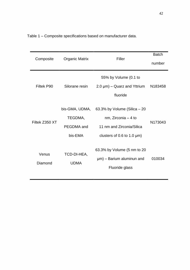
composite specifications based on manufacturer data are listed in Table 1.
2.2. Light-curing Units
The LCUs consisted of a conventional quartz tungsten halogen (QTH)
LCU (Vip Junior, Bisco Inc, Schaumburg, IL, USA) and a light emitting diode
(LED) LCU (Radi Cal, SDI, Bayswater, Victoria, Australia).
The output power (mW) of the LCUs was measured using a power meter
(Ophir Optronics Inc, Danvers, MA, USA). The diameter of the tips was
measured using a digital caliper (Mitutoyo, Kanagawa, Japan) in order to
calculate the total irradiance (QTH: 700 mW/cm2 for 40 second exposure, LED:
1400 mW/cm2 for 20 second exposure). The energy density was calculated
based on the total irradiance and exposure time and standardized at 28 J/cm2.
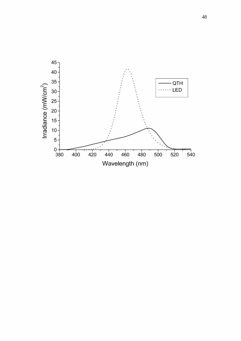
The output spectra of the LCUs (Figure 1) were obtained using a spectrometer
(USB 2000, Ocean Optics, Dunedin, FL, USA). The total irradiance of each LCU
was also obtained through numerical integration of the output spectrum using
the Origin 6.1 software (OriginLab Corp, Northampton, MA, USA).
2.3. Contraction Stress Test
A series of 60 rings (5 mm diameter x 2 mm high) were fabricated from
photoelastic resin (GIII flexible, Polipox, Sao Paulo, Brazil). Following
polymerization, the interior surfaces were abrasive blasted using 50 μm alumina
particles in order to obtain higher micromechanical retention.13 The rings were
divided into 6 groups (n = 10) according to material and LCU.
For the Filtek Z350 XT and Venus Diamond composites, Scotchbond
multi-purpose adhesive was applied and photoactivated, followed by insertion

31
and photoactivation of the restorative materials. For the Filtek P90 samples, the
Filtek P-90 system adhesive was used in place of Scotchbond adhesive.
The specimens were analyzed and the contraction stress (MPa) was
measured using a polariscope (Photostress LF/Z-2, Raleigh, NC, USA). The
contraction stress measurements were subjected to the Kolmogorov-Smirnov
test for normality, followed by two-way ANOVA (composite vs LCU) and Tukey’s
test at 5% significance levels.
2.4. Degree of Conversion Test
Brass rings 8.0 mm in internal diameter, 12.27 mm in external diameter,
and 2.0 mm high were filled with a single layer of the composites. A transparent
polyester strip was placed on the tops and bottoms of the samples to obtain a
flat surface. The photoactivation procedure was performed according to the type
of LCU and composite. For each group, 10 specimens were prepared.
Following photoactivation, the specimens were stored dry at 37ºC for 24 hours.
The top and bottom surfaces were manually flattened using 200, 400, and 600
grit SiC abrasive (Carborundum; Saint-Gobain Abrasives, Recife, PE, Brazil)
while cooling with distilled water. After sanding, the sample and matrix were
washed with distilled water, dried, and stored in closed lightproof containers at
37ºC for 24 hours.
FTIR (Spectrum 100 Optical; Perkin Elmer Analytical Sciences, MA,
USA) spectra were obtained of the top and bottom sample surfaces in order to
determine the degree of conversion. The spectra were obtained using an
attenuated total reflectance (ATR) attachment containing a zinc selenide
horizontal crystal (Pike Technologies, Madison, WI, USA). The Spectrum v6.3.1
software package (Perkin Elmer Analytical Sciences) was used for spectral
visualization and plotting. The spectra were the averages of 16 scans between
300-4000 cm-1 at a resolution of 4 cm-1. The DC in the cured materials was
determined using the three frequency technique. For Filtek P90 containing
silorane monomers, the C-O-C stretching vibrations of the epoxy rings
(884cm−1) were chosen as the analytical absorption band and the Si–CH3
stretching vibrations (695cm−1) were used as the reference absorption band. In

32
this case, the fraction of remaining epoxy rings was calculated from the
normalized peak height ratios of the cured versus the uncured material.15 For
Filtek Z350 XT and Venus Diamond, the C=C stretching vibrations (1638 cm−1)
were used as the analytical absorption band and the aromatic C-C stretching
vibrations (1608 cm−1 for Filtek Z350 XT and 1716 cm−1 for Venus Diamond)
were selected as the reference absorption band. The DC was calculated from
the peak height ratio of the analytical and the reference absorption bands
normalized to the corresponding ratio in the uncured material.
In order to compare the degree of monomer conversion between the top
and bottom surfaces, the data were subjected to the Kolmogorov-Smirnov test
for normality and Student’s t-test at 5% significance with respect to the type of
light curing unit used. The dependence of monomer conversion on
composite/LCU interactions were investigated by subjecting the data to the
Kruskal-Wallis one way analysis of variance on ranks followed by pairwise
multiple comparisons using Tukey’s test at 5% significance level.
2.5. Knoop Hardness and Cross-link Density Test
Standardized cylindrical specimens were prepared by placing the
composites in a circular elastomeric mold (2 mm thick x 4 mm diameter). The
bottom and top surfaces were covered with a transparent polyester strip and
photoactivated using an LCU. The curing tip was positioned close to the
mold/composite assembly. The photoactivation procedures were selected
based on the LCU and composite type. A total of 10 specimens of each group
were prepared.
Following photoactivation, the specimens were stored dry at 37ºC for 24
h. The top and bottom surfaces were flattened using 200, 400, and 600-grit SiC
abrasive paper. Indentations for Knoop hardness (KHN1) measurements were
performed sequentially in a hardness testing machine (HMV 2; Shimadzu,
Tokyo, Japan). Five readings were obtained on the top and bottom surfaces
under a load of 50 g for 15 s. KHN1 for each surface was recorded as the mean
of the five indentations. When comparing the top and bottom surfaces, the data
33
were subjected to the Kolmogorov-Smirnov test for normality and Student’s t-
test at 5% significance level with respect to the type of light curing unit used.
The specimens were soaked in absolute ethanol for 24 hours at room
temperature to soften the material,18 and the hardness was again determined
(KHN2). The CLD was estimated from the degree of softening caused by
ethanol immersion. The decrease in hardness was calculated for both surfaces
of each specimen.6 The data were subjected to the Kolmogorov-Smirnov test
for normality, two-way ANOVA (composite vs LCU), and Tukey’s test at 5%
significance levels.
3. Results
There was not statistically significant different for the factor LCU and
composite/LCU interaction (p>0.05). However, for the factor composite (Table
2, p<0.05), the composite Venus Diamond showed lower contraction stress
values than the Filtek Z350 XT and Filtek P90 composites; and the Filtek Z350
XT and Filtek P90 composites did not differ statistically.
The results in Table 3 (p<0.05) demonstrate a higher degree of conversion
at the top surface independent of curing unit or composite type.
The conversion in Venus Diamond was lower than Filtek P90 at both
surfaces using either LCU. Filtek Z350 XT composite did not differ significantly
from the other composites at either surface using either curing unit (Table 4,
p<0.05).
For each composite and LCU, the Knoop hardness of the top surface
was higher than the bottom surface (Table 5, p<0.05).
There were no significant differences in the LCU and composite/LCU
interaction factors (p>0.05). However, the PD of Venus Diamond was lower
than Filtek Z350 XT or Filtek P90 at both surfaces (Table 6, p<0.05). The PD of
Filtek Z350 XT and Filtek P90 did not differ significantly at either surface.
The emission spectra of the LCUs are depicted in Figure 1. The QTH
emission was concentrated in the 390 to 520 nm range, with an emission peak
34
at 490 nm. The LED unit exhibited a narrower curve concentrated in the 420 to
520 nm range with a peak at 462 nm.
4. Discussion
Resin composites exhibit viscoelastic behavior and are transformed
during polymerization from a viscous plastic to a rigid elastic structure.15,19
Adhesive bonding of composites to teeth results in contraction stresses, the
magnitude of which is dependent on several factors. The contraction stresses in
polymeric materials used in dentistry are typically measured using a
tensiometer.13 These measurements may be used to determine the maximum
stress on the specimen under specific conditions, but the stress near the
external margins of the cavity will be different from the stress near the pulpal
wall.13 Contraction stress may also be modeled using finite element analysis
and elasticity theory.20 However, analysis of the stresses generated in complex
restoration configurations is difficult.13 The round, uniform nature of the
specimens in this study resulted in a regular stress distribution and permitted
the use of photoelastic stress analysis.
In contrast to free radical polymerization of methacrylates, the ring-
opening polymerization of silorane-based composite resin occurs via cleavage
and opening of ring structures, counteracting the inevitable loss of volume due
to bond formation.5 Previous reports have described lower shrinkage,4,15 lower
contraction stress,4 and reduced cuspal deflection5,21 when using silorane-
based materials. In the present study, the contraction stress in the Filtek P90
system was similar to that in the nanoparticle-filled Filtek Z-350 XT system, and
the low-shrinkage nanohybrid Venus Diamond system exhibited lower
contraction than the other two systems (Table 1). The development of
contraction stress in dental composites depends on material composition,
including the type of monomer, the type and amount of filler, filler/matrix
interactions, polymerization parameters such as the degree and rate of
polymerization, placement, and curing technique.5,22 According to Hooke’s Law,
the stress should be determined by the product of the volumetric shrinkage and

35
the elastic modulus of the material (in a totally elastic situation). However, the
setting of dental composites is not purely elastic, and increased elastic modulus
has been related to higher stress.5,22 Previous studies4,5 have reported that
while the elastic modulus of silorane-based composite resins is higher than that
of methacrylate-based resins, they generate similar contraction stresses. Venus
Diamond is characterized by the presence of a novel monomer (TCD-Di- HEA,
(Bis-(acryloyloxymethyl)tricyclo[5.2.1.02,6]decane) that according to the
manufacturer combines low shrinkage with low viscosity and may account for
the lower stress values recorded with this material.5 This assertion is in
agreement with the results of this present study, in which Venus Diamond had
the lowest contraction stress.
Adequate polymerization is a crucial factor in obtaining optimal physical
and mechanical performance from dental resin composites.23 The appearance
of the composite is influenced by factors such as filler and polymeric matrix
refractive index, monomer type, and filler type and content.24 During the
photoactivation process, light passing through the resin composite is absorbed
by the resin and scattered by the filler material.6,25 The light intensity is
attenuated and the curing effectiveness is reduced as the depth increases.26
The polymerization depth depends on the light irradiance and exposure time as
well as factors such as material composition,27 resin composite shade,28 and
translucency.29 An advantage of testing composites using the Knoop hardness
test is the correlation between the Knoop hardness and the DC.30 In the present
study, the top surface of the three composites displayed greater conversion and
Knoop hardness with either LCU (Tables 3 and 5).
The polymerization of siloranes occurs via a cationic ring-opening
reaction, while methacrylate-based composite resins polymerize via a radical
addition reaction to their double bonds.4 During photopolymerization of
dimethacrylate resins, the rapid development of a highly cross-linked network
drastically restricts the mobility of reacting species, resulting in phenomena
such as auto-acceleration, reaction-diffusion-controlled termination, and
incomplete conversion.31,32 The DC of methacrylate-based composite resins is
determined from the conversion of aliphatic C=C double bonds. However,

36
silorane-based monomers do not contain carbon-carbon double bonds, and the
degree of polymerization is calculated based on the conversion of epoxides to
C-O-C- chain units.33,34 In the present study, the DC of the Filtek P90 system
was the same as the DC of the nanoparticle-filled Filtek Z-350 XT (Table 4) for
both surfaces and LCUs, in contrast to a previous study.34 However, that study
performed FTIR analysis using the region between 730 and 950 cm−1 with
oxirane ring peaks at 882cm−1, while the present study used the region
described by Papadogiannis et al. (884cm−1 for epoxy stretching vibrations and
695cm−1 for Si–CH3 vibrations). The DC of the Venus Diamond composite was
lower than that of the Filtek P90 composite, but was not significantly different
from Filtek Z-350 XT. This finding is clinically relevant because DC is related to
contraction stress. In this case, the Venus Diamond composite exhibited similar
DC with less tension due to contraction.
Cross-linked dimethacrylate networks swell when exposed to solvents.
This occurs because the attractive forces between the solvent molecules and
the chain are greater than the forces between the polymer chains.35 Therefore,
the solvent penetrates into the resin matrix and expands the openings among
chains.18 Solvent penetration ability is related to the solubility parameter, which
describes the ability of a molecule to penetrate and dissolve another
substance.18,35 The differences in the solubility parameter between the polymer
and the solvent will determine the extent of solvent uptake,30 with smaller
differences in the solubility parameters of the solvent and the polymer resulting
in greater solvent uptake.18,35 The CLD exerts a major effect on the polymer
properties, as highly cross-linked materials generally possess increased
fracture strength and wear resistance.35 Polymers with a high CLD may be
advantageous not only because of their enhanced mechanical properties, but
also by being less susceptible to softening by food substances and to enzymatic
attack.17 In the current study, the PD of Venus Diamond composite was lower
than the other composites (Table 6). Since solvent uptake and swelling are
directly related to CLD, a polymer with fewer cross-links is more sensitive to the
plasticizing action of solvents.35 However, a cross-linking agent may be
incorporated into the formulation of this composite to minimize this drawback,
37
and addition of this agent did not affect desirable properties such as low
contraction stress and high DC.
In this study, the curing performance of the introduced LED LCU was
similar36 to or better37 than that of the QTH LCU. In contrast, others have
reported better performance from QTH LCUs.38,39 The conventional QTH LCU
(Vip Junior, Bisco Inc) used in this study emitted an energy density of 28 J/cm2,
yielding an irradiance of 700 mW/cm2 during a 40 second exposure (energy
density (J/cm2) = irradiance – mW/cm2 x exposure time – s / 1000). Under
similar conditions, the LED LCU (Radi Cal, SDI) emitted an energy density of 28
J/cm2 (1400 mW/cm2 over 20 seconds). Figure 1 contains plots of the LCU
wavelength distributions. The reduced curing time using the LED unit is
clinically relevant in terms of the reduced time required to prepare restorations,
which is beneficial for both the patient and the practitioner.
Based on the results of this study hypothesis (1) must be rejected, as the
Filtek P90 system resulted in contraction stresses similar to those of Filtek Z350
XT. Hypothesis (2) must also be rejected, as Venus Diamond experienced
smaller decreases in hardness (and therefore in cross-link density) than Filtek
Z350 XT or Filtek P90.
5. Conclusions
Within the limitations of the current study, the authors conclude that:
The low-shrinkage Venus Diamond composite showed be an option to try
to reduce stress at the restoration/tooth interface.
There was no difference in degree of conversion between Venus
Diamond and Filtek Z-350 XT composite, but Venus Diamond resulted in lower
contraction stress, resulting in lower stress at the substrate interface. However,
no benefit was obtained from the contraction stress reduction if the cross-link
density was also decreased.

38
References
1. Harris JC, Jacobsen PH, O’Doherty DM. The effect of curing light intensity
and test temperature on the dynamic mechanical properties of two polymer
composites. Journal of Oral Rehabilitation 1999;26:635-9.
2. Navarra CO, Cadenaro M, Armstrong SR, Jessop J, Antoniolli F, Sergo V,
et al. Degree of conversion of Filtek Silorane Adhesive System and Clearfil SE
Bond within the hybrid and adhesive layer: an in situ Raman analysis. Dental
Materials 2009;25:1178-85.
3. Guiraldo RD, Consani S, Consani RL, Berger SB, Mendes WB, Sinhoreti
MA, et al. Comparison of silorane and methacrylatebased composite resins on
the curing light transmission. Brazilian Dental Journal 2010;21:538-42.
4. Weinmann W, Thalacher C, Guggenberger R. Siloranes in dental
composites. Dental Materials 2005;21:68-74.
5. Marchesi G, Breschi L, Antoniolli F, Di Lenarda R, Ferracane J, Cadenaro
M. Contraction stress of low-shrinkage composite materials assessed with
different testing systems. Dental Materials 2010;26:947-53.
6. Guiraldo RD, Consani S, Consani RL, Berger SB, Mendes WB, Sinhoreti
MA. Light energy transmission through composite influenced by material
shades. Bulletin of Tokyo Dental College 2009;50:183-90.
7. Uhl A, Mills RW, Jandt KD. Polymerization and light-induced heat of dental
composites cured with LED and halogen technology. Biomaterials
2003;24:1809-20.
8. Rueggeberg F. Contemporary issues in photocuring. Compendium of
Continuing Education in Dentistry 1999;20:S4-15.
9. Loureiro FH, Consani S, Guiraldo RD, Consani RL, Berger SB, Carvalho
RV, et al. Comparison between two methods to evaluate temperature changes
produced by composite light curing units and polymerization techniques.
Minerva Stomatologica 2011;60:501-8.
10. Faria-e-Silva AL, Lima AF, Moraes RR, Piva E, Martins LR. Degree of
conversion of etch-and-rinse and self-etch adhesives light-cured using QTH or
LED. Operative Dentistry 2010;35:649-54.

39
11. Leonard DL, Charlton DG, Roberts HW, Cohen ME. Polymerization
efficiency of LED curing lights. Journal of Esthetic and Restorative Dentistry
2002;4:286-95.
12. Arikawa H, Kanie T, Fujii K, Takahashi H, Ban S. Effect of inhomogeneity
of light from light curing units on the surface hardness of composite resin.
Dental Materials Journal 2008;27:21-8.
13. Lopes MB, Valarini N, Moura SK, Guiraldo RD, Gonini Júnior A.
Photoelastic analysis of stress generated by a silorane-based restoration
system. Brazilian Oral Research 2011;25:302-6.
14. Standlee JP, Caputo AA. Load transfer by fixed partial dentures with three
abutments. Quintessence International 1988;19:403-10.
15. Papadogiannis D, Kakaboura A, Palaghias G, Eliades G. Setting
characteristics and cavity adaptation of low-shrinking resin composites. Dental
Materials 2009;25:1509-16.
16. Sakaguchi RL, Berge HX. Reduced light energy density decreases post-
gel contraction while maintaining degree of conversion in composites. Journal of
Dentistry 1998;26:695-700.
17. Asmussen E, Peutzfeldt A. A Influence of pulse-delay curing on softening
of polymer structures. Journal of Dental Research 2001;80:1570-3.
18. Schneider LF, Moraes RR, Cavalcante LM, Sinhoreti MA, Correr-Sobrinho
L, Consani S. Cross-link density evaluation through softening tests: effect of
ethanol concentration. Dental Materials 2008;24:199-203.
19. Davidson CL, Feilzer AJ. Polymerization shrinkage and polymerization
shrinkage stress in polymer-based restoratives. Journal of Dentistry
1997;25:435-40.
20. Rees JS, Jacobsen PH. Stresses generated by luting resins during
cementation of composite and ceramic inlays. Journal of Oral Rehabilitation
1992;19:115-22.
21. Bouillaguet S, Gamba J, Forchelet J, Krejci I, Wataha JC. Dynamics of
composite polymerization mediates the development of cuspal strain. Dental
Materials 2006;22:896-902.
40
22. Braga RR, Ballester RY, Ferracane JL. Factors involved in the
development of polymerization shrinkage stress in resin-composites: a
systematic review. Dental Materials 2005;21:962-70.
23. Knezevic A, Tarle Z, Meniga A, Sutalo J, Pichler G, Ristic M. Degree of
conversion and temperature rise during polymerization of composite resin
samples with blue diodes. Journal of Oral Rehabilitation 2001;28:586-91.
24. Campbell PM, Johnst WM, O”Brien WJ. Light scattering and gloss of an
experimental quartz-filled composite. Journal of Dental Research 1986;65:892-
4.
25. Dos Santos GB, Monte Alto RV, Filho HR, da Silva EM, Fellows CE. Light
transmission on dental resin composites. Dental Materials 2008;24:571-6.
26. Vargas MA, Cobb DS, Schmit JL. Polymerization of composite resins:
argon laser vs conventional light. Operative Dentistry 1998;23:87-93.
27. Atmadja G, Bryant RW. Some factors influencing the depth of cure of
visible light-activated composite resins. Australian Dental Journal 1990;35:213-
8.
28. Tanoue N, Koishi Y, Matsumura H, Atsuta M. Curing depth of different
shades of a photo-actived prosthetic composite material. Journal of Oral
Rehabilitation 2001;28:618-23.
29. Ferracane JL, Aday P, Matsumura H, Atsuta M. Relationship between
shade and depth of cure for light-activated dental composite resins. Dental
Materials 1986;2:80-84.
30. Ferracane JL. Correlation between hardness and degree conversion
during the setting reaction of unfilled dental restorative resins. Dental Materials
1985;1:11-4.
31. Bowman CN, Anseth KS. Microstructural evolution in polymerizations of
tetrafunctional monomers. Macromolecular Symposia 1995;93:269-76.
32. Lu H, Stansbury JW, Bowman CN. Impact of Curing Protocol on
Conversion and Shrinkage Stress. Journal of Dental Research 2005;84:822-6.
33. Ilie N, Hickel R. Silorane-based dental composite: behavior and abilities.
Dental Materials Journal 2006;25:445-54.

41
34. Kusgoz A, Ülker M, Yesilyurt C, Yoldas OH, Ozil M, Tanriver M. Silorane-
Based composite: depth of cure, surface hardness, degree of conversion, and
cervical microleakage in class II cavities. Journal of Esthetic and Restorative
Dentistry 2011;23:324-37.
35. Ferracane JL. Hygroscopic and hydrolytic effects in dental polymer
networks. Dental Materials 2006;22:211-22.
36. Bala O, Olmez A, Kalayci S. Effect of LED and halogen light curing on
polymerization of resin-based composites. Journal of Oral Rehabilitation
2005;32:134-40.
37. Price RB, Felix CA, Andreou P. Knoop hardness of ten resin composites
irradiated with high-power LED and quartz-tungsten-halogen lights. Biomaterials
2005;26:2631-41.
38. Dunn WJ, Bush AC. A comparison of polymerization by light-emitting
diode and halogen-based light-curing units. Journal of the American Dental
Association 2002;133:335-41.
39. Beun S, Glorieux T, Devaux J, Vreven J, Leloup G. Characterization of
nanofilled compared to universal and microfilled composites. Dental Materials
2007;23:51-9.
42
Table 1 – Composite specifications based on manufacturer data.
Composite Organic Matrix Filler Batch
number
Filtek P90 Silorane resin
55% by Volume (0.1 to
2.0 μm) – Quarz and Yttrium
fluoride
N183458
Filtek Z350 XT
bis-GMA, UDMA,
TEGDMA,
PEGDMA and
bis-EMA
63.3% by Volume (Silica – 20
nm, Zirconia – 4 to
11 nm and Zirconia/Silica
clusters of 0.6 to 1.0 μm)
N173043
Venus
Diamond
TCD-DI-HEA,
UDMA
63.3% by Volume (5 nm to 20
μm) – Barium aluminun and
Fluoride glass
010034

43
Table 2 – Mean values of contraction stress.
Composite Contraction Stress (MPa)
Filtek Z350 XT 5.00 (2.29) a
Filtek P90 7.05 (2.97) a
Venus Diamond 1.20 (1.37) b
Mean values followed by different lowercase letters in the columns differed statistically by
Tukey’s test at 5% level for different light curing units. Standard deviations are given in
parentheses.

44
Table 3 – Mean values of degree of conversion for top and bottom surfaces
using LED and QTH light curing units.
Composite QTH LED
Top (%) Bottom (%) Top (%) Bottom (%)
Filtek Z350 XT 53.32 (3.26) a 45.98 (4.50) b 52.96 (2.89) a 43.94 (3.74) b
Filtek P90 63.86 (1.82) a 53.88 (6.49) b 63.84 (1.59) a 56.32 (4.30) b
Venus Diamond 38.74 (4.87) a 33.06 (5.92) b 39.18 (4.86) a 31.35 (8.77) b
Mean values followed by different lowercase letters in the row differed statistically by Student’s
t-test at 5% level for different light curing units. Standard deviations are given in
parentheses.

45
Table 4 – Median values of degree of conversion for composite/LCU
interaction at top and bottom surfaces.
Composite Top Bottom
QTH (%) LED (%) QTH (%) LED (%)
Filtek Z350 XT 54.44 ab A 52.13 ab A 45.65 ab A 44.20 ab A
Filtek P90 63.36 a A 63.17 a A 54.29 a A 57.03 a A
Venus Diamond 38.32 b A 38.33 b A 34.02 b A 32.51 b A
Median values followed by different uppercase letters in rows and lowercase letters in columns
differ statistically by Tukey’s test at 5% level of significance.

46
Table 5 – Comparison of mean Knoop hardness number between top and
bottom surfaces for LED and QTH light curing units.
Composite QTH LED
Top (KNH) Bottom (KNH) Top (KNH) Bottom (KNH)
Filtek Z350 XT 92.98 (4.77) a 77.80 (7.04) b 86.56 (7.81) a 72.66 (5.97) b
Filtek P90 60.37 (4.30) a 47.96 (4.52) b 63.13 (2.69) a 49.73 (2.51) b
Venus Diamond 73.17 (9.33) a 53.00 (8.55) b 64.71 (7.61) a 42.29 (8.45) b
Mean values followed by different lowercase letters in the row differed statistically by Student’s
t-test at 5% level for different light curing units. Standard deviations are given in
parentheses.

47
Table 6 – Mean values of KHN1, KHN2, and percent of decrease in hardness
after ethanol immersion (PD).
Composite KHN1
(KHN)
KHN2
(KHN) Top PD (%)
KHN1
(KHN)
KHN2
(KHN) Bottom PD (%)
Filtek Z350 XT 89.77
(7.10)
61.91
(3.93) 69.34 (6.78) a
75.23
(6.88)
55.37
(3.27) 73.97 (5.54) a
Filtek P90 62.25
(4.55)
42.95
(2.68) 69.26 (5.92) a
48.85
(3.68)
32.66
(2.56) 67.20 (7.28) a
Venus Diamond 68.94
(9.35)
33.67
(4.46) 49.58 (8.76) b
47.64
(9.93)
26.94
(4.98) 58.26 (13.23) b
Mean values followed by different lowercase letters in the columns differed statistically by
Tukey’s test at 5% level for different surfaces. Standard deviations are given in
parentheses.
48

49
Figure Legend
Figure 1 – Wavelength distributions of light curing units.
50
5- CONCLUSÃO GERAL
De acordo com os materiais e métodos empregados no presente estudo,
foi possível concluir que:
1- O compósito de baixa contração Venus Diamond mostrou ser uma
opção para reduzir a tensão na interface restauração-dente, tendo em vista que
apresentou menor valor de tensão de contração quando comparado aos
compósitos Filtek, Z350XT e P90.
2- O compósito Venus Diamond não mostrou diferença no grau de
conversão para o compósito Z350 Filtek XT, como o grau de conversão está
relacionado com a tensão de contração e Venus Diamond compósito mostrou
grau semelhante de conversão com menos tensão de contração. Assim, houve
benefício para a redução da contração do compósito Venus Diamond na
geração de tensão na interface do substrato.
3- O compósito Venus Diamond mostrou menor PD quando
comparado aos outros compósitos.
51
REFERÊNCIAS
ASMUSSEN E, PEUTZFELDT A. A Influence of pulse-delay curing on
softening of polymer structures. J Dent Res. 2003; 80: 1570-1573.
ARIKAWA H, KANIE T, FUJII K, TAKAHASHI H, BAN S. Effect of
inhomogeneity of light from light curing units on the surface hardness of
composite resin. Dent Mater J. 2008; 27: 21-28.
ATMADJA G, BRYANT RW. Some factors influencing the depth of cur of
visible light-activated composite resins. Aust Dent J. 1990; 35: 213-218.
BOWEN RL. Dental filling material comprising vynil-silano treated fused
silica and a binder consisting of the reaction product of bisphenol and
glycidil metacrylate. US Patent 3.066.112; 1962.
BUONOCORE MG. A simple method of increasing the adhesion of acrylic
filling materials to enamel surfaces. J. Dent Res. 1955; 34: 849-853.
BURGESS JO, WALKER RS, PORCHE C, RAPPOLD AJ. Light curing – Na
up date. Compend Contin Educ Dent. 2002; 23: 889-906.
CAUGHMAN WF, CAUGHMAN GB, SHIFLETT RA, RUEGGEBERG F,
SCHUSTER GS. Correlation of cytotoxicity, filler loading and curing time
of dental composites. Biomaterials. 1991; 12: 737-740.
COOK WD. Spectral distributions of dental photo-polymerization sources.
J Dent Res. 1982; 61: 1436-1438.

52
CORRER AB. Avaliação da dureza knoop de compósitos restauradores
odontológicos foto-ativados por diferentes métodos. [dissertação].
Piracicaba: FOP/UNICAMP; 2005.
FARIA-e-SILVA AL, LIMA AF, MORAES RR, PIVA E, MARTINS LR. Degree of
conversion of etch-and-rinse and self-etch adhesives light-cured using
QTH or LED. Oper Dent. 2010; 35: 649-654.
FERRACANE JL, ADAY P, MATSUMOTO H, MARKER VA. Relationship
between shade and depth of cure for light-activated dental composite
resins. Dent Mater. 1986; 2: 80-84.
FERRACANE JL, MITCHEM JC, CONDON JR, TODD R. Wear and marginal
breakdown of composites with various degrees of cure. J Dent Res. 1997;
76: 1508-1516.
GUIRALDO RD, CONSANI S, De SOUZA AS, CONSANI RL, SINHORETI MA,
CORRER-SOBRINHO L. Influence of light energy density on heat
generation during photoactivation of dental composites with different
dentin and composite thickness. J Appl Oral Sci. 2009; 17: 289-293.
GUIRALDO RD, CONSANI S, CONSANI RL, BERGER SB, MENDES WB,
SINHORETI MA. Light energy transmission through composite influenced
by material shades. Bull Tokyo Dent Coll. 2009; 50: 183-190.
GUIRALDO RD, CONSANI S, CONSANI RL, BERGER SB, MENDES WB,
SINHORETI MA. Comparison of silorane and methacrylate-based
composite resins on the curing light transmission. Braz Dent J. 2010; 21:
538-42.

53
HARRIS JS, JACOBSEN PH, O´DOHERTY DM. The effect of curing light
intensity and test temperature on the dynamic mechanical properties o
two polymer composites. J Oral Rehab. 1999; 26: 635-639.
ILIE N, HICKEL R. Macro-,micro- and nano-mechanical investigations on
silorane and methacrylate_based composites. Dent Mater. 2009; 25: 810-
819.
KLAPDOHR S, MOSZNER N. New inorganic components for Dental Filling
Composites. Monatsh Chem. 2005; 136: 21-45.
KURACHI C, TUBOY AM, MAGALHÃES DV, BAGNATO VS. Hardness
evaluation of a dental composite polymerized with experimental LED-
based devices. Dent Mater. 2001; 17: 309-315.
LEONARD DL, CHARLTON DG, ROBERTS HW, COHEN ME. Polymerization
efficiency of LED curing lights. J Esthet Restor Dent. 2002; 4: 286-295.
LOPES MB, VALARINI N, MOURA SK, GUIRALDO RD, GONINI JÚNIOR A.
Photoelastic analysis of stress generated by a silorane-based restoration
system. Braz Oral Res. 2011; 25: 302-306.
LOUREIRO FH, CONSANI S, GUIRALDO RD, CONSANI RL, BERGER SB,
CARVALHO RV. Comparison between two methods to evaluate
temperature changes produced by composite light curing units and
polymerization techniques. Minerva Stomatol. 2011; 60: 501-518.
MARCHESI G, BRESCHI L, ANTONIOLLI A, Di LENARDA R, FERRACANE J,
CADENARO M. Contraction stress of low-shrinkage composite materials
assessed with different testing systems. Dental Mater. 2010; 26: 947-953.

54
MUNKSGAARD EC, PEUTZFELDT A, ASMUSSEN E. Elution o TEGDMA and
BisGMA rom a resin and a resin composite cured with halogen or plasma
light. Eur J Oral Sci. 2000; 108: 341-345.
NAVARRA CO, CADENARO M, ARMSTRONG SR, JESSOP J, ANTONIOLLI
F, SERGO V. Degree of conversion of Filtek Silorane Adhesive System
and Clearfil SE Bond within the hybrid and adhesive layer: an in situ
Raman analysis. Dent Mater. 2009; 25: 1178-1185.
NOMOTO R. Effect of light wavelength on polymerization of light-cured
resins. Dent Mater. 1997; 16: 60-73.
PAPADOGIANNIS D, KAKABOURA A, PALAGHIAS G, ELIADES G. Setting
characteristics and cavity adaptation of low-shrinking resin composites.
Dent Mater. 2009; 25: 1509-1516.
PEUTZFELDT A. Resin composites in dentistry: monomer systems. Eur J
Oral Sci. 1997; 105: 97-116.
PEUTZFELDT A, SAHAFI A, ASMUSSEN E. Characterization of resin
composites polymerized with plasma arc curing units. Dent Mater. 2000;
16: 330-336.
RUEGGEBERG FA. Contemporary issues in photocuring. Compend Contin
Educ Dent. 1999; 20: S4-S15.
RUYTER IE, OYSAED H. Conversion in different depths of ultraviolet and
visible light activated composite materials. Acta Odontol Scand. 1982;
40(3): 179-192.

55
SAHAFI A, PEUTZFELDT A, ASMUSSEN E. Soft-start polymerization and
marginal gap formation in vitro. Am J Dent. 2001; 14: 145-147.
SAKAGUCHI RL, BERGE HX. Reduced light energy density decreases
post-gel contraction while maintaining degree of conversion in
composites. J Dent. 1998; 26: 695–700.
STANDLEE JP, CAPUTO AA. Load transfer by fixed partial dentures with
three abutments. Quintessence Int. 1988; 19: 403-410.
TANOUE N, KOISHI Y, MATSUMURA H, ATSUTA M. Curing depth of
different shades of a photo-activated prosthetic composite material. J Oral
Rehab. 2001; 28: 618-623.
UHL A, MILLS RW, JANDT KD. Polymerization and light-induced heat of
dental composites cured with LED and halogen technology. Biomaterials.
2003; 24: 1809-1820.
WEINMANN W, THALACKER C, GUGGENBERGER R. Siloranes in dental
composites. Dent Mater. 2005; 21: 68-74.

56
ANEXO