Embed Size (px)
Citation preview

UNIVERSIDADE FEDERAL DE SANTA MARIA
CENTRO DE CIÊNCIAS DA SAÚDE
PROGRAMA DE PÓS-GRADUAÇÃO EM FARMACOLOGIA
Karen Luise dos Santos Moreira
O EFEITO NEUROPROTETOR DO EXERCÍCIO FÍSICO EM RATOS
SUBMETIDOS AO MODELO ANIMAL DE FIBROMIALGIA
INDUZIDO POR RESERPINA
Santa Maria, RS
2018

1
Karen Luise dos Santos Moreira
O EFEITO NEUROPROTETOR DO EXERCÍCIO FÍSICO EM RATOS
SUBMETIDOS AO MODELO ANIMAL DE FIBROMIALGIA INDUZIDO POR
RESERPINA
Dissertação apresentada ao curso Pós-Graduação
em Farmacologia, área de concentração em
Neuropsicofarmacologia e Imunofarmacologia, da
Universidade Federal de Santa (UFSM, RS), como
requisito parcial para obtenção do título de Mestre
em Farmacologia.
Santa Maria, RS
2018
2

3
Orientadora: Profa Dra Patrícia Severo do Nasciment

4
AGRADECIMENTOS
Agradeço primeiramente a Deus que está sempre presente guiando meus passos.
Aos meus pais, Vilmar e Sônia, e também ao meu padrasto, Jairo, por todo apoio, incentivo e
compreensão, pois com certeza sem vocês nada seria possível até aqui. É difícil expressar
tamanha gratidão e amor em ter vocês na minha vida. Muito obrigada por tudo!
À minha orientadora, Prof. Patrícia Severo do Nascimento, pelos ensinamentos, pelo
exemplo, pela paciência e pela compreensão durante esta fase. Reconheço e agradeço de
coração a oportunidade e também o exemplo de profissional. O caminho ainda é longo, mas
sigo na certeza de que os ensinamentos serão essenciais e me acompanharão nas próximas
etapas. De coração, a minha gratidão e admiração!
Agradeço ao Professor Marcelo Veiga, e à Professora Maria Izabel Ugalde, por sempre me
acolherem com muito carinho desde o período da faculdade até hoje.
Aos demais professores e funcionários do Programa de Pós-Graduação em Farmacologia, que
contribuíram de alguma forma para minha formação.
Aos amigos e colegas do Labitex pelo carinho, amizade e companheirismo. Em especial,
gostaria de agradecer à Marina e à Gabi, pelas risadas (que não foram poucas!), pelos vários
trabalhos na histologia, pelos conselhos, almoços no RU e no 20. Adoro vocês!
Ao amigos e colegas do NeuronLAB e também à Professora Gabriela e o Professor
Guilherme, que me receberam de “braços abertos” neste ano que passou. Obrigada!
A CAPES e ao CNPq, pela bolsa de estudos e pelos recursos financeiros concedidos.
Aos animais utilizados na realização deste trabalho, todo o meu respeito.
Enfim, agradeço à Universidade Federal de Santa Maria e ao Programa de Pós-Graduação em
Farmacologia pela possibilidade de realização desta etapa profissional.
A todos, muito obrigada!
5
RESUMO
O EFEITO NEUROPROTETOR DO EXERCÍCIO FÍSICO EM RATOS
SUBMETIDOS AO MODELO ANIMAL DE FIBROMIALGIA INDUZIDO POR
RESERPINA
AUTORA: Karen Luise dos Santos Moreira
ORIENTADORA: Patrícia Severo do Nascimento
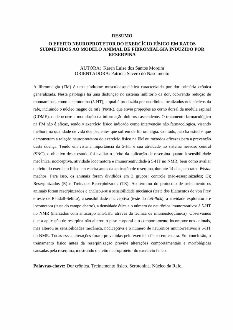
A fibromialgia (FM) é uma síndrome musculoesquelética caracterizada por dor primária crônica
generalizada. Nesta patologia há uma disfunção no sistema inibitório da dor, ocorrendo redução de
monoaminas, como a serotonina (5-HT), a qual é produzida por neurônios localizados nos núcleos da
rafe, incluindo o núcleo magno da rafe (NMR), que envia projeções ao corno dorsal da medula espinal
(CDME), onde ocorre a modulação da informação dolorosa ascendente. O tratamento farmacológico
na FM não é eficaz, sendo o exercício físico indicado como intervenção não farmacológica, visando
melhora na qualidade de vida dos pacientes que sofrem de fibromialgia. Contudo, não há estudos que
demonstrem a relação neuroprotetora do exercício físico na FM ou métodos eficazes para a prevenção
desta doença. Tendo em vista a importância da 5-HT e sua atividade no sistema nervoso central
(SNC), o objetivo deste estudo foi avaliar o efeito da aplicação de reserpina quanto à sensibilidade
mecânica, nociceptiva, atividade locomotora e imunorreatividade à 5-HT no NMR; bem como avaliar
o efeito do exercício físico em esteira antes da aplicação de reserpina, durante 14 dias, em ratos Wistar
machos. Para isso, os animais foram divididos em 3 grupos: controle (não-reserpinizados; C);
Reserpinizados (R) e Treinados-Reserpinizados (TR). Ao término do protocolo de treinamento os
animais foram reserpinizados e analisou-se a sensibilidade mecânica (teste dos filamentos de von Frey
e teste de Randall-Selitto), a sensibilidade nociceptiva (teste do tail-flick), a atividade exploratória e
locomotora (teste do campo aberto), a densidade ótica e o número de neurônios imunorreativos à 5-HT
no NMR (marcados com anticorpo anti-5HT através da técnica de imunoistoquímica). Observamos
que a aplicação de reserpina não alterou o peso corporal e o comportamento locomotor nos animais,
mas alterou as sensibilidades mecânica, nociceptiva e o número de neurônios imunorreativos à 5-HT
no NMR. Todas essas alterações foram prevenidas pelo exercício físico em esteira. Em conclusão, o
treinamento físico antes da reserpinização previne alterações comportamentais e morfológicas
causadas pela reserpina, mostrando o efeito neuroprotetor do exercício físico.
Palavras-chave: Dor crônica. Treinamento físico. Serotonina. Núcleo da Rafe.
6
ABSTRACT
NEUROPROTECTIVE EFFECT OF PHYSICAL EXERCISE IN RATS SUBMITTED
TO ANIMAL MODEL OF FIBROMYALGIA INDUCED BY RESERPINE
AUTHOR: Karen Luise dos Santos Moreira
ADVISOR: Patrícia Severo do Nascimento
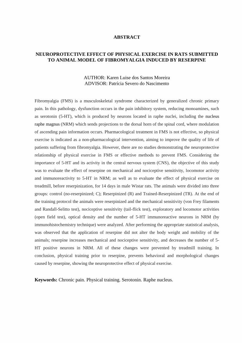
Fibromyalgia (FMS) is a musculoskeletal syndrome characterized by generalized chronic primary
pain. In this pathology, dysfunction occurs in the pain inhibitory system, reducing monoamines, such
as serotonin (5-HT), which is produced by neurons located in raphe nuclei, including the nucleus
raphe magnus (NRM) which sends projections to the dorsal horn of the spinal cord, where modulation
of ascending pain information occurs. Pharmacological treatment in FMS is not effective, so physical
exercise is indicated as a non-pharmacological intervention, aiming to improve the quality of life of
patients suffering from fibromyalgia. However, there are no studies demonstrating the neuroprotective
relationship of physical exercise in FMS or effective methods to prevent FMS. Considering the
importance of 5-HT and its activity in the central nervous system (CNS), the objective of this study
was to evaluate the effect of reserpine on mechanical and nociceptive sensitivity, locomotor activity
and immunoreactivity to 5-HT in NRM; as well as to evaluate the effect of physical exercise on
treadmill, before reserpinization, for 14 days in male Wistar rats. The animals were divided into three
groups: control (no-reserpinized; C); Reserpinized (R) and Trained-Reserpinized (TR). At the end of
the training protocol the animals were reserpinized and the mechanical sensitivity (von Frey filaments
and Randall-Selitto test), nociceptive sensitivity (tail-flick test), exploratory and locomotor activities
(open field test), optical density and the number of 5-HT immunoreactive neurons in NRM (by
immunohistochemistry technique) were analyzed. After performing the appropriate statistical analysis,
was observed that the application of reserpine did not alter the body weight and mobility of the
animals; reserpine increases mechanical and nociceptive sensitivity, and decreases the number of 5-
HT positive neurons in NRM. All of these changes were prevented by treadmill training. In
conclusion, physical training prior to reserpine, prevents behavioral and morphological changes
caused by reserpine, showing the neuroprotective effect of physical exercise.
Keywords: Chronic pain. Physical training. Serotonin. Raphe nucleus.
7
LISTA DE ILUSTRAÇÕES
Figura 1- Representação esquemática das informações nociceptivas conduzidas até o corno
dorsal da medula espinal...........................................................................................................15
Figura 2- Esquema representativo da principal via ascendente da dor.....................................16
Figura 3- Localização dos 18 pontos dolorosos (tender points) ..............................................19
Figura 4- Esquema representativo da via inibitória ou descendente da dor..............................21

8
LISTA DE QUADROS
Quadro 1- Tender points bilaterais e suas localizações ...........................................................18

9
LISTA DE FIGURAS
Figure 1- Experimental design. Rats were treated with reserpine (1 mg/kg/day, s.c.) or vehicle
(saline) during 3 days. After treatment (day 4), rats from each group were weighted and
evaluated. The experimental groups were as follows: no-reserpinized rats (C), reserpinized
rats (R) and rats submitted to treadmill training for two weeks and reserpinized (TR). The
brains were removed from the skulls and were used for
immunohistochemistry, which was used to evaluate the optical densitometry and number of 5-
HT immunopositive neurons in the NRM. Behavioral evaluations were performed on day 4,
one day after last reserpinization (Von frey filaments, Randall Selitto, Tail-flick and Open
field tests). Font: Author………...............................................................................................29
Figure 2- A) Body weight from C, R and TR groups before reserpinization and after the last
reserpinization. B) Mechanical sensitivity, von Frey filaments test from C, R and TR
groups.*: P < 0.05, compared to C and TR groups C) Mechanical sensitivity, Randall-Selitto
test from C, R and TR groups. *: P < 0.05, compared to C and TR groups. D) Nociceptive
sensitivity, tail flick test from C, R and TR groups. *: P < 0.05, compared to C and TR
groups. E) Open field, crossed squares from C, R and TR groups. F) Optical densitometry of
5-HT immunoreactive neurons from C, R and TR groups. G) Number of 5-HT positive cells
in RMN. *: P < 0.05, compared to C and TR
groups…………………………………………………………………………………………35
Figure 3- Illustrative images from 5-HT positive neurons in NRM from no-reserpinized (C),
Reserpinized (R) and treadmill training and reserpinized (TR) groups. Scale bar: 100
µm.............................................................................................................................................36

10
LISTA DE ABREVIATURAS E SIGLAS
5-HT Serotonina
5-HTir Imunorreatividade à Serotonina
Aδ A-delta
ACMS (do Inglês: Colégio Americano de Medina Esportiva)
ACR (do Inglês: Colégio Americano de Reumatologia)
AOI Área de Interesse
C Grupo Controle (não-reserpinizados)
CDME Corno dorsal da medula espinal
CID Classificação Internacional de Doenças
CNS Central Nervous system (do inglês: Sistema Nervoso Central)
FM Fibromialgia
FMS Fibromyalgia Syndrome (do inglês: síndrome da fibromialgia) GRD Gânglio da Raiz Dorsal
IASP (do Inglês: Associação Internacional do Estudo da Dor)
ICD (do Inglês: Classificação Internacional de Doenças)
ME Medula espinal
MET Maximal Exercise Training (do inglês: Teste de esforço máximo)
NMR Núcleo Magno da Rafe
NRM Nucleus Raphe Magnus (do inglês: Núcleo Magno da Rafe)
PAG Substância cinzenta periaquedutal
R Grupo Reserpinizados
SEM Standart error of the mean (do inglês: média de erro padrão)
SNC Sistema Nervoso Central
SP Substância P
TR Grupo treinados-reserpinizados
VMAT Transportador Vesicular de Monoaminas
VO2R Consumo de oxigênio de reserva
11
SUMÁRIO
1 INTRODUÇÃO ...................................................................................................................12
2 REVISÃO BIBLIOGRÁFICA ..........................................................................................14
2.1 DOR ...................................................................................................................................14
2.2 FIBROMIALGIA ..............................................................................................................17
2.3 SEROTONINA ..................................................................................................................20
2.4 EXERCÍCIO FÍSICO .........................................................................................................22
3 OBJETIVOS ........................................................................................................................24
3.1 OBJETIVO GERAL ..........................................................................................................24
3.2 OBJETIVOS ESPECÍFICOS .............................................................................................24
4 MANUSCRITO ...................................................................................................................25
5 CONCLUSÕES ...................................................................................................................41
6 PERSPECTIVAS ................................................................................................................42
7 REFERÊNCIAS ..................................................................................................................43
12
1 INTRODUÇÃO
Fibromialgia (FM) é uma síndrome musculoesquelética caracterizada por dor primária
crônica generalizada, fadiga, sensibilidade à palpação, depressão e ansiedade. Seus sintomas
variam em intensidade, podem aumentar e diminuir ao longo do tempo, sendo o estresse um
agravante para os sintomas. Esta, afeta 2% da população em geral e é mais comum em
mulheres na idade adulta. O diagnóstico é baseado no exame físico e nos sintomas relatados
pelos pacientes (AMERICAN COLLEGE OF RHEUMATOLOGY, 2010; TREÉDE et al.,
2015).
Apesar da patogênese dessa doença ainda não estar bem elucidada, esta pode ser
associada a agentes infecciosos virais, tais como o vírus da Hepatite B e C, HIV, traumas
físicos, como os da coluna cervical (ABLIN; NEUMANN; BUSKILA, 2008).
Ainda, a fibromialgia tem sido relacionada com alterações nos sistemas de
neurotransmissão serotoninérgicos, noradrenérgicos e dopaminérgicos no SNC, havendo uma
diminuição do conteúdo desses neurotransmissores no fluido cerebroespinal de pacientes
fibromiálgicos, contribuindo para o conceito de que nessa patologia ocorre diminuição da
atividade do sistema descendente inibitório da dor e um aumento dos níveis de aminoácidos e
peptídeos excitatórios envolvidos na transmissão da dor (RUSSELL et al., 1992, 1994).
Como modelo animal de fibromialgia se tem utilizado injeções musculares de salina
ácida (KIM et al., 2009), estresse por frio intermitente (NISHIYORI; UEDA, 2008) e o
modelo de depleção de aminas biogênicas. Neste último, a aplicação subcutânea durante três
dias consecutivos de reserpina (1 mg/kg) causa a depleção dos neurotransmissores dopamina,
noradrenalina e 5-HT, surgindo um quadro clínico nos ratos semelhante ao da fibromialgia em
humanos, com os sinais de hiperalgesia generalizada, sensibilidade à palpação, depressão e
ansiedade (NAGAKURA et al., 2009).
A 5-HT é um neurotransmissor produzido no tronco encefálico por neurônios
localizados nos núcleos da rafe que enviam projeções ao CDME, onde modulam a informação
dolorosa ascendente. Dessa forma a serotonina é considerada um importante
neurotransmissor, o qual está envolvido na modulação da sensação dolorosa (informação
nociceptiva) (OSSIPOV; DUSSOR; PORRECA; 2010).
Foi demonstrado o efeito anti-hiperalgésico de drogas inibidoras seletivas da
recaptação da serotonina e noradrenalina (milnacipran em conjunto com tramadol; 40/40
mg/kg) em modelo animal de fibromialgia em ratos, no qual foram observados aumento do

13
limiar de retirada da pata ao estímulo mecânico, corroborando com o conceito do
envolvimento desses neurotransmissores nesta patologia (KIM et al., 2009).
Uma vez que na FM ocorrem alterações no sistema serotoninérgico e que o exercício
físico aeróbico é capaz de alterar as respostas aos estímulos nociceptivos (MAZZARDO-
MARTINS et al., 2010), o comportamento motor de ratos (DO NASCIMENTO et al., 2011),
bem como, o conteúdo de 5-HT no tronco encefálico (BOBINSKI et.al, 2015) e nos núcleos
da rafe (KORB et al., 2010), têm-se indicado o mesmo, como parte do tratamento de pessoas
com fibromialgia (AMERICAN COLLEGE OF RHEUMATOLOGY, 2010; BUSCH et al.,
2011).
Na literatura, há evidências dos efeitos benéficos do exercício físico. Foi demonstrado
que o exercício físico previne anormalidades e mantêm a morfologia do nervo sural em ratos
diabéticos (DO NASCIMENTO et al., 2013), auxilia no controle da glicemia (VANCEA et
al., 2009) e na redução de doenças cardiovasculares (WORLD HEALTH ORGANIZATION,
2010), bem como, no controle da dor, ansiedade, melhora do sono e bem-estar e como uma
alternativa não-farmacológica para pacientes com FM (BUSH et al., 2002; VALIM et al.,
2003). Porém, ainda não se sabe se o exercício físico possui efeito neuroprotetor de forma
que possa ser utilizado como abordagem profilática nesta patologia.
Tendo em vista o que foi exposto, o objetivo deste estudo foi avaliar o efeito da
aplicação de reserpina quanto à sensibilidade mecânica, nociceptiva, atividade locomotora e a
imunorreatividade à 5-HT no NMR. Ainda, este trabalho teve como objetivo avaliar o efeito
do exercício físico, antes da aplicação de reserpina, em ratos submetidos ao modelo de
fibromialgia, induzido por reserpina.
14
2 REVISÃO BIBLIOGRÁFICA
2.1 DOR
A dor tem um impacto profundo na qualidade de vida (social, econômica, psicológica
e emocional) de pacientes que convivem diariamente nesta situação. Mesmo o tratamento da
dor sendo considerado um direito humano, estima-se que 80% da população mundial possui
acesso limitado à um tratamento adequado para dor moderada à grave (LOHMAN;
SCHLEIFER; AMON, 2010).
Segundo a Associação Internacional para o Estudo da Dor (Internacional Association
for the study of pain, IASP), “a dor é uma experiência sensorial e emocional desagradável,
associada a um dano real ou potencial tecidual, ou apenas descrita como tal lesão” (LOESER;
TREEDE, 2008).
A dor é considerada uma experiência subjetiva e pessoal, visto que sua percepção é
complexa e envolve os fatores emocionais e cognitivos de cada indivíduo. Durante muito
tempo foi considerada como uma reação a um estímulo nociceptivo, funcionando apenas
como um mecanismo de proteção do organismo. Hoje em dia se sabe que a dor é muito mais
complexa do que um sistema de ação e reação (BERNACCHIO; CONTIN; MORI, 2005;
DELLAROZA et al., 2008).
A importância de reconhecer a dor como uma experiência sensorial, antes do que uma
sensação, é reconhecer primeiro, que sensações apresentam vias neuroatômicas com
receptores específicos para permitir a detecção e mediação de um estímulo. Em contraste,
uma experiência sensorial incorpora componentes pessoais e influências ambientais. O
componente sensorial da dor é denominado nocicepção (RUSSO; BROSE, 1998).
Sabendo-se disso, a nocicepção é considerada o processo de codificação de um
estímulo nocivo, por terminações periféricas livres, denominados nociceptores. Estes
nociceptores se localizam na pele, bem como nos músculos, articulações, vísceras, e outros
órgãos, atuando como alerta para possíveis danos pela detecção de estímulos mecânicos,
térmicos e químicos, como também pela transdução e transmissão de sinais elétricos
enviados, os quais podem ser interpretados como “dolorosos” pelo cérebro (WOOLF, 2010).
As fibras sensoriais aferentes que participam da transmissão da informação
nociceptiva são as fibras C e as fibras Aδ. Estas fibras sensoriais diferem entre si, quanto à
velocidade de condução e quanto à categoria de estímulo conduzido. As fibras C, são fibras
de pequeno diâmetro, não-mielinizadas que conduzem o estímulo doloroso de forma lenta; as
15
fibras Aδ, são de médio diâmetro e levemente mielinizadas, responsáveis pela velocidade de
condução rápida (BASBAUM, 2009; WOOLF, 2010).
Quando um estímulo é capaz de ativar os nociceptores periféricos, ocasionando um
potencial de ação, este se projeta, através das fibras C ou Aδ, principalmente para as lâminas
I, II e V, do CDME, onde ocorre a liberação de neurotransmissores excitatórios, como o
glutamato e a substância P (SP). O glutamato, liberado excessivamente na medula espinal
promove a ativação de células gliais (microglia e astrócitos). Uma vez que estas células se
tornam ativadas, elas contribuem para o desenvolvimento e manutenção da sensibilização
central (WATKINS; MILLIGAN; MAIER, 2001).
Ainda, essa sensibilização desencadeada pode fazer com que o paciente tenha uma
sensibilidade sensorial aumentada, ou seja, passe a reconhecer estímulos que antes eram
inócuos como dolorosos (alodinia), assim como estímulos que antes já eram caracterizados
como dolorosos, sejam percebidos mais exacerbadamente (hiperalgesia) (LOESER;
TREEDE, 2008; COSTIGAN et al., 2009; WOOLF, 2010).
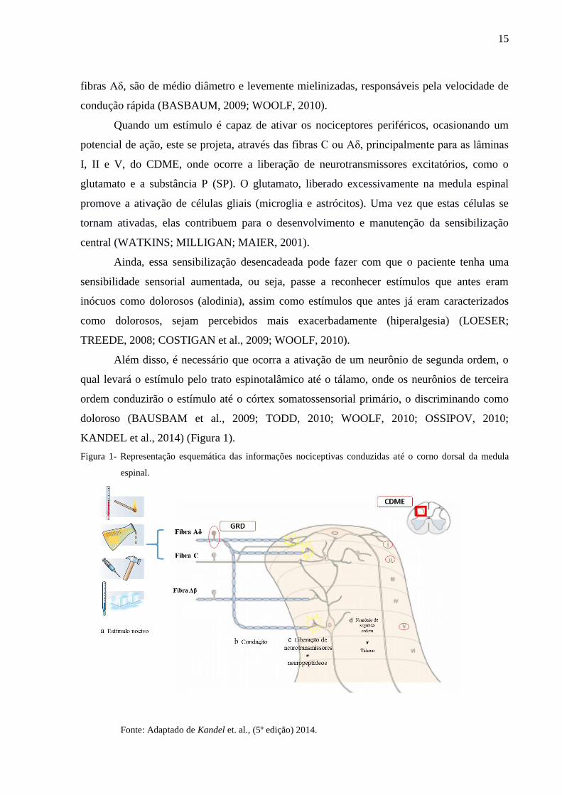
Além disso, é necessário que ocorra a ativação de um neurônio de segunda ordem, o
qual levará o estímulo pelo trato espinotalâmico até o tálamo, onde os neurônios de terceira
ordem conduzirão o estímulo até o córtex somatossensorial primário, o discriminando como
doloroso (BAUSBAM et al., 2009; TODD, 2010; WOOLF, 2010; OSSIPOV, 2010;
KANDEL et al., 2014) (Figura 1).
Figura 1- Representação esquemática das informações nociceptivas conduzidas até o corno dorsal da medula
espinal.
Fonte: Adaptado de Kandel et. al., (5º edição) 2014.
16
Nota: (a) Logo após o estímulo nocivo, ocorre a ativação dos nociceptores, que são receptores sensoriais
nociceptivos cujos corpos celulares estão localizados no gânglio da raiz dorsal (GRD). Estes neurônios
sensoriais possuem fibras do tipo Aδ ou C. (b) As fibras sensoriais (Aδ e C) conduzem o estímulo nociceptivo até o CDME, onde chegam de forma
organizada em diferentes locais nas lâminas de Rexed (dependendo da modalidade sensorial as quais
transmitem), mas, principalmente, nas lâminas I, II e V. (c) No CDME, ocorre a liberação de neurotransmissores excitatórios, como o glutamato e de neuropeptídeos,
como a substância P, ocasionando sensibilização central. (d) Ativação de um neurônio de segunda ordem, o qual conduzirá o estímulo pelo trato espinotalâmico (via
ascendente da dor) até o tálamo.
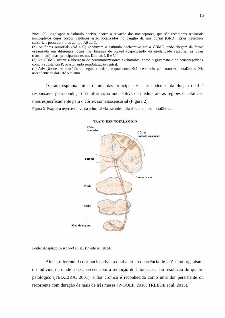
O trato espinotalâmico é uma das principais vias ascendentes da dor, o qual é
responsável pela condução da informação nociceptiva da medula até as regiões encefálicas,
mais especificamente para o córtex somatossensorial (Figura 2).
Figura 2- Esquema representativo da principal via ascendente da dor, o trato espinotalâmico.
Fonte: Adaptado de Kandel et. al., (5º edição) 2014.
Ainda, diferente da dor nociceptiva, a qual alerta a ocorrência de lesões no organismo
do indivíduo e tende a desaparecer com a remoção do fator causal ou resolução do quadro
patológico (TEIXEIRA, 2001), a dor crônica é reconhecida como uma dor persistente ou
recorrente com duração de mais de três meses (WOOLF, 2010; TREEDE et al, 2015).
17
A dor crônica é considerada um problema de saúde global, com maior gravidade e está
presente em mais de 70% dos pacientes que buscam os consultórios médicos brasileiros por
motivos diversos. Apesar de ser a razão de consultas médicas, em um terço dos casos, é de
difícil tratamento devido sua persistência e redução na qualidade de vida do paciente, já que
está associada a comorbidades comuns como: distúrbios de sono, medo, ansiedade e
depressão (RUSSO; BROSE, 1998; LOHMAN; SCHLEIFER; AMON, 2010; TRACEY;
DICKENSON, 2012).
Diversas patologias levam a uma redução da qualidade de vida do paciente, devido sua
sintomatologia e a não existência de um tratamento adequado, dentre estas temos a
fibromialgia (WOLF, 2010; TREEDE et al., 2015).
2.2 FIBROMIALGIA
A fibromialgia (FM; CID-10 M797) é caracterizada, principalmente, como dor
primária crônica generalizada, não-inflamatória e que possui destaque por seu diagnóstico
apresentar bastante controvérsias no campo da reumatologia (TREEDE et al., 2015). A FM
tem prevalência de 1-3% da população e afeta, principalmente, mulheres dos 35 aos 50 anos e
o diagnóstico é feito, exclusivamente, pela avaliação dos sintomas do paciente
(JESCHONNECK et al., 2001).
A dor é a principal característica desta patologia, porém há outros sintomas relevantes
como ansiedade, depressão, fadiga, rigidez matinal, distúrbios do sono e psicológicos
(MARQUES et al., 2002). Numerosos estudos relacionam os problemas de sono tanto à dor,
como à depressão, uma vez que, quando deprimidos, os pacientes apresentam níveis de
atividade reduzidos e níveis de sono aumentados durante o dia, caracterizando dificuldade ao
adormecer e interrupções de sono na madrugada (BIGATTI et al., 2008).
Uma vez que não existem exames laboratoriais específicos para o diagnóstico da FM,
ele se faz pela história clínica do paciente. Além disso, os pacientes com esta síndrome
musculoesquelética possuem sítios anatômicos dolorosos específicos, os tender points, que
auxiliam no diagnóstico da FM, a partir de escores propostos em 1990 pelo Colégio
Americano de Reumatologia (American College of Reumathology - ACR, 1990). Estes escores
variam de acordo com a seguinte escala: 0 – ausência de dor; 1 – dor leve; 2 – exclamação
verbal da dor; 3 – movimento de retirada ou expressão facial de dor.

18
Além desses escores preestabelecidos, se faz uso de duas variáveis: dor bilateral,
acima e abaixo da cintura pélvica; dor nos lados esquerdos e direitos do corpo como
representado no Quadro 1 (WOLFE et al., 1990; ATALLAH-HAUN et al., 1999,
PROVENZA et al., 2004).
Quadro 1- Tender points bilaterais e suas localizações.
Cervical baixo No ligamento intertransverso C5-C6
Segunda costela Na segunda junção costocondral
Epicôndilo lateral À uma distância de 2cm do epicôndilo
Joelho Acima da linha média do joelho
Occipital Na inserção do músculo suboccipital
Trapézio No ponto médio da borda superior
Supra – espinhoso Acima da escápula
Glúteo Na porção anterior do glúteo
Trocânter maior do fêmur Posterior à eminência trocantérica
Fonte: Adaptada de WOLFE et.al,1990.
Será considerado um quadro clínico de fibromialgia se os critérios citados acima,
forem satisfeitos (WOLFE et al., 1990; 1997) somado à dor generalizada crônica com mais de
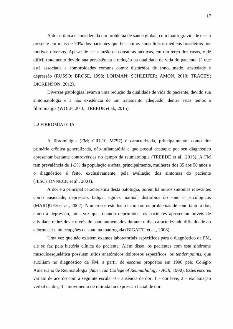
três meses de duração e dor à palpação em pelo menos 11 dos 18 pontos dolorosos (Figura 3),
onde um ponto é considerado positivo, quando o indivíduo classificar e declarar a força
aplicada no local (palpação) como dolorosa.
Em contrapartida com o que foi citado acima, outro estudo adiciona critérios mais
simples e práticos que contribuem para o diagnóstico da FM, sem a quantificação do número
de tender points. Estes novos critérios avaliam o índice de dor generalizada (0-19 pontos) nas
costas, quadril, mandíbula, ombros, braços, pernas, abdômen, peito (todas estas áreas
bilateralmente) e uma escala 0-3 para cada sintoma como: fadiga, insônia e sintomas
cognitivos, somados à severidade total destes sintomas (0-3), totalizando uma escala de 0-12.
O paciente é considerado acometido por FM se apresentar um índice de dor generalizada ≥ 7 e
escala ≥ 5; ou quando o índice de dor generalizada for de 3–6 e escala ≥ 9. Esta escala
classifica corretamente 88% dos casos de FM, definidos pelo American College of
Reumathology, não necessitando de avaliação física ou palpação dos sítios dolorosos
(WOLFE et al., 2010a).
Wolfe (2010) afirma que com a utilização deste critério é possível estudar mais
amplamente, de forma simples e sem muitos custos, conduzindo para uma “nova era” de
pensamento sobre a FM e dor crônica.
19
Apesar da etiopatologia desta síndrome não ser muito conhecida, vários estudos
evidenciam a complexa interação entre o SNC e o periférico (ELVIN et al., 2006), e
principalmente a disfunção neuro-hormonal que acarreta na redução ou deficiência de
neurotransmissores, como a 5-HT (HELFENSTEIN et al., 2002). Como tratamento são
usados antidepressivos tricíclicos, os quais atuam auxiliando na redução da sintomatologia no
indivíduo, contribuindo assim, para uma melhora na sua qualidade de vida (LITTLEJOHN;
GUYMER; 2006; BELLATO, 2012).
Figura 3 – Representação ilustrativa da localização dos 18 pontos dolorosos (tender points).
Fonte: https://trynabpainfreemomma.wordpress.com/
Como modelo animal de fibromialgia se tem utilizado injeções musculares de salina
ácida (KIM et al, 2009), estresse por frio intermitente (NISHIYORI; UEDA, 2008) e o
modelo de depleção de aminas biogênicas. Este último modelo foi escolhido para o presente
trabalho, no qual há a aplicação subcutânea durante três dias consecutivos de reserpina (1
mg/kg) (NAGAKURA et.al, 2009).
A reserpina é um depletor de aminas biogênicas, no qual age no transportador
vesicular de monoaminas (VMAT), bloqueando a transmissão e o armazenamento neuronal,
participando então da patofisiologia da dor (LOHR; KUCZENSKI; NICULESCU,2003).
Ainda, a reserpina era utilizada como antipsicótico no tratamento de esquizofrenia
(CONLEY; KELLY, 2001), assim como em diversos modelos de distúrbios neurológicos,
como a doença de Parkinson (CARLSSON; LINDQVIST; MAGNUSSON, 1957; BURGER

20
et al., 2003; RECKZIEGEL et al., 2015) e discinesia tardia (movimentos de mastigação no
vácuo) (BURGER et al., 2004; BUSANELLO et al., 2011; RECKZIEGEL et. al, 2015).
2.3 SEROTONINA
A 5-HT é uma indolamina notada por seu papel como neurotransmissor e proveniente
da hidroxilação e de descarboxilação do aminoácido triptofano obtido da dieta (O’MAHONY
et.al, 2015). A síntese de 5-HT ocorre nos neurônios serotoninérgicos que constituem um
grupo de células multipolares distribuídas e localizadas no núcleo medial e dorsal da rafe
(REIS, 2007). Atua influenciando quase todas as funções cerebrais de forma direta ou
estimulando o sistema GABAérgico, modulando o humor, sono, atividade sexual, apetite,
ritmo circadiano, funções neuroendócrinas, temperatura corporal, sensibilidade à dor,
atividade motora e funções cognitivas (MOHAMMAD-ZADEH et al., 2008).
Na transdução do sinal nociceptivo, em condições fisiológicas, a substância P e a 5-
HT atuam em conjunto, ocorrendo aumento de SP, demonstrado no fluido cerebroespinal e ao
mesmo tempo há aumento dos níveis de 5-HT e consequentemente, redução da liberação de
SP na medula espinal (ERNBERG; 1999; STAUD et al., 2001; 2003; STAUD;
SMITHERMAN, 2002). No entanto, um desequilíbrio neuroquímico ou funcional entre
substância P e 5-HT estão envolvidos no processo da dor. Um estudo realizado em pacientes
com FM, mostrou que estes apresentam redução da concentração de serotonina, que atua
inibindo a SP (RUSSEL, et al., 1994), ou seja, os níveis de SP são menos controlados pelos
neurônios serotoninérgicos, contribuindo para o aparecimento do sintoma de dor
(NARANJO; TREMBLAY; BUSTO 2001).
Estes neurônios serotoninérgicos são originados no tronco encefálico e participam da
modulação descendente da dor. As estruturas envolvidas nesta modulação incluem a
substância cinzenta periaquedutal (PAG) e bulbo rostroventromedial, mais precisamente o
NMR (Figura 4). A PAG expressa grande quantidade de encefalinas, dinorfinas e receptores
opióides e envia projeções para o bulbo rostroventromedial, especialmente para o NMR, onde
a 5-HT é o neurotransmissor mais abundante encontrado (MILLAN, 2004; BASBAUM et al.,
2009; OSSIPOV, 2010).
No corno dorsal da medula espinal ocorrerá a interconexão de fibras descendentes
(serotoninérgicas), interneurônios inibitórios (encefalinérgicos) e de fibras nociceptivas
(ascendentes). As fibras serotoninérgicas irão fazer conexão com os interneurônios inibitórios

21
presentes no CDME, ativando-os e fazendo com que ocorra a liberação de encefalinas, assim,
suprimindo a atividade dos neurônios de projeção espinotalâmicos (MILLAN, 2002;
BAUSBAM et al., 2009; OSSIPOV, 2010; KANDEL et al., 2014; WOOLF, 2010).
Figura 4 – Esquema representativo da via inibitória ou descendente da dor.
Fonte: Adaptado de Kandel et. al., (5º edição) 2014.
2.4 EXERCÍCIO FÍSICO
O exercício físico tem sido um aliado importante na manutenção do bem-estar e como
recurso terapêutico na redução de patologias e doenças crônicas. Vários trabalhos mostram a
importância e os benefícios que a atividade física proporciona, como a melhora da força e
potência muscular em idosos (CECI et al., 2014), redução dos problemas de dependentes
químicos, com estimulação na liberação de substâncias neurotróficas que auxiliam na
prevenção e tratamento da dependência (FERREIRA et. al., 2016), e também como parte do
tratamento de pacientes diabéticos, mantendo um índice glicêmico adequado (VANCEA,
2009).
22
Ainda, o exercício físico tem sido capaz de beneficiar a saúde física e mental,
melhorando a qualidade de vida e diminuindo a incidência de doenças relacionadas ao estilo
de vida sedentário, tais como diabetes e obesidade (RADÁK et al., 2006; HAYES et al., 2008
MEDEIROS et al., 2011), melhorando fatores relacionados ao aprendizado e a cognição
(VAN PRAAG et al., 1999; DAVRANCHE; MCMORRIS, 2009) e auxiliando no controle de
algumas condições dolorosas (WRIGHT; SLUKA, 2001).
Há também forte evidência de que o exercício aeróbio supervisionado reduz a dor, o
número de pontos dolorosos, depressão, ansiedade, melhora a qualidade de vida, e outros
aspectos psicológicos. Uma vez que o treino aeróbio provoca mudanças neuroendócrinas,
aumentando a liberação de aminas biogênicas, como serotonina e norepinefrina (VALIM et
al., 2003; BUSH et al., 2002).
Para ser considerado um treino aeróbio, o indivíduo deve atingir 55-90% da
frequência cardíaca máxima ou 40-85% do consumo de oxigênio de reserva (VO2R). O
volume de oxigênio máximo traz dados objetivos que auxiliam na prescrição de exercícios ou
protocolos de treinamento apropriados para humanos, porém sua utilização em
experimentação animal, muitas vezes é inviável pelo alto custo com equipamentos, fazendo
com que outros métodos alternativos sejam escolhidos, como o teste de esforço máximo que é
considerado seguro e tolerado (RODRIGUES et al., 2007)
Rodrigues e colaboradores (2007), ainda relata que o teste de esforço máximo (TEM),
relaciona-se com a avaliação da capacidade física e é utilizado na prescrição de atividade e
treinamentos físicos. Tem sido uma prática comum em experimentos com animais, onde os
animais caminham em esteira ergométrica com velocidade progressiva até seu esgotamento
físico.
Atualmente, o Colégio Americano de Medicina de Esportes (American College of
Sports Medicine – ACMS) reconhece que a intensidade de treinamento para promoção de
saúde (redução do risco coronariano, melhora metabólica e de doenças degenerativas) é
menor do que aquela necessária para aumentar a aptidão cardiorrespiratória. Entretanto, é
importante conhecer a intensidade mínima de treino capaz de promover a melhora da dor
(POLLOCK et al, 1998) e se considera um treino de 50-70% de VO2máx de baixa intensidade
(HARTHMANN et al., 2007).
Para estudos de funções básicas do encéfalo em animais, se tem utilizado protocolos
de exercício físico forçado, como ocorre na esteira, os quais visam estimular respostas ao um
23
treinamento com predominância do metabolismo aeróbico, pois este tipo de exercício físico
está associado aos benefícios da saúde em geral (PLOUGHMAN et al., 2005).
A corrida em esteira ativa as respostas neuroendócrinas de estresse e mantém o animal
correndo em uma velocidade constante, de acordo com as configurações de treinamento físico
do experimento: tempo, duração, velocidade. A preferência por este tipo de atividade é
geralmente devido às maiores respostas do metabolismo aeróbio e por ser responsável pela
indução dos maiores e mais consistentes efeitos do treinamento físico (KEMI et al., 2002).
Há estudos demonstrando que exercício físico pode modificar a neurotransmissão
serotoninérgica em diferentes regiões do SNC, aumentando a concentração de 5-HT na
medula espinal, tronco encefálico, mesencéfalo, córtex cerebral e hipocampo de ratos
(GERIN; BECQUET; PRIVAT, 1995; GOMEZ-MERINO et al., 2001), no corno dorsal e
ventral da medula espinal de gatos (VEASEY et al., 1997) e atuando na melhora do
metabolismo da 5-HT no córtex cerebral (DEY; SINGH; DEY, 1992).
Sabe-se que programas de exercícios são considerados parte do tratamento de
pacientes com FM. As indicações de exercício físico são frequentes e de forma geral,
auxiliam na qualidade de vida destes indivíduos, produzindo uma diminuição do impacto da
síndrome na qualidade de vida dos pacientes (SABBAG et al., 2000).
Os efeitos benéficos do exercício físico já estão descritos e bem esclarecidos em
algumas patologias, como na prevenção de acidente vascular encefálico (HAYES et al.,
2008), e na regeneração de nervos periféricos (BOBINSKY, 2011), porém pouco se sabe
sobre neuroproteção em casos de fibromialgia.

24
3 OBJETIVOS
3.1 OBJETIVO GERAL
Avaliar o efeito neuroprotetor do exercício físico quanto à sensibilidade mecânica,
nociceptiva, comportamento locomotor, conteúdo e quantidade de neurônios marcados
com serotonina no núcleo magno da rafe em ratos submetidos ao modelo experimental
de fibromialgia induzido por reserpina.
3.2 OBJETIVOS ESPECÍFICOS
Avaliar o efeito da aplicação de reserpina quanto à sensibilidade mecânica e
nociceptiva, usando estesiômetro (filamentos de von Frey), analgesímetro (teste de
Randall-Selitto) e analgesímetro digital (teste de tail-flick).
Avaliar o efeito da aplicação de reserpina quanto à locomoção, pela análise no campo
aberto.
Mensurar o conteúdo de 5-HT no NMR, por meio de análise da densidade óptica e o
número de neurônios imunorreativos à 5-HT no NMR de ratos submetidos à injeção
de reserpina.
Avaliar o efeito de um protocolo de treinamento em esteira de 14 dias, antes da aplicação
de reserpina, nos animais submetidos ao modelo animal de fibromialgia quanto à
sensibilidade mecânica e nociceptiva, usando estesiômetro (filamentos de von Frey),
analgesímetro (teste de Randall-Selitto) e analgesímetro digital (teste de tail-flick).
Analisar o efeito de um protocolo de treinamento em esteira de 14 dias, antes da
aplicação de reserpina, nos animais submetidos ao modelo animal de fibromialgia
quanto à locomoção, pela análise no campo aberto.
Analisar o efeito de um protocolo de treinamento em esteira de 14 dias, antes da
aplicação de reserpina, nos animais submetidos ao modelo animal de fibromialgia
quanto ao conteúdo de 5-HT no NMR, por meio de análise da densidade óptica e o
número de neurônios imunomarcados com 5-HT no NMR.

25
4 MANUSCRITO
O manuscrito está disposto conforme as especificações requisitadas pela revista
BEHAVIOURAL BRAIN RESEARCH, ao qual foi submetido para publicação.

26
Physical training prevents behavioral and morphological alterations in Fibromyalgia
animal model induced by reserpine
Karen Luise dos Santos Moreiraa,c, Sílvia Barbosab, Patrícia Severo do Nascimentoa,c*
aPrograma de Pós-Graduação em Farmacologia, Universidade Federal de Santa Maria, RS,
Brazil
bLaboratório de Histofisiologia Comparada, Departamento de Ciências Morfológicas,
Instituto de Ciências Básicas da Saúde, Universidade Federal do Rio Grande do Sul, RS,
Brazil
cLaboratório de Pesquisa Experimental da Dor, Departamento de Fisiologia e Farmacologia,
Universidade Federal de Santa Maria, RS, Brazil
All the authors contributed equally to the development of this project.
*Corresponding author: Patrícia Severo do Nascimento. Departamento de Fisiologia e
Farmacologia, Universidade Federal de Santa Maria, Avenida Roraima 1000, prédio 21, sala
5210, Camobi, CEP 97105-900, Santa Maria, RS, Brazil. Telephone number: +55 55 3220
8097. E-mail address: [email protected]
Highlights
Reserpine induced mechanical and nociceptive alterations in rats.
Reserpine decreased the number of 5-HT neurons in the Nucleus Raphe Magnus.
Treadmill training prevents mechanical, nociceptive and morphological alterations caused by
reserpine.
Abstract
In fibromyalgia there are alterations in the serotoninergic system, but there is no
evidence regarding to the effect of reserpine in the nucleus raphe magnus. Physical exercise
has been recommended as complementary therapy in the treatment of this syndrome, but there
is no evidence about its neuroprotective effects. Thus, the aim of the current study was to
investigate the effect of reserpine in nucleus raphe magnus and to evaluate the effect of the
treadmill training for two weeks before reserpinization. Twenty-nine male Wistar rats were
divided in three groups: non-reserpinized rats, reserpinized rats and rats submitted to treadmill
training and reserpinized. Treadmill training was performed for two weeks. Animals were
27
reserpinized and after the last reserpinization, mechanical and nociceptive sensitivities were
assessed by von Frey, Randall-Selitto and tail-flick tests. Also, locomotor activity was
evaluated by open field test. Then, animals were transcardially perfused and the brains were
post-fixed, cryoprotected and sectioned in cryostat. Immonuhistochemistry for serotonin was
performed in the nucleus raphe magnus to evaluate the optical density and number of neurons.
Our results showed that reserpine increased mechanical and nociceptive sensitivities, as well
as decreased the number of serotonin-immunoreactive neurons in the nucleus raphe magnus.
These alterations were prevented by treadmill physical training before reserpinization. In
conclusion, our data showed that altered sensitivities were related to decreased number of
neurons in the nucleus raphe magnus. Also, physical training before reserpinization prevents
behavioral and morphological alterations caused by reserpine, showing physical training as a
neuroprotective agent in animal model of fibromyalgia.
Keywords: Widespread chronic pain. Physical exercise. Serotonin. Nucleus raphe magnus.
Introduction
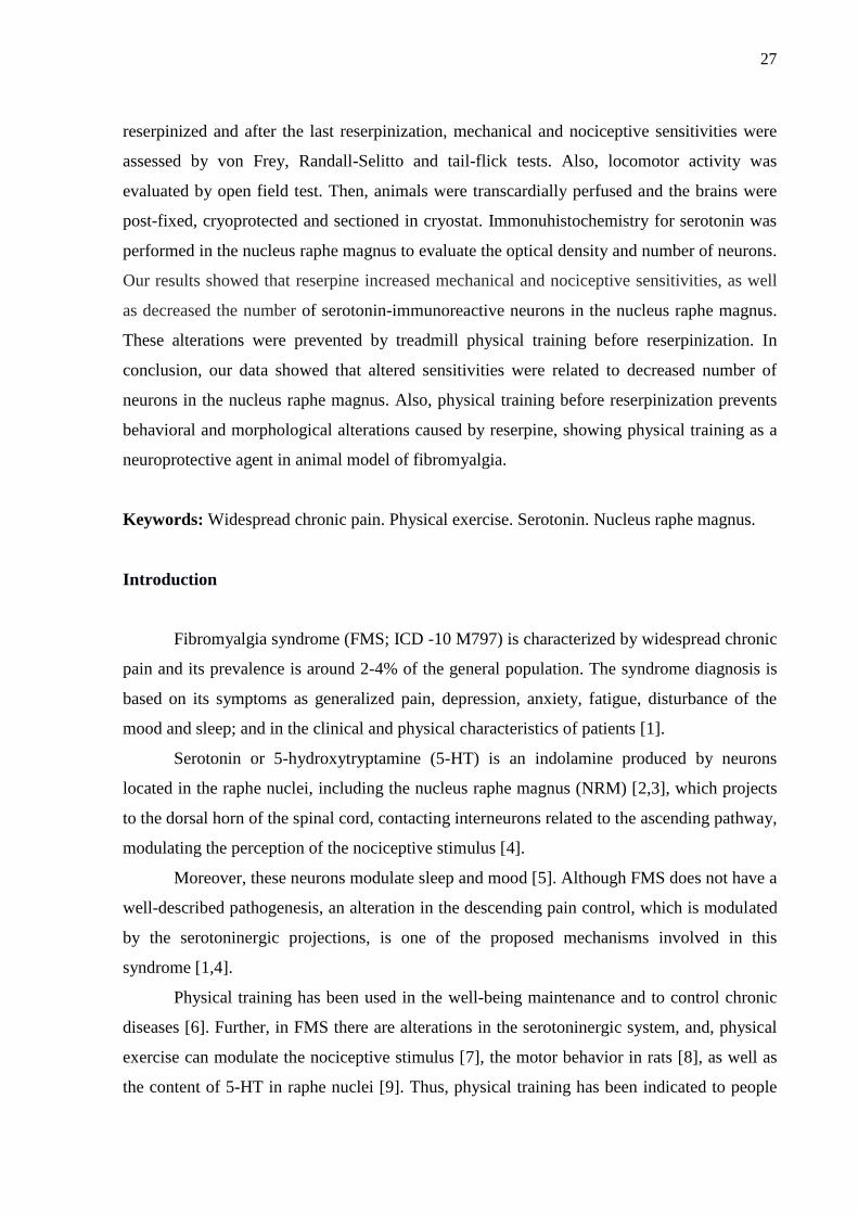
Fibromyalgia syndrome (FMS; ICD -10 M797) is characterized by widespread chronic
pain and its prevalence is around 2-4% of the general population. The syndrome diagnosis is
based on its symptoms as generalized pain, depression, anxiety, fatigue, disturbance of the
mood and sleep; and in the clinical and physical characteristics of patients [1].
Serotonin or 5-hydroxytryptamine (5-HT) is an indolamine produced by neurons
located in the raphe nuclei, including the nucleus raphe magnus (NRM) [2,3], which projects
to the dorsal horn of the spinal cord, contacting interneurons related to the ascending pathway,
modulating the perception of the nociceptive stimulus [4].
Moreover, these neurons modulate sleep and mood [5]. Although FMS does not have a
well-described pathogenesis, an alteration in the descending pain control, which is modulated
by the serotoninergic projections, is one of the proposed mechanisms involved in this
syndrome [1,4].
Physical training has been used in the well-being maintenance and to control chronic
diseases [6]. Further, in FMS there are alterations in the serotoninergic system, and, physical
exercise can modulate the nociceptive stimulus [7], the motor behavior in rats [8], as well as
the content of 5-HT in raphe nuclei [9]. Thus, physical training has been indicated to people
28
with FMS [10]. However, there is no evidence regarding the neuroprotective effects of
physical training in FMS. Therefore, the aim of this study was to evaluate the effect of the
reserpine (an alkaloid classically used to reproduce FMS animal model) in the serotonin (5-
HT) immunoreactivity in the NRM, as well as, to evaluate the neuroprotective effect of
treadmill physical training, in rats submitted to FMS animal model.
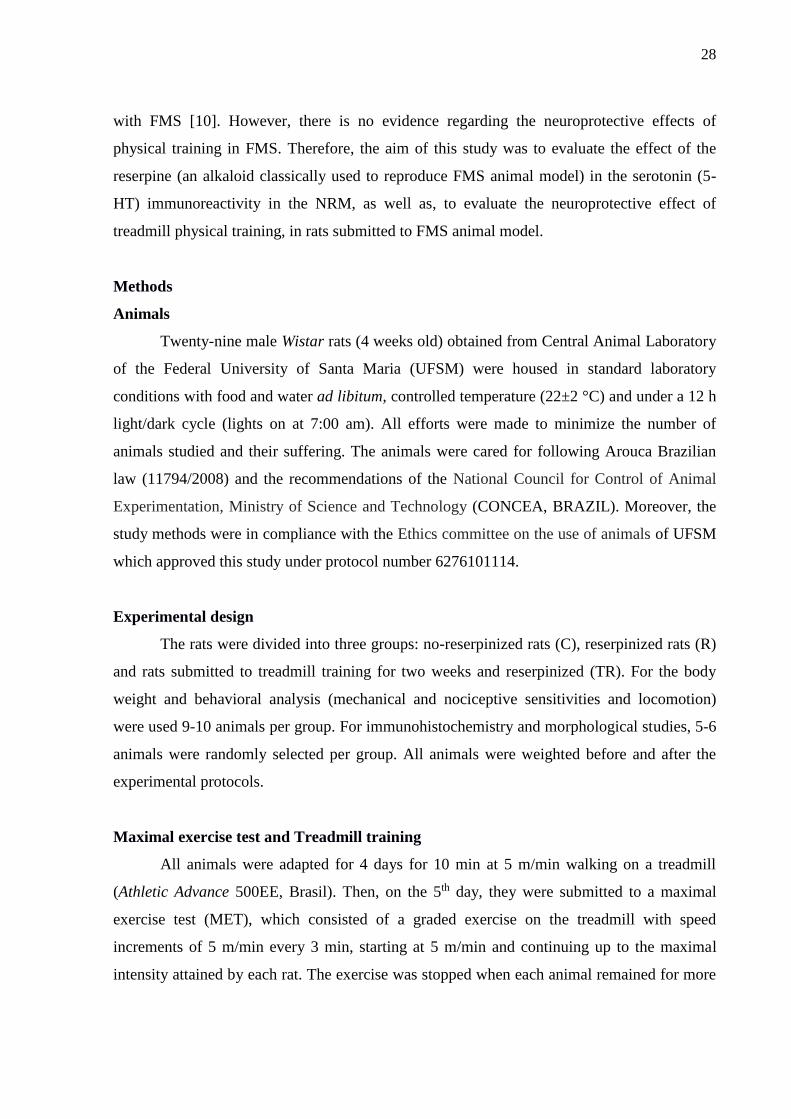
Methods
Animals
Twenty-nine male Wistar rats (4 weeks old) obtained from Central Animal Laboratory
of the Federal University of Santa Maria (UFSM) were housed in standard laboratory
conditions with food and water ad libitum, controlled temperature (22±2 °C) and under a 12 h
light/dark cycle (lights on at 7:00 am). All efforts were made to minimize the number of
animals studied and their suffering. The animals were cared for following Arouca Brazilian
law (11794/2008) and the recommendations of the National Council for Control of Animal
Experimentation, Ministry of Science and Technology (CONCEA, BRAZIL). Moreover, the
study methods were in compliance with the Ethics committee on the use of animals of UFSM
which approved this study under protocol number 6276101114.
Experimental design
The rats were divided into three groups: no-reserpinized rats (C), reserpinized rats (R)
and rats submitted to treadmill training for two weeks and reserpinized (TR). For the body
weight and behavioral analysis (mechanical and nociceptive sensitivities and locomotion)
were used 9-10 animals per group. For immunohistochemistry and morphological studies, 5-6
animals were randomly selected per group. All animals were weighted before and after the
experimental protocols.
Maximal exercise test and Treadmill training
All animals were adapted for 4 days for 10 min at 5 m/min walking on a treadmill
(Athletic Advance 500EE, Brasil). Then, on the 5th day, they were submitted to a maximal
exercise test (MET), which consisted of a graded exercise on the treadmill with speed
increments of 5 m/min every 3 min, starting at 5 m/min and continuing up to the maximal
intensity attained by each rat. The exercise was stopped when each animal remained for more
29
than 50% of the time without run or walk [11, 12]. The values obtained in the MET were used
to plan the treadmill training program.
The exercise protocol was performed on a treadmill once per day, during two weeks.
In the first week animals started to running for 30 minutes and reached the time of 60
minutes. In the second week, the animals kept running for 60 min. The training intensity was
increased gradually according to the MET results. The training sessions consisted of a warm-
up period, a main period and a cool-off period. During the warm-up period, the rats ran 30%
of the maximum velocity determined by the MET, per 5 minutes; during the main period, the
rats ran 50 minutes at 40-60% of the maximum velocity obtained in MET; and during the
cool-off period, the rats ran 5 minutes at 30% of the maximum MET values, modified from
[11].
After physical training (day 15) the rats from R and TR groups received repeated
administration of reserpine (Sigma-Aldrich, USA; 1mg/kg; subcutaneously, dissolved in
glacial acetic acid, which was diluted to a final concentration of 0.5% in distilled water) once
a day, for three consecutive days. Animals from the C group received repeated administration
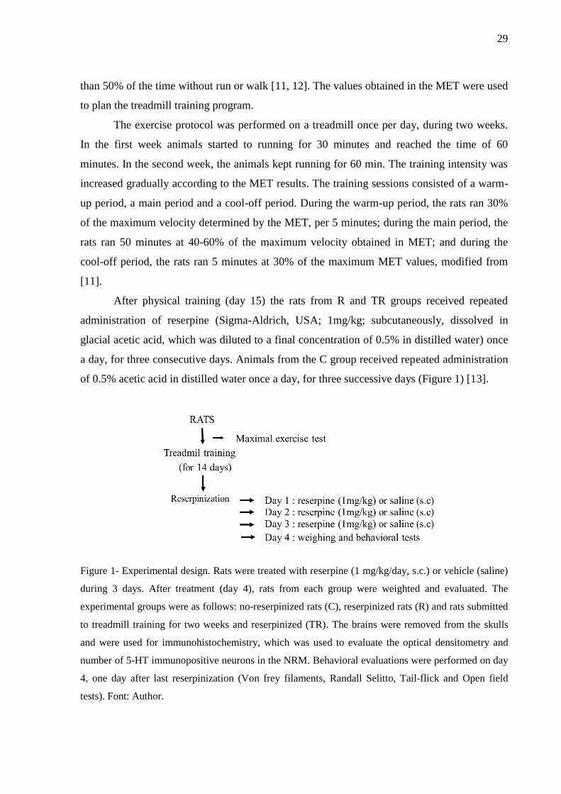
of 0.5% acetic acid in distilled water once a day, for three successive days (Figure 1) [13].
Figure 1- Experimental design. Rats were treated with reserpine (1 mg/kg/day, s.c.) or vehicle (saline)
during 3 days. After treatment (day 4), rats from each group were weighted and evaluated. The
experimental groups were as follows: no-reserpinized rats (C), reserpinized rats (R) and rats submitted
to treadmill training for two weeks and reserpinized (TR). The brains were removed from the skulls
and were used for immunohistochemistry, which was used to evaluate the optical densitometry and
number of 5-HT immunopositive neurons in the NRM. Behavioral evaluations were performed on day
4, one day after last reserpinization (Von frey filaments, Randall Selitto, Tail-flick and Open field
tests). Font: Author.
30
Behavioral Tests
Mechanical sensitivity analysis
Von Frey filaments test
The mechanical sensitivity was evaluated using a series of von Frey filaments, and the
rats were acclimatized for 30 minutes in a box (27 X 27 X 20 cm) with a mesh floor. The
filaments were applied in a consecutive sequence (15, 26, 60, and 100 g), three times, on the
plantar surface of the left and right hind paw pads. Vertical elevation of the paw immediately
upon removal of the testing hair was considered a positive response. To confirm the reaction,
this filament was replaced by a weaker one. In the absence of paw withdrawal, filaments of
increasing weight were applied sequentially [14]. This test was done one day after the last
reserpinization.
Randall- Selitto test
In the day after the last reserpinization, the animals were immobilized with soft cotton
cloth, and the same hand used to hold the tested paw. The test consisted of the application of
an increasing mechanical force, in which the tip of the device was applied to the medial
portion of the hind paws until the withdrawal response. Was applied a maximum force of
250 g to avoid skin damage. The test was performed to the left and right hind paws and the
data represents the mean of the responses from both hind paws (in grams) [15].
Nociceptive sensitivity analysis
Tail-Flick test
In the day after the last reserpinization, nociceptive sensitivity was assessed using the
tail-flick apparatus (Insight, Brazil). The rats were placed on the equipment wrapped in a
towel. A light source positioned below the tail was focused on a point 2–3 cm rostral to the tip
of the tail. With the removal of the tail, the photocell is activated, and automatically the test is
terminated. The animals were placed in the tail-flick apparatus, at the week when they were in
adaptation for the MET, to familiarize them with the procedure, as the novelty of the device
can itself induce antinociception [16]. The tail-flick latency represented the period from the
beginning of the test to the tail deflection (in seconds) [17].

31
Locomotor and exploratory activity
Open field test
The rats were gently placed in the corner of a 40 cm×50 cm×60 cm box, in which the
floor was divided into 12 squares, and then filmed with a digital camcorder for 3 min. The
number of crossings from one square to another were counted. Data represented the mean ±
standard error of the mean (SEM) of crossed squares [8].
Immunohistochemistry procedure
One day after the analyses of the motor skills, rats were anesthetized with sodium
thiopental (i.p; 50 mg/kg; Cristalia, Brazil). Heparin (1000 IU; Cristalia, Brazil) was injected
into the left cardiac ventricle, then the animals were transcardially perfused using a peristaltic
pump (Control Company, Brazil, 20 mL/min) with 400 mL of 0.9% saline solution, and after
by 400 mL of a fixative solution 4% paraformaldehyde (Synth, Brazil) in solution of the 0.1
M phosphate buffer, pH 7.4 (PB).
The brains were removed from the skulls, post-fixed in the same solution at room
temperature for 4 hours and cryoprotected by immersion in a 15% and 30% sucrose (Synth,
Brazil) solution in PB at 4 °C until they sank. After these procedures, the brains were quickly
frozen in isopentane (Neon, Brazil) cooled in liquid nitrogen and kept in a freezer (−80 °C)
for further analyses.
Coronal sections (50 μm) from nucleus raphe magnus (NRM) were obtained from
each brain using a cryostat (CM1850, Leica, Germany) and collected in a PB saline (PBS),
pH 7.4, with a temperature at −20 °C. These areas were identified using Paxinos and Watson's
Atlas (1998). The free-floating sections were pre-treated with 3% hydrogen peroxide for 30
min, carefully washed and treated with 2% bovine serum albumin (Inlab, Brazil) in PBS
containing 0.4% Triton X-100 (PBS-Tx) for 30 min and incubated with polyclonal anti-
serotonin antibody produced in rabbit (Sigma Chemical Co., USA), diluted 1:4000 in PBS-Tx
for 48 h at 4 °C. Sections were again washed in PBS-Tx and incubated in an anti-rabbit
antibody conjugated with peroxidase (Sigma Chemical Co., USA) diluted 1:200 in PBS-Tx
for two h at room temperature. The reaction was revealed in a medium containing 0.06% 3,3′-
diaminobenzidine (DAB, Sigma Chemical Co., USA) dissolved in PBS for 10 min and then 1
μL of 3% H2O2/mL was added to the DAB medium for additional 10 min. Finally, the
sections were rinsed in PBS, dehydrated in ethanol, cleared with xylene and covered with

32
Canada Balsam (MediQuímica, Brazil) and coverslips. Control sections were prepared to omit
the primary antibody by replacing it with PBS [9].
Optical densitometry
The semi-quantitative densitometric analysis was used to measure the intensity of the
5-HT immunoreaction using a digital camera (Opton®, 10.0 MP) coupled to a light
microscope (Olympus®, design CX40). The camera functions were controlled and
configurated by the interfaced software IS Capture® and the software Image J® 1.46 (NHI,
USA). The digitized images obtained from the selected areas were converted to an 8-bit
grayscale (0–255 gray levels). All lighting conditions and magnifications were held constant.
Both left and right sides of each brain were used. For each rat, five measures were taken from
the NRM. In each image two area of interest (AOI) were analyzed in the NRM. Picture
elements (pixels) employed to measure optical density were obtained from squares measuring
30,639 μm2 overlaid on the grayscale image. Background correction was done using the
rolling ball radius method.
The formula:
OD = −log[INT(x;y)−BL)]/(INC−BL)]
was used to calculated optical density (OD). Where "OD(x,y)" is the optical density at
pixel(x,y), "INT(x,y)" or intensity is the intensity at pixel(x,y), "BL" or black is the intensity
generated when no light goes through the material and "INC" is the intensity of the incidental
light [26, 12]. The results shown were the mean value ± standard error of the mean (SEM)
from the NRM [8].
Counting cells method
The same images from NRM for the optical densitometry analysis were used to count
cells. For each rat, five images were analyzed. In each image, all the 5HT-neurons in the AOI
(44,684 μm2) were counted [9]. Data represented the mean ± standard error of the mean
(SEM) from counted cells.
Statistical Analysis
Data from the body weight was evaluated by repeated measures analysis of variance
(ANOVA), and differences between the groups were assessed using the Bonferroni post-hoc
test. The data obtained from mechanical sensitivity test (von Frey filaments test and Randall-

33
Selitto test) and nociceptive sensitivities (tail-flick test), open field test (crossed squares), as
well as optical densitometry of 5HT-ir and counted cells in the NRM were analyzed using
one-way ANOVA and Bonferroni post-hoc test. The data were considered significative when
P<0.05. Data were run on Statistica 6.0 software package (StatSoft, Inc., USA). All data are
represented by the mean ± standard error of the mean (SEM).
Results
Body Weight
The body weight was not altered among the C, R and TR groups before (111.3 ± 2.5 g;
106.06 ± 2.5 g; 108.4 ± 2.6 g, respectively) and after (222.1 ± 5.5 g; 225.4 ± 5.5 g; 221.2 ±
5.8 g, respectively) training period (P>0.05). Nevertheless, there were differences in the
groups before and after the training period (P<0.05; Figure 2A).
Mechanical sensitivity results
Von Frey filaments test
One day after the last application of reserpine there were differences between R (17.2
± 3 g) and C (56.6 ± 2.9 g; P<0.05) and TR (56.2 ± 3.1 g; P<0.05). However, there was no
difference between C and TR groups (P >0.05; Figure 2B).
Randall-Selitto test
One day after the last reserpinization data showed the R (45.0 ± 5.9 g) has a decreased
threshold response compared to C (76.5 ± 5.9 g; P<0.05). Moreover, there was no difference
between C and TR groups (84.2 ± 6.2 g; P>0.05; Figure 2C).
Nociceptive sensitivity results
Tail-Flick test
After the last reserpinization, the R (1.96 ± 0.1 s) was different of C (2.42 ± 0.09 s)
and TR (2.60 ± 0.1 s) groups (P<0.05). Moreover, there were no differences between C and
TR groups (P>0.05; Figure 2D).
Open Field test
There were no differences in the number of crossed squares among C (29.3 ± 3.7), R
(35.4 ± 3.9) and TR (33.9 ± 3.9) groups (P>0.05; Figure 2E).

34
Optical densitometry and 5-HT positive counted cells
Statistical analysis showed that there were no differences in the optical densitometry
of 5-HT-ir among the C (0.0048 ± 0.0006), R (0.0035 ± 0.0007) and TR (0.0044 ± 0.0006)
groups (P>0.05; Figure 2F).
However, in the R (4.2 ± 0.2) decreased 5-HT NRM counted cells compared to C (5.4
± 0.2) and TR groups (5.7 ± 0.2; P<0.05). There was no difference between C and TR groups
(P > 0.05; Figure 2G). Figure 3 shows illustrative images from C, R and TR groups.
Discussion
In this study, we analyzed the neuroprotective effect of physical training on treadmill
before reserpinization. The mechanical and nociceptive sensitivities, locomotor activity, as
well as the optical density and the number of 5-HT immunoreactive neurons were evaluated.
In control, reserpinized and trained-reserpinized groups, the treadmill training did not
alter body weight, both in the initial and final evaluations. These data are in agreement with
other studies in which no differences in body weight were observed after training [19].
The mechanical sensitivity tests in the reserpinized animals showed an increased
mechanical sensitivity, demonstrated by the decrease in the withdrawal threshold in the von
Frey filaments and Randall-Selitto tests. However, in the trained-reserpinized animals, the
withdrawal threshold in these tasks were similar to the no-reserpinized animals. This
neuroprotective effect of physical training was shown in animal models of neuropathic pain
[20] and stroke.
Reserpinized animals had increased nociceptive sensitivity, demonstrated by the
decreased latency of tail deflection in the tail flick test. However, the latency to tail deflection
was similar in no-reserpinized animals and trained-reserpinized animals, showing the
effectiveness of treadmill training on the protection from the nociceptive effects of the
reserpine. Nevertheless, although there are no studies showing neuroprotective effects of the
treadmill training in FMS, several studies showed its benefits in oxidative stress [19], in
therapeutic of the neuropathic pain and peripheral regeneration [17,8,20], as well as in the
rats’ cognition improvement [19].

35
Figure 2- A) Body weight from C, R and TR groups before reserpinization and after the last
reserpinization. B) Mechanical sensitivity, von Frey filaments test from C, R and TR groups. *:
36
P<0.05, compared to C and TR groups. C) Mechanical sensitivity, Randall-Selitto test from C, R and
TR groups. *: P<0.05, compared to C and TR groups. D) Nociceptive sensitivity, tail flick test from C,
R and TR groups. *: P<0.05, compared to C and TR groups. E) Open field, crossed squares from C, R
and TR groups. F) Optical densitometry of 5-HT immunoreactive neurons from C, R and TR groups.
G) Number of 5-HT immunoreactive cells. *: P<0.05, compared to C and TR groups.
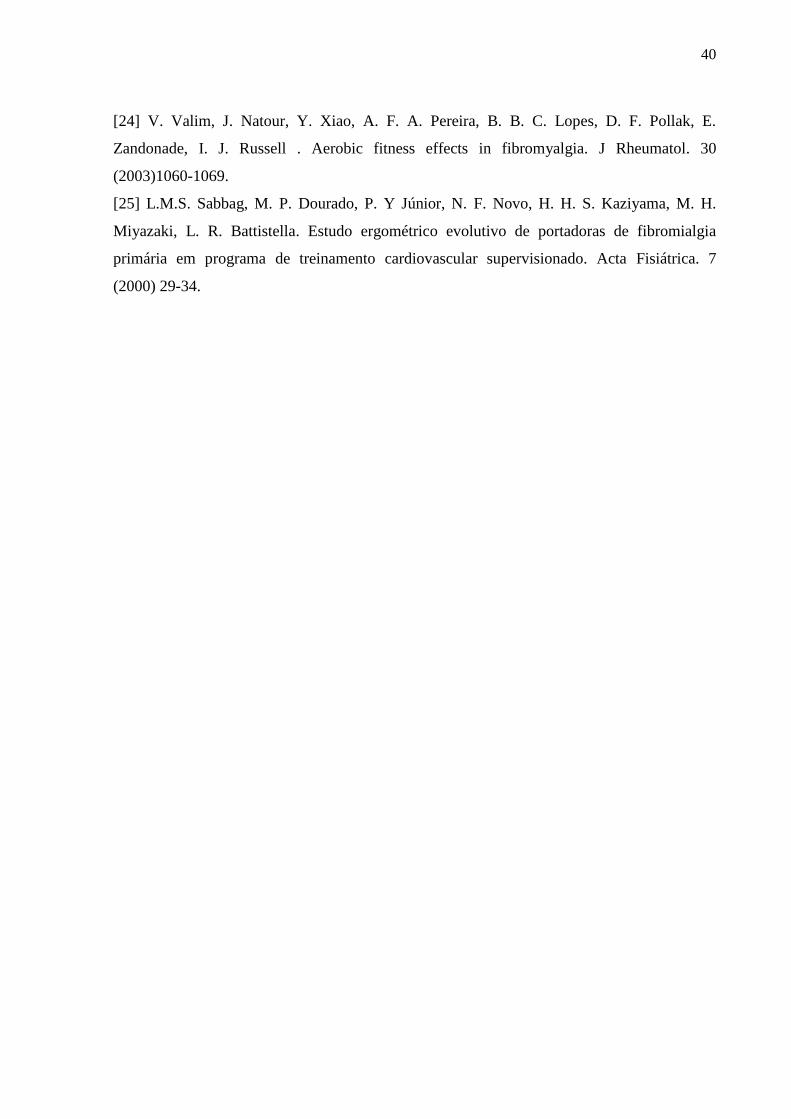
Figure 3- Illustrative images from 5-HT positive neurons in NRM from no-reserpinized (C),
reserpinized (R) and treadmill training and reserpinized (TR) groups. Scale bar: 100 µm.
In this study, the open field test showed reserpinized animals crossing the same
amount of quadrants as no-reserpinized animals and trained reserpinized. This fact indicates
that application of reserpine did not cause hypokinesia, unlikely showed by other studies in
which reserpine reducing locomotion. However, a study showed acute or chronic application
of reserpine inducing different types of locomotor behavior. While chronic application of
reserpine causes immobility, the acute application, as we did in the present study, does not
change the motor function of animals [21]. Also, to cause alterations in the locomotor activity
in rodents is needed 2 mg/kg or 10 mg/kg of reserpine, as showed in previous studies [22].
The 5-HT immunoreactivity in NRM neurons was similar in all groups. However,
reserpinized animals showed fewer 5-HT neurons than no-reserpinized and trained-
reserpinized animals. In literature, some studies reveal that application of reserpine for three
consecutive days reproduces the animal model of fibromyalgia, causing monoamines
depletion, such as serotonin [13,5] dopamine and noradrenaline in spinal cord, thalamus, and
prefrontal cortex [23,5]. Moreover, it is well known that NRM neurons are pain modulators,
and their serotoninergic neurons stimulate opioids neurons in the spinal cord that inhibits
ascending pain pathway [2].
In addition, our study shows that previous physical exercise prevents the decreases in
the number of 5HT- positive neurons in reserpinized rats, showing physical training protects

37
NRM neurons from reserpine. Studies had demonstrated physical exercise as an important
allied in the maintenance of quality of life and as a tool in reduction risk of pathologies and
chronic diseases. Many studies have shown physical exercise benefiting physical and
emotional health, providing muscular strength in old-aged people [6] and controlling pain,
anxiety, sleep and psychological well-being [24]. Nevertheless, literature describes protocols,
that in general, reduces the impact of the syndrome in quality of life of affected patients with
fibromyalgia [25], but not the physical exercise as neuroprotective in this syndrome, as
showed in our study.
In conclusion, our study showed that application of reserpine caused alterations in
mechanical and nociceptive sensitivities, as well as decreased number of 5-HT positive
neurons in NRM neurons. Physical training before reserpinization prevents behavioral and
morphological alterations caused by reserpine, showing physical training as a neuroprotective
agent in animal model of fibromyalgia.
Aknowledgements
We thank to Eliane Maria Zanchet for the Randall Slitto apparatus loan. We are in
debt with Simone Marcuzzo and Laboratório de Histofisiologia Comparada (UFRGS) by
technical support. This study was supported by grants from CNPq. K.L.S. Moreira was
supported by a Master Scholarship from CAPES.
References
[1] E. Bellato, E. Marini, F. Castoldi, N. Barbasetti, L. Mattei, E. Bonasia, D. Blonna.
Fibromyalgia Syndrome: Etiology, Pathogenesis, Diagnosis and Treatment. Pain Res Treat.
(2012) 1-17.
[2] J.P Hornung. The human raphe nuclei and the serotonergic system. J Chem Neuroanat. 6
(2003) 331- 343.
[3] G. Paxinos; C. Watson. The rat brain in stereotaxic coordinates. Academic express.
Compact. 6th-London 2009.
[4] M. H. Ossipov, G. O. Dussor, F. Porreca. Central modulation of pain. J Clin Invest. (2010)
120 (11), 3779–3787.
[5] A. Blasco-Serra, F. Escrihuela-Vidal, E.M. González-Soler, F. Martínez-Expósito, M.C.
Blasco-Ausina, S. Martínez-Bellver, A. Cervera-Ferri, V. Teruel-Martí, A.A. Valverde-

38
Navarro. Depressive-like symptoms in a reserpine-induced model of fibromyalgia in rats.
Physiol Behav. 151 (2015) 456–462.
[6] R. Ceci, M.R.B. Valls, G. Duranti, I. Dimauro, F. Ouaranta, M. Pittaluga, S. Stefania, P.
Caserotti, P. Parisi, A. Parisi, D. Caporossi. Oxidative stress responses to a graded maximal
exercise test in older adults following explosive-type resistance training. Redox Biol. 2 (2014)
65–72.
[7] L. Mazzardo - Martins, D.F Martins, R. Marcon, U.D Dos Santos, B. Speckhann, V.M
Gadotti, A.R Sigwalt, L.G Guglielmo, A.R Santos. High-Intensity extended swimming
exercise reduces pain-related behavior in mice: involvement of endogenous opioids and the
serotonergic system. J Pain. 11 (2010) 1384-1393.
[8] P. S Do Nascimento, G. A Lovatel, S. Barbosa, J. Ilha, L. A Centenaro, T. Malysz, L.L.
Xavier, B.D Schaan, M. Achaval. Treadmill training improves motor skills and increases
tyrosine hydroxylase immunoreactivity in the substantia nigra pars compacta in diabetic rats.
Brain Res. 1382 (2011) 173-180.
[9] A. Korb, L.V. Bonetti, S.A Da Silva, S. Marcuzzo, J. Ilha, M. Bertagnolli, W.A Partata,
M.C Faccioni-Heuser. Effect of treadmill exercise on serotonin immunoreactivity in
medullary raphe nuclei and spinal cord following sciatic nerve transection in rats. Neurochem
Res. 35 (2010) 380-389.
[10] AMERICAN COLLEGE OF RHEUMATOLOGY. Fibromyalgia.
www.rheumatology.org 2010 (accessed 31 March 2017).
[11] J. Ilha, R.T Araujo, T. Malysz, E.E Hermel, P. Rigon, L.L Xavier, M. Achaval.
Endurance and resistance exercise training programs elicit specific effects on sciatic nerve
regeneration after experimental traumatic lesion in rats. Neurorehabil Neural Repair. 22
(2008) 355-366.
[12] B. Rodrigues, D.D. Figueroa, C.T. Mostarda, M.V. Heeren, M.C. Irigoyen, K. De
Angelis. Maximal exercise test is a useful method for physical capacity and oxygen
consumption determination in streptozotocin-diabetic rats. Cardiovasc. Diabetol. 6 (2007) 1–
7.
[13] Y. Nagakura, T. Oe, T. Aoki, N. Matsuoka. Biogenic amine depletion causes chronic
muscular pain and tactile allodynia accompanied by depression: A putative animal model of
fibromyalgia. Pain. 146 (2009) 26-33.

39
[14] O. Ilnytska, V.V Lyzogubov, M.J. Stevens, V.R. Drel, N. Mashtalir, P. Pacher, M.A.
Yorek, I.G. Obrosova. Poly (ADP-Ribose) polymerase inhibition alleviates experimental
diabetic sensory neuropathy. Diabetes. 55 (2006) 1686-1694.
[15] E. Santos-Nogueira, E.R. Castro, R. Mancuso, X. Navarro. Randall-Selitto Test: A New
Approach for the Detection of Neuropathic Pain after Spinal Cord Injury. J Neurotrauma. 29
(2012) 898–904.
[16] C.A. Netto, B. Siegfried, I. Izquierdo. Analgesia induced by exposure to a novel
environment in rats: effect of concurrent and post-training stressful stimulation. Behav Neural
Biol. 48(2) (1987) 304-309.
[17] P.S . Diabetes increases mechanical sensitivity and causes morphological abnormalities
in the sural nerve that are prevented by treadmill training. Muscle Nerve (Print). 47 (2013) 46-
52.
[18] L.L. Xavier, G.G. Viola, A.C. Ferraz, C. Da Cunha, J.M. Deonizio, C.C. Netto, M.
Achaval. A simple and fast densitometric method for the analysis of tyrosine hydroxylase
immunoreactivity in the substantia nigra pars compacta and in the ventral tegmental area.
Brain Res Brain Res Protoc. 16 (2005) 58-64.
[19] Z. Radak, T. Kaneko, S. Tahara, H. Nakamoto, J. Pucsok, M. Sasvari. Regular exercise
improves cognitive function and decreases oxidative damage in rat brain. Neurochem Int. 38
(2001) 17–23.
[20] F. Bobinski, D.F. Martins, T. Bratti, L. Mazzardo-Martins, E.C. Winkelmann-Duarte,
L.G. Guglielmo, A.R. Santos. Neuroprotective and neuroregenerative effects of low intensity
aerobic exercise on sciatic nerve crush injury in mice. Neuroscience. 194 (2011) 337-48.
[21] L. Antkiewicz-Michaluk, A.Wąsik, E. Możdżeń, I. Romańska, J. Michaluk. Withdrawal
from repeated administration of a low dose of reserpine induced opposing adaptive changes in
the noradrenaline and serotonin system function: A behavioral and neurochemical ex vivo and
in vivo studies in the rat. Prog Neuropsychopharmacol Biol Psychiatry. 57 (2015) 146–154.
[22] L.L. Skalisz, V. Beijamini, S.L. Joca, M.A.B.F. Vital, C. Da Cunha, R. Andreatini.
Evaluation of the face validity of reserpine administration as an animal model of depression–
Parkinson’s disease association. Prog Neuropsychopharmacol Biol Psychiatry. 26 (2002)
879– 883.
[23] T. OE, T.Y. Nagakura. Reserpine causes biphasic nociceptive sensitivity alteration in
conjunction with brain biogenic amine tones in rats. Neuroscience. 169 (2010) 1860-1871.
40
[24] V. Valim, J. Natour, Y. Xiao, A. F. A. Pereira, B. B. C. Lopes, D. F. Pollak, E.
Zandonade, I. J. Russell . Aerobic fitness effects in fibromyalgia. J Rheumatol. 30
(2003)1060-1069.
[25] L.M.S. Sabbag, M. P. Dourado, P. Y Júnior, N. F. Novo, H. H. S. Kaziyama, M. H.
Miyazaki, L. R. Battistella. Estudo ergométrico evolutivo de portadoras de fibromialgia
primária em programa de treinamento cardiovascular supervisionado. Acta Fisiátrica. 7
(2000) 29-34.
41
5 CONCLUSÕES
De acordo com os resultados obtidos podemos observar que a aplicação de reserpina
causa alterações mecânicas e nociceptivas nos animais, como também causa um decréscimo
no número de neurônios imunorreativos à 5-HT no núcleo magno da rafe.
O treinamento físico em esteira antes da aplicação de reserpina, previne estas
alterações comportamentais e morfológicas causadas pela reserpina, mostrando que o
exercício físico tem efeito neuroprotetor na Fibromialgia.

42
6 PERSPECTIVAS
Após a realização deste trabalho, se tornam necessários estudos futuros com o objetivo
de avaliar os efeitos do exercício físico em esteira antes da aplicação de reserpina onde seja
analisado o conteúdo de serotonina e a quantidade de neurônios marcados com serotonina no
corno dorsal da medula espinal.
Ainda, novos estudos, com protocolo similar ao usado neste estudo, devem ser
realizados com objetivo de serem avaliadas as aminas dopamina e noradrenalina, as quais
também são depletadas pela reserpina.
Finalmente, devem ser também estudados os efeitos do exercício físico após a
aplicação de reserpina, com objetivo de serem entendidos de melhor forma os mecanismos
pelos quais o exercício atua, em conjunto ou não com terapias farmacológicas, como parte do
tratamento do indivíduo que desenvolveu fibromialgia.
43
7 REFERÊNCIAS
ABLIN, J; NEUMANN, L; BUSKILA, D. Pathogenesis of fibromyalgia - A review. Joint
Bone Spine, v. 75, p. 273-279, 2008.
AMERICAN COLLEGE OF RHEUMATOLOGY. Fibromyalgia. www.rheumatology.org ,
2010. Acessado em 31/03/2016.
ATALLAH-HAUN, M. V. et.al. Validação dos critérios do Colégio Americano de
Reumatologia (1990) para classificação da fibromialgia, em uma população brasileira.
Revista Brasileira de Reumatologia, 39:221-30, 1999.
BASBAUM, A. L et al. Cellular and molecular mechanisms of pain. Cell, v. 139(2); p. 267-
284, 2009.
BELLATO, E. et. al. Fibromyalgia Syndrome: Etiology, Pathogenesis, Diagnosis, and
Treatment. Pain Research and Treatment, 2012, Article ID 426130, 17 pages
doi:10.1155/2012/426130, 2012
BERNACCHIO, R. M. G.; CONTIN, I.; MORI, M.. Fatores modificadores da percepção da
dor. Revista da Dor, v. 8, n. 3, p. 621- 633, jul./set. 2005.
BIGATTI. M.S. et. al. Sleep Disturbances in Fibromyalgia Syndrome: Relationship to Pain
and Depression. Arthritis & Rheumatism (Arthritis Care & Research), v. 59, No. 7, p.
961–967, 2008.
BOBINSKI, F. et al. Neuroprotective and neuroregenerative effects of lowintensityaerobic
exercise on sciatic nerve crush injury in mice. Neuroscience, v. 194, p. 337–348, 2011.
BOBINSKI, F. et al. Role of brainstem serotonin in analgesia produced by low-intensity
exercise on neuropathic pain after sciatic nerve injury in mice. Pain, v. 156(12), p. 2595-
2606, 2015.
BURGER, M.E et al. Ebselen attenuates reserpine-induced orofacial dyskinesia and oxidative
stress in rat striatum, Progress in Neuropharmacology Biology and Psychiatry, v. 27, p.
135–140, 2003.
BURGER, M. et al. Effects of age on reserpine-induced orofacial dyskinesia and possible
protection of diphenyl diselenide. Brain Research Bulletin, v.64 (4), p.339-345, 2004.
BUSANELLO, A. et al. Resveratrol protects against a model of vacuous chewing movements
induced by reserpine in mice. Behavioral Pharmacology, v. 22 (1), p.71-75, 2011.
BUSCH, A. et.al. Exercise for treating fibromyalgia syndrome. Cochrane Database
Systematic Review 2, v. 3, 2002.
44
BUSCH, A. J. et al. Exercise Therapy for Fibromyalgia. Current Pain Headache Reports, v.
15 (5), p. 358-367, 2011.
CARLSSON, A.; LINDQVIST, M.; MAGNUSSON, T. 3,4-Dihydroxyphenylalanine
and 5-hydroxytryptophan as reserpine antagonists. Nature, v. 180, p. 1200, 1957.
CECI. R. et. al. Oxidative stress responses to a graded maximal exercise test in older adults
following explosive-type resistance training. Redox Biology, v. 2, 65–72, 2014.
CONLEY, R.R; KELLY, D.L Management of treatment resistance in schizophrenia,
Biologycal Psychiatry, v. 50, p. 898–911, 2001.
COSTIGAN, M.; SCHOLZ, J.; WOOLF, C. J. Neuropathic pain: a maladaptive response of
nervous system damage. Annual Review of Neuroscience, v. 32, p. 1-32, 2009.
DAVRANCHE, K.; MCMORRIS, T. Specific effects of acute moderate exercise on cognitive
control. Brain and Cognition, v. 69, n. 3, p. 565-70, 2009.
DELLAROZA et. al., Caracterização da dor crônica e métodos analgésicos utilizados por
idosos da comunidade. Revista Associação Médica Brasileira; v. 54(1), p. 36-41, 2008.
DEY, S.; SINGH, R. H.; DEY, P. K. Exercise training: significance of regional alterations in
serotonin metabolism of rat brain in relation to antidepressant effect of exercise. Physiology
& Behavior, v. 52, n. 6, p. 1095-9, 1992.
DO NASCIMENTO, P.S. et.al. Treadmill training improves motor skills and increases
tyrosine hydroxylase immunoreactivity in the substantia nigra pars compacta in diabetic rats.
Brain Research, v. 1382, p. 173-180, 2011.
et.al. Diabetes increases mechanical sensitivity and causes morphological abnormalities in
the sural nerve that are prevented by treadmill training. Muscle & Nerve (Print), v. 47, p. 46-
52, 2013.
ELVIN, A. et.al. Decreased muscle blood flow in fibromyalgia patients during standardised
muscle exercise: a contrast media enhanced colour doppler study. European Journal of
Pain, London, v. 10, no. 2, p. 137-144, 2006.
ERNBERG, M. Significance os serotonina for pain, allodynia, and hyperalgesia in the human
masseter muscle. KI Open Archive, openarchive.ki.se/xmlui/handle/10616/4487, acessado
em 15/02/2018.
FERREIRA. S.E. et. al. Efeitos agudos do exercício físico no tratamento da dependência
química. Revista Brasileira de Ciências do Esporte. +Model RBCE-1956; No. of Pages 9,
2016.
45
GERIN, C.; BECQUET, D.; PRIVAT, A. Direct evidence for the link between
monoaminergic descending pathways and motor activity. I. A study with microdialysis probes
implanted in the ventral funiculus of the spinal cord. Brain Research, v. 704, n. 2, p. 191-
201, 1995.
GOMEZ-MERINO, D. et al. Site-dependent effects of an acute intensive exercise on
extracellular 5-HT and 5 HIAA levels in rat brain. Neurosci Letters, v. 301, n. 2, p. 143-6,
2001.
HARTHMANN, A.D.et. Al. Exercise training improves arterial baro- and chemoreflex in
control and diabetic rats. Autonomic Neuroscience, v. 133, 115–120, 2007.
HAYES, K. et al. Forced, not voluntary, exercise effectively induces neuroprotection in
stroke. Acta Neuropathologica, v. 115, n. 3, p. 289-96, 2008.
HELFENSTEIN, M.; FELDMAN, D. Síndrome da fibromialgia: características clínicas e
associações com outras síndromes disfuncionais. Revista Brasileira de Reumatologia, v. 42,
n. 1, p. 8-14, 2002.
JESCHONNECK, M.et.al. Abnormal microcirculation and temperature in skin above tender
points in patients with fibromyalgia. Rheumatology, v. 39, n. 8, p. 917-921, 2001.
KANDEL et al. Princípios de neurociências. Artmed, Porto Alegre, 5ed. 2014.
KEMI OJ, et.al. Intensity-controlled treadmill running in mice: cardiac and muscle
hypertrophy. Journal of Applied Physiology, v. 93:1301-9, 2002.
KIM, S. H., et.al. Effect of the combined use of tramadol and milnacipran on pain threshold in
an animal model of fibromyalgia. Korean Journal of Internal Medicine, v. 24, p. 139-142,
2009.
KORB, A. et.al. Effect of treadmill exercise on serotonin immunoreactivity in medullary
raphe nuclei and spinal cord following sciatic nerve transection in rats. Neurochemical
Research, v. 35, p. 380-389, 2010.
LITTLEJOHN, G. O; GUYMER, E. R. Fibromyalgia syndrome: which antidepressant drug
should we choose. Current Pharmaceutical Design, v. 12(1), p. 3-9, 2006.
LOESER, J. D; TREEDE, R. D. The Kyoto protocol of IASP Basic Pain terminology. Pain,
v.137(3), p. 473-477, 2008.
LOHMAN, D.; SCHLEIFER, R.; AMON, J. J. Access to pain treatment as a human right.
BMC Medical, v. 20; 8:8. doi: 10.1186/1741-7015-8-8., 2010.
LOHR, J.B; KUCZENSKI, R.; NICULESCU, A.B. Oxidative mechanisms and tardive
dyskinesia. CNS Drugs, v. 17, P. 47–62, 2003.
46
MARQUES, A. et.al. Physical theraphy in the treatment of patients with fibromyalgia: a
literature review. Revista Brasileira de Reumatologia, v. 42, n.1, 2002.
MAZZARDO-MARTINS,L., et.al. High-Intensity extended swimming exercise reduces pain-
related behavior in mice: involvement of endogenous opioids and the serotonergic system.
The Journal of Pain, v. 11, p. 1384-1393, 2010.
MEDEIROS, C. et al. Exercise training reduces insulin resistance and upregulates the
mTOR/p70S6k pathway in cardiac muscle of diet-induced obesity rats. Journal of Cell
Physiology, v. 226, n. 3, p. 666-74, 2011.
MILLAN, M. J. Descending control of pain. Progress in Neurobiology, v. 66, p.355–474,
2002.
MILLAN, M. J. The role of monoamines in the actions of established and novel
antidepressant agents: a critical review. European Journal of Pharmacology, v.500, p.371–
384, 2004.
MOHAMMAD-ZADEH, L.F. et. al. Serotonin: a review. Journal of Veterinary
Pharmacology and Therapeutics, v. 31, n. 3, p. 187-99, 2008.
NAGAKURA, Y. et.al. Biogenic amine depletion causes chronic muscular pain and tactile
allodynia accompanied by depression: A putative animal model of fibromyalgia. Pain, v. 146,
p. 26-33, 2009.
NARANJO, C.A; TREMBLAY, L.K.; BUSTO, U.E. The role of the brain reward system in
depression. Prog. Neuro-Psychopharmacol. Biological Psychiatry, v.25, p.781– 823, 2001.
NISHIYORI, M.; UEDA, H. Prolonged gabapentin analgesia in an experimental mouse
model of fibromyalgia. Molecular Pain, v. 6, p. 4-52, 2008.
O’MAHONY, S. M. et al. Serotonin, tryptophan metabolism and the brain-gut-microbiome
axis. Behavioural Brain Research, v. 277, p. 32–48, 2015.
OSSIPOV, M. H; DUSSOR. G. O; PORRECA, F. Central modulation of pain. The Jounal
of Clinical Investigation, v. 120 (11), p. 3779–3787, 2010.
PLOUGHMAN, M. et.al. Endurance exercise regimens induce differential effects on brain-
derived neurotrophic factor, synapsin-I and insulin-like growth factor I after focal ischemia.
Neuroscience, v. 136, p 991-1001, 2005.
POLLOCK, M.L. et al. The recommended quantity and quality of exercise for developing and
and maintaining cardiorespiratory and muscular fitness, and flexilibility in healthy adults.
Medicine Science in Sport and Exercise, v. 30, p. 975-91, 1998.

47
PROVENZA J.R. et. al. Fibromialgia. Special article, Revista Brasileira de Reumatologia,
v. 44, p. 443-9, 2004.
RADAK, Z. et al. The effects of training and detraining on memory, neurotrophins and
oxidative stress markers in rat brain. Neurochemistry International, v. 49, p. 387-92, 2006.
RECKZIEGEL et al. Extracellular dopamine and alterations on dopamine transporter are
related to reserpine toxicity in Caenorhabditis elegans. Archives Toxicology, DOI
10.1007/s00204, v. 15, p. 1451-1457, 2015.
REIS, L.C. Role of the serotoninergic system in the sodium appetite. Anais da Academia
Brasileira de ciências, v. 79, p. 261-83, 2007.
RODRIGUES, B. et. al. Maximal exercise test is a useful method for physical capacity and
oxygen consumption determination in streptozotocin-diabetic rats. Cardiovascular
Diabetology, v. 6, p.1–7, 2007.
RUSSELL I.J. et al. Cerebrospinal fluid biogenic amine metabolites in fibromyalgia/fibrositis
syndrome and rheumatoid arthritis. Arthritis and Rheumatism, v. 35, p. 550-556, 1992.
RUSSELL I.J. et al. Elevated cerebrospinal fluid levels of substance P in patients with the
fibromyalgia syndrome. Arthritis and Rheumatism, v. 37, p. 1593-1601, 1994.
RUSSO, C. M.; BROSE, W. G. Chronic pain. Annual Review of Medicine, v. 49, p. 123-
133, 1998.
SABBAG, L.M.S, et.al. Estudo ergométrico evolutivo de portadoras de fibromialgia primária
em programa de treinamento cardiovascular supervisionado. Acta Fisiátrica 7: 29-34, 2000.
STAUD, R. et al. Abnormal sensitization and temporal summation of second pain (wind-up)
in patients with fibromyalgia syndrome. Pain, v.91, p.165-175, 2001.
STAUD, R.; SMITHERMAN, M.L. Peripheral and central sensitization in fibromyalgia:
pathogenetic role. Current Pain and Headache Reports. v. 6, p. 259–266, 2002.
STAUD, R. et al. Temporal summation of pain from mechanical stimulation of muscle tissue
in normal controls and subjects with fibromyalgia syndrome. Pain. v. 102, p. 87–95, 2003.
TEIXEIRA, M.J. Fisiopatologia da nocicepção e da supressão da dor. JBA. v.1, p.329-334,.
2001.
TODD, A.J. Neuronal circuitry for pain processing in the dorsal horn. Nature Reviews
Neuroscience, v. 11, p. 823–836, 2010.
TRACEY, I. DICKENSON, A. Pain perception. Cell, v. 148, p. 1308 (a-b), 2012.
TREEDE, R. et al. A classification of chronic pain for ICD-11. Pain, v. 156, p. 1003–1007,
2015.

48
VALIM, V. et al. Aerobic fitness effects in fibromyalgia. Journal of Rheumatology, v. 30, p.
1060-1069, 2003.
VAN PRAAG, H. et al. Running enhances neurogenesis, learning, and long-term potentiation
in mice. Proceedings of the National Academy of Sciences USA, v. 96, p. 13427-31, 1999.
VANCEA, D. M. M. et al. Effect of Frequency of Physical Exercise on Glycemic Control and
Body Composition in Type 2 Diabetic Patients. Arquivos Brasileiros de Cardiologia, v.
92(1), p. 23-30, 2009.
VEASEY, S. C. et al. Single-unit responses of serotonergic dorsal raphe neurons to specific
motor challenges in freely moving cats. Neuroscience, v. 79, p. 161-169, 1997.
WATKINS, L. R; MILLIGAN, E. D; MAIER, S. F. Glial activation: a driving force for
pathological pain. Trends in Neuroscience, v. 24(8): p. 450-455, 2001.
WOLFE, F, et al. The American College of Rheumatology. Criteria for the Classification of
Fibromyalgia. Report of the Multicenter Criteria Committee. Arthritis and Rheumatism, v.
33, p. 160-72, 1990.
WOLFE, F, et al. Serotonin levels, pain threshold, and fibromyalgia symptoms in the general
population. Journal of Rheumatology, v. 24, n. 3, p. 555–559, 1997.
WOLFE, F. New American College of Rheumatology criteria for fibromyalgia: a twenty-year
journey. Arthritis Care and Research, v. 62, n. 5, p. 583–584, 2010.
WOLFE, F. et al. The American College of Rheumatology preliminary diagnostic criteria for
fibromyalgia and measurement of symptom severity. Arthritis Care Res (Hoboken), v.62, p.
600 –10, 2010a.
WOOLF, C. J. What is this called pain? The Journal of Clinical Investigation, v. 120(11), p.
3742–3744. doi:10.1172/JCI45178; 2010.
WORLD HEALTH ORGANIZATION. Cardiovascular disease.
http://www.who.int/cardiovascular_diseases/en/. Acesso em: Agosto de 2017.
WRIGHT, A.; SLUKA, K. A. Nonpharmacological treatments for musculoskeletal pain. The
Clinical of Journal of Pain, v. 17, n. 1, p. 33-46, 2001.