Embed Size (px)
Citation preview

Leila do Nascimento Vieira
DESENVOLVIMENTO DE UM PROTOCOLO DE
ISOLAMENTO DE CLOROPLASTOS E DNA PLASTIDIAL EM
CONÍFERAS E SEQUENCIAMENTO DO GENOMA
PLASTIDIAL DE Podocarpus lambertii Klotzch ex Endl.
Dissertação submetida ao Programa de
Pós-Graduação em Recursos
Genéticos Vegetais da Universidade
Federal de Santa Catarina para a
obtenção do Grau de mestre em
Recursos Genéticos Vegetais
Orientador: Prof. Dr. Miguel P. Guerra
Florianópolis
2014


Este trabalho é dedicado aos meus queridos pais, Rogério e Eloisa e ao
Julião (in memoriam).

AGRADECIMENTOS
Agradeço aos meus queridos pais, Rogério e Eloisa, e ao meu irmão
Felipe, pelo apoio incondicional durante toda minha formação acadêmica.
Ao Hugo, que esteve do meu lado durante toda a realização desse
trabalho, por seu carinho, compreensão e infinita paciência.
Ao meu orientador, Prof. Miguel Pedro Guerra, por todos os
ensinamentos em nessa caminhada acadêmica, acima de tudo pela confiança
depositada para realização desse projeto e diversas oportunidades de
aprendizado junto ao LFDGV.
Ao Prof. Rubens Onofre Nodari e Prof. Marcelo Rogalski pela
atenção, por todas as trocas de experiências que tornaram esse trabalho possível
e pelo apoio de sempre.
À toda a equipe da UFPR, Prof. Emanuel M. de Souza, Prof. Fábio de
O. Pedrosa, ao Rodrigo L.A. Cardoso e principalmente ao Helisson Faoro por
terem tornado possível o sequenciamento e análises de dados presentes nesse
trabalho.
Aos amigos Douglas Steinmacher e Angelo Heringer, que mesmo de
longe sempre estiveram dando seu apoio, pela amizade construída e os ótimos
momentos compartilhados.
Aos queridos Ramon, Clarissa, Dorival, Montagna, Liliana, Catarina,
Antônio, Sabrina, Morgana, Carol Cristofolini, Sara Ferrigo, Larissa Zanette e
aos demais colegas do LFDGV pelos momentos de descontração e apoio ao
longo dessa caminhada.
Ao Eddie Vedder e ao Corey Taylor pela indispensável trilha sonora
durante os longos momentos de análises de bioinformática e escrita do trabalho.
À Bernadete Ribas pelo suporte administrativo durante o mestrado.
Ao CNPq pelo apoio financeiro através da bolsa de estudos, à
FAPESC pelo financiamento das atividades e à UFSC por prover um ensino de
qualidade.


RESUMO
O sequenciamento do genoma plastidial de coníferas vem sendo
realizado por meio do isolamento do DNA total, seguido por
amplificações do DNA plastidial através de PCR de longo alcance. Esse
método de sequenciamento é mais trabalhoso do que o sequenciamento
a partir DNA plastidial. O sequenciamento do genoma plastidial permite
a realização de várias análises comparativas, como estudos
filogenéticos, filogeografia, evolução e estrutura, localização de regiões
repetidas, identificação de sítios de edição de RNA, entre outras. Dessa
forma, o presente trabalho visou desenvolver um protocolo de
isolamento de cloroplastos e DNA plastidial em coníferas e sequenciar
o genoma plastidial do Podocarpus lambertii. O protocolo foi
desenvolvido a partir da Araucaria angustifolia e Araucaria bidwilli (Araucariaceae), P. lambertii (Podocarpaceae) e Pinus patula
(Pinaceae). O protocolo se baseia no isolamento plastidial a partir de um
tampão salino, seguido por gradiente salino de Percoll. Essas duas
estratégias combinadas, reduziram a contaminação e aumentaram o
rendimento do DNA plastidial. O DNA plastidial foi sequenciado em
sequenciador Illumina MiSeq, obtendo-se uma cobertura média do
genoma de: 24,63 para A. angustifolia, 135,97 para A. bidwilli, 1196,10
para P. lambertii e 64,68 para P. patula. O genoma plastidial do P.
lambertii é formado por 133.734 pb e apresentou a perda de uma das
regiões invertidas repetidas. O genoma contém 118 genes únicos e 1
tRNA duplicado (trnN-GUU). Estruturalmente, o genoma plastidial do
P. lambertii apresenta quatro grandes inversões de aproximadamente
20.000 pb quando comparado ao Podocarpus totara. O genoma
plastidial do P. lambertii apresenta um total de 28 repetições em tandem
e 156 simple sequence repeat (SSRs). Os resultados obtidos são
inovadores uma vez que um protocolo viável para o isolamento de DNA
plastidial de coníferas com alta qualidade e quantidade de DNA foi
obtido. Adicionalmente, a sequência do genoma plastidial do P. lambertii revelou diferenças estruturais significativas, mesmo em
relação à outra espécie do mesmo gênero. Os diversos SSRs
encontrados no genoma plastidial do P. lambertii podem ser avaliados
como regiões polimórficas intraespecíficas, que podem levar, entre
outros, a estudos filogenéticos com maior sensibilidade.
Palavras-chave: cpDNA, genoma plastidial, coníferas, cloroplasto,
sequenciamento, Podocarpus lambertii.


ABSTRACT
Plastid genome sequencing protocols for conifer species have been
based mainly on long-range PCR, which is known to be time-
consuming and difficult to implement than sequencing from isolated
plastid DNA. Plastid genome sequencing are useful for several
comparative analyzes, as phylogeny, phylogeography, evolution and
structure, location of repeated regions, identification of RNA editing
sites, among others. Thus, this study aimed to develop a protocol for
chloroplast and plastid DNA isolation in conifers and sequence the
chloroplast genome of Podocarpus lambertii. The protocol was
developed using Araucaria angustifolia and Araucaria bidwilli (Araucariaceae), P. lambertii (Podocarpaceae) and Pinus patula
(Pinaceae) species. The protocol is based on plastid isolation with saline
buffer followed by saline Percoll gradient. These two combined
strategies reduced contamination and increased the plastid DNA yield.
The plastid DNA was sequenced in MiSeq Illumina sequencer, and the
average genome coverage were 24.63 to A. angustifolia, 135.97 to A.
bidwilli, 1196.10 to P. lambertii, and 64.68 to P. patula. The chloroplast
genome of P. lambertii is 133.734 bp in length and lacks one of the
inverted repeat regions. The genome contains 118 unique genes and one
duplicated gene, the tRNA (trnN-GUU). Structurally, the plastid
genome of P. lambertii shows four large inversions of approximately
20,000 bp compared to Podocarpus totara. The plastid genome of P.
lambertii shows a total of 28 tandem repeats and 156 simple sequence
repeat (SSR). Results show that this improved protocol is suitable for
enhanced quality and yield of chloroplasts and cpDNA isolation from
conifers. Additionally, the sequence of the plastid genome of P. lambertii revealed significant structural differences, even in relation to
other species of the same genus. The various SSRs found in the plastid
genome of P. lambertii can be evaluated for intraspecific polymorphic
regions, which may allow highly sensitive phylogeographic and
population structure studies, as well as phylogenetic studies of species
of this genus.
Keywords: cpDNA, plastid genome, conifers, chloroplast, next
generation sequencing, Podocarpus lambertii.


LISTA DE FIGURAS
ESTADO DA ARTE
Figura 1. Semente madura do Podocarpus lambertii...........................................3
Figura 2. Distribuição geográfica da família Podocarpaceae...............................4
Figura 3. Podocarpus lambertii (em primeiro plano) e Araucaria angustifolia
em mata nativa no município de Lages-SC..........................................................6
Figura 4. Mapa genético do genoma plastidial de Cycas taitungensis...............12
CAPÍTULO 1
Figure 1. Flowchart showing the major steps for chloroplast isolation according
to high salt plus saline Percoll. ......................................................................... 20 Figure 2. Chloroplast visualization of Araucaria angustifolia in phase contrast
microscopy........................................................................................................ 24 Figure 3. Chloroplast DNA visualization of Araucaria angustifolia in 0.7%
agarose gel stained with ethidium bromide. ...................................................... 25 Figure 4. Reference graph track showing observed coverage values. ............... 28
CAPÍTULO 2
Figure 1. Gene map of Podocarpus lambertii chloroplast genome.. ................. 36 Figure 2. Dot-plot analyses of eight sampled conifer chloroplast DNAs against
Podocarpus lambertii.. ..................................................................................... 40 Figure 3. Comparison of IR and genome structure in 5 cupressophytes.. ......... 42


LISTA DE QUADROS
CAPÍTULO 1
Table 1. Composition of chloroplast isolation buffers and wash buffers for
modified high salt method, high salt plus saline Percoll method and sucrose
gradient method. ............................................................................................... 21 Table 2. cpDNA from different isolation methods in Araucaria angustifolia
sample. Ratios evaluated with Nanodrop®, in a final volume of 40 µl. ............ 26
Table 3. cpDNA ratios of selected conifers evaluated using Nanodrop®.
Samples were isolated with the high salt plus Percoll method in different
conifer species. Final volume of 40 µl. ............................................................. 27 Table 4. Average coverage of cpDNA evaluated from selected conifers with
CLC Genomics Workbench 5.5 software. cpDNA reads were mapped to
reference genomes. ........................................................................................... 29
CAPÍTULO 2
Table 1. List of genes identified in Podocarpus lambertii chloroplast genome.
.......................................................................................................................... 37 Table 2. List of simple sequence repeats identified in Podocarpus lambertii
chloroplast genome. .......................................................................................... 43 Table 3. Distribution of tri-, tetra-, penta-, and hexapolymer simple sequence
repeats (SSRs) loci in Podocarpus lambertii chloroplast genome. ................... 44 Table 4. Distribution of tandem repeats in Podocarpus lambertii chloroplast
genome. ............................................................................................................ 45


LISTA DE ABREVIATURAS E SIGLAS
ATP - Adenosine triphosphate
BSA - Bovine serum albumin
CDS - Coding sequences
cp - Chloroplast
cpDNA – chloroplast DNA
DOGMA – Dual organellar genome annotator
DTT - Dithiothreitol
EDTA - Ethylenediamine tetraacetic acid
IGS - Intergenic spacers
IR – Inverted repeat
KAc - Potassium acetate
LSC – Large sequence copy
NADH - Nicotinamide adenine dinucleotide
NCBI – National center for biotechnology information
OGDRAW – Organellar genome draw
ORF – Open reading frame
PCR – Polymerase chain reaction
PVP-40 - Polyvinylpyrrolidone
RFLP - Restriction fragment length polymorphism
SSC – Small sequence copy
SSR – Simple sequence repeat TIC - Translocon of inner membrane
TOC - Translocon of outer membrane
TRF – Tandem repeats finder

SUMÁRIO
INTRODUÇÃO....................................................................................... 2 REVISÃO BIBLIOGRÁFICA ....................................................... 3 1. Família Podocarpaceae 3 2. Podocarpus lambertii Klotzch ex Endl. 5 3. Estrutura e aplicação do sequenciamento do genoma plastidial 6 CAPÍTULO 1 ................................................................................... 13 Abstract 15 1 Introduction 16 2 Material and methods 17 2.1 Plant material 17 2.2 Protocols 18 2.3 Chloroplast DNA isolation 21 2.4 Microscopy analysis 22 2.5 Chloroplast genome sequencing 22 3 Results and Discussion 23 3.1 Chloroplast isolation 23 3.2 Chloroplast DNA isolation 25 3.3 Chloroplast genome sequencing 27 CAPÍTULO 2 ................................................................................... 30 Abstract 31 1 Introduction 32 2 Material and Methods 34 2.1 Plant material and cp DNA purification 34 2.2 Chloroplast genome sequencing, assembling and annotation 34 2.3 Comparative analysis of genome structure 34 2.4 Repeat sequence analysis and IR identification 35 3 Results and Discussion 35 3.1 Chloroplast genome sequencing, assembling and annotation 35 3.2 Gene content differences 37 3.3 Comparative analysis of genome structure 39 3.4 Repeat sequence analysis 42 CONSIDERAÇÕES FINAIS E PERSPECTIVAS FUTURAS ... 47 REFERÊNCIAS .............................................................................. 49


1
2
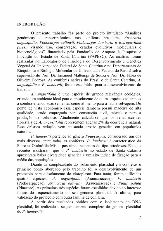
INTRODUÇÃO
O presente trabalho faz parte do projeto intitulado “Análises
genômicas e transcriptômicas nas coníferas brasileiras Araucaria angustifolia, Podocarpus sellowii, Podocarpus lambertii e Retrophyllum
piresii visando uso, conservação, estudos evolutivos, moleculares e
biotecnológicos” financiado pela Fundação de Amparo à Pesquisa e
Inovação do Estado de Santa Catarina (FAPESC). As análises foram
realizadas no Laboratório de Fisiologia do Desenvolvimento e Genética
Vegetal da Universidade Federal de Santa Catarina e no Departamento de
Bioquímica e Biologia Molecular da Universidade Federal do Paraná sob a
supervisão do Prof. Dr. Emanuel Maltempi de Souza e Prof. Dr. Fábio de
Oliveira Pedrosa. As coníferas nativas do Brasil e de Santa Catarina, A.
angustifolia e P. lambertii, foram escolhidas para o desenvolvimento do
trabalho.
A. angustifolia é uma espécie de grande relevância ecológica,
criando um ambiente ideal para o crescimento de outras espécies tolerantes
à sombra e tendo suas sementes como alimento para a fauna selvagem. Do
ponto de vista econômico essa espécie também possui madeira de alta
qualidade, sendo empregada para construção civil, móveis e para a
produção de celulose. Atualmente calcula-se que os remanescentes
florestais de A. angustifolia representem apenas 2% da ocorrência natural.
Essa drástica redução vem causando erosão genética em populações
naturais.
P. lambertii pertence ao gênero Podocarpus, considerado um dos
mais diversos entre todas as coníferas. P. lambertii é característico da
Floresta Ombrófila Mista, possuindo sementes do tipo ortodoxas. Estudos
recentes mostraram que o P. lambertii no estado de Santa Catarina
apresentara baixa diversidade genética e um alto índice de fixação para a
média das populações.
Diante da complexidade do isolamento plastidial em coníferas o
primeiro ponto abordado pelo trabalho foi o desenvolvimento de um
protocolo para o isolamento do cloroplasto. Para tanto, foram utilizadas
quatro espécies: A. angustifolia (Araucariaceae), P. lambertii
(Podocarpaceae), Araucaria bidwillii (Araucariaceae) e Pinus patula (Pinaceae). As primeiras três espécies foram escolhidas devido ao interesse
futuro do sequenciamento do seu genoma plastidial. A última, para
validação do protocolo com outra família de conífera.
A partir dos resultados obtidos com o isolamento do DNA
plastidial, foi realizado o sequenciamento completo do genoma plastidial
do P. lambertii.

3
REVISÃO BIBLIOGRÁFICA
1. Família Podocarpaceae
Dentre as Gimnospermas, a família Podocarpaceae se destaca pela
presença de um receptáculo dilatado aderido à semente, conhecido como
epimácio (Figura 1). Esta família compreende 18 gêneros e
aproximadamente 170 espécies distribuídas principalmente no hemisfério
sul (Figura 2), estendendo-se também ao norte até a China subtropical,
Japão, México e Caribe (FARJON, 1998; BIFFIN, CONRAN e LOWE,
2010).
Figura 1. Semente madura do Podocarpus lambertii. Seta: epimácio. Asterisco:
semente.
Dentre as espécies da família Podocarpaceae encontradas no
Brasil, destaca-se o P. lambertii, P. sellowii e o R. piresii. O P. sellowii distribui-se ao desde o Rio Grande do Sul até as
pequenas serras e brejos de altitude no Nordeste, junto as Floresta
Ombrófilas Densa e Floresta Estacional Semidecidual, além das florestas
de galerias serranas no domínio do cerrado (CARVALHO, 1994).
A semente do P. sellowii, sem o receptáculo carnoso, possui em
média 9,01 mm (GARCIA, 2006). Suas sementes apresentam
características recalcitrantes, com o grau crítico de umidade em torno de
26% (GARCIA, 2008).
*

4
Figura 2. Distribuição geográfica da família Podocarpaceae. Fonte: Missouri
Botanical Garden.
Até a década de 80 o gênero Decussocarpus compreendia três dos
atuais gêneros da família Podocarpaceae: Nageia, Retrophyllum e
Afrocarpus. Dessa forma, a espécie Retrophyllum piresii (Silba) C.N. Page
foi inicialmente classificada no gênero Decussocarpus, sendo em 1988,
reclassificada por C. N. Page para o gênero Retrophyllum. Atualmente, o
gênero Retrophyllum compreende apenas cinco espécies: Retrophyllum
comptonii, Retrophyllum minor, Retrophyllum piresii, Retrophyllum
rospigliosii, Retrophyllum vitiense. O gênero Retrophyllum difere de todos
os outros existentes membros da família Podocarpaceae por ter folhas
reduzidas e inclinadas nos ramos e folhas maiores e fotossinteticamente
dominantes nos brotos laterais (HILL E POLE, 1992).
R. piresii é uma espécie endêmica de Rondônia, no Parque
Nacional dos Pacaás Novos. Essa espécie foi classificada em 1976 por João
M. Pires, levando o nome de Decussocarpus piresii J. Silba em
homenagem ao seu descobridor. Nessa viagem foi realizada a coleta de
algumas sementes dos espécimes encontrados no Parque Nacional dos
Pacaás Novos, que foram levadas ao Museu Emílio Goeldi, onde
atualmente se encontram exemplares dessa espécie (WILLIAN
RODRIGUES, Comunicação pessoal). Devido à dificuldade de acesso ao
local de ocorrência natural da espécie, pouco se tem estudo sobre a mesma.
No entanto, registrou-se que João Murça Pires tinha dúvidas sobre a conceituação da espécie, devido à semelhança com a espécie andina R.
rospigliosii, pois as diferenças das formas andina e amazônica segundo ele
não eram convincentes, talvez tratando-se apenas de variedades botânicas
(LISBOA E ALMEIDA, 1995).

5
2. Podocarpus lambertii Klotzch ex Endl.
O gênero Podocarpus é um dos mais diversos entre todas as
coníferas, englobando mais de 100 espécies, muitas das quais apresentam
grande interesse florestal. Além disso, o gênero apresenta ampla
distribuição geográfica mundial, sendo registrada sua presença na Nova
Caledônia, Sudeste da Ásia, China, Japão, Malásia, Austrália, Nova
Zelândia, Bornéu, Nova Guiné, Ilhas do Pacífico, Ilhas Fiji, Antilhas,
Américas Central e do Sul (KELCH, 1998).
P. lambertii ocorre desde o estado do Rio Grande do Sul até a
Bahia, sendo encontrado também no nordeste da Argentina. A espécie é
característica da Floresta Ombrófila Mista (Figura 3), onde se apresenta
associada à A. angustifolia (CARVALHO, 1994). Apresenta características
de uma árvore perenifólia de altura variável, medindo de 1 a 10 m de altura,
apresentando tronco geralmente tortuoso, inclinado e curto, podendo
apresentar-se reto na floresta (CARVALHO, 1994). Prefere locais pouco
pedregosos, pouco inclinados, relativamente úmidos, com alta frequência
de indivíduos e alta densidade do sub-bosque, indicando ser esta uma
espécie secundária tardia tolerante a sombra (LONGHI, 2010).
Dentre as Gimnospermas, a família Podocarpaceae se destaca pela
presença de um receptáculo carnoso aderido à semente, conhecido como
epimácio. A semente do P. lambertii, sem esse receptáculo carnoso, possui
em média 4,7 mm (GARCIA, NOGUEIRA e ALQUINI, 2006). Suas
sementes são classificadas como ortodoxas, visto que a partir de um grau
de umidade inicial de 28,7% podem sofrer dessecação atingindo 5,7% de
umidade com uma taxa de germinação de 72,06% (GARCIA e
NOGUEIRA, 2008).
BITTENCOURT (2011) caracterizou a diversidade genética e
estrutura de populações naturais P. lambertii no estado de Santa Catarina. As populações avaliadas apresentaram baixa diversidade genética e um alto
índice de fixação para a média das populações, fortes indícios de que as

6
Figura 3. Podocarpus lambertii (em primeiro plano) e Araucaria angustifolia em
mata nativa no município de Lages-SC.
populações avaliadas sofreram sérios desequilíbrios resultantes de ações
antrópicas de fragmentação e exploração. Alelos raros foram também
observados nas populações e uma divergência genética significativa entre
as populações, evidenciando um reduzido fluxo gênico histórico e um
grande risco de perda de diversidade.
Alguns estudos filogenéticos foram realizados na família
Podocarpaceae com base tanto em caracteres morfológicos (KELCH, 1998)
quanto em caracteres moleculares (KELCH, 1998, 2002; BIFFIN,
CONRAN e LOWE, 2010). Em geral, a filogenia molecular está de acordo
com as classificações baseadas nos caracteres morfológicos para relações
intergenéricas. No entanto, muitas das relações intragenéricas permanecem
com pouco apoio.
3. Estrutura e aplicação do sequenciamento do genoma plastidial
O cloroplasto é uma organela originada a partir da endossimbiose,
sendo as cianobactérias o grupo mais próximo do genoma plastidial. Não

7
havendo definição clara de qual linhagem desse grupo de cianobactérias
originou essa organela (TIMMIS et al., 2004). Durante mais de um bilhão
de anos de evolução, os três genomas das células vegetais (nuclear,
plastidial e mitocondrial) sofreram mudanças estruturais dramáticas para
otimizar a expressão compartimentalizada do material genético e a
comunicação entre esses compartimentos (GREINER e BOCK, 2013).
Dentre essas modificações, destaca-se a transferência em larga
escala de informação genética do cloroplasto para o núcleo, resultando em
uma expressiva redução no tamanho do genoma plastidial (GREINER e
BOCK, 2013). Por conseguinte, os genomas plastidiais contemporâneos
contêm apenas uma pequena proporção dos genes dos seus ancestrais. No
entanto, os genes remanescentes permanecem com a característica
procariótica de organização em operons (WICKE et al., 2011). Calcula-se
que essa redução foi de em torno de 3.000 genes para cerca de 120 genes, o
que levou a uma capacidade limitada de codificação do plastoma. Estima-
se que os cloroplastos importam mais de 95% das suas proteínas do citosol
(BOCK, 2007).
Para tanto, é necessário que as pre-proteínas endereçadas ao
cloroplasto atravessem três distintos sistemas de membranas – a membrana
exterior e interior, que circundam a organela, e a membrana do tilacóide,
que contém as proteínas fotossinteticamente ativas. As pre-proteínas
destinadas ao tilacóide são sintetizadas com uma extensão amino-terminal
chamada de pre-sequência, que é reconhecida pelo translocon da membrana
exterior (TOC) e da membrana interior (TIC). No estroma ocorre a
clivagem da extensão amino-terminal, produzindo a forma madura da
proteína (SOLL e SCHLEIFF, 2004).
O DNA plastidial (cpDNA) possui características próprias distintas
do DNA nuclear. Essas características incluem diferentes porcentagens de
conteúdo GC e, na maior parte das vezes, ausência de metilação no DNA,
ou seja, ausência da 5´-metilcitosina. Alguns autores apontam que há um
baixo percentual de metilação do DNA no cloroplasto (VANYUSHIN,
2006; HUANG et al., 2012). No entanto, FOJTOVA, KOVARIK e
MATYASEK (2001) consideram que na maioria dos casos a detecção de
metilação no cpDNA ocorre devido à “artefatos” gerados pelas enzimas de
restrição utilizadas para as análises. AHLERT et al. (2009) mostraram
através da introdução dos genes da adenina e citosina DNA
metiltransferase de cianobactérias que plantas com cpDNA metilado não
apresentam diferenças morfológicas em relação ao controle não
transformado.
Apesar de pequeno, o cpDNA compreende uma fração significante
do DNA total, sendo estimado em 9% em Nicotiana tabacum (TEWARI e

8
WILDMAN, 1966). Dependendo da espécie, tecido, estádio de
desenvolvimento e condições ambientais o nível de ploidia de um
cloroplasto pode chegar a mais de 10.000 cópias idênticas do seu genoma
por célula (BENDICH, 1987).
Os plastídios e mitocôndrias são herdados de forma não-
Mendeliana em todos os eucariotos. Na maioria dos organismos, os
genomas organelares são herdados de apenas um dos pais, com a herança
materna sendo muito mais difundida do que a herança paterna
(HAGEMANN, 2004). Sabe-se que a maior parte das angiospermas possui
herança materna, com raros casos de herança biparental ou paterna
(ZHANG e SODMERGEN, 2010). Já as gimnospermas possuem
majoritariamente herança paterna (NEALE, MARSHALL e SEDEROFF,
1989). Era esperado que essa ausência de recombinação resultasse no
acúmulo de mutações deletérias, em um processo conhecido como Catraca
de Muller. No entanto, as taxas de mutação encontradas no genoma
plastidial são menores do que no genoma nuclear (GREINER e BOCK,
2013).
Todo e qualquer organismo apresenta um sistema de reparo que
mantém a estabilidade do genoma em um grau que previne excessivas
mutações, mas sem impedir a evolução (MARÉCHAL e BRISSON, 2010).
Por um lado, em nível de um único organismo, parece preferível manter o
genoma absolutamente intacto. Por outro lado, certo grau de instabilidade é
crucial para a evolução das espécies, eliminando completamente os
mecanismos que produzem essa diversidade, também se destrói a
resiliência e capacidade de adaptação das espécies às mudanças das
condições ambientais (MARECHAL e BRISSON, 2010). No entanto, os
cloroplastos mantém um mecanismo de reparo que ainda não foi
completamente esclarecido (GREINER e BOCK, 2013).
Considerou-se por muito tempo que a estrutura do genoma
plastidial seria exclusivamente circular, devido à sua origem procariótica
(BOCK, 2007; WICKE et al., 2011). No entanto, estudos recentes de
hibridização in situ demonstraram que apenas uma pequena parte das
moléculas ocorre de forma circular, ocorrendo em sua maioria de forma
concatenada de duas ou mais moléculas, em forma circular ou linear
(WICKE et al., 2011).
No âmbito da transformação genética, o sequenciamento completo
do genoma plastidial é essencial para o desenvolvimento de biotecnologias
ligadas à transformação genética de cloroplastos (CLARKE, DANIELL e
NUGENT 2011). As regiões intergênicas dos genomas plastidiais não são
bem conservadas e o desenho eficaz do vetor para a transformação de

9
novas espécies exige dados da sequência do genoma plastidial específico
de cada espécie (CLARKE, DANIELL e NUGENT 2011).
Há vantagens consideráveis associadas com a transformação
gênica de cloroplastos em vez de nuclear. A primeira é o elevado número
de plastídios e o elevado número de cópias no genoma do plastídio por
célula, oferecendo níveis de expressão do transgene extraordinariamente
altos. A segunda é que a integração do transgene no genoma do cloroplasto
ocorre exclusivamente por recombinação homóloga, tornando genoma
plastidial uma técnica de engenharia genética de alta precisão para as
plantas. A terceira é que como um sistema procarioto, o sistema genético
do cloroplasto é desprovido de silenciamento gênico e de outros
mecanismos que interferem epigeneticamente com a expressão estável do
transgene. A quarta é que da mesma forma que o genoma bacteriano,
muitos genes plastidiais são dispostos em operons, oferecendo a
possibilidade de arranjar genes em operons artificiais. Finalmente, a
transformação plastidial tem recebido atenção significativa como uma
ferramenta excelente para contenção do transgene devido ao modo de
herança materna do plastídio característica da maioria das espécies de
angiospermas, o que reduz drasticamente a segregação do transgene através
de pólen (BOCK, 2014).
As tecnologias de transformação plastidial têm sido intensamente
utilizadas em genômica funcional, através da realização de nocautes de
genes dirigidos ao cloroplasto. Estes estudos têm contribuído enormemente
para a compreensão dos processos fisiológicos e bioquímicos dentro do
compartimento plastidial (BOCK, 2001). A tecnologia de transformação de
cloroplastos também tem potencial para melhoramento de plantas,
desenvolvimento de plantas como biorreatores para a produção sustentável
e de baixo custo de produtos biofarmacêuticos, enzimas e matérias-primas
para a indústria química (BOCK, 2014).
O genoma plastidial tem se mostrado, desde a década de 80, uma
ferramenta viável para realização de estudos filogenéticos (PALMER,
1985). Em virtude da sua fácil amplificação, tamanho pequeno, baixa taxa
evolutiva e da maior conservação observada entre os genomas em evolução
conhecidos, o genoma plastidial foi considerado adequado para estudos
filogenéticos em diferentes níveis taxonômicos (PALMER, 1985).
O gene rbcL, que codifica a grande subunidade da ribulose 1,5-
bifosfato-carboxilase/oxigenase (RUBISCO), começou a ser amplamente
sequenciado a partir de um grande número de espécies, gerando uma boa
base de dados para estudos de filogenia de plantas (PALMER et al., 1988).
Estudos filogenéticos em nível de família e de taxas maiores foram

10
realizados com sucesso em gimnospermas (HASEBE et al., 1992;
BRUNSFELD et al., 1994; SETOGUCHI et al., 1998).
No entanto, em algumas situações as relações continuavam não
claras pelo fato do gene rbcL ser muito conservado para esclarecer dúvidas
entre gêneros próximos (GIELLY e TABERLET, 1994). A análise de
regiões não-codificantes do cpDNA, como introns e espaçadores
intergênicos, foi a estratégia encontrada para esclarecer as relações em
níveis taxonômicos mais baixos. Estas zonas tendem a evoluir mais
rapidamente do que as regiões codificantes, por acumulação de
inserções/eliminações a uma taxa pelo menos igual à das substituições de
nucleotídeos (GIELLY e TABERLET, 1994).
Ainda que o uso de regiões não-codificantes tenha solucionado
algumas das dúvidas no âmbito dos estudos filogenéticos, percebeu-se mais
tarde que muitas regiões não exploradas do cloroplasto poderiam trazer
informações adicionais a essa linha de estudo (SHAW et al., 2007). A
partir do sequenciamento completo do genoma plastidial pode-se encontrar
regiões com maior número de caracteres para estudos filogenéticos em
menores taxa, assim como as regiões com menor número (SHAW et al.,
2007).
Quando apenas um gene é investigado, faz-se uma amostragem de
um segmento do genoma; comparando mais genes reduz-se o erro de
amostragem inerente à amostragem de apenas um ou poucos genes;
comparando genomas completos se pode descobrir o padrão de sítios que
estão disponíveis para comparação (MARTIN et al., 2005). Além disso,
SHAW et al. (2007) aponta que não há uma região única ou um conjunto
de regiões mais indicados para todas as linhagens taxonômicas, e sim que
deve ser feito um rastreamento em cada linhagem para determinar quais as
regiões mais adequadas.
Dessa forma, há a possibilidade de se encontrar um grupo de
regiões com características em que: (i) os resultados da análise usando este
conjunto de regiões é semelhante ao dos resultados da análise multigênica
usando o número máximo de regiões, (ii) a região deve ser compacta e ser
significativamente menor do que o conjunto de multigenes (LOGACHEVA
et al., 2007).
Análises filogenéticas a partir do sequenciamento completo do
genoma plastidial foram realizadas com sucesso em Vitis (JANSEN et al.,
2006), fornecendo forte apoio ao posicionamento de Vitaceae como a
primeira linhagem divergente das rosídeas. Outros estudos, como na
família Poaceae, entre as subfamílias Panicoideae, Bambusoideae e
Pooideae, revelou uma maior proximidade entre Bambusoideae e Pooideae
(MATSUOKA et al., 2002).

11
Nas plantas terrestres, a maior parte dos genomas plastidiais é
divida em quatro partes: grande região de cópia simples (LSC), pequena
região de cópia simples (SSC) e regiões invertidas repetidas (IRA e IRB)
(Figura 3). As duas IR são idênticas na composição, dessa forma, todos os
genes contidos nessas regiões apresentam duas cópias no genoma, porém
em sentido de leitura contrário (Figura 4).
A função dessas regiões IR permanece obscura. Teorias como o
aumento na quantidade de genes altamente expressos, como genes que
codificam para rRNA e a estabilização do genoma foram propostas
(PALMER e THOMPSON, 1982).
Uma evidência circunstancial para a ação da conversão gênica nas
IR vem da observação de que a frequência de mutação de genes das regiões
IR é significativamente menor do que para genes localizados em ambas as
regiões de cópia única do genoma plastidial (WOLFE, LI e SHARP, 1987;
MAIER et al., 1995). Essas regiões IR estão frequentemente sujeitas a
expansão, contração e até mesmo perda completa (WICKE et al., 2011),
eventos esses que influenciam no tamanho do genoma plastidial (WU e
CHAW, 2013).

12
Figura 4. Mapa genético do genoma plastidial de Cycas taitungensis. Genes
mostrados no lado de dentro e do lado de fora do círculo grande são transcritos no
sentido horário e anti-horário, respectivamente (WU et al., 2007).
Algumas espécies de gimnospermas com seu genoma plastidial
sequenciado perderam umas dessas regiões IR, como é o caso da
Cryptomeria japonica, Cephalotaxus oliveri, Cephalotaxus wilsoniana,
Taiwania cryptomerioides e algumas Pinaceaes (CRONN et al., 2008;
HIRAO et al., 2008; WU et al., 2011; YI et al., 2013). Até hoje, não está
inteiramente claro se essas espécies perderam diferentes cópias das regiões
IR (IRa ou IRb). Por um lado, WU et al., (2011) e WU e CHAW, 2013
alegam que Pinaceas e Cupressophytas perderam diferentes cópias das
regiões IR, enquanto YI et al., (2013) demonstra que não há dados
suficientes para afirmar nem que sim, nem que não.

13
CAPÍTULO 1
Manuscrito publicado no periódico PLoS ONE.
An improved protocol for intact chloroplasts and cpDNA isolation in
conifers
Vieira LN, Faoro H, Fraga HPFF, Rogalski M, Souza EM, Pedrosa FO,
Nodari RO, Guerra MP. 2014. An improved protocol for intact chloroplasts
and cpDNA isolation in conifers. PLoS ONE. 9(1): e84792.

14

15
Abstract
Background: Performing chloroplast DNA (cpDNA) isolation is
considered a major challenge among different plant groups, especially
conifers. Isolating chloroplasts in conifers by such conventional methods as
sucrose gradient and high salt has not been successful. So far, plastid
genome sequencing protocols for conifer species have been based mainly
on long-range PCR, which is known to be time-consuming and difficult to
implement.
Methodology/Principal findings: We developed a protocol for cpDNA
isolation using three different conifer families: Araucaria angustifolia and
Araucaria bidwilli (Araucariaceae), Podocarpus lambertii (Podocarpaceae)
and Pinus patula (Pinaceae). The present protocol is based on high salt
isolation buffer followed by saline Percoll gradient. Combining these two
strategies allowed enhanced chloroplast isolation, along with decreased
contamination caused by polysaccharides, polyphenols, proteins, and
nuclear DNA in cpDNA. Microscopy images confirmed the presence of
intact chloroplasts in high abundance. This method was applied to cpDNA
isolation and subsequent sequencing by Illumina MiSeq (2 x 250 bp), using
only 50 ng of cpDNA. Reference-guided chloroplast genome mapping
showed that high average coverage was achieved for all evaluated species:
24.63 for A. angustifolia, 135.97 for A. bidwilli, 1196.10 for P. lambertii,
and 64.68 for P. patula.
Conclusion: Results show that this improved protocol is suitable for
enhanced quality and yield of chloroplasts and cpDNA isolation from
conifers, providing a useful tool for studies that require isolated
chloroplasts and/or whole cpDNA sequences.
Keywords: chloroplast; cpDNA; organelle isolation; Percoll gradient;
conifers; plants

16
1 Introduction
The chloroplast genome of land plants usually harbors a conserved
set of approximately 120 genes in a 120-160 kb pair genome, out of a
genome of some 3,200 genes present in their cyanobacterial ancestor
(KANEKO et al., 1996). Land plant plastomes are mostly conserved and
present little variation in size and gene content, ranging from 70,028
nucleotides and 25 protein coding genes in the nonphotosynthetic parasitic
plant, Epifagus virginiana (WOLFE et al., 1992), to 217,942 nucleotides
and 131 protein coding genes in Pelargonium x Hortorum (CHUMLEY et al., 2006). Although chloroplast genomes contain highly conserved
essential genes for plant growth and development, they also contain
variable regions, i.e., intergenic regions and structural variations. In
addition, they contain one of the few sets of characters that can transcend
the life history of green plants and, hence, generate important evolutionary
information. Therefore, chloroplast genome sequences can be used for
comparative evolutionary studies within and between different groups of
plants (TIMMIS et al., 2004;WOLF et al., 2010; GREINER et al., 2013),
as demonstrated by several works (JANSEN et al., 2007; MOORE et al.,
2007; MOORE et al., 2010; WU et al., 2011; YI et al., 2013). Furthermore,
chloroplast genome sequences have been used to investigate gene function
(ROGALSKI et al., 2008; ALKATIB et al., 2012), and they have been
targeted for biotechnological applications (CLARKE, DANIELL E
NUGENT, 2011; MALIGA E BOCK, 2011; ROGALSKI E CARRER,
2011; BOCK, 2013). Based on the importance of land plant chloroplast
DNA (cpDNA) in plant genetics, evolution and biotechnology, it has been
a target in many plant genome sequencing projects (WU et al., 2007;
HIRAO et al., 2008; LIN et al., 2010). To date, complete cpDNAs of more
than 300 plants have been sequenced
(ncbi.nlm.nih.gov/genomes/GenomesGroup.cgi?taxid=2759&opt=plastid).
With rapid progress in sequencing technologies, chloroplast genome
sequencing can be realized quickly as a result of small size and structural
simplicity when compared to nuclear genomes. However, chloroplast
genome sequences have been determined for only a very few families
belonging to gymnosperms (WU et al., 2007; WERNER et al., 2009; LIN
et al., 2010). Especially for conifers, chloroplast genome sequences are
available for families that include Cephalotaxaceae (YI et al., 2013),
Cupressaceae (HIRAO et al., 2008), Pinaceae (WAKASUGI et al., 1994;
CRONN et al., 2008; LIN et al., 2010), Podocarpaceae (database accession

17
no. NC_020361.1) and Taxaceae (database accession no. NC_020321.1),
but not the Araucariaceae family.
Chloroplast DNA isolation has been a major challenge, hindering
widespread applications in different plant groups. Chloroplast isolation in
conifers by such conventional methods as sucrose gradient (PALMER E
SEIN, 1986) and high salt (BOOKJANS, STUMMANN E HENNINGSEN,
1984) has, thus far, not been successful. This most likely results from the
high volume of contaminants, including polyphenols, oleoresins, terpenoids
and polysaccharides, present in conifer needles, making difficult the
acquisition of intact isolated chloroplasts and high quality cpDNA
(KEELING E BOHLMANN, 2006). For conifers, whole cpDNA
sequencing protocols have been based on total DNA isolation, followed by
cpDNA fragments amplification by use of polymerase chain reaction
(PCR) with degenerate primers (CRONN et al., 2008; LIN et al., 2010;
WU et al., 2011; YI et al., 2013). However, this strategy is known to be
time-consuming and difficult to implement because of differences in gene
organization among different plant species (ATHERTON et al., 2010) and
“promiscuous” cpDNA present in the nucleus and mitochondrial genome
(AYLIFFE E TIMMIS, 1992; AYLIFFE, SCOTT E TIMMIS, 1998;
GOREMYKIN et al., 2009; ROUSSEAU-GUEUTIN, AYLIFFE E
TIMMIS, 2011).
Therefore, the overall aim of the present work was to develop an
efficient protocol for chloroplast isolation and subsequent high quality
cpDNA extraction in conifers, using three different conifer families:
Araucaria angustifolia and Araucaria bidwilli (Araucariaceae),
Podocarpus lambertii (Podocarpaceae) and Pinus patula (Pinaceae).
2 Material and methods
2.1 Plant material
Local A. angustifolia and P. patula seeds were purchased and
germinated in the greenhouse of Federal University of Santa Catarina,
Brazil. Needles were collected from 6 months plants; this procedure does
not require authorization. P. lambertii young plants (n=10) were collected
at a private area, located at Lages, Santa Catarina, Brazil (27° 48' 57" S,
50° 19' 33" W), where the species is abundant, with the previous owner
permission (José Antônio Ribas Ribeiro). This species are not considered
under threat. After, the young plants were transplanted to greenhouse and
maintained under this condition until the collection of needles. A. bidwilli young needles were collected at Botanical Garden, authorized by Federal

18
University of Santa Catarina, Brazil. For each plant species, 25 g of fresh
young needles were collected and stored in 4ºC refrigerator for further
chloroplast extractions.
2.2 Protocols
The three chloroplast DNA isolation methods used here are described as
follows:
A. High salt plus saline Percoll gradient method (Figure 1)
All the following steps were carried out at 0ºC, if not otherwise stated.
1. Prior to extraction, 25 g (fresh weight) of young needles were collected
and kept in dark for 10 days at 4ºC to decrease starch and resin level. Fresh
needles were cleaned with 0.5% sarkosyl (Fluka, Ronkonkoma, NY) for 5
min to reduce microbial contamination and then washed 4 times with
distilled water.
2. Needles were homogenized in 400 ml ice-cold isolation buffer (Table 1)
for 30 s in a pre-chilled blender. Homogenate was filtered primarily into
two layers of gauze bandage and then filtered again using two layers of
Miracloth by softly squeezing the cloth.
3. Homogenate was centrifuged at 200 g for 15 min at 4ºC. The nucleus
pellet and cell-wall debris were discarded. The supernatant included
chloroplasts suspended in it.
4. The supernatant was centrifuged at the higher centrifugal force of 3000 g
for 20 min at 4ºC, resulting in a chloroplast pellet with some
contamination.
5. The pellet was gently resuspended in 12 ml of wash buffer (Table 1)
using a paintbrush.
6. Homogenate was divided into 6 tubes (50 ml), each containing 20 ml
Percoll (GE Healthcare, Uppsala, Sweden) gradient (70%-30%) and then
centrifuged at 5000 g for 25 min at 4ºC. The interface 70%-30% containing
chloroplasts was collected.
7. Collected interface containing chloroplasts was washed twice with 100
ml of wash buffer and centrifuged at 3000 g for 20 min at 4ºC to obtain the
purified chloroplast pellet.
B) Modified high salt method (SHI et al., 2012)
All the following steps were carried out at 0ºC, if not otherwise stated.
1. Prior to extraction, 25 g (fresh weight) of young needles were collected
and kept in dark for 72 h at 4ºC to decrease starch level stored in the
needles. Fresh needles were cleaned with distilled water.
19
2. Needles were homogenized in 400 ml of isolation buffer (Table 1) for 30
s. Homogenate was filtered into centrifuge bottles, using two layers of
Miracloth (Calbiochem, San Diego, CA) by softly squeezing the cloth.
3. The homogenate was centrifuged twice at 200 g for 20 min at 4ºC. The
nucleus pellet and cell-wall debris were discarded. Supernatant included
chloroplasts suspended in it.
4. The supernatant was submitted to a higher centrifugal force (3500 g) for
20 min at 4ºC, resulting in a chloroplast pellet contaminated with some
nuclear DNA.
5. The pellet was gently resuspended in 250 ml of wash buffer (Table 1),
using a paintbrush to wash the nuclear DNA attached to the chloroplast
membrane, followed by centrifugation at 3500 g for 20 min at 4º C. The
supernatant was discarded.
6. The pellet was resuspended again with 250 ml wash buffer and
centrifuged at 3500 g for 20 min at 4ºC to obtain the final chloroplast
pellet.
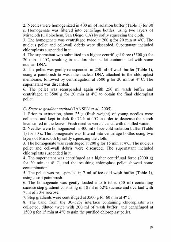
C) Sucrose gradient method (JANSEN et al., 2005)
1. Prior to extraction, about 25 g (fresh weight) of young needles were
collected and kept in dark for 72 h at 4ºC in order to decrease the starch
level stored in the leaves. Fresh needles were cleaned with distilled water.
2. Needles were homogenized in 400 ml of ice-cold isolation buffer (Table
1) for 30 s. The homogenate was filtered into centrifuge bottles using two
layers of Miracloth by softly squeezing the cloth.
3. The homogenate was centrifuged at 200 g for 15 min at 4ºC. The nucleus
pellet and cell-wall debris were discarded. The supernatant included
chloroplasts suspended in it.
4. The supernatant was centrifuged at a higher centrifugal force (2000 g)
for 20 min at 4º C, and the resulting chloroplast pellet showed some
contamination.
5. The pellet was resuspended in 7 ml of ice-cold wash buffer (Table 1),
using a soft paintbrush.
6. The homogenate was gently loaded into 6 tubes (50 ml) containing
sucrose step gradient consisting of 18 ml of 52% sucrose and overlaid with
7 ml of 30% sucrose.
7. Step gradients were centrifuged at 3500 g for 60 min at 4º C.
8. The band from the 30–52% interface containing chloroplasts was
collected, diluted twice with 200 ml of wash buffer, and centrifuged at
1500 g for 15 min at 4ºC to gain the purified chloroplast pellet.

20
Figure 1. Flowchart showing the major steps for chloroplast isolation
according to high salt plus saline Percoll.

21
Table 1. Composition of chloroplast isolation buffers and wash buffers for
modified high salt method, high salt plus saline Percoll method and sucrose
gradient method.
High salt plus saline Percoll method Modified high salt method Sucrose gradient
Isolation Buffer (pH 3.8) Isolation Buffer (pH 3.8) Isolation Buffer
1.25 M NaCl 1.25 M NaCl 50 mM Tris-HCl (pH 8.0)
0.25 M ascorbic acid 0.25 M ascorbic acid 0.35 M sorbitol
10 mM sodium metabisulfite 10 mM sodium metabisulfite 7 mM EDTA
0.0125 M Borax 0.0125 M Borax 0.1% 2-mercaptoethanol
50 mM Tris-HCl (pH 8.0) 50 mM Tris-HCl (pH 8.0) 0.1% BSA
7 mM EDTA 7 mM EDTA
1% PVP-40 (w/v) 1% PVP-40 (w/v)
0.1% BSA (w/v) 0.1% BSA (w/v)
1 mM DTT
Wash Buffer (pH 8.0) Wash Buffer (pH 8.0) Wash Buffer
1.25 M NaCl 1.25 M NaCl 50 mM Tris-HCl (pH 8.0)
0.0125 M Borax 0.0125 M Borax 0.35 M sorbitol
50 mM Tris-HCl (pH 8.0) 50 mM Tris-HCl (pH 8.0) 25 mM EDTA
25 mM EDTA 25 mM EDTA
1% PVP-40 (w/v) 1% PVP-40 (w/v)
0.1% BSA (w/v) 0.1% BSA (w/v)
1 mM DTT Both BSA and DTT were added just before the start of the experiment.
Percoll gradient solutions consisted of wash buffer with Percoll at a final
concentration of 70% (v/v) and 30% (v/v).
Sucrose gradient solutions consisted of 50 mM Tris-HCl (pH 8.0), 25 mM EDTA
and sucrose addition for a final concentration of 52% sucrose (w/v) and 30% (w/v)
sucrose.
2.3 Chloroplast DNA isolation
Chloroplast DNA isolation was the same for all chloroplast pellets
obtained using the three different isolation methods. DNA isolation buffer
consisted of 100 mM NaCl, 100 mM Tris-HCl (pH 8.0), 50 mM EDTA,
and 1 mM DTT.
1. The chloroplast lyse was obtained by incubating the chloroplast pellet
with 8 ml of DNA isolation buffer, 1.5 ml 20% SDS, 20 µl 2-
Mercaptoethanol and 30 µl Proteinase K (10 mg/ml) into a centrifuge tube at 55ºC for 4 h.
2. The centrifuge tube was incubated on ice for 5 min, and then 1.5 ml 5 M
KAc (pH 5.2) was added to the lyse mixture and chilled for more than 30

22
min. After that, the tube was centrifuged at 10000 g for 15 min at 4°C, and
the pellet was discarded.
3. The supernatant was extracted with an equal volume of saturated phenol
and chloroform:isoamyl-alcohol (24:1) and centrifuged twice at 10000 g
for 20 min.
4. An equal volume of isopropyl alcohol (about 10 ml) was added to the
upper aqueous phase and incubated at -20ºC overnight.
5. To obtain the DNA pellet, the tube was centrifuged at 10000 g for 20
min at 4 ºC. The cpDNA pellet was washed with 70% and 96% ethanol, air
dried, and redissolved in 50 μl TE buffer.
6. The cpDNA samples were treated with RNAse, and the DNA band was
visualized on a 0.7% agarose gel.
7. DNA purity and concentration were evaluated with Nanodrop®
, based on
260/280 and 260/230 ratios.
2.4 Microscopy analysis
The integrity of isolated chloroplasts was assessed with a phase-
contrast light microscopy using an inverted Olympus IX81 microscope
(WALKER, CEROVIC E ROBINSON, 1987). Intact chloroplasts were
considered those with pale yellow-green color and refractive, with a bright
halo appearance around each plastid, whereas broken chloroplasts were
those with a dark green, granular, and non-refractive appearance
(WALKER, CEROVIC E ROBINSON, 1987).
2.5 Chloroplast genome sequencing
A. angustifolia, A. bidwilli, P. patula and P. lambertii cpDNAs were
isolated using the high salt plus Percoll method. For each species,
approximately 50 ng of DNA were prepared with the Nextera DNA Sample
Prep Kit (Illumina, San Diego, USA) according to the manufacturer’s
instructions. Chloroplast DNAs were sequenced using Illumina MiSeq (2 x
250 read length) at the Federal University of Paraná - Brazil. The obtained
paired-end reads were assembled to reference genome sequence and
estimate genome coverage, using the CLC Genomics Workbench 5.5
software. The reference chloroplast genome sequences of Podocarpus
totara (NC_020361.1) and Pinus thunbergii (NC_001631.1) were
downloaded from GenBank.

23
3 Results and Discussion
3.1 Chloroplast isolation
Chloroplast isolation protocols are generally based on methods that
employ high salt concentration buffers (SHI et al., 2012), sucrose density
gradient (JANSEN et al., 2005), high salt buffers followed by sucrose
gradient (DIEKMANN et al., 2008) and high sorbitol concentration buffers
followed by Percoll gradient (KUBIS, LILLEY E JARVIS, 2008). As the
purity of intact chloroplasts is one of the critical steps of whole sequencing,
a previous paper (HIRAO et al., 2008) considered the use of sucrose
density gradients as the best method for separating nuclear DNA
contamination from cpDNA. The present protocol is based on a high salt
isolation buffer followed by saline Percoll gradient. The combination of
these two strategies provided two advantages: better isolation of
chloroplasts by use of the Percoll gradient and decreased contamination by
polysaccharides, polyphenols, and proteins.
The first significant change in isolation protocol was the increase in
storage time to 10 days at 4°C prior to extraction. This change led to a
significant reduction in the viscosity of extraction buffer, possibly caused
by a decrease in the polysaccharides and oleoresin concentrations. A
similar strategy has been used in other protocols, in which 48-72 h at 4°C
was enough to reduce the stored polysaccharides (JANSEN et al., 2005;
SHI et al., 2012). However, because of the high amount of oleoresins and
thick outer periclinal walls in conifers (MASTROBERTI E MARIATH,
2003; YAMAMOTO, OTTO E SIMONEIT, 2004), a longer time is
required to reduce the concentration of these compounds.
Subsequently, needles were washed with sarkosyl, reducing material
contamination. At the time of homogenization, it was observed that the
high salt buffer was also responsible for the decrease in viscosity of the
solution. The viscosity normally found in the homogenate (conifer needles
and isolation buffer) is related to the high amount of resins and
polysaccharides present in conifer needles, and its reduction enables faster
and more efficient filtration, with lower material loss. In a previous paper
(ARIF et al., 2010), it was observed that the use of high salt buffers for
DNA extraction increased the quality and yield of DNA extracted from
plant tissues rich in polysaccharides.
Similarly, the initial centrifugation step, performed to pellet cellular
debris was reduced to 200 g. We also reduced the initial centrifugation step
at 200 g to only one centrifugation, while in the modified high salt method
(SHI et al., 2012), it was performed twice. This reduction increased

24
chloroplast yield and did not entail any reduction in the quality of isolated
chloroplast as a result of the Percoll gradient step. The major steps for
chloroplast isolation using the high salt plus Percoll method, including
original, new and modified steps, are summarized in Figure 1.
Microscopy images showed the presence of some intact chloroplasts
and a large amount of broken chloroplasts and other cellular debris in
extraction when using the modified high salt and sucrose methods (Fig. 2A,
B). On the other hand, the high salt plus saline Percoll method resulted in
the presence of abundant intact chloroplasts (Fig. 2C).
Figure 2. Chloroplast visualization of Araucaria angustifolia in phase
contrast microscopy. (A) Chloroplasts isolated with improved high salt
method; (B) Chloroplasts isolated with sucrose method; (C) Chloroplasts
isolated with high salt plus Percoll method; (D-F) Micrographs during
chloroplast isolation with high salt plus saline Percoll method; (D) Broken and
intact chloroplasts before Percoll gradient centrifugation; (E) Intact isolated
chloroplasts in interface 70 / 30% after Percoll gradient centrifugation; (F)
Broken chloroplasts in upper 30% phase after Percoll gradient centrifugation.
Dotted arrows indicate broken chloroplasts. Solid arrows indicate intact
chloroplasts. Bar – 50 µM.
Aiming to better characterize the efficiency of Percoll gradient,
microscopy analysis was performed prior to the Percoll gradient
centrifugation step, and the presence of both intact and ruptured
chloroplasts could be observed (Fig. 2D). However, after centrifugation in
Percoll gradient, the 70%/30% interface (Fig. 2E) contains abundant intact
chloroplasts and only a few broken chloroplasts, while the upper phase
contains many broken chloroplasts (Fig. 2F). In addition, below the 70%

25
gradient, the formation of a white colored pellet composed of
polysaccharides and other contaminants could be seen. Despite the high
purity of chloroplasts observed immediately after Percoll gradient
centrifugation, two subsequent centrifugations are essential to remove any
residue of Percoll. The isolation of cpDNA without performing these two
washes would be greatly affected by its presence. It is noteworthy that
changes in the protocol enabled the isolation of the best quality
chloroplasts, without the need of ultracentrifugation, which could be a
limiting point in the procedure.
In addition to facilitating whole cpDNA sequencing, isolation of
intact chloroplasts can also be applied in plastid proteome characterization
studies. Comparative proteomics in Triticum aestivum and Arabidopsis
thaliana chloroplasts have been recently developed using intact isolated
chloroplasts. The results demonstrated that the quality of chloroplast
isolation is a fundamental step of complete proteome characterization
(GARGANO et al., 2013; HE et al., 2013).
3.2 Chloroplast DNA isolation
We also obtained isolated cpDNA with better quality and yield
using this high salt plus saline Percoll method. Using the modified high salt
and sucrose methods, bands in agarose gel revealed the presence of
degraded DNA, indicating contamination with nuclear DNA and
polysaccharides (Fig. 3A, B, respectively), while isolated cpDNA formed a
well-defined band, which is indicative of high purity and polysaccharide-
free cpDNA (Fig. 3C).
Figure 3. Chloroplast DNA visualization of Araucaria angustifolia in 0.7%
agarose gel stained with ethidium bromide. (A) Ladder 1 kb and cpDNA isolated
with modified high salt method; (B) Ladder 1 kb and cpDNA isolated with sucrose

26
method; (C) Ladder 1 kb and cpDNA isolated with high salt plus saline Percoll
method.
In addition, Nanodrop evaluation indicated higher cpDNA yield with
the high salt plus saline Percoll method, about 3 times higher when
compared to high salt methods and almost 9 times higher when compared
to the sucrose method. Enhanced 260/280 and 260/230 ratios were
observed in the high salt plus saline Percoll method, 2.05 and 1.99,
respectively (Table 2).
Table 2. cpDNA from different isolation methods in Araucaria angustifolia
sample.
Isolation Method DNA concentration (ng/µl) 260/280 260/230
Modified high salt method 975.6 1.66 0.92
High Salt plus saline Percoll method 3438.3 2.05 1.99
Sucrose Gradient Method 472.4 1.52 0.47 Ratios evaluated with Nanodrop
®, in a final volume of 40 µl.
These ratios indicate a high purity of isolated cpDNA, which is a
prerequisite for whole chloroplast sequencing. In the two other methods
evaluated, contamination was observed with polyphenols and
polysaccharides (Table 2). Moreover, when we used the sucrose method, a
highly contaminated and oxidized DNA pellet was obtained. Taken
together, we considered the high salt plus saline Percoll protocol as having
the best yield and quality for cpDNA isolation from A. angustifolia. Thus,
this method was applied to cpDNA isolation of A. bidwilli, P. patula and P. lambertii. As expected, a high quality in cpDNA isolated from all evaluated
species was realized at 260/280> 1.95 and 260/230> 1.74 ratios (Table 3).
All species showed cpDNA yield similar to A. angustifolia, with the
exception of P. lambertii. However, even its cpDNA yield was sufficient
for sequencing (Table 3).

27
Table 3. cpDNA ratios of selected conifers evaluated using Nanodrop®.
Plant species DNA concentration (ng/µl) 260/280 260/230
Araucaria angustifolia 3438.3 2.05 1.99
Araucaria bidwilli 3038.0 1.95 1.74
Podocarpus lambertii 430.6 2.01 1.89
Pinus patula 1799.5 2.05 2.10 Samples were isolated with the high salt plus Percoll method in different conifer
species. Final volume of 40 µl.
3.3 Chloroplast genome sequencing
Improving technologies have made DNA sequencing faster, more
accurate and far cheaper, creating opportunities to sequence the whole
chloroplast genome in order to perform evolutionary and phylogenomic
studies. To test the quality of cpDNA isolated by our new method, we
sequenced the plastid genome of four conifer species (A. angustifolia, A.
bidwilli, P. patula and P. lambertii) using the Illumina sequencing
technology.
To estimate the efficiency of chloroplast genome sequence assembly
with our cpDNA isolation protocol, we sequenced these four chloroplast
genomes by using MiSeq Illumina sequencing with only 50 ng of cpDNA.
In other sequencing protocols, about 5-10 µg (JANSEN et al., 2005; SHI et
al., 2012) were used for sequencing, thereby increasing the amount of plant
material required for isolation and often limiting the use of the technique. A
reference-guided chloroplast genome mapping was performed to estimate
the genome average coverage (Figure 4). The cpDNA sequencing
generated a high average coverage for all species evaluated: 24.63 for A.
angustifolia, 135.97 for A. bidwilli, 1,196.10 for P. lambertii, and 64.68 for
P. patula (Table 4). Thus, in this study, all of the reference genomes were
sufficiently covered for assembly.
This protocol presents higher genome coverage when compared to
protocols recently applied to conifers chloroplast genome sequencing, as
those using total DNA followed by PCR amplification with degenerated
primers that resulted in genome coverage only about 8-fold (LIN et al.,
2010; YI et al., 2013). Furthermore, this strategy is time-consuming and
difficult to implement because of differences in gene organization among different plant species. Cryptomeria japonica cp genome was sequenced
using sucrose gradient method, followed by DNA isolation with
phenol/chloroform, DNA purification with DNeasy Plant Mini Kit
(QIAGEN) and ATP-dependent DNase (TOYOBO) (HIRAO et al., 2008).

28
Figure 4. Reference graph track showing observed coverage values. Different
colors show the minimum (light blue), mean (blue), and maximum (dark blue)
observed coverage values for all genomic regions (data aggregation above 100 bp).
Araucaria angustifolia, Araucaria bidwilli, and Podocarpus lambertii sequence
reads were mapped on Podocarpus totara; Pinus patula sequence reads were
mapped on Pinus thunbergii.
As shown in the present work, the protocol based on saline buffer
followed by Percoll gradient results in higher quality DNA than sucrose
gradient. Moreover, all these purification steps applied to the isolated
DNA, such as the utilization of ATP-dependent DNase, led to a lower
DNA yield (SHI et al., 2012).

29
Table 4. Average coverage of cpDNA evaluated from selected conifers with CLC
Genomics Workbench 5.5 software.
Plant Species Average Coverage Reference Genome
Araucaria angustifolia 24.63 Podocarpus totara
Araucaria bidwilli 135.97 Podocarpus totara
Podocarpus lambertii 1196.10 Podocarpus totara
Pinus patula 64.68 Pinus thunbergii cpDNA reads were mapped to reference genomes.
In summary, the results obtained in the present work show that these
improvements in the general protocol for chloroplasts and cpDNA isolation
in conifers enhance the overall quality and yield of chloroplasts and
cpDNA isolation, providing a useful tool for studies that require isolated
chloroplasts and/or plastid genome sequence. Facilitating chloroplast
sequencing of this species group and, hence, increasing the amount of
information about the plastid genome of conifers may, in turn, lead to
greater understanding about plant evolution, as well as the structural and
functional genomics in plants other than conifers.
Financial Disclosure
This work was supported by Coordenação de Aperfeiçoamento de
Pessoal de Nível Superior (CAPES), Conselho Nacional de
Desenvolvimento Científico e Tecnológico (CNPq), and Fundação de
Amparo à Pesquisa e Inovação do Estado de Santa Catarina (FAPESC
14848/2011-2, 3770/2012, and 2780/2012-4). The funders had no role in
study design, data collection and analysis, decision to publish, or
preparation of the manuscript.

30
CAPÍTULO 2
Manuscrito formatado de acordo com as normas do periódico PLoS ONE.
The complete chloroplast genome sequence of Podocarpus lambertii:
genome structure, evolutionary aspects, gene content and SSR
detection
Vieira LN, Faoro H, Rogalski M, Fraga HPF, Cardoso RLA, Souza EM,
Pedrosa FO, Nodari RO, Guerra MP. The complete chloroplast genome
sequence of Podocarpus lambertii: genome structure, evolutionary aspects,
gene content and SSR detection.

31
Abstract
Background: Podocarpus lambertii (Podocarpaceae) is a native conifer
from the Brazilian Atlantic Forest Biome, which is considered one of the
25 biodiversity hotspots in the world. The advancement of next-generation
sequencing technologies has enabled the rapid acquisition of whole
chloroplast (cp) genome sequences at low cost. Several studies have proven
the potential of cp genomes as tools to understand enigmatic and basal
phylogenetic relationships at different taxonomic levels, as well as further
probe the structural and functional evolution of plants. In this work, we
present the complete cp genome sequence of P. lambertii. Methodology/Principal Findings: The P. lambertii cp genome is 133,734
bp in length, and similar to other sequenced cupressophytes, it lacks one of
the large inverted repeat regions (IR). It contains 118 unique genes and one
duplicated tRNA (trnN-GUU), which occurs as an inverted repeat
sequence. The rps16 gene was not found, which was previously reported
for the plastid genome of another Podocarpaceae (Nageia nagi) and
Araucariaceae (Agathis dammara). Structurally, P. lambertii shows 4
inversions of a large DNA fragment ~20,000 bp compared to the
Podocarpus totara cp genome. These unexpected characteristics may be
attributed to geographical distance and different adaptive needs. The P.
lambertii cp genome presents a total of 28 tandem repeats and 156 SSRs,
with homo- and dipolymers being the most common and tri-, tetra-, penta-,
and hexapolymers occurring with less frequency.
Conclusion: The complete cp genome sequence of P. lambertii revealed
significant structural changes, even in species from the same genus. These
results reinforce the apparently loss of rps16 gene in Podocarpaceae cp
genome. In addition, several SSRs in the P. lambertii cp genome are likely
intraspecific polymorphism sites, which may allow highly sensitive
phylogeographic and population structure studies, as well as phylogenetic
studies of species of this genus.

32
1 Introduction
Extant gymnosperms are considered the most ancient group of
seed-bearing plants that first appeared approximately 300 million years ago
(MURRAY, 2013). They consist of four major groups, including
Gnetophytes, Conifers, Cycads and Ginkgo. Podocarpaceae are considered
the most diverse family of Conifers, and much of this diversity has taken
place within the Podocarpus and Dacrydium genera (KELCH, 1998). The
Podocarpaceae family comprises 18 genera and 173 species distributed
mainly in the Southern Hemisphere, but extending to the north in
subtropical China, Japan, Mexico and the Caribbean (FARJON, 1998;
BIFFIN et al., 2011). The Podocarpus sensu lato (s.l.) genus comprises nearly 100
species, widely spread throughout the Southern Hemisphere and northward
to the West Indies, Mexico, southern China and southern Japan (PAGE,
1990). LEDRU et al., (2007) described that Podocarpus populations in
Brazil are widely dispersed in eastern Brazil, from north to south, and three
endemic species have been reported: Podocarpus sellowii Klotzch ex Endl,
Podocarpus lambertii Klotzch ex Endl, and Podocarpus brasiliensis de
Laubenfels (de LAUBENFELS, 1985). P. lambertii is a native species from
the Araucaria Forest, a subtropical moist forest ecoregion of the Atlantic
Forest Biome, which is considered one of the 25 biodiversity hotspots of
the world (MYERS et al., 2000). It is a dioecious evergreen tree of variable
height, measuring 1-10 m, shade-tolerant, adapted to high frequency and
density of undergrowth (LONGHI, 2010).
Phylogeny analyses by maximum parsimony of Podocarpaceae
family using 18S rDNA gene sequencing and morphological characteristics
indicated Podocarpaceae as monophyletic and Podocarpus s.l. and
Dacrydium s.l. genera as unnatural (KELCH, 1998). This author concluded
that single-gene studies rarely result in perfect phylogenies, but they could
provide a basis for choosing between competing hypotheses. PARKS et al.,
(2009) suggested chloroplast (cp) genome sequencing as an efficient option
for increasing phylogenetic resolution at lower taxonomic levels in plant
phylogenetic and genetic population analyses.
The advancement of next-generation sequencing technologies has
enabled the rapid acquisition of whole cp genome sequences at low cost
when compared with traditional sequencing approaches. Chloroplast
sequences are available for all families of Conifers: Cephalotaxaceae (YI et al., 2013), Cupressaceae (HIRAO et al., 2008), Pinaceae (WAKASUGI et
al., 1994; CRONN et al., 2008; LIN et al., 2010), Podocarpaceae
(NC_020361.1; WU E CHAW, 2013), Taxaceae (NC_020321.1), and

33
Araucariaceae (WU E CHAW, 2013). For Podocarpus genus, the cp
sequence of only one species has recently been obtained: the endemic New
Zealand Podocarpus totara G. Benn. ex Don (NC_020361.1).
Several studies have proven the potential of cp genomes as tools to
understand enigmatic and basal phylogenetic relationships at different
taxonomic levels, as well as probe the structural and functional evolution of
plants (MOORE et al., 2007; JANSEN et al., 2007; MOORE et al., 2010;
WU et al., 2011a; YI et al., 2013). HIRAO et al., (2008) sequenced the cp
genome of the first species in the Cupressaceae family, Cryptomeria
japonica. They reported the deletion of one large inverted repeat (IR),
numerous genomic rearrangements, and many differences in genomic
structure between C. japonica and other land plants, thus supporting the
theory that a pair of large IR can stabilize the cp genome against major
structural rearrangements and, in turn, providing new insights into both the
evolutionary lineage of coniferous species and the evolution of the cp
genome (PALMER E THOMPSON, 1982; STRAUSS et al., 1988; HIRAO
et al., 2008).
Chloroplast genome sequencing in gymnosperms also brought
insights into evolutionary aspects in Gnetophytes. WU et al., (2009)
considered that the reduced cp genome size in Gnetophyte was based on a
selection toward a lower-cost strategy by deletions of genes and noncoding
sequences, leading to genomic compactness and accelerated substitution
rates. More recently, comparative analysis of the cp genomes in
cupressophytes and Pinaceae provided inferences about the loss of large IR
(WU et al., 2011a; YI et al., 2013). On one hand, WU et al., (2011a) and
WU E CHAW (2013) argue that each Pinaceae and cupressophyte lost a
different copy of IR. On the other hand, YI et al., (2013) showed that
distinct isomers are considered as alternative structures for the ancestral cp
genome of cupressophyte and Pinaceae lineages. Therefore, it is not
possible to distinguish between hypotheses favoring retention or
independent loss of the same IR region in cupressophyte and Pinaceae cp
genomes.
The present study focuses on establishing the complete cp genome
sequence of a further member of the Podocarpaceae family, the Brazilian
endemic species P. lambertii. Here, we characterize the cp genome
organization of P. lambertii and compare its cp genome structure with
other conifer species.

34
2 Material and Methods
2.1 Plant material and cp DNA purification
Chloroplast isolation of P. lambertii was performed from young
plants collected at a private area located at Lages, Santa Catarina, Brazil
(27° 48' 57" S, 50° 19' 33" W), where the species is abundant, with
previous permission from the owner (José Antônio Ribas Ribeiro). This
species is not considered threatened. Afterwards, the young plants were
transplanted to the greenhouse until the collection of needles. The cpDNA
isolation was performed according to VIEIRA et al., (2014).
2.2 Chloroplast genome sequencing, assembling and annotation
Approximately 50 ng of cp DNA were used to prepare sequencing
libraries with Nextera DNA Sample Prep Kit (Illumina Inc., San Diego,
CA) according to the manufacturer’s instructions. Chloroplast DNA was
sequenced using Illumina MiSeq (Illumina Inc., San Diego, CA) at the
Federal University of Paraná, Brazil. In total, 495,071 paired-end reads (2 x
250 bp) were obtained, and de novo assembly was performed using
Newbler 2.6v. The obtained paired-end reads were mapped on P. lambertii
cp genome and the genome coverage estimated using the CLC Genomics
Workbench 5.5 software. By using this approach, a total of 377,437 paired-
end reads (76.23%) was obtained from cpDNA, resulting in 1,200-fold
genome coverage. Initial annotation of the P. lambertii cp genome was
performed using Dual Organellar GenoMe Annotator (DOGMA)
(WYMAN et al., 2004). From this initial annotation, putative starts, stops,
and intron positions were determined based on comparisons to homologous
genes in other cp genomes. The tRNA genes were further verified by using
tRNAscan-SE (SCHATTNER et al., 2005). A physical map of the cp
circular genome was drawn using OrganellarGenomeDRAW (OGDRAW)
(LOHSE et al., 2013). The resulting annotated sequence has been
submitted to the National Center for Biotechnology Information (NCBI)
under the accession number KJ010812.
2.3 Comparative analysis of genome structure
We used the PROtein MUMmer (PROmer) Perl script in
MUMmer 3.0 (KURTZ et al., 2004), available at
http://mummer.sourceforge.net/, to visualize gene order conservation (dot-
plot analyses) between P. lambertii and the non-Pinaceae conifer

35
representatives P. totara (Podocarpaceae), Cephalotaxus oliveri,
Cephalotaxus wilsoniana (Cephalotaxaceae), Taxus mairei (Taxaceae),
Taiwania cryptomerioides, T. flousiana (Cupressaceae), C. japonica
(Cupressaceae), as well as Pinus thunbergii, a Pinaceae representative.
2.4 Repeat sequence analysis and IR identification
Simple sequence repeats (SSRs) were detected using MISA perl
script, available at (http://pgrc.ipk-gatersleben.de/misa/), with thresholds of
eight repeat units for mononucleotide SSRs, four repeat units for di- and
trinucleotide SSRs, and three repeat units for tetra-, penta- and
hexanucleotide SSRs. Tandem repeats were analyzed using Tandem
Repeats Finder (TRF) (BENSON, 1999) with parameter settings of 2, 7 and
7 for match, mismatch, and indel, respectively. The minimum alignment
score and maximum period size were set as 50 and 500, respectively. All of
the repeats found were manually verified, and the nested or redundant
results were removed. REPuter (KURTZ et al., 2001) was used to visualize
the remaining IRs in P. lambertii by forward vs. reverse complement
(palindromic) alignment. The minimal repeat size was set to 30 bp and the
identity of repeats ≥ 90%.
3 Results and Discussion
3.1 Chloroplast genome sequencing, assembling and annotation
P. lambertii cp genome size was determined to be 133,734 bp,
which is very similar to P. totara (133,259 bp) (MARSHALL,
Unpublished, NC_020361.1) and larger than the sequenced cp genomes of
Pinaceae species, which range from 116,479 bp in Pinus monophylla
(CRONN et al., 2008) to 124,168 bp in Picea morrisonicola (WU et al.,
2011b). P. lambertii cp genome size is smaller than the cp sequences in the
cycads Cycas taitungensis (163,403 bp) (WU et al., 2007) and Cycas
Revoluta (162,489 bp) (LI et al., Unpublished, NC_020319.1). The genome
size of P. lambertii cp is consistent with the size of non-Pinaceae conifer
species, which ranges from 127,665 bp in T. mairei (LI et al., unpublished,
NC_020321.1) to 136,196 bp in C. wilsoniana (WU et al., 2011). A total of
119 genes were identified in the P. lambertii cp genome, of which 118
genes were single copy and one gene, trnN-GUU, was duplicated and
occurred as an inverted repeat sequence. The following genes were
identified and are listed in Figure 1 and Table 1: 4 ribosomal RNA genes,
31 unique transfer RNA genes, 20 genes

36
Figure 1. Gene map of Podocarpus lambertii chloroplast genome. Genes drawn
inside the circle are transcribed clockwise, and genes drawn outside are
counterclockwise. Genes belonging to different functional groups are color-coded.
The darker gray in the inner circle corresponds to GC content, and the lighter gray
corresponds to AT content.
encoding large and small ribosomal subunits, 1 translational initiation
factor, 4 genes encoding DNA-dependent RNA polymerases, 50 genes
encoding photosynthesis-related proteins, 8 genes encoding other proteins,
including the unknown function gene ycf2, and 1 pseudogene, ycf68.
Among these 118 single copy genes, 14 were genes containing introns
(Table 1). The GC content determined for P. lambertii cp genome is
37.1%, which is higher than C. oliveri (35.2%), C. wilsoniana (35.1%), T.
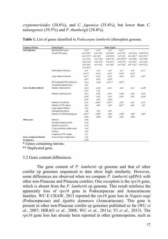
37
cryptomerioides (34.6%), and C. japonica (35.4%), but lower than C.
taitungensis (39.5%) and P. thunbergii (38.8%).
Table 1. List of genes identified in Podocarpus lambertii chloroplast genome.
Category of Genes Group of gene Name of gene
Self-replication Ribosomal RNA genes rrn16 rrn23 rrn5 rrn4.5
Transfer RNA genes trnA -UGC * trnC -GCA trnD -GUC trnE -UUC trnF -GAA trnfM -CAU
trnG -UCC* trnG -GCC trnH -GUG trnI -CAU trnI -GAU * trnK -UUU *
trnL -CAA trnL -UAG trnM -CAU trnN -GUU** trnP -GGG trnP -UGG
trnQ -UUG trnR -ACG trnR -UCU trnR -CCG trnS -GCU trnS -UGA
trnS -GGA trnT -UGU trnT -GGU trnV -GAC trnV -UAC * trnW -CCA
trnY -GUA
Small subunit of ribosome rps2 rps3 rps4 rps7 rps8 rps11
rps12 * rps14 rps15 rps18 rps19
Large subunit of ribosome rpl2 * rpl14 rpl16 rpl20 rpl22 rpl23
rpl32 rpl33 rpl36
DNA-dependent RNA polymerase rpoA rpoB rpoC1 * rpoC2
Translational initiation factor infA
Genes for photosynthesis Subunits of photosystem I psaA psaB psaC psaI psaJ psaM
ycf3* ycf4
Subunits of photosystem II psbA psbB psbC psbD psbE psbF
psbH psbI psbJ psbK psbL psbM
psbN psbT psbZ
Subunits of cytochrome petA petB * petD * petG petL petN
Subunits of ATP synthase atpA atpB atpE atpF * atpH atpI
Large subunit of Rubisco rbcL
Chlorophyll biosynthesis chlB chlL chlN
Subunits of NADH dehydrogenase ndhA * ndhB * ndhC ndhD ndhE ndhF
ndhG ndhH ndhI ndhJ ndhK
Other genes Maturase matK
Envelope membrane protein cemA
Subunit of acetyl-CoA accD
C-type cytochrome synthesis gene ccsA
Protease clpP
Component of TIC complex ycf1
Genes of unknown function Conserved open reading frames ycf2
Pseudogenes ycf68 * Genes containing introns.
** Duplicated gene.
3.2 Gene content differences
The gene content of P. lambertii cp genome and that of other
conifer cp genomes sequenced to date show high similarity. However,
some differences are observed when we compare P. lambertii cpDNA with
other non-Pinaceae and Pinaceae conifers. One exception is the rps16 gene,
which is absent from the P. lambertii cp genome. This result reinforce the
apparently loss of rps16 gene in Podocarpaceae and Araucariaceae
families. WU E CHAW, 2013 reported the rps16 gene loss in Nageia nagi
(Podocarpaceae) and Agathis dammara (Araucariaceae). This gene is
present in other non-Pinaceae conifer cp genomes published so far (WU et al., 2007; HIRAO et al., 2008; WU et al., 2011a; YI et al., 2013). The
rps16 gene loss has already been reported in other gymnosperms, such as

38
Pinaceae and Gnetophyte species (TSUDZUKI et al., 1992; WU et al.,
2007; WU et al., 2009). WU et al., (2011a) considered rps16 gene loss as a
structural mutation unique to the cpDNAs of gnetophytes and Pinaceae, but
since the loss of this gene has been identified in Podocarpaceae and
Araucariaceae families, we can consider that some cupressophytes may
also present this mutation. This gene is also absent, or nonfunctional, in
some angiosperm species of the Fabaceae family, such as Medicago
truncatula, in which it is completely absent, and in Phaseolus vulgaris and
Vigna radiata, in which it is nonfunctional. In this angiosperm family, the
coding sequence contains many internal stop codons and a modified initial
stop codon (GUO et al., 2007; TANGPHATSORNRUANG et al., 2010).
Since this gene was shown to be essential for cell survival in tobacco
(FLEISCHMANN et al., 2011), it was probably transferred to the nucleus,
as observed for different species of the Fabaceae family (GUO et al., 2007;
TANGPHATSORNRUANG et al., 2010), and has since become a
functional nuclear gene required for normal plastid translation.
The trnP-GGG and trnR-CCG genes are considered to be relics of
plastid genome evolution in gymnosperms, pteridophytes and bryophytes
(SUGIURA E SUGITA, 2004). The trnP-GGG gene is present in the P. lambertii cp genome, as well as such conifer species as C. japonica, P.
thunbergii, C. oliveri and C. wilsoniana and other gymnosperm species,
such as C. taitungensis, Gnetum and Ginkgo. The trnR-CCG gene is
present as complete and functional tRNA in P. lambertii (Podocarpaceae),
as well as the cp genomes of P. thunbergii (Pinaceae), C. taitungensis (Cycadaceae) (WU et al., 2007), whereas it is absent from C. japonica
(Cupressaceae), C. oliveri and C. wilsoniana (Cephalotaxaceae), and T.
mairei (Taxaceae) (HIRAO et al., 2008; YI et al., 2013; LI et al., unpublished). HIRAO et al., (2008) suggested that trnR-CCG might have
been completely lost in the Cupressaceae s.l., which has only relatively
recently diverged during the long evolutionary history of plants. These data
corroborate the hypothesis based on phytochrome phylogenetic trees, in
which the most ancient branch of the conifers seems to be the Pinaceae,
and the next split appears to have separated Araucariaceae plus
Podocarpaceae from the Taxaceae/Taxodiaceae/Cupressaceae group
(SCHMIDT E SCHNEIDER-POETSCH, 2002). This trnR-CCG gene may
have been lost during the second split separating Araucariaceae and
Podocarpaceae taxa. In addition, trnT-GGU occurs as a pseudogene in the
C. japonica cp genome, with only 43 bp, while it is present and completely
functional in P. lambertii and C. oliveri, C. wilsoniana, duplicated in P.
thunbergii, and totally absent from the C. taitungensis cp genome.
Interestingly, the trnT-GGU gene is highly conserved in angiosperms, and

39
knockout of this gene in tobacco plants produced viable plants, whereas the
growth of these plants was strongly affected, suggesting an important role
during plastid translation (ALKATIB et al., 2012). The loss of the trnT-
GGU gene in several gymnosperm species suggests that a uridine
modification in the anticodon position of the trnT-GGU gene occurred
during evolution, which would facilitate the reading of threonine codons
and make the trnT-GGU gene dispensable in these species
(AMBROGELLY et al., 2007; WEIXLBAUMER et al., 2007; ALKATIB
et al., 2012). Evolutionarily, the loss of this tRNA gene could be used as a
tool, or marker gene, to study the possible ways that the conifers diverged
during evolution. However, it remains to be determined whether structural
differences in the cp ribosome or modification in the structure of this
tRNA, between angiosperms and gymnosperms, would facilitate the
decoding.
3.3 Comparative analysis of genome structure
Chloroplast genome organization is much conserved in
angiosperms, as well as the presence of IRs, with very few exceptions. As
reported by TERAKAMI et al., (2012) in Pyrus, Malus and Nicotiana, neither translocation nor inversion was detected in the three species. In
addition, considering the many dicot and monocot species, only one large
inversion was reported (TERAKAMI et al., 2012).
In addition to the loss of the large IR in conifers, many genome
rearrangements were observed in the cp genome, and such rearrangements
appear to play an important role in their evolution. Dot-plot analyses
indicate that the structure of the P. lambertii cp genome differs
significantly from cp genomes of other conifer species, and, surprisingly, it
has significant differences when compared to P. totara (Figure 2A-H).
For the genus Cephalotaxus s.l., specifically C. wilsoniana and C. Oliveri, it was shown that the genome structures were almost the same (YI
et al., 2013). Similar results were observed in the present study, as revealed
by the high similarity in the dot-plot analyses between Podocarpus and
Cephalotaxus genera, as represented by P. lambertii x C. wilsoniana
(Figure 2E) and P. Lambertii x C. oliveri (Figure 2F), and between the
Podocarpus and Taiwania genera, as represented by P. lambertii x T.
flousiana (Figure 2G) and P. lambertii x T. cryptomerioides (Figure 2H).
This high similarity in dot-plot analysis indicates the occurrence of exactly
the same structural modifications between P. lambertii and these two
Cephalotaxus and Taiwania species.

40
Figure 2. Dot-plot analyses of eight sampled conifer chloroplast DNAs against
Podocarpus lambertii. A positive slope denotes that the two compared sequences
are in the same orientation, whereas a negative slope indicates that the compared
sequences can be aligned, but their orientations are opposite. Graphs represents
comparisons between Podocarpus lambertii (axis X) and Podocarpus totara (A),
Taxus mairei (B), Pinus thunbergii (C), Cryptomeria japonica (D), Cephalotaxus
wilsoniana (E), Cephalotaxus oliveri (F), Taiwania flousiana (G), and Taiwania
cryptomerioides (H) in axis Y.

41
Differently, for P. lambertii and P. totara (Figure 2A), we
observed four large inversions of about 20,000 bp in length each. In both
Cephalotaxus and Taiwania genera, the two sequenced species share the
same region of natural occurrence, which is not true for either Podocarpus
species sequenced. Thus, these large inversions can be explained by, and
probably result from, the large distance between the natural occurrence of
these two species in that P. lambertii occurs in Brazil, while P. totara
occurs in New Zealand. Moreover, podocarps have a rich fossil record that
suggests an origin in the Triassic period (about 220 million years) and a
distribution in both the Northern and Southern Hemispheres through the
Cretaceous and earliest Tertiary periods, about 100 million years ago
(HILL E BRODRIBB, 1999; FARJON, 2008; MORLEY, 2011). Thus,
geographic distance and different adaptive traits could explain the
structural differences found between these two species of the same genera.
In addition, the loss of one large IR copy already reported in other
conifer species were also observed in the P. lambertii cp genome (HIRAO
et al. 2008; WU et al. 2011; YI et al., 2013). However, short remaining IR
sequences of 326 bp can be found in P. lambertii, 544 bp in C. oliveri, 530
bp in C. wilsoniana, 277 bp in T. cryptomerioides and 284 bp in C. japonica (YI et al., 2013). These short remaining IR sequences also differ
in the nucleic acid sequences and gene content between different conifer
species. In P. lambertii, trnN-GUU remain from the lost IR copy region,
while in T. cryptomerioides and C. japonica, trnI-CAU remained after the
rearrangements that determined the loss of one IR copy (YI et al., 2013). In
C. oliveri and C. wilsoniana, the trnQ-UUG is duplicated; however, this
gene is not normally present in the IR region, and its duplication was
probably produced by other rearrangements not involved with the IR
regions (WU et al., 2011). After much evidence provided by different
conifer plastid genomes, it can be concluded that the loss of one IR copy
occurred after a reduction in sequence and gene content and that such loss
was most likely caused by this reduction (TSUDZUKI et al., 1992; WU et
al., 2007; CRONN et al., 2008; HIRAO et al., 2008; WU et al., 2009; WU
et al., 2011; YI et al., 2013). However, this speculation remains to be
established. To date, it is not entirely clear whether cupressophytes and
Pinaceae species have lost different IR regions (YI et al., 2013). However,
we can observe in P. lambertii an inversion in the direction of transcription
of ribosomal RNA genes spanning rrn5-rrn16 and protein-coding genes,
ndhB and ycf2, when compared to C. oliveri, C. wilsoniana, T.
cryptomerioides and C. japonica (Figure 3).

42
Figure 3. Comparison of IR and genome structure in 5 cupressophytes. Five
cupressophyte species from top to bottom are Taiwania cryptomerioides,
Cryptomeria japonica, Cephalotaxus oliveri, Cephalotaxus wilsoniana and
Podocarpus lambertii. Genes are represented by boxes extending above or below
the baseline, according to the direction of transcription; genes with the same
function have the same color. Transfer RNA genes are abbreviated as the type of
one letter. Dashed boxes represent the retained IR region, and arrows indicate the
short IR on each species. Adapted from YI et al., (2013).
3.4 Repeat sequence analysis
The cp genome mode of inheritance, paternal in most
gymnosperms, allows us to elucidate the relative contributions of seed and
pollen flow to the genetic structure of natural populations by comparison of
nuclear and cp markers (PROVAN et al., 2001). The cp microsatellites, or
SSRs, may be identified in completely sequenced plant cp genomes by
simple database searches, followed by primers designed to screen for
polymorphism. To date, studies of cp microsatellites have revealed much
higher levels of diversity than have those of cp restriction fragment length
polymorphisms (RFLP) (PROVAN et al., 1997, 1999, 2001).
We have analyzed the occurrence, type, and distribution of SRRs
in the P. lambertii cp genome. In total, 156 SSRs were identified. Among
them, homo- and dipolymers were the most common with, respectively, 80
and 63 occurrences, whereas tri- (4), tetra- (7), penta- (1), and
hexapolymers (1) occur with lower frequency (Table 2). Most
homopolymers are constituted by A/T sequences (87.5%), and of the
dipolymers, 61.1% were also constituted by multiple A and T bases.

43
Table 2. List of simple sequence repeats identified in Podocarpus lambertii
chloroplast genome.
SSR sequence Number of repeats TOTAL
3 4 5 6 7 8 9 10 11 12 13 14 15
A/T - - - - - 39 14 6 4 6 - - 1 70
C/G - - - - - - 3 3 - 1 1 - 2 10
AC/GT - 1 - 1 - - - - - - - - - 2
AG/CT - 21 1 - - - - - - - - - - 22
AT/AT - 24 7 2 2 - 3 1 - - - - - 39
AAG/CTT - 1 - - - - - - - - - - - 1
AAT/ATT - 3 - - - - - - - - - - - 3
AATC/ATTG 2 - - - - - - - - - - - - 2
AATG/ATTC 1 - - - - - - - - - - - - 1
AATT/AATT 1 - - - - - - - - - - - - 1
ACAT/ATGT 1 - - - - - - - - - - - - 1
ACCT/AGGT 1 - - - - - - - - - - - - 1
AGAT/ATCT 3 - - - - - - - - - - - - 3
AAATG/ATTTC 1 - - - - - - - - - - - - 1
AGATAT/ATATCT 1 - - - - - - - - - - - - 1
TOTAL 158
In this study, we identified 78 repeats with more than one
nucleotide repeat, totaling almost 50% of all SSRs identified. The 13 tri-,
tetra-, penta-, and hexapolymers are shown in Table 3, as well as their size
and location. From these 13 polymers identified, 9 are localized in
intergenic spacers, 3 in coding sequences, and only 1 inside an intron.
These results reveal the presence of several SSR sites in P. lambertii. Hereafter, these sites can be assessed for the intraspecific level of
polymorphism, leading to highly sensitive phylogeographic and population
structure studies for this species.
Tandem repeats with more than 30 bp and with a sequence identity
of more than 90% have also been examined. Twenty-eight tandem repeats
were identified in the P. lambertii cp genome (Table 4), of which 15 are
located in coding regions of accD (2), rps18 (1), rps19 (1), rps11 (1), ycf1
(8), rpl32 (1), ycf2 (1); 11 are distributed in the intergenic spacers of
atpA/atpF (1), trnR-CCG/accD (1), rpl2/rps19 (1), clpP/ycf1 (2),
ndhE/psaC (1), trnR-ACG/rrn5 (1), rps12/rps7 (1), ycf2/trnI-CAU (1), trnQ-UUG/psbK (1), psbK/psbI (1); and 2 are located in the intron
sequence of rpoC1. The cp genome of P. lambertii has 11 tandem repeats,
more than the cp genome of C. oliveri, as well as a higher number of
repeats in the ycf1 (6) gene coding sequence (YI et al., 2013). The ycf1
gene, previously considered as an enigmatic function in the cp genome, has

44
recently been identified as encoding an essential protein component of the
cp translocon at the inner envelope membrane (TIC) (KIKUCHI et al.,
2013). In Salvia miltiorrhiza and Cocos nucifera, two angiosperms, only 7
and 8 tandem repeats, respectively, of about 20 bp were identified, none of
them located at the ycf1 coding sequence (HUANG et al., 2013; QIAN et
al., 2013), corroborating the theory that the IR influences the stability of
the plastid genome.
Table 3. Distribution of tri-, tetra-, penta-, and hexapolymer simple sequence
repeats (SSRs) loci in Podocarpus lambertii chloroplast genome.
SSR type SSR sequence Size Start End Location
penta (AATGA)3 15 21884 21898 trnE -UUC/trnT -GGU (IGS)
hexa (AGATAT)3 18 37894 37911 trnF -GAA/ndhJ (IGS)
tetra (ATCA)3 12 44346 44357 atpE /rbcL (IGS)
tri (AAG)4 12 75761 75772 Ycf1 (CDS)
tetra (AATG)3 12 86350 86361 ndhA (intron)
tetra (TGAT)3 12 97140 97151 ndhF /trnN -GUU (IGS)
tetra (CTAC)3 12 99809 99820 rrn23 (CDS)
tri (ATT)4 12 103664 103675 trnI -GAU/rrn16 (IGS)
tri (ATA)4 12 120539 120550 rps7 /ndhB (IGS)
tri (TTA)4 12 122046 122057 chlL (CDS)
tetra (AATT)3 12 122977 122988 chlL /trnH -GUG (IGS)
tetra (CATA)3 12 125437 125448 psbA /trnK -UUU (IGS)
tetra (ATAG)3 12 125570 125581 psbA /trnK -UUU (IGS) CDS, coding sequences; IGS, intergenic spacers.
YI et al., 2013 attributed the expansion of the accD ORF to the
presence of tandemly repeated sequences. In the P. lambertii cp genome,
we identified 2 tandem repeats in accD CDS, totaling 132 bp, or 44 codons.
The accD reading frame length of the P. lambertii cp genome is 864
codons, similar to other cupressophyte species, such as C. oliveri (936
codons), C. wilsoniana (1,056 codons), C. japonica (700 codons) and T.
cryptomerioides (800 codons). In contrast, the reading frame lengths of
cycads, Ginkgo and Pinaceae, range from 320 to 359 codons, less than half
the size found in cupressophytes. These results support the hypothesis of
HIRAO et al., (2008) and YI et al., (2013) which holds that the accD
reading frame has displayed a tendency toward enlarging sizes in
cupressophytes.

45
Table 4. Distribution of tandem repeats in Podocarpus lambertii chloroplast
genome. Serial Number Repeat Length (bp) Consensus size x Copy number Start-End Location
1 32 16 x 2 3450-3482 atpA /atpF (IGS)
2 284 142 x 2 13170-13454 rpoC1 (Intron)
3 60 30 x 2 13496-13557 rpoC1 (Intron)
4 30 15 x 2 46625-46653 trnR -CCG/accD (IGS)
5 90 30 x 3 47533-47619 accD (CDS)
6 42 21 x 2 48149-48192 accD (CDS)
7 52 26 x 2 57988-58043 rps18 (CDS)
8 32 16 x 2 61875-61905 rpl2 /rps19 (IGS)
9 54 18 x 3 62177-62237 rps19 (CDS)
10 63 21 x 3 66568-66630 rps11 (CDS)
11 32 16 x 2 75172-75203 clpP /ycf1 (IGS)
12 104 52 x 2 75412-75529 clpP /ycf1 (IGS)
13 36 18 x 2 79255-79292 ycf1 (CDS)
14 162 52 x 3 79351-79504 ycf1 (CDS)
15 162 81 x 2 79362-79519 ycf1 (CDS)
16 108 27 x 4 79401-79519 ycf1 (CDS)
17 132 33 x 4 80478-80619 ycf1 (CDS)
18 96 24 x 4 80732-80820 ycf1 (CDS)
19 273 21 x 13 81305-81571 ycf1 (CDS)
20 96 48 x 2 82408-82528 ycf1 (CDS)
21 30 15 x 2 89787-89817 ndhE /psaC (IGS)
22 126 42 x 3 93843-93963 rpl32 (CDS)
23 64 32 x 2 97838-97902 trnR -ACG/rrn5 (IGS)
24 300 60 x 5 109209-109531 rps12 /rps7 (IGS)
25 36 12 x 3 116515-116547 ycf2 (CDS)
26 60 20 x 3 119998-120055 ycf2 /trnI -CAU (IGS)
27 128 64 x 2 131733-131853 trnQ -UUG/psbK (IGS)
28 26 13 x 2 132530-132556 psbK /psbI (IGS)
CDS, coding sequences; IGS, intergenic spacers.
The complete cp genome sequence of P. lambertii revealed
significant structural changes occurring in the cp genome, even in species
from the same genus. These results reinforce the apparently loss of rps16
gene in Podocarpaceae cp genome. In addition, several SSRs in the P. lambertii cp genome are likely intraspecific polymorphism sites which may
allow highly sensitive phylogeographic and population structure studies, as
well as phylogenetic studies, of species of this genus.
Financial Disclosure
This work was supported by Coordenação de Aperfeiçoamento de
Pessoal de Nível Superior (CAPES), Conselho Nacional de
Desenvolvimento Científico e Tecnológico (CNPq), and Fundação de
Amparo à Pesquisa e Inovação do Estado de Santa Catarina (FAPESC
14848/2011-2, 3770/2012, and 2780/2012-4). The funders had no role in

46
study design, data collection and analysis, decision to publish, or
preparation of the manuscript.

47
CONSIDERAÇÕES FINAIS E PERSPECTIVAS FUTURAS
Os resultados do presente trabalho apresentam avanços
expressivos na área de sequenciamento de genomas plastidiais em
coníferas. Primeiramente, foi desenvolvido um protocolo para isolamento
de cloroplasto com alta qualidade em coníferas. A partir disso, possibilitou-
se o isolamento de DNA plastidial com alta qualidade, podendo ser
empregado para sequenciamento em sequenciadores de nova geração. Com
o DNA plastidial isolado, aumenta-se significativamente o número de
reads obtidos que realmente são plastidiais. No caso do P. lambertii,
estima-se que 76% dos reads obtidos foram de DNA plastidial, e não de
possíveis contaminações com DNA mitocondrial ou nuclear. Esse é um
importante fator facilitador no momento da montagem do genoma, devido à
alta cobertura média do genoma, onde com o P. lambertii, por exemplo, obteve-se uma cobertura média de 1200x no genoma.
Além disso, é possível que esse protocolo possa ser aplicado para
outras espécies nativas de difícil isolamento do cloroplasto por seu alto
conteúdo endógeno de polifenóis, polissacarídeos e outros contaminantes.
Os tampões e reagentes usados nesse protocolo contam com uma alta carga
de antioxidantes, somado ao uso do Percoll que aumenta significativamente
a pureza do cloroplasto isolado.
O cloroplasto isolado com alta qualidade também torna possível a
aplicação para o isolamento de proteínas, permitindo também análises de
proteômica em cloroplastos de coníferas. Esses estudos podem esclarecer
importantes questões em proteínas diferenciais no cloroplasto sob
diferentes estímulos, como estresse osmótico, intensidade luminosa e
outros. O isolamento dessas proteínas permite um trabalho mais específico
em relação às proteínas contidas no cloroplasto.
Adicionalmente, a partir dos dados obtidos com o sequenciamento
do genoma plastidial do P. lambertii foram apontadas diversas regiões
repetidas no genoma que podem ser verificadas para possíveis
polimorfismos. A partir disso, pode ser realizado o desenho de iniciadores
espécie-específicos para filogeografia dessa espécie e possivelmente de
espécies relacionadas.
Além disso, foram geradas informações que servem de base para
futuros trabalhos a serem realizados no Laboratório de Fisiologia do
Desenvolvimento e Genética Vegetal (LFDGV) e em outros laboratórios do
Programa de Pós-Graduação em Recursos Genéticos Vegetais. Essa nova
linha de pesquisa aberta no LFDGV/UFSC pode dar continuidade com
trabalhos de isolamento, sequenciamento de organelas e análises de
bioinformática, as quais serão de grande importância para qualificação e

48
formação de recursos humanos e para o avanço do conhecimento científico
nesta área.

49
REFERÊNCIAS
AHLERT, D.; STEGEMANN, S.; KAHLAU, S.; RUF, S.; BOCK, R.
Insensitivity of chloroplast gene expression to DNA methylation.
Molecular Genetics and Genomics, v. 282, p. 17–24, 2009.
ALKATIB, S.; SCHARFF, L. B.; ROGALSKI, M.; FLEISCHMANN, T.
T.; MATTHES, A. et al. The contributions of wobbling and superwobbling
to the reading of the genetic code. PLoS Genetics, 8(11):e1003076, 2012
AMBROGELLY, A.; PALIOURA, S.; SOLL, D. Natural expansion of the
genetic code. Nature Chemical Biology, v. 3, p.29-35, 2007.
ARIF, I. A.; BAKIR, M. A.; KHAN, H. A.; AHAMED, A.; FARHAN, A.
H. et al. (2010) A simple method for DNA extraction from mature date
palm leaves: impact of sand grinding and composition of lysis buffer.
International Journal of Molecular Sciences, v. 11, p. 3149-3157.
ATHERTON, R. A.; MCCOMISH, B. J.; SHEPHERD, L. D.; BERRY, L.
A.; ALBERT, N. W. et al. Whole genome sequencing of enriched
chloroplast DNA using the Illumina GAII platform. Plant Methods, v. 6,
n. 22, 2010.
AYLIFFE, M. A.; TIMMIS, J. N. Tobacco nuclear DNA contains long
tracts of homology to chloroplast DNA. Theoretical and Applied
Genetics, v. 85, p. 229–238, 1992.
AYLIFFE, M. A.; SCOTT, N. S.; TIMMIS, J. N. Analysis of plastid DNA
like sequences within the nuclear genomes of higher plants. Molecular
Biology Evolution, v. 15, p. 738–745, 1998.
BENDICH, A. J. Why do chloroplasts and mitochondria contain so many
copies of their genome? BioEssays, v. 6, p. 279-282, 1987.
BIFFIN, E.; CONRAN, J.; LOWE, A. Podocarp Evolution: A Molecular
Phylogenetic Perspective. In: TURNER, B. L.; CERNUSAK, L. A. (Org.).
Ecology of the Podocarpaceae in Tropical Forests. Washington:
Smithsonian Institution Scholarly Press, 2011. p. 1–20.
BITTENCOURT, R. Distribuição da diversidade genética e estratégias
para conservação de quatro espécies na Floresta Ombrófila Mista
ameaçadas no Estado de Santa Catarina. 2011. 135 f. Tese (Doutorado
em Recursos Genéticos Vegetais) - Centro de Ciências Agrárias,
Universidade Federal de Santa Catarina, Florianópolis, 2011

50
BOCK, R. Transgenic plastids in basic research and plant biotechnology.
Journal of Molecular Biology, v. 312, p. 425–438, 2001.
BOCK, R. Plastid biotechnology: prospects for herbicide and insect
resistance, metabolic engineering and molecular farming. Current
Opinion in Biotechnology, v. 18, p. 100–106, 2007.
BOCK, R. Strategies for metabolic pathway engineering with multiple
transgenes. Plant Molecular Biology, v. 83, p. 21-31, 2013.
BOCK, R. Genetic engineering of the chloroplast: novel tools and new
applications. Current Opinion in Biotechnology, v. 26, p. 7-13, 2014.
BOOKJANS, G.; STUMMANN, B. M.; HENNINGSEN, K. W.
Preparation of chloroplast DNA from pea plastids isolated in a medium of
high ionic strength. Analytical Biochemestry, v. 141, p. 244–247, 1984.
BRUNSFELD, S. J.; SOLTIS, P. S.; SOLTIS, D. E.; GADEK, P. A.;
QUINN, C. J.; STRENGE, D. D.; RANKER, T. A. Phylogenetic
relationships among the genera of Taxodiaceae and Cupressaceae: evidence
from rbcL sequences. Systematic Botany, v. 19, p. 253-262, 1994.
CARVALHO, P. E. R. Espécies florestais brasileiras. Recomendações
silviculturais, potencialidades e uso da madeira. Brasília: EMBRAPA,
1994.
CHUMLEY, T. W.; PALMER, J. D.; MOWER, J. P.; BOORE, J. L.;
FOURCADE, H. M. et al. The complete chloroplast genome sequence of
Pelargonium x hortorum: organization and evolution of the largest and
most highly rearranged chloroplast genome of land plants. Molecular
Biology Evolution, v. 23, p. 1–16, 2006.
CLARKE, J. L.; DANIELL, H.; NUGENT, J. M. Chloroplast
biotechnology, genomics and evolution: current status, challenges and
future directions. Plant Molecular Biology, v. 76, p. 207-209, 2011
CRONN, R.; LISTON, A.; PARKS, M.; GERNANDT, D. S.; SHEN, R. et
al. Multiplex sequencing of plant chloroplast genomes using Solexa
sequencing-by-synthesis technology. Nucleic Acids Research, v. 36, n.
19:e122, 2008.
DIEKMANN, K.; HODKINSON, T. R.; FRICKE, E.; BARTH, S. An
optimized chloroplast DNA extraction protocol for grasses (Poaceae) prove
suitable for whole plastid genome sequencing and SNP detection. PLoS
ONE, n.3: e2813, 2008.

51
FARJON, A. World checklist and bibliography of conifers. Kew: The
Royal Botanical Gardens, 1988.
FARJON, A. A natural history of conifers. Portland: Timber Press, 2008.
FLEISCHMANN, T. T.; SCHARFF, L. B.; ALKATIB, S.; HASDORF, S.;
SCHOTTLER, M. A. et al. Nonessential plastid-encoded ribosomal
proteins in tobacco: a developmental role for plastid translation and
implications for reductive genome evolution. Plant Cell, v. 23, p. 3137–
3155, 2011.
FOJTOVA, M.; KOVARIK, A.; MATYASEK, R. Cytosine methylation of
plastid genome in higher plants. fact or artefact? Plant Science, v. 160, p.
585–593, 2001.
GARCIA, L. C.; NOGUEIRA, A. C.; ALQUINI, Y. Aspectos morfo-
anatômicos de sementes de Podocarpus lambertii Klotz. e Podocarpus
sellowii Klotz. - Podocarpaceae. Revista Brasileira de Sementes, v. 28, n.
3, p. 129-134, 2006.
GARCIA, C. G.; NOGUEIRA, A. C. Resposta de sementes de Podocarpus lambertii e Podocarpus sellowii - (Podocarpaceae) à dessecação. Ciência
Florestal, v. 18, n. 3, p. 347-352, 2008.
GARGANO, D.; MAPLE-GRØDEM, J.; REISINGER, V.; EICHACKER,
L.A.; MØLLE, S.G. Analysis of the chloroplast proteome in arc mutants
and identification of novel protein components associated with FtsZ2.
Plant Molecular Biology, v. 81, p. 235–244, 2013.
GIELLY, L.; TABERLET, P. The use of chloroplast DNA to resolve plant
phylogenies: noncoding versus rbcL sequences. Molecular Biology and
Evolution, v. 11, p. 769-777, 1994.
GOREMYKIN, V. V.; SALAMINI, F.; VELASCO, R.; VIOLA, R.
Mitochondrial DNA of Vitis vinifera and the issue of rampant horizontal
gene transfer. Molecular Biology Evolution, v. 26, p. 99–110, 2009.
GREINER, S.; BOCK, R. Tuning a ménage à trois: Co-evolution and co-
adaptation of nuclear and organellar genomes in plants. Bioessays, v. 35, p.
354–365, 2013.
HAGEMANN, R. The sexual inheritance of plant organelles. In:
DANIELL, H.; CHASE, C. (Org,). Molecular Biology and Biotechnology
of Plant Organelles. Heidelberg: Springer, 2004, p. 93-113.

52
HASEBE, M.; KOFUGI, R.; ITO, M.; KATO, M.; IWATSUKI, K.;
UEDA, K. Phylogeny of gymnosperms inferred from rbcL gene sequences.
The Botanical. Magazine, Tokyo, v. 105, p. 673-679, 1992.
HE, Z. H.; LI, H. L.; SHEN, Y.; LI, Z. S.; MI, H. Comparative analysis of
the chloroplast proteomes of a wheat (Triticum aestivum L.) single seed
descent line and its parents. Journal of Plant Physiology, v. 13, p. 1139-
1147, 2013.
HILL, R. S.; POLE, M. S. Leaf and Shoot Morphology of Extant
Afrocarpus, Nageia and Retrophyllum (Podocarpaceae) Species, and
Species with Similar Leaf Arrangement, from Tertiary Sediments in
Australasia. Australian Systematic Botany, v. 5, p. 337-358, 1992.
HILL, R. S.; BRODRIBB, T. J. Southern Conifers in Time and Space.
Australian Journal of Botany, v. 47, p. 639-696, 1999.
HIRAO, T.; WATANABE, A.; KURITA, M.; KONDO, T.; TAKATA, K.
Complete nucleotide sequence of the Cryptomeria japonica D. Don.
chloroplast genome and comparative chloroplast genomics: diversified
genomic structure of coniferous species. BMC Plant Biology, v. 8, p. 70,
2008.
HUANG, M.; FRISO, G.; NISHIMURA, K.; QU, X.; OLINARES, P. D.;
MAJERAN, W.; SUN, Q.; VAN WIJK, K. J. Construction of plastid
reference proteomes for maize and Arabidopsis and evaluation of their
orthologous relationships; the concept of orthoproteomics. Journal of
Proteome Research, v. 12, p. 491–504, 2013.
JANSEN, R. K.; RAUBESON, L. A.; BOORE, J. L.; DEPAMPHILIS, C.
W.; CHUMLEY, T. W. et al. Methods for obtaining and analyzing whole
chloroplast genome sequences. Methods of Enzymology, v. 395, p. 348-
384, 2005.
JANSEN, R. K.; KAITTANIS, C.; SASKI, C.; LEE, S.; TOMKINS, J.:
ALVERSON, A. J.; DANIELL, H. Phylogenetic analyses of Vitis
(Vitaceae) based on complete chloroplast genome sequences: effects of
taxon sampling and phylogenetic methods on resolving relationships
among rosids. BMC Evolutionary Biology, v. 6, p. 1-14, 2006.
JANSEN, R. K., CAI, Z.; RAUBESON, L. A.; DANIELL, H.;
DEPAMPHILIS, C. W. et al. Analysis of 81 genes from 64 plastid
genomes resolves relationships in angiosperms and identifies genome-scale
evolutionary patterns. Proceedings of the National Academy of Sciences
USA, v. 104, p. 19369–19374, 2007.

53
KANEKO, T.; SATO, S.; KOTANI, H.; TANAKA, A.; ASAMIZU, E. et
al. Sequence analysis of the genome of the unicellular cyanobacterium
Synechocystis sp. strain PCC6803: II. Sequence determination of the entire
genome and assignment of potential protein-coding regions. DNA
Research, v. 3, p. 109–136, 1996.
KEELING, C. I.; BOHLMANN, J. Diterpene resin acids in conifers.
Phytochemistry, 67: 2415–2423, 2006.
KELCH, D. G. Phylogeny of Podocarpaceae: comparison of evidence from
morphology and 18S rDNA. American Journal of Botany, v. 85, p. 986-
996, 1998.
KELCH, D. G. Phylogenetic assessment of the monotypic genera
Sundacarpus and Manoao (Coniferales: Podocarpaceae) utilising evidence
from 18S rDNA sequences. Australian Systematic Botany, v. 15, p. 29–
35, 2002.
KIKUCHI, S.; BÉDARD, J.; HIRANO, M.; HIRABAYASHI, Y.; OISHI,
M. et al. Uncovering the protein translocon at the chloroplast inner
envelope membrane. Science, 339: 571–574, 2013.
KUBIS, S. E.; LILLEY, K. S.; JARVIS, P. Isolation and preparation of
chloroplast from Arabidopsis thaliana plants. In: Posch A, editor. 2D-
PAGE: sample preparation and fractionation: Humana Press. pp. 171-
183, 2008.
de LAUBENFELS, D. J. D. A taxonomic revision of the genus
Podocarpus. Blumea, v. 30, p. 251–278, 1985.
LEDRU, M.; SALATINO, M. L. F.; CECCANTINI, G.; SALATINO, A.;
PINHEIRO, F. et al. Regional assessment of the impact of climatic change
on the distribution of a tropical conifer in the lowlands of South America.
Diversity and Distributions, v. 13, p. 761–771, 2007.
LIN, C. P.; HUANG, J. P.; WU, C. S.; HSU, C. Y.; CHAW, S. M.
Comparative chloroplast genomics reveals the evolution of Pinaceae genera
and subfamilies. Genome Biology Evolution, v. 2, p. 504–517, 2010.
LISBOA P. L. B.; ALMEIDA, S. S. Vida e obra de João Murça Pires
(1917-1994). Acta Botanica Brasilica, v. 9, p. 303-314, 1995.
LOGACHEVA, M. D.; PENIN, A. A.; SAMIGULLIN, T. H.; VALLEJO-
ROMAN, C. M.; ANTONOV, A. S. Phylogeny of flowering plants by the
chloroplast genome sequences: in search of a “Lucky Gene”.
Biochemistry, v. 72, p. 1324-1330, 2007.

54
LOHSE, M.; DRECHSEL, O.; KAHLAU, S.; BOCK, R.
OrganellarGenomeDRAW: a suite of tools for generating physical maps of
plastid and mitochondrial genomes and visualizing expression data sets.
Nucleic Acids Research, v. 41, n. W575-81, 2013.
LONGHI, S. J.; BRENA, D. A.; RIBEIRO, S. B.; GRACIOLI, C. R.;
LONGHI, R. V., et al. Fatores ecológicos deteminantes na ocorrência de
Araucaria angustifolia e Podocarpus lambertii na floresta Ombrófila mista
da FLONA de São Francisco de Paula, RS, Brasil. Ciência Rural, Santa
Maria, v. 40, p. 57-63, 2010.
KURTZ, S.; PHILLIPPY, A.; DELCHER, A. L.; SMOOT, M.;
SHUMWAY, M.; et al. Versatile and open software for comparing large
genomes. Genome Biology, v. 5, n. R12, 2004.
LIN, C.; HUANG, J.; WU, C.; HSU, C.; CHAW, S. Comparative
chloroplast genomics reveals the evolution of Pinaceae genera and
subfamilies. Genome Biology Evolution, v. 2, p. 504–517, 2010.
MAIER, R. M.; NECKERMANN, K.; IGLOI, G. L.; KÖSSEL, H.
Complete sequence of the maize chloroplast genome: gene content,
hotspots of divergence and fine tuning of genetic information by transcript
editing. Journal Molecular Biology, v. 251, p. 614-628, 1995.
MALIGA, P.; BOCK, R. Plastid biotechnology: food, fuel and medicine
for the 21st century. Plant Physiology, v. 155, p. 1501–1510, 2011.
MARÉCHAL, A.; BRISSON, N. Recombination and the maintenance of
plant organelle genome stability. New Phytologist, v. 186, p. 299-317,
2010.
MARTIN, W.; DEUSCH, O.; STAWSKI, N.; GRUNHEIT, N.;
GOREMYKIN, V. Chloroplast genome phylogenetics: why we need
independent approaches to plant molecular evolution. Trends in Plant
Science, v. 10, p. 203-209, 2005.
MASTROBERTI, A. A.; MARIATH, J. E. A. Leaf anatomy of Araucaria angustifolia (Bertol.) Kuntze (Araucariacea). Revista Brasileira de
Botânica, v. 26, p. 343-353, 2003.
MATSUOKA, Y.; YAMAZAKI, Y.; OGIHARA, Y.; TSUNEWAKI, K. Whole chloroplast genome comparison of rice, maize, and wheat:
implications for chloroplast gene diversification and phylogeny of cereals.
Molecular Biology and Evolution, v. 19, p. 2084–2091, 2002.

55
MOORE, M. J.; BELL, C.D.; SOLTIS, P.S.; SOLTIS, D.E. Using plastid
genomic-scale data to resolve enigmatic relationships among basal
angiosperms. Proceedings of the National Academy of Sciences USA, v.
104, p. 19363 – 19368, 2007.
MOORE, M. J.; SOLTIS, P.S.; BELL, C. D.; BURLEIGH, J. G.; SOLTIS,
D. E. Phylogenetic analysis of 83 plastid genes further resolves the early
diversification of eudicots. Proceedings of the National Academy of
Sciences USA, v. 107, p. 4623–4628, 2010.
MORLEY, R. J. Dispersal and paleoecology of tropical podocarps. In:
TURNER, B. L.; CERNUSAK, L. A.; editors. Ecology of the
Podocarpaceae in tropical forests. Washington: Smithsonian Institute
Scholarly Press. pp. 21-42, 2011.
MURRAY, B. G. Karyotype variation and evolution in gymnosperms. In
LEITCH, I. J.; GREILHUBER, J.; DOLEZEL, J.; WENDEL, J. F. editors.
Plant genome diversity 2: physical structure, behaviour and evolution
of plant genomes. Springer. pp 231-243, 2013.
MYERS, N.; MITTERMEIER, R. A.; MITTERMEIER, C. G.; FONSECA,
G. A. B.; KENT, J. Biodiversity hotspots for conservation priorities.
Nature, v. 403, p. 853-858, 2000.
NEALE, D. B.; MARSHALL, K. A.; SEDEROFF, R. R. Chloroplast and
mitochondrial DNA are paternally inherited in Sequoia sempervirens D.
Don Endl. Proceedings of the National Academy of Sciences, v. 86, p.
9347-9349, 1989.
PAGE, C. N. Coniferophytina. In: KRAMER, K. U.; GREEN, P.S. editors.
The families and genera of vascular plants, Pteridophytous and
Gymnosperms: Springer. pp. 332-346, 1990.
PALMER, J. D.; THOMPSON, W. F. Chloroplast DNA rearrangements
are more frequent when a large inverted repeat sequence is lost. Cell, v. 29,
p. 537-550, 1982.
PALMER, J. D. Chloroplast DNA and molecular phylogeny. BioEssays, v.
2, p. 263-267, 1985.
PALMER, J. D.; SEIN, D. B. Conservation of chloroplast genome structure among vascular plants. Current Genetics, v. 11, p. 823-834, 1986.
PALMER, J. D.; JANSEN, R. K.; MICHAELS, H. J.; CHASE, M. W.;
MANHART, J.R. Chloroplast DNA variation and plant phylogeny. Annals
Missouri Botany Garden, v. 75, p. 1180-1206, 1988.

56
PARKS, M.; CRONN, R.; LISTON, A. Increasing phylogenetic resolution
at low taxonomic levels using massively parallel sequencing of chloroplast
genomes. BMC Biology, v. 7, p. 84, 2009
PROVAN, J.; CORBETT, G.; MCNICOL, J. W.; POWELL,
W. Chloroplast variability in wild and cultivated rice (Oryza spp.) revealed
by polymorphic chloroplast simple sequence repeats. Genome, v. 40, p.
104–110, 1997.
PROVAN, J.; RUSSELL, J. R.; BOOTH, A.; POWELL, W. Polymorphic
chloroplast simple-sequence repeat primers for systematic and population
studies in the genus Hordeum. Molecular Ecology, v. 8, p. 505-511, 1999.
PROVAN, J.; POWELL, W.; HOLLINGSWORTH, P. M. Chloroplast
microsatellites: new tools for studies in plant ecology and evolution.
Trends in Ecology & Evolution, v. 16, p. 142–147, 2001.
ROGALSKI, M.; SCHOETTLER, M. A.; THIELE, W.; SCHULZE, W.
X.; BOCK, R. Rpl33, a nonessential plastid encoded ribosomal protein in
tobacco, is required under cold stress conditions. Plant Cell, v. 20, p.
2221–2237, 2008.
ROGALSKI, M.; CARRER, H. Engineering plastid fatty acid biosynthesis
to improve food quality and biofuel production in higher plants. Plant
Biotechnology Journal, v. 9, p. 554–564, 2011.
ROUSSEAU-GUEUTIN, M.; AYLIFFE, M. A.; TIMMIS, J. N.
Conservation of plastid sequences in the plant nuclear genome for millions
of years facilitates endosymbiotic evolution. Plant Physiology, v. 157, p.
2181–2193, 2011.
SCHATTNER, P.; BROOKS, A. N.; LOWE, T. M. The tRNAscan-SE,
snoscan and snoGPS web servers for the detection of tRNAs and snoRNAs.
Nucleic Acids Research, v. 33, n. W686-689, 2005.
SCHMIDT, M.; SCHNEIDER-POETSCH, H. A. The evolution of
gymnosperms redrawn by phytochrome genes: the Gnetatae appear at the
base of the gymnosperms. Journal of Molecular Evolution, v. 54, p. 715-
724, 2002.
SETOGUCHI, H.; OSAWA, T. A.; PINTAUD, J.; JAFFRÉ, T.; VEILLON, J. Phylogenetic relationships within Araucariaceae based on
rbcL gene sequences. American Journal of Botany, v. 85, p. 1507–1516,
1998.

57
SHAW, J.; LICKEY, E. B.; SCHILLING, E. E.; SMALL, R. L.
Comparison of whole chloroplast genome sequences to choose noncoding
regions for phylogenetic studies in angiosperms: the tortoise and hare III.
American Journal of Botany, v. 94, p. 275–288. 2007.
SHI, C.; HU, N.; HUANG, H.; GAO, J.; ZHAO, Y. et al. An improved
chloroplast DNA extraction procedure for whole plastid genome
sequencing. PLoS ONE, v. 7, n. e31468, 2012.
SOLL, J.; SCHLEIFF, E. Protein import into chloroplasts. Nature
Reviews, v. 5, p. 198–208, 2004.
STRAUSS, S. H.; PALMER, J. D.; HOWE, G. T.; DOERKSEN, A. H.
Chloroplast genomes of two conifers lack a large inverted repeat and are
extensively rearranged. Proceedings of the National Academy of
Sciences of USA, v. 85, p. 3898-3902, 1988.
SUGIURA, C.; SUGITA, M. Plastid transformation reveals that moss
tRNA(Arg)-CCG is not essential for plastid function. The Plant Journal, v.
40, p. 314-321, 2004.
TERAKAMI, S.; MATSUMURA. Y.; KURITA, K.; KANAMORI, H.;
KATAYOSE, Y. et al. Complete sequence of the chloroplast genome from
pear (Pyrus pyrifolia): genome structure and comparative analysis. Tree
Genetics and Genomes, v. 8, p. 841–854, 2012.
TEWARI, K. K.; WILDMAN, S. G. Chloroplast DNA from tobacco
leaves. Science, v. 153, p. 1269-1271, 1966.
TIMMIS, J. N.; AYLIFFE, M. A.; HUANG, C. Y.; MARTIN, W.
Endosymbiotic gene transfer: organelle genomes forge eukaryotic
chromosomes. Nature Reviews Genetics, v. 5, p. 123–135, 2004.
TSUDZUKI, J.; NAKASHIMA, K.; TSUDZUKI, T.; HIRATSUKA, J.;
SHIBATA, M. et al. Chloroplast DNA of black pine retains a residual
inverted repeat lacking rRNA genes: nucleotide sequences of trnQ, trnK,
psbA, trnI and trnH and the absence of rps16. Molecular and General
Genetics, v. 232, p. 206–214, 1992.
YAMAMOTO, E. S.; OTTO, A.; SIMONEIT, B. R. T. Lignans and resin
of Araucaria angustifolia by gas chromatography/mass spectrometry. Journal of Mass Spectrometry, v. 39, p. 1337-1347, 2004.
YI, X.; GAO, L.; WANG, B.; SU, Y.; WANG, T. The complete chloroplast
genome sequence of Cephalotaxus oliveri (Cephalotaxaceae): evolutionary
comparison of cephalotaxus chloroplast DNAs and insights into the loss of
58
inverted repeat copies in gymnosperms. Genome Biology Evolution, v. 5,
p. 688–698, 2013.
VANYUSHIN, B.F. DNA methylation in plants. Berlin: Springer-Verlag,
2006.
VIEIRA, L. N.; FAORO, H.; FRAGA, H. P. F.; ROGALSKI, M.; SOUZA,
E. M. et al. (2014) An improved protocol for intact chloroplasts and
cpDNA isolation in conifers. PLoS ONE, v. 9, n. e84792, 2014.
WAKASUGI, T.; TSUDZUKI, J.; ITO, S.; NAKASHIMA, K.;
TSUDZUKI, T. et al. Loss of all ndh genes as determined by sequencing
the entire chloroplast genome of the black pine Pinus thunbergii.
Proceedings of the National Academy of Sciences of the USA, v. 91, p.
9794–9798, 1994.
WALKER, D. A.; CEROVIC, Z. E. G.; ROBINSON, S. P. Isolation of
intact chloroplasts: general principles and criteria of integrity. Methods
Enzymology, v. 148, p. 145-157, 1987.
WEIXLBAUMER, A.; MURPHY, F. V.; DZIERGOWSKA, A.;
MALKIEWICZ, A.; VENDEIX, F. A. et al. Mechanism for expanding the
decoding capacity of transfer RNAs by modification of uridines. Nature
structural and molecular biology, v. 14, p. 498-502, 2007.
WERNER, O.; PATINO, J.; GONZÁLES-MANCEBO, J. M.; GABRIEL,
R. M. D.; ROS, R. M. The taxonomic status and the geographical
relationships of the Macaronesian endemic moss Fissidens luisieri
(Fissidentaceae) based on DNA sequence data. Bryologist, v. 112, p. 315–
324, 2009.
WICKE, S.; SCHNEEWEISS, G. M.; DEPAMPHILIS, C. W.; MÜLLER,
K. F.; QUANDT, D. Evolution of the plastid chromosome in land plants:
gene content, gene order, gene function. Plant Molecular Biology, v, 76,
p. 273–297, 2011.
WOLF, P. G.; ROPER, J. M.; DUFFY, A. M. The evolution of chloroplast
genome structure in ferns. Genome, v. 53, p. 731–738, 2010.
WOLFE, K. H.; LI, W. H.; SHARP, P. M. Rates of nucleotide substitution
vary greatly among plant mitochondrial, chloroplast, and nuclear DNAs. Proceedings of the National Academy of Sciences, v. 84, p. 9054-9058,
1987.
WOLFE, K. H.; MORDEN, C. W.; PALMER, J. D. Function and evolution
of a minimal plastid genome from a non-photosynthetic parasitic plant.

59
Proceedings of the National Academy of Sciences USA, v. 89, p. 10648-
10652, 1992.
WU, C. S.; WANG, Y. N.; LIU, S. M.; CHAW, S. M. Chloroplast genome
(cpDNA) of Cycas taitungensis and 56 cp protein-coding genes of Gnetum
parvifolium: insights into cpDNA evolution and phylogeny of extant seed
plants. Molecular Biology Evolution, v. 24, p. 1366–1379, 2007.
WU, C. S.; LAI, Y. T.; LIN, C. P.; WANG, Y. N.; CHAW, S. M.
Evolution of reduced and compact chloroplast genomes (cpDNAs) in
gnetophytes: selection towards a lower cost strategy. Molecular
Phylogenetics and Evolution, v. 52, p. 115–124, 2009.
WU, C. S.; WANG, Y. N.; HSU, C. Y.; LIN, C. P.; CHAW, S. M. Loss of
different inverted repeat copies from the chloroplast genomes of Pinaceae
and Cupressophytes and influence of heterotachy on the evaluation of
gymnosperm phylogeny. Genome Biology Evolution, v. 3, p. 1284-1295,
2011a.
WU, C. S.; LIN, C. P.; HSU, C. Y.; WANG, R. J.; CHAW, S. M.
Comparative chloroplast genomes of Pinaceae: insights into the mechanism
of diversified genomic organizations. Genome Biology and Evolution, v.
3, p. 309-319, 2011b.
WU, C. S.; CHAW, S. M. Highly rearranged and size-variable chloroplast
genomes in conifer II clade (cupressophytes): evolution towards shorter
intergenic spacers. Plant Biotechnology Journal, doi: 10.1111/pbi.12141,
2013.
WYMAN, S. K.; JANSEN, R. K.; BOORE, J. L. Automatic annotation of
organellar genomes with DOGMA. Bioinformatics, v. 20, p. 3252–3255,
2004.
ZHANG, Q.; SODMERGEN. Why does biparental plastid inheritance
revive in angiosperms? Journal of Plant Research, v, 123, p. 201–206,
2010.