Embed Size (px)
Citation preview
Maternal administration of cannabidiol promotes ananti-inflammatory effect on the intestinal wall in a
gastroschisis rat model
G.H. Callejas1, R.L. Figueira1, F.L.L. Goncalves1, F.A.P. Volpe1, A.W. Zuardi2, J.A. Crippa2,J.E. Hallak2 and L. Sbragia1
1Laboratório de Cirurgia Experimental Fetal e Neonatal ‘‘Michael Harrison’’ Divisão de Cirurgia Pediátrica, Departamento deCirurgia e Anatomia Faculdade de Medicina de Ribeirão Preto, Universidade de São Paulo, Ribeirão Preto, SP, Brasil
2Departmento de Neurociências e Comportamento, Faculdade de Medicina de Ribeirão Preto, Universidade de São Paulo,Ribeirão Preto, SP, Brasil
Abstract
Gastroschisis (GS) is an abdominal wall defect that results in histological and morphological changes leading to intestinalmotility perturbation and impaired absorption of nutrients. Due to its anti-inflammatory, antioxidant, and neuroprotective effects,cannabidiol (CBD) has been used as a therapeutic agent in many diseases. Our aim was to test the effect of maternal CBD inthe intestine of an experimental model of GS. Pregnant rats were treated over 3 days with CBD (30 mg/kg) after the surgicalinduction of GS (day 18.5 of gestation) and compared to controls. Fetuses were divided into 4 groups: 1) control (C); 2) C+CBD(CCBD); 3) gastroschisis (G), and 4) G+CBD (GCBD). On day 21.5 of gestation, the fetuses were harvested and evaluated for:a) body weight (BW), intestinal weight (IW), and IW/BW ratio; b) histometric analysis of the intestinal wall; c) immuno-histochemically analysis of inflammation (iNOS) and nitrite/nitrate level. BW: GCBD was lower than CCBD (Po0.005), IW and IW/BW ratio: GCBD was smaller than G (Po0.005), GCBD presented lower thickness in all parameters compared to G (Po0.005),iNOS and nitrite/nitrate were lower concentration in GCBD than to G (Po0.005). Maternal use of CBD had a beneficial effect onthe intestinal loops of GS with decreased nitrite/nitrate and iNOS expression.
Key words: Gastroschisis; Experimental model; Prenatal treatment; Cannabidiol
Introduction
Gastroschisis (GS) is a congenital abdominal wall defectcharacterized by a small hole, generally located to the rightof the navel, resulting in the herniation and permanent expo-sure of the bowel loops to amniotic fluid and its componentsduring pregnancy (1). This exposure promotes morphologicaland histological changes to the intestinal wall, such asreduction of intestine length, thickening of intestinal layers,and disturbance of neuronal cells maturation, leading tointestinal hypomotility and impaired absorption of nutri-ents, which in turn raises the occurrence of post-operatorycomplications, elevating the morbidity, mortality, and timeof stay treatment cost in the neonatal period (2,3).
Simple GS may have a survival rate close to 100%.On the other hand, association with malrotation, infarction,atresia, perforation, and stenosis has a mortality rate of28%, besides higher risk of long-term complicationssuch as short gut syndrome (1). Molecular mechanismsinvolved in GS have not been completely unraveled.
However, it is known that its inflammatory process causeshistological and physiological changes associated with highlocal concentration of nitric oxide (NO) and nitric oxidesynthases (NOS) (4).
NO is a free radical gas that has numerous moleculartargets and a short half-life in biological systems. As amethod to indirectly measure the levels of NO, the con-centration of nitrite, a stable product of NO formation, inblood and tissue has been used as routine (5,6).
In normal conditions, NO and NOS are expressed atlow levels. NO promotes smooth muscle relaxation and itis involved with epithelial permeability, whereas at highlevels NO can affect the gut barrier permeability leading toincreased bacterial translocation and epithelial damage.NO production is mediated mainly by two NOS, eNOS(endothelial) and iNOS (induced) through catalysis. eNOSis constitutive, while iNOS is upregulated during inflam-matory processes increasing NO production (6–8).
Correspondence: L. Sbragia: <[email protected]>
Received October 11, 2017 | Accepted December 19, 2017
Braz J Med Biol Res | doi: 10.1590/1414-431X20177132
Brazilian Journal of Medical and Biological Research (2018) 51(5): e7132, http://dx.doi.org/10.1590/1414-431X20177132ISSN 1414-431X Research Article
1/9

Prevention or reduction of the inflammatory intestinalfetal lesion has been a recent target of treatment strategyin experimental GS. Maternal administration of corti-costeroids showed, beside the anti-inflammatory action,favorable effects upon fetal pulmonary and intestinaldevelopment (2,3,9,10). However, this strategy may lead todeleterious effects such as increased maternal infection (11),brain growth deficits, periventricular leukomalacia, poor atten-tion and cognitive performance (12), and low neuropsychia-tric functioning (13). Thus, there is a clear need to explorenew pharmacological possibilities for antenatal management.
Cannabidiol (CBD) possesses anti-inflammatory, anti-oxidant and neuroprotective effects with a wide range ofpossible therapeutic effects in diseases such as Parkinson’s,Alzheimer’s, brain ischemia, diabetes, nausea, cancer, andrheumatoid arthritis (14). This cannabinoid has shown afavorable safety profile in humans (15). CBD can modulatetumor necrosis factor in vitro and suppress the production ofchemokines by human B cells in a model of rheumatoidarthritis in synovial cells isolated from treated knees (16). Inacute inflammation and experimental chronic pain, it has hada beneficial effect upon edema and hyperalgesia (17,18).Considering the bowel loops inflammation and the immatu-rity of the myenteric plexus that occur in fetal GS (19,20), ouraim was to evaluate the effect of maternal administration ofCBD in an experimental rat model of the disease.
Material and Methods
This study was approved by the Ethics Committee inAnimal Experimentation of Faculdade de Medicina deRibeirão Preto, Universidade de São Paulo, Ribeirão Preto,SP, Brazil (#215/2014).
AnimalsFemale Sprague-Dawley rats were submitted to mating.
A male/female pair was maintained together overnight. Thefollowing morning, a vaginal swab was performed and thepresence of sperm marked day zero of pregnancy (time ofgestation, 22 days).
GroupsA total of 10 pregnant rats were operated, resulting in
40 fetuses that were subdivided in 4 groups (n=10 pergroup): 1) control (C); 2) control+CDB (CCBD); 3) gastro-schisis (G), and 4) gastroschisis+CBD (GCBD). Thematernal administration of CBD was 30 mg/kg via intra-peritoneal (ip) injection, once per day for 3 days (fromsurgical day 18.5 until harvest day 21.5).
Gastroschisis surgical procedureSurgery was performed at 18.5 days of gestation.
Pregnant rats were anesthetized with 50 mg/mL ketamine(Ketaminas, Pfizer, Brazil) and 10 mg/mL xylazine (Rom-pums, Bayer, Brazil). The abdominal cavity was opened bymedian laparotomy in 2 layers. Fetuses were counted from
the uterine isthmus, and GS was created on the secondfetus of each horn according to the technique describedby Correia-Pinto et al. (21). The lower body of the fetuswas exposed and a right para-umbilical laparotomy withstandardized extension of 5 mm was performed, openingthe fetal abdominal cavity with caution not to damage theumbilical vessels and liver. The bowel loops were gentlyexposed by the delicate compression of the fetal abdomenwith cotton swabs. After creating the defect, the fetus wascarefully placed back into the uterine cavity, and a pre-viously placed purse string suture closed the uterus.
CBD PreparationCBD was provided in powder form with 99.9% purity
(BSPG-Pharm, UK). The drug was suspended in poly-oxyethylenesorbitan monooleate (Tween 80) 2% saline, ata concentration of 30 mg/kg. The concentration was basedon previous studies in rat neonates and adapted to thismodel (22). CBD was administrated in the pregnant rat by ipinjection on 18.5, 19.5, and 20.5 days of gestation.
HarvestPregnant rats were submitted to harvesting on day
21.5. Fetuses were removed, weighed, and dissected toharvest the ileum. The intestinal segment was removedfrom ileum until the proximal colon, approximately 2 cm.The tissue was then fixed in 10% formalin solution forimmunohistochemistry and histologic analysis, and frozenfor nitrite/nitrate analysis.
Morphological and histometric analysisBody weight (BW), intestinal weight (IW), and the IW/
BW ratio were analyzed (n=10). For weight analysis, thewhole intestine was considered (wet weight). The intesti-nal histology samples from ileum were stained by Masson’strichrome and photographed using the Nikon Eclipse E20080i photomicroscope (Nikon, Japan), with a 400� magni-fication for measurement of the intestinal wall layers. Theimages were digitized, allowing measurement of the layersserosa (SE), longitudinal muscle (LM), circular muscle(CM), submucosal mucous membrane (SM), and total wall(TL) using Image Pro Plus 6.0 software (Media CyberneticsInc., USA). Twelve fields per slide were analyzed in a totalof 4 slides per group.
iNOS Immunohistochemical analysisTwelve fields per slide and four slides per group were
analyzed by three blind investigators. The immunohisto-chemistry score was graded according to the intensity ofstaining from 0 to 4: 0=negative, 1=weak staining, 2=moderatestaining, 3=strong staining, 4=very strong staining (23).
The slides were deparaffinized in xylol and dehydrat-ed with ethanol. After these steps, blocking of endogenperoxidases were prepared by incubation of the slides inH2O2 (30%) and 3% methanol for 10 min. For antigenicexposure, the slides were placed in 50 mM Tris-HCl buffer
Braz J Med Biol Res | doi: 10.1590/1414-431X20177132
Cannabidiol decreases inflammation in gastroschisis rat 2/9

solution, pH 9.5 (containing 5% urea) and in an OptisteamPlus steamer, model 652, (Krups North America, USA) for40 min, then cooled in an ice water bath for 15 min, andlastly washed in distilled water. Blocking was performedwith 10% goat serum in phosphate buffered saline (PBS,Na2HPO4 20 mM + NaCl 0.45 M, pH 7.4) for 30 min in ahumidity chamber. The slides were incubated overnight at4°C with the primary antibody (rabbit anti-iNOS sc-651Santa Cruz Biotechnology, USA) diluted 1:200 in PBS with3% bovine serum albumin (BSA). After removal of theprimary antibody with PBST 2% solution, a PolymerDetection Kit (MACH 4 Universal HRP-Polymer M4U534,Biocare Medical, USA) was added for 30 min. The printingwas performed with a solution of 3,30-diaminobenzidine-tetra-hidrocloret (Sigma, USA) and 0.03% hydrogen per-oxide in triphosphate buffered saline (TBS) 0.05 M, pH 7.6,for 5 min. Lastly, the slides were counter-stained with Harrishematoxylin, washed in running water, dehydrated by aseries of ascending concentrations of alcohol and xylol baths,and cover slipped with Permounts (Fisher Scientific, USA).
Sample preparation for Griess reactionEach 100 mg of intestine was homogenized in 400 mL
of buffer containing 10 mM EDTA, 100 mM Trizma base,10 mM sodium pyrophosphate, 100 mM sodium fluoride,10 mM sodium orthovanadate, 0.1 mg/mL aprotinin, 2 mMphenylmethylsulfonyl fluoride, and distilled water thencentrifuged in a Mikro 200R centrifuge (Hettich, Germany)at 12,000 rpm at 4°C for 30 min; the supernatant wasaliquoted for analysis. To avoid the process of tyrosinenitration and S-nitrosocysteine formation observed inbiological samples submitted to slow freezing, we rapidlyfroze our samples in liquid nitrogen (11).
Determination of nitrite/nitrate concentration byGriess reaction
The concentration of nitrite/nitrate (NO2/NO3) was per-formed from the terminal ileum homogenate by the Griessreaction after enzymatic reduction of nitrate to nitrite bynitrate reductase enzyme (Sigma, USA) in a reducingsolution containing 0.5 M phosphate buffer, pH 7.5, andNADPH. For this reaction, a 96-well flat bottom (USACorning, USA) plate was used in which were deposited40 mL of the reducing solution and 40 mL of each samplein triplicate, and maintained at 37°C for 12 h. Then, nitritewas detected through the addition of 20 mL of Griessreagent. After 10 min, the absorbance was measured at540 nm using an automatic microplate reader (MolecularDevices, Spectra Max 250, USA). Nitrite concentrationswere calculated based on the standard curve of NaNO2
and data are reported as mM/mg intestinal tissue (24).
Statistical analysisFor morphological, histological, and nitrite/nitrate data
analysis, ANOVA and Tukey’s post-test were used. Forimmunohistochemistry concentration data, the Kruskal
Wallis with Dunn’s post-test were used. Significance wasconsidered when Po0.05. The software used was Graph-Pad Prism 6.0 (GraphPad Software Inc., USA). Results arereported as means±SD, and median and interquartile range(IQR) for immunohistochemistry analysis.
Results
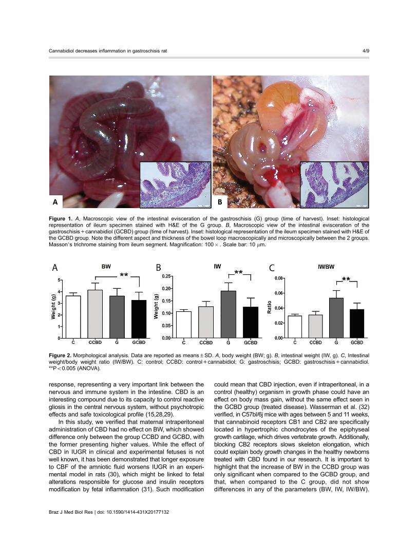
Morphological and histometric analysisFigure 1 shows macroscopical and microscopical differ-
ences of the bowel loop between G and GCBD groups.A significant difference was found for BW between
CCBD vs GCBD (4.142±0.598 vs 3.270±0.683 g, respec-tively Po0.005). For IW analysis, the GCBD group showedreduced values (0.124±0.037 g) compared to the G group(0.190±0.033 g, Po0.005) and similar to the other groupsC and CCBD (0.107±0.008 and 0.126±0.021 g, respec-tively, P40.05). The GCBD group showed lower IW/BWratio (0.038±0.008) compared to G group (0.053±0.010,Po0.005) and higher than the C group (0.029±0.002), andthere was no difference to the CCBD group (0.030±0.004,P40.05). Results are shown in Figure 2.
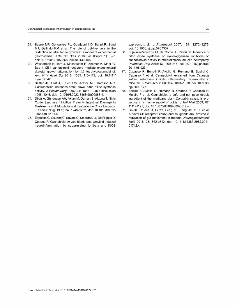
In the histometric analysis of tissue layers, (SE, ML,CM, SM, and TL), the GCBD group presented reducedthickness compared to group G, and no difference com-pared to groups C and CBD in any of the layers (Figure 3).
iNOS immunohistochemical analysisThe group GCBD showed reduced iNOS activity in
comparison to the G group, and similar to the groups Cand CCBD (Figure 4).
Nitrite/nitrate concentration of the intestine tissuehomogenate
The GCBD group showed reduction of NO2/NO3
concentration compared to group G and was similar togroups C and CCCB, (56.862±14.644 vs 122.377±15.463,Po0.005, 55.249±29.607 and 53.120±15.089, P40.05,respectively, Figure 5).
Discussion
GS is an abdominal wall defect that occurs in 1/4,000live births and is increasing all over the world (25,26).Maternal therapy with dexamethasone in a GS rat modelshowed reversion of intrauterine growth restriction (IUGR),reduction of intestinal weight and mucosal thickness(3,5). Treatment with hydrogel associated to an NOdonor covering the GS fetal bowel loops as well as lowconcentration release of NO in the amniotic fluid re-duced morphological parameters and intestinal wall thick-ness (4,27).
Enteric glial cells actively mediate acute and chronicinflammation in the intestine promoting proliferation andrelease of neurotrophins, growth factors and pro-inflammatorychemokines that in turn may increase the immunological
Braz J Med Biol Res | doi: 10.1590/1414-431X20177132
Cannabidiol decreases inflammation in gastroschisis rat 3/9
response, representing a very important link between thenervous and immune system in the intestine. CBD is aninteresting compound due to its capacity to control reactivegliosis in the central nervous system, without psychotropiceffects and safe toxicological profile (15,28,29).
In this study, we verified that maternal intraperitonealadministration of CBD had no effect on BW, which showeddifference only between the group CCBD and GCBD, withthe former presenting higher values. While the effect ofCBD in IUGR in clinical and experimental fetuses is notwell known, it has been demonstrated that longer exposureto CBF of the amniotic fluid worsens IUGR in an experi-mental model in rats (30), which might be linked to fetalalterations responsible for glucose and insulin receptorsmodification by fetal inflammation (31). Such modification
could mean that CBD injection, even if intraperitoneal, in acontrol (healthy) organism in growth phase could have aneffect on body mass gain, without the same effect seen inthe GCBD group (treated disease). Wasserman et al. (32)verified, in C57bl/6j mice with ages between 5 and 11 weeks,that cannabinoid receptors CB1 and CB2 are specificallylocated in hypertrophic chondrocytes of the epiphysealgrowth cartilage, which drives vertebrate growth. Additionally,blocking CB2 receptors slows skeleton elongation, whichcould explain body growth changes in the healthy newbornstreated with CBD found in our research. It is important tohighlight that the increase of BW in the CCBD group wasonly significant when compared to the GCBD group, andthat, when compared to the C group, did not showdifferences in any of the parameters (BW, IW, IW/BW).
Figure 1. A, Macroscopic view of the intestinal evisceration of the gastroschisis (G) group (time of harvest). Inset: histologicalrepresentation of ileum specimen stained with H&E of the G group. B, Macroscopic view of the intestinal evisceration of thegastroschisis+cannabidiol (GCBD) group (time of harvest). Inset: histological representation of the ileum specimen stained with H&E ofthe GCBD group. Note the different aspect and thickness of the bowel loop macroscopically and microscopically between the 2 groups.Masson’s trichrome staining from ileum segment. Magnification: 100� . Scale bar: 10 mm.
Figure 2. Morphological analysis. Data are reported as means±SD. A, body weight (BW; g). B, intestinal weight (IW, g). C, Intestinalweight/body weight ratio (IW/BW). C: control; CCBD: control+cannabidiol; G: gastroschisis; GCBD: gastroschisis+cannabidiol.**Po0.005 (ANOVA).
Braz J Med Biol Res | doi: 10.1590/1414-431X20177132
Cannabidiol decreases inflammation in gastroschisis rat 4/9
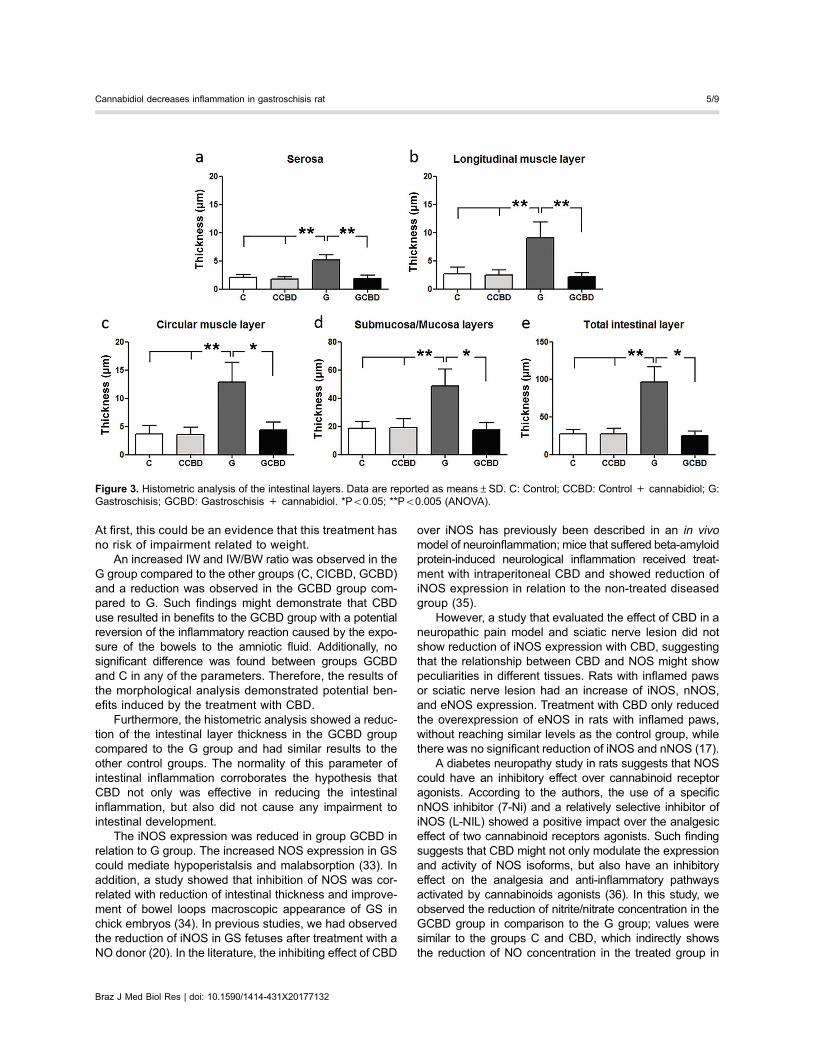
At first, this could be an evidence that this treatment hasno risk of impairment related to weight.
An increased IW and IW/BW ratio was observed in theG group compared to the other groups (C, CICBD, GCBD)and a reduction was observed in the GCBD group com-pared to G. Such findings might demonstrate that CBDuse resulted in benefits to the GCBD group with a potentialreversion of the inflammatory reaction caused by the expo-sure of the bowels to the amniotic fluid. Additionally, nosignificant difference was found between groups GCBDand C in any of the parameters. Therefore, the results ofthe morphological analysis demonstrated potential ben-efits induced by the treatment with CBD.
Furthermore, the histometric analysis showed a reduc-tion of the intestinal layer thickness in the GCBD groupcompared to the G group and had similar results to theother control groups. The normality of this parameter ofintestinal inflammation corroborates the hypothesis thatCBD not only was effective in reducing the intestinalinflammation, but also did not cause any impairment tointestinal development.
The iNOS expression was reduced in group GCBD inrelation to G group. The increased NOS expression in GScould mediate hypoperistalsis and malabsorption (33). Inaddition, a study showed that inhibition of NOS was cor-related with reduction of intestinal thickness and improve-ment of bowel loops macroscopic appearance of GS inchick embryos (34). In previous studies, we had observedthe reduction of iNOS in GS fetuses after treatment with aNO donor (20). In the literature, the inhibiting effect of CBD
over iNOS has previously been described in an in vivomodel of neuroinflammation; mice that suffered beta-amyloidprotein-induced neurological inflammation received treat-ment with intraperitoneal CBD and showed reduction ofiNOS expression in relation to the non-treated diseasedgroup (35).
However, a study that evaluated the effect of CBD in aneuropathic pain model and sciatic nerve lesion did notshow reduction of iNOS expression with CBD, suggestingthat the relationship between CBD and NOS might showpeculiarities in different tissues. Rats with inflamed pawsor sciatic nerve lesion had an increase of iNOS, nNOS,and eNOS expression. Treatment with CBD only reducedthe overexpression of eNOS in rats with inflamed paws,without reaching similar levels as the control group, whilethere was no significant reduction of iNOS and nNOS (17).
A diabetes neuropathy study in rats suggests that NOScould have an inhibitory effect over cannabinoid receptoragonists. According to the authors, the use of a specificnNOS inhibitor (7-Ni) and a relatively selective inhibitor ofiNOS (L-NIL) showed a positive impact over the analgesiceffect of two cannabinoid receptors agonists. Such findingsuggests that CBD might not only modulate the expressionand activity of NOS isoforms, but also have an inhibitoryeffect on the analgesia and anti-inflammatory pathwaysactivated by cannabinoids agonists (36). In this study, weobserved the reduction of nitrite/nitrate concentration in theGCBD group in comparison to the G group; values weresimilar to the groups C and CBD, which indirectly showsthe reduction of NO concentration in the treated group in
Figure 3. Histometric analysis of the intestinal layers. Data are reported as means±SD. C: Control; CCBD: Control + cannabidiol; G:Gastroschisis; GCBD: Gastroschisis + cannabidiol. *Po0.05; **Po0.005 (ANOVA).
Braz J Med Biol Res | doi: 10.1590/1414-431X20177132
Cannabidiol decreases inflammation in gastroschisis rat 5/9

comparison to the diseased non-treated group. The NOreduction could be explained by the decrease of cytotoxicaction mediated by macrophages and other immune cells,but further studies should be made to confirm this hypothesis.
CBD does not modify intestinal motility in controlanimals, but can inhibit the hypermotility associated toexperimental ileitis in mice (37). Borrelli et al. (38) evaluatedthe CDB effect in experimental colitis in mice and verified areduction of intestinal inflammation shown by the reductionof iNOS and the interleukins 1-beta and 10, as well as
reduction of the intestinal mucous oxidative stress. Linet al. evaluated the activity of the cannabinoid receptorG Protein-coupled Receptor 55 (GPR55) and its ligandsO-1602 in the gut movement of rodents with lipopolysac-charide (LPS)-induced intestinal inflammatory diseaseand concluded that CBD selectively normalizes motilityperturbation through potential mechanisms involving sys-temic anti-inflammatory effect and regulation of the myoe-lectrical activity in the intestine (39). De Filippis et al. (28)also confirmed in human biopsy samples and in mice with
Figure 4. Immunohistochemistry score for iNOS expression and the histological transversal slides stained by immunohistochemistry.Data are reported as means±SD. Magnification 200� , bar=100 mm. C: control; CCBD: control+cannabidiol; G: gastroschisis; GCBD:gastroschisis+cannabidiol. *Po0.05; **Po0.005 (Kruskal Wallis).
Braz J Med Biol Res | doi: 10.1590/1414-431X20177132
Cannabidiol decreases inflammation in gastroschisis rat 6/9

LPS-induced enteritis that CBD treatment resulted inreduction of iNOS and S100B.
We aimed to evaluate the CBD impact on intestinalinflammation promoted by amniotic fluid exposure. Ourstudy had some limitations. Although the analyzed param-eters were not direct indications of an inflammatory state,they have been previously studied and correlated with GSas a consequence of the inflammation and the cannabidioltreatment showed similar results in comparison to thoseobserved in previous studies that aimed reduction of theinflammation of the intestine (3,9,10). Also, we focusedon the intestine responses and thus did not discuss thecannabidiol effects in the brain or other organs, although wecan assert that clinical and visible alterations were notobserved in the rats.
There was no evidence in the analyzed parametersthat the administration of CBD 30 mg/kg via ip in femalepregnant rats have caused impairments to fetal developmentin the diseased (GCBD) or unaffected groups (CCBD).Although CBD treatment did not correlate with anychanges between C and CCBD groups in the evaluatedparameters, long term studies are necessary to demon-strate possible secondary effects of CBD.
Finally, the treatment with CDB of experimental GS inrats reduced the weight, thickness of intestinal layers,concentration of NO2/NO3, and expression of iNOS of thebowel loops showing an effective anti-inflammatory action
and pharmacological application for pre-natal use. Furtherstudies could clarify the interaction between NO and CBDpathways. In addition, investments for future studies trans-lating the present findings to the clinical practice aredesirable and suitable.
Declaration of potential conflict of interestA.W. Zuardi, J.E. Hallak, and J.A. Crippa are
co-inventors (Mechoulam R, JAC, Guimaraes FS, AWZ,JEH, Breuer A) of the patent ‘‘Fluorinated CBD compounds’’,compositions and uses thereof. Pub. No.: WO/2014/108899.International Application No.: PCT/IL2014/050023’’ Def. USno. Reg. 62193296; 29/07/2015; INPI on 19/08/2015(BR1120150164927). The University of São Paulo haslicensed the patent to Phytecs Pharm (USP ResolutionNo. 15.1.130002.1.1). The University of São Paulo hasan agreement with Prati-Donaduzzi (Toledo, Brazil) to‘‘develop a pharmaceutical product containing syntheticcannabidiol and prove its safety and therapeutic efficacy inthe treatment of epilepsy, schizophrenia, Parkinson’s disease,and anxiety disorders’’. JAC and JEH have received travelsupport from and are medical advisors of BSPG-Pharm.
Acknowledgments
A.W. Zuardi, J.E. Hallak, J.A. Crippa, and L. Sbragiaare recipients of Conselho Nacional de DesenvolvimentoCientífico e Tecnológico (CNPq) productivity fellowships.The research was supported in part by grants fromFundacão de Amparo à Pesquisa do Estado de São Paulo(FAPESP, Processo 2012/09601-4), Conselho Nacional deDesenvolvimento Científico e Tecnológico (CNPq); Coor-denacão de Aperfeicoamento de Pessoal de Nível Super-ior (CAPES); Fundacão de Apoio ao Ensino, Pesquisa eAssistência do Hospital das Clínicas da Faculdade deMedicina de Ribeirão Preto da Universidade de São Paulo(FAEPA); Center for Interdisciplinary Research on AppliedNeurosciences (NAPNA), University of São Paulo, SãoPaulo, SP, Brazil; and National Institute for TranslationalMedicine (INCT-TM; CNPq, Brazil). The present studywas supported by a CNPq grant (CNPq/MS/SCTIE/DECIT No. 26/2014, Pesquisas sobre Distúrbios Neuro-psiquiátricos; 466805/2014-4). J.A. Crippa is the recipientof a grant from University Global Partnership Network(UGPN) - Global priorities in cannabinoid research excel-lence. BSPG-Pharm (Sandwich, UK) has kindly suppliedCBD at no cost.
References
1. Dalton BG, Gonzalez KW, Reddy SR, Hendrickson RJ, IqbalCW. Improved outcomes for in born babies with uncompli-cated gastroschisis. J Pediatr Surg 2017; 52: 1132–1134,doi: 10.1016/j.jpedsurg.2016.12.003.
2. Jobe AH, Soll RF. Choice and dose of corticosteroid forantenatal treatments. Am J Obst and Gynecol 2004; 190:878–881, doi: 10.1016/j.ajog.2004.01.044.
3. Yu J, Gonzalez-Reyes S, Diez-Pardo JA. Effects of prenataldexamethasone on the intestine of rats with gastroschisis.
Figure 5. Nitrite/nitrate concentration in intestine tissue homo-genate. Data are reported as means±SD C: control; CCBD: control+cannabidiol; G: gastroschisis; GCBD: gastrosquisis+cannabidiol.**Po0.005 (Kruskal Wallis).
Braz J Med Biol Res | doi: 10.1590/1414-431X20177132
Cannabidiol decreases inflammation in gastroschisis rat 7/9

J Pediatr Surg 2003; 38: 1032–1035, doi: 10.1016/S0022-3468(03)00185-4.
4. Goncalves FL, Bueno MP, Schmidt AF, Figueira RL, SbragiaL. Treatment of bowel in experimental gastroschisis witha nitric oxide donor. Am J Obstet Gynecol 2015; 212: 383.e1–e7, doi: 10.1016/j.ajog.2014.09.025.
5. Bryan NS, Grisham MB. Methods to detect nitric oxideand its metabolites in biological samples. Free Radic BiolMed 2007; 43: 645–657, doi: 10.1016/j.freeradbiomed.2007.04.026.
6. Hrabák A, Bajor T, Temesi A, Mészáros G. The inhibitoryeffect of nitrite, a stable product of nitric oxide (NO) formation,on arginase. FEBS Lett 1996; 390: 203–206, doi: 10.1016/0014-5793(96)00659-X.
7. Bealer JF, Graf J, Bruch SW, Adzick NS, Harrison MR.Gastroschisis increases small bowel nitric oxide synthaseactivity. J Pediatr Surg 1996; 31: 1043–1045 ; discussion1045–1046, doi: 10.1016/S0022-3468(96)90083-4.
8. Grishin A, Bowling J, Bell B, Wang J, Ford HR. Roles ofnitric oxide and intestinal microbiota in the pathogenesis ofnecrotizing enterocolitis. J Pediatr Surg 2016; 51: 13–17,doi: 10.1016/j.jpedsurg.2015.10.006.
9. Yu J, Gonzalez-Reyes S, Diez-Pardo JA. Local dexametha-sone improves the intestinal lesions of gastroschisis in chickembryos. Pediatr Surg Int 2004; 19: 780–784, doi: 10.1007/s00383-003-0958-9.
10. Bittencourt DG, Barreto MW, Franca WM, Goncalves A,Pereira LA, Sbragia L. Impact of corticosteroid on intestinalinjury in a gastroschisis rat model: morphometric analysis.J Pediatr Surg 2006; 41: 547–553, doi: 10.1016/j.jpedsurg.2005.11.050.
11. Althabe F, Belizán JM, McClure EM, Hemingway-Foday J,Berrueta M, Mazzoni A et al. A population-based, multi-faceted strategy to implement antenatal corticosteroidtreatment versus standard care for the reduction of neonatalmortality due to preterm birth in low-income and middle-income countries: the ACT cluster-randomised trial. Lancet2015; 385: 629–639, doi: 10.1016/S0140-6736(14)61651-2.
12. Damsted SK, Born AP, Paulson OB, Uldall P. Exogenousglucocorticoids and adverse cerebral effects in children. EurJ Paediatr Neurol 2011; 15: 465–477, doi: 10.1016/j.ejpn.2011.05.002.
13. Drozdowicz LB, Bostwick JM. Psychiatric adverse effectsof pediatric corticosteroid use. Mayo Clin Proc 2014; 89:817–834, doi: 10.1016/j.mayocp.2014.01.010.
14. Zuardi AW. Cannabidiol: from an inactive cannabinoid to adrug with wide spectrum of action. Rev Bras Psiquiatr 2008;30: 271–280, doi: 10.1590/S1516-44462008000300015.
15. Bergamaschi MM, Queiroz RH, Zuardi AW, Crippa JA.Safety and side effects of cannabidiol, a Cannabis sativaconstituent. Curr Drug Saf 2011; 6: 237–249, doi: 10.2174/157488611798280924.
16. Malfait AM, Gallily R, Sumariwalla PF, Malik AS, AndreakosE, Mechoulam R et al. The nonpsychoactive cannabisconstituent cannabidiol is an oral anti-arthritic therapeutic inmurine collagen-induced arthritis. Proc Natl Acad Sci USA2000; 97: 9561–9566, doi: 10.1073/pnas.160105897.
17. Costa B, Colleoni M, Conti S, Parolaro D, Franke C, TrovatoAE et al. Oral anti-inflammatory activity of cannabidiol, a non-psychoactive constituent of cannabis, in acute carrageenan-induced inflammation in the rat paw. Naunyn Schmiedebergs
Arch Pharmacol 2004; 369: 294–299, doi: 10.1007/s00210-004-0871-3.
18. Costa B, Trovato AE, Comelli F, Giagnoni G, Colleoni M.The non-psychoactive cannabis constituent cannabidiol isan orally effective therapeutic agent in rat chronic inflam-matory and neuropathic pain. Eur J Pharmacol 2007; 556:75–83, doi: 10.1016/j.ejphar.2006.11.006.
19. Vannucchi MG, Midrio P, Flake AW, Faussone-PellegriniMS. Neuronal differentiation a myoenteric plexus organiza-tion are delayed in gastroschisis: an immunohistochemicalstudy in a rat model. Neurosci Lett 2003; 339: 77–81,doi: 10.1016/S0304-3940(02)01473-8.
20. Pet GE, Stark RA, Meehan JJ, Javid PJ. Outcomes ofbedside sutureless umbilical closure without endotrachealintubation for gastroschisis repair in surgical infants. Am J Surg2017; 213: 958–962, doi: 10.1016/j.amjsurg.2017.03.017.
21. Correia-Pinto J, Tavares ML, Baptista MJ, Estevao-Costa J,Flake AW, Leite-Moreira AF. A new fetal rat model of gastro-schisis: development and early characterization. J PediatrSurg 2001; 36: 213–216, doi: 10.1053/jpsu.2001.20057.
22. Perez M, Benitez SU, Cartarozzi LP, del Bel L, GuimarãesFA, Oliveira ALR. Neuroprotection and reduction of glialreaction by cannabidiol treatment after sciatic nerve transec-tion in neonatal rats. Eur J Neurosci 2013; 38: 3424–3434,doi: 10.1111/ejn.12341.
23. Geggel RL, Murphy JD, Langleben D, Crone RK, Vacanti JP,Reid LM. Congenital diaphragmatic hernia: arterial structuralchanges and persistent pulmonary hypertension after surgicalrepair. J Pediatr 1985; 107: 457–464, doi: 10.1016/S0022-3476(85)80534-5.
24. Ding AH, Nathan CF, Stuehr DJ. Release of reactivenitrogen intermediates and reactive oxygen intermediatesfrom mouse peritoneal macrophages. Comparison of acti-vating cytokines and evidence for independent production.J Immunol 1988; 141: 2407–2412.
25. Baird PA, MacDonald EC. An epidemiologic study ofcongenital malformations of the anterior abdominal wall inmore than half a million consecutive live births. Am J HumGenet 1981; 33: 470 e8.
26. Suzuhigashi M, Kaji T, Nakame K, Mukai M, Yamada W,Onishi S et al. Abdominal wall regenerative medicine for alarge defect using tissue engineering: an experimentalstudy. Pediatr Surg Int 2016; 32: 959–965, doi: 10.1007/s00383-016-3949-3.
27. Goncalves FL, da Silva R, Schmidt AF, de Oliveira MG,Sbragia L. Hydrogel protection: a novel approach to reducebowel inflammation in experimental gastroschisis. Eur JObstet Gynecol Reprod Biol 2010; 148: 35–39, doi: 10.1016/j.ejogrb.2009.10.009.
28. De Filippis D, Esposito G, Cirillo C, Cipriano M, De WinterBY, Scuderi C et al. Cannabidiol reduces intestinal inflamma-tion through the control of neuroimmune axis. PLoS One2011; 6: e28159, doi: 10.1371/journal.pone.0028159.
29. Esposito G, Filippis DD, Cirillo C, Iuvone T, Capoccia E,Scuderi et al. Cannabidiol in inflammatory bowel diseases: abrief overview. Phytother Res 2013; 27: 633–636, doi:10.1002/ptr.4781.
30. Franchi-Teixeira AR, Barreto MG, Nogueira B, Bittencourt D,Violin L, Sbragia L. Aminiotic fluid and intrauterine growthrestriction in a gastroschisis fetal rat model. Fetal Diagn Ther2005; 20: 494–497, doi: 10.1159/000088037.
Braz J Med Biol Res | doi: 10.1590/1414-431X20177132
Cannabidiol decreases inflammation in gastroschisis rat 8/9
31. Bueno MP, Goncalves FL, Guadagnini D, Barini R, SaadMJ, Gallindo RM et al. The role of gut-liver axis in therestriction of intrauterine growth in a model of experimentalgastroschisis. Acta Cir Bras 2013; 28 (Suppl 1): 3–7,doi: 10.1590/S0102-86502013001300002.
32. Wasserman E, Tam J, Mechoulam R, Zimmer A, Maor G,Bab I. CB1 cannabinoid receptors mediate endochondralskeletal growth attenuation by D9 tetrahydrocannabinol.Ann N Y Acad Sci 2015; 1335: 110–119, doi: 10.1111/nyas.12642.
33. Bealer JF, Graf J, Bruch SW, Adzick NS, Harrison MR.Gastroschisis increases small bowel nitric oxide synthaseactivity. J Pediatr Surg 1996; 31: 1043–1045 ; discussion1045–1046, doi: 10.1016/S0022-3468(96)90083-4.
34. Dilsiz A, Gündogan AH, Aktan M, Duman S, Aktung T. NitricOxide Synthase Inhibition Prevents Intestinal Damage inGastroschisis: A Morphological Evaluation in Chick Embryos.J Pediatr Surg 1999; 34: 1248–1252, doi: 10.1016/S0022-3468(99)90161-6.
35. Esposito G, Scuderi C, Savani C, Steardo L Jr, De Filippis D,Cottone P. Cannabidiol in vivo blunts beta-amyloid inducedneuroinflammation by suppressing IL-1beta and iNOS
expression. Br J Pharmacol 2007; 151: 1272–1279,doi: 10.1038/sj.bjp.0707337.
36. Bujalska-Zadrożny M, de Cordé A, Pawlik K. Influence ofnitric oxide synthase or cyclooxygenase inhibitors oncannabinoids activity in streptozotocin-induced neuropathy.Pharmacol Rep 2015; 67: 209–216, doi: 10.1016/j.pharep.2014.08.023.
37. Capasso R, Borrelli F, Aviello G, Romano B, Scalisi C,Capasso F et al. Cannabidiol, extracted from Cannabissativa, selectively inhibits inflammatory hypermotility inmice. Br J Pharmacol 2008; 154: 1001–1008, doi: 10.1038/bjp.2008.177.
38. Borrelli F, Aviello G, Romano B, Orlando P, Capasso R,Maiello F et al. Cannabidiol, a safe and non-psychotropicingredient of the marijuana plant Cannabis sativa, is pro-tective in a murine model of colitis. J Mol Med 2009; 87:1111–1121, doi: 10.1007/s00109-009-0512-x.
39. Lin XH, Yuece B, Li YY, Feng YJ, Feng JY, Yu L et al.A novel CB receptor GPR55 and its ligands are involved inregulation of gut movement in rodents. NeurogastroenterolMotil 2011; 23: 862-e342, doi: 10.1111/j.1365-2982.2011.01742.x.
Braz J Med Biol Res | doi: 10.1590/1414-431X20177132
Cannabidiol decreases inflammation in gastroschisis rat 9/9


![Histomorphological and immunohistochemical ... · -histoquímica de 172 neoplasias cutâneas de células redondas em cães.] Este trabalho descreve o uso de um painel de anticorpos](https://img.document.onl/doc/110x75/603c8bf4ed5d747ffc03960a/histomorphological-and-immunohistochemical-histoqumica-de-172-neoplasias.jpg)