Embed Size (px)
Citation preview
Dissertação Artigo de Revisão Bibliográfica Mestrado Integrado em Medicina 2013/2014
MESIAL TEMPORAL LOBE EPILEPSY: NEW EMERGING
THERAPEUTIC APPROACHES
Sara Maria Filipe Amaral
Orientadora:
Prof. Doutora Maria da Graça Borges Lobo
Porto 2014

Dissertação Artigo de Revisão Bibliográfica
Mestrado Integrado em Medicina 2013/2014
MESIAL TEMPORAL LOBE EPILEPSY: NEW EMERGING
THERAPEUTIC APPROACHES
Sara Amaral1; Graça Lobo2
1. 6º Ano Profissionalizante Mestrado Integrado em Medicina
Instituto de Ciências Biomédicas Abel Salazar
Universidade do Porto
Correio electrónico: [email protected]
2. Professora auxiliar Departamento de Imunofisiologia e Farmacologia
Instituto de Ciências Biomédicas Abel Salazar
Universidade do Porto
Porto 2014

MESIAL TEMPORAL LOBE EPILEPSY: NEW EMERGING THERAPEUTIC APPROACHES
2
Acknowledgements
To Prof. Graça Lobo for the remarkable support, countless help and enthusiasm. A
special thanks for introducing me to the fascinating world of neuropharmacology.
To Drs. Miguel Cordeiro and Pedro Coelho for the usefull coments to the manuscript.
To my family and friends for the notable presence, countless help and patience and
everlasting encouragement throughout the years. A huge thanks for believing in me
most of all.

MESIAL TEMPORAL LOBE EPILEPSY: NEW EMERGING THERAPEUTIC APPROACHES
3
Contents
Abstract...........................................................................................................................4
Resumo...........................................................................................................................5
.. 6
Introduction .....................................................................................................................7
Objectives........................................................................................................................8
Epilepsy ..........................................................................................................................9
Mesial Temporal Lobe Epilepsy....................................................................................10
...11
Neuronal Degeneration..........................................................................11
Gliosis.....................................................................................................11
Mossy Fiber Sprouting...........................................................................12
Potencial Targets for Seizure control............................................................................13
Adenosine A Re .............................................13
Adenosine Receptor-Dependent Pathways . ..14
.. 14
.15
DNA Methylation Hypothesis ..16
.. 17
. 18
Discussion.....................................................................................................................19
Conclusions...................................................................................................................22
References....................................................................................................................23
Annex A.........................................................................................................................32
MESIAL TEMPORAL LOBE EPILEPSY: NEW EMERGING THERAPEUTIC APPROACHES
4
Abstract
Epilepsy is one of the most prevalent neurological disorders, and mesial temporal lobe
epilepsy (MTLE) is the most pharmacoresistant form of epilepsy. About 30% of these
patients are not controlled by antiepileptic drugs (AED) therapy, being necessary the
use of surgical intervention to control the seizures, although only less than 10% of
patients are eligible for surgery. Current AED only treat symptoms with no reduction or
even cure of epilepsy. The high prevalence and percentage of refractory epilepsy have
motivated extensive research concerning new regulatory systems of neurotransmitters
or neuromodulators, in order to reduce this pathology.
Adenosine (ADO) modulates neuronal activity, being considered an endogenous
anticonvulsant. ADO levels increase dramatically during seizures leading to activation
of inhibitory A1 receptors, decreasing excitatory neurotransmission contributing in this
way to the cessation of seizures and to the post-ictal refractoriness. Being ADO an
ubiquitary molecule, its administration per se would lead to numerous adverse effects,
mainly cardiovascular and inactivation mechanisms in the brain are
spotlighted by researchers aiming new therapeutic targets, with focal adenosine
augmentation in the hippocampus as one as the most feasible approaches in the near
future. Implants applied in the hippocampus of experimental models regulate or release
ADO in situ. These implants contained release-control ADO polymers, histaminals cells
manipulated to release ADO or "anti-sense" genes against the adk gene of
dysregulated adenosine kinase (ADK), which controls the extracellular levels of ADO.
The ketogenic diet, used mainly in the control of refractory epilepsy in children, has
also been studied.
The domain of these mechanisms will allow the development of completely innovative
conceptual strategies for the treatment and possible cure of a syndrome as complex as
epilepsy, in a broader sense that goes beyond mere symptomatic suppression.
Keywords: Epilesy; MTLE; Adenosine; ADK, ADO-based therapies.

MESIAL TEMPORAL LOBE EPILEPSY: NEW EMERGING THERAPEUTIC APPROACHES
5
Resumo
A epilepsia é uma das doenças neurológicas mais prevalentes, e a epilepsia do lobo
mesial temporal (MTLE) a mais frequente das epilepsias resistentes a fármacos. Cerca
de 30% dos pacientes com MTLE não são controlados por fármacos anti-epilépticos
(AED), sendo a cirúrgica a única alternativa para controlar as convulsões, em que
apenas 10% dos pacientes são elegíveis. A terapia actual com AED apenas trata os
sintomas, sem redução ou possível cura da epilepsia. A prevalência e percentagem
elevadas de doentes com epilepsia farmacorresistente justificam a vasta investigação
sobre os sistemas reguladores e/ou neuromoduladores da hiperexcitabilidade
neuronial.
A adenosina (ADO) modula a atividade neuronial, sendo considerada um
anticonvulsivante endógeno. Os níveis da ADO aumentam dramaticamente durante as
convulsões. A activação dos receptores inibitórios A1 da ADO, reduz a
hiperexcitabilidade, controla a crise e assegura a refractoriedade pós-convulsiva.
Sendo a ADO uma molécula ubiquitária, a administração sistémica conduziria a
inúmeros efeitos adversos, nomeadamente cardiovasculares.
O domínio dos mecanismos de formação e inactivação da ADO no cérebro está na
base da intensa investigação visando novos alvos terapêuticos. O aumento da
adenosina focal no hipocampo é uma das abordagens mais viáveis num futuro
próximo: implantes aplicados no hipocampo de modelos experimentais regulam ou
libertam ADO in situ. Esses implantes podem conter polímeros de libertação
controlada de ADO, células histaminais manipuladas que libertam ADO ou genes "anti-
sense" contra o gene adk da enzima adenosina cinase (ADK) desregulada, que
controla os níveis extracelulares de ADO. A dieta cetogénica, usada sobretudo no
controlo de epilepsia refractária em crianças, tem sido também estudada.
Estratégias conceptuais inovadoras que vão além da supressão dos sintomomas, são
a esperança para o tratamento de uma das doenças neurológicas mais prevalentes no
mundo.
Palavras-chave: Epilepsia; MTLE; Adenosina; ADK; terapias baseadas na adenosina.

MESIAL TEMPORAL LOBE EPILEPSY: NEW EMERGING THERAPEUTIC APPROACHES
6
Abbreviation List
ADK Adenosine Kinase
Adk gene Adenosine Kinase gene
ADO Adenosine
AMP -monophosphate
AR Adenosine Receptor
ATP -triphosphate
BDNF Brain-derived Neurotrophic Factor
CNS Central Nervous System
COX - Ciclooxygenase
COXIBE Selective COX2-inhibitor
GABA -Aminobutyric Acid
HS - Hippocampal Sclerosis
KA Kainic Acid
KATP inwardly rectifying potassium channels
MTLE - Mesial Temporal Lobe Epilepsy
mTOR - Mammalian Target of Rapamycin Protein
NADH Nicotinamide Adenine Dinucleotide
RNA - Ribonucleic Acid
SE Status Epilepticus
VGLUT2 - Vesicular Glutamate Transporter
MESIAL TEMPORAL LOBE EPILEPSY: NEW EMERGING THERAPEUTIC APPROACHES
7
Introduction
Epilepsy is a chronic neurological disease, the most common worldwide. According to
the World Health Organization (WHO, 2009), about 50 million people are affected by
this disease.
Partial epilepsies occurring in adulthood are most likely to be mesial temporal lobe
epilepsies (MTLE), the most frequent form of pharmacoresistant epilepsy (Theodore,
1991). This neurological disorder cannot be controlled with medication in 30% of cases.
Therefore, surgical intervention is necessary, in order to control the seizures (Spencer
et al., 1984).
Adenosine (ADO) is an extracellular signaling molecule, able to coordinate
on and repair (Linden, 2005). In
the nervous system ADO also exerts a rather specific neuromodulatory role, controlling
synaptic transmission and synaptic plasticity (Gomes et al., 2011), as well as
coordinating neuronal networks (Sperlagh and Vizi, 2011).
allows considering ADO as a therapeutic target in the management of neurologic
disorders. However, part of the role seems to be related to the patophysiology
of epilepsy because ADO acts as an endogenous anticonvulsant (Boison, 2006).
Subsequently impaired adenosinergic modulation is thought to be involved in the
eventually, allow researchers to develop entirely new conceptual strategies, in order
reverse and eventually cure epilepsy as a whole and not only suppressing its symtoms.
MESIAL TEMPORAL LOBE EPILEPSY: NEW EMERGING THERAPEUTIC APPROACHES
8
Objectives
This present review focuses on the relevance of the ADO-mediated mechanisms in the
pathophysiology of epilepsy, in particular on MTLE. It aims to report the most relevant
therapeutic strategies developed in recent years, and which will provide pioneering
drugs in the treatment of epilepsy.
Therefore, a short overview of epileptogenesis, epilepsy and MTLE will be made. The
role of ADO in brain function and its clinical implications in epilepsy will be highlighted.
Describing the various approaches designed to increase ADO brain levels will be the
core of this work.
MESIAL TEMPORAL LOBE EPILEPSY: NEW EMERGING THERAPEUTIC APPROACHES
9
Epilepsy
As proposed by the International League Against Epilepsy (ILAE) and the International
Bureau for Epilepsy (IBE) in 2005, epilepsy is defined as a brain disorder characterized
by an enduring predisposition to generate epileptic seizures and by the neurobiologic,
cognitive, psychological, and social consequences of this condition (Fisher et al, 2005).
Traditionally, the diagnosis of epilepsy requires the occurrence of at least two
unprovoked seizures. Some clinicians also diagnose epilepsy when one unprovoked
seizure occurs in the setting of a predisposing cause (such as a focal cortical injury) or
if a generalized interictal discharge occurs suggestive of a genetic predisposition.
Seizures are the manifestation of abnormal hypersynchronous or hyperexcitable
discharges of cortical neurons. The clinical signs or symptoms of seizures depend on
the location of the epileptic discharges in the cerebral cortex and the extent and pattern
of the propagation of the epileptic discharge in the brain. The probability of having at
least one epileptic seizure in lifetime is about 9% and the probability of being
diagnosed as epileptic is almost 3%, while the prevalence of active epilepsy is only
about 0.8% (Pugliatti et al., 2007).
In a substantial number of cases, the cause of epilepsy remains unknown. Identified
causes tend to vary with patient age: Inherited syndromes, congenital brain
malformations, infection, and head trauma are primary causes in children; head trauma
is the most common known cause in young adults; strokes, tumors, and head trauma
become more frequent in middle age adults; stroke is the main cause in the elderly,
along with Alzheimer disease and other degenerative conditions.
Anti-epileptic drugs (AEDs) are the frontline treatment for epilepsy (Wiebe and Jette,
2012). There are over twenty AEDs in clinical use, including phenytoin, sodium
valproate and carbamazepine, and newer generation drugs such as levetiracetam and
lacosamide. The introduction of newer generation AEDs has provided a wider range of
treatment options that can reduce potential side effects and allow better tailoring of
therapies to an individual specific syndrome. However, the proportion of patients with
pharmacoresistant epilepsy has changed relatively little, remaining at ~30% (Wiebe
and Jette, 2012). Existing AEDs target a relatively small number of proteins. These
include: enhancement of inhibitory (GABAergic) transmission; reduction of excitatory
(glutamatergic) neurotransmission; modulation of neurotransmitter release and
targeting voltage-gated ion channels (MacDonald and Rogawski, 2008). Patients with
poorly-controlled seizures suffer additional reductions in quality of life, severe
limitations on work and personal activities, and are at increased risk of neurological
deficits, accidents and death (Pugliatti et al., 2007).
MESIAL TEMPORAL LOBE EPILEPSY: NEW EMERGING THERAPEUTIC APPROACHES
10
Mesial Temporal Lobe Epilepsy
Mesial Temporal Lobe Epilepsy (MTLE), the most common form of pharmacoresistant
epilepsy (Wieser, 2004), is often associated with previous injuries, including trauma,
status epilepticus (SE), febrile seizures, and infection (Kharatishvili and Pitkanen, 2010;
Yang et al., 2010).
Typically, a standard course of events leads to epileptic status. After the initial insult, a
latency period of 5 10 years occurs without any symptoms or complications (Wieser,
2004; Boison, 2008). The latency period ends when the patient begins to suffer from
spontaneous seizures. At the beggining of spontaneous seizure activity, seizures are
often controllable with medication. This period is known as the silent period. As the
disease progresses, patients commonly develop intractable symptoms that cannot be
managed with the current antiepileptic drugs (Wieser, 2004). The latency period
associated with epileptogenesis is thought to involve structural and biochemical
changes resulting in spontaneous seizures. These changes, presumably, are initiated
by the primary agression and occur over an extended time course (Sharma et al.,
2007).
A plethora of changes have been observed in epileptic tissue. Among which,
astrogliosis (astroglial scar), neuronal death and aberrant mossy ber sprouting are
observed both in animal models (e.g. Pitkanen et al., 2009; Hayashi et al., 2011; Zheng
et al., 2011) and in resected human tissue (e.g. de Lanerolle et al., 2003; Bae et al.,
2010; Yang et al., 2010). Although the underlying biochemical pathways remain
unclear, the first structural changes are caused by the primary insult (e.g. Sloviter,
1996; Sharma et al., 2007). Other authors have proposed that said modifications
continue to accumulate together with disease progression (Pitkanen and Sutula, 2002;
Wieser, 2004; Yang et al., 2010).
Upregulation of in ammatory indicators was observed (Crespel et al., 2002; Yang et
al., 2010; Ravizza et al., 2011) with an increase of proin ammatory cytokines, namely:
l-calpain, interleukin (IL)-1b, IL-6, and transforming growth factor (TGF)-b1 in resected
human anterior temporal lobe specimens (Feng et al., 2011). Supporting the
in ammatory hypothesis even further, downstream cyclooxygenase-2 (COX-2)
inhibition in epileptic rats, by selective COX-2 inhibitors (COXIBE (Jung et al., 2006;
Polascheck et al., 2010; Holtman et al., 2009) is found to reduce seizure initiation,
severity, and frequency while preserving neurons. The in ammatory hypotheses should
MESIAL TEMPORAL LOBE EPILEPSY: NEW EMERGING THERAPEUTIC APPROACHES
11
be treated with caution because although brain inflammation during childhood often
precedes adult epilepsy, adult-onset epilepsy may emerge when no in ammatory event
happened in childhood (Ravizza et al., 2011).
Morphological Changes
Neuronal Degeneration
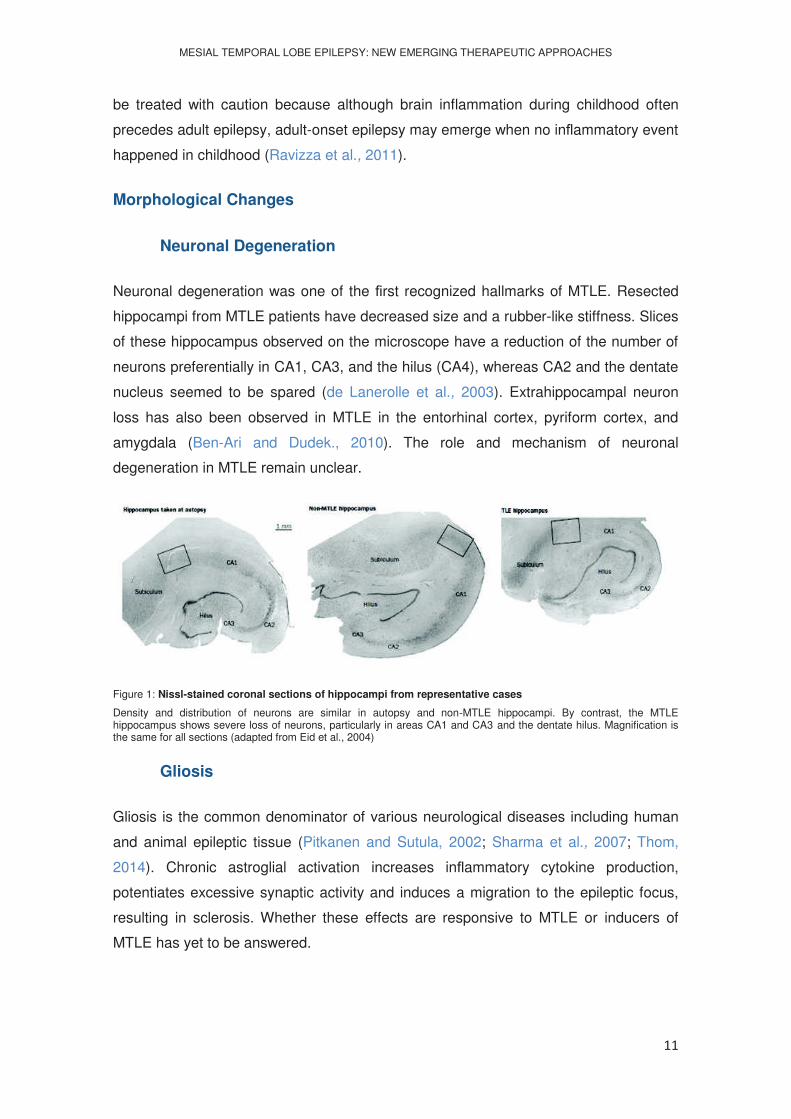
Neuronal degeneration was one of the rst recognized hallmarks of MTLE. Resected
hippocampi from MTLE patients have decreased size and a rubber-like stiffness. Slices
of these hippocampus observed on the microscope have a reduction of the number of
neurons preferentially in CA1, CA3, and the hilus (CA4), whereas CA2 and the dentate
nucleus seemed to be spared (de Lanerolle et al., 2003). Extrahippocampal neuron
loss has also been observed in MTLE in the entorhinal cortex, pyriform cortex, and
amygdala (Ben-Ari and Dudek., 2010). The role and mechanism of neuronal
degeneration in MTLE remain unclear.
Figure 1: Nissl-stained coronal sections of hippocampi from representative cases
Density and distribution of neurons are similar in autopsy and non-MTLE hippocampi. By contrast, the MTLE hippocampus shows severe loss of neurons, particularly in areas CA1 and CA3 and the dentate hilus. Magnification is the same for all sections (adapted from Eid et al., 2004)
Gliosis
Gliosis is the common denominator of various neurological diseases including human
and animal epileptic tissue (Pitkanen and Sutula, 2002; Sharma et al., 2007; Thom,
2014). Chronic astroglial activation increases in ammatory cytokine production,
potentiates excessive synaptic activity and induces a migration to the epileptic focus,
resulting in sclerosis. Whether these effects are responsive to MTLE or inducers of
MTLE has yet to be answered.

MESIAL TEMPORAL LOBE EPILEPSY: NEW EMERGING THERAPEUTIC APPROACHES
12
Mossy Fiber Sprouting
Mossy ber sprouting is characterized by dentate granule cell axons forming synapses
with cells in the granule cell layer and inner molecular layer rather than in the CA3
orn (Sharma et al., 2007). Wich changes deriving from seizures
conduct to sprouting are currently unknown. Gliosis and the release of growth factors,
cytokines, and adhesion molecules from activated astrocytes and microglia (Yang et
al., 2010) probably are in the basis of the sprouting phenomenon. Sprouting is
progressive and is brought on by recurrent seizures (Pitkanen and Sutula, 2002).
Hiperexcitation hypothesis purport that dentate granule cells become hyperexcitable as
a result of recurrent sprouting since mossy bers are glutamatergic axons and
establish excitatory circuits within the inner molecular layer (Sharma et al., 2007).
Conversely, other investigators believe that mossy ber sprouting serves to reform
inhibitory circuits that are lost during the initial damage of neurons, supported by the
fact that mossy bers synapse primarily on inhibitory interneurons in control animals
(Gorter et al., 2001). Additionally, it has been noted that mossy ber sprouting occurs
secondary to neuron loss (Jankowsky and Patterson, 2001). Observations of
reinnervation of dormant basket cells by mossy bers and of collateral sprouts from
interneurons forming inhibitory feedback to granule cells also rienforces the
antiepileptogenic hypothesis (Ben-Ari and Dudek, 2010). Recently, Sharma and
colleagues (2007) noted that aberrant sprouting precedes seizure onset in rats
exposed to kainic acid (KA). This observation does not clarify the question of the role of
mossy bers, unfortunately, because whether the sprouting itself leads to seizure
production or the inability to sprout further leads to seizure production is unclear.
Clarifying the role that mossy bers play in epileptogenesis is an important step in
better understanding epilepsy and epileptogenesis and may lead to new treatment
options.
Figure 2. Axonal sprouting in Hippocampal sclerosis (HS)/temporal lobe epilepsy (TLE).
Mossy fibre sprouting identified with Timm staining in human hippocampal sclerosis (HS) (A-C). In (A) the black silver
granules are mainly confined to the subgranular zone and significant sprouting into the molecular layer (arrow) is not
observed. In (B) there is focal sprouting in the molecular layer (arrow) and in (C) marked sprouting is shown with a
dense band of zinc positive granules in the molecular layer (adapted from Thom M 2014).

MESIAL TEMPORAL LOBE EPILEPSY: NEW EMERGING THERAPEUTIC APPROACHES
13
Potencial Targets for Seizure Control
The need for new AEDs is a widely-recognized goal for the improved treatment of
epilepsy (Baulac and Pitkanen, 2008). Innovative treatments may either be targeted to
epileptogenesis, the morphological and functional changes leading to epilepsy after an
initial brain insult, or to ictogenesis, the processes involved in initiation, propagation
and amplification of seizures in the epileptic brain. Possible features of new AED
targets include proteins with more subtle influence on excitatory neurotransmission to
avoid the common side effects of many AEDs. Anti-neuroinflammatory (Venazzi et al.,
2011) or neuroprotective properties (Acharya et al., 2008) could also yield disease-
modifying effects that would mitigate the underlying pathology.
Adenosine A Retaliatory Metabolite
The ribonucleoside adenosine (ADO) has early evolutionary origins and likely played a
role in prebiotic evolution (Oro and Kimball, 1961). Importantly, ADO is not only part of
the energy molecule Adenosine Triphosphate (ATP) but also of Ribonucleic Acid
(RNA), the nucleic acid thought to be at the origin of life (Lahav, 1993; Dworkin et al.,
2003; Robertson and Joyce, 2012). While ATP re ects the energy pool in the
environment, RNA re ects the metabolic activities of a cell. Thus, ADO assumes a
central place between energy availability and metabolic demands and has therefore
been termed a retaliatory metabolite (Newby et al., 1985). The early evolutionary
principle to conserve energy was likely a rise in ADO as a consequence to ATP
depletion and to use the increase in ADO as a negative feedback regulator to attenuate
all cellular activities that consume energy. This early evolutionary principle is
omnipresent in all living systems and in every human organ. In the brain, epileptic
seizures cause a rapid drop in energy, which results in the generation of ADO levels
that can exceed the basal level more than 40 times (During and Spencer, 1992); it is
this rise of ADO that acts as endogenous terminator of seizures and which is
responsible for the postictal refractoriness that normally follows a seizure (Lado and
Moshe, 2008). Seizure suppression by ADO depends on the activation of G protein
coupled adenosine A1 receptors (Fredholm et al, 2005a); however, new evidence
suggests that ADO retains important ADO receptor-independent regulatory functions,
which are based on interactions with mitochondrial bioenergetics, interference with
enzyme reactions, and epigenetic functions. Thereby ADO assumes a unique role as
homeostatic network regulator.
MESIAL TEMPORAL LOBE EPILEPSY: NEW EMERGING THERAPEUTIC APPROACHES
14
Adenosine Receptor-Dependent Pathways
A number of actions are mediated by a group of speci c receptors, G protein-
linked transmembrane proteins of the P1 family, distinguished from the P2 ATP
receptor family. Four members of the P1 family have been cloned in mammals:
adenosine subtype 1 (A1R), 2A (A2AR), 2B (A2BR) and 3 (A3R) receptors (Fredholm
et al, 2011). Homologous genes have been found in numerous other animal groups
(Sazanov et al, 2000; Petersen et al., 2003; Dolezelova et al., 2007; Boehmler et al.,
2009; Malik and Buck, 2010). These receptors have biochemical speci city as each act
through a particular set of G proteins to in uence second messengers: A1R and A3R
activation inhibit the second messenger cAMP production, whereas A2AR and A2BR
activation increase cAMP (Fredholm et al., 2011). Other second messengers such as
diacylglycerol, inositol triphosphate, and Ca2+ are also modulated. Each receptor
presents a distinct pharmacology, and each has a particular distribution in tissues and
cell types. A1Rs are expressed mosthighly in brain, whereas A2BRs and A3Rs have
their highest expression in the periphery (Dixon et al., 1996). Within the brain, A1Rs
are widespread with particularly high levels in the limbic system, whereas A2ARs are
expressed mostly in the basal ganglia (Dixon et al., 1996). ADO can have powerful
receptor-mediated effects on synaptic transmission in the brain (Fredholm et al., 2011).
Presynaptic A1Rs inhibit synaptic release of most, if not all, neurotransmitters, with an
apparently greater effect on excitatory transmission. Thus, if ADO levels are raised
suf ciently, synaptic transmission can be blocked altogether. On the postsynaptic side,
A1Rs hyperpolarize membranes by opening inwardly rectifying K+ channels. These
combined A1R effects play a major role in the ef cacious anticonvulsant effect of ADO
and A1R agonists (Boison, 2007). The effect of A2ARs on network excitability is less
clear, and more anatomically restricted, but if seizures re ect brain network imbalance,
then one seizure model suggests that A1Rs and A2ARs may cooperate to promote
homeostasis (De Sarro et al., 1999).
Mitochondrial Bioenergetics
Mitochondria generate ATP via oxidative phosphorylation, and this is the main process
of energy generation. Regarding the relationship between ADO and ATP, intracellular
ADO is dephosphorylated from Ade -monophosphate (AMP) and is converted
back to AMP via adenosine kinase (ADK). The ADO-AMP cycle is linked to ADP and
ATP through ADK. Thus, ADO is linked tightly to energy metabolism. Whereas
mitochondrial uncouplers decrease ATP and increase ADO, mitochondrial enhancers
which boost ATP levels also appear to increase ADO. Therefore, improving
MESIAL TEMPORAL LOBE EPILEPSY: NEW EMERGING THERAPEUTIC APPROACHES
15
mitochondrial bioenergetics has the potential to offer dual bene ts of improving
metabolic dysfunction and restoring ADO homeostasis (Boison et al., 2013).
The intracellular concentration of ATP is nearly 50 times higher than that of AMP (Arch
and Newsholme, 1978) and about 10,000 times higher than that of ADO (Pazzagli et
al., 1995; Delaney and Geiger, 1996) and minor decreases in intracellular ATP leads to
a large rise of intracellular ADO level. Thus, various excitatory stimuli cause decreased
brain energy and a subsequent increase in ADO (Shepel et al., 2005). As a retaliatory
metabolite ADO is thought to be one of the keylinks between neuronal network
homeostasis and mitochondrial bioenergetics with both adenosine receptor-dependent
and independent pathways.
Ketogenic Diet
In the early 1920s, the ketogenic diet was first introduced to treat patients,mainly
children, with refractory epilepsy. However, with the introduction of diphenylhydantoin
in the following decade the diet faded from the clinical arsenal. In the 1990s its clinical
interest was renewed and since then more palatable diet variants were developed.
The diet is high in fat and low in carbohydrate and protein, providing sufficient protein
for growth but insufficient amounts of carbohydrates for all the metabolic needs
(Freeman et al., 2006). During high rates of fatty acid oxidation, in mitochondria, large
amounts of acetyl-CoA are generated, leading to the synthesis of three ketone bodies -
hydroxybutyrate, acetoacetate, and acetone. Ketone bodies pass into circulation,
causing serum levels to rise severalfold, and then are utilized as an energy source in
extrahepatic tissues, including the brain. The ketone bodies are converted to acetyl-
CoA by D-hydroxybutyrate dehydrogenase, acetoacetate-succinyl-CoA transferase,
and acetoacetyl-CoAthiolase and then enter the Krebs cycle within brain mitochondria,
leading to ATP production (Masino and Geiger, 2008).
Ketone bodies have an important role on seizure protection and the proposed
mechanisms for this occurrence are:
a) glutamate transport into synaptic vesicles (vesicular glutamate transporter
VGLUT2), is inhibited by the ketone body acetoacetate (Juge et al, 2010),
decreasing glutamate release;
b) increased production of the inhibitory neurotransmitter -Aminobutyric acid
(GABA) by glutamate recycling via glutamine; reduction of brain derived
neurotrophic factor (BDNF) and its receptor TrkB expression, both being

MESIAL TEMPORAL LOBE EPILEPSY: NEW EMERGING THERAPEUTIC APPROACHES
16
implicated in epileptogenesis, by a decline in the cytosolic reduced nicotinamide
adenine dinucleotide (NADH) (Garriga-Canut et al., 2006);
c) ATP activation of nearby ATP-sensitive potassium (KATP) channels (Haller et al.,
2001; Tanner et al., 2011), generating hyperpolarizing K+ currents (Ashcroft and
Gribble, 1998);
d) Increased extracellular ADO levels by mitochondrial ATP and consequent
reduction of neuronal excitability by activating pre and postsynaptic A1Rs.
The increase on extracellular ADO levels is critical to the therapeutic success of the
ketogenic diet (Masino and Geiger, 2008).
DNA Methylation Hypothesis
Epigenome modifications which include changes in DNA methylation, histone tail
modi cations, and incorporation of histone variants are mechanisms by which network
homeostasis can be dramatically altered and consequently alter the entire gene
expression pro le of a tissue. There are a number of epilepsy-associated neurological
diseases that are directly attributed to primary genetic mutations and result in
secondary deregulation of the epigenome (Kobow and Blumcke, 2011). Gene
promoters from MTLE patients have altered DNA methylation patterns and decreased
DNA methyltransferase gene expression (Kobow et al., 2009; Zhu et al., 2012). This
hypermethylated state forms the basis of the methylation hypothesis of
epileptogenesis, suggesting that seizures by themselves can induce epigenetic
chromatin modi cations and thereby aggravate the epileptogenic condition (Kobow and
Blumcke, 2011). Hypermethylation of DNA can be triggered by a variety of
mechanisms, however, the mechanisms underlying the gradual increase in DNA
methylation during the course of epileptogenesis are not well characterized and subject
of speculation (Kobow and Blumcke, 2012). The epigenetic drift hypothesis suggests
that a gradual shift in the ratio of active DNA demethylation and de novo methylation,
triggered by an injury and modi ed by environmental and intrinsic factors leads to
increased DNA methylation, altered gene expression, and an altered (e.g., seizure)
phenotype (Feil and Fraga, 2011).
Peripheral changes in the transmethylation pathway are conserved within the brain. It
was recently found that hippocampal ADO levels regulate the global state of DNA
methylation by shifting the equilibrium constant of the transmethylation pathway either
increasing (high ADK and low ADO) or decreasing (low ADK and high ADO)
methylation. These ADO dependent changes in DNA methylation are receptor-
independent and can be evoked by either a single pharmacological bolus of ADO (icv

MESIAL TEMPORAL LOBE EPILEPSY: NEW EMERGING THERAPEUTIC APPROACHES
17
administration) or in response to endogenous changes in ADK expression or activity
(Williams-Karnesky et al., 2013).
As an obligatory endproduct of transmethylation, ADO tone non-speci cally drives the
transmethylation pathway by regulating substrate availability. Consequently,
tone does not regulate site speci c DNA methylation, but instead the homeostasis of
the DNA-methylome.
Through this mechanism, astrogliosis and associated overexpression of ADK could
contribute to continued epileptogenesis through maintenance of a hypermethylated
state of hippocampal DNA (Williams-Karnesky et al., 2013).
Unbalance of Adenosine Kinase in the Brain
Astrocytes have the main regulative function on maintenance of extracellular ADO
levels. The astrocyte-based enzyme ADK phosphorylates adenosine to AMP driving
the in ux of ADO into the astrocyte through equilibrative transporters. Injuries to the
brain such as trauma, stroke or SE trigger an acute surge in ADO, which is
accompanied by transient downregulation of ADK (Clark et al, 1997; Pignataro et al,
2008). The initial injury and the associated surge in ADO can trigger several
mechanisms possibly implicated in epileptogenesis, among which the induction of
A2AR expression in glial cells appears to play a prominent role. The proliferation of
astrocytes and thereby the development of astrogliosis is in part regulated by the ratio
of the different ADO receptors expressed on the astrocyte membrane. The increased
activation of A2ARs by an injury- associated surge in ADO can increase astrocyte
proliferation and activation, whereas the blockade of excitatory A2ARs prevents the
induction of astrogliosis by BDNF, which is a known transactivator of the A2AR
(Hindley et al., 1994; Brambilla et al., 2003; Rajagopal et al., 2004). ADO de ciency in
human epilepsy has directly been identi ed via the analysis of microdialysis samples. A
25% reduction of ADO in the epileptogenic versus the contralateral control
hippocampus was found (During and Spencer, 1992).
MESIAL TEMPORAL LOBE EPILEPSY: NEW EMERGING THERAPEUTIC APPROACHES
18
Focal Adenosine Augmentation
Systemic augmentation of ADO signaling by either A1 receptor agonists or by ADK-
inhibitors effectively suppress seizures in a wide range of epilepsy models including
those resistant to conventional AEDs (Gouder et al., 2003; McGaraughty et al., 2005;
Benarroch, 2008). Unfortunately, systemic augmentation of ADO signaling is not a
therapeutic option due to widespread, mainly cardiovascular, side effects, and due to
the liver toxicity of ADK disruption (Boison et al., 2002; Fredholm et al., 2005a).
Therefore, focal ADO augmentation approaches, with the aim to reconstruct ADO
homeostasis within an epileptogenic brain area, become a therapeutic necessity. Focal
ADO augmentation to suppress epileptic seizures was rst successfully performed in
rat kindling models of epilepsy using intraventricular implants engineered to release
ADO (Huber et al., 2001). Importantly, focal ADO augmentation, in contrast to systemic
ADO augmentation, was not associated with any sedative side effects (Guttinger et al.,
2005). Unfortunately, duration of seizure control (12 days) was limited by a reduced life
expectancy of the encapsulated cells (Huber et al., 2001). Currently, new approaches
are being developed to make therapeutic use of ADO augmentation:
- Silk is a Food and Drug Administration (FDA) approved biocompatible
biopolymer that can be engineered for controlled focal drug release.
Infrahippocampal implants of silk which were engineered to release de ned
daily doses of up to 1 mg of ADO provided complete control of seizures in the
kindled rat for the duration of the ADO release (1 mg ADO per day for 10 days)
(Szybala et al., 2009). Mouse embryonic stem cells and human mesenchymal
stem cells were also engineered to release ADO from silk implants (Shetty and
Hattiangady, 2007)
- ADO releasing stem cells based on genetic disruption of the endogenous
adenosine kinase (adk) gene or by expressing a micro RNA (miRNA) directed
against adk gene via a lentiviral vector (Fedele et al., 2004; Ren et al., 2007)
potently suppressed kindling epileptogenesis after transplantation into the
infrahippocampal ssure with upregulation of ADK (Li et al., 2007b). Marked
astrogliosis attenuation, normalization of ADK expression levels and total
absence of seizures were registrated three weeks later, supporting a novel and
persistant antiepileptogenic effect of focal ADO delivery.

MESIAL TEMPORAL LOBE EPILEPSY: NEW EMERGING THERAPEUTIC APPROACHES
19
Discussion
Epilepsy is a neuronal disorder characterized by abnormal and excessive neuronal
firing where each seizure represents a rapid loss of homeostatic equilibrium, with
altered energy and molecular gradients, and a corresponding interruption of normal
behavior and consciousness. Because having a seizure can increase the likelihood of
future seizures, seizures themselves contribute to epileptogenesis (Boison et al.,
2013).
Over the past few decades, many studies have sought to identify and treat key
changes associated with epilepsy and spontaneous seizures. Many morphological and
biochemical changes identified agravate epilepsy and may steer to a refractory state,
where no AED therapy is able to control seisures. MTLE is the most frequent of those
drug resistant epilepsies.
The majority of MTLE patients have HS lesions, including astrogliosis, neuronal death
de Lanerolle et al., 2003; Bae et al., 2010;
Yang et al., 2010). The aberrant sprouting of mossy fibers occurs secondary to neuron
loss and so, a comprehensive knowledge of this phenomenon will have a key role for
the development of new treatment options.
At the present, antiepileptic drugs focus on neuronal targets, mainly glutamatergic and
GABAergic mechanisms or ion channels. These therapies are largely symptomatic and
do not affect the underlying disease processes. These drugs often result in deleterious
side effects including cognitive impairment. Moreover, a large subpopulation of patients
does not respond to current AEDs, suggesting that there is a great need for new
therapeutic directions (Wiebe and Jette, 2012).
Endogenous mechanisms for the termination of seizure activity include the release of
high enough amounts of ADO, contributing to seizure arrest and postictal
refractoriness. Because ADO modulates neuronal activity it is considered an
endogenous anticonvulsant. The activation of ADO inhibitory A1 receptors decreases
excitatory neurotransmission, and for this reason several ADO-based therapies for
epilepsy are currently under development.
Ketogenic diet has been confined to a last resort therapy for refractory epilepsy,
nevertheless this diet often succeeds in control of seizures when drugs fail. This
indicates that the metabolic changes produced by the diet tap into anticonvulsant
mechanisms that are not targeted by existing medication. The diet is unlikely to

MESIAL TEMPORAL LOBE EPILEPSY: NEW EMERGING THERAPEUTIC APPROACHES
20
produce treatment resistance and significant untoward side effects (Boison et al.,
2011): One of these mechanisms is the enhancement of ADO levels, leading to a net
inhibition of excitatory transmission; other proposed mechanisms include disruption of
glutamatergic synaptic transmission, inhibition of glycolysis, and activation of ATP-
sensitive potassium channels. Follow up studies will show how potent it is the
antiseizure action of this diet by the integration of individual findings on metabolic
modification of neuronal activity.
ADO-dependent changes in DNA methylation were pinpointed as an underlying
mechanism for the antiepileptogenic properties of ADO therapy. Several targets that
function by either interacting with DNA or playing a role in gene transcription and
translation responded to ADO therapy and exhibited a decrease in DNA
methylation. This fact constitutes a benefit since the use of DNA methyltransferase
(DNMT) inhibitors is associated with complications or side effects (Williams-Karnesky
et al., 2013).
Pathologically increased expression of ADK is linked to astrogliosis in many central
nervous system (CNS) pathologies, leading to reductions in ADO tone and leaving
neurons more susceptible to damage (Boison, 2008). It has been observed that the
upregulation of ADK occurs before gross morphological changes in the hippocampus,
offering a time window for therapeutic intervention (Gouder et al., 2004).
Consequentely, the use of ADK inhibitors may possibly slow down
deterioration.
Local administration of ADO in the hippocampus in animal kindling models through icv
injection markedly attenuated seizures and contributed to postictal refractoriness.
Systemic administration of selective agonist analogues of the A1 inhibitory ADO
receptor or selective antagonist analogues of the A2A exitatory ADO receptor
mimicked ADO icv injection, but the occurrence of intense cardiovascular side effects
were also observed. Being ADO an ubiquitary molecule, its administration per se (and
its analogues) leads to numerous adverse effects. Nowadays we find a new era in
which focal approaches of ADO augmentation are the leading strategies for seizure
control: Cell- and polymer- based focal ADO augmentation therapies were found to
provide effective seizure control (Boison, 2009a; Boison & Stewart, 2009); The
transplantation of ADO releasing stem cells (modified by viral-mediated insertion of
antisense sequences) potently suppressed kindling epileptogenesis (Li et al., 2007b).
This viral ADK antisense approach apparently causes permanent reversal of ADK-
based ADO dysregulation in the epileptogenic focus in which ADK levels are
MESIAL TEMPORAL LOBE EPILEPSY: NEW EMERGING THERAPEUTIC APPROACHES
21
pathologically high (Boison, 2010a). Yet it is necessary to establish the efficacy of the
virus in post SE chronic models of epilepsy.
Polymer-, and in particular silk-based ADO delivery (Wilz et al., 2008; Szybala et al.,
2009), are ideal to start clinical safety and feasibility tests of focal ADO-augmentation.
Notwithstanding, these systems need to be further improved in order to provide long-
term delivery options for ADO. Furthermore, the translational potential of such studies
in humans is uncertain.
Rapamycin has recently received broad attention as a potential broad-spectrum
effector in seizure suppression and irregular structural modification of the epileptic
hippocampus (Chong et al., 2010). This drug affects the mammalian target of
rapamycin (mTOR) protein, which is involved with growth factor production, neuronal
plasticity and remodeling, glial activation, and cellular motility. As such, it would seem
that rapamycin has the potential to be a catch-all therapy for MTLE and for epilepsy as
a whole. Because rapamycin was originally approved as a cancer chemotherapeutic
(powerfull inhibitor of mitosis and motility of cells) it may not be safe as a long-term
MTLE treatment.
New therapeutic targets, which do not include the increase of ADO in the
hippocampus, have been emerging. Therapies based on the mechanisms now
evidenced from the study of rapamycin in the hippocampus of rats, in induced
metabolic changes in neurons by ketogenic diet, and in the mechanisms involved in
neuro-inflammation, are rapidly developing. However, in the near future, the ADO-
based therapies are the ones that most likely will jump to clinical trial .
MESIAL TEMPORAL LOBE EPILEPSY: NEW EMERGING THERAPEUTIC APPROACHES
22
Conclusions
The number of AEDs available for treatment of MTLE are greater than ever, but the
effectiveness of any AED remains arguably unchanged. Contraindications are better
managed today but effective seizure-eliminating therapy is not (Löscher and Schmidt,
2011).
Much of the mystery of epilepsy is
experiments will allow further insites on the interplay among ion channels, cellular
signaling, neurotransmitter release and reuptake, and inflammatory factors in pre-
epileptic and epileptic tissue. Basic understanding of morphological changes such as
mossy fiber sprouting, gliosis, and neuron death may lead to identification of specific
molecules released either as a result of cellular changes or as a prequel to the
changes. These molecules may constitute good targets for future therapeutic
approaches.
MESIAL TEMPORAL LOBE EPILEPSY: NEW EMERGING THERAPEUTIC APPROACHES
23
References
Acharya MM, Hattiangady B, Shetty AK (2008) Progress in neuroprotective strategies
for preventing epilepsy. Prog Neurobiol 84:363-404.
-nucleotidase,
adenosine kinase and adenosine deaminase in tissues from vertebrates and
invertebrates in relation to the control of the concentration and the physiological role of
adenosine. Biochem J 174:965 977.
Ashcroft F, Gribble F (1998) Correlating structure and function in ATP-sensitive K+
channels. Trends Neurosci 21:288 294.
Bae EK, Jung KH, Chu K, Lee ST, Kim JH, Park KI, Kim M, Chung CK, Lee SK, Roh
JK (2010) Neuropathologic and clinical features of human medial temporal lobe
epilepsy. J Clin Neurol 6:73 80.
Baulac M and Pitkanen A (2008) Research Priorities in Epilepsy for the Next Decade-A
Representative View of the European Scientific Community. Epilepsia 50:571-578.
Ben-Ari Y, Dudek FE (2010) Primary and secondary mechanisms of epileptogenesis in
the temporal lobe: there is a before and an after Epilepsy Curr 10:118 125.
Benarroch EE (2008) Adenosine and its receptors: multiple modulatory functions and
potential therapeutic targets for neurologic disease. Neurology 70:231 236.
Boehmler W, Petko J, Woll M, Frey C, Thisse B, Thisse C
Patterns 9:144 151.
Boison D (2006) Adenosine kinase, epilepsy and stroke: mechanisms and therapies.
Trends Pharmacol Sci 27(12):652 658.
Boison D (2007) Adenosine as a modulator of brain activity. Drug News Perspect
20:607 611.
Boison D (2008) The adenosine kinase hypothesis of epileptogenesis. Prog Neurobiol
84:249 262.
Boison D (2009a) Adenosine augmentation therapies (AATs) for epilepsy: prospect of
cell and gene therapies. Epilepsy Res 85:131 141.
MESIAL TEMPORAL LOBE EPILEPSY: NEW EMERGING THERAPEUTIC APPROACHES
24
Boison D (2010a) Inhibitory RNA in epilepsy: research tool and therapeutic
perspectives. Epilepsia 51:1659 1668.
Boison D (2013) Adenosine kinase: exploitation for therapeutic gain. Pharmacol Rev
65:906 943.
Boison D, Masino SA, Geiger JD (2011) Homeostatic bioenergetic network regulation -
a novel concept to avoid pharmacoresistance in epilepsy. Expert Opin Drug Discov.
6(7):713-724.
Boison D, Sandau US, Ruskin DN, Kawamura M Jr, Masino SA (2013) Homeostatic
control of brain function - new approaches to understand epileptogenesis. Front Cell
Neurosci. 7:109
Boison D, Scheurer L, Zumsteg V, Rülicke T, Litynski P, Fowler B, et al. (2002)
Neonatal hepatic steatosis by disruption of the adenosine kinase gene. Proc Natl Acad
Sci U.S.A. 99:6985 6990.
Boison D, Stewart K-A (2009) Therapeutic epilepsy research: from pharmacological
rationale to focal adenosine augmentation. Biochem Pharmacol 78:1428 1437.
Brambilla R, Cottini L, Fumagalli M, Ceruti S and Abbracchio MP (2003) Blockade of
A2A -induced reactive
astrogliosis in rat striatal primary astrocytes. Glia 43:190 194.
Chong ZZ, Shang YC, Zhang L, Wang S, Maiese K (2010) Mammalian Target of
Rapamycin: Hitting the Bull's-Eye for Neurological Disorders. Oxidative Medicine and
Cellular Longevity 3(6):374-391
Clark RS, Carcillo JA, Kochanek PM, Obrist WD, Jackson EK, Mi Z, et al. (1997)
oxidative metabolism after severe head injury in humans. Neurosurgery 41:1284 1292.
Crespel A, Coubes P, Rousset MC, Brana C, Rougier A, Rondouin G, Bockaert J,
Baldy-Moulinier M, Lerner-
temporal lobe epilepsy with hippocampal sclerosis. Brain Res 952:159 169.
de Lanerolle NC, Kim JH, Williamson A, Spencer SS, Zaveri HP, Eid T, Spencer DD
(2003) A retrospective analysis of hippocampal pathology in human temporal lobe
epilepsy: evidence for distinctive patient subcategories. Epilepsia 44:677 687.

MESIAL TEMPORAL LOBE EPILEPSY: NEW EMERGING THERAPEUTIC APPROACHES
25
De Sarro G, De Sarro A, Di Paola ED and Bertorelli R (1999) Effects of adenosine
receptor agonists and antagonists on audiogenic seizure-senbible DBA/2 mice. Eur J
Pharmacol 371:137 145.
Delaney SM and Geiger JD (1996) Brain regional levels of adenosine and adenosine
nucleotides in rats killed by high-energy focused microwave irradiation. J Neurosci
Methods 64:151 156.
Dixon AK, Gubitz AK, Sirinathsinghji DJ, Richardson PJ and Freeman TC (1996)
Tissue distribution of adenosine receptor mRNAs in the rat. Br J Pharmacol 118:1461
1468.
Dolezelova E, Nothacker HP, Civelli O, Bryant PJ and Zurovec M (2007) A Drosophila
adeno- sine receptor activates cAMP and calcium signaling. Insect Biochem Mol Biol
37:318 329.
During MJ and Spencer DD (1992) Adenosine: a potential mediator of seizure arrest
and postictal refractoriness. Ann Neurol 32:618 624.
Dworkin JP, Lazcano A and Miller SL (2003) The roads to and from the RNA world. J
Theor Biol 222:127 134.
Fedele DE, Koch P, Brüstle O, Scheurer L, Simpson EM, Mohler H, Boison D (2004)
Engineering embryonic stem cell derived glia for adenosine delivery. Neurosci Lett
370:160 165.
Feil R and Fraga MF (2011) Epigenetics and the environment: emerging patterns and
implications. Nat Rev Genet 13:97 109.
Feng ZH, Hao J, Ye L, Dayao C, Yan N, Yan Y, Chu L, Shi FD (2011) Overexpression
of mu-calpain in the anterior temporal neocortex of patients with intractable epilepsy
correlates with clinicopathological characteristics. Seizure 20:395 401.
Fisher RS, van Emde Boas W, Blume W. (2005) Epileptic seizures and epilepsy:
definitions proposed by the International League Against Epilepsy (ILAE) and the
International Bureau for Epilepsy (IBE). Epilepsia 46(4):470-2.
Fredholm BB, Chen JF, Cunha RA, Svenningsson P, Vaugeois JM (2005a) Adenosine
and brain function. Int Rev Neurobiol 63:191 270.
MESIAL TEMPORAL LOBE EPILEPSY: NEW EMERGING THERAPEUTIC APPROACHES
26
Fredholm BB, Ijzerman AP, Jacobson KA, Linden J and Muller CE (2011) International
adenosine receptors an update. Pharmacol Rev 63:1 34.
Freeman J, Veggiotti P, Lanzi G, Tagliabue A, Perucca E (2006) The ketogenic diet:
from molecular mechanisms to clinical effects. Epilepsy Res 68:145-80.
Garriga-Canut M, Schoenike B, Qazi R et al. (2006) 2-Deoxy-D-glucose reduces
epilepsy progression by NRSF-CtBP-dependent metabolic regulation of chromatin
structure. Nature Neuroscience 9:1382 1387.
Gomes CV, Kaster MP, Tome AR, Agostinho PM, Cunha RA (2011) Adenosine
receptors and brain diseases: neuroprotection and neurodegeneration. Biochim
Biophys Acta 1808:1380-1399.
Gorter JA, van Vliet EA, Aronica E, Lopes da Silva FH (2001) Progression of
and extensive bilateral loss of hilar parvalbumin and somatostatin-immunoreactive
neurons. Eur J Neurosci 13:657 669.
Gouder N, Fritschy JM, Boison D (2003) Seizure suppression by adenosine A1
receptor activation in a mouse model of pharmacoresistant epilepsy. Epilepsia 44:877
885.
Gouder N, Scheurer L, Fritschy JM, Boison D (2004) Overexpression of adenosine
kinase in epileptic hippocampus contributes to epileptogenesis. J Neurosci 24:692
701.
Güttinger M, Padrun V, Pralong W, Boison D (2005) Seizure suppression and lack of
adenosine A1 receptor desensitization after focal long-term delivery of adenosine by
encapsulated myoblasts. Exp Neurol 193:53 64.
Haller M, Mironov SL, Karschin A, Richter DW (2001) Dynamic activation of KATP
channels in rhythmically active neurons. J Physiol. 537:69 81.
Hayashi K, Ueshima S, Ouchida M, Mashimo T, Nishiki T, Sendo T, Serikawa T,
Matsui H, Ohmori I (2011) Therapy for hyperthermia-induced seizures in Scn1a mutant
rats Epilepsia 52:1010 1017.

MESIAL TEMPORAL LOBE EPILEPSY: NEW EMERGING THERAPEUTIC APPROACHES
27
Hindley S, Herman MA and Rathbone MP (1994) Stimulation of reactive astrogliosis in
vivo by extracellular adenosine diphosphate or an adenosine A2 receptor agonist. J
Neurosci Res 38:399 406.
Holtman L, van Vliet EA, van Schaik R, Queiroz CM, Aronica E, Gorter JA (2009)
Effects of SC58236, a selective COX-2 inhibitor, onmepileptogenesis and spontaneous
seizures in a rat model for temporal lobe epilepsy. Epilepsy Res 84:56 66.
Huber A, Padrun V, Deglon N, Aebischer P, Mohler H, Boison D (2001) Grafts of
adenosine-releasing cells suppress seizures in kindling epilepsy. Proc Natl Acad Sci
USA 98:7611 7616.
Jankowsky JL, Patterson PH (2001) The role of cytokines and growth factors in
seizures and their sequelae. Prog Neurobiol 63:125 149.
Juge N, Gray JA, Omote H, Miyaji T, Inoue T, Hara C, Uneyama H, Edwards RH, Nicoll
RA,NMoriyama Y (2010) Metabolic control of vesicular glutamate transport and
release. Neuron 68:99 211.
Jung KH, Chu K, Lee ST, Kim J, Sinn DI, Kim JM, Park DK, Lee JJ, Kim SU, Kim M,
Lee SK, Roh JK (2006) Cyclooxygenase-2 inhibitor, celecoxib, inhibits the altered
hippocampal neurogenesis with attenuation of spontaneous recurrent seizures
following pilocarpine-induced status epilepticus. Neurobiol Dis 23:237 246.
Kharatishvili I, Pitkanen A (2010) Posttraumatic epilepsy. Curr Opin Neurol 23:183
188.
Kobow K and Blumcke I (2011) The methylation hypothesis: do epigenetic chromatin
enesis? Epilepsia 52(Suppl. 4):15 19.
Kobow K and Blumcke I (2012) The emerging role of DNA methylation in
epileptogenesis. Epilepsia 53 (Suppl.9):11 20.
Kobow K, Jeske I, Hildebrandt M, Hauke J, Hahnen E, Buslei R, et al. (2009) Increased
reelin promoter methylation is associated with granule cell dispersion in human
temporal lobe epilepsy. J Neuropathol Exp Neurol 68:356 364.
Lado FA and Moshe SL (2008) How do seizures stop? Epilepsia 49:1651 1664.
Lahav N (1993) The RNA-world and co-evolution hypotheses and the origin of life:
implications, research strategies and perspectives. Orig Life Evol Biosph 23:329 344.
MESIAL TEMPORAL LOBE EPILEPSY: NEW EMERGING THERAPEUTIC APPROACHES
28
Li T, Steinbeck JA, Lusardi T, Koch P, Lan JQ, Wilz A, Segschneider M, Simon RP,
Brustle O, Boison D (2007b) Suppression of kindling epileptogenesis by adenosine
releasing stem cell-derived brain implants. Brain 130:1276 1288.
Linden J (2005) Adenosine in tissue protection and tissue regeneration. Mol Pharmacol
67:1385-1387.
Löscher W, Schmidt D (2011) Modern antiepileptic drug development has failed to
deliver: ways out of the current dilemma. Epilepsia 52(4):657-78.
MacDonald RL and Rogawski MA (2008) Cellular effects of antiepileptic drugs. In:
Epilepsy: A comprehensive textbook (Engel J Jr, Pedley TA, eds), pp: 1433-1445.
Philadelphia: Lippincott, Williams & Wilkins.
Malik A and Buck LT (2010) Adenosinergic modulation of neuronal activity in the pond
snail Lymnaea stagnalis. J Exp Biol 213:1126 1132.
Masino SA, Geiger, JD (2008) Are purines mediators of the anticonvulsant/
neuroprotective effects of ketogenic diets?. Trends Neurosci 31:273 278.
McGaraughty S, Cowart M, Jarvis MF, Berman RF (2005) Anticonvulsant and
antinociceptive actions of novel adenosine kinase inhibitors. Curr Top Med Chem 5:43
58.
Newby AC, Worku Y and Holmquist CA (1985) Adenosine formation. Evidence for a
direct biochemical link with energy metabolism. Adv Myocardiol 6:273 284.
Oro J and Kimball AP (1961) Synthesis of purines under possible primitive earth
conditions. I. Adenine from hydrogen cyanide. Arch Biochem Biophys 94:217 227.
Pazzagli M, Corsi C, Fratti S, Pedata F and Pepeu G (1995) Regulation of extracellular
adenosine levels in the striatum of aging rats. Brain Res 684:103 106.
Petersen AM, Gleeson TT and Scholnick DA (2003) The effect of oxygen and
adenosine on lizard thermoregulation. Physiol Biochem Zool 76:339 347.
Pignataro G, Maysami S, Studer FE, Wilz A, Simon RP, and Boison D (2008)
Downregulation of hippocampal adenosine kinase after focal ischemia as potential
endogenous neuroprotective mechanism. J Cereb Blood Flow Metab 28:17 23.
MESIAL TEMPORAL LOBE EPILEPSY: NEW EMERGING THERAPEUTIC APPROACHES
29
Pitkanen A, Immonen RJ, Grohn OH, Kharatishvili I (2009) From traumatic brain injury
to posttraumatic epilepsy: what animal models tell us about the process and treatment
options. Epilepsia 50 Suppl 2:21 29.
Pitkanen A, Sutula TP (2002) Is epilepsy a progressive disorder? Prospects for new
therapeutic approaches in temporal-lobe epilepsy. Lancet Neurol 1:173 181.
Polascheck N, Bankstahl M, Löscher W (2010) The COX-2 inhibitor parecoxib is
neuroprotective but not antiepileptogenic in the pilocarpine model of temporal lobe
epilepsy. Exp Neurol 224:219 233.
Pugliatti M, Beghi E, Forsgren L, Ekman M and Sobocki P (2007) Estimating the cost of
epilepsy in Europe: a review with economic modeling. Epilepsia 48:2224-2233.
Rajagopal R, Chen ZY, Lee FS and Chao MV (2004) Transactivation of Trk
neurotrophin receptors by G-protein-coupled receptor ligands occurs on intracellular
membranes. J Neurosci 24:6650 6658.
epileptogenesis. Neurosci Lett 497:223 230.
Ren G, Li T, Lan JQ, Wilz A, Simon RP, Boison D (2007) Lentiviral RNAi-induced
downregulation of adenosine kinase in human mesenchymal stem cell grafts: a novel
perspective for seizure control. Exp Neurol 208:26 37.
Robertson MP and Joyce GF (2012) The origins of the RNA world. Cold Spring Harb
Perspect Biol 4, pii:a003608.
Sazanov A, Atkinson MR, Buitkamp J and Fries R (2000) Chromosomal mapping of
adenosine receptor genes in chicken suggests clustering of two members of the gene
family. Chromosome Res 8:173 176.
Shetty, A.K. and Hattiangady, B. (2007) Prospects of stem cell therapyfor temporal
lobe epilepsy. Stem Cells 25:2396 2407
Sharma AK, Reams RY, Jordan WH, Miller MA, Thacker HL, Snyder PW (2007) Mesial
temporal lobe epilepsy: pathogenesis, induced rodent models and lesions. Toxicol
Pathol 35:984 999.
Shepel PN, Ramonet D, Stevens P and Geiger JD (2005) Purine level regulation during
energy depletion associated with graded excitatory stimulation in brain. Neurol Res
27:139 148.

MESIAL TEMPORAL LOBE EPILEPSY: NEW EMERGING THERAPEUTIC APPROACHES
30
Spencer DD, Spencer SS, Mattson RH, Williamson PD, Novelly R (1984) Access to the
posterior medial temporal lobe structures in the surgical treatment of temporal lobe
epilepsy. Neurosurgery 15:667 671.
Sperlagh B, Vizi ES (2011) The role of extracellular adenosine in chemical
neurotransmission in the hippocampus and Basal Ganglia: pharmacological and clinical
aspects. Curr Top Med Chem 11:1034-1046.
Szybala C, Pritchard EM, Wilz A, Kaplan DL and Boison D (2009) Antiepileptic effects
of silk-polymer based adenosine release in kindled rats. Exp Neurol 219:126 135.
Tanner GR, Lutas A, Martínez-François JR, Yellen G (2011) Single KATP channel
opening in response to action potential firing in mouse dentate granule neurons. J
Neurosci 31:8689 8696.
Theodore W (1991) What is uncontrolled epilepsy, and who should be referred for
surgery? In: Surgery for epilepsy (Spencer SS, Spencer DD, eds), pp:3-17. Boston:
Blackwell.
Thom M (2014) Hippocampal Sclerosis in Epilepsy: A neuropathology review.
Neuropathology and Applied Neurobiology (in press)
Vezzani A, French J, Bartfai T and Baram TZ (2011) The role of inflammation in
epilepsy. Nat Rev Neurol 7:31-40.
WHO (2009) Epilepsy. Fact sheet N°999.
Wiebe S and Jette N (2012) Pharmacoresistance and the role of surgery in difficult to
treat epilepsy. Nat Rev Neurol 8:669-677.
Wieser HG (2004) ILAE Commission Report. Mesial temporal lobe epilepsy with
hippocampal sclerosis. Epilepsia 45:695 714.
Williams-Karnesky RL, Sandau US, Lusardi TA, Lytle NK, Farrell JM, Pritchard EM, et
al. (2013) Epigenetic changes induced by adenosine augmentation therapy prevent
epileptogenesis. J Clin Inv 123(8):3552-63.
Wilz A, Pritchard EM, Li T, Lan JQ, Kaplan DL, Boison D (2008) Silk polymer-based
adenosine release: therapeutic potential for epilepsy. Biomaterials 29:3609 3616.

MESIAL TEMPORAL LOBE EPILEPSY: NEW EMERGING THERAPEUTIC APPROACHES
31
Yang T, Zhou D, Stefan H (2010) Why mesial temporal lobe epilepsy with hippocampal
progression?. J
Neurol Sci 296:1 6.
Zheng XY, Zhang HL, Luo Q, Zhu J (2011) Kainic acid-induced neurodegenerative
model: potentials and limitations. J Biomed Biotechnol 2011:457079.
Zhu Q, Wang L, Zhang Y, Zhao FH, Luo J, Xiao Z, et al. (2012) Increased expression
of DNA methyltransferase 1 and 3a in human temporal lobe epilepsy. J Mol Neurosci
46:420 426.
MESIAL TEMPORAL LOBE EPILEPSY: NEW EMERGING THERAPEUTIC APPROACHES
32
Annex A
lobo mesial temporal: novas abordagens terapêuticas
- Resumo
Introdução
A epilepsia é uma doença neurológica crónica, sendo a mais comum em todo o
mundo, com cerca de 50 milhões de pessoas afectadas por esta patologia. A epilepsia
do lobo mesial temporal (MTLE) é a forma de epilepsia parcial mais comum em
adultos e uma das formas de epilepsia farmacorresistente mais frequentes, sendo
necessário o recurso à intervenção cirúrgica. A adenosina (ADO) é uma molécula
sinalizadora extracelular, capaz de coordenar a homeostasia nas células, com efeitos
de protecção e reparação tecidual. No sistema nervoso tem um papel neuromodulador
bastante específico, controlando a transmissão e plasticidade sináptica e coordenando
as redes neuroniais. Esta dupla acção, aliada à evidência de que a ADO actua no
cérebro como um anticonvulsivante endógeno e que a sua disfunção pode estar
envolvida no processo epileptogénico, faz da ADO uma molécula cuja compreensão
dos seus mecanismos de síntese e inactivação permitirá desenvolver estratégias
terapêuticas que visem a cura desta patologia mais do que o tratamento sintomático.
Objectivos
Esta revisão pretende destacar a importância dos mecanismos fisiológicos mediados
pela ADO na epilepsia, em particular na MTLE. Destina-se a resumir as evidências
atuais das diversas estratégias terapêuticas emergentes nos últimos anos, que num
futuro próximo pode dar origem a medicamentos inovadores para o tratamento da
epilepsia.
Epilepsia
A epilepsia é uma desordem cerebral caracterizada pela predisposição para gerar
crises epilépticas, pelas consequências neurobiológicas, cognitivas, psicológicas e
sociais desta condição. O diagnóstico de epilepsia é assumido pelos clínicos quando
decorrem pelo menos duas crises epilépticas não provocadas, embora outros
considerem apenas uma convulsão, desde que haja uma causa predisponente ou uma
descarga interictal generalizada sugestiva de predisposição genética. As convulsões
resultam de descargas excitatórias anormais de neurónios corticais e a sua
manifestação sintomática depende da localização destas no córtex e da sua medida e
MESIAL TEMPORAL LOBE EPILEPSY: NEW EMERGING THERAPEUTIC APPROACHES
33
padrão de propagação no cérebro, sendo altamente variável, mas estereotipada na
maioria dos doentes. Embora a probabilidade de diagnosticar epilepsia seja cerca de
3%, a sua prevalência real é próxima de 0,8%. Na maior parte dos casos a causa da
epilepsia não é conhecida. As causas identificadas variam com a idade do doente,
sendo os síndromes hereditários mais comuns nas crianças, o traumatismo e o AVC
em adultos. Os fármacos anti-epilépticos (AED) (ex.: Fenitoína, valproato de sódio,
carbamazepina) constituem o tratamento de primeira linha para a epilepsia. Os AEDs
de nova geração (ex.: levetiracetam e lacosamida) têm menores efeitos laterais,
melhor adaptação mas não alteraram a percentagem de pacientes com epilepsia
farmacorresistente (~ 30%). Os alvos destes fármacos visam o aumento da
transmissão inibitória (GABAérgica), a redução da transmissão excitatória
(glutamatérgica), a modulação da liberação de neurotransmissores e de canais iónicos
dependentes de voltagem.
MTLE
A epilepsia do lobo mesial temporal (MTLE), a forma mais comum de epilepsia
farmacorresistente, é frequentemente associada a lesões prévias, incluindo traumas,
convulsões febris, e infecção, levando ao estado epiléptico. Após a lesão inicial,
decorre um período de latência de 5 a 10 anos sem sintomas ou complicações.
Quando a atividade convulsiva recomeça a se manifestar, é normalmente controlável
com medicação (período silencioso), mas no decurso da doença, os sintomas tornam-
se incontroláveis com AEDs comuns. Pensa-se que o período latente envolve
mudanças estruturais e bioquímicas iniciadas pela lesão primária que conduzem ao
aparecimento das convulsões. Lesões criadas pela astrogliose (cicatriz astroglial),
morte neural e arborização aberrante das fibras musgosas têm sido observados em
tecido epiléptico humano e animal. As alterações bioquímicas subjacentes
permanecem obscuras, mas muitos estudos indicam que a convulsão inicial causa
alterações estruturais primárias que continuam a desenvolver-se no decurso da
doença. Além das alterações macro-estruturais, muitas alterações bioquímicas e de
sinalização têm sido descritas, como a sobre-regulação de indicadores inflamatórios
por aumento de citocinas pro-inflamatórias e fatores de crescimento. A dificuldade da
hipótese inflamatória é que, apesar da inflamação do cérebro na infância poder
preceder a epilepsia no adulto, doentes epilépticos que iniciaram a sua patologia já na
idade adulta também apresentam indicadores inflamatórios no foco epiléptico, não
relacionados com um episódio inflamatório anterior.
MESIAL TEMPORAL LOBE EPILEPSY: NEW EMERGING THERAPEUTIC APPROACHES
34
Alterações morfológicas
Degeneração Neuronial
A degeneração neuronial foi uma das primeiras características associadas à MTLE
reconhecidas, com alterações no hipocampo incluindo diminuição de tamanho,
endurecimento e perda de neurónios, especialmente nas áreas CA1, CA3 e no hilo
(CA4) e perda de nerónios extra hipocampais no córtex entorrínico, piriforme e
amígdala. A importância bem como os mecanismos de degeneração neuronial na
MTLE permanecem pouco claros.
Gliose
A gliose pode revelar-se num aumento de cerca de dez vezes da micróglia ativada. A
hipótese da cicatriz glial epileptogénica propõe que os astrócitos reativos libertam
fatores neurotróficos que conduzem à arborização axonial, à formação de sinapses e à
hiperexcitabilidade. A ativação crónica dos astrócitos aumenta a produção de citocinas
inflamatórias, potencializa a atividade sináptica excessiva (direta ou indiretamente) e
induz a migração para o foco epilético, resultando em esclerose. Se estes efeitos são
a consequência da MTLE ou a causa desta, continua ainda sem resposta.
Arborização das fibras nervosas
A arborização é caracterizada por formação de sinapses das células axoniais
granulosas do dentado com células da camada granular e da camada molecular
interna sobretudo na região CA3 do corno de Ammon. A ocorrência de gliose, a
libertação de fatores de crescimento, citocinas, adesões moleculares oriundas da
ativação de astrócitos e micróglia são alguns dos mecanismos que se assumem estar
na base deste fenómeno. A hipótese da hiperexcitabilidade defende que as células
granulosas do dentado se tornam hiperexcitáveis devido à arborização recorrente.
Outros investigadores afirmam que o aparecimento de células musgosas serve para
reformular circuitos inibitórios que se perderam durante os danos iniciais nos
neurónios. O esclarecimento do papel que as fibras musgosas desempenham na
epileptogénese é importante para a compreensão desta patologia, podendo conduzir a
novas opções de tratamento.
Alvos potenciais para o controlo de convulsões
Pesquisas intensivas estão em curso na tentativa de obter novos AEDs que que visem
proteínas modulatórias diferentes na transmissão excitatória. Sabendo que a neuro-
MESIAL TEMPORAL LOBE EPILEPSY: NEW EMERGING THERAPEUTIC APPROACHES
35
inflamação com libertação de inúmeras citocinas e interleucinas estão na base da
epileptogénese, o desenvolvimento de anti-inflamatórios neuroniais com propriedades
neuroprotetoras poderá atenuar as alterações decorrentes das convulsões, reduzindo
assim a doença subjacente.
Adenosina um metabolito retaliatório
A ADO não é apenas um metabolito do ATP, que reflete o reservatório de energia,
mas também do RNA, que reflete as atividades metabólicas da célula. Assim, esta
assume um papel central entre a disponibilidade energética e as exigências
metabólicas, sendo apelidada metabolito retaliatório para conservar
energia assenta no aumento da ADO resultante do consumo de ATP, que vai atenuar
todas as atividades da célula que consomem energia para preservar as reservas de
ATP. No cérebro, as convulsões provocam uma queda rápida de energia, resultando
no aumento dos níveis de ADO até cerca de 40 vezes. A ADO vai atuar como um
terminador endógeno das convulsões, assim como induzir um ambiente de depressão
pós-convulsiva que impede a propagação dos estímulos a partir do foco epiléptico. A
supressão das convulsões pela ADO depende da ativação dos recetores inibitórios de
adenosina A1 (A1R), acoplados à proteína G. Evidências recentes sugerem que a
ADO intervem numa série de funções importantes de regulação independentes dos
seus receptores, nomeadamente interações com a bioenergética mitocondrial,
interferência com reações enzimáticas e nas funções epigenéticas. Assim sendo, a
ADO, assume um papel único enquanto regulador da homeostasia.
Vias dependentes do receptor de adenosina
Um vasto conjunto de mecanismos produzidos pela ADO é mediado pela interacção
com recetores purinérgicos específicos, da família P1, que se distinguem da família de
recetores purinérgicos sensíveis ao ATP, da família P2. Existem 4 membros da classe
P1: A1R, A2AR, A2BR e A3R. Cada recetor apresenta uma farmacologia distinta e tem
uma distribuição particular nos tecidos e sub-tipos celulares: Ao nível do cérebro os
A1R são expressos principalmente, no sistema límbico, os A2AR nos núcleos da base
e os A2BR e A3R principalmente na periferia. Os A1R localizados pré-sinapticamente
inibem a libertação sináptica da maioria dos neurotransmissores, com um efeito
aparentemente maior na transmissão excitatória. No componente pós-sináptico, os
A1R hiperpolarizam as membranas através da abertura dos canais retificadores de
potássio, sensíveis ao ATP. Estes dois efeitos combinados da ADO e dos seus
agonistas selectivos para os A1R, contribuem para a inibição da excitabilidade da rede
sináptica, exercendo um efeito anti-convulsivante marcado. O efeito que resulta da

MESIAL TEMPORAL LOBE EPILEPSY: NEW EMERGING THERAPEUTIC APPROACHES
36
activação dos A2AR na excitabilidade neuronial é menos evidente, e anatomicamente
restrito. Assim, os A1R e A2AR podem cooperar para promover a homeostasia.
Bioenergética mitocondrial
O mecanismo da ADO na manutenção da homeostasia do neurónio deve-se à sua
interação direta com a bioenergética mitocondrial e com o metabolismo energético,
através do ciclo Adenosina-AMP. Assim sendo, a reposição da bioenergética
mitocondrial pela ADO tem o potencial de oferecer o duplo benefício de melhorar a
disfunção metabólica e restaurar a homeostasia neuronial.
Dieta Cetogénica
Desde 1920, a dieta cetogénica é utilizada para tratar doentes (principalmente
crianças) com epilepsia refractária aos AEDs. A sua composição é maioritariamente à
base de lípidos com pequenas quantidades de hidratos de carbono e de proteínas. A
elevada oxidação dos ácidos gordos gera grandes concentrações de Acetil-CoA e
consequente síntese de corpos cetónicos, que passam a ser utilizados como principal
fonte de energia para os tecidos. Os corpos cetónicos têm um papel anti-convulsivante
importante: reduzem a excitabilidade neuronial ao diminuir a secreção de glutamato;
aumentam a transmissão inibitória ao aumentar a produção de GABA; reduzem a
expressão do factor neurotrófico BDNF e do seu receptor TrkB (ambos relacionados
com a epileptogénese) ao reduzir a glicólise; levam à mobilização de ATP mitocondrial
que vai por sua vez activar canais de potássio sensíveis ao ATP (KATP)
hiperpolarizando os neurónios pós-sinapticos. A metabolização deste ATP mitocondrial
aumenta também os níveis de ADO extracelulares, que por sua vez vai reduzir ainda
mais a excitabilidade neuronial ao activar os A1R pré e pós-sinápticos. A ADO vai
inibir pré-sinapticamente o influxo de cálcio e vai activar pós-sinapticamente os canais
rectificadores de K+, potenciando a hiperpolarização induzida pelo ATP nos neurónios
pós-sinápticos. Este aumento nos níveis de adenosina extracelular desempenha um
papel fundamental no sucesso terapêutico da dieta cetogénica.
Hipotese da metilação do DNA
Há uma série de doenças neurológicas relacionadas com a epilepsia que são
diretamente atribuídas a mutações genéticas primárias, resultando na
desregulamentação secundária do epigenoma. O que se observa nos tecidos
excisados de pacientes com MTLE são promotores de genes com padrões de
hipermetilação do DNA e com expressão diminuída do gene da DNA
MESIAL TEMPORAL LOBE EPILEPSY: NEW EMERGING THERAPEUTIC APPROACHES
37
metiltransfererase. Os mecanismos subjacentes ao aumento da metilação do DNA na
epileptogénese não estão bem caraterizados e são alvo de grande especulação. A
hipótese epigenética sugere que a mudança gradual no rácio de desmetilação e
metilação de novo do DNA, desencadeada por uma lesão precipante e modificada por
fatores intrínsecos e ambientais, conduz ao aumento da metilação, expressão alterada
de genes e um fenótipo alterado (ex: convulsão). Enquanto produto obrigatório da
transmetilação, a ADO exerce um efeito tónico sobre a via de transmetilação
regulando a disponibilidade do substrato e consequentemente a homeostasia do
metiloma do DNA. A astrogliose e a sobre-expressão da ADK no hipocampo, ao
reduzirem os níveis tónicos de ADO, podem contribuir para a epileptogénese através
da manutenção de um estado hipermetilado do DNA do hipocampo.
Desequilibrio da Adenosina cinase no cérebro
Os astrócitos constituem a via principal de recaptação da ADO, controlando a sua
disponibilidade extracelular através do balanceamento da expressão da enzima
astrocitária ADK. Esta enzima ao converter a ADO em AMP, promove o seu influxo
para o astrócito, através de transportadores equilibrativos. Lesões no cérebro
desencadeiam um aumento agudo de ADO e a regulação negativa transitória de ADK.
A lesão inicial e o aumento associado de ADO podem induzir a expressão de A2AR
nas células da glia e que estão implicados na proliferação e ativação de astrócitos. A
astrogliose é um achado típico patológico que tem sido consistentemente associado à
sobre-expressão da ADK, responsável pelos níveis reduzidos de ADO.
Aumento focal de adenosina
O aumento sistémico dos níveis de ADO ou dos seus agonistas selectivos não é uma
opção terapêutica devido aos seus efeitos secundários generalizados, principalmente
cardiovasculares. Do mesmo modo o recurso a inibidores da ADK não é opção devido
à toxicidade hepática destes compostos. O aumento focal no hipocampo de ADO pode
ultrapassar todos os efeitos secundários decorrentes da sua administração sistémica,
repondo a sua homeostasia no foco epiléptico. As pesquisas que visam a obtenção de
dispositivos de libertação local de ADO no hipocampo epiléptico estão em franco
desenvolvimento.
Os primeiros implantes testados eram feitos com membranas semipermeáveis que
libertavam doses controladas de ADO no hipocampo. Posteriormente foram feitos
implantes com membrana de seda (menos imunogénica) contendo células-tronco
embrionárias, manipuladas para libertar ADO. Estes implantes controlaram por

MESIAL TEMPORAL LOBE EPILEPSY: NEW EMERGING THERAPEUTIC APPROACHES
38
completo as convulsões nos ratos epilépticos, efeito esse de curta duração, devido às
quantidades limitadas de ADO ou ao tempo de vida curto das células nos dispositivos.
Novos dispositivos contendo células-tronco, com sequências anti-sense contra o gene
adk, foram testados. Três semanas depois da sua implantação registou-se uma
atenuação marcada da astrogliose, a normalização dos níveis de ADK e a ausência
total de crises nos hipocampos dos ratos epilépticos, sugerindo uma regulação
persistente dos níveis de ADO. Estes dados são promissores uma vez que só o
restabelecimento definitivo dos níveis de ADO no hipocampo por estes dispositivos
poderá ser viável terapeuticamente.
Discussão
Ao longo das últimas décadas, muitos estudos têm tentado identificar as alterações
morfológicas e bioquímicas que perpetuam a progressão da epilepsia até um estado
refratário, resistente ao controlo por AEDs. A MTLE é a epilepsia farmacorresistente
mais frequente, e a maioria dos doentes com MTLE apresenta lesões escleróticas
hipocampais, incluindo astrogliose, morte neuronal e arborização aberrante das fibras
musgosas. O esclarecimento destes achados histológicos é um passo importante na
melhor compreensão da epileptogénese, e de novas opções de tratamento.
Os AEDs disponíveis visam o tratamento sintomático, não se registando quaisquer
modificações nos processos subjacentes da epilepsia. Apesar dos AEDs de terceira
geração causarem menos efeitos adversos, não conseguem tratar ainda a grande sub-
população de doentes com epilepsia farmacorresistente, o que implica desenvolver
opções terapêuticas inovadoras. A ADO modula a atividade neuronal, sendo
considerada um anti-convulsionante endógeno por diminuir a transmissão excitatória.
Tendo em conta essa evidência, novas terapias baseadas na ADO estão atualmente a
ser desenvolvidas.
O uso da dieta cétogénica tem sido sucesso no controlo de crises epilépticas
(sobretudo em crianças) quando os AEDs falham. As alterações metabólicas por ela
produzidas ativam mecanismos anticonvulsivos diferentes dos alvos visados pela
medicação existente. O aumento dos níveis de ADO é um dos principais mecanismos
anticonvulsivos activados, contribuindo para uma inibição global da transmissão
excitatória. Outras alterações metabólicas identificadas, contribuem para o potente
efeito anti-convulsivo produzido por esta dieta.
Mudanças na metilação do DNA no hipocampo induzidas pela ADO foram apontadas
como um dos mecanismos subjacentes às propriedades anti-epileptogénicas desta
MESIAL TEMPORAL LOBE EPILEPSY: NEW EMERGING THERAPEUTIC APPROACHES
39
molécula. A redução da metilação do DNA hipermetilado em hipocampos de ratos
epilépticos pela ADO constitui um benefício, uma vez que o uso de inibidores da DNA
metiltransferase (DNMT) na redução das crises convulsivas, está associado a vários
efeitos adversos.
O aumento patológico de ADK, decorrente da astrogliose, reduz os níveis tónicos de
ADO, deixando os neurónios mais susceptíveis a danos. Sabendo que a sobre-
regulação da ADK ocorre antes da ocorrência de modificações morfológicas
significativas no hipocampo, há um período de tempo em que é possível modificar esta
sobre-expressão, recorrendo ao uso de inibidores da ADK como agentes terapêuticos.
A administração sistémica de ADO ou dos seus agonistas estáveis para o controlo das
crises epilépticas é impraticável devido aos numerosos efeitos adversos,
maioritariamente no sistema cardiovascular. Uma nova era de pesquisa de técnicas
que aumentem focalmente a ADO está em franca expansão. Dispositivos que libertam
ADO, contendo polímeros de libertação controlada ou células histaminais manipuladas
geneticamente, controlaram as crises em modelos de rato epiléptico. Células
histaminais contendo sequências anti-sense contra o gene adk, incorporadas em
implantes de seda, reverteram de forma definitiva a desregulação da ADK, reduziram a
astrogliose e cessaram completamente as crises nos ratos epilépticos.
Apesar dos dispositivos de libertação de ADO serem neste momento os mais
plausíveis para testes clínicos de segurança e viabilidade, estes sistemas necessitam
de ser melhorados para fazerem libertação de ADO durante períodos prolongados.
Além disso, ainda é incerto a translação dos estudos feitos em modelos animais para o
homem.
Conclusões
Tem-se verificado um vasto aumento do número de AEDs disponíveis para o
tratamento da MTLE, mas a sua eficácia no tratamento e possível cura mantêm-se
inalterada. Novas experiências irão permitir esclarecer melhor a forma como interagem
os canais iónicos com a sinalização celular, a libertação de neurotransmissores e os
fatores inflamatórios em tecidos epilépticos e pré-epilépticos. A compreensão das
alterações morfológicas como a arborização das fibras musgosas, gliose e morte dos
neurónios pode levar à identificação de moléculas específicas libertadas como
resultado de alterações celulares ou como precursoras das mudanças e que poderão
ser bons alvos para futuras abordagens terapêuticas.


![Bibliografia - Estudo Geral · 2019-11-07 · Conference. Epilepsy Res. 25:3 (1996) 299-319. B IALER , M. [ et al. ] – Progress report on new antiepileptic drugs: a summary of the](https://img.document.onl/doc/110x75/5f0f01e17e708231d44206af/bibliografia-estudo-geral-2019-11-07-conference-epilepsy-res-253-1996-299-319.jpg)