Embed Size (px)
Citation preview

O papel das quimiocinas CCL3, CCL2 e seus
receptores na movimentação dentária ortodôntica
Silvana Rodrigues de Albuquerque Taddei
Programa de Pós-Graduação em Biologia Celular
Instituto de Ciências Biológicas
Universidade Federal de Minas Gerais
Belo Horizonte, novembro de 2011
1
Silvana Rodrigues de Albuquerque Taddei
O papel das quimiocinas CCL3, CCL2 e seus
receptores na movimentação dentária ortodôntica
Tese apresentada ao Programa de Pós-graduação
em Biologia Celular do Departamento de Morfologia
do ICB/UFMG, como requisito parcial para obtenção
do título de doutor em Biologia Celular.
Programa de Pós-Graduação em Biologia Celular
Instituto de Ciências Biológicas
Universidade Federal de Minas Gerais
Belo Horizonte, novembro de 2011

2
Trabalho realizado no Laboratório de Imunofarmacologia
(Departamento de Bioquímica e Imunologia – ICB/UFMG) e no
Laboratório de Patologia Bucal (Departamento de Clínica Patologia
e Cirurgia odontológicas – Faculdade de Odontologia/ UFMG)
Orientadora: Profa. Dra. Tarcília Aparecida Silva
Co-orientadores: Prof. Dr. Mauro Martins Teixeira
Prof. Dr. Ildeu Andrade Junior
Apoio Financeiro: Fundação de Amparo a Pesquisa do Estado de
Minas Gerais (FAPEMIG); Conselho Nacional de Desenvolvimento
Científico e Tecnológico (CNPq); e Coordenação de Aperfeiçoamento
de Pessoal do Ensino Superior (CAPES).

3
Dedico esta tese ao meu pai Antônio
Fernando, pelo incentivo constante, por
acreditar, confiar e orgulhar-se do meu
trabalho. Ao meu marido Lúcio e à minha
mãe Maria José pelo companheirismo, amor
e apoio durante todos esses anos. À minha
pequena Gabriela, por ser a fonte de amor e
motivação da minha vida.

4
“Viva como se fosse morrer amanhã.
Aprenda como se fosse viver para sempre".
Mahatma Gandhi

5
AGRADECIMENTOS
À Profa. Tarcília Aparecida da Silva, pela confiança, atenção e contribuição que
foram fundamentais para a elaboração deste trabalho. Por ser exemplo de
seriedade, competência e empenho que aprendi a respeitar e admirar, influenciando
muito meu crescimento profissional. Além disso, principalmente, pela compreensão
e paciência que teve comigo durante as minhas grandes “perdas” e “ganhos” desta
fase da vida.
Ao Prof. Mauro Martins Teixeira, pela competência e criatividade como pesquisador,
e por ceder, gentilmente, a sua estrutura laboratorial para realização deste estudo.
Ao Prof. Ildeu Andrade Junior, por acreditar no meu potencial e proporcionar-me a
oportunidade de dar continuidade ao desenvolvimento do modelo experimental
utilizado neste estudo.
Aos amigos Celso Martins Queiroz-Junior e Adriana Pedrosa Moura por
contribuírem muito para o meu aprendizado e pela ajuda nos experimentos. Mas,
principalmente, por termos construído juntos uma das coisas mais importantes da
vida: amizade.
Ao Prof. Gustavo Garlet e Thiago Garlet pela colaboração na realização deste
trabalho.

6
Aos amigos do Laboratório de Patologia Bucal, Janine, Mila, Soraia, Jôice,
Davidson, e aos demais colegas do Laboratório de Imunofarmacologia pela
convivência.
À todos os professores da pós-graduação em Biologia Celular pela oportunidade
oferecida e ensino que contribuíram para minha formação.
Ao meu marido, Lúcio Flávio Taddei, pelo incentivo e pelo apoio oferecido para que
eu pudesse me dedicar ao doutorado e atingir meus objetivos. Obrigada pela
paciência, amor e compreensão nos momentos que tive que dedicar grande parte
do meu tempo à realização deste trabalho.
À minha pequena Gabriela de Albuquerque Taddei, por compreender minha
ausência e agitação, num momento que precisava tanto de mim em sua vida.
À minha família, que sempre incentivou o meu crescimento profissional,
principalmente meus pais (Antônio e Maria José), irmã (Fernanda), avó (Leonilha),
tia (Darcy) e sobrinho (Bernardo). A todas as amigas que torceram por mim,
principalmente Barbra, Mariana, Marcele, Manuela, Sheila, Dulce, Marina, Rosana,
Elany, Carol, Jerusa, Eduarda, e por serem irmãs que a vida me deu oportunidade
de escolher.

7
SUMÁRIO
LISTA DE ABREVIATURAS..........................................................................… 08
RESUMO..................................................………………………………………… 10
ABSTRACT........................................................................................................ 12
1. SÍNTESE BIBLIOGRÁFICA……………………………………………………. 14
1.1 Biologia do Movimento Dentário Ortodôntico………………………… 14
1.2 Biologia dos Osteoclastos………………………………………………... 18
1.3 Quimiocinas no Remodelamento Ósseo……………………………….. 20
1.3.1 Eixo CCL3/CCR5/CCR1……………………………………………… 22
1.3.2 Eixo CCL2/CCR2……………………………………………………… 26
2. ARTIGOS ............................…...................................................................... 28
2.1 Artigo 1: CCR5 down-regulates osteoclast function in orthodontic
tooth movement……….................................................................................
28
2.2 Artigo 2: The effect of CCL3 and CCR1 in bone remodeling
induced by mechanical loading……………………………………………....
39
2.3 Artigo.3: Role of CCR2 in orthodontic tooth movement.......……..…. 66
3. DISCUSSÃO......................................................…........................................ 93
3.1 O Papel da Quimiocina e Receptores CCL3/CCR1/CCR5 na
Reabsorção/Remodelação Óssea Durante Movimentação Dentária
Ortodôntica……………………………………………………………………….
95
3.2 O Papel do CCL2 e CCR2 na Movimentação Dentária Ortodôntica.. 99
4. CONCLUSÕES .......................................................................................… 102
5. PERSPECTIVAS ………………………………………………………………… 103
6. REFERÊNCIAS BIBLIOGRÁFICAS ………………………………………….. 105
8
LISTA DE ABREVIATURAS
CCL – CC chemokine ligand
CCR – CC chemokine receptor
CSF – Fator estimulador de colônia
CGRP - Peptídeo relacionado ao gene da calcitonina
COL-1 – Colágeno tipo 1
DNA - Ácido desoxirribonucleico
EGF - Fator de crescimento epidermal
FGF-2 - Fator de crescimento de fibroblastos 2
HIV – Vírus da imunodeficiência adquirida
IFN- γ – Interferon gama
IGF-1 - Fator de crescimento do tipo insulina 1
IL- Interleucina
MMP – Metaloproteinase da matriz
MCP-1 – Proteína Quimiotática para Monócitos -1
MIP-1α – Proteína Inflamatória de Macrófago – 1 alfa
M-CSF – Fator estimulador de colônia de macrófago
OCN – Osteocalcina
OPG – Osteoprotegerina
PCR – Reação em cadeia da polimerase
PGE2 - Prostaglandina E 2
RANK – Receptor activator of NFkB / Receptor ativador de NF-kappa-B
RANKL – Receptor activator of NFkB ligand / Ligante do receptor ativador de NF-
kappa-B

9
RANTES – Regulated upon activation, normal T-cell expressed, and secreted
RUNX2 – Runt-related transcription factor 2
TGF-β - Transforming growth factor beta
TNF- α – Fator de necrose tumoral alfa
TRAP – Fosfatase ácida resistente ao tartarato
VEGF - fator de crescimento endotelial vascular
WT – Wild-type / selvagem

10
RESUMO
O movimento dentário ortodôntico (MDO) é obtido pela remodelação do
ligamento periodontal (LP) e osso alveolar em resposta à carga mecânica. Este
processo é regulado por mediadores pró-inflamatórios, como citocinas e
quimiocinas. Entre as quimiocinas, CCL2, CCL3 e CCL5 têm um papel importante
na osteoclastogênese e seus níveis são aumentados nos tecidos periodontais após
a aplicação de uma força ortodôntica. Como o efeito destas quimiocinas é mediado
pela ligação aos seus receptores, nesta tese objetivou-se investigar o papel das
quimiocinas e receptores CCL3/CCR1/CCR5 e CCL2/CCR2 no recrutamento e
ativação dos osteoclastos durante a MDO. Para tal, um aparelho ortodôntico foi
instalado em camundongos selvagens (WT) e animais deficientes para os
receptores CCR5 (CCR5-/-), CCR1 (CCR1-/-), e CCR2 (CCR2-/-), para a quimiocina
CCL3 (CCL3-/-) e animais tratados com Met-RANTES (antagonista dos receptores
CCR1 e CCR5), com P8A (análogo de CCL2) e com veículo (PBS). O número de
osteoclastos TRAP-positivos e a quantidade de movimentação ortodôntica foram
quantificados histomorfometricamente. Além disso, real-time PCR foi utilizado para
avaliar a expressão dos mediadores envolvidos na remodelação óssea. Nossos
resultados demonstraram que o número de células TRAP-positivas, a quantidade de
MDO e a expressão de RANKL, Catepsina K e MMP13 aumentaram
significativamente nos camundongos CCR5-/-. Por outro lado, o número de
osteoclastos e a MDO foram reduzidos nos animais CCL3-/- e CCR1-/- comparados
aos WT, bem como nos tratados com Met-RANTES em relação aos tratados com
veículo. Estes resultados foram consistentes com a menor expressão de RANK,
RANKL e TNF-α no grupo CCL3-/-. O tratamento com o Met-RANTES resultou ainda

11
na redução da expressão de Catepsina K e MMP13. Os resultados sugerem que o
CCR5 tem um papel anti-reabsortivo, enquanto o receptor CCR1 apresenta função
pró-reabsortiva. Além disso, a ação do CCR1 é dependente, ao menos em parte, de
sua ligação à quimiocina CCL3. Os resultados também mostraram que o número de
células TRAP-positivas e a quantidade de MDO diminuíram nos camundongos
CCR2-/- e nos animais tratados com P8A. Paralelamente, a diminuição da expressão
do eixo RANKL/RANK foi observada no grupo CCR2-/-. Estes dados sugerem que o
eixo CCL2/CCR2 está relacionado ao recrutamento e ativação de osteoclastos,
durante a MOD.

12
ABSTRACT
Orthodontic tooth movement (OTM) is achieved by the remodeling of
periodontal ligament (PDL) and alveolar bone in response to mechanical loading.
This process is regulated by pro-inflammatory mediators, such as cytokines and
chemokines. CCL2, CCL3 and CCL5 are chemokines involved in osteoclastogenesis
and are upregulated in periodontium after mechanical loading. As their cellular
effects are mediated by binding to receptors, this study aimed to investigate the role
of the chemokines and receptors CCL3/CCR1/CCR5 and CCL2/CCR2 in osteoclast
recruitment and activation during OTM. An orthodontic appliance was placed in wild-
type mice (WT), CCR5-deficient mice (CCR5-/-), CCR1-deficient mice (CCR1-/-),
CCL3-deficient mice (CCL3-/-), CCR2-deficient mice (CCR2-/-) and mice treated with
Met-RANTES (antagonist of CCR1 and CCR5), P8A (analog of CCL2) and vehicle
(PBS). The number of TRAP-positive osteoclasts and the amount of OTM were
quantified histomorphometrically. Moreover, the expression of mediators involved in
bone remodeling was evaluated by Real-time PCR. Our data showed that the
number of TRAP-positive cells, the amount of OTM and RANKL, Cathepsin K and
MMP13 levels were significantly higher in CCR5-/- compared to WT mice. On the
other hand, the number of osteoclasts and the amount of OTM were significantly
diminished in CCL3-/- mice, CCR1-/- mice and Met-RANTES treated mice when
compared to WT and vehicle treated mice, respectively. In accordance with these
results, the levels of RANK, RANKL and TNF-α decreased in CCL3-/- mice.
Moreover, the treatment with Met-RANTES also reduced the expression of
Cathepsin K and MMP13. These results suggest that CCR1 is one of the main pro-
resorbing chemokine receptors, while CCR5 is an anti-resorbing receptor involved in

13
OTM. In addition, the CCR1 action is dependent, at least in part, on CCL3 binding.
Furthermore, TRAP-positive cells and the amount of OTM were significantly
decreased in CCR2-/- and P8A-treated mice, when compared to WT and vehicle
treated mice, respectively. In agreement with these data, the expression of the
RANKL/RANK axis was lower in CCR2-/- than in WT mice. In summary, our results
suggest that the CCL2/CCR2 axis might be involved in osteoclast activity and
recruitment during OTM.
Síntese Bibliográfica
14
1. SÍNTESE BIBLIOGRÁFICA
1.1 Biologia do Movimento Dentário Ortodôntico
O movimento dentário ortodôntico é obtido pela remodelação do
ligamento periodontal e osso alveolar em resposta à carga mecânica. Portanto,
quando uma força ortodôntica é aplicada sobre o dente surgem mudanças
macroscópicas e microscópicas significativas nos tecidos periodontais. Dentre estas
alterações, pode-se observar a formação de duas áreas distintas e opostas no
ligamento periodontal: a de compressão e a de tensão. Na área de compressão, a
raiz dentária é deslocada contra o osso alveolar, comprimindo o ligamento
periodontal adjacente. Nesta região, ocorre, então, reabsorção óssea por
osteoclastos e consequente movimentação dentária em direção ao osso
reabsorvido. Na área oposta, uma força de tensão é aplicada nas fibras do ligamento
periodontal e novo tecido ósseo é depositado neste local por osteoblastos (Cattaneo
et al., 2005; Krishnan e Davidovitch, 2006; Wise e King, 2008; Krishnan e
Davidovitch, 2009).
O processo de movimentação dentária, seguido da aplicação de força
ortodôntica, realiza-se em três fases: inicial, com movimento instantâneo do dente;
platô, na qual nenhum movimento visível do dente ocorre; e linear ou aceleração,
quando o dente volta a movimentar-se com maior velocidade (Krishnan e
Davidovitch, 2006; Andrade Jr. et al., 2007a; Wise e King, 2008). Na fase inicial, o
movimento rápido do dente é observado imediatamente após a aplicação da força
ortodôntica. Neste momento, o dente se desloca dentro do espaço do ligamento
periodontal, resultando no movimento do fluido periodontal da área de compressão

Síntese Bibliográfica
15
para a área de tensão (Krishnan e Davidovitch, 2006; Wise e King, 2008). Em virtude
desta movimentação dentária, no lado de compressão, as fibras periodontais são
desorganizadas, os vasos sanguíneos são comprimidos (Andrade Jr. et al., 2007a;
Krishnan e Davidovitch, 2006; Wise e King, 2008), e as células e os tecidos são
danificados. Isto resulta em hipóxia, na formação de um processo inflamatório agudo
e no desenvolvimento de uma área acelular denominada área de hialinização
(Krishnan e Davidovitch, 2006; Wise e King, 2008). Desta forma, quando as células
e matriz extracelular dos tecidos periodontais sofrem estresse, a transdução direta
da força mecânica ortodôntica para o núcleo da célula estressada é realizada
através do citoesqueleto, processo este denominado mecanotransdução (Krishnan e
Davidovitch, 2009). Esta sinalização intracelular leva à ativação de genes
específicos e, consequentemente, produção e liberação de várias citocinas
[interleucina 1 beta (IL-1β), interleucina 6 (IL-6), interleucina 8 (IL-8), fator de
necrose tumoral alfa (TNF-α)], fator de crescimento [fator de crescimento endotelial
vascular (VEGF)], prostaglandina E2 (PGE2) e neuropeptídios [peptídeo relacionado
ao gene da calcitonina (CGRP) e substância P] para o meio extracelular (Masella e
Meister, 2006; Krishnan e Davidovitch, 2006). Estas substâncias interagem direta ou
indiretamente com a população de células periodontais residentes, promovem
dilatação dos vasos sanguíneos e consequente extravasamento do plasma e
migração, por diapedese, dos leucócitos para o espaço extravascular,
caracterizando a formação de um infiltrado inflamatório agudo (Krishnan e
Davidovitch, 2006; Wise e King, 2008).
Estudos relatam que a área de hialinização, formada durante este
processo inicial, é responsável por interromper o movimento dentário por alguns dias
(Bohl et al., 2004; Krishnan e Davidovitch, 2006). Este período de intervalo, com taxa

Síntese Bibliográfica
16
relativamente baixa ou ausente de deslocamento do dente, caracteriza a segunda
fase do movimento dentário ortodôntico, denominado platô (Bohl et al., 2004;
Krishnan e Davidovitch, 2006; Andrade Jr. et al., 2007a; Wise e King, 2008). Em
humanos, o movimento do dente alcança um platô que dura cerca de 4 a 20 dias
(Krishnan e Davidovitch, 2006), variando de acordo com a magnitude de força
aplicada e tipo de movimento dentário, enquanto que o período é de 3 a 4 dias em
camundongos (Yoshimatsu et al., 2006; Andrade Jr. et al., 2007a). Nenhum
movimento adicional ocorre até que o tecido acelular e o osso alveolar adjacente
sejam removidos (Bohl et al., 2004; Andrade Jr. et al., 2007a).
A eliminação do tecido hialinizado se inicia um ou dois dias depois da
aplicação da força ortodôntica, quando a fase aguda da inflamação é diminuída e
substituída por um processo inflamatório crônico (Krishnan e Davidovitch, 2006).
Desta forma, os leucócitos, que migraram para o tecido periodontal na fase inicial da
movimentação dentária, sintetizam e liberam moléculas de sinalização específicas,
que estão envolvidas no processo de reabsorção óssea (citocinas, quimiocinas,
fatores de crescimento, fator estimulador de colônia (CSF) e metabólitos do ácido
araquidônico) (Masella e Meister, 2006; Krishnan e Davidovitch, 2006; Krishnan e
Davidovitch, 2009). As interações dos vários tipos de células com estas substâncias
desencadeiam o recrutamento de células fagocíticas, tais como células da linhagem
de monócitos/macrófagos e precursores de osteoclastos (Rody et al., 2001; Krishnan
e Davidovitch, 2006; Wise e King, 2008). Estas células sofrem diferenciação em
macrófagos e osteoclastos maduros e, posteriormente, removem o tecido acelular
da área do ligamento periodontal comprimido e osso alveolar adjacente,
respectivamente, permitindo que o dente continue o seu movimento (Bohl et al.,
2004; Krishnan e Davidovitch, 2006). O movimento dentário ortodôntico passa,

Síntese Bibliográfica
17
então, para a terceira fase, denominada fase de aceleração ou linear, a qual é
caracterizada pelo deslocamento propriamente dito da unidade dentária (Krishnan e
Davidovitch, 2006; Wise e King, 2008).
Na área de tensão, é observada deposição óssea pelos osteoblastos
(Krishnan e Davidovitch, 2006; Wise e King, 2008; Krishnan e Davidovitch, 2009).
Este processo é iniciado quando os osteócitos (mecanosensores) “percebem”
alterações estruturais, por meio de alteração no fluxo dos canais e canalículos
ósseos, causadas pela força de tensão e transmitem esta mensagem para os
osteoblastos locais desempenharem sua função (Krishnan e Davidovitch, 2009).
Além disso, as células periodontais estiradas (outro mecanosensor) estimulam a
replicação celular e a produção de quimiocinas, citocinas e fatores de crescimento
específicos para recrutamento e diferenciação dos osteoblastos e,
consequentemente, para a formação óssea (Krishnan e Davidovitch, 2006). Ao
mesmo tempo, fatores de crescimento [Transforming growth factor beta (TGF-β) e
fator de crescimento do tipo insulina 1 (IGF-1)] estimulam a proliferação de células
do ligamento periodontal e a síntese de colágeno, resultando na manutenção do
aparato do tecido periodontal (Palioto et al., 2004; Fujii et al., 2010).
Desta forma, fica claro que os osteoclastos e osteoblastos desempenham
papel importante no processo de reabsorção e formação de osso alveolar durante a
movimentação dentária ortodôntica. Portanto, entender melhor os mecanismos
envolvidos na diferenciação e na ativação destas células durante a remodelação
óssea induzida por força mecânica faz-se necessário.

Síntese Bibliográfica
18
1.2 Biologia dos Osteoclastos
Os osteoclastos são células multinucleadas derivadas de células tronco
hematopoiéticas ou de células da linhagem de monócitos/macrófagos (Sims e Gooi,
2008). Estas células são marcadas positivamente pela fosfatase ácida resistente ao
tartarato (TRAP) (Faust et al., 1999; Liu et al., 2003; Pan et al., 2005) e expressam
catepsina K e metaloproteinases (MMP), que participam da degradação de colágeno
tipo I da matriz óssea (Nakamura et al., 2004). A osteoclastogênese ocorre seguindo
uma seqüência ordenada de eventos, como: (1) divisão das células tronco e
proliferação de precursores de osteoclastos em tecido hematopoiéticos (medula
óssea de ossos longos ou baço) ou de medula óssea do osso alveolar adjacente, (2)
migração do precursor de osteoclasto para o local de reabsorção óssea, (3)
diferenciação do precursor de osteoclasto em pré-osteoclasto, e (4) fusão dos pré-
osteoclastos para formar osteoclastos multinucleados (Udagawa et al., 1999; Graves
et al., 1999; Rody et al., 2001; Cumano e Godin, 2007; Sims e Gooi, 2008).
Os osteoblastos desempenham papel importante no recrutamento, na
diferenciação e na regulação da atividade dos osteoclastos, por meio de sinalização
parácrina ou interação direta entre estes dois tipos celulares (Sims e Gooi, 2008;
Matsuo e Iries, 2008; Boyce e Xing, 2008). Estudos demonstraram que osteoblastos,
estimulados por IL-1 e TNF-α, são fontes de quimiocinas (CCL2, CCL3, CCL5), que
por sua vez atuam no recrutamento dos precursores de osteoclastos (Graves et al.,
1999; Yu et al., 2004; Yano et al., 2005) para o sítio de reabsorção óssea, no qual
irão se diferenciar em osteoclastos maduros. Além disso, PGE2 e citocinas, tais
como IL-1, IL-6, IL-8 e TNF-α, estimulam os osteoblastos/células estromais a
produzirem os principais reguladores da diferenciação de osteoclastos: o CSF

Síntese Bibliográfica
19
derivado de macrófagos (M-CSF) (Boyle et al., 2003; Boyce et al., 2006) e o ligante
para o receptor ativador de NF-kappa B (RANKL) (Boyle et al., 2003; Boyce et al.,
2006; Sims e Gooi, 2008; Boyce e Xing, 2008). Este processo de diferenciação de
osteoclasto é realizado quando M-CSF e RANKL ligam-se aos seus respectivos
receptores c-Fms e receptor ativador do NF-kappa B (RANK) expressos nos
precursores de osteoclastos (Sims e Gooi, 2008; Boyce e Xing, 2008). Além da
diferenciação, a expressão do RANKL pelos osteoblastos também modula
positivamente a atividade dos osteoclastos maduros (Udagawa et al., 1999; Boyle et
al., 2003; Sims e Gooi, 2008; Boyce e Xing, 2008). Portanto, pode-se afirmar que a
interação RANK-RANKL favorece a reabsorção óssea. Por outro lado, o osteoblasto
pode regular negativamente a reabsorção óssea ao expressar a osteoprotegerina
(OPG). A OPG é uma molécula que pode se apresentar na forma solúvel ou ligada à
membrana e que, ao se ligar ao RANKL, inibe a interação RANK-RANKL (Sims e
Gooi, 2008; Boyce e Xing, 2008; Aoki et al., 2010). É importante ressaltar também
que não somente o RANKL, mas também outras citocinas (IL-1β, TNF-α, IL-6, IL-11),
fatores de crescimento [fator de crescimento de fibroblastos-2 (FGF-2), fator de
crescimento epidermal (EGF)] e quimiocinas (CCL2, CCL3, CCL5, CCL7, CCL9, IL-
8) podem, direta ou indiretamente, aumentar a diferenciação, sobrevida, e atividade
dos osteoclastos (Kawaguchi et al., 2000; Yu et al., 2004; Yano et al., 2005; Wei et
al., 2005; Silva et al., 2007; Yao et al., 2008; Alves et al., 2009).
Como o osteoblasto desempenha importante papel na diferenciação e
ativação de osteoclastos e na formação óssea, a avaliação da expressão dos
marcadores de atividade e diferenciação dos osteoblastos pode contribuir para o
entendimento da remodelação óssea induzida por força mecânica. O Runt-related
transcription factor 2 (RUNX2) é o fator transcricional relacionado à diferenciação de

Síntese Bibliográfica
20
osteoblastos e formação de osso (Franceschi et al., 2007). Uma vez diferenciado, os
osteoblastos produzem proteínas tais como osteocalcina (OCN) e colágeno tipo 1
(COL-1) para formação da matriz óssea, sendo, então, considerados importantes
marcadores de osteoblastos maduros e formação óssea (Franceschi et al., 2007).
Diante do exposto, o recrutamento de precursores de osteoclastos,
diferenciação em osteoclastos maduros e ativação destas células são mediados por
citocinas, fatores de crescimento, M-CSF e quimiocinas (Boyle et al., 2003; Boyce et
al., 2006; Sims e Gooi, 2008; Boyce e Xing, 2008). Estudos adicionais devem ser
realizados para aprofundamento dos mecanismos envolvidos na reabsorção óssea
em resposta à carga mecânica ortodôntica. O modelo de movimentação ortodôntica
em camundongos possibilita estudar os mediadores inflamatórios que regulam o
recrutamento e atividade dos osteoclastos, tais como as citocinas e quimiocinas, e
seus receptores. O entendimento a respeito da função destas moléculas na
reabsorção óssea pode contribuir futuramente para modular a movimentação
dentária e evitar danos nos tecidos periodontais adjacentes. Além disso, este
conhecimento pode ser empregado no entendimento de outros processos como o
remodelamento ósseo fisiológico e processos patológicos como osteoporose.
1.3 Quimiocinas na Remodelação Óssea
As quimiocinas são proteínas de baixo peso molecular que pertencem à
grande família de citocinas quimiotáticas (Yadav et al., 2010; Schall e Proudfoot,
2011). Estas moléculas são subdivididas em quatro subfamílias: C, CC, CXC e
CX3C. Estes tipos de quimiocinas são classificados de acordo com a estruturação
das cisteínas residuais que se localizam próximas à região N terminal das proteínas.

Síntese Bibliográfica
21
As quimiocinas que apresentam as cisteínas separadas por aminoácido são
denominadas CXC ou CX3C, enquanto que as não separadas são denominadas CC
ou C (Zlotnik e Yoshie, 2000; Yadav et al., 2010; Schall e Proudfoot, 2011). Os
receptores de quimiocinas apresentam 7 domínios transmembrana e são acoplados
à proteína G. Estas moléculas são nomeadas de acordo com a família de seus
ligantes, e as duas principais subfamílias são CCR ou CXCR (Zlotnik e Yoshie,
2000; Yadav et al., 2010). Muitas das quimiocinas da família CC apresentam
capacidade de interagir com diferentes receptores de quimiocina, e um mesmo
receptor pode se ligar a diferentes quimiocinas (Yadav et al., 2010; Schall e
Proudfoot, 2011). A interação quimiocina/receptor é responsável pelas diferentes
funções exercidas por estas moléculas (Yadav et al., 2010; Schall e Proudfoot,
2011). Funcionalmente, as quimiocinas desempenham papel importante em diversos
processos homeostáticos e patológicos (Yadav et al., 2010).
Na remodelação óssea, as quimiocinas controlam não somente o
recrutamento, como também, a proliferação, diferenciação, ativação e sobrevida das
células ósseas envolvidas neste processo, tais como osteoclastos e osteoblastos
(Watanabe et al., 2004; Yu et al., 2004; Yano et al., 2005; Lee et al., 2007). O
movimento dentário ortodôntico é um exemplo de remodelamento do osso alveolar,
que ocorre após a aplicação de um estímulo mecânico e o consequente
desenvolvimento de processo inflamatório transitório (Krishnan e Davidovitch, 2006;
Wise e King, 2008). Recentemente, alguns estudos mostraram aumento do nível de
quimiocinas, tais como CCL2, CCL3, CCL5, CXCL12, na movimentação dentária
ortodôntica em modelo animal (Alhashimi et al., 1999; Andrade et al., 2007b;
Andrade et al., 2009) e em humanos (Garlet et al., 2008). Porém, pouco se conhece
a respeito da participação destas moléculas no processo de

Síntese Bibliográfica
22
reabsorção/remodelação óssea após aplicação de força ortodôntica. Portanto, o
objetivo geral deste trabalho foi investigar o papel de quimiocinas e
receptores: CCL3/CCR1/CCR5 e CCL2/CCR2, no recrutamento e atividade dos
osteoclastos durante a movimentação dentária ortodôntica.
1.3.1 Eixo CCL3/CCR1/CCR5
Proteína Inflamatória de Macrófago-1α (MIP-1α)/CCL3 é uma quimiocina
importante no desenvolvimento do processo inflamatório, na inibição da proliferação
de células tronco e da infecção pelo vírus da imunodeficiência humana (HIV)
(Menten et al., 2002). Esta quimiocina interage com os receptores CCR1, CCR3 e
CCR5 (Menten et al., 2002; Allen et al., 2007; Sallusto e Baggiolini, 2008). CCL3 é
composta por 92 aminoácidos tanto em ratos quanto em humanos. Além disso, esta
quimiocina em ratos apresenta homologia com a de humanos (69% de similaridade).
Diversas células secretam CCL3, incluindo monócitos, macrófagos, neutrófilos,
células T, eosinófilos, basófilos, osteoclastos e osteoblastos. Esta quimiocina
participa da quimioatração de linfócitos T, monócitos, células dentríticas imaturas,
basófilos e eosinófilos, como também desempenha papel importante na modulação
da produção de citocinas tais como Interferon gama (IFN- γ) pelas células T helper
(Menten et al., 2002).
CCL3 também desempenha um importante papel na reabsorção óssea
(Yu et al., 2004; Watanabe et al., 2004; Oba et al., 2005; Lee et al., 2007; Tsubaki et
al., 2007). Esta quimiocina, quando se liga aos receptores CCR1 (Han et al., 2001;
Yu et al., 2004; Okamatsu et al., 2004; Oba et al., 2005; Lee et al., 2007) e CCR5
(Han et al., 2001; Oba et al., 2005; Lee et al., 2007), expressos nos precursores de

Síntese Bibliográfica
23
osteoclastos, promove a quimiotaxia destas células (Scheven et al., 1999; Yu et al.,
2004). No processo de diferenciação dos precursores de osteoclastos induzido por
RANKL, CCL3 aumenta a formação dos osteoclastos em número e tamanho (Han et
al., 2001; Okamatsu et al., 2004; Yu et al., 2004; Watanabe et al., 2004; Oba et al.,
2005; Lee et al., 2007; Tsubaki et al., 2007), estimula a atividade dos osteoclastos
(Okamatsu et al., 2004) e prolonga a sobrevida destas células maduras (Okamatsu
et al., 2004; Lee et al., 2007). Além disso, RANKL estimula a produção de CCL3
pelos próprios osteoclastos em diferenciação, o que sugere uma sinalização
parácrina e autócrina durante a osteoclastogênese (Yu et al., 2004; Kim et al.,
2006b; Lee et al., 2007).
Além dos osteoclastos, os osteoblastos também expressam os receptores
CCR1 e CCR5 (Yano et al., 2005), e produzem níveis elevados de CCL3 e CCL5
quando estimulados por IL-1 e TNF-α (Yano et al., 2005). A liberação destas
quimiocinas pelos osteoblastos pode, significativamente, contribuir para o
recrutamento e desenvolvimento dos osteoclastos no sítio de osteólise,
exacerbando, desta forma, a perda óssea (Yu et al., 2004; Yano et al., 2005). Além
disso, CCL3 também está envolvida de forma indireta na diferenciação do
osteoclasto, uma vez que esta quimiocina estimula o aumento da expressão de
RANKL pelo osteoblasto (Tsubaki et al., 2007) e induz interação entre o osteoclasto
e o osteoblasto (Watanabe et al., 2004).
Estudos in vitro e in vivo evidenciaram o aumento da expressão de CCL3
em doenças ósseas inflamatórias, tais como doença periodontal (Ryu et al., 2007;
Repeke et al., 2010), artrite reumatóide (Toh et al., 2004), e osteólise associada ao
mieloma múltipo (Han et al., 2001; Abe et al., 2002; Abe et al., 2009). Esta
quimiocina pode desempenhar importante função na osteoclastogênese e aumento

Síntese Bibliográfica
24
da reabsorção óssea nestas doenças (Han et al., 2001; Abe et al., 2002; Toh et al.,
2004; Ryu et al., 2007; Abe et al., 2009). Além disso, o nível de CCL3 também se
apresenta aumentado durante a movimentação dentária ortodôntica (Garlet et al.,
2008; Andrade Jr et al., 2009), porém, estudos adicionais devem ser realizados com
o intuito de avaliar o papel do CCL3 neste processo. Portanto, um dos objetivos
específicos deste trabalho foi avaliar o impacto da deleção da quimiocina
CCL3 no modelo de movimentação dentária ortodôntica.
Estudos sugerem que a quimiocina Regulated upon activation, normal T-
cell expressed, and secreted (RANTES)/CCL5, pode também contribuir efetivamente
para o processo de reabsorção óssea (Yu et al., 2004; Yano et al., 2005). Assim
como a quimiocina CCL3, CCL5 pode ligar-se aos receptores CCR1 e CCR5
expressos nos osteoclastos e osteoblastos (Han et al., 2001; Okamatsu et al., 2004;
Yu et al., 2004; Yano et al., 2005; Oba et al., 2005; Lee et al., 2007). Deste modo,
esta interação receptor/CCL5 estimula tanto a quimiotaxia dos precursores de
osteoclastos, quanto a diferenciação destas células em osteoclastos maduros (Yu et
al., 2004). CCL5 induz também recrutamento e evita a apoptose dos osteoblastos
(Yano et al., 2005). Além disso, estas células secretam níveis elevados de CCL5,
promovendo sinalização autócrina e ao mesmo tempo parácrina para os
osteoclastos, o que pode resultar em aumento da reabsorção óssea (Yu et al., 2004;
Yano et al., 2005). Portanto, pode-se sugerir que CCL5 é uma importante molécula
de comunicação entre os osteoclastos e osteoblastos durante a remodelação óssea
(Yano et al., 2005).
Alguns estudos demonstraram o envolvimento da quimiocina CCL5 na
progressão de doenças inflamatórias crônicas caracterizadas por perda óssea, como
artrite reumatóide, doenças periodontais e osteomielite (Wright e Friedland, 2002;

Síntese Bibliográfica
25
Lisignoli et al., 2002; Repeke et al., 2010). Por isso, surge o questionamento se
CCL5 também está envolvido no processo de reabsorção óssea gerada por forças
mecânicas exercidas por aparelhos ortodônticos. Foi demonstrado que a expressão
do CCL5 aumenta durante o movimento dentário ortodôntico (Alhashimi et al., 1999;
Andrade Jr. et al., 2007b; Garlet et al., 2008; Andrade Jr. et al., 2009). Além disso, a
expressão desta quimiocina é reduzida nos sítios periodontais de animais deficientes
do receptor p55 que apresentaram reabsorção óssea diminuída durante este
processo (Andrade Jr. et al., 2007b). Desta maneira, é importante investigar o papel
dos receptores desta quimiocina na movimentação dentária ortodôntica.
Estudos in vitro e in vivo também demonstraram que os receptores CCR1
e CCR5 estão envolvidos na patogênese de doenças ósseas, tais como mieloma
múltiplo (Oba et al., 2005; Menu et al., 2006; Vallet et al., 2007), doença periodontal
(Repeke et al., 2010; Ferreira Jr. et al., 2011), artrite reumatóide (Shahrara et al.,
2005) e lesão periapical (Rossi et al., 2008). O bloqueio ou ausência do CCR1 e/ou
CCR5 resulta em redução da osteólise no mieloma múltiplo, artrite reumatóide e
doença periodontal (Oba et al., 2005; Shahrara et al., 2005; Menu et al., 2006; Vallet
et al., 2007; Repeke et al., 2010; Ferreira Jr. et al., 2011; Repeke et al., 2011). Em
contraste, o CCR5 é um receptor anti-reabsortivo na lesão periapical (Rossi et al.,
2008), e atua controlando a resolução da inflamação na artrite experimental (Doodes
et al., 2009). Portanto, como a função do CCR5 e CCR1 na reabsorção/remodelação
óssea ainda não está bem definida, o modelo de movimentação dentária ortodôntica
pode ser utilizado para auxiliar no entendimento deste processo. Neste sentido, o
Met-RANTES, uma proteína CCL5 recombinante, apresenta capacidade de se ligar
aos receptores CCR1 e CCR5, impedindo sinalização e resposta celular (Proudfoot
et al., 1996), representando assim uma importante ferramente para o estudo destes

Síntese Bibliográfica
26
receptores. Desta forma, outro objetivo específico deste trabalho foi avaliar o
papel dos receptores CCR1 e CCR5 na reabsorção/remodelação óssea durante
a movimentação dentária ortodôntica.
1.3.2 Eixo CCL2/CCR2
A Proteína Quimiotática para Monócitos-1 (MCP-1)/CCL2 tem um
importante papel no recrutamento de monócitos e macrófagos (Yadav et al., 2010).
Esta quimiocina é sintetizada por monócitos, células endoteliais, células musculares
lisas, células epiteliais, osteoblastos e osteoclastos (Graves et al., 1999; Kim et al.,
2005; Kim et al., 2006a; Kim et al., 2006b; Yadav et al., 2010). Os efeitos celulares
do CCL2 podem ser mediados pela ligação desta quimiocina ao receptor CCR2
(Allen et al., 2007; Yadav et al., 2010).
Em relação ao processo de remodelação óssea, os precursores de
osteoclastos expressam CCR2 (Kim et al., 2006b; Silva et al., 2007; Binder et al.,
2009) e a interação entre CCL2/CCR2 é capaz de promover o recrutamento destas
células para o tecido ósseo (Silva et al., 2007; Binder et al., 2009). CCL2 está
também envolvida no processo de diferenciação dos precursores de osteoclastos
em osteoclastos maduros, promovendo fusão destas células (Kim et al., 2005; Kim
et al., 2006a; Kim et al., 2006b; Miyamoto et al., 2009; Binder et al., 2009). Além
disso, estudos in vitro demonstraram que a reabsorção óssea é aumentada na
presença de CCL2 (Kim et al., 2005; Kim et al., 2006a; Binder et al., 2009). Porém,
estas funções, via interação CCL2/CCR2, são dependentes da ligação
RANK/RANKL (Kim et al., 2005; Kim et al., 2006a; Kim et al., 2006b; Miyamoto et al.,
2009; Binder et al., 2009).

Síntese Bibliográfica
27
Níveis aumentados de CCL2 são observados em condições inflamatórias
associadas com reabsorção óssea in vivo, tais como artrite reumatóide (Iwamoto et
al., 2008), metástase óssea (Lu e Kang, 2009), doença periodontal (Kurtis et al.,
2005; Pradeep et al., 2009), osteólise periapical (Garlet et al., 2010) e movimentação
dentária ortodôntica (Alhashimi et al., 1999; Andrade Jr. et al., 2007b; Andrade Jr. et
al., 2009; Garlet et al., 2008). Além disso, o bloqueio ou ausência do receptor CCR2
previne reabsorção óssea na artrite experimental (Brühl et al., 2004; Brodmerkel et
al., 2005), na osteoporose (Binder et al., 2009) e no reparo de fratura óssea (Xing et
al., 2010). Neste sentido, o P8A também tem sido apresentado como uma estratégia
terapêutica eficaz para evitar reabsorção óssea na artrite experimental (Shahrara et
al., 2008). Esta molécula é um análogo do CCL2 que apresenta efeito inibitório na
migração celular dependente de CCR2 (Handel et al., 2008). Porém, apesar de
estudos anteriores evidenciarem o aumento da expressão de CCL2 na
movimentação dentária ortodôntica (Andrade Jr. et al., 2007b), o papel do eixo
CCL2/CCR2 neste processo de remodelação óssea ainda não foi demonstrado.
Desta maneira, um dos objetivos específicos deste trabalho foi avaliar o
impacto da deleção do receptor CCR2 e o uso do P8A no modelo de
movimentação dentária ortodôntica.

Artigos
28
2. ARTIGOS
2.1 Artigo 1
CCR5 down-regulates osteoclast function in orthodontic tooth movement.
Andrade Jr I, Taddei SRA, Garlet GP, Garlet TP, Teixeira AL, Silva TA, Teixeira MM.
J Dent Res 2009;88:1037-1041.

Artigos
29

Artigos
30

Artigos
31

Artigos
32

Artigos
33

Artigos
34

Artigos
35

Artigos
36

Artigos
37

Artigos
38

Artigos
39
2.2. Artigo 2
The effect of CCL3 and CCR1 in bone remodeling induced by mechanical
loading
Taddei SRA, Queiroz-Junior CM, Moura AP, Andrade Jr I, Garlet GP, Teixeira
MM, Silva TA
(Artigo em preparação para submissão)

Artigos
40
The effect of CCL3 and CCR1 in bone remodeling induced by mechanical
loading
Taddei SRA, Queiroz-Junior CM, Moura AP, Andrade Jr I, Garlet GP, Teixeira MM,
Silva TA
ABSTRACT
Bone remodeling is affected by mechanical loading and inflammatory mediators,
such as chemokines. Of these, CCL3 is involved in bone remodeling, being its
cellular effects mediated by binding to CCR1 and CCR5 receptors, expressed in
osteoclasts and osteoblasts. Our group has previously demonstrated that CCR5
down-regulates strain-induced bone resorption. Thus, the present study aimed to
investigate the role of CCR1 and CCL3 in bone remodeling induced by mechanical
loading. An orthodontic appliance, consisting of a Ni-Ti coil spring, was placed
between incisors and first molar of CCL3-deficient mice (CCL3-/-), wild type (WT)
mice, mice treated with Met-RANTES (an antagonist of CCR5 and CCR1) and
CCR1-deficient mice (CCR1-/-). Histomorphometric analysis was used to determine
the amount of orthodontic tooth movement and number of osteoclasts after 6 and 12
days of mechanical loading. The expression of bone remodeling markers was
evaluated by Real-time PCR. Bone remodeling was significantly decreased in CCL3-/-
mice, in CCR1-/- mice and in Met-RANTES-treated group. In accordance, the mRNA
levels of RANK, RANKL, TNF-α and RANKL/OPG ratio were diminished in
periodontium of CCL3-/- mice and mice treated with Met-RANTES. The Met-RANTES
treatment also reduced the levels of Cathepsin K and MMP13. The expression of
osteoblast markers was also affected by CCL3 deficiency and Met-Rantes treatment.
Altogether, these findings suggest that CCR1 is pivotal for bone remodeling induced
by mechanical loading and these actions depend, at least in part, on CCL3.
Key-words: CCL3, CCR1, bone remodeling, mechanical loading

Artigos
41
INTRODUCTION
Osteoimmune response and mechanical loading are intimately related to the activity
of bone resorbing osteoclasts and bone forming osteoblasts. Imbalances on this
process of bone remodeling may lead to clinical disorders such as osteoporosis
(Jones et al., 2011; Papachroni et al., 2009). Several in vitro studies identify possible
mechanisms through which mechanical loading is converted to biological responses
(Cheung et al., 2011; Al-Dujaili et al., 2011). Nevertheless, there is a lack of data
regarding the evaluation of in vivo consequences triggered by strain. In this context,
the compression strain induces necrosis, hypoxia, physical damage to cells and bone
resorption. In contrast, the tension force promotes angiogenesis, stretch of matrix cell
and bone formation (Krishnan and Davidovitch, 2006; Krishnan and Davidovitch,
2009). These characteristics are not completely simulated in in vitro studies.
Therefore, the use of in vivo models, which allow the evaluation of the effect of
inflammation on bone remodeling induced by mechanical loading, such as the model
of orthodontic tooth movement (OTM) (Andrade et al., 2009; Taddei et al., 2011),
may be useful to study the connection between bone and immune system.
This inflammatory response induced by mechanical loading in periodontium is
characterized by the early release of specific inflammatory mediators. These
molecules induce bone resorption or formation around the teeth, depending on kind
of strain applied (Krishnan and Davidovitch, 2006; Krishnan and Davidovitch, 2009).
Among these, chemokines have pivotal role in strain-managed bone remodeling
(Andrade et al., 2009; Taddei et al., 2011). Accordingly, it has been shown that the
expression of CCL3 and its receptor CCR1 is increased in bone and soft tissues
under mechanical loading (Andrade Jr. et al., 2009). As CCL3 is directly associated
to the recruitment and activation of osteoclast precursor cells and of osteoblasts (Yu

Artigos
42
et al., 2004; Yano et al., 2005; Silva et al., 2007), new studies should investigate the
role of this chemokine on bone remodeling induced by mechanical loading.
In this regard, CCR1 and CCR5 (receptors of CCL3) seem to exert significant pro-
resorptive roles in infectious conditions involving bone loss, such as periodontal
disease (Repeke et al., 2010; Ferreira Jr. et al., 2011). In contrast, CCR5 plays an
opposite function in non-infectious scenario, controlling the resolution of inflammation
in experimental arthritis (Doodes et al., 2009) and reducing bone resorption during
OTM (Andrade Jr. et al., 2009). Therefore, it is important to investigate the effect of
CCL3 and CCR1 in non-infectious bone remodeling induced by mechanical stimulus.
This background would be useful to modulate side effects of OTM (Taddei et al.,
2011) and to control non-infectious pathological bone loss in diseases such as
osteoporosis and rheumatoid arthritis (Papachroni et al., 2009; Shahrara et al.,
2005).
Therefore, the aim of this study was to investigate the role of CCL3 and CCR1 on
bone remodeling triggered by application of mechanical loading.

Artigos
43
MATERIALS and METHODS
Experimental Animals
Twenty five ten-week-old wild-type (WT) (C57BL6/J), 25 CCL3 deficient mice (CCL3-
/-), 10 CCR1 deficient mice (CCR1-/-) obtained from the Jackson Laboratory (Bar
Harbor, ME, USA), five vehicle- (PBS) treated mice (Vehicle) and 25 Met-RANTES-
(an antagonist of CCR1 and CCR5) treated mice (Met) were used in this experiment.
All animals were treated under the ethical regulations for animal experiments, defined
by the Institutional Ethics Committee. Each animal’s weight was recorded throughout
the experimental period, and there was no significant loss of weight.
Experimental Protocol
Induction of tooth movement was performed as previously described (Taddei et al.,
2011). Briefly, an orthodontic appliance consisting of a Ni-Ti 0.25 x 0.76 mm coil
spring (Lancer Orthodontics, San Marcos, CA, USA) was bonded between maxillary
right first molar and the incisors, exerting a force of 0.35 N applied in the mesial
direction. There was no reactivation during the experimental period. This study was
divided in 3 parts. In the first one, 2 groups were compared: WT and CCL3-/- mice. In
the second part, it was evaluated vehicle- in relation to Met-RANTES-treated (s.c.,
0,5 mg/Kg/day) mice. Moreover, CCR1-/- mice were compared to Wt mice. For
histomorphometric analysis, the left side without appliance of maxilla was used as
control. Two sub-groups were achieved for molecular analysis: control (mice without
appliance) and experimental (with activated coil spring) groups. For histopathological
analysis, mice were killed with an overdose of anesthetic after 6 and 12 days of

Artigos
44
mechanical loading. For molecular examination, these groups were sacrificed at 0,
12 and 72 hr. For every set of experiments, 5 animals were used for each time-point.
Histopathological Analysis
The right and the left maxillae halves, including first, second and third molars were
dissected and fixed in 10% buffered formalin (pH 7.4). After fixation, each
hemimaxillae were decalcified in 14% EDTA (pH 7.4) for 20 days and embedded in
paraffin. Samples were cut into sagittal sections of 5 µm thickness. Sections were
stained for tartrate resistant acid phosphatase (TRAP; Sigma-Aldrich, Saint Louis,
MO, USA), counterstained with hematoxylin, and used for histological examination.
The first molar distal-buccal root, on the mesial periodontal site, was used for the
osteoclasts counts, on 5 sections per animal. Osteoclasts were identified as TRAP-
positive multinucleated cells sited on the bone surface. The slides were counted by
two examiners blind of group status.
Measurement of Tooth Movement
Image J software (National Institutes of Health) was used to quantify the amount of
tooth movement, as previously described (Taddei et al., 2011). Tooth movement was
obtained through the difference between the distance of the cementum-enamel-
junction’s (CEJ’s) of the first molar and the second molar (1st and 2nd molar
distance) of the experimental side (right hemi-maxilla) in relation to the control side
(left hemi-maxilla) of the same animal. Five vertical sections per animal were
evaluated under a microscope Axioskop 40 (Carl Zeiss, Göttingen, Niedersachsen,
Germany) adapted to a digital camera (PowerShot A620, Canon, Tokyo, Honshu,

Artigos
45
Japan). Three measurements were conducted for each evaluation and the variability
was below 5%.
RNA Extraction and Real-time PCR
Using a stereomicroscope, periodontal ligament and surrounding alveolar bone
samples were extracted from the upper first molars. Gingival tissue, oral mucosa and
tooth were discarded. These tissues were submitted to RNA extraction using TRIZOL
reagent (Invitrogen, Carlsbad, CA, USA). Complementary DNA (cDNA) was
synthesized using 2 µg of RNA through a reverse transcription reaction (Superscript
II, Invitrogen). Real-time PCR analysis was performed in MiniOpticon (BioRad,
Hercules, CA, USA) using SYBR-green fluorescence quantification system (Applied
Biosystems, Foster City, CA, USA). Standard PCR conditions were 95ºC (10 min),
and then 40 cycles of 94ºC (1 min), 58oC (1 min) and 72 ºC (2 min), followed by the
standard denaturation curve. Primer sequences are described in Table I.
The mean Ct values from duplicate measurements were used to calculate expression
of the target gene, with normalization to a housekeeping gene (β-actin) using the 2-
ΔΔCt formula.
Statistical Analysis
Results in each group were expressed as the mean ± SEM. The differences among
the groups were analyzed by one-way analysis of variance (ANOVA) followed by
Newman-Keuls multiple comparison test. P < 0.05 was considered statistically
significant.

Artigos
46
RESULTS
The role of CCL3 in alveolar bone remodeling induced by mechanical loading
To understand the functions of CCL3 in bone metabolism, we used a model of bone
remodeling induced by mechanical loading in CCL3-/- mice. Our first step was to
analyze the alveolar bone histologic phenotype expressed by WT and CCL3-/- mice
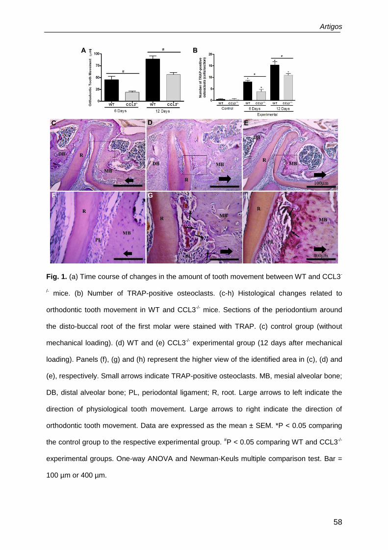
during OTM. The amount of tooth movement (Fig. 1a) and numbers of TRAP-positive
osteoclasts (Fig. 1b) were increased after 6 and 12 days of orthodontic force in WT
mice (P < 0.05). In comparison to WT mice, these histomorphometric analyses
showed diminished tooth movement (Fig. 1a) and fewer TRAP-positive cells (Fig. 1b)
in CCL3-/- mice at the same time points (P < 0.05). Moreover, alveolar bone
morphology without orthodontic appliance (control side) presented increased TRAP
activity on the distal side of the alveolar bone surface, while no activity was noted in
the mesial region of the periodontium in both mice groups (Fig 1c and f), representing
the physiological tooth movement in distal direction. On the other hand, the
mechanical loading applied on the tooth in mesial direction after 6 days induced
increased TRAP activity in the mesial site, reducing this parameter analyses in distal
region. On day 12, TRAP activity seemed to increase more extensively in WT mice
(Fig. 1d and g), which presented a greater alveolar bone resorption area than did
CCL3-/- mice (Fig. 1e and h). These findings indicated that the bone resorption and
osteoclast recruitment, induced by mechanical loading, are positively modulated by
CCL3 chemokine.

Artigos
47
Expression of bone remodeling-related markers in CCL3-/- mice
In view of CCL3-/- mice altered alveolar bone microscopic phenotype, we
characterized the mRNA expression pattern of markers involved in bone resorption.
The mechanical loading induced an increase of receptor activator of nuclear factor
kappa-B (RANK) (Fig. 2a), receptor activator of nuclear factor kappa-B ligand
(RANKL) (Fig. 2b) and tumor necrosis factor alpha (TNF-α) (Fig. 2c) mRNA levels in
WT and CCL3-/- mice (P < 0.05). However, the expression of these molecules was
reduced in CCL3-/- when compared with WT mice (P < 0.05) (Fig. 2a-c). There was
no significant change in the mRNA levels of Cathepsin K (Fig. 2d) and
metalloproteinase 13 (MMP13) (Fig. 2e) between both groups.
In addition, we further investigated if the lack of CCL3 could interfere with the
transcriptional level of osteoblast markers and negative regulators of bone
resorption-related markers. The expression levels of runt-related transcription factor
2 (RUNX2) (a transcription factor considered to be an early marker of osteoblast
differentiation) was upregulated in periodontium of both groups after 12 hr of
mechanical loading in WT mice, but it was reduced in CCL3-/- mice after 72 hr (Fig.
3a) (P < 0.05). There was no difference between the levels of osteocalcin (OCN) (a
later marker of osteoblast differentiation and activity) (Fig. 3b), interleukin 10 (IL-10)
(Fig. 3c) and osteoprotegerin (OPG) (Fig. 3d) in both groups. Moreover, the
RANKL/OPG ratio was decreased in CCL3-/- mice after 12 hr of mechanical loading
(P < 0.05), confirming the pro-resorptive role of CCL3 in this process (Fig. 3e).
Therefore, these data demonstrated that CCL3 is involved in osteoclast and
osteoblast differentiation during bone remodeling induced by orthodontic force.

Artigos
48
The blockade of CCR1 and CCR5 alters alveolar bone microscopic phenotype
after mechanical loading
Since CCL3 is ligand of CCR1 and CCR5, our next step was to blockage these
receptors with Met-RANTES treatment, to evaluate its effect on bone remodeling
incuced by mechanical loading. It was observed a reduction in amount of OTM (Fig.
4a) and numbers of TRAP-positive osteoclasts (Fig. 4b) in mice treated with Met-
RANTES than in untreated mice (P < 0.05). The qualitative analysis of alveolar bone
confirmed the diminished TRAP activity and bone resorption caused by Met-RANTES
treatment (Fig. 4e and h).
Distinct expression of bone remodeling-related markers in mice treated with
Met-RANTES
In order to elucidate if CCR1 alters the expression of bone resorption- and bone
formation-related markers during OTM, we next measured the mRNA levels of these
molecules in peridontium of Met-RANTES-treated mice. The results showed that the
treatment with Met-RANTES reduced the expression of RANK (Fig. 5a), RANKL (Fig.
5b), TNF-α (Fig. 5c), Cathepsin K (Fig. 5d) and MMP13 (Fig. 5e) in periodontium of
mice submitted to orthodontic force when compared with untreated mice in the same
conditions (P < 0.05). Then, our next question was if this receptor also influenced
osteoblast markers expression. The level RUNX2 (Fig. 6a) was reduced in Met-
RANTES-treated mice only after 72 hr of mechanical loading (P < 0.05), whereas this
treatment induced increasing expression of OCN after 12 and 72 hr (Fig. 6b) (P <
0.05). Unexpectedly, the expression of IL-10 (Fig. 6c) and OPG (Fig. 6d) was
reduced in mice treated with Met-RANTES (P < 0.05). However, the RANKL/OPG

Artigos
49
ratio was reduced with Met-RANTES treatment after 72 hr of mechanical loading (P <
0.05), confirming the anti-resorptive action of this drug (Fig. 6e).
CCR1 plays an important role in mechanical loading-induced bone resorption
Our previous study demonstrated that CCR5 has been associated with down
regulation of bone resorption (Andrade Jr. et al., 2009). As the blockage of both
CCR1 and CCR5 resulted in lower amount of OTM and numbers of TRAP-positive
osteoclasts, our next step was to confirm if CCR1 is the key receptor of bone
resorption induced by mechanical loading. For that, we used CCR1-/- mice. The
amount of OTM (Fig. 7a) and numbers of TRAP-positive osteoclasts (Fig. 7b) were
lower in CCR1-/- mice than in WT mice (P < 0.05). These results suggest that CCR1
might be the receptor responsible for osteoclast recruitment and bone resorption
induced by mechanical loading.

Artigos
50
DISCUSSION
Bone remodeling is a lifelong process, which involves the equilibrium between bone
resorption and formation. This process might be modulated by osteoimmune
response and mechanical loading (Jones et al., 2011; Papachroni et al., 2009). In
this context, chemokines have pivotal role in strain-induced bone remodeling
(Andrade et al., 2009; Taddei et al., 2011). As the levels of CCL3 and CCR1 were
increased in periodontium after orthodontic force (Andrade Jr. et al., 2009), the aim of
present study was to evaluate the role of these chemokine and receptor in this
scenario. Ours major findings demonstrated that the CCL3/CCR1 axis plays an
important role in osteoclast recruitment, differentiation and activity during bone
remodeling induced by mechanical loading. Moreover, the blockage of CCR1 was
effective to control bone loss.
Our data demonstrated that CCL3 is a pro-resorptive chemokine in mechanical
loading-induced bone remodeling. In accordance, previous studies demonstrated the
role of CCL3 in osteoclast recruitment (Yu et al., 2004), in increase of osteoclasts
number and size during RANKL-induced osteoclastogenesis (Yu et al., 2004;
Okamatsu et al., 2004; Tsubaki et al., 2007) and in osteoclast activity (Okamatsu et
al., 2004). In contrast, CCL3 does not affect the bone loss involved in the periodontal
disease (Repeck et al., 2010). Therefore, it is important to note that the triggering
factors (i.e., microbial factors vs. mechanical loading), the nature of inflammatory
processes (i.e., chronic vs. transitory inflammation) can change the function of some
inflammatory mediators in bone remodeling process (Ferreira Jr. et al., 2011).
Reinforcing this hypothesis, recent studies demonstrated that CCR5 up-regulates
infectious-related bone loss in periodontal diseases (Repeke et al., 2010; Ferreira Jr.

Artigos
51
et al., 2011), while this same receptor inhibits the bone resorption induced by
mechanical loading (Andrade Jr. et al., 2009).
In line with the reduced bone resorption, the levels of pro-resorptive markers, such as
RANK, RANKL and TNF-α, were decreased in CCL3-/- mice after mechanical loading.
In vitro studies demonstrated that CCL3 increases the expression of RANKL by
osteoblasts and induces osteoclast-osteoblast interaction, increasing osteoclast
differentiation and consequently bone resorption (Watanabe et al., 2004; Tsubaki et
al., 2007). In parallel, TNF-α is widely known to stimulate the progression of disorders
associated with bone loss (Queiroz-Junior et al., 2011) and mechanical loading-
induced bone resorption (Andrade Jr. et al., 2007). It also triggers the release of
other inflammatory mediators in stimulated tissues, including chemokines (Yu et al.,
2004; Silva et al., 2007). In this context, TNF-α has already been demonstrated to
stimulate CCL3 production by osteoblasts (Yu et al., 2004). On the other hand, our
findings showed that the transcription of TNF-α was also up-regulated by CCL3,
showing other mecanism by which CCL3 contributes to strain-induced bone
resorption.
To further strengthen our data, we used a pharmacological strategy with Met-
RANTES, a CCL5 recombinant molecule, which specifically binds to CCR1 and
CCR5, impairing the subsequent signaling and cellular response (Proudfoot et al.,
1996). In the present study, we demonstrated that the blockage of CCR1 and CCR5
by Met-RANTES presented higher effectiveness when compared with the absence of
CCL3 in the attenuation of bone resorption phenotype after mechanical loading. In
this context, our results showed that Met-RANTES treatment not only reduced levels
of the RANK/RANKL axis and TNF-α in bone resorption scenario like observed in
CCL3-/- mice, but also decreased the Cathepsin K and MMP13 (proteases that

Artigos
52
degrade bone matrix) expression. In accordance, Met-RANTES treatment results in
reduced TNF-α and RANKL expression and osteolysis in bone lytic diseases, such
as rheumatoid arthritis and periodontal disease (Shahrara et al., 2005, Repeke et al.,
2011).
With these results, we hypothesize that CCR1 may positively modulate bone
resorption, since previous data from our group indicated that CCR5 is a down-
regulator receptor of bone resorption induced by mechanical loading (Andrade et al.,
2009). Confirming this hypothesis, it was observed diminished amount of OTM and
number of osteoclast in CCR1-/- mice. Thus, it seems that the interaction between
CCL3 and CCR1 is the responsible axis for inducing bone resorption after
mechanical loading. This is in line with the role of CCR1 in physiologic bone
remodeling (Hoshino et al., 2010), in bone loss associated with multiple myeloma
metastasis (Vallet et al., 2007) and periodontal disease (Repeke et al., 2010).
Besides osteoclast, osteoblast also expresses CCR1 and CCR5 receptors (Yano et
al., 2005). As differentiation and function of osteoblast are essential to bone
remodeling process, we investigated the expression of osteoblast markers, RUNX2
and OCN (Liu et al., 2001). We observed a reduction in the levels of RUNX2 in
CCL3-/- and Met-RANTES-treated mice. Moreover, the treatment with Met-RANTES
concomitantly increased OCN expression. Then, our findings suggested that the
blockage of both CCR1 and CCR5 receptors and absence of CCL3 affect the
expression of osteoblast differentiation markers expression.
Our results indicated a reduction in the expression of IL-10 and OPG after treatment
with Met-RANTES. However, this effect was not sufficient to induce greater tooth
movement, probably, because the expression of pro-resorptive mediators (RANKL,
RANK, TNF-α) was also impaired concomitantly. Reinforcing this hypothesis, the

Artigos
53
reduced RANKL/OPG ratio confirms the anti-resorptive scenario after blockage of
CCR1 receptor.
In summary, CCR1 is a pivotal receptor involved in osteoclast recruitment,
differentiation and activity, resulting in development of a pro-resorptive bone scenario
induced by mechanical loading. These actions are dependent, at least in part, on
CCL3. Moreover, the blockage of CCR1 and CCR5, using Met-RANTES, might be a
therapeutic strategy for reducing bone resorption, without affecting bone
homeostasis. Therefore, an adequate pharmacological therapy coupled with
mechanical loading-based treatments may modulate osteoclast and osteoblast
activity and, thus, enhance the effectiveness of bone remodeling therapies.
ACKNOWLEDGMENTS
We are grateful to Fundação de Amparo a Pesquisas do Estado de Minas Gerais
(FAPEMIG, Brazil), Coordenação de Aperfeiçoamento de Pessoal de Nível Superior
(CAPES) and Conselho Nacional de Desenvolvimento Científico e Tecnológico
(CNPq, Brazil) for financial support.

Artigos
54
REFERENCES
1. Al-Dujaili SA, Lau E, Al-Dujaili H, Tsang K, Guenther A, You L. Apoptotic
osteocytes regulate osteoclast precursor recruitment and differentiation in vitro. J
Cell Biochem 2011;112(9):2412-23.
2. Andrade Jr I, Silva TA, Silva GA, Teixeira AL, Teixeira MM. The role of tumor
necrosis factor receptor type 1 in orthodontic tooth movement. J Dent Res
2007;86:1089-94.
3. Andrade Jr I, Taddei SR, Garlet GP, Garlet TP, Teixeira AL, Silva TA, et al.
CCR5 down-regulates osteoclast function in orthodontic tooth movement. J Dent
Res 2009;88:1037-41.
4. Cheung WY, Liu C, Tonelli-Zasarsky RML, Simmons CA, You L. Osteocyte
apoptosis is mechanically regulated and induces angiogenesis in vitro. J Orthop
Res 2011;29:523–30.
5. Doodes PD, Cao Y, Hamel KM, Wang Y, Rodeghero RL, Kobezda T, et al. CCR5
is involved in resolution of inflammation in proteoglycan-induced arthritis. Arthritis
Rheum 2009;60(10):2945-53.
6. Ferreira SB Jr, Repeke CE, Raimundo FM, Nunes IS, Avila-Campos MJ, Ferreira
BR, et al. CCR5 mediates pro-osteoclastic and osteoclastogenic leukocyte
chemoattraction. J Dent Res 2011;90(5):632-7.
7. Hoshino A, Iimura T, Ueha S, Hanada S, Maruoka Y, Mayahara M, et al.
Deficiency of chemokine receptor CCR1 causes osteopenia due to impaired
functions of osteoclasts and osteoblasts. J Biol Chem 2010;285(37):28826-37.
8. Jones D, Glimcher LH, Aliprantis AO. Osteoimmunology at the nexus of arthritis,
osteoporosis, cancer, and infection. J Clin Invest 2011;121:2534-2542.
9. Krishnan V, Davidovitch Z. Cellular, molecular, and tissue-level reactions to

Artigos
55
orthodontic force. Am J Orthod Dentofacial Orthop 2006;129:469e.1-469e.32.
10. Krishnan V, Davidovitch Z. On a path to unfolding the biological mechanisms of
orthodontic tooth movement. J Dent Res 2009;88:597-608.
11. Liu W, Toyosawa S, Furuichi T, Kanatani N, Yoshida C, Liu Y, et al.
Overexpression of Cbfa1 in osteoblasts inhibits osteoblast maturation and
causes osteopenia with multiple fractures. J Cell Biol 2001;155(1):157-66.
12. Okamatsu Y, Kim D, Battaglino R, Sasaki H, Spate U, Stashenko P. MIP-1
gamma promotes receptor-activator-of-NF-kappa-B-ligand-induced osteoclast
formation and survival. J Immunol 2004;173:2084-2090.
13. Papachroni KK, Karatzas DN, Papavassiliou KA, Basdra EK, Papavassiliou AG.
Mechanotransduction in osteoblast regulation and bone disease. Trends Mol
Med 2009;15(5):208-16.
14. Proudfoot AEI, Power CA, Hoogewerf AJ, Montjovent MO, Borlat F, Offord R E,
et al. Extension of recombinant human RANTES by the retention of the initiating
methionine produces a potent antagonist. J Biol Chem 1996;271:2599–2603
15. Queiroz-Junior CM, Madeira MF, Coelho FM, Costa VV, Bessoni RL, Sousa LF,
et al. Experimental arthritis triggers periodontal disease in mice: involvement of
TNF-α and the oral microbiota. J Immunol 2011;187:3821-30.
16. Repeke CE, Ferreira SB Jr, Claudino M, Silveira EM, de Assis GF, Avila-Campos
MJ, et al. Evidences of the cooperative role of the chemokines CCL3, CCL4 and
CCL5 and its receptors CCR1+ and CCR5+ in RANKL+ cell migration throughout
experimental periodontitis in mice. Bone 2010;46(4):1122-30.
17. Repeke CE, Ferreira Jr SB, Vieira AE, Silveira EM, Ávila-Campos MJ, da Silva
JS, et al. Dose-response Met-RANTES treatment of experimental periodontits: a

Artigos
56
narrow edge between the disease severity attenuation and infection control. Plos
One 2011;6:e22526.
18. Shahrara S, Proudfoot AE, Woods JM, Ruth JH, Amin MA, Park CC, et al.
Amelioration of rat adjuvant-induced arthritis by Met-RANTES. Arthritis Rheum
2005;52:1907-19.
19. Silva TA, Garlet GP, Fukada SY, Silva JS, Cunha FQ. Chemokines in oral
inflammatory diseases: apical periodontitis and periodontal disease. J Dent Res
2007;86:306-319.
20. Taddei SRA, Andrade Jr I, Queiroz-Junior CM, Garlet GP, Garlet TP, Cunha FQ,
et al. Role of CCR2 in orthodontic tooth movement. Am J Orthod Dentofacial
Orthop 2011 (in press).
21. Tsubaki M, Kato C, Manno M, Ogaki M, Satou T, Itoh T, et al. Macrophage
inflammatory protein-1alfa (MIP-1a) enhances a receptor activator of nuclear
factor kappaB ligand (RANKL) expression in mouse bone marrow stromal cells
and osteoblasts through MAPK and PI3K/Akt pathways. Mol Cell Biochem
2007;304:53–60.
22. Vallet S, Raje N, Ishitsuka K, Hideshima T, Podar K, Chhetri S, et al. MLN3897, a
novel CCR1 inhibitor, impairs osteoclastogenesis and inhibits the interaction of
multiple myeloma cells and osteoclasts. Blood 2007;110:3744-52.
23. Watanabe T, Kukita T, Kukita A, Wada N, Toh K, Nagata K, et al. Direct
stimulation of osteoclastogenesis by MIP-1alpha: evidence obtained from studies
using RAW264 cell clone highly responsive to RANKL. J Endocrinol
2004;180:193-201.
24. Yano S, Mentaverri R, Kanuparthi D, Bandyopadhyay S, Rivera A, Brown EM, et
al. Functional expression of beta-chemokine receptors in osteoblasts: Role of
Artigos
57
regulated upon activation, normal T cell expressed and secreted (RANTES) in
osteoblasts and regulation of its secretion by osteoblasts and osteoclasts.
Endocrinology 2005;146:2324-35.
25. Yu X, Huang Y, Collin-Osdoby P, Osdoby P. CCR1 chemokines promote the
chemotactic recruitment, RANKL development, and motility of osteoclasts and
are induced by inflammatory cytokines in osteoblasts. J Bone Miner Res
2004;19:2065-77.
Artigos
58
Fig. 1. (a) Time course of changes in the amount of tooth movement between WT and CCL3-
/- mice. (b) Number of TRAP-positive osteoclasts. (c-h) Histological changes related to
orthodontic tooth movement in WT and CCL3-/- mice. Sections of the periodontium around
the disto-buccal root of the first molar were stained with TRAP. (c) control group (without
mechanical loading). (d) WT and (e) CCL3-/- experimental group (12 days after mechanical
loading). Panels (f), (g) and (h) represent the higher view of the identified area in (c), (d) and
(e), respectively. Small arrows indicate TRAP-positive osteoclasts. MB, mesial alveolar bone;
DB, distal alveolar bone; PL, periodontal ligament; R, root. Large arrows to left indicate the
direction of physiological tooth movement. Large arrows to right indicate the direction of
orthodontic tooth movement. Data are expressed as the mean ± SEM. *P < 0.05 comparing
the control group to the respective experimental group. #P < 0.05 comparing WT and CCL3-/-
experimental groups. One-way ANOVA and Newman-Keuls multiple comparison test. Bar =
100 µm or 400 µm.

Artigos
59
Fig. 2. mRNA expression of axis RANK (a)/ RANKL (b); TNF-α (c); and osteoclast-related
markers Cathepsin K (d) and MMP13 (e) in WT and CCL3-/- periodontium after 12 and 72 hr
of mechanical loading. Data are expressed as mean ± SEM. *P < 0.05 comparing control to
the respective experimental group. #P < 0.05 comparing WT and CCL3-/- experimental
groups. One-way ANOVA and Newman-Keuls multiple comparison test.

Artigos
60
Fig. 3. mRNA expression of osteoblast-related markers RUNX2 (a) and OCN (b); down
regulators of bone resorption-related markers IL-10 (c) and OPG (d); and RANKL/OPG ratio
(e) in periodontium of WT and CCL3-/- mice after 12 and 72 hr of mechanical loading. Data
are expressed as mean ± SEM. *P < 0.05 comparing control group to the respective
experimental group. #P < 0.05 comparing WT and CCL3-/- experimental groups. One-way
ANOVA and Newman-Keuls multiple comparison test.

Artigos
61
Fig. 4. (a) Time course of changes in the amount of tooth movement between vehicle- and
Met-RANTES-treated mice. (b) Number of TRAP-positive osteoclasts. (c-h) Histological
changes related to orthodontic tooth movement in vehicle- and Met-RANTES-treated mice.
Sections of the periodontium around the disto-buccal root of the first molar were stained with
TRAP. (c) control group (without mechanical loading). (d) vehicle and (e) Met-RANTES
experimental group (12 days after mechanical loading). Panels (f), (g) and (h) represent the
higher view of the identified area in (c), (d) and (e), respectively. Small arrows indicate
TRAP-positive osteoclasts. MB, mesial alveolar bone; DB, distal alveolar bone; PL,
periodontal ligament; R, root. Large arrows to left indicate the direction of physiological tooth
movement. Large arrows to right indicate the direction of orthodontic tooth movement. Data
are expressed as the mean ± SEM. *P < 0.05 comparing the control group to the respective
experimental group. #P < 0.05 comparing vehicle and Met-RANTES experimental groups.
One-way ANOVA and Newman-Keuls multiple comparison test. Bar = 100 µm or 400 µm.

Artigos
62
Fig. 5. mRNA expression of axis RANK (a)/ RANKL (b); TNF-α (c); and osteoclast-related
markers Cathepsin K (d) and MMP13 (e) in periodontium of vehicle- and Met-RANTES-
treated mice after 12 and 72 hr of mechanical loading. Data are expressed as mean ± SEM.
*P < 0.05 comparing control to the respective experimental group. #P < 0.05 comparing
vehicle and Met-RANTES experimental groups. One-way ANOVA and Newman-Keuls
multiple comparison test.

Artigos
63
Fig. 6. mRNA expression of osteoblast-related markers RUNX2 (a) and OCN (b); down
regulators of bone resorption-related markers IL-10 (c) and OPG (d); and RANKL/OPG ratio
(e) in periodontium of vehicle- and Met-RANTES-treated mice after 12 and 72 hr of
mechanical loading. Data are expressed as mean ± SEM. *P < 0.05 comparing control group
to the respective experimental group. #P < 0.05 comparing vehicle and Met-RANTES
experimental groups. One-way ANOVA and Newman-Keuls multiple comparison test.

Artigos
64
A B
Fig. 7. (a) Time course of changes in the amount of tooth movement between WT and
CCR1-/- mice. (b) Number of TRAP-positive osteoclasts. Data are expressed as the mean ±
SEM. *P < 0.05 comparing the control group to the respective experimental group. #P < 0.05
comparing WT and CCR1-/- experimental groups. One-way ANOVA and Newman-Keuls
multiple comparison test.

Artigos
65
Table I. Primer sequences and reaction properties.
Target Sense and anti-sense sequences At (°C) Mt (°C) Bp
IL-10
AGATC TCCGAGATGC CTTCA
CCGTGGAGCAGGTGAAGAAT
58 85 307
RUNX2
AACCACAGAACCACAAGTGCG
AAATGACTCGGTTGGTCTCGG
58 80 119
OCN
AAGCCTTCATGTCCAAGCAGG
TTTGTAGGCGGTCTTCAAGCC
60 78 170
OPG
GGAACCCCAGAGCGAAATACA
CCTGAAGAATGCCTCCTCACA
57 77 225
RANKL
CAGAAGATGGCACTCACTGCA
CACCATCGCTTTCTCTGCTCT
65 73 203
RANK
CAAACCTTGGACCAACTGCAC
GCAGACCACATCTGATTCCGT
60 84 76
Cathepsin K CTCCCTCTCGATCCTACAGTAATGA
TCAGAGTCAATGCCTCCGTTC
58 80 307
MMP13
AGAGATGCGTGGAGAGTCGAA
AAGGTTTGGAATCTGCCCAGG
65 85 162
TNF-α TGT GCT CAG AGC TTT CAA CAA
CTT GAT GGT GGT GCA TGA GA
58 80 124
-actina
ATGTTTGAGACCTTCAACA
CACGTCAGACTTCATGATGG
56 75 495
At: annealing temperature; Mt: Melting temperature; Bp: base pairs of amplicon size.
Artigos
66
2.3 Artigo 3
Role of CCR2 in orthodontic tooth movement
Taddei SRA, Andrade Jr. I, Queiroz-Junior CM, Garlet TP, Garlet GP, Cunha FQ,
Teixeira MM, Silva TA
Am J Orthod Dentofacial Orthop 2011 (in press)

Artigos
67
Date: 07/21/2011
To: "Ildeu Andrade, Jr." [email protected]
From: "American Journal of Orthodontics" [email protected]
Subject: Your Submission AJODO-D-11-00008R2
Ms. Ref. No.: AJODO-D-11-00008R2 Title: The Role of CCR2 in Orthodontic Tooth Movement American Journal of Orthodontics & Dentofacial Orthopedics Dear Dr. Andrade, Jr., Thank you for resubmitting the revised version of your manuscript. I sent the most recent revision back to the original referees, who are now satisfied that all necessary changes have been made and they recommend acceptance and publication of your research in the AJO-DO. Congratulations. When we approach the publication date, we will forward your article to the publisher, and send you information on checking a proof. Thank you for submitting your work to this journal. I look forward to seeing the article in the AJO-DO. With kind regards, Vincent G. Kokich Editor-in-Chief American Journal of Orthodontics and Dentofacial Orthopedics

Artigos
68
Role of CCR2 in Orthodontic Tooth Movement
ABSTRACT
Introduction: Cytokines and chemokines regulate bone remodeling during
orthodontic tooth movement (OTM). CC chemokine ligand 2 (CCL2) is involved in
osteoclast recruitment and activity and its expression is increased in periodontal
tissues under mechanical loading. This study investigated whether the CC
chemokine receptor 2 (CCR2)/CCL2 axis influences OTM. Methods: A coil spring
was placed in CCR2 deficient (CCR2-/-), wild-type (WT), vehicle treated (vehicle), and
P8A (CCL2 analog) treated mice. In a histopathological analysis, the amount of OTM
and numbers of osteoclast were determined. The expression of mediators involved in
bone remodeling was evaluated by Real-Time PCR. Results: OTM and the number
of TRAP-positive cells were significantly decreased in CCR2-/- and P8A mice in
relation to wild-type (WT) and vehicle treated mice, respectively. The expression of
RANKL, RANK and osteoblasts markers (COL-1 and OCN) was lower in CCR2-/-
than in WT. No significant difference was found in OPG levels between the groups.
Conclusions: These data suggested a reduction of osteoclast and osteoblast
activities in the absence of CCR2. In conclusion, CCR2/CCL2 axis is positively
associated to osteoclast recruitment, bone resorption and OTM. Therefore, the
blockage of CCR2/CCL2 axis might be used in the future for modulating the extent of
OTM.
Key words: orthodontic tooth movement – bone remodeling – chemokines

Artigos
69
INTRODUCTION
Orthodontic tooth movement is achieved by remodeling of periodontal ligament and
alveolar bone in response to mechanical stimulation. Bone is resorbed by osteoclasts
on the pressure sites and it is formed by osteoblasts on the tension sites.1,2 This
process is regulated by no-infectious and transient inflammatory response that is
characterized by releasing of several mediators, such as cytokines and
chemokines.3-8
Chemokines, a large family of chemotactic cytokines, provide key signals for
trafficking, differentiation and activity of bone cells.9,10 The CC chemokine ligand 2
(CCL2, formerly known as monocyte chemotatic protein-1, MCP-1) has been found
to promote chemotaxis, differentiation and activation of osteoclasts.11-16 The cellular
effects of CCL2 are mediated by its engagement with the CC chemokine receptor 2
(CCR2),17 which is expressed by osteoclast precursors.13-15 In addition, CCL2
expression is greatly increased in periodontal tissues submitted to orthodontic
loading,3,4,6,7 as well as in other inflammatory conditions such as rheumatoid
arthritis,18 bone cancer metastasis,19 periodontal disease20,21 and periapical
osteolysis.22
Studies in vitro and in vivo demostrated that the blockage or absence of CCR2
significantly prevents bone resorption in experimental arthritis,23,24 osteoporosis15 and
bone fracture healing.25 Although the expression of CCL2 has been shown in
periodontium submitted to orthodontic force,3,4,6,7 the functional role of CCL2 and
CCR2 in orthodontic tooth movement is not known. This study aimed to investigate
the role of CCR2/CCL2 axis in osteoclast recruitment and activity using a well-
established mouse model of orthodontic tooth movement. We hypothesized that the

Artigos
70
CCR2/CCL2 axis would contribute significantly to osteoclast recruitment and
consequently, to orthodontic tooth movement.
MATERIALS and METHODS
Experimental Animals
Twenty five ten-week-old wild-type (WT) (C57BL6/J), 25 CCR2 deficient mice (CCR2-
/-) obtained from the Jackson Laboratory (Bar Harbor, ME), five vehicle (PBS) treated
mice (Vehicle) and 15 P8A (a monomeric variant of the chemokine CCL2 able to
inhibit CCR2-mediated leukocyte recruitment) treated mice (P8A) were used in this
experiment. CCR2 knockout mice have been previously bred into the C57BL/6
background for 9 generations. In the genome of CCR2 knockout mice, the entire
coding region except the first 39 nucleotides and 5' untranslated region of the CCR2
gene in chromosome 9 were recombined with the neomycin-resistant gene (the
coding region and 3’ untranslated region are replaced with a polII-neo cassette). No
expression of CCR2 has been observed in this mouse. Absence of transcript was
confirmed by RT-PCR using mRNA isolated from spleens and thioglycolate elicited
peritoneal exudate cells of homozygous mutant animals. Overall, mice that are
homozygous for the targeted mutation are viable, fertile, normal in size and do not
display any gross physical or behavioral abnormalities.26
All animals were treated under the ethical regulations for animal experiments, defined
by the Institutional Ethics Committee. Each animal’s weight was recorded throughout
the experimental period, and there was no significant loss of weight.

Artigos
71
Experimental Protocol
Induction of tooth movement was performed as previously described.7 Briefly, mice
were anesthetized i.p. with 0.2 mL of a solution containing xylazine (0.02 mg/mL) and
ketamine (50 mg/mL). An orthodontic appliance consisting of a Ni-Ti 0.25 x 0.76 mm
coil spring (Lancer Orthodontics, San Marcos, CA, USA) was bonded by a light cured
resin (Transbond, Unitek/3M, Monrova, CA, USA) between maxillary right first molar
and the incisors (Fig 1). The magnitude of force was calibrated by a tension gauge
(Shimpo Instruments, Itasca, IL, USA) to exert a force of 35 g applied in the mesial
direction. There was no reactivation during the experimental period. This study was
divided in 2 parts. In the first one, named general, 2 groups were compared: WT and
CCR2-/- mice. In the second part, named specific, 4 groups were evaluated: vehicle
and P8A injected groups at 3 different doses (administration s.c. of 0.5, 1.5 and 3.0
mg/kg/day)). For histomorphometric analysis, the left side without appliance of
maxilla was used as control. Two sub-groups were achieved for molecular analysis:
control (mice without appliance) and experimental (with activated coil spring) groups.
For histopathological analysis, WT and CCR2-/- groups were killed with an overdose
of anesthetic after 6 and 12 days of mechanical loading (Appendix Table I). For
molecular examination, these groups were sacrificed at 0, 12 and 72 hr (Appendix
Table II). Vehicle and P8A treated groups (at the 3 different doses) were killed after
12 days of orthodontic force for histomorphometric analysis (Appendix Table I). For
every set of experiments, 5 animals were used for each time-point.
Histopathological Analysis
The right and the left maxillae halves, including first, second and third molars were
dissected and fixed in 10% buffered formalin (pH 7.4). After fixation, each

Artigos
72
hemimaxillae were decalcified in 14% EDTA (pH 7.4) for 20 days and embedded in
paraffin. Samples were cut into sagittal sections of 5 µm thickness. Sections were
stained for tartrate resistant acid phosphatase (TRAP; Sigma-Aldrich, Saint Louis,
MO, USA), counterstained with hematoxylin, and used for histological examination.
The first molar distal-buccal root, on the mesial periodontal site, was used for the
osteoclasts counts, on 5 sections per animal. Osteoclasts were identified as TRAP-
positive, multinucleated cells sited on the bone surface. The slides were counted by
two examiners, and the intraclass correlation coefficient showed average measures
of 0.977, validating the measurement.
Measurement of Tooth Movement
Image J software (National Institutes of Health) was used to quantify the amount of
tooth movement, as previously described.7 Tooth movement was obtained through
the difference between the distance of the cementum-enamel-junction’s (CEJ’s) of
the first molar and the second molar (1st and 2nd molar distance) of the experimental
side (right hemi-maxila) in relation to the control side (left hemi-maxila) of the same
animal. Five vertical sections per animal were evaluated under a microscope
Axioskop 40 (Carl Zeiss, Göttingen, Niedersachsen, Germany) adapted to a digital
camera (PowerShot A620, Canon, Tokyo, Honshu, Japan). Three measurements
were conducted for each evaluation and the variability was below 5%.
RNA Extraction and Real-time PCR
Using a stereomicroscope, periodontal ligament and surrounding alveolar bone
samples were extracted from the upper first molars. Gingival tissue, oral mucosa and
tooth were discarded. These tissues were sujbected to RNA extraction and Real

Artigos
73
Time-PCR to evaluate the expression of molecules known to regulate osteoclast
function (receptor activator of nuclear factor kappa-B (RANK), receptor activator of
nuclear factor kappa-B ligand (RANKL), osteoprotegerin (OPG)) and osteoblast
markers (osteocalcin (OCN) and colagen-1 (COL-1)). RNA was extracted using
TRIZOL reagent (Invitrogen, Carlsbad, CA, USA). Complementary DNA (cDNA) was
synthesized using 2 µg of RNA through a reverse transcription reaction (Superscript
II, Invitrogen). Real-time PCR analysis was performed in MiniOpticon (BioRad,
Hercules, CA, USA) using SYBR-green fluorescence quantification system (Applied
Biosystems, Foster City, CA, USA). Standard PCR conditions were 95 ºC (10 min),
and then 40 cycles of 94 ºC (1 min), 58 ºC (1 min) and 72 ºC (2 min), followed by the
standard denaturation curve. Primer sequences are described in the Appendix Table
III.
The mean Ct values from duplicate measurements were used to calculate expression
of the target gene, with normalization to a housekeeping gene (β-actin) using the 2-
ΔΔCt formula.
Statistical Analysis
Results in each group were expressed as the mean ± SEM. As the data sets
presented a normal distribution, differences among the groups were analyzed by
one-way analysis of variance (ANOVA) followed by Newman-Keuls multiple
comparison test. P < 0.05 was considered statistically significant.

Artigos
74
RESULTS
The Amount of Tooth Movement and The Number of TRAP-positive Cells
Histomorphometric results showed that the amount of tooth movement (Fig 2, A and
Table I) and numbers of TRAP-positives osteoclasts (Fig 2, B) were increased after 6
and 12 days of mechanical loading in WT mice (P < 0.05). On the other hand, a
diminished tooth movement (Fig 2, A and Table I) and fewer TRAP-positive cells (Fig
2, B) were observed in CCR2-/- mice after 12 days (P < 0.05). There was no
significant difference between the groups after 6 days of mechanical loading.
Moreover, microscopic analysis revealed that, in the control side, TRAP activity was
found on the distal side of the alveolar bone surface, and no activity was observed in
the mesial region of the periodontium (Fig 2, C). After 6 days of orthodontic loading,
there appeared to be an increase in TRAP activity on the mesial periodontium of the
distobuccal root (the side of pressure) and a reduction on the distal side of this root
(the side of tension). On day 12, TRAP activity appeared to increase more
extensively in WT mice (Fig 2, D and F), which presented a greater alveolar bone
resorption area than did CCR2-/- mice (Fig 2, E and G). In contrast, a wide hyalinized
area on the mesial side was observed in CCR2-/- mice (Fig 2, F and G).
In order to investigate the importance of CCR2 ligand (CCL2) in this model, we
further analyzed whether P8A, a CCL2 monomeric variant that is able to inhibit
CCR2-mediated leukocyte recruitment, would also change the amount of tooth
movement and osteoclast recruitment. Tooth movement (Fig 3, A) and the number of
TRAP-positive osteoclasts (Fig 3, B) were reduced in P8A-treated mice in a dose-
dependent way (Fig 3, A and B) when compared to mice treated with vehicle (P <
0.05).
Artigos
75
Expression of Osteoclast and Osteoblast Markers
To understand the mechanisms involved in the altered bone remodeling of CCR2-/-
mice during orthodontic tooth movement, we also evaluated the expression of
osteoclasts regulators (RANK, RANKL and OPG) and osteoblast markers (OCN and
COL-1). RANK and RANKL mRNA levels were significantly increased after
mechanical loading (P < 0.05), but were smaller in CCR2-/- than in WT mice (P <
0.05) (Fig 4, A and B). The mechanical stress up-regulated OPG levels but the
increase was similar in WT and CCR2-/- mice (Fig 4, C). Expression of OCN and
COL-1 was significantly increased after mechanical loading (P < 0.05), but it was of
lower in CCR2-/- than in WT mice (P < 0.05) (Fig 5, A and B).
Artigos
76
DISCUSSION
We have previously observed increased CCL2 expression during orthodontic tooth
movement.4,7 In the present study, the functional role of CCL2 and CCR2 was
evaluated. Our major findings were a reduced number of osteoclasts and a
diminished tooth movement when CCL2 interactions were absent (CCR2-/- mice) or
antagonized (P8A-treated mice). CCR2 deficiency was associated with lower
expression of RANKL, RANK and osteoblasts markers (COL-1 and OCN), reinforcing
the role of CCL2/CCR2 interactions in driving bone remodeling during orthodontic
tooth movement.
Several studies have shown increased CCL2 expression during orthodontic tooth
movement3,4,6,7 as well as in other sites of bone remodeling, such as rheumatoid
arthritis,18 periodontal disease20,21 and bone metastasis,19 in which
osteoclastogenesis is highly stimulated. As the cellular effects of CCL2 might be
mediated by CCR2,17 its absence might interfere with osteoclast differentiation and,
consequently, with bone remodeling.15 Our results suggest that not only CCL2 is
expressed but also the CCL2/CCR2 axis plays a significant role in osteoclast
recruitment and bone resorption in orthodontic tooth movement. Although previous
studies have already shown increased bone mineral density in CCR2-/- mice, which
makes these animals more resistant to compressive loadings,15 this does not seem
to fully explain the decreased orthodontic tooth movement in these animals. Indeed,
similar results were observed after treatment with a CCL2 inhibitor in WT mice,
arguing that an innate effect in bone physiology could not explain the results
observed. In contrast, the diminished CCL2/CCR2-mediated osteoclast recruitment

Artigos
77
and differentiation could account for our observations, an effect also seen in a model
of osteoporosis model in vivo.15
The results obtained in animals treated with P8A, a monomeric variant of CCL2
which inhbits CCR2-dependent cell migration in vivo,27 suggested that CCL2 is the
most important CCR2 ligand in this model. The results reported a smaller amount of
tooth movement and fewer numbers of TRAP-positive osteoclasts in P8A treated
mice after mechanical loading. In accordance, P8A reduced bone lesions in rats with
arthritis.28 Moreover, CCL2-/- mice have been shown to present a high bone mass
phenotype owing to a smaller number of osteoclasts.15 Taken all together, these data
confirm that the CCL2/CCR2 axis is involved in the recruitment of osteoclast
precursors and, consequently, in orthodontic tooth movement. Therefore, this effect
does not seem to result from the differences in the bone density observed in CCR2-/-
mice, but from the reduction of osteoclast recruitment due to the lack of CCR2, since
WT mice treated with P8A and CCR2-/- mice presented similar results.
Osteoclastogenesis and bone resorption activity are up-regulated by RANKL,
produced by osteoblast/stroma cells, through their binding to their receptor RANK
expressed in osteoclast progenitor cells.29 This process can be inhibited by the decoy
receptor OPG, which avoids RANK-RANKL engagement.29,30 In order to understand
the molecular basis of the impaired osteoclast differentiation and activity in the
absence of CCR2, these osteoclast regulators were analyzed. The expression of
both RANKL and RANK was decreased in CCR2-/- mice when compared to WT mice,
while there was no significant difference in the OPG levels between both groups. In
accordance, previous studies showed that CCR2 deficiency decreases RANK
expression by preosteoclasts,15 and reduces osteoclastic bone resorption in vitro and
in vivo models.15,25 Moreover, treatment with P8A reduced RANKL levels and bone
Artigos
78
erosion in rats with arthritis.28 Taken together, our results suggest that the observed
reduction in the number of osteoclasts, led to less bone resorption and,
consequently, to a diminished amount orthodontic tooth movement in CCR2-/-, which
might be related to a down-regulation of RANKL/RANK gene expression.
Considering that osteoblasts may interact with osteoclasts and regulate bone
remodeling,29,30 we also evaluated the expression of osteoblasts markers. In
agreement with other studies,5,6,7 our results demonstrated a significant increase of
COL-1 and OCN mRNA expression in periodontal tissues of WT mice after
orthodontic tooth movement. Nevertheless, COL-1 and OCN expression was lower in
CCR2-/- mice than in WT mice, suggesting that osteoblast differentiation and activity
were decreased in the absence of CCR2. This reduced osteoblast activity may be
linked to a decrease of osteoclasts stimulatory signals (as RANKL), resulting in
diminished bone resorption in CCR2-/- mice. On the other hand, in an osteoporosis
model, both bone formation and OCN levels in serum were not changed in CCR2-/-
mice when compared to WT mice.15 Further research is required to confirm the role
of CCR2 in differentiation and activity of osteoblasts.
CONCLUSIONS
1. The absence of CCR2 decrease osteoclast chemoattraction and decreased
osteoclast and osteoblast activities, leading to reduced tooth movement. This
is the first demonstration that CCR2 plays an important role in bone
remodeling during orthodontic tooth movement.
2. CCL2 is the primary CCR2 ligand, and plays a central role in osteoclast
recruitment and, consequently, in orthodontic tooth movement.

Artigos
79
3. The blockade of CCR2/CCL2 axis might be used for future therapeutic
interventions, limiting inflammatory bone loss diseases, such as osteoporosis
and rheumatoid arthritis, or modulating the extent of orthodontic tooth
movement.

Artigos
80
ACKNOWLEDGMENTS
We are grateful to Fundação de Amparo a Pesquisas do Estado de Minas Gerais
(FAPEMIG, Brazil), Coordenação de Aperfeiçoamento de Pessoal de Nível Superior
(CAPES) and Conselho Nacional de Desenvolvimento Científico e Tecnológico
(CNPq, Brazil) for financial support.

Artigos
81
REFERENCES
1. Krishnan V, Davidovitch Z. Cellular, molecular, and tissue-level reactions to
orthodontic force. Am J Orthod Dentofacial Orthop 2006;129:469.e1-32.
2. Wise GE, King GJ. Mechanisms of tooth eruption and orthodontic tooth movement.
J Dent Res 2008;87:414-34.
3. Alhashimi N, Frithiof L, Brudvik P, Bakhiet M. Chemokines are upregulated during
orthodontic tooth movement. J Interferon Cytokine Res 1999;19:1047-52.
4. Andrade Jr I, Silva TA, Silva GA, Teixeira AL, Teixeira MM. The role of tumor
necrosis factor receptor type 1 in orthodontic tooth movement. J Dent Res
2007;86:1089-94.
5. Garlet TP, Coelho U, Silva JS, Garlet GP. Cytokine expression pattern in
compression and tension sides of the periodontal ligament during orthodontic tooth
movement in humans. Eur J Oral Sci 2007;115:355-62.
6. Garlet TP, Coelho U, Repeke CE, Silva JS, Cunha FQ, Garlet GP. Differential
expression of osteoblast and osteoclast chemmoatractants in compression and
tension sides during orthodontic movement. Cytokine 2008;42:330-5.
7. Andrade Jr I, Taddei SRA, Garlet GP, Garlet TP, Teixeira AL, Silva TA, Teixeira
MM. CCR5 down-regulates osteoclast function in orthodontic tooth movement. J
Dent Res 2009;88:1037-41.
8. Teixeira CC, Khoo E, Tran J, Chartres I, Liu Y, Thant LM Khabensky I, Gart LP,
Cisneros G, Alikhani M. Cytokine expression and accelerated tooth movement. J
Dent Res 2010;89:1135-41.
9. Yu X, Huang Y, Collin-Osdoby P, Osdoby P. CCR1 chemokines promote the
chemotactic recruitment, RANKL development, and motility of osteoclasts and are

Artigos
82
induced by inflammatory cytokines in osteoblasts. J Bone Miner Res 2004;19:2065-
77.
10. Yano S, Mentaverri R, Kanuparthi D, Bandyopadhyay S, Rivera A, Brown EM,
Chattopadhyay N. Functional expression of β-chemokine receptors in osteoblasts:
Role of regulated upon activation, normal T cell expressed and secreted (RANTES)
in osteoblasts and regulation of its secretion by osteoblasts and osteoclasts.
Endocrinology 2005;146:2324-35.
11. Kim MS, Day CJ, Morrison NA. MCP-1 is induced by receptor activator of nuclear
factor-{kappa}B ligand, promotes human osteoclast fusion, and rescues granulocyte
macrophage colony-stimulating factor suppression of osteoclast formation. J Biol
Chem 2005;280:16163-9.
12. Kim MS, Day CJ, Selinger CI, Magno CL, Stephens SRJ, Morrison NA. MCP-1-
induced human osteoclast-like cells are tartrate-resistant acid phosphatase, NFATc1,
and calcitonin receptor-positive but require receptor activator of Nf kappa B ligand for
bone resorption. J Biol Chem 2006;281:1274-85.
13. Kim MS, Magno CL, Day CJ, Morrison NA. Induction of chemokines and
chemokine receptors CCR2b and CCR4 in authentic human osteoclasts
differentiated with RANKL and osteoclast like cells differentiated by MCP-1 and
RANTES. J Cell Biochem 2006;97:512-8.
14. Silva TA, Garlet GP, Fukada SY, Silva JS, Cunha FQ. Chemokines in oral
inflammatory diseases: apical periodontitis and periodontal disease. J Dent Res
2007;86:306-319.
15. Binder NB, Niederreiter B, Hoffmann O, Stange R, Pap T, Stulnig TM, Mack M,
Erben RG, Smolen JS, Redlich K. Estrogen-dependent and C-C chemokine receptor-

Artigos
83
2-dependent pathways determine osteoclast behavior in osteoporosis. Nat Med
2009;15:417-24.
16. Miyamoto K, Ninomiya K, Sonoda K, Miyauchi Y, Hoshi H, Iwasaki R, Miyamoto
H, Yoshida S, Sato Y, Morioka H, Chiba K, Egashira K, Suda T, Toyama Y,
Miyamoto T. MCP-1 expressed by osteoclasts stimulates osteoclastogenesis in an
autocrine/paracrine manner. Biochem Biophys Res Commun 2009;383:373-7.
17. Yadav A, Saini V, Arora S. MCP-1: Chemoattractant with a role beyond immunity:
A review. Clin Chim Acta 2010;411:1570-9.
18. IwamotoT, Okamoto H, Toyama Y, Momohara S. Molecular aspects of
rheumatoid arthritis: chemokines in the joints of patients. FEBS J 2008;275:4448-55.
19. Lu X, Kang Y. Chemokine (C-C motif) ligand 2 engages CCR2+ stromal cells of
monocytic origin to promote breast cancer metastasis to lung and bone. J Biol Chem
2009;284:29087-96.
20. Kurtis B,Tu¨ter G, Serdar M, Akdemir P, Uygur C, Firatli E, Bal B. Gingival
crevicular fluid levels of monocyte chemoattractant protein-1 and tumor necrosis
factor-alpha in patients with chronic and aggressive periodontitis. J Periodontol
2005;76:1849-55.
21. Pradeep RA, Daisy H, Hadge P. Serum levels of monocyte chemoattractant
protein-1 in periodontal health and disease. Cytokine 2009;47:77-81.
22. Garlet TP, Fukada SY, Saconato IF, Avila-Campos MJ, Silva TA, Garlet GP,
Cunha FQ. CCR2 Deficiency results in increased osteolysis in experimental
periapical lesions in mice. J Endod 2010;36:244-50.
23. Brühl H, Cihak J, Schneider MA, Plachy´ J, Rupp T, Wenzel I, Shakarami M, Milz
S, Ellwart JW, Stangassinger M, Schlöndorff D, Mack M. Dual role of CCR2 during

Artigos
84
initiation and progression of collagen-induced arthritis: evidence for regulatory activity
of CCR2+ T cells. J Immunol 2004;172:890-8.
24. Brodmerkel CM, Huber R, Covington M, Diamond S, Hall L, Collins R, Leffet L,
Gallagher K, Feldman P, Collier P, Stow M, Gu X, Baribaud F, Shin N, Thomas B,
Burn T, Hollis G, Yeleswaram S, Solomon K, Friedman S, Wang A, Xue CB, Newton
RC, Scherle P, Vaddi K. Discovery and pharmacological characterization of a novel
rodent-active CCR2 antagonist, INCB3344. J Immunol 2005;175:5370-8.
25. Xing Z, Lu C, Hu D, Yu Y, Wang X, Colnot C, Nakamura M, Wu Y, Miclau T,
Marcucio RS. Multiple roles for CCR2 during fracture healing. Dis Model Mech
2010;3:451-8.
26. Boring L, Gosling J, Chensue SW, Kunkel SL, Farese RV Jr, Broxmeyer HE,
Charo IF. Impaired monocyte migration and reduced type 1 (Th1) cytokine responses
in C-C chemokine receptor 2 knockout mice. J Clin Invest 1997;100:2552-61.
27. Handel TM, Johnson Z, Rodrigues DH, Santos AC, Cirillo R, Muzio V, Riva S,
Mack M, Déruaz M, Borlat F, Vitte PA, Wells TN, Teixeira MM, Proudfoot AE. An
engineered monomer of CCL2 has anti-inflammatory properties emphasizing the
importance of oligomerization for chemokine activity in vivo. J Leukoc Biol
2008;84:1101-8.
28. Shahrara S, Proudfoot AE, Park CC, Volin MV, Haines GK, Woods JM, Aikens
CH, Handel TM, Pope RM. Inhibition of monocyte chemoattractant protein-1
ameliorates rat adjuvant-induced arthritis1. J Immunol 2008;180:3447-56.
29. Boyce BF, Xing L. Bruton and tec: New links in osteoimmunology. Cell Metab
2008;7:283-5.

Artigos
85
30. Aoki S, Honma M, Kariya Y, Nakamichi Y, Ninomiya T, Takahashi N, Udagawa
N, Suzuki H. Function of OPG as a traffic regulator for RANKL is crucial for controlled
osteoclastogenesis. J Bone Miner Res 2010;25:1907-21.

Artigos
86
Fig 1. Occlusal view of a nickel-titanium (Ni-Ti) open coil spring placed between the
upper right first molar and the incisors.
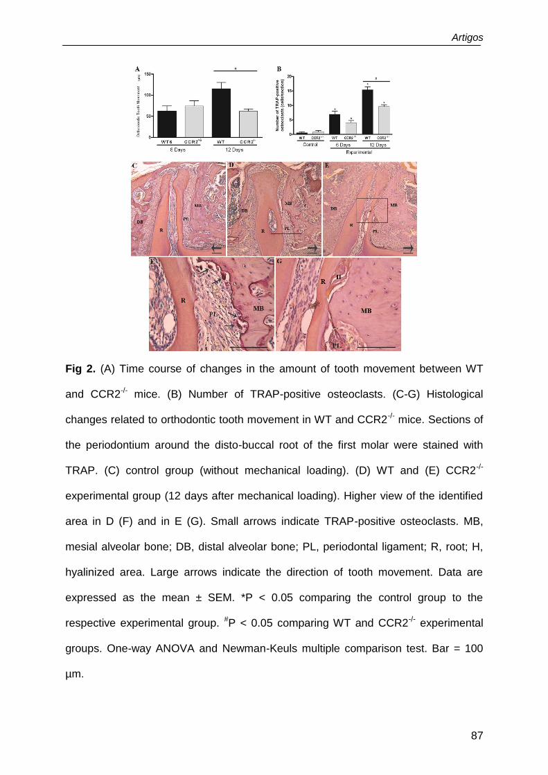
Artigos
87
Fig 2. (A) Time course of changes in the amount of tooth movement between WT
and CCR2-/- mice. (B) Number of TRAP-positive osteoclasts. (C-G) Histological
changes related to orthodontic tooth movement in WT and CCR2-/- mice. Sections of
the periodontium around the disto-buccal root of the first molar were stained with
TRAP. (C) control group (without mechanical loading). (D) WT and (E) CCR2-/-
experimental group (12 days after mechanical loading). Higher view of the identified
area in D (F) and in E (G). Small arrows indicate TRAP-positive osteoclasts. MB,
mesial alveolar bone; DB, distal alveolar bone; PL, periodontal ligament; R, root; H,
hyalinized area. Large arrows indicate the direction of tooth movement. Data are
expressed as the mean ± SEM. *P < 0.05 comparing the control group to the
respective experimental group. #P < 0.05 comparing WT and CCR2-/- experimental
groups. One-way ANOVA and Newman-Keuls multiple comparison test. Bar = 100
µm.

Artigos
88
Fig 3. (A) Effect of different doses of P8A in the amount of tooth movement and (B)
number of TRAP-positive osteoclasts. Data are expressed as the mean ± SEM. *P <
0.05 comparing the control group to the respective experimental group. #P < 0.05
compared vehicle and P8A experimental groups. +P < 0.05 comparing P8A 0.5
mg/Kg/day to the two other P8A-treated groups (1.5 and 3.0 mg/kg/day). One-way
ANOVA and Newman-Keuls multiple comparison test.
Fig 4. mRNA expression of osteoclast differetiation and activity markers RANKL (A),
RANK (B) and OPG (C) in WT and CCR2-/- periodontium after 12 and 72 hr of
mechanical loading. Data are expressed as mean ± SEM. *P < 0.05 comparing
control to the respective experimental group. #P < 0.05 comparing WT and CCR2-/-
experimental groups. One-way ANOVA and Newman-Keuls multiple comparison test.

Artigos
89
Fig 5. mRNA expression of osteoblastic markers OCN (A) and COL-1 (B) in WT and
CCR2-/- periodontium after 12 and 72 hr of mechanical loading. Data are expressed
as mean ± SEM. *P < 0.05 comparing control group to the respective experimental
group. #P < 0.05 comparing WT and CCR2-/- experimental groups. One-way ANOVA
and Newman-Keuls multiple comparison test.

Artigos
90
Table I. Time course of changes in the amount of tooth movement between WT and
CCR2-/- mice.
Orthodontic Tooth Movement
WT (mean ± SEM)
CCR2-/-
(mean ± SEM) P valor
6 Days 62.5 ± 12.5 75 ± 11.9 P ˃ 0.05
12 Days 115 ± 15.5 62.5 ± 4.7 P < 0.05

Artigos
91
Appendix Table I. Number of mice in each group for histopathological analysis.
Histopathological Analysis
WT CCR2-/- Vehicle P8A 0,5
mg/Kg
P8A 1,5
mg/Kg
P8A 3,0
mg/Kg
6 Days 5 5 - - - -
12 Days 5 5 5 5 5 5
Appendix Table II. Number of mice in each group for molecular analysis.
Real Time-PCR
WT CCR2-/-
0 hr 5 5
12 hr 5 5
72 hr 5 5

Artigos
92
Appendix Table III. Primer sequences and reaction properties.
Target/ GI Forward or Reverse sequences At(°C) Mt(°C) Bp
RANK GI:110350008
(F) 5’-CAAACCTTGGACCAACTGCAC-3’ (R) 5’-GCAGACCACATCTGATTCCGT-3’
60 84 76
OPG GI:2072182
(F) 5’-GGAACCCCAGAGCGAAATACA-3’ (R) 5’-CCTGAAGAATGCCTCCTCACA-3’
57 77 225
RANKL GI:114842414
(F) 5’-CAGAAGATGGCACTCACTGCA-3’ (R) 5’-CACCATCGCTTTCTCTGCTCT-3’
65 73 203
OCN GI:508299
(F) 5’-AAGCCTTCATGTCCAAGCAGG-3’ (R) 5’-TTTGTAGGCGGTCTTCAAGCC-3’
60 78 170
COL-1 GI:118131144
(F) 5’-AATCACCTGCGTACAGAACGG-3’ (R) 5’-CAGATCACGTCATCGCACAAC-3’
62 84 114
β-actin GI:145966868
(F) 5’-ATGTTTGAGACCTTCAACA-3’ (R) 5’-CACGTCAGACTTCATGATGG-3’
56 75 495
At: annealing temperature; Mt: Melting temperature; Bp: base pairs of amplicon size.

Discussão
93
3. DISCUSSÃO
A remodelação óssea é um processo fisiológico, responsável pela
manutenção do turnover ósseo. Este processo ocorre por meio de uma ação
coordenada das células ósseas, resultando em reabsorção óssea por osteoclastos e
deposição óssea por osteoblastos. Um desequilíbrio entre estes dois processos
pode resultar em diversas alterações ósseas (Jones et al., 2011; Papachroni et al.,
2009). Os estudos apresentados nesta tese pertencem à linha de pesquisa intitulada
“Mecanismos envolvidos na reabsorção/remodelação óssea alveolar em
modelos experimentais”. O desenvolvimento desta linha de pesquisa iniciou-se em
2005 com a criação do modelo experimental de movimentação dentária ortodôntica
em camundongos (Andrade Jr. et al., 2007a). A movimentação dentária ortodôntica
é realizada através do remodelamento do osso alveolar e ligamento periodontal
(Krishnan e Davidovitch, 2006; Krishnan e Davidovitch, 2009). A alteração da
homeostase no periodonto, provocada pela força ortodôntica, desencadeia a
formação de um ambiente pró-reabsortivo nas áreas de pressão e anti-reabsorptivo
nas áreas de tensão, induzindo reabsorção e formação óssea alveolar,
respectivamente (Krishnan e Davidovitch, 2006; Krishnan e Davidovitch, 2009). Esta
remodelação óssea é resultado do surgimento do processo inflamatório asséptico
transitório no ligamento periodontal, caracterizada pela liberação de vários
mediadores, tais como: neurotransmissores, fatores de crescimento, quimiocinas e
citocinas (Krishnan e Davidovitch, 2006). O aumento do nível de quimiocinas e
citocinas na movimentação dentária ortodôntica tem sido demonstrado em modelo
animal (Alhashimi et al., 1999; Andrade et al., 2007b; Andrade et al., 2009) e em
humanos (Garlet et al., 2007; Garlet et al., 2008), tais como TNF-α, IL-10, CCL2,

Discussão
94
CCL3, CCL5, CXCL12. Porém, poucos estudos têm investigado o papel específico
de cada citocina e/ou quimiocina neste processo. Portanto, o objetivo geral desta
tese foi avaliar o papel das quimiocinas e receptores CCL3/CCR1/CCR5 e
CCL2/CCR2 na reabsorção/remodelação óssea induzida por força mecânica.
É importante salientar que a remodelação óssea pode ser regulada pela
resposta osteoimune e pela força mecânica (Jones et al., 2011; Papachroni et al.,
2009). Portanto, os tratamentos para doenças osteodesgenerativas buscam utilizar
terapias farmacológicas para neutralizar a ação de mediadores pro-reabsortivos
(Repeke et al., 2011; Saharara et al., 2005), associadas com tratamentos baseados
em força mecânica, visando aumento da massa óssea (Papachroni et al., 2009).
Neste contexto, alguns estudos in vitro vêm investigando quais são as vias
moleculares ativadas pelo estímulo mecânico em células ósseas (Cheung et al.,
2011; Al-Dujaili et al., 2011). Entretanto, os estudos in vitro não simulam, ao mesmo
tempo, algumas condições observadas in vivo, como a hipóxia, necrose e o dano
físico da célula desencadeados pela força mecânica compressiva, assim como
angiogênese e remodelamento da matriz celular induzidos pela força de tensão
(Krishnan e Davidovitch, 2006; Krishnan e Davidovitch, 2009). Portanto, o modelo in
vivo de movimentação dentária ortodôntica permitiu estudar o papel de mediadores
pro- e anti-reabsortivos na remodelação óssea induzida por força mecânica
(Andrade et al., 2009; Taddei et al., 2011). Este conhecimento pode ser empregado
em estratégias terapêuticas para modular a movimentação dentária ortodôntica e
outras doenças osteodegenerativas.

Discussão
95
3.1 O Papel da Quimiocina e Receptores CCL3/CCR1/CCR5 na
Reabsorção/Remodelação Óssea Durante Movimentação Dentária
Ortodôntica
Estudos in vitro demonstraram que a quimiocina CCL5 (ligante dos
receptores CCR1 e CCR5) estimula o recrutamento e diferenciação de osteoclastos
e osteoblastos, estando, então, envolvida no processo de remodelação óssea (Yu et
al., 2004; Yano et al., 2005). Além disso, o primeiro estudo realizado por nosso
grupo demonstrou que CCL5 pode ser uma molécula importante na reabsorção
óssea induzida por força ortodôntica (Andrade Jr. et al., 2007b). Considerando estes
achados, o primeiro estudo desta tese investigou o papel do CCR5 na
movimentação dentária ortodôntica, realizando experimentos em animais deficientes
para o receptor CCR5 (CCR5-/-). Verificamos que os animais CCR5-/- apresentaram
maior movimentação ortodôntica e número de osteoclastos TRAP-positivos, além de
aumento da expressão de marcadores da atividade osteoclástica (catepsina K,
RANKL e MMP13) em relação aos animais selvagens. Os nossos resultados
sugerem que o receptor CCR5 atua como regulador negativo da reabsorção óssea
alveolar induzida por força mecânica (Andrade Jr. et al., 2009). Consistente com
estes achados, a reabsorção óssea periapical e a artrite experimental foi
exarcebada na ausência de CCR5 (Rossi et al., 2008; Doodes et al., 2009).
Já que os osteoblastos expressam também o receptor CCR5 (Yano et al.,
2005) e são essenciais para o processo de remodelação óssea, nós investigamos a
expressão dos marcadores de diferenciação e atividade destas células, tais como
RUNX2 e OCN (Andrade Jr. et al., 2009). Os resultados mostraram que RUNX2 e
OCN foram diminuídas em animais CCR5-/-, sugerindo que a diferenciação e

Discussão
96
atividade dos osteoblastos podem ser reguladas pelo receptor CCR5. Entretanto, o
papel do CCR5 na diferenciação e atividade dos osteoblastos deve ser confirmado,
empregando-se outros métodos/modelos de estudo.
Em paralelo, observou-se que animais CCR5-/- também apresentaram os
níveis de IL-10 e OPG (reguladores negativos da reabsorção óssea) reduzidos
(Andrade Jr. et al., 2009). Como o osteoblasto é uma das fontes de OPG (Boyce e
Xing, 2008), estes achados sugerem que a redução na diferenciação de
osteoblastos possa ter resultado na diminuição dos sinais inibitórios para os
osteoclastos, causando aumento da reabsorção do osso alveolar e maior
movimentação dentária ortodôntica. Um estudo prévio demonstrou que as células T
regulatórias podem ser responsáveis pela produção de IL-10 e supressão da
formação de osteoclastos (Zaiss et al., 2007). Neste contexto, como a migração
destas células é dependente do receptor CCR5 (Yurchenko et al., 2006), nossos
dados podem sugerir um papel destas células na reabsorção óssea (Andrade Jr. et
al., 2009). Entretanto esta hipótese deve ser melhor investigado empregando-se
sistemas in vivo e in vitro.
Ao contrário do CCR5, CCR1 é o receptor que apresenta maior
expressão nos precursores de osteoclastos quando estimulados por RANKL (Yu et
al., 2004). Além disso, CCR1 está envolvido na atividade dos osteoclastos e,
conseqüentemente, na indução de osteólise in vitro (Oba et al., 2005; Menu et al.,
2006; Hoshino et al., 2010) e in vivo (Menu et al., 2006). Portanto, nós investigamos
a expressão de CCR1 nos animais CCR5-/-. O nível de CCR1 foi aumentado no
periodonto de camudongos WT após aplicação de força ortodôntica, ao mesmo
tempo em que ocorreu o aumento do nível de RANKL (Andrade Jr. et al., 2009).
Além disso, somado ao fenótipo de reabsorção óssea aumentada (observação

Discussão
97
microscópica), os animais CCR5-/- apresentaram maior expressão de CCR1 e
RANKL (Andrade Jr. et al., 2009), sugerindo que CCR1 possivelmente está
envolvido na formação deste ambiente pró-reabsortivo.
Além do aumento da expressão de CCR1, foram observados níveis
aumentados de CCL3 (ligante dos receptores CCR1 e CCR5) após aplicação de
força ortodôntica (Andrade Jr. et al., 2009). Estes receptores e esta quimiocina
podem modular a reabsorção óssea por promoverem quimiotaxia, recrutamento e
atividade dos osteoclastos in vitro (Yu et al., 2004). Portanto, investigamos, em
seguida, o papel do CCR1 e CCL3 na reabsorção/remodelação óssea induzida por
força mecânica (Taddei et al., sd). Foi observado menor movimentação dentária e
número de osteoclastos TRAP-positivos nos camundongos CCL3-/-, CCR1-/- e
tratados com Met-RANTES. Então, nossos principais achados demonstraram que o
eixo CCL3/CCR1 desempenha um importante papel no recrutamento, atividade e
diferenciação dos osteoclastos durante a reabsorção/remodelação óssea induzida
por força mecânica (Taddei et al., sd). Estes achados corroboram com a função pró-
reabsortiva do CCR1 demonstrada na remodelação óssea fisiológica (Hoshino et al.,
2010), na perda óssea de mieloma múltiplos (Vallet et al., 2007) e na doença
periodontal (Repeke et al., 2010). Entretanto, é importante ressaltar que a
reabsorção óssea induzida por força ortodôntica não é apenas dependente da
quimiocina CCL3 (Taddei et al., sd), já que outra quimiocina ligante do CCR1, CCL5,
pode ser importante neste processo (Andrade Jr. et al., 2007b).
Em concordância com a reabsoção óssea reduzida, os níveis de
marcadores pró-rebasortivos, tais como RANK, RANKL and TNF-α, foram
diminuídos em camundongos CCL3-/- durante movimentação dentária ortodôntica
(Taddei et al., sd). Como descrito anteriormente em estudos in vitro, CCL3 aumenta

Discussão
98
a expressão de RANKL pelos osteoblastos (Tsubaki et al., 2007) e induz interação
osteoblasto-osteoclasto (Watanabe et al., 2004), o que pode resultar no aumento de
diferenciação de osteoclastos e consequentemente reabsorção óssea. Em paralelo,
nossos achados mostraram também que a produção de TNF-α, uma citocina pró-
reabsortiva, parece ser dependente de CCL3 (Taddei et al., sd). A redução na
expressão de TNF-α pode ter contribuído para menor reabsorção óssea observada
em camundongos CCL3-/-.
Além disso, não somente os níveis de RANK/RANKL e TNF-α foram
reduzidos em animais tratados com Met-RANTES, mas também a expressão de
catepsina K e MMP13. Em concordância com nossos achados, o tratamento com
Met-RANTES resultou em expressão reduzida de TNF-α e RANKL, e diminuição da
perda óssea em doenças como artrite reumatoide e doença periodontal (Shahrara et
al., 2005, Repeke et al., 2011). É importante ressaltar também que o bloqueio
utilizando Met-RANTES foi mais efetivo para controlar a perda óssea que a deleção
genética de CCL3, sendo demonstrado pela redução na expressão demarcadores
de reabsorção óssea, catepsina K e MMP13, observada após o tratamento com
Met-RANTES (Taddei et al., sd). Estudos prévios, demonstraram a eficácia do
tratamento com Met-RANTES na redução de perda óssea na doença periodontal
(Repeke et al., 2011) e artrite reumatoide (Shaharara et al., 2005). Neste contexto, o
Met-RANTES deve ser testado como uma estratégia terapêutica para reduzir
reabsorção óssea em outras doenças, como na osteoporose, já que o bloqueio de
receptores de quimiocina não interfere tanto na homeostase do osso, quanto os
tratamentos que impedem a função do sistema RANKL/RANK (Binder et al., 2009).
Os resultados deste estudo demonstraram também que a expressão de
RUNX2 foi diminuída nos animais CCL3-/- e nos animais tratados com Met-RANTES,

Discussão
99
porém ocorreu aumento de OCN após tratamento com Met-RANTES (Taddei et al.,
sd). Estes dados sugerem que o bloqueio de ambos CCR1 e CCR5 e a deleção
genética de CCL3 afetam a expressão dos marcadores de osteoblastos. Em
resumo, CCR1 parece funcionar como receptor pró-reabsortivo, enquanto o CCR5 é
anti-reabsortivo na remodelação óssea induzida por força mecânica. A ação do
CCR1 é dependente, pelo menos em parte, da quimiocina CCL3. Além disso, o
bloqueio de CCR1 e CCR5, usando Met-RANTES, pode ser uma estratégia
terapêutica para doenças ósseas.
3.2. O Papel do CCL2 e CCR2 na Movimentação Dentária Ortodôntica
Em nossos estudos anteriores (Andrade Jr. et al., 2007b; Andrade Jr. et
al., 2009) observamos que a expressão de CCL2 apresentou-se aumentada durante
a movimentação dentária ortodôntica, tão bem quanto em outros sítios de
remodelação óssea, tais como artrite reumatoide (Iwamoto et al., 2008), doença
periodontal (Kurtis et al., 2005; Pradeep et al., 2009) e metástase ósseas (Lu e Kang
, 2009), nas quais osteoclastogênese é altamente estimulada. Estudos in vitro têm
demonstrado que CCL2 promove quimiotaxia, diferenciação e ativação dos
osteoclastos (Kim et al., 2005; Kim et al., 2006a; Kim et al., 2006b; Silva et al., 2007;
Binder et al., 2009; Miyamoto et al., 2009), ao se ligar ao receptor CCR2 (Yadav et
al., 2010). Portanto, nosso próximo objetivo foi investigar o papel do eixo
CCL2/CCR2 na movimentação dentária ortodôntica. Os resultados mostraram que a
quantidade de movimentação dentária e o número de células TRAP-positivas foram
significativamente diminuídos nos camundongos deficientes para o receptor CCR2 e
tratados com P8A (análogo de CCL2). Em concordância com estes dados, a

Discussão
100
expressão dos marcadores de atividade e diferenciação dos osteoclastos (RANKL e
RANK) e dos osteoblastos (COL-1 e OCN) foi menor nos camundongos CCR2-/- que
nos WT. Estes dados sugerem que o eixo CCL2/CCR2 está positivamente envolvido
no recrutamento de osteoclastos, atividade dos osteoclastos e osteoblastos,
reabsorção óssea e movimentação dentária ortodôntica (Taddei et al. 2011).
Estudos prévios mostraram que a densidade óssea do camundongo
CCR2-/- é aumentada, o que faz deste animal mais resistente à força compressiva
(Binder et al., 2009). Entretanto, este efeito inato da fisiologia óssea não parece
explicar completamente a menor movimentação dentária observada neste animal, já
que resultados similares foram observados após tratamento com um inibidor para
CCL2/CCR2 (P8A) em camundongos selvagens. Ao contrário, a redução do
recrutamento dos osteoclastos, regulado pelo eixo CCL2/CCR2, pode justificar a
menor reabsorção óssea e movimentação dentária nos animais CCR2-/- observadas
neste estudo (Taddei et al. 2011). Além disso, nossos resultados sugerem que o
fenótipo histológico, apresentado pelos camundongos CCR2-/- durante a
movimentação dentária, parece estar relacionado com a redução da expressão do
eixo RANKL/RANK (Taddei et al. 2011). Estudos prévios corroboram nossos
achados, mostrando que na ausência de CCR2 ocorre diminuição da expressão de
RANK nos precursores de osteoclastos (Binder et al., 2009), e redução da
reabsorção óssea em modelos in vitro e in vivo (Binder et al., 2009; Xing et al.,
2010). Além disso, o tratamento com P8A reduziu os níveis de RANKL e reabsorção
óssea em ratos com artrite (Shahrara et al., 2008).
Os resultados obtidos em animais tratados com P8A, uma variável
monomérica do CCL2, a qual inibe a migração de células dependentes de CCR2 in
vivo (Handel et al., 2008), sugere que a função de CCR2 ocorre principalmente via

Discussão
101
ligação ao CCL2 neste modelo (Taddei et al., 2011). Semelhante aos nossos
resultados, a ausência de CCL2 resulta em menor número de osteoclastos (Binder
et al., 2009) e o tratamento com P8A reduz as lesões ósseas em ratos com artrite
(Shahrara et al., 2008).
A expressão de COL-1 e OCN foi diminuída em camundongos CCR2-/-
neste estudo, sugerindo que a diferenciação e atividade de osteoblastos foram
reduzidas na ausência de CCR2 (Taddei et al. 2011). Esta menor diferenciação de
osteoblastos pode estar relacionada a uma redução dos sinais estimulatórios (como
RANKL), resultando em menor reabsorção óssea nos animais CCR2-/- (Taddei et al.
2011). Por outro lado, em um modelo de osteoporose, tanto a formação do osso
quando os níveis de OCN não foram alterados em camundongos CCR2-/- (Binder et
al., 2009). Estudos adicionais podem confirmar o papel do CCR2 na diferenciação e
atividade dos osteoblastos.
Em resumo, a quimiocina CCL2 e o receptor CCR2 regulam
positivamente o recrutamento e atividade dos osteoclastos durante a movimentação
dentária. Além disso, o bloqueio do eixo CCL2/CCR2 pode ser usado para futuras
intervenções terapêuticas, limitando a progressão de perda óssea inflamatória em
doenças tais como osteoporoses e artrite reumatoide, ou modulando a extensão da
movimentação dentária ortodôntica (Taddei et al. 2011).

Conclusões
102
4. CONCLUSÕES
Os resultados deste trabalho permitem-nos concluir que:
1) O receptor CCR5 interfere negativamente no recrutamento e atividade dos
osteoclastos, diminuindo a movimentação dentária ortodôntica.
2) O eixo CCL3/CCR1 induz o recrutamento e ativação de osteoclastos, sendo
uma via pró-reabsortiva durante a movimentação dentária ortodôntica.
3) O eixo CCL2/CCR2 regula positivamente o recrutamento e atividade dos
osteoclastos durante a movimentação dentária induzida pela força
ortodôntica.

Perspectivas
103
5. PERSPECTIVAS
O papel das quimiocinas e receptores CCL3/CCR1/CCR5 e CCL2/CCR2
na reabsorção óssea durante a movimentação dentária foram abordados nos 3
tópicos deste trabalho de tese. Entretanto, alguns pontos merecem aprofundamento:
Confirmar o papel do CCR1 na remodelação óssea induzida por força
mecânica, utilizando animais deficientes para este receptor (estes foram adquiridos
recentemente pelo nosso grupo).
Avaliar o papel da IL-10 na movimentação dentária ortodôntica.
Investigar o papel do CCR5 na diferenciação e atividade das células
ósseas in vitro.
Desenvolver um modelo de osteoporose experimental induzida por
deficiência de estrógeno, para extrapolar nossos conhecimentos a respeito da
reabsorção/remodelação óssea patológica. Portanto, outra perspectiva é investigar o
papel do CCR1 e CCR5 na reabsorção/remodelamento ósseo neste processo. Além
disso, testar novas estratégias terapêuticas para osteoporose, empregando-se Met-
RANTES e P8A.
Diante deste conhecimento obtido, surgiu o interesse de extrapolar
nossas investigações para a área clínica da Ortodontia. Portanto, outras
perspectivas são:
Determinar a cinética de expressão das citocinas, quimiocinas e de
marcadores de reabsorção/remodelação óssea no ligamento periodontal após
movimentação dentária ortodôntica em humanos, empregando-se Real-Time PCR.

Perspectivas
104
Avaliar a cinética de expressão das citocinas, quimiocinas e de
marcadores de reabsorção/remodelação óssea no fluido gengival após força
ortodôntica em humanos e correlacionar estes dados com a expressão destes
marcadores no ligamento periodontal.
Estas informações obtidas terão potencial para utilização clínica como
marcadores do processo de remodelação óssea, com o propósito de uma
identificação precoce dos efeitos indesejáveis causados pelo uso de força
ortodôntica excessiva, os quais podem apenas ser detectados quando em estágio
avançado. Cumpre-nos salientar que estudos dessa natureza também podem
contribuir fornecendo dados importantes sobre os mediadores que regulam a
reabsorção óssea e, consequentemente, determinam o sucesso da terapia
ortodôntica e ampliam, sobremaneira, as estratégias terapêuticas para o tratamento
de doenças ósseas.

Referências Bibliográgicas
105
6. REFERÊNCIAS BIBLIOGRÁFICAS
Abe M, Hiura K, Ozaki S, Kido S, Matsumoto T (2009). Vicious cycle between
myeloma cell binding to bone marrow stromal cells via VLA-4–VCAM-1
adhesion and macrophage inflammatory protein-1α and MIP-1β production. J
Bone Miner Metab 27:16–23.
Abe M, Hiura K, Wilde J, Moriyama K, Hashimoto T, Ozaki S, et al. (2002). Role
for macrophage inflammatory protein (MIP)-1α and MIP-1β in the development
of osteolytic lesions in multiple myeloma. Blood 100:2195-2202.
Al-Dujaili SA, Lau E, Al-Dujaili H, Tsang K, Guenther A, You L (2011). Apoptotic
osteocytes regulate osteoclast precursor recruitment and differentiation in
vitro. J Cell Biochem 112(9):2412-23.
Alhashimi N, Frithiof L, Brudvik P, Bakhiet M (1999). Chemokines are
upregulated during orthodontic tooth movement. J Interferon Cytokine Res
19:1047-1052.
Allen SJ, Crown SE, Handel TM (2007). Chemokine: receptor structure,
interactions, and antagonism. Annu Rev Immunol 25:787-820.
Alves JB, Ferreira CL, Martins AF, Silva GA, Alves GD, Paulino TP, et al. (2009).
Local delivery of EGF-liposome mediated bone modeling in orthodontic tooth
movement by increasing RANKL expression. Life Sci 85:693-699.
Andrade Jr I, Silva TA, Silva GA, Teixeira AL, Teixeira MM (2007a). The role of
tumor necrosis factor receptor type 1 in orthodontic tooth movement. Tese de
Doutorado, Departamento de Biologia Celular – ICB/UFMG, Set;1- 141.

Referências Bibliográgicas
106
Andrade Jr I, Silva TA, Silva GA, Teixeira AL, Teixeira MM (2007b). The role of
tumor necrosis factor receptor type 1 in orthodontic tooth movement. J Dent
Res 86:1089-1094.
Andrade Jr I, Taddei SRA, Garlet GP, Garlet TP, Teixeira AL, Silva TA, et al.
(2009). CCR5 down-regulates osteoclast function in orthodontic tooth
movement. J Dent Res 88:1037-1041.
Aoki S, Honma M, Kariya Y, Nakamichi Y, Ninomiya T, Takahashi N, et al.
(2010). Function of OPG as a traffic regulator for RANKL is crucial for
controlled osteoclastogenesis. J Bone Miner Res 25(9):1907-21.
Binder NB, Niederreiter B, Hoffmann O, Stange R, Pap T, Stulnig TM, et al.
(2009). Estrogen-dependent and C-C chemokine receptor-2-dependent
pathways determine osteoclast behavior in osteoporosis. Nat Med 15:417-424.
Bohl M, Maltha JC, Von Den Hoff JW, Kuijpers-Jagtman AM (2004). Focal
hyalinization during experimental tooth movement in beagle dogs. Am J
Orthod Dentofacial Orthop 125:615-23.
Boyce BF, Schwarz EM, Xing L (2006). Osteoclast precursors: cytokine-
stimulated immunomodulators of inflammatory bone disease. Current Opinion
in Rheumatology 18:427–432.
Boyce BF, Xing L (2008). Functions of RANKL/RANK/OPG in bone modeling and
remodeling. Arch Biochem Biophys 473:139-146.
Boyle WJ, Simonet WS, Lacey DL (2003). Osteoclast differentiation and
activation. Nature 423:337-342.
Brodmerkel CM, Huber R, Covington M, Diamond S, Hall L, Collins R, et al.
(2005). Discovery and pharmacological characterization of a novel rodent-
active CCR2 antagonist, INCB3344. J Immunol 175:5370-5378.

Referências Bibliográgicas
107
Brühl H, Cihak J, Schneider MA, Plachy J, Rupp T, Wenzel I, et al. (2004). Dual
role of CCR2 during initiation and progression of collagen-induced arthritis:
evidence for regulatory activity of CCR2 + T cells. J Immunol 172:890-898.
Cattaneo PM, Dalstra M, Melsen B (2005). The finite element method: a tool to
study orthodontic tooth movement. J Dent Res 84:428-433.
Cheung WY, Liu C, Tonelli-Zasarsky RML, Simmons CA, You L (2011).
Osteocyte apoptosis is mechanically regulated and induces angiogenesis in
vitro. J Orthop Res 29:523–30.
Cumano A, Godin I (2007). Ontogeny of the hematopoietic system. Annu Rev
Immunol 25:745-785.
Doodes PD, Cao Y, Hamel KM, Wang Y, Rodeghero RL, Kobezda T, et al.
(2009). CCR5 is involved in resolution of inflammation in proteoglycan-induced
arthritis. Arthritis Rheum 60(10):2945-53.
Faust J, Lacey DL, Hunt P, Burgess TL, Scully S, Van G, et al. (1999).
Osteoclast markers accumulate on cells developing from human peripheral
blood mononuclear precursors. J Cell Biochem 72(1):67-80.
Ferreira SB Jr, Repeke CE, Raimundo FM, Nunes IS, Avila-Campos MJ, Ferreira
BR, et al. (2011). CCR5 mediates pro-osteoclastic and osteoclastogenic
leukocyte chemoattraction. J Dent Res 90(5):632-7.
Franceschi RT, Ge C, Xiao G, Roca H, Jiang D (2007). Transcriptional regulation
of osteoblasts. Ann N Y Acad Sci 1116:196-207.
Fujii S, Maeda H, Tomokiyo A, Monnouchi S, Hori K, Wada N, et al. (2010).
Effects of TGF-β1 on the proliferation and differentiation of human periodontal
ligament cells and a human periodontal ligament stem/progenitor cell line. Cell
Tissue Res 342:233-242.

Referências Bibliográgicas
108
Garlet TP, Coelho U, Repeke CE, Silva JS, Cunha FQ, Garlet GP (2008).
Differential expression of osteoblast and osteoclast chemmoatractants in
compression and tension sides during orthodontic movement. Cytokine
42:330-335.
Garlet TP, Coelho U, Silva JS, Garlet GP (2007). Cytokine expression pattern in
compression and tension sides of the periodontal ligament during orthodontic
tooth movement in humans. Eur J Oral Sci 115:355-62.
Garlet TP, Fukada SY, Saconato IF, Avila-Campos MJ, Silva TA, Garlet GP, et
al. (2010). CCR2 Deficiency results in increased osteolysis in experimental
periapical lesions in mice. J Endod 36:244–250.
Graves DT, Jiang Y, Valente AJ (1999). The expression of monocyte
chemoattractant protein-1 and other chemokines by osteoblasts. Front Biosci
4:D571-580.
Han JH, Choi SJ, Kurihara N, Koide M, Oba Y, Roodman GD (2001).
Macrophage inflammatory protein-1alpha is an osteoclastogenic factor in
myeloma that is independent of receptor activator of nuclear factor kappaB
ligand. Blood 97:3349-3353.
Handel TM, Johnson Z, Rodrigues DH, Santos AC, Cirillo R, Muzio V, et al.
(2008). An engineered monomer of CCL2 has anti-inflammatory properties
emphasizing the importance of oligomerization for chemokine activity in vivo. J
Leukoc Biol 84:1101–1108.
Hoshino A, Iimura T, Ueha S, Hanada S, Maruoka Y, Mayahara M, et al. (2010).
Deficiency of chemokine receptor CCR1 causes osteopenia due to impaired
functions of osteoclasts and osteoblasts. J Biol Chem 285(37):28826-37.

Referências Bibliográgicas
109
IwamotoT, Okamoto H, Toyama Y, Momohara S (2008). Molecular aspects of
rheumatoid arthritis: chemokines in the joints of patients. FEBS J 275:4448–
4455.
Jones D, Glimcher LH, Aliprantis AO (2011). Osteoimmunology at the nexus of
arthritis, osteoporosis, cancer, and infection. J Clin Invest 121:2534-2542.
Kawaguchi H, Chikazu D, Nakamura K, Kumegawa M, Hakeda Y (2000). Direct
and indirect actions of FGF-2 on osteoclastic bone resorption in cultures. J
Bone Miner Res 15:466-473.
Kim MS, Day CJ, Morrison NA (2005). MCP-1 is induced by receptor activator of
nuclear factor-{kappa}B ligand, promotes human osteoclast fusion, and
rescues granulocyte macrophage colony-stimulating factor suppression of
osteoclast formation. J Biol Chem 280:16163–16169.
Kim MS, Day CJ, Selinger CI, Magno CL, Stephens SRJ, Morrison NA (2006a).
MCP-1-induced human osteoclast-like cells are tartrate-resistant acid
phosphatase, NFATc1, and calcitonin receptor-positive but require receptor
activator of Nf kappa B ligand for bone resorption. J Biol Chem 281:1274–
1285.
Kim MS, Magno CL, Day CJ, Morrison NA (2006b). Induction of chemokines and
chemokine receptors CCR2b and CCR4 in authentic human osteoclasts
differentiated with RANKL and osteoclast like cells differentiated by MCP-1
and RANTES. J Cell Biochem 97:512–518.
Krishnan V, Davidovitch Z (2006). Cellular, molecular, and tissue-level reactions
to orthodontic force. Am J Orthod Dentofacial Orthop 129:1-32.
Krishnan V, Davidovitch Z (2009). On a path to unfolding the biological
mechanisms of orthodontic tooth movement. J Dent Res 88:597-608.

Referências Bibliográgicas
110
Kurtis B, Tu¨ter G, Serdar M, Akdemir P, Uygur C, Firatli E, et al. (2005). Gingival
crevicular fluid levels of monocyte chemoattractant protein-1 and tumor
necrosis factor-alpha in patients with chronic and aggressive periodontitis. J
Periodontol 76:1849-1855.
Lee J, Shin H, Lee E, Phan TV, Choi H (2007). Stimulation of osteoclastogenesis
by enhanced levels of MIP-1a in BALB/c mice in vitro. Experimental
Hematology 35:1100–1108.
Lisignoli G, Toneguzzi S, Grassi F, Piacentini A, Tschon M, Cristino S, et al.
(2002). Different chemokines are expressed in human arthritic bone biopsies:
IFN-gamma and IL-6 differently modulate IL-8, MCP-1 and rantes production
by arthritic osteoblasts. Cytokine 20(5):231-238.
Liu Y, Shi Z, Silveira A, Liu J, Sawadogo M, Yang H, et al. (2003). Involvement of
upstream stimulatory factors 1 and 2 in RANKL-induced transcription of
tartrate-resistant acid phosphatase gene during osteoclast differentiation. J
Biol Chem 278:20603–20611.
Lu X, Kang Y (2009). Chemokine (C-C motif) ligand 2 engages CCR2+ stromal
cells of monocytic origin to promote breast cancer metastasis to lung and
bone. J Biol Chem 284:29087-29096.
Masella RS, Meister M (2006). Current concepts in the biology of orthodontic
tooth movement. Am J Orthod Dentofacial Orthop 129(4):458-468.
Matsuo K, Irie N (2008). Osteoclast-osteoblast communication. Arch Biochem
Biophys 473:201-209.
Menten P, Wuyts A, Damme JV (2002). Macrophage inflammatory protein-1.
Cytokine Growth Factor Rev 13:455-481.

Referências Bibliográgicas
111
Menu E, De Leenheer E, De Raeve H, Coulton L, Imanishi T, Miyashita K, et al.
(2006). Role of CCR1 and CCR5 in homing and growth of multiple myeloma
and in the development of osteolytic lesions: a study in the 5TMM model. Clin
Exp Metastasis 23:291-300.
Miyamoto K, Ninomiya K, Sonoda K, Miyauchi Y, Hoshi H, Iwasaki R, et al.
(2009). MCP-1 expressed by osteoclasts stimulates osteoclastogenesis in an
autocrine/paracrine manner. Biochem Biophys Res Commun 383:373–377.
Nakamura H, Sato G, Hirata A, Yamamoto T (2004). Immunolocalization of
matrix metalloproteinase-13 on bone surface under osteoclasts in rat tibia.
Bone 34(1):48-56.
Oba Y, Lee JW, Ehrlich LA, Chung HY, Jelinek DF, Callander NS, et al. (2005).
MIP-1alpha utilizes both CCR1 and CCR5 to induce osteoclast formation and
increase adhesion of myeloma cells to marrow stromal cells. Exp Hematol
33:272-278.
Okamatsu Y, Kim D, Battaglino R, Sasaki H, Spate U, Stashenko P (2004). MIP-
1 gamma promotes receptor-activator-of-NF-kappa-B-ligand-induced
osteoclast formation and survival. J Immunol 173:2084-2090.
Palioto DB, Coletta RD, Graner E, Joly JC, de Lima AF (2004). The influence of
enamel matrix derivative associated with Insulin-Like Growth Factor-I on
periodontal ligament fibroblasts. J Periodontol 75:498-504.
Pan W, Mathews W, Donohue JM, Ramnaraine ML, Lynch C, Selski DJ, et al.
(2005). Analysis of distinct tartrate-resistant acid phosphatase promoter
regions in transgenic mice. J Biol Chem 280:4888–4893.
Referências Bibliográgicas
112
Papachroni KK, Karatzas DN, Papavassiliou KA, Basdra EK, Papavassiliou AG
(2009). Mechanotransduction in osteoblast regulation and bone disease.
Trends Mol Med 15(5):208-16.
Pradeep RA, Daisy H, Hadge P (2009). Serum levels of monocyte
chemoattractant protein-1 in periodontal health and disease. Cytokine 47:77–
81.
Proudfoot AEI, Power CA, Hoogewerf AJ, Montjovent MO, Borlat F, Offord R E,
et al. (1996). Extension of recombinant human RANTES by the retention of the
initiating methionine produces a potent antagonist. J Biol Chem 271:2599–
2603.
Repeke CE, Ferreira Jr SB, Vieira AE, Silveira EM, Ávila-Campos MJ, da Silva
JS, et al. (2011). Dose-response Met-RANTES treatment of experimental
periodontits: a narrow edge between the disease severity attenuation and
infection control. Plos One 6:e22526.
Repeke CE, Ferreira Jr SB, Claudino M, Silveira EM, Assis GF, Avila-Campos
MJ, et al. (2010). Evidences of the cooperative role of the chemokines CCL3,
CCL4 and CCL5 and its receptors CCR1+ and CCR5+ in RANKL+ cell
migration throughout experimental periodontitis in mice. Bone 46:1122–1130.
Rody WJ Jr, King GJ, Gu G (2001). Osteoclast recruitment to sites of
compression in orthodontic tooth movement. Am J Orthod Dentofacial Orthop
120:477-489.
Rossi A, Rocha LB, Rossi MA (2008). Interferon-gamma, interleukin-10,
intercellular adhesion molecule-1, and chemokine receptor 5, but not
interleukin-4, attenuate the development of periapical lesions. J Endod 34:31-
38.

Referências Bibliográgicas
113
Ryu OH, Choi SJ, Linares AMG, Song IS, Kim YJ, Jang KT et al. (2007). Gingival
epithelial cell expression of macrophage inflammatory protein-1a induced by
interleukin-1b and lipopolysaccharide. J Periodontol 78:1627-1634.
Sallusto F, Baggiolini M (2008). Chemokines and leukocyte traffic. Nat Immunol
9:949-952.
Schall TJ, Proudfoot AEI (2011). Overcoming hurdles in developing successful
drugs targeting chemokine receptors. Nat Rev Immunol 11:355-363.
Scheven BA, Milne JS, Hunter I, Robins SP (1999). Macrophage-inflammatory
protein-1a regulates preosteoclast differentiation in vitro. Biochem Biophys
Res Commun 254:773–778.
Shahrara S, Proudfoot AEI, Park CC, Volin MV, Haines GK, Woods JM, et al.
(2008). Inhibition of monocyte chemoattractant protein-1 ameliorates rat
adjuvant-induced arthritis1. J. Immunol 180:3447-3456.
Shahrara S, Proudfoot AEI, Woods JM, Ruth JH, Amin MA, Park CC, et al.
(2005). Amelioration of rat adjuvant-induced arthritis by Met-RANTES. Arthritis
Rheum 52:1907-1919.
Silva TA, Garlet GP, Fukada SY, Silva JS, Cunha FQ (2007). Chemokines in oral
inflammatory diseases: apical periodontitis and periodontal disease. J Dent
Res 86:306-319.
Sims NA, Gooi JH (2008). Bone Remodeling: Multiple cellular interactions
required for coupling of bone formation and resorption. Semin Cell Dev Biol
19:444-451.
Taddei SRA, Andrade Jr I, Queiroz-Junior CM, Garlet TP, Garlet GP, Cunha FQ,
et al. Role of CCR2 in orthodontic tooth movement. Am J Orthod Dentofacial
Orthop 2011 (in press).

Referências Bibliográgicas
114
Taddei SRA, Queiroz-Junior CM, Moura AP, Andrade Jr I, Garlet GP, Teixeira
MM, et al. (2012). The effect of CCL3 and CCR1 in bone remodeling induced
by mechanical loading. sd.
Toh K, Kukita T, Wu Z, Kukita A, Sandra F, Tang KY, et al. (2004). Possible
involvement of MIP-1a in the recruitment of osteoclast progenitors to the distal
tibia in rats with adjuvant-induced arthritis. Lab Invest 84:1092–1102.
Tsubaki M, Kato C, Manno M, Ogaki M, Satou T, Itoh T, et al. (2007).
Macrophage inflammatory protein-1a (MIP-1a) enhances a receptor activator
of nuclear factor KB ligand (RANKL) expression in mouse bone marrow
stromal cells and osteoblasts through MAPK and PI3K/Akt pathways. Mol Cell
Biochem 304:53–60.
Udagawa N, Takahashi N, Jimi E, Matsuzaki K, Tsurukai T, Itoh K, et al. (1999).
Osteoblasts/stromal cells stimulate osteoclast activation through expression of
osteoclast differentiation factor/RANKL but not macrophage colony-stimulating
factor: receptor activator of NF-kappa B ligand. Bone 25(5):517-523.
Vallet S, Raje N, Ishitsuka K, Hideshima T, Podar K, Chhetri S, et al. (2007).
MLN3897, a novel CCR1 inhibitor, impairs osteoclastogenesis and inhibits the
interaction of multiple myeloma cells and osteoclasts. Blood 110:3744-3752.
Watanabe T, Kukita T, Kukita A, Wada N, Toh K, Nagata K, et al. (2004). Direct
stimulation of osteoclastogenesis by MIP-1alpha: evidence obtained from
studies using RAW264 cell clone highly responsive to RANKL. J Endocrinol
180:193-201.
Wei S, Kitaura H, Zhou P, Ross FP, Teitelbaum SL (2005). IL-1 mediates TNF-
induced osteoclastogenesis. J Clin Invest 115:282-290.

Referências Bibliográgicas
115
Wise GE, King GJ (2008). Mechanisms of tooth eruption and orthodontic tooth
movement. J Dent Res 87:414-434.
Wright KM, Friedland JS (2002). Differential regulation of chemokine secretion in
tuberculous and staphylococcal osteomyelitis. J Bone Miner Res 17:1680-
1690.
Xing Z, Lu C, Hu D, Yu Y, Wang X, Colnot C, et al. (2010). Multiple roles for
CCR2 during fracture healing. Dis Model Mech 3:451-458.
Yadav A, Saini V, Arora S (2010). MCP-1: Chemoattractant with a role beyond
immunity: A review. Clin Chim Acta 411:1570-9.
Yano S, Mentaverri R, Kanuparthi D, Bandyopadhyay S, Rivera A, Brown EM, et
al. (2005). Functional expression of β-chemokine receptors in osteoblasts:
Role of regulated upon activation, normal T cell expressed and secreted
(RANTES) in osteoblasts and regulation of its secretion by osteoblasts and
osteoclasts. Endocrinology 146:2324-2335.
Yao Z, Xing L, Qin C, Schwarz EM, Boyce BF (2008). Osteoclast precursor
interaction with bone matrix induces osteoclast formation directly by an
interleukin-1-mediated autocrine mechanism. J Biol Chem 2008;283:9917-
9924.
Yoshimatsu M, Shibata Y, Kitaura H, Chang X, Moriishi T, Hashimoto F, et al.
(2006). Experimental model of tooth movement by orthodontic force in mice
and its application to tumor necrosis factor receptor-deficient mice. J Bone
Miner Metab 24:20-27.
Yu X, Huang Y, Collin-Osdoby P, Osdoby P (2004). CCR1 chemokines promote
the chemotactic recruitment, RANKL development, and motility of osteoclasts

Referências Bibliográgicas
116
and are induced by inflammatory cytokines in osteoblasts. J Bone Miner Res
19:2065-2077.
Yurchenko E, Tritt M, Hay V, Shevach EM, Belkaid Y, Piccirillo CA (2006).
CCR5-dependent homing of naturally occurring CD4+ regulatory T cells to
sites of Leishmania major infection favors pathogen persistence. J Exp Med
203:2451-2460.
Zaiss MM, Axmann R, Zwerina J, Polzer K, Gückel E, Skapenko A, et al. (2007).
Treg cells suppress osteoclast formation: a new link between the immune
system and bone. Arthritis Rheum 56:4104-4112.
Zlotnik A, Yoshie O (2000). Chemokines: a new classification system and their
role in immunity. Immunity 12:121-127.