Embed Size (px)
Citation preview

LILIANA RAQUEL FERNANDES PIRES
PhD Thesis
Bridging the Lesion – Engineering a Permissive Substrate Towards Nerve Regeneration
Dissertação submetida à Faculdade de Engenharia da Universidade do Porto para
obtenção do grau de Doutor em Engenharia Biomédica
Faculdade de Engenharia
Universidade do Porto
2014


This thesis was supervised by:
Doctor Ana Paula Pêgo (supervisor)
INEB – Instituto de Engenharia Biomédica
Doctor Luigi Ambrosio (co-supervisor)
ICBM – Institute of Composite and Biomedical Materials,
University of Naples “Frederico II”, Italy
The work described in this thesis was performed in:
INEB - Instituto de Engenharia Biomédica, Divisão de Biomateriais, Universidade
do Porto, Portugal;
and
ICBM - Institute of Composite and Biomedical Materials, University “Frederico II”,
Naples, Italy.
The research described in this thesis was financed by:
Fundação para a Ciência e a Tecnologia (FCT)
- PhD grant: SFRH/BD/46015/2008;
- Projects: POCI/SAU-BMA/58170/2004, PTDC/CTM-NAN/115124/2009, and
PEst-C/SAU/LA0002/2011 and PEst-C/SAU/LA0002/2013-14.
FEDER funds through the Programa Operacional Factores de Competitividade –
COMPETE.


“So many of our dreams at first seem impossible, then they seem improbable, and
then, when we summon the will, they soon become inevitable.”
Christopher Reeve


A (multiple) kind of magic!


ix
ACKNOWLEDGEMENTS
Quando escrevi a minha tese de Mestrado usei como “frase inspiradora” um excerto de Fernando
Pessoa onde se lê: “Pedras no caminho? Guardo todas. Um dia vou construir um castelo.”. Penso
que desde dessa altura sonhei que a minha tese de Doutoramento seria o meu castelo. E cá está
ele! Construi-o com algumas pedras que apanhei durante o Mestrado (algumas basilares) e com
outras novas que encontrei durante Doutoramento. Algumas foram-me oferecidas, da experiência
de outros, outras encontrei-as enquanto escavava com outros a meu lado. Depois foi (só)
encaixá-las… Para tudo isto contribuíram muitas pessoas, de muitas formas. A todos os
engenheiros, picheleiros e decoradores deste castelo, o meu muito obrigada!
O meu primeiro e maior agradecimento é dirigido à minha orientadora, uma das pedras basilares
do meu castelo. Obrigada Ana Paula pela oportunidade que me deu de seguir para
Doutoramento, obrigada pela orientação, pela partilha, pela confiança e por aquela
cumplicidade… Obrigada por todas as pedras que me deu para o meu castelo e pelas que me
mostrou o caminho para encontrar. As janelas do meu castelo são suas, porque é daí que vem o
+ + + brilho
+ + + deste trabalho!
I also would like to express my gratitude to Professor Luigi Ambrosio for accepting to co-supervise
this thesis and, particularly, for welcoming me in his lab at Napoli and for the great discussions we
shared.
O meu agradecimento ao Professor Mário pela oportunidade de fazer o meu Doutoramento no
INEB e pelas discussões que partilhámos durante estes anos.
Gostaria de demonstrar a minha gratidão por aqueles que estiveram diretamente envolvidos em
trabalhos incluídos nesta tese, os meus co-autores. De A a S: António José Pereira, Cristina
Barrias, Cristina Ribeiro, Daniela Rocha, Hélder Maiato, Maria José Oliveira, Mónica Sousa,
Paula Sampaio, Sérgio Simões.
I spent 6 months from my PhD at Napoli and some people managed to provide logistic and
scientific assistance during my stay. My most sincere thanks to Vincenzo Guarino, who introduce
me to the electrospinning world. Thanks to Valentina Cirillo and Marco Alvarez for the endless
conversations about tricks to solve electrospinning issues. Thanks also to Maria Grazia Raucci for
the kindness and to my dear friend Mariagemiliana Dessi, who was “volunteered” to share desk,
space and internet cable with me, but shared also friendship and amazing touristic journeys.
O meu sincero agradecimento à Sofia Santos, João Relvas, Renato Sodocato, Ana Marques,
Marlene Morgado e Joana Faria, do IBMC, pelo apoio, discussões científicas e pelo apoio
experimental.
O meu muito obrigada ao Sr Carlos pela preciosa ajuda na montagem do electrospinning no
INEB.

x
Gostaria de agradecer ao Sérgio Simões, por ser sempre tão disponível para nos ajudar e por me
ter dado a oportunidade de passar alguns dias na Bluepharma, num contexto empresarial, que eu
nunca tinha experienciado. Obrigada pela assistência e acompanhamento à Yara Roque, Isabel
Lapa e Sónia Alfar.
Quem faz electrospinning precisa muito de um SEM, por isso o meu agradecimento ao Engº
Carlos Sá por me ter permitido usar o equipamento do CEMUP. Obrigada ao Rui não só pelas
“melhores imagens de SEM de sempre”, mas pela simpatia e disponibilidade para me encaixar
num qualquer furinho na agenda. Obrigada à Liliana pela assistência nas minhas inúmeras
visitas.
Faço parte desta casa há muitos anos e há dias em que o céu continua a ser mais azul no INEB!
Cresci, vi crescer. Vi chegar e vi partir. Como numa família, de alguma forma, todos fazem parte
do meu percurso e deste trabalho.
Em primeiro lugar, gostava de agradecer aos INEBianos que, não sendo co-autores, contribuíram
diretamente para esta tese com algum trabalho experimental. Um simpático obrigada à Cátia,
Aida, Marta Pinto, Daniela Salvador, Vicky, Patrick e Ana Pinto.
No INEB existe um núcleo duro, coeso, que nos torna a todos muito mais fortes. O meu doce
obrigada for being so inspiring à Barrias, Martins, Maria, Perpétua, Pedro, Professor Fernando
Jorge; e um amistoso obrigada for being there for me à Meriem, Isabel, Ana Paula Filipe, Dulce,
Eliana, Virgínia e Ricardini (roses are red, violets are blue... ha!ha! quem haveria de usar rolhas
de champagne como rodas :).
Pela partilha da experiência, pelas discussões (mais ou menos) científicas, pela amizade e pelos
expert advices, o meu agradecimento repenicado à Raquel Gonçalves, Inês Gonçalves, Catarina
Almeida, Marta Oliveira, Daniela Sousa, Diana Nascimento, Juliana Alves, Susana Santos.
À malta jovem Boa-Onda que não se esqueceu de mandar uma piada quando eu andava às
escuras em busca da “banda perdida”; o meu obrigada malandrinho à Estrelaça, Ritusca, Filipa,
Ana Freire, Catarina Pereira, Mariana Valente, Luísa (às vezes Sofia :s), Daniela Vasconcelos,
Ana Silva, Andreia Silva, David, Bianca, Maria Molinos, Tiago Laúndos, Tiago Santos.
Quando nos sentimos parte de uma família, temos de agradecer a todos os que dela fazem parte.
Um simpático obrigada para a Ana Sadio, Catarina Seabra, Carla Gomes, Carla Cunha, Cláudia
Monteiro, Daniel Vasconcelos, Daniela Azevedo, Diana Leite, Fabíola Moutinho, Inês Alencastre,
Joana Antunes, Joana Silva, João Cortez, Manuela Brás, Miguel Xavier, Nilza Ribeiro, Pedro
Moreno, Raquel Maia, Rita Bento, Susana Carrilho, Tatiana Resende, ...
O meu agradecimento àqueles que sendo parte da família seguiram o seu caminho para outros
laboratórios (outros países, outras vidas), mas que de uma qualquer forma contribuíram na
edificação deste castelo. Um saudoso obrigada para a Sandrinha, Maritie Grellier, Alejo, Ana
Lopes, Sidónio, Keila, Paula Parreira, Joana Maciel.

Acknowledgements
xi
Aos meus amigos com quem comecei a procura de pedras basilares para construir castelos. Um
refrescante obrigada à Carla. Continuo a encontrar-te em coisas que faço e por isso, apesar do
tempo que já passou, és parte integrante deste meu castelo.
Hugo, para ti um carinhoso “obrigadinha”, por me teres deixado uma enorme herança de
reagentes, amostras, logbooks codificados e protocolos por escrever (:p)! Obrigada por teres
partilhado comigo toda a tua genialidade e sentido crítico – ainda os trago comigo. Deixaste-me
na ingrata posição de ser depois de ti … e por isso sentir sempre a tua falta!
Para a Sívlia (☼), Pat e Suse o meu obrigada muito apertadinho pela amizade, por me
compreenderem, ouvirem e apoiarem. Nem sabem a falta que me fazem!
Um arrebatado obrigada para os meus amigos “não-INEBianos”, que entendendo melhor ou pior
naquilo em que eu me tinha metido foram dando o seu apoio, arrastando-me de casa ou do lab e
desafiando-me para programas sociais.
Não há palavras que compreendam o agradecimento a uma Família.
Prrriiiiiiiimmmmaaaasss e Tiiiiiiiiiaaaaaaaasssssssssss, para vós um divertido obrigada pelo
carinho, pelo vosso apoio e boa disposição! Talentoso obrigada para a minha avó por me ensinar
que a melhor forma de “superar” as nossas fragilidades é sermos capazes de nos rirmos de nós
próprios. Nem fazes ideia de quanto esse ensinamento encaixa num Doutoramento!
Obrigada (com ternura) para a minha querida sobrinha por me vir “desorientar” na reta final com
tamanha alegria que trouxe para a nossa casa.
Maninha, uma tese não se escreve de pés frios, nem de coração vazio. Tu cuidas-te dos dois.
Um obrigada simbiótico para ti!
Obrigada poético para ti Toni, por não teres desistido.
Um emocionado obrigada aos meus pais, pelo apoio incondicional, pela motivação e pelo
orgulho. Obrigada pela compreensão sem explicação, pelos mimos, pelos bifes e pelas
“mouladinhas”. Este é em Engenharia Biomédica, mas ainda hei-de ter um Doutoramento como o
vosso, em Generosidade.


xiii
ABSTRACT
Injury to the spinal cord is marked by the disruption of ascending and descending axonal
pathways, interrupting the communication between the brain and other parts of the body. The
primary lesion, which essentially leads to cell death, is followed by a cascade of secondary events
that include inflammation, activation of myelin associated-inhibitory pathways, and glutamate
excitotoxicity. In this inhibitory environment regeneration fails to occur and the process ends up
with the formation of a cavity delimited by a glial scar. Developing a therapeutic strategy to
address a spinal cord lesion demands a multi-target approach that can counteract the inhibitory
signalling process that is triggered upon injury and also bridge the interrupted connectivity.
The main aim of this thesis was to design a scaffold that combines multiple cues to assist and
enhance nerve regeneration in the context of the spinal cord, providing physical support, guidance
and the delivery of therapeutic molecules constituting, ultimately, a permissive substrate for
axonal regrowth.
In the present work, poly(trimethylene carbonate-co-ε-caprolactone) [P(TMC-CL)] was applied as
starting material for building up such a structure. The preparation of fibrous structures based on
this polymer by electrospinning is described. Taking into consideration that microglia is in the front
line of the central nervous system (CNS) response to injury, we investigated the effect of the
fibrous topography on the behaviour of primary microglia, in comparison to P(TMC-CL) solvent
cast films. Microglia was found to organize their cytoskeleton according to the topography of the
substrate, being an elongated shape favoured when cells are cultured on P(TMC-CL) fibres,
where an increased release of the pro-inflammatory cytokine tumour necrosis factor-α (TNFα) was
also observed. This study highlighted the importance of specifically address microglia response in
the context of tissue engineering for CNS regeneration. Moreover, we showed that microglia
cultured on P(TMC-CL) surfaces can actively contribute for myelin phagocytosis and conditioned
medium from microglia cultured on these substrates does not trigger astrogliosis markers in
astrocytes. These results suggest that P(TMC-CL) scaffolds can actively contribute to modulate
microglia towards a pro-regenerative phenotype.
An alternative to modulate cellular response at the lesion site is to combine the scaffolds with the
delivery of an anti-inflammatory drug. We tested the incorporation of ibuprofen (a non-steroidal
anti-inflammatory drug) in P(TMC-CL) fibres by single solution electrospinning. Ibuprofen-loaded
fibres were successfully prepared and, by changing the solvent composition, we showed that
fibres of different diameter could be obtained. When the loaded fibres were incubated in
physiological medium in sink conditions, the drug was released in 24 hrs. The secretion of
prostaglandin E2 by human monocyte-derived macrophages was found to be reduced when cells
were in the presence of ibuprofen-loaded fibres, confirming the bioactivity of the released drug.
xiv
Apart from its anti-inflammatory properties, the selection of ibuprofen to load on P(TMC-CL)
scaffolds relied also on the recent evidence that this drug can inhibit Ras homolog gene family,
member A (RhoA) activation, a convergence effector to several inhibitory pathways triggered after
a lesion in the CNS. Envisaging an application in vivo, in a spinal cord injury scenario, a bilayer
P(TMC-CL) scaffold was prepared. A solvent cast film was used as outer layer and preferentially
longitudinally aligned fibres composed the inner layer. Both layers were loaded with ibuprofen. It
was demonstrated that the drug released from the scaffolds limits RhoA activation in ND7/23 cells
(a neuronal cell line) when these are stimulated with lysophosphatidic acid. Additionally, the
scaffolds were tested in vivo, in a dorsal hemisection model of spinal cord injury. The preliminary
results showed that the scaffold can be implanted at the lesion site and the implantation of
ibuprofen-loaded scaffolds had no impact on animal survival.
An alternative to provide scaffolds with biochemical cues is to combine gene therapy approaches
that can assure the in situ expression of proteins of interest. Chitosan has been under
investigation as a promising alternative non-viral gene delivery vector due to its biodegradability
and biocompatibility. In order to understand how chitosan-mediated gene delivery can be
modulated, an in vitro study on transfection, intracellular trafficking and degradation was firstly
conducted. Chitosan-based vectors were found to be able to mediate a long-term gene expression
that can be tuned by adjusting the polymer degradation rate. In order to translate this knowledge
into a 3D scaffold, chitosan-based nanoparticles were mixed with P(TMC-CL) solutions prior to
electrospinning. Following this approach it was not possible to obtain a homogeneous mixture that
one would be able to electrospun. Due to the better solubility and nanoparticle stability of
trimethylated-chitosan, the use of quaternized chitosan was explored. By freeze-drying and
subsequent resuspension in organic solvents, the nanoparticles based on trimethyl-chitosan were
electrospun along with a P(TMC-CL) solution. In this manner, fibres with a homogeneous
morphology were prepared opening new avenues for the design of a scaffold combining
electrospun fibres and nanoparticle-based gene delivery.
Overall, the results presented in this thesis point out P(TMC-CL)-based scaffolds as a promising
platform for building up a multi-target strategy, combining different cues that, as a whole, can
contribute for nerve regeneration after SCI.

xv
RESUMO
Uma lesão na medula espinhal caracteriza-se pela interrupção de tratos axonais ascendentes e
descendentes, suspendendo a comunicação entre o cérebro e as outras partes do corpo. A lesão
primária, que leva essencialmente a morte celular, é seguida por uma cascata de eventos
secundários que incluem inflamação, ativação de mecanismos inibidores associados à mielina e
excitotoxicidade mediada pelo glutamato. Neste ambiente inibitório, a regeneração falha e o
processo culmina com a formação de uma cavidade delimitada por uma cicatriz glial. Para
desenvolver uma estratégia adequada ao tratamento de uma lesão na medula espinhal é
necessária uma terapêutica multi-direcionada que consiga contrariar o processo inibitório ativado
pela lesão, ao mesmo tempo que reestabelece a conectividade interrompida.
O objetivo do trabalho desta tese foi desenvolver uma estrutura tridimensional (3D) que
combinasse múltiplos sinais com vista a assegurar e favorecer a regeneração nervosa no
contexto da medula espinhal, fornecendo suporte físico, orientação espacial e entrega de
moléculas terapêuticas, e constituindo, como um todo, um substrato permissivo para o
crescimento axonal.
Neste trabalho, o poli(carbonato de trimetileno-co-ε-caprolactona) [P(TMC-CL)] foi usado como
material inicial para desenvolver essa estrutura, e a técnica de electrospinning foi utilizada para o
processar sobre a forma de micro/nanofibras. Tendo em consideração que a microglia está na
linha da frente da resposta do sistema nervoso central (CNS) à injúria, foi investigado o efeito da
topografia de substratos fibrosos no comportamento de culturas primárias de microglia, em
comparação com filmes de P(TMC-CL) obtidos por evaporação do solvente. Observou-se que a
microglia organiza o seu citoesqueleto de acordo com a topografia do substrato, sendo a forma
alongada favorecida quando as células são cultivadas sobre fibras de P(TMC-CL), onde se
verifica também um aumento da libertação de uma citoquina pro-inflamatória, o fator de necrose
tumoral-α (TNFα). Este estudo realça a importância de estudar especificamente a resposta da
microglia no contexto da engenharia de tecidos para o CNS. Adicionalmente, foi demonstrado
que microglia cultivada sobre superfícies de P(TMC-CL) pode contribuir ativamente para a
fagocitose da mielina e que os meios condicionados de microglia cultivada nestes substratos não
induzem um aumento de marcadores de astrogliose em astrócitos. Estes resultados sugerem que
as estruturas 3D à base de P(TMC-CL) podem contribuir ativamente para modular a reposta da
microglia, direcionando-a para um fenótipo de pro-regeneração.
Uma alternativa para modular a resposta celular no local da lesão é combinar as estruturas 3D
com a libertação de um fármaco anti-inflamatório. Neste trabalho, foi testada a incorporação de
ibuprofeno (um fármaco anti-inflamatório não esteroide) nas fibras de P(TMC-CL) por
electrospinning a partir de uma solução única. Fibras com diferentes diâmetros podem ser
xvi
obtidas, ajustando a composição do solvente. Quando as fibras com ibuprofeno são incubadas
em meio fisiológico (condições “sink”), o fármaco é libertado em 24 hrs. A secreção de
prostaglandina E2 mediada por macrófagos derivados de monócitos humanos diminuiu quando as
células foram incubadas com fibras de P(TMC-CL) contendo ibuprofeno, confirmando a atividade
biológica do fármaco.
Além das suas propriedades anti-inflamatórias, foi recentemente descrito que o iburpofeno está
envolvido na inibição da via de sinalização do RhoA (Ras homolog gene family, member A), uma
molécula de convergência de vários mecanismos inibitórios despoletados pela lesão medular.
Com vista a uma aplicação in vivo, num cenário de lesão, foi criada uma estrutura tridimensional
de P(TMC-CL) com ibuprofeno constituída por duas camadas. Um filme preparado por
evaporação do solvente foi usado para constituir a camada externa, sendo a camada interna
composta por fibras longitudinalmente alinhadas. Demonstrou-se que o fármaco libertado da
estrutura 3D de P(TMC-CL) reduz a ativação do RhoA em células neuronais (ND7/23) quando
estas são estimuladas com ácido lisofosfatídico. As estruturas 3D foram testadas in vivo num
modelo de lesão da medula espinhal, a hemi-secção dorsal sendo que foi demonstrado que a
estrutura 3D desenvolvida pode ser implantada no local da lesão e que a implantação destas
estruturas com ibuprofeno não afeta a sobrevivência dos animais operados.
Uma forma para prover as estruturas 3D com sinais bioquímicos é combinar estratégias de
terapia génica que possam garantir uma expressão prolongada de proteínas de interesse no local
da lesão da medula espinhal. O quitosano tem sido investigado como uma alternativa promissora
na mediação não-viral de genes devido à sua biodegradabilidade e biocompatibilidade. De forma
a compreender como é que sistemas de entrega de genes à base de nanopartículas de quitosano
podem ser modulados, foi elaborado um estudo in vitro abordando a transfeção, o tráfego
intracelular e a degradação. Foi detetada uma expressão prologada do gene de interesse, sendo
que esta pode ser modulada ajustando a taxa de degradação do quitosano. Com o objetivo de
traduzir esse conhecimento para uma estrutura 3D, foi testada a possibilidade de incorporar as
nanopartículas de quitosano nas fibras através da sua mistura com soluções de P(TMC-CL) antes
do processo electrospinning. No entanto, este método não permitiu obter uma mistura
homogénea nem a formação de fibras. A aplicabilidade de nanopartículas à base de quitosano
trimetilado foi alternativamente investigada, uma vez que estas apresentam maior solubilidade e
estabilidade. Após liofilização e posterior re-suspensão em solventes orgânicos, as
nanopartículas de quitosano quaternizado foram processadas por electrospinning juntamente
com uma solução de P(TMC-CL). Desta forma foram preparadas fibras de morfologia
homogénea, abrindo caminho para o desenvolvimento de uma estrutura 3D que combine fibras
obtidas por electrospinning e sistemas de entrega de genes baseados em nanopartículas.
Em resumo, os resultados apresentados nesta tese apontam as estruturas tridimensionais
baseadas em P(TMC-CL) como promissoras para o desenvolvimento de estratégias multi-
direcionadas, combinando diferentes sinais que, como um todo, podem contribuir para a
regeneração nervosa depois de uma lesão na medula espinhal.

xvii
TABLE OF CONTENTS
Acknowledgments . . . . . . . . . . . . . . . . . . . . . . . . . . . . . . . . . . . . . . . . . . . . . . . . ix
Abstract. . . . . . . . . . . . . . . . . . . . . . . . . . . . . . . . . . . . . . . . . . . . . . . . . .. . . . . . . xiii
Resumo. . . . . . . . . . . . . . . . . . . . . . . . . . . . . . . . . . . . . . . . . . . . . . . . . . . . . . . . . xv
Table of contents. . . . . . . . . . . . . . . . . . . . . . . . . . . . . . . . . . . . . . . . . . . . . . . . . xvii
List of abbreviations. . . . . . . . . . . . . . . . . . . . . . . . . . . . . . . . . . . . . . . ........... . xix
Chapter I – Aim and structure of the thesis . . . . . . . . . . . . . . . . . . . . . . . . . . 1
Chapter II – State of the art: Current strategies for spinal cord injury. . . . 9
1. Pathophysiology of spinal cord injury . . . . . . . . . . . . . . . . . . . . . . . . 11
2. Inhibitory signals . . . . . . . . . . . . . . . . . . . . . . . . . . . . . . . . . . . . . . . . . 12
2.1. Inflammation . . . . . . . . . . . . . . . . . . . . . . . . . . . . . . . . . . . . . . . . 12
2.2. The glial scar . . . . . . . . . . . . . . . . . . . . . . . . . . . . . . . . . . . . . . . 14
2.3. Myelin-associated inhibition . . . . . . . . . . . . . . . . . . . . . . . . . . . . 15
3. Therapeutic approaches . . . . . . . . . . . . . . . . . . . . . . . . . . . . . . . . . . . . 17
3.1. Promoting neuroprotection . . . . . . . . . . . . . . . . . . . . . . . . . . . . . 18
3.2. Targeting inflammatory cells . . . . . . . . . . . . . . . . . . . . . . . . . . . 19
3.3. Digesting chondroitin sulfate proteoglicans . . . . . . . . . . . . . . . . 20
3.4. Blocking myelin-associated signaling . . . . . . . . . . . . . . . . . . . . . 20
3.5. Cell-based therapies . . . . . . . . . . . . . . . . . . . . . . . . . . . . . . . . .. 22
3.6. Other therapeutic strategies . . . . . . . . . . . . . . . . . . . . . . . . . . . . 23
4. The biomaterials-based approach for spinal cord injury .. . . . . . . . . 23
4.1. Scaffold materials . . . . . . . . . . . . . . . . . . . . . . . . . . . . . . . . . . . . 24
4.2. Scaffold design . . . . . . . . . . . . . . . . . . . . . . . . . . . . . . . . . . . . . . 28
4.2.1. Electrospun scaffolds . . . . . . . . . . . . . . . . . . . . . . . . . . . . . . 29
4.3. Combinatorial strategies . . . . . . . . . . . . . . . . . . . . . . . . . . . . . . 33
4.3.1. Drug releasing bridges . . . . . . . . . . . . . . . . . . . . . . . . . . . . . 33
4.3.2. Drug releasing bridges with cells . . . . . . . . . . . . . . . . . . . . . 34
5. References . . . . . . . . . . . . . . . . . . . . . . . . . . . . . . . . . . . . . . . . . . . . . . . 37

xviii
Chapter III – Effect of surface topography on microglia - implications for
central nervous tissue engineering . . . . . . . . . . . . . . .
51
Chapter IV – Ibuprofen-loaded poly(trimethylene carbonate – co – ε-
caprolactone) electrospun fibres for nerve regeneration . . . . . . . . . . . . . . .
75
Chapter V – Ibuprofen-loaded scaffolds for spinal cord injury
regeneration – targeting RhoA at the lesion site . . . . . . .
105
Chapter VI – Imidazole-grafted chitosan mediated gene delivery: in vitro
study on transfection, intracellular trafficking, and degradation . . . . . . . . . .
125
Appendix I – Preliminary results on the incorporation of chitosan-
based nanoparticles in poly(trimethylene carbonate – co – ε-
caprolactone) electrospun fibres . . . . . . . . . . . . . . . . . . . . . . . . . . . . . . . . .
159
Chapter VII – Concluding Remarks and Future Perspectives. . . . . . . .. . . . 177

xix
LIST OF ABBREVIATIONS
ASIA American Spinal Cord Injury Association
ATR Attenuated Total Reflectance
BBB Blood Brain Barrier
BDNF Brain Derived Growth Factor
BSA Bovine Serum Albumin
cAMP Cyclic Adenosine Monophosphate
CH Chitosan
CHimi Imidazole-grafted chitosan
CL ε-Caprolactone
CLSM Confocal laser scanning microscopy
CNS Central Nervous System
COX Ciclooxygenase
CSPG Chondroitin Sulfate Proteoglycan
DA Degree N-acetylation
DAPI 4’,6-diamidino-2-phenylindole
DCM Dichloromethane
DDA Degree of deacetylation
DMEM Dulbecco’s modified Eagle medium
DMF Dimethylformamide
DOTMA bis[oleyl]oxipropyltrimethylammonium chloride
DQ Degree of quaternization
EDTA Ethylenediamine Tetraacetic Acid
EGFR Epidermal Growth Factor Receptor
ELISA Enzyme-Linked Immunosorbent Assay
FACS Fluorescence activated cell sorting
FBS Foetal Bovine Serum
FTIR Fourier Transform Infrared Spectroscopy
GDNF Glial Derived Growth Factor
GFAP Glial Fibrillar Acidic Protein
GFP Green fluorescence Protein
HEK Human Embrionic Kidney
HPLC High Pressure Liquid Chromatography
IBU Ibuprofen
IGF-1 Insulin-like Growth Factor
IL Interleukin
LPA Lysophosphatidic acid
LPS Lipopolysaccharide
MAG Myelin-associated glycoprotein
MBP Myelin Binding Protein

xx
MGC Multinucleated Giant Cells
MP Methylprednisolone
Mw Molecular weight
N/P Primary amines to phosphate group molar ratio
NGF Nerve Growth Factor
NgR Nogo-66 receptor
NMR Nuclear Magnetic Resonance
NT-3 Neurotrophin-3
OCT Optimum Cutting Medium
OMgp Oligodendrocyte-myelin glycoprotein
P(HEMA) Poly(2-hydroxyehtyl methacrylate)
P(HPMA) Poly(N-2-hydroxypropyl methacrylate)
P(TMC-CL) Poly(trimethylene carbonate-co-ε-caprolactone)
P/S Penicillin/Streptomycin
PBS Phosphate Buffered Saline
PCL Poly(ε-caprolactone)
PDI Polydispersity Index
PDL Poly(D-lysine)
PEI Poly(ethylene imine)
PGA Poly(glycolide)
PGE Prostaglandin
PLA Poly(lactide)
PLGA Poly(lactide-co-glycolide)
PPAR γ Peroxisome Proliferator-Activated Receptor γ
RhoA Ras homolog gene family, member A
ROCK RhoA-associated kinase
ROX 5(6)-Carboxy-X-rhodamine N-succinimidyl ester
SCI Spinal Cord Injury
SEC Size Exclusion Chromatography
SEM Scanning Electron Microscopy
TMC Trimethylene carbonate
TNFα Tumour Necrosis Factor α
TriM-CH Trimethylated Chitosan
TTR Transthyretin
UV/Vis Ultraviolet/Visible spectroscopy
VIM Vimentin
β-gal β-galactosidase

CHAPTER I
Aim and structure of the thesis


Chapter I
3
It is estimated that lesions in the spinal cord affect around 2.5 million people worldwide, being the
annual incidence in the order of 22 per million [1, 2]. Spinal cord injury (SCI) is characterized by
the loss of sensorial, motor and involuntary functions below the site of lesion, resulting in severe
psychological, social and economic burdens for patients [3]. Furthermore, the majority of SCI
patients require lifelong medical care and physical therapy, representing high costs for the health
systems, particularly because SCI affects more frequently individuals before the age of 40 [3].
Notwithstanding the need, currently there is no treatment for SCI.
The development of therapies for this multi-faced condition resulted to be a tremendous
challenge. SCI is frequently caused by a mechanical impact on the spinal cord that leads to
cellular damage and death. However, the injury is not limited to the loss of cells. The physical
support for axonal growth is also interrupted and a number of inhibitory mechanisms are triggered,
turning the lesion site into a hostile environment for axonal regrowth. These mechanisms
constitute the secondary injury and include the recruitment of inflammatory cells, cytokine release,
activation of myelin-associated inhibitory pathways and release of inhibitory molecules. This
process ends up with the formation of a glial scar that constitutes, ultimately, a physical barrier
thwarting the re-wiring of the central nervous system (CNS) [4].
The ultimate goal of the work described in this thesis is to design a scaffold that gathers physical
and chemical cues, providing a permissive substrate for nerve regeneration after a lesion in the
spinal cord.
Significant progress was achieved in the last few years in the understanding of the mechanisms
associated with the secondary injury and identifying potential targets for new therapies. This
knowledge constitutes the basis for a number of strategies presently being investigated for
promoting regeneration in the aftermath of SCI. These are reviewed in Chapter II, giving particular
emphasis to the most recent innovations on biomaterials-based regenerative therapies for SCI.
There is agreement in the current field supporting the need of a multi-target approach in order to
create a therapeutic strategy that can support regeneration after SCI [5]. This should assure
physical support for axonal re-growth, and also physical/chemical signals that can counteract the
inhibitory environment. Taking this into account, the work presented in this thesis focused on the
design of a scaffold that provides physical cues to support and guide axonal regrowth, while
modulating cells present at the lesion site into a pro-regenerative activity and serving as platform
for the in situ delivery of molecules known to contribute to the nerve regeneration process.
Previous studies using poly(trimethylene carbonate-co-ε-caprolactone) [P(TMC-CL)] showed that
this synthetic copolymer owns appropriate properties to serve as nerve conduit [6, 7], being able
to support peripheral nerve regeneration in vivo [8]. In the context of the CNS, P(TMC-CL) showed
to stimulate cortical neuron polarization and promote axonal elongation. Moreover, even in the
presence of myelin, cortical neurons cultured on P(TMC-CL) films were found to extend more

Aim and structure of the thesis
4
neurites, showing P(TMC-CL)'s ability to tame myelin inhibition in a CNS lesion scenario [9].
These results motivated the use of this polymer as the starting material for building up a scaffold
to promote regeneration at the spinal cord.
Electrospinning has been attracting an ample interest in the tissue-engineering field for the
preparation of scaffolds, as fibrous structures can be obtained at the nano/micrometer scale,
emulating the structure of the extracellular matrix [10, 11]. The topographic signals provided by
electrospun fibres have previously showed to promote axonal guidance and growth [12-14] and
stem cell differentiation into the neuronal lineage [15, 16], as well as to modulate astrocytic cell
phenotype [17].
In view of an application in the CNS regeneration, we investigated the impact of the topography of
P(TMC-CL) fibres on microglia cells. Microglia are the immune cells from the CNS and they are in
the front line of the response to an injury. Even so, studies concerning microglia-biomaterials
interaction are still very limited, being the effect of electrospun fibres on microglia behaviour
described for the first time in this thesis. In the Chapter III, it is reported the effect of P(TMC-CL)
fibrous surface on primary microglia cells in comparison to solvent cast (flat) films. This study was
conducted in view of the impact of topography on key processes that occur at the lesion site and
involving microglia, namely assessing myelin phagocytosis by microglia and evaluating the effect
of these cells on astrogliosis. This study shows that P(TMC-CL) surfaces can favour the activation
of a pro-regenerative program on microglia, putting forward these structures for an application in a
SCI scenario.
To combine topographic cues with the delivery of a molecule with a role on the nerve regeneration
process, we pursued to the preparation of P(TMC-CL) electrospun fibres loaded with ibuprofen, a
non-steroidal anti-inflammatory drug used worldwide. The anti-inflammatory effect of ibuprofen
has been attributed to the inhibition of the cyclooxygenases (COX), enzymes responsible for the
formation of prostaglandins, associated with fever and pain [18, 19]. Recently, it has been
highlighted that ibuprofen can also inhibit RhoA [20, 21]. RhoA is a small GTPase protein, and its
activation has been associated with regeneration failure after SCI, since it leads to actin
depolymerisation and growth cone collapse, hindering axonal outgrowth [22, 23].
In Chapter IV, the incorporation of ibuprofen on P(TMC-CL) fibres during the electrospinning
process is described. The preparation of the fibres was optimized and we show that the drug
released from the fibres was able to reduce the amount of prostaglandin E2 produced by human
monocyte-derived macrophages. This result indicates that ibuprofen remains bioactive and the
preparation of P(TMC-CL) fibres with anti-inflammatory properties was achieved.
As the use of ibuprofen-loaded P(TMC-CL) fibres envisaged a double target strategy, the
subsequent step was to evaluate the impact of ibuprofen released from the fibres on the RhoA
pathway. A bilayer ibuprofen-loaded scaffold has been developed foreseeing its implantation in a
SCI animal model. The scaffold was composed by an outer layer based on a P(TMC-CL) solvent
cast film, and, taking advantage of the electrospinning technique, the inner layer was made up of
Chapter I
5
longitudinally aligned fibres. In Chapter V it is reported the characterization of the bilayer scaffolds,
loaded- or non-loaded with ibuprofen, as well as their performance in vitro and in vivo. It is
demonstrated that the released ibuprofen can limit RhoA activation in a neuronal cell line,
confirming the drug bioactivity. In this chapter the preliminary results of the in vivo assessment
conducted with the developed scaffolds in a dorsal hemisection SCI animal model (rat) is also
reported. So far, no harmful effect on animal survival was observed, but further analysis is needed
to evaluate whether this strategy is influencing the RhoA pathway.
To combine gene delivery with the proposed drug loaded scaffolds would constitute a step forward
in the design of a multiple strategy to address the challenge of promoting CNS regeneration.
Implantable devices have previously been explored as vehicles of nanoparticles carrying genes
encoding for proteins with a therapeutic effect in the context of a SCI [24, 25]. Chitosan is a
natural polymer previously investigated to serve as gene carrier. Due to its biocompatibility and
biodegradability the polymer holds great promise in view of an application on tissue regeneration
[26, 27]. Our group have been focused on designing new strategies to improve the vector
efficiency as gene carrier [28, 29]. Here we report a detailed mechanistic study on chitosan-based
nanoparticles mediated DNA delivery. The results presented in Chapter VI suggest that the
expression of a delivered gene can be modulated by tuning the degradation rate of chitosan. To
apply this knowledge into a 3D approach, we tested the incorporation of these nanoparticles into
P(TMC-CL) fibres. However, the combination of chitosan nanoparticles and P(TMC-CL) solutions
lead to the formation of large precipitates, impeding the preparation of electrospun fibres
containing these nanoparticles. As alternative the use of nanoparticles based on trimethylated
chitosan was investigated, and it is described in Chapter VI. Quaternization is known to increase
chitosan solubility and nanoparticle stability [30]. Based on this knowledge we hypothesized that a
more homogeneous electrospun solution may be obtained if the nanoparticle stability is improved.
The preliminary results show that the formation of fibres can be achieved, suggesting that this
approach can be applied in the design of a multi-target strategy for SCI regeneration.
In Chapter VII the overall results presented in this thesis are analyzed considering each chapter
and integrating the whole results. The more striking findings are highlighted and new avenues to
pursue in this line of research are proposed.


Chapter I
7
References
1. Sebastià-Alcácer V, Alcanyis-Alberola M, Giner-Pascual M, and Gomez-Pajares F (2014). "Are the characteristics of the patient with a spinal cord injury changing?". Spinal Cord, 52 (1): 29-33.
2. Rossignol S, Schwab M, Schwartz M, and Fehlings MG (2007). "Spinal cord injury: Time to move?". Journal of Neuroscience, 27 (44): 11782-11792.
3. Rowland JW, Hawryluk GW, Kwon B, and Fehlings MG (2008). "Current status of acute spinal cord injury pathophysiology and emerging therapies: promise on the horizon". Neurosurgical focus, 25 (5): E2.
4. Schwab JM, Brechtel K, Mueller CA, Failli V, Kaps HP, Tuli SK, and Schluesener HJ (2006). "Experimental strategies to promote spinal cord regeneration - An integrative perspective". Progress in Neurobiology, 78 (2): 91-116.
5. Pêgo AP, Kubinova S, Cizkova D, Vanicky I, Mar FM, Sousa MM, and Sykova E (2012). "Regenerative medicine for the treatment of spinal cord injury: More than just promises?". Journal of Cellular and Molecular Medicine, 16 (11): 2564-2582.
6. Pêgo AP, Poot AA, Grijpma DW, and Feijen J (2001). "Copolymers of trimethylene carbonate and
epsilon-caprolactone for porous nerve guides: Synthesis and properties". Journal of Biomaterials Science, Polymer Edition, 12 (1): 35-53.
7. Pêgo AP, Van Luyn MJA, Brouwer LA, Van Wachem PB, Poot AA, Grijpma DW, and Feijen J (2003). "In vivo behavior of poly(1,3-trimethylene carbonate) and copolymers of 1,3-trimethylene carbonate with D,L-lactide or epsilon-caprolactone: Degradation and tissue response". Journal of Biomedical Materials Research - Part A, 67 (3): 1044-1054.
8. Vleggeert-Lankamp CLAM, Wolfs J, Pêgo AP, Van Den Berg R, Feirabend H, and Lakke E (2008). "Effect of nerve graft porosity on the refractory period of regenerating nerve fibers: Laboratory investigation". Journal of Neurosurgery, 109 (2): 294-305.
9. Rocha DN, Brites P, Fonseca C, and Pêgo AP (2014). "Poly(Trimethylene Carbonate-co-ε-Caprolactone) Promotes Axonal Growth". Plos One, 9(2): e88593.
10. Agarwal S, Wendorff JH, and Greiner A (2009). "Progress in the Field of Electrospinning for Tissue Engineering Applications". Advanced Materials, 21 (32-33): 3343-3351.
11. Greiner A and Wendorff JH (2007). "Electrospinning: A fascinating method for the preparation of ultrathin fibers". Angewandte Chemie - International Edition, 46 (30): 5670-5703.
12. Liu T, Houle JD, Xu J, Chan BP, and Chew SY (2012). "Nanofibrous collagen nerve conduits for spinal cord repair". Tissue Engineering - Part A, 18 (9-10): 1057-1066.
13. Nisbet DR, Rodda AE, Horne MK, Forsythe JS, and Finkelstein DI (2009). "Neurite infiltration and cellular response to electrospun polycaprolactone scaffolds implanted into the brain". Biomaterials, 30 (27): 4573-4580.
14. Yucel D, Kose GT, and Hasirci V (2010). "Polyester based nerve guidance conduit design". Biomaterials, 31 (7): 1596-1603.
15. Xie J, Willerth SM, Li X, Macewan MR, Rader A, Sakiyama-Elbert SE, and Xia Y (2009). "The differentiation of embryonic stem cells seeded on electrospun nanofibers into neural lineages". Biomaterials, 30 (3): 354-362.
16. Lim SH, Liu XY, Song H, Yarema KJ, and Mao HQ (2010). "The effect of nanofiber-guided cell alignment on the preferential differentiation of neural stem cells". Biomaterials, 31 (34): 9031-9039.
17. Zuidema JM, Hyzinski-García MC, Van Vlasselaer K, Zaccor NW, Plopper GE, Mongin AA, and Gilbert RJ (2014). "Enhanced GLT-1 mediated glutamate uptake and migration of primary astrocytes directed by fibronectin-coated electrospun poly-l-lactic acid fibers". Biomaterials, 35 (5): 1439-1449.
18. Mitchell JA, Akarasereenont P, Thiemermann C, Flower RJ, and Vane JR (1993). "Selectivity of nonsteroidal antiinflammatory drugs as inhibitors of constitutive and inducible cyclooxygenase". Proceedings of the National Academy of Sciences of the United States of America, 90 (24): 11693-11697.
19. Rainsford KD (2009). "Ibuprofen: pharmacology, efficacy and safety". Inflammopharmacology, 17 (6): 275-342.
Aim and structure of the thesis
8
20. Dill J, Patel AR, Yang XL, Bachoo R, Powell CM, and Li S (2010). "A molecular mechanism for ibuprofen-mediated RhoA inhibition in neurons". Journal of Neuroscience, 30 (3): 963-972.
21. Fu Q, Hue J, and Li S (2007). "Nonsteroidal anti-inflammatory drugs promote axon regeneration via RhoA inhibition". Journal of Neuroscience, 27 (15): 4154-4164.
22. Dubreuil CI, Winton MJ, and McKerracher L (2003). "Rho activation patterns after spinal cord injury and the role of activated Rho in apoptosis in the central nervous system". Journal of Cell Biology, 162 (2): 233-243.
23. Niederast B, Oertle T, Fritsche J, McKinney RA, and Bandtlow CE (2002). "Nogo-A and myelin-associated glycoprotein mediate neurite growth inhibition by antagonistic regulation of RhoA and Rac1". Journal of Neuroscience, 22 (23): 10368-10376.
24. Martins A, Reis RL, and Neves NM (2008). "Electrospinning: Processing technique for tissue engineering scaffolding". International Materials Reviews, 53 (5): 257-274.
25. He S, Xia T, Wang H, Wei L, Luo X, and Li X (2012). "Multiple release of polyplexes of plasmids VEGF and bFGF from electrospun fibrous scaffolds towards regeneration of mature blood vessels". Acta Biomaterialia, 8 (7): 2659-2669.
26. Mao SR, Sun W, and Kissel T (2010). "Chitosan-based formulations for delivery of DNA and siRNA". Advanced Drug Delivery Reviews, 62 (1): 12-27.
27. Gomes CP, Ferreira Lopes CD, Duarte Moreno PM, Varela-Moreira A, Alonso MJ, and Pêgo AP
(2014). "Translating chitosan to clinical delivery of nucleic acid-based drugs". MRS Bulletin, 39 (1): 60-70.
28. Moreira C, Oliveira H, Pires LR, Simões S, Barbosa MA, and Pêgo AP (2009). "Improving chitosan-mediated gene transfer by the introduction of intracellular buffering moieties into the chitosan backbone". Acta Biomaterialia, 5 (8): 2995-3006.
29. Oliveira H, Pires LR, Fernandez R, Martins MCL, Simões S, and Pêgo AP (2010). "Chitosan-based gene delivery vectors targeted to the peripheral nervous system". Journal of Biomedical Materials Research - Part A, 95 (3 A): 801-810.
30. Mao Z, Lie M, Jiang Y, Yan M, Gao C, and Shen J (2007). "N,N,N-trimethylchitosan chloride as a gene vector: Synthesis and application". Macromolecular Bioscience, 7 (6): 855-863.

CHAPTER II
State of the art:
Current strategies for spinal cord injury


Chapter II
11
1. Spinal cord injury – overview
Spinal cord injury (SCI) can be caused by compression, contusion, penetration or maceration of
the spinal cord tissue, being very heterogeneous in cause as well as in the outcome. A lesion
inflicted to the spinal cord leads to the interruption of motor and sensory neuronal pathways,
resulting in the loss of motor, sensory and involuntary functions below the point of injury.
Depending on the severity and location of trauma, SCI can be complete or incomplete, leading to
different degrees of functional impairment. The primary injury triggers widespread cell death,
including neurons, oligodendrocytes, astrocytes or precursor cells. In parallel, edema, ischemia
and haemorrhage take place, resulting in the enlargement of the damaged area and creating,
ultimately, a fluid-filled cyst (see Figure 1). Subsequently, in the sub-acute phase of SCI, a
cascade of events that constitute the secondary damage begins with oligodendrocyte apoptosis
and loss of myelin, glutamate excitotoxicity, increase of free radicals and inflammation. These
secondary injury results in a protracted period of tissue destruction. In the chronic phase, a glial
scar is formed and the lesion site turn out to be a particularly hostile scenery for axonal
regeneration (see [1-4] for a review).
Figure 1. Scheme of spinal cord lesion site. Reproduced with permission from [5]; Macmillan Publishers Ltd.,
copyright 2006.
For a long time, it was considered that neurons from the central nervous system (CNS) could not
regenerate and the field of research on regeneration on the follow up of a SCI was quiescent.
About three decades ago the first indications that regeneration can occur in the CNS was obtained
using peripheral nerve grafts in the CNS [6, 7]. More recently, it was demonstrated that, although

Current strategies for spinal cord injury
12
for short distances, axonal sprouting occurs after lesion, contributing to compensatory recovery
and to the formation of new pathways that bypass the lesion [8]. Despite this regenerative
potential, the fact is that after SCI the interrupted neuronal connections are not rewired and the
impaired functions cannot be completely restored. This failure is mainly attributed to the
establishment of an inhibitory environment for regeneration. Several inhibitory pathways are
activated and the formation of a cavity withdraws the physical support for regrowth. Additionally,
the formation of a scar tissue constitutes a real physical hurdle for regeneration (see [9-11] for a
revision on this subject).
The last thirty years of research brought important findings both at the cellular and molecular level
on the mechanisms underlying regrowth inhibition after SCI. Although these knowledge still did not
succeed being translated into the clinical setting, it formed a solid ground for the current view in
the field that considers that to address such a multi-faced inhibitory environment a combination of
therapies is required [4].
2. Inhibitory signals
After a lesion in the spinal cord, several pathways are activated creating an inhibitory environment
for regeneration. These include: the inflammatory response, the formation of the glial scar and the
activation of myelin-associated inhibitory signals, which are described below in more detail.
2.1. Inflammation
The increase of the blood brain barrier (BBB) permeability is taken as a prelude to the
inflammatory response elicited by CNS trauma [12]. The breach caused by the mechanical impact
is maximum in the first day after the lesion and it rapidly declines thereafter [13]. The mechanical
forces contribute to the initial disruption of the BBB, but the trauma also activates endothelial and
glial cells, promoting the release of vasoactive molecules – oxygen species, kinins, nitric oxide,
and histamines – that influence endothelial function and enhance the BBB permeability. Moreover,
pro-inflammatory cytokines like tumour necrosis factor-α (TNFα) and interleukin-1β (IL-1β) are up-
regulated upon injury, contributing to further increase vascular permeability [13]. These vasoactive
molecules produced at the site of injury can also lead to toxic effects. Nitric oxide and oxygen
species are known to produce free radicals that are involved in the oxidation of nucleic acids or
lipids, as well as in the impairment of the mitochondrial function and consequent energy depletion
and cell death [14]. These events result in the enlargement of the injured area and exacerbation of
damage and neurotoxicity.

Chapter II
13
Figure 2. Temporal correlation between functional recovery, secondary neurodegenerative events and
inflammatory cascades, in SCI rodents. (A) Anatomical and functional outcomes. (B) Activation of resident microglia and accumulation of leukocytes. Dashed lines depict data from SCI mice whereas solid curves indicate data from SCI rats. Solid curves before these break points are from both species. (C) Expression of pro-inflammatory cytokines and reactive oxygen species (ROS). (D) Expression of neurotrophic cytokines. (E) Blood–brain barrier permeability to α-aminoisobutyric acid (AIB; 104 Da), horseradish peroxidase (HRP; 44000 Da) and luciferase (61000 Da). Values on the vertical axis represent relative changes and are not to scale. Reproduced from [12], Copyright (2008), with permission from Elsevier.
At the cellular level, when a lesion in the spinal cord occurs, the first cells to arrive to the lesion
site are the microglia – the resident immune cells of the CNS – followed by infiltrating
macrophages [15] (see Figure 2). Microglia exist in the CNS in a quiescent state and, upon injury,
Current strategies for spinal cord injury
14
are activated in a graded fashion. The first stage of this process is characterized by cell
proliferation, migration as well as morphological, immunophenotypical and functional changes.
Only in a second stage microglia transform into phagocytic cells, also known as microglia-derived
brain macrophages [16]. Then, microglia cells start to express specific cell surface molecules and
releasing cytokines (IL-1β and TNFα) and chemokines (leucotrienes and prostaglandins) [17]. At
this stage of activation, resident microglia cells and infiltrated blood-born macrophages express
similar immunohistochemical profile. This fact make difficult to discriminate the role of each cell
type in the inflammatory response after SCI [17].
Other immune cells will also populate the site of injury. Neutrophils are rapidly recruited upon
injury and, as phagocytic cells, produce cytokines, oxygen reactive species and neutrophil
proteases, augmenting vascular damage [18]. T-lymphocytes are also recruited after injury playing
a major role recruiting other cells and producing a number of cytokines [19].
The described inflammatory reaction occurs within days after injury. However, high levels of pro-
inflammatory cytokines, such as IL-2 and IL-6, are detected in patients with chronic SCI, pointing
to the existence of a continuous and prolonged inflammatory process [20].
2.2. The glial scar
The glial scar formed in the site of injury is mainly an astrocytic tissue consisting of
hyperfilamentous astrocytes, with processes tightly packed, with many gap and tight junctions and
limited extracellular space [15]. The scar is formed to isolate the injury, reseal the BBB and
prevent the damage of the spared tissue and the spreading of excitotoxicity and cytotoxic
molecules [21]. The glial scar constitutes primarily a mechanical barrier for axonal regeneration,
but it is also a source of chemical inhibitors for axonal re-growth. Reactive astrocytes in the scar
can produce a variety of inhibitory molecules, like tenascin [22], semaphorin-3 [23], ephrin B2 [24]
and chondroitin sulfate proteoglycans (CSPGs) [25].
CSPGs are mainly produced by astrocytes and constitute a large family of sulphated
glycosaminoglycans including aggrecan, brevican, versican and NG2 [26]. CSPGs are the major
component of the extracellular matrix in the CNS and play an important role in determining the
functional responses of cells to their environment during development, cell migration,
differentiation, maturation and survival, and tissue homeostasis [27]. During maturation of the
nervous system, the CSPGs are involved in the “lock in” of synaptic connections, avoiding
disturbances on the functional connectivity [26]. In an injury scenario, the regeneration failure has
been correlated with axons contacting scar tissue rich in CSPGs [25], being axonal re-growth
stopped where CSPGs are deposited [15]. Therefore, CSPGs are considered the major inhibitory
species associated with the glial scar [25]. Nevertheless, the mechanism by which these
molecules exert their inhibitory action is not completely understood. Some authors proposed that
the action of CSPGs is mechanical, hindering axonal growth by masking adhesion molecules in
the matrix, like laminin or fibronectin [28] and inactivating integrins [29]. Alternatively, other studies

Chapter II
15
associated CSPGs with specific intracellular pathways. It is accepted that CSPGs can inhibit axon
outgrowth by activating Rho signalling and its downstream effector the Rho-associated kinase
(ROCK) via the epidermal growth factor receptor (EGFR) (see Figure 3) [30, 31]. Significant
findings have been published in the recent years describing this pathway that is also activated by
myelin-associated inhibitors, as will be further discussed in the next section.
Figure 3. Glial inhibitors and intracellular signalling mechanisms. Dashed arrows show still ambiguous
pathways. Adapted with permission from [5]; Macmillan Publishers Ltd., copyright 2006.
2.3. Myelin-associated inhibition
Observations by Ramón y Cajal suggested that white matter can hinder regeneration of the CNS
(reviewed in [32]). These early findings were confirmed more recently and it is nowadays
established that myelin and oligodendrocytes are not permissive substrates for axonal growth [33,
34] and many blockers of regeneration in the CNS are exposed when myelin is damaged [32].
Current strategies for spinal cord injury
16
After a lesion, oligodendrocyte cell death results in axon demyelinization and neuron
degeneration, known as Wallerian degeneration. As in the CNS the myelin debris clearance by
microglia/macrophages is very slow, it accumulates in the site of injury [15]. Some authors
suggested that an inefficient ability to remove the myelin debris is one reason for the limited
regeneration of the CNS [1]. This theory is based on the observation that in the peripheral nervous
system, where regeneration is successful, the first event occurring upon injury is the rapid
clearance of the myelin debris by macrophages [35].
Molecules already identified as inhibitors for axon growth that are present in myelin include Nogo-
A, myelin-associated glycoprotein (MAG) and oligodendrocyte-myelin glycoprotein (OMgp).
Nogo-A, the first myelin-associated inhibitor described [36], is a membrane protein (~200 kDa)
particularly predominant in oligodendrocytes. Nogo belongs to the reticulon family of proteins,
which are mainly associated with the endoplasmic reticulum. Three isoforms were already
identified – Nogo A, B, and C. The function of Nogo-B and Nogo-C in the CNS is still not fully
described [37]. Nogo-B was found to be increased in hippocampus of rat receiving amyloid-β
infusion and to be involved in the activation of microglia [38]. Although further research is needed,
these first results suggest that Nogo isoforms, other than Nogo-A, can also be involved in the
nerve regeneration process. Two inhibitory domains were identified in Nogo-A: a 66 amino acid
sequence (Nogo-66), which is common to the three isoforms, and the unique amino terminal of
Nogo-A (amino-Nogo). It is considered that upon injury, oligodendrocytes are damaged and both
inhibitory domains would be exposed to the extracellular environment, contacting with axons that
are attempting to regenerate [32]. The inhibitory ability of Nogo-A is in line with the observation
that this isoform appeared late in evolution and it does not exist in fish or salamander, species
with high regeneration potential [37].
Although the mechanism is still not completely understood, it has been shown that Nogo-A
mediates axonal growth inhibition by activating the Nogo-66 receptor (NgR) (see Figure 3). This
receptor appears to act as a major convergence point on the surface of growth cones for detecting
many of the inhibitory influences of myelin. It is also activated by MAG [39] and OMgp [40]. Two
homologues for NgR (NgR2 and NgR3) were identified in CNS neurons, but their function is still
not fully described [41]. NgR is a glycosylphosphatidylinositol-linked protein with no
transmembrane domain. For activation of a cascade of events, it likely works in a complex with
transmembrane protein(s) capable of transducing inhibitory signals to neurons [42]. It was shown
that p75 [43], a neurotrophin receptor, and LINGO-1, a nervous system-specific transmembrane
protein, are needed to form a complex capable to transmit an inhibitory signal to axons [1], as
represented in Figure 3. The activation of this ternary complex leads to Ras homolog gene family
member A (RhoA) mediated stimulation of ROCK and actin-myosin contractility, which ultimately
results in the inhibition of neurite outgrowth and growth cone collapse. RhoA and Rac belong to
the small GTPases family and their effect on the organization of actin cytoskeleton is well
characterized [44]. It has been shown that the inhibition of RhoA leads to axon growth in inhibitory
substrates [40, 45]. Rac was also involved in the myelin-mediated inhibition of axonal growth [40].
Chapter II
17
Cytosolic calcium transients were proposed as downstream effectors of Nogo-A. Calcium was
inversely correlated with axonal extension and can play a role in mediating this growth cone
response [42]. In addition to Nogo-A, MAG and OMgp, other repulsive guidance cues with roles
on axon pathfinding during development, such as ephrin B3 [46] and Semaphorin 4D [47] can also
be found in myelin and are likely to be involved in the inhibition of axonal growth after injury.
3. Therapeutic approaches
For some time, the recommended pharmacological treatment for SCI was the systemic
administration of high doses of methylprednisolone (MP). MP is a synthetic glucocorticoid with
anti-inflammatory and antioxidant properties, thought to induce neuroprotection and reduce the
secondary damage upon injury [48]. A clinical trial in 1990 indicated the bolus injection of MP (30
mg/kg) during the first 8 hrs after injury as a mean to improve neurological recovery [49]. Based
on this report, MP has been prescribed worldwide for non-penetrating acute SCI. However, the
use of MP has been debated and the design of that clinical trial, as well as the data analysis
performed, were considered of dubious value [50, 51]. Some other studies have reported limited
beneficial effect of MP and large secondary effects caused by the high dose administrated, like
gastric bleeding [50, 52]. Additionally, a randomized clinical trial for head injury, demonstrated that
the mortality rate increases 2% with administration of MP [53]. Still, there is recent experimental
data supporting the use of MP for SCI [54, 55], and the controversy remains because negative
reports are also being published [56, 57]. Consequently, the use of MP is no longer “standard of
care” for acute SCI, although it is still in medical practice.
The current intervention in SCI is limited to spinal stabilization, rehabilitation, compensation of the
disturbed or missing sensorimotor functions and complication-prevention [58]. However, there are
a number of pre-clinical studies and clinical trials ongoing, supported by a highly active research
on the neurobiology and on the neuropathophysiology of SCI that will result, hopefully, in a
number of strategies being translated into the clinics in the next few years.
According to Ramer et al. [2], potential treatments for SCI can be included in one or more of five
categories according to their target for intervention:
(1) Protection of spared neural cells;
(2) Stimulation of axonal growth;
(3) Bridging the lesion, providing a permissive substrate;
(4) Enhancing axonal transmission to alleviate conduction blockade;
(5) Rehabilitation to enhance functional plasticity.
The boundaries between these categories are subtle and, as previously mentioned, it is expected
that a combinatorial approach will be needed to circumvent the action of the large variety of
endogenous cells and molecules that act in concert to prevent functional connectivity after SCI.
Nonetheless, this classification highlights major keywords on SCI therapeutics: protect, stimulate,
bridge, enhance and rehabilitate. Some of the strategies currently under investigation are

Current strategies for spinal cord injury
18
described in the next sections, giving particular emphasis to the ones that are/were tested in
clinical trials.
3.1. Promoting neuroprotection
A number of molecules are being studied for administration since the first hours after injury in
order to promote neuroprotection; some were already tested in clinical trials. An example is a
phase II clinical trial using erythropoietin [59], an hormone known for its effects in the bone
marrow. It has been shown that erythropoietin can have a neuroprotective effect by reducing
apoptotic cell death and decreasing the release of pro-inflammatory cytokines [60]. However,
some concern arose about its use for a prolonged period, since it can increase erythrocyte volume
and consequently exacerbate the injury [61]. Current research is focused on the development of
erythropoietin derivatives, like carbamylated-erythropoietin, that preserves erythropoietin
neuroprotective effects without increasing erythropoiesis [62]. These derivatives are considered
very promising and testing in clinical trials is imperative [63].
Minocycline, an antibiotic with anti-inflammatory properties, has also been tested recently in a
phase I/II clinical trial. The drug is known due to its immunomodulatory properties, being able to
tune the expression of cytokines, attenuate oligodendrocyte and microglia cell death, and improve
functional recovery in SCI rat models [64, 65]. In the clinical trial for acute SCI, minocycline
showed to be safe and, although the functional evaluation did not accomplish statistical
significance, there is a clear tendency towards improvement that encouraged the phase III clinical
trial [66], currently recruiting participants [59].
Riluzole has also been tested in phase I clinical trials [59]. Riluzole is a sodium channel blocker
and the rationale for its use in acute SCI is that removing sodium excess upon injury, neuronal
depolarization is prevented, reducing the accumulation of glutamate and excitotoxicity. It has been
shown that the administration of riluzole after SCI in rats reduces edema and improves motor
recovery [67]. The clinical trial aimed at evaluating the safety of the drug administrated in 36
patients within 12 hrs after injury. Full results await publication, but a phase II/III trial is currently
recruiting participants [59].
Neurotrophic factors are molecules with interest in the context of SCI as they can promote
neuroprotection. Neurotrophins have been investigated due to their important role in neural
development, survival and regeneration [68]. Injection of nerve growth factor (NGF) [69], brain-
derived growth factor (BDNF), or neurotrophin-3 (NT-3) [70] was performed in SCI animal models
with different degrees of success. Bradbury and colleagues found that NT-3 is significantly more
effective than BDNF promoting the growth of injured axons in a rat dorsal crush model [70]. A
large-scale animal study indicate that the topical application of BDNF can induce neuroprotection
if applied at high doses and shortly after trauma [71]. Other neurotrophic factors such as glial-
derived growth factor (GDNF) [70] and insulin-like growth factor (IGF-1) [71] were already
proposed to treat SCI. Regardless the promising results obtained in vitro and in animal models, a

Chapter II
19
clinical trial using systemic delivery of growth factors for diabetic neuropathy showed limited
efficacy and significant side effects [72], slowing down the progress of new clinical studies with
these molecules. Currently, the use of neurotrophic factors appears to be particularly relevant
when combined with drug/gene delivery strategies and/or cell-based therapies [4], as will be
detailed afterwards in this chapter.
3.2. Targeting inflammatory cells
The role of inflammation and inflammatory cells after SCI has been for some time a controversial
issue. Neuroinflammation is considered a dual-edged sword and both neurotoxic and
neuroprotective properties are ascribed to inflammatory cells [52].
Traditionally, inflammatory cell infiltration in the CNS is regarded as pathological [73] and there
are important experimental data supporting this theory. To impair macrophage function is the
rationale behind the use of some neuroprotective drugs referred above, like methylprednisolone or
minocycline [74], or other anti-inflammatory molecules, such as IL-10 [75, 76]. Macrophages were
proposed to be the secondary damage effectors in SCI and their depletion showed to enhance
axonal sprouting and improve motor function in a contusion SCI model [77]. On the other hand,
some authors claim that a well-controlled innate and adaptive immune response is pivotal for
repair in SCI [78]. The work of M. Schwartz group has been based on the observation that the
injection of what they called “alternatively ex vivo activated macrophages” in a complete SCI
promotes functional recovery [79]. Macrophages activated prior injection in the spinal cord by co-
culturing with peripheral nerves showed increased phagocytic and proteolytic activity, and reduced
pro-inflammatory bias. In the late nineties, this work was very controversial. Nowadays,
macrophage polarization is well accepted (see [80, 81] for review) and to learn how to control the
opposing functions that these cells can exert depending on their phenotype is a topic of interest in
many different research fields.
The use of macrophages had also been inspired by the observation of the importance of these
cells in mediating repair in the peripheral nervous system, by means of an effective cleaning of
myelin debris [35]. The CNS is considered to have a sluggish macrophage/microglia response to
injury and this has been pointed out as one of the reasons for its limited ability to regenerate [1]. A
clinical trial for the injection of autologous macrophages (ProCord, Proneuron Biotechnologies,
USA) was conducted and improvement was detected in 5 out of the 16 acute phase patients [73].
The trial evolved to phase II but, the published results, show no improvement on the primary
outcome comparing treated and non-treated individuals [82].
A more provocative approach was also proposed by Schwartz and colleagues that championed
the idea of a “protective autoimmunity”. Their assumption is that T lymphocytes, activated by the
presence of myelin proteins, can trigger an advantageous response to CNS injury; however it was
found to be insufficient [19]. Boosting these T-cell response at the appropriate timing, location,
duration, and dosing is proposed as a mean to augment CNS repair and renewal [78]. They

Current strategies for spinal cord injury
20
showed that using therapeutic vaccines of T-lymphocytes responding to myelin antigens could
contribute to CNS recovery after axonal injury [83]. Immunization can induce a local immune
response that promotes migration of stem/progenitor cells to the injury site [84]. This vaccination
approach is particularly exciting for application on neurodegenerative disorders like multiple
sclerosis, Alzheimer and Parkinson’s disease [78].
3.3. Degrading chondroitin sulfate proteoglicans
As a major constituent of the glial scar and being an inhibitory signal for axonal growth, CSPGs
are an evident target for SCI therapeutics. It was demonstrated that digestion of CSPGs by
chondroitinase ABC promotes axon regeneration and plasticity, leading to functional recovery of
locomotor and proprioceptive behaviour after SCI [85]. Chondroitinase ABC is a bacterial enzyme
that cleaves glycosaminoglican side chains from the protein core. Treatment with this enzyme is
likely to be advantageous even 7 days after injury [86], making this strategy particularly interesting
for non-acute spinal cord lesions. However, the origin of the enzyme (bacteria), as well as the
degradation products formed, have been issue of concern due to the possibility of triggering the
immune response [87]. Moreover, these degradation products can exert some inhibitory influence
on the growth of spinal axons [88]. The use of lentivirus-based delivery of a modified
chondroitinase gene (that encodes for a secreted form of the enzyme that can be expressed by
mammalian cells) is under investigation, as a mean to circumvent some of these caveats [89].
Some authors proposed that the mechanism by which chondroitinase ABC improves functional
recovery after SCI is beyond the degradation of CSPGs. The enzyme can degrade other
extracellular components interfering on cell adhesion [90] and on the release of growth factors
bounded to the CSPGs [87].
3.4. Blocking myelin-associated signalling
Antibodies against Nogo-A had shown to partially neutralize the myelin inhibitory activity [91].
Three different blocking antibodies have been used in vivo over the last 15 years [37]. The IN-1
antibody was the first to be described [36] and has been injected in the cerebrospinal fluid, leading
to enhanced regenerative sprouting from injured fibres, long-distance regeneration of
subpopulations of fibres, and impressive recoveries of sensorimotor functions [37, 92]. A Phase I
clinical trial using an humanized anti-Nogo antibody, ATI355 produced by Novartis, is currently
being finalized [59]. The anti-Nogo therapy is being tested in acute phase patients, since the time
window for application of this therapy is limited, showing a progressive loss of responsiveness
[93].
As referred previously, Nogo-A mediates its inhibitory function by activation of NgR receptor. This
receptor is also activated by other myelin inhibitory components, such as MAG [39] or OMgp [40].
Being a convergence point to trigger inhibition, NgR emerged as a very attractive target to SCI
therapeutics. A competitive antagonist based on the peptide sequence of Nogo-A was already

Chapter II
21
developed (NEP1-40). The subcutaneous application of NEP1-40 immediately or seven days after
hemisection of the spinal cord of mice leads to improved axonal sprouting and locomotor recovery
[94]. However, on a re-assessment study only a slight and unpredictable improvement on axonal
regeneration was observed [95].
Inactivation of RhoA has been shown by several groups to overcome axonal growth inhibition by
individual inhibitors and by myelin in general. Inactivation of Rho by the application at the site of
injury of the toxin C3 (Clostridium botulinum) promotes an extensive regeneration and functional
recovery in mice [96]. Hindlimb recovery was also reported after administration of the toxin or
Y27632 – a specific inhibitor for ROCK [45]. These two molecules had also shown to allow growth
of primary cortical neurons on inhibitory substrates, like myelin or CSPGs [31, 45]. Additionally,
blocking RhoA over-activation after SCI has also showed to protect cells from apoptosis mediated
by the activation of p75 neurotrophin receptor [33]. According to these data, RhoA is a
convergence molecule for many inhibitors of axonal regeneration and it is, for that reason, a
promising target for SCI therapeutics. Nonetheless, the use of blockers of second messenger
pathways (as RhoA) encloses the risk of complex effects on other cell types and functions [73].
The first results of a phase I clinical trial using a cell-permeable Rho antagonist, called BA-210
(Cethrin®, a recombinant protein), were recently published by Alseres Pharmaceuticals [59].
Cethrin was administered by extradural application with a fibrin sealant to patients with acute
cervical SCI during spinal decompression surgery conducted within 72 hrs after injury [97]. Twelve
months after intervention, 5 out of 13 patients (38%) showed marked recovery of motor and
sensory function after treatment, as measured by a 2-grade improvement or higher in the
American Spinal Cord Injury Association (ASIA) impairment scale [98]. The results are
encouraging and a multicenter, randomized, double blind, placebo-controlled, Phase IIb study
sponsored by Bioaxone Biosciences is expected to start soon.
Ibuprofen is used worldwide as a non-steroidal anti-inflammatory drug. Its action has been
attributed to the inhibitory effect on cyclooxygenase (COX), the enzyme responsible for the
conversion of arachidonic acid in prostaglandins. Prostaglandins, like prostaglandin E2 (PGE2),
are associated with pain, fever and acute inflammatory reaction [99, 100]. In 2007 it was
described for the first time that ibuprofen can inhibit the activation of RhoA in a SCI scenario [101].
The drug prevents myelin inhibition of neurite outgrowth by reducing RhoA activation in vitro, and
also stimulates corticospinal axonal regeneration after spinal cord transection [101]. Ibuprofen
effects were observed in two different SCI rat models: when administrated immediately after spinal
cord transection or seven days after spinal cord contusion [101]. Recovery of locomotion and axon
growth stimulation activity was also reported by Wang and co-authors, although in this case,
ibuprofen failed to support corticospinal regeneration [102]. More recently, the administration of
ibuprofen showed to support peripheral nerve regeneration [103], as well as oligodendrocyte
survival and axonal myelination following traumatic contusion of the spinal cord [104]. The
molecular mechanism by which ibuprofen inhibits RhoA is suggested to be related with

Current strategies for spinal cord injury
22
transcription factor peroxisome proliferator-activated receptor (PPAR) [105]. Even though in a
recent re-assessment study the authors were able only to partially replicate the results obtained in
2007 [106], the number of publications that report positive effects of ibuprofen on nerve
regeneration is significant and the use of this drug is considered very promising [107]. Due to
ibuprofen widespread use, its effects are very well documented; the long-term use has a quite
acceptable risk profile and the clinical application would not be meaningful in economical terms
[107]. Furthermore, the release of PGE2 was associated with neuropathic pain after SCI [108] and
targeting COX2 pathway is pointed out as a new avenue to treat this condition [109]. In fact, the
effect of the chronic administration of ibuprofen after SCI has recently shown to reduce
neuropathic pain, although in this study significant functional improvement were not achieved
[110].
3.5. Cell-based therapies
According to clinicaltrials.gov [59,112], currently there are 14 open clinical trials for SCI using
cellular therapies, representing more than 5% from all the open trials for this condition. This is a
consequence of an energetic activity in the stem cell field and emerges, probably, on the outcome
of the progress attained on stem cell research (see [111-114] for review). Even so, cell-based
therapies have been facing important caveats when being translated into the clinic. Most of the
pre-clinical studies are performed with non-human cells and the source and culture conditions of
these cells vary significantly, compromising result replication. Furthermore, there is still some
concern about cell survival and integration in the host tissue, what have been slowing down the
progress of cell-based therapies [114].
The implantation in a spinal cord lesion of stem cells holds the promise of repopulating the injury
site, promote the production of growth factors and cell plasticity. Current literature suggests that
cell-based therapies will be of particular interest in acute or sub-acute phases of SCI, since
transplantation in chronic patients showed to yield limited functional benefit [114]. Bone marrow
stromal cells, umbilical cord blood cells and neural stem cells are stem cells currently under
investigation in clinical trials. Bone marrow stromal cells present the great advantage of a
minimally invasive and autologous source. However, some authors claim that the benefit of their
implantation in SCI is due to immunomodulation and environment modification rather than cell
differentiation onto neuronal lineage cells [112]. Neural stem cells and also umbilical cord blood
cells are difficult to obtain from an autologous source; therefore, patients need to be subjected to
immunosuppressive therapy. Nonetheless, a clinical trial using human-derived stem cells is
ongoing, supported by Stem Cells, Inc.. The clinical use of embryonic stem/progenitor cells
showed promising results in early stages [115]. The authors showed that the cells can differentiate
in oligodendrocytes, astrocytes and neurons. However, its clinical use has been mainly hampered
by ethical issues.

Chapter II
23
Alternative cell types studied on the context of SCI include macrophages [79] (already mentioned
in this review), Schwann cells [116], and olfactory ensheathing cells [117, 118]. Schwann cells are
the responsible for the formation of myelin in the peripheral nerve regeneration, being able to
physically support nerve regeneration after injury. Implanted cells can be of autologous origin by
scarifying a peripheral nerve. However, the studies reporting Schwann cell implantation in a SCI
show limited success, being these cells of particular interest when combined with other SCI
therapeutic strategies [114]. Olfactory ensheathing cells can be obtained from the olfactory bulb in
a minimally invasive surgery and can also be of autologous origin. There are publications
reporting the use of these cells alone [119], combined with other cell types [117] or in pieces of
olfactory bulb [118]. Although these cells showed an inconsistent regenerative capacity in
independently replicated experiments [120], the more recent results from the clinics seem to be
encouraging [118, 119].
3.6. Other therapeutic strategies
In addition to the cellular- and molecular-based strategies for the treatment of SCI, other
approaches are being clinically explored and applied, such as rehabilitation therapy. In fact, this is
among the few approaches that have shown clear benefits [121]. Rehabilitation can promote
sensorimotor recovery after SCI by promoting neuronal re-organization and functional plasticity
[122]. Other procedures, such as early decompression after lesion and electrical stimulation are
being investigated and are likely to be part of a SCI treatment regimen [123].
4. The biomaterials-based approach for spinal cord injury
As mentioned in the beginning of section 3, one of the strategies for SCI treatment concerns the
bridging of the lesion. Here, biomaterials are major players. Nerve regeneration research based
on the use of biomaterials was primarily focused on the development of scaffolds that can connect
the lesion site, providing physical support and a path for axonal regrowth. These scaffolds have
been evolving from the simple hollow conduit to more sophisticated devices with improved
physical guidance architectures combined with molecules that can contribute for the nerve
regeneration process (see Figure 4). The application of biomaterials in SCI is nowadays
considered particularly interesting for the modification of the inhibitory environment at the lesion
site, either by the release in loco of molecules incorporated in the matrix, or by the delivery of cells
[123]. Scaffolds can be used as cell vectors, serving as reservoir of molecular or physical cues for
cell survival and differentiation. Significant amount of research has been conducted on the design
of the scaffolds and also on its combination with specific molecules or drugs and cells, as
reviewed in the next sections.

Current strategies for spinal cord injury
24
Figure 4. Different approaches on the design of nerve guidance channels. Reproduced from [124], Copyright
(2012), with permission from Royal Society publishing.
4.1. Scaffold materials
The use of nerve conduits to bridge a nerve lesion was firstly explored for peripheral nervous
system regeneration, as an alternative to autologous nerve grafting (see [125] for a review).
Based on the evidence that neurons from the CNS can regenerate into peripheral nerve grafts [6],
the development of nerve conduits for SCI was also proposed. Nerve conduits should ensure
permeability to oxygen and nutrients, limited swelling, should be flexible but not kink [125], should
allow sterilization and processing to the desired dimensions and to be easy to handle and suture
[35].
Several materials have been used to prepare bridges for SCI. Those include biodegradable, and
non-biodegradable polymers, either natural or synthetic. Although it is considered that the
application in vivo of non-biodegradable materials has limited interest due to chronic nerve
compression [126], materials like poly(acrylonitrile) /poly(vinylchloride) [127] or poly(2-
hydroxyethyl methacrylate) [128] and copolymers with methyl methacrylate [129] have been
implanted in the spinal cord. The mechanical properties of these materials closely resemble those
of the spinal cord, characteristic considered important to avoid necrosis of the tissue in the
interface tissue-implant [129]. Even so, non-degradable materials are not the focus of most
current research efforts [130]. Silicone, another non-biodegradable material, has been used in the
context of SCI research but mainly for testing the effect on axonal elongation of specific molecules
[131], or extracellular components [126].

Chapter II
25
Biodegradable conduits hold the potential of an ultimate restoration of function without the need of
removing the device [132] but, to achieve this goal, the polymer degradation rate should match the
new tissue formation and maturation. The application of degradable materials encloses the
concern of a potential inflammatory response triggered by the degradation process and products
[132].
The use of synthetic materials to prepare nerve conduits has the advantage of manufacturing
control and tuning the structure, mechanical properties and degradation rate. Among them,
aliphatic polyesters, like poly(lactide) (PLA), poly(glycolide) (PGA) and their copolymers are the
most explored [133-135], probably encouraged by the fact that these are FDA approved (see
Table 1). Other popular synthetic polymer in the nerve conduits research field is poly(ε-
caprolactone) (PCL) [136, 137], although the number of in vivo studies concerning its application
in a SCI scenario is still limited. PCL has a very low degradation rate, and to tune its properties it
has been co-polymerized with 1,3-trimethylene carbonate [138, 139] or ethyl ethylene phosphate
[140]. Porous scaffolds of poly(trimethylene carbonate-co-ε-caprolactone) [P(TMC-CL)] showed to
support peripheral nerve regeneration in vivo [141]. To enhance the bioactivity of conduits
prepared from synthetic polymers, these have been modified with cell adhesion peptides [142,
143] or immobilized extracellular matrix proteins like fibronectin [134], or laminin [134, 135, 144].
The incorporation of extracellular components on nerve bridges holds the premise of improving
cell adhesion and promoting axon pathfinding.
Alternatively, natural polymers are generally considered biocompatible, able to support cell
migration and avoid the occurrence of toxic effects. These properties make natural polymers
advantageous materials for the preparation of nerve tissue engineering constructs [126]. Chitosan
[161, 162] and collagen [165] are popular natural materials used to prepare nerve conduits (for the
peripheral or central nervous system). Other materials tested in the context of spinal cord injury
include hyaluronic acid, agarose, fibrin, gelatin, gellan gum or alginate (see Table 1). To combine
the advantages of both natural and synthetic polymers, blending have been actively investigated,
as shown by the different combinations of PCL with gelatin [182], collagen [183] or chitosan [184].

Current strategies for spinal cord injury
26
Table 1. Materials studied for nerve regeneration and tested in SCI models. POLYMER NATURE TYPE OF BRIDGE COMBINATIONS REF.
SY
NT
HE
TIC
PO
LY
ME
RS
PLA poly(lactide)
Synthetic, degradable
Single walled conduit, electrospun fibres
Drug release [145-147]
PLGA poly(lactide-co-glycolide)
Synthetic, degradable
Multiple channel; electrospun fibres
Plasmid DNA; Schwann cells; self-assembling peptides for growth factor delivery; drug delivery
[148-152]
PCL poly(ε-caprolactone)
Synthetic, degradable
Porous scaffold Neural stem cells [153, 154]
Peptide amphiphiles Synthetic; degradable
Hydrogel, fibres Modified with IKVAV peptide
[143, 155]
P(HEMA) poly(2-hydroxyehtyl methacrylate) and copolymers
Synthetic; non-degradable
Hydrogel; scaffold Drug delivery; modified with SIKVAV
[128, 129, 156, 157]
P(HPMA) poly(N-2-hydroxypropyl methacrylate)
Synthetic; non-degradable
Hydrogel Modified with RGD; mesenchymal stem cells
[158-160]
NA
TU
RA
L P
OL
YM
ER
S
Chitosan Natural; degradable
Porous scaffold Endothelial cells; Collagen hydrogel as filler; Bone marrow stem cells
[161-164]
Collagen Natural, degradable
Electrospun fibres; Oriented pore channels; hydrogel
Growth factors; chondroitinase ABC
[163, 165-167]
Gelatin Natural; degradable
Scaffold Neuronal + endothelial cell lines
[168]
Self assembling peptides
Natural; degradable
Hydrogel Neural progenitors; Schwann cells
[169, 170]
poly-β-hydroxybutyrate
Natural; degradable
Hydrogel Schwann cells [171]
Agarose Natural; degradable
Hydrogel/scaffold Growth factors [55, 172]
Hyaluronic acid Natural Hydrogel Growth factors [173, 174]
Fibrin Natural, degradable
Hydrogel Growth factors; chondroitinase ABC; Neural progenitor cells
[175-179]
Alginate Natural; slowly degradable
Hydrogel [180]
Gellan gum Natural; degradable
Hydrogel Modified with GRGDS; neural progenitor cells; olfactory ensheating cells.
[181]

Chapter II
27
Some of the above mentioned materials can be processed as solid conduits as well as injectable
materials. The use of injectable materials is of particular interest in SCI lesions where dura matter
is not compromised. In this case, injectable materials can fill the cavity formed without need for
further lesion to implant a solid device [123]. Although polymers like chitosan or hyaluronic acid
can form hydrogels, they need to be crosslinked. Traditionally, chemical agents like
glutaraldheyde were used, but these were abandoned due to toxicity, being non-toxic crosslinking
methods currently under investigation. In the SCI research, chitosan has been grafted with
poly(ethylene glycol) as a mean to allow physical crosslinking driven by the presence of
thermosensitive moieties [185]. Gupta and colleagues combined hyaluronic acid and
methylcellulose to obtain a fast-gelling drug delivery system, compatible with the injection in the
intrathecal space in SCI model [173]. However, this gel does not allow cell adhesion, being
inappropriate to a direct application into the lesion cavity [173].
Fibrin is a natural polymer formed by enzymatic cleavage of fibrinogen by thrombin in the
presence of calcium ions. It is widely used in clinical practice (“fibrin glue”) as surgical haemostatic
[186]. These gels can support axonal growth and are suitable for an implantation in vivo [175].
Fibrin has been actively investigated for the delivery of growth factors in the spinal cord [176, 177,
179]. The combination of fibrin with fibronectin gels improves axonal infiltration and integration
with the host tissue after an incomplete lesion in the spinal cord [187]. Furthermore, fibrin
hydrogels are very promising for cell transplantation, and have been applied in a number of
recently published reports [178, 188], as will be described in more detail afterwards.
Self-assembling nanofibrous scaffolds gained in the last few years, considerable importance
within the research field of injectable materials. Self-assembling peptides (SAP) are based on
short amino acid sequences that self-assemble in response to mild stimulus, like the increase on
temperature or ionic strength, allowing gelation in cell culture media or after injection in vivo.
These synthetic hydrogels have been tested in a wide range of applications, namely for neuronal
cell growth, being commercially available as PuraMatrix™ [189]. In SCI research, these hydrogels
are commonly investigated for the delivery of cells, such as Schwann cells [170] and neural
progenitor cells [169]. Some improvement on axonal growth through the scaffold is reported [169],
but the most important benefit described for SAPs is on cell survival [169, 170]. To improve the
ability of these hydrogels to contribute for regeneration after SCI, researchers focused on SAPs
functionalization [190] or incorporation of molecules that can contribute for axonal regrowth [191].
Similarly to SAPs, peptide amphiphiles can self-assemble into nanofibrous hydrogels in vivo due
to the environmental ionic strength. Peptide amphiphiles present the advantage of containing a
region that can be modified with no significant interference in the gelation process [192]. It has
been shown that the implantation of amphiphiles with IKVAV sequence (sequence from laminin
adhesion protein) can promote a significant improvement on locomotor function in rats with a
contusive [143, 155] or compressive SCI [155]. These peptides showed to reduce astrogliosis and
enhance neuronal serotonergic innervation at the lesion site [143, 155]. Peptide amphiphiles are
claimed to be biodegradable and, although detailed studies are still required, it is believed that the

Current strategies for spinal cord injury
28
degradation products will most probably not affect nerve regeneration [192]. Furthermore, they
can also be used as drug delivery devices [193], putting forward these hydrogels as very
promising materials to use as nerve conduits [192].
An important class of materials that has been proposed for the preparation of nerve conduits is the
one of conductive polymers. It has been shown that electrical stimulation increases neurite
outgrowth [194]; therefore, to provide scaffolds with conductive properties can positively contribute
for the regeneration process. However, synthetic conductive polymers, like poly(pyrrole) and
poly(aniline), have poor biocompatibility, biodegradability and are difficult to process in different
scaffold designs. In view of the above, these polymers have been applied as coating or blends
with other synthetic [195, 196] or natural [197] polymers as a mean to improve their
biocompatibility while maintaining the electrical conductance and, ultimately allowing the
stimulation of neurite outgrowth.
4.2. Scaffold design
It is well accepted that axons need guidance for regeneration and that growth is useless if axons
wander randomly [198]. However, the use of hollow conduits in the spinal cord showed limited
success [145, 162, 163]. Oudega and co-workers implanted a PLA single walled conduit in a
complete transection SCI model and showed that the tube collapsed soon after implantation,
compromising axonal regeneration [145]. Conduits made of chitosan [163] or chitosan modified
with laminin by plasma treatment [162] were implanted in the spinal cord after excision of piece of
tissue, but no improvement on axonal regrowth or function were detected comparing to injured
rats without implanted device. Furthermore, there is mounting evidence that axons orient their
processes according to the substrate and are sensible to patterns on the surface [199-202],
suggesting that to include these features in nerve conduits can improve the guidance performance
of the device. In view of the above, the development of an internal lumen for the conduits is being
investigated, namely the incorporation of smaller channels, gels, or nanofibres as illustrated in
Figure 5. These can favour cell attachment due to a higher surface area and can also serve for
the incorporation of other molecular cues, like extracellular matrix components and growth factors
[203].

Chapter II
29
Figure 5. Cross-section illustration of nerve
conduits. (A) basic hollow nerve conduit with a
void lumen; (B) nerve conduit comprising different
luminal structures: a) multichannel, b) inner wall
surface furnished with nanofibres, c) array of
nanofibres, d) gel or extracellular matrix or other
materials. Adapted from [204], Copyright (2012),
with permission from Elsevier.
Multiple channel bridges of poly(D,L-lactide-co-glycolide) (PLGA) have been prepared by gas
foaming and particulate leaching [205]. The conduits containing channels between 150-250 μm
maintained their integrity 13 days after subcutaneous implantation [205]. When implanted in the
spinal cord, limited macrophage infiltration is found and the conduits are stable at least for 6
weeks after implantation [148]. Furthermore, the preparation of these conduits is compatible with
the delivery of bioactive growth factors [205] or DNA [149]. To develop more complex internal
lumen architectures, the design of moulds by computer aided design and solid freeform fabrication
has been proposed [206]. Using this approach 7 channel conduits were prepared by injection
moulding using a number of synthetic polymers. After implantation in a complete transection
model for SCI combined with Schwann cells [150, 206], conduits of oligo(polyethylene glycol)
fumarate positively charged hydrogel showed better results in terms of number of axons
colonizing the scaffold comparing to PLGA scaffolds. Nonetheless, none of the materials tested
achieved a functional improvement 4 weeks after lesion [150].
Although still not tested in vivo, collagen multichannel conduits based on electrospun fibres are
being investigated and show great potential due to the very high surface area of the structure,
mimicking the fascicles in nerve [207]. The preparation of scaffolds with oriented microchannels
has also been explored using freeze-drying technique for polymers like collagen [167] or gelatin
[168] and blends with chitosan [208].
The combination of hydrogels and hollow conduits have been mainly investigated for peripheral
nerve repair [209, 210]. In the context of SCI, a large-scale study was performed using a chitosan
conduit filled with a semi-fluid collagen. It was reported a significant improvement on the number
of axons that can cross the bridge in rats where collagen was used to fill the chitosan tube,
leading also to a significant increment in the Basso, Beattie and Bresnahan locomotor rating scale
[163]. Other studies combine nerve conduits with hydrogels and cells, but these combinatorial
approaches will be reviewed in section 4.3.
4.2.1. Electrospun scaffolds
In the last few years, the tissue engineering field has directed much attention to the preparation of
nanofibrous scaffolds, since their structure emulates the extracellular matrix. The nanofibrous
scaffolds provide large surface area to volume ratio and interconnected porous geometry [211].

Current strategies for spinal cord injury
30
Comparing to other techniques available for the preparation of nanofibrous scaffolds, like self-
assembling, electrospinning is the most popular. It is simple and cost-effective; it can be applied to
a wide range of materials (synthetic and natural polymers, proteins, polymer blends, polymer
suspensions with nanoparticles, or active agents, metals and ceramics) [212, 213]. The method
was first described more than a century ago and the first patent dates from 1934, by Formhals
[214]. However, only more recently the popularity of electrospinning had increased, as can be
perceived from the recent exponential increase in the number of publications [215]. Fibre
thickness and morphology can be controlled by adjusting electrospinning parameters, like solution
properties (viscosity, elasticity, conductivity and surface-tension), electric field strength, distance
between the spinneret and the collecting plate (see Figure 6), temperature and humidity [216].
Electrospun fibres can be used to fill nerve conduits, but also for the preparation of the conduit
itself. The electrospinning technique also allows some control on the scaffold size, shape,
thickness and fibre orientation by adjustments on the grounded target [217]. The use of aligned
nanofibres is particularly promising on the nerve regeneration field [133, 140, 218, 219].
Figure 6. Illustration of an electrospinning setup.
Electrospun scaffolds have been broadly investigated for peripheral nerve regeneration [133, 135,
140, 182, 218, 220-222], and more recently, also explored in the context of SCI [146, 151, 152,
165, 223-226]. The first report testing electrospun fibres in a SCI scenario was published in 2007.
Meiners and colleagues implanted a fabric based on randomly-oriented polyamide fibres in a

Chapter II
31
hemisection SCI model. The results were unsatisfactory, since the fabric tends to fold, impairing
axonal outgrowth [223]. This study highlighted the importance of developing oriented nanofibrillar
scaffolds for directing axonal growth. Indeed, it has been shown that neurite extension is
increased when cells are cultured on aligned fibres of PCL or PLA, comparing to anisotropic
substrates [218, 227]. Furthermore, the alignment of astrocytes [224] or olfactory ensheathing
cells [228] on aligned electrospun fibres has also been demonstrated. The lack of alignment of
glial cells after injury is associated to the regeneration failure; therefore, the use of these
substrates where also glial cells can organize can further contribute for the regeneration process.
Fibre alignment showed also to play a role on adult stem cell differentiation, potentiating
differentiation into the neuronal lineage when in differentiation cell culture media [229]. Previous
reports describe that there is a higher number of cells expressing Tuj1 (marker for neurons) and
O4 (oligodendrocyte marker) when cells are cultured on aligned PCL nanofibres as compared to
random fibres. Conversely, the number of cells that differentiate into astrocytes is diminished
[225].
The topography of the electrospun substrates was also found to influence the behaviour of
immune cells. This issue has been investigated with particular focus on monocyte and
macrophages. It has been shown that when cultured on electrospun fibres of PLA [230] or PCL
[231] macrophages secrete less pro-inflammatory cytokines comparing to cells cultured on
solvent-cast films, suggesting that the use of these biomimetic approach improves biocompatibility
of the surfaces [230]. To modulate macrophage response via topographic cues provided by nerve
conduits is a relevant approach towards spinal cord regeneration taking in consideration their role
on secondary injury after SCI [77]. Although microglia is the responsible for the early pro-
inflammatory environment after SCI [17], little is known about the response of these cells to
surface topography. The effect of nanostructured silicone on BV-2 (a microglia cell line) cell
adhesion was investigated, showing that cells can undergo marked morphogenic changes,
according to feature size (30 nm - 2 µm) [232]. Additionally, it was demonstrated that microglia
can interact mechanically with nanostructured 3D features, like 4.7 µm pillars, adapting actin
cytoskeleton to these structures [233]. These studies clearly demonstrate morphologic plasticity of
microglia [232-234], however, it is still necessary to understand if this can have an impact on cell
function.
Despite the fact that the overall reports indicate that the use of electrospun fibres on nerve
conduits can be beneficial for the regeneration process, the number of studies that tested
electrospun scaffolds in SCI is still low. To the best of our knowledge the first report using
electrospun conduits in a SCI in vivo model was published in 2011. Hurtado and co-workers
tested a rolled conduit of PLA random and aligned micrometer fibres (Figure 7, A) after complete
transection of the spinal cord. Although the functional recovery was not assessed in the study, the

Current strategies for spinal cord injury
32
authors showed a robust axonal regeneration in conduits with aligned fibres, 4 weeks after
implantation. Interestingly, their results suggest that the regeneration is supported by astrocyte
migration [146]. A preliminary in vivo study using collagen nanofibrous scaffold (Figure 7, C)
showed limited success after 4 weeks of implantation [165]. The results are believed to be related
to the degradation of the conduit and the size of the fibres. Fibres with a mean diameter around
200 μm [165] seem to be less effective guiding axonal growth than micrometer fibres used in other
studies [146], although the role of fibre diameter on axonal growth after SCI needs to be
investigated in more detail [165].
An alternative scaffold design using electrospun fibres was proposed by Gelain and colleagues
[151]. The authors implanted layers of electrospun tubes with 210 μm diameter filled with self-
assembling peptides and fixed by a PLGA / PCL electrospun sealing lamina (Figure 7, B) 4 weeks
after contusive lesion. To implant the scaffold, the scar tissue and debris were removed and then
the resulting cavity was filled. The results showed significant axonal growth inside and between
the channels, spanning the lesion. Functional motor recovery was also observed, being statistical
significance achieved only 22 weeks after scaffold implantation [151].
Nanofibrous scaffolds have also been applied in SCI research as patches for the delivery of
rolipram (Figure 7, D) [152, 226], a small molecule that can enhance cAMP activity in neurons and
suppress inflammatory response, favouring nerve regeneration [235]. It has been shown that
locomotor function is improved comparing to unloaded patches from the third week on after
implantation. To better control the release of the drug and increase drug loading, the electrospun
fibres were combined with an alginate hydrogel. However, the use of high drug doses showed to
lead to toxic effects and an increased mortality rate [226].
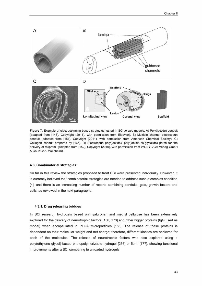
Chapter II
33
Figure 7. Example of electrospinning-based strategies tested in SCI in vivo models. A) Poly(lactide) conduit
(adapted from [146], Copyright (2011), with permission from Elsevier). B) Multiple channel electrospun
conduit (adapted from [151], Copyright (2011), with permission from American Chemical Society). C)
Collagen conduit prepared by [165]. D) Electrospun poly(lactide)/ poly(lactide-co-glycolide) patch for the
delivery of rolipram (Adapted from [152], Copyright (2010), with permission from WILEY-VCH Verlag GmbH
& Co. KGaA, Weinheim).
4.3. Combinatorial strategies
So far in this review the strategies proposed to treat SCI were presented individually. However, it
is currently believed that combinatorial strategies are needed to address such a complex condition
[4], and there is an increasing number of reports combining conduits, gels, growth factors and
cells, as reviewed in the next paragraphs.
4.3.1. Drug releasing bridges
In SCI research hydrogels based on hyaluronan and methyl cellulose has been extensively
explored for the delivery of neurotrophic factors [156, 173] and other bigger proteins (IgG used as
model) when encapsulated in PLGA microparticles [156]. The release of these proteins is
dependent on their molecular weight and net charge; therefore, different kinetics are achieved for
each of the molecules. The release of neurotrophic factors was also explored using a
poly(ethylene glycol)-based photopolymerizable hydrogel [236] or fibrin [177], showing functional
improvements after a SCI comparing to unloaded hydrogels.

Current strategies for spinal cord injury
34
Not only growth factors have been delivered at a spinal cord lesion via release from hydrogels. An
agarose hydrogel containing PLGA nanoparticles loaded with methylprednisolone implanted in a
contusion model of SCI showed to reduce lesion volume and macrophage infiltration [55]. The
localized delivery has been proposed to reduce side effects associated to high amount of the drug
administered systemically [55]. Chondroitinase ABC has also been delivered through fibrin
hydrogels in a injured spinal cord [176], and alternatively, from electrospun fibres [166]. Indeed,
hydrogels are particularly interesting as drug delivery vectors for intrathecal injection after SCI.
However, when physical support is also required, other materials have to be applied. PLGA
multiple lumen conduits were investigated for the delivery of NGF [205]; and PLA electrospun
fibres loaded with 6-aminonicotinamide were studied as mean to limit astrocyte proliferation [147].
Collagen fibres were also prepared for the delivery of NT-3 and chondroitinase ABC [166].
Although these studies still require in vivo testing, the incorporation of growth factors cocktails in
nerve conduits have already demonstrated to be a valuable strategy to improve functional
recovery after SCI [151].
4.3.2. Drug releasing bridges with cells
The delivery of growth factors can also be used to improve the survival of cells implanted at the
injury site or to modulate stem cell differentiation/proliferation after injection. Johnson and co-
workers used a sub-acute dorsal hemisection SCI model to test the effect of embryonic neural
progenitor cell implantation when in a fibrin gel loaded with growth factors (NT-3 and platelet
derived growth factor). The authors reported an increased cell survival and differentiation into
neural lineages; however, and particularly in growth factor loaded-hydrogels, overproliferation of a
subset of the implanted cells occurred and was accompanied with loss of function [178]. More
exciting results were recently published after implanting neural progenitor cells embedded in a
fibrin matrix containing a cocktail of growth factors (including: brain-derived growth factor, NT-3,
platelet-derived growth factor, insulin-like growth factor 1, epidermal growth factor, basic fibroblast
growth factor, acidic fibroblast growth factor, glial-cell-line-derived neurotrophic factor, hepatocyte
growth factor, and calpain inhibitor). The authors claimed long-distance axonal regeneration
without the need of other drugs to ameliorate the inhibitory environment. Additionally, two stem
cell lines were tested with similar results, being one already under clinical trials for amyotrophic
lateral sclerosis, putting this study a step ahead for testing in clinical trials [188].
An alternative to growth factor entrapment in polymeric scaffolds is the genetic modification of the
cells prior implantation, as a mean to convert the cell into a growth factor supplier. The premise is
that the implantation of cells expressing growth factors can promote their survival and/or modulate
the differentiation into neuronal lineages. Different engineered cells were already implanted in vivo
including Schwann cells expressing NGF [237], bone marrow cells expressing BDNF [238],
fibroblasts expressing GDNF [239], or olfactory ensheathing cells expressing NT-3 [240].

Chapter II
35
Recently, Gao and co-workers published promising results after implanting mesechymal stem
cells expressing BDNF seeded on an agarose injectable gel. The number of axons that can cross
the bridge is significantly higher when the cells seeded on the polymeric bridge are expressing the
growth factor, as compared to cells expressing GFP, where the number of axons is similar to the
implantation of the scaffold without cells [172]. The use of genetically-modified cells still have legal
and ethical implications that should be solved [241].
Scaffolds containing genetic material can serve as depots for the in situ delivery of genes to cells
at a lesion, potentially inducing the expression of a therapeutic protein for longer periods and
higher concentrations, as compared to direct protein delivery [242]. In the context of nerve
regeneration, PLGA disks loaded with poly(ethylenimine)-DNA nanoparticles containing a plasmid
encoding for NGF showed to promote axonal elongation in dorsal root ganglia neurons co-cultured
with human embryonic kidney (HEK) 293T cells [243]. Particularly in a SCI scenario, lipid-DNA
particles were incorporated in a PLGA channel bridge and a high expression of the reporter gene
was detected in the spinal cord during three weeks [134]. However, to achieve functional
improvements the implantation of conduits containing more efficient gene delivery vectors
(lentivirus encoding NT-3 or BDNF) was needed [244]. Alternatively, the use of nanoparticles for
the delivery of small interference RNA (siRNA) incorporated in fibres was also proposed [245,
246], but these strategies still need to prove their efficiency in vivo.

Chapter II
37
5. References
1. Schwab JM, Brechtel K, Mueller CA, Failli V, Kaps HP, Tuli SK, and Schluesener HJ (2006). "Experimental strategies to promote spinal cord regeneration - An integrative perspective". Progress in Neurobiology, 78 (2): 91-116.
2. Ramer LM, Ramer MS, and Steeves JD (2005). "Setting the stage for functional repair of spinal cord injuries: A cast of thousands". Spinal Cord, 43 (3): 134-161.
3. Rowland JW, Hawryluk GW, Kwon B, and Fehlings MG (2008). "Current status of acute spinal cord injury pathophysiology and emerging therapies: promise on the horizon". Neurosurgical focus, 25 (5): E2.
4. McCreedy DA and Sakiyama-Elbert SE (2012). "Combination therapies in the CNS: Engineering the environment". Neuroscience Letters, 519 (2): 115-121.
5. Yiu G and He Z (2006). "Glial inhibition of CNS axon regeneration". Nature Reviews Neuroscience, 7 (8): 617-627.
6. David S and Aguayo AJ (1981). "Axonal elongation into peripheral nervous system 'bridges' after central nervous system injury in adult rats". Science, 214 (4523): 931-933.
7. Richardson PM, Issa VMK, and Aguayo AJ (1984). "Regeneration of long spinal axons in the rat". Journal of Neurocytology, 13 (1): 165-182.
8. Bareyre FM, Kerschensteiner M, Raineteau O, Mettenleiter TC, Weinmann O, and Schwab ME (2004). "The injured spinal cord spontaneously forms a new intraspinal circuit in adult rats". Nature Neuroscience, 7 (3): 269-277.
9. Fitch MT and Silver J (2008). "CNS injury, glial scars, and inflammation: Inhibitory extracellular matrices and regeneration failure". Experimental Neurology, 209 (2): 294-301.
10. Woerly S (2000). "Restorative surgery of the central nervous system by means of tissue engineering using Neurogen implants". Neurosurgical Review, 23 (2): 59-77.
11. He J, Wang XM, Spector M, and Cui FZ (2012). "Scaffolds for central nervous system tissue engineering". Frontiers of Materials Science, 6 (1): 1-25.
12. Donnelly DJ and Popovich PG (2008). "Inflammation and its role in neuroprotection, axonal regeneration and functional recovery after spinal cord injury". Experimental Neurology, 209 (2): 378-388.
13. Schnell L (1999). "Acute inflammatory responses to mechanical lesions in the CNS: Differences between brain and spinal cord". European Journal of Neuroscience, 11 (10): 3648-3658.
14. Sullivan PG, Krishnamurthy S, Patel SP, Pandya JD, and Rabchevsky AG (2007). "Temporal characterization of mitochondrial bioenergetics after spinal cord injury". Journal of Neurotrauma, 24 (6): 991-999.
15. Fawcett JW and Asher RA (1999). "The glial scar and central nervous system repair". Brain Research Bulletin, 49 (6): 377-391.
16. Kreutzberg GW (1996). "Microglia: A sensor for pathological events in the CNS". Trends in Neurosciences, 19 (8): 312-318.
17. Hausmann ON (2003). "Post-traumatic inflammation following spinal cord injury". Spinal Cord, 41 (7): 369-378.
18. Taoka Y, Okajima K, Murakami K, Johno M, and Naruo M (1998). "Role of neutrophil elastase in compression-induced spinal cord injury in rats". Brain Research, 799 (2): 264-269.
19. Schwartz M, Moalem G, Leibowitz-Amit R, and Cohen IR (1999). "Innate and adaptive immune responses can be beneficial for CNS repair". Trends in Neurosciences, 22 (7): 295-299.
20. Segal JL, Gonzalez E, Yousefi S, Jamshidipour L, and Brunnemann SR (1997). "Circulating levels
of IL-2R, ICAM-1, and IL-6 in spinal cord injuries". Archives of Physical Medicine and Rehabilitation, 78 (1): 44-47.
21. Ridet JL, Malhotra SK, Privat A, and Gage FH (1997). "Reactive astrocytes: Cellular and molecular cues to biological function". Trends in Neurosciences, 20 (12): 570-577.
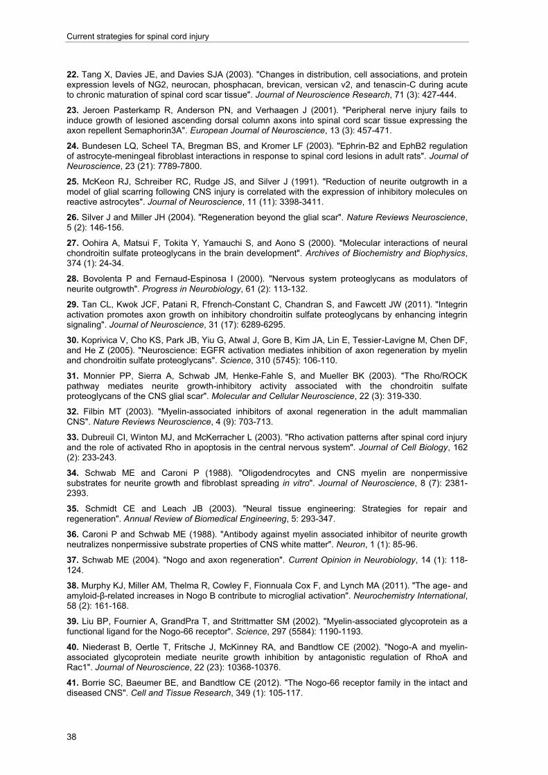
Current strategies for spinal cord injury
38
22. Tang X, Davies JE, and Davies SJA (2003). "Changes in distribution, cell associations, and protein expression levels of NG2, neurocan, phosphacan, brevican, versican v2, and tenascin-C during acute to chronic maturation of spinal cord scar tissue". Journal of Neuroscience Research, 71 (3): 427-444.
23. Jeroen Pasterkamp R, Anderson PN, and Verhaagen J (2001). "Peripheral nerve injury fails to induce growth of lesioned ascending dorsal column axons into spinal cord scar tissue expressing the axon repellent Semaphorin3A". European Journal of Neuroscience, 13 (3): 457-471.
24. Bundesen LQ, Scheel TA, Bregman BS, and Kromer LF (2003). "Ephrin-B2 and EphB2 regulation of astrocyte-meningeal fibroblast interactions in response to spinal cord lesions in adult rats". Journal of Neuroscience, 23 (21): 7789-7800.
25. McKeon RJ, Schreiber RC, Rudge JS, and Silver J (1991). "Reduction of neurite outgrowth in a model of glial scarring following CNS injury is correlated with the expression of inhibitory molecules on reactive astrocytes". Journal of Neuroscience, 11 (11): 3398-3411.
26. Silver J and Miller JH (2004). "Regeneration beyond the glial scar". Nature Reviews Neuroscience, 5 (2): 146-156.
27. Oohira A, Matsui F, Tokita Y, Yamauchi S, and Aono S (2000). "Molecular interactions of neural chondroitin sulfate proteoglycans in the brain development". Archives of Biochemistry and Biophysics, 374 (1): 24-34.
28. Bovolenta P and Fernaud-Espinosa I (2000). "Nervous system proteoglycans as modulators of
neurite outgrowth". Progress in Neurobiology, 61 (2): 113-132.
29. Tan CL, Kwok JCF, Patani R, Ffrench-Constant C, Chandran S, and Fawcett JW (2011). "Integrin activation promotes axon growth on inhibitory chondroitin sulfate proteoglycans by enhancing integrin signaling". Journal of Neuroscience, 31 (17): 6289-6295.
30. Koprivica V, Cho KS, Park JB, Yiu G, Atwal J, Gore B, Kim JA, Lin E, Tessier-Lavigne M, Chen DF, and He Z (2005). "Neuroscience: EGFR activation mediates inhibition of axon regeneration by myelin and chondroitin sulfate proteoglycans". Science, 310 (5745): 106-110.
31. Monnier PP, Sierra A, Schwab JM, Henke-Fahle S, and Mueller BK (2003). "The Rho/ROCK pathway mediates neurite growth-inhibitory activity associated with the chondroitin sulfate proteoglycans of the CNS glial scar". Molecular and Cellular Neuroscience, 22 (3): 319-330.
32. Filbin MT (2003). "Myelin-associated inhibitors of axonal regeneration in the adult mammalian CNS". Nature Reviews Neuroscience, 4 (9): 703-713.
33. Dubreuil CI, Winton MJ, and McKerracher L (2003). "Rho activation patterns after spinal cord injury and the role of activated Rho in apoptosis in the central nervous system". Journal of Cell Biology, 162 (2): 233-243.
34. Schwab ME and Caroni P (1988). "Oligodendrocytes and CNS myelin are nonpermissive substrates for neurite growth and fibroblast spreading in vitro". Journal of Neuroscience, 8 (7): 2381-2393.
35. Schmidt CE and Leach JB (2003). "Neural tissue engineering: Strategies for repair and regeneration". Annual Review of Biomedical Engineering, 5: 293-347.
36. Caroni P and Schwab ME (1988). "Antibody against myelin associated inhibitor of neurite growth neutralizes nonpermissive substrate properties of CNS white matter". Neuron, 1 (1): 85-96.
37. Schwab ME (2004). "Nogo and axon regeneration". Current Opinion in Neurobiology, 14 (1): 118-124.
38. Murphy KJ, Miller AM, Thelma R, Cowley F, Fionnuala Cox F, and Lynch MA (2011). "The age- and amyloid-β-related increases in Nogo B contribute to microglial activation". Neurochemistry International, 58 (2): 161-168.
39. Liu BP, Fournier A, GrandPra T, and Strittmatter SM (2002). "Myelin-associated glycoprotein as a functional ligand for the Nogo-66 receptor". Science, 297 (5584): 1190-1193.
40. Niederast B, Oertle T, Fritsche J, McKinney RA, and Bandtlow CE (2002). "Nogo-A and myelin-associated glycoprotein mediate neurite growth inhibition by antagonistic regulation of RhoA and Rac1". Journal of Neuroscience, 22 (23): 10368-10376.
41. Borrie SC, Baeumer BE, and Bandtlow CE (2012). "The Nogo-66 receptor family in the intact and diseased CNS". Cell and Tissue Research, 349 (1): 105-117.

Chapter II
39
42. Huber AB, Kolodkin AL, Ginty DD, and Cloutier JF, Signaling at the growth cone: Ligand-receptor complexes and the control of axon growth and guidance, in Annual Review of Neuroscience. 2003, Palo Alto, Calif., Annual Reviews, inc. p. 509-563.
43. Wang KC, Kim JA, Sivasankaran R, Segal R, and He Z (2002). "p75 interacts with the Nogo receptor as a co-receptor for Nogo, MAG and OMgp". Nature, 420 (6911): 74-78.
44. Nobes CD and Hall A (1995). "Rho, Rac, and Cdc42 GTPases regulate the assembly of multimolecular focal complexes associated with actin stress fibers, lamellipodia, and filopodia". Cell, 81 (1): 53-62.
45. Dergham P, Ellezam B, Essagian C, Avedissian H, Lubell WD, and McKerracher L (2002). "Rho signaling pathway targeted to promote spinal cord repair". Journal of Neuroscience, 22 (15): 6570-6577.
46. Benson MD, Romero MI, Lush ME, Lu QR, Henkemeyer M, and Parada LF (2005). "Ephrin-B3 is a myelin-based inhibitor of neurite outgrowth". Proceedings of the National Academy of Sciences of the United States of America, 102 (30): 10694-10699.
47. Moreau-Fauvarque C, Kumanogoh A, Camand E, Jaillard C, Barbin G, Boquet I, Love C, Jones EY, Kikutani H, Lubetzki C, Dusart I, and Chédotal A (2003). "The transmembrane semaphorin Sema4D/CD100, an inhibitor of axonal growth, is expressed on oligodendrocytes and upregulated after CNS lesion". Journal of Neuroscience, 23 (27): 9229-9239.
48. Barnes PJ (1998). "Anti-inflammatory actions of glucocorticoids: Molecular mechanisms". Clinical Science, 94 (6): 557-572.
49. Bracken MB, Shepard MJ, Collins WF, Holford TR, Young W, Baskin DS, Eisenberg HM, Flamm E, Leo-Summers L, Maroon J, Marshall LF, Perot Jr PL, Piepmeier J, Sonntag VKH, Wagner FC, Wilberger JE, and Winn HR (1990). "A randomized, controlled trial of methylprednisolone or naloxone in the treatment of acute spinal-cord injury. Results of the Second National Acute Spinal Cord Injury Study". New England Journal of Medicine, 322 (20): 1405-1411.
50. Short DJ, El Masry WS, and Jones PW (2000). "High dose methylprednisolone in the management of acute spinal cord injury - A systematic review from a clinical perspective". Spinal Cord, 38 (5): 273-286.
51. Sipski ML and Pearse DD (2006). "Methylprednisolone and other confounders to spinal cord injury clinical trials". Nature Clinical Practice Neurology, 2 (8): 402-403.
52. Kwon BK, Tetzlaff W, Grauer JN, Beiner J, and Vaccaro AR (2004). "Pathophysiology and pharmacologic treatment of acute spinal cord injury". The Spine Journal, 4 (4): 451-464.
53. (2004). "Effect of intravenous corticosteroids on death within 14 days in 10 008 adults with clinically significant head injury (MRC CRASH trial): Randomised placebo-controlled trial". Lancet, 364 (9442): 1321-1328.
54. Kim Yt, Caldwell JM, and Bellamkonda RV (2009). "Nanoparticle-mediated local delivery of methylprednisolone after spinal cord injury". Biomaterials, 30 (13): 2582-2590.
55. Chvatal SA, Kim YT, Bratt-Leal AM, Lee H, and Bellamkonda RV (2008). "Spatial distribution and acute anti-inflammatory effects of Methylprednisolone after sustained local delivery to the contused spinal cord". Biomaterials, 29 (12): 1967-1975.
56. Lee JM, Yan P, Xiao Q, Chen S, Lee KY, Hsu CY, and Xu J (2008). "Methylprednisolone protects oligodendrocytes but not neurons after spinal cord injury". Journal of Neuroscience, 28 (12): 3141-3149.
57. Schrater A, Lustenberger RM, Obermair FJ, and Thallmair M (2009). "High-dose corticosteroids after spinal cord injury reduce neural progenitor cell proliferation". Neuroscience, 161 (3): 753-763.
58. Schiller MD and Mobbs RJ (2012). "The historical evolution of the management of spinal cord injury". Journal of Clinical Neuroscience, 19 (10): 1348-1353.
59. Clinical Trials.gov. cited 2014; Available from: http://www.clinicaltrials.gov/.
60. Ghezzi P and Brines M (2004). "Erythropoietin as an antiapoptotic, tissue-protective cytokine". Cell Death and Differentiation, 11 (SUPPL. 1).
61. Rabchevsky AG, Patel SP, and Springer JE (2011). "Pharmacological interventions for spinal cord injury: Where do we stand? How might we step forward?". Pharmacology and Therapeutics, 132 (1): 15-29.

Current strategies for spinal cord injury
40
62. Leist M, Gliezzi P, Grasso G, Bianchi R, Vilia P, Fratelli M, Savino C, Bianchi M, Nielsen J, Gerwien J, Kallunki P, Kirstine Larsen A, Helboe L, Christensen S, Pedersen LO, Nielsen M, Torup L, Sager T, Sfacteria A, Erbayraktar S, Erbayraktar Z, Gokmen N, Yilmaz O, Cerami-Hand C, Xie QW, Coleman T, Cerami A, and Brines M (2004). "Derivatives of erythropoitein that are tissue protective but not erythropoietic". Science, 305 (5681): 239-242.
63. Matis GK and Birbilis TA (2009). "Erythropoietin in spinal cord injury". European Spine Journal, 18 (3): 314-323.
64. Lee SM, Yune TY, Kim SJ, Park DW, Lee YK, Kim YC, Oh YJ, Markelonis GJ, and Oh TH (2003). "Minocycline Reduces Cell Death and Improves Functional Recovery after Traumatic Spinal Cord Injury in the Rat". Journal of Neurotrauma, 20 (10): 1017-1027.
65. Stirling DP, Khodarahmi K, Liu J, McPhail LT, McBride CB, Steeves JD, Ramer MS, and Tetzlaff W (2004). "Minocycline Treatment Reduces Delayed Oligodendrocyte Death, Attenuates Axonal Dieback, and Improves Functional Outcome after Spinal Cord Injury". Journal of Neuroscience, 24 (9): 2182-2190.
66. Casha S, Zygun D, McGowan MD, Bains I, Yong VW, and Hurlbert RJ (2012). "Results of a phase II placebo-controlled randomized trial of minocycline in acute spinal cord injury". Brain, 135 (4): 1224-1236.
67. Ates O, Cayli SR, Gurses I, Turkoz Y, Tarim O, Cakir CO, and Kocak A (2007). "Comparative
neuroprotective effect of sodium channel blockers after experimental spinal cord injury". Journal of Clinical Neuroscience, 14 (7): 658-665.
68. Markus A, Patel TD, and Snider WD (2002). "Neurotrophic factors and axonal growth". Current Opinion in Neurobiology, 12 (5): 523-531.
69. Oudega M and Hagg T (1996). "Nerve growth factor promotes regeneration of sensory axons into adult rat spinal cord". Experimental Neurology, 140 (2): 218-229.
70. Bradbury EJ, Khemani S, King VR, Priestley JV, and McMahon SB (1999). "NT-3 promotes growth of lesioned adult rat sensory axons ascending in the dorsal columns of the spinal cord". European Journal of Neuroscience, 11 (11): 3873-3883.
71. Sharma HS, Neuroprotective effects of neurotrophins and melanocortins in spinal cord injury: An experimental study in the rat using pharmacological and morphological approaches, in Annals of the New York Academy of Sciences. 2005, New York, The Academy. p. 407-421.
72. Apfel SC, Nerve growth factor for the treatment of diabetic neuropathy: What went wrong, what went right, and what does the future hold?, in International review of neurobiology. 2002, New York Ny : Academic Press. p. 393-413.
73. Rossignol S, Schwab M, Schwartz M, and Fehlings MG (2007). "Spinal cord injury: Time to move?". Journal of Neuroscience, 27 (44): 11782-11792.
74. Popovich PG and Longbrake EE (2008). "Can the immune system be harnessed to repair the CNS?". Nature Reviews Neuroscience, 9 (6): 481-493.
75. Brewer KL, Bethea JR, and Yezierski RP (1999). "Neuroprotective effects of interleukin-10 following excitotoxic spinal cord injury". Experimental Neurology, 159 (2): 484-493.
76. Vidal PM, Lemmens E, Dooley D, and Hendrix S (2013). "The role of " anti-inflammatory" cytokines in axon regeneration". Cytokine and Growth Factor Reviews, 24 (1): 1-12.
77. Popovich PG, Guan Z, Wei P, Huitinga I, Van Rooijen N, and Stokes BT (1999). "Depletion of hematogenous macrophages promotes partial hindlimb recovery and neuroanatomical repair after experimental spinal cord injury". Experimental Neurology, 158 (2): 351-365.
78. Schwartz M, London A, and Shechter R (2009). "Boosting T-cell immunity as a therapeutic approach for neurodegenerative conditions: The role of innate immunity". Neuroscience, 158 (3): 1133-1142.
79. Rapalino O, Lazarov-Spiegler O, Agranov E, Velan GJ, Yoles E, Fraidakis M, Solomon A, Gepstein R, Katz A, Belkin M, Hadani M, and Schwartz M (1998). "Implantation of stimulated homologous macrophages results in partial recovery of paraplegic rats". Nature Medicine, 4 (7): 814-821.
80. Mantovani A, Sica A, Sozzani S, Allavena P, Vecchi A, and Locati M (2004). "The chemokine system in diverse forms of macrophage activation and polarization". Trends in Immunology, 25 (12): 677-686.

Chapter II
41
81. Mosser DM and Edwards JP (2008). "Exploring the full spectrum of macrophage activation". Nature Reviews Immunology, 8 (12): 958-969.
82. Lammertse DP, Jones LAT, Charlifue SB, Kirshblum SC, Apple DF, Ragnarsson KT, Falci SP, Heary RF, Choudhri TF, Jenkins AL, Betz RR, Poonian D, Cuthbert JP, Jha A, Snyder DA, and Knoller N (2012). "Autologous incubated macrophage therapy in acute, complete spinal cord injury: Results of the phase 2 randomized controlled multicenter trial". Spinal Cord, 50 (9): 661-671.
83. Hauben E, Butovsky O, Nevo U, Yoles E, Moalem G, Agranov E, Mor F, Leibowitz-Amit R, Pevsner E, Akselrod S, Neeman M, Cohen IR, and Schwartz M (2000). "Passive or active immunization with myelin basic protein promotes recovery from spinal cord contusion". Journal of Neuroscience, 20 (17): 6421-6430.
84. Ziv Y, Avidan H, Pluchino S, Martino G, and Schwartz M (2006). "Synergy between immune cells and adult neural stem/progenitor cells promotes functional recovery from spinal cord injury". Proceedings of the National Academy of Sciences of the United States of America, 103 (35): 13174-13179.
85. Bradbury EJ, Moon LDF, Popat RJ, King VR, Bennett GS, Patel PN, Fawcett JW, and McMahon SB (2002). "Chondroitinase ABC promotes functional recovery after spinal cord injury". Nature, 416 (6881): 636-640.
86. Garcia-Alias G, Lin R, Akrimi SF, Story D, Bradbury EJ, and Fawcett JW (2008). "Therapeutic time
window for the application of chondroitinase ABC after spinal cord injury". Experimental Neurology, 210 (2): 331-338.
87. Crespo D, Asher RA, Lin R, Rhodes KE, and Fawcett JW (2007). "How does chondroitinase promote functional recovery in the damaged CNS?". Experimental Neurology, 206 (2): 159-171.
88. Lemons ML, Sandy JD, Anderson DK, and Howland DR (2003). "Intact aggrecan and chondroitin sulfate-depleted aggrecan core glycoprotein inhibit axon growth in the adult rat spinal cord". Experimental Neurology, 184 (2): 981-990.
89. Zhao RR, Muir EM, Alves JN, Rickman H, Allan AY, Kwok JC, Roet KCD, Verhaagen J, Schneider BL, Bensadoun JC, Ahmed SG, Yáñez-Muñoz RJ, Keynes RJ, Fawcett JW, and Rogers JH (2011). "Lentiviral vectors express chondroitinase ABC in cortical projections and promote sprouting of injured corticospinal axons". Journal of Neuroscience Methods, 201 (1): 228-238.
90. Massey JM, Hubscher CH, Wagoner MR, Decker JA, Amps J, Silver J, and Onifer SM (2006). "Chondroitinase ABC digestion of the perineuronal net promotes functional collateral sprouting in the cuneate nucleus after cervical spinal cord injury". Journal of Neuroscience, 26 (16): 4406-4414.
91. Schnell L and Schwab ME (1990). "Axonal regeneration in the rat spinal cord produced by an antibody against myelin-associated neurite growth inhibitors". Nature, 343 (6255): 269-272.
92. Gonzenbach RR and Schwab ME (2008). "Disinhibition of neurite growth to repair the injured adult CNS: Focusing on Nogo". Cellular and Molecular Life Sciences, 65 (1): 161-176.
93. Gonzenbach RR, Zoerner B, Schnell L, Weinmann O, Mir AK, and Schwab ME (2012). "Delayed anti-Nogo-A antibody application after spinal cord injury shows progressive loss of responsiveness". Journal of Neurotrauma, 29 (3): 567-578.
94. Li S and Strittmatter SM (2003). "Delayed systemic Nogo-66 receptor antagonist promotes recovery from spinal cord injury". Journal of Neuroscience, 23 (10): 4219-4227.
95. Steward O, Sharp K, Yee KM, and Hofstadter M (2008). "A re-assessment of the effects of a Nogo-66 receptor antagonist on regenerative growth of axons and locomotor recovery after spinal cord injury in mice". Experimental Neurology, 209 (2): 446-468.
96. Lehmann M, Fournier A, Selles-Navarro I, Dergham P, Sebok A, Leclerc N, Tigyi G, and McKerracher L (1999). "Inactivation of rho signaling pathway promotes CNS axon regeneration". Journal of Neuroscience, 19 (17): 7537-7547.
97. McKerracher L and Higuchi H (2006). "Targeting Rho to stimulate repair after spinal cord injury". Journal of Neurotrauma, 23 (3-4): 309-317.
98. Fehlings MG, Theodore N, Harrop J, Maurais G, Kuntz C, Shaffrey CI, Kwon BK, Chapman J, Yee A, Tighe A, and McKerracher L (2011). "A phase I/IIa clinical trial of a recombinant Rho protein antagonist in acute spinal cord injury". Journal of Neurotrauma, 28 (5): 787-796.

Current strategies for spinal cord injury
42
99. Mitchell JA, Akarasereenont P, Thiemermann C, Flower RJ, and Vane JR (1993). "Selectivity of nonsteroidal antiinflammatory drugs as inhibitors of constitutive and inducible cyclooxygenase". Proceedings of the National Academy of Sciences of the United States of America, 90 (24): 11693-11697.
100. Rainsford KD (2009). "Ibuprofen: pharmacology, efficacy and safety". Inflammopharmacology, 17 (6): 275-342.
101. Fu Q, Hue J, and Li S (2007). "Nonsteroidal anti-inflammatory drugs promote axon regeneration via RhoA inhibition". Journal of Neuroscience, 27 (15): 4154-4164.
102. Wang X, Budel S, Baughman K, Gould G, Song KH, and Strittmatter SM (2009). "Ibuprofen enhances recovery from spinal cord injury by limiting tissue loss and stimulating axonal growth". Journal of Neurotrauma, 26 (1): 81-95.
103. Madura T, Tomita K, and Terenghi G (2011). "Ibuprofen improves functional outcome after axotomy and immediate repair in the peripheral nervous system". Journal of Plastic, Reconstructive and Aesthetic Surgery, 64 (12): 1641-1646.
104. Xing B, Li H, Wang H, Mukhopadhyay D, Fisher D, Gilpin CJ, and Li S (2011). "RhoA-inhibiting NSAIDs promote axonal myelination after spinal cord injury". Experimental Neurology, 231 (2): 247-260.
105. Dill J, Patel AR, Yang XL, Bachoo R, Powell CM, and Li S (2010). "A molecular mechanism for
ibuprofen-mediated RhoA inhibition in neurons". Journal of Neuroscience, 30 (3): 963-972.
106. Sharp KG, Yee KM, Stiles TL, Aguilar RM, and Steward O (2013). "A re-assessment of the effects of treatment with a non-steroidal anti-inflammatory (ibuprofen) on promoting axon regeneration via RhoA inhibition after spinal cord injury". Experimental Neurology, 248: 321-327.
107. Kopp MA, Liebscher T, Niedeggen A, Laufer S, Brommer B, Jungehulsing GJ, Strittmatter SM, Dirnagl U, and Schwab JM (2012). "Small-molecule-induced Rho-inhibition: NSAIDs after spinal cord injury". Cell and Tissue Research, 349 (1): 119-132.
108. Zhao P, Waxman SG, and Hains BC (2007). "Extracellular signal-regulated kinase-regulated microglia-neuron signaling by prostaglandin E2 contributes to pain after spinal cord injury". Journal of Neuroscience, 27 (9): 2357-2368.
109. Ma W, St-Jacques B, and Cruz Duarte P (2012). "Targeting pain mediators induced by injured nerve-derived COX2 and PGE2 to treat neuropathic pain". Expert Opinion on Therapeutic Targets, 16 (6): 527-540.
110. Redondo-Castro E and Navarro X (2014). "Chronic ibuprofen administration reduces neuropathic pain but does not exert neuroprotection after spinal cord injury in adult rats". Experimental Neurology, 252: 95-103.
111. Pêgo AP, Kubinova S, Cizkova D, Vanicky I, Mar FM, Sousa MM, and Sykova E (2012). "Regenerative medicine for the treatment of spinal cord injury: More than just promises?". Journal of Cellular and Molecular Medicine, 16 (11): 2564-2582.
112. Ruff CA, Wilcox JT, and Fehlings MG (2012). "Cell-based transplantation strategies to promote plasticity following spinal cord injury". Experimental Neurology, 235 (1): 78-90.
113. Thuret S, Moon LDF, and Gage FH (2006). "Therapeutic interventions after spinal cord injury". Nature Reviews Neuroscience, 7 (8): 628-643.
114. Tetzlaff W, Okon EB, Karimi-Abdolrezaee S, Hill CE, Sparling JS, Plemel JR, Plunet WT, Tsai EC, Baptiste D, Smithson LJ, Kawaja MD, Fehlings MG, and Kwon BK (2011). "A systematic review of cellular transplantation therapies for spinal cord injury". Journal of Neurotrauma, 28 (8): 1611-1682.
115. McDonald JW, Liu XZ, Qu Y, Liu S, Mickey SK, Turetsky D, Gottlieb DI, and Choi DW (1999). "Transplanted embryonic stem cells survive, differentiate and promote recovery in injured rat spinal cord". Nature Medicine, 5 (12): 1410-1412.
116. Hill CE, Moon LDF, Wood PM, and Bunge MB (2006). "Labeled Schwann cell transplantation: Cell loss, host Schwann cell replacement, and strategies to enhance survival". Glia, 53 (3): 338-343.
117. Amemori T, Jendelová P, Růžičková K, Arboleda D, and Syková E (2010). "Co-transplantation of olfactory ensheathing glia and mesenchymal stromal cells does not have synergistic effects after spinal cord injury in the rat". Cytotherapy, 12 (2): 212-225.

Chapter II
43
118. Lima C, Escada P, Pratas-Vital J, Branco C, Arcangeli CA, Lazzeri G, Maia CA, Capucho C, Hasse-Ferreira A, and Peduzzi JD (2010). "Olfactory mucosal autografts and rehabilitation for chronic traumatic spinal cord injury". Neurorehabilitation and neural repair, 24 (1): 10-22.
119. Huang H, Xi H, Chen L, Zhang F, and Liu Y (2012). "Long-term outcome of olfactory ensheathing cell therapy for patients with complete chronic spinal cord injury". Cell Transplantation, 21 (SUPPL. 1): S23-S31.
120. Steward O, Sharp K, Selvan G, Hadden A, Hofstadter M, Au E, and Roskams J (2006). "A re-assessment of the consequences of delayed transplantation of olfactory lamina propria following complete spinal cord transection in rats". Experimental Neurology, 198 (2): 483-499.
121. Fouad K and Tetzlaff W (2012). "Rehabilitative training and plasticity following spinal cord injury". Experimental Neurology, 235 (1): 91-99.
122. Knikou M (2012). "Plasticity of corticospinal neural control after locomotor training in human spinal cord injury". Neural Plasticity, 2012.
123. Wilcox JT, Cadotte D, and Fehlings MG (2012). "Spinal cord clinical trials and the role for bioengineering". Neuroscience Letters, 519 (2): 93-102.
124. Daly W, Yao L, Zeugolis D, Windebank A, and Pandit A (2012). "A biomaterials approach to peripheral nerve regeneration: Bridging the peripheral nerve gap and enhancing functional recovery". Journal of the Royal Society Interface, 9 (67): 202-221.
125. de Ruiter GC, Malessy MJ, Yaszemski MJ, Windebank AJ, and Spinner RJ (2009). "Designing ideal conduits for peripheral nerve repair". Neurosurgical focus, 26 (2): E5.
126. Ciardelli G and Chiono V (2006). "Materials for peripheral nerve regeneration". Macromolecular Bioscience, 6 (1): 13-26.
127. Chau CH, Shum DK, Li H, Pei J, Lui YY, Wirthlin L, Chan YS, and Xu XM (2004). "Chondroitinase ABC enhances axonal regrowth through Schwann cell-seeded guidance channels after spinal cord injury". The FASEB journal 18 (1): 194-196.
128. Giannetti S, Lauretti L, Fernandez E, Salvinelli F, Tamburrini G, and Pallini R (2001). "Acrylic hydrogel implants after spinal cord lesion in the adult rat". Neurological Research, 23 (4): 405-409.
129. Tsai EC, Dalton PD, Shoichet MS, and Tator CH (2004). "Synthetic hydrogel guidance channels facilitate regeneration of adult rat brainstem motor axons after complete spinal cord transection". Journal of Neurotrauma, 21 (6): 789-804.
130. Straley KS, Foo CWP, and Heilshorn SC (2010). "Biomaterial design strategies for the treatment of spinal cord injuries". Journal of Neurotrauma, 27 (1): 1-19.
131. Hattori T, Matsuyama Y, Sakai Y, Ishiguro N, Hirata H, and Nakamura R (2008). "Chondrotinase ABC enhances axonal regeneration across nerve gaps". Journal of Clinical Neuroscience, 15 (2): 185-191.
132. Friedman JA, Windebank AJ, Moore MJ, Spinner RJ, Currier BL, Yaszemski MJ, Bartolomei J, Piepmeier JM, Chu G, Fehlings MG, Hodge Jr CJ, and Wagner Jr FC (2002). "Biodegradable polymer grafts for surgical repair of the injured spinal cord". Neurosurgery, 51 (3): 742-752.
133. Corey JM, Lin DY, Mycek KB, Chen Q, Samuel S, Feldman EL, and Martin DC (2007). "Aligned electrospun nanofibers specify the direction of dorsal root ganglia neurite growth". Journal of Biomedical Materials Research - Part A, 83 (3): 636-645.
134. De Laporte L, Lei Yan A, and Shea LD (2009). "Local gene delivery from ECM-coated poly(lactide-co-glycolide) multiple channel bridges after spinal cord injury". Biomaterials, 30 (12): 2361-2368.
135. Koh HS, Yong T, Chan CK, and Ramakrishna S (2008). "Enhancement of neurite outgrowth using nano-structured scaffolds coupled with laminin". Biomaterials, 29 (26): 3574-3582.
136. Yucel D, Kose GT, and Hasirci V (2010). "Polyester based nerve guidance conduit design". Biomaterials, 31 (7): 1596-1603.
137. Nisbet DR, Rodda AE, Horne MK, Forsythe JS, and Finkelstein DI (2009). "Neurite infiltration and cellular response to electrospun polycaprolactone scaffolds implanted into the brain". Biomaterials, 30 (27): 4573-4580.

Current strategies for spinal cord injury
44
138. Pêgo AP, Poot AA, Grijpma DW, and Feijen J (2001). "Copolymers of trimethylene carbonate and epsilon-caprolactone for porous nerve guides: Synthesis and properties". Journal of Biomaterials Science, Polymer Edition, 12 (1): 35-53.
139. Pêgo AP, Poot AA, Grijpma DW, and Feijen J (2003). "Biodegradable elastomeric scaffolds for soft tissue engineering". Journal of Controlled Release, 87 (1-3): 69-79.
140. Chew SY, Mi R, Hoke A, and Leong KW (2007). "Aligned protein-polymer composite fibers enhance nerve regeneration: A potential tissue-engineering platform". Advanced Functional Materials, 17 (8): 1288-1296.
141. Vleggeert-Lankamp CLAM, Wolfs J, Pêgo AP, Van Den Berg R, Feirabend H, and Lakke E (2008). "Effect of nerve graft porosity on the refractory period of regenerating nerve fibers: Laboratory investigation". Journal of Neurosurgery, 109 (2): 294-305.
142. de Luca AC, Stevens JS, Schroeder SLM, Guilbaud JB, Saiani A, Downes S, and Terenghi G (2013). "Immobilization of cell-binding peptides on poly-ε-caprolactone film surface to biomimic the peripheral nervous system". Journal of Biomedical Materials Research - Part A, 101 (2): 491-501.
143. Tysseling-Mattiace VM, Sahni V, Niece KL, Birch D, Czeisler C, Fehlings MG, Stupp SI, and Kessler JA (2008). "Self-assembling nanofibers inhibit glial scar formation and promote axon elongation after spinal cord injury". Journal of Neuroscience, 28 (14): 3814-3823.
144. Matsumoto K, Ohnishi K, Kiyotani T, Sekine T, Ueda H, Nakamura T, Endo K, and Shimizu Y
(2000). "Peripheral nerve regeneration across an 80-mm gap bridged by a polyglycolic acid (PGA)-collagen tube filled with laminin-coated collagen fibers: A histological and electrophysiological evaluation of regenerated nerves". Brain Research, 868 (2): 315-328.
145. Oudega M, Gautier SE, Chapon P, Fragoso M, Bates ML, Parel JM, and Bartlett Bunge M (2001). "Axonal regeneration into Schwann cell grafts within resorbable poly(α-hydroxyacid) guidance channels in the adult rat spinal cord". Biomaterials, 22 (10): 1125-1136.
146. Hurtado A, Cregg JM, Wang HB, Wendell DF, Oudega M, Gilbert RJ, and McDonald JW (2011). "Robust CNS regeneration after complete spinal cord transection using aligned poly-l-lactic acid microfibers". Biomaterials, 32 (26): 6068-6079.
147. Schaub NJ and Gilbert RJ (2011). "Controlled release of 6-aminonicotinamide from aligned, electrospun fibers alters astrocyte metabolism and dorsal root ganglia neurite outgrowth". Journal of Neural Engineering, 8 (4): 046026.
148. Yang Y, De Laporte L, Zelivyanskaya ML, Whittlesey KJ, Anderson AJ, Cummings BJ, and Shea LD (2009). "Multiple Channel Bridges for Spinal Cord Injury: Cellular Characterization of Host Response". Tissue Engineering Part A, 15 (11): 3283-3295.
149. De Laporte L, Yang Y, Zelivyanskaya ML, Cummings BJ, Anderson AJ, and Shea LD (2009). "Plasmid releasing multiple channel bridges for transgene expression after spinal cord injury". Molecular Therapy, 17 (2): 318-326.
150. Chen BK, Knight AM, Madigan NN, Gross L, Dadsetan M, Nesbitt JJ, Rooney GE, Currier BL, Yaszemski MJ, Spinner RJ, and Windebank AJ (2011). "Comparison of polymer scaffolds in rat spinal cord: A step toward quantitative assessment of combinatorial approaches to spinal cord repair". Biomaterials, 32 (32): 8077-8086.
151. Gelain F, Panseri S, Antonini S, Cunha C, Donega M, Lowery J, Taraballi F, Cerri G, Montagna M, Baldissera F, and Vescovi A (2011). "Transplantation of nanostructured composite scaffolds results in the regeneration of chronically injured spinal cords". ACS Nano, 5 (1): 227-236.
152. Zhu Y, Wang A, Shen W, Patel S, Zhang R, Young WL, and Li S (2010). "Nanofibrous patches for spinal cord regeneration". Advanced Functional Materials, 20 (9): 1433-1440.
153. Hwang DH, Kim HM, Kang YM, Joo IS, Cho CS, Yoon BW, Kim SU, and Kim BG (2011). "Combination of multifaceted strategies to maximize the therapeutic benefits of neural stem cell transplantation for spinal cord repair". Cell Transplantation, 20 (9): 1361-1379.
154. Wong DY, Leveque JC, Brumblay H, Krebsbach PH, Hollister SJ, and LaMarca F (2008). "Macro-architectures in spinal cord scaffold implants influence regeneration". Journal of Neurotrauma, 25 (8): 1027-1037.
155. Tysseling VM, Sahni V, Pashuck ET, Birch D, Hebert A, Czeisler C, Stupp SI, and Kessler JA (2010). "Self-assembling peptide amphiphile promotes plasticity of serotonergic fibers following spinal cord injury". Journal of Neuroscience Research, 88 (14): 3161-3170.

Chapter II
45
156. Baumann MD, Kang CE, Stanwick JC, Wang Y, Kim H, Lapitsky Y, and Shoichet MS (2009). "An injectable drug delivery platform for sustained combination therapy". Journal of Controlled Release, 138 (3): 205-213.
157. Kubinová Š, Horák D, Kozubenko N, Vaněček V, Proks V, Price J, Cocks G, and Syková E (2010). "The use of superporous Ac-CGGASIKVAVS-OH-modified PHEMA scaffolds to promote cell adhesion and the differentiation of human fetal neural precursors". Biomaterials, 31 (23): 5966-5975.
158. Hejcl A, Lesný P, Prádný M, Michálek J, Jendelová P, Stulík J, and Syková E (2008). "Biocompatible hydrogels in spinal cord injury repair". Physiological research / Academia Scientiarum Bohemoslovaca, 57 Suppl 3: S121-132.
159. Hejčl A, Šedý J, Kapcalová M, Toro DA, Amemori T, Lesný P, Likavčanová-Mašínová K, Krumbholcová E, Přádný M, Michálek J, Burian M, Hájek M, Jendelová P, and Syková E (2010). "HPMA-RGD hydrogels seeded with mesenchymal stem cells improve functional outcome in chronic spinal cord injury". Stem Cells and Development, 19 (10): 1535-1546.
160. Loh NK, Woerly S, Bunt SM, Wilton SD, and Harvey AR (2001). "The regrowth of axons within tissue defects in the cns is promoted by implanted hydrogel matrices that contain BDNF and CNTF producing fibroblasts". Experimental Neurology, 170 (1): 72-84.
161. Amaral IF, Neiva I, Ferreira da Silva F, Sousa SR, Piloto AM, Lopes CDF, Barbosa MA, Kirkpatrick CJ, and Pêgo AP (2013). "Endothelialization of chitosan porous conduits via immobilization of a recombinant fibronectin fragment (rhFNIII 7-10)". Acta Biomaterialia, 9 (3): 5643-5652.
162. Cheng H, Huang YC, Chang PT, and Huang YY (2007). "Laminin-incorporated nerve conduits made by plasma treatment for repairing spinal cord injury". Biochemical and Biophysical Research Communications, 357 (4): 938-944.
163. Li X, Yang Z, Zhang A, Wang T, and Chen W (2009). "Repair of thoracic spinal cord injury by chitosan tube implantation in adult rats". Biomaterials, 30 (6): 1121-1132.
164. Chen X, Yang Y, Yao J, Lin W, Li Y, Chen Y, Gao Y, Gu X, and Wang X (2011). "Bone marrow stromal cells-loaded chitosan conduits promote repair of complete transection injury in rat spinal cord". Journal of Materials Science: Materials in Medicine, 22 (10): 2347-2356.
165. Liu T, Houle JD, Xu J, Chan BP, and Chew SY (2012). "Nanofibrous collagen nerve conduits for spinal cord repair". Tissue Engineering - Part A, 18 (9-10): 1057-1066.
166. Liu T, Xu J, Chan BP, and Chew SY (2012). "Sustained release of neurotrophin-3 and chondroitinase ABC from electrospun collagen nanofiber scaffold for spinal cord injury repair". Journal of Biomedical Materials Research - Part A, 100 A (1): 236-242.
167. Madaghiele M, Sannino A, Yannas IV, and Spector M (2008). "Collagen-based matrices with axially oriented pores". Journal of Biomedical Materials Research - Part A, 85 (3): 757-767.
168. Saglam A, Perets A, Canver AC, Li HL, Kollins K, Cohen G, Fischer I, Lazarovici P, and Lelkes PI (2012). "Angioneural Crosstalk in Scaffolds with Oriented Microchannels for Regenerative Spinal Cord Injury Repair". Journal of Molecular Neuroscience: 1-13.
169. Guo J, Su H, Zeng Y, Liang YX, Wong WM, Ellis-Behnke RG, So KF, and Wu W (2007). "Reknitting the injured spinal cord by self-assembling peptide nanofiber scaffold". Nanomedicine: Nanotechnology, Biology, and Medicine, 3 (4): 311-321.
170. Moradi F, Bahktiari M, Joghataei MT, Nobakht M, Soleimani M, Hasanzadeh G, Fallah A, Zarbakhsh S, Hejazian LB, Shirmohammadi M, and Maleki F (2012). "BD PuraMatrix peptide hydrogel as a culture system for human fetal Schwann cells in spinal cord regeneration". Journal of Neuroscience Research, 90 (12): 2335-2348.
171. Novikova LN, Pettersson J, Brohlin M, Wiberg M, and Novikov LN (2008). "Biodegradable poly-β-hydroxybutyrate scaffold seeded with Schwann cells to promote spinal cord repair". Biomaterials, 29 (9): 1198-1206.
172. Gao M, Lu P, Bednark B, Lynam D, Conner JM, Sakamoto J, and Tuszynski MH (2013). "Templated agarose scaffolds for the support of motor axon regeneration into sites of complete spinal cord transection". Biomaterials, 34 (5): 1529-1536.
173. Gupta D, Tator CH, and Shoichet MS (2006). "Fast-gelling injectable blend of hyaluronan and methylcellulose for intrathecal, localized delivery to the injured spinal cord". Biomaterials, 27 (11): 2370-2379.

Current strategies for spinal cord injury
46
174. Park J, Lim E, Back S, Na H, Park Y, and Sun K (2010). "Nerve regeneration following spinal cord injury using matrix metalloproteinase-sensitive, hyaluronic acid-based biomimetic hydrogel scaffold containing brain-derived neurotrophic factor". Journal of Biomedical Materials Research - Part A, 93 (3): 1091-1099.
175. Herbert CB, Nagaswami C, Bittner GD, Hubbell JA, and Weisel JW (1998). "Effects of fibrin micromorphology on neurite growth from dorsal root ganglia cultured in three-dimensional fibrin gels". Journal of Biomedical Materials Research, 40 (4): 551-559.
176. Hyatt AJT, Wang D, Kwok JC, Fawcett JW, and Martin KR (2010). "Controlled release of chondroitinase ABC from fibrin gel reduces the level of inhibitory glycosaminoglycan chains in lesioned spinal cord". Journal of Controlled Release, 147 (1): 24-29.
177. Johnson PJ, Parker SR, and Sakiyama-Elbert SE (2009). "Controlled release of neurotrophin-3 from fibrin-based tissue engineering scaffolds enhances neural fiber sprouting following subacute spinal cord injury". Biotechnology and Bioengineering, 104 (6): 1207-1214.
178. Johnson PJ, Tatara A, McCreedy DA, Shiu A, and Sakiyama-Elbert SE (2010). "Tissue-engineered fibrin scaffolds containing neural progenitors enhance functional recovery in a subacute model of SCI". Soft Matter, 6 (20): 5127-5137.
179. Taylor SJ, Rosenzweig ES, McDonald Iii JW, and Sakiyama-Elbert SE (2006). "Delivery of neurotrophin-3 from fibrin enhances neuronal fiber sprouting after spinal cord injury". Journal of Controlled Release, 113 (3): 226-235.
180. Prang P, Müller R, Eljaouhari A, Heckmann K, Kunz W, Weber T, Faber C, Vroemen M, Bogdahn U, and Weidner N (2006). "The promotion of oriented axonal regrowth in the injured spinal cord by alginate-based anisotropic capillary hydrogels". Biomaterials, 27 (19): 3560-3569.
181. Silva NA, Cooke MJ, Tam RY, Sousa N, Salgado AJ, Reis RL, and Shoichet MS (2012). "The effects of peptide modified gellan gum and olfactory ensheathing glia cells on neural stem/progenitor cell fate". Biomaterials, 33 (27): 6345-6354.
182. Alvarez-Perez MA, Guarino V, Cirillo V, and Ambrosio L (2010). "Influence of gelatin cues in PCL electrospun membranes on nerve outgrowth". Biomacromolecules, 11 (9): 2238-2246.
183. Prabhakaran MP, Venugopal J, Chan CK, and Ramakrishna S (2008). "Surface modified electrospun nanofibrous scaffolds for nerve tissue engineering". Nanotechnology, 19 (45).
184. Bhattarai N, Li Z, Gunn J, Leung M, Cooper A, Edmondson D, Veiseh O, Chen MH, Zhang Y, Ellenbogen RG, and Zhang M (2009). "Natural-synthetic polyblend nanofibers for biomedical applications". Advanced Materials, 21 (27): 2792-2797.
185. Bhattarai N, Ramay HR, Gunn J, Matsen FA, and Zhang M (2005). "PEG-grafted chitosan as an injectable thermosensitive hydrogel for sustained protein release". Journal of Controlled Release, 103 (3): 609-624.
186. Eyrich D, Göpferich A, and Blunk T, Fibrin in tissue engineering, in Advances in experimental medicine and biology. 2006, New York, Plenum Press. p. 379-392.
187. King VR, Alovskaya A, Wei DYT, Brown RA, and Priestley JV (2010). "The use of injectable forms of fibrin and fibronectin to support axonal ingrowth after spinal cord injury". Biomaterials, 31 (15): 4447-4456.
188. Lu P, Wang Y, Graham L, McHale K, Gao M, Wu D, Brock J, Blesch A, Rosenzweig ES, Havton LA, Zheng B, Conner JM, Marsala M, and Tuszynski MH (2012). "Long-distance growth and connectivity of neural stem cells after severe spinal cord injury". Cell, 150 (6): 1264-1273.
189. Zhang S (2003). "Fabrication of novel biomaterials through molecular self-assembly". Nature Biotechnology, 21 (10): 1171-1178.
190. Gelain F, Cigognini D, Caprini A, Silva D, Colleoni B, Donegá M, Antonini S, Cohen BE, and Vescovi A (2012). "New bioactive motifs and their use in functionalized self-assembling peptides for NSC differentiation and neural tissue engineering". Nanoscale, 4 (9): 2946-2957.
191. Zhang W, Zhan X, Gao M, Hamilton AD, Liu Z, Jiang Y, Su H, Dai X, He B, Kang X, Zeng Y, Wu W, and Guo J (2012). "Self-assembling peptide nanofiber scaffold enhanced with RhoA inhibitor CT04 improves axonal regrowth in the transected spinal cord". Journal of Nanomaterials, 2012; nr 54.
192. Tan A, Rajadas J, and Seifalian AM (2012). "Biochemical engineering nerve conduits using peptide amphiphiles". Journal of Controlled Release, 163 (3): 342-352.
Chapter II
47
193. Webber MJ, Matson JB, Tamboli VK, and Stupp SI (2012). "Controlled release of dexamethasone from peptide nanofiber gels to modulate inflammatory response". Biomaterials, 33 (28): 6823-6832.
194. Schmidt CE, Shastri VR, Vacanti JP, and Langer R (1997). "Stimulation of neurite outgrowth using an electrically conducting polymer". Proceedings of the National Academy of Sciences of the United States of America, 94 (17): 8948-8953.
195. Lee JY, Bashur CA, Goldstein AS, and Schmidt CE (2009). "Polypyrrole-coated electrospun PLGA nanofibers for neural tissue applications". Biomaterials, 30 (26): 4325-4335.
196. Prabhakaran MP, Ghasemi-Mobarakeh L, Jin G, and Ramakrishna S (2011). "Electrospun conducting polymer nanofibers and electrical stimulation of nerve stem cells". Journal of Bioscience and Bioengineering, 112 (5): 501-507.
197. Huang J, Hu X, Lu L, Ye Z, Zhang Q, and Luo Z (2010). "Electrical regulation of Schwann cells using conductive polypyrrole/chitosan polymers". Journal of Biomedical Materials Research - Part A, 93 (1): 164-174.
198. Geller HM and Fawcett JW (2002). "Building a bridge: Engineering spinal cord repair". Experimental Neurology, 174 (2): 125-136.
199. Mahoney MJ, Chen RR, Tan J, and Mark Saltzman W (2005). "The influence of microchannels on neurite growth and architecture". Biomaterials, 26 (7): 771-778.
200. Yao L, Wang S, Cui W, Sherlock R, O'Connell C, Damodaran G, Gorman A, Windebank A, and
Pandit A (2009). "Effect of functionalized micropatterned PLGA on guided neurite growth". Acta Biomaterialia, 5 (2): 580-588.
201. Fan YW, Cui FZ, Hou SP, Xu QY, Chen LN, and Lee IS (2002). "Culture of neural cells on silicon wafers with nano-scale surface topograph". Journal of Neuroscience Methods, 120 (1): 17-23.
202. Schnell E, Klinkhammer K, Balzer S, Brook G, Klee D, Dalton P, and Mey J (2007). "Guidance of glial cell migration and axonal growth on electrospun nanofibers of poly-ε-caprolactone and a collagen/poly-ε-caprolactone blend". Biomaterials, 28 (19): 3012-3025.
203. Wang M, Zhai P, Chen X, Schreyer DJ, Sun X, and Cui F (2011). "Bioengineered scaffolds for spinal cord repair". Tissue Engineering - Part B: Reviews, 17 (3): 177-194.
204. Madduri S and Gander B (2012). "Growth factor delivery systems and repair strategies for damaged peripheral nerves". Journal of Controlled Release, 161 (2): 274-282.
205. Yang Y, De Laporte L, Rives CB, Jang JH, Lin WC, Shull KR, and Shea LD (2005). "Neurotrophin releasing single and multiple lumen nerve conduits". Journal of Controlled Release, 104 (3): 433-446.
206. Moore MJ, Friedman JA, Lewellyn EB, Mantila SM, Krych AJ, Ameenuddin S, Knight AM, Lu L, Currier BL, Spinner RJ, Marsh RW, Windebank AJ, and Yaszemski MJ (2006). "Multiple-channel scaffolds to promote spinal cord axon regeneration". Biomaterials, 27 (3): 419-429.
207. Jeffries EM and Wang Y (2012). "Biomimetic micropatterned multi-channel nerve guides by templated electrospinning". Biotechnology and Bioengineering, 109 (6): 1571-1582.
208. Hu XY, Huang JH, Ye ZX, Xia L, Li M, Lv BC, Shen XF, and Luo ZJ (2009). "A Novel Scaffold with Longitudinally Oriented Microchannels Promotes Peripheral Nerve Regeneration". Tissue Engineering Part A, 15 (11): 3297-3308.
209. Lee AC, Yu VM, Lowe Iii JB, Brenner MJ, Hunter DA, Mackinnon SE, and Sakiyama-Elbert SE (2003). "Controlled release of nerve growth factor enhances sciatic nerve regeneration". Experimental Neurology, 184 (1): 295-303.
210. Midha R, Munro CA, Dalton PD, Tator CH, and Shoichet MS (2003). "Growth factor enhancement of peripheral nerve regeneration through a novel synthetic hydrogel tube". Journal of Neurosurgery, 99 (3): 555-565.
211. Murugan R and Ramakrishna S (2006). "Nano-featured scaffolds for tissue engineering: A review of spinning methodologies". Tissue Engineering, 12 (3): 435-447.
212. Greiner A and Wendorff JH (2007). "Electrospinning: A fascinating method for the preparation of ultrathin fibers". Angewandte Chemie - International Edition, 46 (30): 5670-5703.
213. Zhang Y, Chwee TL, Ramakrishna S, and Huang ZM (2005). "Recent development of polymer nanofibers for biomedical and biotechnological applications". Journal of Materials Science: Materials in Medicine, 16 (10): 933-946.
Current strategies for spinal cord injury
48
214. Formhals A. 1934. US Patent version number: 1,975,504.
215. Scopus. cited 2014; Available from: http://www.scopus.com/search/form.url.
216. Liang D, Hsiao BS, and Chu B (2007). "Functional electrospun nanofibrous scaffolds for biomedical applications". Advanced Drug Delivery Reviews, 59 (14): 1392-1412.
217. Barnes CP, Sell SA, Boland ED, Simpson DG, and Bowlin GL (2007). "Nanofiber technology: Designing the next generation of tissue engineering scaffolds". Advanced Drug Delivery Reviews, 59 (14): 1413-1433.
218. Patel S, Kurpinski K, Quigley R, Gao H, Hsiao BS, Poo MM, and Li S (2007). "Bioactive nanofibers: Synergistic effects of nanotopography and chemical signaling on cell guidance". Nano Letters, 7 (7): 2122-2128.
219. Gupta D, Venugopal J, Prabhakaran MP, Dev VRG, Low S, Choon AT, and Ramakrishna S (2009). "Aligned and random nanofibrous substrate for the in vitro culture of Schwann cells for neural tissue engineering". Acta Biomaterialia, 5 (7): 2560-2569.
220. Chew SY, Wen J, Yim EKF, and Leong KW (2005). "Sustained release of proteins from electrospun biodegradable fibers". Biomacromolecules, 6 (4): 2017-2024.
221. Christopherson GT, Song H, and Mao HQ (2009). "The influence of fiber diameter of electrospun substrates on neural stem cell differentiation and proliferation". Biomaterials, 30 (4): 556-564.
222. Ghasemi-Mobarakeh L, Prabhakaran MP, Morshed M, Nasr-Esfahani MH, and Ramakrishna S
(2008). "Electrospun poly(epsilon-caprolactone)/gelatin nanofibrous scaffolds for nerve tissue engineering". Biomaterials, 29 (34): 4532-4539.
223. Meiners S, Ahmed I, Ponery AS, Amor N, Harris SL, Ayres V, Fan Y, Chen Q, Delgado-Rivera R, and Babu AN (2007). "Engineering electrospun nanofibrillar surfaces for spinal cord repair: A discussion". Polymer International, 56 (11): 1340-1348.
224. Chow WN, Simpson DG, Bigbee JW, and Colello RJ (2007). "Evaluating neuronal and glial growth on electrospun polarized matrices: Bridging the gap in percussive spinal cord injuries". Neuron Glia Biology, 3 (2): 119-126.
225. Xie J, Willerth SM, Li X, Macewan MR, Rader A, Sakiyama-Elbert SE, and Xia Y (2009). "The differentiation of embryonic stem cells seeded on electrospun nanofibers into neural lineages". Biomaterials, 30 (3): 354-362.
226. Downing TL, Wang A, Yan ZQ, Nout Y, Lee AL, Beattie MS, Bresnahan JC, Farmer DL, and Li S (2012). "Drug-eluting microfibrous patches for the local delivery of rolipram in spinal cord repair". Journal of Controlled Release, 161 (3): 910-917.
227. Yao L, O'Brien N, Windebank A, and Pandit A (2009). "Orienting neurite growth in electrospun fibrous neural conduits". Journal of Biomedical Materials Research - Part B Applied Biomaterials, 90 (2): 483-491.
228. Shen Y, Qian Y, Zhang H, Zuo B, Lu Z, Fan Z, Zhang P, Zhang F, and Zhou C (2010). "Guidance of olfactory ensheathing cell growth and migration on electrospun silk fibroin scaffolds". Cell Transplantation, 19 (2): 147-157.
229. Lim SH, Liu XY, Song H, Yarema KJ, and Mao HQ (2010). "The effect of nanofiber-guided cell alignment on the preferential differentiation of neural stem cells". Biomaterials, 31 (34): 9031-9039.
230. Saino E, Focarete ML, Gualandi C, Emanuele E, Cornaglia AI, Imbriani M, and Visai L (2011). "Effect of electrospun fiber diameter and alignment on macrophage activation and secretion of proinflammatory cytokines and chemokines". Biomacromolecules, 12 (5): 1900-1911.
231. Cao H, McHugh K, Chew SY, and Anderson JM (2010). "The topographical effect of electrospun nanofibrous scaffolds on the in vivo and in vitro foreign body reaction". Journal of Biomedical Materials Research - Part A, 93 (3): 1151-1159.
232. Persheyev S, Fan Y, Irving A, and Rose MJ (2011). "BV-2 microglial cells sense micro-
nanotextured silicon surface topology". Journal of Biomedical Materials Research - Part A, 99 A (1): 135-140.
233. Minev IR, Moshayedi P, Fawcett JW, and Lacour SP (2013). "Interaction of glia with a compliant, microstructured silicone surface". Acta Biomaterialia, 9 (6): 6936-6942.
Chapter II
49
234. Amadio S, De Ninno A, Montilli C, Businaro L, Gerardino A, and Volonté C (2013). "Plasticity of primary microglia on micropatterned geometries and spontaneous long-distance migration in microfluidic channels". BMC Neuroscience, 14.
235. Pearse DD, Pereira FC, Marcillo AE, Bates ML, Berrocal YA, Filbin MT, and Bunge MB (2004). "cAMP and Schwann cells promote axonal growth and functional recovery after spinal cord injury". Nature Medicine, 10 (6): 610-616.
236. Piantino J, Burdick JA, Goldberg D, Langer R, and Benowitz LI (2006). "An injectable, biodegradable hydrogel for trophic factor delivery enhances axonal rewiring and improves performance after spinal cord injury". Experimental Neurology, 201 (2): 359-367.
237. Tuszynski MH, Weidner N, McCormack M, Miller I, Powell H, and Conner J (1998). "Grafts of genetically modified Schwann cells to the spinal cord: Survival, axon growth, and myelination". Cell Transplantation, 7 (2): 187-196.
238. Lu P, Jones LL, and Tuszynski MH (2005). "BDNF-expressing marrow stromal cells support extensive axonal growth at sites of spinal cord injury". Experimental Neurology, 191 (2): 344-360.
239. Lo WC, Hsu CH, Wu ATH, Yang LY, Chen WH, Chiu WT, Lai WF, Wu CH, Gelovani JG, and Deng WP (2008). "A novel cell-based therapy for contusion spinal cord injury using GDNF-delivering NIH3T3 cells with dual reporter genes monitored by molecular imaging". Journal of Nuclear Medicine, 49 (9): 1512-1519.
240. Yu-Hai M, Zhang Y, Cao L, Su JC, Wang ZW, Xu AB, and Zhang SC (2010). "Effect of neurotrophin-3 genetically modified olfactory ensheathing cells transplantation on spinal cord injury". Cell Transplantation, 19 (2): 167-177.
241. Hutmacher DW and Garcia AJ (2005). "Scaffold-based bone engineering by using genetically modified cells". Gene, 347 (1): 1-10.
242. Salvay DM and Shea LD (2006). "Inductive tissue engineering with protein and DNA-releasing scaffolds". Molecular BioSystems, 2 (1): 36-48.
243. Houchin-Ray T, Swift LA, Jang JH, and Shea LD (2007). "Patterned PLG substrates for localized DNA delivery and directed neurite extension". Biomaterials, 28 (16): 2603-2611.
244. Tuinstra HM, Aviles MO, Shin S, Holland SJ, Zelivyanskaya ML, Fast AG, Ko SY, Margul DJ, Bartels AK, Boehler RM, Cummings BJ, Anderson AJ, and Shea LD (2012). "Multifunctional, multichannel bridges that deliver neurotrophin encoding lentivirus for regeneration following spinal cord injury". Biomaterials, 33 (5): 1618-1626.
245. Mittnacht U, Hartmann H, Hein S, Oliveira H, Dong M, Pêgo AP, Kjems J, Howard KA, and Schlosshauer B (2010). "Chitosan/siRNA nanoparticles biofunctionalize nerve implants and enable neurite outgrowth". Nano Letters, 10 (10): 3933-3939.
246. Chen M, Gao S, Dong M, Song J, Yang C, Howard KA, Kjems J, and Besenbacher F (2012). "Chitosan/siRNA nanoparticles encapsulated in PLGA nanofibers for siRNA delivery". ACS Nano, 6 (6): 4835-4844.


CHAPTER III
Effect of surface topography on microglia -
implications for central nervous tissue engineering*
Liliana R Pires1, 2, Daniela N Rocha1,2, Luigi Ambrosio3, Ana Paula Pêgo1, 2,4
1 – INEB – Instituto de Engenharia Biomédica, Universidade do Porto, Rua do Campo Alegre 823, 4150-180 Porto, Portugal.
2 – Universidade do Porto – Faculdade de Engenharia, Rua Roberto Frias, s/n, 4200-465 Porto, Portugal.
3 – Institute of Composite and Biomedical Materials, National Research Council, P. le Tecchio 80, 80125 Naples, Italy.
4 – Universidade do Porto – Instituto de Ciências Biomédicas Abel Salazar, Rua de Jorge Viterbo Ferreira 228, 4050-313 Porto, Portugal.
*Submitted for publication

Chapter III
53
Abstract
Microglia play an important role in the central nervous system (CNS) homeostasis and response
to injury that has been overlooked so far in the field of tissue engineering. Here the response of
primary microglia cells to topographic cues provided by electrospun fibres and flat films of
poly(trimethylene carbonate-co-ε-caprolactone) [P(TMC-CL)] was investigated, envisaging the
design of instructive surfaces that can contribute to the challenging process of CNS regeneration.
It was observed that cell morphology is remarkably affected by the substrate topography, mirroring
the surface main features. Cells cultured on flat substrates presenting a round shape, while cells
with elongated processes being observed on the electrospun fibres. Unexpectedly, a higher
concentration of the pro-inflammatory cytokine TNFα was detected in cell culture media from
microglia cultured on fibres. Still, it was observed that astrogliosis is not exacerbated when
astrocytes are cultured in the presence of microglia conditioned media obtained from cultures in
contact with either substrates. Furthermore, a significant percentage of microglia was found to
participate in the process of myelin phagocytosis, with the formation of multinucleated giant cells
being only observed on P(TMC-CL) films. Altogether, the results presented suggest that microglia
in contact with the tested substrates is triggered towards a pro-regenerative phenotype.
Furthermore, the present findings highlight the role of microglia in the context of CNS tissue
engineering and the need to consider these cells as active contributors to the regeneration
process.
Keywords:
Microglia; surface topography; electrospinning; myelin; multinucleated giant cell; nerve tissue
engineering.

Chapter III
55
1. Introduction
Microglia, the resident immune cells of the central nervous system (CNS), play a key role on the
maintenance of CNS homeostasis and in the management of tissue response to injury, although
representing only about 10% of the total number of glial cells [1]. Microglia can secrete both pro-
inflammatory cytokines that may lead to cell death, or anti-inflammatory molecules and
neurotrophic factors that contribute to neuroprotection and regeneration [2]. Furthermore, in the
context of an insult to the CNS, microglia is involved in the clearance of myelin debris that
accumulate due to Wallerian degeneration. This process is of paramount importance as the
accumulation of debris has been associated with the inhibition of axonal regeneration [3].
Consequently, the diversity of microglia activities turn these cells into an interesting target for new
therapies in the context of CNS regeneration [4].
It is now well established that topographic cues can have a considerable influence on cellular
processes such as cell adhesion and differentiation (see [5, 6] for a review). The role of surface
topography on cells from the CNS has been investigated under the scope of the development of
tissue engineering scaffolds. Neurons have been under the spotlight of the research in the field,
so far. It has been shown that fibrous topographies support axonal guidance and growth [7-9], as
well as stem cell differentiation into the neuronal lineage [10, 11]. Similarly, micropatterned
surfaces have been successfully applied to promote directional axonal growth and neural stem
cell differentiation [12]. The number of studies concerning the influence of topographic cues on
glial cells is, on the other hand, still limited and these have been focused on astrocytes, mainly
due to their key role on the formation of the glial scar in response to an insult to the CNS [13].
Astrocytes have been found to orient their filamentous structure according to the topography of
the surface [14-17]. Moreover, although some authors did not found significant alterations on
astrocyte activation when cells were seeded on a fibrous surface in comparison to cells cultured
on flat solvent cast films (assessed in terms of glial fibrillary acidic protein (GFAP) and vimentin
protein expression) [15], others claimed that the contact of astrocytes with fibres is able to
promote a decrease on GFAP expression [16] and an increase on glutamate uptake, what can
contribute for neuroprotection in vivo [17]. Furthermore, by using micropatterned grooved
scaffolds, mature astrocytes were found to be reverted into radial glia-like cells, and consequently
to a more pro-regenerative phenotype [18]. These studies highlight that by providing appropriate
physical stimuli it is possible to bias the response of glial cells to injury.
Despite the important role ascribed to microglia, studies on microglia-material interaction are still
in the infancy and have been focused on materials/structures for the design of implantable
electrodes. The chemistry of the surface was found to influence the cytokine release profile of
microglia depending on its hydrophobicity [19]. In what concerns topography, the effect of
nanostructured silicone or poly(dimethylsiloxane) surfaces on microglia morphology, adhesion [20,
21] or motility [20] were also investigated. More recently, it was demonstrated that microglia
interacts mechanically with silicone micropillars on a surface, being affected by surface stiffness
[22].

Effect of surface topography on microglia
56
Foreseeing the design of a tissue engineering scaffold that can contribute to regeneration in the
CNS, we explored the use of poly(trimethylene carbonate-co-ε-caprolactone) [P(TMC-CL)] to
obtain matrices with different topographic cues. The preparation of fibres of this biodegradable
polymer by electrospinning was previously reported [23], as well as its remarkable properties in
the context of tissue engineering for the peripheral [24, 25] and the central nervous system
regeneration [26]. Noteworthy, P(TMC-CL) showed to stimulate cortical neuron polarization and
promote axonal elongation. Moreover, even in the presence of myelin, cortical neurons cultured
on P(TMC-CL) films were found to extend more neurites, showing P(TMC-CL)'s ability to tame
myelin inhibition in a CNS lesion scenario [26]. Here we investigate microglia response to different
topographic cues provided by electrospun fibres and solvent-cast films of P(TMC-CL) in order to
obtain important clues towards the design of instructive surfaces that can contribute to the
challenging process of CNS regeneration.
2. Materials and Methods
2.1. Polymer synthesis and characterization
The statistical P(TMC-CL) copolymer was prepared by ring-opening polymerization and
subsequently purified as previously described [24]. Chemical composition of the purified
copolymer was assessed by 1H nuclear magnetic resonance (NMR) and found to contain 11%
mol of TMC, being in accordance to the monomer ratio charged (10% mol TMC). The average
number molecular weight and polydispersity index of the purified polymer were determined by size
exclusion chromatography [23] and were found to be 8.2x10⁴ and 1.61, respectively.
2.2. Substrate preparation
P(TMC-CL) fibres were prepared by electrospinning as previously described [23]. In brief, 10%
(w/v) P(TMC-CL) solutions in dichloromethane (DCM, Merck, Germany) were dispensed at a
controlled flow rate of 1 ml.h-1
using a syringe pump (Ugo Basille, Italy). An electric field of 1
kV.cm-1
was applied (Gamma High Voltage source, FL, USA) between the spinneret (inner
diameter 0.8 mm) and the flat collector (15x15 cm). Fibres were collected during 1-1.5 hrs onto 13
mm glass coverslips (Menzel-Glaser, Germany) distributed on top of aluminium foil.
P(TMC-CL) films were prepared by solvent casting as follows. A P(TMC-CL) solution in DCM (6%
(w/v)) was casted onto a glass petri dish. The solvent was left to evaporate overnight under a
DCM saturated atmosphere at room temperature (20-25°C).
After preparation, electrospun fibres and solvent cast films were vacuum dried during 24 hrs
(vacuum oven, Raypa, Spain). Subsequently, 14 mm discs were punched out, packed under
vacuum after an Argon purge and sterilized by gamma irradiation (25 kGy, 60
Co source).

Chapter III
57
2.3. Surface characterization
P(TMC-CL) samples were sputter-coated with gold-palladium for 90 seconds (SPI Supplies, PA,
USA). Afterwards, the P(TMC-CL) surfaces were observed by scanning electron microscopy
(SEM) using a FEI Quanta 400FEG microscope (FEI, the Netherlands). Fibre diameter was
quantified from SEM micrographs using image analysis software (Image J, version 1.39, NIH, MD,
USA). Fibre mean diameter and fibre diameter distribution were calculated from at least 100
measurements from 3 independent samples.
2.4. Primary cell isolation and culture
Primary cultures of microglia and astrocytes were obtained from postnatal (1-2 days) Wistar rat
pups based on previously described procedures [27, 28]. All experiments involving animals and
their care were conducted in compliance with institutional ethical guidelines and with the approval
of Portuguese Veterinary Authorities – Direcção-Geral de Alimentação e Veterinária (DGAV).
Briefly, pups were decapitated, the meninges were carefully stripped off and cortices dissected.
Subsequently, the tissue was enzimatically digested using a papain solution (0.2 units.ml-1
,
Sigma-Aldrich Química, Portugal) for 30 min at 37ºC. The tissue was further dissociated using a
pipette and, subsequently, plated into tissue culture treated flasks (Thermo Scientific, Thermo
Fisher Scientific, Portugal). The mixed glial cultures were maintained for 8 to 10 days at 37°C in
high-glucose Dulbecco’s modified Eagle medium (DMEM) supplemented with 10% (v/v) of heat
inactivated (56°C, 30 min) foetal bovine serum (FBS) and 1% (v/v) penicillin/streptomycin (P/S,
10,000 U.ml-1
penicillin, 10,000 μg.ml-1
streptomycin), all supplied by Gibco (Life technologies
S.A., Spain).
To obtain microglia, after culture confluence, the mixed glial cultures were shaken for 1 hr using
an orbital shaker (IKA, Germany) at 160 rpm and 37ºC. The supernatant enriched with microglia
was collected and centrifuged (200 g, 5 minutes). Microglial cell culture purity was quantified after
immunolabelling using CD11b antibody (1:200, Abcam, Belgium) and found to be above 90% (see
details in supporting information) in accordance to previous reports that used a similar isolation
technique [19, 29]. 6x10⁴ viable cells.ml
-1 were seeded on P(TMC-CL) substrates secured at the
bottom of a 24-well plate with a silicone o-ring (Epidor, Spain) and cultured in DMEM/F12 medium
(Gibco) supplemented with 10% FBS and 1% P/S for 1 or 5 days.
After collecting microglia, mixed glial cultures were shaken for additional 22 hrs to remove
oligodendrocytes. The remaining cell layer, mainly composed by astrocytes, was maintained in
culture in supplemented DMEM.
2.5. Cytoskeleton immunolabelling
To analyze cell morphology, cells were fixed with paraformaldehyde (4% (w/v), in phosphate
buffered saline – PBS) and immunostained for F-actin as follows. Cell external fluorescence was

Effect of surface topography on microglia
58
quenched using 50 mM NH4Cl (Merck) for 10 minutes. After washing with PBS (three times, 5
minutes) cells were permeabilized with 0.1% (v/v) Triton X-100 (in PBS) for 5 minutes. Afterwards,
cells were washed with PBS, incubated with 5% (w/v) bovine serum albumin (BSA, Merck) in PBS
for 30 minutes and, thereafter, incubated with Alexa Fluor® 488 Phalloidin (1:40 Invitrogen, Life
Technologies). Subsequently, cells were washed with PBS and stained with 4’,6-diamidino-2-
phenylindole (DAPI, 0.1 μg.ml-1
in PBS, Sigma-Aldrich). Samples were observed under an
inverted fluorescence microscope (Axiovert 200, Zeiss, Germany) or confocal microscope (Leica
Microsystems, Germany).
2.6. Microglia morphology analysis
Microglia morphology was analysed using the Fraclac plug-in for ImageJ. The box counting fractal
dimension (DB) [30] as well as morphometrics based on the convex hull were calculated. Images
of the cytoskeleton (F-actin) of individual cells (n = 50) were applied, after conversion to binary
images and manually outlining the cell contour. Morphometric parameters calculated include area
and circularity, and results are presented in pixels.
2.7. Cytokine quantification
At the defined time point, cell culture supernatants from microglia seeded on different P(TMC-CL)
substrates were collected and, after centrifugation (16,000g, 4ºC, 10 minutes) to remove cell
debris, stored at -20ºC for posterior analysis. Cell culture media from cells activated with
lipopolysaccharide (LPS, 100 ng.ml-1
, 3 hrs, Sigma-Aldrich) was also analyzed to serve as positive
control for microglia activation [31].
Tumour necrosis factor-α (TNFα, RayBiotech, GA, USA) and interleukin-10 (IL-10, Biolegend, CA,
USA) were quantified from microglia culture supernatants by enzyme-linked immunosorbent assay
(ELISA) following manufacturer instructions.
2.8. Myelin phagocytosis assay
Myelin phagocytosis by microglia when seeded on different substrates was evaluated as follows.
Rat brain myelin was obtained as previously described [32]. Five days after microglia seeding, a
myelin suspension was added to the cell culture media to a final concentration of 2.5 µg.ml-1
[33].
After 24 hrs in contact with myelin, cells were washed, stained for CD11b and subsequently fixed
and permeabilized as described above. Cells were counterstained using myelin binding protein
(MBP) antibody (1:200, Chemicon, Millipore, MA, USA) at 4ºC, overnight followed by 1 hr of
incubation with Alexa Fluor® 488 donkey anti-rat IgG (1:1,000, Invitrogen, Life Technologies).
DAPI was applied to label cell nuclei. Cultures were observed using an inverted fluorescence
microscope (Axiovert, Zeiss) and the percentage of cells with detectable MBP was used as a
measure for myelin ingestion.

Chapter III
59
2.9. Effect of microglia conditioned media on astrocyte metabolic activity and gene
expression
Astrocytes (4x10⁴ viable cells.ml
-1) (passage 4-7) were seeded on 24-well plates using
supplemented DMEM. After adhesion overnight, the cell culture medium was changed by
microglia conditioned media collected after 5 days in contact with P(TMC-CL) substrates. As a
control condition (non-treated cells), astrocyte cultures were conducted in supplemented
DMEM/F12 (microglia culture medium, see section 2.4).
Cell metabolic activity was assessed after 24 and 72 hrs by two different methods. Cellular ATP
content was measured using Celltiter-Glo® (Promega, WI, USA), following the manufacturer
instructions. To assess resazurin metabolization, cells were incubated (4 hrs, 37°C) with a
resazurin (Sigma-Aldrich) solution (0.1 mg.ml-1, in PBS) and the fluorescence (λex=530nm,
λem=590nm) in the cell culture medium was measured (SynergyMx, Biotek, Portugal).
Gene expression of genes related to astrogliosis, namely glial fibrillary acidic protein (GFAP),
collagen IV and vimentin was assessed. Cell lysis and RNA purification were performed using
Quick-RNA MiniPrep from Zymo Research (CA, USA), according to the manufacturer's
instructions. Reverse transcription was done with SuperScript III (Invitrogen). Primer sequences
are provided in supporting information. Hypoxanthine-guanine phosphoribosyltransferase (Hprt)
was applied as reference gene. PCR was performed using HotStarTaq DNA polymerase (Qiagen,
USA) for 34 cycles. Quantification of band intensity was done using ImageLab software, version
3.0 (Bio-Rad, Portugal).
2.10. Statistical analysis
Statistical analysis was performed using PRISM 5.0 software (GraphPad, CA, USA). A parametric
t-test was applied to assess differences on cell morphology parameters. Statistical differences
between groups on cytokine concentration, astrogliosis markers and myelin phagocytosis were
calculated applying the nonparametric Mann-Whitney test. A p-value lower than 0.05 was
considered statistically significant.
3. Results
3.1. Substrate characterization
By using different processing techniques (electrospinning and solvent casting), distinctive P(TMC-
CL) surface topographies were obtained, as observed in the representative SEM micrographs
presented in Figure 1. Solvent cast films show a spherulitic morphology (Figure 1, A, B)
characteristic of a semicrystalline material [34]. The preparation of P(TMC-CL) fibres by
electrospinning was previously optimized [23]. Under the conditions selected for the present study,
the prepared electrospun membranes show a typical fibrous and randomly oriented structure

Effect of surface topography on microglia
60
(Figure 1, C, D). Bead defects are not observed. Mean fibre diameter was determined to be
1.09 ± 0.1 μm, being fibre diameter distribution as depicted in Figure 1, E.
Figure 1. Scanning electron microscopy (SEM) photomicrographs of the prepared P(TMC-CL) surfaces. (A
and B) Films obtained by solvent casting; and (C and D) fibres obtained by electrospinning. (E) Fibre
diameter distribution as calculated from 100 measurements from 3 independent samples; bars represent
mean values and error bars show standard deviation.
3.2. Effect of surface topography on microglia
3.2.1 Microglia morphology
The morphology of microglia cells when seeded on different P(TMC-CL) surface topographies was
analyzed after immunolabelling of F-actin. Cell cytoskeleton organization was found to be
significantly affected by the surface topography, as can be observed in Figure 2. On P(TMC-CL)
films, microglia presents a round shape and long protrusions (Figure 2, A). Conversely, microglia
seeded on P(TMC-CL) electrospun fibres show a smaller and more elongated cytoplasm (Figure
2, B), being actin concentrated at the points of cell adhesion along the fibre (Figure 2, B). Image
analysis shows that microglia seeded on P(TMC-CL) films has an increased complexity comparing
to cells cultured on fibres, as indicated by the higher box counting fractal dimension DB [30].
Moreover, cell area was also found to be significantly increased on microglia seeded on P(TMC-
CL) films (Figure 2, C). Although no statistical differences were found comparing mean values of
circularity, it can be observed from the graphs representing the percentage of cells distributed in
equally weighted grades (Figure 2, D), that on P(TMC-CL) films a higher percentage of cells show
a circularity close to 1 – the theoretical circularity of a circle.

Chapter III
61
Figure 2. Microglia morphology when cultured (5 days) on P(TMC-CL) substrates. (A and B) Confocal Z-
projection images of F-actin and cell nuclei of microglia seeded on P(TMC-CL) (A) films or (B) fibres. In the
presented detail of (B) it is also shown the fibrous structure of the electrospun mat (gray). (C and D)
Characterization of microglia morphology by image analysis using box counting fractal dimension (DB) and
morphometrics based on convex hull (n=50). (C) Average ± standard deviation values for the morphological
parameters investigated: DB, cell area and circularity. (D) Graphic representation of the percentage of cells
with different grade for each parameter. Three equalized grades were defined. * denotes statistical
significance, p < 0.05.
3.2.2 Cytokine release profile
Variations on microglia morphology have been traditionally associated with distinct functional
states [35, 36]. Therefore, to evaluate if the differences found on microglia morphology, as a
consequence of the different P(TMC-CL) surface topography, can lead to alterations on cytokine
release profile, IL-10 and TNFα were quantified in the cell culture medium at day 1 and 5 of
culture (Figure 3). Although the differences did not achieve statistical significance, higher
concentration of the anti-inflammatory cytokine IL-10 was detected on the cell culture medium
from microglia cultured on P(TMC-CL) fibres, comparing to medium obtained from cells seeded on
solvent cast films (Figure 3, A). Additionally, TNFα was found to be increased in cell culture

Effect of surface topography on microglia
62
medium of cells adhered to P(TMC-CL) fibrous topography as compared to cells adhered on
solvent cast films (Figure 3, B), being this difference statistically significant at day 1 of culture. It is
worthwhile mentioning that a sharp increase on TNFα concentration was observed when microglia
was stimulated with LPS (Figure 3, C).
Analyzing the concentration of these cytokines in the cell culture media over time, it can be
observed that when cells were cultured in contact with P(TMC-CL) fibres IL-10 concentration
tended to increase, whereas TNFα was maintained. In the case of cells cultured on P(TMC-CL)
films, no alteration on cytokine concentration was detected between the two time points of
analysis (Figure 3).
Figure 3. Box-whiskers plot representing the concentration of (A) IL-10 and (B and C) TNFα released to the
cell culture medium by primary microglia over time. Cells were cultured in P(TMC-CL) films or electrospun
fibres during 1 and 5 days (n=5). Non-treated cells (n=5) and cells treated with lipopolysacharide (LPS) (n=3)
were cultured on regular glass coverslips. DIV - days in vitro. * denotes statistical significance, p < 0.05.
3.2.3. Myelin phagocytosis
One of the key functions of microglia in the aftermath of a lesion to the CNS is the clearance of
myelin debris. since myelin accumulation exposes inhibitory molecules converting the lesion
region in a non-permissive substrate for axonal regrowth [3]. To investigate if the surface
topography can influence microglia ability to phagocytise myelin, myelin was added in suspension
to cells cultured on the different substrates and the percentage of cells engulfing myelin was
quantified after immunolabelling.
Figure 4. Myelin phagocytosis assay. (A) Quantification of the percentage of microglia cells that co-localize
with myelin. Bars represent mean values and error bars show standard deviation (n=3). (B and C)
representative fluorescence microscopy images of microglia cultured on P(TMC-CL) (B) films or (C) fibres
when in contact with myelin. Arrows indicate myelin inside the cells.

Chapter III
63
The overall percentage of microglia found to engulf myelin was above 60% for both cells cultured
on P(TMC-CL) films and fibres, tending this parameter to be higher for cells seeded on films
(Figure 4, A).
As previously mentioned, cell morphology is markedly influenced by the P(TMC-CL) surface
topography. Figure 4 shows that it is further affected by the presence of myelin. The round cells
with long protrusions found on P(TMC-CL) films (see Figure 2) were able to form multinucleated
giant cells (MGC) when in contact with myelin (Figure 4, B). On the other hand, in the microglia
cultures performed in contact with fibrous substrates, MGC were not observed. Conversely, cells
tend to increase the number of ramifications (Figure 4, C).
3.3. Effect of microglia conditioned media on primary astrocyte cultures
3.3.1 Astrocyte metabolic activity
The increase on astrocyte proliferation is one of the events associated with reactive astrogliosis,
which is widely used as a pathological hallmark of the injured CNS [37]. Microglia cells are the
immune regulators of astrogliosis [38], namely by releasing a variety of cytokines [37]. To
understand if microglia cultured on different (TMC-CL) topographies can release factors with an
impact on astrocytes, astrocyte metabolic activity was assessed after being cultured with microglia
conditioned media. Measures of metabolic activity were applied as indicative of cell proliferation.
Figure 5. Box-whisker plots (n=4) showing (A) ATP production and (B) resazurin metabolism by astrocytes
when in contact with microglia conditioned media (µglia CM) during 24 hrs. The medium was recovered from
microglial cultures after 5 days in contact with P(TMC-CL) films or fibres. Non-treated cells were maintained
in supplemented DMEM/F12 media. (C) F-actin labelling of astrocytes incubated with microglia conditioned
media obtained from cultures on fibrous topography.
Cell metabolic activity of astrocytes when in contact with microglia conditioned media showed a
tendency to increase as compared to non-treated cells (Figure 5, A, B). Conditioned media
obtained from microglia cultures on P(TMC-CL) fibres or solvent cast films were found to have
similar effect on astrocyte metabolic activity (Figure 5, A, B). Comparable results were obtained
when astrocyte metabolic activity was assessed after 72 hrs in contact with microglia conditioned
media (data not shown). Figure 5, C shows the typical morphology [39] of the astrocytic cell

Effect of surface topography on microglia
64
culture. No alterations were identified after incubating astrocytes with the different microglia
conditioned media under investigation.
3.3.2. Astrocyte gene expression
Astrogliosis has been associated to the up-regulation of some genes, namely GFAP and vimentin
[13]. Collagen type IV is the main constituent of the glial scar and its expression is increased in
astrocytes in response to injury [40].
Astrocyte expression of astrogliosis gene markers was found not to be significantly affected by
microglia conditioned media in comparison to non-treated cells (Figure 6). Additionally, the
topography of the surface on which microglia was cultured do not shown an effect on GFAP,
vimentin, or collagen type IV gene expression.
Figure 6. mRNA expression of glial fibrillary acidic protein (GFAP), vimentin (VIM) and collagen type IV (Col
IV) on astrocytes when in contact with microglia conditioned media. Conditioned media was obtained from
microglia seeded on P(TMC-CL) solvent cast films, or electrospun fibres, after 5 days in culture. Non treated
cells were maintained in supplemented DMEM/F12. Bars represent mean values and error bars show
standard deviation (n=4).
4. Discussion
In the past few years, the understanding of the role of topographic cues has gained substantial
relevance in the context of the design of tissue engineering scaffolds for nerve regeneration.
Focus was primarily directed to neuronal cells [7, 9, 11] but more recent studies are contributing to
shed some light on the effect of this parameter on other CNS cellular key players, as astrocytes
[15-18]. It is known that microglia, the immune cells of the CNS, play a critical role on CNS
homeostasis as well as being in the frontline of the tissue response to injury [1]. Particularly,
microglia cells can release cytokines and other molecules, activating cells at the lesion site,
recruiting others, and modulating its own function in an autocrine effect [38]. However, taking the
role of microglia in a lesion scenario into consideration, the impact of the surface properties, in
particular of surface topography, on the microglia response has been overlooked at large. This
was the main goal of the present study.
Chapter III
65
As previously reported for other cell types [6], in this work it was shown that microglia organize
their cytoskeleton according to the topography of the surface to which they adhere. On P(TMC-
CL) solvent cast films, microglia presents a rounder shape and long protrusions, whereas on
fibres, cell cytoskeleton elongates along the fibre direction and cell area is smaller. Variations on
microglia cell shape have been commonly taken as indication of distinct functional states.
Amoeboid features have been traditionally associated with increased phagocytic activity and a
pro-inflammatory profile, whereas a ramified morphology has been associated with a quiescent
state [35, 36]. The morphological aspects of the microglia seeded on both tested substrates do not
show neither the marked amoeboid nor the ramified features. It was demonstrated that cells
cultured on P(TMC-CL) films are larger (increased area) and tend to present an increased
circularity what can be considered an indication of a more pro-inflammatory phenotype. However,
the concentration of TNFα found in the cell culture medium from microglia in contact with these
films is low, as compared to the one detected on cultures in contact with electrospun fibres,
particularly at day one of culture. These results indicate that when different topographic cues are
involved, microglia shape is not a parameter based on which one can directly predict its functional
state. A similar issue has been previously raised by Bartneck and colleagues when comparing
macrophages cultured on 2D or 3D substrates [41]. The authors claimed that the effect on cell
morphology and the expression of surface-markers is strongly affected by the biomaterial where
cells adhere to and suggest that, for macrophages in contact with biomaterials, cytokine release
should be taken as main criterion instead of surface-markers for macrophage phenotype
classification [41]. The analysis of microglia morphology using box counting analysis can,
however, bring new insights into this topic. The presented results show that cells seeded on
P(TMC-CL) films have a higher complexity comparing to cells on fibres, as measured by DB
parameter. It has been suggested that microglia in resting state has an increased complexity [30].
Thus, in the present context, DB is a morphological parameter that better correlates with the
cytokine release profile of microglia cultured on the different surface topographies.
Interestingly, the effect of the surface topography on the cytokine release by primary microglia
herein reported shows a different trend comparing to that described for macrophages. Previous
studies using poly(L-lactide) [42], or poly(ε-caprolactone) [43] demonstrated that the concentration
of pro-inflammatory molecules is lower in cultures in contact with electrospun fibrous surfaces, as
compared to cells on solvent cast films. Surface topographies that induce macrophage elongation
were found to favour macrophage polarization into an anti-inflammatory phenotype, and, although
the mechanisms are still not fully described, it was suggested that polarization via topographic
signalling is mediated by actin cytoskeleton contractility [44]. The differences found in the present
study on microglia behaviour highlight the need for studying microglia in detail. Even though
sharing relevant lineage features with macrophages, these cells can react differently to stimuli, as
previously reported when testing different chemical factors [33, 45].
In the context of an insult to the CNS, the contribution of microglia to the clearance of debris is of
primary importance, as an inefficient removal of myelin debris is associated with the inhibition of

Effect of surface topography on microglia
66
nerve regeneration [3]. It has been demonstrated that myelin phagocytosis is affected by the
stimulation of microglia with different cytokines [33]. Thus, in the present work it was investigated
whether culturing cells on substrates with a different topography can have an impact on microglia-
mediated phagocytosis. A previous report showed that microglia in basal conditions or stimulated
with anti-inflammatory cytokines (IL-4 and IL-13) were more efficient on myelin phagocytosis,
being found that 70-75% of these cells were able to incorporate myelin in a phagocytosis assay.
Conversely, less than 50% of the cells engulfed myelin if stimulated with LPS and interferon-γ
[33]. In the context of Alzheimer’s disease, it has been demonstrated that the accumulation of pro-
inflammatory molecules such as LPS, IL-1β or β-amyloid fibrils induces microglia dysfunction,
limiting their phagocytosis activity [46]. In the present study, the percentage of cells that engulfed
myelin was found to be above 60% for cultures conducted either on P(TMC-CL) solvent cast films
or on fibrous topography. This result suggests that the P(TMC-CL) surfaces provide physical
and/or chemical cues that promote phagocytosis without the need of additional chemical stimuli,
and may actively contribute for the establishment of a pro-regenerative environment.
Despite the fact that the percentage of cells that engulfed myelin was found not to be influenced
by the topographic cues provided by the surface, a remarkable difference was observed on the
morphology of microglia when in the presence of myelin. Microglia seeded on P(TMC-CL) films
were found to form multinucleated giant cells (MGCs), a phenomenon that was not observed
when cells were adhered to fibres. Though the morphology of microglia cells was affected by the
surface topography as described above, the formation of MGC was clearly a consequence of the
presence of myelin, as this event was not detected in its absence. The role of MGCs derived from
microglia has been poorly discussed in the open literature. These cells have been found to
accumulate with age [47], being also associated with some neuropathologies, namely HIV-related
dementia [48]. Microglia activation to form MGCs can be triggered by inflammatory cytokines [49-
51] as well as in response to phagocytosis of cell debris [50, 52]. MGCs have an increased
phagocytic activity [52] what could represent an advantage when large amounts of debris
accumulate due to Wallerian degeneration. The results obtained in this work do not directly point
to an increased percentage of cells engaged in phagocytosis when microglia was cultured in the
presence of the P(TMC-CL) films in which MGCs were detected. However, it is important to note
that in the calculation of the percentage of cells with engulfed myelin, multinuclear cells were
considered as one cellular entity. To the best of our knowledge this is the first study that analyzes
the effect of biomaterials on microglia in light of MGC formation. A recent publication using
monocyte-derived macrophages demonstrates that orthogonal features on chitosan scaffolds
favoured macrophage fusion and MGC formation, comparing to a diagonal architecture [53].
Nonetheless, the authors were able to correlate this effect with the increase of TNFα in the cell
culture media. In the present study, the concentration of TNFα when cells were seeded on
P(TMC-CL) films was found to be low, suggesting that this cytokine was not involved on the
stimulation of MGC formation. It cannot be excluded that concentration of TNFα was altered in the
presence of myelin, but if it was the case, it remains to be clarified why only in cells seeded on
P(TMC-CL) films. In this context, the topography of the substrate may be influencing directly the
Chapter III
67
formation of MGCs. In our interpretation of the obtained results, the surface provided by
electrospun fibres may be hampering cytoskeleton re-arrangement, cytoplasm enlargement and
cell fusion compromising, therefore, the formation of MGC in comparison to what occurs on
solvent cast films.
There is increasing evidence that a reciprocal modulation between microglia and astrocytes takes
place after CNS injury [54]. Microglia are the first cells arriving to the lesion site and the cytokines
released by these cells, namely TNFα and IL-1β, can induce astrocyte proliferation, influencing the
glial scar formation [55]. On the other hand, molecules produced by astrocytes are believed to
modulate microglia activation in the chronic phase of injury [54]. Taking these aspects into
consideration, in this study it was investigated how the response of microglia to different surface
topographies can influence astrocyte activation markers. Microglia conditioned media was applied
to astrocyte primary cultures and it was found that none of the markers investigated was
significantly up-regulated. It is worthwhile mentioning that in these experiments microglia
activation with LPS led to a dramatic increase on TNFα concentration in the cell culture medium in
comparison to that detected for microglia seeded on P(TMC-CL) fibres or films. This result points
to the fact that the amount of TNFα produced by cells when seeded on P(TMC-CL) substrates
may not be sufficient to trigger a significant activation of microglia that could, consequently, have
an impact on astrocytes. The obtained results are in accordance with a previous study reporting
no alteration on astrogliosis markers when astrocytes were treated with conditioned media from
resting microglia [56].
5. Conclusion
This work describes for the first time the effect of scaffold surface topography – fibres and flat
films – on primary microglia cells. Overall the results presented show that both structures provide
topographic cues that can modulate microglia towards a pro-regenerative phenotype, while
remarkable differences were found on cell morphology, in line with the topography of the surface.
Accordingly, it was pointed out that, when different surface topographies are under investigation,
cell behaviour cannot be anticipated from cellular shape. Although TNFα concentration was found
to be increased in response to fibrous substrates, overall, the factors released by the cells were
not able to trigger astrogliosis, independently of the surface’s topography. Noteworthy, a
significant percentage of microglia seeded on P(TMC-CL) substrates was found to participate on
the phagocytosis of myelin, putting forward these materials as supportive of tissue regeneration in
the context of an insult to the CNS.
Acknowledgements
This work was financed by FEDER funds through the Programa Operacional Factores de
Competitividade – COMPETE and by Portuguese funds through FCT – Fundação para a Ciência

Effect of surface topography on microglia
68
e a Tecnologia in the framework of the project PEst-C/SAU/LA0002/2011 and PTDC/CTM-
NAN/115124/2009. LR Pires and DN Rocha thank FCT for their PhD grants (SFRH / BD / 46015 /
2008 and SFRH / BD / 64079 / 2009). Authors acknowledge the Centro de Materiais da
Universidade do Porto (CEMUP; REEQ/1062/CTM/2005 from FCT) for SEM and 1H NMR
analysis. The authors wish to thank Renato Socodato for the fruitful discussions.

Chapter III
69
References
[1] Aguzzi A, Barres BA, Bennett ML. Microglia: Scapegoat, saboteur, or something else? Science. 2013;339:156-61.
[2] David S, Kroner A. Repertoire of microglial and macrophage responses after spinal cord injury. Nat Rev Neurosci. 2011;12:388-99.
[3] Schwab JM, Brechtel K, Mueller CA, Failli V, Kaps HP, Tuli SK, et al. Experimental strategies to promote spinal cord regeneration - An integrative perspective. Prog Neurobiol. 2006;78:91-116.
[4] Hernandez-Ontiveros DG, Tajiri N, Acosta S, Giunta B, Tan J, Borlongan CV. Microglia activation as a biomarker for traumatic brain injury. Front Neurol. 2013;26:30.
[5] Curtis A, Wilkinson C. Topographical control of cells. Biomaterials. 1997;18:1573-83.
[6] Martínez E, Engel E, Planell JA, Samitier J. Effects of artificial micro- and nano-structured surfaces on cell behaviour. Ann Anat. 2009;191:126-35.
[7] Liu T, Houle JD, Xu J, Chan BP, Chew SY. Nanofibrous collagen nerve conduits for spinal cord repair. Tissue Eng Part A. 2012;18:1057-66.
[8] Nisbet DR, Rodda AE, Horne MK, Forsythe JS, Finkelstein DI. Neurite infiltration and cellular response to electrospun polycaprolactone scaffolds implanted into the brain. Biomaterials. 2009;30:4573-80.
[9] Yucel D, Kose GT, Hasirci V. Polyester based nerve guidance conduit design. Biomaterials. 2010;31:1596-603.
[10] Xie J, Willerth SM, Li X, Macewan MR, Rader A, Sakiyama-Elbert SE, et al. The differentiation of embryonic stem cells seeded on electrospun nanofibers into neural lineages. Biomaterials. 2009;30:354-62.
[11] Lim SH, Liu XY, Song H, Yarema KJ, Mao HQ. The effect of nanofiber-guided cell alignment on the preferential differentiation of neural stem cells. Biomaterials. 2010;31:9031-9.
[12] Recknor JB, Sakaguchi DS, Mallapragada SK. Directed growth and selective differentiation of neural progenitor cells on micropatterned polymer substrates. Biomaterials. 2006;27:4098-108.
[13] Pekny M, Nilsson M. Astrocyte activation and reactive gliosis. Glia. 2005;50:427-34.
[14] Chow WN, Simpson DG, Bigbee JW, Colello RJ. Evaluating neuronal and glial growth on electrospun polarized matrices: Bridging the gap in percussive spinal cord injuries. Neuron Glia Biol. 2007;3:119-26.
[15] Cao H, Marcy G, Goh ELK, Wang F, Wang J, Chew SY. The Effects of Nanofiber Topography on Astrocyte Behavior and Gene Silencing Efficiency. Macromol Biosci. 2012;12:666-74.
[16] Min SK, Kim SH, Kim CR, Paik SM, Jung SM, Shin HS. Effect of topography of an electrospun nanofiber on modulation of activity of primary rat astrocytes. Neurosci Lett 2013;534:80-4.
[17] Zuidema JM, Hyzinski-García MC, Van Vlasselaer K, Zaccor NW, Plopper GE, Mongin AA, et al. Enhanced GLT-1 mediated glutamate uptake and migration of primary astrocytes directed by fibronectin-coated electrospun poly-l-lactic acid fibers. Biomaterials. 2014;35:1439-49.
[18] Mattotti M, Alvarez Z, Ortega JA, Planell JA, Engel E, Alcántara S. Inducing functional radial glia-like progenitors from cortical astrocyte cultures using micropatterned PMMA. Biomaterials. 2012;33:1759-70.
[19] Leung BK, Biran R, Underwood CJ, Tresco PA. Characterization of microglial attachment and cytokine release on biomaterials of differing surface chemistry. Biomaterials. 2008;29:3289-97.
[20] Amadio S, De Ninno A, Montilli C, Businaro L, Gerardino A, Volonté C. Plasticity of primary microglia on micropatterned geometries and spontaneous long-distance migration in microfluidic channels. BMC Neurosci. 2013;14.
[21] Persheyev S, Fan Y, Irving A, Rose MJ. BV-2 microglial cells sense micro-nanotextured silicon surface topology. J Biomed Mater Res A. 2011;99 A:135-40.
[22] Minev IR, Moshayedi P, Fawcett JW, Lacour SP. Interaction of glia with a compliant, microstructured silicone surface. Acta Biomater. 2013;9:6936-42.
Effect of surface topography on microglia
70
[23] Pires LR, Guarino V, Oliveira MJ, Ribeiro CC, Barbosa MA, Ambrosio L, et al. Ibuprofen-loaded poly(trimethylene carbonate-co-ε-caprolactone) electrospun fibers for nerve regeneration. J Tissue Eng Regen Med. 2013:Accepted for publication.
[24] Pêgo AP, Poot AA, Grijpma DW, Feijen J. Copolymers of trimethylene carbonate and epsilon-caprolactone for porous nerve guides: Synthesis and properties. J Biomater Sci Polym Ed. 2001;12:35-53.
[25] Pêgo AP, Poot AA, Grijpma DW, Feijen J. Biodegradable elastomeric scaffolds for soft tissue engineering. J Control Release. 2003;87:69-79.
[26] Rocha DN, Brites P, Fonseca C, Pêgo AP. Poly(Trimethylene Carbonate-co-ε-Caprolactone) Promotes Axonal Growth. PLoS One. 2014; 9(2):e88593.
[27] McCarthy K, de Vellis J. Preparation of separate astroglial and oligodendroglial cell cultures from rat cerebral tissue. J Cell Biol. 1980;85:890-902.
[28] Barateiro A, Miron VE, Santos SD, Relvas JB, Fernandes A, ffrench-Constant C, et al. Unconjugated Bilirubin Restricts Oligodendrocyte Differentiation and Axonal Myelination. Mol Neurobiol. 2012:1-13.
[29] Cristóvão AC, Saavedra A, Fonseca CP, Campos F, Duarte EP, Baltazar G. Microglia of rat ventral midbrain recovers its resting state over time in vitro: Let microglia rest before work. J Neurosci Res. 2010;88:552-62.
[30] Karperien A, Ahammer H, Jelinek HF. Quantitating the subtleties of microglial morphology with fractal analysis. Front Cell Neurosci. 2013:1-34.
[31] Chang Y, Lee J, Hsieh C, Hsiao G, Chou D, Sheu J. Inhibitory effects of ketamine on lipopolysaccharide-induced microglial activation. Mediators Inflamm. 2009;2009:705379.
[32] Norton WT, Poduslo SE. Myelination in rat brain: Method of myelin isolation. J Neurochem. 1973;21:749-57.
[33] Durafourt BA, Moore CS, Zammit DA, Johnson TA, Zaguia F, Guiot MC, et al. Comparison of polarization properties of human adult microglia and blood-derived macrophages. Glia. 2012;60:717-27.
[34] Pêgo AP, Vleggeert-Lankamp CLAM, Deenen M, Lakke EAJF, Grijpma DW, Poot AA, et al. Adhesion and growth of human schwann cells on trimethylene carbonate (co)polymers. J Biomed Mater Res A. 2003;67:876-85.
[35] Streit WJ, Walter SA, Pennell NA. Reactive microgliosis. Prog Neurobiol. 1999;57:563-81.
[36] Shechter R, Schwartz M. Harnessing monocyte-derived macrophages to control central nervous system pathologies: No longer if' but how'. J Pathol. 2013;229:332-46.
[37] Sofroniew MV, Vinters HV. Astrocytes: Biology and pathology. Acta Neuropathol. 2010;119:7-35.
[38] Zhang D, Hu X, Qian L, O'Callaghan JP, Hong JS. Astrogliosis in CNS pathologies: Is there a role for microglia? Mol Neurobiol. 2010;41:232-41.
[39] Duarri A, Lopez de Heredia M, Capdevila-Nortes X, Ridder MC, Montolio M, López-Hernández T, et al. Knockdown of MLC1 in primary astrocytes causes cell vacuolation: A MLC disease cell model. Neurobiol Dis. 2011;43:228-38.
[40] Liesi P, Kauppila T. Induction of type IV collagen and other basement-membrane-associated proteins after spinal cord injury of the adult rat may participate in formation of the glial scar. Exp Neurol. 2002;173:31-45.
[41] Bartneck M, Heffels KH, Pan Y, Bovi M, Zwadlo-Klarwasser G, Groll J. Inducing healing-like human primary macrophage phenotypes by 3D hydrogel coated nanofibres. Biomaterials. 2012;33:4136-46.
[42] Saino E, Focarete ML, Gualandi C, Emanuele E, Cornaglia AI, Imbriani M, et al. Effect of electrospun fiber diameter and alignment on macrophage activation and secretion of proinflammatory cytokines and chemokines. Biomacromolecules. 2011;12:1900-11.
[43] Chen S, Jones JA, Xu Y, Low HY, Anderson JM, Leong KW. Characterization of topographical effects on macrophage behavior in a foreign body response model. Biomaterials. 2010;31:3479-91.
[44] McWhorter FY, Wang T, Nguyen P, Chung T, Liu WF. Modulation of macrophage phenotype by cell shape. Proc Natl Acad Sci USA. 2013;110:17253-8.

Chapter III
71
[45] Lambert C, Ase AR, Séguéla P, Antel JP. Distinct migratory and cytokine responses of human microglia and macrophages to ATP. Brain Behav Immun. 2010;24:1241-8.
[46] Pan XD, Zhu YG, Lin N, Zhang J, Ye QY, Huang HP, et al. Microglial phagocytosis induced by fibrillar β-amyloid is attenuated by oligomeric β-amyloid: Implications for Alzheimer's disease. Mol Neurodegener. 2011;6.
[47] Hart AD, Wyttenbach A, Hugh Perry V, Teeling JL. Age related changes in microglial phenotype vary between CNS regions: Grey versus white matter differences. Brain Behav Immun. 2012;26:754-65.
[48] Tambuyzer BR, Ponsaerts P, Nouwen EJ. Microglia: Gatekeepers of central nervous system immunology. J Leukoc Biol. 2009;85:352-70.
[49] Lively S, Schlichter LC. The microglial activation state regulates migration and roles of matrix-dissolving enzymes for invasion. J Neuroinflam. 2013;10: 75.
[50] Hornik TC, Neniskyte U, Brown GC. Inflammation induces multinucleation of Microglia via PKC inhibition of cytokinesis, generating highly phagocytic multinucleated giant cells. J Neurochem. 2014; 128(5):650-61.
[51] Mosley K, Cuzner ML. Receptor-mediated phagocytosis of myelin by macrophages and microglia: Effect of opsonization and receptor blocking agents. Neurochem Res. 1996;21:481-7.
[52] Beyer M, Gimsa U, Eyüpoglu IY, Hailer NP, Nitsch R. Phagocytosis of neuronal or glial debris by microglial cells: Upregulation of MHC class II expression and multinuclear giant cell formation in vitro. Glia. 2000;31:262-6.
[53] Almeida CR, Serra T, Oliveira MI, Planell JA, Barbosa MA, Navarro M. Impact of 3-D printed PLA- and chitosan-based scaffolds on human monocyte/macrophage responses: Unraveling the effect of 3-D structures on inflammation. Acta Biomater. 2014; 10(2): 613-22.
[54] Gao Z, Zhu Q, Zhang Y, Zhao Y, Cai L, Shields CB, et al. Reciprocal modulation between microglia and astrocyte in reactive gliosis following the CNS injury. Mol Neurobiol. 2013;48:690-701.
[55] Buffo A, Rolando C, Ceruti S. Astrocytes in the damaged brain: Molecular and cellular insights into their reactive response and healing potential. Biochem Pharmacol. 2010;79:77-89.
[56] Röhl C, Lucius R, Sievers J. The effect of activated microglia on astrogliosis parameters in astrocyte cultures. Brain Res. 2007;1129:43-52.


Chapter III
Supporting information
73
Supporting information
1. Microglial culture purity
The purity of microglial cultures was assessed by immunocytochemistry after labelling the
conducted cultures with an antibody against CD11b (Abcam), following the manufacturer
instructions. In brief, 24 hrs after seeding, cell culture medium was removed and the cells washed
with phosphate buffered saline (PBS). The cells were incubated 20 minutes with CD11b antibody
(diluted in DMEM, 1:200) at 37ºC and, subsequently, with the secondary antibody (20 minutes,
1:500, anti-mouse AlexaFluor® 488, Invitrogen). Cells were then fixed using 4% (w/v)
paraformaldehyde, permeabilized with Triton X-100 (0.2% (v/v) in PBS) and counterstained with
4’,6-diamidino-2-phenylindole (DAPI, 0.1 μg.ml-1
in PBS, Sigma). Cells were observed using an
inverted fluorescence microscope (Axiovert, Zeiss) and the percentage of CD11b+ cells was
calculated relative to the total number of cells (number of nuclei labelled with DAPI) and found to
be above 90% (Figure 1, A).
Microglia cultures were also labelled for glial fibrilary acidic protein (GFAP) antibody in order to
assess possible culture contamination with astrocytes. In brief, after fixation, cells were
permeabilized 0.2% (v/v) Triton X-100 solution containing 5% (v/v) of normal goat serum (NGS,
Sigma-Aldrich) during 30 minutes. Afterwards an anti-GFAP (Dako, 1:500) solution containing 1%
(v/v) NGS and 0.15% (v/v) Triton X-100 was added and incubated overnight at 4°C. After washing
with PBS (three times, 5 minutes), the cells were incubated with anti-rabbit AlexaFluor® 568
(Invitrogen) for 1 hr at room temperature. Cells were thereafter counterstained with DAPI as
described above. The occurrence of GFAP positive cells was rare, consisting in less than 5% of
the total number of cells (Figure 1, B).
Figure 1. Microglia cultures immunolabelling. (A) CD11b+ cell detection or (B) combined with GFAP, an
astrocytic marker.

Effect of surface topography on microglia
74
2. Primer sequences applied in RT-PCR
Primer sequences used for RT-PCR were as follows:
GFAP sense 5'AGGCTGGAGGCGGAGAAC3'
GFAP anti-sense 5'GCTGTGAGGTCTGGCTTGG3'
Vimentin sense 5'CGTGATGTCCGCCAGCAGTATG3'
Vimentin anti-sense 5'GGCATCCACTTCGCAGGTGAG3'
Collagen IV sense 5'AAGGCGAGGAAGGCATCATG3'
Collagen IV anti-sense 5'GGGTGAGTAGGCTGGAGGTC3'
Hprt sense 5'ATGGACTGATTATGGACAGGACTG3'
Hprt anti-sense 5'GCAGGTCAGCAAAGAACTTATAGC3'

CHAPTER IV
Ibuprofen-loaded poly(trimethylene carbonate-co-ε-
caprolactone) electrospun fibres for nerve
regeneration*
Liliana R Pires1, 2, Vincenzo Guarino3, Maria J Oliveira1, 4, Cristina C Ribeiro1,5, Mário A
Barbosa1, 2, 6, Luigi Ambrosio3, Ana Paula Pêgo1, 2, 6
1 – INEB – Instituto de Engenharia Biomédica, Universidade do Porto, Rua do Campo Alegre 823, 4150-180 Porto, Portugal.
2 – Universidade do Porto – Faculdade de Engenharia, Rua Roberto Frias, s/n, 4200-465 Porto,
Portugal.
3 – Institute of Composite and Biomedical Materials, National Research Council, P. le Tecchio 80, 80125 Naples, Italy.
4 – Departamento de Patologia e Oncologia- Faculdade de Medicina- Universidade do Porto – Alameda Prof. Hernâni Monteiro, 4200-319 Porto, Portugal.
5 – ISEP – Instituto Superior de Engenharia do Porto, Departamento de Física, Porto, Portugal.
6 – Universidade do Porto – Instituto de Ciências Biomédicas Abel Salazar, Rua de Jorge Viterbo Ferreira 228, 4050-313 Porto, Portugal.
* Journal of Tissue Engineering and Regenerative Medicine in press (DOI: 10.1002/term.1792)

Chapter IV
77
Abstract
The development of scaffolds that combine the delivery of drugs with the physical support
provided by electrospun fibres holds great potential in the field of nerve regeneration. Here it is
proposed the incorporation of ibuprofen, a well-known non-steroidal anti-inflammatory drug, in
electrospun fibres of the statistical copolymer poly(trimethylene carbonate-co-ε-caprolactone)
[P(TMC-CL)] to serve as a drug delivery system to enhance axonal regeneration in the context of
a spinal cord lesion, by limiting the inflammatory response. P(TMC-CL) fibres were electrospun
from mixtures of dichloromethane (DCM) and dimethylformamide (DMF). The solvent mixture
applied influenced fibre morphology, as well as mean fibre diameter, which decreased as the DMF
content in solution increased. Ibuprofen-loaded fibres were prepared from P(TMC-CL) solutions
containing 5% ibuprofen (w/w of polymer). Increasing drug content to 10% led to jet instability,
resulting in the formation of a less homogeneous fibrous mesh. Under the optimized conditions,
drug-loading efficiency was above 80%. Confocal Raman mapping showed no preferential
distribution of ibuprofen in P(TMC-CL) fibres. Under physiological conditions ibuprofen was
released in 24 h. The release process being diffusion-dependent for fibres prepared from DCM
solutions, in contrast to fibres prepared from DCM–DMF mixtures where burst release occurred.
The biological activity of the drug released was demonstrated using human-derived macrophages.
The release of prostaglandin E2 to the cell culture medium was reduced when cells were
incubated with ibuprofen-loaded P(TMC-CL) fibres, confirming the biological significance of the
drug delivery strategy presented. Overall, this study constitutes an important contribution to the
design of a P(TMC-CL)-based nerve conduit with anti-inflammatory properties.
Keywords
Confocal Raman microscopy; drug delivery; electrospinning; ibuprofen; inflammation; nerve guide

Chapter IV
79
1. Introduction
The first patent on electrospinning dates from 1934 by Formhals (1934). More recently, the
technique was ‘reinvented’, becoming very popular for the preparation of tissue engineering
scaffolds (Martins et al., 2007; Agarwal et al., 2009). Electrospinning allows the fabrication of
nanofibrous scaffolds that can emulate the extracellular matrix, providing a biomimetic
environment for cell growth, polarization and differentiation. In addition, a number of parameters in
the electrospinning setup can be adjusted in order to modulate fibre diameter and orientation, as
well as scaffold size and shape. The possibility of preparing aligned fibres has been especially
explored in the context of nerve repair (Xie et al., 2010; Lee and Arinzeh, 2011). Previous reports
show that neurons align their cellular processes in the direction of electrospun fibres in vitro
(Corey et al., 2007; Yao et al., 2009). A similar outcome was observed in vivo, both in the
peripheral (Yu et al., 2011; Jiang et al., 2012) and in the central nervous system (Hurtado et al.,
2011). Electrospun scaffolds can also serve as drug delivery devices. Owing to the versatility of
the electrospinning technique, different types of molecules can be incorporated in fibres. For nerve
regeneration applications, fibres have been loaded with neurotrophins (Chew et al., 2005; Liu et
al., 2012) or drugs, like 6-aminonicotinamide, known to limit astrocyte proliferation (Schaub and
Gilbert, 2011). To promote central nervous system regeneration, the combination of nanofibrous
scaffolds with molecules that can locally hinder the inhibitory environment after a lesion is of
particular interest (Liu et al., 2012).
Inflammation is one of the secondary events activated after a central nervous system lesion and
one of the most relevant targets in nerve regeneration strategies (Thuret et al., 2006; Fitch and
Silver, 2008). Although the role of inflammation in central nervous system regeneration is currently
an issue of active debate (Chan, 2008; Schwartz et al., 2009), the results of clinical trials that
involved assessment of drugs with described anti-inflammatory properties, namely the antibiotic
minocycline (Casha et al., 2012), support strategies targeting the modulation of the inflammatory
response.
This report proposes the incorporation of ibuprofen in electrospun fibres as a drug delivery system
to enhance axonal regeneration in the context of a spinal cord lesion by limiting the inflammatory
response. Ibuprofen is a non-steroidal anti-inflammatory drug and its action is attributed to the
inhibitory effect on cyclooxygenase (COX). This enzyme is responsible for the conversion of
arachidonic acid in prostaglandins, the latter being associated with pain, fever and acute
inflammatory reaction (Mitchell et al., 1993; Rainsford, 2009). In addition to the classical view of
the action of ibuprofen, the release of prostaglandin E2 (PGE2) has been more recently associated
with neuropathic pain after spinal cord injury (Zhao et al., 2007). Consequently, targeting the COX
pathway is currently indicated as a new avenue to treat this condition (Ma et al., 2012), providing
added value to the strategy proposed in this manuscript.
Poly(trimethylene carbonate-co-ϵ-caprolactone) [P(TMC-CL)] is a biodegradable elastomer
previously studied in the field of nerve regeneration research. The polymer possesses mechanical
properties and a degradation profile appropriate to serve as nerve conduit (Pêgo et al., 2001,

Ibuprofen-loaded P(TMC-CL) fibres
80
2003) and it has shown to be able to support peripheral nerve regeneration in vivo (Vleggeert-
Lankamp et al., 2008). In the present work, it is proposed that P(TMC-CL) serve as polymeric
matrix for the delivery of ibuprofen. The optimization of electrospun P(TMC-CL) fibre preparation,
the incorporation of the drug and its release profile and bioactivity are investigated.
2. Materials and methods
2.1. Polymer synthesis
Poly(trimethylene carbonate-co-ε-caprolactone) was prepared by ring-opening polymerization as
previously described (Pêgo et al., 2001). In brief, ε-caprolactone (CL) (Merck, Darmstadt,
Germany) was dried overnight (calcium hydride; Sigma-Aldrich Química, Sintra, Portugal) and
distilled before the polymerization with trimethylene carbonate (TMC, used as received from
Boehringer Ingelheim, Ingelheim am Rhein, Germany). Polymerization was carried out in
evacuated and sealed glass ampoules using stannous octoate (Sigma-Aldrich) as catalyst (2×10⁻⁴
mol per mol of monomer). After 3 days of reaction at 130 °C the polymer obtained was purified by
dissolution in chloroform (BDH-Prolabo, Carnaxide, Portugal) and subsequent precipitation into a
tenfold volume of ethanol (96%, v/v; AGA, Prior Velho, Portugal). The chemical composition of the
copolymer was assessed by 1H nuclear magnetic resonance (NMR) and found to contain 11% mol
of TMC, which is in accord with the monomer ratio charged (10% mol TMC). The molecular weight
of the obtained polymer was determined by size exclusion chromatography using chloroform as
the mobile phase. The average weight molecular weight was found to be 8.2×10⁴ g/mol and the
polydispersity index was 1.61.
2.2. P(TMC-CL) and ibuprofen-loaded P(TMC-CL) fibre preparation by electrospinning
Initially, a range of electrospinning parameters was assessed. The parameters tested included
polymer concentration in the electrospun solution (6–10%, w/v), polymer solvent [dichloromethane
(DCM; Merck), chloroform (Sigma-Aldrich) and N,N-dimethylformamide (DMF), Merck)], flow rate
(0.1–1.5ml/h), and electric field applied (0.5–1 kV /cm). Based on the morphology of the fibres
obtained (data not shown) the selected conditions for the subsequent experiments were: P(TMC-
CL) solutions (10%, w/v) dispensed at a flow rate of 1ml/h using a syringe pump (Ugo Basile,
Italy); an electric field (Gamma High Voltage source; Ormond Beach, FL, USA) of 1 kV/cm applied
between the spinneret (inner diameter 0.8mm) and a flat copper plate (15×15 cm) separated by 14
cm; DCM and DMF mixtures used as solvent at the volume ratios of 1:0, 6:1, 3:1 and 1:1. Fibres
were collected into an aluminium foil for 1–1.5 h. After vacuum drying (vacuum oven, Raypa,
Barcelona, Spain) for 24 h, 14mm discs were punched out from the electrospun membranes and
stored at room temperature (20-25 °C) until further use.

Chapter IV
81
Ibuprofen-loaded fibres were obtained by adding 5% and 10% of ibuprofen powder (w/w of
polymer) to the polymer solution 5 h before electrospinning. Pharmaceutical grade ibuprofen
(purity>99%) was kindly supplied by Sérgio Simões (Bluepharma, Coimbra, Portugal).
2.3. Fibre characterization
2.3.1. Fibre morphology
Fibre morphology was analysed by scanning electron microscopy (SEM). A low vacuum (5 kV)
Phenom™ G2 (Phenom-World, Eindhoven, the Netherlands) and a Quanta 400FEG ESEM (FEI,
Eindhoven, the Netherlands) microscopes were used. Fibre diameter was quantified from SEM
micrographs using image analysis software (Image J, version 1.39; NIH, Bethesda, MD, USA).
Fibre mean diameter and fibre diameter distribution were calculated from at least 100
measurements from three independent samples.
2.3.2. Drug loading efficiency
1H NMR spectroscopy was used in order to quantify ibuprofen in the P(TMC-CL) electrospun
meshes. The analyses were performed in an AVANCE III 400 spectrometer (Bruker Corporation,
Barcelona, Spain), operating at 400MHz. The 1H chemical shifts were internally referenced to the
tetramethylsilane (TMS; Eurisotop, Saint-Aubin, France) signal (0.00 ppm) for spectra recorded in
CDCl3 (Sigma-Aldrich). Ibuprofen-loaded P(TMC-CL) fibres were dissolved in CDCl3 before
analysis. Characteristic peaks from ibuprofen and P(TMC-CL) were used to identify both species.
The drug loading efficiency was calculated from the ratio between the area of the signal at δ=2.45
ppm corresponding to the CH2 group of ibuprofen (2H), and the area of the peak corresponding to
the resonance of the α-methylene (δ=2.30, 2H, CH2) of polymeric caprolactone.
2.3.3. Ibuprofen distribution in P(TMC-CL) fibres
Ibuprofen powder and P(TMC-CL) fibres prepared from 1:0 and 3:1 DCM–DMF mixtures with and
without ibuprofen incorporated (5%, w/w, of polymer) were analysed using Fourier transform
infrared spectroscopy (FTIR) and confocal Raman microscopy.
The FTIR characterization was performed using a Perkin Elmer 2000 spectrometer (Perkin Elmer,
Waltham, MA, USA) and an attenuated total reflectance (ATR) accessory (SplitPea™; Harrick
Scientific, Pleasantville, NY USA), provided with a silicon internal reflection element and
configured for external reflectance mode, where the spectra were acquired from a 200 μm
diameter sampling area. A nitrogen purge was performed before each experiment. All samples
were run at a spectral resolution of 4/cm and 200 scans were accumulated in order to obtain a
high signal-to-noise level. The band at 1675–1775/cm was deconvoluted by applying the
derivative and curve fitting algorithms using PEAKFIT from AISN Software (Florence, Oregon,

Ibuprofen-loaded P(TMC-CL) fibres
82
USA). Initial peak positions were obtained from second derivative spectra of the raw data. A
Lorentzian band-shape was used to fit the contours.
Confocal Raman microscopy analyses were performed using a LabRAM HR 800 confocal Raman
microscope system (Horiba Jobin Yvon, Lille, France) comprising a spectrometer and a fully
integrated Olympus BX41 confocal microscope (Olympus Iberia, S.A.U., Lisboa, Portugal). Raman
spectra were generated using a 514 nm laser diode as excitation source, focused on the sample
with a x100 objective, a confocal hole of 100 μm and an exposure time of 100 s. For the
experimental setup used, the spatial resolution is between 0.5 μm and 1 μm. The scattered light
was dispersed by a grating with 1800 lines/mm (Jobin-Yvon) at 4/cm spectral resolution. Spectral
analysis was carried out using LABSPEC5 software (Horiba Jobin Yvon). Imaging experiments on
fibres were performed by scanning the laser beam over the region of interest and accumulating a
full Raman spectrum at each pixel. Raman images were constructed by plotting the integrated
intensity of the vibrational bands of interest as a function of position. For these experiments, fibres
with a diameter > 2 μm were selected and step size for data acquisition was approximately 0.6
μm. The spectral range measured was 1400–1800/cm and the mapping area varied according to
the fibre dimension.
2.4. Drug-release studies
The amount of ibuprofen released from the electrospun P(TMC-CL) fibres was evaluated as
follows. Samples loaded with 5% of ibuprofen (w/w of polymer) were incubated at 37 °C and 120
rpm (Orbital Shaker Oven; IKA, Staufen, Germany) in phosphate buffered saline (PBS) at the final
concentration of 5 mg/ml (mass of fibres/volume of PBS). At defined time-points (0.5, 1, 2, 4, 6, 8,
and 24 h) the releasing medium was refreshed. Ultraviolet/ visible spectroscopy (UV/Vis) at
230nm (SynergyMx; Biotek, Carnaxide, Portugal) was used to monitor the amount of ibuprofen
released. Values were interpolated from an ibuprofen calibration curve (see the Supporting
Information, Figure S4). Cumulative release was calculated relative to the maximum loading of 5%
(w/w of polymer). The drug release kinetics was analysed using the Higuchi simplified model:
Mt=M∞ ¼ k√t (1)
where Mt/M∞ represents cumulative ibuprofen release, t is time of incubation and k is a constant
reflecting the design variables of the system (Siepmann and Peppas, 2001).
2.5. Biological effect of ibuprofen on human macrophages
2.5.1. Peripheral blood-derived monocyte isolation
Human peripheral blood-derived monocytes were isolated from Buffy coats (kindly donated by
Instituto Português do Sangue, Porto, Portugal) by negative selection using Rosettesep™
(StemCell Technologies, Grenoble, France) as previously described (Oliveira et al., 2012). A day
after isolation, adherent cells were collected applying a 5mM solution of ethylenediamine
Chapter IV
83
tetraacetic acid (EDTA; BDH-Prolabo) and reseeded on glass coverslips at a cell density of
1.25×10⁵ cells/cm
2. The cell population contained >70% of CD14 positive cells and no
contamination by CD3-positive T lymphocytes, as determined by flow cytometry (Oliveira et al.,
2012). Cells were allowed to differentiate in RPMI medium (Gibco, Life technologies S.A., Madrid,
Spain) supplemented with 10% of heat-inactivated (56 °C, 30 min) fetal bovine serum (Lonza,
Barcelona, Spain) for additional 8 days. Ten days after isolation, monocyte-derived macrophages
were stimulated with 10 ng/ml lipopolysaccharide (LPS; Sigma) for 72 h. Specific cell treatments
were performed after LPS activation.
2.5.2. Cell culture
The effect of ibuprofen on macrophage metabolic activity was assessed by means of a resazurin-
based assay. In brief, different ibuprofen solutions in ethanol–water mixtures (7:3) were prepared
and added (5 μl) to the cell culture media (500 μl) in order to obtain a final drug concentration
ranging from 0.001 mg/ml to 1 mg/ml. Cell metabolic activity was evaluated at 24 h and 72 h after
treatment. At the defined time-point cells were incubated (4 h, 37 °C) with a resazurin (Sigma-
Aldrich) solution (0.1mg/ml, in PBS) and the fluorescence (λex=530nm, λem=590nm) in the cell
culture medium was measured (SynergyMx; Biotek). Results are represented as percentage of
cell viability relative to cells treated with equal volume of the ibuprofen solvent (5 μl).
In order to evaluate the bioactivity of ibuprofen released from P(TMC-CL) electrospun fibres, fibre
discs (14mm) were incubated with macrophages for 72 h. The fibres tested were prepared from
1:0 DCM–DMF solutions loaded with 5% ibuprofen (w/w of polymer). The punched discs weighed
between 0.9 mg and 3 mg. Fibre sterilization was performed by irradiating (gamma rays, 25 kGy,
60Co source) samples previously packed under vacuum. Fibre discs were suspended in the well
without direct contact with the cells. Macrophages treated with ibuprofen in the medium (final
concentration 0.1mg/ml), as well as cells cultured in presence of unloaded P(TMC-CL) fibres,
were used as control.
2.5.3. Immunofluorescence
To analyse cell morphology, macrophages were fixed with 4% (w/v) paraformaldehyde (Merck)
and immunostained for α-tubulin and F-actin as follows. Cell external fluorescence was quenched
by treating the cells with 50mM NH4Cl for 10 min. Subsequently, cells were permeabilized with
0.1% (v/v) Triton X-100 (in PBS) for 5 min. After washing with PBS, cells were incubated with 5%
(w/v) bovine serum albumin (BSA; Sigma-Aldrich) in PBS for 30 min and, thereafter, incubated
with the primary antibody mouse anti-α-tubulin (1:4000; Sigma-Aldrich) for 1 h. Subsequently,
cells were thoroughly washed and incubated with Alexa Fluor 594 goat anti-mouse IgG (1:1000;
Invitrogen, Life technologies Madrid, Spain) for 45 min. F-actin was stained for 15 min using 5 μM
Phalloidin-FITC (Sigma-Aldrich). Cells were washed with PBS and mounted on Vectashield with
4′,6- diamidino-2-phenylindole (DAPI; Vector Laboratories, Peterborough, UK). Samples were

Ibuprofen-loaded P(TMC-CL) fibres
84
observed under an inverted fluorescence microscope (Axiovert 200; Zeiss, Oberkochen,
Germany).
2.5.4. PGE2 and quantification of cytokines
At the defined time-point (72 h after treatment), cell culture supernatants were collected and, after
centrifugation (16000 g, 4 °C, 10min) to remove cell debris, stored at –20 °C for posterior analysis.
The concentration in the cell culture supernatant of PGE2 (Cayman Chemical, Ann Arbor, MI,
USA), interleukin 6 (IL-6), IL-10 and tumour necrosis factor-α (TNFα) were quantified by enzyme-
linked immunosorbent assay (ELISA; Biolegend, San Diego, CA, USA) following the
manufacturer's instructions. Results are presented normalized for the total protein content in the
cell culture medium, as determined by the DC protein assay (Bio-Rad, Amadora, Portugal).
2.6. Statistical analysis
Statistical analysis was performed using PRISM 5.0 software (GraphPad, La Jolla, CA, USA).
Statistical differences between two groups were calculated applying a t-test when analysing
results from PGE2 and release of cytokines. Mean fibre diameters obtained when using different
solvent combinations loaded or unloaded with ibuprofen were analysed using non-parametric
Kruskal–Wallis test and Bonferroni correction for multiple comparisons. A p-value lower than 0.05
was considered statistically significant.
3. Results
3.1. Characterization of P(TMC-CL) and ibuprofen-loaded P(TMC-CL) fibres
3.1.1. Fibre morphology
The influence of solvent composition on P(TMC-CL) fibre morphology was evaluated by testing
different DCM–DMF mixtures (Figure 1, Table 1). Fibres prepared from 1:0 DCM–DMF solutions
showed a broad diameter distribution. Improved homogeneity of the fibres was observed when
increasing the DMF fraction in solution, as shown by the narrowing of the fibre diameter
distribution (Figure 1). The fibre mean diameter was found to be 1.09±0.10 μm for fibres prepared
from 1:0 DCM–DMF solutions, decreasing to 0.48±0.03 μm for fibres prepared from 1:1 DCM–
DMF solutions (Table 1).
Chapter IV
85
Figure 1. Representative scanning electron microscopy photomicrographs of poly(trimethylene carbonate-
co-ϵ-caprolactone) [P(TMC-CL)] fibres and fibre diameter distribution (n=3). Samples were prepared using
P(TMC-CL) solutions in a dichloromethane (DCM)–N,N-dimethylformamide (DMF) mixture with increasing
amounts of DMF: (A) 1:0 DCM–DMF; (B) 6:1 DCM–DMF; (C) 3:1 DCM–DMF; (D) 1:1 DCM–DMF. Higher
magnification images scale bar=20 μm.
Table 1: Poly(trimethylene carbonate-co-ε-caprolactone) [P(TMC-CL)] electrospun fibres diameter.
Fibre diameter (μm)
1:0 DCM-DMF 6:1 DCM-DMF 3:1 DCM-DMF 1:1 DCM-DMF
Non-loaded 1.09 ± 0.10 1.02 ± 0.19 0.67 ± 0.12 0.48 ± 0.03
Ibuprofen 5% 0.84 ± 0.08 0.91 ± 0.09 0.76 ± 0.06 –
Ibuprofen 10% 1.2 ± 0.05 0.91 ± 0.2 0.84 ± 0.12 –
Mean diameter ± standard deviation (n=3) of P(TMC-CL) fibres prepared from dichloromethane (DCM)–N,N-
dimethylformamide (DMF) solutions at ratios of 1:0, 6:1, 3:1 and 1:1 in the absence or presence of 5% and
10% of ibuprofen (w/w of polymer), respectively. Standard deviation represents variability between different
samples.
Ibuprofen-loaded P(TMC-CL) fibres were prepared by adding 5 % and 10 % of the drug (w/w of
polymer) to the polymer solution before electrospinning. Electrospinning parameters applied were
the same as those optimized for the preparation of the non-loaded fibres. When 5% of ibuprofen
(w/w of polymer) was added to the P(TMC-CL) solution, smaller fibres were formed, as indicated
by the decrease in mean fibre diameter in comparison with unloaded fibres (Table 1). When
analysing the effect of the solvent composition on the morphology of 5% ibuprofen-loaded fibres,
under the conditions tested, fibres prepared from 1:1 DCM–DMF solutions fused (Figure 2). For
this reason, a 1:1 DCM–DMF solutions was not tested for 10% ibuprofen loaded fibres. No
significant differences in terms of mean fibre diameter were detected when comparing loaded
fibres prepared from 1:0, 6:1 and 3:1 DCM–DMF solutions. However, in terms of fibre diameter
distribution a higher percentage of bigger fibres (> 3 μm) were formed from 1:0 DCM–DMF

Ibuprofen-loaded P(TMC-CL) fibres
86
solutions (Figure 2). When fibres were prepared from solutions with 10% ibuprofen (w/w of
polymer), the high drug content led to jet instability, resulting in a less homogeneous fibre mesh.
Under these conditions a tendency towards the formation of defects and large-diameter fibres was
observed, as indicated by the fibre diameter distribution graphs (Figure 2), albeit the mean fibre
diameter was not remarkably affected (Table 1). For fibres prepared from 3:1 DCM–DMF
solutions, in particular, fusion of the deposited fibres was also observed.
Figure 2. Scanning electron microscopy photomicrographs of ibuprofen-loaded poly(trimethylene carbonate-
co-ϵ-caprolactone) [P(TMC-CL)] fibres and respective fibre diameter distribution (n=3). Fibres were obtained
from solutions containing 5% (A–D) and 10% (E–G) of ibuprofen (w/w of polymer) and applying different
dichloromethane (DCM)–N,N-dimethylformamide (DMF) mixtures as solvent: (A,E) 1:0 DCM–DMF; (B,F) 6:1
DCM–DMF; (C,G) 3:1 DCM–DMF; (D) 1:1 DCM–DMF. Higher magnification images scale bar=20 μm.
3.1.2. Drug loading and distribution
The chemical composition of ibuprofen-loaded P(TMC-CL) fibres was analysed by means of three
spectroscopic techniques: 1H NMR, ATR-FTIR and Raman.
By 1H NMR spectroscopy, ibuprofen was clearly distinguished from P(TMC-CL) signals (see the
Supporting information, Figure S1). The amount of drug relative to the polymer was quantified
using this technique. It was found that in the 5% ibuprofen (w/w of polymer) loaded P(TMC-CL)
fibres the actual loading was 4.21±0.02 % (w/w of polymer; n=3), corresponding to a loading
efficiency of over 80%.

Chapter IV
87
As NMR provides information about the bulk chemical composition of the prepared samples, to
obtain complementary chemical characterization of the fibres, these were also analysed by ATR-
FTIR. Figure 3A shows the spectra of ibuprofen-loaded P(TMC-CL) fibres prepared from 1:0
DMC-DMF solution, unloaded fibres and ibuprofen powder. The most intense bands of ibuprofen
are located at 2955, 1721 and 1231/cm, being assigned to CH3 asymmetric stretching, C=O
stretching and C–C stretching, respectively (see full FTIR spectra in Figure S2). These bands
cannot be distinguished in the ibuprofen-loaded fibres because of overlapping with the polymer
signals. Although weak, the band corresponding to the aromatic C=C stretching vibration of
ibuprofen (1509/cm) can be identified in the ibuprofen-loaded P(TMC-CL) fibre spectrum,
confirming the presence of the drug in the fibres (Figure 3A). In order to identify the presence of
subtle spectral changes in the region of the most intense bands of ibuprofen and P(TMC-CL)
(1670–1800/cm) a second derivatization and curve fitting of the raw data was performed. Figure
3AI, AII shows that the number of bands in that particular zone of the spectrum increases in the
loaded polymer samples further supporting the presence of ibuprofen in the fibres. Analysis of the
ibuprofen-loaded P(TMC-CL) spectrum compared with those of P(TMC-CL) and ibuprofen showed
no differences other than the characteristic bands of the starting materials, suggesting that no
chemical interaction between ibuprofen and the polymer occurred. ATR-FTIR analysis of P(TMC-
CL) fibres prepared from 3:1 DCM–DMF solutions was also performed and similar spectra were
obtained.
As a complementary technique to FTIR P(TMC-CL), individual fibres were analysed by confocal
Raman spectroscopy. By using this technique it was possible to identify the C–C stretching
(1610/cm) on the fingerprint region of ibuprofen (Figure 3B). The presence of this clear marker
band allowed the use of Raman mapping to determine and compare the spatial distribution of
ibuprofen in P(TMC-CL) fibres prepared with 1:0 and 3:1 DCM–DMF solutions. Data was acquired
on an area of the fibre as shown in Figure 3CI, DI. Mapping was performed by rationing the
ibuprofen fingerprint region to the background signal in two different spectral regions: 1510–
1525/cm and 1645–1665/cm. Regions with high concentration of ibuprofen are depicted in bright
green, while regions with low ibuprofen concentration are shown in black (see Figure 3E).
Although drug distribution is not completely homogeneous, results show no preferential
distribution of the drug at fibre edges or in the centre for both ibuprofen containing fibres prepared
from 1:0 and 3:1 DCM–DMF P(TMC-CL) solutions (Figure 3 (CII, DII)).

Ibuprofen-loaded P(TMC-CL) fibres
88
Figure 3. (A) Attenuated total reflectance–Fourier transform infrared spectroscopy spectrum of ibuprofen-
loaded poly(trimethylene carbonate-co-ϵ-caprolactone) [P(TMC-CL)] fibres prepared from 1:0
dichloromethane (DCM)–N,N-dimethylformamide (DMF) solutions (pink). The spectra of ibuprofen (in green)
and non-loaded fibres (blue) is shown for comparison. Curve fitting in the spectral region between 1670/cm
and 1800/cm of (I) P(TMC-CL) fibres and (II) ibuprofen-loaded P(TMC-CL) fibres. (B) Raman spectra of
ibuprofen (dark blue), P(TMC-CL) fibres (green), and ibuprofen-loaded P(TMC-CL) fibres (light blue) obtained
from 1:0 DCM–DMF solutions. (C, D) Confocal Raman microscopy analysis of ibuprofen-loaded P(TMC-CL)
fibres. (I) In images blue indicates the region analysed from ibuprofen-loaded P(TMC-CL) fibres prepared
from (C) 1:0 DCM–DMF and (D) 3:1 DCM–DMF solutions, respectively (axis indicates distance in μm). (II)
For each sample the mapping of the 1610/cm ibuprofen Raman band relative to background bands (1510–
1525/cm and 1645–1665/cm; axis indicates distance in μm) is presented. These are representative images
from three different areas analysed. The overlay of the spectra obtained for each point is presented in Figure
S3 (see Supporting Information). Regions with high concentration of ibuprofen are depicted in bright green (*)
and regions with lower concentration are depicted in dark green (+). (E) Representative spectra of these
regions are shown.
Chapter IV
89
3.2. Drug-release studies
The release of ibuprofen from P(TMC-CL) fibres was evaluated in PBS at 37°C, to mimic
physiological conditions. The amount of ibuprofen released was interpolated from a calibration
curve and the percentage of cumulative release was calculated relative to the maximum loading of
5% (w/w of polymer).
Figure 4. Cumulative release of ibuprofen from poly(trimethylene carbonate-co-ϵ-caprolactone) [P(TMC-CL)]
fibres in phosphate buffered saline (PBS) (37 °C). Samples were prepared from (A) 1:0 dichloromethane
(DCM)–N,N-dimethylformamide (DMF), (B) 6:1 DCM–DMF and (C) 3:1 DCM–DMF solutions containing 5%
ibuprofen (w/w of polymer). Fibre concentration in PBS was 5 mg/ml (n=9).
Under the experimental conditions tested, ibuprofen was released from the P(TMC-CL) fibres
within the first 24 h of incubation in PBS (37°C), independently of the solvent mixture used for fibre
preparation. None, or residual amounts of ibuprofen were detected in the releasing medium when
loaded fibres were incubated for longer periods (data not shown). In the case of fibres prepared
from 6:1 and 3:1 DCM–DMF solutions, a burst release appeared to occur (Figure 4B, C). In
contrast, the release kinetics of ibuprofen from fibres prepared in 1:0 DCM– DMF was slower,
suggesting time-dependency (Figure 4A).
Analysing ibuprofen release using the Higuchi model it was found that the release profile of
ibuprofen from P(TMC-CL) fibres prepared from 1:0 DCM–DMF solutions fitted better in the
model, indicating that the release is diffusion dependent for the first 8 h of incubation in PBS (see
fitting curve in Figure S5). Observation of the fibres after the drug release experiments showed
that fibre morphology was maintained upon drug release (Figure S6).
3.3. Biological evaluation
Ibuprofen anti-inflammatory properties have been associated to its inhibitory action on COX
(Mitchell et al., 1993). This enzyme is responsible for the formation of prostaglandins (such as
PGE2) from arachidonic acid, and is related with the inflammatory response (for a review see
Rainsford, 2009). To ensure that the ibuprofen incorporated into the P(TMC-CL) fibres exerted its
biological activity, the release of cytokines and PGE2 by monocyte-derived human macrophages
was quantified after incubating the cells with the fibres or soluble ibuprofen (positive control).

Ibuprofen-loaded P(TMC-CL) fibres
90
Taking advantage of the fact that P(TMC-CL) density is similar to water density and consequently
the discs hang in cell culture medium, the fibres were incubated without direct contact with the
adhered cells. This set up made it possible to distinguish the effect of the drug from any effect
triggered by the polymer surface, as macrophage response and differentiation is affected by
surface chemistry (Brodbeck et al., 2002), and by its topography (Cao et al., 2010). In this study
the aim was to discern the effect of the released drug regardless of cell–material interaction.
3.3.1. Effect of ibuprofen on macrophage cell viability and morphology
To assess ibuprofen cytotoxic profile on monocyte-derived human macrophages, the drug was
added in its soluble form to the cell culture medium to a final concentration ranging from 0.001–
1mg/ml. Ibuprofen solvent (ethanol 70% v/v) was also applied as a negative control. The graph
presented in Figure 5A indicates that, at the highest concentration tested (1mg/ml), ibuprofen was
toxic for macrophages, significantly reducing cell viability (< 10%). Similar results were obtained
when cell metabolic activity was assessed 24 h post-treatment (data not shown). Taking into
consideration these results, 0.1mg/ml soluble of ibuprofen was applied in the following
experiments as control.
Figure 5. (A) Macrophage viability when incubated for 72 h with ibuprofen at different concentrations. The
percentage of viable cells was calculated relative to cells treated with ibuprofen solvent (ethanol 70% v/v).
Bars represent mean values and error bars show standard deviation. Results are representative of three
independent experiments. (B–E) Actin–tubulin cytoskeleton immunolabelling of macrophages. Macrophages
were incubated for 72 h in the presence of (B) ethanol 70% (v/v), (C) ibuprofen 0.1mg/ml, (D)
poly(trimethylene carbonate-co-ϵ-caprolactone) [P(TMC-CL)] fibres, and (E) ibuprofen-loaded P(TMC-CL)
fibres. Scale bar=100 μm. α-Tubulin is shown in red, F-actin in green and the cell nucleus in blue. Magnified
images of each condition are also presented (scale bar=20 μm).
The effect of ibuprofen-loaded P(TMC-CL) fibres on macrophage morphology was investigated by
observing the distribution pattern of cytoskeleton proteins (α-tubulin and F-actin). Therefore,
human primary macrophages were incubated for 72 h with soluble ibuprofen at a final
concentration of 0.1 mg/ml, with ethanol (70% v/v, ibuprofen solvent), with P(TMC-CL) fibres or

Chapter IV
91
with ibuprofen-loaded P(TMC-CL) fibres. In all the experimental conditions tested macrophages
showed evidence of heterogeneous cell morphology, with round-shaped cells and F-actin staining
concentrated at the cell periphery in podosome-like structures, and elongated cells with less
intense and peripheral F-actin staining. In contrast, α- tubulin staining was always homogeneously
distributed along the cell body and according the cell axis (Figure 5). No significant differences in
terms of macrophage morphology were observed between the different experimental conditions.
3.3.2. Anti-inflammatory properties of ibuprofen loaded P(TMC-CL) fibres
To assess if the ibuprofen released from ibuprofen-loaded P(TMC-CL) electrospun fibres is
bioactive, the concentration of soluble PGE2 produced by exposed macrophages was quantified in
the cell culture supernatants after 72 h of incubation.
Figure 6. Effect of ibuprofen on (A) prostaglandin E2 (PGE2) and (B) cytokine [interleukin (IL)-6 and IL-10]
release by human macrophages. Results are expressed as box-whisker plots showing the quantification of
(A) PGE2 or (B) IL-6 and IL-10 released into the cell culture medium after 72 h in contact with soluble
ibuprofen added in solution to the cell culture medium or released from P(TMC-CL) electrospun fibres. The
P(TMC-CL) fibres were prepared from 1:0 dichloromethane (DCM)–N,N-dimethylformamide (DMF) solutions.
Cells incubated with non-loaded fibres (Fibre) or with ibuprofen (IBU) solvent (ethanol 70% v/v, Control) were
used as controls. Results were obtained from cells from five independent donors and seven samples and are
normalized by the total amount of protein in the supernatant. The p-value calculated by t-test.
The results (Figure 6) indicate that when ibuprofen is added to the medium the release of PGE2
decreases, suggesting that COX is being inhibited. The same tendency is observed when
comparing the effect of ibuprofen-loaded fibres and non-loaded fibres (Figure 6), although none of
Ibuprofen-loaded P(TMC-CL) fibres
92
the differences achieved statistical significance. In terms of inhibition, considering the mean
values, when ibuprofen is added in solution there is a 56% decrease in PGE2 release, while
ibuprofen released from P(TMC-CL) electrospun fibres can reduce the release of PGE2 by 47%.
However, when comparing the effect of ibuprofen released from P(TMC-CL) fibres directly with
control conditions one should take into account that the amount of drug that can be released from
P(TMC-CL) fibres is in a concentration range and can slightly differ from the control concentration
used in this assay.
The effect of ibuprofen on the release of IL-6, IL-10 and TNFα was also evaluated. Under the
experimental conditions of this study, ibuprofen was found to induce no significant effect on the
release of IL-6 or IL-10 when added in solution or when released from electrospun P(TMC-CL)
fibres (Figure 6). The concentration of TNFα secreted into the cell culture medium was found to be
below the detection limit (3.5 pg/ml) of the ELISA assay (data not shown).
4. Discussion
The preparation of nerve conduits by electrospinning holds the promise of allowing easy
preparation of fibres, at the nanometre scale, that can guide axonal growth and be loaded with
biologically active molecules able to enhance nerve regeneration processes (Lee and Arinzeh,
2011). In the present work, the aim was to prepare fibres of a statistical copolymer of TMC and CL
with low TMC content (11 mol%) by electrospinning and to load these with an anti-inflammatory
drug. The idea beyond this strategy is to design scaffolds that can provide physical support for
nerve cell growth, and that simultaneously minimize, at the lesion site, the inflammatory reaction
that could counteract nerve regeneration. The preparation of electrospun structures based on a
block copolymer of TMC and CL (Jia et al., 2006) or blends of P(TMC) and P(CL) (Han et al.,
2010) have been reported in the literature. Nevertheless, a statistical copolymer holds the
advantage of reducing the formation of crystalline domains and reducing phase separation within
the polymer structure, which is desirable when envisaging the use of these materials in
implantable devices (Pêgo et al., 2001). The authors have previously reported on the use of
selected statistical P(TMC-CL) for the preparation of microporous and macroporous conduits for
nerve reconstruction in the peripheral nervous system. P(TMC-CL) with a high CL content has
been shown to possess adequate mechanical properties and degradation rate to be used in a
nerve regeneration strategy (Pêgo et al., 2001, 2003), as it is able to support nerve regeneration
in vivo (Vleggeert-Lankamp et al., 2008). This paper describes for the first time the preparation of
electrospun fibres from this copolymer.
By using different DCM–DMF mixtures in the electrospinning solution it was possible to prepare
fibrous meshes with variable mean fibre diameter. Increasing the DMF content in solution, mean
fibre diameter was decreased from 1.09 μm to 0.48μm. DMF is a high conductivity solvent, and its
use in the preparation of solutions for electrospinning leads to an increase in jet splaying and a
reduction of fibre diameter (Hsu and Shivkumar, 2004). Typically, DMF is used below 30% in

Chapter IV
93
solution, as described for the preparation of fibres of P(CL) (Bölgen et al., 2005) and
P(CL)/P(TMC) blends (Han et al., 2010). Herein, the preparation of fibres from solutions
containing up to 50% of DMF was explored. Results show that by increasing the DMF content one
can obtain very homogeneous fibre meshes, with narrower fibre diameter distribution and smaller
mean fibre diameter. However, the use of 1:1 DCM–DMF solutions was revealed to be unsuitable
for the preparation of ibuprofen-loaded P(TMC-CL) fibres at the drug concentrations tested. It was
previously described that the incorporation of drugs in electrospinning solutions can lead to an
increase in solution conductivity (Kim et al., 2004). This increase, combined with the high DMF
content, may cause fibres to bind together because of the high conductivity (Heikkilä and Harlin,
2008) and high boiling point of DMF, which prevent solvent evaporation during fibre deposition
(Hsu and Shivkumar, 2004).
Ibuprofen-loaded fibres were obtained from 1:0, 6:1 and 3:1 DCM–DMF mixtures. In terms of
morphology, when applying a 5% of ibuprofen (w/w of polymer) load, a tendency towards a
decrease in mean fibre diameter is observed compared with unloaded fibres. This effect is
particularly noticeable for 1:0 DCM–DMF solutions, probably because the presence of the drug led
to a more marked increase in solution conductivity compared with solutions containing DMF (Kim
et al., 2004). Although the differences in terms of mean fibre diameter are not significant, when
loading 10% ibuprofen in solution, jet stability and solvent evaporation are reduced, the latter
being particularly evident in the case of the 3:1 DCM–DMF solution. The increase in jet instability
with higher drug loading has also been reported previously (Natu et al., 2010).
The presence of ibuprofen in P(TMC-CL) fibres was clearly demonstrated by ATR-FTIR and
Raman spectroscopy. Both techniques showed that the chemical stability of ibuprofen is
maintained after electrospinning. In addition, no alterations in the characteristic peaks of P(TMC-
CL) and ibuprofen are seen in ibuprofen-loaded P(TMC-CL) spectrum, indicating that there is no
significant chemical interaction between the polymer and the drug, as previously observed in
ibuprofen-loaded cellulose acetate fibres (Tungprapa et al., 2007).
The ibuprofen release kinetics from P(TMC-CL) fibres were assessed in physiological medium
(PBS, 37 °C). Results demonstrate that ibuprofen is released within the first 24 h after incubation
in PBS, independently of the solvent mixture used for the preparation of the fibres. It was
previously reported that the expression of cycloxygenase-2 peaks 3 h after spinal cord injury
(SCI), and is maintained for 3 days (Adachi et al., 2005). In this context, the release of ibuprofen in
the early hours after the lesion can provide the expected therapeutic benefit. A complete ibuprofen
release in the first 24 h of incubation under physiological conditions has also been reported using
cellulose acetate fibres (Tungprapa et al., 2007). In terms of kinetics, we found an initial burst
release for fibres prepared from 6:1 and 3:1 DCM–DMF mixtures. However, in the case of fibres
prepared from 1:0 DCM–DMF the release was found to be diffusion dependent, as it fits the
Higuchi model for drug release (Siepmann and Peppas, 2001). Indeed, the cumulative amount of
ibuprofen correlates with the square root of the time (R2>0.94) for the first 8 h of incubation for
fibres prepared from 1:0 DCM–DMF solutions. Conversely, no linearity was observed for ibuprofen

Ibuprofen-loaded P(TMC-CL) fibres
94
release from P(TMC-CL) fibres prepared from the 3:1 and 6:1 DCM–DMF solutions. It was
hypothesized that the presence of DMF in solution could affect the drug distribution within the
fibre, leading to a burst release compared with fibres prepared from solutions without DMF. To
address this point samples were analysed using confocal Raman microscopy. To the best of the
authors' knowledge this is the first report using confocal Raman microscopy to assess drug
distribution in an electrospun fibre. Mapping experiments by confocal Raman allowed screening of
specific areas within an electrospun fibre. By using a step slightly smaller than the theoretical size
of the spot of the laser beam (0.7 μm), mapping experiments provided the profiling of all the
sample area and discrimination of subtle differences in composition (Adar, 2008). The mapping of
the drug in P(TMC-CL) fibres showed that ibuprofen distribution was not completely homogenous.
Nevertheless, at the spatial resolution offered by the experimental setup used, no preferential
localization of the drug was identified that could be correlated with the burst release (for example,
at the fibre edge). In addition, no significant differences were detected when comparing fibres
prepared from 1:0 and 3:1 DCM–DMF solutions, suggesting that, at the submicrometer scale, the
drug distribution is independent of the solvent mixture applied during electrospinning. The results
indicate that other parameters are probably playing a role in ibuprofen release, for example the
fibre diameter (Cui et al., 2006). Although no significant differences were detected in terms of
mean fibre diameter, the fibre diameter distribution was different between these two types of
samples. In fibres prepared from 1:0 DCM–DMF mixtures the presence of a small percentage of
fibres with a large diameter (> 3 μm) was observed and could have contributed to delaying the
release of the drug by increasing the drug diffusion pathway within the polymeric fibre structure.
Owing to the important role of macrophages as effectors of an inflammatory response and as
these cells are targets of ibuprofen, primary human monocyte-derived macrophages were
selected to evaluate ibuprofen bioactivity after the release from electrospun fibres. Macrophages
are highly dynamic and versatile cells, and their response to exogenous stimuli is generally
accompanied by alterations in actin assembly/disassembly and cell morphology. These alterations
may occur as a consequence of a number of effects such as surface topography (Cao et al.,
2010), drugs (Chiou et al., 2003) or soluble factors (Shinji et al., 1991; Porcheray et al., 2005).
Thus, the effect of ibuprofen-loaded P(TMC-CL) fibres on macrophage morphology was
investigated by observing the distribution patterns of cytoskeleton proteins (α-tubulin and F-actin).
The results show no major alterations of actin/tubulin cytoskeleton organization in macrophages
incubated with ibuprofen or ibuprofen-loaded P(TMC-CL) fibres. However, it cannot be excluded
that, to be perceived, considerable alterations would need to have occurred in the heterogeneous
macrophage cell population under study. Cells incubated with ibuprofen-loaded P(TMC-CL) fibres
secreted less PGE2 into the cell culture medium than did non-loaded fibres. Although the result did
not accomplish the statistical significance (p=0.06) because of the high variability between cell
donors, this result strongly suggests that the drug incorporated in the electrospun fibres retains its
bioactivity. This result is reinforced by the fact that the percentage of inhibition obtained (47%) is
similar to that found with treatment with ibuprofen in solution (56%).

Chapter IV
95
In addition to the classical view of ibuprofen activity, acting on the prostaglandin pathway, there is
mounting evidence that lowering levels of eicosanoids is not the only mechanism by which
ibuprofen exerts its effects (Stuhlmeier et al., 1999; Zhou et al., 2003). Stuhlmeier and co-workers
(1999) showed that ibuprofen can inhibit the nuclear translocation of the nuclear factor kappa B
(NF-kB), a transcription factor critical for the up-regulation of expression of pro-inflammatory
genes. These reports prompted evaluation of the concentration of pro-inflammatory cytokines
(TNFα and IL-6) and an anti-inflammatory cytokine (IL–10) in the cell culture medium in this study.
Under the experimental conditions applied in this study, no significant levels of TNFα were found
in the cell culture medium. For IL-6 and IL-10, no major differences were found when comparing
cytokine levels secreted by cells incubated with ibuprofen-loaded P(TMC-CL) fibres or non-loaded
fibres. Similar results were obtained when cells were treated with ibuprofen in the medium
(0.1mg/ml), suggesting that under the set conditions the drug exerts no effect on the cytokine
release profile. In the literature divergent effects on cytokine release are ascribed to ibuprofen.
Some authors have shown that ibuprofen induces a decrease in the secretion of TNFα and IL-1β
by mononuclear cells (Stuhlmeier et al., 1999; Lamanna et al., 2012), whereas a concentration-
dependent increase of TNFα and IL-6 has been observed by others (Sirota et al., 2001; Lee and
Chuang, 2010). Recently, Lamanna and colleagues (2012) reported the inhibition of TNFα
secretion by a macrophage cell line when cells were incubated with a high concentration of
ibuprofen (1mg/ml). However, when applying this concentration, the authors (Lamanna et al.,
2012) also found ibuprofen-mediated cytotoxicity and, in agreement with the results of the present
study, incubating cells with 0.1mg/ml of ibuprofen was found to have no effect on IL-6 and TNFα
release into the culture medium.
5. Conclusions
Fibres from P(TMC-CL) were successfully prepared by electrospinning. It is shown here that by
adjusting the solvent composition, one can change the mean fibre diameter in a controlled
manner. An anti-inflammatory drug can be loaded in P(TMC-CL) fibres, the release kinetics being
dependent on fibre morphology, which is tuned by the solvent mixture applied for preparation of
the electrospinning solution. Ibuprofen was found to maintain its chemical stability and bioactivity
after electrospinning, as demonstrated by the fact that the drug was able to reduce the amount of
PGE2 secreted into the cell culture medium by human macrophages. The use of confocal Raman
microscopy as a mean to assess the drug distribution within electrospun fibres is also proposed
for the first time, being a promising technique to provide new cues on the drug-release process.
The results provide an important insight into the design of a P(TMC-CL)-based nerve conduit
combining physical cues provided by the fibres with an anti-inflammatory signalling molecule,
which, together, can assist nerve regeneration.

Ibuprofen-loaded P(TMC-CL) fibres
96
Acknowledgements
This work was financed by FEDER funds through the Programa Operacional Factores de
Competitividade – COMPETE and by Portuguese funds through FCT – Fundação para a Ciência
e a Tecnologia in the framework of the project PEst-C/SAU/ LA0002/2011 and PTDC/CTM-
NAN/115124/2009, PTDC/SAUONC/112511/2009. L.R.P. thanks FCT for her PhD grant (SFRH
/BD / 46015 / 2008) and M.J.O. is a FCT Ciência 2007 fellow. The authors acknowledge Centro
de Materiais da Universidade do Porto (CEMUP; REEQ/1062/CTM/2005 from FCT) for the 1H
NMR analysis.
Conflict of interest
The authors have declared that there is no conflict of interest.

Chapter IV
97
References
Adachi K, Yimin Y, Satake K et al. 2005; Localization of cyclooxygenase-2 induced following traumatic spinal cord injury. Neurosci Res 51: 73–80.
Adar F. 2008; Raman images from raman maps – spatial resolution, mapping speed, and multivariate techniques for constructing the image. Spectroscopy 23: 14–21.
Agarwal S, Wendorff JH, Greiner A. 2009; Progress in the field of electrospinning for tissue engineering applications. Adv Mater 21: 3343–3351.
Bölgen N, Menceloglu YZ, Acatay K, Vargel I, Piskin E. 2005; In vitro and in vivo degradation of non-woven materials made of poly(ϵ-caprolactone) nanofibres prepared by electrospinning under different conditions. J Biomater Sci Polym Ed 16: 1537–1555.
Brodbeck WG, Nakayama Y, Matsuda T et al. 2002; Biomaterial surface chemistry dictates adherent monocyte/macrophage cytokine expression in vitro. Cytokine 18: 311–319.
Cao H, McHugh K, Chew SY et al. 2010; The topographical effect of electrospun nanofibrous scaffolds on the in vivo and in vitro foreign body reaction. J Biomed Mater Res A 93: 1151–1159.
Casha S, Zygun D, McGowan MD et al. 2012; Results of a phase II placebo-controlled randomized trial of minocycline in acute spinal cord injury. Brain 135: 1224–1236.
Chan CC. 2008; Inflammation: beneficial or detrimental after spinal cord injury? Recent Pat CNS Drug Discov 3: 189–199.
Chew SY, Wen J, Yim EKF et al. 2005; Sustained release of proteins from electrospun biodegradable fibres. Biomacromolecules 6: 2017–2024.
Chiou WF, Shum AYC, Peng CH et al. 2003; Piperlactam S suppresses macrophage migration by impeding F-actin polymerization and filopodia extension. Eur J Pharmacol 458: 217–225.
Corey JM, Lin DY, Mycek KB et al. 2007; Aligned electrospun nanofibres specify the direction of dorsal root ganglia neurite growth. J Biomed Mater Res A 83: 636–645.
Cui W, Li X, Zhu X et al. 2006; Investigation of drug release and matrix degradation of electrospun poly(DL-lactide) fibres with paracetanol inoculation. Biomacromolecules 7: 1623–1629.
Fitch MT, Silver J. 2008; CNS injury, glial scars, and inflammation: Inhibitory extracellular matrices and regeneration failure. Exp Neurol 209: 294–301.
Formhals A. 1934; US patent (1,975,504).
Han J, Branford-White CJ, Zhu LM. 2010; Preparation of poly(ϵ-caprolactone)/ poly(trimethylene carbonate) blend nanofibres by electrospinning. Carbohydr Polym 79: 214–218.
Heikkilä P, Harlin A. 2008; Parameter study of electrospinning of polyamide-6. Eur Polym J 44: 3067–3079.
Hsu CM, Shivkumar S. 2004; N,Ndimethylformamide additions to the solution for the electrospinning of poly(ϵ- caprolactone) nanofibres. Macromol Mater Eng 289: 334–340.
Hurtado A, Cregg JM, Wang HB et al. 2011; Robust CNS regeneration after complete spinal cord transection using aligned poly-l-lactic acid microfibres. Biomaterials 32: 6068–6079.
Jia YT, KimHY, Gong J et al. 2006; Electrospun nanofibres of block copolymer of trimethylene carbonate and ecaprolactone. J Appl Polym Sci Symp 99: 1462–1470.
Jiang X,Mi R, Hoke A et al. 2012; Nanofibrous nerve conduit-enhanced peripheral nerve regeneration. J Tissue Eng Regen Med. DOI: 10.1002/term.1531. [Epub ahead of print]
KimK, Luu YK, Chang C et al. 2004; Incorporation and controlled release of a hydrophilic antibiotic using poly(lactide-co-glycolide)-based electrospun nanofibrous scaffolds. J Control Release 98: 47–56.
Lamanna G, Russier J, Dumortier H et al. 2012; Enhancement of anti-inflammatory drug activity by multivalent adamantine-based dendrons. Biomaterials 33: 5610–5617.
Lee YJ, Chuang YC. 2010; Ibuprofen augments pro-inflammatory cytokine release in a mouse model of Vibrio vulnificus infection. Microbiol Immunol 54: 542–550.
Lee YS, Arinzeh TL. 2011; Electrospun nanofibrous materials for neural tissue engineering. Polymers 3: 413–426.
Ibuprofen-loaded P(TMC-CL) fibres
98
Liu T, Xu J, Chan BP et al. 2012; Sustained release of neurotrophin-3 and chondroitinase ABC from electrospun collagen nanofibre scaffold for spinal cord injury repair. J Biomed Mater Res A 100A: 236–242.
Ma W, St-Jacques B, Cruz Duarte P. 2012; Targeting pain mediators induced by injured nerve-derived COX2 and PGE2 to treat neuropathic pain. Expert Opin Ther Targets 16: 527–540.
Martins A, Araújo JV, Reis RL et al. 2007; Electrospun nanostructured scaffolds for tissue engineering applications. Nanomedicine 2: 929–942.
Mitchell JA, Akarasereenont P, Thiemermann C et al. 1993; Selectivity of nonsteroidal antiinflammatory drugs as inhibitors of constitutive and inducible cyclooxygenase. Proc Natl Acad Sci USA 90:11693–11697.
Natu MV, de Sousa HC, Gil MH. 2010; Effects of drug solubility, state and loading on controlled release in bicomponent electrospun fibres. Int J Pharm 397:50–58.
Oliveira MI, Santos SG, Oliveira MJ et al. 2012; Chitosan drives anti-inflammatory macrophage polarisation and proinflammatory dendritic cell stimulation. Eur Cell Mater 24: 136–153.
Pêgo AP, Poot AA, Grijpma DW et al. 2001; Copolymers of trimethylene carbonate and epsilon-caprolactone for porous nerve guides: Synthesis and properties. J Biomater Sci Polym Ed 12: 35–53.
Pêgo AP, Poot AA, Grijpma DW et al. 2003; Biodegradable elastomeric scaffolds for soft tissue engineering. J Control Release 87: 69–79.
Porcheray F, Viaud S, Rimaniol AC et al. 2005; Macrophage activation switching: an asset for the resolution of inflammation. Clin Exp Immunol 142: 481–489.
Rainsford KD. 2009; Ibuprofen: pharmacology, efficacy and safety. Inflammopharmacology 17: 275–342.
Schaub NJ, Gilbert RJ. 2011; Controlled release of 6-aminonicotinamide from aligned, electrospun fibres alters astrocyte metabolism and dorsal root ganglia neurite outgrowth. J Neural Eng 8: 046026.
Schwartz M, London A, Shechter R. 2009; Boosting T-cell immunity as a therapeutic approach for neurodegenerative conditions: The role of innate immunity. Neuroscience 158: 1133–1142.
Shinji H, Kaiho S, Nakano T et al. 1991; Reorganization of microfilaments in macrophages after LPS stimulation. Exp Cell Res 193: 127–133.
Siepmann J, Peppas NA. 2001; Modeling of drug release from delivery systems based on hydroxypropyl methylcellulose (HPMC). Adv Drug Deliv Rev 48: 139–157.
Sirota L, Shacham D, Punsky I et al. 2001; Ibuprofen affects pro- and anti-inflammatory cytokine production by mononuclear cells of preterm newborns. Biol Neonate 79: 103–108.
Stuhlmeier KM, Li H, Kao JJ. 1999; Ibuprofen: New explanation for an old phenomenon. Biochem Pharmacol 57: 313–320.
Thuret S, Moon LDF, Gage FH. 2006; Therapeutic interventions after spinal cord injury. Nat Rev Neurosci 7: 628–643.
Tungprapa S, Jangchud I, Supaphol P. 2007; Release characteristics of four model drugs from drug-loaded electrospun cellulose acetate fibre mats. Polymer 48: 5030–5041.
Vleggeert-Lankamp CLAM, Wolfs J, Pêgo AP et al. 2008; Effect of nerve graft porosity on the refractory period of regenerating nerve fibres: laboratory investigation. J Neurosurg 109: 294–305.
Xie J, MacEwan MR, Schwartz AG et al. 2010; Electrospun nanofibres for neural tissue engineering. Nanoscale 2: 35–44.
Yao L, O'Brien N, Windebank A et al. 2009; Orienting neurite growth in electrospun fibrous neural conduits. J Biomed Mater Res B Appl Biomater 90: 483–491.
Yu W, Zhao W, Zhu C et al. 2011; Sciatic nerve regeneration in rats by a promising electrospun collagen/poly(ϵ-caprolactone) nerve conduit with tailored degradation rate. BMC Neurosci 12: 68.
Zhao P, Waxman SG, Hains BC. 2007; Extracellular signal-regulated kinase-regulated microglia-neuron signaling by prostaglandin E2 contributes to pain after spinal cord injury. J Neurosci 27: 2357–2368.

Chapter IV
99
Zhou Y, Su Y, Li BL et al. 2003; Nonsteroidal anti-inflammatory drugs can lower amyloidogenic A beta(42) by inhibiting Rho. Science 302: 1215–1217.

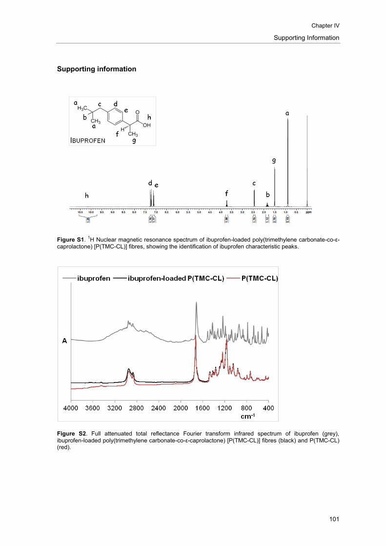
Chapter IV
Supporting Information
101
Supporting information
Figure S1. 1H Nuclear magnetic resonance spectrum of ibuprofen-loaded poly(trimethylene carbonate-co-ε-
caprolactone) [P(TMC-CL)] fibres, showing the identification of ibuprofen characteristic peaks.
Figure S2. Full attenuated total reflectance Fourier transform infrared spectrum of ibuprofen (grey),
ibuprofen-loaded poly(trimethylene carbonate-co-ε-caprolactone) [P(TMC-CL)] fibres (black) and P(TMC-CL) (red).

Ibuprofen-loaded P(TMC-CL) fibres
102
Figure S3. Overlay Overlay of spectra obtained from mapping experiments of ibuprofen-loaded
poly(trimethylene carbonate-co-ε-caprolactone) [P(TMC-CL)] fibres prepared from (A) 1:0 dichloromethane
(DCM)–N,N-dimethylformamide (DMF) and (B) 3:1 DCM–DMF solutions.
Figure S4. Standard calibration curve obtained for ibuprofen.
Figure S5. Fittings according to Higuchi model for drug release for fibres prepared from (A) 1:0
dichloromethane (DCM)–N,N-dimethylformamide (DMF), (B) 6:1 DCM:DMF and (C) 3:1 DCM–DMF.

Chapter IV
Supporting Information
103
Figure S6. Scanning electron microscopy photomicrographs of ibuprofen-loaded poly(trimethylene
carbonate-co-ε-caprolactone) [P(TMC-CL)] fibres prepared from 1:0 dichloromethane (DCM)–N,N-dimethylformamide (DMF) solution (A) before and (B) after ibuprofen release.


CHAPTER V
Ibuprofen-loaded scaffolds for spinal cord injury
regeneration – targeting RhoA at the lesion site
Liliana R Pires1,2, Cátia DF Lopes1,3, Daniela N Rocha1,2, Luigi Ambrosio4, Mónica M
Sousa5, Ana Paula Pêgo1,2,6
1 – INEB – Instituto de Engenharia Biomédica, Universidade do Porto, Rua do Campo Alegre, 823, 4150-180 Porto, Portugal.
2 – Universidade do Porto – Faculdade de Engenharia, Rua Roberto Frias, s/n, 4200-465 Porto, Portugal.
3 – Universidade do Porto – Faculdade de Medicina, Alameda Prof. Hernâni Monteiro, 4200-319 Porto, Portugal
4 – Institute of Composite and Biomedical Materials, National Research Council, P. le Tecchio 80, 80125 Naples, Italy
5 – Nerve Regeneration Group, IBMC - Instituto de Biologia Molecular e Celular, Universidade do Porto, Rua do Campo Alegre, 823, 4150-180 Porto, Portugal.
6 – Universidade do Porto – Instituto de Ciências Biomédicas Abel Salazar, Largo Prof. Abel Salazar, 4099-003 Porto, Portugal.


Chapter V
107
Abstract
It is now well accepted that a therapeutic strategy for spinal cord injury demands a multi-target
approach. Here we propose the use of a poly(trimethylene carbonate-co-ε-caprolactone) [P(TMC-
CL)]-based scaffold that gathers physical guidance cues provided by electrospun aligned fibres
and the delivery of ibuprofen as a mean to reduce the inhibitory environment at the lesion site by
targeting RhoA activation. Bilayer scaffolds were prepared being composed by a solvent cast film
onto which electrospun aligned fibres have been deposited. Both layers were loaded with the
ibupofen. The release of the drug was found to occur in the first 24 hrs of incubation when this
was assessed in vitro under physiological conditions. The bioactivity of the released drug was
demonstrated by the inhibition of RhoA activation when the neuronal ND7/23 cells were
challenged with lysophosphatidic acid. The ibuprofen-loaded bilayer scaffolds were successfully
implanted in vivo in a dorsal hemisection SCI model. The implantation of the scaffold did not
compromise animal survival. The effect of scaffold implantation and ibuprofen release on RhoA
activity, and the histological characterization of the tissues are under investigation.
Keywords
Spinal cord injury; Ibuprofen; electrospinning, RhoA.

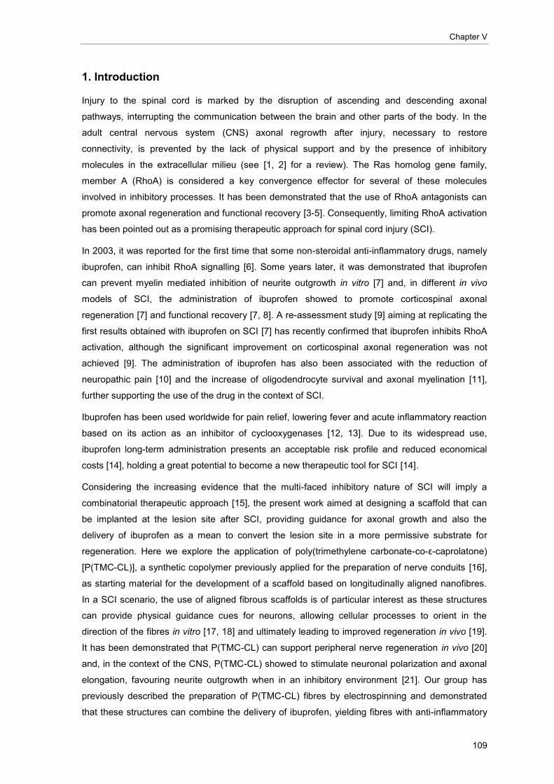
Chapter V
109
1. Introduction
Injury to the spinal cord is marked by the disruption of ascending and descending axonal
pathways, interrupting the communication between the brain and other parts of the body. In the
adult central nervous system (CNS) axonal regrowth after injury, necessary to restore
connectivity, is prevented by the lack of physical support and by the presence of inhibitory
molecules in the extracellular milieu (see [1, 2] for a review). The Ras homolog gene family,
member A (RhoA) is considered a key convergence effector for several of these molecules
involved in inhibitory processes. It has been demonstrated that the use of RhoA antagonists can
promote axonal regeneration and functional recovery [3-5]. Consequently, limiting RhoA activation
has been pointed out as a promising therapeutic approach for spinal cord injury (SCI).
In 2003, it was reported for the first time that some non-steroidal anti-inflammatory drugs, namely
ibuprofen, can inhibit RhoA signalling [6]. Some years later, it was demonstrated that ibuprofen
can prevent myelin mediated inhibition of neurite outgrowth in vitro [7] and, in different in vivo
models of SCI, the administration of ibuprofen showed to promote corticospinal axonal
regeneration [7] and functional recovery [7, 8]. A re-assessment study [9] aiming at replicating the
first results obtained with ibuprofen on SCI [7] has recently confirmed that ibuprofen inhibits RhoA
activation, although the significant improvement on corticospinal axonal regeneration was not
achieved [9]. The administration of ibuprofen has also been associated with the reduction of
neuropathic pain [10] and the increase of oligodendrocyte survival and axonal myelination [11],
further supporting the use of the drug in the context of SCI.
Ibuprofen has been used worldwide for pain relief, lowering fever and acute inflammatory reaction
based on its action as an inhibitor of cyclooxygenases [12, 13]. Due to its widespread use,
ibuprofen long-term administration presents an acceptable risk profile and reduced economical
costs [14], holding a great potential to become a new therapeutic tool for SCI [14].
Considering the increasing evidence that the multi-faced inhibitory nature of SCI will imply a
combinatorial therapeutic approach [15], the present work aimed at designing a scaffold that can
be implanted at the lesion site after SCI, providing guidance for axonal growth and also the
delivery of ibuprofen as a mean to convert the lesion site in a more permissive substrate for
regeneration. Here we explore the application of poly(trimethylene carbonate-co-ε-caprolatone)
[P(TMC-CL)], a synthetic copolymer previously applied for the preparation of nerve conduits [16],
as starting material for the development of a scaffold based on longitudinally aligned nanofibres.
In a SCI scenario, the use of aligned fibrous scaffolds is of particular interest as these structures
can provide physical guidance cues for neurons, allowing cellular processes to orient in the
direction of the fibres in vitro [17, 18] and ultimately leading to improved regeneration in vivo [19].
It has been demonstrated that P(TMC-CL) can support peripheral nerve regeneration in vivo [20]
and, in the context of the CNS, P(TMC-CL) showed to stimulate neuronal polarization and axonal
elongation, favouring neurite outgrowth when in an inhibitory environment [21]. Our group has
previously described the preparation of P(TMC-CL) fibres by electrospinning and demonstrated
that these structures can combine the delivery of ibuprofen, yielding fibres with anti-inflammatory

Ibuprofen-loaded scaffolds – targeting RhoA
110
properties [22]. Here the inhibition of RhoA pathway by ibuprofen-loaded P(TMC-CL) scaffolds
was investigated both in vitro and in vivo.
2. Materials and Methods
2.1. Scaffolds preparation and design
Bilayer scaffolds were prepared using a statistical P(TMC-CL) copolymer. The copolymer was
prepared by ring-opening polymerization and subsequently purified as previously described [23].
In brief, ε-caprolactone (CL) (Merck, Germany) was dried overnight (calcium hydride; Sigma-
Aldrich Química, Portugal) and distilled before the polymerization with trimethylene carbonate
(TMC, used as received from Boehringer Ingelheim, Germany). Polymerization was carried out in
evacuated and sealed glass ampoules using stannous octoate (Sigma-Aldrich Química, Portugal)
as catalyst (2×10⁻⁴
mol per mol of monomer). After 3 days of reaction at 130°C the polymer
obtained was purified by dissolution in chloroform (BDH-Prolabo, Portugal) and subsequent
precipitation into a tenfold volume of ethanol (96%, v/v; AGA, Portugal). The chemical composition
of the purified copolymer was assessed by 1H nuclear magnetic resonance (NMR) and found to
contain 11% mol of TMC, being in accordance to the monomer ratio charged (10% mol TMC). The
average number molecular weight and polydispersity index of the purified polymer were
determined by size exclusion chromatography [22] and were found to be 8.2x10⁴ and 1.61,
respectively.
Bilayer scaffolds were composed by an outer layer comprising a P(TMC-CL) film prepared by
solvent casting, and an inner layer, made of preferentially aligned electrospun P(TMC-CL) fibres
(Figure 5, A). To prepare solvent cast films, a 6% (w/v) P(TMC-CL) solution in dichloromethane
(DCM; Merck, Germany) was casted onto a glass mould. The solvent was left to evaporate
overnight under a DCM saturated atmosphere at room temperature (20-25 ºC) and subsequently,
the films were vacuum dried for 24 hrs (vacuum oven, Raypa, Spain). For the preparation of
ibuprofen-loaded scaffolds, 5% of ibuprofen (w/w of polymer) was added to the polymer solution
and slowly stirred for 5 hrs, before casting. Pharmaceutical grade ibuprofen (purity>99%) was
kindly supplied by Sérgio Simões (Bluepharma, Portugal).
Based on previous findings from our group [22], P(TMC-CL) electrospun fibres were prepared as
follows. P(TMC-CL) was dissolved overnight in a mixture of DCM and N,N-dimethylformamide
(DMF, Merck) at a volume ratio of 3:1. When preparing ibuprofen-loaded fibres, the drug (5% (w/w
of polymer)) was added to the polymer solution and stirred for 5 hrs before electrospinning. The
prepared solutions were dispensed at a controlled flow rate of 1 ml.h-1
using a syringe pump (Ugo
Basille, Italy). The blunt needle serving as spinneret (inner diameter 0.8 mm) was placed at 13 cm
from the collector and an 18 kV electric field was applied (Gamma High Voltage source; FL, USA).
The fibres were collected onto P(TMC-CL) solvent cast films covering a cylindrical drum (10 cm
diameter, 15 cm of length, Yflow, Spain) rotating at 3000 rpm. The fibres were collected during 2.5
hrs and subsequently the scaffolds were vacuum dried for 24 hrs. 6x5 mm bilayer scaffolds were

Chapter V
111
punched out and packed under vacuum after an argon purge. Samples were sterilized by gamma
irradiation (25 kGy, 60
Co source) prior further use.
2.2. Scaffolds characterization
The scaffolds were observed by scanning electron microscopy (SEM) using a FEI Quanta
400FEG microscope (FEI, the Netherlands) after being sputter-coated with gold-palladium for 90
seconds (SPI Supplies, PA, USA). Fibre diameter and alignment were quantified from SEM
micrographs using Image analysis software (Image J, version 1.39, NIH, MD, USA). The fibre
mean diameter was calculated from, at least, 100 measurements from 3 independent samples. To
calculate fibre alignment, the preferential direction of the alignment was defined for each image
and subsequently the angle formed between the fibres and this axis was measured. Results are
presented as percentage relative to the total number of fibres measured from 3 independent
samples.
2.3. Drug release quantification
The amount of ibuprofen released from P(TMC-CL) scaffolds was evaluated as follows. Samples
were incubated at 37ºC and 120 rpm (Orbital Shaker Oven, IKA, Germany) in phosphate buffered
saline (PBS) at the final concentration of 5 mg.ml-1
(mass of fibres/volume of PBS). At defined time
points (0.5, 1, 2, 4, 6, 8, 24 and 48 hrs), the releasing medium was refreshed. The amount of
ibuprofen released was quantified by high-pressure liquid chromatography (HPLC). In brief, the
equipment used was an HPLC system (LaChrom Elite®, Hitachi Portugal) equipped with a variable
wavelength diode array detector (L-2455). Separation was performed using a Lichrosorb RP-18
column (5 μm, 12.5 x 0.4 cm, Merck) and a mobile phase consisting on a mixture of acetonitrile
and ortho-phosphoric acid 0.015 M (45:55, (v/v), all supplied by Merck). Flow rate was set to 2.5
ml.h-1
and separation occurred at 40ºC, whereas detection was performed at 25ºC. Ibuprofen was
detected at 230 nm after 10 μl sample injection. Cumulative release was calculated relative to the
theoretical maximum loading of 5% (w/w of polymer).
2.4. Cell Culture
ND7/23 cell line (mouse neuroblastoma (N18 tg 2) x rat dorsal root ganglion neuron hybrid) was
obtained from ECACC (United Kingdom) and routinely cultured in Dulbecco’s Modified Eagle
Medium (DMEM) with Glutamax, supplemented with 10% (v/v) of heat inactivated (56ºC, 30
minutes) foetal bovine serum (FBS) and 1% penicillin/streptomycin (PS, 10,000 units.ml-1
penicillin
and 10,000 g.ml-1
streptomycin), all supplied by Gibco (Life technologies S.A., Spain).
To test the effect of ibuprofen on these cells, ND7/23 cells were sub-cultured using supplemented
DMEM at a cellular density of 2x10⁴ viable cells.cm
-2. To promote cell differentiation, 24 hrs after
seeding the cell culture medium was replaced by differentiation medium containing 0.5% FBS, 1%

Ibuprofen-loaded scaffolds – targeting RhoA
112
PS, 200 nM nerve growth factor (NGF, Calbiochem, Merck), and 1 mM cyclic adenosine
monophosphate (cAMP, Sigma-Aldrich). Cells were allowed to differentiate for 2 days before
further treatment.
2.5. Effect of ibuprofen on ND7/23 cell metabolic activity
The effect of ibuprofen on ND7/23 cell metabolic activity was assessed by means of a resazurin-
based assay. In brief, different ibuprofen solutions in ethanol:water (7:3) were prepared and added
(5 μl) to the cell culture medium (500 μl) in order to obtain a final drug concentration ranging from
50 to 2000 μM. After a 24 hrs treatment, cells were incubated (4 hrs, 37 °C) with a resazurin
(Sigma-Aldrich) solution (0.1 mg.ml-1, in PBS). Fluorescence (λex= 530 nm, λem= 590 nm) in the
cell culture medium was measured (SynergyMx, Biotek, Portugal). Results are represented as
percentage of cell metabolic activity relative to cells treated with equal volume of the ibuprofen
solvent (5 μl).
2.6. In vitro inhibition of RhoA activation by ibuprofen
To investigate whether ibuprofen released from P(TMC-CL) scaffolds could influence RhoA
activation in ND7/23 cells, extracts from the ibuprofen-loaded scaffolds (Ibuprofen scaffold) were
firstly prepared as follows. Scaffolds were incubated in DMEM at 37ºC (80 rpm) during 72 hrs. The
volume of DMEM used was adjusted according to the weight of scaffolds in order to achieve, after
the total release of ibuprofen, a theoretical drug concentration of 500 µM. The concentration was
selected based on a previous report showing ibuprofen-mediated inhibition of RhoA pathway in
the PC12 cell line [7]. The obtained extracts were then tested on differentiated ND7/23 cells. In
brief, after a starving period of 2 hrs in DMEM without serum, cell culture medium was replaced by
the ibuprofen-containing extracts obtained from the P(TMC-CL) scaffolds. Ibuprofen solution (100x
in ethanol:water (7:3)) was added to the cell culture media to a final concentration of 500 µM to
serve as control (Ibuprofen soluble). After 30 minutes, the cells were activated with 45 µM of
lysophosphatidic acid (LPA, Sigma-Aldrich) for 10 minutes. Non-treated cells were applied as
negative control, and cells activated with LPA served as positive control for RhoA activation.
RhoA activation was assessed using Rhotekin RBD-coupled beads (Millipore, MA, USA)
according to manufacturer instructions. Cell lysates were prepared using Mg2+
lysis buffer (MLB)
containing 25 mM HEPES, 1% Tergitol (v/v) and 10 mM MgCl2 (Sigma-Aldrich), and 150 mM
NaCl, 1% (w/v) EDTA and 10% (v/v) glycerol from BDH-Prolabo (Portugal) and a protease
inhibitors cocktail (Sigma-Aldrich). Lysates were clarified by centrifugation (20,000xg, 4ºC, 5
minutes) and snap frozen prior further use. Total protein content was quantified using DC protein
assay (Bio-Rad, Portugal). Lysates containing 1 mg of total protein were incubated with RBD-
coupled beads during 1 hr at 4ºC under slow rotation. After two washing steps, the beads were
resuspended in 2x laemmli buffer (Bio-Rad) containing β-mercaptoethanol, boiled at 95ºC (10
minutes) and filtered using Mobicol filters (35 µm, Mobitec, Germany). Bound GTP-RhoA along

Chapter V
113
with 25 µg of total lysate were separated in a 12% (w/v) SDS-polyacrilamide gel and detected by
western blot. Briefly, gels were transferred to Hybond nitrocellulose ECL membrane (Amersham,
GE Healthcare Europe GmbH, Portugal) and blocked in a 5% skim milk in PBS containing 0.1%
(v/v) Tween-20 (PBS-T). The membranes were incubated overnight with a mouse anti-RhoA
antibody (1:500, sc-418; Santa Cruz Biotechnology, CA, USA) followed by a 1 hr incubation with
horseradish peroxidase-labelled sheep anti-mouse IgG (1:3,000, Amersham). After extensive
washing the immunoreactive bands were detected using WesternBright Quantum
Chemiluminescent Substrate (Advansta, CA, USA) and visualized by autoradiography on an
Amersham Hyperfilm ECL (GE Healthcare Europe). The relative intensity of protein bands was
analyzed using Quantity One (version 4.6) program (Bio-Rad). Results are expressed as the ratio
between the activated protein form of RhoA (RhoA-GTP) and the total protein (RhoA).
2.7. Preliminary in vivo testing – dorsal hemisection model
All experiments involving animals and their care were conducted in compliance with institutional
ethical guidelines and with the approval of Portuguese Veterinary Authorities – Direcção Geral de
Veterinária (DGAV). Female adult Wistar rats (10 - 13 weeks) were anesthetized with
ketamine/medetomidine (100 mg.kg-1
ketamine; 1 mg.kg-1
medetomidine). Laminectomies were
performed at the 8 and 9 vertebrae and the spinal dura mater was incised to expose the spinal
cord. Dorsal hemisection at the T9 level was performed using a micro-scissor. The scaffolds (6x5
mm) were implanted underneath the dura, immediately after the lesion was carried out. The
fibrous side of the scaffolds was placed in contact with the spinal cord, ensuring that fibres were
longitudinally aligned with the axonal pathways. The surgery site was closed in layers using
absorbable suture (Surgicryl, Smi, Belgium). Atipamezole (1 mg.kg-1
) was administrated to
counteract anaesthesia. The rats were kept in heating pads 24 hrs after surgery and abdominal
massage was performed twice a day to help bladder evacuation. Butorphanol (1 mg.kg-1
) was
administrated twice a day during the first two days after surgery to manage pain.
A total of twenty animals undergone surgery and were distributed in three experimental groups as
follows: (A) non-treated (n=6); (B) P(TMC-CL) scaffold (n=7); (C) ibuprofen-loaded P(TMC-CL)
scaffold (n=7). All the animals were subjected to dorsal hemisection as described above being that
animals from group A were subjected to no further treatment, and animals from group B and C
were implanted with P(TMC-CL) scaffolds, plain or loaded with ibuprofen, respectively. Five days
after implantation, the animals were euthanized. The scaffold was removed and 1 cm of the spinal
cord tissue centred at the lesion site was collected and divided sagitally. Part of the tissue was
snap frozen in dry ice and stored at -80 ºC for posterior RhoA quantification. The second portion of
the spinal cord, was fixed using 4% (w/v) paraformaldehyde at 4ºC (overnight). The samples were
then transferred to a 30% (w/v) sucrose solution containing 0.1% (w/v) sodium azide for
cryoprotection and stored at -20ºC before cryostat sectioning.

Ibuprofen-loaded scaffolds – targeting RhoA
114
2.7.1. Histology and Immunohistochemistry
Spinal cord tissue was embedded on optimum cutting medium (OCT, Thermo Scientific, Thermo
Fisher Scientific, Portugal) and cut onto 16 µm thick sections using a cryostat (Thermo Scientific).
Sections were mounted onto glass coverslips (Superfrost, VWR) and stored at -20ºC till further
use.
Sections were stained using haematoxilin and eosin according to standard procedures.
For immunohistochemistry, cryostat sections were firstly incubated with 0.1% (w/v) sodium
borohydride (5 minutes) and ammonium chloride (50 mM, 15 minutes). Afterwards, sections were
incubated 1 hr at 37ºC with blocking buffer containing 5% (v/v) normal donkey serum (Sigma-
Aldrich) and 0.3% (v/v) Triton X-100 in PBS. The primary antibody for glial fibrillary acidic protein
(GFAP, rabbit anti GFAP, Dako) was diluted in the blocking buffer (1:500) and incubated with the
cryosections overnight at 4ºC. Subsequently, sections were incubated 1 hr at room temperature
with Alexa 488-conjugated anti-rabbit secondary antibody (Invitrogen, 1:1,000). Afterwards,
Hoechst solution (Invitrogen, 1:30,000) was applied (15 minutes at room temperature) in order to
label cell nuclei. Stained sections were coverslipped using Fluoromount (Sigma-Aldrich) and kept
at -20ºC until analysis. The immunolabelled tissue sections were analyzed using an inverted
fluorescence microscope (Axiovert 200, Zeiss, Germany) .
2.8. Statistical analysis
Statistical analysis was performed using the Graphpad Prism 5.0 software (GraphPad, CA, USA).
Statistical differences were calculated using one-way ANOVA followed by the Dunnett’s test for
multiple comparisons. A p value lower than 0.05 was considered statistically significant.
3. Results
3.1. Scaffold morphology
By combining solvent casting and electrospinning a bilayer scaffold was prepared. The obtained
scaffolds were analysed by SEM and representative images are presented in Figure 1. The
surface of the solvent cast flat outer layer reveals a spherulitic morphology, typical of a
semicrystalline material [24] (Figure 1, A, C; E, G). The inner layer composed by electrospun
fibres shows a preferentially longitudinally aligned fibrous structure (Figure 1, B, D, F, H). The
presence of ibuprofen alters jet stability as denoted by the formation of bended fibres (Figure 1, F
and H). Nonetheless, on the whole, the fibre mean diameter and alignment were not significantly
altered as one can observe by comparing non-loaded and ibuprofen-loaded fibres. Fibre mean
diameter was found to be 0.661 and 0.646 μm for non-loaded and ibuprofen-loaded fibres,
respectively (Figure 1, K).

Chapter V
115
Figure 1. Characterization of the P(TMC-CL) scaffolds under investigation. Scanning electron microscopy
photomicrographs of (A-D) non-loaded and (E-H) ibuprofen-loaded scaffolds. Images A, C, E and G show the
outer layer of the scaffold composed by solvent cast films; images B, D, F and H shows the inner layer made
of preferentially aligned fibres prepared by electrospinning. (I and J) Graphic representation of the
quantification of fibre alignment for (I) non-loaded and (J) ibuprofen-loaded fibres. Bars represent average
and error bars show standard deviation (n=3). (K) Fibre mean diameter as calculated from at least 100
measurements (n=3).
3.2. Drug release from P(TMC-CL) scaffolds
The release of ibuprofen from P(TMC-CL) bilayer scaffolds was evaluated when in PBS at 37ºC.
The percentage of cumulative release was calculated relative to a theoretical maximum loading of
5% (w/w of polymer).
Under the experimental conditions tested, ibuprofen was released from the P(TMC-CL) scaffolds
within the first 24 hrs of incubation in PBS (37°C) (Figure 2). When fibres were incubated in PBS
for longer period, none or residual amounts of ibuprofen were detected, being that the limit of
quantitation of the applied experimental setup was determined to be 0.001 mg.ml-1
. The scaffolds
were analysed by SEM after drug release and the fibrous morphology was maintained (data not
shown).
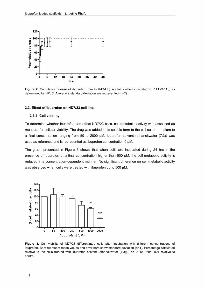
Ibuprofen-loaded scaffolds – targeting RhoA
116
Figure 2. Cumulative release of ibuprofen from P(TMC-CL) scaffolds when incubated in PBS (37°C), as
determined by HPLC. Average ± standard deviation are represented (n=7).
3.3. Effect of ibuprofen on ND7/23 cell line
3.3.1. Cell viability
To determine whether ibuprofen can affect ND7/23 cells, cell metabolic activity was assessed as
measure for cellular viability. The drug was added in its soluble form to the cell culture medium to
a final concentration ranging from 50 to 2000 μM. Ibuprofen solvent (ethanol:water (7:3)) was
used as reference and is represented as ibuprofen concentration 0 μM.
The graph presented in Figure 3 shows that when cells are incubated during 24 hrs in the
presence of ibuprofen at a final concentration higher than 500 μM, the cell metabolic activity is
reduced in a concentration-dependent manner. No significant difference on cell metabolic activity
was observed when cells were treated with ibuprofen up to 500 μM.
Figure 3. Cell viability of ND7/23 differentiated cells after incubation with different concentrations of
ibuprofen. Bars represent mean values and error bars show standard deviation (n=4). Percentage calculated
relative to the cells treated with ibuprofen solvent (ethanol:water (7:3)). *p< 0.05; ***p<0.001 relative to
control.
0 6 12 18 24 30 36 42 480
20
40
60
80
100
120
hrs
%c
um
ula
tiv
e r
ele
as
e

Chapter V
117
3.3.2. RhoA activation
Pre-treatment of cells with ibuprofen has previously shown to limit RhoA-induced activation,
namely by LPA [7]. To investigate if ibuprofen released from P(TMC-CL) bilayer scaffolds retains
its bioactivity and can have an effect on the RhoA pathway, RhoA activation was assessed in
treated ND7/23 cells after stimulation with LPA. The cells were pre-treated with extracts from
P(TMC-CL) ibuprofen-loaded scaffolds, or with medium supplemented with ibuprofen solution.
Non-activated cells (no LPA stimulation) and cells activated with LPA, but non-treated with
ibuprofen were used as negative and positive control, respectively.
Figure 4. RhoA activation on ND7/23 cells as determined by western blot after precipitation of the active form
of RhoA (RhoA-GTP). Stimulation with LPA (45 µM, 10 minutes, 2-LPA) and pre-treatment with ibuprofen (3-
Ibuprofen soluble) or ibuprofen released from P(TMC-CL) scaffolds (4-Ibuprofen scaffold) were tested. The
RhoA basal activation was also assessed (lane 1).
The activated form of RhoA was not detected in non-stimulated ND7/23 cells. Treatment with LPA
leads to an increase on RhoA activation as demonstrated by the detected high levels of RhoA-
GTP (Figure 4, lane 2). Similarly to the effect observed when cells were incubated with soluble
ibuprofen, cells treated with extracts of the ibuprofen-loaded P(TMC-CL) scaffolds showed a
significant reduction (approximately 5 times) of the LPA-mediated RhoA activation (Figure 4).
3.4. Preliminary in vivo testing of ibuprofen-loaded P(TMC-CL) scaffolds
The developed P(TMC-CL) bilayer scaffolds were tested in an in vivo situation by implantation in
contact with a spinal cord immediately after dorsal hemisection (Figure 5).
As shown in Figure 5, the P(TMC-CL) scaffolds were placed at the lesion site wrapping the spinal
cord tissue. We found that the scaffolds bend properly to allow fixation in the bone and close
contact with the spinal cord tissue. Five days after implantation, the retrieved scaffolds were found
to be covered by fibrous tissue, and no signs of scaffold degradation were observed (Figure 5, C).

Ibuprofen-loaded scaffolds – targeting RhoA
118
Figure 5. Photographs of the aspect of the bilayer P(TMC-CL) scaffold (A) prior implantation (gamma
sterilized); (B) after implantation in an injured (dorsal hemisection) spinal cord; and (C) as collected after
animal euthanasia (5 days post implantation). Arrows indicate the P(TMC-CL) scaffold.
From the twenty animals used in this experiment, one, from group B, was excluded due to poor
health at the day 5 of the experiment. This unique event was considered to be independent from
the treatment, as animals with SCI are generally more susceptible to infections [25]. The
implantation of ibuprofen-loaded P(TMC-CL) scaffolds did not compromise animal survival after
SCI.

Chapter V
119
Figure 6. Light microscopy images of the haematoxylin-eosin staining (A-C) and fluorescence microscopy
images of GFAP (green) and DAPI (blue) immunolabelling (D-F) of cryosections from the spinal cord of (A
and D) control animal dorsal hemisection and animals with implanted (B and E) P(TMC-CL) scaffold or (C
and F) ibuprofen-loaded scaffold. Tissue was collected 5 days after implantation. The images show the
region of the lesion (delimited in white).
The histological evaluation of the tissues collected is ongoing. Figure 6 shows the preliminary
analysis of one tissue from each experimental group. Figure 6 (A, B, C) shows the haemotoxilin-
eosin staining of the area of the lesion in tissues from group A (non-treated), group B (P(TMC-CL)
scaffold) and group C (ibuprofen-loaded P(TMC-CL) scaffold), respectively. In the case of group

Ibuprofen-loaded scaffolds – targeting RhoA
120
B, the region of the lesion is more difficult to demarcate, as it seems more spread than in the other
tissues, where it is clearly identified. It is worthwhile mentioning that in the animals where P(TMC-
CL) scaffolds were implanted, the scaffold was separated from the spinal cord prior
cryopreservation of the tissue. This process seems to have affected tissue cohesion and cause
the appearance of additional debris in the stained sections (see Figure 6, B). Comparing the
tissue from group A and C, the images from haemotoxilin-eosin staining suggest that the
extension of the tissue damage is different among groups. When analysing the cryosections
immunolabelleded for GFAP, it can be observed that on group B and C (Figure 6, E and F) a
higher number of cells is found in the lesion site, suggesting significant cellular infiltration.
Nevertheless, the number of GFAP positive cells in that region was found to be residual. This
observation is not replicated in group A (Figure 6, D), where signs of astrogliosis (GFAP positive
cells) are detected at the time of the evaluation.
4. Discussion
It is commonly accepted that a multi-target strategy will be required to tackle the challenge of
promoting nerve regeneration after SCI [15]. Following the primary injury that interrupts axonal
pathways, the rewiring of the system is limited due to the lack of physical support, the activation of
inhibitory pathways, and, ultimately, due to the formation of a glial scar that physically halts axonal
re-connection. In the present work, we describe the preparation of a bilayer scaffold that can
provide physical support, guidance cues and can serve as vehicle for the in situ delivery of
ibuprofen, a drug expected to be able to reduce the inhibitory environment at the lesion site.
The use of aligned electrospun fibres to guide axonal growth has been previously explored in the
literature both for the peripheral and the central nervous system regeneration (see [26] for a
review). In the first report using aligned electrospun fibres in a SCI scenario, the authors claimed a
robust rostral regeneration, four weeks after implantation of poly(L-lactide) longitudinally aligned
fibres. The success of the strategy was ascribed to the anisotropy of the substrates as modest
regeneration was observed when randomly oriented fibres or solvent cast films were implanted
[19]. Here, we report the successful preparation of P(TMC-CL) preferentially aligned fibres. Fibre
mean diameter was found to be around 650 nm, in accordance with that previously described for
randomly oriented structures using the same solvent mixture [22]. The aligned fibres were
collected onto solvent cast films in order to improve the final mechanical properties of the scaffold.
Nanofibrous scaffolds have, in general, weaker mechanical properties than bulky structures [27],
particularly when constituted by aligned fibres [28]. This can compromise the success of an
implantable device, as demonstrated in a previous study showing that the folding of an
electrospun fibrous fabric impaired nerve regeneration [29].
The vectorization through scaffolds of molecules that have a positive effect on regeneration holds
a great potential to become a therapeutic strategy for SCI. It was previously shown that the
implantation after lesion of a nanofibrous patch with rolipram physically adsorbed to poly(L-

Chapter V
121
lactide)-based electrospun fibres lead to an improved functional recovery from the third week on,
comparing to non-loaded scaffolds that showed a similar result as non-treated animals [28]. In the
present study, it is proposed the use of ibuprofen incorporated in a P(TMC-CL) scaffold, based on
the reported inhibitory action of the drug on the RhoA pathway [7, 8]. Bilayer ibuprofen-loaded
scaffolds were prepared by mixing the drug with the polymer solution prior to electrospinning and
solvent casting, for the preparation of the inner and outer scaffold layer, respectively. It was
observed that the drug released from the scaffolds is bioactive as confirmed by the hindrance of
RhoA activation in a neuronal-like cell population challenged with LPA – a known RhoA activator.
This result encouraged the testing of P(TMC-CL) bilayer scaffolds in an in vivo SCI model.
The ibuprofen release profile from the P(TMC-CL) bilayer scaffolds conducted in sink conditions
indicated that the majority of the drug is released within the first 24 hrs of incubation. When
translating this data to an in vivo scenario, one can expect that this release will be delayed as in
the conducted studies the media was completely refreshed at each evaluation time point, what
favours the washing out of the drug from the scaffold. Moreover, we hypothesized that a
significant release of ibuprofen in the initial stages of the tissue response after a lesion can also
play a role on the early inflammatory response triggered, namely reducing microglia activation [10]
and that this can positively contribute to regeneration in the aftermath of a SCI [30].
The results reported in this manuscript concerning the in vivo performance of the developed
P(TMC-CL) scaffolds are still preliminary. This experiment aimed at constituting a proof-of-concept
from the feasibility of using ibuprofen-loaded bilayer scaffolds for inhibiting RhoA activation. In that
view, the study was designed to address the early response on the RhoA pathway and not
attempt to assess axonal regeneration or achieve functional improvements. So far, we
demonstrated that the implantation of P(TMC-CL) scaffolds can be successfully achieved and
does not compromised animal survival rate. Although we were not expecting any deleterious
effect caused by the implantation of P(TMC-CL) scaffolds, some concern existed about the drug
loading applied. The published studies using ibuprofen for treatment of SCI report the
subcutaneous administration of the drug at the dose of 60 mg.kg-1
.day-1
[7, 8]. In the present study
the drug loading in the P(TMC-CL) scaffolds was in average 500 µg.cm-2
, corresponding to
approximately 0.75 mg.kg-1
. However, it must be noted that in the present study, the release of the
drug occurs in situ, as the scaffold is in direct contact with the spinal cord tissue. A previous work
using rolipram loaded patches implanted in contact with the spinal cord, showed that implantation
of high drug doses (65 µg.cm-2
) lead to an increase on animal mortality rate [31]. It should be
mentioned however, that the effective dose of ibuprofen and rolipram are significantly different,
and improvements on regeneration after SCI by the administration of rolipram are achieved by the
subcutaneous administration of 1 mg.kg-1
of drug [32], 60 times less than that reported for
ibuprofen [7, 8].
The preliminary histological characterization of the tissues collected from animals treated with
ibuprofen-loaded P(TMC-CL) bilayer scaffolds showed that at the time of evaluation (5 days after
the lesion) considerable cellular infiltration occurred at the lesion site, but no signs of astrogliosis

Ibuprofen-loaded scaffolds – targeting RhoA
122
were identified so far. In comparison, the untreated animals showed already signs of the presence
of a glial scar at the lesion site, as indicated by the presence of GFAP positive cells. The ongoing
characterization of the infiltrated cells will shed light on the effect of the released ibuprofen on the
modulation of the lesion microenvironment. Ultimately, the quantification of RhoA activation in
tissues in contact with ibuprofen-loaded scaffolds will contribute for disclosing the potential of the
proposed strategy in providing a more permissive milieu for axonal regeneration.
5. Conclusion and Future Perspectives
This study describes the successful preparation of bilayer P(TMC-CL) scaffolds containing
longitudinally aligned P(TMC-CL) fibres, and loaded with ibuprofen. It is demonstrated that
ibuprofen released from P(TMC-CL) scaffolds can effectively reduce RhoA activation in ND7/23
cells putting forward these scaffolds to be applied in a SCI scenario.
A preliminary in vivo experiment was performed and showed, so far, that the scaffolds can be
implanted in the spinal cord after injury. No effect on animal survival was observed. The detailed
histological characterization of the retrieved tissues is ongoing and will provide critical information
on the success of the proposed strategy, bringing also new insights to future improvements on
scaffold design. Drug loading, or the drug release profile are parameters that can be modulated in
order to develop a scaffold that better supports cells in the hostile environment of SCI.
To assess the effect of the strategy proposed in this study on axonal growth or functional recovery
after SCI, it is necessary to perform an extended experiment in which the scaffolds are implanted
for a longer period. This study is key to evaluate the effect of the early inhibition of RhoA pathway
on regeneration and also the contribution of P(TMC-CL) and fibre alignment in the process.
Acknowledgements
This work was financed by FEDER funds through the Programa Operacional Factores de
Competitividade – COMPETE and by Portuguese funds through FCT – Fundação para a Ciência
e a Tecnologia in the framework of the project PEst-C/SAU/LA0002/2011 and PTDC/CTM-
NAN/115124/2009. LR Pires and DN Rocha thank FCT for their PhD grants (SFRH / BD / 46015 /
2008 and SFRH / BD / 64079 / 2009). The authors wish to thank Ana Marques and Marlene
Morgado for the technical assistance. Authors acknowledge the Centro de Materiais da
Universidade do Porto (CEMUP; REEQ/1062/CTM/2005 from FCT) for SEM and 1H NMR analysis
and to Sérgio Simões for the kind help making available the use of HPLC equipment at
Bluepharma (Coimbra).

Chapter V
123
References
1. Schwab JM, Brechtel K, Mueller CA, Failli V, Kaps HP, Tuli SK, and Schluesener HJ (2006). "Experimental strategies to promote spinal cord regeneration - An integrative perspective". Progress in Neurobiology, 78 (2): 91-116.
2. Silver J and Miller JH (2004). "Regeneration beyond the glial scar". Nature Reviews Neuroscience, 5 (2): 146-156.
3. Dergham P, Ellezam B, Essagian C, Avedissian H, Lubell WD, and McKerracher L (2002). "Rho signaling pathway targeted to promote spinal cord repair". Journal of Neuroscience, 22 (15): 6570-6577.
4. McKerracher L and Higuchi H (2006). "Targeting Rho to stimulate repair after spinal cord injury". Journal of Neurotrauma, 23 (3-4): 309-317.
5. Monnier PP, Sierra A, Schwab JM, Henke-Fahle S, and Mueller BK (2003). "The Rho/ROCK pathway mediates neurite growth-inhibitory activity associated with the chondroitin sulfate proteoglycans of the CNS glial scar". Molecular and Cellular Neuroscience, 22 (3): 319-330.
6. Zhou Y, Su Y, Li BL, Liu F, Ryder JW, Wu X, Gonzalez-DeWhitt PA, Gelfanova V, Hale JE, May PC, Paul SM, and Ni BH (2003). "Nonsteroidal anti-inflammatory drugs can lower amyloidogenic A beta(42) by inhibiting Rho". Science, 302 (5648): 1215-1217.
7. Fu Q, Hue J, and Li S (2007). "Nonsteroidal anti-inflammatory drugs promote axon regeneration via RhoA inhibition". Journal of Neuroscience, 27 (15): 4154-4164.
8. Wang X, Budel S, Baughman K, Gould G, Song KH, and Strittmatter SM (2009). "Ibuprofen enhances recovery from spinal cord injury by limiting tissue loss and stimulating axonal growth". Journal of Neurotrauma, 26 (1): 81-95.
9. Sharp KG, Yee KM, Stiles TL, Aguilar RM, and Steward O (2013). "A re-assessment of the effects of treatment with a non-steroidal anti-inflammatory (ibuprofen) on promoting axon regeneration via RhoA inhibition after spinal cord injury". Experimental Neurology, 248: 321-327.
10. Redondo-Castro E and Navarro X (2014). "Chronic ibuprofen administration reduces neuropathic pain but does not exert neuroprotection after spinal cord injury in adult rats". Experimental Neurology, 252: 95-103.
11. Xing B, Li H, Wang H, Mukhopadhyay D, Fisher D, Gilpin CJ, and Li S (2011). "RhoA-inhibiting NSAIDs promote axonal myelination after spinal cord injury". Experimental Neurology, 231 (2): 247-260.
12. Mitchell JA, Akarasereenont P, Thiemermann C, Flower RJ, and Vane JR (1993). "Selectivity of nonsteroidal antiinflammatory drugs as inhibitors of constitutive and inducible cyclooxygenase". Proceedings of the National Academy of Sciences of the United States of America, 90 (24): 11693-11697.
13. Rainsford KD (2009). "Ibuprofen: pharmacology, efficacy and safety". Inflammopharmacology, 17 (6): 275-342.
14. Kopp MA, Liebscher T, Niedeggen A, Laufer S, Brommer B, Jungehulsing GJ, Strittmatter SM, Dirnagl U, and Schwab JM (2012). "Small-molecule-induced Rho-inhibition: NSAIDs after spinal cord injury". Cell and Tissue Research, 349 (1): 119-132.
15. McCreedy DA and Sakiyama-Elbert SE (2012). "Combination therapies in the CNS: Engineering the environment". Neuroscience Letters, 519 (2): 115-121.
16. Pêgo AP, Poot AA, Grijpma DW, and Feijen J (2003). "Biodegradable elastomeric scaffolds for soft tissue engineering". Journal of Controlled Release, 87 (1-3): 69-79.
17. Corey JM, Lin DY, Mycek KB, Chen Q, Samuel S, Feldman EL, and Martin DC (2007). "Aligned electrospun nanofibers specify the direction of dorsal root ganglia neurite growth". Journal of Biomedical Materials Research - Part A, 83 (3): 636-645.
18. Yao L, O'Brien N, Windebank A, and Pandit A (2009). "Orienting neurite growth in electrospun fibrous neural conduits". Journal of Biomedical Materials Research - Part B Applied Biomaterials, 90 (2): 483-491.

Ibuprofen-loaded scaffolds – targeting RhoA
124
19. Hurtado A, Cregg JM, Wang HB, Wendell DF, Oudega M, Gilbert RJ, and McDonald JW (2011). "Robust CNS regeneration after complete spinal cord transection using aligned poly-l-lactic acid microfibers". Biomaterials, 32 (26): 6068-6079.
20. Vleggeert-Lankamp CLAM, Wolfs J, Pêgo AP, Van Den Berg R, Feirabend H, and Lakke E (2008). "Effect of nerve graft porosity on the refractory period of regenerating nerve fibers: Laboratory investigation". Journal of Neurosurgery, 109 (2): 294-305.
21. Rocha DN, Brites P, Fonseca C, and Pêgo AP (2014). "Poly(Trimethylene Carbonate-co-ε-Caprolactone) Promotes Axonal Growth". Plos One, 9(2): e88593.
22. Pires LR, Guarino V, Oliveira MJ, Ribeiro CC, Barbosa MA, Ambrosio L, and Pêgo AP (2013). "Ibuprofen-loaded poly(trimethylene carbonate-co-ε-caprolactone) electrospun fibers for nerve regeneration". Journal of Tissue Engineering and Regenerative Medicine: Accepted for publication.
23. Pêgo AP, Poot AA, Grijpma DW, and Feijen J (2001). "Copolymers of trimethylene carbonate and epsilon-caprolactone for porous nerve guides: Synthesis and properties". Journal of Biomaterials Science, Polymer Edition, 12 (1): 35-53.
24. Pêgo AP, Vleggeert-Lankamp CLAM, Deenen M, Lakke EAJF, Grijpma DW, Poot AA, Marani E, and Feijen J (2003). "Adhesion and growth of human schwann cells on trimethylene carbonate (co)polymers". Journal of Biomedical Materials Research - Part A, 67 (3): 876-885.
25. Balsara ZR, Ross SS, Dolber PC, Wiener JS, Tang Y, and Seed PC (2013). "Enhanced
susceptibility to urinary tract infection in the spinal cord-injured host with neurogenic bladder". Infection and Immunity, 81 (8): 3018-3026.
26. Xie J, MacEwan MR, Schwartz AG, and Xia Y (2010). "Electrospun nanofibers for neural tissue engineering". Nanoscale, 2 (1): 35-44.
27. Teo WE and Ramakrishna S (2006). "A review on electrospinning design and nanofibre assemblies". Nanotechnology, 17 (14): R89-R106.
28. Zhu Y, Wang A, Shen W, Patel S, Zhang R, Young WL, and Li S (2010). "Nanofibrous patches for spinal cord regeneration". Advanced Functional Materials, 20 (9): 1433-1440.
29. Meiners S, Ahmed I, Ponery AS, Amor N, Harris SL, Ayres V, Fan Y, Chen Q, Delgado-Rivera R, and Babu AN (2007). "Engineering electrospun nanofibrillar surfaces for spinal cord repair: A discussion". Polymer International, 56 (11): 1340-1348.
30. Casha S, Zygun D, McGowan MD, Bains I, Yong VW, and Hurlbert RJ (2012). "Results of a phase II placebo-controlled randomized trial of minocycline in acute spinal cord injury". Brain, 135 (4): 1224-1236.
31. Downing TL, Wang A, Yan ZQ, Nout Y, Lee AL, Beattie MS, Bresnahan JC, Farmer DL, and Li S (2012). "Drug-eluting microfibrous patches for the local delivery of rolipram in spinal cord repair". Journal of Controlled Release, 161 (3): 910-917.
32. Schaal SM, Garg MS, Ghosh M, Lovera L, Lopez M, Patel M, Louro J, Patel S, Tuesta L, Chan WM, and da Pearse D (2012). "The Therapeutic Profile of Rolipram, PDE Target and Mechanism of Action as a Neuroprotectant following Spinal Cord Injury". Plos One, 7 (9): e43634.

CHAPTER VI
Imidazole-grafted chitosan mediated gene delivery:
in vitro study on transfection, intracellular
trafficking, and degradation*
Liliana R Pires1, 2, Hugo Oliveira1, Cristina C Barrias1, Paula Sampaio3, António J
Pereira3, Hélder Maiato3, Sérgio Simões4,5, Ana Paula Pêgo1
1 – INEB – Instituto de Engenharia Biomédica, NEWTherapies Group, Universidade do Porto, Rua do Campo Alegre 823, 4150-180 Porto, Portugal.
2 – Universidade do Porto – Faculdade de Engenharia, Rua Roberto Frias, s/n, 4200-465 Porto, Portugal.
3 – Instituto de Biologia Molecular e Celular (IBMC), Rua do Campo Alegre, 823, 4150-180 Porto, Portugal.
4 – Centro de Neurociências e Biologia Celular, Universidade de Coimbra, 3004-517 Coimbra, Portugal
5 – Departamento de Tecnologia Farmacêutica, Faculdade de Farmácia, Universidade de Coimbra, 3000-295 Coimbra, Portugal.
* Nanomedicine, 6(9): 1499-1512 (2011).

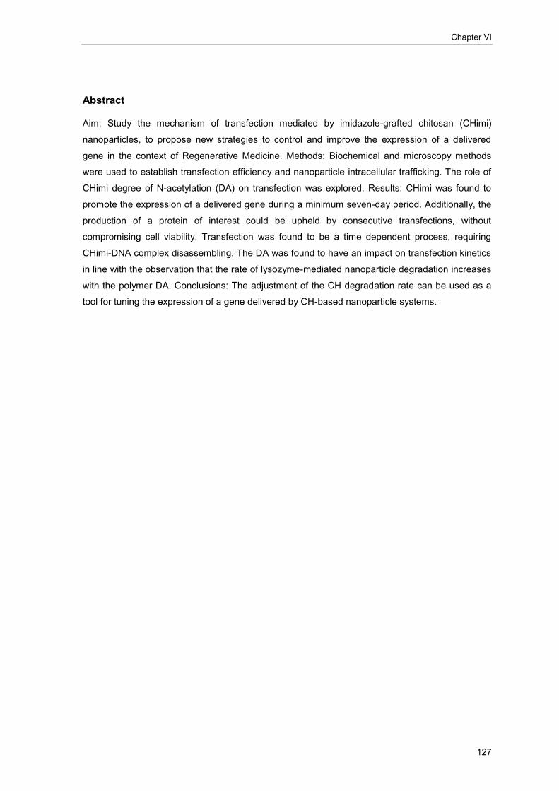
Chapter VI
127
Abstract
Aim: Study the mechanism of transfection mediated by imidazole-grafted chitosan (CHimi)
nanoparticles, to propose new strategies to control and improve the expression of a delivered
gene in the context of Regenerative Medicine. Methods: Biochemical and microscopy methods
were used to establish transfection efficiency and nanoparticle intracellular trafficking. The role of
CHimi degree of N-acetylation (DA) on transfection was explored. Results: CHimi was found to
promote the expression of a delivered gene during a minimum seven-day period. Additionally, the
production of a protein of interest could be upheld by consecutive transfections, without
compromising cell viability. Transfection was found to be a time dependent process, requiring
CHimi-DNA complex disassembling. The DA was found to have an impact on transfection kinetics
in line with the observation that the rate of lysozyme-mediated nanoparticle degradation increases
with the polymer DA. Conclusions: The adjustment of the CH degradation rate can be used as a
tool for tuning the expression of a gene delivered by CH-based nanoparticle systems.

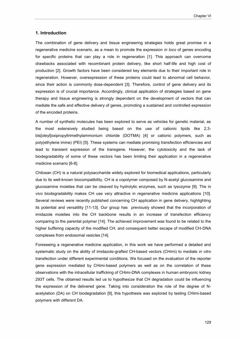
Chapter VI
129
1. Introduction
The combination of gene delivery and tissue engineering strategies holds great promise in a
regenerative medicine scenario, as a mean to promote the expression in loco of genes encoding
for specific proteins that can play a role in regeneration [1]. This approach can overcome
drawbacks associated with recombinant protein delivery, like short half-life and high cost of
production [2]. Growth factors have been considered key elements due to their important role in
regeneration. However, overexpression of these proteins could lead to abnormal cell behavior,
since their action is commonly dose-dependent [3]. Therefore, control of gene delivery and its
expression is of crucial importance. Accordingly, clinical application of strategies based on gene
therapy and tissue engineering is strongly dependent on the development of vectors that can
mediate the safe and effective delivery of genes, promoting a sustained and controlled expression
of the encoded proteins.
A number of synthetic molecules has been explored to serve as vehicles for genetic material, as
the most extensively studied being based on the use of cationic lipids like 2,3-
bis[oleyl]oxipropyltrimethylammonium chloride (DOTMA) [4] or cationic polymers, such as
poly(ethylene imine) (PEI) [5]. These systems can mediate promising transfection efficiencies and
lead to transient expression of the transgene. However, the cytotoxicity and the lack of
biodegradability of some of these vectors has been limiting their application in a regenerative
medicine scenario [6-8].
Chitosan (CH) is a natural polysaccharide widely explored for biomedical applications, particularly
due to its well-known biocompatibility. CH is a copolymer composed by N-acetyl glucosamine and
glucosamine moieties that can be cleaved by hydrolytic enzymes, such as lysozyme [9]. The in
vivo biodegradability makes CH use very attractive in regenerative medicine applications [10].
Several reviews were recently published concerning CH application in gene delivery, highlighting
its potential and versatility [11-13]. Our group has previously showed that the incorporation of
imidazole moieties into the CH backbone results in an increase of transfection efficiency
comparing to the parental polymer [14]. The achieved improvement was found to be related to the
higher buffering capacity of the modified CH, and consequent better escape of modified CH-DNA
complexes from endosomal vesicles [14].
Foreseeing a regenerative medicine application, in this work we have performed a detailed and
systematic study on the ability of imidazole-grafted CH-based vectors (CHimi) to mediate in vitro
transfection under different experimental conditions. We focused on the evaluation of the reporter
gene expression mediated by CHimi-based polymers as well as on the correlation of these
observations with the intracellular trafficking of CHimi-DNA complexes in human embryonic kidney
293T cells. The obtained results led us to hypothesize that CH degradation could be influencing
the expression of the delivered gene. Taking into consideration the role of the degree of N-
acetylation (DA) on CH biodegradation [9], this hypothesis was explored by testing CHimi-based
polymers with different DA.
Chitosan-mediated gene delivery
130
2. Materials and Methods
2.1. Materials
Unless mentioned otherwise, all reagents were obtained from Sigma-Aldrich and were of
analytical grade.
Technical grade chitosan (Chimarin™, DA 13%, apparent viscosity 8 mPa.s, supplied by
Medicarb, Sweden) was purified and characterized prior further use. The average weight
molecular weight (
M w), polydispersity, polydispersity index and DA of the purified polymer were
found to be 1.2x10⁵, 2.1 and 16.4%, respectively (see below for experimental details) [14].
Endotoxin levels of CH extracts were found to be lower than 0.1 EU.ml-1
[14], respecting the US
Department of Health and Human Services guidelines for implantable devices [15].
Plasmids carrying β-galactosidase (pCMV-Sport β Gal, Invitrogen, 7.8 Kbp), green fluorescence
protein (pCMV-GFP, 7.4 Kbp) or transthyretin (TTR) (pET-3a based plasmid, 4.9 Kbp) genes were
used in this study. Plasmids were amplified in DH5 Escherichia coli (E. coli) and isolated using
GENELUTE™ high performance endotoxin-free plasmid maxiprep kit according to the
manufacturer instructions. Plasmid concentration and purity were determined
spectrophotometrically (Beckman DU®650, USA). The ratio between optical density at 260 nm and
280 nm was found to be higher than 1.7.
2.2. Chemical modification of chitosan
Chitosan deacetylation. CH with a low DA was prepared by heterogeneous deacetylation [16] of
the purified polymer (CH16), under argon atmosphere. The deacetylated polymer (CH05) was
precipitated by addition of a mixture of water/ammonia 7:3 (v:v) and recovered by centrifugation,
neutralized by washing with deionized water, freeze dried (Labconco) and, subsequently, vacuum
dried at 60°C for 24 hrs.
Chitosan acetylation. CHs with different DA were obtained from re-acetylation of CH05 according
to a previously described procedure [17]. The obtained polymers were purified as described
above.
Imidazole grafting of chitosan. CH05, CH10, CH16 and CH18 were modified with imidazole-4-
acetic acid sodium salt by amidation of the glucosamine residues using an EDC/NHS
condensation system (1-Ethyl-3-[3-dimethylaminopropyl]carbodiimide hydrochloride – EDC; N-
hydroxysuccinimide – NHS) [14]. Polymer stock solutions (0.1% (w/v)) were prepared according to
a previously described procedure [14], and stored at 4°C till further use.
2.3. Polymer characterization
Fourier Transform – Infrared Spectroscopy (FT-IR) was performed in Perkin-Elmer 2000 system.
Samples were prepared as potassium bromide (KBr) discs by mixing 2 mg of the polymer

Chapter VI
131
(vacuum dried 24 hrs at 60°C) with 200 mg of KBr (dried 24 hrs at 105°C). The IR spectra were
recorded, after a 5-min purge of the sample chamber with N2, by accumulation of 200
interferograms, at a 4 cm-1
spectral resolution.
Size exclusion chromatography (SEC) was used to determine the molecular weight of CH05,
CH10, CH16 and CH18. The modular system employed was composed by an isocratic pump (K-
1001 Knaeur), a vacuum degasser (K-5002 Knaeur), a viscometer/right angle laser light scattering
(RALLS) dual detector (T60 Viscotek), and a refractive index detector (K-5002 Knaeur) operating
at the same wavelength as the RALLS detector (670 nm). Separations were performed in a set of
PL aquagel-OH mixed columns. 0.2 M NaCH3COO/0.5 M CH3COOH was used as eluent at a flow
rate of 1ml.min-1
and the dn/dc used for molecular weight calculations was 0.205 ml.g-1
[18].
Sample concentration in a 0.1 to 0.2% (w/v) range and an injection volume of 100 µl were applied.
All measurements were performed in triplicate, at room temperature (RT).
2.4. CHimi-DNA particle preparation
CHimi-DNA particles were prepared by mixing, while vortexing, equal volumes of a CHimi solution
in 5 mM CH3COONa buffer pH 5.5 (0.1% (w/v)) and a plasmid DNA solution (in 25 mM Na2SO4),
both previously heated at 55°C for 10 min. Complexes were allowed to form and stabilize for 15
min at RT before further use. Unless mentioned otherwise, complexes were prepared at a molar
ratio of CH primary amines to DNA phosphate group (N/P) of 18.
2.5. CHimi-based particle characterization
Complexes were prepared as described above, using 10 µg of pCMV-GFP and 150 µg of CHimi,
independently of the polymer DA. After stabilization, complexes were diluted to a final volume of 1
ml, using 5 mM CH3COONa buffer (pH 5.5). Complex size and zeta potential were assessed using
a Zetasizer Nano Zs (Malvern, UK). The Smoluchowski model was applied for zeta potential
determination and cumulant analysis was used for mean particle size determination. All
measurements were performed in triplicate, at 25°C.
2.6. Cell culture
293T human embryonic kidney endothelial cells were cultured under standard conditions in
Dulbecco's Modified Eagle's Medium (DMEM) high glucose (4500 mg.l-1
) supplemented with 10%
(v/v) heat-inactivated (56°C, 30 min) fetal bovine serum (FBS) and 1% (v/v) penicillin/streptomycin
(10.000 U.ml-1
penicillin, 10.000 g.ml-1
streptomycin) (all supplied by Gibco). Cells were routinely
tested for mycoplasma contamination by PCR [19, 20].

Chitosan-mediated gene delivery
132
2.7. Transfection
293T cells were seeded (2.7x10⁴ viable cells.cm
-2) on poly-D-Lysine hydrobromide (PDL) coated
24-well tissue culture plates (Greiner Bio-one, CellStar), 24 hrs prior transfection. Cell culture
medium was replaced for 500 μl of complete fresh medium 2 hrs before transfection. In all
transfection experiments, complexes were added to the cells at a final DNA concentration of 1.3
g.cm-2
. Cell culture medium was refreshed everyday.
2.8. In vitro gene expression studies
In this set of studies the plasmid carrying the β-galactosidase (β-gal) reporter gene was used.
The gene expression mediated by CHimi with a DA of 16% and two degrees of substitution with
imidazole (13% - CH16imi1 and 22% - CH16imi2) was evaluated up to 168 hrs post-transfection.
Cultures dilution was performed 72 hrs post-transfection. In brief, the cell monolayer was rinsed
with pre-warmed phosphate buffered saline (PBS) and harvested by trypsinization (5 min, 37°C).
Cells were re-suspended in supplemented DMEM (1 ml/well), diluted (7x) and re-seeded on PDL-
coated 24-well plates.
The effect of multiple transfections on transfection activity (expressed as specific activity of -gal)
and cell viability was assessed as follows: 72 hrs post-transfection cells were harvested by
trypsinization, as described above, and re-seeded on PDL-coated 24-well plates at the initial cell
density (2.7x10⁴ viable cells.cm
-2). 24 hrs after plating, cells were subjected to a second
transfection. This procedure was repeated once more. In total three transfection treatments were
performed. Transfection activity was evaluated at 48 and 72 hrs after each transfection. At each
time point, cells were processed for β-gal activity evaluation according to manufacturer
instructions (β-gal assay kit, Invitrogen). Non-transfected cells were used as blank. The total
protein was determined by the BCA assay (Pierce), following the manufacturer instructions.
Cell viability was determined using a resazurin-based assay [21], as previously described [22].
Results are represented as percentage of metabolic activity of transfected cells relative to non-
transfected cells.
2.9. Intracellular trafficking studies
Fluorescence microscopy studies
To allow the tracking of the CHimi-based particles inside cells, both polymer and plasmid DNA
were fluorescently labeled. A rhodamine (λex=575 nm, λem=600 nm) activated derivative [5(6)-
Carboxy-X-rhodamine N-succinimidyl ester, ROX (Fluka)] was used to label CH16imi1. In brief, 10
mg of CH16imi1 was dissolved overnight in 10 ml of a 1% (v/v) acetic acid solution and added to
an equal volume ROX solution (0.13 mg.ml-1
in dehydrated methanol, Molecular Sieves, Merck).
The reaction was let to occur for 3 hrs, under constant stirring, protected from light. The

Chapter VI
133
fluorescently-labeled CH16imi1 (CHimiROX) was recovered by precipitation with 5 ml of 0.5 M
NaOH. The precipitated polymer was washed with deionized water till no fluorescence was
detected in the supernatant and, subsequently, freeze-dried. pCMV-GFP plasmid was
fluorescently labeled using the commercial kit Label IT Cy5 (Mirus™) according to the
manufacturer instructions. The fluorescently labeled DNA (DNACy5) was recovered by precipitation
in ethanol and concentration was assessed by spectrophotometry (λ=260 nm) (Beckman DU®650,
USA).
Complexes prepared with the fluorescently labeled CHimiROX and DNACy5 were used to transfect
cells 24 hrs after seeding on PDL-coated glass coverslips. The intracellular localization of the
complexes was analyzed in both live and fixed cells as follows. Twenty four hrs after cell seeding,
the cultures on PDL glass coverslips (2.5 cm2) were transferred into a closed chamber and
incubated with L-15 medium (Gibco) supplemented with 10% (v/v) FBS and 1% (v/v)
penicillin/streptomycin containing the complexes prepared with fluorescently labeled DNA and
polymer. Cells were maintained at 37°C and images were acquired using an inverted
epifluorescence microscope (Nikon eclipse TE2000-U) equipped with a Cool Snap HQ2 camera.
Images were collected each hr during the first 6 hrs after transfection and at 24 hrs post-
transfection. A series of z-sections were collected in order to capture images in all cell depth. At
least five different areas of the sample were followed in time, in three independent experiments. At
48 hrs post-transfection images of GFP positive cells were collected. To prepare fixed cell
samples, at defined time points (2, 4, 6, 24 and 48 hrs post-transfection), cultures on PDL glass
coverslips (1.3 cm2) transfected with fluorescently labeled polymer and DNA were rinsed with pre-
warmed PBS and fixed for 15 min at 37°C with paraformaldehyde (4% (w/v), in PBS),
supplemented with 2% (w/v) sucrose. After fixation, cell cytoskeleton and nuclei were stained. In
brief, fixed cells were permeabilized according to a previously described procedure [23] and
incubated with 1% (w/v) bovine serum albumin (BSA) for 1 hr. Cell nuclei were stained for 4 min
with 4'-6-diamidino-2-phenylindole (DAPI, 0.1 g.ml-1
in PBS) and cell cytoskeleton filamentous
actin (F-actin) was counterstained with Alexafluor 488-conjugated phalloidin (5 U.ml-1
, in PBS with
1% (w/v) BSA, 20 min, Molecular Probes). Samples were mounted in Vectashield (Vector) and
observed by confocal laser scanning microscopy (CLSM, Leica Microsystems). Cytoskeleton
staining was not performed in samples collected 48 hrs post-transfection in order to allow the
detection of GFP positive cells. Cells were analyzed in depth by z-stacking. A minimum of 20
fields per time point were collected, from three independent experiments.
Cell-free gene expression assay
The occurrence of reporter gene transcription and/or translation, when in a complex form with
CHimi-based polymer, was evaluated using a TNT® Quick Coupled Transcription/Translation
System (Promega). This system allows protein production from genes under a T7 promoter. A
pET-3a based plasmid encoding for the TTR protein [22] was used in this experiment to complex
with CH16imi1. Following the manufacturer instructions, 1 g of plasmid DNA was used in each

Chitosan-mediated gene delivery
134
reaction. CHimi-based complexes were prepared by mixing 2 l of DNA solution (in 25 mM
Na2SO4 solution) with 13.4 l of CH16imi1. The complex suspension was added to the TNT mix.
The modification in complex formation procedure, as well as the experimental conditions of the
assay (complex formation procedure, medium pH, temperature and ionic strength) showed not to
alter the polymer ability to retain DNA in an agarose gel electrophoresis (data not shown). Since
the final volume of the reaction mixture containing the complex solution exceeds the 50 l
recommended by the manufacturer, two controls were added to the experiment where the
increase on the final reaction volume and the presence of CH3COONa buffer pH 5.5 (CH16imi
solvent) were equated. The reaction was let to occur for 90 min at 30°C. The reaction products
were resolved by an SDS-polyacrylamide gel electrophoresis (SDS-PAGE) on a 15% (w/v) gel.
The gel was dried and, since 35S[methionine] was included in the reaction mixture, the presence of
TTR protein was analyzed by phosphorimaging (Typhoon 8600 variable mode imager; Molecular
Dynamics), after an overnight exposure. The position within the gel of the TTR band was
confirmed by running in the same gel a molecular weight marker and the recombinant human TTR
protein (produced in E. Coli) (data not shown).
2.10. DNase I protection assay CHimi-DNA particle stability and DNA protection
To evaluate the stability in physiological media of CHimi-DNA particles prepared with CHimi with
different degree of acetylation, DNA electrophoretic mobility and DNase I protection were
assessed. The electrophoretic mobility of DNA and CHimi-DNA complexes was analyzed in an
agarose gel, as previously described [14]. Prior to running the gel, the complexes were prepared
as described above and incubated for 20 minutes with equal volume of supplemented DMEM
(10% FCS and 1% P/S). The gel was scanned using a Bio Rad Gel DocTM
XR system.
The ability of the polymer to protect DNA from DNase I degradation was assessed as follows.
Complexes were suspended in buffer solution (10 mM Tris HCl, 150 mM NaCl, 1 mM MgCl2; pH
7.4) and incubated with DNase I (1 U/μl, Fermentas) at 37ºC. Absorbance (260 nm) was recorded
for 30 min, using a PowerWave™ Microplate Spectrophotometer (BioTek, USA).
2.11. Extent of internalization and transfection efficiency
The extent of internalization and transfection efficiency of complexes prepared with constant mass
of pCMV-GFP and CHimi (independently of the polymer DA) were assessed by flow cytometry
(FACSCalibur, BD Biosciences). For the internalization studies, pCMV-GFP was labeled with
YoYo-1 (Invitrogen, 1:200 bp) prior to complex formation, according to the manufacturer
instructions. After 2 hrs of contact with complexes, cells were processed for fluorescence
activated cell sorting (FACS) analysis. Briefly, trypan blue (0.2% (w/v)) was added to the medium
and incubated for 5 min in order to quench fluorescence external to cells [24]. Subsequently, the
cells were washed with cold PBS, and harvested by trypsinization. After two washes with PBS,

Chapter VI
135
cells were recovered in 100 µl of PBS containing 0.01% (w/v) of sodium azide. For transfection
efficiency determination, cells were processed as described above, skipping the trypan blue
incubation step. The number of cells expressing GFP was determined at 24, 48 and 72 hrs post-
transfection. The individual fluorescence of 10,000 cells was quantified and results were analyzed
using the FlowJo software (version 8.3.7). Results are expressed as the percentage of labeled
DNA (for extent of internalization) or GFP positive cells (for transfection efficiency).
2.12. Enzymatic degradation of CHimi-based complexes
To assess CH-based particle degradation, complexes were prepared using a constant CHimi-
pCMV-GFP mass ratio, independently of the polymer DA, and incubated with lysozyme in the
presence of a fluorogenic substrate for the enzyme. In brief, 4-methylumbelliferyl β-D-N,N′,N′′-
triacetylchitotrioside (MU-[GlcNAc]3) was dissolved in CH3COONa (1M, pH=5.5):H2O:DMF (1:1:1)
[25] and added to freshly prepared CHimi-DNA complexes. Equimolar solutions of MU-[GlcNAc]3
and CHimi (4.45 µM) were applied. Subsequently, the mixture was incubated with lysozyme (from
chicken egg white) at a final concentration of 0.5 mg.ml-1
, at 37°C under constant stirring. After 1,
2, 3 and 4 hrs of reaction, NaOH was added to a final concentration of 0.05 M to stop the reaction.
The fluorescence resulting from the enzymatic degradation of the substrate was measured
(λexc=360nm; λem=455nm – Spectra Max Gemini XS; Molecular Devices). For each DA, CHimi-
DNA complexes were incubated in the same conditions but without fluorogenic substrate, and
used as blank.
2.13. Statistical data analysis
Data are presented as average ± standard deviation (SD). The statistical analysis of the results
was performed using the non-parametric Mann-Whitney U-test. For multiple comparisons,
homogeneity of variances was assessed by the Bartlett’s test. If homogeneity of variances could
be assumed the post-hoc Bonferroni test was performed, otherwise, the Dunnett T3 test was
applied. Results were considered statistically significant when p<0.05. Calculations were
performed using SPSS® software for Windows (version 16.0).
3. Results
3.1. In vitro gene expression study
In a previous work we have optimized CHimi mediated transfection [14] . It was found that the best
transfection results are attained when CHimi-DNA complexes are prepared with CHimi containing
13% (CH16imi1) or 22% (CH16imi2) of the primary amines substituted with imidazole moieties, at
an N/P molar ratio of 18. These two formulations were selected to be further studied in the present
work. To follow CHimi-mediated gene expression over time, β-gal activity was evaluated during 7
days after transfection (Figure 1).

Chitosan-mediated gene delivery
136
Figure 1. (A) Transfection activity as function of time for 293T cultures treated with CH16imi1- and
CH16imi2-based vectors (N/P=18). Cells were trypsinized and diluted (7x) at 72 hrs post-transfection. (B)
Independent plotting of -gal activity and respective total cell protein content. Representative experiment out
of the three performed (average ± SD; n=3).
Under the experimental conditions used, a transfection activity maximum is reached 72 hrs post-
transfection for both polymers tested. After trypsinization, a significant decrease on β-gal specific
activity was observed (Figure 1 (A)), in accordance to a previously published report [26].
Nevertheless, when the enzymatic activity (nmolONPG.min-1
) and the total cell protein content are
plotted separately (Figure 1 (B)), one can observe that the decrease in enzymatic activity is of the
same magnitude of the dilution performed (7x dilution) rather than resulting from an effective
decrease in the production of the reporter protein. Furthermore, β-gal production remained stable
in the period between 96 and 168 hrs post-transfection. Cell viability was monitored at the same
time points. No significant alteration was found in terms of metabolic activity of cells transfected
with CHimi-based polymers relative to non-transfected cells (Figure S1, supplementary material).
The possibility of performing successive transfection treatments using CHimi-based polymers,
without compromising cell viability, was addressed in vitro. In terms of transfection activity the
results show that cells can be re-transfected, maintaining high levels of the reporter gene
expression (Figure 1 (A)). One should refer that a burst increase in transfection efficiency was
observed at times, as illustrated in Figure 2 (A). This could be resulting from the series of
trypsinization steps introduced every 3 days of culture that could not be circumvented in the
implementation of this study. Even so, independently of the polymer system tested, cell viability
relative to untreated cells remained above 80% after every treatment (see Figure 2 (B) for an
example).
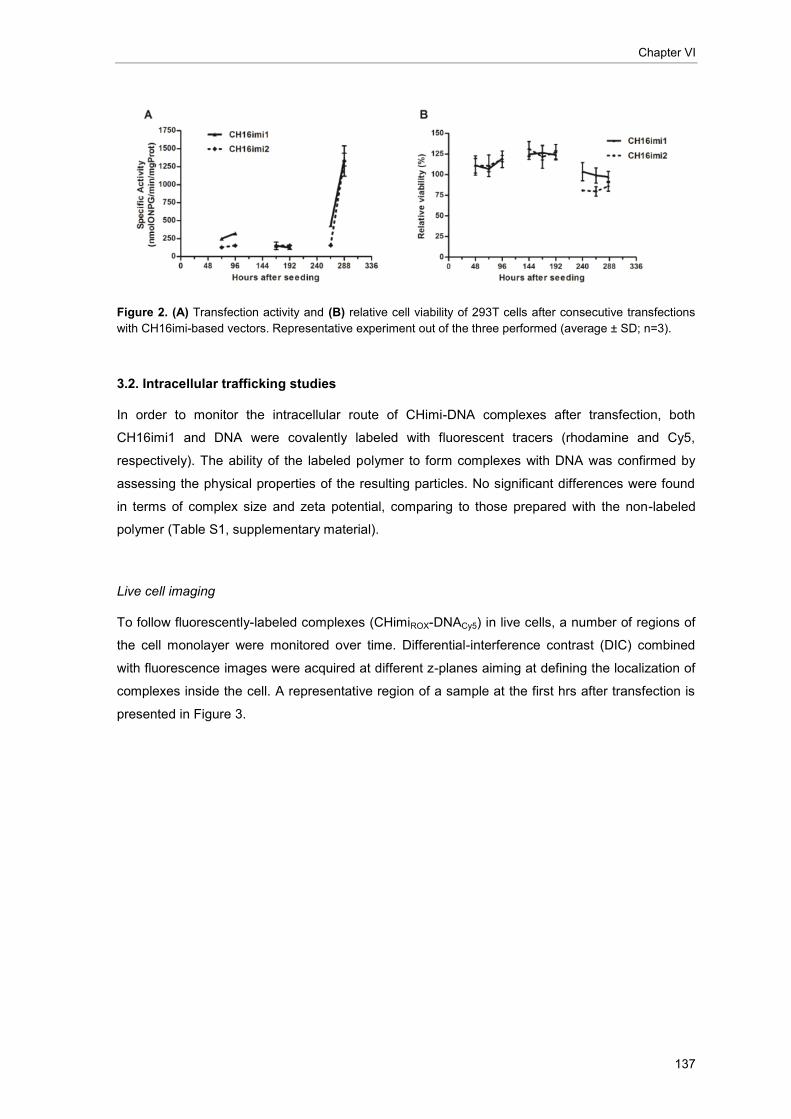
Chapter VI
137
Figure 2. (A) Transfection activity and (B) relative cell viability of 293T cells after consecutive transfections
with CH16imi-based vectors. Representative experiment out of the three performed (average ± SD; n=3).
3.2. Intracellular trafficking studies
In order to monitor the intracellular route of CHimi-DNA complexes after transfection, both
CH16imi1 and DNA were covalently labeled with fluorescent tracers (rhodamine and Cy5,
respectively). The ability of the labeled polymer to form complexes with DNA was confirmed by
assessing the physical properties of the resulting particles. No significant differences were found
in terms of complex size and zeta potential, comparing to those prepared with the non-labeled
polymer (Table S1, supplementary material).
Live cell imaging
To follow fluorescently-labeled complexes (CHimiROX-DNACy5) in live cells, a number of regions of
the cell monolayer were monitored over time. Differential-interference contrast (DIC) combined
with fluorescence images were acquired at different z-planes aiming at defining the localization of
complexes inside the cell. A representative region of a sample at the first hrs after transfection is
presented in Figure 3.
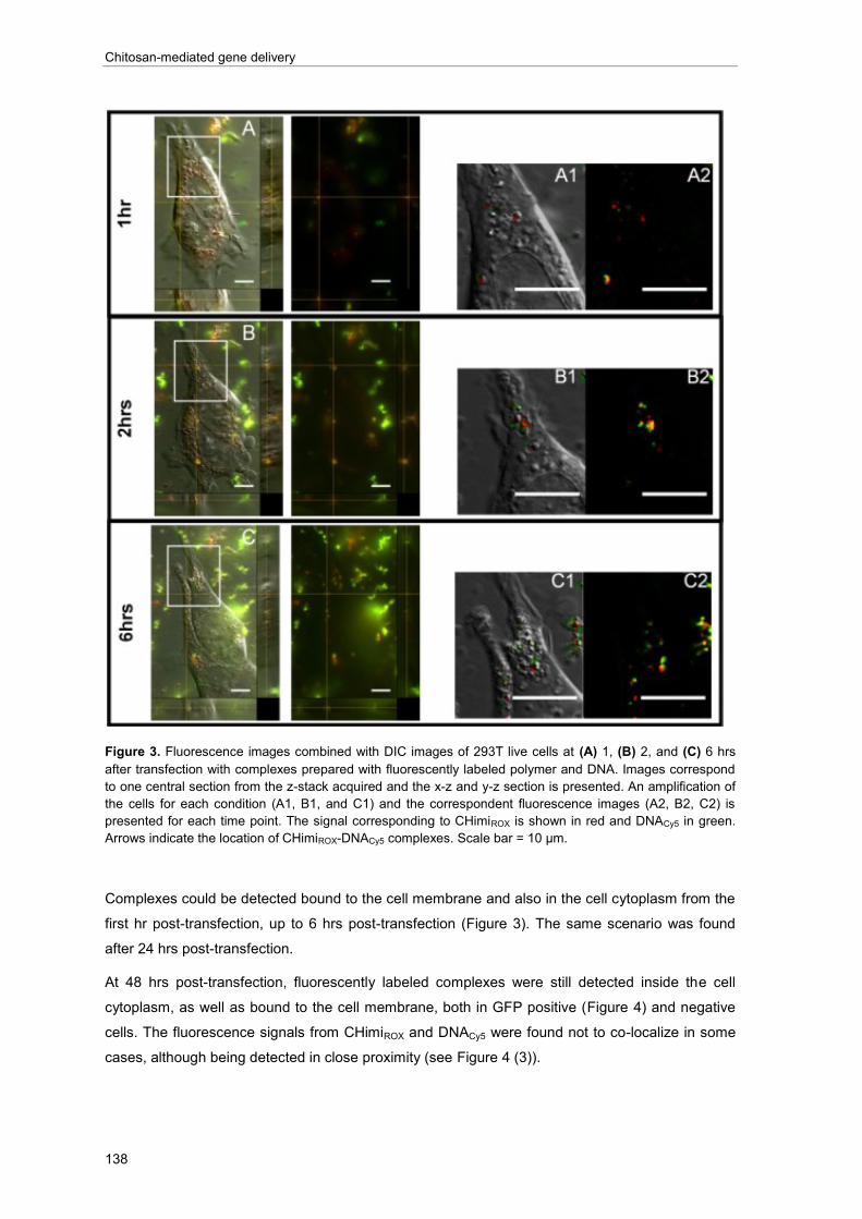
Chitosan-mediated gene delivery
138
Figure 3. Fluorescence images combined with DIC images of 293T live cells at (A) 1, (B) 2, and (C) 6 hrs
after transfection with complexes prepared with fluorescently labeled polymer and DNA. Images correspond
to one central section from the z-stack acquired and the x-z and y-z section is presented. An amplification of
the cells for each condition (A1, B1, and C1) and the correspondent fluorescence images (A2, B2, C2) is
presented for each time point. The signal corresponding to CHimiROX is shown in red and DNACy5 in green.
Arrows indicate the location of CHimiROX-DNACy5 complexes. Scale bar = 10 µm.
Complexes could be detected bound to the cell membrane and also in the cell cytoplasm from the
first hr post-transfection, up to 6 hrs post-transfection (Figure 3). The same scenario was found
after 24 hrs post-transfection.
At 48 hrs post-transfection, fluorescently labeled complexes were still detected inside the cell
cytoplasm, as well as bound to the cell membrane, both in GFP positive (Figure 4) and negative
cells. The fluorescence signals from CHimiROX and DNACy5 were found not to co-localize in some
cases, although being detected in close proximity (see Figure 4 (3)).

Chapter VI
139
Figure 4. Z-section and the correspondent x-z and x-y section of 293T cells expressing GFP captured at 48
hrs post-transfection. The amplification of the fluorescence images corresponding to the area within the
rectangle is shown in 1-4. The individual signals of (1) CHimiROX and (2) DNACy5 are presented. (3) shows the
merged image of CHimiROX (in red) and DNACy5 (in blue). (4) combines also the GFP signal. Arrows indicate
the location of CHimiROX-DNACy5 complexes within the GFP expressing cell. Scale bar = 10 µm.
Fixed cells analysis
The CHimiROX-DNACy5 complexes distribution was analyzed in fixed cells by CLSM at the same
time points: 2, 6, and 48hrs after transfection. Representative images of each time point are
presented in Figure 6.
Figure 5. CLSM images captured at (A) 2, (B) 6, and (C) 48 hrs after transfection of 293T cells with
CHimiROX-DNACy5 complexes. Images correspond to one central section from the z-stack acquired. The insert
corresponds to an amplification of the specific area indicated in the original image. The fluorescence signal
corresponding to CHimiROX is shown in red and DNACy5 in gray. Cells were stained with DAPI (genomic DNA,
blue) and phalloidin (filamentous actin, green; A-B). (C) Cells are shown in green due to GFP expression.
Arrows indicate the location of CHimiROX-DNACy5 complexes. Scale bar = 10 µm. The images of each of the
fluorescence channels are presented in supplementary material (Figure S2).
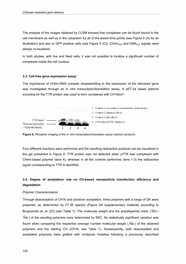
Chitosan-mediated gene delivery
140
The analysis of the images obtained by CLSM showed that complexes can be found bound to the
cell membrane as well as in the cytoplasm for all of the tested time points (see Figure 5 (A) for an
illustration) and also in GFP positive cells (see Figure 5 (C)). CHimiROX and DNACy5 signals were
always co-localized.
In both studies, with live and fixed cells, it was not possible to localize a significant number of
complexes inside the cell nucleus.
3.3. Cell-free gene expression assay
The importance of CHimi-DNA complex disassembling to the expression of the delivered gene
was investigated through an in vitro transcription/translation assay. A pET-3a based plasmid
encoding for the TTR protein was used to form complexes with CH16imi1.
Figure 6. Phosphor imaging of the in vitro transcription/translation assay reaction products.
Four different reactions were performed and the resulting radioactive products can be visualized in
the gel presented in Figure 6. TTR protein was not detected when pTTR was complexed with
CHimi-based polymer (lane 4), whereas in all the controls performed (lane 1-3) the radioactive
signal corresponding to TTR is identified.
3.4. Degree of acetylation role on CH-based nanoparticle transfection efficiency and
degradation
Polymer Characterization
Through deacetylation of CH16 and posterior acetylation, three polymers with a range of DA were
prepared, as determined by FT-IR spectra (Figure S4 supplementary material) according to
Brugnerotto et. al. [27] (see Table 1). The molecular weight and the polydispersity index ( wM /
nM ) of the resulting polymers were determined by SEC. No statistically significant variation was
found when comparing the respective average number molecular weight ( nM ) of the obtained
polymers and the starting CH (CH16, see Table 1). Subsequently, both deacetylated and
acetylated polymers were grafted with imidazole moieties following a previously described

Chapter VI
141
procedure [14]. Briefly, grafting of the starting materials was explored with a molar ratio of
imidazole-4-acetic acid sodium salt to glucosamine residues of 0.30. The obtained degree of
substitution of each grafted polymer was assessed by FT-IR [14] (spectra in Figure S5
supplementary material) and it is presented in the Table 1. For the polymers with lower DA (CH16
and CH18) the obtained degree of substitution was lower than the expected value of 30% of
imidazole moieties per mol of primary amino groups (22.1 and 23.4%, respectively). A possible
explanation is that the lower polymer solubility could be limiting the reaction efficiency, as
previously discussed [14].
Table 1. Degree of N-acetylation, percentage of the average number molecular weight (
M w) relative to the
initial polymer (CH16), polydispersity index ( nMwM ) and degree of substitution of primary amines of CH
with imidazole moieties (imi) of the resulting polymers (average ± SD; n=3).
CHimi-based particle characterization
The physical properties of the CHimi-DNA complexes prepared with a constant polymer to DNA
mass ratio, using CHimi with different DA were determined. No significant variations in terms of
particle size and zeta potential were detected between complexes prepared with the different
polymers. Particle zeta potential was found to be around +20 mV, whereas the mean particle size
ranged between 244 and 331 nm (Table 2).
Table 2. Zeta potential, average size (Z-Average), and polydispersity index (PdI) of CHimi-DNA complexes
prepared with the pCMV-GFP plasmid and CHimi with different DA (average ± SD; n=3).

Chitosan-mediated gene delivery
142
CHimi-DNA particle stability and DNA protection
The ability of CHimi with variable DA to form stable particles with DNA and to protect the plasmid
from nuclease degradation in physiological conditions was assessed. Results show that only
naked DNA is able to migrate under an electrophoretic field, demonstrating that, independently of
the CHimi DA, the complexes are stable in the presence of supplemented cell culture medium
(Figure S3, supplementary material (A)). Furthermore, CHimi-based polymers are able to protect
DNA from DNase I degradation. The performed assay is based on the fact that intact DNA
molecules possess hypochromicity and that the absorbance at 260 nm increases upon enzymatic
digestion [26]. The results show that naked DNA is fully degraded by DNase I following a 30 min
incubation period, whereas for DNA complexed with CHimi-based polymers, the absorbance at
260 nm does not significantly increases upon incubation with the enzyme (Figure S3,
supplementary material (B)).
Extent of internalization and transfection efficiency
The percentage of YoYo-1 positive cells was determined after 2 hrs of incubation of the cells with
complexes based on CHimi with different DA. As shown in Figure 7 (A), internalization of
complexes based in CH05imi, CH10imi or CH16imi was observed in approximately 30% of 293T
cells. However, a significant lower extent of internalization was observed for CH18imi-based
complexes.
Transfection efficiency mediated by CHimi-based complexes with variable DA was assessed for
up to 72 hrs post-transfection (Figure 7 (B)). As can be observed, CH16imi is the most efficient
polymer among the various CHs tested, being able to transfect over 40% of the cell population (at
72 hrs post-transfection). Although CH18imi-based complexes are able to be internalized by the
cells, a significantly lower transfection efficiency is attained, in comparison to the other three
polymers. The rate of internalization of CH18imi-DNA complexes is similar to the one obtained
when naked DNA is added to cells (see representative histogram plots in Figure S6
supplementary material), being the later also ineffective transfecting cells [14]. For CH05imi-,
CH10imi- and CH16imi-based complexes, transfection efficiency increased over time, in
accordance to what was previously described in section 3.1. For CH10imi and CH16imi, the most
significant increase of transfection occurred from 24 to 48 hrs post-transfection. However, in the
case of CH05imi this effect was observed from 48 to 72 hrs post-transfection. A commercial
formulation of PEI (Escort V® – Sigma) was used as a control for transfection experiments, and
transfection efficiency was found to be above 80% at all the time points tested. A more detailed
comparison between CHimi and PEI can be found in a previous publication [14].

Chapter VI
143
Figure 7. (A) Percentage of 293T cells positive for YoYo-1 after 2 hrs of incubation with CHimi-based
complexes with variable DA. (B) Percentage of 293T cells expressing GFP at 24, 48 and 72 hrs post-
transfection with CHimi-based complexes with variable DA. The increase in the percentage of GFP positive
cells over time for each polymer is presented. Representative experiment out of the three performed
(average ± SD; n=3). * denotes statistical significant differences relative to CH05imi, CH10imi and CH16imi
(p<0.05). Representative histogram plots and mean fluorescence intensity data are presented in Figure S6,
Figure S7 and Figure S8 of supplementary material.
Enzymatic degradation of CHimi-based nanoparticles
To evaluate the effect of the DA of CHimi on the degradation of CHimi-based complexes, a
competition assay was setup using lysozyme as a model enzyme for CH intracellular degradation
[9, 28-31]. The assay is based on the competition for lysozyme between a fluorogenic substrate
(MU-[GlcNAc]3) [25] and CHimi-based complexes. It is expected that a reduction on the
fluorescence produced is observed when CHimi is competing with the fluorogenic substrate, since
less substrate will be degraded.
Figure 8. Extent of MU-[GlcNAc]3 hydrolysis after 4 hrs of incubation with lysozyme in the absence or
presence of CHimi-DNA complexes. Complexes were prepared with pCMV-GFP and CHimi with different
DA. The insert shows the fluorescence produced as a function of time. Representative experiment out of the
three performed (average ± SD; n=3).
As seen in Figure 8, a reduction in fluorescence can be observed as CHimi DA increases. The
decrease on fluorescence results from less substrate being degraded, indicating that more

Chitosan-mediated gene delivery
144
polymer degradation has occurred. This difference is identified also at earlier time points (insert of
Figure 8), although being more evident as incubation time increases.
4. Discussion
The application of gene delivery strategies in regenerative medicine has been proposed as a
promising approach to induce the production in loco of proteins that can play a role in tissue
regeneration. In a previous study we showed that by grafting CH with imidazole moieties,
transfection efficiency mediated by this polymer can be improved, without impairing cell viability
[14]. In the present work, we aim at investigating the mechanism of transfection mediated by
CHimi-based vectors, with the final objective of finding new avenues to further improve the system
and tune the expression of a delivered gene.
It has been proposed that gene delivery mediated by CH is a time dependent process [32];
however, most of the related publications in this field report transfection efficiency in the 3 to 4
days after transfection [33-36]. Transfection experiments are usually conducted at high initial cell
densities (>50% cell confluence), which may be considered a limiting factor regarding the
maximum time period to render such evaluation feasible. Li and co-workers studied CH-mediated
transfection up to 15 days post-transfection [37], but the cell culture conditions applied in that
study were not described in detail. In the present work we introduced a trypsinization step 72 hrs
post-transfection, as an experimental strategy to extend the time period for evaluation of
transfection activity and cell viability, while maintaining cells in exponential growth conditions. Two
CHs with a degree of substitution of primary amine with imidazole moieties of 13% (CH16imi1)
and 22% (CH16imi2) were tested, as in previous studies in our laboratory these were found to be
the most effective in mediating cell transfection [14]. The obtained results showed that both
polymers could mediate a sustained expression of a reporter gene for the time period of the study
- up to 7 days post-transfection. Additionally, successive transfections with CHimi-based carriers
could be performed to uphold the levels of gene expression, without a significant reduction on cell
viability.
The understanding of the intracellular mechanisms occurring during transfection is considered a
valuable tool in the design of efficient and functional gene delivery systems [38, 39]. A number of
intracellular trafficking studies has been conducted so far [40-45], nevertheless the importance of
complex disassembling on transfection is yet to be established for the cationic polymer-based
gene delivery systems, as recently discussed [46]. Some authors suggested that DNA can be
transcribed while complexed with PEI [42], whereas in other studies it was hypothesized that the
release of DNA from CH is the limiting step for an efficient in vitro transfection [47]. In an attempt
to clarify whether disassembling of CHimi-DNA complex occurs and to better define the
intracellular localization of such process, fluorescence microscopy studies were performed in live
and fixed cells, using complexes prepared with fluorescently labeled CHimi and DNA. In both
conditions CHimiROX-DNACy5 complexes could be detected inside 293T cells from the first hrs post-

Chapter VI
145
transfection up to 48 hrs post-transfection, including in GFP expressing cells. In the process of
assessing the occurrence of complex disassembling by fluorescent signal co-localization, an
important difference was noted between conditions. In live cells the signal from CHimiROX and
DNACy5 did not always overlap, whereas in CLSM images of fixed cells CHimiROX and DNACy5
were found to co-localize at all time periods. The disparity in the observations suggested that
cell/complex movement could be responsible for the separation of signals observed during
acquisition in live cells. By fast time-lapse consecutive acquisition of individual fluorescence
channels, we were able to confirm that particles change position in the time frame of the image
collection (data not shown). This artifact is not observed when using CLSM for live cell imaging,
because different wavelengths are acquired simultaneously (see Figure S9 of supplementary
material). Therefore, fluorescent microscopy studies were not able to clearly demonstrate complex
disassembling; nonetheless these studies highlighted the importance of a cautious analysis of
fluorescence images on intracellular trafficking studies. In our attempt to establish complex
disassembling, we proposed an innovative assay in gene delivery studies. By an in vitro
transcription/translation assay we were able to show that the production of the reporter protein is
impaired when DNA is complexed with CHimi, clearly suggesting that disassembling is required
for gene expression to occur.
The observed sustained gene expression profile combined with the fact that CHimi-DNA
complexes could be found in cell cytoplasm up to 48 hrs post-transfection, indicate that gene
expression mediated by CHimi is a time dependent process, as previously hypothesized for CH
based gene delivery systems [32]. Furthermore, DNA transcription and/or translation were found
to be impaired in the presence of the polymer, pointing out the importance of DNA release for the
expression of the delivered gene. Being CH established as a biodegradable polymer [9, 48], we
hypothesized that the complex disassembling process could be dependent on CH degradation,
and, consequently, closely dependent on the polymer DA [9]. To address this question,
transfection mediated by CHimi with different DA was explored. By the deacetylation of CH16 and
posterior homogeneous acetylation [17], three polymers with a DA of 5, 10 and 18% were
produced, while maintaining unchanged the initial molecular weight of the polymer. Imidazole
moieties were subsequently grafted to each of the prepared CHs. The variation of CH DA neither
did affect the CHimi-DNA complex physical properties, in accordance to a previous report [49], nor
the CHimi ability to form stable complexes with DNA in the serum supplemented culture medium
and to protect it from DNase I-mediated degradation under physiologic conditions. Nonetheless,
significant differences were found in terms of complex internalization in 293T cells, as well as on
transfection efficiency. Internalization of CH18imi-based complexes was found to be significantly
lower than the other nanoparticle formulations. Furthermore, the internalized CH18imi-based
nanoparticles showed a limited ability to transfect cells, comparing to the other tested systems.
These results suggest that other factors rather than the extent of internalization are contributing to
the observed transfection outcome. There is evidence in the open literature that a small difference
on chitosan degree of acetylation can lead to significant changes on polymer properties [29],
being the distribution of the acetylated monomers in the polymer chain one of the contributors to
Chitosan-mediated gene delivery
146
this effect [9]. The effect of the DA on CH enzymatic degradation has been previously described
namely for CH films [29] and scaffolds [31]. The data concerning CH degradation, when the
polymer is incorporated in nanoparticles is limited and inconclusive, though. Campos et al. [30]
had shown a slight decrease of CH-tri-polyphosphate nanoparticle size (DA 16%) after 4 hrs of
incubation with lysozyme. Similar results were reported by Bernkop-Schnurch and colleagues
using thiolated CH [50]. By applying an assay in which CHimi-based complexes compete with a
fluorogenic substrate for lysozyme, we showed that as CHimi DA increases, less fluorescence is
produced, indicating that the polymer present in the particles is being more extensively degraded.
According to this result, CH18imi was found to be the polymer under investigation that degrades
at the fastest rate, what can justify its low efficiency as a gene delivery vector. Upon
internalization, the polymer may be readily degraded leading to a premature release of DNA,
therefore compromising the plasmid DNA protection intracellularly and, consequently, impairing
transfection. On the other hand, only a small decrease on fluorescence was observed when
CH05imi-based particles competed for lysozyme, suggesting that the degradation of this polymer
was limited. The slower degradation could justify the delayed expression of the reporter gene
mediated by CH05imi, when in comparison to CH10imi and CH16imi. In the former case the boost
on transfection efficiency occurs only after 48 hrs post-transfection. Additional indication of CHimi
degradation was obtained by CLSM in an experiment where a CHimiROX-DNACy5 complex
entrapped in a vesicle within the cell cytoplasm was followed by live imaging (time lapse video
supplied in supporting info. For experimental details see caption of Figure S9, supplementary
material). The fluorescence emitted by CHimiROX was clearly reduced during the experiment,
whereas the one emitted by DNACy5 is not altered (Figure S10, Supplementary material). The
results provide experimental evidence for CH-based complexes degradation both in vitro and
intracellularly, putting forward CH degradation rate as a parameter influencing transfection
efficiency mediated by CHimi.
5. Conclusion
The opportunity to tune gene expression as a function of CHimi biodegradability, and the fact that
this polymer promotes a sustained gene expression that can be upheld by successive
transfections, emphasizes the potential of CHimi-based polymers as gene vectors in vivo in a
regenerative medicine scenario. Therefore, our group is currently exploring different possibilities to
incorporate CHimi-based polymers in the design of targeted nanoparticles aiming at cell-specific
gene delivery to the peripheral nervous system [51].
Executive summary
In vitro gene expression study
CHimi-based vectors show appropriate properties to be used in a regenerative medicine scenario,
being able to mediate a transient, and sustained expression of a delivered gene without cytotoxic

Chapter VI
147
effects. Successive transfections with CHimi-based vectors can be carried out to uphold the levels
of expression of a therapeutic protein without compromising cell viability.
Intracellular trafficking studies
The sustained gene expression is consistent with the fact that complexes are detected inside the
cells up to 48hrs after transfection.
Cell-free gene expression assay
Complex disassembling is a critical step for the transcription and/or translation of the delivered
gene. The in vitro transcription/translation assay proved to be a valuable tool in gene delivery
studies to disclose the role of complex disassembling on gene expression.
Degree of acetylation role on CH-based nanoparticle transfection efficiency and degradation
CHimi, when complexed with DNA, can be enzymatically degraded. The degradation rate is
directly dependent on the chitosan degree of acetylation. Gene expression kinetics can be related
to the CHimi degradation.
Conclusion
CHimi-based polymers have high potential as gene vectors for an in vivo application in a
regenerative medicine scenario. Tuning their degradation rate could be used as a strategy to
adapt the overall expression process of a transgene to fulfill the therapeutic end.
Acknowledgements
This project was carried out under the Portuguese Foundation for Science and Technology (FCT)
contract POCI/SAU-BMA/58170/2004. Work in the laboratory of Hélder Maiato was supported by
the grants PTDC/BIA-BCM/66106/2006 and PTDC/SAU-OBD/66113/2006 from FCT and the
Gulbenkian Programme on the Frontiers in Life Sciences. Liliana Pires (SFRH/BD/46015/2008)
and Hugo Oliveira (SFRH/BD/22090/2005) acknowledge FCT for their PhD scholarships. The
authors would like to thank Elsa Leitão and Maria Rosário Almeida (IBMC) for their help on the in
vitro transcription/translation assay.


Chapter VI
149
References
1. Hosseinkhani H, Hosseinkhani M, Gabrielson NP, Pack DW, Khademhosseini A, and Kobayashi H (2008). "DNA nanoparticles encapsulated in 3D tissue-engineered scaffolds enhance osteogenic differentiation of mesenchymal stem cells". Journal of Biomedical Materials Research - Part A, 85 (1): 47-60.
2. Salvay DM and Shea LD (2006). "Inductive tissue engineering with protein and DNA-releasing scaffolds". Molecular BioSystems, 2 (1): 36-48.
3. Carmeliet P (2000). "VEGF gene therapy: Stimulating angiogenesis or angioma-genesis?". Nature Medicine, 6 (10): 1102-1103.
4. Simões S, Filipe A, Faneca H, Mano M, Penacho N, Duzgunes N, and de Lima MP (2005). "Cationic liposomes for gene delivery". Expert Opinion on Drug Delivery, 2 (2): 237-254.
5. Lungwitz U, Breunig M, Blunk T, and Gopferich A (2005). "Polyethylenimine-based non-viral gene delivery systems". European Journal of Pharmaceutics and Biopharmaceutics, 60 (2): 247-266.
6. Lv H, Zhang S, Wang B, Cui S, and Yan J (2006). "Toxicity of cationic lipids and cationic polymers in gene delivery". Journal of Controlled Release, 114 (1): 100-109.
7. Itaka K, Ohba S, Miyata K, Kawaguchi H, Nakamura K, Takato T, Chung UI, and Kataoka K (2007). "Bone regeneration by regulated in vivo gene transfer using biocompatible polyplex nanomicelles". Molecular Therapy, 15 (9): 1655-1662.
8. Arote RB, Jere D, Jiang HL, Kim YK, Choi YJ, Cho MH, and Cho CS (2010). "Structure activity relationship for poly(ester amine)s as gene carriers". Materials Technology, 25 (3-4): 196-204.
9. Nordtveit RJ, Varum KM, and Smidsrød O (1994). "Degradation of fully water-soluble, partially N-acetylated chitosans with lysozyme". Carbohydrate Polymers, 23 (4): 253-260.
10. Dang JM and Leong KW (2006). "Natural polymers for gene delivery and tissue engineering". Advanced Drug Delivery Reviews, 58 (4): 487-499.
11. Kim TH, Jiang HL, Jere D, Park IK, Cho MH, Nah JW, Choi YJ, Akaike T, and Cho CS (2007). "Chemical modification of chitosan as a gene carrier in vitro and in vivo". Progress in Polymer Science (Oxford), 32 (7): 726-753.
12. Lai WF and Lin MCM (2009). "Nucleic acid delivery with chitosan and its derivatives". Journal of Controlled Release, 134 (3): 158 - 168.
13. Mao SR, Sun W, and Kissel T (2010). "Chitosan-based formulations for delivery of DNA and siRNA". Advanced Drug Delivery Reviews, 62 (1): 12-27.
14. Moreira C, Oliveira H, Pires LR, Simões S, Barbosa MA, and Pêgo AP (2009). "Improving chitosan-mediated gene transfer by the introduction of intracellular buffering moieties into the chitosan backbone". Acta Biomaterialia, 5 (8): 2995-3006.
15. US Department of Health and Human Services, Public Health Service, Food and Drug Administration, Guideline on Validation of the Limulus Amebocyte Lysate Test as an End-product Endotoxin Test for Human and Animal Parental Drugs, Biological Products, and Medical Devices, W. US Department of Health and Human Services, DC Editor. 1987.
16. Mima S, Miya M, Iwamoto R, and Yoshikawa S (1983). "Highly deacetylated chitosan and its properties". Journal of Applied Polymer Science, 28 (6): 1909-1917.
17. Vachoud L, Zydowicz N, and Domard A (1997). "Formation and characterisation of a physical chitin gel". Carbohydrate Research, 302 (3-4): 169-177.
18. Terbojevich M, Cosani A, and Muzzarelli RAA (1996). "Molecular parameters of chitosans depolymerized with the aid of papain". Carbohydrate Polymers, 29 (1): 63-68.
19. Van Kuppeveld FJM, Van der Logt JTM, Angulo AF, Van Zoest MJ, Quint WGV, Niesters HGM,
Galama JMD, and Melchers WJG (1992). "Genus- and species-specific identification of mycoplasmas by 16S rRNA amplification". Applied and Environmental Microbiology, 58 (8): 2606-2615.
20. van Kuppeveld FJM, van der Logt JTM, Angulo AF, van Zoest MJ, Quint WGV, Niesters HGM, Galama JMD, and Melchers WJG (1993). "Genus- and species-specific identification of mycoplasmas by 16S rRNA amplification". Applied and Environmental Microbiology, 59 (2): 655.

Chitosan-mediated gene delivery
150
21. O'Brien J, Wilson I, Orton T, and Pognan F (2000). "Investigation of the Alamar Blue (resazurin) fluorescent dye for the assessment of mammalian cell cytotoxicity". European Journal of Biochemistry, 267 (17): 5421-5426.
22. Oliveira H, Fernandez R, Pires LR, Martins MCL, Simões S, Barbosa MA, and Pêgo AP (2010). "Targeted gene delivery into peripheral sensorial neurons mediated by self-assembled vectors composed of poly(ethylene imine) and tetanus toxin fragment c". Journal of Controlled Release, 143 (3): 350-358.
23. Dalby MJ, McCloy D, Robertson M, Wilkinson CDW, and Oreffo ROC (2006). "Osteoprogenitor response to defined topographies with nanoscale depths". Biomaterials, 27 (8): 1306-1315.
24. Innes NPT and Ogden GR (1999). "A technique for the study of endocytosis in human oral epithelial cells". Archives of Oral Biology, 44 (6): 519-523.
25. Van Steijn GJ, Nieuw Amerongen AV, Veerman ECI, Kasanmoentalib S, and Overdijk B (1999). "Chitinase in whole and glandular human salivas and in whole saliva of patients with periodontal inflammation". European Journal of Oral Sciences, 107 (5): 328-337.
26. Mao HQ, Roy K, Troung-Le VL, Janes KA, Lin KY, Wang Y, August JT, and Leong KW (2001). "Chitosan-DNA nanoparticles as gene carriers: synthesis, characterization and transfection efficiency". Journal of Controlled Release, 70 (3): 399-421.
27. Brugnerotto J, Lizardi J, Goycoolea FM, Arguelles-Monal W, Desbrieres J, and Rinaudo M (2001).
"An infrared investigation in relation with chitin and chitosan characterization". Polymer, 42 (8): 3569-3580.
28. Nordtveit RJ, Varum KM, and Smidsrød O (1996). "Degradation of partially N-acetylated chitosans with hen egg white and human lysozyme". Carbohydrate Polymers, 29 (2): 163-167.
29. Tomihata K and Ikada Y (1997). "In vitro and in vivo degradation of films of chitin and its deacetylated derivatives". Biomaterials, 18 (7): 567-575.
30. De Campos AM, Diebold Y, Carvalho ELS, Sanchez A, and Alonso MJ (2004). "Chitosan nanoparticles as new ocular drug delivery systems: In vitro stability, in vivo fate, and cellular toxicity". Pharmaceutical Research, 21 (5): 803-810.
31. Ren D, Yi H, Wang W, and Ma X (2005). "The enzymatic degradation and swelling properties of chitosan matrices with different degrees of N-acetylation". Carbohydrate Research, 340 (15): 2403-2410.
32. Koping-Hoggard M, Tubulekas I, Guan H, Edwards K, Nilsson M, Varum KM, and Artursson P (2001). "Chitosan as a nonviral gene delivery system. Structure-property relationships and characteristics compared with polyethylenimine in vitro and after lung administration In vivo". Gene Therapy, 8 (14): 1108-1121.
33. Corsi K, Chellat F, Yahia L, and Fernandes JC (2003). "Mesenchymal stem cells, MG63 and HEK293 transfection using chitosan-DNA nanoparticles". Biomaterials, 24 (7): 1255-1264.
34. Zhao X, Yu SB, Wu FL, Mao ZB, and Yu CL (2006). "Transfection of primary chondrocytes using chitosan-pEGFP nanoparticles". Journal of Controlled Release, 112 (2): 223-8.
35. Jiang HL, Kim YK, Arote R, Nah JW, Cho MH, Choi YJ, Akaike T, and Cho CS (2007). "Chitosan-graft-polyethylenimine as a gene carrier". Journal of Controlled Release, 117 (2): 273-280.
36. Lee D, Zhang W, Shirley SA, Kong X, Hellermann GR, Lockey RF, and Mohapatra SS (2007). "Thiolated chitosan/DNA nanocomplexes exhibit enhanced and sustained gene delivery". Pharmaceutical Research, 24 (1): 157-167.
37. Li XW, Lee DKL, Chan ASC, and Alpar HO (2003). "Sustained expression in mammalian cells with DNA complexed with chitosan nanoparticles". Biochimica et Biophysica Acta - Gene Structure and Expression, 1630 (1): 7-18.
38. Roth CM and Sundaram S (2004). "Engineering synthetic vectors for improved DNA delivery:
Insights from intracellular pathways". Annual Review of Biomedical Engineering, 6: 397-426.
39. Khalil IA, Kogure K, Akita H, and Harashima H (2006). "Uptake pathways and subsequent intracellular trafficking in nonviral gene delivery". Pharmacological Reviews, 58 (1): 32-45.
40. Godbey WT, Barry MA, Saggau P, Wu KK, and Mikos AG (2000). "Poly(ethylenimine)-mediated transfection: A new paradigm for gene delivery". Journal of Biomedical Materials Research, 51 (3): 321-328.

Chapter VI
151
41. Ishii T, Okahata Y, and Sato T (2001). "Mechanism of cell transfection with plasmid/chitosan complexes". Biochimica Et Biophysica Acta-Biomembranes, 1514 (1): 51-64.
42. Bieber T, Meissner W, Kostin S, Niemann A, and Elsasser HP (2002). "Intracellular route and transcriptional competence of polyethylenimine-DNA complexes". Journal of Controlled Release, 82 (2-3): 441-454.
43. Huang M, Fong CW, Khor E, and Lim LY (2005). "Transfection efficiency of chitosan vectors: Effect of polymer molecular weight and degree of deacetylation". Journal of Controlled Release, 106 (3): 391-406.
44. Hashimoto M, Morimoto M, Saimoto H, Shigemasa Y, and Sato T (2006). "Lactosylated chitosan for DNA delivery into hepatocytes: The effect of lactosylation on the physicochemical properties and intracellular trafficking of pDNA/chitosan complexes". Bioconjugate Chemistry, 17 (2): 309-316.
45. Nam HY, Kwon SM, Chung H, Lee SY, Kwon SH, Jeon H, Kim Y, Park JH, Kim J, Her S, Oh YK, Kwon IC, Kim K, and Jeong SY (2009). "Cellular uptake mechanism and intracellular fate of hydrophobically modified glycol chitosan nanoparticles". Journal of Controlled Release, 135 (3): 259-267.
46. Won YY, Sharma R, and Konieczny SF (2009). "Missing pieces in understanding the intracellular trafficking of polycation/DNA complexes". Journal of Controlled Release, 139 (2): 88-93.
47. Koping-Hoggard M, Varum KM, Issa M, Danielsen S, Christensen BE, Stokke BT, and Artursson P
(2004). "Improved chitosan-mediated gene delivery based on easily dissociated chitosan polyplexes of highly defined chitosan oligomers". Gene Therapy, 11 (19): 1441-1452.
48. Onishi H and Machida Y (1999). "Biodegradation and distribution of water-soluble chitosan in mice". Biomaterials, 20 (2): 175-182.
49. Lavertu M, MeÌthot S, Tran-Khanh N, and Buschmann MD (2006). "High efficiency gene transfer using chitosan/DNA nanoparticles with specific combinations of molecular weight and degree of deacetylation". Biomaterials, 27 (27): 4815-4824.
50. Bernkop-Schnurch A, Heinrich A, and Greimel A (2006). "Development of a novel method for the preparation of submicron particles based on thiolated chitosan". European Journal of Pharmaceutics and Biopharmaceutics, 63 (2): 166-172.
51. Oliveira H, Pires LR, Fernandez R, Martins MCL, Simões S, and Pêgo AP (2010). "Chitosan-based gene delivery vectors targeted to the peripheral nervous system". Journal of Biomedical Materials Research - Part A, 95 (3 A): 801-810.


Chapter VI
Supporting information
153
Supporting information
Figure S1. Cell viability as a function of post-transfection time. Relative viability was defined as the
percentage of metabolic activity of transfected cells relative to non-transfected cells. Representative
experiment out of the three performed (average ± SD; n=6).
Table S1. Zeta potential, average size (Z-Average) and polydispersity index (PdI) of CHimi1-DNA based
complexes. Measurements were performed in acetate buffer 5 mM (pH 5.5) at 25C. Zeta potential was
calculated according to the Smoluchowski model. CHimiROX indicates the fluorescent labeled polymer
(average ± SD; n=3).

Chitosan-mediated gene delivery
154
Figure S2. CLSM images obtained at 2, 6, and 48 hrs after transfecting 293T cells with CHimiROX-DNACy5
complexes. Merged and individual signals are presented. In merged images CHimiROX is shown in red,
DNACy5 in gray, genomic DNA is stained in blue and phalloidin (filamentous F-actin) is in green. At 48 hrs
post-transfection GFP positive cells are depicted in green. White arrows indicate CHimiROX-DNACy5
complexes. Scale bar: 10 µm.
Figure S3. CHimi-DNA particle stability and protection. Complexes were prepared with the same polymer
mass, independently of the DA. Naked DNA was used as control. (A) Agarose gel electrophoresis of naked
DNA and CHimi–DNA complexes in the presence/absence of supplemented DMEM (10% FCS and 1% PS).
(B) Variation of absorbance values at 260 nm (t-t0) as a function of time after addition of DNase I (pH 7.4,
37°C).

Chapter VI
Supporting information
155
Figure S4. FT-IR spectra of chitosan with different degree of acetylation prepared in this study.
Figure S5. FT-IR spectra of imidazole-grafted chitosan obtained from chitosan with different degree of
acetylation.

Chitosan-mediated gene delivery
156
Figure S6.: Representative histogram plot of the extent of internalization in 293T cells of DNA or complexes
prepared with CHimi with different degree of acetylation.
Figure S7. Representative histogram plot of GFP expression by 293T cells 24, 48 and 72 hrs after being
transfected with CHimi-based complexes with variable degree of acetylation.
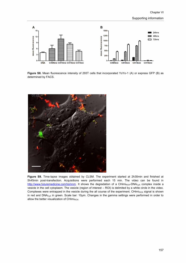
Chapter VI
Supporting information
157
Figure S8. Mean fluorescence intensity of 293T cells that incorporated YoYo-1 (A) or express GFP (B) as
determined by FACS.
Figure S9. Time-lapse images obtained by CLSM. The experiment started at 2h30min and finished at
5h45min post-transfection. Acquisitions were performed each 15 min. The video can be found in
http://www.futuremedicine.com/loi/nnm. It shows the degradation of a CHimiROX-DNACy5 complex inside a
vesicle in the cell cytoplasm. The vesicle (region of interest – ROI) is delimited by a white circle in the video.
Complexes were entrapped in the vesicle during the all course of the experiment. CHimiROX signal is shown
in red and DNACy5 in green. Scale bar: 10µm. Changes in the gamma settings were performed in order to
allow the better visualization of CHimiROX.
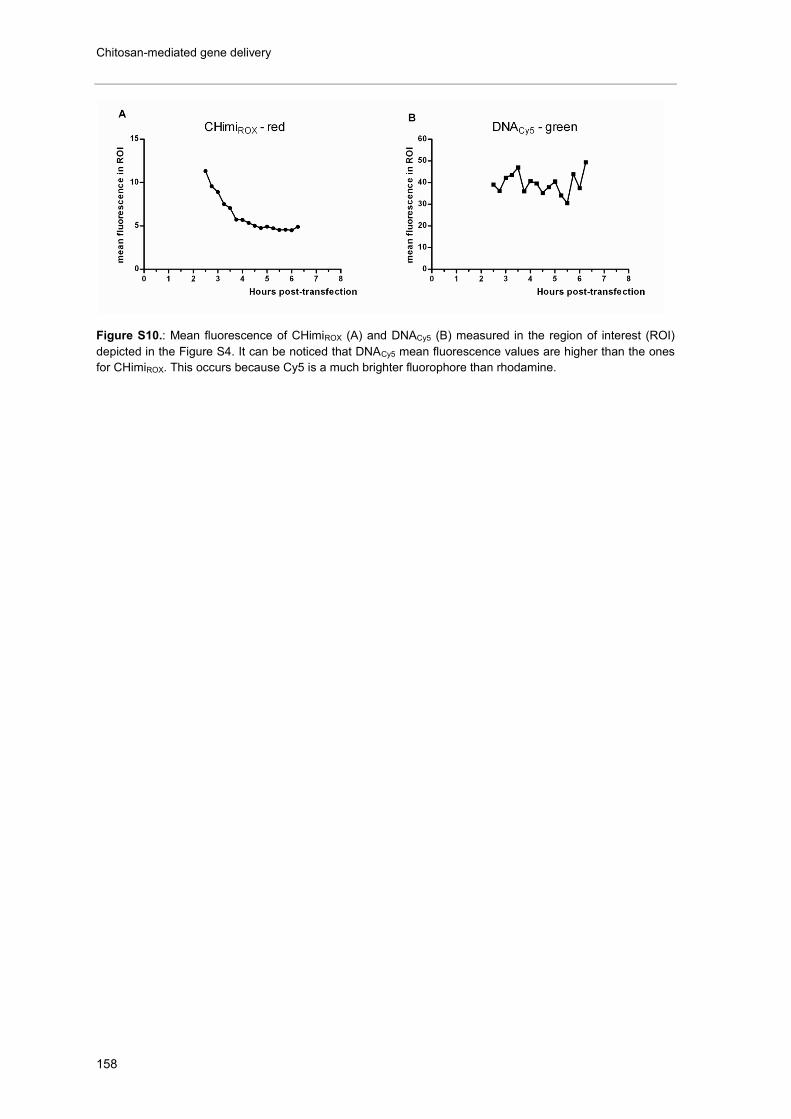
Chitosan-mediated gene delivery
158
Figure S10.: Mean fluorescence of CHimiROX (A) and DNACy5 (B) measured in the region of interest (ROI)
depicted in the Figure S4. It can be noticed that DNACy5 mean fluorescence values are higher than the ones
for CHimiROX. This occurs because Cy5 is a much brighter fluorophore than rhodamine.

Chapter VI - Appendix
159
APPENDIX I
Preliminary results on the incorporation of chitosan-based
nanoparticles in poly(trimethylene carbonate-co-ε-caprolactone)
electrospun fibres
1. Introduction
The combination of gene therapy-based strategies with tissue engineering scaffolds holds the
promise of providing both physical support for cell adhesion and growth, and genetic material to
tune specific cellular processes [1]. Nucleic acids have been directly incorporated into the
matrices [2-5], or vectorized through virus [6, 7] or nanoparticles formed by interaction with lipids
[3] or cationic polymers, such as poly(ethylene imine) (PEI) [8] and chitosan [9].
In the context of spinal cord injury, the use of this combined strategy has already showed
promising results in vivo. The implantation of a multiple channel bridge loaded with lipid-DNA
nanoparticles promoted the expression of a reporter gene during at least 3 weeks [3]. More
recently, Yao and co-workers reported the sustained expression of a growth factor - neurotrophin-
3 (NT-3) - at the lesion site after implantation of a collagen conduit loaded with polymeric particles
containing the NT-3 gene. While a statistically significant functional improvement was not
achieved in the time frame of the study (4 weeks), an increased number of axons was found
crossing the conduit at the time of evaluation [10].
Currently, electrospinning is being actively investigated for the preparation of tissue engineering
scaffolds since it allows the formation of fibres at the nano/micrometer scale, mimicking the
extracellular matrix [11, 12]. Furthermore, alignment of these fibrous structures can be achieved,
by applying a rotating target. The use of aligned fibres is very promising in the field of nerve
regeneration as these can provide physical guidance for axonal regrowth [13, 14]. Although the
scientific publications concerning the incorporation of genetic material into electrospun fibres in
the context of nerve regeneration is still in its infancy, this combined strategy was already
investigated in vitro using DNA [15, 16], siRNA [17, 18] or nanocomplexes with nucleic acids [16,
18-20]. Furthermore, positive results were already reported in vivo [16, 21], namely in a diabetic
ulcer model [16].
The application of electrospun-based scaffolds for the delivery of genetic material into a SCI can
combine topographic guidance with the delivery of gene vectors that mediate the sustained
expression of therapeutic molecules (such as growth factors) or that can block the expression of
specific inhibitory factors, leading, ultimately, to an enhancement on the process of nerve
regeneration. In this study, we aim at incorporating chitosan-based nanoparticles in
poly(trimethylene carbonate-co-ε-caprolactone) [P(TMC-CL)] electrospun fibres, in order to create
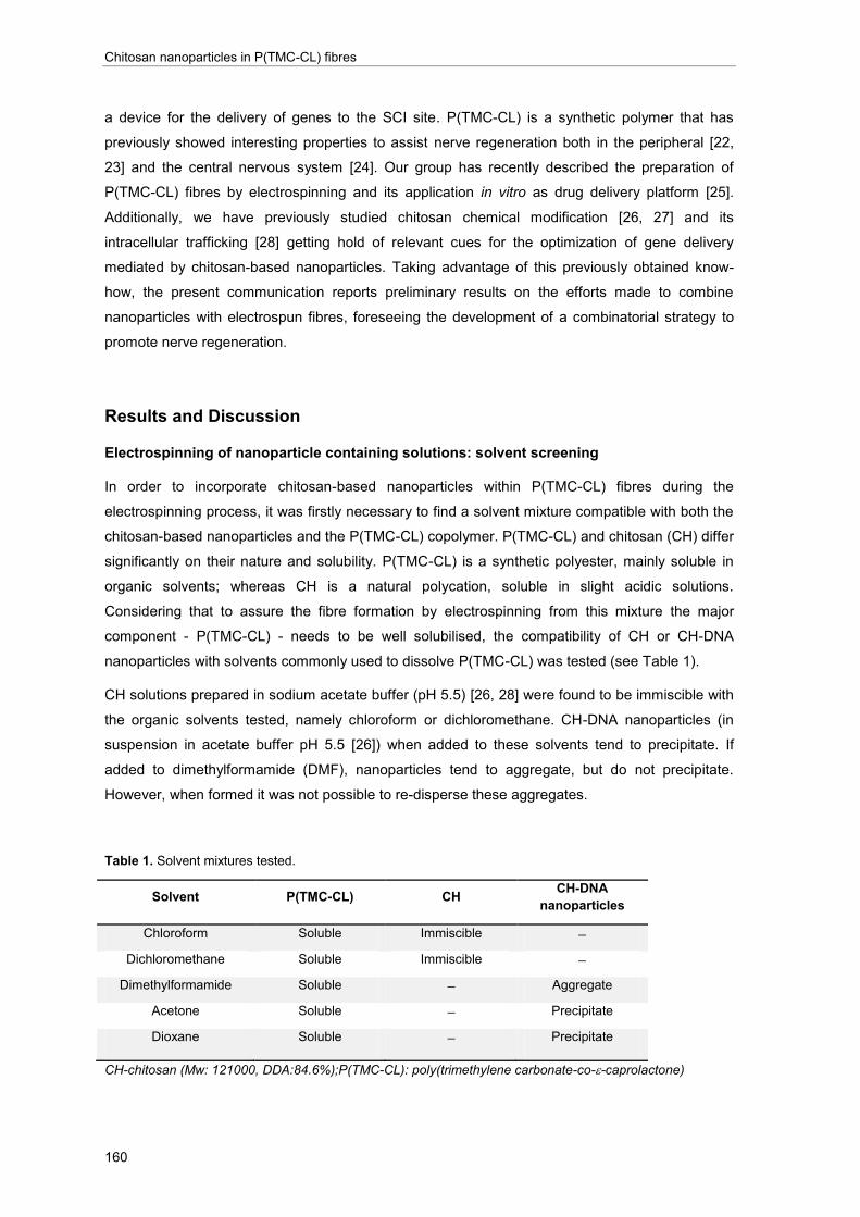
Chitosan nanoparticles in P(TMC-CL) fibres
160
a device for the delivery of genes to the SCI site. P(TMC-CL) is a synthetic polymer that has
previously showed interesting properties to assist nerve regeneration both in the peripheral [22,
23] and the central nervous system [24]. Our group has recently described the preparation of
P(TMC-CL) fibres by electrospinning and its application in vitro as drug delivery platform [25].
Additionally, we have previously studied chitosan chemical modification [26, 27] and its
intracellular trafficking [28] getting hold of relevant cues for the optimization of gene delivery
mediated by chitosan-based nanoparticles. Taking advantage of this previously obtained know-
how, the present communication reports preliminary results on the efforts made to combine
nanoparticles with electrospun fibres, foreseeing the development of a combinatorial strategy to
promote nerve regeneration.
Results and Discussion
Electrospinning of nanoparticle containing solutions: solvent screening
In order to incorporate chitosan-based nanoparticles within P(TMC-CL) fibres during the
electrospinning process, it was firstly necessary to find a solvent mixture compatible with both the
chitosan-based nanoparticles and the P(TMC-CL) copolymer. P(TMC-CL) and chitosan (CH) differ
significantly on their nature and solubility. P(TMC-CL) is a synthetic polyester, mainly soluble in
organic solvents; whereas CH is a natural polycation, soluble in slight acidic solutions.
Considering that to assure the fibre formation by electrospinning from this mixture the major
component - P(TMC-CL) - needs to be well solubilised, the compatibility of CH or CH-DNA
nanoparticles with solvents commonly used to dissolve P(TMC-CL) was tested (see Table 1).
CH solutions prepared in sodium acetate buffer (pH 5.5) [26, 28] were found to be immiscible with
the organic solvents tested, namely chloroform or dichloromethane. CH-DNA nanoparticles (in
suspension in acetate buffer pH 5.5 [26]) when added to these solvents tend to precipitate. If
added to dimethylformamide (DMF), nanoparticles tend to aggregate, but do not precipitate.
However, when formed it was not possible to re-disperse these aggregates.
Table 1. Solvent mixtures tested.
Solvent P(TMC-CL) CH CH-DNA
nanoparticles
Chloroform Soluble Immiscible
Dichloromethane Soluble Immiscible
Dimethylformamide Soluble Aggregate
Acetone Soluble Precipitate
Dioxane Soluble Precipitate
CH-chitosan (Mw: 121000, DDA:84.6%);P(TMC-CL): poly(trimethylene carbonate-co--caprolactone)

Chapter VI - Appendix
161
In view of the results summarized in Table 1, it was considered that to efficiently suspend CH-
based nanoparticles in a polymeric organic solution required the stabilization of the nanoparticles.
Electrospinning of P(TMC-CL) fibres containing TriM-CH based nanoparticles
Our group is currently investigating the use of the CH derivative trimethyl chitosan (TriM-CH). The
quaternization of the primary amine groups of CH with methyl groups has been found to
considerably improve CH solubility, rendering it soluble in a wider range of pHs, including at
physiological pH. The group has tested TriM-CH of different molecular weights and degrees of
quaternization, as these parameters can significantly affect the polymer properties, and,
ultimately, transfection. In general, in comparison to CH, TriM-CH forms by electrostatic
interaction with plasmid DNA nanoparticles with smaller diameter and with an improved
complexation stability in different dispersants [29]. In particular, it was found that TriM-CH-based
nanoparticles have an improved stability, comparing to CH-based ones, after freezing or freeze-
drying process [29].
In the present work, we tested two modified CHs with similar degree of quaternization (< 30%), but
different molecular weights - high (110,000, TriM-CH(H)) and low molecular weight (30,000, TriM-
CH(L)). The detailed description of polymer characterization is supplied in the experimental
section (Table 3). Taking advantage of the described stability to the freeze-drying process, we
explored the possibility of resuspending these nanoparticles in an organic solvent – DMF – to,
subsequently, mix it with P(TMC-CL) solution. DMF has been commonly applied in electrospinning
solutions as a mean to increase solution conductivity [30]. Particularly, it showed to improve
P(TMC-CL) fibre morphology and homogeneity, decreasing mean fibre diameter [25].
In the preparation of the TriM-CH-DNA complexes for transfection, we have been employing TriM-
CH polymer solutions in glucose 5% (w/v), pH 7.4 (Aida Moreira, unpublished data). In order to
avoid interference of glucose in the electrospinning process, the stability of the TriM-CH based
nanoparticles to the freeze-drying process when in a water solution (MilliQ grade) was firstly
investigated.
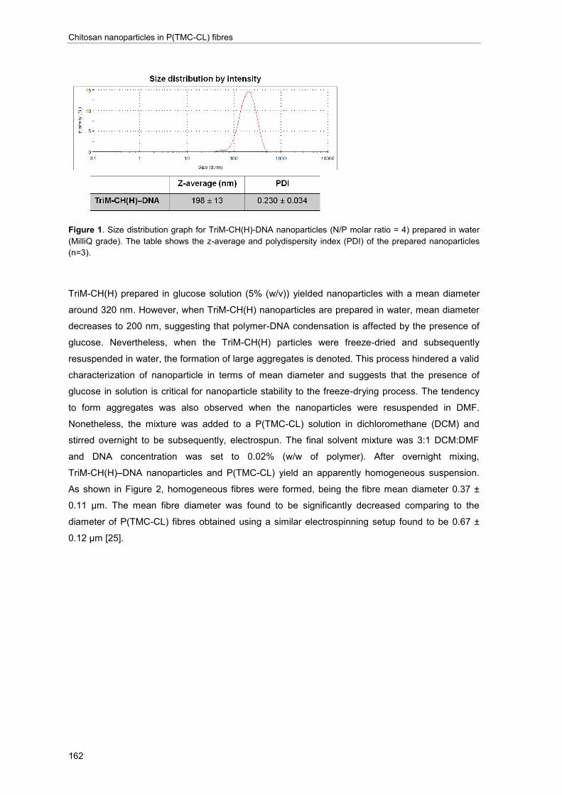
Chitosan nanoparticles in P(TMC-CL) fibres
162
Figure 1. Size distribution graph for TriM-CH(H)-DNA nanoparticles (N/P molar ratio = 4) prepared in water
(MilliQ grade). The table shows the z-average and polydispersity index (PDI) of the prepared nanoparticles
(n=3).
TriM-CH(H) prepared in glucose solution (5% (w/v)) yielded nanoparticles with a mean diameter
around 320 nm. However, when TriM-CH(H) nanoparticles are prepared in water, mean diameter
decreases to 200 nm, suggesting that polymer-DNA condensation is affected by the presence of
glucose. Nevertheless, when the TriM-CH(H) particles were freeze-dried and subsequently
resuspended in water, the formation of large aggregates is denoted. This process hindered a valid
characterization of nanoparticle in terms of mean diameter and suggests that the presence of
glucose in solution is critical for nanoparticle stability to the freeze-drying process. The tendency
to form aggregates was also observed when the nanoparticles were resuspended in DMF.
Nonetheless, the mixture was added to a P(TMC-CL) solution in dichloromethane (DCM) and
stirred overnight to be subsequently, electrospun. The final solvent mixture was 3:1 DCM:DMF
and DNA concentration was set to 0.02% (w/w of polymer). After overnight mixing,
TriM-CH(H)–DNA nanoparticles and P(TMC-CL) yield an apparently homogeneous suspension.
As shown in Figure 2, homogeneous fibres were formed, being the fibre mean diameter 0.37 ±
0.11 μm. The mean fibre diameter was found to be significantly decreased comparing to the
diameter of P(TMC-CL) fibres obtained using a similar electrospinning setup found to be 0.67 ±
0.12 μm [25].
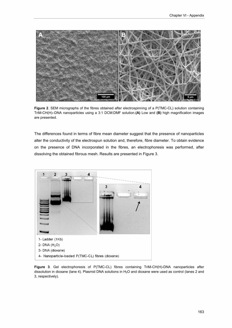
Chapter VI - Appendix
163
Figure 2. SEM micrographs of the fibres obtained after electrospinning of a P(TMC-CL) solution containing
TriM-CH(H)–DNA nanoparticles using a 3:1 DCM:DMF solution.(A) Low and (B) high magnification images
are presented.
The differences found in terms of fibre mean diameter suggest that the presence of nanoparticles
alter the conductivity of the electrospun solution and, therefore, fibre diameter. To obtain evidence
on the presence of DNA incorporated in the fibres, an electrophoresis was performed, after
dissolving the obtained fibrous mesh. Results are presented in Figure 3.
Figure 3. Gel electrophoresis of P(TMC-CL) fibres containing TriM-CH(H)-DNA nanoparticles after
dissolution in dioxane (lane 4). Plasmid DNA solutions in H2O and dioxane were used as control (lanes 2 and
3, respectively).

Chitosan nanoparticles in P(TMC-CL) fibres
164
Gel electrophoresis of the fibrous mesh solution was performed along with a DNA solution in
water or in the same solvent used to dissolve the fibrous mesh. Figure 3 shows that dioxane
interferes with the plasmid DNA run, but does not hamper its detection (Figure 3, lane 3). When
analysing the run of the fibrous mesh solution (lane 4), one should take into account that P(TMC-
CL) fibres were prepared with a DNA loading of 0.02% (w/w of polymer). Consequently, in this
particular experiment, the maximum amount of DNA present in the fibres and therefore, loaded in
the gel, was 70 ng, corresponding to approximately 15 times less than the control (1 µg, lane 3).
Figure 3 (lane 4) shows a band that although significantly smaller in comparison to control,
suggests the presence of DNA that has been retained near the loading well. This result points to
the presence of TriM-CH and that the positive charge from the polymer is hindering the DNA to
move electrophoretically. Although suggesting that both DNA and polymer are present, the result
obtained in this assay cannot assure that in the fibre both are still complexed in the form of
nanoparticles.
To prove the presence of the nanoparticles in the fibres we prepared fluorescently-labelled
nanoparticles, by labelling TriM-CH(L) with rhodamine (TriM-CH(L)ROX) as previously described for
chitosan [28]. We proceeded these studies with TriM-CH(L) with a lower molecular weight (see
Table 3) as in the course of this work this polymer was found to be more efficient in terms of
stability to storage and transfection efficiency than TriM-CH(H) (Aida Moreira, unpublished data).
The nanoparticles prepared using TriM-CH(L)ROX dissolved in water (MilliQ grade) were
characterized in terms of size as prepared and after freeze-drying and resuspension (Table 2).
Table 2. Characterization in terms of Z-average diameter (nm) and polydispersity index (PDI) of
nanoparticles prepared with TriM-CH(L)ROX (n=3) at an N/P molar ratio of 2.
Nanoparticles Z-average (nm) PDI
TriM-CH(L)ROX–DNA 136 ± 31 0.248 ± 0.02
TriM-CH(L)ROX–DNA (freeze dryed) 349 ± 29 0.349 ± 0.02
Z-ave (nm): Z-average nanoparticle diameter; PDI: polydispersity index.

Chapter VI - Appendix
165
Figure 4. Size distribution graph of fluorescently-labelled TriM-CH(L)ROX-DNA nanoparticles (N/P molar
ratio= 2) prepared in water, before and after freeze-drying process.
Nanoparticles prepared using the fluorescently-labelled TriM-CH(L)ROX show a mean diameter
around 136 nm. After freeze-drying and resuspension, particle diameter increases above 300 nm,
being observed the formation of a small population of particles of larger diameter. These effects
may be caused by the use of water as solvent, as mentioned above for TriM-CH(H). The use of
water as dispersant for nanoparticles does not favour stability to the freeze-drying process,
comparing to glucose solutions. Indeed, glucose and other sugars have been previously applied
as cryopreservatives [31]. Nonetheless, it was observed that nanoparticles based on TriM-
CH(L)ROX can be easily suspended after freeze-drying without forming aggregates.
To prepare nanoparticle-loaded P(TMC-CL) fibres, TriM-CH(L)ROX–DNA nanoparticles were
resuspended in DMF and added to a P(TMC-CL) solution. The mixture showed a light pink colour,
indicating the presence of the fluorochrome. The fibres obtained after electrospinning of this
mixture are presented in Figure 5.
The fibre formation was found not to be homogeneous and two distinct regions of deposition were
obtained. In the main region, fibres are formed (Figure 5, A-B), being the fibre mean diameter
found to be 1.04 ± 0.38 μm. In the second region some fibres are deposited, as well as drops of
the suspension (Figure 5, C). Since the two deposition areas were independent, fibres collected in
the first area were considered for the posterior analyses.
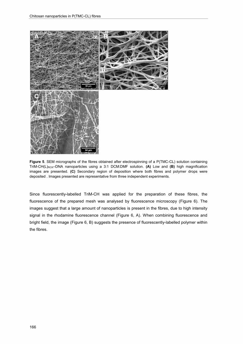
Chitosan nanoparticles in P(TMC-CL) fibres
166
Figure 5. SEM micrographs of the fibres obtained after electrospinning of a P(TMC-CL) solution containing
TriM-CH(L)ROX–DNA nanoparticles using a 3:1 DCM:DMF solution. (A) Low and (B) high magnification
images are presented. (C) Secondary region of deposition where both fibres and polymer drops were
deposited . Images presented are representative from three independent experiments.
Since fluorescently-labelled TriM-CH was applied for the preparation of these fibres, the
fluorescence of the prepared mesh was analysed by fluorescence microscopy (Figure 6). The
images suggest that a large amount of nanoparticles is present in the fibres, due to high intensity
signal in the rhodamine fluorescence channel (Figure 6, A). When combining fluorescence and
bright field, the image (Figure 6, B) suggests the presence of fluorescently-labelled polymer within
the fibres.

Chapter VI - Appendix
167
Figure 6. (A) Fluorescence images (B) combined with bright field image of the P(TMC-CL) fibres containing
TriM-CH(L)ROX nanoparticles. Amplification image suggests that fluorescence from TriM-CH(L)ROX co-localize
with the fibre as indicated by the arrows.
To investigate if the TriM-CH(L)ROX were present in the P(TMC-CL) fibres and can be released
when incubated in phosphate buffered saline (PBS), the fluorescence of the releasing medium
was followed during 23 days (Figure 7).
Figure 7. Cumulative release of fluorescent particles (λex=575nm, λex=605nm) to the releasing medium when
P(TMC-CL) fibres containing TriM-CH(L)ROX nanoparticles were incubated with PBS at 37°C. RFU: relative
fluorescence units.
The results presented in Figure 7 show that there is a gradual release of fluorescent particles to
the medium (PBS) over time, suggesting that TriM-CH(L)ROX based nanoparticles are being
released from P(TMC-CL) fibres. The fluorescence intensity of the prepared fibres was mapped
using the area scan functionality of the fluorometer before and after incubation in PBS.

Chitosan nanoparticles in P(TMC-CL) fibres
168
Figure 8. Area scan of fluorescence (λex=575 nm, λex=605 nm) found in P(TMC-CL) fibres containing TriM-
CH(L)ROX–DNA nanoparticles before (A) and after (B) incubation in PBS during 23 days.
The mapping of the fluorescence of P(TMC-CL) fibres demonstrates that fluorescence intensity
decreases after incubation with PBS in all the sample. Since some fluorescence can still be found,
this suggests that the majority of the nanoparticles were released from the P(TMC-CL) fibres
during the 23 days of incubation, but some can still be entrapped within the polymer mesh.
In a very preliminary experiment, ND7/23 cells were seeded on top of P(TMC-CL) fibres loaded
with TriM-CH(L)ROX nanoparticles. As shown in Figure 9 a significant amount of cells are found
adhered to the fibres, suggesting that these can be a good substrate for cell growth. Additionally,
the presence of fluorescence signal in the rhodamine channel (shown in red) suggests that the
fluorescent polymer is present in the fibrous mesh. In this experiment a plasmid DNA encoding for
green fluorescence protein (GFP) was applied, in order to identify transfected cells by their green
fluorescence. However, no GFP signal was observed, indicating that transfection was not efficient
in the time frame of the experiment (72 hrs). This result could be caused by the limited period of
incubation with the fibres. The sustained/delayed release of the nanoparticles from the fibrous
mesh may limit the amount of nanoparticles available to be internalized by the cells, and
consequently can limit transfection. Previous reports using polymeric nanoparticles based on PEI
incorporated on electrospun fibres showed that transfection efficiency increases up to day 7 after
seeding [16, 19]. Alternatively, we hypothesize that the amount of plasmid DNA available to be
delivered in this experiment can be insufficient. since the prepared fibres were loaded with 0.02%
(w/w of polymer) DNA whereas others report the use of 0.1% (w/w of polymer) DNA loading [19].
A quantification of the actual amount of DNA that is released over time is necessary to adjust DNA
loading in the P(TMC-CL) fibres and also to establish appropriate cell culture time-points.

Chapter VI - Appendix
169
Figure 9. Fluorescence image of ND7/23 cells seeded on top of P(TMC-CL) electrospun fibre loaded with
TriM-CH(L)ROX-DNA nanoparticles. The nanoparticles are shown in red and cell nucleus is shown in blue.
The auto-fluorescence of the P(TMC-CL) fibres allows its identification below the cells.
4. Conclusion and future directions
The results presented in this section, although preliminary, provide important insights for a
successful preparation of electrospun fibres containing chitosan-based nanoparticles.
We found that nanoparticles based on TriM-CH are appropriate to combine with P(TMC-CL)
electrospun fibres. Their stability to freeze-drying process allowed resuspension in a organic
solvent, favouring fibre formation process. Our preliminary results suggest that nanoparticles
prepared with TriM-CH(H), though tending to form aggregates after freeze-drying, can be
homogeneously dispersed in the P(TMC-CL) solution, yielding more homogeneous electrospun
fibres. Conversely, TriM-CH(L)ROX based nanoparticles dispersed in P(TMC-CL) solution fibres
yield higher diameter fibres and a less homogeneous mesh. The higher molecular weight of TriM-
CH(H) can better fit in the electrospinning requirements. Future efforts will be focused on the
preparation of homogeneous solutions in order to avoid the formation of droplets during
electrospinning, effect that we were not able to circumvent when using TriM-CH(L)ROX, so far.
The presented results also highlight the importance of using labelled nanoparticles to allow its
identification within the fibrous mesh. The fluorescent labelling was explored in this study but,
although the presence of fluorescence signal in the fibres suggests the presence of particles, one
cannot exclude that some unbound fluorochrome exists. Furthermore, it is important to figure out if
the fluorescence signal corresponds to nanoparticles (polymer and DNA) or only to TriM-CH
entrapped in the P(TMC-CL) mesh. Double fluorescent-labelling of DNA and TriM-CH can address
this issue. Alternative labelling methods using quantum dots or gold nanoparticles can improve
sensibility of detection. The use of chemical characterization techniques, like confocal-Raman

Chitosan nanoparticles in P(TMC-CL) fibres
170
should be taken into consideration, as it can also provide information on the nanoparticle
distribution within the mesh.
Future work on this project will certainly include the investigation of DNA bioactivity in transfection
experiments in order to clarify if electrospinning process alters DNA or if CH-based nanoparticles
maintain their ability to deliver DNA. The quantitative evaluation of the amount of DNA released
over time will be paramount for establishing appropriate time-points for cell culture studies on
transfection efficiency.
Experimental section
Materials
Poly(trimethylene carbonate-co-ε-caprolactone) [P(TMC-CL)] was synthesized as previously
described [25, 32] using trimethylene carbonate as received (Boehringer Ingelheim, Germany)
and ε-caprolactone (Merck, Germany) distilled before co-polymerization reaction. The synthesized
polymer was purified by precipitation into a tenfold volume of ethanol (96%, v/v; AGA, Portugal).
The purified polymer contains 11% mol of TMC; the number weight molecular weight was found to
be 8.2x10⁴ and the polydispersity index was 1.61 [25].
Technical grade chitosan (CH) (Chimarin™, degree of deacetylation (DDA) 87%, apparent
viscosity 8 mPa.s, supplied by Medicarb, Sweden) was purified as previously described [26]. The
average weight molecular weight (
M w), polydispersity index and DDA of the purified polymer were
found to be 1.2x10⁵, 2.1 and 84.6%, respectively [26] (Table 3). Chitosan stock solutions (0.1%
(w/v)) were prepared in acetate buffer solution (5 mM, pH 5.5) and stored at 4°C till further use.
Trimethylchitosan (TriM-CH) of fungal origin was obtained from Kitozyme (Belgium). Two TriM-CH
of different molecular weight were tested in these experiments. The characterization of the
polymers under investigation is presented in Table 3. TriM-CH solutions were prepared by
dissolving the polymer overnight in water (MilliQ grade) at a final concentration of 0.1% (w/v).
Table 3. Characterization of the polymers tested in this study.
Mw x 103 DQ (%) DDA (%)
CH 121 84.6
TriM-CH(L) 30 22 88.8
TriM-CH(H) 110 29 77.7
Mw: average weight molecular weight; DQ: degree of quaternization; DDA: degree of deacetylation;
Plasmid DNA encoding for GFP (pCMV-GFP, 7.4 kbp) was used in this study to prepare
nanoparticles with CH or TriM-CH. Plasmid was amplified in DH5 Escherichia coli (E. coli) and

Chapter VI - Appendix
171
isolated using GENELUTE™ high performance endotoxin-free plasmid Gigaprep kit according to
the manufacturer instructions (Sigma-Aldrich). Plasmid concentration and purity were determined
spectrophotometrically (NanoDrop, Thermo Scientific). The ratio between optical density at 260
nm and 280 nm was found to be higher than 1.7.
Preparation of nanoparticles
To prepare nanoparticles, equal volumes of pre-heated (55°C, 10 minutes) CH or TriM-CH and
DNA (in MilliQ-grade water) solutions were mixed by adding the DNA solution dropwise to the
polymer solution, under vortex. Nanoparticles were allowed to form and stabilize for 15 minutes at
RT before further use.
Taking into consideration the transfection activity results previously obtained [26, 28], CH-DNA
complexes were prepared at a molar ratio of chitosan primary amines to DNA phosphate groups
(N/P) of 18. For TriM-CH-based complexes, N/P molar ratio is expressed in terms of moles of
quaternized amine groups of the polymer (-N-(CH3)3), to moles of phosphate groups of DNA. The
N/P molar ratios applied in this study were 4 and 2, respectively for TriM-CH(H) and Tri-CH(L).
For electrospinning experiments, TriM-CH-based nanoparticles were prepared containing 20 μg of
plasmid DNA, freeze-dried (-80ºC, 0.008 mBar, 48 hrs, Labconco, USA) and stored at room
temperature till further use.
Solvent screening
The compatibility of P(TMC-CL) and CH or CH-DNA nanoparticles with different solvents was
investigated. Solvents tested include dichloromethane (DCM), dimethylformamide (DMF),
chloroform, acetone, and dioxane. Visual analysis was performed.
Chitosan-based nanoparticle characterization
Nanoparticles prepared as previously mentioned were characterized in terms of size using a
Zetasizer Nano Zs (Malvern, UK). Cumulant analysis was used for mean particle size
determination. All measurements were performed in triplicate, at 25°C.
Trimethyl chitosan fluorescent labelling
Fluorescent labelling of TriM-CH was performed based on the previous described procedure [28]
with minor modifications. In brief, TriM-CH(L) was dissolved overnight in MilliQ water (1% (w/v)).
5(6)-Carboxy-X-rhodamine N-succinimidyl ester (ROX, Fluka) was dissolved in dimethylsulfoxide
(7.5 mg.ml-1
) and added dropwise to the polymer solution, under stirring. The reaction occurred
during 3 hrs at room temperature, under constant agitation. The labelled polymer was washed
with distilled water using Amicon® ultra centrifugal filters (3,000 kDa, Millipore), till no significant
fluorescence was detected in the supernatant (λex 575 nm, λem 605 nm). The polymer solution was
collected and freeze dried. Stock solutions of the fluorescently-labelled polymer (TriM-CH(L)ROX)
were prepared in water (MilliQ grade) at a final concentration of 0.1% (w/v).
Chitosan nanoparticles in P(TMC-CL) fibres
172
Preparation of electrospun P(TMC-CL) fibres loaded with nanoparticles
To prepare P(TMC-CL) fibres loaded with TriM-CH nanoparticles, P(TMC-CL) was dissolved
overnight in DCM. Lyophilized TriM-CH-based nanoparticles were resuspended in DMF and
stirred at 1,400 rpm overnight (Thermomixer, Eppendorf Iberica, Spain). Nanoparticle suspension
was added dropwise to the P(TMC-CL) solution and the mixture was under magnetic agitation for
additional 4 hrs. The final concentration of P(TMC-CL) was 10% (w/v) and the solvent mixture
corresponds to a 3:1 DCM:DMF ratio. DNA loading tested was 0.02% (w/w of polymer),
corresponding to 20 μg of DNA per each 100 mg of P(TMC-CL).
Although variations in the electrospinning parameters were tested, the prepared solution was
electrospun using similar electrospinning setup as described in previous work [25]. In brief, using
a vertical configuration of electrospinning, solutions were dispensed at a controlled flow rate of 1
ml.h-1
using a syringe pump (Ugo Basille, Italy). An electric field of 1 kV.cm-1
was created (Gamma
High Voltage source, USA) between the spinneret (inner diameter 0.8 mm) and the flat collector
(15x15 cm) covered with aluminium foil. Fibres were collected during 30-40 minutes and
subsequently, vacuum dried during 24 hrs.
Fibre morphology analysis
The morphology of the P(TMC-CL) fibres was observed by scanning electron microscopy (SEM,
FEI Quanta 400FEG, FEI, the Netherlands) after being sputter-coated with gold-palladium for 90
seconds (SPI Supplies, USA). Fibre diameter was quantified from SEM micrographs using an
image analysis software (Image J, version 1.39).
Gel electrophoresis
P(TMC-CL) fibres containing TriM-CH–DNA nanoparticles were dissolved in dioxane (18
mg.ml-1) overnight at room temperature. 20 μl of this solution were loaded along with 5 μl of
loading buffer (Fermentas) in a 1% (w/v) agarose gel containing 0.05 μg.ml-1
of ethidium bromide
(Q-BioGene, USA). 1 μg of plasmid DNA was used as control. The electrophoresis was run at 100
V for 45 minutes. The gel was visualized using a Gel Doc™ system (Bio-Rad, Portugal).
Evaluation of nanoparticle release from P(TMC-CL) electrospun fibres
To investigate the release of fluorescently-labelled nanoparticles the prepared fibres were
incubated in PBS at 37ºC and at day 1, 2, 5, 8, 12, 16 and 23 of incubation, the releasing medium
was completely refreshed. Fluorescence (λex=575nm, λex=605nm) of the collected medium was
analyzed using a SynergyMax (Biotek, Portugal) fluorometer. The fluorescence of P(TMC-CL)
fibres was mapped using the functionality of area scan of this equipment.
Preliminary transfection experiment using ND7/23 cell line
ND7/23 cell line (mouse neuroblastoma (N18 tg 2) x rat dorsal root ganglion neuron hybrid) was
obtained from ECACC (UK) and routinely cultured in Dulbecco’s Modified Eagle Medium (DMEM)
with Glutamax, supplemented with 10% (w/v) foetal bovine serum (FBS) (heat-inactivated at 56ºC
for 30 minutes) and 1% penicillin/streptomycin (PS, 10,000 units.ml-1
penicillin and 10,000 μg.ml-1
Chapter VI - Appendix
173
streptomycin), all supplied by Gibco (Life Technologies S.A., Spain). The cells were seeded
(4x10⁴ viable cells.cm
-2) on top of P(TMC-CL) fibres loaded with TriM-CH(L)ROX–DNA
nanoparticles. After 72 hrs of incubation, the cells were fixed in 4% (w/v) paraformaldehyde and
incubated with 4’,6-diamidino-2-phenylindole (DAPI, Vector Laboratories) for 10 minutes, to allow
nucleus fluorescent labelling. The cells were observed under an inverted fluorescence microscope
(Axiovert 200, Zeiss, Germany).


Chapter VI - Appendix
175
References
1. Jang JH, Houchin TL, and Shea LD (2004). "Gene delivery from polymer scaffolds for tissue engineering". Expert Review of Medical Devices, 1 (1): 127-138.
2. Guo T, Zhao J, Chang J, Ding Z, Hong H, Chen J, and Zhang J (2006). "Porous chitosan-gelatin
scaffold containing plasmid DNA encoding transforming growth factor-beta1 for chondrocytes proliferation". Biomaterials, 27 (7): 1095-1103.
3. De Laporte L, Lei Yan A, and Shea LD (2009). "Local gene delivery from ECM-coated poly(lactide-co-glycolide) multiple channel bridges after spinal cord injury". Biomaterials, 30 (12): 2361-2368.
4. Hosseinkhani H, Hosseinkhani M, Gabrielson NP, Pack DW, Khademhosseini A, and Kobayashi H (2008). "DNA nanoparticles encapsulated in 3D tissue-engineered scaffolds enhance osteogenic differentiation of mesenchymal stem cells". Journal of Biomedical Materials Research - Part A, 85 (1): 47-60.
5. Shea LD, Smiley E, Bonadio J, and Mooney DJ (1999). "DNA delivery from polymer matrices for tissue engineering". Nature Biotechnology, 17 (6): 551-554.
6. Liao IC, Chen SL, Liu JB, and Leong KW (2009). "Sustained viral gene delivery through core-shell fibers". Journal of Controlled Release, 139 (1): 48-55.
7. Wu HF, Cen JS, Zhong Q, Chen L, Wang J, Deng DYB, and Wan Y (2013). "The promotion of functional recovery and nerve regeneration after spinal cord injury by lentiviral vectors encoding Lingo-1 shRNA delivered by Pluronic F-127". Biomaterials, 34 (6): 1686-1700.
8. Saul JM, Linnes MP, Ratner BD, Giachelli CM, and Pun SH (2007). "Delivery of non-viral gene carriers from sphere-templated fibrin scaffolds for sustained transgene expression". Biomaterials, 28 (31): 4705-4716.
9. Nie H and Wang CH (2007). "Fabrication and characterization of PLGA/HAp composite scaffolds for delivery of BMP-2 plasmid DNA". Journal of Controlled Release, 120 (1-2): 111-121.
10. Yao L, Daly W, Newland B, Yao S, Wang W, Chen BKK, Madigan N, Windebank A, and Pandit A (2013). "Improved axonal regeneration of transected spinal cord mediated by multichannel collagen conduits functionalized with neurotrophin-3 gene". Gene Therapy, 20 (12): 1149-1157.
11. Agarwal S, Wendorff JH, and Greiner A (2009). "Progress in the Field of Electrospinning for Tissue Engineering Applications". Advanced Materials, 21 (32-33): 3343-3351.
12. Sill TJ and von Recum HA (2008). "Electrospinning: Applications in drug delivery and tissue engineering". Biomaterials, 29 (13): 1989-2006.
13. Hurtado A, Cregg JM, Wang HB, Wendell DF, Oudega M, Gilbert RJ, and McDonald JW (2011). "Robust CNS regeneration after complete spinal cord transection using aligned poly-l-lactic acid microfibers". Biomaterials, 32 (26): 6068-6079.
14. Patel S, Kurpinski K, Quigley R, Gao H, Hsiao BS, Poo MM, and Li S (2007). "Bioactive nanofibers: Synergistic effects of nanotopography and chemical signaling on cell guidance". Nano Letters, 7 (7): 2122-2128.
15. Luu YK, Kim K, Hsiao BS, Chu B, and Hadjiargyrou M (2003). "Development of a nanostructured DNA delivery scaffold via electrospinning of PLGA and PLA-PEG block copolymers". Journal of Controlled Release, 89 (2): 341-353.
16. Yang Y, Xia T, Chen F, Wei W, Liu C, He S, and Li X (2012). "Electrospun fibers with plasmid bFGF polyplex loadings promote skin wound healing in diabetic rats". Molecular Pharmaceutics, 9 (1): 48-58.
17. Cao H, Jiang X, Chai C, and Chew SY (2010). "RNA interference by nanofiber-based siRNA delivery system". Journal of Controlled Release, 144 (2): 203-212.
18. Rujitanaroj PO, Wang YC, Wang J, and Chew SY (2011). "Nanofiber-mediated controlled release of siRNA complexes for long term gene-silencing applications". Biomaterials, 32 (25): 5915-5923.
19. He S, Xia T, Wang H, Wei L, Luo X, and Li X (2012). "Multiple release of polyplexes of plasmids VEGF and bFGF from electrospun fibrous scaffolds towards regeneration of mature blood vessels". Acta Biomaterialia, 8 (7): 2659-2669.
Chitosan nanoparticles in P(TMC-CL) fibres
176
20. Mittnacht U, Hartmann H, Hein S, Oliveira H, Dong M, Pêgo AP, Kjems J, Howard KA, and Schlosshauer B (2010). "Chitosan/siRNA nanoparticles biofunctionalize nerve implants and enable neurite outgrowth". Nano Letters, 10 (10): 3933-3939.
21. Rujitanaroj PO, Jao B, Yang J, Wang F, Anderson JM, Wang J, and Chew SY (2013). "Controlling fibrous capsule formation through long-term down-regulation of collagen type I (COL1A1) expression by nanofiber-mediated siRNA gene silencing". Acta Biomaterialia, 9 (1): 4513-4524.
22. Pêgo AP, Poot AA, Grijpma DW, and Feijen J (2003). "Biodegradable elastomeric scaffolds for soft tissue engineering". Journal of Controlled Release, 87 (1-3): 69-79.
23. Vleggeert-Lankamp CLAM, Wolfs J, Pêgo AP, Van Den Berg R, Feirabend H, and Lakke E (2008). "Effect of nerve graft porosity on the refractory period of regenerating nerve fibers: Laboratory investigation". Journal of Neurosurgery, 109 (2): 294-305.
24. Rocha DN, Brites P, Fonseca C, and Pêgo AP (2014). "Poly(Trimethylene Carbonate-co-ε-Caprolactone) Promotes Axonal Growth". Plos One, 9(2): e88593.
25. Pires LR, Guarino V, Oliveira MJ, Ribeiro CC, Barbosa MA, Ambrosio L, and Pêgo AP (2013). "Ibuprofen-loaded poly(trimethylene carbonate-co-ε-caprolactone) electrospun fibers for nerve regeneration". Journal of Tissue Engineering and Regenerative Medicine: Accepted for publication.
26. Moreira C, Oliveira H, Pires LR, Simões S, Barbosa MA, and Pêgo AP (2009). "Improving chitosan-mediated gene transfer by the introduction of intracellular buffering moieties into the chitosan backbone". Acta Biomaterialia, 5 (8): 2995-3006.
27. Oliveira H, Pires LR, Fernandez R, Martins MCL, Simões S, and Pêgo AP (2010). "Chitosan-based gene delivery vectors targeted to the peripheral nervous system". Journal of Biomedical Materials Research - Part A, 95 (3 A): 801-810.
28. Pires LR, Oliveira H, Barrias CC, Sampaio P, Pereira AJ, Maiato H, Simões S, and Pêgo AP (2011). "Imidazole-grafted chitosan-mediated gene delivery: In vitro study on transfection, intracellular trafficking and degradation". Nanomedicine, 6 (9): 1499-1512.
29. Gomes CP, Varela-Moreira A, Moreno PDM, and Pêgo AP. Trimethylchitosan as a gene carrier to the peripheral nervous system. in 25th European Conference on Biomaterials. Madrid, 2013.
30. Hsu CM and Shivkumar S (2004). "N,N-dimethylformamide additions to the solution for the electrospinning of poly(ε-caprolactone) nanofibers". Macromolecular Materials and Engineering, 289 (4): 334-340.
31. Abdelwahed W, Degobert G, Stainmesse S, and Fessi H (2006). "Freeze-drying of nanoparticles: Formulation, process and storage considerations". Advanced Drug Delivery Reviews, 58 (15): 1688-1713.
32. Pêgo AP, Poot AA, Grijpma DW, and Feijen J (2001). "Copolymers of trimethylene carbonate and epsilon-caprolactone for porous nerve guides: Synthesis and properties". Journal of Biomaterials Science, Polymer Edition, 12 (1): 35-53.

CHAPTER VII
Concluding Remarks and Future Perspectives


Chapter VII
179
After the primary insult that leads to the interruption of the axonal pathways, a lesion to the spinal
cord is followed by the activation of a number of inhibitory mechanisms and a protracted period of
tissue destruction. Axonal regeneration fails in this hostile environment and the process ends up
with the formation of a cavity surrounded by scar tissue [1]. In order to promote regeneration in
such inhibitory scenery, it is well accepted that a multi-targeted approach is required [2]. In this
context, the ultimate goal of the work described in this thesis is to propose a scaffold to be
implanted in the spinal cord after a lesion, that provides physical support, guidance and
biochemical cues, constituting, utterly, a permissive substrate for axonal regrowth. Each of the
presented experimental chapters were sought to contribute to the design of such a structure.
Poly(trimethylene carbonate-co-ε-caprolactone) [P(TMC-CL)] was chosen as starting material for
the development of this scaffold, based on previous reports showing the interesting properties of
the polymer in the context of peripheral [3, 4], and central nervous system regeneration [5].
The use of electrospinning for the preparation of scaffolds for tissue engineering has been largely
investigated due to the characteristics of the resulting fibrous structure that emulates the features
of the extracellular matrix [6, 7]. Herein we report the preparation of P(TMC-CL) fibres by
electrospinning. Taking into consideration the importance of microglia, the resident immune cells
on the central nervous system (CNS), in triggering the response to injury, we described for the first
time the effect of electrospun fibres on primary microglia cells in comparison to flat solvent cast
films. In line with what is described in the open literature for other cell types, we showed that
microglia morphology is remarkably affected by the topography of the surface. It was shown that
the fibrous structure favours microglia cytoplasm elongation, and, surprisingly, the release of the
pro-inflammatory cytokine - TNFα. The classical classification of microglia ascribes a pro-
inflammatory phenotype to cells with an amoeboid morphology [8]. Moreover, in macrophages, an
elongated cell shape has been associated with an anti-inflammatory phenotype [9]. Our study
highlights the importance of specifically addressing the response of microglia in the context of
CNS regeneration. Indeed, although sharing important lineage features with macrophages,
microglia can respond differently to stimuli. This study also showed that the P(TMC-CL) surfaces
under investigation do not significantly activate microglia, as astrogliosis markers were not
exacerbated when astrocytes were in contact with microglia conditioned media. Furthermore, it
was demonstrated that microglia seeded on P(TMC-CL) fibres or solvent cast films was able to
actively contribute in myelin phagocytosis, a critical step on the progress of regeneration as myelin
debris accumulation after injury leads to the release/exposure of molecules inhibitory to axonal
regrowth. These results put forward the P(TMC-CL) surfaces as contributors for the CNS
regeneration process, modulating microglia towards a pro-regenerative activity.
Another contribute to the design of a multi-target strategy to promote axonal regeneration in the
aftermath of a spinal cord injury (SCI) is to convert these three-dimensional (3D) structures into
Concluding Remarks and Perspectives
180
drug delivery devices. By adding a drug to the electrospinning solution, drug-loaded fibres can be
obtained. To tame the inflammatory response at the spinal cord lesion site, we explored the
incorporation of a non-steroidal anti-inflammatory drug – ibuprofen – in P(TMC-CL) fibres.
Ibuprofen-loaded P(TMC-CL) fibres were successfully prepared. The fibre formation process was
optimized and by adjusting solvent mixture applied in the electrospun solution, it was
demonstrated that fibre mean diameter can be tuned. The release of ibuprofen in vitro in sink
conditions occurred in the first 24 hrs, being the released drug able to reduce the secretion of
prostaglandin E2 by human-derived macrophages, pointing out that the drug bioactivity is
maintained after the process. Furthermore, this study shows that ibuprofen-loaded P(TMC-CL)
fibres can be applied as structures with anti-inflammatory properties.
The use of ibuprofen in scaffolds to implant after a SCI enclosed, however, a double target
strategy. If on the one hand, ibuprofen can reduce cyclooxygenase activity at the lesion site and
consequently, might contribute to tame the inflammatory response [10]; on the other hand,
ibuprofen has been described to limit RhoA-mediated axonal growth inhibition, improving
functional recovery after SCI [11-13]. Consequently, we explored the effect of ibuprofen released
from drug loaded P(TMC-CL) fibre on the RhoA pathway in neuronal cells. Foreseeing an
application in vivo, we firstly created an ibuprofen-loaded bilayer scaffold composed by an outer
layer based on a P(TMC-CL) solvent cast film and an inner layer made of preferentially aligned
electrospun fibres to provide physical guidance cues for axonal regrowth. Here we report the
preparation of the scaffold and its assessment both in vitro and in vivo. It was demonstrated that
ibuprofen released from these bilayer P(TMC-CL) scaffolds is able to limit RhoA activation in
neuronal cells when these are stimulated with lysophosphatidic acid. This result encouraged the in
vivo testing of the prepared scaffolds. As proof-of-concept of the effect of ibuprofen released from
the implanted scaffold on the RhoA pathway, a preliminary study using a dorsal hemisection SCI
model was conducted. The ibuprofen-loaded P(TMC-CL) scaffolds were implanted immediately
after the lesion and maintained during five days. So far, it was observed that the scaffold is
suitable for implantation at the lesion site, wrapping the spinal cord tissue. No harmful effects were
detected; particularly, the implantation of ibuprofen-loaded scaffolds showed to have no impact on
animal survival rate. The analysis of the results of this study is currently in progress and will be
paramount to determine whether this strategy can move forward to an extended study in order to
understand if the early effects can have a consequence in axonal growth and functional outcome
and to assess the contribution of P(TMC-CL) and fibre alignment in the process.
An alternative to load scaffolds with biochemical cues is to use gene therapy-based strategies.
The premise is that the release of nanoparticles containing genetic material can guarantee the
long-term expression of proteins of interest in the spinal cord lesion site. Chitosan-based vectors
were previously proposed as gene delivery vectors [14, 15]. In this regard, we firstly performed an
extensive in vitro work on the characterization of chitosan-based vectors gene delivery, namely its

Chapter VII
181
intracellular trafficking, degradation and time window of gene expression. We showed that these
vectors mediate a long-term gene expression that can be modulated by adjusting chitosan
degradation rate.
In order to translate these knowledge into a 3D scaffold, we tested the incorporation of chitosan-
based nanoparticles in P(TMC-CL) fibres during the electrospinning process. However, it was not
possible to obtain a homogeneous polymer solution that could allow the formation of fibres. As
alternative, we proposed the use of trimethylated-chitosan based nanoparticles. This polymer has
higher solubility and nanoparticle stability is improved. In preliminary tests we showed that
P(TMC-CL) fibres containing nanoparticles can be obtained. The nanoparticle release profile and
the bioactivity of the delivered gene wait for a more detailed study. Still, the preliminary tests
presented in this thesis are encouraging indicating the feasibility of incorporating such
nanoparticles as vectors for nucleic acid delivery within electrospun polymeric fibres.
Overall, the work described in this thesis provides relevant knowledge that contributes to the
design of a multi-target scaffold to be used as a therapeutic strategy in the context of a SCI.
Engineering a permissive substrate for axonal regrowth by means of combining topography,
ibuprofen and trimethyl-chitosan-based nanoparticles for gene delivery might constitute a
successful approach towards nerve regeneration in the CNS.
While this thesis reports major findings, some questions remain to be answered as well as
additional ones can be raised. The more relevant points that remain to be addressed are
described below.
The detailed analysis of the tissues collected in the preliminary in vivo study performed is critical
to determine the significance of the proposed strategy in a SCI scenario. The evaluation of RhoA
activity in the tissues, and the characterization of the cellular populations at the lesion site can
also provide new insights for future improvements on scaffold design. In particular, the drug
loading or the drug release profile can be adjusted accordingly. The short-term study (5 days)
performed only addresses the early effect of the released drug. To assess the contribution of
P(TMC-CL) or fibre alignment on axonal growth and, ultimately, on functional recovery, the
implantation of ibuprofen-loaded scaffolds for a longer period will be of remarkable importance.
The second question that comes up from the reported work is whether one can prepare a scaffold
that combines chitosan-based nanoparticles (vectorizing a therapeutic gene) with P(TMC-CL)
fibres already loaded with ibuprofen. The combination of both gene and drug delivery strategies

Concluding Remarks and Perspectives
182
would be a major achievement, and to tune timely the release of each, would certainly constitute
the major challenge.
Additionally, of remarkable interest in the context of this thesis is to investigate the effect of
ibuprofen loaded P(TMC-CL) fibres on microglia, and to figure out how microglia response can be
altered when using surfaces based on aligned fibres.

Chapter VII
183
References
1. Schwab JM, Brechtel K, Mueller CA, Failli V, Kaps HP, Tuli SK, and Schluesener HJ (2006). "Experimental strategies to promote spinal cord regeneration - An integrative perspective". Progress in Neurobiology, 78 (2): 91-116.
2. McCreedy DA and Sakiyama-Elbert SE (2012). "Combination therapies in the CNS: Engineering the environment". Neuroscience Letters, 519 (2): 115-121.
3. Pêgo AP, Poot AA, Grijpma DW, and Feijen J (2001). "Copolymers of trimethylene carbonate and epsilon-caprolactone for porous nerve guides: Synthesis and properties". Journal of Biomaterials Science, Polymer Edition, 12 (1): 35-53.
4. Vleggeert-Lankamp CLAM, Wolfs J, Pêgo AP, Van Den Berg R, Feirabend H, and Lakke E (2008). "Effect of nerve graft porosity on the refractory period of regenerating nerve fibers: Laboratory investigation". Journal of Neurosurgery, 109 (2): 294-305.
5. Rocha DN, Brites P, Fonseca C, and Pêgo AP (2014). "Poly(Trimethylene Carbonate-co-ε-Caprolactone) Promotes Axonal Growth". Plos One, 9(2): e88593.
6. Agarwal S, Wendorff JH, and Greiner A (2009). "Progress in the Field of Electrospinning for Tissue
Engineering Applications". Advanced Materials, 21 (32-33): 3343-3351.
7. Sill TJ and von Recum HA (2008). "Electrospinning: Applications in drug delivery and tissue engineering". Biomaterials, 29 (13): 1989-2006.
8. Streit WJ, Walter SA, and Pennell NA (1999). "Reactive microgliosis". Progress in Neurobiology, 57 (6): 563-581.
9. McWhorter FY, Wang T, Nguyen P, Chung T, and Liu WF (2013). "Modulation of macrophage phenotype by cell shape". Proceedings of the National Academy of Sciences of the United States of America, 110 (43): 17253-17258.
10. Kopp MA, Liebscher T, Niedeggen A, Laufer S, Brommer B, Jungehulsing GJ, Strittmatter SM, Dirnagl U, and Schwab JM (2012). "Small-molecule-induced Rho-inhibition: NSAIDs after spinal cord injury". Cell and Tissue Research, 349 (1): 119-132.
11. Fu Q, Hue J, and Li S (2007). "Nonsteroidal anti-inflammatory drugs promote axon regeneration via RhoA inhibition". Journal of Neuroscience, 27 (15): 4154-4164.
12. Wang X, Budel S, Baughman K, Gould G, Song KH, and Strittmatter SM (2009). "Ibuprofen enhances recovery from spinal cord injury by limiting tissue loss and stimulating axonal growth". Journal of Neurotrauma, 26 (1): 81-95.
13. Zhou Y, Su Y, Li BL, Liu F, Ryder JW, Wu X, Gonzalez-DeWhitt PA, Gelfanova V, Hale JE, May PC, Paul SM, and Ni BH (2003). "Nonsteroidal anti-inflammatory drugs can lower amyloidogenic A beta(42) by inhibiting Rho". Science, 302 (5648): 1215-1217.
14. Mao HQ, Roy K, Troung-Le VL, Janes KA, Lin KY, Wang Y, August JT, and Leong KW (2001). "Chitosan-DNA nanoparticles as gene carriers: synthesis, characterization and transfection efficiency". Journal of Controlled Release, 70 (3): 399-421.
15. Moreira C, Oliveira H, Pires LR, Simões S, Barbosa MA, and Pêgo AP (2009). "Improving chitosan-mediated gene transfer by the introduction of intracellular buffering moieties into the chitosan backbone". Acta Biomaterialia, 5 (8): 2995-3006.