Embed Size (px)
Citation preview

Pontifícia Universidade Católica do Rio Grande do Sul Faculdade de Biociências
Programa de Pós-Graduação em Biologia Celular e Molecular
DISSERTAÇÃO DE MESTRADO: Estudo das variantes polimórficas 896A>G (Asp299Gly) e 1196C>T (Thr399Ile) do Toll like Receptor 4 (TLR4) e a suscetibilidade à sepse e às disfunções orgânicas em pacientes com condições críticas de saúde
PÓS-GRADUANDO PAULO ROBERTO VARGAS FALLAVENA
ORIENTADOR CLARICE SAMPAIO ALHO
Porto Alegre, RS Abril / 2007

2
SUMÁRIO
Assunto Página RESUMO 3 FUNDAMENTAÇÃO TEÓRICA 4 1 – Paciente crítico internado em UTI 4 2 – Infecção e Sepse 6 3 – Quadro Séptico 8 4 – Imunidade Inata 9 5 – Receptores Toll-like 14 6 – Receptor Toll-like 4 (TLR4) 15 7 – O gene humano do TLR4 16 JUSTIFICATIVA E OBJETIVO 20 REFERÊNCIAS BIBLIOGRÁFICAS 21 MANUSCRITO DO TRABALHO EXPERIMENTAL 26 Title of The Paper 26 Abstract 29 Introduction 30 Subjects and Methods 32 Results 36 Discussion 39 Aknowledgement 42 References 42 Table 1 46 Table 2 47 Figure 1 48 Figure 2 49 Figure 3 50 Figure 4 51 CONSIDERAÇÕES FINAIS 52

3
RESUMO
Objetivo: Investigar se há associação entre as variantes polimórficas 896A>G
(Asp299Gly) e 1196C>T (Thr399Ile) do gene que codifica para o toll like receptor 4
(TLR4) e a suscetibilidade a sepse e às disfunções orgânicas em pacientes com
condições críticas de saúde. Pacientes e Métodos: Foram selecionados para este
estudo 107 pacientes com sepse secundária a infecção por bactérias Gram-
negativas, internados na unidade de tratamento intensivo geral (UTI) do Hospital São
Lucas da PUCRS, admitidos de março de 2002 a dezembro de 2005. O grupo
controle foi constituído por 111 amostras de DNA de doadores voluntários oriundos
da mesma população gaúcha. A disfunção orgânica dos pacientes sépticos foi
avaliada durante a primeira semana após admissão na UTI, através do escore
SOFA, e foram consideradas as ocorrências de choque séptico e óbito. Os genótipos
das variantes polimórficas 896A>G e 1196C>T foram determinados por análise de
fragmentos de restrição dos produtos da reação em cadeia da polimerase (PCR).
Após, foi analisada a freqüência da distribuição dos genótipos e dos alelos entre os
grupos de pacientes e de indivíduos saudáveis. Resultados: A freqüência de
heterozigotos 896AG foi significativamente mais elevada entre pacientes sépticos
(26%; 28/107) do que entre indivíduos saudáveis (10%; 11/111) (P=0,002; OR=3,22,
CI95%: 1,43-7,38), da mesma forma como foi mais elevada se comparados pacientes
com choque séptico (24%; 18/74) e saudáveis (P=0,008; OR=2.92, CI95%: 1,20-7,17).
Correspondentemente, houve maior freqüência do alelo 896G entre pacientes
sépticos (13%; 28/214) que controles (5%; 11/222) (P=0,003; OR=2,89, CI95%: 1,33-
6,36), e entre pacientes com choque séptico (14%; 18/148) que indivíduos saudáveis
(P=0,011; OR=2,66, CI95%: 1,15-6,23). Pacientes 1196CT+1196TT apresentaram
escores SOFA médios mais altos (8.12±3.89) que pacientes 1196CC (6.82±3.52)
(Mann-Whitney test, P=0,007). Portadores do alelo 1196T apresentaram durante a
primeira semana de internação na UTI significativamente mais casos de escores
SOFA ≥ 7 (69%; 49/71) que homozigotos 1196CC (49%; 304/626) (Chi-square test,
P=0,043; OR=1.62, CI95%:0.99-2.67). Nós observamos que a herança de qualquer
tipo de alelo não interfere nas taxas de mortalidade dos pacientes. Conclusão: As
variantes polimórficas -896A>G e 1196C>T do TLR4 podem estar associadas com a
uma maior suscetibilidade ao desenvolvimento de disfunções orgânicas graves e
sepse nos pacientes críticos.

4
FUNDAMENTAÇÃO TEÓRICA
1 – Paciente crítico internado em UTI
Os pacientes internados na Unidade de Terapia Intensiva (UTI) são
caracterizados por apresentarem um quadro patológico crítico e complexo,
decorrente de fragilidades fisiológicas graves e responsáveis pela elevada taxa de
mortalidade que varia de 30% a 50% [Vincent JL, et al., 2002]. Em um estudo
recente estimou-se que nos Estados Unidos 50.000 pessoas morrem a cada ano
decorrente de doenças críticas manifestadas nas UTIs como, por exemplo a sepse,
com custo de até dez bilhões de dólares [Guha M, et al., 2001]. A despeito dos
progressos no diagnóstico e no tratamento das doenças infecciosas, a incidência de
sepse tem aumentado nas últimas décadas. O aumento das infecções causadas por
bactérias resistentes a antibióticos e o desenvolvimento de tecnologias de
manutenção de vida, com o uso de procedimentos e dispositivos invasivos, podem
explicar esse fato [Niederman MS, et al., 1990]. Nos últimos 20 anos, vários
instrumentos de medida de predição de risco têm sido aplicados aos pacientes
críticos internados em UTIs na tentativa de reconhecer as melhores estratégias
terapêuticas. A avaliação do quadro crítico, nos dias de hoje, é principalmente
realizada através de instrumentos que analisam a disfunção de órgãos e sistemas
através do monitoramento diário de seu estado fisiológico. O escore SOFA
(Sequentical Organ Failure Assessment) avalia diariamente a condição de seis
sistemas orgânicos (respiratório, renal, hepático, hematopoiético, cardiovascular e
neurológico), independentemente da terapia a qual o paciente está sendo submetida
[Vincent JL, et al., 1998]. Dado que pacientes de UTIs são indivíduos afetados por

5
múltiplas disfunções orgânicas e que, além disto, estão expostos ao ambiente
hospitalar o qual é rico em diversidade de microorganismos infecciosos, o risco de
que estes pacientes venham a desenvolver uma infecção é muito elevado (25-30%).
Uma parcela elevada de pacientes desenvolve infecção bacteriana, sendo que cerca
de 40% a 70%, acaba por desencadear quadro de sepse, choque séptico e falência
de múltiplos órgãos [Russell JA, 2006]. O quadro de sepse é conseqüência de
processos celulares em resposta a uma agressão de origem infecciosa. A
manifestação clínica da sepse pode se agravar chegando a um quadro de choque, o
qual é caracterizado pela presença de uma vasodilatação periférica acentuada e por
uma excessiva presença de agentes pró-inflamatórios, que juntos acentuam ainda
mais a disfunção e a falência de múltiplos órgãos. Observa-se o desenvolvimento de
disfunção muito antes da falência de órgãos, resultado de uma reação inflamatória
pela massiva liberação de citocinas. A resposta sistêmica à infecção é mediada
através das citocinas derivadas de macrófagos que alvejam os receptores da
extremidade-órgão em resposta a um ferimento ou infecção. A resposta inflamatória
à infecção ou ao ferimento é uma reação altamente conservada e regulada do
organismo. A liberação concomitante de agentes pró e antiinflamatório mantém a
homeostasia do organismo. A reação antiinflamatória pode ser maior e algumas
vezes mais longa que a pró-inflamatória; o objetivo disto é diminuir a produção da
síntese de agentes pró-inflamatórios, assim mantendo o equilíbrio homeostático
[Bone RC, et al., 1997; Haddad JJ, et al., 2002].
Apesar dos inúmeros progressos obtidos nas últimas décadas na tentativa de
se dar suporte ao paciente crítico com foco infeccioso e sepse, a mortalidade neste
grupo tem se mantido na faixa de 50% [Friedman G, et al., 1998].
Sendo a sepse uma condição freqüente no âmbito da terapia intensiva, que
cursa com elevada mortalidade e com tratamento com custo econômico elevado,

6
sua abordagem é de interesse direto do sistema de saúde. O estudo da sepse deve,
no entanto, contribuir para os levantamentos epidemiológicos e pautar-se numa
abordagem direcionada para o conhecimento dos mecanismos moleculares e
celulares que desencadeiam as variações fisiopatológicas. Este conhecimento
básico poderá contribuir para a modulação da seqüência de eventos que culmina
nos desfechos desfavoráveis. Conhecer as bases genéticas de tais eventos é,
portanto, fundamental.
2 - Infecção e Sepse
A sepse é caracterizada por uma ativação consistente do sistema imune após
uma infecção bacteriana (bacteremia) que se torna amplificada e, então,
desregulada [Friedman G, et al., 1998]. As infecções, na maioria dos casos, são
erradicadas com sucesso pelo hospedeiro por uma resposta inflamatória intensa e
localizada. Em contraste, as infecções fatais são caracterizadas por uma inabilidade
em conter a resposta inflamatória, na qual ocorre a liberação de potentes citocinas
na circulação sistêmica, ativando a resposta inflamatória celular, em posições
remotas, uma condição conhecida como Síndrome Sistêmica da Resposta
Inflamatória (SIRS; systemic inflamatory response syndrome). Em 1991, o conceito
SIRS foi postulado para definir o estado de pacientes que exibiam uma resposta
sistêmica para episódios inflamatórios [Crouser ED, 2004]. Hoje, a sepse é definida
como SIRS induzida por uma infecção. A SIRS é diagnosticada como uma
combinação de sinais clínicos e sintomas disponíveis, apresentando pelo menos,
dois dos critérios a seguir: (I) Febre, temperatura corporal >38ºC ou hipotermia,
temperatura corporal <36ºC; (II) Taquicardia, freqüência cardíaca >90 bpm; (III)
Taquipnéia, freqüência respiratória >20 irpm ou PaCO2 <32 mmHg; (IV) Leucocitose
ou leucopenia, Leucócitos >12.000 cels/mm3 ou <4.000 cels/mm3, ou presença de

7
>10% de leucócitos de formas jovens (bastões) [Heard SO, et al., 1991 ; Varon J, et
al., 1999; Vincent JL, et al., 2002].
Considera-se sepse-grave quando a sepse está associada a manifestações
de hipoperfusão tecidual e disfunção orgânica, caracterizada por acidose lática,
oligúria ou alteração do nível de consciência, ou hipotensão arterial com pressão
sistólica inferior a 90mmHg (sem a necessidade, porém, de agentes vasopressores).
Aproximadamente 40% dos pacientes sépticos progridem para o choque séptico
[Kreger BE, et al., 1980]. A redução da perfusão sistêmica durante o choque
combinada com uma cascata de eventos inflamatórios iniciado durante a sepse,
geralmente causa o desenvolvimento de falência de múltiplos órgãos, seguido de
morte [Liang E, et al., 2003]. As possibilidades para o desenvolvimento de sepse
pela SIRS incluem a oxigenação inadequada do tecido relacionado à má distribuição
do fluxo do sangue, a causa colateral dos danos nos tecidos causada pelas células
imunes ativadas, e as mudanças citoplasmáticas conseqüentes às interações do
receptor citocina-celula. O que quer que cause o dano celular, que afete os sistemas
orgânicos, perpetua a SIRS pela ativação dos macrófagos. Este feed-foward é o
mecanismo que explica a morte de pacientes pela chamada Síndrome da Disfunção
de Múltiplos Órgãos (MODS; multiple organ dysfunction sindrome) após dias ou
semanas do início da infecção inicial e, em muitos casos, depois que a infecção
original já tenha até mesmo sido erradicada. Apesar dos bilhões de dólares
investidos em investigações variadas, nenhuma droga ou terapia especifica foi
desenvolvida para impedir efetivamente o início da SIRS ou da MODS. Por exemplo,
as estratégias de imuno-modulação, tais como o anticorpo anti-TNF-α e anti-
endotoxinas, parecem ser de pouco ou de nenhum benefício nos seres humanos. O
esforço para aumentar a distribuição de oxigênio aos tecidos, da mesma forma, não
8
se mostrou benéfico além de ter sido, inclusive, prejudicial aos pacientes com sepse
estabelecida [Crouser ED, 2004].
Choque séptico é uma complicação da sepse caracterizado por uma
hipotensão refratária, a qual é a principal causa dos óbitos. Se a hipotensão ou
hipoperfusão induzidas pela sepse são refratárias à ressuscitação volêmica
adequada, e se há necessidade subseqüente de administração de agentes
vasopressores, as complicações circulatórias podem levar à Falência de Múltiplos
Órgãos (MOF; multiple organ failure) ou à MODS. A falência de múltiplos órgãos é
uma alteração tão severa na função orgânica que sua homeostasia não pode ser
mantida sem intervenção terapêutica artificial [Levy MM, et al., 2003].
3 - Quadro séptico
Observa-se no quadro séptico o aparecimento de estágios progressivos e
complexos causadores de desordens no sistema imunológico desencadeados pela
infecção. Diferentes fatores podem levar a SIRS, entre eles, infecção, trauma,
queimaduras, isquemia, reperfusão e inflamação. A SIRS é caracterizada por um
desequilíbrio no sistema imunológico de hiper-atividade ou hipo-atividade [Akira S,
2000].
3.1 - O estágio inicial da Sepse (Fase Hiperdinâmica)
Caracteriza-se pela ativação intensa de citocinas pró e antiinflamatórias. Esta
fase é marcada pela produção de interleucinas IL-1, IL-6 e IL-8 e do fator de necrose
tumoral TNF-α. Também há um aumento na proteína C-reativa que se acredita ser a
responsável pela ativação das espécies reativas de oxigênio e da produção de óxido
nítrico (NO) [Akira S, 2000; Hoesel LM, et al., 2004].

9
3.2 - O estágio tardio da sepse (Fase Hipodinâmica)
Esta etapa é marcada pela liberação das interleucinas IL-10 e IL-13 para
conter o efeito pró-inflamatório da fase hiperdinâmica, suprimindo a ativação do fator
nuclear NFκB. Há também um fenômeno conhecido como CARS (compensatory anti
response syndrome) que consiste em uma resposta inflamatória compensatória. No
final deste estágio há diminuição da fagocitose dos macrófagos, quimiotaxia,
produção de citocinas e de radicais livres. Existe também uma grande diferença na
capacidade de resposta das citocinas associadas a outros fatores nestes dois
estágios [Titheradge MA, 1999]. Ratos cujo gene do receptor para o TNF-α, TNF-
αRp55 foi inativado ou de alguma maneira bloqueado (ratos knock-out), tornaram-se
resistentes ao choque endotóxico embora o TNF-α não esteja relacionado
diretamente com letalidade, evento que depende de um efeito sinérgico do TNF-α,
IL-β, IFN-γ e NO [Moncada S, et al., 1991].
4 - Imunidade Inata
Ao contrário da imunidade adaptativa, a qual é específica e se molda ao
agente infeccioso criando uma memória imunológica, o sistema imune inato
reconhece classes genéricas de moléculas produzidas por vários microorganismos
patogênicos. A imunidade inata é a primeira linha de defesa do organismo contra
microorganismos invasores, desempenhando um papel fundamental nas doenças
infecciosas e inflamatórias, uma vez que provoca uma resposta inflamatória
generalista, na qual certas células (macrófagos, monócitos, granulócitos e células
dendríticas) detêm o agente invasor, impedindo que ele se espalhe [Zweigner J, et
al., 2001]. Uma função importante para a resposta imune inata é de recrutar mais
células fagocitárias e moléculas efetoras para o local da infecção, através da
liberação de uma bateria de citocinas e de outros mediadores inflamatórios que têm

10
profundos efeitos sobre os fatos subseqüentes. As citocinas, cuja síntese é
estimulada quando os macrófagos reconhecem constituintes microbianos, são
chamadas freqüentemente de monocinas, uma vez que são elaboradas
principalmente por células da linhagem monócito-macrófago; as monocinas
compreendem um grupo estruturalmente diferenciado de moléculas e incluem as
interleucinas IL-1, IL-6, IL-8, IL-12 e o fator de necrose tumoral (TNF-α), todos
apresentam importantes efeitos locais e sistêmicos [Bevilacqua MP, 1993; Downey
GP, 1994; Springer TA, 1994]. Os efeitos combinados desses mediadores
contribuem para as reações locais contra a infecção na forma de resposta
inflamatória. As respostas inflamatórias operacionais são caracterizadas pela dor,
rubor, calor e pelo tumor no sítio de uma infecção. A primeira dessas alterações
reside num aumento do diâmetro vascular, levando a um aumento do volume
sanguíneo local. Uma vez que a infecção se dissemine para a corrente circulatória,
os mesmos mecanismos através dos quais o TNF-α continha a infecção local com
tanta eficiência, tornam-se ineficazes. A sepse é, portanto, acompanhada pela
liberação de TNF-α pelos macrófagos no fígado, no baço e em outros órgãos. A
liberação sistêmica de TNF-α causa vasodilatação e perda do volume plasmático,
devido a um aumento da permeabilidade vascular, conduzindo ao choque. No
choque séptico, a coagulação intravascular disseminada é igualmente deflagrada
pelo TNF-α, levando à formação de microtrombos e ao consumo de proteínas de
coagulação, de modo que o paciente perde a capacidade de coagular o sangue de
maneira apropriada. Tal condição leva com freqüência à falência de órgãos vitais tais
como os rins, o fígado, o coração e os pulmões, que são rapidamente
comprometidos pela insuficiência da perfusão normal [Biron CA, 1992; Sen GC,
1992; Weiss WI, et al., 1992; Pfieffer K, et al., 1993; Emsley J, et al., 1994; Janeway
CAJR, 1997; Fraser IP, et al., 1998; Lamping N, et al., 1998].

11
O reconhecimento de componentes de bactérias, e a subseqüente ativação de
citocinas são características importantes envolvidas durante o quadro séptico.
Recentemente, vários polimorfismos de genes associados com estas respostas têm
sido demonstrados: entre eles, variantes polimórficas no gene do TNF-α têm sido
relacionadas como fator importante no curso clínico da sepse. O polimorfismo -
308G>A do gene TNF-α foi identificado e associado a capacidade transcricional
diferenciada do mesmo [Nakada TA, et al., 2005 ]. A liberação sistêmica de TNF-α
causa vasodilatação e perda de volume plasmático, devido a um aumento da
permeabilidade vascular, conduzindo ao choque séptico. Um estudo com pacientes
sépticos mostrou que a mortalidade foi significativamente mais elevada entre
heterozigotos -308GA quando comparados com os homozigotos 308GG [Nakada
TA, et al., 2005].
Infecções devido a bactérias Gram-negativas são comumente associadas à
principal causa de sepse e choque séptico [Holmes CL, et al., 2003]. Estudos
baseados no reconhecimento por monócitos de lipopolissacarídeos (LPS) presentes
em bactérias Gram-negativas e na regulação de genes envolvidos no processo
inflamatório podem identificar novos instrumentos terapêuticos. O processo da
imunidade inata, de resposta rápida para detectar e eliminar infecções causadas por
agentes contagiosos (bactérias Gram-negativa), é iniciado pela molécula CD14
(cluster of differenciation 14) e mediado pelos receptores Toll-Like (Toll-like
receptors; TLRs), os quais fazem parte de uma família de receptores conservada
desde de artrópodes até mamíferos [Ebong SJ, et al., 2001; Xu D, et al., 2005; Glück
T, et al., 2001] . O CD14, um receptor de membrana que tem papel fundamental no
reconhecimento de LPS por monócitos, tem sido citado frequentemente como um
dos principais genes envolvidos em doenças inflamatórias. No promotor do gene
CD14 foi encontrado um polimorfismo de substituição de um único nucleotídeo

12
(single nucleotide polymorphism; SNP) que parece estar associado à regulação da
expressão do gene CD14 [Niederman MS, et al., 1990]. A regulação do CD14 parece
ser de extrema importância em vários estados de doenças inflamatórias [Le Van TD,
et al., 2001], e sua expressão diferenciada pode ser crucial para o curso clínico
dessas patologias [Glück T, et al., 2001]. LPSs de bactérias invasoras, presentes no
plasma humano, ligam-se à receptores HDL (high density lipoprotein) e são
reconhecidas por LBPs (lipopolysaccharide binding proteins). As LBPs, por sua vez,
transferem às LPSs do HDL para o CD14 [Landmann R, et al., 1996; Goyert SM, et
al., 1998]. A sinalização imunológica é, então, iniciada pelo complexo LBP-LPS-
CD14 [Jerala R, 2007]. O CD14 é uma proteína que pode ser encontrada fixa na
membrana de células do sistema imune (mCD14) ou livre no soro (sCD14). O CD14,
contudo, não tem conexão com o citoplasma celular não podendo ativar a resposta
imune isoladamente [Silva E, et al., 2005]. A mediação do processo infeccioso inato
se dá, portanto, pelos TLRs [Agnese MD, et al., 2002, Jerala R, 2007].
O LPS é um composto carregado negativamente e consiste de três estruturas
interconectadas que dão à molécula uma característica polar e anfipática. A
estrutura básica consiste de um lipídio A (um núcleo estrutural de oligossacarídeo) e
um polissacarídeo de repetição de cadeia que da a especificidade sorotípica e
características polar a toda a estrutura do LPS [Opal SM, et al., 1998]. A estrutura do
lipídio A geralmente consiste de uma desfosforilação β1-6 ligado com uma
glicosamina, na qual se liga um número variado (usualmente seis) de amidos
assimétricos ou éster-ligantes de ácidos graxos [Opal SM, et al., 1998].
Acredita-se que a porção do lipídio A seja a responsável pela ligação do LPS
aos receptores de vários alvos, conferindo toxicidade ao LPS [Wong N, et al., 2003].
A parede celular das bactérias Gram-negativas é assimétrica: o LPS cobre mais de
90% da superfície celular na face externa e os fosfolipídios, que têm uma

13
composição similar à da membrana citoplasmática, estão localizados na face interna.
A parede celular serve como uma barreira fixa fornecendo proteção à bactéria em
resposta a agentes anti-bacterianos. Assim, é a primeira barreira encontrada pelos
peptídeos de reconhecimento do sistema imune inato dos organismos multicelulares
que devem atravessá-la a fim de alcançar a membrana citoplasmática interna.
Inicialmente, os peptídeos de reconhecimento interagem com a face exterior do LPS
e deslocam competitivamente os cátions bivalentes que neutralizam parcialmente
sua carga negativa [Rosenfeld Y, et al., 2006]. A presença da endotoxina LPS alerta
o hospedeiro eucariótico sobre a presença de um invasor bacteriano [Opal SM, et
al., 1998]. A presença de concentrações patológicas de endotoxina nos pacientes
com sepsis severa e choque séptico tem sido documentada em numerosos estudos
clínicos [Danner RL, et al., 1991; Opal SM, et al., 1998; Van Deventer, et al., 1998;
Opal SM, et al., 1999].
Huang e colaboradores [Huang Q, et al., 2001] demonstraram que a
transcrição de muitos genes está alterada após exposição a endotoxina bacteriana
e, dos 166 genes que foram alterados na exposição à Escherichia coli, cerca de 88%
também foram alterados após a exposição a LPS purificado. Isto indica que o LPS é
a molécula dominante que é reconhecida por células do sistema imunológico [Opal
SM, et al., 2003]. O reconhecimento de componentes de bactérias, tais como as
LPS, pelo sistema imune inato desencadeia a resposta inflamatória com o objetivo
de acabar com o agente infeccioso. Uma resposta inflamatória sem controle, no
entanto, pode ser a causa de disfunções orgânicas importantes [Zweigner J, et al.,
2001].
Estudos recentes têm se baseado na inativação do LPS por um composto
sintético similar ao lipídio A, componente do LPS [Liang E, et al., 2003; Wong N, et
al., 2003]. Esse antagonista, conhecido como E5564, mostrou ser bastante eficiente

14
em bloquear a ação do LPS em modelos experimentais in vitro e in vivo, e vem
sendo testado em pesquisas clínicas de fase II como um alvo terapêutico para sepse
[Liang E, et al., 2003; Wong N, et al., 2003]. O E5564 {α-D-Glicopiranose, 3-O-Decil-
2-deoxi-6-O-[2-deoxi-3-O-[(3R)-3-metoxidecil]-6-O-metil-2-[[((11Z)-1-oxo-11-
octanodecenil) amino]-4-O-fosfono-β-D glicopiranosil] -2- [(1,3-dioxitetradecil)
amino]-1- (dehidrogenio-fosfato)} é um sal tetrasódico, de segunda geração da
bactéria não tóxica Rhodobacter sphoaeroides, e mostrou-se eficiente em bloquear
todos os efeitos do LPS após sua adição ao sangue in vivo, ainda que sua atividade
seja rapidamente perdida em conseqüência da ligação com proteínas séricas ou
plasmáticas [Wong N, et al., 2003].
5 – Receptores Toll-like (TLRs)
Os TLRs são proteínas transmembrânicas que, em vertebrados, servem como
estimulador da interação do sistema inato com o adquirido. São também conhecidos
como Receptores de Reconhecimento Padrão (pattern recognition receptor; PRR).
Seu domínio extracelular consiste em um número variado de repetições ricas em
leucina (Leu) e regiões ricas em cisteina (Cys) precedentes ao domínio
transmembrana. O domínio citoplasmático é chamado de receptor Toll-interleucina-1
(toll interleucine receptor; TIR) [Musheigian A, et al., 2001, Matsushima N, et al.,
2007, Sheedy FJ, et al., 2007,]. A principal função dos TLRs, associada ao controle
da resposta inflamatória e resposta imune, foi bem demonstrada na sua análise em
camundongos com TLRs knockout. O nome Toll é derivado de uma seqüência
homologa da Drosophila spp., o gene toll [O’ Neill LA, 2005], o qual foi descoberto
em 1996 e é relacionado com a formação do eixo ventral dorsal, assim como, com a
resposta imune a infecção fungal [O’ Neill LA, 2005]. Identificação entre a
similaridade do domínio citoplasmática do Toll de Drosophila e o receptor IL-1 de

15
mamíferos impeliram a busca por receptores ortólogos, subsequentemente levando
a descoberta do Toll humano (nomeado como TLR4) [Pandey S, et al., 2006].
Estima-se que haja aproximadamente 10 diferentes TLRs em humanos [Dunne A, et
al., 2003; Goldstein DR, 2004; O’ Neill LA, 2005].
Um processo de defesa descontrolado do organismo contra agentes
infecciosos, no entanto, causa uma série de disfunções orgânicas, entre elas
coagulopatias, febre, vasodilatação e redução da pressão arterial, o que provoca
necrose em tecidos e órgãos causados pelo acúmulo de citocinas [Cohen J, 2002]. A
resposta do organismo em caso de falha no reconhecimento do LPS das bactérias
Gram-negativas pelo TLR4 se mostra exacerbada, devido ao fato do reconhecimento
do LPS ser feito pelo TLR2 que não possui a mesma afinidade ao LPS como o
TLR4, o que provoca uma resposta exacerbada a infecção, podendo assim levar à
falência de múltiplos órgãos e tecidos [Kopp E, et al., 2003].
6 -Toll-like receptor 4 (TLR4)
O TLR4 é responsável pela mediação do reconhecimento à LPS e sinalização
para liberação das citocinas. A resposta inflamatória às bactérias Gram-negativas se
inicia após a interação do LPS com a proteína sérica LBP, que prende e transfere os
monômeros de LPS para o CD14 [Musheigian A, et al., 2001]. O receptor CD14, o
qual não possui porção intracelular [Silva E, et al., 2005], aciona o TLR4 através de
uma molécula adaptadora MD2 [O’ Neill LA, 2005]. A transdução do sinal tem início
pela interação do domínio TIR do TLR4 com outro domínio TIR presente numa
molécula citoplasmática denominada MyD88 (Myeloid differentiation protein 88)
[Kopp E, et al., 2001]. O MyD88 possui um caminho independente envolvendo
proteínas com domínios TIR. O death-domain do MyD88 se prende ao death-domain
de uma serina/treonina kinase (em geral, uma kinase IRAK-Interleukin-1-receptor-

16
associated kinase) e se propaga via TRAF6 (TNF-receptor-associated factor 6)
[Kopp E, et al., 2001]. TRAF6 induz a produção de um regulador mestre de
inflamação, o NF-κB [Scröder WJN, et al., 2005, Costalonga M, et al., 2007]. Esse
regulador aciona os genes que codificam os ativadores imunes, o que inclui o TNF-α
e as interleucinas IL-1, IL-8, IL-12 e IL-6 [Musheigian A, et al., 2001] (Figura 1).
Figura 1: Mecanismo de desencadeamento do processo da resposta imune inata provocado por LPS de bactérias Gram-negativas nas seguintes ligações e ativações: LPS→LPB→CD14→MD2→TLR4→NF-κB→citocinas. Extraído do site http://www.glycoforum.gr.jp/science/word/immunity/IS-A01J.html

17
7 – Gene humano TLR4
O gene que codifica para o TLR4 foi mapeado no cromossomo 9 (locus 9q32-
q33) [Schmitt C, et al., 2002; Scröder WJN, et al., 2005]. Além do mapeamento,
foram descritas duas alterações polimórficas do gene TLR4 que alteram a estrutura
protéica do TLR4 humano: (I) uma substituição de um único nucleotídeo (SNP),
dentro do éxon 4 do gene TLR4, de uma adenina (A) para uma glicina (G) na base
896 (896A>G) que leva à modificação do resíduo conservado de ácido aspártico
para uma glicina no aminoácido 299 da seqüência protéica (Asp299Gly) do domínio
da estrutura extracelular do TLR4 [Arbour NC, et al., 2000; Schmitt C, et al., 2002]; e
(II) uma transição de uma citosina (C) para uma timina (T) na posição 1196
(1196C>T) que gera a substituição do aminoácido treonina por uma isoleucina no
resíduo 399 (Thr399Ile) da região extracelular do TLR4 [Agnese MD, et al., 2002;
Schmitt C, et al., 2002; Barber RC, et al., 2004]. Ambas variantes mutantes são raras
nas populações (freqüências alélicas de 896T e 1196T variando de 6 a 10%) e
parecem promover dificuldades no reconhecimento do TLR4 à LPS [Arbour NC,
2000; Agnese MD, et al.,2002; Schmitt C, 2002; Barber RC, et al., 2004].
As variantes polimórficas mutantes 896G e 1196T foram encontradas em
proporções significativamente mais elevadas em pacientes com uma baixa resposta
imune ao inalar LPS [indivíduos hipo-responsivos] (6.6%) quando comparados à
população controle [Arbour NC, et al.,2000; Scröder WJN, et al., 2005]. Células
epiteliais derivadas destes pacientes exibiram uma diminuição na resposta ao
estímulo com LPS in vitro, em estado homozigótico (896TT e 1196TT) ou
heterozigótico (896AG e 1196CT) [Kopp E, et al., 2003]. Achados similares foram
observados em estudos in vitro com macrófagos alveolares (9.6%) [Edfeldt K, et al.,
2004].

18
A obstrução das vias aérea induzida por LPS esta associada com a ativação
de macrófagos alveolares, liberando citocinas pró-inflamatorias. A transfecção de
células THP-1 (na qual contem o TLR4 do tipo selvagem) com também o alelo
mutante ou alelo selvagem do TLR4 demonstrou que as células transfectadas com o
alelo 896AG não responde normalmente ao estimulo a LPS contudo aqueles que
tiveram a transfecção do alelo 1196CT tiveram uma resposta intermediaria ao LPS.
Indivíduos heterozigotos para ambos os polimorfismos não tiveram resposta ao
estimulados ao LPS e também menor expressão do TLR4 na superfície apical das
vias aéreas. A transfecção adenoviral com o genótipo duplo selvagem (contendo
alelos 896A e 1196C) promoveu a recuperação da resposta à infecção por bactérias
Gram-negativas pela presença de LPS, causada anteriormente pelas variantes
mutantes 896G e 1196T [Arbour NC, et al., 2000; Barber RC, et al., 2004]. Estes
resultados reforçaram a sugestão de que as variantes mutantes 896G e 1196T
diminuem a função de sinalização da imunidade inata do TLR4. O que se torna muito
intrigante ao se realizar um estudo meta-analítico destas variantes polimórficas que
afetam a ação do TLR4 é que, embora humanos com as variantes 896G e/ou 1196T
do gene TLR4 apresentem-se mais resistentes a uma resposta inflamatória induzida
(indivíduos hiporesponsivos), em algumas populações, esses indivíduos estão mais
suscetíveis a uma resposta inflamatória sistêmica [Allen A, et al., 2003]. Assim, foi
observado que a herança das variantes 896G e/ou 1196T está relacionada à
propensão a ocorrência de diabetes I e II [Kolek JM, et al., 2004], de arteriosclerose
[Kolek JM, et al., 2004], infarto agudo do miocárdio (IAM) e doença arterial
coronariana (CAD) [Edfeldt K, et al., 2004]. Foi encontrada uma associação entre a
baixa resposta imune (indivíduos hiporesponsivos) relacionada à herança das
variantes 896T e/ou 1196T e o risco a diabetes e a IAM, mostrando prevalência de

19
pacientes portadores das variantes 896G e 1196T do gene TLR4 com IAM [Edfeldt
K, et al., 2004].
Foi observado uma associação entre a herança do alelo 896G e níveis
reduzidos de proteína C reativa (CRP), a qual é um marcador de resposta às
inflamações. No entanto, neste estudo, investigando pacientes sem IAM que
passaram por uma angiografia, os autores observaram que portadores do alelo
896G tinham reduzida prevalência de CAD e diabetes I e II. Este achado sugeriu que
uma menor resposta a regulação do sistema imune pode ser benéfica, modificando o
risco de CAD e da diabete [Kolek JM, et al., 2004].
Um estudo realizado com pacientes com traumas de queimaduras mostrou
uma forte relação do SNP 896A>G com o aumento dos riscos para desencadear
sepse severa (OR= 2.46, CI 0.88 a 6.90) [Barber RC, et al., 2004]: pacientes
portadores do alelo 896G (genótipos 896AG ou 896GG) apresentaram grande
redução à resposta às LPS (hipo-responsivos), mas, por outro lado, estes pacientes
apresentaram um aumento do risco de desenvolverem choque séptico e uma
infecção sistêmica por bactérias gram-negativa [Barber RC, et al., 2004].

20
JUSTIFICATIVA E OBJETIVO
A condição crítica de um paciente, a gravidade de suas disfunções orgânicas, a
evolução para sepse, para o choque séptico ou para o óbito são características
complexas determinadas pela influência simultânea de centenas de fatores externos
e de fatores herdados. O estudo das variantes polimórficas do gene que codifica
para a proteína transmembrana toll-like receptor 4 (TLR4) pode ser útil na busca em
desvendar parte do efeito que a herança genética pode ter no momento da doença
crítica, dado que esse receptor faz parte da imunidade inata. Nosso objetivo neste
trabalho foi, portanto, responder à pergunta que questiona se as variantes
polimórficas 896A>G (Asp299Gly) e 1196C>T (Thr399Ile) do gene que codifica para
o TLR4 podem influenciar a susceptibilidade à sepse e às disfunções orgânicas em
pacientes em condições críticas de saúde.

21
REFERÊNCIAS BIBLIOGRÁFICAS
1. Abbas AK, Lichtman AH. Cellular and molecular Immunology. Sunders. Elsevier
Science 2003.
2. Agnese MD, Calvano JE, Hahm SJ, et al. Human toll-like receptor 4 Mutation but
not CD14 Polymorphisms are associated with an increase Risk of Gram-negative
infections. J Infect Dis 2002; 186: 1522-5.
3. Akira S. Toll like receptors: lessons from knockout mice. Bichem Soc Trans 2000;
28(5): 551-6.
4. Allen A, Obaro S, Bojang K, et al. Variation in Toll-like receptor 4 and
susceptibility to group a meningococcal meningitis in Gambian children. Pediatr Infect
Dis J 2003; 22(11): 1018-9.
5. Arbour NC, Lorenz E, Schutte BC, et al. TLR4 mutations are associated with
endotoxin hyporesponsiveness in humans. Nat Genet. 2000; 25: 187-91.
6. Barber RC, Aragaki CC, Rivera-Chaves FA, Purdue GF, Hunt JL, Horton JW.
TLR4 and TNF-α polymorphisms are associated with an increased risk for severe
sepsis following Burn injury. J Med Gen 2004; 48: 808-13.
7. Bevilacqua MP. Endothelial leukocyte adhesion molecules. Annu Rev Immunol
1993; (11): 767-804.
8. Biron CA. Role of early cytokines, icluding a and 0 interferons in innate and
adaptive immune in responses to viral infection. Semin Immunol 1998; (10): 383-90.
9. Bone RC, Grodzin CJ, Balk RA. Sepsis: A New Hypothesis for Pathogenesis of
the Disease Process. Chest 1997; 112: 235-43.
10. Cohen J. The immunopathogenesis of sepsis. Nature 2002; 420; 885-91.
11. Costalonga M, Zell T. Lipopolysaccharide enhances in vivo interleukin-2
production and proliferation by naïve antigen-specific CD4 T cells via a Toll-like
receptor 4-dependent mechanism. Immunol 2007:1-7.
12. Crouser ED. Mitochondrial dysfunction in septic shock and multiple organ
dysfunction syndrome. Mitochondrion 2004; (4): 729-41.
13. Danner RL, Elin RJ, Hosseini JM, Wesley RA, Reilly JM, Parillo JE. Endotoxemia
in human septic shock. Chest 1991; 99:169-75.
14. Downey GP. Mechanism of leukocyte motility and chemotaxis. Curr Opin Immunol
1994; (6): 113-24.

22
15. Dunne A, O'Neill LA. The interleukin-1 receptor/Toll-like receptor super family:
signal transduction during inflammation and host defense. Sci STKE. 2003; 1-17.
16. Ebong SJ, Goyert SM, Nemzek JA, Kim J, Bolgos GL, Remick DG. Critical role of
CD14 for production of proinflammatory Cytokines and cytokine inhibitors during
sepsis with failure to alter morbidity or mortality. Infect Immun 2001; 69(4):2099-106.
17. Edfeldt K, Bennet A M, Eriksson P, et al. Association of hypo-responsive toll-like
receptor 4 variants with risk of myocardial infarction. Eur Heart J 2004; 25: 1447-53.
18. Emsley J, White HE, O’Hara BP, et al. Sctructure of pentameric human serum
amylold P componet. Nature 1994: 338-67.
19. Fraser IP, Koziel H, Ezekowitz RAB. The serum mannose-binding protein and the
macrophage mannose receptor are pattern recognition molecules that link innate and
adaptive immunity. Semin Immunol 1998; (10): 363-372.
20. Friedman G, Silva E, Vincent JL. Has the mortality of septic shock changed with
time? Crit Care Med 1998; 26: 2078-86.
21. Glück T, Silver J, Epstein M, Cao P, Farber B, Goyert SM. Parameters influencing
membrane CD14 expression and soluble Cd14 levels in sepsis. Eur J Med Res
2001;(6):351-8.
22. Glück T, Silver J, Epstein M, Cao P, Farber B, Goyert SM. Parameters influencing
membrane CD14 expression and soluble CD14 levels in sepsis. Eur J Med Res 2001;
(6): 351-8.
23. Goldstein DR. Toll-like receptors and other links between innate and acquired
alloimmunity. Curr Opin Immunol 2004; 16(5): 538-44.
24. Goyert SM, Ferrero E, Rettig WJ, Yenamandra AK, Obata F, Le Beau MM. The
CD14 monocyte differentiation antigen maps to region encoding growth factors and
receptors. Science 1998; 239(4839): 497-500.
25. Guha M, Mackman N. LPS induction of gene expression in human monocytes.
Cellular Signalling 2001; (13): 85-94.
26. Haddad JJ, Fahlman CS. Nuclear Factor-κB- Independet Regulation of
Lipopolyssaccharide- Mediated Interleukin-6 Biosynthesis. Biochem Biophys Res
Commun 2002; 291: 1045-51.
27. Heard SO, Fink MP. Multiple organ failure syndrome – Part I: Epidemiology,
prognosis, and pathophisiology. Int Care Med 1991; 6: 279-94.
28. Hoesel LM, Ward PA. Mechanisms of inflammatory response syndrome in sepsis.
Drug Discovery Today 2004; 3(1): 345 -50.

23
29. Holmes CL, Russell JA, Walley KR. Genetic polymorphisms in sepsis and septic
shock: role in prognosis and potential for therapy. Chest 2003; (124): 1103-15.
30. Huang Q, Liu D, Majewski P, et al. The plasticity of dendritic cell responses to
pathogens and their components. Science 2001; 294: 870-5.
31. Janeway CAJR, Travers P. Imunobiologia - O sistema imunológico na saúde e na
doença. Art Med 1997.
32. Jerala R. Structural biology of the LPS recognition. Int J Med Microbiol 2007: 1-
11.
33. Kolek JM, Carlquist FJ, Muhlestein BJ, et al. Toll-like receptor 4 gene Asp299Gly
polymorphism is associated with reductions in vascular inflammation, angiographic
coronary artery disease, and clinical diabetes. Am Heart J 2004; 148 (6): 1034-40.
34. Kopp E, Medzhitov R. Recognition of microbial infection by toll-like receptors. Curr
Opin Immunol 2003; 15: 396-401.
35. Kreger BE, Craven DE, McCabe WR. Gram-negative bacteriemia: IV. Re-
evaluation of clinical features and treatments in 612 patients. AM J Med 1980; (250):
3324-7.
36. Lamping N, Dettermer R, Schroder NW, et al. LPs-binding protein protects mice
from septic shock caused by LPS or Gram- Negative bacteria. J Clin Invest 1998;
(101): 2065-71.
37. Landmann R, Reben AM, Sansano S,Zimmerli W. Function of soluble CD14 in
serum from patients with septic shock. J Infect Dis 1996; 173: 661-8.
38. Le Van TD, Bloom JW, Bailey TJ. A common single nucleotide polymorphism in
the CD14 promoter decreases the affinity of Sp protein binding and enhances
transcriptional activity. J Immunol 2001; 167(10): 5838-44.
39. Levy MM, Marshal JC, Abraham E Angus, et al. International Sepsis Definitions
Conference. Int Care Med 2003; 29(4): 530-8.
40. Liang E, Wong N, Allen I, et al. Pharmacokinetics of E5564, a Lipopolysaccharide
Antagonist, in patients with impaired Hepatic function. J Clin Pharmacol 2003; (43):
1361-9.
41. Matsushima N, Tanaka T, Enkhbaya P, et al. Comparative sequence analysis of
leucine-rich repeats (LRRs) within vertebrate toll-like receptors. BMC Genomics 2007;
8(1):124
42. Moncada S, Palmer RMJ, Higgs EA. Nitric oxide: physiology, pathophysiology,
and pharmacology. Pharmacol Rev 1991; (43): 109-42.

24
43. Musheigian A, Medzhitov R. Evolutionary perspective on innate immune
recognition. J Cell Bio 2001; 155: 705-10.
44. Nakada TA, Hirasawa H, Oda S, et al. Influence of toll-like receptor 4, CD14,
tumor necrosis factor, and interleukine-10 gene polymorphisms on clinical outcome in
Japanese critically ill patients. J Surg Res 2005; (129): 322-8.
45. Niederman MS, Fein AM. Sepsis syndrome, the adult respiratory distress
syndrome, and nosocomial pneumonia. A common clinical sequence. Clin Chest Med
1990; 11(4): 633-6.
46. O’ Neill LA. Estopim Imunológico. Scientific American Brasil 2005; 68-75.
47. Opal SM, Glück T. Endotoxin as drug target. Crit Care Med 2003; 31: 57-64.
48. Opal SM, Scannon PJ, Vincent JL, et al. Relationship between plasma levels of
lipopolusaccharide (LPS) and LPS-Binding protein in patients with severe sepsis and
septic shock. J Infect Dis 1999; 80:1584-9.
49. Opal SM, Yu RLJ. Antiendotoxin strategies for the prevention and treatment of
septic shock. New approaches and future directions. Drugs 1998; 55: 497-508.
50. Pandey S, Agrawal DK. Immunobiology of Toll-like receptors: Emerging trends.
Immunol Cell Biol 2006; (84): 333-41.
51. Pfieffer K, Matsuyama T, Kudig TM, et al. Mice deficient for 55Kd tumor necrosis
factor receptor are resintant to endotoxic shock, yetvsucumb to L. monocytogenes
infection. Cell 1993; (73): 457-67.
52. Rosenfeld Y, Shai Y. Lipopolysaccharide (Endotoxin)-host defense antibacterial
peptides interactions: Role in bacterial resistance and prevention of sepsis. Biochim
Biophys Acta 2006; 1-9.
53. Russell JA. Management of sepsis. N Engl J Med 2006; (355): 1699-713.
54. Schmitt C, Humeny A, Becker CM, Brune K, Pahl A. Polymorphism of TLR4:
Rapid Genotyping and Reduced Response to Lipopolysaccharide of TLR4 Mutant
Alleles. Clin Chem 2002; 48(10): 1661-7.
55. Scröder WJN, Schumann RR. Single nucleotide polymorphisms of Toll-like
receptor and susceptibility in infectious disease. Lancet Infect Dis 2005; (5): 156-64.
56. Sen GC, Lengyel P. The interferon system. A bird’s eye view of its biochemistry.
J Bio Chem 1992; (267): 5017-20.
57. Sheedy FJ, O’Neill LA. The Troll in Toll: Mal and Tram as bridges for TLR2 and
TLR4 signaling. J Leukoc Biol 2007; (82): 1-8.
58. Silva E, Othero J, Sogayar ACB. Consenso Brasileiro de sepse- disfunção de
múltiplos órgãos. http://www.einstein.br/sepse/pdf/2.pdf (28/08/2005).
25
59. Springer TA. Traffic signals for lymphocyte recirculation and leukocyte emigration:
the multi- step paradigm. Cell 1994; (76): 301-04.
60. Titheradge MA. Nitric oxide in septic shock. Biochim Biophys Acta 1999; (1411):
437-55.
61. Van Deventer SJM, Buller HR, ten Cate JW, Sturk A, Pauw W. Endotoxemia as
an early prediciton of septicaemia in febrile patients. Lancet 1998; 1:605-8.
62. Varon J, Marik PE, Irwin RS, Cerra FB, Rippe JM. Multiple organ dysfunction
sindrome. In Intensive Care Medicine. Lippincott-Raven Philadelphia 1999; 2044-8.
63. Vincent JL, Carlet J, Opal SM. Sepsis: The magnitude of the problem. The Sepsis
Text 2002; Kluwer Academy Publisher.
64. Vincent JL, de Mendonca A, Cantraine F, et al. Use of the SOFA score to assess
the incidence of organ dysfunction/failure in intensive care units: Results of a
multicenter, prospective study. Crit Care Med 1998; 26:1793-1800.
65. Vincent JL, Ferreira FL, Evans TW. Multiple organ failure: Clinical syndrome. In
Mechanisms of organ dysfunction in critical illness. Int Care Med Springer 2002; 394-
403.
66. Weiss WI, Drickmer K, Hendrickson WA. Structure of a C-type mannose binding
protein complexed with an oligosaccharide. Nature 1992; (360): 127-34.
67. Wong N, Rossignol D, Rose JR, Kao R, Carter A, Lynn M. Safety,
pharmacokinetics, and pharmacodynamics of E5564, a lipid A antagonist, during an
ascending single-dose clinical study. J Clin Pharmacol 2003; (43); 735-42.
68. Xu D, Komai-Koma M, Liew FY. Expression and function of Toll-like receptor on T
cells. Cell Immunol 2005; 233(2):85-9.
69. Zweigner J, Gramm HJ, Singer OC, Wegscheider K, Schumann RR. High
concentrations of lipopolysaccharide-binding protein in serum of patients with severe
sepsis or septic shock inhibit the lipopolysaccharide response in human monocytes.
Blood J 2001; (98): 3800-8.

26
MANUSCRITO COM RESULTADOS DO TRABALHO EXPERIMENTAL
TITLE OF THE MANUSCRIPT
Single nucleotide polymorphisms of the toll-like receptor 4 (TLR4) and
susceptibility to organ dysfunction and sepsis.
First name, middle initial and last name of each author / highest academic
degree, and institutional affiliation for each author:
• Paulo Roberto Vargas Fallavena / Pontifícia Universidade Católica do Rio Grande
do Sul (PUCRS). Porto Alegre, RS – Brazil.
• Diego D’Avila Paskulin
• Luis Glock/ PhD
• Fernando Suparregui Dias / MD
• Clarice Sampaio Alho / PhD
Name of the institution(s) where the work was performed:
Faculdade de Biociências (FaBio) and Hospital São Lucas (HSL) - Pontifícia
Universidade Católica do Rio Grande do Sul (PUCRS)
Address for reprints:
Faculdade de Biociências. Pontifícia Universidade Católica do Rio Grande do Sul.
Av. Ipiranga, 6681 P12 - 2o andar. 90619-900 - Porto Alegre, RS - Brazil; Telephone
number: (55) (51) 33203568; E-mail address: [email protected]

27
Financial support used for the study, including any institutional
departmental funds:
• Conselho Nacional de Desenvolvimento Científico e Tecnológico (CNPq)
• Faculdade de Biociências (FaBio), Pontifícia Universidade Católica do Rio
Grande do Sul (PUCRS)
Index words:
TLR4 polymorphism; 896A>G SNP; 1194C>T SNP; genetic risk factors; sepsis;
organ dysfunction.
Periodic chosen for submission:
Journal of Infectious Disease
Abbreviations:
A - Adenine
AN - ANOVA test
APACHE-II - Acute Physiology and Chronic Health Evaluation II
Asp – Aspartate
C – Citosine
CD14 – Cluster of diferentiation 14
DNA - Deoxyribonucleic acid
eNOS – endothelial nitric oxide synthase
G - Guanine
Glu – Glutamate
Hosp – hospital
HSL - Hospital São Lucas
ICU - Intensive Care Unit
IFN-γ – interferon- γ
IL-1 - interleukin-1
LOS - length of stay

28
LPS - lipopolysaccharide
mRNA – Messenger ribonucleic acid
MW - mann-whitney U-test
nNOS – neuronal nitric oxide synthase
NO – nitric oxide
NOS – nitric oxide synthase
PCR - Polymerase chain reaction
PMNs – Polymorphonuclear neutrophils
PUCRS - Pontifícia Universidade Católica do Rio Grande do Sul
RFLP – Restriction Fragment Length Polymorphism
SD - standard deviation
SNP – Single nucleotide polymorphism
SNPs - Single nucleotide polymorphisms
SOFA - Sequential Organ Failure Assessment
SOFA1 - SOFA score at day 1, ICU admission
SOFA2 - SOFA score at day 2
SOFA3 - SOFA score at day 3
SOFA4 - SOFA score at day 4
SOFA5 - SOFA score at day 5
SOFA6 - SOFA score at day 6
SOFA7 - SOFA score at day 7
T - Timine
TLR - Toll-like receptor
TNF-α - tumor necrosis factor-alpha
χ2 - Pearson Chi-squared test

29
ABSTRACT
OBJECTIVE: To investigate the association between the single nucleotide
polymorphisms (SNPs) of toll-like receptor 4 (TLR4) and susceptibility to organ
dysfunction and sepsis.
PATIENTS AND METHODS: One hundred and seven patients with sepsis
secondary to major Gram-negative bacteria infection admitted to the intensive care
unit (ICU) from March 2002 to December 2005 were enrolled for this study. A further
111 non-septic volunteer DNA donors from a gaúcha population were chosen to
serve as the normal control group. The organ dysfunction of septic patients was
evaluated during the first week after ICU admission, sepsis, septic shock, and
mortality in ICU and hospital were considered. Genotypes of 896A>G (Asp299Gly
protein alteration) and 1196C>T (Thr399Ile) SNPs were determined in septic patients
and non-septic controls by means of restriction fragment length polymorphism
(RFLP) analysis of polymerase chain reaction (PCR) products. Distributional
differences of genotypes and alleles between patients and non-septic subjects were
analyzed.
RESULTS: The overall presence of 896AG heterozygotes was significantly
higher in septic patients (26%; 28/107) than in non-septic individuals (10%; 11/111)
(P=0.002, OR=3.22, CI95%: 1.43-7.38), as well as in septic shock patients (24%;
18/74) than in non-septic individuals (P=0.008, OR=2.92, CI95%: 1.20-7.17).
Correspondingly, there was more 896G allele among septic patients (13%; 28/214)
than in non-septic subjects (5%; 11/222) (P=0.003, OR=2.89, CI95%: 1.33-6.36), as
well as in septic shock patients (14%; 18/148) than in non-septic individuals
(P=0.011, OR=2.66, CI95%: 1.15-6.23). 1196CT+1196TT patients present higher
mean SOFA scores (8.12±3.89) when compared to 1196CC (6.82±3.52) (Mann-
Whitney test, P=0.007). During the first ICU week, the 1196T allele carriers present
significantly more SOFA scores equal to or above 7 (69%; 49/71) than 1196CC
homozygotes (49%; 304/626) (Chi-square test, P=0.043, OR=1.62, CI95%:0.99-2.67).
We also observed that the inheritance of any of these alleles did not affect mortality.
CONCLUSION: The 896A>G and 1196C>T TLR4 SNPs may be associated
with a higher risk of susceptibility to the development of more severe organ failure
and sepsis.

30
INTRODUCTION
All Gram-negative bacteria have the lipopolysaccharide (LPS) with a lipid-A
invariant portion (called pathogen-associated molecular pattern, PAMP) that is
recognized by the animal innate immune system and stimulates a powerful immune
response. When PAMPs are recognized by the pattern-recognition receptors (PRR)
of the animal innate immune system, pro-inflammatory effect begins [1].
In animal immune systems, toll-like receptors (TLR) are PRRs that have the
unique and essential function of being signal-transducing receptors for LPS [2]. TLRs
comprise a family of type I transmembrane receptors, which are characterized by an
extracellular leucine-rich repeat (LRR) domain and an intracellular Toll/IL-1 receptor
(TIR) domain [3]. The human TLR4 was the first mammalian Toll to be characterized
[4] and it is expressed in a variety of cell types, predominantly in cells of the immune
system, including macrophages and dendritic cells (DCs) [3]. Recognition of LPS by
TLR4 is complex and requires several accessory molecules, among them CD14 and
MD-2.
After exposure to LPS, the effective TLR4 induces the inflammatory response
by prompting macrophages and DC to release the cytokine tumor necrosis factor-
alpha (TNF-alpha) and interleukin-1 (IL-1), which can cause fever and even lead to
sepsis, and septic shock [4,5].
Inactive versions of TLR4 [knock-out mice (resultant from TLR4 gene
mutations)] render the animal innate immune system hypo- or in-sensitive to LPS.
Normal mice die of sepsis within an hour of being injected with LPS (an acute
infection induced artificially), but mutant mice with a defective version of TLR4 fail to
respond to LPS and survive in the same period [6]. However, a reduced TLR4

31
function would not efficiently induce the innate inflammatory response and,
consequently, an individual could be more prone to systemic infections than would be
the case with a normal TLR4 function.
In human genome, two cosegregating single nucleotide polymorphisms
(SNPs) within the TLR4 gene (9q32-q33 locus) coding region have been described:
896A>G (Asp299Gly protein alteration) and 1196C>T (Thr399Ile) [7-10]. These SNPs
modify the amino acid sequence of the TLR4 extra cellular domain structure, but it is
not yet known if both are functional mutations. Growing amounts of data suggest that
the ability of certain individuals to respond properly to TLR ligands may be impaired
by these SNPs within the TLR4 gene, resulting in an altered susceptibility to, or
course of, infectious or inflammatory disease [7].
The mutant variants 896G (Gly) and 1196T (Ile) have been found in
significantly higher proportions in patients with a low immune response to LPS (hipo-
responsive individuals) when compared to a control population [7, 9]. Epithelial cells
derived from hipo-responsive patients have shown a reduction in response to
stimulation with LPS in vitro, in homozygosis (896GG; 1196TT) as well as in
heterozygosis (896AG; 1196CT) [11]. Similar findings have been observed in studies
with alveolar macrophages [12]. The adenoviral transfection with the 896A and 896G
alleles promoted the recovery of the response to the infection by Gram-negative
bacteria caused by the mutant variants 896G and 1196T [9, 10]. People with reduced
TLR4 activity would not efficiently induce the innate inflammatory response and,
consequently, could be more prone to have systemic infections than those with a
normal TLR4. Indeed, the mutations in TLR4 appear to influence the susceptibility to
cardiovascular and respiratory diseases which are associated with systemic
inflammatory reactions. Compared with wild-type carriers, men with the 896G and the
1196T alleles had an increased risk of myocardial infarction [12]. The 896G allele

32
carriers were linked to an increased prevalence of asthma in Swedish children [13],
but another study investigating asthma found no association of TLR4 variants with
the incidence of this disease; however, the 896G allele carriers were found to be
significantly correlated with disease severity [14].
Patients with meningococcal disease show a highly significant excess of TLR4
mutant coding variation when compared with ethnically matched controls [15]. 896G
allele carriers were associated with reduced levels of C reactive protein (CRP), which
is a marker of response to inflammation [16], and were significantly associated with
an increased risk of severe sepsis following burn trauma [17]. Similarly, other recent
studies have also suggested that altered TLR4 signaling plays a role in the
development of sepsis and septic shock [8, 10, 18].
These data have strengthened the suggestion that the mutant variants 896G
and 1196T reduce the function of signaling of the innate immunity of the TLR4 which
could be potentially involved in the pathogenesis of severe systemic infections and
organ dysfunction. For this reason, TLR4 is a good candidate gene for genetic
susceptibility studies. In our work, we analyze the evidence for a genetic
susceptibility in the course of organ dysfunction, the development of septic shock,
and in death from sepsis in septic patients. For this purpose, we used genetic
evaluations in order to understand the role of 896A>G and 1196C>T TLR4 SNPs
during human systemic infection. We also compared the alternative genotype and
allele frequencies between septic patients and non-septic subjects from a southern
Brazilian population.

33
PATIENTS AND METHODS
We studied 107 critically ill patients (61 men and 46 women) admitted to the
General Intensive Care Unit (ICU) of the São Lucas Hospital (HSL), Brazil, between
May 2002 and December 2005, and 111 non-septic, DNA donor, subjects that served
as the normal control group. The enrolled controls were allegedly healthy at the time
of the donation of biological material. All patients and controls were from the southern
Brazilian population. All patients or their legal representatives gave written, informed
consent. Approval for human study protocols was obtained from the Research Ethics
Committee of the Pontifícia Universidade Católica do Rio Grande do Sul (PUCRS)
(Protocol # 06/03140).
Patient Data.
We monitored the patients on a daily basis during their entire ICU and post-
ICU (hospital) stay, which resulted in measurements from the ICU admission day to a
maximum of 165 days. All patients had sepsis secondary from, at least, one Gram-
negative bacterium infection focus during their ICU stay. Some critically ill adult
patients were previously described partially by D’Avila et al. (2006) [19]. Patients
were not eligible if they were under 18 years old or diagnosed with HIV-infection,
pregnant, or lactating or taking immunosuppressive drugs.
For diagnosis of sepsis and septic shock we used the American College of
Chest Physicians/Society of Critical Care Medicine Consensus Conference criteria
[20, 21]: at least two of the following criteria: Fever or hypothermia (temperature in
the core of the body > 38°C or < 36°C); Tachycardia (ventricular rate > 90 bpm);
Tachypnea or Hyperventilation (> 20 breaths/min or PaCO2 < 32 mmHg);
Leucocytosis or Leucopenia.

34
For illness severity evaluation we used the APACHE-II (Acute Physiology and
Chronic Healthy Evaluation II) [20, 22] score obtained on ICU admission day. The
organ dysfunction and failure was evaluated using the SOFA (Sequential Organ
Failure Assessment) score obtained on ICU admission day (SOFA-1) and during the
first seven days following the ICU admission [20, 22]. Six different systems are
evaluated regarding their clinical conditions in order to determine each SOFA score:
cardiovascular function (systolic and diastolic blood pressure, use of vasopressors),
liver function (serum bilirrubin levels), respiratory (PaO2, FiO2), neurological function
(Glasgow coma score), coagulation function (platelet count), and renal function
(serum creatinine levels, urine output). For each system, we considered without
organ dysfunction SOFA scores equal to zero, organ dysfunction scores 1 and 2, and
organ failure scores 3 and 4. Total diary SOFA corresponds to the sum of six
systems SOFA scores.
Clinical endpoints of the study were discharge from the hospital or death.
Mortality was measured in days until death. For those patients with multiple ICU
admission during the study period, only data from the first entrance was considered.
Genotyping of 896A>G and 1196C>T TLR4 SNPs:
A 5ml blood sample was collected in a sterile system with EDTA and
maintained refrigerated at 4ºC or frozen at -20ºC until DNA extraction. Genomic DNA
were isolated from leucocytes by standard procedures [23] and maintained at -20°C.
Genotyping protocols for 896A>G (Asp299Gly) and 1196C>T (Thr399Ile)
polymorphisms were previously described by Arbour et.al [8]. Both DNA sequences
were amplified in a 25 µL reaction containing 10-100 ng of DNA, 0.2 mmol/L of each
dNTP, 2 mmol/L of MgCl2 and 1.25 U Taq DNA Polymerase in Taq Buffer (Invitrogen-
Life Technologies, São Paulo, SP, Brazil). The 896A>G SNP was amplified using 10

35
pmol of each sense 5’ GATTAGCATACTTAGACTACTACCTCCATG 3’; and
antisense 5’ GATCAACTTCTGAAAAAGCATTCCCAC 3’ primers (synthesized by
Invitrogen-Life Technologies, São Paulo, SP, Brazil) in a PTC-100 Thermocycler (MJ
Research, Watertown, MA, USA). The 1196C>T SNP was similarly amplified using
sense 5’ GGTGAGTGTGACTATTGAAAGGGTAAAAG 3’ and antisense 5’
GAAGCTCAGATCTAAATACTTTAGGCCG 3’ primers (synthesized by Invitrogen-Life
Technologies, São Paulo, SP, Brazil) in the same thermocycler. Cycling conditions
for both polymorphisms were 95°C for 5 min, followed by 36 cycles at 95°C for 30s,
55°C for 30s and 72°C for 30s; and, finally, a 5 min extension at 72°C.
The amplified PCR products (20 µL) were cleaved, in an appropriate buffer,
with 5U of the NcoI (5’C/CATGG3`; New England Biolabs™, USA) for the 896A>G
SNP; and 5U of MspI (5’C/CGG3’; New England Biolabs™, USA) for 1196C>T SNP
in a total volume of 25µL, at 37ºC for 4 hours. The restriction digests were visualized
on a 2% agar gel, stained with ethidium bromide, and visualized over a UV light to
determine the genotypes 896AA (249bp fragment); 896AG (249bp, 222bp, and
27bp); 896GG (222bp and 27bp) and 1196TT (280bp fragment); 1196CT (280bp,
256bp and 24bp); 1196CC (256bp and 26bp). The Homo sapien toll-like receptor
transcript variant sequence is registered in the EMBL data base as GI: 88758616
(GenBank accession number: NM138554.2). Genotyping was performed in a blinded
fashion, i.e., investigators were unaware of patient data.
Statistical analysis:
Statistical calculations were carried out using the SPSS 11.5 statistical
package (SPSS, Chicago, USA). Continuous variable results are expressed as mean
± standard deviation (SD) and the categorical variables as frequencies and percents.
Non-normally distributed scalar variables were analyzed as non-parametric using the

36
Kruskal–Wallis and Mann–Whitney tests. For categorical data, we used the Pearson
Chi-squared test. To test Hardy–Weinberg equilibrium, the Chi-squared test was
used. All reported P values are two-tailed, with 0.05 or less taken as significant.
RESULTS
One hundred and seven patients with secondary sepsis to major Gram-
negative bacteria infection were included in the study, 74 of them developed septic
shock (69.2%, 74/107). Table 1 illustrates a complete description of the 107 septic
patients presented in two groups; all septic patients (n=107), and patients with septic
shock (n=74). As the table results show, all patients had sepsis secondary to, at
least, one major Gram-negative bacteria focus. We found 14 different varieties of
Gram-negative bacteria. Pseudomonas sp. and Acynetobacter sp. were the most
frequent cause of infections [42.1% (45/107) and 40.2% (43/107), respectively], and
the pulmonary system was the most affected (62.6%, 67/107). All patients presented
more than one bacterial variety and more than one focus of infection. Data on age,
gender, APACHE II and SOFA-1 scores, organ dysfunction and failure, length of stay
in ICU and hospital, together with mortality rates can be seen in Table 1.
We evaluated the influence of 896A>G and 1196C>T SNPs in the two patient
groups mentioned above, in an additional 111 non-septic subjects, genetic
susceptibility to sepsis and septic shock was analyzed (Figure 1). Figure 1-A shows
that there were significant differences among rates of 896A>G genotypes (896AA
and 896AG) when we compared all septic patients and non-septic subjects (Chi-
square test, P=0.002), and only patients with septic shock and non-septic subjects
(Chi-square test, P=0.008). The overall presence of 896AG heterozygotes was
significantly higher in septic patients (26%; 28/107) than in non-septic individuals

37
(10%; 11/111) (P=0.002, OR=3.22, CI95%: 1.43-7.38), as well as in septic shock
patients (24%; 18/74) than in non-septic individuals (P=0.008, OR=2.92, CI95%: 1.20-
7.17). Figure 1-B confirms statistical associations among rates of 896A>G alleles;
correspondingly there was more 896G allele among septic patients (13%; 28/214)
than in non-septic subjects (5%; 11/222) (P=0.003, OR=2.89, CI95%: 1.33-6.36), as
well as in septic shock patients (14%; 18/148) than in non-septic individuals
(P=0.011, OR=2.66, CI95%: 1.15-6.23).
Figure 1-C illustrates that there were no significant differences among rates of
1196C>T genotypes (1196CC and 1196CT+1196TT) when we contrasted all septic
patients and the non-septic subjects (Chi-square test, P=0.368), or septic shock
patients and non-septic subjects (Chi-square test, P=0.495). Figure 1-D specifies no
differences among rates of 1196C>T alleles when we compared all septic patients
and the non-septic subjects (Chi-square test, P=0.305), or only septic shock patients
and non-septic subjects (Chi-square test, P=0.572). For the entire evaluation
presented in Figure 1 all statistical differences with P<0.05 was showed.
Finally, we analyzed the isolated effect of variants 896G and 1196T in septic
and septic shock patients, compared with the non-septic group (Figure 2). Because
the frequency of mutant alleles is low and only one patient was mutant homozygote
(1196TT), in this evaluation we segregated subjects in two classes of double
genotypes: 896AA/1196CC versus 896AG/1196CT (in this last cluster there is only
one mutant allele for each SNP). Figure 2 shows that there were no significant
differences among rates of two double genotype classes when we compared septic
patients and the non-septic subjects (Chi-square test, P<0.353), or between the
septic shock patients and the non-septic subjects (Chi-square test, P<0.829). When
observed simultaneously, the overall presence of one mutant allele for each SNP

38
(896AG/1196CT) was similar in septic (4.8%; 5/104), shock septic (5.6%; 4/71)
patients, and in non-septic individuals (7%; 7/100) (Figure 2).
We found that total genotype and allele frequencies (patients and non-septic
subjects) to both SNPs were at Hardy-Weinberg equilibrium (896AA=0.83,
896AG=0.17, 896GG=0 and 896A=0.92, 896G=0.08; Pearson Chi-square test
P=0.702; 1196CC=0.85, 1196CT=0.14, 1196TT=0.01 and 1196C=0.92, 1196T=0.08;
Pearson Chi-square test P=0.995). Also, the isolated frequencies from samples
obtained from patients or non-septic volunteers did not differ from the values
expected by the Hardy–Weinberg model (patients: 896AA=0.74, 896AG=0.26,
896GG=0 and 896A=0.87, 896G=0.13; Pearson Chi-square test P=0.753;
1196CC=0.88, 1196CT=0.11, 1196TT=0.01 and 1196C=0.93, 1196T=0.07; Pearson
Chi-square test P=0.836; non-septic subjects: (896AA=0.89, 896AG=0.11, 896TT=0
and 896A=0.95, 896G=0.05; Pearson Chi-square test P=0.817; 1196CC=0.82,
1196CT=0.17, 1196TT=0.01 and 1196C=0.91, 1196T=0.09; Pearson Chi-square test
P=0.852).
The effect of 896A>G and 1196C>T SNPs was estimated during the first ICU
week in order to analyze the genetic susceptibility in the course of a critical clinical
condition based on organ dysfunction measured by SOFA scores (Figure 3). Figure
3-A demonstrates that among 896A>G genotypes there was no significant difference
in SOFA scores during the first ICU week (Mann-Whitney test, P=0.674).
Nevertheless, Figure 3-B shows that among 1196C>T genotypes there was a
significant difference in SOFA scores throughout the first ICU week; 1196CT+1196TT
patients presented higher mean SOFA scores (8.12±3.89) when compared to
1196CC (6.82±3.52) (Mann-Whitney test, P=0.007). As a prognostic score, the total
SOFA score consensually can predict mortality [24]; lower frequency of deaths are
observed in the group with scores under 7 and worst prognosis when the total SOFA

39
score is equal or above 7. In our analysis, during the first ICU week, 1196T allele
carriers (1196CT+1196TT genotypes) presented significantly more total SOFA
scores equal or above 7 (69%; 49/71) than 1196CC homozygotes (49%; 304/626)
(Chi-square test, P=0.043, OR=1.62, CI95%:0.99-2.67).
The frequencies of 896A>G and 1196C>T genotypes were determined
considering the mortality rates. Figure 4 demonstrates that there were no significant
differences among 896A>G (Figure 4-A) and 1196C>T (Figure 4-B) genotypes in the
mortality rates of patients monitored daily during their entire ICU and post-ICU
(hospital) stay from the ICU admission to a maximum of 165 days. Hospital deaths of
septic patients showed similar results; 896AA (57%, 46/81) and 896AG (54%, 14/26)
(Chi-square test, P=0.792), and 1196CC (55%, 50/91) and 1196CT+TT (62%, 8/13)
(Chi-square test, P=0.654). Other P values are shown in the figure legend.
DISCUSSION
In 2000, Arbour and colleagues originally reported the two cosegregating
TLR4 SNPs that were found at a substantially higher proportion among people
hyporesponsive to inhaled lipopolysaccharide (LPS), compared with a control
population [8]. Arbour and colleagues’ report was followed by a series of studies
investigating the potential impact of these SNPs on the incidence and course of
infectious diseases, which can be better examined in Schröder and Schumann’s
review [7]. In 2002, Lorenz and colleagues identified an association of the 896G, but
not the 1196T allele, with the incidence of septic shock during infections with Gram-
negative bacteria in 91 patients [18]. In the same year, Agnese and colleagues
detected that both SNPs were associated with an increased risk of Gram-negative
infections in 77 a ten-fold increase in pregnant women carrying 1196T allele [25]. In

40
contrast, more recently, two studies failed to detect a significant correlation between
these SNPs and the incidence of sepsis [26] or severity of SIRS (severe inflammatory
response syndrome) [27].
The nonsynonymous adenine to timine mutation at nucleotide +896 of the
human TLR4 gene (896A>G) causes an asparagine to glycine replacement at 299
amino acid in protein (Asp299Gly). The 1196 citosine to timine transition (1196C>T)
originates a treonine to isoleucine (Thr399Ile) substitution at 399 residue. The area
containing the residues 299 and 399 belongs to the cellular extra domain
(ectodomain) of connection to LPS. Thus, because these SNPs are situated within
the extracellular domain coding area, the impact is most likely caused by a
decreased recognition of ligands. Overexpression studies indicated that the
Asp299Gly substitution might have a greater functional impact compared with
Thr399Ile [8]. Indeed, comparative analysis shows that the sequence of TLR4 is
highly conserved in vertebrates, and the preservation of the cellular extra domain is
the assurance of effectiveness in the recognition of LPS. Hence, considering the
structural and chemical characteristics of the amino acids, the change Asp (acid
residue) to Gly (neutral) may be a more significant alteration than Thr to Ile (both
neutral). It may also be a cumulative effect of two SNPs, and one or the two SNPs to
be in linkage disequilibrium with other rare functional variants within TLR4 gene or
with other gene.
Our results showed that there was a three-fold increase in the presence of the
896G allele in patients with sepsis secondary to Gram-negative bacteria infection
when compared to non-septic individuals. When we studied the course of organ
dysfunction during ICU stay, we detected that those patients with significant higher
dysfunction SOFA scores were 1196T, but not 896G, carriers. We also observed that
the inheritance of any of these alleles did not affect mortality. Looking for a single

41
interpretation of inheritance of two polymorphic variants in our sample, we can
consider that if the proposition that 896G and 1196T alleles interfere in the TLR4
recognized function is correct, it would explain our data which suggests that patients
with 896G and 1196T alleles have greater susceptibility to unregulated inflammatory
response.
Even with the possible controversy regarding the genetic association data, our
results emphasize the idea that the mutant alleles at the 896A>G and 1196C>T
SNPs can have some impact in the phenotype, and carriers of these alleles could
have increased susceptibility to Gram-negative sepsis or severe organ dysfunction. A
reduced signal in response to LPS by a TLR4 mutant molecule does not induce an
inflammatory response that is strong enough to clear the bacteria colonization, which
spreads and becomes systemic. Systemic infection is what triggers an unregulated
inflammatory response which is the basis of sepsis, shock, and organ failure. A
reduced signal in response to LPS by a TLR4 was experimentally observed by novel
amphipathic compounds E5531 and E5564 that have been developed as direct
antagonists of LPS at the TLR4 LPS receptor [28-30]. E5564 is a synthetic second
generation analog of the lipid-A component of LPS that blocks TLR4 and has been
proven to be a powerful inhibitor of the release of cytokines and other cell mediators
[31, 32]. Patients treated with E5564 were seen to become hypo-responsive [32, 33].
Because 896A>G and 1196C>T SNPs seem to interfere in the TLR4 function,
consequently affecting innate immunity, these genetic variants may be independent
and significant risk factors of septic and severe critical clinical conditions. In
summary, based on present and previous observations, we propose that 896A>G
and 1196C>T TLR4 SNPs can be considered potential prognostic genetic markers
for sepsis, septic shock, and organ dysfunction.

42
ACKNOWLEDGEMENT
This study was suported by the Conselho Nacional de Desenvolvimento
Científico e Tecnológico – CNPq (Process # 505536/2004-8). The study is part of the
Master’s Degree dissertation of the first author.
REFERENCES
1. Janeway CAJR. The immune system evolved to discriminate infectious nonself from noninfectious self. Immunol Today 1992; (13): 11–6.
2. Hoshino K, Takeuchi O, Kawai T, et al. Cutting edge: Toll-like receptor 4 (TLR4)-deficient mice are hyporesponsive to lipopolysaccharide: evidence for TLR4 as the LPS gene product. J Immunol 1999; 162(7): 3749-52.
3. Medzhitov R, Preston-Hurlburt P, Janeway CAJR. A human homologue of the Drosophila Toll protein signals activation of adaptive immunity. Nature 1997; 388(6640): 394-7.
4. Modlin RL, Brightbill HD, Godowski PJ. The toll of innate immunity on microbial pathogens. N Engl J Med 1999; 340: 1834-5.
5. Ulevitch RJ. Toll gates for pathogen selection. Nature 1999; 401(6755): 755-6
6. Beutler B. Inferences, Questions and Possibilities in Toll-like Receptor
Signaling. Nature 2004; 430(6996): 257-63.
7. Scröder WJN, Schumann RR. Single nucleotide polymorphisms of Toll-like receptor and susceptibility in infectious disease. The Lancet 2005; 5: 156-64.
8. Arbour NC, Lorenz E, Schutte BC, et al. TLR4 mutations are associated with endotoxin hyporesponsiveness in humans. Nat Genet 2000; 25(2): 187–91.
9. Schmitt C, Humeny A, Becker CM. Polymorphism of TLR4: Rapid Genotyping and Reduced Response to Lipopolysaccharide of TLR4 Mutant Alleles. Clin Chem 2002; 48(10): 1661-7.
10. Agnese DM, Calvano JE, Hahm SJ, et al. Human toll-like receptor 4 mutations but not CD14 polymorphisms are associated with an increased risk of gram-negative infections. J Infect Dis 2002; 186(10): 1522–5.
11. Kopp E, Medzhitov R. Recognition of microbial infection by toll-like receptors. Curr Opin Immunol 2003; 15: 396-401.
12. Edfeldt K, Bennet AM, Eriksson P, Frostegard J. Association of hypo-responsive toll-like receptor 4 variants with risk of myocardial infarction. Eur Heart J 2004; 25: 1447-53.

43
13. Fageras Bottcher M, Hmani-Aifa M, Lindstrom A, et al. A TLR4 polymorphism
is associated with asthma and reduced lipopolysaccharide-induced interleukin-12(p70) responses in Swedish children. J Allergy Clin Immunol 2004; 114(3): 561-7.
14. Yang IA, Barton SJ, Rorke S, et al. Toll-like receptor 4 polymorphism and severity of atopy in asthmatics. Genes Immun 2004; 5: 41–5.
15. Smirnova I, Mann N, Dols A, et al. Assay of locus-specific genetic load implicates rare Toll-like receptor 4 mutations in meningococcal susceptibility Proc Natl Acad Sci 2003; 100(10): 6075-80.
16. Kolek JM, Carlquist FJ, Muhlestein BJ, et al. Toll-like receptor 4 gene Asp299Gly polymorphism is associated with reductions in vascular inflammation, angiographic coronary artery disease, and clinical diabetes. Am Heart J 2004; 148 (6): 1034-40.
17. Barber RC, Aragaki CC, Rivera-Chavez FA, Purdue GF, Hunt JL, Horton JW. TLR4 and TNF-alpha polymorphisms are associated with an increased risk for severe sepsis following burn injury. J Med Genet 2004; 41(11): 808-13.
18. Lorenz E, Mira JP, Frees KL, Schwartz DA. Relevance of mutations in the TLR4 receptor in patients with gram-negative septic shock. Arch Intern Med 2002; 162(9): 1028–32.
19. D'Avila LC, Albarus MH, Franco CR, et al. Effect of CD14 -260C>T
polymorphism on the mortality of critically ill patients. Immunol Cell Biol 2006; 84(3): 342-8.
20. Knaus WA, Draper EA, Wagner DP, Zimmerman JE. APACHE II: a severity of disease classification system. Crit Care Med 1985; 13(10): 818-29.
21. Levy MM, Fink MP, Marshall JC, et al. International Sepsis Definitions Conference. Crit Care Med 2003; 31(4): 1250-6.
22. Vincent JL, Moreno R, Takala J, et al: The SOFA (Sepsis-related Organ Failure Assessment) score to describe organ dysfunction/failure. On behalf of the Working Group on Sepsis-Related Problems of the European Society of Intensive Care Medicine. Intensive Care Med 1996; 22(7): 707-10.
23. Lahiri DK, Nurnberger JIJR. A rapid non-enzymatic method for the preparation of HMW DNA from blood for RFLP studies. Nucleic Acids Res 1991;19 (19): 5444.
24. Ceriani R, Mazzoni M, Bortone F, et al: Application of the Sequential Organ
failure Assesment Score to cardiac surgery patients. Chest 2003; 123(4): 1229-39.

44
25. Genc MR, Vardhana S, Delaney ML,et al. Relationship between a toll-like receptor-4 gene polymorphism, bacterial vaginosis-related flora and vaginal cytokine responses in pregnant women. Eur J Obstet Gynecol Reprod Biol 2004; 116(2): 152-6.
26. Child NJA, Yang IA, Pulletz MCK, et al. Polymorphisms in Toll-like receptor 4
and the systemic inflammatory response syndrome. Biochem Soc Trans 2003; 31(Pt 3): 652-3.
27. Feterowski C, Emmanuilidis K, Miethke T, et al. Effects of functional Toll-like Receptor-4 on the immune response to human and experimental sepsis. Immunol 2003; 109(3): 426-31.
28. Wasan M, Sivak O, Cote AR, MacInnes AI, et al. Association of the Endotoxin
Antagonist E5564 with High-Density Lipoproteins In Vitro: Dependence on Low-Density and Triglyceride-Rich Lipoprotein Concentrations. Antimicrob Agents Chemother 2003; 47(9): 2796-2803.
29. Lynn MY, Wong YN, L.Wheeler J, Kao RJ, et al. Extended in Vivo
Pharmacodynamic Activity of E5564 in Normal Volunteers with Experimental Endotoxemia. J Pharmacol Exp Ther 2003; (308): 175-81.
30. Liang E, Wong YN, Allen I, Kao R, Marino M, DiLea C. Pharmacokinetics of
E5564, a Lipopolysaccharide Antagonist, in Patients with Impaired Hepatic Function. J Clin Pharmacol 2003; 43(12): 1361-9.
31. Rossignol DP, Christ WJ, Hawkins LD, et al. Synthetic endotoxin antagonists,
in Endotoxin in Health and Disease (Brade H, Opal SM, Vogel SN, and Morrison DC eds) Marcel Dekker Inc 1999; 699-717.
32. Mullarkey M, Rose JR, Bristol J, et al. Inhibition of endotoxin response by
E5564, a novel toll-like receptor 4-directed endotoxin antagonist. J Pharmacol Exp Ther 2003; 304:1093-102.
33. Rossignol DP and Lynn M. Antagonism of in vivo and ex vivo response to
endotoxin by E5564, a novel synthetic lipid A antagonist. J. Endotoxin Res 2003; 8: 483–8.
34. Newport MJ, Allen A, Awomoyi AA, et al. The toll-like receptor 4 Asp299Gly
variant: no influence on LPS responsiveness or susceptibility to pulmonary tuberculosis in The Gambia.Tuberculosis 2004; 84(6): 347-52.
35. Allen A, Obaro S, Bojang K, et al. Variation in Toll-like receptor 4 and
susceptibility to group a meningococcal meningitis in Gambian children. Pediatr Infect Dis J 2003; 22(11): 1018-9.
36. Gazouli M, Mantzaris G, Kotsinas A, et al. Association between
polymorphisms in the Toll-like receptor 4, CD14, and CARD15/NOD2 and inflammatory bowel disease in the Greek population. World J Gastroenterol 2005 11(5): 681-5.

45
37. Hang J, Zhou W, Zhang H, Sun B, et al. TLR4 Asp299Gly and Thr399Ile polymorphisms are very rare in the Chinese population. J endotoxin Res 2004; 10(4): 238-40.
38. van Rijn BB, Roest M, Franx A, et al. Single step high-throughput
determination of Toll-like receptor 4 polymorphisms. J immunol Methods 2004; 289(1-2): 81-7.
39. Reismann P, Lichy C, Rudofsky G, et al. Lack of association between
polymorphisms of the toll-like receptor 4 gene and cerebral ischemia. J Neurol 2004; 251(7): 853-8.
40. Rittersma SZ, Kremer Hovinga JA, Koch KT, et al. Relationship between in
vitro lipopolysaccharide-induced cytokine response in whole blood, angiographic in-stent restenosis, and toll-like receptor 4 gene polymorphisms. Clim Chem 2005; 51(3): 516-21.
41. Erridge C, Stewart J, Poxton IR. Monocytes heterozygous for the Asp299Gly
and Thr399Ile mutations in the Toll-like receptor 4 gene show no deficit in lipopolysaccharide signalling. J Exp Med 2003; 197(12): 1787-91.
42. Hawn TR, Verbon A, Janer M, Zhao LP, Beutler B, Aderem A. Toll-like
receptor 4 polymorphisms are associated with resistance to Legionnaires' disease. Proc Natl Acad Sci USA 2005; 102(7): 2487-9.
43. Imahara SD, Jelacic S, Junker CE, O'Keefe GE. The TLR4 +896
polymorphism is not associated with lipopolysaccharide hypo-responsiveness in leukocytes. Genes Immun 2005; 6(1): 37-43.
44. Lakatos PL, Lakatos L, Szalay F, et al. Toll-like receptor 4 and
NOD2/CARD15 mutations in Hungarian patients with Crohn's disease: phenotype-genotype correlations. World J Gastoenterol 2005; 11(10): 1489-95.

46
Table 1: Septic patient characterization.
Characteristics All septic patients Only septic shock N (%) 107 (100) 74 (69.2) Age [years; mean (SD)] 56.05 (18.88) 57.47 (18.57) Age Range [median (min/max)] 59.00 (13 / 93) 61.00 (13 / 93) Gender [male (%) / female (%)] 61 (57) / 46 (43) 44 (59) / 30 (41) Bacterial variety [N (%)]
Acinetobacter sp 43 (40.18) 23 (31.08) Alcaligenes sp. 1 (0.90) 0 Burkhoderia sp 3 (2.80) 2 (2.70) Enterobacter 10 (9.34) 7 (6.54) Enterococcus 10 (9.34) 5 (6.76) Escherichia coli 8 (7.47) 3 (4.05) Haemophilus 1 (0.90) 0 Klebsiella 28 (26.16) 24 (32.43) Bacilo gram-negativa 1 (0.90) 1 (1.35) Proteus 7 (6.54) 4 (5.40) Pseudomonas 45 (42.05) 36 (48.65) Serratia 1 (0.90) 1 (1.35) Stenophomonas 1 (0.90) 1 (1.35) Streptococcus 6 (5.60) 6 (8.11) Infection focus [n (%)]
Pulmonary system 67 (62.61) 56 (75.67) Urinary system 13 (12.14) 7 (9.46) Abdominal 34 (31.77) 18 (24.32) Catheter 1 (0.90) 0 Neural system 6 (5.60) 3 (4.05) Skin 8 (7.47) 5 (6.76) Other 5 (4.67) 3 (4.05) APACHE II score [mean (SD)] 20.65 (7.28) 21.00 (7.25) SOFA-1 [mean (SD)] 7.23 (3.131) 8.11 (2.850) With only organ dysfunction [N (%)] 1 (0.93) 0 With organ failure [N (%)] 106 (99.07) 74 (100) ICU LOS [days; median (min/max)] 24.50 (1/128) 27 (1/118) ICU+H LOS [days; median (min/max)] 44 (8/165) 50 (8/165) ICU mortality [N (%)] 52 (48.59) 46 (62.16) ICU+H mortality [N (%)] 60 (56.07) 51 (68.91) One hundred and seven critically ill patients included in the study presented in two groups: all septic patients (n=107), and patients with septic shock of them (n=74). N, number of patients; SD, Standard deviation of the mean; ICU: Intensive care unit; APACHE II: Acute Physiology And Chronic Healthy Evaluation II score obtained on ICU admission day; SOFA-1: Sequential Organ Failure Assessment score obtained daily during the first week from the ICU admission (day 1); Organ dysfunction: when six systems SOFA scores were only 1 or 2 during a first ICU week; Organ failure: when six systems SOFA scores were, at least once, 3 or 4 during a first ICU week. ICU LOS: Length of stay in intensive care unit, measured by number of days; ICU+H LOS: Total length of stay in ICU and post-ICU (hospital), measured by number of days; Mortality: outcome of septic patients in total length of stay in ICU and ICU+H.

47
Table 2: Characteristics of studies reporting 896A>G and 1196C>T allele frequencies
in different populations.
Author N 896A
Allele 896G Allele
1196C Allele
1196T Allele
Location
Hawn et al., 2005 616 0.64 0.36 0.64 0.36 USA Gazouli et al., 2005
305 0.85 0.14 0.97 0.02 Greece
Edfeldt et al., 2004
2774 0.87 0.13 0.87 0.13 Sweden
Rittersma et al., 2005
236 0.87 0.13 0.88 0.12 Netherlands
Barber et al., 2004
159 0.88 0.11 - - USA
Newport et al., 2004
245 0.88 0.11 - - Gambia
Allen et al., 2003 524 0.88 0.11 - - Gambia Erridge et al., 2003
80 0.90 0.10 0.90 0.10 Scotland
Imahara et al., 2005
80 0.90 0.10 - - USA
Lakatos et al., 2005
727 0.90 0.10 - - Hungary
Genc et al., 2004 238 0.90 0.10 - - USA Reismann et al., 2004
1411 0.91 0.08 0.91 0.08 Germany
Kolek et al., 2004 1894 0.91 0.09 - - USA Van Rijn et al., 2004
345 0.93 0.07 0.93 0.07 Netherlands
Hang et al., 2004 491 1.00 0 1.00 0 China
N: total number of studied subjects (cases + controls); USA: United States of
America.

48
Figure 1. 896A>G and 1196C>T SNPs frequencies in the two patient groups and in non-septic subjects. A and B: Percentiles of 896A>T genotypes and alleles, respectively, of septic patients (n=107), patients with septic shock (n=74), and in non-septic subjects (n=111). C and D: Percentiles of 1196A>T genotypes and alleles, respectively, of septic patients (n=104), patients with septic shock (n=71), and in non-septic subjects (n=100). Genotypic and allele frequencies are indicated as percents and numbers (N). Genotype and allele classes are indicated below the colored bars. Statistical associations were calculated by Chi-square test. Only the significant differences are shown (P<0.05).
P=0.002
P=0.008
20%
40%
60%
80%
81 26
AA AT
Septic
56 18
AA AT
Septic shock
100 11
AA AT
Non septic
A
N
Genotype
Group
100%
20%
40%
60%
80%
91 13
CC CT,TT
Septic
61 10
CC CT,TT
Septic shock
82 18
CC CT,TT
Non septic
C
N
Genotype
Group
100%
P=0.003
P=0.011
20%
40%
60%
80%
188 26
A T
Septic
130 18
A T
Septic shock
211 11
A T
Non septic
B
N
Genotype
Group
100%
20%
40%
60%
80%
194 14
C T
Septic
131 11
C T
Septic shock
181 19
C T
Non septic
D
N
Genotype
Group
100%
AG AG AG T T T
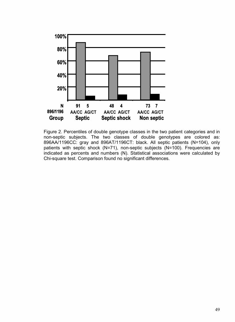
49
Figure 2. Percentiles of double genotype classes in the two patient categories and in non-septic subjects. The two classes of double genotypes are colored as: 896AA/1196CC: gray and 896AT/1196CT: black. All septic patients (N=104), only patients with septic shock (N=71), non-septic subjects (N=100). Frequencies are indicated as percents and numbers (N). Statistical associations were calculated by Chi-square test. Comparison found no significant differences.
20%
40%
60%
80%
91 5
AA/CC AT/CT
Septic
48 4
AA/CC AT/CT
Septic shock
73 7
AA/CC AT/CT
Non septic
N
896/1196
Group
100%
AA/CC AG/CT AA/CC AG/CT AA/CC AG/CT
20%
40%
60%
80%
91 5
AA/CC AT/CT
Septic
48 4
AA/CC AT/CT
Septic shock
73 7
AA/CC AT/CT
Non septic
N
896/1196
Group
100%
AA/CC AG/CT AA/CC AG/CT AA/CC AG/CT

50
Figure 3. Means of total SOFA scores by 896A>T and 1196C>T genotypes during the first ICU week. A: Mean and standard deviation of daily SOFA score in 896AA (solid bars) and 896AT (striped bars) genotypes. Scale of the SOFA values is on the left. The lines above represent the percents of surviving patients by 896AA (square) and 896AT (triangle) genotypes, with scale on the right. B: Mean and standard deviation of daily SOFA score in 1196CC (solid bars) homozygote and 1196T carriers (striped bars). The lines above represent the percents of 1196CC (square) and 1196CT+TT (triangle) surviving patients. Statistical associations were calculated in A and B by Mann-Whitney test. Genotype classes and SOFA-days are indicated below the colored bars. Only the significant differences are shown (P>0.05).
A B
0
7
14 100
80
60
40
20
10
0
CC CT,TT CC CT,TT CC CT,TT CC CT,TT CC CT,TT CC CT,TT CC CT,TT
SOFA-1 SOFA-2 SOFA-3 SOFA-4 SOFA-5 SOFA-6 SOFA-7
P=0.007
0
7
14
AA AT AA AT AA AT AA AT AA AT AA AT AA AT
SOFA-1 SOFA-2 SOFA-3 SOFA-4 SOFA-5 SOFA-6 SOFA-7
100
80
60
40
20
10
0AA AG AA AG AA AG AA AG AA AG AA AG AA AG
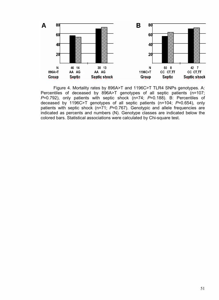
51
Figure 4. Mortality rates by 896A>T and 1196C>T TLR4 SNPs genotypes. A: Percentiles of deceased by 896A>T genotypes of all septic patients (n=107; P=0.792), only patients with septic shock (n=74; P=0.188). B: Percentiles of deceased by 1196C>T genotypes of all septic patients (n=104; P=0.654), only patients with septic shock (n=71; P=0.767). Genotypic and allele frequencies are indicated as percents and numbers (N). Genotype classes are indicated below the colored bars. Statistical associations were calculated by Chi-square test.
AA AG AA AGAA AG AA AG

52
CONSIDERAÇÕES FINAIS
A sepse e a falência orgânica são características complexas determinadas
pela interferência de múltiplos fatores. Centenas ou milhares de fatores externos e
de fatores intrínsecos determinarão simultaneamente a susceptibilidade para e o
desfecho de uma condição clínica patológica crítica. Cada efeito externo e cada um
dos genes herdados exercerá isoladamente um pequeno efeito, mas que
cumulativamente, no somatório com os demais, definirá a evolução do quadro
clínico. O estado de saúde, o prognóstico e o desfecho de pacientes em condições
críticas de saúde estão, portanto, também relacionados a herança genética que
indivíduo recebeu.
Ainda que centenas de genes estejam envolvidos na modulação fisiológica
final de um indivíduo com um quadro patológico complexo, aqueles genes que
interferem sistemicamente são sempre muito decisivos. Trabalhar com as variantes
polimórficas do gene que codifica para a proteína trans-membrana toll-like receptor 4
(TLR4) foi um esforço na busca de desvendar parte do pequeno efeito que a
herança genética pode ter no momento da doença crítica e a sepse.
Como apresentado ao longo deste trabalho, nosso objetivo foi responder à
pergunta que questiona se variantes polimórficas do gene que codifica para o TLR4
poderiam influenciar a susceptibilidade à sepse e às disfunções orgânicas em
pacientes críticos. Nós verificamos que, pelo menos em parte, a susceptibilidade à
sepse ao choque séptico e à gravidade das disfunções orgânicas pode ser
decorrente da herança das variantes polimóficas do TLR4.
Baseado em registros prévios, pode-se considerar que as variantes
polimóficas 896A>G e 1196C>T estão ligadas ou interferem diretamente na função
diferencial do TLR4 e isso afeta, consequentemente, a resposta imune inata. Sendo

53
assim, nossos resultados mostram que a herança diferencial de alelos destes SNPs
(single nucleotide polymorphism) pode ser, de fato, um fator de risco independente e
significante de susceptibilidade para a gravidade da condição clínica crítica e para a
evolução da sepse.
Em conclusão, baseado em observações prévias e no presente estudo, nós
propomos que as variantes polimórficas 896A>G e 1196C>T do gene que codifica
para o TLR4 podem ser considerados potenciais marcadores genéticos para a sepse
e para gravidade das disfunções orgânicas em pacientes em estado crítico de
saúde.