Embed Size (px)
Citation preview

1
UNIVERSIDADE FEDERAL DE PERNAMBUCO
CENTRO DE CIÊNCIAS BIOLÓGICAS
DOUTORADO EM CIÊNCIAS BIOLÓGICAS
PRODUÇÃO E PURIFICAÇÃO DE DNA PLASMIDIAL A PARTIR
DE Escherichia coli RECOMBINANTE
KEILA APARECIDA MOREIRA
RECIFE
2003

2
UNIVERSIDADE FEDERAL DE PERNAMBUCO
CENTRO DE CIÊNCIAS BIOLÓGICAS
DOUTORADO EM CIÊNCIAS BIOLÓGICAS
KEILA APARECIDA MOREIRA
PRODUÇÃO E PURIFICAÇÃO DE DNA PLASMIDIAL A PARTIR
DE Escherichia coli RECOMBINANTE
RECIFE
Novembro, 2003

3
KEILA APARECIDA MOREIRA
PRODUÇÃO E PURIFICAÇÃO DE DNA PLASMIDIAL A PARTIR
DE Escherichia coli RECOMBINANTE
Orientador: Prof. Dr. José Luiz de Lima Filho
(Departamento de Bioquímica; LIKA – UFPE)
Co-Orientadora: Profa. Dra. Ana Lúcia Figueiredo Porto
(Departamento de Morfologia e Fisiologia Animal – UFRPE)
RECIFE
NOVEMBRO, 2003
Tese apresentada ao Curso de Doutorado emCiências Biológicas da Universidade Federalde Pernambuco, para obtenção do título deDoutor em Ciências Biológicas, Área deConcentração em Biotecnologia .

4
PRODUÇÃO E PURIFICAÇÃO DE DNA PLASMIDIAL A PARTIR
DE Escherichia coli RECOMBINANTE
KEILA APARECIDA MOREIRA
COMISSÃO EXAMINADORA
Membros Titulares
Prof. Dr. José Luiz de Lima Filho
Departamento de Bioquímica; LIKA – UFPE (Orientador)
Profa. Dra. Ana Lúcia Figueiredo Porto
Departamento de Morfologia e Fisiologia Animal – UFRPE
Profa. Dra. Maria de Mascena Diniz Maia
Departamento de Biologia – UFRPE
Prof. Dr. Adalberto Pessoa Júnior
Departamento de Tecnologia Bioquímica e Farmacêutica – USP
Profa. Dra. Leonie Asfora Sarubbo
Departamento de Química – UNICAP
Membros Suplementares
Profa. Dra. Maria Tereza dos Santos Correia
Departamento de Bioquímica - UFPE
Profa. Dra. Maria das Graças Carneiro da Cunha
Departamento de Bioquímica – UFPE


5
À Raimunda Moreira Lustoza e Joaquim
Moreira, pelo esforço que me educaram e
Tébio Macedo, pela dedicação e
compreensão.
Dedico este trabalho.

6
AGRADECIMENTOS
Meus sinceros agradecimentos, que no decorrer de mais uma etapa da minha vida,
recebi incentivo de pessoas e de instituições.
A toda minha família. Minha tia Lúcia, ao primo Dermival, e aos meus irmãos,
Claudia, Kátia, Cairo e Carla pelo incentivo, compreensão e apoio em todos os
momentos.
Ao Prof. Dr. José Luiz de Lima, meu orientador, pela dedicação e paciência.
Registro aqui minha imensa admiração, um exemplo de simplicidade e de sabedoria.
Agradeço pela confiança depositada e por lições de vida adquiridas desde a iniciação
científica. Meus agradecimentos especiais e muito obrigada.
A Profa. Dra. Ana Lúcia Figueiredo Porto, minha co-orientadora, pela a
oportunidade e incentivo dado para o ingresso na pesquisa científica, pelos
conhecimentos transmitidos e amizade durante estes anos. Meu eterno agradecimento e
muito obrigada.
A Coordenação de Aperfeiçoamento de Pessoal de Nível Superior (CAPES) pelo
apoio financeiro no decorrer deste curso.
A Profa. Dra. Luana Cassandra B. B. Coelho, Coordenadora do Curso de
Doutorado em Ciências Biológicas da Universidade Federal de Pernambuco (UFPE), e as
funcionárias, Adenilda, Jaci e Liane pelo o apoio sempre prestado.
Ao Prof. Dr. William Ledingham, Coordenador Geral do Programa ALFA da
Comunidade Européia – Rede BIOENGE, pela oportunidade e consentimento de uma
bolsa para estagiar no início do doutorado no Instituto Superior Técnico-IST, Lisboa,
Portugal.
Aos Profs. Duarte Miguel Prazeres e Joaquim Manuel Sampaio Cabral, do
Instituto Superior Técnico de Lisboa por disponibilizarem infra-estrutura e recursos para

7
a realização inicial deste trabalho. As doutorandas Margarida Diogo e Sofia Ribeiro, pela
disponibilização de conhecimentos, paciência e amizade durante o período que
permaneci em Lisboa.
Aos Profs. Ernesto Marques, pesquisador da Universidade Johns Hopkins, por
amostras cedidas dos plasmídeos, e a Adilson Chaves pela disponibilização de materiais
imprescindíveis para a realização deste trabalho.
Ao Prof. Dr. Walter Azevedo, Diretor do Departamento de Química Fundamental,
pelo auxílio durante as quantificações do DNA plasmidial no espectrofluorímetro.
A Profa. Dra. Neiva Tinti de Oliveira, responsável pelo setor de Biologia
Molecular do Departamento de Micologia e a mestranda Meiriana Vila Nova, pela
disponibilização da infra-estrutura deste laboratório para realização de eletroforeses.
As minhas estagiárias de iniciação científica: Márcia Azenha, Andréa Guedes e
Monilk Duarte, pelo auxílio e execução deste trabalho. Aos colegas do Curso de
Doutorado Cristina Motta, Danyelly Martins, Jorge Silva e Sandra Botelho pela amizade
e companheirismo ao longo deste curso.
Aos colegas do Setor de Biotecnologia do LIKA, Alessandro Albertini, Daniela
Viana, Danyelly Santos, Eduardo Alécio, Alexandre Libanio, Fernanda Borba, Rosa
Fireman, Maria de Mascena, Raquel Pedrosa, Taciana Cavalcanti e Tatiana Porto. E em
especial a Cosme Martinez pelo auxílio estatístico em um dos trabalhos.
Aos funcionários do LIKA, Cleide, Conceição, Ilma, Moisés, Oscar, Ver e Sr.
Otaviano, por nos darem condições de trabalhar bem.
As minhas amigas Laura Bruno e Simone Gaia, pelo incentivo, amizade e
estímulo ao longo de mais uma etapa em minha vida. A todas as pessoas que conviveram
comigo durante a realização deste trabalho, contribuindo direta ou indiretamente, com
incentivos, amizades e confianças, meu muito obrigada.
A Deus ...

8
SUMÁRIO
LISTA DE TABELAS i
LISTA DE FIGURAS ii
LISTA DE ABREVIATURAS iii
RESUMO iv
ABSTRACT v
1. INTRODUÇÃO 1
1.1 Caracterização estrutural dos plasmídeos 3
1.2 Produção de DNA plasmidial 5
1.3 Estabilidade plasmidial 6
1.4 Marca seletiva 7
2. PROCESSOS DE PURIFICAÇÃO 7
2.1 Isolamento primário 8
2.2 Clarificação e concentração 10
2.3 Técnicas cromatográficas 10
2.3.1 Cromatografia de interação hidrofóbica 12
3. EXTRAÇÃO LÍQUIDO-LÍQUIDO 13
3.1 Sistemas de duas fases aquosas 14
3.2 Obtenção dos sistemas de duas fases aquosas 15
3.3 Termodinâmica do diagrama de fases 17
3.4 Fatores que influenciam o comportamento das fases 19
3.5 Fatores que influenciam a partição de biomoléculas em sistemas de duas fases aquosas 19
3.6 Partição de ácidos nucléicos 21
4. OBJETIVOS 23
4.1 Objetivo geral 23
4.2 Objetivos específicos 23
5. REFERÊNCIAS BIBLIOGRÁFICAS 24
6. CAPÍTULOS 32

9
Capítulo 1. Effect of cultural conditions on plasmid DNA production and stability 33
Capítulo 2. Extraction of dengue 2 plasmid DNA vaccine (pD2) from cell lysates by aqueous two-
phase systems
51
Capítulo 3. Purification of plasmid (pVaxLacZ) by hydrophobic interaction chromatography 73
7. CONCLUSÕES 87
8. SUGESTÕES PARA ESTUDOS FUTUROS 89
9. ANEXOS 90

i
LISTA DE TABELAS
Tabela 1. Propriedade de plasmídeos conjugativos e não-conjugativos de
organismos Gram-positivos
2
Tabela 2. Fatores que afetam a estabilidade plasmidial em leveduras 7
Tabela 3. Exemplos de sistemas de duas fases aquosas 16

ii
LISTA DE FIGURAS
Figura 1. Exemplos de diferentes conformações do plasmídeo 4
Figura 2. Diagrama de fases de um sistema de duas fases aquosas com os
componentes A e B
18

iii
LISTA DE ABREVIATURAS
DNA – ácido desoxirribonucléico
RNA – ácido ribonucléico
ETDA – ácido etilenodiaminotetracético
NaOH – hidróxido de sódio
SDS – dodecil sulfato de sódio
PEG – polietileno glicol
NaCl – cloreto de sódio
MgCl2 – cloreto de magnésio
CFR – cromatografia de fase reversa
CIH – cromatografia de interação hidrofóbica
SDFA – sistemas de duas fases aquosas
K- coeficiente de partição

iv
RESUMO
A produção e a purificação de DNA plasmidial com elevado grau de pureza tem
tido um elevado interesse nos últimos anos, devido fundamentalmente aos
desenvolvimentos de técnicas de medicina molecular, como a terapia gênica e a
vacinação com DNA. O plasmídeo pD2 foi produzido em diferentes condições de
cultivo, sendo analisado várias parâmetros durante o crescimento, tais como, influência
da velocidade de agitação (120, 160 e 200 rpm), concentrações de canamicina (10, 20,
30, 40 e 50µg ml-1) e meio de cultura (Terrific Broth - TB ou Luria Bertani com glicose
- LBG). Foram também investigados o efeito da partição de DNA plasmidial, RNA e
proteínas totais nos sistemas de duas fases aquosas (SDFA) do tipo polietileno glicol
(PEG)/sal de fosfato de sódio dibásico (K2HPO4) do plasmídeo (pD2) durante o
processo de purificação. Analisou-se a influência da massa molar do PEG, massa que
carrega o sistema da solução de lise (20, 40 ou 60% m/m) na partição do plasmídeo. A
técnica de precipitação com sulfato de amônio (2,0, 2,5 e 3,0M), seguido da técnica
cromatográfica de interação hidrofóbica (CIH) utilizando como matriz estacionária o
suporte Phenyl Sepharose 6 Fast Flow foram utilizadas para purificar o plasmídeo
pVax-LacZ. A velocidade de agitação que apresentou os melhores resultados para a
produção de biomassa e DNA plasmidial foi a 200 rpm, enquanto as concentrações 30-
50µg mL-1 de canamicina apresentaram resultados semelhantes para todas as condições
estudadas. O meio de cultura TB apresentou ser o melhor em termos de produção de
DNA plasmidial e biomassa, neste meio de cultura o plasmídeo foi mais estável,
apresentando 82% de células contendo o plasmídeo. O DNA plasmidial foi particionado
entre a fase superior e inferior na dependência da massa molar do polímero constituinte
do sistema, tendo uma grande quantidade de proteína do lisado celular acumulada na
interfase dos sistemas. O melhor rendimento plasmidial (37%) foi obtido com sistema
PEG 400/K2HPO4 (20/20% m/m) carregado com 60% (m/m) de lisado celular. O
procedimento de precipitação com sulfato de amônio (2,5M) seguido da cromatografia
de interação hidrofóbica foram capazes de separar proteínas e DNA genômico do
plasmídeo, obtendo 51% de rendimento e um fator de purificação de 3,3.

v
ABSTRACT
The production and purification of plasmid DNA with high degree of purity
have been having a large interest in the last years, mainly due to the development of
several techniques, such as molecular medicine, gene therapy and DNA vaccination.
The different bacterial growth conditions were set up in order to analyze the plasmid
pD2 production, such as the influence of agitation velocities (120, 160 and 200 rpm),
kanamycin concentrations (10, 20, 30, 40 and 50µg ml-1) and culture medium (Terrific
Broth - TB or Luria Bertani with glucose - LBG). Were also investigated the effect of
plasmid DNA, RNA and total proteins in the aqueous two phases systems (ATPS),
polyethylene glycol (PEG)/ sodium phosphate salt (K2HPO4) on the plasmid (pD2).
Was also analyzed the influence of the molecular weight of PEG, load of lyse solution
(20, 40 or 60% w/w total mass of the systems) on the plasmid DNA partition. With the
pVax-LacZ plasmid the precipitation with ammonium sulphate (2.0, 2.5 and 3.0M)
followed by hydrophobic interaction chromatography (HIC) using as support Phenyl
Sepharose 6 Fast Flow was applied. The agitation velocity that presented better results
related to biomass and plasmid DNA production was 200 rpm, while 30-50µg mL-1 of
kanamycin presented similar results for all the studied conditions. The TB medium was
the best medium for biomass and DNA production. The plasmid was stable in TB
medium, showing 82% plasmid containing cells. The plasmid DNA are partitioned
between the top an bottom phases showing to be dependent to the molecular weight of
the system and a good amount of cell lysate protein was accumulated in the interphase
of the system. The best recovery plasmid yield (37%) was obtained with PEG 400
system with a 60% (w/w) lysate load. The precipitation procedure with ammonium
sulphate (2.5M) followed by the hydrophobic interaction chromatography were capable
to separate proteins and genomic DNA of the plasmid, obtaining 51% of revenue and a
purification factor of 3.3 after the precipitation with ammonium sulphate.

1
1. INTRODUÇÃO
Plasmídeos são elementos genéticos constituídos por moléculas de DNA
extracromossômicas em cadeia dupla covalentemente fechada que podem existir em
diferentes conformações topológicas e são capazes de manter-se estáveis em populações
microbianas (SINDEN, 1994; GANUSOV; BRILKOV, 2002).
Todos os plasmídeos possuem uma seqüência de DNA que pode atuar como
origem de replicação, de modo que eles são capazes de se multiplicar na célula
independentemente da molécula principal de DNA. Os plasmídeos menores fazem uso
das próprias enzimas replicativas de DNA da célula para fazer cópias de si mesmos, ao
passo que alguns dos plasmídeos maiores possuem genes que codificam enzimas
especiais, específicas para sua replicação (BROWN, 1999).
Alguns plasmídeos também são capazes de se replicar inserindo-se na molécula
principal de DNA. Esses plasmídeos são integrativos ou epissomos, podem ser mantidos
de modo estável durante várias multiplicações celulares, mas sempre em algum estágio
existem como elementos independentes (PROCTOR, 1994; BROWN, 1999).
Os plasmídeos podem ser categorizados em dois grandes grupos, os plasmídeos
conjugativos ou não conjugativos, dependendo se transportam ou não um conjunto de
elementos utilizados na transferência de genes que promovem a conjugação bacteriana
(Tabela 1). Estes ainda podem ser agrupados em relação à quantidade de cópias que eles
podem manter por células, múltiplas cópias ou limitadas cópias (OLD; PRIMROSE,
1981).
Geralmente os plasmídeos conjugativos são de alta massa molar e estão presentes
com uma a três cópias por cromossomo, enquanto que os plasmídeos não conjugativos
são de baixa massa molar e estão presentes com múltiplas cópias. Eles ainda podem ser
classificados de acordo com o controle preferencial na duplicação, plasmídeos

2
restritos ou relaxados. Os primeiros duplicam-se em sincronia com o cromossomo,
enquanto que os plasmídeos relaxados duplicam-se independentemente da coordenação
cromossômica (OLD; PRIMROSE, 1981).
A partir de 1946, quando Lederberg e colaboradores descreveram o plasmídeo F,
responsável pela fertilidade em Escherichia coli K12, centenas de outros plasmídeos
foram descritos, alguns deles codificando informações de grande relevância para a saúde
humana, animal e vegetal, como os plasmídeos R (resistência múltipla a drogas) em
diversos grupos bacterianos Gram-negativos e positivos. Mais de 250 plasmídeos já
foram descritos somente em Escherichia coli. Além desses plasmídeos naturais, existem
dezenas de plasmídeos artificiais (COSTA, 1987).
Tabela 1. Propriedades de plasmídeos conjugativos e não-conjugativos de organismos
Gram-positivos.
Plasmídeo Tamanho
(Kb)
Conjugativo Número de cópias
plasmídeos/cromossomo
Fenótipo
Col E1 4,2 Não 10-15 Produção de colicina E1
RSF 1030 5,6 Não 20-40 Resistência a ampicilina
Clo DF13 6 Não 10 Produção de cloacina
R6K 25 Sim 13-28 Resistência a estreptomicina e
ampicilina
F 62 Sim 1-2 --------------------------------
RI 62,5 Sim 3-6 Resistência múltipla a drogas
Ent P 307 65 Sim 1-3 Produção de enterotoxina
Fonte: OLD; PRIMROSE, 1981.

3
Os plasmídeos são comumente conhecidos como veículos para introdução de
novos genes dentro de uma célula viva (BAHERI et al., 2001). Estes são freqüentemente
usados como modelo para estudos de replicação, recombinação e segregação de DNA em
células microbianas. O DNA plasmidial tem adquirido recentemente considerável
interesse devido ao seu potencial de aplicação em terapia gênica e vacinas de DNA
(PRAZERES et al., 1999; LEVY et al., 2000; FERREIRA et al., 2000; WANG et al.,
2001; RIBEIRO et al., 2002).
1.1 Caracterização estrutural dos plasmídeos
As moléculas de DNA são hélices formadas por duas cadeias alinhadas segundo
um eixo de forma antiparalela. Cada cadeia é um polímero linear de nucleotídeos ligados
entre si por ligações fosfodiéster. Na estrutura da molécula de DNA as bases
nitrogenadas encontram-se empacotadas junto ao centro do eixo da hélice, enquanto que
no exterior da hélice se encontram os grupos fosfato, negativamente carregados. O
interior da hélice é, portanto, altamente hidrofóbico devido à presença das bases
aromáticas (SINDEN, 1994).
A estrutura em cadeia dupla é altamente estável. Esta estabilidade é dada através
das forças de empacotamento (hidrofóbicas e de van der Walls) entre as bases da
molécula, mas também das ligações de hidrogênio e da camada de moléculas de água que
solvatam o DNA. A desnaturação do DNA (separação das cadeias) pode resultar da
quebra das ligações de hidrogênio a pH<2 ou pH>12 devido à ionização das bases, do
aumento da temperatura ou da força iônica e, ainda, de cortes ou rearranjos na sua
estrutura (RIBEIRO, 2000).
Na molécula do plasmídeo, se o eixo da hélice do DNA estiver também enrolado,
forma-se uma estrutura altamente ordenada, superenrolada (Figura 1); se o plasmídeo
sofrer um corte numa das cadeias, a estrutura superenrolada perde-se, resultando numa
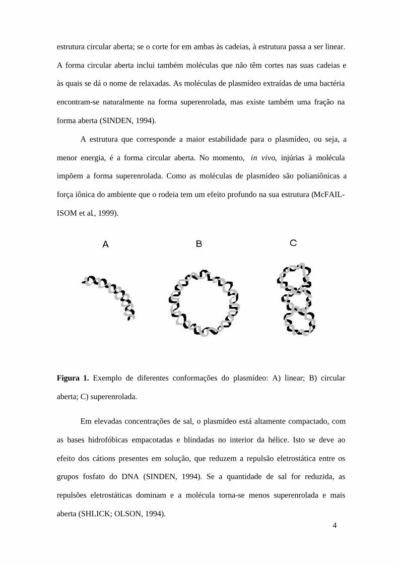
4
estrutura circular aberta; se o corte for em ambas às cadeias, à estrutura passa a ser linear.
A forma circular aberta inclui também moléculas que não têm cortes nas suas cadeias e
às quais se dá o nome de relaxadas. As moléculas de plasmídeo extraídas de uma bactéria
encontram-se naturalmente na forma superenrolada, mas existe também uma fração na
forma aberta (SINDEN, 1994).
A estrutura que corresponde a maior estabilidade para o plasmídeo, ou seja, a
menor energia, é a forma circular aberta. No momento, in vivo, injúrias à molécula
impõem a forma superenrolada. Como as moléculas de plasmídeo são polianiônicas a
força iônica do ambiente que o rodeia tem um efeito profundo na sua estrutura (McFAIL-
ISOM et al., 1999).
Figura 1. Exemplo de diferentes conformações do plasmídeo: A) linear; B) circular
aberta; C) superenrolada.
Em elevadas concentrações de sal, o plasmídeo está altamente compactado, com
as bases hidrofóbicas empacotadas e blindadas no interior da hélice. Isto se deve ao
efeito dos cátions presentes em solução, que reduzem a repulsão eletrostática entre os
grupos fosfato do DNA (SINDEN, 1994). Se a quantidade de sal for reduzida, as
repulsões eletrostáticas dominam e a molécula torna-se menos superenrolada e mais
aberta (SHLICK; OLSON, 1994).
5
1.2 Produção de DNA plasmidial
O sucesso de fermentações baseadas em tecnologias de DNA recombinante é uma
combinação das interações entre o ambiente fermentativo, o organismo hospedeiro e os
elementos genéticos recombinantes (O’ KENNEDY et al., 2000). A produção de
plasmídeos por processos fermentativos em larga escala podem apresentar limitações
quanto ao baixo rendimento, instabilidade plasmidial e altas densidades celulares
(DIOGO, 1999).
A produção de plasmídeos inclui três fases principais: a fermentação, o
isolamento primário e a purificação. Em cada uma destas etapas existem problemas
específicos, na sua maioria relacionados com a natureza estrutural dos plasmídeos, como
a elevada massa molar (normalmente entre 5 a 20 kilobases), o que resulta em soluções
muito viscosas e uma estrutura não globular flexível e altamente carregada (PRAZERES
et al., 1999).
Meios de cultura comerciais também podem ser utilizados, sendo recomendado o
desenvolvimento de meios que sejam adaptados ao sistema plasmídeo/hospedeiro em
questão, de modo a aumentar a produtividade (PRAZERES et al., 1998).
Deve-se ter cuidados especiais com a escolha de uma espécie como hospedeira de
um plasmídeo, de maneira a obter rendimentos e prevenir alguns problemas em etapas
posteriores, como a purificação. Assim, a espécie deve ser escolhida para minimizar a
quantidade de impurezas que necessitam ser removidas (PRAZERES et al., 1999). Em
geral, deve ser escolhida uma espécie hospedeira que tenha sido completamente
caracterizada, que esteja livre de qualquer contaminação e que não seja prejudicial para o
ambiente, para o produto final, para os pacientes e para pessoal envolvido na produção e
manipulação (SCHLEEF, 1999).
Trabalhos anteriores envolvendo tecnologias de DNA recombinante focalizam
seus estudos em seres procariontes, como a bactéria Escherichia coli. Esta é

6
considerada como um hospedeiro ideal, pois tem reconhecida segurança ao longo da
história científica na utilização em produtos farmacêuticos (VYAS et al., 1994; ZHANG
et al., 1996).
1.3 Estabilidade plasmidial
Na produção comercial com microrganismos recombinantes, a maioria dos
problemas está na estabilidade plasmidial. A instabilidade é a tendência das células
transformadas perderem suas propriedades de engenharia molecular por causa de
mudanças ou perdas do plasmídeo (ZHANG et al., 1996). A instabilidade plasmidial em
cultura microbiológica provoca redução dos níveis do produto desejado no cultivo, tendo
impacto negativo em atividades de proteínas específicas e aumento nos custos de
produção, visto que os substratos são consumidos por células não produtivas (VYAS et
al., 1994).
Existem dois tipos de instabilidade plasmidial: a estrutural e a segregacional. A
instabilidade estrutural é causada usualmente pela deleção, inserção, recombinação ou
outros eventos, ao nível de DNA; enquanto que a instabilidade segregacional é causada
pela partição desigual de plasmídeos durante a divisão celular (OLD; PRIMROSE,
1981).
Após o plasmídeo recombinante ser introduzido dentro de células hospedeiras, as
interações entre plasmídeo e o hospedeiro são substanciais (Tabela 2). Estas interações
determinam a instabilidade plasmidial e o grau de expressão dos genes clonados.
Portanto, estes fatores combinados a fatores ambientais podem levar a perda plasmidial
(MÔO-YOUNG et al., 1996), resultando em perda da produtividade do produto
desejado, sendo um grande obstáculo para o aumento da escala no uso de
microrganismos geneticamente modificados (ZHANG et al., 1996).
7
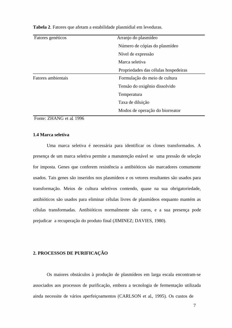
Tabela 2. Fatores que afetam a estabilidade plasmidial em leveduras.
Fatores genéticos Arranjo do plasmídeo
Número de cópias do plasmídeo
Nível de expressão
Marca seletiva
Propriedades das células hospedeiras
Fatores ambientais Formulação do meio de cultura
Tensão do oxigênio dissolvido
Temperatura
Taxa de diluição
Modos de operação do biorreator
Fonte: ZHANG et al. 1996
1.4 Marca seletiva
Uma marca seletiva é necessária para identificar os clones transformados. A
presença de um marca seletiva permite a manutenção estável se uma pressão de seleção
for imposta. Genes que conferem resistência a antibióticos são marcadores comumente
usados. Tais genes são inseridos nos plasmídeos e os vetores resultantes são usados para
transformação. Meios de cultura seletivos contendo, quase na sua obrigatoriedade,
antibióticos são usados para eliminar células livres de plasmídeos enquanto mantém as
células transformadas. Antibióticos normalmente são caros, e a sua presença pode
prejudicar a recuperação do produto final (JIMINEZ; DAVIES, 1980).
2. PROCESSOS DE PURIFICAÇÃO
Os maiores obstáculos à produção de plasmídeos em larga escala encontram-se
associados aos processos de purificação, embora a tecnologia de fermentação utilizada
ainda necessite de vários aperfeiçoamentos (CARLSON et al., 1995). Os custos de

8
produção de proteínas terapêuticas, cerca de 70%, podem estar associados às operações
de separação e purificação. Parece ser improvável que as especificações de pureza e
custos de recuperação venham a ser menor no caso dos vetores de terapia gênica
(LYDDIATT; O’SULLIVAN, 1998).
2.1 Isolamento primário
As dificuldades mais comuns encontradas no isolamento primário de plasmídeos
são: a alta densidade celular, tensões de corte elevada, desnaturação, ação das nucleases,
manipulação dos lisados e fragmentação do DNA genômico (PRAZERES et al., 1999).
A etapa de recuperação das células ao final da fase de produção de
microrganismos recombinantes, inicia-se por operações de separação sólido-líquido,
ocorrendo através de centrifugação ou microfiltração. A ressuspensão dessas células é
feita em tampão que contém ácido etilenodiaminotetracético (EDTA), usado como
agente quelante. O EDTA remove íons de Ca2+ e Mg2+ da superfície celular,
desorganizando a sua estrutura e reduzindo a atividade das nucleases que podem
degradar o plasmídeo (PRAZERES et al., 1999).
A técnica de lise alcalina inicia-se pela adição e agitação suave de um
determinado volume de células com uma solução de hidróxido de sódio (NaOH)
contendo dodecil sulfato de sódio (SDS). A reação de lise na parede celular, entre o SDS
e os lipídios e as proteínas, solubiliza o material da parede celular, provocando liberação
do conteúdo intracelular. Resultados de alguns pesquisadores parecem indicar que a lise
química da parede celular e a liberação dos materiais intracelulares se completam ao fim
de 30-40 segundos dependendo da espécie bacteriana (CICCOLINI et al., 1998).
O ambiente fortemente alcalino da mistura causa também a desnaturação
reversível do DNA plasmidial. O DNA cromossômico de alta massa molar é também
desnaturado nesta altura causando o aumento da viscosidade da solução até um máximo,

9
antes de diminuir para um estado estacionário. Esta diminuição é resultante da
fragmentação do DNA genômico induzida pela tensão de corte (CICCOLINI et al.,
1998).
Quando a lise e a desnaturação ficam completas, a mistura é neutralizada pela
adição de uma solução concentrada de acetato de potássio. De fato, a solubilização do
SDS decresce a baixas temperaturas e as altas concentrações de sal, resultando na
formação de uma suspensão floculenta, formada por complexos SDS-proteínas. Estes
flocos, muito sensíveis às tensões de corte, vão se agregando lentamente, formando uma
matriz contendo debris celulares, RNA, DNA genômico de alta massa molar e outras
impurezas que vão ficando retidas nesse reticulado. Os plasmídeos, majoritariamente
dissolvidos na fase aquosa, recuperam a forma original superenrolada nesta fase do
processo o que lhe permite resistir melhor às tensões mecânicas. O precipitado formado é
separado da fase aquosa por centrifugação ou filtração. Nesta operação pode ser perdido
algum DNA plasmidial, o que pode ser minimizado removendo o máximo de líquido da
fase sólida (PRAZERES et al., 1999).
Ao longo de todo o processo de purificação, o processo de lise alcalina é um dos
passos de maior dificuldade. A literatura descreve métodos alternativos para isolamento
primário dos plasmídeos, nomeadamente a sonicação e a homogeneização a alta pressão
(CARLSON et al., 1995). Todos os métodos testados provocaram uma grande
fragmentação do DNA genômico, o que constitui um problema adicional em termos dos
passos subseqüentes (THEODOSSIOU et al., 1997).
A liberação de DNA genômico fragmentado aumenta a viscosidade da solução,
tornando o processo de mistura mais difícil e dispendioso. Adicionalmente, a agitação
deve ser feita de forma suave, de modo a manter o DNA genômico com a massa molar o
mais alto possível de modo a maximizar a sua precipitação nas etapas seguintes

10
(RIBEIRO, 2000).
Em contraste, a adição dos reagentes que provocam a lise celular deve ser
suficientemente eficaz de modo a evitar a formação de locais com extremos valores de
pH, uma vez que valores superiores a 12 poderão provocar a desnaturação irreversível
dos plasmídeos, passando estas isoformas a ser consideradas contaminantes do processo.
A agitação deve também assegurar a lise completa e eficiente de toda a população
celular. A lise alcalina é um processo difícil de controlar, apresenta falta de
reprodutividade e pode implicar perdas significativas de plasmídeo (PRAZERES et al.,
1999).
2.2 Clarificação e concentração
A clarificação e a concentração são processos que têm por finalidade a remoção
de alguns contaminantes do plasmídeo com a simultânea redução do volume da solução
de lise. A concentração plasmidial geralmente é obtida por precipitação com alguns sais
ou álcoois, usualmente o isopropanol ou o etanol (LYDDIATT; O’SULLIVAL, 1998).
A precipitação com polietilenoglicol (PEG) tem sido utilizada, sistemas PEG-
8000/NaCl ou MgCl2. Este método baseia-se no fato do tamanho do DNA precipitado ser
dependente do massa molar e da concentração polímero (LIS; SCHLEIF, 1975). Sendo
assim, pode-se fracionar o DNA de acordo com o seu tamanho ou, simplesmente
precipitar todo o DNA (HORN et al., 1995).
Após a concentração do plasmídeo, proteínas, lipopolissacarídios e ácidos
nucléicos contaminantes podem ainda ser removidos por precipitação com sais (por
exemplo, sulfato ou acetato de amônio) conduzindo a um aumento na pureza do DNA
plasmidial (DIOGO et al., 2000).
2.3 Técnicas cromatográficas
A cromatografia é uma das técnicas mais importantes na separação e

11
purificação de produtos biológicos (PRAZERES et al., 1999), exercendo um papel
central na purificação de plasmídeos em larga escala, seja como uma etapa do processo
ou como uma ferramenta analítica para o monitoramento dos processos e controle de
qualidade (PRAZERES et al., 1998).
Diversos processos cromatográficos têm sido descritos na literatura como forma
de purificar ácidos nucléicos, incluindo técnicas de filtração em gel, troca iônica, fase
reversa e afinidade (DIOGO, 1999).
Na cromatografia de filtração em gel as moléculas são separadas de acordo com o
seu tamanho. Esta técnica permite separar as endotoxinas e RNA dos plasmídeos (HORN
et al., 1995; FERREIRA et al., 1997). A lentidão, a baixa resolução, limitação na
capacidade (menor do que 10% do volume da coluna para uma boa resolução do
produto), diluição do produto final são algumas desvantagens desta técnica
(LYDDIATT; O´SULLIVAN, 1998).
Na cromatografia de troca iônica os grupos fosfato do plasmídeo (carregados
negativamente) interagem com a fase estacionária da coluna, a qual é carregada
positivamente. É necessária a utilização de um gradiente de sal para eluir os ácidos
nucléicos, que em princípio devem eluir por ordem do aumento da sua carga total, o que
por sua vez é uma função do comprimento da cadeia (PRAZERES et al., 1998). Este
método tem a desvantagem de co-purificar justamente com o plasmídeo, DNA
genômico, endotoxinas e RNA de alta massa molar devido à semelhante afinidade que
estes apresentam para a matriz iônica (DIOGO, 1999).
Green e colaboradores (1997) descreveram um protocolo de cromatografia de
fase reversa (CFR) para purificação de plasmídeo em larga escala que utiliza uma coluna
de cromatografia líquida de alto desempenho contendo um polímero não poroso e inerte.
A adsorção, mecanismo de ligação das moléculas, envolve a interação das moléculas
com grupos hidrofóbicos ligados a um material cromatográfico. No entanto, os

12
suportes usados em CFR possuem uma densidade de ligantes muito superior, portanto a
inserção é muito mais forte (PHARMACIA, 1993).
Em 1997, pesquisadores desenvolveram um método de afinidade específico para
uma seqüência de DNA para purificação de plasmídeos. Este método baseia-se na
formação de uma tripla hélice entre um oligonucleotídeo covalentemente ligado a uma
matriz cromatográfica e uma seqüência dupla presente no plasmídeo a ser purificado. As
triplas hélices formadas são estáveis apenas para valores ácidos de pH, pelo que a eluição
é facilmente conseguida com eluentes básicos. A coluna pode ser reutilizada e permite
obter DNA plasmidial purificado. Entretanto, o rendimento mais alto obtido foi de 50%
devido às características intrínsecas da própria coluna (WILLS et al., 1997). Além disso,
a cinética de formação da tripla hélice é bastante baixa, sendo necessário despender
bastante tempo na etapa de ligação (SCHLUEP et al., 1998).
2.3.1 Cromatografia de interação hidrofóbica
A cromatografia de interação hidrofóbica (CIH) é uma técnica cada vez mais
utilizada, sobretudo porque exibe características de ligação complementares a outras
técnicas, tais como a cromatografia de filtração em gel e de troca iônica (JANSON;
RYDÉN, 1993). A cromatografia de interação hidrofóbica apresenta grandes vantagens
mediante a diversidade de potenciais condições de eluição que permitem a resolução de
misturas complexas que seriam muito difíceis de separar por outros métodos (DIOGO,
1999).
Na cromatografia de interação hidrofóbica procura-se promover a retenção de
moléculas de caráter hidrofóbico através da presença de ligantes hidrofóbicos na fase
estacionária. A força de interação depende da densidade dos grupos hidrofóbicos à
superfície da biomolécula e do tipo e grau de substituição dos ligantes hidrofóbicos
ligados à matriz polimérica (JANSON; RYDÉN, 1993).

13
A cromatografia de interação hidrofóbica foi recentemente descrita por Diogo e
colaboradores (2000) para purificação de DNA plasmidial. Esta técnica tem como
fundamento a retenção de moléculas de caráter hidrofóbico através da presença de
ligantes na fase estacionária. Estas interações têm como base o fato das moléculas de
água repelirem os grupos hidrofóbicos, de forma a que estes se juntem, minimizando
assim o seu efeito de perturbação na rede de ligações de hidrogênio da água. Usando um
eluente de elevada força iônica (de forma a promover este tipo de interação) consegue-se
eluir, primeiramente o plasmídeo superenrolado, que não interage com a coluna por ter as
suas bases no interior da hélice, e por último o RNA, proteínas, DNA genômico e
endotoxinas. Se a força iônica for muito baixa, o plasmídeo fica retido na coluna
(DIOGO et al., 2000).
3. EXTRAÇÃO LÍQUIDO-LÍQUIDO
A maioria das técnicas de separação utilizada em processos bioquímicos
industriais para a recuperação e isolamento de enzimas, tais como filtração e
centrifugação são altamente dependentes do tamanho da partícula (CHAVES, 2000). Os
suportes para cromatografia, originalmente desenvolvidos para proteínas, não permitem a
entrada do plasmídeo nos poros, sendo, portanto a sua ligação ao suporte apenas
superficial (PRAZERES et al., 1998).
A extração líquido-líquido é um processo bem estabelecido na indústria química,
incluindo várias aplicações na indústria bioquímica tradicional, como por exemplo, a de
antibióticos. A sua utilização tem, no entanto, sido limitada pela baixa compatibilidade
entre os materiais de origem biológica e os solventes orgânicos geralmente utilizados, e
pelas inerentes dificuldades de validação de um processo que utiliza este tipo de
solventes tóxicos (CABRAL et al., 1993).

14
Porém, o desenvolvimento nas últimas décadas de novas tecnologias de extração
usando sistemas de duas fases aquosas têm aberto novas perspectivas para a utilização
desta operação unitária. A inserção de etapas de extração líquido-líquido no processo
global de produção e purificação de plasmídeos pode, assim, revelar-se extremamente
útil no aumento da rentabilidade do produto (RIBEIRO, 2000).
3.1 Sistemas de duas fases aquosas
Em 1896, o microbiologista Beijerinck descreveu a formação de duas fases
aquosas macroscópicas quando se misturava uma solução de gelatina (ou amido), agar e
água sob certas concentrações. Sendo a fase superior rica em gelatina, e a fase inferior
rica em agar. Este fenômeno foi redescoberto por Albertsson que iniciou, há mais de 40
anos, a separação de moléculas biológicas e partículas em sistemas de duas fases aquosas
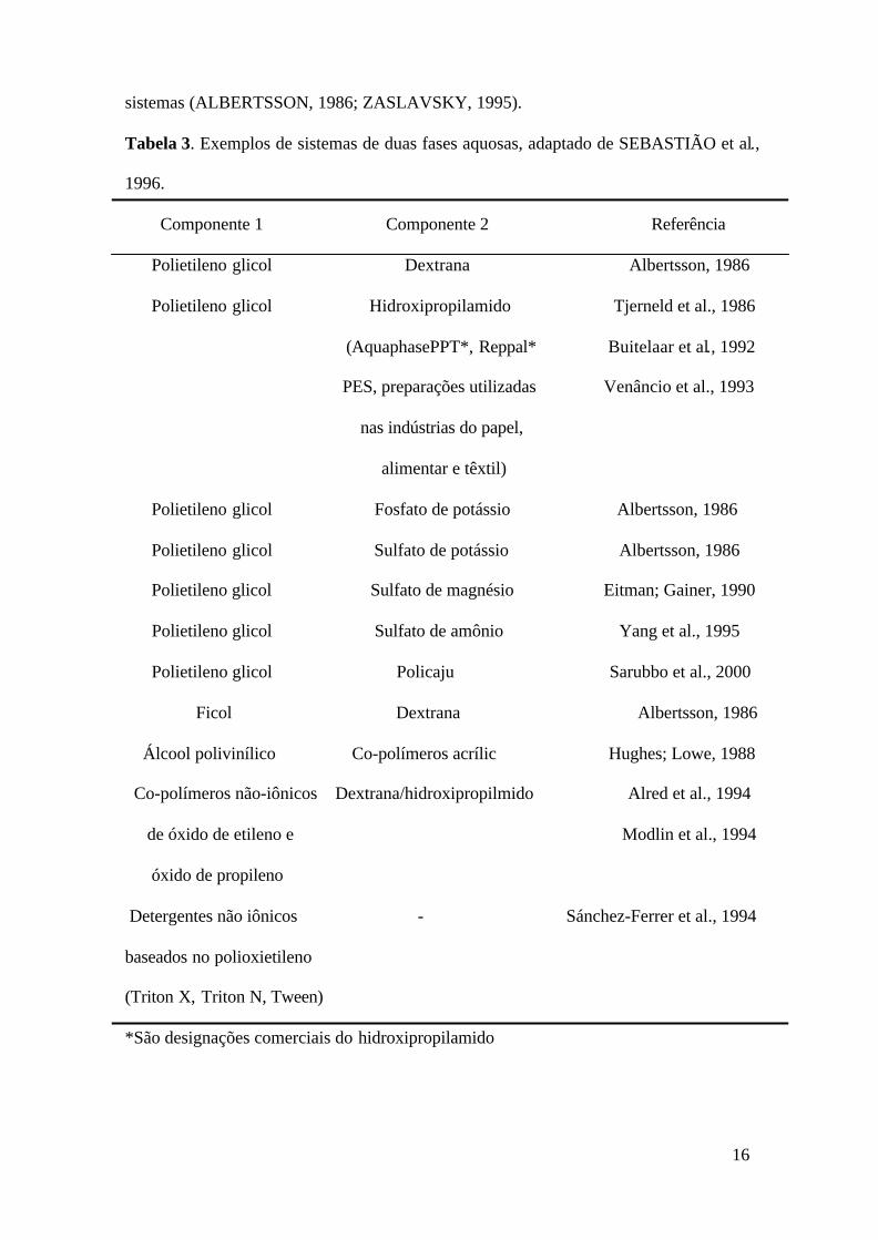
(SDFA). Uma extensa lista destes sistemas foi desenvolvida por diversos pesquisadores e
pode ser analisada na Tabela 3.
Desde então, estes sistemas tornaram-se numa poderosa técnica de separação de
uma vasta gama de materiais biológicos que incluem plantas, células animais,
microrganismos, fungos, vírus, mitocôndrias, proteínas e ácidos nucléicos (HATTI-
KAUL, 1999).
Os sistemas inicialmente mais utilizados eram os de polímero/polímero, usando o
polietileno glicol (PEG), a dextrana e a metilcelulose (ALBERTSSON, 1986). Em
seguida surgiram os sistemas polímero/sal, geralmente com fosfatos, que tinham a
vantagem de ser mais simples e ter menores custos. Nestes sistemas a fase superior é
constituída majoritariamente por PEG e a fase inferior por sal (SARUBBO, 2000).
Os sistemas de polímeros termoestáveis foram os últimos a serem usados. As
soluções destes polímeros, quando aquecidas a uma determinada temperatura, se separam
em duas fases; uma enriquecida com o polímero e outra com uma concentração muito
15
pequena do polímero termoestável (ALRED, 1993).
Os SDFA apresentam grandes vantagens em relação à extração convencional com
solventes orgânicos. Os SDFA contêm cerca de 80-95% em água, o que constitui uma
característica particularmente interessante na separação de biomoléculas, já que o
ambiente é muito pouco agressivo, não alterando a atividade e estabilidade da maioria
das biomoléculas. A aplicação destes sistemas permite ainda, tratar grandes volumes
num só passo. Os constituintes destes sistemas são de baixa toxicidade. O PEG é
biodegradável, foi extensivamente testado na indústria farmacêutica e está registrado em
muitos países para fins alimentares (HATTI-KAUL, 1999).
Além disso, a tensão superficial das fases é extremamente baixa o que permite as
partículas migrarem livremente entre as duas fases. Sabe-se também que os polímeros
têm efeito estabilizante na estrutura e atividade biológica das partículas. Além disso, os
SDFA são de fácil aumento de escala e oferecem a possibilidade de adaptar
equipamentos e métodos de extração em duas fases (orgânica/aquosa), usadas na
indústria química convencional (CUNHA; AIRES-BARROS, 1999).
3.2 Obtenção dos sistemas de duas fases aquosas
Os sistemas de duas fases aquosas são geralmente formados por uma solução
aquosa de dois polímeros hidrófilos ou um polímero e de determinados sais. Acima da
concentração crítica destes componentes ocorre espontaneamente a separação de fases,
predominando um ou outro componente em cada uma das fases resultantes
(ZASLAVSKY, 1995).
Cada SDFA pode ser caracterizado por um único diagrama de fases, que contém a
composição das fases em equilíbrio para o sistema. Os dados fundamentais para qualquer
tipo de processo de extração líquido-líquido são as composições de equilíbrio das fases
(DIAMOND; HSU, 1992). A literatura descreve diagramas de fases para diferentes
16
sistemas (ALBERTSSON, 1986; ZASLAVSKY, 1995).
Tabela 3. Exemplos de sistemas de duas fases aquosas, adaptado de SEBASTIÃO et al.,
1996.
Componente 1 Componente 2 Referência
Polietileno glicol Dextrana Albertsson, 1986
Polietileno glicol Hidroxipropilamido Tjerneld et al., 1986
(AquaphasePPT*, Reppal* Buitelaar et al., 1992
PES, preparações utilizadas Venâncio et al., 1993
nas indústrias do papel,
alimentar e têxtil)
Polietileno glicol Fosfato de potássio Albertsson, 1986
Polietileno glicol Sulfato de potássio Albertsson, 1986
Polietileno glicol Sulfato de magnésio Eitman; Gainer, 1990
Polietileno glicol Sulfato de amônio Yang et al., 1995
Polietileno glicol Policaju Sarubbo et al., 2000
Ficol Dextrana Albertsson, 1986
Álcool polivinílico Co-polímeros acrílic Hughes; Lowe, 1988
Co-polímeros não-iônicos Dextrana/hidroxipropilmido Alred et al., 1994
de óxido de etileno e Modlin et al., 1994
óxido de propileno
Detergentes não iônicos - Sánchez-Ferrer et al., 1994
baseados no polioxietileno
(Triton X, Triton N, Tween)
*São designações comerciais do hidroxipropilamido

17
A Figura 2 mostra um diagrama de fases para um sistema polímero e sal. A
concentração do polímero P é representada em ordenada e a concentração do sal em
abscissa; as concentrações são expressas como percentual (massa/massa). A linha curva
separando as duas áreas é chamada binodal. Todas as misturas que têm composições
representadas pelos pontos acima da linha são pontos de duas fases e os pontos abaixo
representam uma solução homogênea (ZASLAVSKY, 1995).
A linha que une dois pontos sobre a binodal para um determinado sistema
designa-se por linhas de amarração (tie line). Qualquer ponto sobre a mesma tie line
resulta num sistema em que as fases têm a mesma composição, mas diferentes volumes.
Por exemplo, em um sistema com concentração total representada por A, correspondem
duas fases em equilíbrio cuja composição é dada pelos pontos B (fase inferior) e C (fase
superior). Como a densidade das fases é muito próxima da água, a razão dos volumes
pode ser obtida, aproximadamente, a partir das distâncias AB e AC no diagrama. O
comprimento da tie line depende da concentração total do sistema, representando uma
medida da diferença entre as fases em equilíbrio. À medida que o comprimento da tie
line diminui (C’B’), o sistema aproxima-se do ponto crítico (K), isto é, ponto na binodal
em que os volumes e composições das duas fases teoricamente se tornam iguais
(RIBEIRO, 2000).
3.3 Termodinâmica do diagrama de fases
A separação das fases nos SDFA pode ser analisada sob duas importantes
abordagens. A teoria desenvolvida por Flory-Huggins sugere que o enorme aumento de
entalpia associado à mistura das longas cadeias de dois polímeros sobrepõe-se à perda de
entropia gerada pela criação de duas fases distintas, ou seja, é mais desfavorável a
mistura do que a separação de fases (HUDDLESTON et al., 1991). Sendo assim, quanto

18
maior for o polímero mais fácil será a separação e, portanto, menor a concentração
mínima à qual esta ocorre (ALRED, 1993).
Figura 2. Diagrama de fases de um sistema de duas fases aquosas com os componentes
A e B.
Por outro lado, Zaslavsky (1995) afirma que a estrutura da água também deve ser
considerada na separação de fases. As moléculas de PEG são homopolímeros
constituídos por repetições de resíduos de etileno ligados por um átomo de oxigênio
[HO-(HH2CH2O)n-CH2CH2OH]. As moléculas de água podem, portanto, formar ligações
de hidrogênio com este átomo. O PEG está assim rodeado por uma camada de hidratação
estando as zonas hidrofóbicas blindadas nos poros da matriz de polímero. Outro
polímero, ou um sal, terá um padrão de hidratação diferente. Esta diversidade na

19
orientação das moléculas de água é capaz portanto, de provocar uma repulsão entre os
diferentes componentes dos sistemas, levando assim à formação das duas fases
(RIBEIRO, 2000).
A partição de biomoléculas em sistemas de duas fases aquosas resulta do
somatório de uma série de forças que atuam nos sistemas. As forças das quais depende a
partição resultam, por um lado, da composição e interações existentes em cada fase e, por
outro, da interação entre o soluto e as fases do sistema. Em relação à composição das
fases o volume livre e as interações, favoráveis ou desfavoráveis, são determinantes entre
os seus componentes (JOHANSSON et al., 1998).
3.4 Fatores que influenciam o comportamento das fases
O diagrama de fases de um determinado sistema pode ser influenciado por
diversos fatores, incluindo a concentração e a massa molar dos polímeros, pH,
temperatura e adição de sais, embora os efeitos e os mecanismos pelo quais estes
influenciam a separação das fases ainda não estejam completamente elucidados
(DIAMOND; HSU, 1992).
3.5 Fatores que influenciam a partição de biomoléculas em sistemas de duas fases
aquosas
Os fatores inerentes ao próprio sistema podem ser: escolha dos componentes do
sistema, massa molar do polímero, concentração do polímero e de sais, tipos de íon
presentes, (força iônica e pH) e espécie da biomolécula a sofrer a partição: massa molar,
carga, hidrofobicidade, conformação, presença de ligantes bioespecíficos. A seleção de
propriedades dos sistemas de fases apropriada para purificação de uma biomolécula
específica é ainda empírica, embora existam regras gerais com relação ao efeito das
características do polímero e composição iônica da biomolécula a sofrer partição
(CASCONE et al., 1991).
20
Do ponto de vista das propriedades físico-químicas da biomolécula, o coeficiente
de partição, K, definido como a razão entre a concentração de biomolécula na fase
superior e inferior, pode ser traduzido por vários parâmetros, fornecendo a expressão:
ln K=ln K hidrof + ln Kel + ln K bioesp + ln Kcof + ln Ktamanho
onde: ln K hidrof, ln Kel, ln K bioesp, ln Kcof, ln Ktamanho expressam a contribuição, para o
coeficiente de partição total (K), a natureza hidrofóbica, força eletrostática,
bioespecididade, conformação e tamanho da molécula, respectivamente. Estes
parâmetros podem ser manipulados a fim de atingir a partição ótima da biomolécula
(PORTO, 1998).
A partição de uma biomolécula em sistema de duas fases tendo o PEG como fase
superior aumenta com a diminuição da massa molar do PEG; este fato é tanto mais
pronunciado quanto maior for a massa molar da molécula biológica a sofrer a partição
(ALBERTSSON et al., 1987).
A adição de sais, mesmo em concentrações milimolares, influencia fortemente a
partição de materiais carregados eletricamente. Embora os sais se distribuam quase que
igualmente entre as fases, existem pequenas, mas significantes diferentes afinidades
pelas fases, criando uma diferença de potencial elétrico entre as fases, que por sua vez
direciona a partição de materiais biológicos carregados. A influência de diferentes sais na
partição de proteínas a baixas concentrações foi estudada no sistema PEG-dextrana
(ALBERTSSON, 1986).
A alteração do pH do sistema contendo sal poderá alterar a partição pela mudança
na carga da biomolécula. Como a força iônica na maioria dos materiais biológicos é
dependente do pH, a escolha deste e de um sal pode constituir um modo efetivo de ajuste
da partição (JOHANSSON; JOELSSON, 1985).
O fato da partição depender de um número grande de fatores distintos confere

21
considerável versatilidade aos sistemas de duas fases aquosas na separação de misturas
de componentes. Entretanto, a existência de muitas variáveis, na sua maioria
interdependentes, torna extremamente difícil a previsão teórica do coeficiente de partição
de um determinado soluto, obrigando por vezes a um trabalho experimental exaustivo
(SEBASTIÃO et al., 1996).
3.6 Partição de ácidos nucléicos
Desde o início dos anos 60 têm sido descritos fatores que influenciam a partição
de DNA em sistemas de duas fases aquosas (LIF et al., 1961; FRICK; LIF, 1962;
ALBERTSSON, 1962; MULLER, 1985). No entanto, a grande maioria dos estudos
realizados sobre a partição de DNA foi realizada em sistemas polímero/polímero e com
DNA linear ou cromossômico. Estudos de partição de DNA plasmidial em sistemas
PEG/sal são muito escassos (OHLSSON et al., 1978; COLE, 1991; RIBEIRO et al.,
2000).
Em 1962, Albertsson descreveu a partição de DNA cromossômico e do fago T2
em sistemas PEG/dextrana, sendo o DNA particionado para a fase superior a baixas
concentrações de tampão (0,1-0,15 M), o DNA passou a ser transferido para a fase
inferior. Isto parece ser característico destes sistemas e deve-se provavelmente às
características polieletrolíticas das substâncias distribuídas.
O uso de sais de forma a direcionar a partição de uma molécula é muito comum
nos SDFA. Os ácidos nucléicos são extremamente sensíveis a alterações na composição
iônica (ALBERTSSON, 1986). Esta extrema sensibilidade pode ser explicada pela
presença de um grande número de grupos fosfato na superfície do DNA. A preferência
de cada íon para uma das fases e o fato deste não poder particionar independentemente,
uma vez que cada fase tem que ser eletroneutra no equilíbrio, cria uma tensão que se
manifesta como uma diferença de potencial entre as fases (PFENNING et al., 1998).

22
A estrutura primária e secundária do DNA influencia consideravelmente a sua
partição. Através de poliribonucleotídeos de cadeia simples estudou-se a influência das
bases em sistemas PEG/dextrana. Os nucleotídeos polipurínicos têm um K menor que os
polipirimidínicos segundo a ordem: poli(U) > poli(C) > poli(A) > poli(G) (MULLER,
1985).
O efeito da estrutura secundária é muito mais acentuado. A desnaturação pela
temperatura da cadeia dupla de DNA, seguida de rápido resfriamento para evitar a
renaturação, permitiu a separação das cadeias desnaturadas que se particionam para a
fase inferior a baixas forças iônicas. As razões apontadas para este fato foram a mudança
na configuração do DNA (ALBERTSSON, 1962). Estes sistemas podem ser úteis na
verificação da integridade do DNA (RIBEIRO, 2000).
A concentração dos polímeros pode inverter a preferência do DNA por uma das
fases, para uma mesma força iônica. Estudos em sistemas PEG/dextrana a baixa força
iônica (0,005 M de tampão) revelaram que um aumento da concentração dos polímeros
leva a uma diminuição do valor de K. Um aumento de 3,5% PEG/5% dextrana (p/p) para
6% PEG/8% dextrana causou uma alteração no K de cadeias poli-U de 4,4 para 0,02, ou
seja o ácido nucléico passou da fase superior para a inferior (ALBERTSSSON, 1965).
Como era previsível pela influência da estrutura e do ambiente no K, também a
ligação de moléculas aos ácidos nucléicos pode variar o seu comportamento em SDFA.
As maiores diminuições no valor de K foram observadas com a ligação ao DNA das
proteínas quimiotripsina e RNA polimerase (DNA dependente), que diminuem em cerca
de três ou quatro ordens de grandeza o coeficiente de partição (MULLER, 1985).

23
4. OBJETIVOS
4.1 Objetivo Geral
Produzir e purificar DNA plasmidial a partir de Escherichia coli recombinante.
4.2 Objetivos Específicos
Analisar a influência das condições de cultivo (meio de cultura, velocidade de agitação e
concentração de antibiótico) na produção de DNA plasmidial;
Verificar a estabilidade plasmidial em diferentes condições de cultivo;
Investigar o efeito da partição de DNA plasmidial e RNA no sistema de duas fases
aquosas PEG/Sal;
Verificar a partição de proteínas totais no sistema de duas fases aquosas PEG/Sal;
Avaliar o efeito do volume do lisado celular na recuperação do DNA plasmidial e
Purificar DNA plasmidial através de cromatografia de interação hidrofóbica.

24
5. REFERÊNCIAS BIBLIOGRÁFICAS
ALBERTSSON, P. A.; CAJARVILLE, A.; BROOKS, D. E.; TJERNELD, F. Partition
of proteins in aqueous polymer two-phase systems and the effect of molecular
weight of the polymer. Biochimica et Biophysica Acta, v. 926, p. 87-93, 1987.
ALBERTSSON, P. A. Partition of cells particles and macromolecules. 3 ed. Willey,
New York, 1986.
ALBERTSSSON, P. A. Partition studies on nucleic acids. Influence of electrolytes,
polymer concentration and nucleic acid conformation on the partition in the
dextran-polyethulene glycol system. Biochimica et Biophysica Acta, v. 103, p. 1-12,
1965.
ALBERTSSON, P. A., Partition of double-stranded and single-stranded
deoxyribonucleic acid. Archives of Biochemistry and Biophysics, Supplment 1, p. 264-
270, 1962.
ALRED, P. A.; KOZLOWSKI, A.; HARRIS, M.; TJERNELD, F. Application of
temperature-induced phase partitioning at ambient temperature for enzyme
purification. Journal of Chromatography A, v. 659, p. 289-298, 1994.
ALRED, P. A. Biomolecule purification using temperature-induced phase
separation and affinity partitioning. Tese de doutorado, Universidade de Lund, Suécia,
1993.
BAHERI, H. R.; HILL, G. A.; ROESLER, W. J. Modelling plasmid instability in
batch and continuous fermentors. Bichemical Engineering Journal, v. 8, p. 45-50,
2001.
BEIJERICK, M. W. Bakterio Parasitenkd Infektionskr, v. 2, 627-689, 1896.

25
BOLES, T. C.; WHITE, J. H.; COZZARELLI, N. R. Structure of plectonemically
supercoiled DNA. Journal Molecular Biology, v. 213, p. 931-951, 1990.
BROWN, T. A. Genética um enfoque molecular. 3 ed, Editora Guanabara Koogan S.
A. Rio de Janeiro, RJ, 1999.
BUITELAAR, R. M.; LEENEM, E. J. T. M.; TRAMPER, J. Growth and secondary
metabolite production by hairy roots of Tagetes patula in aqueous two-phase
systems . Biocataysis, v. 6, p. 73-80, 1992.
CABRAL, J. M. S.; AIRES-BARROS, M. R. Liquid-liquid extraction of biomolecules
using aqueous two-phase systems : in Recovery process for biological materials, John
Wiley & Sons ltd (editores), New York, p. 273-301, 1993.
CARLSON, A.; SIGNS, M.; LIERMANN, L.; BOOR, R.; JEM K. J. Mechanical
disruption of Escherichia coli for plasmid recovery. Biotechnology Bioengineering, v.
48, p. 303-315, 1995.
CASCONE, O.; ANDREWS, B. A.; ANSEJO, J. A. Partitioning and purification of
thaumatin in aqueous two-phase systems . Enzyme Microbiology Technology, v. 13, p.
629-635, 1991.
CHAVES, A. C. Produção e purificação de proteína recombinada de Schistosoma
mansoni utilizando sistemas de duas fases aquosas. Tese de doutorado, Universidade
Técnica de Lisboa, Instituto Superior Técnico, Lisboa, 2000.
CICCOLINI, L. A. S.; SHAMLOU, P. A.; TITCHENER-HOOKER, N. J.; WARD, J.
M.; DUNNILL, P. Time course of SDS-alkaline lysis of recombinant bacterial cells
for plasmid release. Biotechnology and Bioengineering, v. 6, p. 768-770, 1998.
COLE, K. D. Purification of plasmid and high molecular mass DNA using PEG-salt
two-phase extraction. Biotechniques, v. 11, p. 18-24, 1991.
COSTA, S. O. P. Genética molecular e de microorganismos – Os fundamentos da
engenharia genética. Editora Manole, São Paulo, SP, 1987.

26
CUNHA, T.; AIRES-BARROS, R. Large-scale extraction of proteins . Aqueous two-
phase systems: methods and protocols. Rajni Hatti-Kaul (editor), Humana Press, New
Jersey, 1999.
DIAMOND, A. D.; HSU, J. T. Aqueous two-phase systems for biomolecule
separation. Advances Biochemistry Engineering, v. 47, p. 89-135, 1992.
DIOGO, M. M. F. R. Purificação de plasmídeos para terapia gênica por
cromatografia de interação hidrofóbica. Dissertação de mestrado, Universidade
Técnica de Lisboa, Instituto Superior Técnico, Lisboa, 1999.
DIOGO, M. M.; QUEIROZ, J. A.; MONTEIRO, G. A.; FERREIRA, G. N. M.;
MARTINS, S. A. M.; PRAZERES, D. M. F. Purification of a cystic fibrosis vector for
gene therapy using hydrophobic interction chromatography. Biotechnology and
Bioengineering, v. 68, p. 576-583, 2000.
EITEMAN, M. A.; GAINER, J. L. Peptide hydrophobicity and partitioning in
polyethylene glycol-magnesium sulfate aqueous two-phase systems . Biotechnology
Progress, v. 6, p. 479-484, 1990.
FERREIRA, G. N. M.; MONTEIRO, G. A.; PRAZERES, D. M. F.; CABRAL, J. M. S.
Downstream processing of plasmid DNA for gene therapy and DNA vaccine
applications . Tibtech., v. 18, p. 380-386, 2000.
FERREIRA, G. N. M.; CABRAL, J. M. S.; PRAZERES, D. M. F. A comparison of gel
filtration chromatographic supports for plasmid purification. Biotechnology
Techniques, v. 11, p. 417-420, 1997.
FRICK, G.; LIF, T. Relation between size and distribution of DNA molecules in a
two-phase polymer system. Archives of Biochemistry and Biophysics. v. 1, p. 271-275,
1962.
GANUSOV, V. V.; BRILKOV, A. Estimating tje instability parameters of plasmid-
bearing cells. I. Chemostat culture . Journal of Theoretical Biology, v. 219, p. 193-

27
205, 2002.
GREEN, A. P.; PRIOR, G. M.; HELVESTON, N. M.; TAITTINGER, B. E.; LIU, X.;
THOMPSON, J. A. Preparative purification of supercoiled plasmid DNA for
therapeutic applications . BioPharm, v. 5, p. 52-62, 1997.
HATTI-KAUL, R. Aqueous two-phase systems. Hatti-Kaul, R. (editor). Methods in
biotechnology - Aqueous two-phase systems : Methods and protocols. Human Press:
New Jersey, v. 11, p. 1-10, 1999.
HORN, N. A.; MEEK, J. A.; BUDAHAZI, G.; MARQUET, M. Cancer gene therapy
using plasmid DNA: purification of DNA for human clinical trials. Human Gene
Therapy, v. 6, p. 565-573, 1995.
HUDDLESTON, J.; VEIDE, A.; KOHLER, K.; FLANAGAN, J.; ENFORS, S-O.;
LYDDIATT, A. The molecular basis of partitioning in aqueous two-phase systems .
Trends Biotechnolgy, v. 9, p. 391-388, 1991.
HUGHES, P.; LOWE, C. R. Purification of proteins by aqueous two-phase partition
in novel acrylic co-polimer systems. Enzyme Microbiology Technology, v. 10, p. 115-
122, 1988.
JANSON, J. C.; RYDÉN, L. Protein separation and purification. Biotechnology. v. 3.
Rehm, H. –J & Reed, G. (editores), 617-642, 1993.
JIMINEZ, A.; DAVIES, J. Expression of a transposable antibiotic resistance element
in Saccharomyces. Nature, v. 287, p. 869-871, 1980.
JOHANSSON, G.; JOELSSON, M. Preparation of Cibracron blue F3GA
(polyethylene glycol) in large scale for use affinity partitioning. Biotechnology and
Bioengineering, v. 27, p. 621-625, 1985.
JOHANSSON, H. O.; KARLSTROM, G.; TJERNELD, F.; HAYNES, C. A. Driving
forces for phase separation and partitioning in aqueous two-phase systems. Journal
Chromatography B, v. 718, p. 3-17, 1998.

28
LEVY, M. S.; O´KENNEDY, R. D.; AYAZI-SHAMLOU, P.; DUNNILL, P.
Biochemical engineering approaches to the challenges of producing pure plasmid
DNA. Tibtech, v. 18, p. 296-305, 2000.
LIF, J. T.; FRICK, G.; ALBERTSSON, P. A. Fractionation of nucleic acids in
aqueous polymer two-phase systems. Journal of Molecular Biology, v. 3, p. 1701-
1717, 1961.
LIS, J. T.; SCHLEIF, R. Size fractionation of double-stranded DNA by precipitation
with polyethylene glycol. Nucleic Acids Research. v. 2, p. 383-389, 1975.
LYDDIATT, A.; O’SULLIVAN, D. A. Biochemical recovery and purification of gene
therapy vectors . Current Opinion in Biotechnology, v. 9, p. 177-185, 1998.
McFAIL-ISOM, L.; SINES, C. C.; WILLIAMS, L. D. DNA structure: cations in
charge? Current Opinion in Structural Biology, v. 9, p. 298-304, 1999.
MODLIN, R. F.; ALRED, P. A.; TJERNELD, F. Utilization of temperature induced
phase separation for the purification of ecdysone and 20-hydroxycedsone from
spinach. Journal Chromatography A, v. 668, p. 229-236, 1994.
MÔO-YOUNG, M.; CHISTI, Y.; ZHANG, Z.; GARRIDO, F.; BANERJEE, U.;
VLACH, D. Bioprocessing with genetically modified and other organisms: case
studies in processing constraints. Annals of the New York Academic of Sciences, v.
782, p. 391-401, 1996.
MULLER, W. Partitioning of nucleic acids. In: Partitioning in aqueous two-phase
systems: theory, methods, uses and application to biotechnology. H. Walter; D. E.
Brooks; D. Fischer (editores). Academic Press, Orlando, Fla., pp. 227-266, 1985.
OHLSSON, R.; HENTSCHEL, C. C.; WILLIAMS, J. G. A rapid method for the
isolation of circular DNA using an aqueous two-phase partition system. Nucleic
Acids Research, v. 5, p. 583-590, 1978.
O’KENNEDY, R. D.; BALDWIN, C.; KESHAVARZ-MOORE, E. Effects of

29
growth medium selection on plasmid DNA production and initial processing steps.
Journal of Biotechnology, v. 76, p. 175-183, 2000.
OLD, R. W.; PRIMROSE, S. B. Principles of gene manipulation: an introduction to
genetic engineering, Blackwell Science Inc, 1981.
PFENNING, A.; SCHWERING, A.; GAUBE, J. Consistent view of electrolytes in
aqueous two-phase systems . Journal of Chromatography B, v. 711, p. 45-52, 1998.
PHARMACIA. Hydrophobic interaction chromatography: principles and methods.
Uppsala: Pharmacia Bioprocess Technology, 1993.
PORTO, A. L. F. Extração líquido-líquido de proteínas utilizando sistemas de duas
fases aquosas em coluna de discos perfurados rotativos. Tese de doutorado,
Universidade de Campinas, Campinas, 1998.
PRAZERES, D. M. F.; FERREIRA, G. N. M.; MONTEIRO, G. A.; COONEY, C. L.;
CABRAL, J. M. S. Large-scale production of pharmaceutical-grade plasmid DNA
for gene therapy, problems and bottlenecks. Trends Biotechnology, v. 17, p. 169-174,
1999.
PRAZERES, D. F. M.; SCHUEP, T.; COONEY, C. L. Preparative purification of
supercoiled plasmid DNA using anion-exchange chromatography. Journal of
Chromatography A, v. 806, p. 31-45, 1998.
PROCTOR, G. N. Mathematics of microbial plasmid instability and subsequent
differential growth of plasmid-free and plasmid-containing cells, relevant to the
analysis of experimental colony number data. Plasmid, v. 32, p. 101-130, 1994.
RIBEIRO, S. C.; MONTEIRO, G. A.; CABRAL, J. M. S.; PRAZERES, D. M. F.
Isolation of plasmid DNA from cell lysates by aqueous two-phase systems.
Biotechnology and Bioengineering, v. 78, p. 376-384, 2002.
RIBEIRO, S. C. A. D. Extracção de DNA plasmídico por sistemas de duas fases
aquosas. Dissertação de mestrado, Universidade Técnica de Lisboa, Instituto

30
Superior Técnico, Lisboa, 2000.
RIBEIRO, S. C.; MONTEIRO, G. A.; MARTINHO, G.; CABRAL, J. M. S.;
PRAZERES, D. M. F. Quantitation of plasmid DNA in aqueous two-phases systems
by fluorescence analysis. Biotechnology Letters, v. 22, p. 1101-1104, 2000.
SANCHEZ-FERRER, A.; PEREZ-GILABENT, M.; NÚNUEZ, E.; BRU, R.; GARCI-
CARMONE, F. Triton-114 phase implant protein purificatin. Journal of
Chromatography A, v. 668, p. 75-83, 1994.
SARUBBO , L. A.; OLIVEIRA, L. A.; PORTO, A. L. F.; DUARTE, H. S.;
CARNEIRO-LEÃO, A. M. A.; LIMA-FILHO, J. L.; CAMPOS-TAKAKI, G. M.;
TAMBOURGI, E. B. New aqueous two-phase system based on cashew-nut tree gum
and poly(ethylene glycol). Journal of Chromatography B, v. 743, p. 79-84, 2000.
SCHLEEF, M. Issues of large-scale plasmid DNA manufacturing. In; H.-J. Rehm &
G. Reed (eds). Biotechnology. Wiley-VCH, Weinheim, v. 5, pp. 443-469, 1999.
SCHLICK, T.; OLSON, W. K. The influence of salt on the structure and energetics
of supercoiled DNA. Biophysical Journal, v. 67, p. 2146-2166, 1994.
SCHLUEP, T.; COONEY, C. L. Purification of plasmids by triplex affinity
interation. Nucleic Acids Research, v. 26, p. 4524-4528, 1998.
SEBASTIÃO, M.J.; CABRAL, J.M.S.; AIRES-BARROS, M.R. Improved purification
protocol of a Fusarium solani pisi recombinant cutinase by phase partitioning in
aqueous two-phase systems of polyethylene glycol and phosphate. Enzyme
Microbiology Technology, v. 18, p. 251-260, 1996.
SINDEN, R. R. DNA structure and function. Academic Press, San Diego, 1994.
THEODOSSIOU, I.; COLLINS, I. J.; WARD, J. M., THOMAS, O. R. T.; DUNNILL, P.
The processing of a plasmid-based gene from Escherichia coli. Primary recovery by
filtration. Bioprocess Engineering, v. 16, p. 175-183, 1997.
TJERNELD, F.; BERNER, S.; CAJARVILLE, A.; JOHANSSON, G. New aqueous

31
two-phase system based on hydroxypropil starch useful in enzyme purification.
Enzyme Microbiology Technology, v. 8, p. 417-423, 1986.
VENÂNCIO, A.; TEIXEIRA, J. A.; MOTA, M. Evaluation of crude hydroxypropil
starch as a bioseparation aqueous-phase forming polymer. Biotehnology Progress, v.
9, p. 635-639, 1993.
VYAS, V. V.; GUPTA, S.; SHARMA, P. Stability of a recombinant shutlle plasmid
in Bacillus subtilis and Escherichia coli. Enzyme Microbiology Technology, v. 16, p.
240-246, 1994.
WANG, Z.; LE, G.; SHI, Y.; WE, G. Medium design for plasmid DNA production
based on stoichiometric model. Process Biochemistry, v. 36, p. 1085-1093, 2001.
WILLS, P.; ESCRIOU, V.; WARNEY, A.; LACROIX, F.; LAGNEAUX, D.;
OLLIVIER, M.; CROUZET, J.; MAYAUX, J. F.; SCHERMAN, D. Efficient
purification of plasmid DNA for gene transfer using triple-helix affinity
chromatography. Gene Therapy, v. 4, p. 323-330, 1997.
YANG, W. Y.; LIN, C. D.; CHU, I. M.; LEE, C. J. Extraction of cephalosporin C
from whole broth and separation of desacetyl cephalosporin C by aqueous two-
phase partition. Biotechnology and Bioengineering, v. 43, p. 439-445, 1995.
ZASLAVSKY, B. Y. Aqueous two-phase partitioning: Physical chemistry and
bioanalytical application. New York: Marcel Dekker, 1995.
ZHANG, Z.; MOO-YOUNG, M.; CHISTI, Y. Plasmid stability in recombinant
Saccharomyces cerevisiae. Biotechnology Advances, v. 14, p. 401-435, 1996.

32
6. CAPÍTULOS
33
Capítulo 1 - Effect of cultural conditions on plasmid DNA production and stability
- Manuscrito a ser submetido para publicação na revista “Journal of Biotechnology”.

34
Effect of cultural conditions on plasmid DNA production and stability
Moreira, K. A.1, Souza, A.N.G.1, Duarte, M.S.1, Martinez, C. R. S.1,2, Marques, E. T.
A.3, Porto, A. L. F.1,4 and Lima Filho, J. L.1,5*
1. Laboratório de Imunopatologia Keizo Asami-LIKA/Universidade Federal de
Pernambuco-UFPE, Brazil; 2. Instituto Tecnológico de Pernambuco/ITEP; 3.
Department of Pharmacology and Molecular Sciences-The Johns Hopkins University
School of Medicine, United States; 4. Departamento de Morfologia e Fisiologia
Animal-DMFA/Universidade Federal Rural de Pernambuco-UFRPE, Brazil; 5.
Departamento de Bioquímica-UFPE, Recife, PE, Brazil.
* Corresponding author: Laboratório de Imunopatologia Keizo Asami (LIKA), Campus
Universitário, Cidade Universitária, fax: 55 21 3271 8485 CEP 50670-420, Recife, PE,
Brazil. E-mail: [email protected]

35
Effect of cultural conditions on plasmid DNA production and stability
Abstract
Cultures of recombinant Escherichia coli containing the plasmid pD2 were grown in two
medium TB (Terrific broth) or LBG (Luria Bertani with glucose). Three velocity of
agitation (120, 160 and 200 rpm) and five kanamycin concentrations (10, 20, 30, 40 and
50 µg mL –1) were the parameters studied for to assess their effects of cultural conditions
on plasmid DNA production and stability for plasmid-based gene therapy. The velocity of
agitation that provided highest results in relationship biomass and plasmid DNA
production was 200 rpm. While the concentrations 30 to 50µg mL-1 of kanamycin
provided similar results for all the studied conditions and the culture medium TB medium
which showed of better results for the biomass production and DNA. The plasmid was
(82%) stable in TB medium.
Keywords: culture conditions, growth rate, plasmidial DNA, production.

36
Introduction
The success of any recombinant-based fermentation is combination of
interaction between the fermentation environmental, the host organism and its
recombinant genetic elements (O’ Kennedy et al., 2000). Plasmids are popular vehicles
for introducing new genes into living cells. Many commercial plasmid vectors are
available that can be used to carry a desired gene into a host organism (Baheri et al.,
2001). Plasmid DNA has recently acquired considerable interest to its attractive
potential application in gene therapy and DNA vaccines (Levy et al., 2000; Ferreira et
al., 2000; Wang et al., 2001; Ribeiro et al., 2002). Plasmid instability in recombinant
cultures is often a serious problem as it reduces the overall levels of the desired product
in the process and thus has a negative impact on the economics of the bioprocess (Gupta
et al., 1995). Recombinant plasmid can be lost from cells due to defective segregation of
plasmid during cell division or structural instability of the plasmid material due to
mutation (Baheri et al., 2001). A number of experimental studies have demonstrated
that plasmid stability is affected by plasmid partitioning, growth media, growth rate,
plasmid copy number, recombination backgrounds of the host and size of the insert
(Gupta et al., 1995). The instability of a recombinant plasmid in a microbial culture may
reduce the overall levels of the desired product in the cultivation, have a negative
impact on specific activities of proteins, and increase the production costs, since growth
substrates are consumed by nonproductive cells that may have a significant growth rate
advantage over the cells harboring intact recombinant plasmid (Vyas, 1994).
Escherichia coli is most frequently used host for the industrial production of
recombinant DNA technology-based proteins, offers advantage in that it has the best
understood genetic and physiological systems, fast growth, easy transformation, and a
very large number of vectors available (Gupta et al., 1995). However, across different
species, complex media tend to reduce growth rate-associated plasmid instability

37
while defined media tends to alleviate segregation rate-associated plasmid instability
(O’ Kennedy et al., 2000). In this paper we have studied the effects of two culture media
using different agitation speeds and kanamycin concentrations on the production of the
pD2 plasmid.
Materials and methods
Plasmid and bacterial strains
The plasmid used in this work was pD2, a dengue 2 plasmid DNA vaccine
expressing the pre-membrane and envelope proteins (pre M-E), a plasmid of 4.5 Kb
with an kanamycin (Sigma, St. Louis, MO, USA) resistant marker (Lu et al., 2003). It
was transformed into Escherichia coli XL1 Blue. The host was maintained in 25% (v/v)
glycerol at –70ºC. Details of the techniques used to introduce plasmid into the host
strain are given elsewhere (Sambrook et al., 1989).
Innoculum preparation
The recombinant E. coli was grown overnight (18h) in 50 mL flask containing
10 mL of Luria-Bertani (LB) medium containing tryptone (10 g L -1), yeast extract (5 g
L -1) and NaCl (10 g L -1) supplemented with 30 µg mL -1 of kanamycin in an orbital
shaker at 160 rpm and 37ºC.
Culture conditions
All fermentation were carried out in 250 mL flasks containing 50 mL of Terrific
Broth (TB) medium containing tryptone (20 g L -1), yeast extract (24 g L -1), glycerol
(0.4% v/v), KH2PO4 (0.017M), K2HPO4 (0.072M) or LBG medium containing glucose
(10 g L -1), tryptone (10 g L -1), yeast extract (5 g L -1) and NaCl (10 g L -1). Both media
38
were supplemented with five different kanamycin concentrations (10, 20, 30, 40 and 50
µg mL -1). The innoculum size used was 10% of the culture volume from an overnight
culture. The flasks were incubated in an orbital shaker at 120, 160 and 200 rpm for 8
hours at 37ºC.
Determination of dry cell weight (DCW)
Aliquots (10 mL) of culture fluid were centrifuged at 15,000 g for 10 min at 4ºC
in pre-weighed glass. The supernatant was decanted and cells were resuspended in an
equal volume of sterile reverse osmosis H2O and centrifuged again. The supernatant was
decanted and the cell pellets were dried to a constant weight overnight in 105ºC oven.
Measurement of plasmid stability
The bacterial culture samples were diluted appropriately in physiological saline
(0.9% w/v NaCl), plated onto LB-agar, and incubated at 37ºC for 18 h. Hundred and fifty
colonies were replica plated onto LB-agar and LB-agar containing kanamycin (30 µg mL
-1) and incubated 18-24h. The number of colonies growing on LB-agar, but not on LB-
kanamycin agar represented the proportion of plasmid-containing cells.
Preparation of the total and plasmid DNA
A modified alkaline method was applied for cell lysis (Sambrook et al., 1989).
Cells (50mL) were harvested by centrifugation at 15,000 x g (20 min, 4ºC) and the
pellets resuspended in 50 mM glucose, 25 mM Tris-HCl, 10 mM EDTA, pH 8.0. The
cells were lysed by adding and gently mixing (10 min on ice) of 200 mM NaOH, 1%
(w/v) sodium dodecyl sulphate. The lysate was neutralized with a solution of 3M
potassium acetate, 11.5% (v/v) glacial acetic acid (10 min on ice). This neutralized
lysate was clarified by centrifugation at 12,000 x g for 30 min. The supernatant was

39
precipitated with 0.7 vols isopropanol (45 min at 4ºC). Pellets were obtained by
centrifugation at 10,000 x g (for 20 min at 4ºC) and then redissolved in 10mM TE
buffer (10 mM Tris-HCl, 1mM EDTA, pH 8.0), while for the preparation of plasmid
DNA it was used the Pharmacia purification mini kit, resuspended in TE buffer.
DNA determination
Total and plasmid DNA determination can be carried out using a
spectrophotometer (Model 3000, Pharmacia). Concentration of total and plasmid DNA
was calculated from the absorbance at 260 nm (A260) (an A260 of 1 the corresponds to a
50 µg mL –1 double stranded (ds) DNA solution). Purity of the samples was checked by
the ratio of absorbance at 260 and 280 nm.
Statistical analysis
An analysis of variance for culture medium, agitation velocity and kanamycin
concentrations were carried out and treatment effects were evaluated by using F test
statistic (P<0.05). These variance analyses were carried out with the software Statistic
(Statsoft, Inc., Tulsa, OK). The standard error SE (P<0.05) was estimated and the
comparison of the treatments was averages carried out by adjustment of the best
polynomial.
Results and discussion
Biomass and plasmid production
The velocity of biomass production of a recombinant Escherichia coli XL1 Blue
grown in two different media, five concentrations of kanamycin and three agitation of
velocity are demonstrated in Figure 1. This figure shows the statistical analysis of the

40
biomass production variance data. They indicate that for all the kanamycin concentrations
used in the culture medium the results were significantly the same. The TB culture
medium demonstrated, for all the analyzed velocities, that the biomass production was
higher when compared with LBG medium. Biomass production is a function of nutrient
supplies and is affected by environmental factors, such temperature, pH and aeration.
Recombinant fermentation processes aim at large-scale production, high product yield,
high selectivity and low cost of raw materials. The strategies used aim to keep high
growth rates assuring high cell density and high levels of product (Chaves et al., 1999).
Economic large-scale plasmid production from E. coli requires the concomitant
optimization of plasmid copy number (specific yield) and of biomass concentration
(Swartz, 2001). Production velocity of plasmid DNA is shown in Fig. 2. The best results
for DNA plasmidial production were found for concentrations of 30, 40 and 50µg mL -1
of kanamycin at 200 rpm (230µg mL -1 h-1) in TB medium. For LBG the concentrations
of 40 and 50µg mL -1 of kanamycin demonstrated similar behavior among the three-used
velocity of agitation and were smaller when compared to velocity production in TB
medium. Studies accomplished by Wang et al. (2001) for the medium design goes
plasmid DNA production based on stoichiometric model showed best results with defined
MW1 medium (60.0mg L -1). O´Kennedy et al. (2000), verifying the effect of growth
medium selection on plasmid DNA production and initial processing steps obtained
plasmid yield of 0.56mg L -1. The plasmid DNA specific production is demonstrated in
Fig. 3 for the different cultivation conditions tested. The best DNA specific production
rates were obtained using TB medium in the concentrations of 30, 40 and 50µg mL -1 of
kanamycin for the velocity of 200 rpm. With the increase of the velocity, the specific rate
of production it achieved around 48µg mg -1. These data are higher than that found by
O`Kennedy et al. (2000) working with shake flasks who found 9.12µg mg -1 plasmid
yields. Generally, volumetric plasmid yields achieved in batch cultivation tend to be
41
relatively low, with values ranging from 4 to 40mg L -1. It was observed that the increase
of velocity of agitation promoted the increase of the specific production of biomass.
However, the increase of agitation decreased the specific production of DNA plasmidial
in function of biomass for both used culture medium. When employing rich cultivation
media, the fermentor oxygen transfer capacity is eventually exceeded, resulting in the
creation of an oxygen-limited environment. This lack of oxygen triggers the fermentative
metabolism of E. coli, rapidly leading to the production of toxic by-products, mostly
acetic acid, that severely limit growth and even lead to cell death (Cherrington et al.,
1990; Luli and Strohl, 1990). The results obtained in this work demonstrated that the
conditions environmental used lead to elevated plasmid production in TB culture
medium, supplemented with 30µg mL -1 of kanamycin under agitation of 200rpm are
conditions adequate for the production of the plasmid DNA in study.
Plasmid stability
In Figure 4 the data of the plasmid stability are shown. The plasmid stability was
examined during growth of recombinant E. coli. It is observed that TB medium
provided better stability plasmid as a function time. However, for the LBG medium
there was reduction in the stability. Plasmid stability can be structural, through
modification of the plasmid DNA, or segregational, by the generation of plasmid-free
cells during the fermentation, host strain used for transformation and environmental
factors (Chaves et al., 1999). O´Kennedy et al. (2000) evaluated plasmid stability when
employing several media (complex LB versus semi-defined formulations) and found
that a semi-defined medium supported the highest plasmid stability, while the complex
LB medium offered the lowest plasmid stability. Media that supported similar growth
rates between plasmid-bearing and plasmid-free cells also supported high plasmid
stability. The authors concluded that lower plasmid stability resulted from a

42
wider growth rate difference achieved when using certain formulation, which conferred
an advantage to the plasmid-free cells. Environmental factors such as limiting nutrient
(carbon, nitrogen and phosphorous) supply during fermentation can deeply affect the
microbial physiology and the cellular response to plasmid stability (Lima Filho and
Ledingham, 1992). The effects of medium composition on plasmid production are
closely intertwined composition on plasmid segregational stability. An important factor
contributing to plasmid stability is the presence of an antibiotic, selecting for cells
containing the desired plasmid in the cultivation medium. Antibiotic presence in the
cultivation medium is not problematic since clearance, or clearance of its inactivated
form, can be readily achieved during the downstream steps (Swartz, 2001). However,
the mechanisms of resistance encoded by the plasmid usually lead to either deactivation
or degradation of the antibiotic. In liquid culture, selective pressure may diminish with
time as deactivation of the antibiotic occurs. Therefore, a potential for plasmid loss
exists due to inherent instability, even when antibiotics are added to the cultivation
medium. The mechanisms of plasmid instability observed, dimmer formation,
segregational instability, and degradation, probably remain valid (Prather et al., in
press). Medium composition and cultivation conditions play an important role by
controlling plasmid copy number, stability and the amount of biomass (Greasham and
Herber, 1997). Chemically defined formulations offer the possibility to perform
extensive analytical investigations, which in turn can support metabolic studies and
quality control purposes. In addition, the use of chemically defined media eliminates
most of the uncertainty facing the origin and composition of the raw complex
ingredients. In the context of developing processes for the commercial production of
vaccines, the use of chemically defined cultivation media will also help in achieving a
better position with respect to the regulatory environment by supporting safety and
reproducibility claims (Zhang and Greasham, 1999).
43
Conclusion
The plasmid production in shake flasks was highly dependent on the cultural conditions.
The presence of different selective pressure concentrations (kanamycin) influenced the
production of plasmid DNA. Concentrations among 30-50µg mL-1 of kanamycin
demonstrated behavior in the assays of biomass and plasmid DNA production. As well
as the different used agitations, with the increase of the velocity of agitation (200 rpm) it
occurs increase of the biomass production however it occur decrease of plasmid DNA.
TB medium presented the best results in terms of DNA plasmid production and it
showed to be most stable when compared to LBG medium.
Acknowledgements
This work was supported by CAPES, FINEP, Universidade Federal de Pernambuco and
Universidade Federal Rural de Pernambuco.
References
Baheri, H.R., Hill, G.A., Roesler, W.J., 2001. Modelling plasmid instability in batch and
continuous fermentors. Biochem. Eng. J. 8, 45-50.
Chaves, A.C., Abath, F.G.C., Lima Filho, J.L., Cabral, J.M.S., Lucena-Silva, N., 1999.
Studies on growth kinetics and plasmid stability of a recombinant Escherichia coli
expressing a Schistosoma mansonii antigen. Bioproc. Eng. 21, 355-361.
Cherrington, C., Hinton, M., Chopra, I., 1990. Effect of short-chain organic acids on
macromolecular synthesis in Escherichia coli. J. Bacteriol. 68, 69-74.
Ferreira, G. N. M., Monteiro, G. A., Prazeres, D. M. F., Cabral, J. M. S., 2000.
Downstream processing of plasmid DNA for gene therapy and DNA vaccine
application. Tibtech 18, 380-388.
44
Greasham, R., Herber, W., 1997. Design and optimization of growth media. In: Applied
microbial physiology – a practical approach. Oxford: Oxford University Press, p.53-
74.
Gupta, R., Sharma, P., Vyas, V. V., 1995. Effect of growth environment on the stability
of a recombinant shuttle plasmid, pCPPS-31, in Escherichia coli. J. Biotechnol. 41,
29-37.
Levy, M. S., O’ Kennedy, R. D., Ayazi-Shamlou, P., Dunnil, P., 2000. Biochemical
engineering approaches to the challenges of producing pure plasmid DNA. Tibtech
18, 296-305.
Lima Filho, J.L., Ledingharm, W.M., 1992. Uptaken of ammonia by Saccharomyces
cerevisiae carrying the plasmid pCYG4 related with ammonia assimilation. Appl.
Biochem. Biotechnol. 36, 107-112.
Lu, Y., Raviprakash, K., Leão, I.C., Chikhlikar, P. R., Ewing, D., Anwar, A., Chougnet,
C., Murphy, G., Hayes, C. G., August, T. J., Marques, E. T., 2003. Dengue 2 PreM-
E/LAMP chimera targeted to the MHC class II compartment elicits long-lasting
neutralizing antibodies. Vaccine, 21, 87-92.
Luli, G., Strohl, W., 1990. Comparison of growth, acetate production, and acetate
inhibition of Escherichia coli strains in batch and fed-batch fermentations. Appl.
Environ. Microbiol., 56, 1004-1111.
O’ Kennedy, R. D., Baldwin, C., Keshavarz-Moore, E., 2000. Effects of growth
medium selection on plasmid DNA production and initial processing steps. J.
Biotechnol. 76, 175-183.
Prather, K.J., Sagar, S., Murphy, J., Chartrain, M., 2003. Industrial scale production of
plasmid DNA for vaccine and gene therapy: plasmid design, production, and
purification. Enz. Microbiol. Technol. In press.

45
Rhee, J.I., Bode, J., Diaz-Ricci, J.C., Poock, D., Weigel, B., Kretzmer, G., Schugerl, K.,
1997. Influence of the medium composition and plasmid combination on the growth
of recombinant Escherichia coli JM109 and on the production of the fusion protein
EcoRI:SPA. J. Biotechnol. 55, 69-83.
Ribeiro, S. C., Monteiro, G. A., Cabral, J. M. S., Prazeres, D. M. F., 2002. Isolation of
plasmid DNA from cell lysates by aqueous two-phase systems. Biotechnol. Bioeng.
78, 376-384.
Sambrook, J., Fritch, E. F., Maniatis, T., 1989. Molecular cloning: A Laboratory
Manual. Cold Spring Harbor Laboratory, Cold Spring Harbor, New York.
Swartz, J., 2001. Advances in Escherichia coli production of trerapeutic proteins. Curr.
Opin. Biotechnol. 12, 195-201.
Vyas, V. V., Gupta, S., Sharma, P., 1994. Stability of reombinant shuttle plasmid in
Bacillus subtilis and Escherichia coli. Enzyme Microb. Tehnol. 16, 240-246.
Wang, Z., Le, G., Shi, Y., We, G., 2001. Medium design for plasmid DNA production
based on stoichiometric model. Process Biochem. 36, 1085-1093.
Zhang, J., Greasham, R., 1999. Chemically defined media for commercial
fermentations. Appl. Microbiol. Biotechnol. 51, 407-421.

46
Velocity of agitation (rpm)
Prod
uctio
n of
vel
ocity
bio
mas
s (g
L-1
h-1
)
0.0
0.2
0.4
0.6
0.8
1.0
1.2
1.4
120 140 160 180 200
TB - 10 20 30 40 50 Kan ug.mL -1
LBG - 10 20 30 40 50 Kan ug.mL -1
FIGURE 1

47
Velocity of agitation (rpm)
Prod
uctio
n ve
loci
ty o
f pla
smdi
d D
NA
(ug.
mL
-1.h
-1)
0
40
80
120
160
200
240
280
120 140 160 180 200
TB - 10 20 30 40 Kan ug . mL-1
LBG - 10 20 30 40 50 Kan ug . mL-1
FIGURE 2

48
Velocity of agitation (rpm)
Pla
smid
DN
A s
peci
fic
prod
ucti
on
(ug
DN
Ap.
mg
dry
cell
wei
ght
-1)
0
10
20
30
40
50
60
120 140 160 180 200
TB - 10 20 30 40 50 Kan ug . mL-1
LBG - 10 20 30 40 50 Kan ug . mL-1
FIGURE 3

49
0 2 4 6 80
20
40
60
80
100
Plas
mid
con
tain
ing
cells
(%)
Time (h)
FIGURE 4

50
Figure caption
Figure 1. Biomass production velocity to recombinant E. coli: culture media,
kanamaycin concentration and velocity of agitation effects. TB (___) and LBG (- - -).
Figure 2. Production velocity of plasmid DNA to recombinant E. coli: culture media,
kanamaycin concentration and velocity of agitation effects. TB (___) and LBG (- - -).
Figure 3. Plasmid DNA specific production cultivation of recombinant E. coli at
different media.
Figure 4. Plasmid DNA stability during cultivation of recombinant E. coli at different
medium. Percentage of plasmid-containing cells at media TB (¡) and LBG (•).

51
Capítulo 2 - Extraction of dengue 2 plasmid DNA vaccine (pD2) from cell lysates by
aqueous two-phase systems - Manuscrito submetido para publicação na revista
“Journal of Chromatography B”.

52
Extraction of dengue 2 plasmid DNA vaccine (pD2) from cell lysates by aqueous
two-phase systems
Moreira, K. A.1, Chaves, A. C.1,2, Marques, E. T. A.3, Prazeres, D. M. F.4, Azevedo,
W. M.5, Porto, A. L. F1,6. and Lima Filho, J. L1,7*.
1. Laboratório de Imunopatologia Keizo Asami/UFPE-Brasil; 2. Instituto de Ciências
Biológicas/ICB e Ciências Exatas e Naturais, FFPNM/UPE-Brasil; 3. Department of
Phamacology and Molecular Sciences/The Johns Hopkins University School of
Medicine-United States; 4. Centro de Engenharia Biológica do Instituto Superior
Técnico/IST-Portugal; 5. Departamento de Química Fundamental/UFPE; 6.
Departamento de Morfologia e Fisiologia Animal/UFRPE; 7. Departamento de
Bioquímica/UFPE
* Author for correspondence: Av. Prof. Moraes Rego, s/n 50732-901-Recife PE Brasil,
Tel. 55 21 81 3271 8487 Fax: 55 21 81 3271 8485; E-mail: [email protected].

53
Extraction of dengue 2 plasmid DNA vaccine (pD2) from cell lysates by aqueous
two-phase systems
Abstract
This work describes the partitioning in PEG/phosphate systems of the plasmid pD2,
a dengue 2 plasmid DNA vaccine, present in a clarified E. coli alkaline lysate. Factors
that affect the partition as PEG molecular weight, plasmid concentration, and the lysate
volume loaded in the system were investigated. Results showed that partition behaviour
of plasmid DNA depends on the system molecular weight, while a good amount of
protein of the cell lysate was accumulated in the interphase of the systems. The best
recovery plasmid yield (37%) was obtained with PEG 400 (20/20% w/w) systems with
a 60% (w/w) lysate load.
Keywords: Aqueous two-phase systems, liquid-liquid extraction, plasmid DNA
54
1. Introduction
The widespread interest in gene therapy and DNA-based vaccination has led to an
increased demand for large amount of pure plasmid DNA [1-3]. It is urgent to develop
new methods to purify plasmids with high yields and minimal or no contamination [4].
The cost of the recovery of products with importance for pharmaceutical/clinical
research for industrial use becomes critical to the overall process economics,
representing 50-90% of the total cost [5, 6]. Aqueous two-phase systems (ATPS) had
been widely and successfully used on the extraction and purification of biological
macromolecules [7,8]. Over 40 years ago, Albertsson [9] was one of the first to study
the separation of biomolecules and particles in ATPS in a systematic way. The
technique later proved to be of immense utility in analytical, biochemical and
environmental research and applications [10]. As opposed to proteins, plasmid
purification by aqueous two-phase extraction has evolved very little and few related
have appeared in the literature in the last years [11-13]. Aqueous two-phase systems are
formed by mixing two polymers or a polymer and salt above some threshold
concentration. Both phases contain a high proportion of water (80-95%) providing a
nontoxic environment for biomolecules and low interfacial tension, they provide mild
conditions especially suited for biological macromolecules separation [14-16].
Separation is achieved by the different distribution, between the two phases, of the
target compound and the contaminants. The mechanism of partition is not well
understood and separation of compounds is usually attained by a systematic variation of
system composition [17, 10]. This includes type, molecular weight and concentration of
polymer, type and concentration of salt and pH. The partition of nucleic acids in ATPS
depends on many factors, such as the size and chemical properties of the
macromolecule, the properties of the system components, and the ionic composition
[18-21]. A change in the systems properties will change the surface properties of the

55
partitioning solutes and thus affects partitioning. Some of the interactions between
solutes and phase components must involve hydrogen bonds, charge interaction, van der
Waals forces, hydrophobic interaction and steric effects [14]. Overall, the prediction
partitioning is a difficult task, particularly in the case of large molecules [11]. Here we
describe the partitioning of an plasmid pD2, a dengue 2 plasmid DNA vaccine [22],
present in a clarified E. coli alkaline lysate, in PEG/phosphate systems. Factors that
affect the partition, PEG molecular weight, plasmid concentration, and the lysate
volume loaded the system were investigated.
2. Experimental2.1 Materials
Polyethylene glycol (PEG) 300, 400, 550, 1000 and 8000 were purchased from
Sigma Chemical Company (St Louis, MO, USA). Di-potassium hydrogen phosphate
was from Nuclear (São Paulo, Brazil). Pico Green® ds DNA quantization reagent was
acquired from Molecular Probes (Leiden, The Netherlands). In all experiments were
applied the Plasmid pD2 a dengue 2 plasmid DNA vaccine, expressing the virus pre-
membrane and envelope proteins [22]. All the other reagents used were of analytical
grade.
2.2 Plasmid and bacterial strain
The plasmid was transformed and propagated in Escherichia coli XL1 Blue.
Recombinant bacteria were stored in 25% (v/v) glycerol at -80ºC [23].
2.3 Production of bacterial culture
Bacteria were grown overnight in 1000 ml shake flasks containing 250 ml of
Terrific Broth medium (20 g tryptone L -1, 24 g yeast extract L -1, 4 ml glycerol L -1,

56
0.017 M KH2PO4, 0.072M K2HPO4) supplemented with 30µg/ml of kanamycin, at 37ºC
and 160 rpm. E. coli XL1 Blue cells without plasmid were grown in similar conditions
but without antibiotic.
2.4 Cell lysis
A modified alkaline method was used for cell lysis [23]. Cells (250 ml) were
harvested by centrifugation at 15,000g (20 min, 4ºC) the pellets were resuspended in
12.5 ml of 50 mM glucose, 25 mM Tris-HCl, 10 mM EDTA, pH 8.0. The cells were
lysed by adding gently (10 min on ice) 12.5ml of 200 mM NaOH 1% (w/v) SDS during
10 min on ice. The lysate was neutralized with 9.4 ml of a solution of 3 M potassium
acetate, 11.5% (v/v) glacial acetic acid (10 min on ice). All the solutions were
previously chilled. The precipitate was removed by centrifugation (15,000g, 30 min,
4ºC) and the lysate was kept at –20ºC for further plasmid DNA recovery and
purification with ATPS.
2.5 Plasmid standards
Plasmid standards were prepared from E. coli cultures using the Flexi Prep kit,
Amershan Pharmacia, resuspended in TE buffer (10 mM Tris/HCl, 1mM EDTA, pH
8.0) and quantified by measuring the absorbance at 260 nm.
2.6 Aqueous two-phase systems
The PEG/K2HPO4 ATPS were prepared with typical concentrations for the
chosen PEG molecular weights: PEG 300 (20/20% w/w), 400 (20/20% w/w), 550
(20/20% w/w), 1000 (15/13% w/w) and 8000 (10/10% w/w). The extraction mixtures,
with a total mass of 5 g, were prepared in 15 ml conical tubes. The amount of
neutralized lysate loaded was 1, 2 or 3 g (corresponding to 20, 40 or 60% w/w). After

57
the addition of all the components of the systems, it was agitated in vortex. This mixture
was then centrifuged for 1 min at 1000 g to facilitate phase separation. Top and bottom
phases were carefully isolated and stored at 4ºC for further analysis. Each phase (15 µl)
was analyzed by electrophoresis in 1% agarose gels run with TAE buffer in the presence
of 0.5 µg ml-1 ethidium bromide. The controls of the aqueous two phase systems of
phases had been made with the same composition of the ones described above using the
E. coli lysate without plasmid (XL1 Blue lysate). The procedures followed to obtain top
and bottom phases were the same.
2.7 Plasmid DNA analyses
Plasmid DNA was quantified by fluorescence analysis using Pico Green®
(Molecular Probes, Inc, USA) an ultra-sensitive fluorescent stain that binds specifically
to double-stranded nucleic acids. Before each set of measurements, a Pico Green® stock
solution as per manufacture’s instructions was prepared. The fluorescence was
measured in 490 nm excitation and 520 nm emission, using a spectrometer (Yvon Jobin,
France) connected to a laser (Innova/Coherent, USA). In order to quantify the plasmid
in the pD2 lysate, a calibration curve was made using the lysate without plasmid (XL1
Blue lysate). The XL1 Blue lysate was diluted using sterile TE buffer. Calibration
standards (5-60 ng ml-1) were prepared by adding known amounts of pure pD2 plasmid
to this diluted XL1 Blue lysate. For the quantitatation of plasmid in the ATPS blanks,
the blanks top and bottom phases were diluted with sterile TE buffer. Calibration
standards (5-60 ng ml-1) were prepared by adding known amounts of pure pD2 plasmid
to these diluted top and bottom phases [1].
2.8 Protein analysis
The protein ATPS was estimated using a modification bicinchoninic acid (BCA)

58
assay (Micro well plate protocol; Pierce, Rockford, IL, USA). To overcome the
interference of PEG and salt in the samples, a series of calibration curves were
constructed with appropriate ATPS blanks prepared as follows. A mixture of the buffers
used in the preparation of the lysates (here after named mixture X) with exactly the
same final composition was made. The blanks were then prepared by replacing the
lysate in the ATPS preparation with the mixture X. Top and bottom phases were
carefully separated and kept at 4ºC. Calibration curves were then carried out by adding
bovine serum albumin (concentrations up to 250 µg/ml) to each top and bottom phases
of the previously prepared ATPS blanks. The calibration curve used for the quantization
of protein in the lysate was made by adding BSA directly to the mixture X. For analysis,
100 µl of each sample were mixed with 100 µl of sodium deoxycholate (0.15% w/v)
with 800 µl sterile distilled water. After 10 min at room temperature, 100 µl of
trichloroacetic acid (72% w/v) was added. Samples were then vortex mixed and
centrifuged for 20 min at 8,000g. The supernatant was removed and pellets solubilized
in 50µl of sodium dodecyl sulphate (5% w/v) containing 0.1M NaOH, then BCA
reagent was added (200 µl) and the samples were incubated at 60ºC for 30 min [11].
Absorbance was measured at 595 nm in Bio-Rad (Hercules, CA) model 550 micro plate
reader.
3. Results and discussion
3.1 Plasmid and RNA Partitioning
In systems of aqueous two-phase composed by PEG 300, 400 and 550 for
concentrations of 20 and 40% (w/w) of lysate a well-defined white interphase was
observed. For 60% (w/w) concentration a white interphase was also observed in systems
with PEG 300, 400, 550 and 1000. Agarose gel analysis of the interphase material (data
not shown) corfirmed that, in all systems, plasmid and RNA were lost to in the

59
interphase area. Studies carried out by Kimura [24] with potassium phosphate-PEG
aqueous two-phase system (PEG 1500 and 3000) in the RNA partition, showed that the
RNA of low-molecular-mass was partitioned between the top and bottom phases, if
partitioned alone. However, the RNA low-molecular-mass was caught in the interphase
to a significant extent, if partitioned with the coexisting RNA high-molecular-mass. In
the current work, the studied systems, the RNA was partitioned towards the systems
where the plasmid was partitioned (Fig. 1A, B and C). Probably the RNA that
constitutes cell lysate has low-molecular-mass, therefore partitioned between the two
phases (top and bottom), depending on the PEG molecular weight of the system. By the
analysis in the gel of agarose it was observed that the plasmid was partitioned in the
PEG rich higher phase for PEG 300 and 400, while for PEG 1000 and PEG 8000
systems the plasmid was partitioned in the salt rich phase (Figure 1 A, B and C).
However, using PEG 400 (60% w/w) system probably the plasmid was to the interphase
area. In these figures the plasmid shows two conformations in the cell lysate solution in
the ATPS, one corresponding to the plasmid in its open circular (oc) and the other in a
supercoilded (sc) form. Probably these forms are due to the proper cellular process of
lyse, by the presence of high concentrations of salt and others ion presents in the
solution or as result of the plasmid instability [1]. The partitioning of plasmid in ATPS
is complex and influenced by a large number of factors [11]. In PEG-salt systems, one
of the major factors is the interaction that exists between the components (other than the
solute) in each phase. In these systems the energy of each phase that arises from these
1interactions is considerably different. Top phases are dominated by the repulsive
interaction between PEG and salt, and bottom phases by the strong attraction of salt to
water. Accordingly, solutes (plasmid and RNA molecules, in this work) will prefer the
top phase that has PEG with low molecular weight, since disrupting interactions
between its components is energetically favorable [25]. The type, salts
60
concentration, and the ratio between different ions in both phases, is particularly
important for highly charged molecules such as nucleic acids [14]. The partition of a
charged solute is influenced by the unequal distribution of ions due to different affinites
for the phases, which generates an electrical potential between the phases, ∆ψ , defined
as ψtop - ψbottom. The magnitude and sign of ∆ψ are determined by the partitioning
behavior of ions from the majority abundant salt in the system. In PEG-phosphate
systems, the phase-forming salt will, therefore, determine ∆ψ. In these systems ∆ψ is
positive, for the PEG-phosphate and PEG-sulphate systems, so that it favors partitioning
of net negatively charged biomolecules into the PEG-rich top phase [25]. The
phenomena of the collapse of DNA macromolecules in aqueous solutions of PEG can
also play a role in the partitioning of plasmid to the salt-rich phase. At low PEG
molecular weight (or low PEG concentrations) the flexible polymer chains can penetrate
inside of the DNA which adopts a swollen coil conformation, and regime of good
compatibility between PEG and DNA. If the PEG molecular weight is higher (or if
more PEG is added) the solvent quality for DNA becomes poorer and the effective
attraction between DNA segments in the macrolecules increases. At a certain point, a
discrete transition occurs when the DNA coil contracts abruptly to form a compact
globular structure. In this regime of perfect incompatibility, there is segreagation
between DNA chains and PEG molecules [26].
3.2 Protein Partitioning
The results of the protein partitioning are shown in the Fig. 2A, 2B and 2C. In
systems with 20% (w/w) of lysate (Fig. 2A) it was observed a great accumulation
around 90-76% of protein (PEG 300 to 1000) in the interphase. While for systems with
PEG 8000 the partitioned protein was around 60%. It was also observed that in all the
studied systems and all for the concentrations of cell lysate occur partition of the

61
protein for both the phases, top and bottom, except in PEG 300 system (20% (w/w) of
cell lysate) it did not occur partition for the low phase. With the increase of the loaded
volume of cell lysate occurs reduction of the accumulation of protein in the interphase.
These results are in accordance with published studied, which revealed that the
majoriaty of intracellular proteins show changes in preference phase when it was
partitioned in PEG-phosphate systems with PEG molecular weight between 1000 and
2000 [11]. The preference for the top phase seen for pH values above the proteins
isoeletric point is lost when higher PEG molecular weights are used due to excluded
volume effects [27]. Shibusawa et al. [28] using aqueous two-phase systems PEG 1000
(16%)/potassium phosphate (12.5%) carry on purification of single-strand DNA
binding protein from an Escherichia coli lysate demonstrated that purification occurs
using this solvent system, which has conventionally been used to separate several
proteins. We select for this work systems formed by PEG 300, 400 and 1000 the lives
promising ones will be plasmid isolation and therefore, will be selected to further
partitioning studies.
3.3 Effect of lysate load
The effect of lysate load (20, 40 and 60% of total system mass) on plasmid
extraction yield was analyzed for each of the selected ATPS. The plasmid in the cell
lysate, and in the top and bottom phases obtained after extraction was quantified by
fluorescence analysis. Calibration curves were constructed for all the systems tested.
The slopes were obtained from the corresponding linear regressions. The interception
values constitute an indication of the amount of impurities (gDNA, RNA and proteins)
present in the phase, which increase the fluorescence signal. In all systems this value
increased with an increase in the lysate load. These observations are in accordance with
the agarose gel analysis (Fig 1A, B and C) that shows a significant increase in RNA

62
concentration with the lysate load for the bottom phase of systems PEG 1000. Ribeiro et
al. [11] working with plasmid pCF1-CFTR isolated in PEG/salts phosphate systems
obtained similar results to the presented ones in the present work. The amount of lysate
loaded to the systems (20, 40 or 60%) also affects the partitioning (Table 1). In this
table the results of the recovery plasmid and the protein are presented. It is observed that
with increased of the loaded volume in the systems it is increased recovery of the
plasmid and the protein. The loss of plasmid to the interphase is responsible for the low
recovery yields obtained in some of the systems. The addition of increasing volumes of
cell debris and components, changes the position of the binodal curve in phase diagrams
by displacing it towards the origin [29,30]. This means that for each PEG molecular
weight, the difference in composition of the top an bottom phases increases with the
lysate load, providing an increased driving force for the unequal partition of the solute
[26]. This was observed for plasmid partitioning with all the used systems where the
plasmid recovery yield increased with the lysate load.
Acknowledgements
This work was supported by CAPES, CNPq, FINEP, FACEPE, Federal
University of Pernambuco, and Rural Federal University of Pernambuco.
References
[1] S.C. Ribeiro, G.A. Monteiro, G. Martinho, J.M.S. Cabral, D.M.F. Prazeres,
Biotechnol. Letters 22 (2000) 1101.
[2] A. Mountain, Tibtech 18 (2000) 119.
[3] I.S. Noites, R.D.O’Kennedy, M.S. Levy, M. Abidi, E. Keshavarz-Moore,
Biotechnol. Bioeng. 66 (1999) 195.
[4] G.N.M., Ferreira, G.A. Monteiro, D.M.F. Prazeres, J.M.S. Tibtech. 18 (2000) 380.

63
[5] M.J. Costa, M.T. Cunha, J.M.S. Cabral, M.R. Aires-Barros, Bioseparation, 9 (2000)
231.
[6] K.S.M.S., Raghavarao, N.K. Rastogi, M.K. Gowthaman, N.G. Karanth, Adv. Appl.
Microbiol. 41 (1995) 97.
[7] J.C. Marcos, L.P. Fonseca, M.T. Ramalho, J. Chromatogr. B 711 (1998) 295.
[8] M. Pereira, Y.-T. Wu, A. Venâncio, Biochem. Eng. J. 15 (2003) 131.
[9] P-A, Albertsson, Arch. Biochem. Biophys. 1 (1962) 264.
[10] R. Hatti-Kaul, Methods in Biotechnology, v.11, Humana Press, New Jersey, 1999,
pp. 1-10.
[11] S.C. Ribeiro, G.A. Monteiro, J.M.S. Cabral, D.M.F. Prazeres, Biotechnol. Bioeng.
78 (2002) 376.
[12] K.D. Cole, Biotechniques 11 (1991) 18.
[13] R. Ohlsson, C.C. Hentschel, J.G. Williams, Nucl. Acids. Res. 5 (1978) 583-590.
[14] P.A. Albertsson, Partition of Cell Particles and Macromolecules, Jonh Wiley, New
York, 1986, 346p.
[15] J.M.S. Cabral, M.R. Aires-Barros, Recovery Process for Biological Materilas, John
Wiley, New York, 1993, pp. 273-301.
[16] M.R. Kula, H. Kroner, H. Hustedt, Biochem. Eng. 24 (1982) 73.
[17] J.C. Marcos, L.P. Fonseca, M.T. Ramalho, J.M.S. Cabral, J. Chromatogr. B 734
(1999) 15.
[18] P.A. Albetsson, Biochim. Biophys. Acta 103 (1965) 1.
[19] G. Frick, T, Lif, Arch. Biochem. Biophys. 1 (1962) 271.
[20] T. Lif, G. Frick, P-A. Albertsson, J. Mol. Biol. 3 (1961) 727.
[21] W. Miller, in H. Walter, D.E. Brooks, D. Fisher (Editors) Partioning of Nucleic
Acids, Academic Press, Orlando, FL, 1985, pp. 227-266.

64
[22] Y. Lu, K. Raviprakash, I.C. Leão, P. R. Chikhlikar, D. Ewing, A. Anwar, C.
Chougnet, G. Murphy, C. G. Hayes, T. J. August, E. T. Marques, Vaccine 21 (2003)
2187.
[23] J. Sambrook, E.F. Fritsch, T. Maniatis, Molecular Cloning – A Laboratory Manual,
Cold Spring Harbour Laboratoy Press, New York, 1989.
[24] K. Kimura, J. Chromatogr. B 734 (2000) 421.
[25] H-O. Johansson, G. Karlstron, F. Tjerneld, C.A. Haynes, J. Chromatogr. B 711
(1998) 3.
[26] V.V. Vasilevskaya, A.R. Khokhlov, Y. Matsuzawa, K. Yoshikawa, J. Chem. Phys.
102 (1995) 6595.
[27] J. Huddleston, K.W. Ottomar, D. Ngonyani, J.A. Flanagan A. Lyddiat, in D.L. Pyle
(Editor) Separations for Biotechnology 2, Elsevier Applied Science, London, 1990,
pp.181-190.
[28] Y. Shibusawa, Y. Ino, T. Kinebuchi, M. Shimizu, H. Shindo, Y. Ito, J. Chromatogr.
B in press.
[29] K.K. Alstine, A. Veide, in R. Hatti-Kaul (Editor), Methods in Biotechnology, vol.
11, Humana Press, New Jersey, 1999, pp.251-258.
[30] J. Huddleston, A. Veide, K. Kohler, J. Flanagan, S-O. Enfors, A. Lyddiat, Trends
Biotechnol. 9 (1991) 381.

65
Table 1. Concentrations of plasmid and protein in the lysate and after extraction with ATPS.
Lysate load Plasmid Recovery plasmid Protein Recovery protein
System % (w/w) (µg/ml) yield (%) (µg/ml) yield (%)
Lysate - 238.0 100.00 986.00 100.00
PEG 300 20 52.0 21.84 78.88 8.00
Top phase 40 53.0 22.26 411.16 41.70
60 67.0 28.15 354.96 36.00
PEG 400 20 82.0 34.45 177.48 18.00
Top phase 40 83.0 34.87 364.82 37.00
60 88.5 37.18 640.90 65.00
PEG 1000 20 25.0 10.50 39.44 4.00
Bottom phase 40 26.0 10.92 78.88 8.00
60 32.0 13.44 78.88 8.00

66
1 2 3 4 5 6 7 8 9 10 11 12
Figure 1A
Kb
23.13
9.426.56
4.36
2.322.03
0.56
PEG PEG PEG PEG PEG 300 400 550 1000 8000 T B T B T B T B T B
oc form
sc form
RNA

67
1 2 3 4 5 6 7 8 9 10 11 12
Figure 1B
PEG PEG PEG PEG PEG 300 400 550 1000 8000 T B T B T B T B T B
oc form
sc form
RNA
Kb
23.13
9.426.56
4.36
2.322.03
0.56

68
1 2 3 4 5 6 7 8 9 10 11 12
Figure 1C
Kb
23.13
9.426.56
4.36
2.322.03
0.56
PEG PEG PEG PEG PEG300 400 550 1000 8000T B T B T B T B T B
oc form
sc form
RNA

69
0%
20%
40%
60%
80%
100%
Pro
tein
PEG300
PEG400
PEG550
PEG1000
PEG8000
Systems
Figure 2A

70
0%
20%
40%
60%
80%
100%
Pro
tein
PEG300
PEG400
PEG550
PEG1000
PEG8000
Systems
Figure 2B
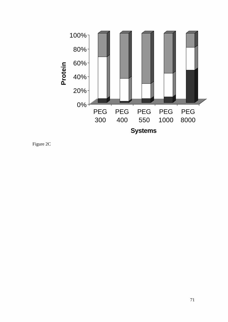
71
0%
20%
40%
60%
80%
100%
Pro
tein
PEG300
PEG400
PEG550
PEG1000
PEG8000
Systems
Figure 2C

72
Figure caption
Figure 1A. Agarose gel analysis of plasmid and RNA partitioning in ATPS with 20%
(w/w) of lysate. T-top phase. B-bottom phase. Lane1.1µg/ml of Lambda DNA/Hind III
marker, lanes 2 to 11: 15µl of each of the indicated phase, lane 12: 15µl of the lysate.
OC: open circular. SC: supercoiled.
Figure 1B. Agarose gel analysis of plasmid and RNA partitioning in ATPS with 40%
(w/w) of lysate. T-top phase. B-bottom phase. Lane1.1µg/ml of Lambda DNA/Hind III
marker, lanes 2 to 11: 15µl of each of the indicated phase, lane 12: 15µl of the lysate.
Figure 1C. Agarose gel analysis of plasmid and RNA partitioning in ATPS with 60%
(w/w) of lysate. T-top phase. B-bottom phase. Lane1.1µg/ml of Lambda DNA/Hind III
marker, lanes 2 to 11: 15µl of each of the indicated phase, lane 12: 15µl of the lysate.
Figure 2A. Protein partitioning in PEG phosphate ATPS using 20% (w/w) of lysate.
The percentage of protein in the top phase (gray bars), interphase (white bars), and
bottom phase (black bars) is shown for the different PEG MW.
Figure 2B. Protein partitioning in PEG phosphate ATPS using 40% (w/w) of lysate.
The percentage of protein in the top phase (gray bars), interphase (white bars), and
bottom phase (black bars) is shown for the different PEG MW.
Figure 2C. Protein partitioning in PEG phosphate ATPS using 60% (w/w) of lysate.
The percentage of protein in the top phase (gray bars), interphase (white bars), and
bottom phase (black bars) is shown for the different PEG MW.

73
CAPÍTULO 3 - Purification of plasmid (pVaxLacZ) by hydrophobic interaction
chromatography - Manuscrito a ser submetido para a publicação na revista “Biotechnology
Letters”.

74
Purification of plasmid (pVaxLacZ) by hydrophobic interaction chromatography
Moreira, K. A.1, Ribeiro, S. C.2, Diogo, M. M..2, Prazeres, D. M. F. 2,
Porto, A. L. F. 1,3, and Lima Filho, J. L. 1,4*
1. Laboratório de Imunopatologia Keizo Asami – LIKA/UFPE, Recife, Brazil; 2. Centro
de Engenharia Biológica e Química – Instituto Superior Técnico, Lisboa, Portugal; 3.
Universidade Federal Rural de Pernambuco – UFRPE; 4. Departamento de Bioquímica –
Universidade Federal de Pernambuco – UFPE.
Author for correspondence – * Av. Prof. Moraes Rego, s/n 50732-901-Recife PE Brasil,
Fax: 55 21 81 3271 8485; E-mail: [email protected].
Key words: gene therapy, hydrophobic interaction chromatography, plasmid, purification.

75
Abstract
Plasmid DNA used for acid nucleic vaccination or nonviral therapeutic gene transfer
has to be highly purified with minimal or zero contamination. A method is described
for the purification of plasmid DNA, which includes an ammonium sulphate
precipitation followed by hydrophobic interaction chromatography (HIC) using
Phenyl Sepharose 6 Fast Flow (low sub). The use of HIC took advantage of the more
hydrophobic character of single stranded nucleic acid impurities when compared with
double-stranded plasmid DNA.
76
Introduction
The use of the recombinant DNA with progress of the gene therapy or nucleic acid
vaccination DNA has an important hole in the prevention and in the cure of diseases
such as cancer and AIDS, have been increasing the need the obtaining of DNA pure
plasmid (Diogo et al. 2000). In some research applications it is possible to use crude
cell extracts of varying degrees of purity. However, plasmid DNA used for non-viral
therapeutic gene transfer or nucleic acid vaccination has to be highly purified and free
of contaminating components such as bacterial proteins, toxins, genomic DNA (gDNA)
or RNA (Prazeres et al. 1999). It is now clear that the demand for large amounts of
plasmid DNA will be enormous in view of the potential number of users and the
prospect of applying DNA vaccines to veterinary diseases (Little-van den Hurk et al.
2000). Additional, the fact that product recovery costs become critical in the overall
economics of modern biotechnology processes, and the need to have a process
complying with the guidelines issued by regulatory agencies has increases the interest in
developing methods for the downstream processing of plasmids (Ribeiro et al. 2002). A
process for the production of plasmid DNA generally follows the steps of fermentation,
primary isolations, and purification (Prazeres et al. 1999). Large-scale purifications
require scalable methods such as column chromatography (Prazeres et al. 1998). The
hydrophobic interaction technique used is very in the purification of therapeutic
proteins, but so far there are no reports of the application of HIC to plasmid purification
(Diogo et al. 2000). The use of HIC will take advantage of the different hydrophobic
character of double-stranded plasmid DNA and other nucleic acids impurities with high
content in single strands, such as RNA and denatured gDNA. The aim of this work is to
study the possibity of purifying plasmid DNA using HIC as the final operation in a
simple purification process.

77
Materials and methodsMaterials
Phenyl Sepharose 6 Fast Flow (low sub) was obtained from Pharmacia (Uppsala,
Sweden). RNase-DNase free was from Boehringer (Mannheim, Germany), Luria broth
(LB) was from Sigma (St. Loius, MO), and agarose was from FMC (Rockland, ME).
All salts used were of analytical grade.
Bacterial culture
Escherichia coli DH5α harboring pVax-LacZ (Invitrogen) was grown overnight at
37ºC, in 100 ml shake flasks containing 25 ml of LB medium with 30µg/ml kanamycin,
at 200 rpm. Larger culture volumes (250 ml) were inoculated with the appropriate
amount of overnight culture and incubated under the same conditions. E. coli DH5α
without plasmid was also grown under the same conditions as described before, but with
no kanamycin present.
Lysis and primary isolation
A modified alkaline method was used for cell lysis (Sambrook et al. 1989). After the
alkaline lysis method the plasmid in the supernatant was precipitated after addition of
0.6 volumes of isopropanol during 45 min incubation at -20ºC. The plasmid was
separated by centrifugation at 10 000 g during 20 min. The pellets were then redissolved
in TE buffer (10 mM Tris-HCl, 1 mM EDTA, pH 8). Next, solid ammonium sulphate
was dissolved in the plasmid solution at a concentration interval between 2.0-3.0 M,
followed by 15 min incubation on ice. Precipitated proteins were removed by
centrifugation at 10 000 g during 20 min at 4ºC. The supernatant was then diluted and
loaded directly on the HIC column. Total plasmid was quantified by anion-exchange
high-performance liquid chromatography (HPLC) throughout the different steps. The

78
same isolation procedure was applied to DH5α cells without plasmids to check the
behaviour of cell impurities in the HIC column.
Preparative chromatography
Chromatography was performed in a Pharmacia fast protein liquid
chromatography (FPLC) system (P 500 pump, LCC 500 chromatography controller).
An XK16/20 (20cm x 1.6cm) column (Pharmacia) was packed with 28 ml of the HIC
gel. Partially purified pVax-LacZ plasmid from the ammonium sulphate precipitation
was loaded onto the column and isocratic elution was carried out with 1.5 M ammonium
sulphate in 10mM Tris-HCl (pH 8.0) at a flow rate of 1 ml/mim. One-milliliter fractions
were collected. The absorbance of the eluate was measured continuously at 254 nm. The
samples analysed by 0.8% agarose gel electrophoresis, stained with ethidium bromide
(0.5 µg ml-1).
Plasmid standards
Plasmid standards were prepared using a plasmid mini-prep kit (Qiagen,
Germany) according to the instructions of the manufacturer.
Analytical chromatography
A 4.6 x 10cm polyether ether ketone (PEEK) column packed with Poros 20 PI
strong anion-exchange media from Perseptive Biosystems was connected to a Merck-
Hitachi HPLC system and equilibrated with 0.7M NaCl in TE buffer. Samples (100 µl)
were injected and eluted at 2 ml/min. These samples were incubated with RNase-DNase
free for 60 min at 37ºC before HPLC analysis. Plasmid was quantified through a
calibration curve, which was constructed using pVax-LacZ standards (2-40 µg ml-1).

79
Protein analysis
The protein concentration was measured by the modification micro
bicinchoninic acid (BCA) assay from Pierce (Rockford, Il, USA) (Rhoderick et al.
1989). A calibration curve was made with bovine serum albumin (BSA) standards. An
absorbance was measured at 595 nm in a micro plate reader.
Genomic DNA analysis
Genomic DNA (gDNA) was analysed by PCR. A 361 bp sequence of the
ribosomal RNA gene from E. coli DH5α was chosen for amplification in the PCR
analysis (forward primer 5´-ACA CGG TCC AGA CTC CTA CG-3´; reverse primer 5´-
ACA ACC TGG AAT TCT ACC CC-3´. The volumes of the samples to analyse were
0.35µl and the final volume of the mixture of used PCR was of 50 µl (Taq DNA 1x
buffer; 200 µM dNTP´s mixture, 1.5 µM of MgCl2 and 2.5 U Taq polymerase, all from
Promega (Madison, WI) and 0.75 µM of each primer from Interactiva (Ulm, Germany).
For each sample analysed, a calibration curve was made using 0.01-100 ng de standard
gDNA. PCR was performed with a first denaturation step at 94ºC for 3 min followed by
40 cycles of amplification (60ºC, 1 min; 74ºC, 1.5 min; 94ºC, 1 min) and a final
extension step (60ºC, 1 min; 74ºC, 10 min). Amplified samples (20 µl) were visualized
by 2% agarose gel analysis stained with ethidium bromide (0.5 µg ml-1).
Results and discussion
The results of purification of the plasmid DNA are summarized in Table 1. It
was observed a decrease of the plasmid mass once along the purification process. In the
stage of alkaline lysis there were 8716 µg after HIC which decreased to it 1230 µg.
There was an increase of purity 5.7 (lysis solution) and after HIC it was in 100%, in all

80
the concentrations ammonium sulphate used. The best purification factor was obtained
after HIC, using precipitation with 2.5M of ammonium sulphate, which yield 51%.
Studies accomplished by Diogo et al. (2000) working in the purification of plasmid
using hydrophobic interaction chromatography showed similar results. Figure 1 shows
the analysis of plasmid solution in HIC after precipitation with 2.5M ammonium
sulphate. In this figure it can be verified two peaks. The first pick (20 to 29 fractions)
corresponds the unretained fraction, the plasmid DNA. The fractions were collected and
analysed by using HPLC. The analytical chromatogram (not results shows) obtained no
longer shows the presence on the impurity peak, indicating an improvement in HPLC
purity (to 100%). The second peak corresponds to the host impurities such as RNA,
gDNA, denatured plasmid DNA and proteins eluted after the plasmid. Agarose gel
electrophoresis also analyzed the complete removal of RNA (not results shows) and the
absence of gDNA (Figure 2) for all the concentrations of ammonium sulphate used.
Genomic DNA from E. coli is double-stranded, but becomes mostly single-stranded
during alkaline lysis. During this process, the complementary strands of gDNA are
completely separated and partially cleaved. The resulting DNA molecules show a high
exposure of the hydrophobic bases and can thus interact with the HIC ligands (Diogo et
al. 2000). According to the specifications, gDNA contamination should be plasmid <0.1
ng µg-1.
Plasmid molecules did not interact with the HIC column, eluting in the flow
through. The reason for this behaviour is that, in double-stranded plasmid molecules,
the hydrophobic bases were packed and shielded inside the helix and thus hydrophobic
interaction with the support ligands was minimal. Previous studies by Diogo et al.
(1999) with the same HIC gel demonstrated that is also possible to separate denatured
plasmid from native plasmid. Because denatured forms of contain large stretches of
single-stranded DNA, there is more exposure of hydrophobic bases and,
81
consequently, the hydrophobic nitration is greater and retention time is longer. In fact,
the HIC support studied by Diogo et al. (2000) was capable of removing denatured
plasmid variants that are usually produced with the widespread method of alkaline lysis
of plasmid isolation. This work also indicates the ability of the HIC support to separate
proteins and genomic DNA from plasmids.
Acknowledgements
The authors acknowledge Dr. Joaquim M. S. Cabral (Instituto Superior Técnico,
Lisbon, Portugal) and Dr. Ernesto Marques Júnior (The Johns Hopkins University
School of Medicine, Baltimore, United states) kit supply and the plasmid for
accomplishment of this work. This work was supported CAPES, FINEP, EC Alfa
Program BIOENGE Network/ALR/B7-3011/94.04-6.0154.9, Universidade Federal de
Pernambuco and Universidade Federal Rural de Pernambuco.
References
Diogo MM, Queiroz JA, Monteiro GA, Prazeres DMF (1999) Separation and analysis
of denatured plasmid forms using hydrophobic interaction chromatography. Anal.
Biochem. 275:122-124.
Diogo MM, Queiroz JA, Monteiro GA, Ferreira GNM, Martins SAM, Prazeres DMF
(2000) Purification of a cystic fibrosis plasmid vector for gene therapy using
hydrophobic interaction chromatography. Biotechnol. Bioeng. 68:576-583.
Little-van den Hurk SVD, Braun RP, Babiuk LA (2000) Veterinary DNA vaccines. In:
DNA Vaccines: Methods and Protocols, Lowrie DB, Whalen RG (eds). Human
Press, Totowa.
Prazeres DMF, Schulep T, Cooney CL (1998) Preparative purification of supercoiled
plasmid DNA using anion exchange chromatography. J. Chromatogr. A. 806:31-
45.

82
Prazeres DMF, Monteiro GA, Ferreira GNM, Cooney CL, Cabral JMS (1999) Large-
scale production of pharmaceutical-grade plasmid DNA for gene therapy:
problems and bottlenecks. Trends Biotechnol. 17:169-174.
Rhoderick EB, Jarvis KL, Hyland KJ (1989) Protein measurement using bicinchoninic
acid: elimination of interfering substances. Anal. Biochem. 180:136-139.
Ribeiro SC, Monteiro GA, Cabral JMS, Prazeres DMF (2002) Isolation of plasmid
DNA from cell lysates by aqueous two-phase systems. Biotechnol. Bioeng.
78:376-384.
Sambrook J, Fritisch EF, Maniatis T (1989) Molecular cloning: laboratory manual. New
York: Cold Spring Harbor Laboratory Press.

83
Table 1. Purification of pVax-LacZ plasmid
Process step Plasmid mass
(µµg)
Purity
(%)
Purification
factor
Yield
(%)
Lysis
Isopropanol precipitation
(NH4)2SO4 precipitation 2.0M
2.5M
3.0M
HIC 2.0M
2.5M
3.0M
8716
4219
1837
1490
1388
1202
1229
1184
5.7
6.7
26
32
37
100
100
100
1.0
1.7
3.1
2.4
2.2
3.2
3.3
3.1
100
48
69
53
49
40
51
53
Results reported are the average of two independents experiments and errors were judged 5% of the mean
value.

84
Figure 1

85
Figure 2.
1 2 3 4 5 6 7 8 9 10
Kb
108
654
3
2
1.5
1
0.5

86
Figure caption
Figure 1. Hydrophobic interaction chromatography performed on plasmid solutions
after 2.5M ammonium sulphate precipitation. Elution was isocratic with 1.5M
ammonium sulphate in 10 mM Tris-HCl (pH 8.0) at a low rate of 1ml/min.
Figure 2. Analysis of gDNA by agarose gel eletrophoresis: molecular weight markers
(lane 1). Calibration curve (lane 2-6). Lysis solution (lane 7). Samples after HIC 2.0M,
2.5M and 3.0M precipitation with sulphate of ammonium (lane 8-10).

87
7. CONCLUSÕES
Os resultados experimentais obtidos permitiram as seguintes conclusões:
q A produção DNA plasmidial em agitador orbital foi dependente das condições
de cultivo. A diferentes concentrações da pressão seletiva (canamicina) influenciou a
produção de DNA plasmidial. Concentrações entre 30-50µg ml-1 demonstraram
comportamento similar para todos os ensaios de produção biomassa e DNA
plasmidial. Como também as diferentes velocidades de agitação usadas=. O meio de
cultura TB apresentou ser o mais eficiente em termos de produção de DNA plasmidial
e apresentou ser mais estável quando comparado como meio de LBG.
q O acréscimo da quantidade de solução de lise aplicado a cada sistema não é
muito vantajoso para as fases analisadas (PEG 300 e 400 fase superior e PEG 1000
fase inferior), o rendimento não é proporcional à quantidade de lisado celular
carregado. O sistema que apresentou melhor rendimento (37,18%) foi o sistema PEG
400 com 60% de lisado celular carregado, porém obteve um alto rendimento de
proteína. O menor rendimento em proteína foi verificado com o sistema PEG 1000
com 20% de lisado obtendo 4% de proteína e rendimento em DNA plasmidial de 10,5.
Sistemas com pesos moleculares menores (300 e 400) ocorreu partição do DNA
plasmidial para a fase superior, enquanto sistemas com pesos moleculares maiores
(1000 e 8000) ocorre partição para a fase inferior e o sistema com peso molecular
intermediário (550) ocorreu partição para a interfase. O RNA preferencialmente
particionou-se para a fase onde o DNA plasmidial foi particionado.
88
q A combinação da técnica de precipitação com sulfato de amônio e a
cromatografia de interação hidrofóbica utilizada foi eficiente para purificar DNA
plasmidial. Obtendo 100% de pureza para todas as concentrações de sulfato de
amônio após sua eluição na matriz cromatográfica e fator de purificação de 3,3 na
concentração de 2,5M de sulfato de amônio.

89
8. SUGESTÕES PARA ESTUDOS FUTUROS
Como todo trabalho de pesquisa, este segue a regra, uma vez que é praticamente
difícil a obtenção de um trabalho totalmente completo, e torna-se importante a
apresentação de algumas sugestões para que possam contribuir para o melhor as
condições de produção e purificação de DNA plasmidial, propõe-se:
q Realizar estudos de produção de DNA plasmidial com diferentes variáveis em
fermentador com auxílio de planejamento estatístico fracionado ou completo;
q Analisar a eficiência de resinas de caráter hidrofóbico visando aumentar o
rendimento em DNA plasmidial;
q Verificar o comportamento de partição do DNA plasmidial no sistema
PEG/citrato;
q Investigar a formação de isoformas do DNA plasmidial durante os processos de
lise celular e na partição em sistemas de duas fases aquosas.

90
9. ANEXOS
Os resultados obtidos neste trabalho foram apresentados em congressos
nacionais e internacionais.
1. Resumo aceito para apresentação no XXII Congresso de Microbiologia: Influência
das condições de cultivo na estabilidade do plasmídeo pD2 em Escherichia coli.
2. Resumo apresentado no 12th International Conference on Biopartitioning and
Purification: A preliminary study of dengue 2 plasmid DNA vaccine (pD2) extraction
from cell lysates by aqueous two-phase systems.
3. Resumo apresentado na XXXI Reunião Anual da Sociedade de Bioquímica e
Biologia Molecular. Effect of antibiotic concentration and culture aeration on pVaxDN2
production E. coli.
4. Resumo apresentado na XXXI Reunião Anual da Sociedade de Bioquímica e
Biologia Molecular. Plasmid stability pVaxDN2-carring E. coli during batch-
fermentation.
5. Resumo apresentado na XXX Reunião Anual da Sociedade de Bioquímica e
Biologia Molecular. Partitioning and purification of plasmid DNA in aqueous two-
phase systems.

91
Durante a realização deste trabalho foram realizadas atividades paralelas, os
resultados obtidos foram submetidos para publicação em periódicos internacionais e
apresentados em congressos nacionais e internacionais.
1. Characterization of protease from Penicillium aurantiogriseum for application in
food industries. 4th European Congress Engineering, 21 a 25 de setembro, Granada,
Espanha, 2003 .
2. Tratamento enzimático utilizando proteases de Nocardiopsis sp. em águas residuais
de uma indústria de laticínios. XXII Congresso Brasileiro de Microbiologia, 17 a 20 de
novembro, Florianópolis, SC, 2003.
3. Otimização da produção de proteases produzidas por Candida butyri. XXII
Congresso Brasileiro de Microbiologia, 17 a 20 de novembro, Florianópolis, SC, 2003.
4. Study of the conditions of production of proteases for Aspergillus sydowii. VII
Simpósio de hidrolise enzimática de biomassas, 06 a 10 de dezembro, Maringá, PR,
2002.
5. Use of aqueous two-phase systems on perforated rotating disc contractor for
continuous extraction of Schitosoma mansoni recombinant antigen. XXXI Reunião
Anual da Sociedade de Bioquímica e Biologia Molecular, 18 a 21 de maio, Caxambu,
MG, 2002.

92
6. Partial purification on inulinase from Aspergillus niveus. XXX Reunião Anual da
Sociedade de Bioquímica e Biologia Molecular, 19 a 22 de maio, Caxambu, MG, 2001.
7. Partitioning and purification of monoclonal antibody of Toxoplasma gondii in
PEG/potassium phosphate aqueous two-phase systems. XXX Reunião Anual da Sociedade
de Bioquímica e Biologia Molecular, 19 a 22 de maio, Caxambu, MG, 2001.