Embed Size (px)
Citation preview
2016/2017
Sérgio Gomes Pinto
Regressão da Remodelagem Ventricular Esquerda no Contexto de
Estenose Válvular Aórtica
Regression of Left Ventricular Remodeling in the context of Aortic
Valve Stenosis
março, 2017
Mestrado Integrado em Medicina
Área: Cirurgia Cardiotorácica
Tipologia:Monografia
Trabalho efetuado sob a Orientação de:
Professor Doutor Joaquim Adelino Correia Ferreira Leite
Moreira
E sob a Coorientação de:
Professora Inês Maria Falcão Sousa Pires Marques
Trabalho organizado de acordo com as normas da revista:
Revista Portuguesa de Cirurgia Cardiotorácica e Vascular
Sérgio Gomes Pinto
Regressão da Remodelagem Ventricular Esquerda no Contexto de Estenose Válvular Aórtica
Regression of Left Ventricular Remodeling in the Context of Aortic Valve Stenosis
março, 2017

DEDICATÓRIA
Queria agradecer a todos os que me acompanharam, tanto no desafio da realização desta
tese, como também ao longo de todo o curso de Medicina. Foi uma aventura que nunca
esquecerei.
Ao Professor Doutor Adelino Leite Moreira e à Professora Inês Falcão Pires, um agradecimento
sincero por toda a ajuda e força que me deram, e pela oportunidade de desenvolver este
trabalho.
Aos meus amigos, dentro e fora da Faculdade de Medicina do Universidade do Porto. Não teria
conseguido sem vocês.
À minha incrivél namorada, que nunca desistiu de mim.
E por ultimo, à minha familia. Não há palavras para agradecer todo o apoio e ajuda
incondicionais que já me deram.
1
INTRODUCTION
BACKGROUND AND OBJECTIVES
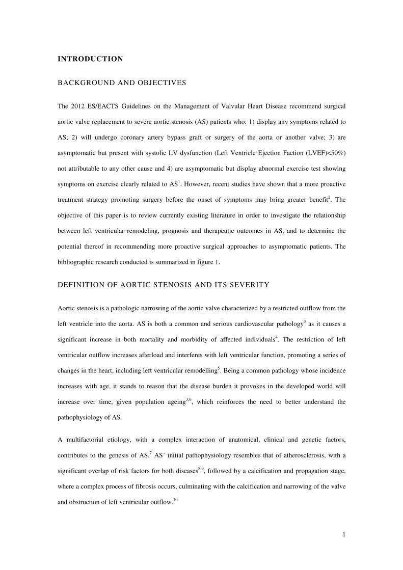
The 2012 ES/EACTS Guidelines on the Management of Valvular Heart Disease recommend surgical
aortic valve replacement to severe aortic stenosis (AS) patients who: 1) display any symptoms related to
AS; 2) will undergo coronary artery bypass graft or surgery of the aorta or another valve; 3) are
asymptomatic but present with systolic LV dysfunction (Left Ventricle Ejection Faction (LVEF)<50%)
not attributable to any other cause and 4) are asymptomatic but display abnormal exercise test showing
symptoms on exercise clearly related to AS1. However, recent studies have shown that a more proactive
treatment strategy promoting surgery before the onset of symptoms may bring greater benefit2. The
objective of this paper is to review currently existing literature in order to investigate the relationship
between left ventricular remodeling, prognosis and therapeutic outcomes in AS, and to determine the
potential thereof in recommending more proactive surgical approaches to asymptomatic patients. The
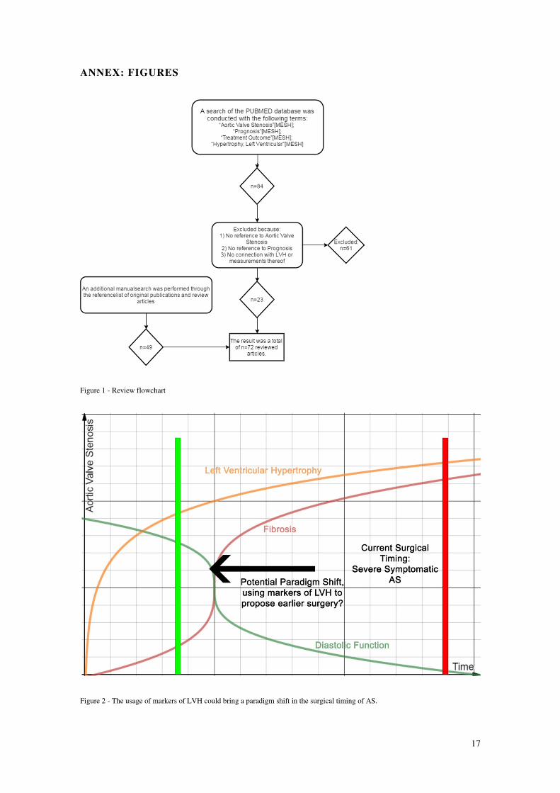
bibliographic research conducted is summarized in figure 1.
DEFINITION OF AORTIC STENOSIS AND ITS SEVERITY
Aortic stenosis is a pathologic narrowing of the aortic valve characterized by a restricted outflow from the
left ventricle into the aorta. AS is both a common and serious cardiovascular pathology3 as it causes a
significant increase in both mortality and morbidity of affected individuals4. The restriction of left
ventricular outflow increases afterload and interferes with left ventricular function, promoting a series of
changes in the heart, including left ventricular remodelling5. Being a common pathology whose incidence
increases with age, it stands to reason that the disease burden it provokes in the developed world will
increase over time, given population ageing3,6
, which reinforces the need to better understand the
pathophysiology of AS.
A multifactorial etiology, with a complex interaction of anatomical, clinical and genetic factors,
contributes to the genesis of AS.7 AS’ initial pathophysiology resembles that of atherosclerosis, with a
significant overlap of risk factors for both diseases8,9
, followed by a calcification and propagation stage,
where a complex process of fibrosis occurs, culminating with the calcification and narrowing of the valve
and obstruction of left ventricular outflow.10
2
Roberts et al studied the natural history of 260 AS patients not subjected to surgical treatment over 50
years. This author reported that heart failure, angina pectoris or syncope, were overwhelmingly common
(68% of men and 67% of women); that the onset of any cardiac symptomatology was associated with an
exceptionally dire prognosis, supporting a median survival of 20 months after symptom onset; and that
patients with cardiac symptomatology were at higher risk for death from cardiac events (both sudden and
non-sudden) when compared to the asymptomatic group (OR: 3.61).11
The therapeutic gold standard for AS is aortic valve replacement (AVR), as confirmed by numerous
studies demonstrating dramatic symptomatic relief and improvement of long-term survival12
. An
alternative to surgical replacement is transcatheter aortic valve implantation (TAVI), especially for
patients with high surgical risk who cannot undergo surgical aortic valve replacement (SAVR)13
. As far
as medical therapy is concerned, no therapeutic regimen has been able to convincingly delay the
progression of AS or reduce mortality once symptoms are established, and thus, it is limited to patients
who require pre-surgical stabilization or for symptomatic management in patients who cannot or who
choose not to undergo SAVR or TAVI.14–16
The 2012 ES/EACTS Guidelines on the Management of Valvular Heart Disease defined severe AS as
having a valve area under 1 cm2; an indexed valve area under 0.6 cm²/m²; a mean gradient greater than
40mmHg; a maximum jet velocity over 4.0 m/s; and a velocity ratio under 0.251. The American Heart
Association Guidelines for the Management of Patients with Valvular Heart Disease17
define four main
stages of AS, summarized in Table 1. Mild LV dysfunction can be observed as early as stage B and LV
remodeling as early as stage C. The distinction between these stages is made primarily through the
presence of symptomatology, the aortic valve morphology and area, the pressure gradient between the LV
and the aorta, and the LVEF.18,19
These latter 3 parameters are determined through echocardiographic
studies, which are the cornerstone of AS staging18,19
, although other imaging procedures may also have a
diagnostic role.20,21
It is important to distinguish between the various stages of the pathology, given the heavy implications
upon prognosis and therapeutic decisions.22,23
The decision to submit the patient to surgical treatment is
based on a risk-benefit assessment of surgical risk associated with the procedure weighed against the risk
posed by the lack of surgical correction. The current ES/EACTS guidelines1 and AHA guidelines
recommend against routine aortic valve replacement for asymptomatic patients on the basis that these
3
patients possess a low risk of cardiac sudden death (>1%) when compared to age-matched controls17
. The
indications for AVR according to ES/EACTS guidelines are summarized in Table 2.
LEFT VENTRICULAR REMODELLING IN THE CONTEXT OF AORTIC
STENOSIS
Ventricular remodeling comprises the functional and anatomic changes undergone by the heart in
response to either physiological stimuli, like exercise24
, high altitude25
, pregnancy26
or to various
pathological stimuli, like volume overload, as in valvular regurgitation,27
pressure overload, as in AS,28
or
other situations where a more complex forms of overload may be present, like acute myocardial infarction
(AMI)27
.
Various classifications exist for LVR and LVH. Verma et al, in the VALIANT study, defined 3 types of
LVR: concentric remodeling, concentric hypertrophy and eccentric hypertrophy.29
Khouri et al suggested
an expansion of this system with subgroups for each category, namely indeterminate eccentric, dilated
eccentric, thick concentric and thick and dilated concentric LVH.30
Situations of volume overload tend to
produce eccentric hypertrophy26
, whereas situations of pressure overload tend to produce concentric
hypertrophy.28
AS imposes a chronic high afterload to the left ventricle, triggering compensatory changes aimed at
reducing wall stress and maintaining cardiac output. The LV undergoes an increase in both relative wall
thickness (RWT) and LV Mass Index (LVMi), which is the LV Mass (LVM) adjusted to body surface
area (BSA)29
, in what is known as concentric hypertrophy. The increase in LVM negatively affects the
contractility of heart31
. However, the increase in afterload is not the only mechanism involved in LVH in
the context of AS. Indeed, LVH is a complex, multifactorial process, modulated by variables such as
gender32
, neurohumoral activation18
, and the association of AS with various comorbidities, like obesity
and hypertension33
.
The aforementioned changes in LV wall thickness and geometry34
occur simultaneously to a fibrotic
process where myocardial areas of apoptosis are replaced by fibrosis35
, with the occurrence of a
pathological sequence of progression from myocyte hypertrophy, to apoptosis, to fibrosis36
, with
correspondent fibroblast recruitment and increase in fibrotic content of the myocardium37
. The
predominant pattern of fibrosis occurring in the context of AS is midwall fibrosis and its presence is
4
usually established via Late Gadolinium Enhancement (LGE) in a cardiac MRI38
. The exact trigger for
apoptosis in these circumstances is unknown, but both direct mechanical factors, such as wall stretch, and
angiotensin II appear to play a relevant role39–41
. This increase in myocardial fibrotic content ultimately
produces a worse prognosis, due either to worsening of diastolic function33
or to the increased
arrythmogenic potential of the fibrotic heart42
and cardiac sudden death43,44
. The increased cardiac wall
stress promotes an increase in circulating natriuretic peptides, particularly N-terminal B-type natriuretic
peptide (NtBNP), as it appears capable of predicting symptom free survival and post-operative
outcomes45
. Myocardial ischemia is also a relevant process in LVH. It occurs mostly as the
microvasculature of the heart becomes unable to keep up with the growing metabolic demands of the left
ventricular mass46
, and thus, may also be a relevant contributor to the increasing fibrosis of the
myocardium. Reflecting the influence of myocardial cell death in this process, higher circulating levels of
cardiac troponins can be found, particularly with the usage of high sensitivity assays.47
While these changes are initially adaptive, with chronicity they tend to become maladaptive48
and thus,
LVH has a significant impact on the prognosis of AS, being an independent determinant of higher
mortality and morbidity from cardiac events.27,49–51
LEFT VENTRICULAR REVERSE REMODELLING AFTER AORTIC VALVE
REPLACEMENT
LV Reverse Remodeling (LVRR) is seen in AS patients who successfully undergo AVR, both with
SAVR techniques or TAVI techniques52
, defined as a normalization of LVMi, RWT, and other
measurements of hypertrophy as well as recovery of cardiac function53
.
However, LVRR is frequently incomplete. Gavina et al54
studied the presence of residual LVH in a post-
SAVR setting, and observed that, in this cohort, 44% of patients maintained some degree of LVH after
valve replacement, and that this incomplete regression heralded a worse prognosis, especially for women.
Magalhães et al55
performed a similar analysis regarding extension of the LVRR process in a TAVI
setting and reported partial or complete normalization of LVMi and RWT in 24% of the population at 1
year follow-up post-TAVI, without a corresponding normalization of left atrial dimensions. In TRITON
trial, Haverich et al56
reported a mean reduction of 14% in LVMi at 1 year follow-up, and a 16%
reduction at 3 years after surgical treatment of AS with a Next Generation Surgical Aortic Valve.
5
Similarly to the extent of LVH, LVRR is modulated by various factors apart from successful AVR.
Hypertension appears to play an exceedingly important role, as those AS patients without hypertension
who undergo AVR have a much greater amount of LVRR when compared to those with hypertension
independently of total afterload32
.Prosthetic-patient mismatch57
is also independently associated with a
negative impact on the extent of LVRR58
, as demonstrated in a large cohort of AS patients by Del Rizzo et
al59
. Gender also has an impact, with women having a tendency towards more complete LVH regression
when compared to men60,61
.
POSITIVE OUTCOMES ASSOCIATED WITH EARLIER SURGICAL
INTERVENTION
Growing evidence begins to suggest, however, that surgical treatment strategies may effectively provide
greater benefit to asymptomatic patients than previously believed.
In a prospective study conducted over 10 years, Kang et al studied patients with asymptomatic severe AS
randomized between two treatment groups, one treated with early surgery, as defined as elective surgery
performed before formal indication per the most recent available guidelines, and one treated with the
conventional treatment strategy. The authors reported a cardiac and all-cause risk of death of 0% and 6%
in the early surgical treatment group, and a 24% and 32% risk for the same variable in the conventional
treatment group, over a median follow-up period of 1501 days. Authors concluded that the benefits of
early surgery outweighed all the risks, and thus, that it was an option for the asymptomatic very severe
AS patient. Also this group presented more LVH regression and better LV systolic function in post-op
echocardiographic studies, indicating that early surgery is able to prevent irreversible myocardial damage
and fibrosis, and therefore reduce the risk of cardiac sudden death. 62
In 2015, Taniguchi et al analyzed data from a large multicenter registry to compare long term outcomes
of patients with asymptomatic severe AS who were treated with AVR (SAVR or TAVI) at diagnosis and
those managed with traditional conservative strategies. The authors reported dire outcomes in the group
managed with the currently recommended conservative strategy when compared to those who underwent
AVR, with a cumulative 5-year incidence of death by all causes of 26.4% vs. 15.4% (p=0.009); a
cumulative 5-year incidence of cardiovascular and aortic valve related death of 18.6% vs. 9.9% (p=0.01);
and a cumulative 5-year incidence of sudden death of 5.8% vs. 3.6 % (p=0.06) thus concluding that the
6
dismal outcomes associated with conservative treatment strategies for severe asymptomatic AS patients
are largely surpassed by earlier AVR2
The AVATAR prospective, multicenter, randomized, controlled, parallel group, event-driven trial began
in 2016 and aimed to evaluate the safety and efficacy of elective AVR for asymptomatic AS patients with
preserved LVEF. To achieve this goal, a cohort of 312 asymptomatic patients with isolated AS and
preserved LVEF will be randomized to one of two groups, one managed with the currently recommended
strategy (medical treatment until symptoms arise or LVEF drops below 50%) and one to be treated with
elective AVR. The primary outcome of the study is a composite variable of all-cause death, acute
myocardial infarction, stroke or unplanned hospitalization for heart failure. Secondary outcomes will
include a safety analysis to determine whether the early surgery group suffers from any increase in
operative and in hospital mortality, or from an increase in valve related complications, when compared to
patients operated after symptom onset.63
This trial will definitely contribute to expand the knowledge
about the best timing for AVR.
POSSIBLE ROLE OF LVR AND LVH FROM A THERAPEUTIC AND
PROGNOSTIC STANDPOINT
This review of current information, summarized in Table 3, regarding LVH in the context of AS revealed
a large amount of relevant information that can potentially be utilized to support the inclusion of LVH in
operative criterion for AS.
Regarding echocardiography, data exists supporting an expansion of the hemodynamic characteristics
evaluated64
, and the usage of Integrated Backscatter (IBS)65
techniques, which are a form of
echocardiographical characterization of myocardial tissues, shows great promise. Cyclic Variation of IBS
(CVIBS), mean IBS at end diastole (IBSed), and mean Cyclic Variation of IBS index (CVIBSi) have
value as predictors of LVRR in a post-AVR setting: CVIBS showed a sensitivity of 84.6% and specificity
of 63.1%. using values equal to or greater than 5.1 dB as a cut- off. IBSed showed a sensitivity of 84.6%
and specificity of 78.9% using values equal to or greater than 34 dB as a cut-off. CVIBSi showed a
sensitivity of 79.5% and specificity of 84.2% using values equal to or greater than 15.7% as a cut-off66
.
Echocardiographic strain measurements have also shown potential. Two-dimensional back longitudinal
scatter shows a strong association with adverse outcomes for the asymptomatic AS patient, when values
7
lower than 13% are found,67
and 3 dimensional global longitudinal strain has a sensitivity and specificity
of 76% and 77%, for a cutoff value of -14.5 for predicting MACE, also in asymptomatic AS patients.
The usage of MRI also shows potential for expansion. LGE; as said above, is the quintessential method
for evaluating the midwall myocardial fibrosis occurring in LVH. LGE and LVMi share an independent
association with plasma cTnI concentrations68
, and LGE is also independently associated with the
aforementioned echocardiographic strain measurements69
. Midwall fibrosis as measured by LGE in an
MRI appears associated with an 8 fold increased risk of all-cause mortality in moderate to severe AS
patients38
.
The common electrocardiogram also shows interest, as measurements of LVH and LV strain have a
direct, independent relationship with prognosis70
.
Perhaps the most interesting data comes in the domain of biomarkers. The usage of high sensitivity
cardiac Troponin T (hs-cTnT) assays is particularly relevant, as high circulating titers of hs-CTnT are
associated with poor prognosis even in asymptomatic AS patients71
, namely cardiovascular death or need
for future AVR68
. B-type Natriuretic Peptide (BNP) is also relevant. Lower levels of NT-BNP correlate
with higher magnitudes of LMVi normalization and better quality of life in a post-AVR setting72
. In a
groundbreaking study, Gárcia et al73
demonstrated the potential relevance of titers of miR-133a in the
timing of the surgical decision for AVR, as higher titers of pre-operative circulating miR-133a revealed
greater potential for LVMi normalization post-op.
8
DISCUSSION
LIMITATIONS OF CURRENT GUIDELINES
As of today, the usage of LVH and markers thereof is sparse for therapeutic decision in the context of AS.
While the 2012 ES/EACTS1 guidelines already include LVH when recommending AVR for
asymptomatic patients, with normal exercise testing, low surgical risk and an excess of LVH in the
absence of hypertension, the class of recommendation is only IIB, and the contemporary 2014 AHA
guidelines17
make no reference whatsoever to these potentially useful markers.
The current management strategy recommended by both European and American guidelines is also a
subject of controversy, with large, multi-center studies showing the currently recommended strategy of
“watchful waiting” prompts a dismal outcome.
With this in mind, several studies have attempted to uncover other parameter that might help to clarify the
best time for ABR. For instance, in 2012, Carabello recommended AVR for asymptomatic AS patients
with either a positive exercise test; heavy valve calcification; documented rapid progression of AS from
serial measurements; excessive left ventricular hypertrophy; or rising natriuretic peptides,74
differing from
the established guidelines.
Various studies found that the persistence of LVH in a post AVR or TAVI setting is associated with
worse outcomes, like an increase in risk of MACE, all-cause death, or cardiac re-hospitalization. There
appears to be a trend showing that while men more frequently exhibit maladaptive cardiac remodeling
when compared to women, women have a worse prognosis in comparison to men when said remodeling
is present54
.
All of these potentially useful markers of prognosis can be evaluated using studies which are already
performed on a routine basis for AS patients (echocardiography, electrocardiography, blood
measurements and MRI), and thus there would be virtually no impediment to the incorporations of these
new measures in newer editions of the guidelines should their utility be confirmed.
However, to fully recommend a change in guidelines, more research is needed. It would be essential to
determine whether earlier surgery is associated with better LVH regression and better prognosis. With
this objective in mind, the AVATAR study63
begun in 2016, will provide key information.
9
Either way, data shows that the risk-benefit analysis regarding surgical recommendation in asymptomatic
AS is overly simplistic, overlooking various highly useful markers of prognosis. The construction on an
integrated risk score for these patients, utilizing various prognostic markers apart from hemodynamic
measurements, like echocardiographic LV strain and LVH, electrocardiographic LVH and LV strain,
various biomarkers and MRI markers, could be integrated into future guidelines to recommend earlier
surgery for asymptomatic AS patients with a particularly dire prognosis.
CONCLUSIONS
While further studies are essential to cement these conclusions, the weight of recent data appears to
indicate that current guidelines present an overly simplistic risk-benefit analysis regarding the surgical
decision for AS patients, and an overly passive and conservative approach to asymptomatic AS patients.
The association of LVH, its echocardiographic translation, namely ECG strain, and various cardiac
biomarkers, with the occurrence of MACE and prognosis even in asymptomatic patients herald great
potential in the design of a newer, more complete integrated risk score for AS, which could reveal
asymptomatic patients with an exceptionally dire prognosis and this, with great potential benefit from
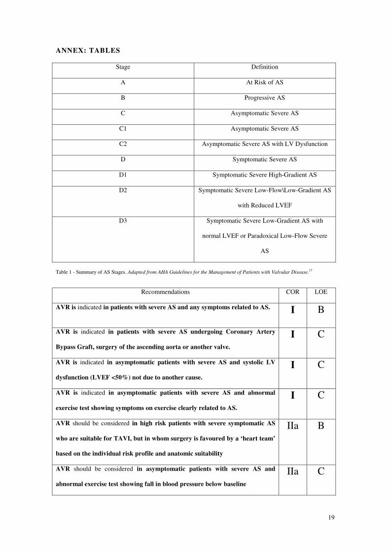
more proactive surgical interventions, as is illustrated in Figure 2.
10
BIBLIOGRAPHY
1. Vahanian A, Alfieri O, Andreotti F, et al. Guidelines on the management of valvular heart disease
(version 2012). Eur Heart J. 2012;33(19):2451-2496. doi:10.1093/eurheartj/ehs109.
2. Taniguchi T, Morimoto T, Shiomi H, et al. Initial Surgical Versus Conservative Strategies in
Patients with Asymptomatic Severe Aortic Stenosis. J Am Coll Cardiol. 2015;66(25):2827-2838.
doi:10.1016/j.jacc.2015.10.001.
3. Nkomo VT, Gardin JM, Skelton TN, Gottdiener JS, Scott CG, Enriquez-Sarano M. Burden of
valvular heart diseases: a population-based study. Lancet. 2006;368(9540):1005-1011.
doi:10.1016/S0140-6736(06)69208-8.
4. Joseph J, Naqvi SY, Giri J, Goldberg S. Aortic Stenosis: Pathophysiology, Diagnosis, and Therapy.
Am J Med. 2016;130(3):253-263. doi:10.1016/j.amjmed.2016.10.005.
5. Rassi AN, Pibarot P, Elmariah S. Left ventricular remodelling in aortic stenosis. Can J Cardiol.
2014;30(9):1004-1011. doi:10.1016/j.cjca.2014.04.026.
6. Berry C, Lloyd SM, Wang Y, MacDonald A, Ford I. The changing course of aortic valve disease in
Scotland: Temporal trends in hospitalizations and mortality and prognostic importance of aortic
stenosis. Eur Heart J. 2013;34(21):1538-1547. doi:10.1093/eurheartj/ehs339.
7. Otto CM, Prendergast B. Aortic-Valve Stenosis — From Patients at Risk to Severe Valve
Obstruction. N Engl J Med. 2014;371(8):744-756. doi:10.1056/NEJMra1313875.
8. Thanassoulis G, Massaro JM, Cury R, et al. Associations of Long-Term and Early Adult
Atherosclerosis Risk Factors With Aortic and Mitral Valve Calcium. J Am Coll Cardiol.
2010;55(22):2491-2498. doi:10.1016/j.jacc.2010.03.019.
9. Stritzke J, Linsel-Nitschke P, Markus MRP, et al. Association between degenerative aortic valve
disease and long-term exposure to cardiovascular risk factors: Results of the longitudinal
population-based KORA/MONICA survey. Eur Heart J. 2009;30(16):2044-2053.
doi:10.1093/eurheartj/ehp287.
10. Pawade TA, Newby DE, Dweck MR. Calcification in aortic stenosis: The skeleton key. J Am Coll
Cardiol. 2015;66(5):561-577. doi:10.1016/j.jacc.2015.05.066.
11. Roberts WC, Vowels TJ, Filardo G, Ko JM, Mathur RP, Shirani J. Natural history of unoperated
aortic stenosis during a 50-year period of cardiac valve replacement. Am J Cardiol.
2013;112(4):541-553. doi:10.1016/j.amjcard.2013.04.020.
12. Nicolini F, Fortuna D, Contini GA, et al. Long-term outcomes of conventional aortic valve
replacement in high-risk patients: Where do we stand? Ann Thorac Cardiovasc Surg.
2016;22(5):304-311. doi:10.5761/atcs.oa.16-00165.
13. Krasopoulos G, Falconieri F, Benedetto U, et al. European real world trans-catheter aortic valve
implantation: systematic review and meta-analysis of European national registries. J
Cardiothorac Surg. 2016;11(1):159. doi:10.1186/s13019-016-0552-6.
14. Carey T, Pearce J. Aortic stenosis: Pathophysiology, diagnosis, and medical management of
nonsurgical patients. Crit Care Nurse. 2007;33(4):174-183. doi:10.1016/S0140-6736(09)60211-7.
15. Otto CM. Calcific Aortic Valve Disease: New Concepts. Semin Thorac Cardiovasc Surg.
2010;22(4):276-284. doi:10.1053/j.semtcvs.2011.01.009.
16. Nadir MA, Wei L, Elder DHJ, et al. Impact of renin-angiotensin system blockade therapy on
outcome in aortic stenosis. J Am Coll Cardiol. 2011;58(6):570-576.
doi:10.1016/j.jacc.2011.01.063.
11
17. Nishimura RA, Otto CM, Bonow RO, et al. 2014 AHA/ACC Guideline for the Management of
Patients with Valvular Heart Disease : A Report of the American College of Cardiology/american
Heart Association Task Force on Practice Guidelines. Vol 129.; 2014.
doi:10.1161/CIR.0000000000000031.
18. Lindman BR, Clavel M-A, Mathieu P, et al. Calcific aortic stenosis. Nat Rev Dis Prim.
2016;2:16006. doi:10.1038/nrdp.2016.6.
19. Capoulade R, Pibarot P. Assessment of Aortic Valve Disease: Role of Imaging Modalities. Curr
Treat Options Cardiovasc Med. 2015;17(11). doi:10.1007/s11936-015-0409-7.
20. Singh A, Steadman CD, McCann GP. Advances in the understanding of the pathophysiology and
management of aortic stenosis: Role of novel imaging techniques. Can J Cardiol. 2014;30(9):994-
1003. doi:10.1016/j.cjca.2014.03.008.
21. Breitenbach I, Harringer W, Tsui S, et al. Magnetic resonance imaging versus echocardiography
to ascertain the regression of left ventricular hypertrophy after bioprosthetic aortic valve
replacement: Results of the REST study. J Thorac Cardiovasc Surg. 2012;144(3):640-645.
doi:10.1016/j.jtcvs.2011.11.017.
22. Herrmann HC, Pibarot P, Hueter I, et al. Predictors of mortality and outcomes of therapy in low-
flow severe aortic stenosis: A placement of aortic transcatheter valves (PARTNER) trial analysis.
Circulation. 2013;127(23):2316-2326. doi:10.1161/CIRCULATIONAHA.112.001290.
23. Tribouilloy C, Rusinaru D, Maréchaux S, et al. Low-gradient, low-flow severe aortic stenosis with
preserved left ventricular ejection fraction: Characteristics, outcome, and implications for
surgery. J Am Coll Cardiol. 2015;65(1):55-66. doi:10.1016/j.jacc.2014.09.080.
24. Ellison GM, Waring CD, Vicinanza C, Torella D. Physiological cardiac remodelling in response to
endurance exercise training: cellular and molecular mechanisms. Heart. 2012;98(1):5-10.
doi:10.1136/heartjnl-2011-300639.
25. Stembridge M, Ainslie PN, Shave R. Short-term adaptation and chronic cardiac remodelling to
high altitude in lowlander natives and Himalayan Sherpa. Exp Physiol. 2015;100(11):1242-1246.
doi:10.1113/expphysiol.2014.082503.
26. Lorell BH, Carabello BA. Left ventricular hypertrophy: pathogenesis, detection, and prognosis.
Circulation. 2000;102(4):470-479. doi:10.1161/01.cir.102.4.470.
27. Cohn JN, Ferrari R, Sharpe N. Cardiac remodeling-concepts and clinical implications: A consensus
paper from an International Forum on Cardiac Remodeling. J Am Coll Cardiol. 2000;35(3):569-
582. doi:10.1016/S0735-1097(99)00630-0.
28. Dadson K, Kovacevic V, Rengasamy P, et al. Cellular, structural and functional cardiac
remodelling following pressure overload and unloading. Int J Cardiol. 2016;216:32-42.
doi:10.1016/j.ijcard.2016.03.240.
29. Verma A, Meris A, Skali H, et al. Prognostic Implications of Left Ventricular Mass and Geometry
Following Myocardial Infarction. The VALIANT (VALsartan In Acute myocardial iNfarcTion)
Echocardiographic Study. JACC Cardiovasc Imaging. 2008;1(5):582-591.
doi:10.1016/j.jcmg.2008.05.012.
30. Khouri MG, Peshock RM, Ayers CR, De Lemos JA, Drazner MH. A 4-tiered classification of left
ventricular hypertrophy based on Left ventricular geometry the dallas Heart study. Circ
Cardiovasc Imaging. 2010;3(2):164-171. doi:10.1161/CIRCIMAGING.109.883652.
31. Huber D, Grimm J, Koch R, Krayenbuehl HP. Determinants of ejection performance in aortic
stenosis. Circulation. 1981;64(1):126-134. doi:10.1161/01.CIR.64.1.126.
32. Gavina C, Falcão-Pires I, Rodrigues J, et al. Load independent impairment of reverse remodeling
12
after valve replacement in hypertensive aortic stenosis patients. Int J Cardiol. 2014;170(3):324-
330. doi:10.1016/j.ijcard.2013.11.006.
33. Cho I-J, Chang H-J, Park H-B, et al. Aortic calcification is associated with arterial stiffening, left
ventricular hypertrophy, and diastolic dysfunction in elderly male patients with hypertension. J
Hypertens. 2015;33(8):1633-1641. doi:10.1097/HJH.0000000000000607.
34. Gaasch WH, Zile MR. Left ventricular structural remodeling in health and disease: With special
emphasis on volume, mass, and geometry. J Am Coll Cardiol. 2011;58(17):1733-1740.
doi:10.1016/j.jacc.2011.07.022.
35. Bing OH, Ngo HQ, Humphries DE, et al. Localization of alpha1(I) collagen mRNA in myocardium
from the spontaneously hypertensive rat during the transition from compensated hypertrophy
to failure. J Mol Cell Cardiol. 1997;29(9):2335-2344. doi:10.1006/jmcc.1997.0465.
36. Hein S, Arnon E, Kostin S, et al. Progression from compensated hypertrophy to failure in the
pressure-overloaded human: Heart structural deterioration and compensatory mechanisms.
Circulation. 2003;107(7):984-991. doi:10.1161/01.CIR.0000051865.66123.B7.
37. Hill JA, Olson EN. Cardiac plasticity. N Engl J Med. 2008;358(13):1370-1380.
doi:10.1056/NEJMra072139.
38. Dweck MR, Joshi S, Murigu T, et al. Midwall fibrosis is an independent predictor of mortality in
patients with aortic stenosis. J Am Coll Cardiol. 2011;58(12):1271-1279.
doi:10.1016/j.jacc.2011.03.064.
39. Gonzalez A, Lopez B, Ravassa S, et al. Stimulation of cardiac apoptosis in essential hypertension:
potential role of angiotensin II.[Erratum appears in Hypertension. 2007 Nov;50(5):e169].
Hypertension. 2002;39(1):75-80.
http://ovidsp.ovid.com/ovidweb.cgi?T=JS&CSC=Y&NEWS=N&PAGE=fulltext&D=med4&AN=1179
9082%5Cnhttp://nhs5531173.on.worldcat.org/atoztitles/link?sid=OVID:medline&id=pmid:1179
9082&id=doi:&issn=0194-911X&isbn=&volume=39&issue=1&spage=75&pages=75-
80&date=2002&title=H.
40. Leri A, Claudio PP, Li Q, et al. Stretch-mediated release of angiotensin II induces myocyte
apoptosis by activating p53 that enhances the local renin-angiotensin system and decreases the
Bcl-2-to-Bax protein ratio in the cell. J Clin Invest. 1998;101(7):1326-1342. doi:10.1172/JCI316.
41. Cheng W, Li BS, Kajstura J, et al. Stretch-Induced Programmed Myocyte Cell-Death. J Clin Invest.
1995;96(5):2247-2259. doi:10.1172/JCI118280.
42. Jin H, Lyon AR, Akar FG. Arrhythmia mechanisms in the failing heart. PACE - Pacing Clin
Electrophysiol. 2008;31(8):1048-1056. doi:10.1111/j.1540-8159.2008.01134.x.
43. Bello D, Fieno DS, Kim RJ, et al. Infarct morphology identifies patients with substrate for
sustained ventricular tachycardia. J Am Coll Cardiol. 2005;45(7):1104-1108.
doi:10.1016/j.jacc.2004.12.057.
44. Assomull RG, Prasad SK, Lyne J, et al. Cardiovascular Magnetic Resonance, Fibrosis, and
Prognosis in Dilated Cardiomyopathy. J Am Coll Cardiol. 2006;48(10):1977-1985.
doi:10.1016/j.jacc.2006.07.049.
45. Bergler-Klein J, Klaar U, Heger M, et al. Natriuretic peptides predict symptom-free survival and
postoperative outcome in severe aortic stenosis. Circulation. 2004;109(19):2302-2308.
doi:10.1161/01.CIR.0000126825.50903.18.
46. Marcus ML, Doty DB, Hiratzka LF, Wright CB, Eastham CL. Decreased Coronary Reserve. N Engl J
Med. 1982;307(22):1362-1366. doi:10.1056/NEJM198211253072202.
47. White HD. Pathobiology of troponin elevations. J Am Coll Cardiol. 2011;57(24):2406-2408.
13
doi:10.1016/j.jacc.2011.01.029.
48. Opie LH, Commerford PJ, Gersh BJ, Pfeffer MA. Controversies in ventricular remodelling. Lancet.
2006;367(9507):356-367. doi:10.1016/S0140-6736(06)68074-4.
49. Lavie CJ, Patel DA, Milani R V., Ventura HO, Shah S, Gilliland Y. Impact of Echocardiographic Left
Ventricular Geometry on Clinical Prognosis. Prog Cardiovasc Dis. 2014;57(1):3-9.
doi:10.1016/j.pcad.2014.05.003.
50. Konstam MA, Kramer DG, Patel AR, Maron MS, Udelson JE. Left ventricular remodeling in heart
failure: Current concepts in clinical significance and assessment. JACC Cardiovasc Imaging.
2011;4(1):98-108. doi:10.1016/j.jcmg.2010.10.008.
51. Lieb W, Gona P, Larson MG, et al. The natural history of left ventricular geometry in the
community: Clinical correlates and prognostic significance of change in LV geometric pattern.
JACC Cardiovasc Imaging. 2014;7(9):870-878. doi:10.1016/j.jcmg.2014.05.008.
52. Tzikas A, Geleijnse ML, Van Mieghem NM, et al. Left ventricular mass regression one year after
transcatheter aortic valve implantation. Ann Thorac Surg. 2011;91(3):685-691.
doi:10.1016/j.athoracsur.2010.09.037.
53. Yarbrough WM, Mukherjee R, Ikonomidis JS, Zile MR, Spinale FG. Myocardial remodeling with
aortic stenosis and after aortic valve replacement: Mechanisms and future prognostic
implications. J Thorac Cardiovasc Surg. 2012;143(3):656-664. doi:10.1016/j.jtcvs.2011.04.044.
54. Gavina C, Falcão-Pires I, Pinho P, et al. Relevance of residual left ventricular hypertrophy after
surgery for isolated aortic stenosis. Eur J Cardio-thoracic Surg. 2016;49(3):952-959.
doi:10.1093/ejcts/ezv240.
55. Magalhaes MA, Koifman E, Torguson R, et al. Outcome of Left-Sided Cardiac Remodeling in
Severe Aortic Stenosis Patients Undergoing Transcatheter Aortic Valve Implantation. Am J
Cardiol. 2015;116(4):595-603. doi:10.1016/j.amjcard.2015.05.018.
56. Haverich A, Wahlers TC, Borger MA, et al. Three-year hemodynamic performance, left
ventricular mass regression, and prosthetic-patient mismatch after rapid deployment aortic
valve replacement in 287 patients. J Thorac Cardiovasc Surg. 2014;148(6):2854-2861.
doi:10.1016/j.jtcvs.2014.07.049.
57. Rahimtoola SH. The Problem of Valve Prosthesis-Patient Mismatch. Circu. 1978;58(1):20-25.
doi:10.1161/01.CIR.58.1.20.
58. Pibarot P. Prosthesis-patient mismatch: definition, clinical impact, and prevention. Heart.
2006;92(8):1022-1029. doi:10.1136/hrt.2005.067363.
59. Del Rizzo DF, Abdoh A, Cartier P, Doty D, Westaby S. Factors affecting left ventricular mass
regression after aortic valve replacement with stentless valves. Semin Thorac Cardiovasc Surg.
1999;11(4 Suppl 1):114-120.
60. Hanayama N, Christakis GT, Mallidi HR, et al. Determinants of incomplete left ventricular mass
regression following aortic valve replacement for aortic stenosis. J Card Surg. 2005;20(4):307-
313. doi:10.1111/j.1540-8191.2005.200485.x.
61. Villa E, Troise G, Cirillo M, et al. Factors affecting left ventricular remodeling after valve
replacement for aortic stenosis. An overview. Cardiovasc Ultrasound. 2006;4:25.
doi:10.1186/1476-7120-4-25.
62. Kang DH, Park SJ, Rim JH, et al. Early surgery versus conventional treatment in asymptomatic
very severe aortic stenosis. Circulation. 2010;121(13):1502-1509.
doi:10.1161/CIRCULATIONAHA.109.909903.
14
63. Banovic M, Iung B, Bartunek J, et al. Rationale and design of the Aortic Valve replAcemenT
versus conservative treatment in Asymptomatic seveRe aortic stenosis (AVATAR trial): A
randomized multicenter controlled event-driven trial. Am Heart J. 2016;174:147-153.
doi:10.1016/j.ahj.2016.02.001.
64. Tan TC, Flynn AW, Chen-Tournoux A, et al. Risk Prediction in Aortic Valve Replacement:
Incremental Value of the Preoperative Echocardiogram. J Am Heart Assoc. 2015;4(10):e002129.
doi:10.1161/JAHA.115.002129.
65. Milunski MR, Mohr G a, Pérez JE, et al. Ultrasonic tissue characterization with integrated
backscatter. Acute myocardial ischemia, reperfusion, and stunned myocardium in patients.
Circulation. 1989;80(3):491-503. doi:10.1161/01.CIR.80.3.491.
66. Fijalkowski M, Koprowski A, Galaska R, et al. Improvement of ultrasonic myocardial properties
after aortic valve replacement for pure severe aortic stenosis: The predictive value of ultrasonic
tissue characterization for left ventricle reverse remodeling. J Am Soc Echocardiogr.
2010;23(10):1060-1066. doi:10.1016/j.echo.2010.07.018.
67. Carstensen HG, Larsen LH, Hassager C, et al. Tissue Velocities and Myocardial Deformation in
Asymptomatic and Symptomatic Aortic Stenosis. J Am Soc Echocardiogr. 2015;28(8):969-980.
doi:10.1016/j.echo.2015.03.013.
68. Chin CWL, Shah AS V, McAllister DA, et al. High-sensitivity troponin i concentrations are a marker
of an advanced hypertrophic response and adverse outcomes in patients with aortic stenosis.
Eur Heart J. 2014;35(34):2312-2321. doi:10.1093/eurheartj/ehu189.
69. Shah AS V, Chin CWL, Vassiliou V, et al. Left ventricular hypertrophy with strain and aortic
stenosis. Circulation. 2014;130(18):1607-1616. doi:10.1161/CIRCULATIONAHA.114.011085.
70. Greve AM, Boman K, Gohlke-Baerwolf C, et al. Clinical implications of electrocardiographic left
ventricular strain and hypertrophy in asymptomatic patients with aortic stenosis: The
simvastatin and ezetimibe in aortic stenosis study. Circulation. 2012;125(2):346-353.
doi:10.1161/CIRCULATIONAHA.111.049759.
71. Røsjø H, Andreassen J, Edvardsen T, Omland T. Prognostic usefulness of circulating high-
sensitivity troponin T in aortic stenosis and relation to echocardiographic indexes of cardiac
function and anatomy. Am J Cardiol. 2011;108(1):88-91. doi:10.1016/j.amjcard.2011.02.346.
72. Lindman BR, Stewart WJ, Pibarot P, et al. Early regression of severe left ventricular hypertrophy
after transcatheter aortic valve replacement is associated with decreased hospitalizations. JACC
Cardiovasc Interv. 2014;7(6):662-673. doi:10.1016/j.jcin.2014.02.011.
73. García R, Villar A V, Cobo M, et al. Circulating levels of miR-133a predict the regression potential
of left ventricular hypertrophy after valve replacement surgery in patients with aortic stenosis. J
Am Heart Assoc. 2013;2(4):e000211. doi:10.1161/JAHA.113.000211.
74. Carabello BA. Should severe aortic stenosis be operated on before symptom onset? Aortic valve
replacement should be operated on before symptom onset. Circulation. 2012;126(1):112-117.
doi:10.1161/CIRCULATIONAHA.111.079350.
75. Debry N, Maréchaux S, Rusinaru D, et al. Prognostic significance of left ventricular concentric
remodelling in patients with aortic stenosis. Arch Cardiovasc Dis. 2016;110(1):26-34.
doi:10.1016/j.acvd.2016.05.010.
76. Güçlü A, Knaapen P, Harms HJ, et al. Myocardial efficiency is an important determinant of
functional improvement after aortic valve replacement in aortic valve stenosis patients: A
combined PET and CMR study. Eur Heart J Cardiovasc Imaging. 2015;16(8):882-889.
doi:10.1093/ehjci/jev009.
15
77. Sjöberg S, Sundh F, Schlegel T, et al. The relationship between electrocardiographic left
ventricular hypertrophy criteria and echocardiographic mass in patients undergoing
transcatheter aortic valve replacement. J Electrocardiol. 2015;48(4):630-636.
doi:10.1016/j.jelectrocard.2015.03.008.
78. Helske-Suihko S, Laine M, Lommi J, et al. Is blockade of the Renin-Angiotensin system able to
reverse the structural and functional remodeling of the left ventricle in severe aortic stenosis? J
Cardiovasc Pharmacol. 2015;65(3):233-240. doi:10.1097/FJC.0000000000000182.
79. Gerdts E, Rossebø AB, Pedersen TR, et al. Relation of Left Ventricular Mass to Prognosis in
Initially Asymptomatic Mild to Moderate Aortic Valve Stenosis. Circ Cardiovasc Imaging.
2015;8(11):1-10. doi:10.1161/CIRCIMAGING.115.003644.
80. Nagata Y, Takeuchi M, Wu VCC, et al. Prognostic value of LV deformation parameters using 2D
and 3D speckle-tracking echocardiography in asymptomatic patients with severe aortic stenosis
and preserved LV ejection fraction. JACC Cardiovasc Imaging. 2015;8(3):232-245.
doi:10.1016/j.jcmg.2014.12.009.
81. Petrov G, Dworatzek E, Schulze TM, et al. Maladaptive remodeling is associated with impaired
survival in women but not in men after aortic valve replacement. JACC Cardiovasc Imaging.
2014;7(11):1073-1080. doi:10.1016/j.jcmg.2014.06.017.
82. Beach JM, Mihaljevic T, Rajeswaran J, et al. Ventricular hypertrophy and left atrial dilatation
persist and are associated with reduced survival after valve replacement for aortic stenosis. J
Thorac Cardiovasc Surg. 2014;147(1):362-369.e8. doi:10.1016/j.jtcvs.2012.12.016.
83. Ben-Dor I, Minha S, Barbash IM, et al. Correlation of brain natriuretic peptide levels in patients
with severe aortic stenosis undergoing operative valve replacement or percutaneous
transcatheter intervention with clinical, echocardiographic, and hemodynamic factors and
prognosis. Am J Cardiol. 2013;112(4):574-579. doi:10.1016/j.amjcard.2013.04.023.
84. Mannacio V, Antignano A, De Amicis V, et al. B-type natriuretic peptide as a biochemical marker
of left ventricular diastolic function: Assessment in asymptomatic patients 1 year after valve
replacement for aortic stenosis. Interact Cardiovasc Thorac Surg. 2013;17(2):371-377.
doi:10.1093/icvts/ivt186.
85. La Manna A, Sanfilippo A, Capodanno D, et al. Left ventricular reverse remodeling after
transcatheter aortic valve implantation: a cardiovascular magnetic resonance study. J Cardiovasc
Magn Reson. 2013;15(1):39. doi:10.1186/1532-429X-15-39.
86. Eleid MF, Sorajja P, Michelena HI, Malouf JF, Scott CG, Pellikka PA. Flow-gradient patterns in
severe aortic stenosis with preserved ejection fraction: Clinical characteristics and predictors of
survival. Circulation. 2013;128(16):1781-1789. doi:10.1161/CIRCULATIONAHA.113.003695.
87. Flett AS, Sado DM, Quarta G, et al. Diffuse myocardial fibrosis in severe aortic stenosis: An
equilibrium contrast cardiovascular magnetic resonance study. Eur Heart J Cardiovasc Imaging.
2012;13(10):819-826. doi:10.1093/ehjci/jes102.
88. Vizzardi E, D’aloia A, Fiorina C, et al. Early regression of left ventricular mass associated with
diastolic improvement after transcatheter aortic valve implantation. J Am Soc Echocardiogr.
2012;25(10):1091-1098. doi:10.1016/j.echo.2012.06.010.
89. Cioffi G, Faggiano P, Vizzardi E, et al. Prognostic effect of inappropriately high left ventricular
mass in asymptomatic severe aortic stenosis. Heart. 2011;97(4):301-307.
doi:10.1136/hrt.2010.192997.
90. Dahl JS, Videbæk L, Poulsen MK, et al. Noninvasive assessment of filling pressure and left atrial
pressure overload in severe aortic valve stenosis: Relation to ventricular remodeling and clinical
outcome after aortic valve replacement. J Thorac Cardiovasc Surg. 2011;142(3):e77-e83.

16
doi:10.1016/j.jtcvs.2011.01.032.
91. Stewart RAH, Kerr AJ, Whalley GA, et al. Left ventricular systolic and diastolic function assessed
by tissue Doppler imaging and outcome in asymptomatic aortic stenosis. Eur Heart J.
2010;31(18):2216-2222. doi:10.1093/eurheartj/ehq159.
92. Villar A V, Cobo M, Llano M, et al. Plasma levels of transforming growth factor-beta1 reflect left
ventricular remodeling in aortic stenosis. PLoS One. 2009;4(12):e8476.
doi:10.1371/journal.pone.0008476.
17
ANNEX: FIGURES
Figure 1 - Review flowchart
Figure 2 - The usage of markers of LVH could bring a paradigm shift in the surgical timing of AS.

18
19
ANNEX: TABLES
Stage Definition
A At Risk of AS
B Progressive AS
C Asymptomatic Severe AS
C1 Asymptomatic Severe AS
C2 Asymptomatic Severe AS with LV Dysfunction
D Symptomatic Severe AS
D1 Symptomatic Severe High-Gradient AS
D2 Symptomatic Severe Low-Flow\Low-Gradient AS
with Reduced LVEF
D3 Symptomatic Severe Low-Gradient AS with
normal LVEF or Paradoxical Low-Flow Severe
AS
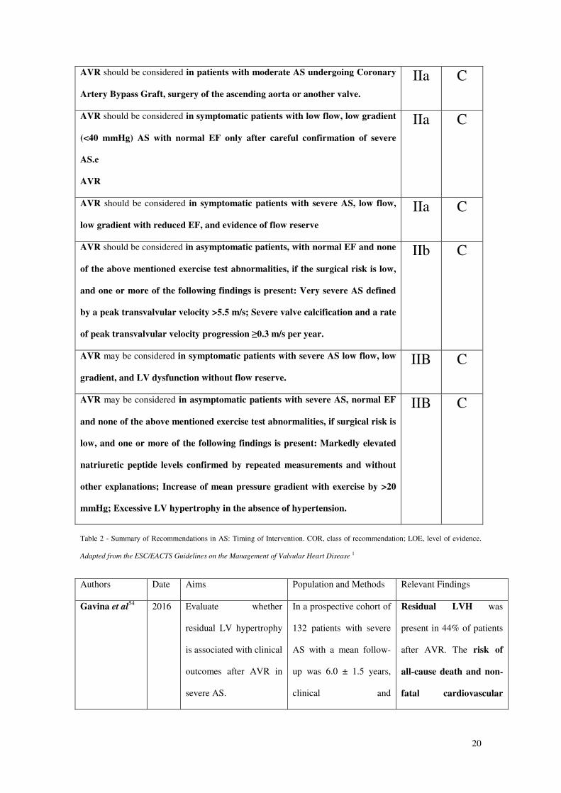
Table 1 - Summary of AS Stages. Adapted from AHA Guidelines for the Management of Patients with Valvular Disease.17
Recommendations COR LOE
AVR is indicated in patients with severe AS and any symptoms related to AS. I B
AVR is indicated in patients with severe AS undergoing Coronary Artery
Bypass Graft, surgery of the ascending aorta or another valve.
I C
AVR is indicated in asymptomatic patients with severe AS and systolic LV
dysfunction (LVEF <50%) not due to another cause.
I C
AVR is indicated in asymptomatic patients with severe AS and abnormal
exercise test showing symptoms on exercise clearly related to AS.
I C
AVR should be considered in high risk patients with severe symptomatic AS
who are suitable for TAVI, but in whom surgery is favoured by a ‘heart team’
based on the individual risk profile and anatomic suitability
IIa B
AVR should be considered in asymptomatic patients with severe AS and
abnormal exercise test showing fall in blood pressure below baseline
IIa C
20
AVR should be considered in patients with moderate AS undergoing Coronary
Artery Bypass Graft, surgery of the ascending aorta or another valve.
IIa C
AVR should be considered in symptomatic patients with low flow, low gradient
(<40 mmHg) AS with normal EF only after careful confirmation of severe
AS.e
AVR
IIa C
AVR should be considered in symptomatic patients with severe AS, low flow,
low gradient with reduced EF, and evidence of flow reserve
IIa C
AVR should be considered in asymptomatic patients, with normal EF and none
of the above mentioned exercise test abnormalities, if the surgical risk is low,
and one or more of the following findings is present: Very severe AS defined
by a peak transvalvular velocity >5.5 m/s; Severe valve calcification and a rate
of peak transvalvular velocity progression ≥0.3 m/s per year.
IIb C
AVR may be considered in symptomatic patients with severe AS low flow, low
gradient, and LV dysfunction without flow reserve.
IIB C
AVR may be considered in asymptomatic patients with severe AS, normal EF
and none of the above mentioned exercise test abnormalities, if surgical risk is
low, and one or more of the following findings is present: Markedly elevated
natriuretic peptide levels confirmed by repeated measurements and without
other explanations; Increase of mean pressure gradient with exercise by >20
mmHg; Excessive LV hypertrophy in the absence of hypertension.
IIB C
Table 2 - Summary of Recommendations in AS: Timing of Intervention. COR, class of recommendation; LOE, level of evidence.
Adapted from the ESC/EACTS Guidelines on the Management of Valvular Heart Disease 1
Authors Date Aims Population and Methods Relevant Findings
Gavina et al54 2016 Evaluate whether
residual LV hypertrophy
is associated with clinical
outcomes after AVR in
severe AS.
In a prospective cohort of
132 patients with severe
AS with a mean follow-
up was 6.0 ± 1.5 years,
clinical and
Residual LVH was
present in 44% of patients
after AVR. The risk of
all-cause death and non-
fatal cardiovascular
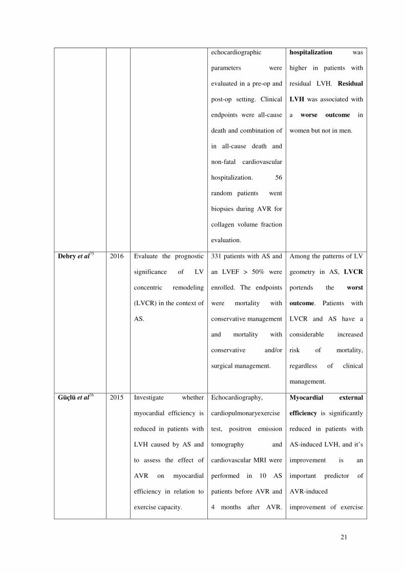
21
echocardiographic
parameters were
evaluated in a pre-op and
post-op setting. Clinical
endpoints were all-cause
death and combination of
in all-cause death and
non-fatal cardiovascular
hospitalization. 56
random patients went
biopsies during AVR for
collagen volume fraction
evaluation.
hospitalization was
higher in patients with
residual LVH. Residual
LVH was associated with
a worse outcome in
women but not in men.
Debry et al75 2016 Evaluate the prognostic
significance of LV
concentric remodeling
(LVCR) in the context of
AS.
331 patients with AS and
an LVEF > 50% were
enrolled. The endpoints
were mortality with
conservative management
and mortality with
conservative and/or
surgical management.
Among the patterns of LV
geometry in AS, LVCR
portends the worst
outcome. Patients with
LVCR and AS have a
considerable increased
risk of mortality,
regardless of clinical
management.
Güçlü et al76 2015 Investigate whether
myocardial efficiency is
reduced in patients with
LVH caused by AS and
to assess the effect of
AVR on myocardial
efficiency in relation to
exercise capacity.
Echocardiography,
cardiopulmonaryexercise
test, positron emission
tomography and
cardiovascular MRI were
performed in 10 AS
patients before AVR and
4 months after AVR.
Myocardial external
efficiency is significantly
reduced in patients with
AS-induced LVH, and it’s
improvement is an
important predictor of
AVR-induced
improvement of exercise
22
Fourteen healthy
individuals served as
control group.
capacity in AS patients.
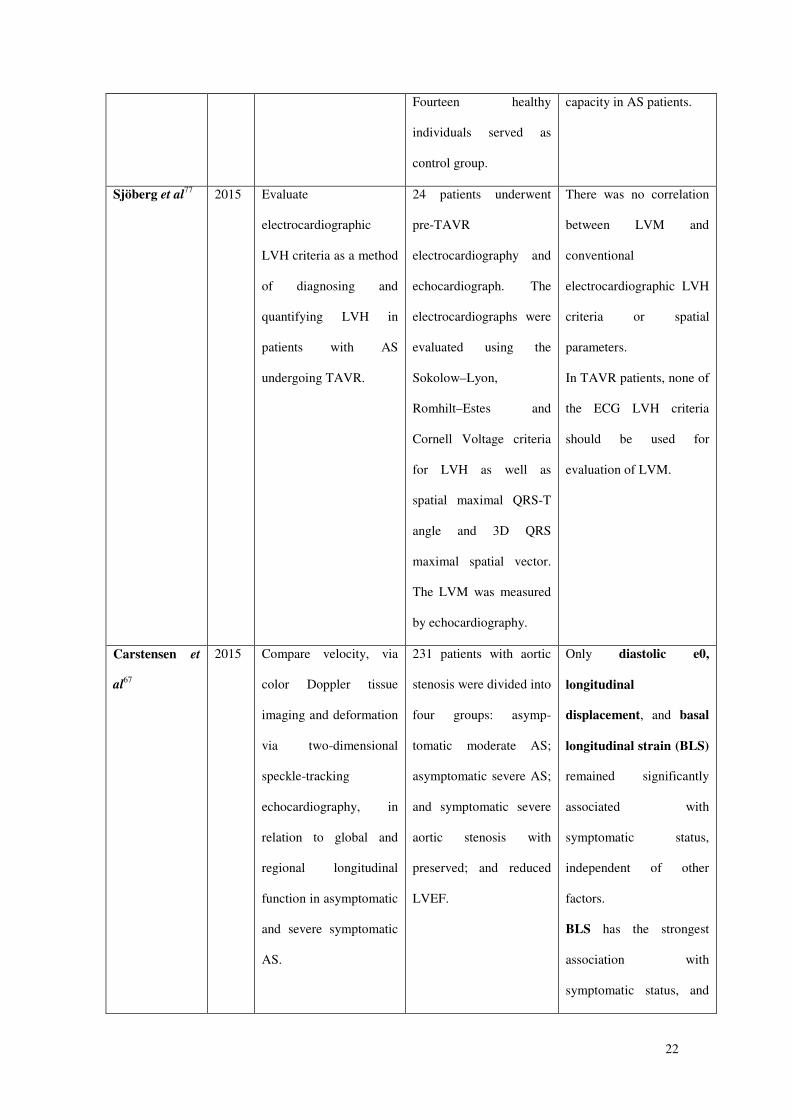
Sjöberg et al77 2015 Evaluate
electrocardiographic
LVH criteria as a method
of diagnosing and
quantifying LVH in
patients with AS
undergoing TAVR.
24 patients underwent
pre-TAVR
electrocardiography and
echocardiograph. The
electrocardiographs were
evaluated using the
Sokolow–Lyon,
Romhilt–Estes and
Cornell Voltage criteria
for LVH as well as
spatial maximal QRS-T
angle and 3D QRS
maximal spatial vector.
The LVM was measured
by echocardiography.
There was no correlation
between LVM and
conventional
electrocardiographic LVH
criteria or spatial
parameters.
In TAVR patients, none of
the ECG LVH criteria
should be used for
evaluation of LVM.
Carstensen et
al67
2015 Compare velocity, via
color Doppler tissue
imaging and deformation
via two-dimensional
speckle-tracking
echocardiography, in
relation to global and
regional longitudinal
function in asymptomatic
and severe symptomatic
AS.
231 patients with aortic
stenosis were divided into
four groups: asymp-
tomatic moderate AS;
asymptomatic severe AS;
and symptomatic severe
aortic stenosis with
preserved; and reduced
LVEF.
Only diastolic e0,
longitudinal
displacement, and basal
longitudinal strain (BLS)
remained significantly
associated with
symptomatic status,
independent of other
factors.
BLS has the strongest
association with
symptomatic status, and
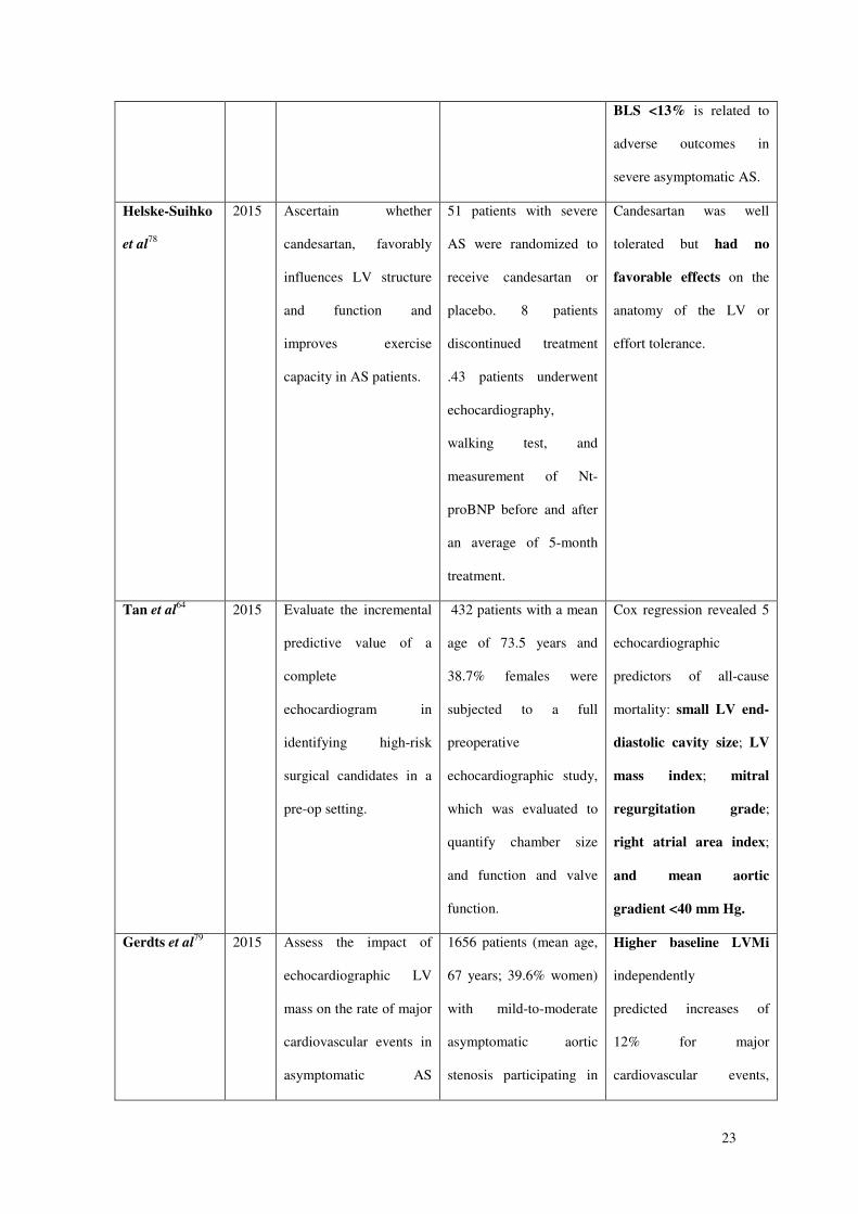
23
BLS <13% is related to
adverse outcomes in
severe asymptomatic AS.
Helske-Suihko
et al78
2015 Ascertain whether
candesartan, favorably
influences LV structure
and function and
improves exercise
capacity in AS patients.
51 patients with severe
AS were randomized to
receive candesartan or
placebo. 8 patients
discontinued treatment
.43 patients underwent
echocardiography,
walking test, and
measurement of Nt-
proBNP before and after
an average of 5-month
treatment.
Candesartan was well
tolerated but had no
favorable effects on the
anatomy of the LV or
effort tolerance.
Tan et al64 2015 Evaluate the incremental
predictive value of a
complete
echocardiogram in
identifying high-risk
surgical candidates in a
pre-op setting.
432 patients with a mean
age of 73.5 years and
38.7% females were
subjected to a full
preoperative
echocardiographic study,
which was evaluated to
quantify chamber size
and function and valve
function.
Cox regression revealed 5
echocardiographic
predictors of all-cause
mortality: small LV end-
diastolic cavity size; LV
mass index; mitral
regurgitation grade;
right atrial area index;
and mean aortic
gradient <40 mm Hg.
Gerdts et al79 2015 Assess the impact of
echocardiographic LV
mass on the rate of major
cardiovascular events in
asymptomatic AS
1656 patients (mean age,
67 years; 39.6% women)
with mild-to-moderate
asymptomatic aortic
stenosis participating in
Higher baseline LVMi
independently
predicted increases of
12% for major
cardiovascular events,
24
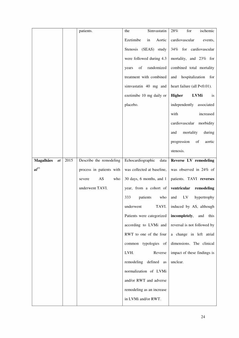
patients. the Simvastatin
Ezetimibe in Aortic
Stenosis (SEAS) study
were followed during 4.3
years of randomized
treatment with combined
simvastatin 40 mg and
ezetimibe 10 mg daily or
placebo.
28% for ischemic
cardiovascular events,
34% for cardiovascular
mortality, and 23% for
combined total mortality
and hospitalization for
heart failure (all P<0.01).
Higher LVMi is
independently associated
with increased
cardiovascular morbidity
and mortality during
progression of aortic
stenosis.
Magalhães et
al55
2015 Describe the remodeling
process in patients with
severe AS who
underwent TAVI.
Echocardiographic data
was collected at baseline,
30 days, 6 months, and 1
year, from a cohort of
333 patients who
underwent TAVI.
Patients were categorized
according to LVMi and
RWT to one of the four
common typologies of
LVH. Reverse
remodeling defined as
normalization of LVMi
and/or RWT and adverse
remodeling as an increase
in LVMi and/or RWT.
Reverse LV remodeling
was observed in 24% of
patients. TAVI reverses
ventricular remodeling
and LV hypertrophy
induced by AS, although
incompletely, and this
reversal is not followed by
a change in left atrial
dimensions. The clinical
impact of these findings is
unclear.
25
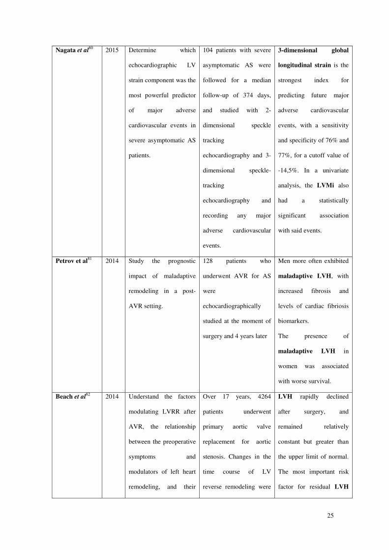
Nagata et al80 2015 Determine which
echocardiographic LV
strain component was the
most powerful predictor
of major adverse
cardiovascular events in
severe asymptomatic AS
patients.
104 patients with severe
asymptomatic AS were
followed for a median
follow-up of 374 days,
and studied with 2-
dimensional speckle
tracking
echocardiography and 3-
dimensional speckle-
tracking
echocardiography and
recording any major
adverse cardiovascular
events.
3-dimensional global
longitudinal strain is the
strongest index for
predicting future major
adverse cardiovascular
events, with a sensitivity
and specificity of 76% and
77%, for a cutoff value of
-14,5%. In a univariate
analysis, the LVMi also
had a statistically
significant association
with said events.
Petrov et al81 2014 Study the prognostic
impact of maladaptive
remodeling in a post-
AVR setting.
128 patients who
underwent AVR for AS
were
echocardiographically
studied at the moment of
surgery and 4 years later
Men more often exhibited
maladaptive LVH, with
increased fibrosis and
levels of cardiac fibriosis
biomarkers.
The presence of
maladaptive LVH in
women was associated
with worse survival.
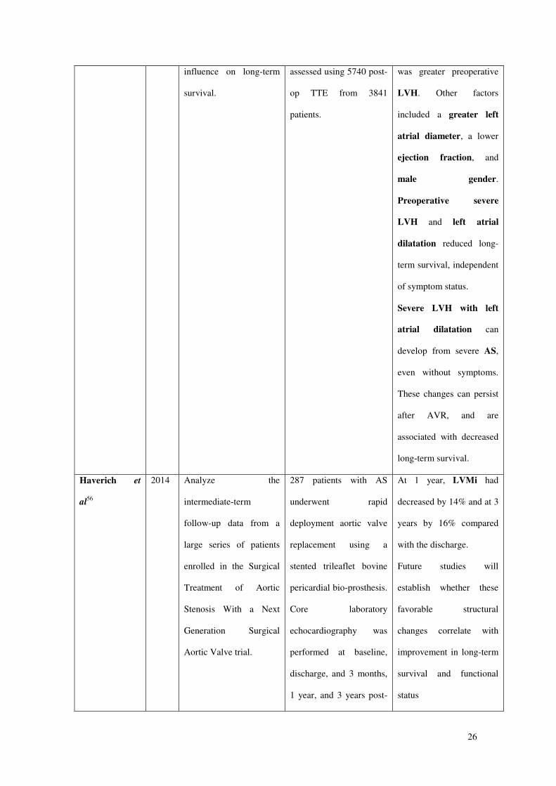
Beach et al82 2014 Understand the factors
modulating LVRR after
AVR, the relationship
between the preoperative
symptoms and
modulators of left heart
remodeling, and their
Over 17 years, 4264
patients underwent
primary aortic valve
replacement for aortic
stenosis. Changes in the
time course of LV
reverse remodeling were
LVH rapidly declined
after surgery, and
remained relatively
constant but greater than
the upper limit of normal.
The most important risk
factor for residual LVH
26
influence on long-term
survival.
assessed using 5740 post-
op TTE from 3841
patients.
was greater preoperative
LVH. Other factors
included a greater left
atrial diameter, a lower
ejection fraction, and
male gender.
Preoperative severe
LVH and left atrial
dilatation reduced long-
term survival, independent
of symptom status.
Severe LVH with left
atrial dilatation can
develop from severe AS,
even without symptoms.
These changes can persist
after AVR, and are
associated with decreased
long-term survival.
Haverich et
al56
2014 Analyze the
intermediate-term
follow-up data from a
large series of patients
enrolled in the Surgical
Treatment of Aortic
Stenosis With a Next
Generation Surgical
Aortic Valve trial.
287 patients with AS
underwent rapid
deployment aortic valve
replacement using a
stented trileaflet bovine
pericardial bio-prosthesis.
Core laboratory
echocardiography was
performed at baseline,
discharge, and 3 months,
1 year, and 3 years post-
At 1 year, LVMi had
decreased by 14% and at 3
years by 16% compared
with the discharge.
Future studies will
establish whether these
favorable structural
changes correlate with
improvement in long-term
survival and functional
status
27
op.
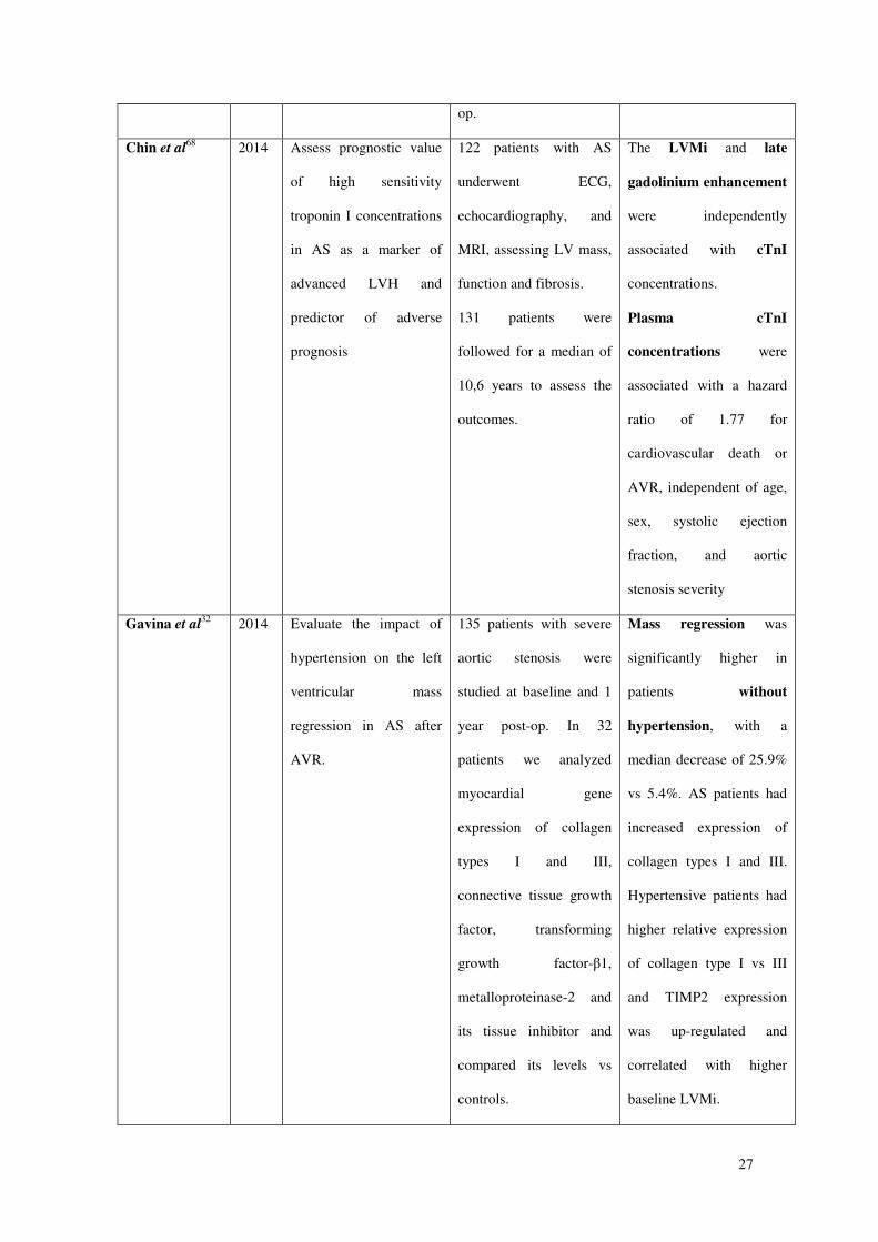
Chin et al68 2014 Assess prognostic value
of high sensitivity
troponin I concentrations
in AS as a marker of
advanced LVH and
predictor of adverse
prognosis
122 patients with AS
underwent ECG,
echocardiography, and
MRI, assessing LV mass,
function and fibrosis.
131 patients were
followed for a median of
10,6 years to assess the
outcomes.
The LVMi and late
gadolinium enhancement
were independently
associated with cTnI
concentrations.
Plasma cTnI
concentrations were
associated with a hazard
ratio of 1.77 for
cardiovascular death or
AVR, independent of age,
sex, systolic ejection
fraction, and aortic
stenosis severity
Gavina et al32 2014 Evaluate the impact of
hypertension on the left
ventricular mass
regression in AS after
AVR.
135 patients with severe
aortic stenosis were
studied at baseline and 1
year post-op. In 32
patients we analyzed
myocardial gene
expression of collagen
types I and III,
connective tissue growth
factor, transforming
growth factor-β1,
metalloproteinase-2 and
its tissue inhibitor and
compared its levels vs
controls.
Mass regression was
significantly higher in
patients without
hypertension, with a
median decrease of 25.9%
vs 5.4%. AS patients had
increased expression of
collagen types I and III.
Hypertensive patients had
higher relative expression
of collagen type I vs III
and TIMP2 expression
was up-regulated and
correlated with higher
baseline LVMi.
28
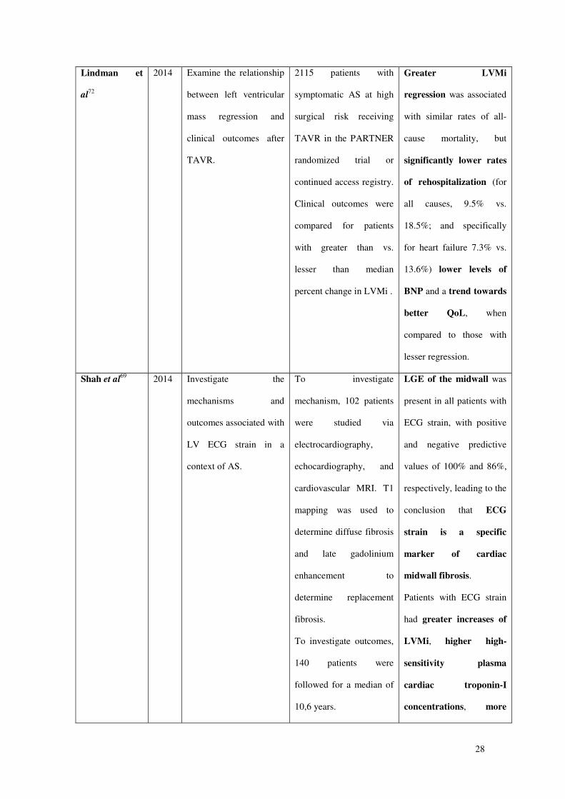
Lindman et
al72
2014 Examine the relationship
between left ventricular
mass regression and
clinical outcomes after
TAVR.
2115 patients with
symptomatic AS at high
surgical risk receiving
TAVR in the PARTNER
randomized trial or
continued access registry.
Clinical outcomes were
compared for patients
with greater than vs.
lesser than median
percent change in LVMi .
Greater LVMi
regression was associated
with similar rates of all-
cause mortality, but
significantly lower rates
of rehospitalization (for
all causes, 9.5% vs.
18.5%; and specifically
for heart failure 7.3% vs.
13.6%) lower levels of
BNP and a trend towards
better QoL, when
compared to those with
lesser regression.
Shah et al69 2014 Investigate the
mechanisms and
outcomes associated with
LV ECG strain in a
context of AS.
To investigate
mechanism, 102 patients
were studied via
electrocardiography,
echocardiography, and
cardiovascular MRI. T1
mapping was used to
determine diffuse fibrosis
and late gadolinium
enhancement to
determine replacement
fibrosis.
To investigate outcomes,
140 patients were
followed for a median of
10,6 years.
LGE of the midwall was
present in all patients with
ECG strain, with positive
and negative predictive
values of 100% and 86%,
respectively, leading to the
conclusion that ECG
strain is a specific
marker of cardiac
midwall fibrosis.
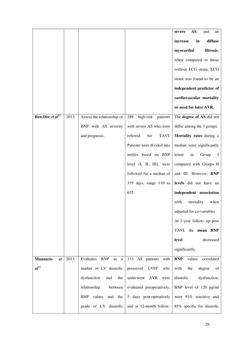
Patients with ECG strain
had greater increases of
LVMi, higher high-
sensitivity plasma
cardiac troponin-I
concentrations, more
29
severe AS, and an
increase in diffuse
myocardial fibrosis,
when compared to those
without ECG strain. ECG
strain was found to be an
independent predictor of
cardiovascular mortality
or need for later AVR.
Ben-Dor et al83 2013 Assess the relationship of
BNP with AS severity
and prognosis.
289 high-risk patients
with severe AS who were
referred for TAVI.
Patients were divided into
tertiles based on BNP
level (I, II, III), were
followed for a median of
319 days, range 110 to
655.
The degree of AS did not
differ among the 3 groups.
Mortality rates during a
median were significantly
lower in Group I
compared with Groups II
and III. However, BNP
levels did not have an
independent association
with mortality when
adjusted for co-variables.
At 1-year follow- up post
TAVI, the mean BNP
level decreased
significantly.
Mannacio et
al84
2013 Evaluates BNP as a
marker of LV diastolic
dysfunction and the
relationship between
BNP values and the
grade of LV diastolic
113 AS patients with
preserved LVEF who
underwent AVR were
evaluated preoperatively,
5 days post-operatively
and at 12-month follow-
BNP values correlated
with the degree of
diastolic dysfunction.
BNP level of 120 pg/ml
were 91% sensitive and
85% specific for diastolic
30
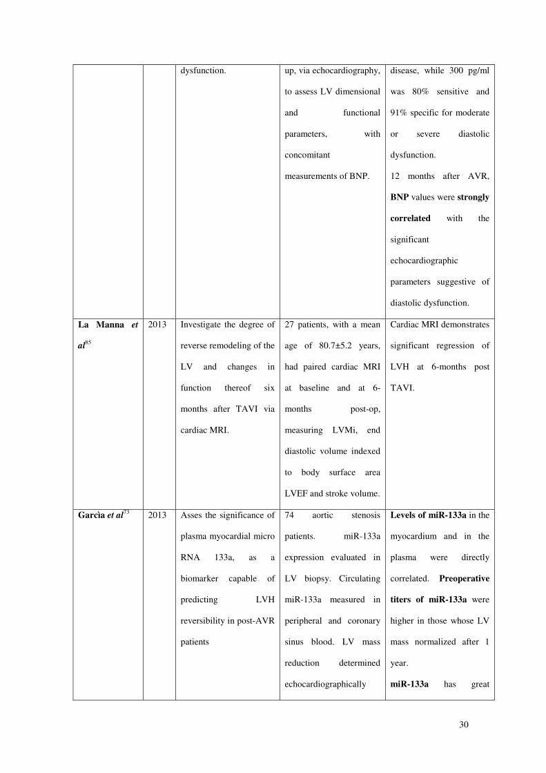
dysfunction. up, via echocardiography,
to assess LV dimensional
and functional
parameters, with
concomitant
measurements of BNP.
disease, while 300 pg/ml
was 80% sensitive and
91% specific for moderate
or severe diastolic
dysfunction.
12 months after AVR,
BNP values were strongly
correlated with the
significant
echocardiographic
parameters suggestive of
diastolic dysfunction.
La Manna et
al85
2013 Investigate the degree of
reverse remodeling of the
LV and changes in
function thereof six
months after TAVI via
cardiac MRI.
27 patients, with a mean
age of 80.7±5.2 years,
had paired cardiac MRI
at baseline and at 6-
months post-op,
measuring LVMi, end
diastolic volume indexed
to body surface area
LVEF and stroke volume.
Cardiac MRI demonstrates
significant regression of
LVH at 6-months post
TAVI.
Garcìa et al73 2013
Asses the significance of
plasma myocardial micro
RNA 133a, as a
biomarker capable of
predicting LVH
reversibility in post-AVR
patients
74 aortic stenosis
patients. miR-133a
expression evaluated in
LV biopsy. Circulating
miR-133a measured in
peripheral and coronary
sinus blood. LV mass
reduction determined
echocardiographically
Levels of miR-133a in the
myocardium and in the
plasma were directly
correlated. Preoperative
titers of miR-133a were
higher in those whose LV
mass normalized after 1
year.
miR-133a has great
31
potential for the decision
of surgical timing,
particularly for
asymptomatic patients.
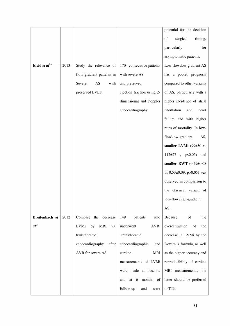
Eleid et al86 2013 Study the relevance of
flow gradient patterns in
Severe AS with
preserved LVEF.
1704 consecutive patients
with severe AS
and preserved
ejection fraction using 2-
dimensional and Doppler
echocardiography
Low flow\low gradient AS
has a poorer prognosis
compared to other variants
of AS, particularly with a
higher incidence of atrial
fibrillation and heart
failure and with higher
rates of mortality. In low-
flow\low-gradient AS,
smaller LVMi (99±30 vs
112±27 , p<0.05) and
smaller RWT (0.49±0.08
vs 0.53±0.09, p>0,05) was
observed in comparison to
the classical variant of
low-flow\high-gradient
AS.
Breitenbach et
al21
2012 Compare the decrease
LVMi by MRI vs.
transthoracic
echocardiography after
AVR for severe AS.
149 patients who
underwent AVR.
Transthoracic
echocardiographic and
cardiac MRI
measurements of LVMi
were made at baseline
and at 6 months of
follow-up and were
Because of the
overestimation of the
decrease in LVMi by the
Devereux formula, as well
as the higher accuracy and
reproducibility of cardiac
MRI measurements, the
latter should be preferred
to TTE.
32
compared. Changes in
mean pressure gradients
were examined using
transthoracic
echocardiography.
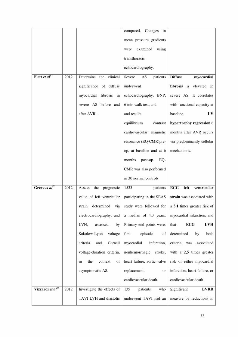
Flett et al87 2012 Determine the clinical
significance of diffuse
myocardial fibrosis in
severe AS before and
after AVR..
Severe AS patients
underwent
echocardiography, BNP,
6 min walk test, and
and results
equilibrium contrast
cardiovascular magnetic
resonance (EQ-CMR)pre-
op, at baseline and at 6
months post-op. EQ-
CMR was also performed
in 30 normal controls
Diffuse myocardial
fibrosis is elevated in
severe AS. It correlates
with functional capacity at
baseline. LV
hypertrophy regression 6
months after AVR occurs
via predominantly cellular
mechanisms.
Greve et al70 2012 Assess the prognostic
value of left ventricular
strain determined via
electrocardiography, and
LVH, assessed by
Sokolow-Lyon voltage
criteria and Cornell
voltage-duration criteria,
in the context of
asymptomatic AS.
1533 patients
participating in the SEAS
study were followed for
a median of 4.3 years.
Primary end points were:
first episode of
myocardial infarction,
nonhemorrhagic stroke,
heart failure, aortic valve
replacement, or
cardiovascular death.
ECG left ventricular
strain was associated with
a 3,1 times greater risk of
myocardial infarction, and
that ECG LVH
determined by both
criteria was associated
with a 2,5 times greater
risk of either myocardial
infarction, heart failure, or
cardiovascular death.
Vizzardi et al88 2012 Investigate the effects of
TAVI LVH and diastolic
135 patients who
underwent TAVI had an
Significant LVRR
measure by reductions in
33
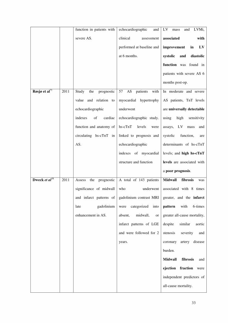
function in patients with
severe AS.
echocardiographic and
clinical assessment
performed at baseline and
at 6 months.
LV mass and LVMi,
associated with
improvement in LV
systolic and diastolic
function was found in
patients with severe AS 6
months post-op.
Røsjø et al71 2011 Study the prognostic
value and relation to
echocardiographic
indexes of cardiac
function and anatomy of
circulating hs-cTnT in
AS.
57 AS patients with
myocardial hypertrophy
underwent
echocardiographic study.
hs-cTnT levels were
linked to prognosis and
echocardiographic
indexes of myocardial
structure and function
In moderate and severe
AS patients, TnT levels
are universally detectable
using high sensitivity
assays, LV mass and
systolic function, are
determinants of hs-cTnT
levels; and high hs-cTnT
levels are associated with
a poor prognosis.
Dweck et al38 2011 Assess the prognostic
significance of midwall
and infarct patterns of
late gadolinium
enhancement in AS.
A total of 143 patients
who underwent
gadolinium contrast MRI
were categorized into
absent, midwall, or
infarct patterns of LGE
and were followed for 2
years.
Midwall fibrosis was
associated with 8 times
greater, and the infarct
pattern with 6-times
greater all-cause mortality,
despite similar aortic
stenosis severity and
coronary artery disease
burden.
Midwall fibrosis and
ejection fraction were
independent predictors of
all-cause mortality.
34
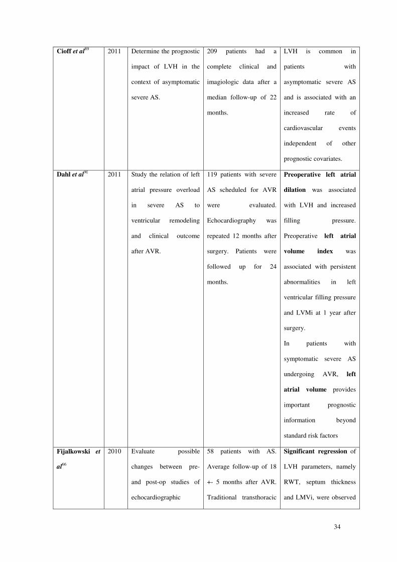
Cioff et al89 2011 Determine the prognostic
impact of LVH in the
context of asymptomatic
severe AS.
209 patients had a
complete clinical and
imagiologic data after a
median follow-up of 22
months.
LVH is common in
patients with
asymptomatic severe AS
and is associated with an
increased rate of
cardiovascular events
independent of other
prognostic covariates.
Dahl et al90 2011 Study the relation of left
atrial pressure overload
in severe AS to
ventricular remodeling
and clinical outcome
after AVR.
119 patients with severe
AS scheduled for AVR
were evaluated.
Echocardiography was
repeated 12 months after
surgery. Patients were
followed up for 24
months.
Preoperative left atrial
dilation was associated
with LVH and increased
filling pressure.
Preoperative left atrial
volume index was
associated with persistent
abnormalities in left
ventricular filling pressure
and LVMi at 1 year after
surgery.
In patients with
symptomatic severe AS
undergoing AVR, left
atrial volume provides
important prognostic
information beyond
standard risk factors
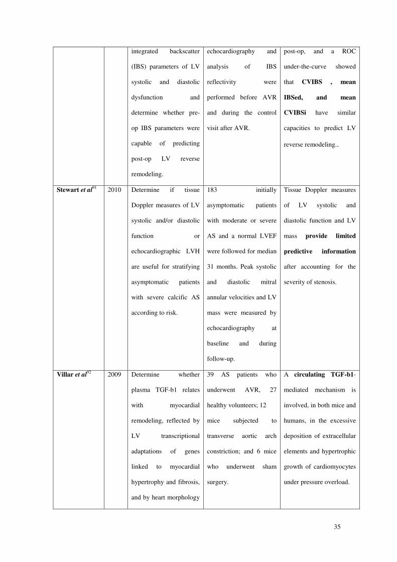
Fijalkowski et
al66
2010 Evaluate possible
changes between pre-
and post-op studies of
echocardiographic
58 patients with AS.
Average follow-up of 18
+- 5 months after AVR.
Traditional transthoracic
Significant regression of
LVH parameters, namely
RWT, septum thickness
and LMVi, were observed
35
integrated backscatter
(IBS) parameters of LV
systolic and diastolic
dysfunction and
determine whether pre-
op IBS parameters were
capable of predicting
post-op LV reverse
remodeling.
echocardiography and
analysis of IBS
reflectivity were
performed before AVR
and during the control
visit after AVR.
post-op, and a ROC
under-the-curve showed
that CVIBS , mean
IBSed, and mean
CVIBSi have similar
capacities to predict LV
reverse remodeling..
Stewart et al91 2010 Determine if tissue
Doppler measures of LV
systolic and/or diastolic
function or
echocardiographic LVH
are useful for stratifying
asymptomatic patients
with severe calcific AS
according to risk.
183 initially
asymptomatic patients
with moderate or severe
AS and a normal LVEF
were followed for median
31 months. Peak systolic
and diastolic mitral
annular velocities and LV
mass were measured by
echocardiography at
baseline and during
follow-up.
Tissue Doppler measures
of LV systolic and
diastolic function and LV
mass provide limited
predictive information
after accounting for the
severity of stenosis.
Villar et al92 2009 Determine whether
plasma TGF-b1 relates
with myocardial
remodeling, reflected by
LV transcriptional
adaptations of genes
linked to myocardial
hypertrophy and fibrosis,
and by heart morphology
39 AS patients who
underwent AVR, 27
healthy volunteers; 12
mice subjected to
transverse aortic arch
constriction; and 6 mice
who underwent sham
surgery.
A circulating TGF-b1-
mediated mechanism is
involved, in both mice and
humans, in the excessive
deposition of extracellular
elements and hypertrophic
growth of cardiomyocytes
under pressure overload.

36
and function.

Anexos