Embed Size (px)
Citation preview
O
RRCs
CARLEMRGJIMTJa
b
c
d
e
f
g
Bh
i
j
k
l
m
n
o
h2c
r e v b r a s r e u m a t o l . 2 0 1 7;5 7(S 2):S421–S437
www.reumato logia .com.br
REVISTA BRASILEIRA DEREUMATOLOGIA
riginal article
ecommendations of the Brazilian Society ofheumatology for diagnosis and treatment ofhikungunya fever. Part 1 – Diagnosis and specialituations
laudia Diniz Lopes Marquesa,b,∗, Angela Luzia Branco Pinto Duartea,c,line Ranzolinb,d, Andrea Tavares Dantasa, Nara Gualberto Cavalcantib,afaela Silva Guimarães Goncalvesb, Laurindo Ferreira da Rocha Juniorb,d,ilian David de Azevedo Valadarese, Ana Karla Guedes de Melo f,utilia Andrade Medeiros Freireg, Roberto Teixeirah, Francisco Alves Bezerra Neto i,arta Maria das Chagas Medeiros j, Jozélio Freire de Carvalhok, Mario Sergio F. Santos l,
egina Adalva de L. Couto Océam, Roger A. Levyn, Carlos Augusto Ferreira de Andradeo,eraldo da Rocha Castelar Pinheiron, Mirhelen Mendes Abreup,
osé Fernando Verztmanq, Selma Merenlenderr, Sandra Lucia Euzebio Ribeiros,zaias Pereira da Costat,u, Gecilmara Pileggiv, Virginia Fernandes Moca Trevisaniw,x,
ax Igor Banks Lopesy, Carlos Britoa, Eduardo Figueiredob, Fabio Queirogaz,iago Feitosaaa, Angélica da Silva Tenórioa, Gisela Rocha de Siqueiraa, Renata Paivaab,osé Tupinambá Sousa Vasconcelosac,ad, Georges Christopoulosad,ae
Universidade Federal de Pernambuco (UFPE), Recife, PE, BrazilUniversidade Federal de Pernambuco (UFPE), Hospital das Clínicas, Recife, PE, BrazilUniversidade Federal de Pernambuco (UFPE), Hospital das Clínicas, Servico de Reumatologia, Recife, PE, BrazilInstituto de Medicina Integral Professor Fernando Figueira (IMIP), Recife, PE, BrazilHospital Getúlio Vargas, Ambulatório de Chikungunya, Recife, PE, BrazilUniversidade Federal da Paraíba (UFPB), João Pessoa, PB, BrazilUniversidade Federal da Paraíba (UFPB), Hospital Universitário Lauro Wanderley (HULW), Servico de Reumatologia, João Pessoa, PB,razilUniversidade Estadual de Ciências da Saúde de Alagoas (UNCISAL), Maceió, AL, BrazilUniversidade Federal do Rio Grande do Norte (UFRN), Natal, RN, BrazilUniversidade Federal do Ceará (UFC), Faculdade de Medicina, Departamento de Medicina Clínica, Fortaleza, CE, BrazilUniversidade Federal da Bahia (UFBA), Instituto de Ciências da Saúde, Salvador, BA, Brazil
resina, PI, Brazil
Universidade Estadual do Piauí (UESPI), Faculdade de Medicina, Te Universidade Federal de Sergipe (UFS), Aracaju, SE, BrazilUniversidade do Estado do Rio de Janeiro (UERJ), Disciplina de Reumatologia, Rio de Janeiro, RJ, BrazilFundacão Oswaldo Cruz (Fiocruz), Escola Nacional de Saúde Pública Sérgio Arouca (ENSP), Rio de Janeiro, RJ, Brazil∗ Corresponding author.E-mail: [email protected] (C.D. Marques).
ttp://dx.doi.org/10.1016/j.rbre.2017.05.006255-5021/© 2017 Published by Elsevier Editora Ltda. This is an open access article under the CC BY-NC-ND license (http://reativecommons.org/licenses/by-nc-nd/4.0/).
S422 r e v b r a s r e u m a t o l . 2 0 1 7;5 7(S 2):S421–S437
p Universidade Federal do Rio de Janeiro (UFRJ), Hospital Universitário Clementino Fraga Filho (HUCFF), Rio de Janeiro, RJ, Brazilq Hospital dos Servidores do Estado do Rio de Janeiro, Rio de Janeiro, RJ, Brazilr Hospital Estadual Eduardo Rabello, Servico de Reumatologia, Rio de Janeiro, RJ, Brazils Universidade Federal do Amazonas (UFAM), Faculdade de Medicina, Manaus, AM, Brazilt Universidade Federal de Mato Grosso do Sul (UFMS), Campo Grande, MS, Brazilu Universidade Federal de Mato Grosso do Sul (UFMS), Hospital Universitário Maria Aparecida Pedrossian (HUMAP), Servico deReumatologia, Campo Grande, MS, Brazilv Universidade de São Paulo (USP), Faculdade de Medicina de Ribeirão Preto (FMRP), Servico de Reumatologia e Imunologia Pediátrica,Ribeirão Preto, SP, Brazilw Universidade Federal de São Paulo (UNIFESP), São Paulo, SP, Brazilx Universidade de Santo Amaro (UNISA), São Paulo, SP, Brazily Universidade de São Paulo (USP), Hospital das Clínicas, Ambulatório da Divisão de Moléstias Infecciosas de Parasitárias, São Paulo, SP,Brazilz Instituto de Medicina Integral Professor Fernando Figueira (IMIP), Hospital Miguel Arraes, Paulista, PE, Brazilaa Universidade Federal de Pernambuco (UFPE), Hospital das Clínicas, Divisão de Gestão do Cuidado, Recife, PE, Brazilab CRP Fisioterapia, Rio de Janeiro, RJ, Brazilac Universidade Estadual do Piauí (UESPI), Teresina, PI, Brazilad Sociedade Brasileira de Reumatologia, São Paulo, SP, Brazilae Santa Casa de Misericórdia de Maceió, Maceió, AL, Brazil
a r t i c l e i n f o
Article history:
Received 19 December 2016
Accepted 22 May 2017
Available online 25 July 2017
Keywords:
Chikungunya fever
Epidemic
Consensus
Brazil
a b s t r a c t
Chikungunya fever has become a relevant public health problem in countries where epi-
demics occur. Until 2013, only imported cases occurred in the Americas, but in October of
that year, the first cases were reported in Saint Marin island in the Caribbean. The first
autochthonous cases were confirmed in Brazil in September 2014; until epidemiological
week 37 of 2016, 236,287 probable cases of infection with Chikungunya virus had been reg-
istered, 116,523 of which had serological confirmation. Environmental changes caused by
humans, disorderly urban growth and an ever-increasing number of international travelers
were described as the factors responsible for the emergence of large-scale epidemics. Clini-
cally characterized by fever and joint pain in the acute stage, approximately half of patients
progress to the chronic stage (beyond 3 months), which is accompanied by persistent and
disabling pain. The aim of the present study was to formulate recommendations for the diag-
nosis and treatment of Chikungunya fever in Brazil. A literature review was performed in the
MEDLINE, SciELO and PubMed databases to ground the decisions for recommendations. The
degree of concordance among experts was established through the Delphi method, involv-
ing 2 in-person meetings and several online voting rounds. In total, 25 recommendations
were formulated and divided into 3 thematic groups: (1) clinical, laboratory and imaging
diagnosis; (2) special situations; and (3) treatment. The first 2 themes are presented in part
1, and treatment is presented in part 2.
© 2017 Published by Elsevier Editora Ltda. This is an open access article under the CC
BY-NC-ND license (http://creativecommons.org/licenses/by-nc-nd/4.0/).
Recomendacões da Sociedade Brasileira de Reumatologia paradiagnóstico e tratamento da febre Chikungunya. Parte 1 – Diagnóstico esituacões especiais
Palavras-chave:
Febre Chikungunya
Epidemia
Consenso
Brasil
r e s u m o
A febre chikungunya tem se tornado um importante problema de saúde pública nos países
onde ocorrem as epidemias. Até 2013, as Américas haviam registrado apenas casos impor-
tados quando, em outubro desse mesmo ano, foram notificados os primeiros casos na
Ilha de Saint Martin, no Caribe. No Brasil, os primeiros relatos autóctones foram confir-
mados em setembro de 2014 e até a semana epidemiológica 37 de 2016 já haviam sido
registrados 236.287 casos prováveis de infeccão pelo chikungunya vírus (CHIKV), 116.523
confirmados sorologicamente. As mudancas ambientais causadas pelo homem, o cresci-
mento urbano desordenado e o número cada vez maior de viagens internacionais têm sido
apontados como os fatores responsáveis pela reemergência de epidemias em grande escala.
r e v b r a s r e u m a t o l . 2 0 1 7;5 7(S 2):S421–S437 S423
Caracterizada clinicamente por febre e dor articular na fase aguda, em cerca de metade
dos casos existe evolucão para a fase crônica (além de três meses), com dor persistente
e incapacitante. O objetivo deste trabalho foi elaborar recomendacões para diagnóstico e
tratamento da febre chikungunya no Brasil. Para isso, foi feita revisão da literatura nas
bases de dados Medline, SciELO e PubMed, para dar apoio às decisões tomadas para definir
as recomendacões. Para a definicão do grau de concordância foi feita uma metodologia
Delphi, em duas reuniões presenciais e várias rodadas de votacão on line. Foram geradas
25 recomendacões, divididas em três grupos temáticos: (1) diagnóstico clínico, laborato-
rial e por imagem; (2) situacões especiais e (3) tratamento. Na primeira parte estão os dois
primeiros temas e o tratamento na segunda.
© 2017 Publicado por Elsevier Editora Ltda. Este e um artigo Open Access sob uma
ca CC
G
E
TdimcTdfaioiw
iuwaiOMpInSpo(b1
P
Dbtaias
licen
eneral considerations
pidemiology
he Chikungunya virus (CHIKV), a member of the Togaviri-ae family, Alphavirus genus, was first isolated from humans
n 1952 during the first urban outbreak identified in theodern scientific age, causing an epidemic that affected the
oastal area of Muawiya, Makondo and Rondo, present-dayanzania.1 The virus name is in the Makonde language, aialect spoken in Southeastern Tanzania, and means “to bendorward or become contorted,” an allusion to the posturedopted by patients as a function of the joint pain caused bynfection.1 While the disease is transmitted by a large numberf mosquitos of the genus Aedes in Africa, the main vectors
n Brazil are Aedes aegypti and Aedeas albopictus, the females ofhich are able to infect humans through biting.2
After the first outbreak of Chikungunya fever in Tanzanian the 1950s, several minor epidemics occurred periodicallyntil 2004, when a major outbreak was identified in Kenya,hich then spread to many islands in the Indian Ocean, India
nd Southeastern Asia.3 Until 2013, all the cases in the Amer-cas were imported, mostly into the United States; however, inctober of that year, the first cases were documented in Saintartin island in the Caribbean. By the end of 2015, 1.6 million
eople had been infected with CHIKV, resulting in 71 deaths.4–6
n Brazil, the first autochthonous cases were almost simulta-eously reported in Oiapoque (Amapa state – AP) and Feira deantana (Bahia state – BA) in September 2014.7 A total of 38,332ossible cases of Chikungunya fever were registered in 2015,f which 13,236 were confirmed. Until epidemiological week
EW) 37 of 2016 (up to September 17th, 2016), 236,287 proba-le cases of infection with CHIKV were registered, of which16,523 had serological confirmation.8
athophysiology
uring the inoculation of CHIKV through the skin by mosquitoites, mosquito saliva enters the body together with the virus;he saliva contains several molecules with anti-hemostatic
nd immunomodulating properties, which induce early cellnfiltration and increased cytokine secretion. Then followsstage characterized by intense replication of the virus inkin fibroblasts and macrophages and spread to lymph nodes,
BY-NC-ND (http://creativecommons.org/licenses/by-nc-nd/4.0/).
where replication intensifies before the virus is released intothe bloodstream, with spread to and consequent infectionof target organs, such as the joints and muscles. The virusmight also be directly inoculated into the bloodstream throughmosquito bites. Of particular relevance is the induction ofthe innate immune response, followed by the cell-mediatedimmune response, involving the release of several proin-flammatory cytokines, such as interferon alpha, interleukins,chemokines and growth factors.9–11
Experimental models of alphavirus-induced arthritis sug-gest that progression to the chronic stage of Chikungunyafever results from the combination of direct cell and tis-sue damage caused by viral replication and, indirectly, byactivation of the immune response in the target tissues.12
Nevertheless, the main hypothesis involves dysregulatedinflammation caused by persistent infection of tissuemacrophages – or the presence of viral RNA within thesecells.13 The synovial histopathological changes after infec-tion with CHIKV are similar to those found among patientswith rheumatoid arthritis (RA) or other chronic inflammatoryjoint diseases, including synovial hyperplasia, vascular prolif-eration and perivascular macrophage infiltration.14 In animalmodels of CHIKV infection, periosteal necrosis, bone prolifer-ation and multifocal bone marrow ischemia were identifiedstarting 5 days after infection.15
Clinical manifestations
The incubation period lasts 3 to 7 days, on average, varyingfrom 1 to 12 days.16 The disease evolves in 3 stages: acute,lasting 7 to 14 days; subacute, which might last up to 3 months;and chronic, namely, persistence of symptoms for more than3 months.17
Acute infection is symptomatic in 80% to 97% ofpatients.18,19 The most common symptoms are fever of sud-den onset and joint pain and/or arthritis (nearly 100% of cases),which is usually symmetric and polyarticular. The joint com-plaints mainly involve the hands, wrists, ankles and feet,being disabling in most cases. Less frequent manifestationsinclude asthenia, muscle pain (60–93%), headache (40–81%),
nausea/vomiting, diarrhea, photophobia, retro-orbital pain,conjunctivitis, axial pain, macular/maculopapular exanthema(34–50%) with or without skin itch, face and limb swelling andcervical or generalized lymphadenopathy.19–24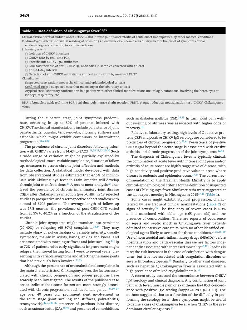
S424 r e v b r a s r e u m a t o l . 2 0 1 7;5 7(S 2):S421–S437
Table 1 – Case definition of Chikungunya fever.17,45
Clinical criteria: fever of sudden onset > 38.5 ◦C and intense joint pain/arthritis of acute onset not explained by other medical conditionsEpidemiological criteria: individual residing at or visiting an endemic or epidemic area 15 days before the onset of symptoms or has
epidemiological connection to a confirmed caseLaboratory criteria
© Isolation of CHIKV in culture© CHIKV RNA by real-time PCR© Specific anti-CHIKV IgM antibodies© Four-fold increase of anti-CHIKV IgG antibodies in samples collected with at least© a 10–14-day interval© Detection of anti-CHIKV neutralizing antibodies in serum by means of PRNT
ClassificationSuspected case: patient meets the clinical and epidemiological criteriaConfirmed case: a suspected case that meets any of the laboratory criteriaAtypical case: laboratory confirmation in a patient with other clinical manifestations (neurologic, cutaneous, involving the heart, eyes orkidneys, respiratory, etc.)
actio
RNA, ribonucleic acid; real-time PCR, real-time polymerase chain revirus.During the subacute stage, joint symptoms predomi-nate, occurring in up to 50% of patients infected withCHIKV. The clinical manifestations include persistence of jointpain/arthritis, bursitis, tenosynovitis, morning stiffness andasthenia, which might exhibit continuous or intermittentprogression.21,24,25
The prevalence of chronic joint disorders following infec-tion with CHIKV varies from 14.4% to 87.2%.14,19,21,23,26–30 Sucha wide range of variation might be partially explained bymethodological issues: variable sample size, duration of followup, measures to assess chronic joint affection and methodsfor data collection. A statistical model developed with datafrom observational studies estimated that 47.6% of individ-uals with Chikungunya fever in Latin America will developchronic joint manifestations.31 A recent meta-analysis32 ana-lyzed the prevalence of chronic inflammatory joint disease(CIJD) after Chikungunya infection (post-CHIK) in 18 selectedstudies (9 prospective and 9 retrospective cohort studies) witha total of 5702 patients. The average length of follow upwas 17.5 months; the prevalence of post-CHIK CIJD variedfrom 25.3% to 40.2% as a function of the stratification of thestudies.
Chronic joint symptoms might translate into persistent(20–40%) or relapsing (60–80%) complaints.33,34 They mayinclude oligo- or polyarthralgia of variable intensity, usuallysymmetric, mainly in wrists, hands, ankles and knees, andare associated with morning stiffness and joint swelling.33 Upto 72% of patients with early significant improvement mightrelapse, the interval lasting from 1 week to several years, pre-senting with variable symptoms and affecting the same jointsthat had previously been involved.29,35
Although the persistence of musculoskeletal complaints isthe main characteristic of Chikungunya fever, the factors asso-ciated with chronic progression and poorer prognosis havescarcely been investigated. The results of the published caseseries indicate that some factors are more strongly associ-ated with chronic progression, such as female gender,34,36–38
age over 40 years old, prominent joint involvement inthe acute stage (joint swelling and stiffness, polyarthritis,tenosynovitis),32,34,36–39 presence of previous joint disease,such as osteoarthritis (OA),39,40 and presence of comorbidities,
n; PRNT, plaque reduction neutralization test; CHIKV, Chikungunya
such as diabetes mellitus (DM).33,35 In turn, joint pain with-out swelling or stiffness was associated with higher odds ofrecovery.34
Relative to laboratory testing, high levels of C-reactive pro-tein (CRP) and positive CHIKV IgG serology are considered to bepredictors of chronic progression.28,41 Persistence of positiveCHIKV IgM beyond the acute stage is associated with erosivearthritis and chronic progression of the joint symptoms.42,43
The diagnosis of Chikungunya fever is typically clinical;the combination of acute fever with intense joint pain and/orarthritis of acute onset are highly suggestive of disease, withhigh sensitivity and positive predictive value in areas wheredisease is endemic and epidemics occur.17,44 The current rec-ommendation of the Brazilian Health Ministry is to applyclinical-epidemiological criteria for the definition of suspectedcases of Chikungunya fever. Similar criteria were suggested atthe last expert meeting in Nicaragua in 201517,45 (Table 1).
Some cases might exhibit atypical progression, charac-terized by less frequent clinical manifestations (Table 2) orsigns of severity.46 The frequency of severe cases is 0.3%and is associated with older age (>65 years old) and thepresence of comorbidities. There are reports of occurrenceof sepsis and septic shock in Chikungunya fever patientsadmitted to intensive care units, with no other identified eti-ological agent likely to account for these conditions.21,25,46–49
Use of nonsteroidal anti-inflammatory drugs (NSAIDs) beforehospitalization and cardiovascular disease are factors inde-pendently associated with increased mortality.46,47 Bleeding israre; the risk increases in the case of coinfection with denguevirus, but it is not associated with coagulation disorders orsevere thrombocytopenia.25 Similarly to other viral diseases,such as hepatitis C, Chikungunya fever is associated with ahigh prevalence of mixed cryoglobulinemia.50
A recent study assessed the concordance between CHIKVIgM serology and clinical diagnosis. Any combination of jointpain with fever, muscle pain or exanthema had 85% concord-ance with positive IgM testing (kappa = 0.289, p < 0.001). The
authors suggested that as a function of the difficulty in per-forming the serology tests, these symptoms might be usefulto define a case of Chikungunya fever when CHIKV is the pre-dominant circulating virus.51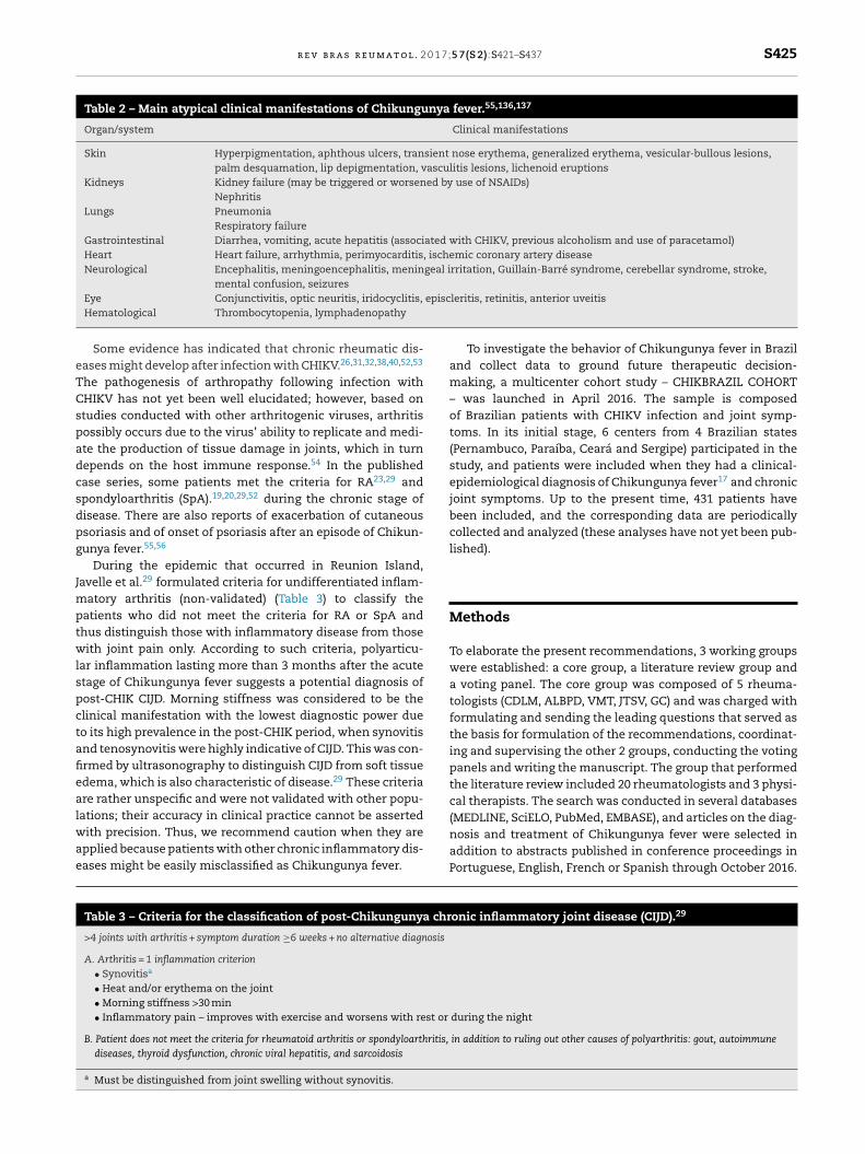
r e v b r a s r e u m a t o l . 2 0 1 7;5 7(S 2):S421–S437 S425
Table 2 – Main atypical clinical manifestations of Chikungunya fever.55,136,137
Organ/system Clinical manifestations
Skin Hyperpigmentation, aphthous ulcers, transient nose erythema, generalized erythema, vesicular-bullous lesions,palm desquamation, lip depigmentation, vasculitis lesions, lichenoid eruptions
Kidneys Kidney failure (may be triggered or worsened by use of NSAIDs)Nephritis
Lungs PneumoniaRespiratory failure
Gastrointestinal Diarrhea, vomiting, acute hepatitis (associated with CHIKV, previous alcoholism and use of paracetamol)Heart Heart failure, arrhythmia, perimyocarditis, ischemic coronary artery diseaseNeurological Encephalitis, meningoencephalitis, meningeal irritation, Guillain-Barré syndrome, cerebellar syndrome, stroke,
mental confusion, seizuresepisc
eTCspadcsdpg
Jmptwlspctafiealwae
Eye Conjunctivitis, optic neuritis, iridocyclitis,
Hematological Thrombocytopenia, lymphadenopathy
Some evidence has indicated that chronic rheumatic dis-ases might develop after infection with CHIKV.26,31,32,38,40,52,53
he pathogenesis of arthropathy following infection withHIKV has not yet been well elucidated; however, based ontudies conducted with other arthritogenic viruses, arthritisossibly occurs due to the virus’ ability to replicate and medi-te the production of tissue damage in joints, which in turnepends on the host immune response.54 In the publishedase series, some patients met the criteria for RA23,29 andpondyloarthritis (SpA).19,20,29,52 during the chronic stage ofisease. There are also reports of exacerbation of cutaneoussoriasis and of onset of psoriasis after an episode of Chikun-unya fever.55,56
During the epidemic that occurred in Reunion Island,avelle et al.29 formulated criteria for undifferentiated inflam-
atory arthritis (non-validated) (Table 3) to classify theatients who did not meet the criteria for RA or SpA andhus distinguish those with inflammatory disease from thoseith joint pain only. According to such criteria, polyarticu-
ar inflammation lasting more than 3 months after the acutetage of Chikungunya fever suggests a potential diagnosis ofost-CHIK CIJD. Morning stiffness was considered to be thelinical manifestation with the lowest diagnostic power dueo its high prevalence in the post-CHIK period, when synovitisnd tenosynovitis were highly indicative of CIJD. This was con-rmed by ultrasonography to distinguish CIJD from soft tissuedema, which is also characteristic of disease.29 These criteriare rather unspecific and were not validated with other popu-
ations; their accuracy in clinical practice cannot be assertedith precision. Thus, we recommend caution when they arepplied because patients with other chronic inflammatory dis-ases might be easily misclassified as Chikungunya fever.
Table 3 – Criteria for the classification of post-Chikungunya chr
>4 joints with arthritis + symptom duration ≥6 weeks + no alternative diagnosis
A. Arthritis = 1 inflammation criterion• Synovitisa
• Heat and/or erythema on the joint• Morning stiffness >30 min• Inflammatory pain – improves with exercise and worsens with rest or
B. Patient does not meet the criteria for rheumatoid arthritis or spondyloarthritis,
diseases, thyroid dysfunction, chronic viral hepatitis, and sarcoidosis
a Must be distinguished from joint swelling without synovitis.
leritis, retinitis, anterior uveitis
To investigate the behavior of Chikungunya fever in Braziland collect data to ground future therapeutic decision-making, a multicenter cohort study – CHIKBRAZIL COHORT– was launched in April 2016. The sample is composedof Brazilian patients with CHIKV infection and joint symp-toms. In its initial stage, 6 centers from 4 Brazilian states(Pernambuco, Paraíba, Ceará and Sergipe) participated in thestudy, and patients were included when they had a clinical-epidemiological diagnosis of Chikungunya fever17 and chronicjoint symptoms. Up to the present time, 431 patients havebeen included, and the corresponding data are periodicallycollected and analyzed (these analyses have not yet been pub-lished).
Methods
To elaborate the present recommendations, 3 working groupswere established: a core group, a literature review group anda voting panel. The core group was composed of 5 rheuma-tologists (CDLM, ALBPD, VMT, JTSV, GC) and was charged withformulating and sending the leading questions that served asthe basis for formulation of the recommendations, coordinat-ing and supervising the other 2 groups, conducting the votingpanels and writing the manuscript. The group that performedthe literature review included 20 rheumatologists and 3 physi-cal therapists. The search was conducted in several databases
(MEDLINE, SciELO, PubMed, EMBASE), and articles on the diag-nosis and treatment of Chikungunya fever were selected inaddition to abstracts published in conference proceedings inPortuguese, English, French or Spanish through October 2016.onic inflammatory joint disease (CIJD).29
during the night
in addition to ruling out other causes of polyarthritis: gout, autoimmune
l . 2 0
S426 r e v b r a s r e u m a t oThis group was charged with reviewing the evidence to providetheoretical grounds for the final recommendations.
The methodological quality of the identified studies wassubjected to critical assessment based on the risk of bias inclinical intervention studies and STROBE (Strengthening theReporting of Observational studies in Epidemiology).57 As themethodological diversity of the studies did not allow per-forming a meta-analysis, the assessment was based on thelevels of evidence and grades of recommendation formulatedby the Oxford Centre for Evidence-Based Medicine, 2011 (Levels ofEvidence).58 Here, the studies are classified into levels of evi-dence 1–5 based on their type and ability to respond to thecorresponding question and to produce the best evidence toground decision-making. As in the present recommendations,we used studies with different levels of evidence, and we choseto indicate the various levels of evidence that grounded thefinal recommendations in 2 ways: sequentially, i.e., separatedby a comma, or as an interval, as indicated by a hyphen.
Due to the low quality of the located evidence, or a com-plete lack of evidence in some cases, we also employedthe preliminary analyses of the CHIKBRAZIL cohort data(described along the text, especially as concerns treatment)and the opinions of the participating experts to grounddecision-making vis-à-vis the recommendations.
The Delphi method was used to establish the degree ofexpert concordance at 2 in-person meetings and in variousonline voting rounds. The voting group included all the mem-bers of the other 2 groups in addition to 3 general practitioners,1 specialist in infectious diseases and 1 representative ofpublic health management. The 2 in-person meetings wereconducted in Recife, Pernambuco state, Brazil (October andNovember 2016); the rate of attendance was over 90%. In addi-tion to in-person voting, several rounds of questioning, votingand amendments were conducted via the internet. To assessthe degree of expert concordance, the participants in the vot-ing panel were requested to attribute scores from 0 to 10 ona continuous scale, zero representing “I fully disagree” and 10“I fully agree.” The mean and standard deviation (SD) corre-sponding to each recommendation were calculated from theindividual scores.
A total of 25 recommendations were formulated and classi-fied into 3 thematic groups: A. Clinical, laboratory and imagingdiagnosis; B. Special situations; and C. Treatment.
The present article includes the first 2 themes (Table 4),comprising 14 recommendations; the recommendations fortreatment are described in part 2.
To facilitate the reading of the text, the levels of evi-dence of the studies used to ground the recommendationsare indicated in the list of references. References correspond-ing to guidelines, treatment protocols, abstracts publishedin conference proceedings and those not directly related toChikungunya fever were not classified.
Recommendations
A. Clinical, laboratory and imaging diagnosis
A.1 Possible occurrence of Chikungunya fever should bestrongly considered in cases with acute fever, severe joint
1 7;5 7(S 2):S421–S437
pain/arthritis and with or without exanthema within the con-text of an epidemic. However, other acute febrile diseasesought to be considered in the differential diagnosis, espe-cially as concerns the severe or atypical cases. Concordance:9.31 (SD ± 1.168); level of evidence (2–4)
The symptoms typical of the acute stage of Chikungunyafever (fever, exanthema and joint pain) might also occur inother viral diseases, dengue fever (DENV) in particular.59,60
While many symptoms and signs are similar, some mani-festations are more characteristic, which might help in thedifferential diagnosis between both conditions. Odynophagia,cough, nausea, vomiting, diarrhea, abdominal pain, anorexiaand tachycardia are more common in dengue fever than inCHIKV infection.61 In addition, in dengue fever, the fever isusually lower; the skin rash appears later (from days 5 to 7);and retro-orbital pain, thrombocytopenia and neutropenia aremore frequent.62,63 As the Zika virus (ZIKV) in Brazil coexistswithin one and the same epidemic, it should also be consid-ered in the differential diagnosis. In the case of ZIKV, the feveris milder or might even be absent, the joint and muscle pain isnot debilitating, and hyperemia of the conjunctiva is common;manifestations might include skin rash, which is also itchy.64
Coinfection with CHIKV and ZIKV and/or DENV might occurin epidemics of arboviral diseases.62–64
In addition to DENV and ZIKV, other arboviruses mightcause symptoms similar to those of the acute stage of Chikun-gunya fever, such as Ross River virus, Barmah Forest virus,O’nyong-nyong virus, Sindbis group and Mayaro virus.60 Fromthese, the only one found in Brazil is Mayaro virus (MAYV),which causes Mayaro fever that, in addition to acute symp-toms similar to those of CHIKV, also exhibits arthritogeniccharacteristics with possible chronic progression. However,Mayaro fever predominantly occurs in wild areas and is gener-ally limited to small epidemics.65 In Brazil, MAYV transmissionis restricted to the Northern area,66 although it has alreadybeen detected in states from other regions.67,68
Other causes of acute febrile viral diseases, such asthose caused by adenoviruses, enteroviruses, parvovirus B19,measles and rubella, should be considered in the differentialdiagnosis of Chikungunya fever, for which purpose data, suchas history of exposure, recent travels and geographical area ofresidence, should be taken into account.69
In addition to viral diseases, acute bacterial infections, suchas leptospirosis, and parasitic diseases, such as malaria, mightbe attended with clinical manifestations similar to those ofthe early stage of Chikungunya fever, such as high fever, mus-cle pain, joint pain, headache, fatigue, diarrhea and, in somecases, abdominal pain, for which reason should be consid-ered in the differential diagnosis, especially as concerns theatypical cases.70,71
Some autoimmune diseases, such as Still’s disease andsystematic lupus erythematosus (SLE), might exhibit symp-toms similar to those of infection with CHIKV, even in itsacute stage. Still’s disease, which presents with high fever,exanthema, joint pain/arthritis, leukocytosis and elevatedtransaminase levels, might be confounded with viral infec-
tions. Fever, joint and skin manifestations; kidney, lung andneurological involvement; and lymphopenia are characteris-tic of SLE and might be confounded with complicated CHIKVinfection (expert’s opinion).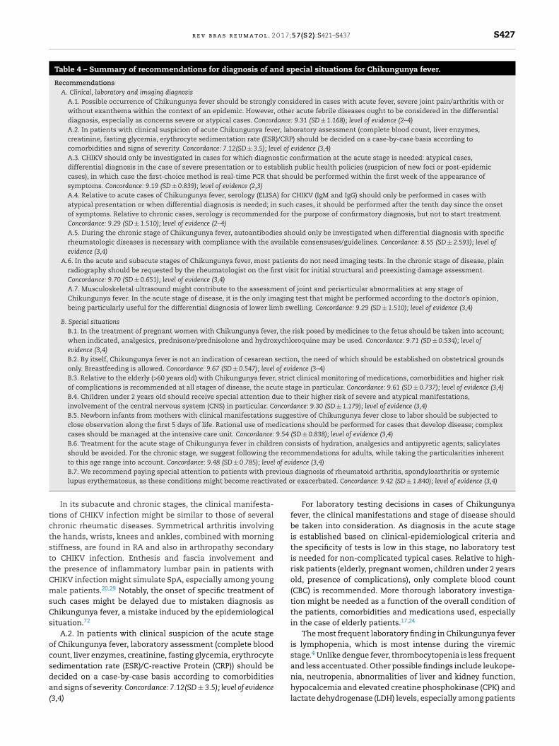
r e v b r a s r e u m a t o l . 2 0 1 7;5 7(S 2):S421–S437 S427
Table 4 – Summary of recommendations for diagnosis of and special situations for Chikungunya fever.
RecommendationsA. Clinical, laboratory and imaging diagnosis
A.1. Possible occurrence of Chikungunya fever should be strongly considered in cases with acute fever, severe joint pain/arthritis with orwithout exanthema within the context of an epidemic. However, other acute febrile diseases ought to be considered in the differentialdiagnosis, especially as concerns severe or atypical cases. Concordance: 9.31 (SD ± 1.168); level of evidence (2–4)A.2. In patients with clinical suspicion of acute Chikungunya fever, laboratory assessment (complete blood count, liver enzymes,creatinine, fasting glycemia, erythrocyte sedimentation rate (ESR)/CRP) should be decided on a case-by-case basis according tocomorbidities and signs of severity. Concordance: 7.12(SD ± 3.5); level of evidence (3,4)A.3. CHIKV should only be investigated in cases for which diagnostic confirmation at the acute stage is needed: atypical cases,differential diagnosis in the case of severe presentation or to establish public health policies (suspicion of new foci or post-epidemiccases), in which case the first-choice method is real-time PCR that should be performed within the first week of the appearance ofsymptoms. Concordance: 9.19 (SD ± 0.839); level of evidence (2,3)A.4. Relative to acute cases of Chikungunya fever, serology (ELISA) for CHIKV (IgM and IgG) should only be performed in cases withatypical presentation or when differential diagnosis is needed; in such cases, it should be performed after the tenth day since the onsetof symptoms. Relative to chronic cases, serology is recommended for the purpose of confirmatory diagnosis, but not to start treatment.Concordance: 9.29 (SD ± 1.510); level of evidence (2–4)A.5. During the chronic stage of Chikungunya fever, autoantibodies should only be investigated when differential diagnosis with specificrheumatologic diseases is necessary with compliance with the available consensuses/guidelines. Concordance: 8.55 (SD ± 2.593); level ofevidence (3,4)
A.6. In the acute and subacute stages of Chikungunya fever, most patients do not need imaging tests. In the chronic stage of disease, plainradiography should be requested by the rheumatologist on the first visit for initial structural and preexisting damage assessment.Concordance: 9.70 (SD ± 0.651); level of evidence (3,4)A.7. Musculoskeletal ultrasound might contribute to the assessment of joint and periarticular abnormalities at any stage ofChikungunya fever. In the acute stage of disease, it is the only imaging test that might be performed according to the doctor’s opinion,being particularly useful for the differential diagnosis of lower limb swelling. Concordance: 9.29 (SD ± 1.510); level of evidence (3,4)
B. Special situationsB.1. In the treatment of pregnant women with Chikungunya fever, the risk posed by medicines to the fetus should be taken into account;when indicated, analgesics, prednisone/prednisolone and hydroxychloroquine may be used. Concordance: 9.71 (SD ± 0.534); level ofevidence (3,4)B.2. By itself, Chikungunya fever is not an indication of cesarean section, the need of which should be established on obstetrical groundsonly. Breastfeeding is allowed. Concordance: 9.67 (SD ± 0.547); level of evidence (3–4)B.3. Relative to the elderly (>60 years old) with Chikungunya fever, strict clinical monitoring of medications, comorbidities and higher riskof complications is recommended at all stages of disease, the acute stage in particular. Concordance: 9.61 (SD ± 0.737); level of evidence (3,4)B.4. Children under 2 years old should receive special attention due to their higher risk of severe and atypical manifestations,involvement of the central nervous system (CNS) in particular. Concordance: 9.30 (SD ± 1.179); level of evidence (3,4)B.5. Newborn infants from mothers with clinical manifestations suggestive of Chikungunya fever close to labor should be subjected toclose observation along the first 5 days of life. Rational use of medications should be performed for cases that develop disease; complexcases should be managed at the intensive care unit. Concordance: 9.54 (SD ± 0.838); level of evidence (3,4)B.6. Treatment for the acute stage of Chikungunya fever in children consists of hydration, analgesics and antipyretic agents; salicylatesshould be avoided. For the chronic stage, we suggest following the recommendations for adults, while taking the particularities inherentto this age range into account. Concordance: 9.48 (SD ± 0.785); level of evidence (3,4)
viousted o
tctsttCmsCs
ocsda(
B.7. We recommend paying special attention to patients with prelupus erythematosus, as these conditions might become reactiva
In its subacute and chronic stages, the clinical manifesta-ions of CHIKV infection might be similar to those of severalhronic rheumatic diseases. Symmetrical arthritis involvinghe hands, wrists, knees and ankles, combined with morningtiffness, are found in RA and also in arthropathy secondaryo CHIKV infection. Enthesis and fascia involvement andhe presence of inflammatory lumbar pain in patients withHIKV infection might simulate SpA, especially among youngale patients.20,29 Notably, the onset of specific treatment of
uch cases might be delayed due to mistaken diagnosis ashikungunya fever, a mistake induced by the epidemiologicalituation.72
A.2. In patients with clinical suspicion of the acute stagef Chikungunya fever, laboratory assessment (complete bloodount, liver enzymes, creatinine, fasting glycemia, erythrocyte
edimentation rate (ESR)/C-reactive Protein (CRP)) should beecided on a case-by-case basis according to comorbiditiesnd signs of severity. Concordance: 7.12(SD ± 3.5); level of evidence3,4)diagnosis of rheumatoid arthritis, spondyloarthritis or systemicr exacerbated. Concordance: 9.42 (SD ± 1.840); level of evidence (3,4)
For laboratory testing decisions in cases of Chikungunyafever, the clinical manifestations and stage of disease shouldbe taken into consideration. As diagnosis in the acute stageis established based on clinical-epidemiological criteria andthe specificity of tests is low in this stage, no laboratory testis needed for non-complicated typical cases. Relative to high-risk patients (elderly, pregnant women, children under 2 yearsold, presence of complications), only complete blood count(CBC) is recommended. More thorough laboratory investiga-tion might be needed as a function of the overall condition ofthe patients, comorbidities and medications used, especiallyin the case of elderly patients.17,24
The most frequent laboratory finding in Chikungunya feveris lymphopenia, which is most intense during the viremicstage.4 Unlike dengue fever, thrombocytopenia is less frequent
and less accentuated. Other possible findings include leukope-nia, neutropenia, abnormalities of liver and kidney function,hypocalcemia and elevated creatine phosphokinase (CPK) andlactate dehydrogenase (LDH) levels, especially among patients
l . 2 0
S428 r e v b r a s r e u m a t owho require hospital admission.25,26,49,73,74 Relative to inflam-matory activity tests, ESR was found to be elevated in mostpatients during the first 10 months of disease in a study con-ducted in India.23 Elevated CRP levels were detected in morethan 70% of patients.20,26
In the chronic stage, in addition to routine testes (CBC,ESR, CRP), investigation of autoantibodies should be consid-ered when the clinical presentation is suggestive of CIJD andaccording to the existing comorbidities. Synovial fluid analy-sis might be necessary to confirm the inflammatory nature ofjoint involvement and help in differential diagnosis (i.e., goutor septic arthritis).
A.3. CHIKV should only be investigated in cases for whichdiagnostic confirmation in the acute stage is needed: atypicalcases, differential diagnosis in the case of severe presentationor to establish public health policies (suspicion of new foci orpost-epidemic cases), in which case the first-choice method isreal-time PCR that should be performed within the first weekof the appearance of symptoms. Concordance: 9.19 (SD ± 0.839);level of evidence (2,3)
Confirmatory diagnosis of infection with CHIKV might beestablished by means of 3 main laboratory tests: virus isola-tion, molecular techniques for viral genomic RNA detection(real-time PCR) and serology.75
Virus isolation through the detection of viral RNA might beperformed on serum samples during the acute stage of dis-ease (≤8 days)76; however, this method is not used in clinicalpractice.
Real-time PCR for the detection of viral genomic RNA hasseveral advantages compared to conventional PCR: faster toperform, quantitative, low contamination risk, simple stan-dardization and high sensitivity/specificity. By means of thistechnique, the presence of viral RNA can be detected duringthe early stage of viremia (0–7 days).77
Considering that the symptoms associated with arbovirusinfection (CHIKV, DENV and ZIKV) might be quite similar, espe-cially in the early stages of disease when differential diagnosisis necessary, triple real-time PCR is the most advantageoustechnique because it simultaneously identifies RNA from all 3viruses.78–80
As a function of the known difficulty in performing real-time PCR in clinical practice, including high cost and noaccess to most of the population, we recommend perform-ing confirmatory diagnosis of acute cases only for atypical orsevere cases during epidemics or for special situations (chil-dren under 2 years old, pregnant women, suspect of new foci,post-epidemic cases). In such cases, sample collection shouldbe performed within the first 6 days of the onset of symptoms.
A.4. Relative to acute cases of Chikungunya fever, serology(ELISA) for CHIKV (IgM and IgG) should only be performed incases with atypical presentation or when differential diagno-sis is needed; in such cases, it should be performed after thetenth day since the onset of symptoms. Relative to chroniccases, serology is recommended for the purpose of confirma-tory diagnosis, but not to start treatment. Concordance: 9.29(SD ± 1.510); level of evidence (2–4)
According to the current recommendations by the Brazil-ian Ministry of Health, in the face of an epidemic, specificserology for CHIKV during the acute stage of infection should
1 7;5 7(S 2):S421–S437
only be performed for atypical cases and complicated clin-ical situations.17 However, in areas where arboviruses areendemic, accurate identification of the specific type of infec-tion is highly relevant for the purpose of patient management,especially for cases that progress to the chronic stage, in orderto implement adequate public health measures and guidehealth managers.66
In practice, serology is the most widely used method forconfirmatory diagnosis of CHIKV infection. ELISA (enzyme-linked immunosorbent assay) and the plaque reductionneutralization test (PRNT) might be used for this purpose.17
IgM antibodies are detected in 4% to 20% of cases fromday 3 onward, reaching 80% positivity after the first week.81,82
Higher levels are detected from week 3 to 5, and the anti-bodies might remain elevated for 1 to 3 months or longer insome cases. IgG antibodies are also detected starting week 1after infection, usually 1 or 2 days after the elevation of IgMantibodies, and might remain detectable for several years.81–83
Serology for CHIKV can be performed starting 5 days afterthe onset of symptoms. However, to obtain a better operationalperformance of this test, it should be performed 7 to 10 daysafter the onset of symptoms for IgM antibodies and 10 to 14days for IgG antibodies for ELISA, especially in areas whereother alphaviruses do not circulate. The sensitivity of the testvaries from 85% to 98%, with specificity above 90%.14,65,81–88
Persistence of a specific IgM response several months after theinitial infection was found in some patients with chronic mus-culoskeletal symptoms.84 The meaning of this phenomenon isnot yet clear; according to the most widely accepted hypothe-sis, it denotes persistence of the virus in some tissues throughstill poorly understood mechanisms.84,88
Coinfection with other arboviruses is an occurrence inareas endemic for Chikungunya fever, dengue fever and Zika.CHIKV is an alphavirus; as such, it might exhibit cross-reactivity with other viruses from this family.65,88 In Brazil,the most frequent of such viruses is Mayaro virus, which alsocauses fever and (limited) joint pain; its circulation is limitedto the Northern region of the country,66 although it has alreadybeen detected in states from other regions.67,68
A study group from Federal University of Rio de Janeiro(Universidade Federal do Rio de Janeiro – UFRJ) reported acase series with 30 patients from areas endemic for ZIKV,CHIKV and DENV coinfection. DENV RNA was not detectedin any of the clinical samples, while ZIKV RNA was detectedin 17 samples (56.7%). ZIKV and CHIKV coinfection was doc-umented in 1 case. From the 17 ZIKV-positive individuals, 8exhibited reactivity for anti-DENV IgM, which is suggestiveof recent infection with DENV, cross-reactivity or coinfection.These findings reinforce the relevance of laboratory testingfor confirmatory diagnosis, especially for cases originating inareas endemic for all 3 arboviruses.75
PRNT, developed in 1959 by Henderson and Taylor,89 iden-tifies and quantifies neutralizing antibodies in serum samplesby calculating the percent viral activity reduction. PRNT iswidely accepted as the most specific test for the diagnosis ofdiseases by arboviruses. In samples from patients with specific
anti-CHIKV neutralizing antibodies, the number of plaqueswas lower compared to the controls due to the presence ofneutralizing antibodies in the infected host cells.90 Diagnosis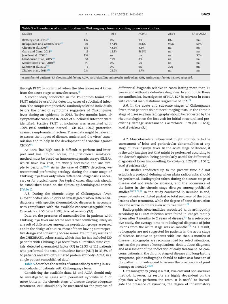
r e v b r a s r e u m a t o l . 2 0 1 7;5 7(S 2):S421–S437 S429
Table 5 – Prevalence of autoantibodies in Chikungunya fever according to various studies.
Studies n RF+ ACPA+ ANF+ RF or ACPA+
Blettery et al., 201672 147 0% 0% 0% naBouquillard and Combe, 200992 21 57.1% 28.6% 9.5% 66%Chopra et al., 200820 156 43.3% 3,3% na naGanu and Ganu, 201193 16 12.5% 56.5% na naJavelle et al., 200529 159 na na na 30%Lambourne et al., 2015136 54 15% 0% na naManimunda et al., 201023 20 0% 5% na naMinner et al., 2015137 8 0% 0% 30% naZhukov et al., 2015138 234 25.2% 1.7% na na
prot
tf
Ptbfsi1atmC
pmwprCsb(
adwC
Caattptw6s
e
bmt
method; however, its results are highly dependent on the
n, number of patients; RF, rheumatoid factor; ACPA, anti-citrullinated
hrough PRNT is confirmed when the titer increases 4 timesrom the acute stage to convalescence.76
A recent study conducted in the Philippines found thatRNT might be useful for detecting cases of subclinical infec-ion. The sample comprised 853 randomly selected individualsefore the onset of symptoms suggestive of Chikungunyaever during an epidemic in 2012. Twelve months later, 19ymptomatic cases and 87 cases of subclinical infection weredentified. Positive PRNT at inclusion was associated with00% (95% confidence interval – CI: 46.1, 100.0) protectiongainst symptomatic infection. These data might be relevanto assess the impact of disease, understand the virus’ trans-
ission and to help in the development of a vaccine againstHIKV.91
As PRNT has high cost, is difficult to perform and inter-ret and has limited access, the first-choice serologicalethod must be based on immunoenzymatic assays (ELISA),hich have low cost, are widely accessible and are sim-le to perform.65,84 As in the case of CHIKV detection, weecommend performing serology during the acute stage ofhikungunya fever only when differential diagnosis is neces-ary or for atypical cases. For typical cases, diagnosis shoulde established based on the clinical-epidemiological criteria
Table 5).A.5. During the chronic stage of Chikungunya fever,
utoantibodies should only be investigated when differentialiagnosis with specific rheumatologic diseases is necessaryith compliance with the available consensuses/guidelines.oncordance: 8.55 (SD ± 2.593); level of evidence (3,4)
Data on the presence of autoantibodies in patients withhikungunya fever are scarce and rather conflicting, likely as
result of differences among the population groups assessednd in the design of studies, most of them having a retrospec-ive design and consisting of case series. Preliminary results ofhe CHIKBRAZIL cohort study, which thus far has included 430atients with Chikungunya fever from 4 Brazilian state capi-als, detected rheumatoid factor (RF) in 28.3% of 113 patientsho performed the test, antinuclear factor (ANF) in 33.3% of
6 patients and anti-citrullinated protein antibody (ACPA) in aingle patient (unpublished data).
Table 5 describes the results of autoantibody testing in sev-ral cohorts of patients with Chikungunya fever.
Considering the available data, RF and ACPA should only
e investigated in cases with persistent synovitis in 1 orore joints in the chronic stage of disease despite adequatereatment. ANF should only be measured for the purpose of
ein antibodies; ANF, antinuclear factor; na, not assessed.
differential diagnosis relative to cases lasting more than 12weeks and without a definitive diagnosis. In addition to theseautoantibodies, investigation of HLA-B27 is relevant in caseswith clinical manifestations suggestive of SpA.24
A.6. In the acute and subacute stages of Chikungunyafever, most patients do not need imaging tests. In the chronicstage of disease, plain radiography should be requested by therheumatologist on the first visit for initial structural and pre-existing damage assessment. Concordance: 9.70 (SD ± 0.651);level of evidence (3,4)
A.7. Musculoskeletal ultrasound might contribute to theassessment of joint and periarticular abnormalities at anystage of Chikungunya fever. In the acute stage of disease, itis the only imaging test that might be performed according tothe doctor’s opinion, being particularly useful for differentialdiagnosis of lower limb swelling. Concordance: 9.29 (SD ± 1.510);level of evidence (3,4)
The studies conducted up to the present time did notestablish a protocol defining when plain radiographs shouldbe performed. Radiographs taken during the acute stage ofdisease did not evidence erosions, and the occurrence ofthe latter in the chronic stage diverges among publishedstudies.20,29,72,92 In the study conducted in Reunion Island,some patients exhibited partial or total recovery of the bonelesions after treatment, while the degree of bone destructionbecame worse in others even with treatment.29
Radiographic abnormalities associated with arthropathysecondary to CHIKV infection were found in images mainlytaken after 3 months to 2 years of disease.93 In a retrospec-tive study, the average time to radiological diagnosis of jointlesions from the acute stage was 45 months.29 As a result,radiographs are not suggested for patients in the acute stageof disease. Relative to patients with less than 3 months ofdisease, radiographs are recommended for select situations,such as the presence of complications, doubts about diagnosisand assessment of the indication of early treatment. As con-cerns patients in the chronic stage of disease and long-lastingsymptoms, plain radiographs should be taken as a function ofthe pattern of involvement to assess the progression of jointdamage as needed.24,29
Ultrasonography (USG) is a fast, low-cost and non-invasive
physician who performs the tests. It is useful to investi-gate the presence of synovitis, the degree of inflammatory
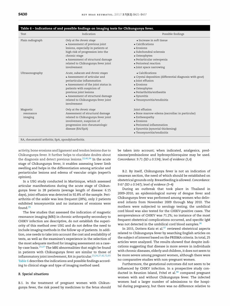
S430 r e v b r a s r e u m a t o l . 2 0 1 7;5 7(S 2):S421–S437
Table 6 – Indications of and possible findings on imaging tests for Chikungunya fever.
Test Indication Possible findings
Plain radiograph Only at the chronic stage• Assessment of previous jointlesions, especially in patients athigh risk of progression into thechronic stage• Assessment of structural damagerelated to Chikungunya fever jointinvolvement
• Increase in soft tissue• Calcifications• Erosions• Subchondral sclerosis• Osteophytes• Periarticular osteopenia• Periosteal reaction• Joint space narrowing
Ultrasonography Acute, subacute and chronic stages• Assessment of articular andperiarticular inflammation• Assessment of the joint status inpatients with suspicion ofprevious joint lesions• Assessment of structural damagerelated to Chikungunya fever jointinvolvement
• Calcifications• Crystal deposition (differential diagnosis with gout)• Joint effusion• Erosions• Osteophytes• Periarthritis/enthesitis• Synovitis• Tenosynovitis/tendinitis
Magneticresonanceimaging
Only at the chronic stageAssessment of structural damagerelated to Chikungunya fever jointinvolvement, suspicion ofprogression into rheumatologicdisease (RA/SpA)
Joint effusion• Bone marrow edema (sacroiliac in particular)• Enthesopathy• Erosions• Periosteal inflammation• Synovitis (synovial thickening)• Tenosynovitis/tendinitis
RA, rheumatoid arthritis; SpA, spondyloarthritis.
activity, bone erosions and ligament and tendon lesions due toChikungunya fever. It further helps to elucidate doubts aboutthe diagnosis and detect previous lesions.22,46,94 In the acutestage of Chikungunya fever, it enables assessing lower limbswelling and helps in the differentiation among articular andperiarticular lesions and edema of vascular origin (expert’sopinion).
In a USG study conducted in Martinique, which assessedarticular manifestations during the acute stage of Chikun-gunya fever in 28 patients (average length of disease: 6.15days), joint effusion was detected in 92.8% of the sample. Peri-arthritis of the ankle was less frequent (28%), only 2 patientsexhibited tenosynovitis and no instances of erosions werefound.95
The few studies that assessed the indication of magneticresonance imaging (MRI) in chronic arthropathy secondary toCHIKV infection are descriptive, do not establish the superi-ority of this method over USG and do not define the need toinclude imaging methods in the follow up of patients. In addi-tion, one needs to take into account the cost and availability oftests, as well as the examiner’s experience in the selection ofthe most adequate method for imaging assessment on a case-by-case basis.23,27 The MRI abnormalities that might be foundin patients with Chikungunya fever are similar to those ofinflammatory joint involvement, RA in particular.23,24,27,42,72,93
Table 6 describes the indications and possible findings accord-ing to clinical stage and type of imaging method used.
B. Special situations
B.1. In the treatment of pregnant women with Chikun-gunya fever, the risk posed by medicines to the fetus should
be taken into account; when indicated, analgesics, pred-nisone/prednisolone and hydroxychloroquine may be used.Concordance: 9.71 (SD ± 0.534); level of evidence (3,4)
B.2. By itself, Chikungunya fever is not an indication ofcesarean section, the need of which should be established onobstetrical grounds only. Breastfeeding is allowed. Concordance:9.67 (SD ± 0.547); level of evidence (3–4)
During an outbreak that took place in Thailand in2009–2010, an epidemiological survey of dengue fever andChikungunya fever was performed among women who deliv-ered infants from November 2009 through May 2010. Themothers were subjected to serology testing; the umbilicalcord blood was also tested for the CHIKV-positive cases. Theseroprevalence of CHIKV was 71.2%; no instance of the mostfrequent obstetrical complications occurred, and specific IgMwas not detected in the umbilical cord blood samples.96
In 2015, Dotters-Katz et al.97 reviewed obstetrical aspectsrelated to Chikungunya fever by searching English articles onthe subject of interest based on the PRISMA criteria. In total, 25articles were analyzed. The results showed that despite indi-cations suggesting that disease is more severe in individualswith chronic diseases, elderly and children, it does not seem tobe more severe among pregnant women, although there wereno comparative studies with non-pregnant women.
Furthermore, the gestational outcomes did not seem to beinfluenced by CHIKV infection. In a prospective study con-ducted in Reunion Island, Fritel et al.98 compared pregnant
women with and without Chikungunya fever. The infectedwomen had a larger number of admissions to the hospi-tal during pregnancy, but there was no difference relative to
0 1 7;
cpRwwt
ocasitttonnfam
gmtagfuapbr
cdtc
isbo
fas(
oteaq7m
s(4
r e v b r a s r e u m a t o l . 2
ongenital defects, prematurity or other disorders. In anotherrospective study by Gérardin et al.,99 also performed ineunion Island, maternal viremia intrapartum was associatedith a higher incidence of abnormalities of the fetal heart rate,hich doubled the frequency of cesarean sections; this was
he single difference found.The pregnancy trimester in which infection with CHIKV
ccurs is the main predictor of fetal outcome. The virus canross the placental barrier in the first and second trimestersnd cause fetal infection and miscarriage, which are con-idered to be rare occurrences. However, maternal activenfection up to 4 days after delivery increases the risk of ver-ical transmission, the incidence of which varies from 27%o 48% among the various published case series.97,99–103 Ver-ical transmission should be suspected when the symptomsf disease manifest within the first week of life and there iso evidence of a mosquito bite.103 As cesarean section doesot prevent vertical transmission,104,105 it is not mandatory
or cases of Chikungunya fever; rather, it should be performeds per obstetrical indications. The transmission of CHIKV byother’s milk is still under discussion.97
The treatment of pregnant women with chronic Chikun-unya fever should take into account the known effects ofedicines on the fetus. It is recommended to start analgesic
reatment in the acute stage of disease with paracetamol, with maximum dose of 4 g/day. NSAIDs are contraindicated fromestational week 24 onward due to the risk of fetal kidneyailure and premature closure of the ductus arteriosus.24 These of non-selective COX-2 inhibitors might be allowed over
short period of time from pregnancy week 16 to 30, but it isreferable to indicate prednisone in low doses, which mighte used all across pregnancy and breastfeeding with no fetalisk.106
Hydroxychloroquine is an option for the treatment ofhronic joint disorders during pregnancy and breastfeedingue its acknowledged safety during these periods106,107 andhe evidence indicating improvement of joint disorders in aase series from the overall population.108,109
A steroid sparing agent, azathioprine, might be used dur-ng pregnancy and breastfeeding, while methotrexate (MTX)hould be avoided by the time of conception, pregnancy andreastfeeding.106 In addition to paracetamol, ibuprofen andpioids might also be indicated in select cases.110
B.3. Relative to the elderly (>60 years old) with Chikungunyaever, strict clinical monitoring of medications, comorbiditiesnd higher risk of complications is recommended in all thetages of disease, acute ones in particular. Concordance: 9.61SD ± 0.737); level of evidence (3,4)
Considering elderly individuals as those aged 60 years andlder, few studies have approached this specific subpopula-ion. However, with a cutoff point of 40 years old, older patientsxhibited higher odds of progression into the chronic stage,s found in the La Virginia, Colombia cohort, where the fre-uency was 52%,38 and the Reunion Island cohort, in which0% of the patients above 45 exhibited persistent joint pain 15onths after acute infection (odds ratio (OR) 4.2 (CI 1.9–9.3)).39
In a study conducted in India with 509 patients, the inten-ity of the acute stage of disease was higher among the elderlyabove 65 years old) who were confined to bed on average for–6 days; 42% of them attained full recovery after 4 weeks, and
5 7(S 2):S421–S437 S431
no deaths occurred.19 The particularities of disease among theelderly are not clear in the remainder of the analyzed cohorts;some of them reported a higher number of medical visits com-pared to other groups of patients.
The factors to be taken into account in the approach andtreatment of the elderly are polypharmacy and the presenceof comorbidities (arterial hypertension – AH – and DM), whichare frequent in this age range. Concomitant use of severalmedications might result in complications derived from theinteraction of the analgesics and steroids indicated for painand arthritis. Many elderly use aspirin prophylactically, whichis associated with Reye’s syndrome, but there are no reportsamong patients with Chikungunya fever. Decompensation ofDM is associated with dehydration, which might be a compli-cation in cases with acute infection (6).
In Brazil, up to EW 27 of 2016, 38 deaths by Chikungunyafever were confirmed, corresponding to the following states:Pernambuco (n = 25), Rio Grande do Norte (n = 5), Paraíba (n = 2),Rio de Janeiro (n = 2), Ceará (n = 2), Maranhão (n = 1) and Alagoas(n = 1); the median age of the deceased was 71 years old.111
Relative to Pernambuco, i.e., the state with the largest numberof deaths in the country, more recent data (EW 37) showedthat 7970 of 53,061 notified cases corresponded to individualsabove 60 years old, with 53 confirmed deaths by Chikungunyafever.112
Although the elderly do not represent the group with thehighest prevalence of disease, most deaths occurred withinthis age range. This shows that this population needs partic-ular attention during the acute stage, more specifically, thosewho live alone and might have no assistance for feeding andpersonal care. Many elderly are confined to bed for up to 7days, which increases the risk of urinary tract infection andpneumonia.113
B.4. Children under 2 years of age should receive specialattention due to their higher risk of severe and atypical man-ifestations and involvement of the central nervous system(CNS). Concordance: 9.30 (SD ± 1.179); level of evidence (3,4)
Up to the present time, few observational studies haveinvestigated clinical and laboratory manifestations of infec-tion with CHIKV among children. Most such studies describemore severe forms of diseases occurring in newborn infantsand emphasize the fact that the clinical presentation might bedifferent in children compared to adults. The rate of asymp-tomatic infection seems to be higher among children, varyingamong the various reports of outbreaks (35–40%).46,114–120 Arecent study analyzed the seroprevalence among children onthe occasion of the first CHIKV outbreak in Nicaragua. Theresults showed detectable antibody levels in 6.1% of childrenaged 2–14 years and 13.1% in those above 15 years of age.121
Relative to the acute stage of infection, the differencesbetween children and adults concern the skin, hemorrhagicand neurological manifestations.117 Maculopapular exan-thema occurs in 35–50% of adults but is less frequent amongchildren, especially those under 2 years old, with hyperpig-mentation a more common finding. In addition, infants under6 months old might exhibit extensive skin bullous lesions,affecting up to 35% of the body surface.122 Hemorrhagic man-
ifestations, including epistaxis, gum bleeding and purpura,occur in 10% of pediatric cases, while they are rare amongadults.117
l . 2 0
S432 r e v b r a s r e u m a t oThe main concern relative to the pediatric population isthe involvement of the CNS. During the Chikungunya feveroutbreak in Reunion Island, 25% of the children exhibitedneurological symptoms. Among such children, a high pro-portion (40–50%) exhibited severe manifestations, includingstatus epilepticus, complex seizures and encephalitis.123,124
The neurological symptoms can last from several months toyears.123
B.5. Newborn infants from mothers with clinical manifes-tations suggestive of Chikungunya fever close to labor shouldbe subjected to close observation during the first 5 days of life.Rational use of medications should be performed for casesthat develop disease; complex cases should be managed atthe intensive care unit. Concordance: 9.54 (SD ± 0.838); level ofevidence (3,4)
Perinatal infection was first described in the Reunion Islandoutbreak in 2005.125 Although intrauterine CHIKV transmis-sion is extraordinarily rare at the onset of pregnancy, its rateincreases to almost 50% when maternal viremia occurs 1 weekbefore birth.126
A recent Latin American multicentric study analyzed 169newborn infants with symptomatic Chikungunya fever at4 large maternity hospitals from 3 countries whose moth-ers had CHIKV infection confirmed by PCR. In these infants,the symptoms started around the fourth day of life, themost frequent ones being fever, irritability, hyperalgesia,diffuse limb swelling, meningoencephalitis, skin rash and bul-lous dermatitis and petechiae. Laboratory testing evidencedthrombocytopenia and lymphopenia in most cases. Compli-cations included intracerebral hemorrhage, status epilepticusand multiple organ failure.103
The mortality rate is high, for which reason long-term out-comes and damage could not be assessed in studies. Amongsurvivors, half of the children exhibited reduced cognitivedevelopment by 2 years of age.99,105,126 These data clearlyshow that infection with CHIKV among children is not alwaysbenign; rather, it might result in permanent sequelae anddeath.
B.6. Treatment for the acute stage of Chikungunya feverin children consists of hydration, analgesics and antipyreticagents; salicylates should be avoided. For the chronic stage, wesuggest following the recommendations for adults, while tak-ing the particularities inherent to this age range into account.Concordance: 9.48 (SD ± 0.785); level of evidence (3,4)
No recommendations specific for the management andtreatment of infection with CHIKV among children werelocated. In most case series published after outbreaks in Asiaand the Americas, the recommendations are similar to thosefor adults: in the acute stage, hyper-hydration, analgesics andantipyretics, with avoidance of salicylates and NSAIDs due tothe risk of triggering bleeding. When the joint symptoms per-sist, the recommendations do not differ from those for adultswho progress to the chronic stage, treatment being based onthe use of NSAIDs, steroids and MTX; however, the benefits forthe pediatric population are still unknown.
The more specific available recommendations target new-
born infants, in which morbidity and mortality are common.Rational use of medications and adequate monitoring arerecommended, which seem to have a direct relationshipwith improved clinical progression. Abuse of medications,1 7;5 7(S 2):S421–S437
such as salicylates, NSAIDs, steroids and antibiotics, mightcontribute to worsening disease through development ofbleeding, thrombocytopenia and gastrointestinal symptoms,leading to complications, including hydroelectrolytic disor-ders, dehydration and kidney failure, which might indirectlytrigger death.99–101,103
B.7. We recommend paying special attention to patientswith previous diagnosis of rheumatoid arthritis, spondy-loarthritis or systemic lupus erythematosus, as theseconditions might become reactivated or exacerbated. Concord-ance: 9.42 (SD ± 1.840); level of evidence (3,4)
Infection with CHIKV among patients with previousrheumatologic diseases is not well characterized in the litera-ture. In the published case series and cohorts, the frequencyof previous musculoskeletal disorders among patients withChikungunya fever varies from 6.9% to 86%,20,23,26,41,127 thehighest prevalence rates being found in studies conducted atspecialized services.
A history of rheumatologic disease was associatedwith persistence of joint symptoms after infection withCHIKV40,41,127 and greater disability.128 Exacerbation of paincomplaints in the previously affected sites was described inpatients with chronic diseases, such as OA, tendinopathy, lum-bar pain and carpal tunnel syndrome.23,29,74,127 Sissoko et al.39
followed up 147 patients along 15 months; 28% of the samplehad a previous diagnosis of OA, which behaved as a predictorof persistence of symptoms (OR 2.9 (95% CI 1.1–7.4; p = 0.029)).
The acute clinical manifestations of patients with pre-vious musculoskeletal pain tend to be similar to those ofpreviously healthy patients with Chikungunya fever; atypi-cal or severe acute manifestations are not more frequent, noteven among patients using immunosuppressants or immuno-biological agents.129–132 Interestingly, patients are able todistinguish the symptoms related to CHIKV infection fromthose of the underlying disease because the former are moreintense or occur in sites different than the latter.133
Javelle et al.29 assessed 18 patients with previous rheuma-tologic disease among 159 patients followed up over 6 years.In 6 patients with RA, 8 with SpA, 2 with SLE and 2 withchronic hepatitis, the joint symptoms worsened immediatelyafter infection with CHIKV, requiring changes in treatment oronset of new treatments in half of them.
Patients with SpA might exhibit exacerbation charac-terized by greater peripheral involvement (arthritis and/orenthesitis),20,29 and those with psoriatic arthritis experienceexacerbation of the skin and/or joint disease.20
Patients with previously controlled RA might exhibitexacerbation of the joint symptoms in the acute stage ofChikungunya fever, eventually with the typical manifesta-tions of RA involving the joints of the hands and feet.19,129
However, assessment of patients with previous rheumatic dis-ease in Martinique did not detect any cases of post-CHIK RAexacerbation, which suggests that previous treatment withdisease-modifying antirheumatic drugs (DMARDs) and/orimmunobiological agents might have a protective effect.72
Retrospective assessment of 167 patients with SLE in Mar-
tinique showed that, among the 56 patients with serologypositive for CHIKV, 82.6% exhibited clinical symptoms com-patible with Chikungunya fever, while only 10.7% exhibitedexacerbation of SLE. Severe manifestations of Chikungunya
0 1 7;
findaaaot
F
Ta
C
T
r
r e v b r a s r e u m a t o l . 2
ever were detected in 4 patients, including encephalopathyn all 4, associated with bullous cutaneous lesions in 3, kid-ey involvement in 1 and multiple organ failure resulting ineath in 1.130 There is risk of severe complications – 1 case ofntiphospholipid antibody syndrome (APS) was described in
patient with a previous diagnosis of SLE.134,135 However, thevailable data are not sufficient to establish whether the riskf atypical manifestations/severe complications is higher inhis specific population.
unding
he Brazilian Society of Rheumatology provided all financialnd logistic support for the performance of the present study.
onflicts of interest
he authors declare no conflicts of interest.
e f e r e n c e s
1. Lumsden WH. An epidemic of virus disease in SouthernProvince Tanganyika Territory, in 1952–53. II. Generaldescription and epidemiology. Trans R Soc Trop Med Hyg.1955;49:33–57.
2. Carvalho RG, Lourenco-de-Oliveira R, Braga IA. Updating thegeographical distribution and frequency of Aedes albopictusin Brazil with remarks regarding its range in the Americas.Mem Inst Oswaldo Cruz. 2014;109:787–96.
3. Staples JE, Breiman RF, Powers AM. Chikungunya fever: anepidemiological review of a re-emerging infectious disease.Clin Infect Dis. 2009;49:942–8.
4. Weaver SC, Lecuit M. Chikungunya virus and the globalspread of a mosquito-borne disease. N Engl J Med.2015;372:1231–9.
5. Leparc-Goffart I, Nougairede A, Cassadou S, Prat C, deLamballerie X. Chikungunya in the Americas. Lancet.2014;383:514.
6. PAHO-WHO. Chikungunya autochthonous transmission inthe Americas. PAHO-WHO; 2016. p. 2016.
7. Faria NR, Lourenco J, Cerqueira EM, Lima MM, Pybus O,Alcantara LCJ. Epidemiology of chikungunya virus in Bahia,Brazil, 2014–2015. PLoS Curr. 2016;8.
8. Brasil. Monitoramento dos casos de dengue, febre dechikungunya e febre pelo vírus Zika até a SemanaEpidemiológica 32. In: Saúde Md, ed. Brasilia 2016.
9. Agarwal A, Joshi G, Nagar DP, Sharma AK, Sukumaran D,Pant SC, et al. Mosquito saliva induced cutaneous eventsaugment chikungunya virus replication and diseaseprogression. Infect Genet Evol. 2016;40:126–35.
10. Rougeron V, Sam IC, Caron M, Nkoghe D, Leroy E, Roques P.Chikungunya, a paradigm of neglected tropical disease thatemerged to be a new health global risk. J Clin Virol.2015;64:144–52.
11. Lum FM, Ng LF. Cellular and molecular mechanisms ofchikungunya pathogenesis. Antiviral Res. 2015;120:165–74.
12. Assuncao-Miranda I, Cruz-Oliveira C, Da Poian AT. Molecularmechanisms involved in the pathogenesis of
alphavirus-induced arthritis. Biomed Res Int.2013;2013:973516.13. Chirathaworn C, Rianthavorn P, Wuttirattanakowit N,Poovorawan Y. Serum IL-18 and IL-18BP levels in patients
5 7(S 2):S421–S437 S433
with chikungunya virus infection. Viral Immunol.2010;23:113–7.
14. Hoarau JJ, Jaffar Bandjee MC, Krejbich Trotot P, Das T,Li-Pat-Yuen G, Dassa B, et al. Persistent chronicinflammation and infection by chikungunya arthritogenicalphavirus in spite of a robust host immune response. JImmunol. 2010;184:5914–27.
15. Goupil BA, McNulty MA, Martin MJ, McCracken MK,Christofferson RC, Mores CN. Novel lesions of bones andjoints associated with chikungunya virus infection in twomouse models of disease: new insights into diseasepathogenesis. PLOS ONE. 2016;11:e0155243.
16. Rudolph KE, Lessler J, Moloney RM, Kmush B, Cummings DA.Incubation periods of mosquito-borne viral infections: asystematic review. Am J Trop Med Hyg. 2014;90:882–91.
17. Febre de chikungunya – manejo clínico. http://portal.cfm.org.br/index.php?option=com content&view=article&id=25398:2015-03-16-17-58-53&catid=3; 2015 [accessed17.09.16].
18. Queyriaux B, Simon F, Grandadam M, Michel R, Tolou H,Boutin JP. Clinical burden of chikungunya virus infection.Lancet Infect Dis. 2008;8:2–3.
19. Chopra A, Anuradha V, Ghorpade R, Saluja M. Acutechikungunya and persistent musculoskeletal pain followingthe 2006 Indian epidemic: a 2-year prospective ruralcommunity study. Epidemiol Infect. 2012;140:842–50.
20. Chopra A, Anuradha V, Lagoo-Joshi V, Kunjir V, Salvi S,Saluja M. Chikungunya virus aches and pains: an emergingchallenge. Arthritis Rheum. 2008;58:2921–2.
21. Dupuis-Maguiraga L, Noret M, Brun S, Le Grand R, Gras G,Roques P. Chikungunya disease: infection-associatedmarkers from the acute to the chronic phase ofarbovirus-induced arthralgia. PLoS Negl Trop Dis.2012;6:e1446.
22. Madariaga M, Ticona E, Resurrecion C. Chikungunya:bending over the Americas and the rest of the world. Braz JInfect Dis. 2016;20:91–8.
23. Manimunda SP, Vijayachari P, Uppoor R, Sugunan AP, SinghSS, Rai SK, et al. Clinical progression of chikungunya feverduring acute and chronic arthritic stages and the changes injoint morphology as revealed by imaging. Trans R Soc TropMed Hyg. 2010;104:392–9.
24. Simon F, Javelle E, Cabie A, Bouquillard E, Troisgros O,Gentile G, et al. French guidelines for the management ofchikungunya (acute and persistent presentations) November2014. Med Mal Infect. 2015;45:243–63.
25. Waymouth HE, Zoutman DE, Towheed TE.Chikungunya-related arthritis: case report and review of theliterature. Semin Arthritis Rheum. 2013;43:273–8.
26. Borgherini G, Poubeau P, Jossaume A, Gouix A, Cotte L,Michault A, et al. Persistent arthralgia associated withchikungunya virus: a study of 88 adult patients on reunionisland. Clin Infect Dis. 2008;47:469–75.
27. Chaaithanya IK, Muruganandam N, Raghuraj U, Sugunan AP,Rajesh R, Anwesh M, et al. Chronic inflammatory arthritiswith persisting bony erosions in patients followingchikungunya infection. Indian J Med Res. 2014;140:142–5.
28. Gerardin P, Fianu A, Michault A, Mussard C, Boussaïd K,Rollot O, et al. Predictors of chikungunya rheumatism: aprognostic survey ancillary to the Telechik cohort study.Arthritis Res Ther. 2013;15:R9.
29. Javelle E, Ribera A, Degasne I, Gauzere BA, Marimoutou C,Simon F. Specific management of post-chikungunyarheumatic disorders: a retrospective study of 159 cases inReunion Island from 2006–2012. PLoS Negl Trop Dis.
2015;9:e0003603.30. Win MK, Chow A, Dimatatac F, Go CJ, Leo YS. Chikungunyafever in Singapore: acute clinical and laboratory features,
l . 2 0
S434 r e v b r a s r e u m a t oand factors associated with persistent arthralgia. J ClinVirol. 2010;49:111–4.
31. Rodriguez-Morales AJ, Cardona-Ospina JA, Villamil-GomezW, Paniz-Mondolfi AE. How many patients withpost-chikungunya chronic inflammatory rheumatism canwe expect in the new endemic areas of Latin America?Rheumatol Int. 2015;35:2091–4.
32. Rodriguez-Morales AJ, Cardona-Ospina JA, Urbano-GarzonSF, Hurtado-Zapata JS. Prevalence of post-chikungunyachronic inflammatory rheumatism: a systematic review andmeta-analysis. Arthritis Care Res (Hoboken).2016;68:1849–58.
33. Schilte C, Staikowsky F, Couderc T, Madec Y, Carpentier F,Kassab S, et al. Chikungunya virus-associated long-termarthralgia: a 36-month prospective longitudinal study. PLoSNegl Trop Dis. 2013;7:e2137.
34. Essackjee K, Goorah S, Ramchurn SK, Cheeneebash J,Walker-Bone K. Prevalence of and risk factors for chronicarthralgia and rheumatoid-like polyarthritis more than 2years after infection with chikungunya virus. Postgrad MedJ. 2013;89:440–7.
35. Couturier E, Guillemin F, Mura M, Léon L, Virion JM, LetortMJ, et al. Impaired quality of life after chikungunya virusinfection: a 2-year follow-up study. Rheumatology (Oxford).2012;51:1315–22.
36. Thiberville SD, Boisson V, Gaudart J, Simon F, Flahault A, deLamballerie X. Chikungunya fever: a clinical and virologicalinvestigation of outpatients on Reunion Island South-WestIndian Ocean. PLoS Negl Trop Dis. 2013;7:e2004.
37. van Genderen FT, Krishnadath I, Sno R, Grunberg MG,Zijlmans W, Adhin MR. First chikungunya outbreak inSuriname: clinical and epidemiological features. PLoS NeglTrop Dis. 2016;10:e0004625.
38. Rodriguez-Morales AJ, Gil-Restrepo AF, Ramirez-Jaramillo V,Montoya-Arias CP, Acevedo-Mendoza WF, Bedoya-Arias JE,et al. Post-chikungunya chronic inflammatory rheumatism:results from a retrospective follow-up study of 283 adult andchild cases in La Virginia, Risaralda. F1000 Res. 2016;5:360.
39. Sissoko D, Malvy D, Ezzedine K, Renault P, Moscetti F,Ledrans M, et al. Post-epidemic chikungunya disease onReunion Island: course of rheumatic manifestations andassociated factors over a 15-month period. PLoS Negl TropDis. 2009;3:e389.
40. Yaseen HM, Simon F, Deparis X, Marimoutou C.Identification of initial severity determinants to predictarthritis after chikungunya infection in a cohort of Frenchgendarmes. BMC Musculoskelet Disord. 2014;15:249.
41. Moro ML, Grilli E, Corvetta A, Silvi G, Angelini R, Mascella F,et al., Study Group Infezioni da Chikungunya inEmilia-Romagna. Long-term chikungunya infection clinicalmanifestations after an outbreak in Italy: a prognosticcohort study. J Infect. 2012;65:165–72.
42. Malvy D, Ezzedine K, Mamani-Matsuda M, Autran B, TolouH, Receveur M-C, et al. Destructive arthritis in a patient withchikungunya virus infection with persistent specific IgMantibodies. BMC Infect Dis. 2009;9:200.
43. Marques CDL, Cavalcanti N, Luna M, Toche P, Andrade C,Dantas A, et al. Chikungunya fever outbreak in Brazil:preliminary assessment in a cohort of patients withrheumatological manifestations. Arthritis Rheumatol.2016;68 Suppl 10. http://acrabstracts.org/abstract/chikungunya-fever-outbreak-in-brazil-preliminary-assessment-in-a-cohort-of-patients-with-rheumatological-manifestations/ [accessed 06.02.17].
44. Sissoko D, Ezzedine K, Moendandze A, Giry C, Renault P,
Malvy D. Field evaluation of clinical features duringchikungunya outbreak in Mayotte, 2005–2006. Trop Med IntHealth. 2010;15:600–7.1 7;5 7(S 2):S421–S437
45. Chikungunya: case definitions for acute, atypical andchronic cases. Conclusions of an expert consultation,Managua, Nicaragua, 20–21 May 2015. Wkly Epidemiol Rec.2015;90:410–4.
46. Economopoulou A, Dominguez M, Helynck B, Sissoko D,Wichmann O, Quenel P, et al. Atypical chikungunya virusinfections: clinical manifestations, mortality and riskfactors for severe disease during the 2005–2006 outbreak onReunion. Epidemiol Infect. 2009;137:534–41.
47. Crosby L, Perreau C, Madeux B, Cossic J, Armand C,Herrmann-Storke C, et al. Severe manifestations ofchikungunya virus in critically ill patients during the2013–2014 Caribbean outbreak. Int J Infect Dis. 2016;48:78–80.
48. Oehler E, Fournier E, Leparc-Goffart I, Larre P, Cubizolle S,Sookhareea C, et al. Increase in cases of Guillain-Barresyndrome during a chikungunya outbreak, French Polynesia,2014 to 2015. Euro Surveill. 2015;20:30079.
49. Staikowsky F, Talarmin F, Grivard P, Souab A, SchuffeneckerI, Le Roux K, et al. Prospective study of chikungunya virusacute infection in the Island of La Reunion during the2005–2006 outbreak. PLoS ONE. 2009;4:e7603.
50. Oliver M, Grandadam M, Marimoutou C, Rogier C,Botelho-Nevers E, Tolou H, et al. Persisting mixedcryoglobulinemia in chikungunya infection. PLoS Negl TropDis. 2009;3:e374.
51. Macpherson C, Noel T, Fields P, Jungkind D, Yearwood K,Simmons M, et al. Clinical and serological insights from theAsian lineage chikungunya outbreak in Grenada, 2014: anobservational study. Am J Trop Med Hyg. 2016;95:890–3.
52. Mathew AJ, Goyal V, George E, Thekkemuriyil DV, JayakumarB, Chopra A, Trivandrum Copcord Study Group.Rheumatic-musculoskeletal pain and disorders in a naivegroup of individuals 15 months following a chikungunyaviral epidemic in south India: a population basedobservational study. Int J Clin Pract. 2011;65:1306–12.
53. Horcada ML, Diaz-Calderon C, Garrido L. Chikungunya feverrheumatic manifestations of an emerging disease in Europe.Reumatol Clin. 2015;11:161–4.
54. Rulli NE, Melton J, Wilmes A, Ewart G, Mahalingam S. Themolecular and cellular aspects of arthritis due to alphavirusinfections: lesson learned from Ross River virus. Ann N YAcad Sci. 2007;1102:96–108.
55. Inamadar AC, Palit A, Sampagavi VV, Raghunath S,Deshmukh NS. Cutaneous manifestations of chikungunyafever: observations made during a recent outbreak in southIndia. Int J Dermatol. 2008;47:154–9.
56. Seetharam KA, Sridevi K. Chikungunya infection: a newtrigger for psoriasis. J Dermatol. 2011;38:1033–4.
57. Higgins JPTG. Cochrane handbook for systematic reviews ofinterventions. Cochrane Colaborations; 2011.
58. Howick J, Glasziou P, Greenhalgh T, Heneghan C, Liberati A,Moschetti I, et al., OCEBM Levels of Evidence Working Group.The Oxford Levels of Evidence 2. Oxford Centre forEvidence-Based Medicine; 2011.
59. Mohan A, Kiran DH, Manohar IC, Kumar DP. Epidemiology,clinical manifestations, and diagnosis of chikungunya fever:lessons learned from the re-emerging epidemic. Indian JDermatol. 2010;55:54–63.
60. Toivanen A. Alphaviruses: an emerging cause of arthritis?Curr Opin Rheumatol. 2008;20:486–90.
61. Lee VJ, Chow A, Zheng X, Carrasco LR, Cook AR, Lye DC, et al.Simple clinical and laboratory predictors of chikungunyaversus dengue infections in adults. PLoS Negl Trop Dis.2012;6:e1786.
62. Chahar HS, Bharaj P, Dar L, Guleria R, Kabra SK, Broor S.Co-infections with chikungunya virus and dengue virus inDelhi, India. Emerg Infect Dis. 2009;15:1077–80.
0 1 7;
r e v b r a s r e u m a t o l . 263. Furuya-Kanamori L, Liang S, Milinovich G, Magalhaes RJS,Clements ACA, Hu W, et al. Co-distribution and co-infectionof chikungunya and dengue viruses. BMC Infect Dis.2016;16:84.
64. Pessoa R, Patriota JV, Lourdes de Souza M, Felix AC, MamedeN, Sanabani SS. Investigation into an outbreak ofdengue-like illness in Pernambuco, Brazil, revealed acocirculation of Zika Chikungunya, and Dengue virus type 1.Medicine (Baltimore). 2016;95:e3201.
65. Suhrbier A, Jaffar-Bandjee MC, Gasque P. Arthritogenicalphaviruses – an overview. Nat Rev Rheumatol.2012;8:420–9.
66. Nunes MR, Faria NR, de Vasconcelos JM, Golding N, KraemerMU, de Oliveira LF, et al. Emergence and potential for spreadof chikungunya virus in Brazil. BMC Med. 2015;13:102.
67. Serra OP, Cardoso BF, Ribeiro AL, Santos FA, SlhessarenkoRD. Mayaro virus and dengue virus 1 and 4 natural infectionin culicids from Cuiaba, state of Mato Grosso, Brazil. MemInst Oswaldo Cruz. 2016;111:20–9.
68. Mota MT, Vedovello D, Estofolete C, Malossi CD, Araujo JP Jr,Nogueira ML. Complete genome sequence of Mayaro virusimported from the Amazon basin to Sao Paulo State, Brazil.Genome Announc. 2015;3:e01341–1415.
69. Arroyo-Avila M, Vila LM. Rheumatic manifestations inpatients with chikungunya infection. P R Health Sci J.2015;34:71–7.
70. Kajeguka DC, Kaaya RD, Mwakalinga S, Ndossi R, Ndaro A,Chilongola JO, et al. Prevalence of dengue and chikungunyavirus infections in north-eastern Tanzania: a cross sectionalstudy among participants presenting with malaria-likesymptoms. BMC Infect Dis. 2016;16:183.
71. Renault P, Boidin E, D’Ortenzio E, Balleydier E, Daniel B,Filleul L. Epidemiological surveillance of leptospirosis onReunion Island in 2004–2008: possible impact ofchikungunya infection on the case fatality rate ofleptospirosis. Bull Soc Pathol Exot. 2011;104:148–52.
72. Blettery M, Brunier L, Polomat K, Moinet F, Deligny C, Arfi S,et al. Management of chronic post-chikungunya rheumaticdisease: the Martinican experience. Arthritis Rheumatol.2016;68:2817–24.
73. Staples JE, Fischer M. Chikungunya virus in the Americas –what a vectorborne pathogen can do. N Engl J Med.2014;371:887–9.
74. Taubitz W, Cramer JP, Kapaun A, Pfeffer M, Drosten C, DoblerG, et al. Chikungunya fever in travelers: clinical presentationand course. Clin Infect Dis. 2007;45:e1–4.
75. Cabral-Castro MJ, Cavalcanti MG, Peralta RH, Peralta JM.Molecular and serological techniques to detectco-circulation of DENV ZIKV and CHIKV in suspecteddengue-like syndrome patients. J Clin Virol. 2016;82:108–11.
76. Brasil. Preparacão e resposta à introducão do víruschikungunya no Brasil. In: Saúde. MdSSdVe, ed. Brasília;2014.
77. Wang SM, Ali UH, Sekaran SD, Thayan R. Detection andquantification of chikungunya virus by real-time RT-PCRassay. Methods Mol Biol. 2016;1426:105–17.
78. Pabbaraju K, Wong S, Gill K, Fonseca K, Tipples GA, Tellier R.Simultaneous detection of Zika, Chikungunya, and Dengueviruses by a multiplex real-time RT-PCR assay. J Clin Virol.2016;83:66–71.
79. Simmons M, Myers T, Guevara C, Jungkind D, Williams M,Houng HS. Development and validation of a quantitative,one-step, multiplex real-time reverse transcriptase PCRassay for detection of dengue and chikungunya viruses. JClin Microbiol. 2016;54:1766–73.
80. Liu J, Ochieng C, Wiersma S, Ströher U, Towner JS, WhitmerS, et al. Development of a TaqMan array card foracute-febrile-illness outbreak investigation and surveillance
5 7(S 2):S421–S437 S435
of emerging pathogens including ebola virus. J ClinMicrobiol. 2016;54:49–58.
81. Blacksell SD, Tanganuchitcharnchai A, Jarman RG, GibbonsRV, Paris DH, Bailey MS, et al. Poor diagnostic accuracy ofcommercial antibody-based assays for the diagnosis ofacute chikungunya infection. Clin Vaccine Immunol.2011;18:1773–5.
82. Rianthavorn P, Wuttirattanakowit N, Prianantathavorn K,Limpaphayom N, Theamboonlers A, Poovorawan Y.Evaluation of a rapid assay for detection of IgM antibodies tochikungunya Southeast Asian. J Trop Med Public Health.2010;41:92–6.
83. Prince HE, Seaton BL, Matud JL, Batterman HJ. Chikungunyavirus RNA and antibody testing at a National ReferenceLaboratory since the emergence of chikungunya virus in theAmericas. Clin Vaccine Immunol. 2015;22:291–7.
84. Dash M, Mohanty I, Padhi S. Laboratory diagnosis ofchikungunya virus: do we really need it? Indian J Med Sci.2011;65:83–91.
85. Kuno G. Persistence of arboviruses and antiviral antibodiesin vertebrate hosts: its occurrence and impacts. Rev MedVirol. 2001;11:165–90.
86. Mardekian SK, Roberts AL. Diagnostic options andchallenges for dengue and chikungunya viruses. Biomed ResInt. 2015;2015:834371.
87. Panning M, Grywna K, van Esbroeck M, Emmerich P, DrostenC. Chikungunya fever in travelers returning to Europe fromthe Indian Ocean region, 2006. Emerg Infect Dis.2008;14:416–22.
88. Prat CM, Flusin O, Panella A, Tenebray B, Lanciotti R,Leparc-Goffart I. Evaluation of commercially availableserologic diagnostic tests for chikungunya virus. EmergInfect Dis. 2014;20:2129–32.
89. Henderson JR, Taylor RM. Arthropod-borne virus plaques inagar overlaid tube cultures. Proc Soc Exp Biol Med.1959;101:257–9.
90. Azami NA, Moi ML, Takasaki T. Neutralization assay forchikungunya virus infection: plaque reductionneutralization test. Methods Mol Biol. 2016;1426:273–82.
91. Yoon IK, Alera MT, Lago CB, Tac-An IA, Villa D, Fernandez S,et al. High rate of subclinical chikungunya virus infectionand association of neutralizing antibody with protection in aprospective cohort in the Philippines. PLoS Negl Trop Dis.2015;9:e0003764.
92. Bouquillard E, Combe B. A report of 21 cases of rheumatoidarthritis following chikungunya fever. A mean follow-up oftwo years. Joint Bone Spine. 2009;76:654–7.
93. Ganu MA, Ganu AS. Post-chikungunya chronic arthritis—ourexperience with DMARDs over two year follow up. J AssocPhysicians India. 2011;59:83–6.
94. Mogami R, de Almeida Vieira A, Junqueira Filho EA, Lopes AJ.Chikungunya fever outbreak in Rio de Janeiro Brazil:ultrasonographic aspects of musculoskeletal complications.J Clin Ultrasound. 2016;45:43–4.
95. Brunier-Agot L, DeBandt M, Jean-Baptiste G, Duvauferrier R,Lebris C, Cabié A. Ankle periarthritis, a new sign forchikungunya: ultrasound study of joint lesions at the acutephase of chikungunya [abstract]. Arthritis Rheumatol.2015;67 Suppl 10 http://acrabstracts.org/abstract/ankle-periarthritis-a-new-sign-for-chikungunya-ultrasound-study-of-joint-lesions-at-the-acute-phase-of-chikungunya/[accessed 06.02.17].
96. Laoprasopwattana K, Suntharasaj T, Petmanee P,Suddeaugrai O, Geater A. Chikungunya and dengue virus
infections during pregnancy: seroprevalence, seroincidence,and maternal–fetal transmission, southern Thailand,2009–2010. Epidemiol Infect. 2016;144:381–8.
l . 2 0
S436 r e v b r a s r e u m a t o97. Dotters-Katz SK, Grace MR, Strauss RA, Chescheir N, KullerJA. Chikungunya fever: obstetric considerations on anemerging virus. Obstet Gynecol Surv. 2015;70:453–7.
98. Fritel X, Rollot O, Gerardin P, Gaüzère B-A, Bideault J, LagardeL, et al. Chikungunya virus infection during pregnancy,Reunion France, 2006. Emerg Infect Dis. 2010;16:418–25.
99. Gerardin P, Barau G, Michault A, Bintner M, Randrianaivo H,Choker G, et al. Multidisciplinary prospective study ofmother-to-child chikungunya virus infections on the islandof La Reunion. PLoS Med. 2008;5:e60.
100. Touret Y, Randrianaivo H, Michault A, Schuffenecker I,Kauffmann E, Lenglet Y, et al. Early maternal-fetaltransmission of the chikungunya virus. Presse Med.2006;35:1656–8.
101. Boumahni B, Bintner M. FIVE-year outcome ofmother-to-child transmission of chikungunya virus. MedTrop (Mars). 2012;72 Spec No:94–6.
102. Senanayake MP, Senanayake SM, Vidanage KK, Gunasena S,Lamabadusuriya SP. Vertical transmission in chikungunyainfection. Ceylon Med J. 2009;54:47–50.
103. Torres JR, Falleiros-Arlant LH, Duenas L, Pleitez-Navarrete J,Salgado DM, Castillo JB. Congenital and perinatalcomplications of chikungunya fever: a Latin Americanexperience. Int J Infect Dis. 2016;51:85–8.
104. Alvarado-Socarras JL, Ocampo-Gonzalez M, Vargas-Soler JA,Rodriguez-Morales AJ, Franco-Paredes C. Congenital andneonatal chikungunya in Colombia. J Pediatric Infect DisSoc. 2016;5:e17–20.
105. Villamil-Gomez W, Alba-Silvera L, Menco-Ramos A,Gonzalez-Vergara A, Molinares-Palacios T, Barrios-CorralesM, et al. Congenital chikungunya virus infection in Sincelejo,Colombia: a case series. J Trop Pediatr. 2015;61:386–92.
106. Gotestam Skorpen C, Hoeltzenbein M, Tincani A,Fischer-Betz R, Elefant E, Chambers C, et al. The Eular pointsto consider for use of antirheumatic drugs before pregnancy,and during pregnancy and lactation. Ann Rheum Dis.2016;75:795–810.
107. Kaplan YC, Ozsarfati J, Nickel C, Koren G. Reproductiveoutcomes following hydroxychloroquine use forautoimmune diseases: a systematic review andmeta-analysis. Br J Clin Pharmacol. 2016;81:835–48.
108. Chopra A, Saluja M, Venugopalan A. Effectiveness ofchloroquine and inflammatory cytokine response inpatients with early persistent musculoskeletal pain andarthritis following chikungunya virus infection. ArthritisRheumatol. 2014;66:319–26.
109. Brighton SW. Chloroquine phosphate treatment of chronicchikungunya arthritis. An open pilot study. S Afr Med J.1984;66:217–8.
110. Hultzsch S, Schaefer C. Analgesic drugs during pregnancy.Schmerz. 2016;30:583–93.
111. Boletim epidemiológico – Monitoramento dos casos dedengue, febre de chikungunya e febre pelo vírus zika até aSemana Epidemiológica 27, 2016. Ministério da Saúde; 2016.
112. Informe epidemiológico – Arboviroses. Recife-PE: Secretariade Saúde do Estado de Pernambuco; 2016.
113. Wawruch M, Krcmery S, Bozekova L, Wsolova L, Lassan S,Slobodova Z, et al. Factors influencing prognosis ofpneumonia in elderly patients. Aging Clin Exp Res.2004;16:467–71.
114. Lemant J, Boisson V, Winer A, Thibault L, André H, Tixier F,et al. Serious acute chikungunya virus infection requiringintensive care during the Reunion Island outbreak in2005–2006. Crit Care Med. 2008;36:2536–41.
115. Sebastian MR, Lodha R, Kabra SK. Chikungunya infection in
children. Indian J Pediatr. 2009;76:185–9.116. Renault P, Solet JL, Sissoko D, Balleydier E, Larrieu S, Filleul L,et al. A major epidemic of chikungunya virus infection on
1 7;5 7(S 2):S421–S437
Reunion Island France, 2005–2006. Am J Trop Med Hyg.2007;77:727–31.
117. Ritz N, Hufnagel M, Gerardin P. Chikungunya in children.Pediatr Infect Dis J. 2015;34:789–91.
118. Valamparampil JJ, Chirakkarot S, Letha S, Jayakumar C,Gopinathan KM. Clinical profile of chikungunya in infants.Indian J Pediatr. 2009;76:151–5.
119. Ernould S, Walters H, Alessandri JL, Llanas B, Jaffar MC,Robin S, et al. Chikungunya in paediatrics: epidemic of2005–2006 in Saint-Denis Reunion Island. Arch Pediatr.2008;15:253–62.
120. Pinzon-Redondo H, Paternina-Caicedo A, Barrios-RedondoK, Zarate-Vergara A, Tirado-Pérez I, Fortich R, et al. Riskfactors for severity of chikungunya in children: a prospectiveassessment. Pediatr Infect Dis J. 2016;35:702–4.
121. Kuan G, Ramirez S, Gresh L, Ojeda S, Melendez M, SanchezN, et al. Seroprevalence of anti-chikungunya virusantibodies in children and adults in Managua, Nicaragua,after the first chikungunya epidemic, 2014–2015. PLoS NeglTrop Dis. 2016;10:e0004773.
122. Robin S, Ramful D, Zettor J, Benhamou L, Jaffar-Bandjee MC,Rivière JP, et al. Severe bullous skin lesions associated withchikungunya virus infection in small infants. Eur J Pediatr.2010;169:67–72.
123. Lewthwaite P, Vasanthapuram R, Osborne JC, Begum A,Plank JLM, Shankar MV, et al. Chikungunya virus and centralnervous system infections in children India. Emerg InfectDis. 2009;15:329–31.
124. Robin S, Ramful D, Le Seach F, Jaffar-Bandjee MC, Rigou G,Alessandri JL. Neurologic manifestations of pediatricchikungunya infection. J Child Neurol. 2008;23:1028–35.
125. Ramful D, Carbonnier M, Pasquet M, Bouhmani B,Ghazouani J, Noormahomed T, et al. Mother-to-childtransmission of chikungunya virus infection. Pediatr InfectDis J. 2007;26:811–5.
126. Gerardin P, Samperiz S, Ramful D, Boumahni B, Bintner M,Alessandri JL, et al. Neurocognitive outcome of childrenexposed to perinatal mother-to-child chikungunya virusinfection: the Chimere cohort study on Reunion Island. PLoSNegl Trop Dis. 2014;8:e2996.
127. Zeana C, Kelly P, Heredia W, Cifuentes A, Franchin G,Purswani M, et al. Post-chikungunya rheumatic disorders intravelers after return from the Caribbean. Travel Med InfectDis. 2016;14:21–5.
128. Rahim AA, Thekkekara RJ, Bina T, Paul BJ. Disability withpersistent pain following an epidemic of chikungunya inrural South India. J Rheumatol. 2016;43:440–4.
129. Rosario V, Munoz-Louis R, Valdez T, Adames S, Medrano J,Paulino I, et al. Chikungunya infection in the generalpopulation and in patients with rheumatoid arthritis onbiological therapy. Clin Rheumatol. 2015;34:1285–7.
130. Bigeard B, Polomat K, Javelle E, Arfi S, Brunier-Agot L, MoinetF, et al. Systemic lupus erythematosus and chikungunyafever: interactions during the 2014 outbreak in Martinique[abstract]. Arthritis Rheumatol. 2015;67 Suppl 10. Availableat: http://acrabstracts.org/abstract/systemic-lupus-erythematosus-and-chikungunya-fever-interactions-during-the-2014-outbreak-in-martinique/ [accessed 06.02.17].
131. Caballero-Uribe CVAA, Buelvas M, Romero A, Ortega S,Viloria S, Viasus D. Acute and chronic clinical features ofchikungunya virus infection in patients with rheumaticdisorders (abstract). J Clin Rheumatol. 2016:22.
132. Ranzolin A, Duarte A, Marques C, Rocha L Jr, Araujo deSouza Studart S, Macieira JC, et al. Chikungunya fever in
patients on biological and on conventional dmards therapy –results from the Brazilian Register Biobadabrasil [abstract].Arthritis Rheumatol. 2016;68 Suppl 10. Available at:
0 1 7;
r e v b r a s r e u m a t o l . 2http://acrabstracts.org/abstract/chikungunya-fever-in-patients-on-biological-and-on-conventional-dmards-therapy-results-from-the-brazilian-register-biobadabrasil/[accessed 06.02.17].
133. Brunier L, Polomat K, Deligny C, Dehlinger V, Numéric P, JeanBaptiste G, et al. Chikungunya virus infection in patients onbiotherapies. Joint Bone Spine. 2016;83:245–6.
134. Betancur JF, Navarro EP, Bravo Bonilla JH, Cortés AD, VélezJD, Echeverry A, et al. Catastrophic antiphospholipidsyndrome triggered by fulminant chikungunya infection in apatient with systemic lupus erythematosus. ArthritisRheumatol. 2016;68:1044.
135. Betancur JF, Navarro EP, Echeverry A, Moncada PA, Canas CA,
Tobon GJ. Hyperferritinemic syndrome: Still’s disease andcatastrophic antiphospholipid syndrome triggered byfulminant chikungunya infection: a case report of twopatients. Clin Rheumatol. 2015;34:1989–92.5 7(S 2):S421–S437 S437
136. Lambourne J, Krutikov M, Manson J. The currentchikungunya epidemic – useful information forrheumatologists [abstract]. Ann Rheum Dis. 2015;74:187.
137. Miner JJ, Aw Yeang HX, Fox JM, Taffner S, Malkova ON, Oh ST,et al. Chikungunya viral arthritis in the United States: amimic of seronegative rheumatoid arthritis. ArthritisRheumatol. 2015;67:1214–20.
138. Zhukov OS, Abolhosn RW, Rivera JG, Nixon ML, Lagier RJ,Prince HE, et al. Chaperonin protein 14-3-3eta cycliccitrullinated peptide antibody and rheumatoid factor in thedifferential diagnosis of chikungunya arthritis versusrheumatoid arthritis [abstract]. Arthritis Rheumatol. 2015;67Suppl 10. Available at: http://acrabstracts.org/abstract/
chaperonin-protein-14-3-3eta-cyclic-citrullinated-peptide-antibody-and-rheumatoid-factor-in-the-differential-diagnosis-of-chikungunya-arthritis-versus-rheumatoid-arthritis/ [accessed 06.02.17].